natural killer cell lymphoma shares strikingly similar molecular
TRANSCRIPT
ORIGINAL ARTICLE
Natural killer cell lymphoma shares strikingly similar molecular features with a groupof non-hepatosplenic cd T-cell lymphoma and is highly sensitive to a novel aurorakinase A inhibitor in vitro
J Iqbal1, DD Weisenburger1, A Chowdhury2, MY Tsai2, G Srivastava3, TC Greiner1, C Kucuk1, K Deffenbacher1, J Vose4, L Smith5,WY Au3, S Nakamura6, M Seto6, J Delabie7, F Berger8, F Loong3, Y-H Ko9, I Sng10, X Liu11, TP Loughran11, J Armitage4
and WC Chan1, for the International Peripheral T-cell Lymphoma Project
1Department of Pathology and Microbiology, University of Nebraska Medical Center, Omaha, NE, USA; 2Eppley Institute forResearch in Cancer and Allied Diseases, University of Nebraska Medical Center, Omaha, NE, USA; 3Departments of Pathologyand Medicine, University of Hong Kong, Queen Mary Hospital, Hong Kong, China; 4Division of Hematology and Oncology,Department of Internal Medicine, University of Nebraska Medical Center, Omaha, NE, USA; 5College of Public Health, Universityof Nebraska Medical Center, Omaha, NE, USA; 6Departments of Pathology and Cancer Genetics, Aichi Cancer Center ResearchInstitute, Nagoya University, Nagoya, Japan; 7Department of Pathology, University of Oslo, Norwegian Radium Hospital, Oslo,Norway; 8Department of Pathology, Centre Hospitalier Lyon-Sud, Lyon, France; 9Department of Pathology, Samsung MedicalCenter, Sungkyunkwan University, Seoul, Korea; 10Department of Pathology, Singapore General Hospital, Singapore and 11PennState Hershey Cancer Institute, Pennsylvania State University College of Medicine, Hershey, PA, USA
Natural killer (NK) cell lymphomas/leukemias are rare neo-plasms with an aggressive clinical behavior. The majority of thecases belong to extranodal NK/T-cell lymphoma, nasal type(ENKTL) in the current WHO classification scheme. Gene-expression profiling (GEP) of 21 ENKTL and NK-cell lymphoma/leukemia patients, 17 NK- and T-cell lines and 5 indolent NK-celllarge-granular-lymphocytic proliferation was performed andcompared with 125 peripheral T-cell lymphoma (PTCL) patientspreviously studied. The molecular classifier derived for ENKTLpatients was comprised of 84 transcripts with the majority ofthem contributed by the neoplastic NK cells. The classifier alsoidentified a set of cd-PTCLs both in the ENKTL cases as wellas in cases initially classified as PTCL-not otherwise specified.These cd-PTCLs expressed transcripts associated with theT-cell receptor (TCR)/CD3 complex, suggesting T cell ratherthan NK-cell lineage. They were very similar to NK-cell tumorsby GEP, but were distinct from cytotoxic (ab)-PTCL andhepatosplenic T-cell lymphoma, indicating derivation froman ontogenically and functionally distinct subset of cd T cells.They showed distinct expression of Vc9, Vd2 transcripts andwere positive for TCRc, but negative for TCRb by immuno-histochemistry. Targeted inhibition of two oncogenic pathways(AURKA and NOTCH-1) by small-molecular inhibitors inducedsignificant growth arrest in NK-cell lines, thus providing arationale for clinical trials of these inhibitors in NK-cellmalignancies.Leukemia (2011) 25, 348–358; doi:10.1038/leu.2010.255;published online 5 November 2010Keywords: NK-cell lymphoma; gd T-cell lymphoma; molecularclassifier; gene-expression signature; NOTCH pathway; aurorakinase A
Introduction
Natural killer (NK)-cell malignancies are rare and presentmainly as a lymphoma and less commonly as leukemia termedaggressive NK-cell leukemia (ANKL).1 Extranodal NK-celllymphoma typically occurs in the nasal/paranasal areas withprominent angioinvasion and angiodestruction and accompany-ing necrosis. Other extranodal sites may also be involved andare frequently associated with hemophagocytic syndrome at theadvanced stage.2 There have also been reports of rare T-celllymphomas in these locations with very similar clinicopatho-logic features, and these lesions are grouped under the headingof extranodal NK/T-cell lymphoma, nasal type (ENKTL).3 ENKTLshows strong geographic predilection, with much higherfrequencies in East Asia and Central and South America.4 ANKLis a related disorder with highly aggressive clinical coursein contrast to the chronic lymphoproliferative disorder ofNK cells.5 Although ENKTL localized to the nasal region isresponsive to radiation therapy, patients with disseminateddisease have a very poor outcome.6,7
There is a spectrum of lymphomas with cytotoxic (CT)molecules belonging to ab or gd T-cell lineage8 includinghepatosplenic gd T-cell lymphomas (HSTCL),9 enteropathy-associated T-cell lymphoma (EATCL)10 and CT T-cell lymphomaof the skin and subcutaneous tissue.11,12 These tumors expressthe surface T-cell receptor (TCR)/CD3 complex, exhibit clonalTCR rearrangement and are negative for Epstein–Barr virus (EBV)genome in the tumor cells. It is unclear if these entities havedistinct gene expression profiles (GEPs) that can distinguishthem from each other and from ENKTL.
Owing to the rarity of the disease and the difficulty inobtaining adequate biopsy specimens, the molecular mecha-nisms underlining ENKTL are largely unknown. Only a fewgenome-wide profiling studies using NK-cell lines13,14 and alimited number of NK-cell15 and gd T-cell lymphoma cases havebeen performed.16 In this study, we have defined molecularsignatures for ENKTL and related malignancies. We have alsoidentified a number of oncogenic pathways in NK-cell tumorsand validated the potential significance of the Notch-1 andaurora kinase A pathways by inhibitor studies in vitro.
Received 26 February 2010; revised 17 August 2010; accepted 7September 2010; published online 5 November 2010
Correspondence: Professor WC Chan, Department of Pathology andMicrobiology, Center for Research in Lymphoma and Leukemia,983135 Nebraska Medical Center, Omaha, NE 68198-3135, USA.E-mail: [email protected] in part at the oral-session at the 51st American Society ofHematology (ASH) Annual Meeting, New Orleans, LA, 5–8 December2009.
Leukemia (2011) 25, 348–358& 2011 Macmillan Publishers Limited All rights reserved 0887-6924/11
www.nature.com/leu
Materials and methods
Patient source and cell linesA series of ENKTL (n¼ 18), ANKL cases (n¼ 2) and an EBV (�)aggressive NK-cell lymphoma (n¼ 1) diagnosed pathologicallywere studied4,7 for their GEP. We compared their GEP with aseries of peripheral T-cell lymphoma (PTCL) cases from ourrecent study17 including 44 PTCL-not otherwise specified (NOS)cases, 4 HSTCL, 2 EATCL cases, 11 CT (ab)-PTCL17 and fiveindolent NK-cell large-granular-lymphocytic proliferation cases.The pathology review, diagnostic criteria and clinical data forthese cases have been described.4,7 The Institutional ReviewBoard of the University of Nebraska Medical Center approvedthis study.
The characteristics of malignant NK-cell lines, gd T-celllines18 and other T-cell lines are summarized in SupplementaryTable 1a. The cell lines were cultured as previously described.17
Normal resting and activated NK cells,19 and T-cell subsets wereused for comparative analysis.
RNA isolation and GEPWe used HG-U133 plus 2 arrays (Affymetrix Inc., Santa Clara,CA, USA) for GEP as described previously.17 The raw data wasuploaded in BRB-ArrayTools (version 3.7.0)20 for analysis. Theclassifier for ENKTL was constructed using the Bayesianalgorithm as described earlier.21 We selected genes at asignificance level (Po0.001) and a mean fold difference(X4-fold ) between the ENKTL and PTCL groups for the Bayesianalgorithm. Classification precisions were evaluated usingleave-one-out cross-validation.20 Differential gene-expressionand pathway analysis was performed using random-varianceT-test (Po0.005), significance analysis of microarrays (withfalse discover rate o0.1 and X3-fold change)22 and gene-set-enrichment-analysis computational programs.23 The micro-array data is available in the Gene Expression Omnibusdatabase of NCBI through the accession numbers GSE19067and GSE8059.
Evaluation of re-classified casesThe re-classified cases by GEP were re-reviewed byDD Weisenburger. Additional immunostains and TCRg generearrangement analyses were performed when feasible toevaluate the diagnoses of these cases. The antibody for TCRg(clone g3.20) (Thermo Scientific/Pierce Biotechnology/Endogen,Rockford, IL, USA) was incubated with the slide for 2 h at 1:80dilution. Antigen retrieval was performed at 115 1C in 1 mM
EDTA (pH 8.0) for 15 min followed by 15 min cooling at roomtemperature. Immunostaining was performed on the DakoAutostainer using a Dako EnVision Dual Link Peroxidase/DABdetection kit (Dako North America Inc., Carpinteria, CA, USA)and reactive tonsil was used as control.
Clinical correlationThe Kaplan–Meier method was used to estimate overall survivaland event-free survival of the patients, and the log-rank test wasused to compare the survival distributions.4
Treatment of NK-cell lines with aurora kinase A(AURKA) and Notch-1 inhibitorsNK-cell lines with (SNK6, NKYS and KAI3) or without (KHYG1)EBV were treated with an AURKA inhibitor (MK-8745) (Merck &Co., Inc., Whitehouse Station, NJ, USA). Cell viability was
determined using CellTiter-GloLuminescent-Cell Viability Assay(Promega Inc, Madison, WI, USA). Notch-1 inhibitors(Compound-E and Compound-34, ENZO Life-Sciences, Inc,Plymouth Meeting, PA, USA) were similarly tested in NK-celllines. B-cell line (DHL16) was used as negative and HL (L428)and T-cell line (Jurkat) as positive controls for Notch-1 experi-ments. AURKA phosphorylation status and co-activators andsubstrates (TPX2, TP53 and Survivin) were evaluated by westernblots (ECL Plus kit; GE-Healthcare Bio-Science, Piscataway, NJ,USA). Apoptosis and cell cycle analysis was performed with flowcytometry24 (BD FACScalibur, BD-Bioscience, San Jose, CA,USA). Analysis of the list mode data was performed using ModFitsoftware (VeritySoftwarehouse, Topsham, ME, USA).
Results
Patient and cell line characteristicsWe have identified four distinct groups of CT lymphomas:NK-cell lineage (NKCL), HSTCL, CT (ab)-PTCL and gd-PTCL(non-hepatosplenic, NHS) according to their lineage and gene-expression profile as detailed in subsequent sections, and theircharacteristics are summarized in Table 1a. NKCL refers toNK-cell lineage malignancies of ENKTL and ANKL entities. The
Table 1a Clinical characteristics of the different groups ofcytotoxic lymphomasa
NKCL gd-PTCL(NHS)
HSTCL CT(ab)-PTCL
Number of cases 17 5 4 11
Age (years)Median 49 55.5 33 58Range (24–79) (18–87) (20–46) (41–80)
Percentage (%)Gender
Male 53 60 75 90Female 47 40 25 10
IPILow (0–2) 63 100 50 50High (3–5) 37 0 50 50
ChemoCHOP like 19 80 25 75Other 56 20 75 25None 25 0 0 0
RadiationNo 56 100 100 100Yes 44 0 0 0
ResponseComplete 50 60 50 50Partial 0 0 0 12No 44 40 50 38Not determined 6 0 0 0
Median survival (years)OS 2.55 2.42 1.17 1.53EFS 2.5 2.4 0.8 0.82
Abbreviations: CT, cytotoxic; EFS, event-free survival; GEP, gene-expression profiling; HSTCL, hepatosplenic gd T-cell lymphomas; OS,overall survival; NHS, non-hepatosplenic; NKCL, NK-cell lineagemalignancies; PTCL, peripheral T-cell lymphoma.aNote: CT-(ab) PTCL subtypes were identified in a previous study (Iqbalet al.17) and gd-PTCL (NHS) subtypes were identified in this study byGEP. Categories with two or fewer cases are not included.
Molecular features of NK-cell and cd T-cell lymphomaJ Iqbal et al
349
Leukemia
gd-PTCL, non hepatosplenic (NHS) refers to cases originallyclassified as PTCL, but reclassified as gd T-cell lineagelymphoma in this study (In GEP analysis, two cases of ENKTLwere observed to belong to the gd T-cell lineage as mentioned inmolecular analysis, see below). The complete clinical data wasavailable in 17 (of 19) NKCL and 2 ENKTL patients of gd T-celllineage. All these patients exhibited an aggressive clinicalcourse with a median overall survival of only 2.5 years(Supplementary Figure 1). The two cases diagnosed with stage-1 disease showed overall survival exceeding 18 years, indicatingthat the disease may be curable at this stage. The majority of thepatients included in this study were from East Asia (B70%) witha male to female ratio of B1:1 and median age at diagnosis of49 years (range, 24–79 years). The majority of NKCL cases wereexamined for pan T- and NK-cell markers and showed expres-sion of the NK-cell marker CD56 and cytoplasmic CD3e, butwere negative for surface CD3e (Leu4) and TCRg rearrangement
(7 of 7, 100%) and other T-cell makers including CD5, CD4 andCD8 (Table 1b). As expected, these cases also showed positivestaining for the CT molecules TIA1 and/or granzyme B(Table 1b). The major characteristics of the cell lines aresummarized in Supplementary Table 1a. The presence ofEBV genome was detected in the majority of NKCL cases(18 of 19, 95%), NK (8 of 10, 80%)- and gd T-cell lines(3 of 3, 100%). The clinicopathological findings of the EBV (�)NK-cell lymphoma case have been reported previously.25
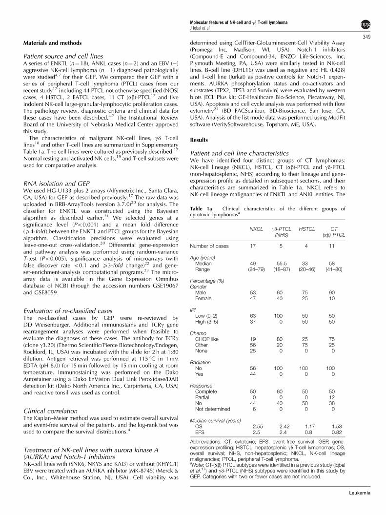
Molecular classifier for ENKTL and the identification ofa group of gd-PTCLUnsupervised hierarchical clustering showed that ENKTL andANKL cases formed a distinct cluster from the other PTCLentities, with a few interspersed PTCL-NOS cases. The majorityof the ENKTL cases showed a very uniform gene-expressionprofile and the molecular classifier consisted of 84 transcripts.There were similar number of up- and down-regulatedtranscripts (41 vs 43) (Figure 1) and most of the up-regulatedgenes were essentially contributed by NK cells, as they showedan expression pattern similar to normal NK cells (resting andIL-2 activated) and NK-cell lines (Figure 2c). These genesincluded killer cell immunoglobulin- or lectin-like receptor(KIR or KLR) family members, NK-cell-associated markers,CT molecules and a group of distinct chemokines primarilyexpressed by NK cells or CT T cells. Some of the transcripts werealso noted in normal CD8þT cells (resting and IL12 activated).TCRd mRNA, but not CD16 (FCGR3A) was represented inthe classifier. Certain up-regulated transcripts, encoding KRT19(mainly expressed in endothelial cells) and VSIG4 (macro-phages) were absent from the NK-cell lines and normal NKcells, implying their derivation from stromal cells (Table 2).The down-regulated genes were largely associated with T-cellbiology and stromal components including T-cell markers (CD3subunits) and genes involved in T-cell differentiation, activationand chemotaxis, transcripts from B cells and macrophages/dendritic cells.
Table 1b NKCL: immunophenotype, TCR rearrangement and EBVstatus
Immune markers Status
Cytoplasmic CD3e 18/19 (+)CD56 16/16 (+)TIA1 13/14 (+)Granzyme B 4/4 (+)EBER-1 18/19 (+)a
CD2 6/7 (+)Surface CD3e (leu4) 6/6 (�)TCRg rearrangement 7/7 (�)TCRb 5/5 (�)CD5 18/18 (�)CD8 8/10 (�)CD4 13/13 (�)
Abbreviations: EBV, Epstein–Barr virus; NKCL, NK-cell lineagemalignancies; TCR, T-cell receptor.aThe pathological characteristics of an EBV-negative NK-cell lympho-ma case has been reported previously by Martin et al.25
84 t
ran
scri
pts
*
NKCL Other PTCLs
NKCLPTCL
Bay
esia
nP
rob
abili
ty 0.9
0.1
* Transcripts are listed in Table1ANKL cases
NKCL refers only to lymphomas derived from the NK-cell lineageEBV(–) NK-cell lymphoma
0.25 1 4
relative level of expression
��
Figure 1 Gene-expression-based molecular predictors for ENKTL. The probability that a case is classified as ENKTL vs PTCL is shown at the top.A small subset of PTCL-NOS cases was re-classified as ENKTL, which, on further examination, showed gd T-cell differentiation. Each columnrepresents a case and each row represents the expression level of a gene. Gene-expression levels are depicted according to the color scale shown.
Molecular features of NK-cell and cd T-cell lymphomaJ Iqbal et al
350
Leukemia
The molecular classifier was initially constructed using allpathologically diagnosed ENKTL cases and later evaluated inPTCL cases as well as two ANKL, one EBV(�) NK-celllymphoma case and five indolent NKLGL. The accuracy of theclassifier was evaluated by leave-one-out cross validation(LOOCV) and was robust in re-classifying 15 of 18 pathologicaldiagnosed ENKTL. The two ANKL cases, the EBV(�) NK-celllymphoma and NK-cell lines were included by the ENKTLclassifier. However, 3 of 3 gd T-cell lines derived from ENKTL,18
1 EATCL and 4 (5 biopsies) of 44 PTCL-NOS cases were alsoclassified as ENKTL with 490% probability. A small number ofPTCL cases (n¼ 5) showed greater similarity (460–85%probability) to ENKTL, when evaluated with the classifier andincluded four HSTCL (72–85% probability) and one CT (ab)PTCL (B60% probability). These cases did not show anyassociation with EBV, and when evaluated with significanceanalysis of microarrays analysis, showed significant gene-expression differences with the cases classified as ENKTL with490% probability (see below). The three molecularly unclassi-fied cases of ENKTL had low expression of NK-cell markers(CD56 and KIR molecules), high expression of endothelial cell-related genes and did not express other PTCL subtype signatures,suggesting that they had a low number of neoplastic cells.
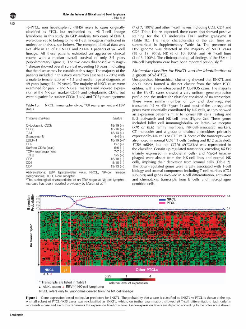
The four PTCL-NOS cases and one EATCL predicted to beENKTL with 490% probability were further examined. Inter-estingly, two transcripts (CD3g and CD3d) were expressedby the re-classified EATCL, PTCL-NOS and two ENKTL cases.Differential gene-expression analysis of the latter group of casesagainst the remaining cases showed significantly (Po0.005)higher expression of transcripts encoding the TCR complexincluding CD3g, CD3d, TCRgC2, TCRVg9 and TARP (410-fold),the activation and differentiation molecules (CD69 and LAT2)consistent with T-cell lineage differentiation (Figure 2a; Supple-mentary Table 2). There was high expression of TCRgV9 andVd2 that correlates with the high incidence of Vg9 and Vd2usage reported previously in nasal T-cell lymphoma and chronicEBV infection.26 We also observed higher expression ofIKZF2 (P¼ 0.0001), a T-cell-restricted IKAROS family mem-ber,27 and T-cell adaptor protein SH2B3 (LNK, P¼ 0.008)28
and a repertoire of transcripts encoding KIR molecules(Supplementary Table 2). The analysis indicated that this groupof cases was T-cell lymphomas expressing the gd TCR. Thetwo ENKTL cases of gd T-cell lineage were positive for EBER,and showed a very similar GEP to three gd T-cell lymphomalines,18 as well as the remaining group of 15 cases derivedfrom NK-cell lineage, thus clearly showing the close relation-ship among the ENKTL cases not only morphologically andclinically, but also in gene expression profiles despite theirseparate lineage derivation. The ENKTL cases of gd T-celllineage were also remarkably similar by GEP to the other fivere-classified cases (4 PTCL-NOS, 1 EATCL) that are designatedas gd-PTCL, (NHS) (see below).
Gene-set-enrichment-analysis identified gene signaturesassociated with IL12 signaling, proliferation, and chromosome6q arm in gd T-cell lineage lymphomas compared with NKCLtumors. The analysis of overall survival showed no significantdifference between these tumors similar to a previous report.29
We performed hierarchical clustering of the ENKTL/ANKL, there-classified gd-PTCL (NHS), CT (ab)-PTCL17 and four HTSCL casesusing the ENKTL classifier. The ENKTL molecular classifierclustered the re-classified gd-PTCL with ENKTL cases in onecluster as they showed very similar expression profile (Figure 2band c), whereas the CT (ab)-PTCL and HSTCL clustered separately.
The re-classified gd-PTCL (NHS) from PTCL-NOS casesshowed extranodal involvement including skin (n¼ 2), lung
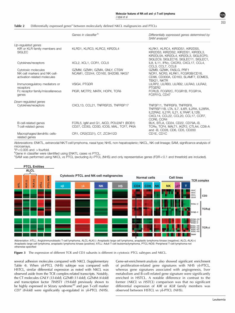
(n¼ 1) or neck LN (n¼ 1) and with a CD3þ and TCRb (bF1)negative immunohistochemical profile, when examined, con-sistent with gd T-cell lineage (Supplementary Table 3). CD56protein expression was not consistent (1 of 2 cases positive), andtwo of the four cases showed CD56 mRNA expression at asimilar range as in NK-cell malignancies. Two of the four caseswere double negative for CD4 and CD8, and one case wasdouble positive. Interestingly, 3 of 3 cases were negative for EBVby EBER in situ hybridization. A representative re-classified caseis shown to be positive for TCRg by immunohistochemistry(Figure 2d). The gd-PTCL (NHS) originally classified as EATCL
HS
TC
L
NKCL/��-PTCL(NHS)/ ��-ENKTL
NK- / �� T-cell lines
CT(��)-PTCL T-celllines
b
a
(NKCL refers only to lymphomas of NK-cell lineage including ENKTL and,ANKL and γδ-T represent two cases of ENKTL with γδ T-cell lineage)
-PDE4DIP-CADM1-GRPR-TRPM3-C20orf186-LOC729173-238410_x_at-1560495_at-237203_at-1559722_at-237849_at-VAPA-LOC441383-242710_at-VAPA-244447_at-ZNF24-TAF9B-GOSR2
-IKZF2-LAT2-PMS2L3-PMS2L11-PMS2L1-UPS44-SH2B3
Rep
rese
nta
tive
tra
nsc
rip
ts
-CD7
-TRGC2-TARP-TRGV9-CD3D-CD3G-FOXP2-CD69
-KIR2DL1-KIR2DS5-KIR2DL3-KIR2DL2-KIR2DL5A-KIR2DS1-KIR2DS2-KIR2DS3-KIR3DL1-KIR3DL2-KIR3DL37
-233364_s_at-232113_at-239841_at-234188_at-231057_at-241019_at-TSPAN16-SLC2A12-GTF3C3-ELAVL3-C21orf116
Molecularly defined
Median expressionγδ-PTCL(NHS)
��-PTCL(NHS) NKCL
NKCL��-E
NK
TL
Figure 2 (a) Differentially expressed genes between gd-PTCL andNKCL. A unique subgroup within PTCL-NOS with features of gdT cells (gd-PTCL) was identified to have a very similar GEP to ENKTLwith only a small subset showing differential expression (Po0.005,448 transcripts). Two cases of ENKTL with gd T-cell phenotype arealso evaluated with this set of differential gene-expression signature.(b) Hierarchical clustering according to the ENKTL classifier showeda distinct cluster of CT (ab)-PTCL, HSTCL and ENKTL with gd-PTCLinterspersed among the ENKTL cases. (c) Expression of ENKTLclassifier genes in different cytotoxic PTCL entities, cell lines andnormal cells. List of the genes on right indicate that they aredifferentially expressed in cytotoxic entities. (d) A representativegd-PTCL case identified by GEP immunostained for TCRg (clone g3.20,Thermo Fisher Scientific Inc., Rockford, IL, USA) (original magnifica-tion � 20).
Molecular features of NK-cell and cd T-cell lymphomaJ Iqbal et al
351
Leukemia
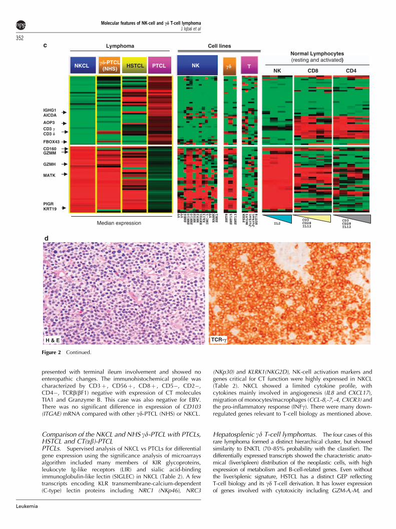
presented with terminal ileum involvement and showed noenteropathic changes. The immunohistochemical profile wascharacterized by CD3þ , CD56þ , CD8þ , CD5�, CD2�,CD4�, TCRb(bF1) negative with expression of CT moleculesTIA1 and Granzyme B. This case was also negative for EBV.There was no significant difference in expression of CD103(ITGAE) mRNA compared with other gd-PTCL (NHS) or NKCL.
Comparison of the NKCL and NHS gd-PTCL with PTCLs,HSTCL and CT(ab)-PTCLPTCLs. Supervised analysis of NKCL vs PTCLs for differentialgene expression using the significance analysis of microarraysalgorithm included many members of KIR glycoproteins,leukocyte Ig-like receptors (LIR) and sialic acid-bindingimmunoglobulin-like lectin (SIGLEC) in NKCL (Table 2). A fewtranscripts encoding KLR transmembrane-calcium-dependent(C-type) lectin proteins including NRC1 (NKp46), NRC3
(NKp30) and KLRK1(NKG2D), NK-cell activation markers andgenes critical for CT function were highly expressed in NKCL(Table 2). NKCL showed a limited cytokine profile, withcytokines mainly involved in angiogenesis (IL8 and CXCL17),migration of monocytes/macrophages (CCL-8,-7,-4, CXCR3) andthe pro-inflammatory response (INFg). There were many down-regulated genes relevant to T-cell biology as mentioned above.
Hepatosplenic gd T-cell lymphomas. The four cases of thisrare lymphoma formed a distinct hierarchical cluster, but showedsimilarity to ENKTL (70–85% probability with the classifier). Thedifferentially expressed transcripts showed the characteristic anato-mical (liver/spleen) distribution of the neoplastic cells, with highexpression of metabolism and B-cell-related genes. Even withoutthe liver/splenic signature, HSTCL has a distinct GEP reflectingT-cell biology and its gd T-cell derivation. It has lower expressionof genes involved with cytotoxicity including GZM-A,-M, and
c
Median expression
CD3 �CD3 �
PIGRKRT19
CD160 GZMM
FBOX43
MATK
GZMH
IGHG1AICDA
AOP3
SNT8
SNT15
SNT13
PEER
MOLT3
Jurkat
Jurkat
HUT78
CD3CD28IL12
CD3CD28IL12
IL2
NKCL��-PTCL
(NHS) HSTCL PTCL NK �� T
Cell lines
NK CD8 CD4
Normal Lymphocytes(resting and activated)
YT
YT
SNK6
SNK10
SNK10
NKYS
NK92
KHYG1
KAI3
IMC-1
YT
HANK
SNK1
Lymphoma
d
TCR-�H & E
Figure 2 Continued.
Molecular features of NK-cell and cd T-cell lymphomaJ Iqbal et al
352
Leukemia
several adhesion molecules compared with NKCL (SupplementaryTable 4). When gd-PTCL (NHS) subtype was compared withHSTCL, similar differential expression as noted with NKCL wasobserved aside from the TCR complex-related transcripts. Notably,the CT molecules GNLY (33-fold), GZMB (11-fold), GZMM (4-fold)and transcription factor TWIST1 (19-fold) previously shown tobe highly expressed in Sezary syndrome30 and pan T-cell markerCD7 (8-fold) were significantly up-regulated in gd-PTCL (NHS).
Gene-set-enrichment-analysis also showed significant enrichmentof proliferation-related gene signatures with NHS gd-PTCL,whereas gene signatures associated with angiogenesis, livermetabolism and B-cell-related gene signature were significantlyenriched in HSTCL. A notable difference in contrast to theformer (NKCL vs HSTCL) comparison was that no significantdifferential expression of KIR or KLR family members wasobserved between HSTCL vs gd-PTCL (NHS).
Table 2 Differentially expressed genesa between molecularly defined NKCL malignancies and PTCLs
Genes in classifier b Differentially expressed genes determined bySAM analysisc
Up-regulated genesKIR or KLR family members andSIGLEC
KLRD1, KLRC3, KLRC2, KIR2DL4 KLRK1, KLRC4, KIR3DS1, KIR2DS5,KIR2DS3, KIR2DS2, KIR2DS1, KIR3DL3,KIR2DL5A, KIR2DL4, KIR2DL3, SIGLECP3,SIGLEC9, SIGLEC16, SIGLEC11, SIGLEC1,
Cytokines/receptors XCL2, XCL1, CCR1, CCL5 IL8, IL11, IFNg, CXCR3, CXCL17, CCL4,CCL3, CCL7, CCL8
Cytotoxic molecules GZMM, GZMH, GZMA, GNLY, CTSW GZMB, GZMK, FASLG, PRF1NK-cell markers and NK-cell-activation-related molecules
NCAM1, CD244, CD160, SH2DIB, NKG7 NCR1, NCR3, KLRK1, FCGR3B/CD16,CD96, CD300A, CD163, SLAMF7, EOMES,TBX21, NKTR
Immunoregulatory mediators orreceptors
VSIG4, PTGDR LILRP2, LILRB3, LILRB2, LILRA3, LILRA2,PTGER2
Fc receptor family/miscellaneousgenes
PIGR, MCTP2, MATK, HOPX, TCRd FCRLB, FCGR2C, FCGR1B, FCGR1A,FCER1G, CD47
Down-regulated genesCytokines/receptors CXCL13, CCL21, TNFRSF25, TNFRSF17 TNFSF11, TNFRSF9, TNFRSF8,
TNFRSF11B, LTA, IL7, IL6R, IL2RA, IL28RA,IL22RA2, IL21R, IL21, IL1RAP, IL12B,CXCL14, CCL22, CCL20, CCL17, CCR7,CCR6, CCR4
B-cell-related genes FCRL5, IgM and G1, AICD, POU2AF1 (BOB1) BLK, BTLA, CD24, CD22, CD79A,-B,T-cell-related genes CD27, CD3G, CD3D, ICOS, MAL, TCF7, PKIA TCRa, TCF4, MALT1, IKZF2, CTLA4, CD8-A
and -B, CD28, CD6, CD5, CD200Macrophages/dendritic cells-related genes
CR1, CR2(CD21), C7, ZC3H12D CD1E, CD1C
Abbreviations: ENKTL, extranodal NK/T-cell lymphoma, nasal type; NHS, non-hepatosplenic; NKCL, NK-cell lineage; SAM, significance analysis ofmicroarrays.aPo0.005 and 4fourfold.bGene in classifier were identified using ENKTL cases vs PTCL.cSAM was performed using NKCL vs PTCL (excluding dg-PTCL (NHS) and only representative genes (FDRo0.1 and threefold) are included).
Abbreviation :ATLL: Angioimmunoblastic T-cell lymphoma, ALCL-ALK(-) :Anaplastic large cell lymphoma, anaplastic lymphoma kinase (negative); ALCL-ALK(+)Anaplastic large cell lymphoma, anaplastic lymphoma kinase (positive); ATLL: Adult T-cell leukemia/lymphoma, PTCL-NOS: Peripheral T-cell lymphoma-nototherwise specified
�� �� NK HSAIT
L
AL
K (
-)
AL
K (
+)
AT
LL
PT
CL
-NO
S
CD4 CD8 NK NK ��T T
PTCL Entities
Cytotoxic PTCL and NK-cell malignanciesNormal cells Cell lines
ALCL
TCR complex
CD3�CD3�CD3�CD3�
TCR�-C
TCR�-V2TCR�-CTCR�-C
TCR�-C
TCR�-V9TCR�-C2TCR�-V7TCR�-V5
TARP
CD3
TCR��
TCR�� TCR��
Figure 3 The expression of different TCR and CD3 subunits is different in cytotoxic PTCL subtypes and NKCL.
Molecular features of NK-cell and cd T-cell lymphomaJ Iqbal et al
353
Leukemia
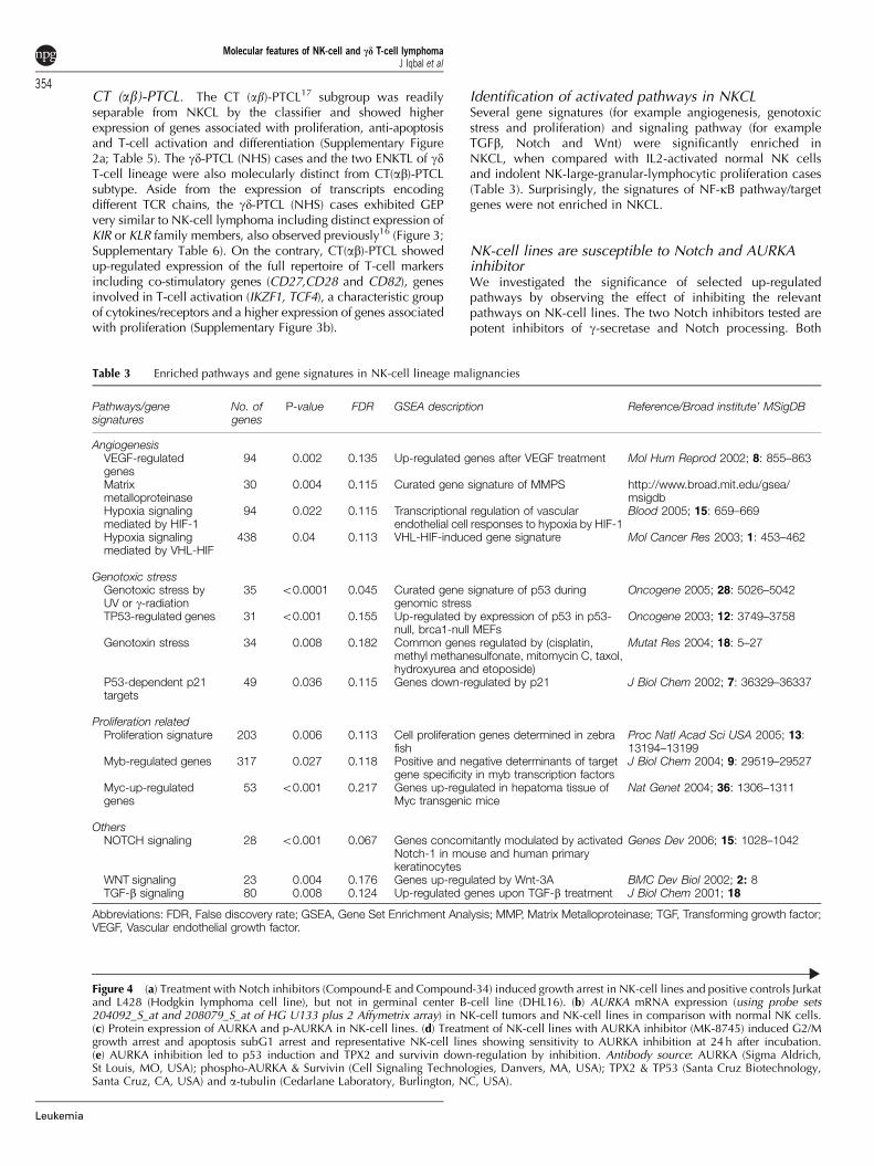
CT (ab)-PTCL. The CT (ab)-PTCL17 subgroup was readilyseparable from NKCL by the classifier and showed higherexpression of genes associated with proliferation, anti-apoptosisand T-cell activation and differentiation (Supplementary Figure2a; Table 5). The gd-PTCL (NHS) cases and the two ENKTL of gdT-cell lineage were also molecularly distinct from CT(ab)-PTCLsubtype. Aside from the expression of transcripts encodingdifferent TCR chains, the gd-PTCL (NHS) cases exhibited GEPvery similar to NK-cell lymphoma including distinct expression ofKIR or KLR family members, also observed previously16 (Figure 3;Supplementary Table 6). On the contrary, CT(ab)-PTCL showedup-regulated expression of the full repertoire of T-cell markersincluding co-stimulatory genes (CD27,CD28 and CD82), genesinvolved in T-cell activation (IKZF1, TCF4), a characteristic groupof cytokines/receptors and a higher expression of genes associatedwith proliferation (Supplementary Figure 3b).
Identification of activated pathways in NKCLSeveral gene signatures (for example angiogenesis, genotoxicstress and proliferation) and signaling pathway (for exampleTGFb, Notch and Wnt) were significantly enriched inNKCL, when compared with IL2-activated normal NK cellsand indolent NK-large-granular-lymphocytic proliferation cases(Table 3). Surprisingly, the signatures of NF-kB pathway/targetgenes were not enriched in NKCL.
NK-cell lines are susceptible to Notch and AURKAinhibitorWe investigated the significance of selected up-regulatedpathways by observing the effect of inhibiting the relevantpathways on NK-cell lines. The two Notch inhibitors tested arepotent inhibitors of g-secretase and Notch processing. Both
Table 3 Enriched pathways and gene signatures in NK-cell lineage malignancies
Pathways/genesignatures
No. ofgenes
P-value FDR GSEA description Reference/Broad institute’ MSigDB
AngiogenesisVEGF-regulatedgenes
94 0.002 0.135 Up-regulated genes after VEGF treatment Mol Hum Reprod 2002; 8: 855–863
Matrixmetalloproteinase
30 0.004 0.115 Curated gene signature of MMPS http://www.broad.mit.edu/gsea/msigdb
Hypoxia signalingmediated by HIF-1
94 0.022 0.115 Transcriptional regulation of vascularendothelial cell responses to hypoxia by HIF-1
Blood 2005; 15: 659–669
Hypoxia signalingmediated by VHL-HIF
438 0.04 0.113 VHL-HIF-induced gene signature Mol Cancer Res 2003; 1: 453–462
Genotoxic stressGenotoxic stress byUV or g-radiation
35 o0.0001 0.045 Curated gene signature of p53 duringgenomic stress
Oncogene 2005; 28: 5026–5042
TP53-regulated genes 31 o0.001 0.155 Up-regulated by expression of p53 in p53-null, brca1-null MEFs
Oncogene 2003; 12: 3749–3758
Genotoxin stress 34 0.008 0.182 Common genes regulated by (cisplatin,methyl methanesulfonate, mitomycin C, taxol,hydroxyurea and etoposide)
Mutat Res 2004; 18: 5–27
P53-dependent p21targets
49 0.036 0.115 Genes down-regulated by p21 J Biol Chem 2002; 7: 36329–36337
Proliferation relatedProliferation signature 203 0.006 0.113 Cell proliferation genes determined in zebra
fishProc Natl Acad Sci USA 2005; 13:13194–13199
Myb-regulated genes 317 0.027 0.118 Positive and negative determinants of targetgene specificity in myb transcription factors
J Biol Chem 2004; 9: 29519–29527
Myc-up-regulatedgenes
53 o0.001 0.217 Genes up-regulated in hepatoma tissue ofMyc transgenic mice
Nat Genet 2004; 36: 1306–1311
OthersNOTCH signaling 28 o0.001 0.067 Genes concomitantly modulated by activated
Notch-1 in mouse and human primarykeratinocytes
Genes Dev 2006; 15: 1028–1042
WNT signaling 23 0.004 0.176 Genes up-regulated by Wnt-3A BMC Dev Biol 2002; 2: 8TGF-b signaling 80 0.008 0.124 Up-regulated genes upon TGF-b treatment J Biol Chem 2001; 18
Abbreviations: FDR, False discovery rate; GSEA, Gene Set Enrichment Analysis; MMP, Matrix Metalloproteinase; TGF, Transforming growth factor;VEGF, Vascular endothelial growth factor.
Figure 4 (a) Treatment with Notch inhibitors (Compound-E and Compound-34) induced growth arrest in NK-cell lines and positive controls Jurkatand L428 (Hodgkin lymphoma cell line), but not in germinal center B-cell line (DHL16). (b) AURKA mRNA expression (using probe sets204092_S_at and 208079_S_at of HG U133 plus 2 Affymetrix array) in NK-cell tumors and NK-cell lines in comparison with normal NK cells.(c) Protein expression of AURKA and p-AURKA in NK-cell lines. (d) Treatment of NK-cell lines with AURKA inhibitor (MK-8745) induced G2/Mgrowth arrest and apoptosis subG1 arrest and representative NK-cell lines showing sensitivity to AURKA inhibition at 24 h after incubation.(e) AURKA inhibition led to p53 induction and TPX2 and survivin down-regulation by inhibition. Antibody source: AURKA (Sigma Aldrich,St Louis, MO, USA); phospho-AURKA & Survivin (Cell Signaling Technologies, Danvers, MA, USA); TPX2 & TP53 (Santa Cruz Biotechnology,Santa Cruz, CA, USA) and a-tubulin (Cedarlane Laboratory, Burlington, NC, USA).
Molecular features of NK-cell and cd T-cell lymphomaJ Iqbal et al
354
Leukemia
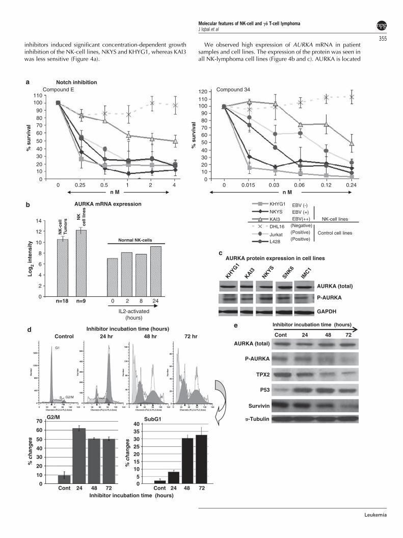
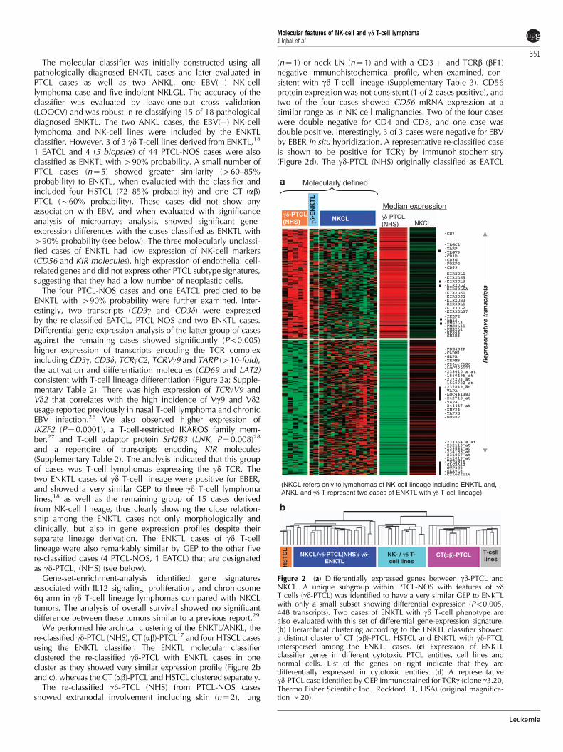
inhibitors induced significant concentration-dependent growthinhibition of the NK-cell lines, NKYS and KHYG1, whereas KAI3was less sensitive (Figure 4a).
We observed high expression of AURKA mRNA in patientsamples and cell lines. The expression of the protein was seen inall NK-lymphoma cell lines (Figure 4b and c). AURKA is located
Notch inhibitiona
b
c
Compound E
% s
urv
ival
n M
Compound 34
% s
urv
ival
n M
EBV (-)
EBV (+)
NK-cell lines
Control cell lines
(Negative)
(Positive)
(Positive)
EBV(++)
110100
90807060
50
4030
2010
0
0
2
4
6
8
10
12
14
110120
1009080706050
40302010
00 0.25 0.5 1 2 4 0 0.015 0.03 0.06 0.12 0.24
KHYG1
NKYS
KAI3
DHL16
L428
Jurkat
AURKA protein expression in cell lines
GAPDH
P-AURKA
AURKA (total)
AURKA mRNA expression
NK
cell
lines
NK
-cel
lT
um
ors
Normal NK-cells
Lo
g2
inte
nsi
ty
0 2 8 24n=18 n=9
IL2-activated(hours)
KHYG1
KAI3
NKYS
SNK6
IMC1
deInhibitor incubation time (hours)
Inhibitor incubation time (hours)Cont 24 48 72Cont 24
70
60
50
40
30
20
10
0 05
10152025303540
48 72
G2/M SubG1
Inhibitor incubation time (hours)
AURKA (total)
P-AURKA
TPX2
P53
Survivin
�-Tubulin
Cont 24 48 72Control 24 hr 48 hr 72 hr
% c
han
ges
% c
han
ges
Molecular features of NK-cell and cd T-cell lymphomaJ Iqbal et al
355
Leukemia
at a frequently (450%) amplified locus (20q13) in lymphomasderived from NKCL31 and is involved in multiple pathways,promoting the proliferative function of MYC32 and WNTsignaling,33 while inhibiting TP53.34 Moreover, AURKA is up-regulated by hypoxia35 that is frequently observed in ENKTL.Many of these signatures were significantly enriched in ENKTL,for example Myc-induced signature, and WNT signaling andgenotoxic pathway (Table 3). The active phosphorylated form(p-AURKA) and its substrates (TPX2, TACC3 and EG5) could beshown. We, therefore, tested the NK-cell lines with a novelsmall-molecule AURKA inhibitor (MK-8745) and all cell linesshowed a significant increase in apoptosis and cell cycle arrest(Figure 4d; Supplementary Figure 3a). This inhibition led todecrease in endogenous p-AURKA and its substrate TPX2, withoutaffecting the level of total AURKA. The downstream target ofAURKA, TP53, was significantly induced and there was a gradualdecrease in survivin (target of TP53) level with time (Figure 4e).
Discussion
The clinicopathological features of ENKTL cases included in thisstudy are consistent with previous studies.7,36 The immuno-phenotype of tumor cells showed expression of NK-cell markersincluding CD56, cytoplasmic CD3e, CT molecules (TIA1 andgranzyme B) and presence of EBV in the majority of the cases,consistent with observations that NK-cell is indeed the cell oforigin in the majority of ENKTL cases. In keeping with thisobservation, our molecular classifier included most of the up-regulated genes that coded for the distinctive phenotypic andfunctional characteristics associated with NK cells and showedhigh specificity in distinguishing NK-cell malignancies includingANKL from most PTCL cases. However, we also identified twocases of gd T-cell lineage in ENKTL, which can be differentiatedfrom their NK-cell counterpart by the high expression of CD3g,CD3d and TCRg-associated transcripts. Despite the difference incell lineage, these ENKTL cases share very similar morphologi-cal and clinical characteristics and as shown in this study, alsoshare a very similar molecular profile. NK-cell neoplasms arecharacterized by the presence of EBV in tumor cells; however,tumor cases/cell lines without EBV have been reported.25,37–40
In our series, EBV status has no impact on GEP either in tumorspecimen or in cell lines.
Our molecular classifier not only identified gd T-cell cases ofENKTL, but also a subset of gd-PTCL (NHS) in other extranodalareas that showed a strikingly similar GEP to NK cells, NK-celllines and to gd T-cell lines derived from ENKTL.18 The gene-set-enrichment-analysis findings of chromosome 6q gene enrich-ment in gd-PTCL (NHS) compared with NKCL is likely related tothe frequent deletion of 6q in the NK-cell counterpart.31 Thisdeletion is not observed in the gd T-cell lines,41 and may besimilarly true for gd-PTCLs (NHS) as well. Gene signaturesassociated with IL12 signaling, shown to be critical for gd T-cellproliferation by resisting apoptosis,42 were up-regulated in gd-PTCL(NHS) compared with NKCL. The immunohistochemical stainingand pathology review provided further evidence for gd T-celllineage in these cases. The gd-PTCL (NHS) case presenting in theskin in our study resemble the primary cutaneous gd T-celllymphoma as defined in the current WHO.3 It would be of interestto study more cases of primary cutaneous gd T-cell lymphomas todetermine whether they also share the GEP of the gd-PTCL (NHS)cases we have defined in this study.
The term mucocutaneous or NHS gd-PTCL mentioned in theWHO-EORTC classification scheme12 would encompass thecases of gd-PTCL described here, although the heterogeneity
within this group of disorders has not been defined. Normalgd T cells are enriched in mucosal surfaces43 and exhibit manyof the characteristics of the innate immune system similar toNK cells.44 There are subsets of gd T cells that differ in theirontogeny as reflected by their time of appearance in the thymus,TCR gene usage, TCR combinatorial complexity and theirlocations and functions in the body. There is also correspondingpreferential gd gene usage in gd-PTCL at different sites such asVd1 in HSTCL9,45, Vd2 in primary cutaneous gd-PTCL46 andVg9Vd2 in ENKTL and gd-PTCL (NHS).26,29 Consistent withthese findings, the re-classified gd-PTCLs in this study exhibitedextranodal disease and expressed Vg9, Vd2 mRNA at highlevels. Different organ sites may be populated preferentially atdifferent stages of development by gd T-cell populations andthere is likely to be further heterogeneity in the mucocutaneousgd-PTCL. Therefore, a detailed study of a large series of gd T-celllymphomas from different sites is needed to decipher themolecular relationship among these gd T-cell lymphomas.
The HSTCL appears to be distinct clinically, pathologicallyand by GEP from NKCL and gd-PTCL of mucocutaneoussites presented in this study as also suggested by a previousobservation.47 Even after excluding the signatures contributedby the liver and spleen, or the differences in TCR usage, NKCLand gd-PTCL (NHS) exhibit distinct GEP, especially the CTprofile with HSTCL expressing TIA-1, but not PRF, GZMA andGZMM. These observations suggest diseases derived from gdT cells in hepatosplenic and NHS sites are distinct.16 It ispossible that the gd-PTCL (NHS) observed in this study arederived from a unique subset of gd T cells with features verysimilar to NK cells compared with other gd-PTCL. It is alsoprobable that the clinicopathological and GEP characteristics ofthese gd-PTCL are influenced by the microenvironment in whichthey are derived. These hypotheses need to be further tested bystudying the TCR usage/sequence and the GEP of diversesubtypes of gd-PTCL.
The NKCL shows a distinct GEP compared with CT(ab)-PTCLdefined previously.17 The re-classified gd-PTCL (NHS) andHSTCL are also distinct from CT(ab)-PTCL. The latter entityexhibits high levels of TCRa and b transcripts, but not TCRg andd transcripts, and shows more functional characteristic of CD8þ
T cells. These cases tend to be more proliferative and showlower expression of CD56, NCR1 (NKP46) and TCRd mRNA,but high expression of TCR signaling molecules. Clinically, allthese CT subtypes of PTCL are highly aggressive and areassociated with a short survival.
As patients with NK-cell malignancies have a poor outcomewith current therapies,4 novel approaches are needed toimprove survival. Comparison of NKCL with PTCL-NOS yieldedmostly pathways that are differentially expressed betweenNK and T cells and provided little insight into the biology ofNKCL. Therefore, we compared NKCL with IL2-activatedNK cells and NK-large-granular-lymphocytic proliferationcases. The enrichment of TGFb and other immunosuppressivepathways indicated an immunosuppressive microenvironmentin NKCL that favors the survival of EBV infected/transformed NKcells. The significant enrichment of angiogenesis pathways inNKCL may be due the vascular destruction in these tumorsresulting in hypoxia and activation of HIF1a. The genotoxicstress responsive gene signature indicated the presence ofconditions that may activate TP53 function. TP53 is negativelyregulated by MDM2, an E3 ubiquitin ligase and positively by anubiquitin-specific protease-7 (USP7).48 EBV nuclear-antigen 1can stably bind to USP7 and promote TP53 degradation byMDM2.49 Thus, targeting MDM2 function may provide atherapeutic option in ENTKL.
Molecular features of NK-cell and cd T-cell lymphomaJ Iqbal et al
356
Leukemia
We also observed activation of WNT and NOTCH-1 path-ways that was also described by Huang et al15 in a seriesof seven ENKTL cases. We tested two g-secretase inhibitors inNK-cell lines, and observed growth and survival inhibition in thetested NK-cell lines validating the significance of the NOTCHpathway in neoplastic cells. We also tested an AURKA inhibitoras the phosphorylated AURKA (activated form) was present inall NK-cell lines and aside from its role in centrosome regulationand mitotic spindle formation,50 AURKA can influence multiplesignaling pathways that promote oncogenesis. Previous reportshave shown that AURKA can phosphorylate TP53 at serine-215resulting in its inactivation and degradation.34 It also up-regulates MYC and telomerase activity32 and promotes WNTsignaling.33 Some of these signatures were significantly enrichedin NKCL (Table 3). AURKA is also located at a frequentlyamplified locus (20q13) in NK-cell lymphoma.31 The AURKAinhibitor MK-874551 induced cell cycle arrest and apoptosis inall NK-cell lines tested. There was a decrease in phosphorylatedAURKA level with a concomitant decrease in its regulatorTPX2.52 AURKA inactivation was associated with increasedTP53 level and decreased Survivin (TP53 target) level. Survivinis not only an anti-apoptotic factor, it is also essential for properchromosome segregation and cytokinesis, thus augmentingAURKA.53 These molecular events converge to induce cellcycle arrest and increased apoptosis. These findings suggest thatMK-8745 could be a novel therapeutic agent for NK-cell-derived lymphomas.
In conclusion our molecular classifier identified lymphomasoriginating from NK cells as well as a subset of gd T-cell with avery similar gene-expression profile to NK cells. These gd PTCLshave distinct GEP compared with CT(ab)-PTCLs and HSTCL andmay be derived from an ontogenically and functionally distinctsubset of gd T cells. The Gene set enrichment analysis indicatedan immunosuppressive microenvironment, genotoxic stress,angiogenesis and activation of NOTCH, and WNT signaling inENKTL. The Notch inhibitors could inhibit growth of NK-celllines, but the AURKA inhibitor was more effective probablybecause it affected multiple pathways. Pathway analysisand testing can rationally identify promising candidates fortherapeutic trials in ENKTL.
Conflict of interest
The authors declare no conflict of interest.
Acknowledgements
We thank Martin Bast for clinical data collection, and Kavita Pateland Lisa Bough for their technical assistance. This work wassupported in part by NCI Grant 5U01/CA114778, LymphomaSPORE P50CA136411-01(NC1) and funds from the InternationalPeripheral T-cell Lymphoma Project and Eppley Cancer InstituteCore Grant CA36727. The UNMC Microarray Core Facility issupported partially by NIH Grant P20 RR016469 from the INBREProgram of the National Center for Research Resources.
References
1 Jaffe ES. Classification of natural killer (NK) cell and NK-like T-cellmalignancies. Blood 1996; 87: 1207–1210.
2 Oshimi K. Leukemia and lymphoma of natural killer lineage cells.Int J Hematol 2003; 78: 18–23.
3 Swerdlow SH, Campo E, Harris NL, Jaffe ES, Pileri SA, Stein H et al.WHO Classification: Pathology and Genetics of Tumors of
Haematopoietic and Lymphoid Tissues, 4th edn (Vol 2) IARCPress: Lyon, France, 2008.
4 Vose J, Armitage J, Weisenburger D. International T-Cell Lympho-ma Project. International peripheral T-cell and natural killer/t-celllymphoma study: pathology findings and clinical outcomes. J ClinOncol 2008; 26: 4124–4130.
5 Sokol L, Loughran Jr TP. Large granular lymphocyte leukemia.Oncologist 2006; 11: 263–273.
6 Li YX, Yao B, Jin J, Wang WH, Liu YP, Song YW et al. Radiotherapyas primary treatment for stage IE and IIE nasal natural killer/T-celllymphoma. J Clin Oncol 2006; 24: 181–189.
7 Au WY, Weisenburger DD, Intragumtornchai T, Nakamura S, KimWS, Sng I et al. Clinical differences between nasal and extranasalnatural killer/T-cell lymphoma: a study of 136 cases from theInternational Peripheral T-Cell Lymphoma Project. Blood 2009;113: 3931–3937.
8 Chiang AK, Chan AC, Srivastava G, Ho FC. Nasal T/natural killer(NK)-cell lymphomas are derived from Epstein-Barr virus-infectedcytotoxic lymphocytes of both NK- and T-cell lineage. Int J Cancer1997; 73: 332–338.
9 Gaulard P, Bourquelot P, Kanavaros P, Haioun C, Le Couedic JP,Divine M et al. Expression of the alpha/beta and gamma/deltaT-cell receptors in 57 cases of peripheral T-cell lymphomas.Identification of a subset of gamma/delta T-cell lymphomas.Am J Pathol 1990; 137: 617–628.
10 Stein H, Dienemann D, Sperling M, Zeitz M, Riecken EO.Identification of a T cell lymphoma category derived fromintestinal-mucosa-associated T cells. Lancet 1988; 2: 1053–1054.
11 Kumar S, Krenacs L, Medeiros J, Elenitoba-Johnson KS, Greiner TC,Sorbara L et al. Subcutaneous panniculitic T-cell lymphoma isa tumor of cytotoxic T lymphocytes. Human Pathol 1998; 29:397–403.
12 Willemze R, Jaffe ES, Burg G, Cerroni L, Berti E, Swerdlow SHet al. WHO-EORTC classification for cutaneous lymphomas.Blood 2005; 105: 3768–3785.
13 Nagato T, Kobayashi H, Kishibe K, Takahara M, Ogino T, Ishii Het al. Expression of interleukin-9 in nasal natural killer/T-celllymphoma cell lines and patients. Clin Cancer Res 2005; 11:8250–8257.
14 Oka T, Yoshino T, Hayashi K, Ohara N, Nakanishi T, Yamaai Yet al. Reduction of hematopoietic cell-specific tyrosine phospha-tase SHP-1 gene expression in natural killer cell lymphoma andvarious types of lymphomas/leukemias: combination analysis withcDNA expression array and tissue microarray. Am J Pathol 2001;159: 1495–1505.
15 Huang Y, de Reynies A, de Leval L, Ghazi B, Martin-Garcia N,Travert M et al. Gene expression profiling identifies emergingoncogenic pathways operating in extranodal NK/T-cell lymphoma,nasal-type. Blood 2010; 115: 1226–1237.
16 Miyazaki K, Yamaguchi M, Imai H, Kobayashi T, Tamaru S, NishiiK et al. Gene expression profiling of peripheral T-cell lymphomaincluding gammadelta T-cell lymphoma. Blood 2009; 113:1071–1074.
17 Iqbal J, Weisenburger DD, Greiner TC, Vose JM, McKeithan T,Kucuk C et al. Molecular signatures to improve diagnosis inperipheral T-cell lymphoma and prognostication in angioimmuno-blastic T-cell lymphoma. Blood 2010; 115: 1026–1036.
18 Nagata H, Konno A, Kimura N, Zhang Y, Kimura M, Demachi Aet al. Characterization of novel natural killer (NK)-cell andgammadelta T-cell lines established from primary lesions of nasalT/NK-cell lymphomas associated with the Epstein-Barr virus. Blood2001; 97: 708–713.
19 Dybkaer K, Iqbal J, Zhou G, Geng H, Xiao L, Schmitz A et al.Genome wide transcriptional analysis of resting and IL2 acti-vated human natural killer cells: gene expression signaturesindicative of novel molecular signaling pathways. BMC Genomics2007; 8: 230.
20 Simon R, Peng A. BRB-ArrayTools User Guide, version 3.6.0.Biometric Research Branch, National Cancer Institute, NationalInstitute of Health (available at: http://linus.nci.nih.gov/BRB-ArrayTools.html).
21 Wright G, Tan B, Rosenwald A, Hurt EH, Wiestner A, Staudt LM. Agene expression-based method to diagnose clinically distinctsubgroups of diffuse large B cell lymphoma. Proc Natl Acad SciUSA 2003; 100: 9991–9996.
Molecular features of NK-cell and cd T-cell lymphomaJ Iqbal et al
357
Leukemia

22 Tusher VG, Tibshirani R, Chu G. Significance analysis ofmicroarrays applied to the ionizing radiation response. Proc NatlAcad Sci USA 2001; 98: 5116–5121.
23 Subramanian A, Tamayo P, Mootha VK, Mukherjee S, Ebert BL,Gillette MA et al. Gene set enrichment analysis: a knowledge-based approach for interpreting genome-wide expression profiles.Proc Natl Acad Sci USA 2005; 102: 15545–15550.
24 Ormerod MG. Investigating the relationship between the cell cycleand apoptosis using flow cytometry. J Immunol Methods 2002;265: 73–80.
25 Martin AR, Chan WC, Perry DA, Greiner TC, Weisenburger DD.Aggressive natural killer cell lymphoma of the small intestine. ModPathol 1995; 8: 467–472.
26 Oyoshi MK, Nagata H, Kimura N, Zhang Y, Demachi A, Hara Tet al. Preferential expansion of Vgamma9-JgammaP/Vdelta2-Jdelta3 gammadelta T cells in nasal T-cell lymphoma andchronic active Epstein-Barr virus infection. Am J Pathol 2003;162: 1629–1638.
27 Hahm K, Cobb BS, McCarty AS, Brown KE, Klug CA, Lee R et al.Helios, a T cell-restricted Ikaros family member that quantitativelyassociates with Ikaros at centromeric heterochromatin. Genes Dev1998; 12: 782–796.
28 Li Y, He X, Schembri-King J, Jakes S, Hayashi J. Cloning andcharacterization of human Lnk, an adaptor protein with pleckstrinhomology and Src homology 2 domains that can inhibit T cellactivation. J Immunol 2000; 164: 5199–5206.
29 Arnulf B, Copie-Bergman C, Delfau-Larue MH, Lavergne-Slove A,Bosq J, Wechsler J et al. Nonhepatosplenic gammadelta T-celllymphoma: a subset of cytotoxic lymphomas with mucosal or skinlocalization. Blood 1998; 91: 1723–1731.
30 van Doorn R, Dijkman R, Vermeer MH, Out-Luiting JJ, van derRaaij-Helmer EM, Willemze R et al. Aberrant expression of thetyrosine kinase receptor EphA4 and the transcription factor twist inSezary syndrome identified by gene expression analysis. CancerRes 2004; 64: 5578–5586.
31 Iqbal J, Kucuk C, Deleeuw RJ, Srivastava G, Tam W, Geng Het al. Genomic analyses reveal global functional alterationsthat promote tumor growth and novel tumor suppressor genesin natural killer-cell malignancies. Leukemia 2009; 23:1139–1151.
32 Yang H, Ou CC, Feldman RI, Nicosia SV, Kruk PA, Cheng JQ.Aurora-A kinase regulates telomerase activity through c-Myc inhuman ovarian and breast epithelial cells. Cancer Res 2004; 64:463–467.
33 Dutta-Simmons J, Zhang Y, Gorgun G, Gatt M, Mani M,Hideshima T et al. Aurora kinase A is a target of Wnt/beta-catenininvolved in multiple myeloma disease progression. Blood 2009;114: 2699–2708.
34 Liu Q, Kaneko S, Yang L, Feldman RI, Nicosia SV, Chen J et al.Aurora-A abrogation of p53 DNA binding and transactivationactivity by phosphorylation of serine 215. J Biol Chem 2004; 279:52175–52182.
35 Klein A, Flugel D, Kietzmann T. Transcriptional regulation ofserine/threonine kinase-15 (STK15) expression by hypoxia andHIF-1. Mol Biol Cell 2008; 19: 3667–3675.
36 Suzuki R. Leukemia and lymphoma of natural killer cells.J Clin Exp Hematopathol 2005; 45: 51–70.
37 Chan JK, Sin VC, Wong KF, Ng CS, Tsang WY, Chan CH et al.Nonnasal lymphoma expressing the natural killer cell markerCD56: a clinicopathologic study of 49 cases of an uncommonaggressive neoplasm. Blood 1997; 89: 4501–4513.
38 Matano S, Nakamura S, Nakamura S, Annen Y, Hattori N,Kobayashi K et al. Monomorphic agranular natural killer celllymphoma/leukemia with no Epstein-Barr virus association.Acta Haematol 1999; 101: 206–208.
39 Yagita M, Huang CL, Umehara H, Matsuo Y, Tabata R, Miyake Met al. A novel natural killer cell line (KHYG-1) from a patient withaggressive natural killer cell leukemia carrying a p53 pointmutation. Leukemia 2000; 14: 922–930.
40 Chen IM, Whalen M, Bankhurst A, Sever CE, Doshi R, HardekopfD et al. A new human natural killer leukemia cell line, IMC-1. Acomplex chromosomal rearrangement defined by spectral karyo-typing: functional and cytogenetic characterization. Leuk Res2004; 28: 275–284.
41 Zhang Y, Nagata H, Ikeuchi T, Mukai H, Oyoshi MK, Demachi Aet al. Common cytological and cytogenetic features of Epstein-Barrvirus (EBV)-positive natural killer (NK) cells and cell lines derivedfrom patients with nasal T/NK-cell lymphomas, chronic active EBVinfection and hydroa vacciniforme-like eruptions. Br J Haematol2003; 121: 805–814.
42 Lopez RD, Xu S, Guo B, Negrin RS, Waller EK. CD2-mediatedIL-12-dependent signals render human gamma delta-T cellsresistant to mitogen-induced apoptosis, permitting the large-scaleex vivo expansion of functionally distinct lymphocytes: implica-tions for the development of adoptive immunotherapy strategies.Blood 2000; 96: 3827–3837.
43 Deusch K, Luling F, Reich K, Classen M, Wagner H, Pfeffer K.A major fraction of human intraepithelial lymphocytes simulta-neously expresses the gamma/delta T cell receptor, the CD8accessory molecule and preferentially uses the V delta 1 genesegment. Eur J Immunol 1991; 21: 1053–1059.
44 Carding SR, Egan PJ. Gammadelta T cells: functional plasticity andheterogeneity. Nat Rev Immunol 2002; 2: 336–345.
45 Mastovich S, Ratech H, Ware RE, Moore JO, Borowitz MJ.Hepatosplenic T-cell lymphoma: an unusual case of a gammadelta T-cell lymphoma with a blast-like terminal transformation.Human Pathol 1994; 25: 102–108.
46 Przybylski GK, Wu H, Macon WR, Finan J, Leonard DG, Felgar REet al. Hepatosplenic and subcutaneous panniculitis-like gamma/delta T cell lymphomas are derived from different Vdelta subsets ofgamma/delta T lymphocytes. J Mol Diagn 2000; 2: 11–19.
47 Boulland ML, Kanavaros P, Wechsler J, Casiraghi O, Gaulard P.Cytotoxic protein expression in natural killer cell lymphomas andin alpha beta and gamma delta peripheral T-cell lymphomas.J Pathol 1997; 183: 432–439.
48 Li M, Chen D, Shiloh A, Luo J, Nikolaev AY, Qin J et al.Deubiquitination of p53 by HAUSP is an important pathway forp53 stabilization. Nature 2002; 416: 648–653.
49 Saridakis V, Sheng Y, Sarkari F, Holowaty MN, Shire K, Nguyen Tet al. Structure of the p53 binding domain of HAUSP/USP7 boundto Epstein-Barr nuclear antigen 1 implications for EBV-mediatedimmortalization. Mol Cell 2005; 18: 25–36.
50 Katayama H, Sasai K, Kawai H, Yuan ZM, Bondaruk J, Suzuki Fet al. Phosphorylation by aurora kinase A induces Mdm2-mediateddestabilization and inhibition of p53. Nat Genet 2004; 36: 55–62.
51 Bayliss R, Sardon T, Vernos I, Conti E. Structural basis of Aurora-Aactivation by TPX2 at the mitotic spindle. Mol Cell 2003; 12: 851–862.
52 Kufer TA, Sillje HH, Korner R, Gruss OJ, Meraldi P, Nigg EA.Human TPX2 is required for targeting Aurora-A kinase to thespindle. J Cell Biol 2002; 158: 617–623.
53 Lens SM, Vader G, Medema RH. The case for Survivin as mitoticregulator. Curr Opin Cell Biol 2006; 18: 616–622.
Supplementary Information accompanies the paper on the Leukemia website (http://www.nature.com/leu)
Molecular features of NK-cell and cd T-cell lymphomaJ Iqbal et al
358
Leukemia