myocardial t 2 mapping with respiratory navigator and automatic nonrigid motion correction
TRANSCRIPT
NOTE
Myocardial T2 Mapping With Respiratory Navigator andAutomatic Nonrigid Motion Correction
Shivraman Giri,1,2 Saurabh Shah,3 Hui Xue,4 Yiu-Cho Chung,5
Michael L. Pennell,6 Jens Guehring,4 Sven Zuehlsdorff,3 Subha V. Raman,2,7,8
and Orlando P. Simonetti1,2,7,8*
Quantitative T2 mapping was recently shown to be superior toT2-weighted imaging in detecting T2 changes across myocar-dium. Pixel-wise T2 mapping is sensitive to misregistrationbetween the images used to generate the parameter map. Inthis study, utility of two motion-compensation strategies—(i)navigator gating with prospective slice correction and (ii) non-rigid registration—was investigated for myocardial T2 map-ping in short axis and horizontal long axis views. Navigatorgating provides respiratory motion compensation, whereasregistration corrects for residual cardiac and respiratorymotion between images; thus, the two strategies providedcomplementary functions. When these were combined, respi-ratory-motion-induced T2 variability, as measured by bothstandard deviation and interquartile range, was comparableto that in breath-hold T2 maps. In normal subjects, this com-bined motion-compensation strategy increased the percent-age of myocardium with T2 measured to be within normalrange from 60.1% to 92.2% in short axis and 62.3% to 92.7%in horizontal long axis. The new motion-compensated T2 map-ping technique, which combines navigator gating, prospectiveslice correction, and nonrigid registration to provide through-plane and in-plane motion correction, enables a method forfully automatic and robust free-breathing T2 mapping. MagnReson Med 000:000–000, 2011. VC 2011 Wiley Periodicals, Inc.
Key words: T2 mapping; motion-compensation; nonrigidregistration; respiratory navigator
Myocardial T2 is altered in certain pathologies such as
acute ischemia (1), myocarditis (2), and heart transplant
rejection (3). Previous studies have proposed the use of
T2-weighted imaging or T2 quantification (4–6) to detect
these changes. Recently, we described a technique for T2
mapping of the myocardium and demonstrated its ability
to overcome some of the limitations of qualitative T2-
weighted imaging (7). Techniques for T2 quantification
generally require multiple T2-weighted images, each
acquired at a different echo or T2-prep (T2P) time; there-
after, T2 values are computed by fitting an exponential
decay curve across the multiple images at each pixel (7)
or within desired regions of interest (ROIs) (8). This pro-
cess of fitting a curve through points (pixel values or
average ROI intensities) from different images renders
T2-quantification sensitive to misregistration of the
images caused by breathing motion, inconsistent cardiac
rhythm, or gross subject motion. The T2 values of misre-
gistered pixels and ROIs may be inaccurate, degrading
the overall precision of myocardial T2 estimation. Thus,
it is important to ensure proper alignment of multiple-
echo images prior to T2 quantification.Recently, a fast nonrigid registration algorithm (9) was
successfully adapted to registering first-pass perfusionimages (10). However, to the best of our knowledge, theuse of registration in T2 quantification has not yet beendemonstrated.
In this work, we propose an enhancement of our previ-ous T2 mapping scheme: the new sequence includes arespiratory navigator to gate the acquisition based ondiaphragm position and to prospectively correct forhead-to-foot displacement; thereafter, any residual misre-gistration is corrected by a nonrigid registration algo-rithm optimized for myocardial T2 mapping. Wehypothesize that this combination of motion-correctionstrategies will enable free-breathing T2 mapping withresults comparable to breath-hold acquisitions. We testedthis hypothesis in normal subjects by comparing threeparameters: (i) pixel-wise standard deviation (SD) ofmyocardial T2 (sT2) throughout the myocardium, (ii)interquartile range of T2 (IQR-T2), and (iii) percentage ofmyocardium with normal T2 (%T2Normal). These parame-ters were measured using three different acquisitionschemes (breath-hold, free-breathing without navigator,and free-breathing with navigator) and two postprocess-ing modes (with and without nonrigid registration). Theeffect of the two motion compensation schemes on theseparameters was first evaluated individually; thereafter,the proposed technique—free-breathing acquisition withnavigator and registration—was compared with breath-hold acquisition. Results were evaluated for two slices ineach subject: a mid-ventricular short axis (SAX) in
1Department of Biomedical Engineering, The Ohio State University,Columbus, Ohio, USA.2Dorothy M. Davis Heart & Lung Research Institute, The Ohio StateUniversity, Columbus, Ohio, USA.3Siemens Healthcare, Chicago, Illinois, USA.4Siemens Corporate Research, Princeton, New Jersey, USA.5Shenzhen Institute of Advanced Technology, Chinese Academy of Sciences,Shenzhen, China.6Division of Biostatistics, College of Public Health, The Ohio State University,Columbus, Ohio, USA.7Department of Internal Medicine, Division of Cardiovascular Medicine, TheOhio State University, Columbus, Ohio, USA.8Department of Radiology, The Ohio State University, Columbus, Ohio, USA.
Grant sponsor: National Heart, Lung, and Blood Institute; Grant numbers:R01HL102450, R01HL095563.
*Correspondence to: Orlando P. Simonetti, Ph.D., The Ohio StateUniversity, 460 West 12th Avenue, Room 316 Biomedical Research Tower,Columbus, OH 43210. E-mail: [email protected]
Received 17 June 2011; revised 2 December 2011; accepted 5 December2011.
DOI 10.1002/mrm.24139Published online in Wiley Online Library (wileyonlinelibrary.com).
Magnetic Resonance in Medicine 000:000–000 (2011)
VC 2011 Wiley Periodicals, Inc. 1
which the heart tends to demonstrate in-plane motionand the horizontal long axis (HLA) view in whichbreathing can induce through-plane motion of the heart.
MATERIALS AND METHODS
The basic T2 mapping sequence has been previouslydescribed (7). Briefly, T2 maps were generated using amonoexponential, log-transformed linear least-squares fitto signal intensities at each pixel of three T2 PreparedSteady-State Free Precession (T2P-SSFP) images (11). TheT2P times used were 0 (no prep), 24, and 55 ms, and theensuing SSFP readout was single-shot acquisition withlinear k-space reordering. The nonzero T2P times wereachieved by a T2P pulse consisting of a 90� tip-down fol-lowed by four 180� composite pulses with Malcolm-Lev-itt (MLEV) phase cycling scheme and a composite 90�
tip-up pulse; all the pulses were nonselective. Such aT2P scheme has been shown to be robust against ampli-tude of static (polarizing) field and amplitude of (excita-tion) radiofrequency field inhomogeneities at 1.5 T (12).
Two new features were incorporated into thissequence: respiratory navigator and nonrigid motion cor-
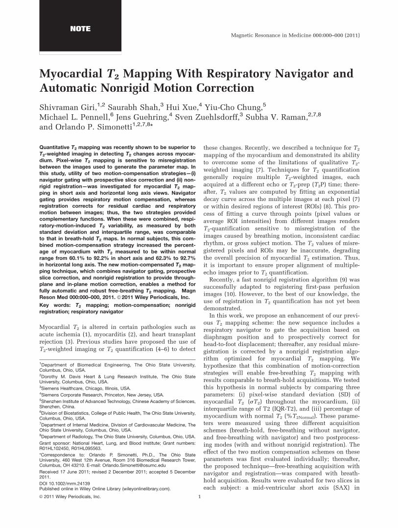
rection. Figure 1a shows the new acquisition scheme;note that a navigator echo was acquired prior to eachimage, and T2P-SSFP images were acquired every 3 RRintervals to allow sufficient T1 recovery. Navigator echotracking of diaphragm position was used to compensatefor respiratory motion using a combined strategy ofgating and prospective correction of slice position; forgating, an acceptance window of 8 mm was used, empir-ically chosen as a good balance between imaging effi-ciency and motion sensitivity. An empirical correctionfactor of 0.6 (13) was used to prospectively correct forresidual displacement of the slice in the head-to-foot direction. Other imaging parameters are listed inTable 1.
A fast variational nonrigid registration algorithm (9)was applied to register the T2P-SSFP images in a pair-wise manner, that is, the 24-ms T2P image was selectedas the reference frame, and both 0- and 55-ms imageswere registered to it. The registration algorithm could beconsidered as an extension of the classic optical flowmethod (14). In this framework, a dense deformationfield is estimated as the solution to a calculus of varia-tion problem, which is solved by performing a
FIG. 1. Acquisition scheme andpostprocessing work-flow. a: Ac-quisition scheme for navigator-gated T2 map using T2P-bSSFP.
Images were acquired once inevery three cardiac cycles toallow for sufficient T1 recovery
between acquisitions. Acquiredimages were accepted only if
the diaphragm position waswithin the acceptance window of8 mm (‘‘prospective correction of
slice position’’ is not shown inthe schematic). The sequence
consists of navigator pulses, T2Ppulse (4 MLEV weighted), linearramp catalyzation (for bSSFP)
and single-shot bSSFP readout.b: Block diagram of motion cor-rection using nonrigid registra-
tion. *‘‘Registration sufficient’’indicates that either the prede-
fined similarity measure hasbeen satisfied or the maximumnumber of iterations has been
reached.
2 Giri et al.
compositional update step corresponding to a transportequation. The regularization is added by low-pass filter-ing the gradient images which are in turn used as a ve-locity field to drive the transport equation. To acceleratethe convergence and to avoid local minima, a multiscaleimage pyramid is created. We selected the local crosscorrelation as the image similarity measure, as itsexplicit derivative can be efficiently calculated, and it isgeneral enough to cope with intensity fluctuation andimaging noise between two adjacent T2P images. Figure1b summarizes the process.
The nonrigid registration of three T2P-SSFP images andthe subsequent generation of T2 maps were performed ina fully automatic manner and implemented as part of theimage reconstruction software of the MR scanner (1.5 TMAGNETOM Avanto, Siemens Healthcare, Erlangen, Ger-many). The motion-corrected images and T2 maps weretypically available within �1 s after image acquisition.
Phantom Study
To ensure that the nonrigid registration algorithm did notbias the estimated T2 values, the T2 maps of a static two-compartment nickel-doped agar-phantom (True T1/T2:1099/49 ms and 1242/58 ms) were acquired with andwithout the nonrigid registration algorithm. True T1 andT2 values of these phantoms were measured using spinecho sequences with and without inversion recovery prep-aration, respectively, with pulse repetition time ¼ 10 s foreach sequence (acquisition time per image was �17 min).
Volunteer Study
Ten healthy subjects consented to participate in thisHealth Insurance Portability and Accountability Act(HIPAA)-compliant study approved by the institution’sHuman Subjects Committee. For each subject, localizerscouts were first acquired to define mid-ventricular SAXand HLA slices. Thereafter, T2P-SSFP images wereacquired under three conditions: (i) free-breathing with-out respiratory navigator, (ii) breath-hold (duration ¼ 7RR intervals), and (iii) free-breathing with respiratory nav-
igator. In each case, the acquisition was gated to mid-dias-tole, and T2 maps were generated with and without non-rigid registration, yielding a total of six maps per slice.
Image Analysis
As the registration algorithm uses the center frame (T2P¼ 24 ms) as reference, this frame was unaffected by thealgorithm and was the same in both unregistered andregistered T2P image sets; this image was selected todraw a ROI covering the entire myocardium in SAXview. For the HLA view, the apex was excluded fromthe ROI due to limited resolution and also to avoid par-tial volume effects. The ROI was then transferred toother images and to the T2 maps for both unregisteredand registered sets. These steps were performed usingMatlab software (The Mathworks, Natick, MA).
First, mean T2 values before and after registration werecompared for any T2 variation that may be caused as aside-effect of registration. Thereafter, the performance ofmotion-compensation strategies was evaluated with twoperformance metrics:
i. Variability of myocardial T2, which was measuredusing SD (sT2) and interquartile range of T2 (IQR-T2) values. While sT2 is a standard measure of dis-persion, it is sensitive to extreme outliers; therefore,we include IQR-T2, as it is less sensitive to outliers.
ii. Percentage of myocardial pixels with normal T2
(%T2Normal), where normal T2 was defined as therange of values within two SDs of mean T2 in thebreath-hold acquisition.
Additionally, pixels with abnormal T2 values wereinvestigated for connectivity, and a set of 10 or moreconnected abnormal pixels were considered to be aregion of T2 abnormality (15); given that the volunteerswere healthy, all such cases represented false-positivediagnosis. The different motion-compensation strategies(registration, navigator, and navigator þ registration)were compared with (i) free-breathing without compen-sation to demonstrate the improvement offered by thestrategy and (ii) breath-hold acquisition (considered thegold-standard) to demonstrate the feasibility of using thestrategy in a clinical setting.
Statistical Analysis
Myocardial T2 values are expressed as mean 6 SD. Totest the hypothesis that nonrigid registration did notalter mean myocardial T2 values, Student’s t-test wasused for the phantom and in vivo data.
Signal intensities of misregistered pixels may not fol-low an exponential decay curve, or even a monotonicallydecreasing pattern; as a result, the fitted T2 can vary overa wide range, including negative and very large positivevalues. The presence of such outlier pixels, along withother misregistered pixels, had two implications: (i) thehigh and low T2 pixels could balance each other out,leaving the mean T2 value unaltered; thus, we do notreport the mean T2 values and do not perform tests ofsignificance for the mean T2 values for T2 maps gener-ated from misregistered images. (ii) The resulting
Table 1Imaging Parameters
Parameter name Value
Number of T2P 3
T2P times 0 (no prep), 24, 55 msAcquisition window �160 ms
Acquisition mode Single shotImage matrix (human studies) 130 � 160Field of view (human studies) 203–285 � 250–350 mm2
Image matrix (phantom study) 104 � 128Field of view (phantom study) 178 � 220 mm2
Slice thickness 8 mmParallel acquisition/
acceleration/reference linesGRAPPA/2/24
Repetition time 3 RR intervalsTotal scan time
(without navigator gating)
7 RR intervals
Flip angle 70�
Field strength 1.5 T
Motion-Compensated Myocardial T2 Mapping 3
distributions of sT2, IQR-T2, and %T2Normal values acrosssubjects were nongaussian; to account for this, sT2, IQR-T2, and %T2Normal for different cases (combinations ofacquisition and postprocessing) were compared usingnonparametric Wilcoxon signed rank tests. A P < 0.05was deemed statistically significant, and Bonferroni cor-rection was used for multiple comparisons.
All analyses were performed using MINITAB statisticalsoftware package (Minitab Inc., State College PA).
RESULTS
Effect of Registration on T2 Values
Phantom results are shown in Table 2 and indicate that theimage registration algorithm caused no change inT2 values.For breath-hold acquisitions in the 10 subjects, the pixel-wise T2 values in SAX view were 52.36 6.5 ms before and53.46 6.1 ms after registration, whereas for HLA view, thevalues were 54.56 7.1 ms and 566 6.8 ms (P value ¼ non-significant (NS) for both). These results show that the useof nonrigid registration did not have a significant side-effect on the distribution ofmyocardial T2 values.
Results from individual and combined motion-compen-sation strategies are shown in Tables 3 and 4. In summary,registration alone reduced sT2 and IQR-T2 and increased%T2Normal in all cases, giving T2 maps that were compara-ble to those from breath-hold acquisitions. On the otherhand, although navigator alone reduced sT2 and IQR-T2
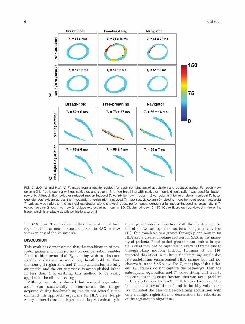
and increased %T2Normal, the resulting T2 maps hadhigher variability of pixel-wise T2 than those from breath-hold acquisitions; this was corrected for by combiningnavigator with registration, with the latter compensatingfor residual motion. Figures 3 and 4 demonstrate the ben-efit of adding registration to navigator acquisitions.
Performance of Nonrigid Registration Only
T2 maps from free-breathing acquisitions compensatedwith only nonrigid registration were comparable to thosefrom breath-hold acquisitions. Figure 2 demonstrates theeffect of registration on T2 values. In all 10 subjects, theproposed nonrigid registration significantly improved (P< 0.05) T2 maps by reducing pixel-wise T2 variability asdefined by (i) sT2 from 51.2 to 6.5 ms in SAX and 79.3 to
7.1 ms in HLA and (ii) IQR-T2 from 25.8 to 6.5 ms forSAX and 26.1 to 10.1 ms for HLA, and increasing %T2Nor-
mal from 60.1 to 93.2% in SAX and 62.3 to 89.3% in HLA.After registration, T2 maps from free-breathing acquisi-
tion were comparable to those from breath-hold acquisi-tion, with the median difference of sT2 ¼ 0.1 ms/0.1 ms (P¼ NS for both), of IQR-T2 ¼ 1.1 ms (P ¼ 0.08)/1.6 ms (P ¼0.01), and that of %T2Normal ¼ 1.9% (P ¼ 0.48)/5.7% (P ¼0.01) for SAX/HLA. Further, the residual outlier pixels inregistered T2 maps did not form a region of 10 or more con-nected pixels in any view (see column 2 in Figs. 3 and 4).
Performance of Respiratory Navigator Only
For the 20 acquisitions (two views in 10 subjects), navi-gator efficiency ranged from 15 to 100% (Median ¼ 50%;IQR ¼ 67%), resulting in a total scan time that rangedfrom a minimum of 7 to a maximum of 40 RR intervals.The use of respiratory navigator alone improved T2 mapsin free-breathing acquisition; these maps were, however,inferior to those from breath-hold acquisitions.
Although navigator alone significantly improved T2
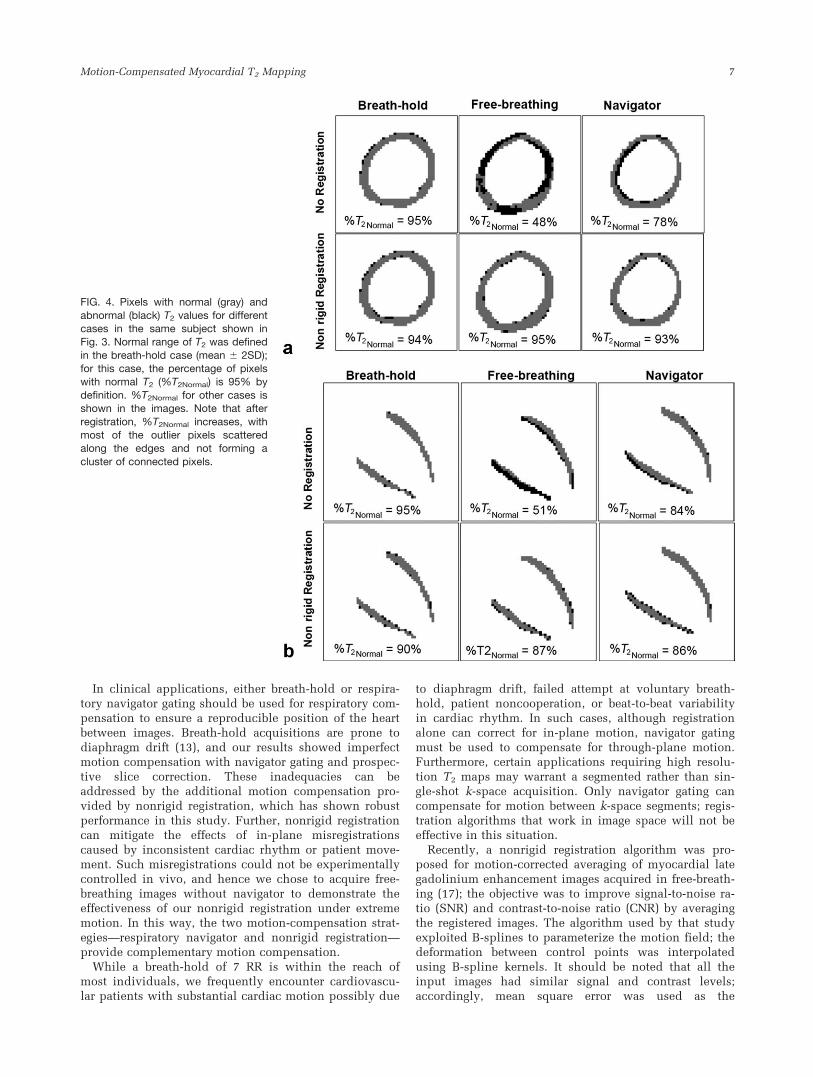
maps (P value < 0.05) by reducing pixel-wise T2 variabil-ity as defined by (i) sT2 from 51.2 to 12.2 ms in SAXand 79.3 to 11.6 ms in HLA and (ii) IQR-T2 from 25.8 to9.4 ms for SAX and 26.1 to 9.9 ms for HLA and increas-ing %T2Normal from 60.1 to 86.5% in SAX and 62.3 to86% in HLA, Figs. 3 and 4 show volunteer images wherenavigator alone did not provide adequate motion com-pensation. Note also in Fig. 4 that the outlier pixels wereconnected, mimicking a region with T2 abnormality; sim-ilar results were observed in 8/20 other views.
Navigator alone gave T2 maps that were inferior tothose from breath-hold acquisition, with the median dif-ference of sT2 ¼ 6.8 ms (P ¼ 0.05)/5.2 ms (P ¼ 0.01),and that of %T2Normal ¼ 8.5%/8.7% (P value < 0.05) forSAX/HLA. However, when expressed as IQR, the pixel-wise T2 variability was comparable to those from breath-hold acquisition; the median difference of IQR-T2 was1.7 ms for SAX and 1.8 ms for HLA (P value ¼ NS).
Performance of Navigator þ Registration
The benefit of additional compensation by registrationover navigator gating alone is seen in column 3, bottom
Table 2Phantom Results Demonstrating the Effect of Nonrigid Registration on T2 Values
Phantom 1 (True T1/T2 ¼ 1099 ms/49 ms) Phantom 2 (True T1/T2 ¼ 1242 ms/58 ms)
Without registration 55.0 ms 6 1.0 ms 63.3 ms 6 0.9 ms
With registration 54.9 ms 6 1.0 ms 63.2 ms 6 0.9 ms
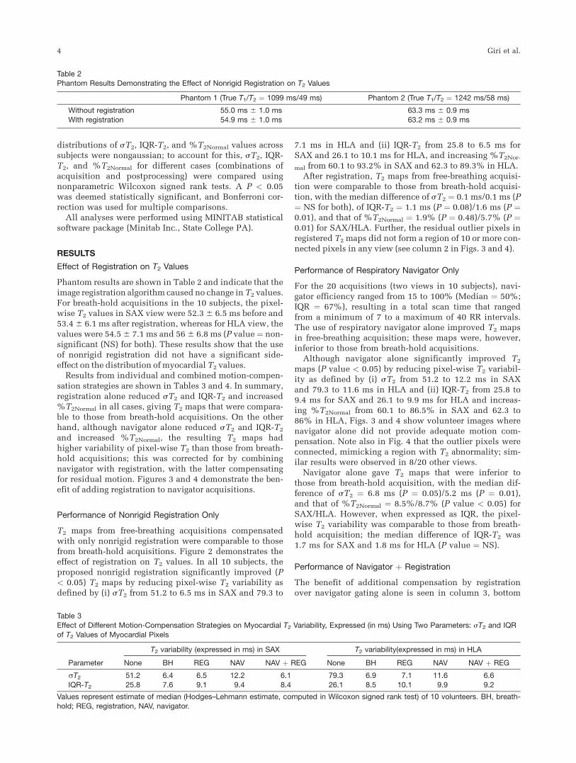
Table 3Effect of Different Motion-Compensation Strategies on Myocardial T2 Variability, Expressed (in ms) Using Two Parameters: sT2 and IQR
of T2 Values of Myocardial Pixels
Parameter
T2 variability (expressed in ms) in SAX T2 variability(expressed in ms) in HLA
None BH REG NAV NAV þ REG None BH REG NAV NAV þ REG
rT2 51.2 6.4 6.5 12.2 6.1 79.3 6.9 7.1 11.6 6.6IQR-T2 25.8 7.6 9.1 9.4 8.4 26.1 8.5 10.1 9.9 9.2
Values represent estimate of median (Hodges–Lehmann estimate, computed in Wilcoxon signed rank test) of 10 volunteers. BH, breath-hold; REG, registration, NAV, navigator.
4 Giri et al.
row of Figs. 3 and 4. Similar results were noted in othercases where navigator alone was inadequate.
For free-breathing T2 mapping, the combined use ofnavigator and registration significantly improved T2
maps (P value < 0.05) by reducing pixel-wise T2 variabil-ity as defined by (i) sT2 from 51.2 to 6.1 ms in SAX andHLA 79.3 to 6.6 ms in HLA and (ii) IQR-T2 from 25.8 to
8.4 ms for SAX and 26.1 to 9.2 ms for HLA, and increas-ing %T2Normal from 60.1 to 92.2% for SAX and 62.3 to92.7% for HLA.
These T2 maps were comparable to those from breath-hold acquisition, with median difference of sT2 ¼ 0.3ms/0.4 ms (P ¼ NS), of IQR-T2 ¼ 0.5 ms/0.9 ms (P ¼NS), and of %T2Normal ¼ 2.8% (P ¼ 0.2)/1.9 % (P ¼ 0.04)
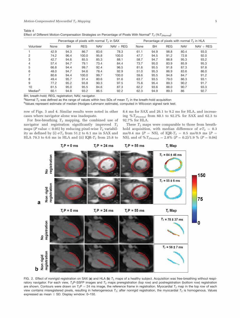
Table 4Effect of Different Motion-Compensation Strategies on Percentage of Pixels With Normala T2 (%T2Normal)
Volunteer
Percentage of pixels with normal T2 in SAX Percentage of pixels with normal T2 in HLA
None BH REG NAV NAV þ REG None BH REG NAV NAV þ REG
1 42.9 94.3 86.7 83.6 78.3 61.1 94.9 98.8 80.4 93.0
2 74.2 96.4 100.0 95.8 100.0 47.7 94.5 91.2 72.8 92.03 42.7 94.6 85.5 85.3 88.1 58.7 94.7 88.8 95.3 93.24 57.4 94.7 79.1 73.4 84.4 73.7 95.0 83.9 85.9 95.3
5 66.8 94.4 99.7 92.4 96.5 81.6 95.5 91.8 87.3 97.86 48.0 94.7 94.6 78.4 92.9 51.0 95.3 86.9 83.6 86.0
7 80.6 94.4 100.0 99.7 100.0 59.6 95.5 94.8 84.7 91.28 49.4 95.7 91.4 80.6 91.6 63.7 93.5 79.0 86.3 93.19 77.2 95.2 93.8 90.3 97.5 75.8 95.4 89.3 90.2 91.7
10 61.5 95.0 95.5 84.6 87.3 62.2 93.6 88.0 90.7 93.3Medianb 60.1 94.8 93.2 86.5 92.2 62.3 94.9 89.3 86 92.7
BH, breath-hold; REG, registration; NAV, navigator.aNormal T2 was defined as the range of values within two SDs of mean T2 in the breath-hold acquisitionbValues represent estimate of median (Hodges–Lehmann estimate), computed in Wilcoxon signed rank test.
FIG. 2. Effect of nonrigid registration on SAX (a) and HLA (b) T2 maps of a healthy subject. Acquisition was free-breathing without respi-ratory navigator. For each view, T2P-SSFP images and T2 maps preregistration (top row) and postregistration (bottom row) registrationare shown. Contours were drawn on T2P ¼ 24 ms image, the reference frame in registration. Myocardial T2 map in the top row of each
view contains misregistered pixels, resulting in heterogeneous T2; after nonrigid registration, the myocardial T2 is homogenous. Valuesexpressed as mean 6 SD. Display window: 0–150.
Motion-Compensated Myocardial T2 Mapping 5
for SAX/HLA. The residual outlier pixels did not formregions of ten or more connected pixels in SAX or HLAviews in any of the volunteers.
DISCUSSION
This work has demonstrated that the combination of nav-igator gating and nonrigid motion compensation enablesfree-breathing myocardial T2 mapping with results com-parable to data acquisition during breath-hold. Further,the nonrigid registration and T2 map calculation are fullyautomatic, and the entire process is accomplished inlinein less than 1 s, enabling this method to be easilyapplied in the clinical setting.
Although our study showed that nonrigid registrationalone can successfully motion-correct the imagesacquired during free-breathing, we do not generally rec-ommend this approach, especially for HLA view. Respi-ratory-induced cardiac displacement is predominantly in
the superior–inferior direction, with the displacement inthe other two orthogonal directions being relatively less(13); this translates to a greater through-plane motion forHLA and a greater in-plane motion for SAX in the major-ity of patients. Focal pathologies that are limited in spa-tial extent may not be captured in every 2D frame due tothrough-plane motion; indeed, Kellman et al. (16)reported this effect in multiple free-breathing single-shotlate gadolinium enhancement HLA images but did notobserve it in the SAX view. For T2 mapping, if the differ-ent T2P frames do not capture the pathology, then thesubsequent registration and T2 curve-fitting will lead toinaccuracies in T2 quantification; this was not a problemin this study in either SAX or HLA view because of thehomogeneous myocardium found in healthy volunteers.We included the case of free-breathing acquisition withonly nonrigid registration to demonstrate the robustnessof the registration algorithm.
FIG. 3. SAX (a) and HLA (b) T2 maps from a healthy subject for each combination of acquisition and postprocessing. For each view,column 2 is free-breathing without navigator, and column 3 is free-breathing with navigator; nonrigid registration was used for bottomrow only. Although the navigator reduced motion-induced T2 variability (row 1, column 2 vs. column 3 for both views), residual T2 heter-
ogeneity was evident across the myocardium; registration improved T2 map (row 2, column 3), yielding more homogeneous myocardialT2 values. Also note that the nonrigid registration alone showed robust performance, correcting for motion-induced heterogeneity in T2values (column 2, row 1 vs. row 2). Values expressed as mean 6 SD. Display window: 0–150. [Color figure can be viewed in the onlineissue, which is available at wileyonlinelibrary.com.]
6 Giri et al.
In clinical applications, either breath-hold or respira-tory navigator gating should be used for respiratory com-pensation to ensure a reproducible position of the heartbetween images. Breath-hold acquisitions are prone todiaphragm drift (13), and our results showed imperfectmotion compensation with navigator gating and prospec-tive slice correction. These inadequacies can beaddressed by the additional motion compensation pro-vided by nonrigid registration, which has shown robustperformance in this study. Further, nonrigid registrationcan mitigate the effects of in-plane misregistrationscaused by inconsistent cardiac rhythm or patient move-ment. Such misregistrations could not be experimentallycontrolled in vivo, and hence we chose to acquire free-breathing images without navigator to demonstrate theeffectiveness of our nonrigid registration under extrememotion. In this way, the two motion-compensation strat-egies—respiratory navigator and nonrigid registration—provide complementary motion compensation.
While a breath-hold of 7 RR is within the reach ofmost individuals, we frequently encounter cardiovascu-lar patients with substantial cardiac motion possibly due
to diaphragm drift, failed attempt at voluntary breath-hold, patient noncooperation, or beat-to-beat variabilityin cardiac rhythm. In such cases, although registrationalone can correct for in-plane motion, navigator gatingmust be used to compensate for through-plane motion.Furthermore, certain applications requiring high resolu-tion T2 maps may warrant a segmented rather than sin-gle-shot k-space acquisition. Only navigator gating cancompensate for motion between k-space segments; regis-tration algorithms that work in image space will not beeffective in this situation.
Recently, a nonrigid registration algorithm was pro-posed for motion-corrected averaging of myocardial lategadolinium enhancement images acquired in free-breath-ing (17); the objective was to improve signal-to-noise ra-tio (SNR) and contrast-to-noise ratio (CNR) by averagingthe registered images. The algorithm used by that studyexploited B-splines to parameterize the motion field; thedeformation between control points was interpolatedusing B-spline kernels. It should be noted that all theinput images had similar signal and contrast levels;accordingly, mean square error was used as the
FIG. 4. Pixels with normal (gray) and
abnormal (black) T2 values for differentcases in the same subject shown inFig. 3. Normal range of T2 was defined
in the breath-hold case (mean 6 2SD);for this case, the percentage of pixelswith normal T2 (%T2Normal) is 95% by
definition. %T2Normal for other cases isshown in the images. Note that after
registration, %T2Normal increases, withmost of the outlier pixels scatteredalong the edges and not forming a
cluster of connected pixels.
Motion-Compensated Myocardial T2 Mapping 7
similarity measure. Our nonrigid registration algorithm,on the other hand, solves a partial differential equationto get the pixel-wise deformation, using local cross-corre-lation as the similarity measure. Although bothapproaches rely on gradient descent optimization, withtheir respective performances being largely determinedby the similarity measure (mean square error vs. localcross-correlation), the partial differential equation-basedmethod leads to so-called diffeomorphic deformation,which is a motion field free of folding or tearing; thisdiffeomorphic property is generally not a characteristicof the B-spline deformation, unless explicit constraintsare added to avoid any possible foldings by limiting themagnitude of spatial deformation on the B-spline controlpoints (18); this will lead to slower convergence as aside-effect. Given this, and our objective of registeringmultiple T2P images, each with a different signal andcontrast level, we chose to use the partial differentialequation-based nonrigid registration algorithm.
The pixel-wise sT2 found in this study is higher thanpreviously reported values (7), possibly because con-tours, drawn to cover the entire myocardium, includedborder pixels with partial-volume effects. Additionally,the misregistered pixels varied over a very wide range.In typical clinical studies, such pixels are excluded fromimage analysis by appropriate contouring. Given that thepurpose of this study is to quantitatively demonstratethe effect of different motion-compensation strategies onT2 maps, we did not exclude such pixels. We used twostatistical measures of dispersion—SD and IQR—to dem-onstrate how motion correction and navigator gatingeffectively reduced motion-induced variability in myo-cardial T2 values. While SD is a valid measure of disper-sion even for nongaussian spread, it is sensitive toextreme outliers; accordingly, we utilized IQR as anadditional measure of motion-induced T2 variability thatis less sensitive to the influence of outliers.
Several recent studies have demonstrated T2 mappingwith respiratory compensation. Foltz et al. (19) used re-spiratory bellows to monitor respiratory phase; thisapproach necessitated repetition of the acquisitions thatwere farthest from the median respiratory position. Theauthors of that study speculated that motion uncer-tainty was a primary contributor to remaining sT2 invivo, suggesting inadequacy of their motion-compensa-tion strategy. Huang et al. (20) used navigator gating forrespiratory motion compensation and relied on a sub-jective analysis to either adjust ROI position or to dis-card images with more than 4 pixel misregistration.Blume et al. (21) also used navigator gating but did notaccount for the in-plane misregistration that may arisedue to patient movement or inconsistent cardiacrhythm. Compared to all previous approaches, the solu-tion proposed in this paper is the only one to combinetwo techniques that provide complementary motioncompensation (through-plane and in-plane). The non-rigid registration has shown robust performance even inthe extreme case of free-breathing acquisition withoutnavigator. Further, the navigator gating, nonrigid regis-tration, and T2 map generation are fully automatic andintegrated with the sequence, obviating the need forany user input.
Limitations
Given that the primary purpose of this study was to vali-date the nonrigid registration for T2 mapping, patientswere not recruited as the myocardial T2 variability inpatients would likely have confounded the investigationof variability due to misregistration. We did not investi-gate the effects of variable myocardial signal, as may befound in pathologies, on registration and subsequent T2
mapping. We have recently demonstrated the feasibilityof using breath-hold T2 mapping with nonrigid motioncorrection (without navigator gating) in patients withacute myocardial infarction, myocarditis, and Tako-tsubocardiomyopathy (6,22).
While the sequence overestimated phantom T2 values,which is consistent with and discussed in our earlierwork (7), phantom results showed that registration didnot affect T2 quantification. Similarly, while the in vivoT2 values postregistration did show a small systematicbias (�þ3%), it was not statistically significant. %T2Nor-
mal in the proposed technique (navigator þ registration)was less than the target 95% in many cases (Table 4);given that sT2 was similar to breath-hold acquisition, thereduced %T2Normal may be due to the aforementionedbias. In a quantitative approach, the resulting outlier pix-els could potentially be flagged as pathologic tissue. Arecent clinical study (15) defined a region as pathologicif 10 or more connected pixels had abnormal T2-weighted signal. With this as a reference, the proposedtechnique (navigator þ registration) performed well,because most of the outlier pixels were scattered aroundthe borders, without forming a connected region (maxi-mum eight connected pixels in this study), thus preclud-ing potential false-positive cases (Fig. 4, column 3).
CONCLUSIONS
We have demonstrated a fully automatic and reliablefree-breathing myocardial T2 mapping technique basedon single-shot, T2-prepared SSFP that provides two com-plementary motion-compensation schemes: respiratorynavigator and nonrigid registration. Both schemes wereshown to independently provide significant motion com-pensation by reducing myocardial T2 variability andincreasing the percentage of myocardium with normal T2
in healthy volunteers. While the navigator technique wasused to ensure reproducible slice position, our resultsindicated that it alone did not provide adequate motioncompensation for reliable T2 mapping. A new nonrigidregistration algorithm optimized for myocardial T2 map-ping was able to correct for any residual misregistrations;further, this algorithm alone showed robust performanceeven during breathing for both SAX and the motion-sen-sitive HLA views. When combined, these two strategiesprovide complementary motion compensation and canbe used to generate fully automatic and reliable free-breathing myocardial T2 maps.
ACKNOWLEDGMENTS
The content is solely the responsibility of the authorsand does not necessarily represent the official views of
8 Giri et al.
the National Heart, Lung, and Blood Institute or theNational Institutes of Health.
REFERENCES
1. Abdel-Aty H, Cocker M, Meek C, Tyberg JV, Friedrich MG. Edema as
a very early marker for acute myocardial ischemia: a cardiovascular
magnetic resonance study. J Am Coll Cardiol 2009;53:1194–1201.
2. Friedrich MG, Sechtem U, Schulz-Menger J, Holmvang G, Alakija P,
Cooper LT, White JA, Abdel-Aty H, Gutberlet M, Prasad S, Aletras A,
Laissy JP, Paterson I, Filipchuk NG, Kumar A, Pauschinger M, Liu P.
Cardiovascular magnetic resonance in myocarditis: A JACC White
Paper. J Am Coll Cardiol 2009;53:1475–1487.
3. Marie PY, Angioi M, Carteaux JP, Escanye JM, Mattei S, Tzvetanov
K, Claudon O, Hassan N, Danchin N, Karcher G, Bertrand A, Walker
PM, Villemot JP. Detection and prediction of acute heart transplant
rejection with the myocardial T2 determination provided by a black-
blood magnetic resonance imaging sequence. J Am Coll Cardiol 2001;
37:825–831.
4. Abdel-Aty H, Zagrosek A, Schulz-Menger J, Taylor AJ, Messroghli D,
Kumar A, Gross M, Dietz R, Friedrich MG. Delayed enhancement and T2-
weighted cardiovascular magnetic resonance imaging differentiate acute
from chronic myocardial infarction. Circulation 2004;109:2411–2416.
5. Aletras AH, Tilak GS, Natanzon A, Hsu LY, Gonzalez FM, Hoyt RF
Jr, Arai AE. Retrospective determination of the area at risk for reper-
fused acute myocardial infarction with T2-weighted cardiac magnetic
resonance imaging: histopathological and displacement encoding
with stimulated echoes (DENSE) functional validations. Circulation
2006;113:1865–1870.
6. Verhaert D, Thavendiranathan P, Giri S, Mihai G, Rajagopalan S, Simo-
netti OP, Raman SV. Direct T2 quantification of myocardial edema in
acute ischemic injury. JACC Cardiovasc Imaging 2011;4:269–278.
7. Giri S, Chung YC, Merchant A, Mihai G, Rajagopalan S, Raman SV,
Simonetti OP. T2 quantification for improved detection of myocardial
edema. J Cardiovasc Magn Reson 2009;11:56.
8. He T, Gatehouse PD, Anderson LJ, Tanner M, Keegan J, Pennell DJ,
Firmin DN. Development of a novel optimized breathhold technique
for myocardial T2 measurement in thalassemia. J Magn Reson Imag-
ing 2006;24:580–585.
9. Chef d’hotel C, Hermosillo G, Faugeras O. Flows of diffeomorphisms
for multimodal image registration. 2002 IEEE International Sympo-
sium on Biomedical Imaging, Proceedings 2002:753–756.
10. Xue H, Guehring J, Srinivasan L, Zuehlsdorff S, Saddi K, Chefdhotel
C, Hajnal JV, Rueckert D. Evaluation of rigid and non-rigid motion
compensation of cardiac perfusion MRI. Med Image Comput Comput
Assist Interv Int Conf Med Image Comput Comput Assist Interv
2008;11 (pt 2):35–43.
11. Kellman P, Aletras AH, Mancini C, McVeigh ER, Arai AE. T2-pre-
pared SSFP improves diagnostic confidence in edema imaging in
acute myocardial infarction compared to turbo spin echo. Magn
Reson Med 2007;57:891–897.
12. Brittain JH, Hu BS, Wright GA, Meyer CH, Macovski A, Nishimura
DG. Coronary angiography with magnetization-prepared T2 contrast.
Magn Reson Med 1995;33:689–696.
13. Scott AD, Keegan J, Firmin DN. Motion in cardiovascular MR imag-
ing. Radiology 2009;250:331–351.
14. Xue H, Zuehlsdorff S, Kellman P, Arai A, Nielles-Vallespin S, Chefd-
hotel C, Lorenz CH, Guehring J. Unsupervised inline analysis of car-
diac perfusion MRI. Med Image Comput Comput Assist Interv 2009;
12 (pt 2):741–749.
15. Friedrich MG, Abdel-Aty H, Taylor A, Schulz-Menger J, Messroghli
D, Dietz R. The salvaged area at risk in reperfused acute myocardial
infarction as visualized by cardiovascular magnetic resonance. J Am
Coll Cardiol 2008;51:1581–1587.
16. Kellman P, Larson AC, Hsu LY, Chung YC, Simonetti OP, McVeigh
ER, Arai AE. Motion-corrected free-breathing delayed enhancement
imaging of myocardial infarction. Magn Reson Med 2005;53:194–200.
17. Ledesma-Carbayo MJ, Kellman P, Hsu LY, Arai AE, McVeigh ER.
Motion corrected free-breathing delayed-enhancement imaging of
myocardial infarction using nonrigid registration. J Magn Reson
Imaging 2007;26:184–190.
18. Rueckert D, Aljabar P, Heckemann RA, Hajnal JV, Hammers A. Dif-
feomorphic registration using B-splines. Med Image Comput Comput
Assist Interv 2006;9 (pt 2):702–709.
19. Foltz WD, Al-Kwifi O, Sussman MS, Stainsby JA, Wright GA. Opti-
mized spiral imaging for measurement of myocardial T2 relaxation.
Magn Reson Med 2003;49:1089–1097.
20. Huang TY, Liu YJ, Stemmer A, Poncelet BP. T2 measurement of the
human myocardium using a T2-prepared transient-state TrueFISP
sequence. Magn Reson Med 2007;57:960–966.
21. Blume U, Lockie T, Stehning C, Sinclair S, Uribe S, Razavi R,
Schaeffter T. Interleaved T(1) and T(2) relaxation time mapping for
cardiac applications. J Magn Reson Imaging 2009;29:480–487.
22. Thavendiranathan P, Walls M, Giri S, Verhaert D, Rajagopalan S,
Moore S, Simonetti OP, Raman SV. Improved detection of myocar-
dial involvement in acute inflammatory cardiomyopathies using T2
mapping. Circ Cardiovasc Imaging, 2011 Oct. 28, Epub ahead of
print.
Motion-Compensated Myocardial T2 Mapping 9