mycotoxin occurrence, exposure and health implications in
TRANSCRIPT
foods
Review
Mycotoxin Occurrence, Exposure and HealthImplications in Infants and Young Children inSub-Saharan Africa: A Review
Cynthia Adaku Chilaka * and Angela MallyInstitute of Pharmacology and Toxicology, Julius Maximilian University of Würzburg, Versbacher Straβe 9,97078 Würzburg, Germany; [email protected]* Correspondence: [email protected]
Received: 1 October 2020; Accepted: 29 October 2020; Published: 1 November 2020�����������������
Abstract: Infants and young children (IYC) remain the most vulnerable population group toenvironmental hazards worldwide, especially in economically developing regions such as sub-SaharanAfrica (SSA). As a result, several governmental and non-governmental institutions including health,environmental and food safety networks and researchers have been proactive toward protecting thisgroup. Mycotoxins, toxic secondary fungal metabolites, contribute largely to the health risks of thisyoung population. In SSA, the scenario is worsened by socioeconomic status, poor agricultural andstorage practices, and low level of awareness, as well as the non-establishment and lack of enforcementof regulatory limits in the region. Studies have revealed mycotoxin occurrence in breast milk andother weaning foods. Of concern is the early exposure of infants to mycotoxins through transplacentaltransfer and breast milk as a consequence of maternal exposure, which may result in adverse healtheffects. The current paper presents an overview of mycotoxin occurrence in foods intended for IYCin SSA. It discusses the imperative evidence of mycotoxin exposure of this population group inSSA, taking into account consumption data and the occurrence of mycotoxins in food, as well asbiomonitoring approaches. Additionally, it discusses the health implications associated with IYCexposure to mycotoxins in SSA.
Keywords: mycotoxin; occurrence; exposure; child health; sub-Saharan Africa
1. Introduction
Fungi are ubiquitous in nature, having the capacity to colonise a wide range of ecosystems,including crops and foodstuffs worldwide. Several studies have reported the potential of theseorganisms in the production of useful organic compounds with numerous pharmaceutical benefits suchas antibiotics, e.g., penicillin [1]. However, toxigenic fungi, particularly those belonging to the generaAspergillus, Fusarium, Penicillium, Alternaria, and Claviceps are of importance because of their abilityto produce several toxic secondary metabolites (mycotoxins) under favourable environmental andecological conditions [2,3]. The production of these mycotoxins, though not necessarily important forthe fungal growth, is considered to serve as a defence against predators and change in environmentalconditions in fungi’s ecological niche [1,4,5]. Mycotoxins are a chemically diverse group of lowmolecular compounds, which occur in agricultural commodities and processed food products, as wellas in the environment. To date, 300 to 400 mycotoxins are known, with those of special interestbeing aflatoxins (AFs), fumonisins (FBs), ochratoxins (OTs), trichothecenes (THs), zearalenone (ZEN),citrinin (CIT), and Alternaria toxins. The importance placed on these toxins may be linked to theirprincipal roles in occurrence, distribution, and toxicity.
The contamination of food products by mycotoxins can occur all through the life and processcycle of crops, starting from the pre-harvest, harvest, and storage stages, as well as the process stage.
Foods 2020, 9, 1585; doi:10.3390/foods9111585 www.mdpi.com/journal/foods
Foods 2020, 9, 1585 2 of 40
Moreover, certain fungi and types of toxin production are dominant at specific stages. While pre-harveststages favour the growth of species of Fusarium, Alternaria, and Claviceps and associated mycotoxinproduction, Aspergillus and Penicillium species strive maximally during storage [6,7]. The occurrence ofmycotoxins worldwide is unavoidable; however, the concentration at which these toxins occur in foodproducts is of great concern. Mycotoxin occurrence has been reported in several agricultural crops andproducts, including cereals and cereal-based products, pulses and pulse-based products, fruits andfruit-based products, nuts, spices, coffee, and tea [8–12]. A great concern is the occurrence of this arrayof toxins in foods destined for infants and young children (IYC) and their possible presence in breastmilk as a result of maternal exposure. Although constant efforts are being made to reduce fungalinfestation and mycotoxin contamination in food products, human and environmental factors such asclimatic change, pest activities, and improper agricultural and storage practices hamper these efforts.In addition to the economic cost on crops, which is worth billions of dollars, mycotoxins have thepotentials to induce acute or chronic adverse health consequences, known as mycotoxicoses, in humansand animals.
Exposure to these toxins can be through ingestion, inhalation, or/and dermal absorption.The degree of toxic effect is dependent on the toxin type, exposure dose and duration, age, sex,and health status of the host, exposure route, and possible synergistic effects of other chemicalsto which the individual is exposed [13–17]. The potential subgroup most affected by mycotoxincontamination is IYC, probably because of their high ingestion of mycotoxins due to frequentconsumption of cereal-based food in proportion to their body weight [18,19]. Additionally, the youngdeveloping organs and immune systems of IYC may predispose them to the toxic effect. Mycotoxinexposure has been linked to a wide range of diseases ranging from mild symptoms of nausea, vomiting,and dizziness to long-term degenerative diseases as a result of carcinogenic, nephrotoxic, neurotoxic,hepatotoxic, genotoxic, immunotoxic, estrogenic, and teratogenic properties [20–22]. In the case ofchildren, mycotoxins have been associated with developmental defects, such as neuro-developmentaldisorders. Recent epidemiological studies by De Santis et al. reported a significant association betweenmycotoxins and autism spectrum disorder (ASD) [23,24]. A high incidence of neural tube defects(NTDs) in the areas of the world, such as African communities, where maize serves as a major staplefood has been linked to FB contamination in maize [25,26].
Another study also reported the capability of mycotoxins such as AFs to cross the placental barrier,thus causing alteration of foetal health [27,28]. Their implication with pulmonary hemosiderosis ininfants, resulting in anaemia, chronic cough, dyspnea, wheezing, and often cyanosis, has also beenreported [29]. Other health effects, such as undernutrition and increased morbidity and mortality ofinfants as a result of chronic aflatoxicosis, resulting in immune malfunction and poor absorption ofmicronutrients, has also been recorded [30]. A study in 1989 implicated mycotoxins, specifically ZENand its metabolites, to have an estrogenic effect, thus resulting in premature breast development andprecocious sexual development in children in Puerto Rico between 1978 and 1981 [31,32]. In addition,exposure to toxigenic fungi and their metabolites have been linked to child growth and weightimpairment [6,33–41]. This scenario of stunting and wasting is often seen in the developing continentssuch as Africa [42], especially in SSA. This region is characterised by the frequent occurrence and highlevels of contaminants such as mycotoxins in food products and food for infants and young children,including complementary and weaning foods, as well as breast milk [9,38,43–46].
Stringent regulatory limits for mycotoxins have been established by countries, especially in thedeveloped world, thus translating into reasonable protection for consumers, specifically IYC. However,developing regions, such as SSA, hitherto still face outbreaks of mycotoxicosis directly or indirectly,which are often unreported. Of most concern is the attitude of the region towards protecting the mostvulnerable group, which is evident in the non-establishment and enforcement of a collective regulatorylaw for the region. In 2004, the Food and Agricultural Organization (FAO) reported the outcome of aworldwide survey, which identified only 15 African countries to have mycotoxin regulation, out ofwhich 11 are situated in SSA [47]. Unfortunately, to date, there is little or no improvement in the region
Foods 2020, 9, 1585 3 of 40
with respect to mycotoxin regulation, with very weak enforcement power in the countries with existinglaws. The shortfalls experienced in this region may be attributed to food security status which impedesfood safety efforts within the food value chain [48]. The non-proactiveness of the food safety bodiesand the complex agricultural farming system in the region further complicate the situation. Anotherpossibility is inadequate scientific data, including occurrence, exposure, and risk assessment data inSSA. This necessitates the urgent need for a comprehensive review to summarise existing publisheddata regarding the occurrence of mycotoxins and its associated risk in SSA, particularly with referenceto IYC.
Therefore, in the current review, we present an overview of the data on the occurrence ofmycotoxins in IYC foods, including complementary foods, as well as breast milk, in SSA. Furthermore,it summarises the previous studies on mycotoxin exposure of IYC as well as health implications ofmycotoxins in IYC with particular reference to the SSA region.
2. Mycotoxin Occurrence in Foods for Infants and Young Children
The complex and ubiquitous nature of mycotoxins makes them occur virtually in everyenvironment. There is ambient evidence for their occurrence in human foods, animal feeds,and environmental air [49–53]. While there is a wealth of data on the occurrence of mycotoxinsin infant foods in developed countries, it will become evident from this review that only a few studiesare available on this subject in SSA. Therefore, it is important to assess the extent to which foodsintended for the most vulnerable populace group (IYC) in SSA are contaminated with these toxins.
2.1. Cereal-Based Products
Cereals are ranked the major susceptible crops to mycotoxin contamination, with maize being atthe forefront [54]. Because of their excellent energy and nutritional sources, these crops serve as themain ingredient used in the production of foods for IYC [55]. Although studies have shown that theprocessing of food can lead to a reduction or degradation of mycotoxins, it is important to highlightthat mycotoxins can be carried over to the final processed products, thus necessitating the need forgood quality raw material [56–58]. Available studies in the world have revealed the occurrence ofboth regulated and non-regulated mycotoxins in foods for IYC, including the so-called “emergingmycotoxins” [59–71]. In SSA, only a few studies have focused on food products for infants and children.Notwithstanding the few available data, the prevalence of mycotoxins in IYC foods is obvious.
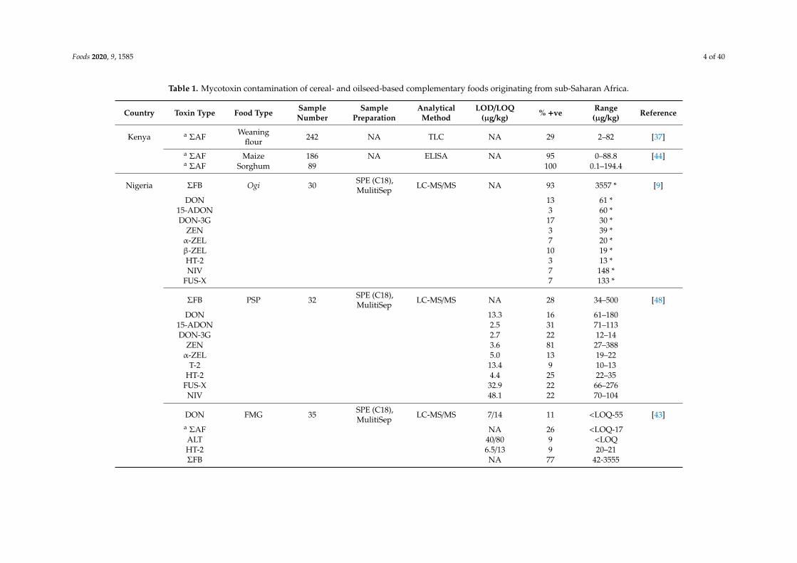
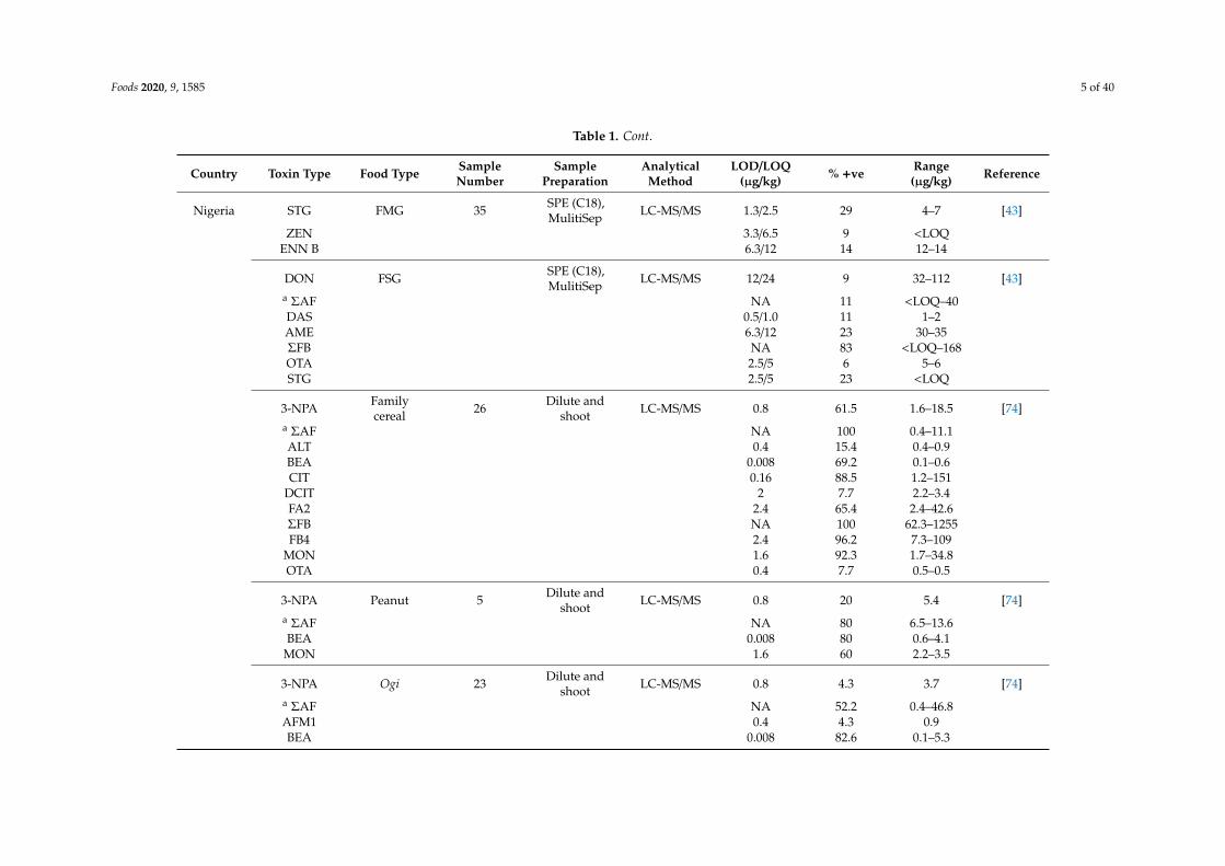
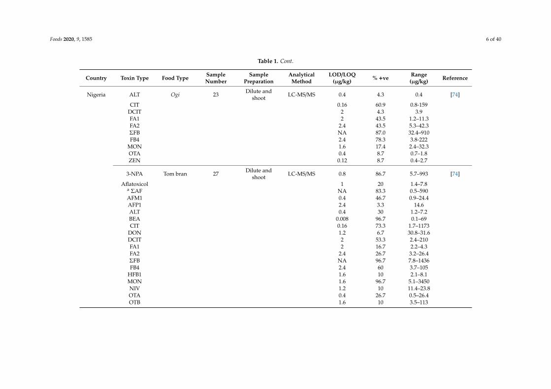
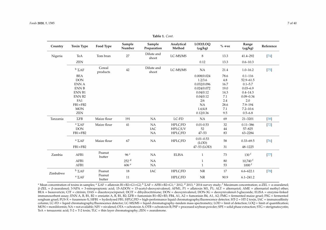
Mycotoxins have been found at high concentrations in foods for IYC in SSA countries, such asNigeria, Tanzania, and Burkina Faso, with most levels exceeding the regulatory limits set by the EUand some SSA countries (Table 1) [9,38,72,73]. Kimanya et al. [38] reported the frequency of FBs inready-to-cook maize flour consumed by infants in the division of Tarakea, Tanzania. Of the 191 samplesexamined in their study, the authors observed that 131 of the samples were contaminated with FBslevels, ranging from 21 to 3201 µg/kg. The same group of authors examined maize flour consumed by41 children in the rural village of Kikelelwa, Tanzania and detected multiple mycotoxins, includingAFs (range, 0.11–386 µg/kg), deoxynivalenol (DON) (range, 57–825 µg/kg), and FBs (63–2284 µg/kg) at32%, 44%, and 83%, respectively [72]. A similar trend was observed in other studies carried out in othercountries of SSA. A Nigerian study reported a high occurrence rate (97%) of Fusarium mycotoxins intraditional cereal-based infant foods (ogi) sampled from local markets, with one of the samples havinga total FB level as high as 3557 µg/kg. Another study on ogi from Nigerian and South African marketsalso reported the high tendencies of mycotoxin contamination in the product [43].
Foods 2020, 9, 1585 4 of 40
Table 1. Mycotoxin contamination of cereal- and oilseed-based complementary foods originating from sub-Saharan Africa.
Country Toxin Type Food Type SampleNumber
SamplePreparation
AnalyticalMethod
LOD/LOQ(µg/kg) % +ve Range
(µg/kg) Reference
Kenya a ΣAF Weaningflour 242 NA TLC NA 29 2–82 [37]
a ΣAF Maize 186 NA ELISA NA 95 0–88.8 [44]a ΣAF Sorghum 89 100 0.1–194.4
Nigeria ΣFB Ogi 30 SPE (C18),MulitiSep LC-MS/MS NA 93 3557 * [9]
DON 13 61 *15-ADON 3 60 *DON-3G 17 30 *
ZEN 3 39 *α-ZEL 7 20 *β-ZEL 10 19 *HT-2 3 13 *NIV 7 148 *
FUS-X 7 133 *
ΣFB PSP 32 SPE (C18),MulitiSep LC-MS/MS NA 28 34–500 [48]
DON 13.3 16 61–18015-ADON 2.5 31 71–113DON-3G 2.7 22 12–14
ZEN 3.6 81 27–388α-ZEL 5.0 13 19–22
T-2 13.4 9 10–13HT-2 4.4 25 22–35
FUS-X 32.9 22 66–276NIV 48.1 22 70–104
DON FMG 35 SPE (C18),MulitiSep LC-MS/MS 7/14 11 <LOQ-55 [43]
a ΣAF NA 26 <LOQ-17ALT 40/80 9 <LOQHT-2 6.5/13 9 20–21ΣFB NA 77 42-3555
Foods 2020, 9, 1585 5 of 40
Table 1. Cont.
Country Toxin Type Food Type SampleNumber
SamplePreparation
AnalyticalMethod
LOD/LOQ(µg/kg) % +ve Range
(µg/kg) Reference
Nigeria STG FMG 35 SPE (C18),MulitiSep LC-MS/MS 1.3/2.5 29 4–7 [43]
ZEN 3.3/6.5 9 <LOQENN B 6.3/12 14 12–14
DON FSG SPE (C18),MulitiSep LC-MS/MS 12/24 9 32–112 [43]
a ΣAF NA 11 <LOQ–40DAS 0.5/1.0 11 1–2AME 6.3/12 23 30–35ΣFB NA 83 <LOQ–168OTA 2.5/5 6 5–6STG 2.5/5 23 <LOQ
3-NPA Familycereal 26 Dilute and
shoot LC-MS/MS 0.8 61.5 1.6–18.5 [74]a ΣAF NA 100 0.4–11.1ALT 0.4 15.4 0.4–0.9BEA 0.008 69.2 0.1–0.6CIT 0.16 88.5 1.2–151
DCIT 2 7.7 2.2–3.4FA2 2.4 65.4 2.4–42.6ΣFB NA 100 62.3–1255FB4 2.4 96.2 7.3–109
MON 1.6 92.3 1.7–34.8OTA 0.4 7.7 0.5–0.5
3-NPA Peanut 5 Dilute andshoot LC-MS/MS 0.8 20 5.4 [74]
a ΣAF NA 80 6.5–13.6BEA 0.008 80 0.6–4.1
MON 1.6 60 2.2–3.5
3-NPA Ogi 23 Dilute andshoot LC-MS/MS 0.8 4.3 3.7 [74]
a ΣAF NA 52.2 0.4–46.8AFM1 0.4 4.3 0.9BEA 0.008 82.6 0.1–5.3
Foods 2020, 9, 1585 6 of 40
Table 1. Cont.
Country Toxin Type Food Type SampleNumber
SamplePreparation
AnalyticalMethod
LOD/LOQ(µg/kg) % +ve Range
(µg/kg) Reference
Nigeria ALT Ogi 23 Dilute andshoot LC-MS/MS 0.4 4.3 0.4 [74]
CIT 0.16 60.9 0.8-159DCIT 2 4.3 3.9FA1 2 43.5 1.2–11.3FA2 2.4 43.5 5.3–42.3ΣFB NA 87.0 32.4–910FB4 2.4 78.3 3.8-222
MON 1.6 17.4 2.4–32.3OTA 0.4 8.7 0.7–1.8ZEN 0.12 8.7 0.4–2.7
3-NPA Tom bran 27 Dilute andshoot LC-MS/MS 0.8 86.7 5.7–993 [74]
Aflatoxicol 1 20 1.4–7.8a ΣAF NA 83.3 0.5–590AFM1 0.4 46.7 0.9–24.4AFP1 2.4 3.3 14.6ALT 0.4 30 1.2–7.2BEA 0.008 96.7 0.1–69CIT 0.16 73.3 1.7–1173
DON 1.2 6.7 30.8–31.6DCIT 2 53.3 2.4–210FA1 2 16.7 2.2–4.3FA2 2.4 26.7 3.2–26.4ΣFB NA 96.7 7.8–1436FB4 2.4 60 3.7–105
HFB1 1.6 10 2.1–8.1MON 1.6 96.7 5.1–3450NIV 1.2 10 11.4–23.8OTA 0.4 26.7 0.5–26.4OTB 1.6 10 3.5–113
Foods 2020, 9, 1585 7 of 40
Table 1. Cont.
Country Toxin Type Food Type SampleNumber
SamplePreparation
AnalyticalMethod
LOD/LOQ(µg/kg) % +ve Range
(µg/kg) Reference
Nigeria TeA Tom bran 27 Dilute andshoot LC-MS/MS 8 13.3 41.4–292 [74]
ZEN 0.12 13.3 0.6–10.3
b ΣAFCereal
products 42 Dilute andshoot LC-MS/MS NA 21.4 1.0–16.2 [75]
BEA 0.008/0.024 78.6 0.1–116DON 1.2/3.6 4.8 52.9–61.5
ENN A 0.032/0.096 16.7 0.1–5.7ENN B 0.024/0.072 19.0 0.03–6.9ENN B1 0.04/0.12 14.3 0.4–14.3ENN B2 0.04/0.12 7.1 0.09–0.34
FA1 2/6 2.4 2.0FB1+FB2 NA 28.6 7.9–194
MON 1.6/4.8 7.1 7.2–10.6ZEN 0.12/0.36 9.5 0.5–6.8
Tanzania ΣFB Maize flour 191 NA LC-FD NA 69 21–3201 [38]a ΣAF Maize flour 41 NA HPLC/FD 0.01-0.53 32 0.11–386 [72]DON IAC HPLC/UV 52 44 57–825
FB1+FB2 NA HPLC/FD 47–53 83 63–2284
a ΣAF Maize flour 67 NA HPLC/FD 0.01–0.53(LOD) 58 0.33–69.5 [76]
FB1+FB2 47–53 (LOD) 31 48–1225
Zambia AFB1 Peanutbutter 96 c NA ELISA 1 73 130 f [77]
AFB1 252 d NA 1 80 10,740 f
AFB1 606 e NA 1 53 1000 f
Zimbabweb ΣAF Peanut 18 IAC HPLC/FD NR 17 6.6–622.1 [78]a ΣAF Peanut
butter 11 HPLC/FD NR 90.9 6.1–241.2
* Mean concentration of toxins in samples; a ΣAF = aflatoxin B1+B2+G1+G2; b ΣAF = AFB1+B2+G1; c 2012, d 2013, e 2014 survey study; f Maximum concentration; α-ZEL = α-zearalenol;β-ZEL = β-zearalenol; 3-NPA = 3-nitropropionic acid; 15-ADON = 15-acetyl-deoxynivalenol; AFM1, P1 = aflatoxin M1, P1; ALT = alternariol; AME = alternariol methyl ether;BEA = beauvericin; CIT = citrinin; DAS = diacetoxyscirpenol; DCIT = dihydrocitrinone; DON = deoxynivalenol; DON-3G = deoxynivalenol-3-glucoside; ELISA = enzyme-linkedimmunosorbent assay; ENN A, B, B1, B2 = enniatin A, B, B1, B2; ΣFB = fumonisin B1+B2+B3; FB4, A1, A2 = fumonisin B4, A1, A2; FMG = fermented maize gruel; FSG = fermentedsorghum gruel; FUS-X = fusarenon-X; HFB1 = hydrolysed FB1; HPLC/FD = high-performance liquid chromatography/fluorescence detector; HT-2 = HT-2 toxin, IAC = immunoaffinitycolumn; LC-FD = liquid chromatography/fluorescence detector; LC-MS/MS = liquid chromatography–tandem mass spectrometry; LOD = limit of detection; LOQ = limit of quantification;MON = moniliformin; NA = not available; NIV = nivalenol; OTA = ochratoxin A; OTB = ochratoxin B; PSP = processed soybean powder; SPE = solid phase extraction; STG = sterigmatocystin;TeA = tenuazonic acid; T-2 = T-2 toxin; TLC = thin layer chromatography; ZEN = zearalenone.
Foods 2020, 9, 1585 8 of 40
In a more recent study, the mycotoxicological contamination of 137 industrially processed andhousehold formulated complementary food samples fed to Nigerian IYC was assessed [74]. Out of themycotoxins quantified, 29 were detected in 84 cereal- and nut-based complementary food samples.A total of 27 and 16 mycotoxins were detected in the industrially processed products—tom bran (mixedgrains containing peanut) and family cereal had a total AF frequency of 83.3% and 100%, respectively.As for the traditionally processed product (ogi), 19 toxins were detected, with a 52.2% occurrence rate oftotal AFs (Table 1). In addition, the authors recorded the occurrence of other EU-regulated mycotoxinsin the cereal-based foods, including FBs, ZEN, ochratoxin A (OTA), and DON. These mycotoxins werealso reported in a more recent study from the same country [75]. Similarly, Ware et al. [73] investigatedthe occurrence of AFs, OTA, and FBs in 199 infant formulas and 49 cereal- and oilseed-based infantproducts marketed and consumed in the capital city of Burkina Faso using high-performance liquidchromatography/fluorescence detector (HPLC/FD). The study revealed a 73.4% (182/248) occurrencerate of mycotoxins in the samples. The authors further highlighted that out of the 17 samples of maizeand rice analysed, 23.5% and 17.7% exceeded the EU regulatory limits for AFB1 and AFs, respectively.Meanwhile, for peanuts, cereals and cereal–based products other than maize and rice, 39.3% and 35.7%of the samples were above EU limits for AFB1 and AFs, respectively.
2.2. Breast Milk and Infant Formula
Although the literature has recognised the numerous benefits of breast milk in the developmentand health of infants [79], it has also been established that breast milk may serve as a route of exposureof infants to environmental toxins such as mycotoxins. The contamination of breast milk is highlyrelated to the maternal dietary habits through the consumption of contaminated foods, which ishighly influenced by the socio-demographic status of the mother and seasonal variations. While itis evident that the physicochemical properties of the toxic compounds, as well as the biochemicalcharacteristics of milk, such as high lipid content and low pH, contribute to the excretion of toxicsubstances including mycotoxins into milk [80], there are still major knowledge gaps on the uptakeand lactational transfer of mycotoxins to human breast milk. Moreover, there is evidence to suggestthat the transfer of mycotoxins into breast milk may also depend on the frequency of infant feeding aswell as the occurrence of breast infections as a result of breast milk production and feeding [80,81].
Several studies, especially from the EU, have demonstrated the occurrence of mycotoxins andtheir metabolites in breast milk [81–90]. While aflatoxin M1 (AFM1) and OTA are the most studiedmycotoxins in breast milk, OTA seems to be the major mycotoxin detected in human breast milkfrom EU regions [88,89]. In contrast, AFB1 and its metabolite AFM1 dominate human breast milksamples from SSA (Table 2), with the majority exceeding the EU regulatory limits set for processedinfant/child foods [91–94]. Alegbe et al. [92] and Adejumo et al. [91] reported high occurrence rates of82% of AFM1 in breast milk samples from Nigeria, with the highest concentrations being 70 ng/L and35 ng/L, respectively. A much lower frequency was observed in an earlier study from Nigeria usingthin layer chromatography (TLC) which reported AFM1 contamination of five (18%) out of 28 breastmilk samples [95]. The result reported by these authors may have been influenced by the sensitivity ofthe analytical method used. Kang’ethe et al. [96] performed a comprehensive study on the occurrenceof AFM1 in breast milk from two counties (Makueni and Nandi) in Kenya using two different analyticalmethods (enzyme-linked immunosorbent assay (ELISA) and HPLC) and observed the occurrence ofAFM1 in the samples irrespective of the methods used. A high frequency of AFM1 contaminationof 87% and 57% out of 98 and 67 samples, respectively, and a concentration range of 0.23–48 ng/L(Makueni) and 0.003–3.7 ng/L (Nandi), were registered using the ELISA method. When using theHPLC method, 22.2% (range: 1.4–153 ng/L) and 9.5% (range: 0.5–0.8 ng/L) out of 18 and 21 samplesfrom Makueni and Nandi, respectively, were contaminated with AFM1.
Foods 2020, 9, 1585 9 of 40
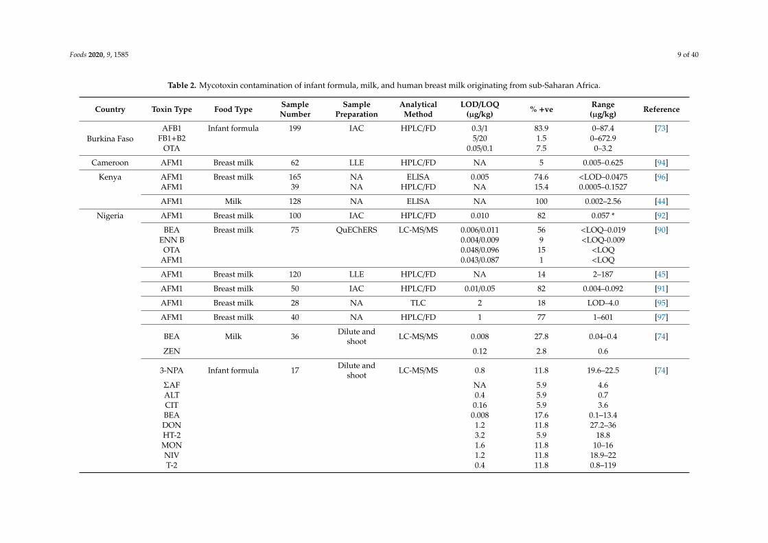
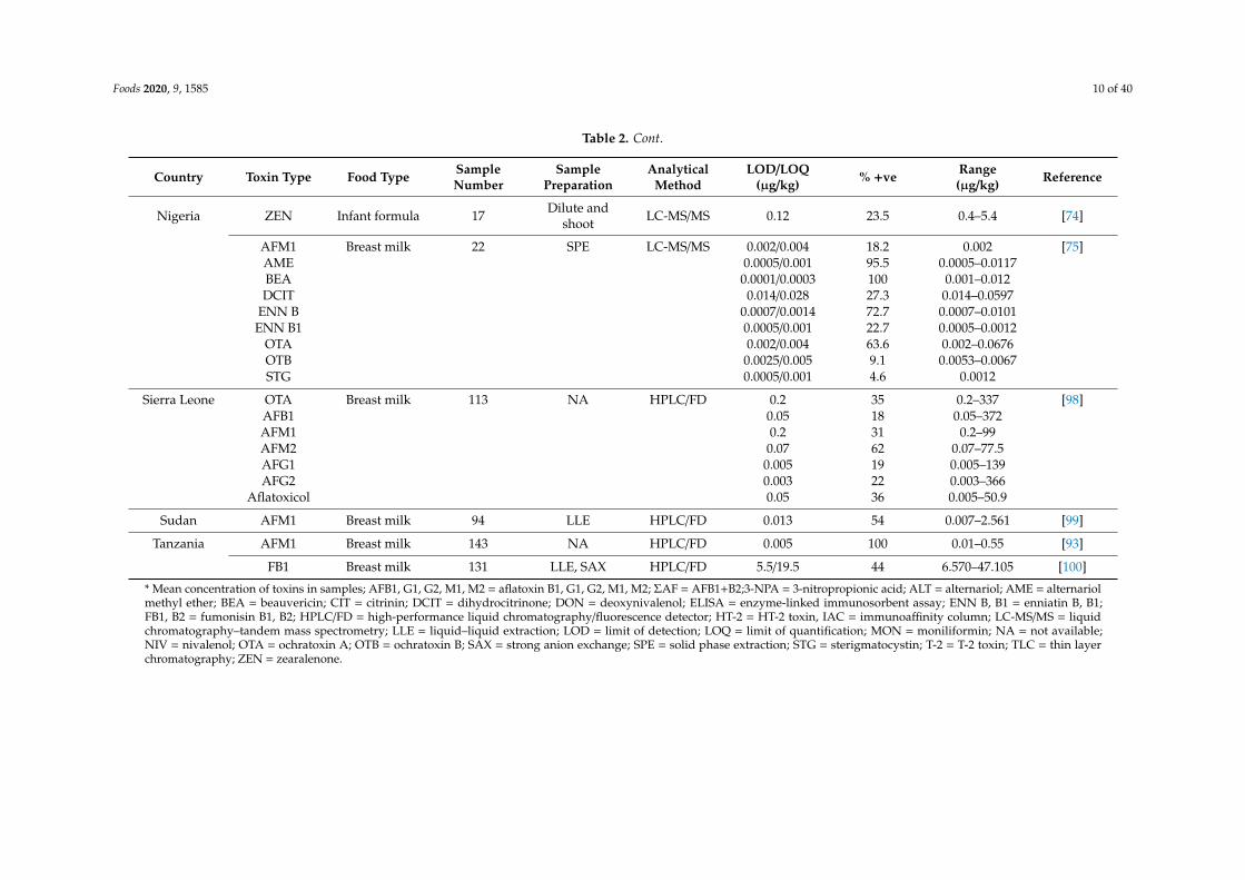
Table 2. Mycotoxin contamination of infant formula, milk, and human breast milk originating from sub-Saharan Africa.
Country Toxin Type Food Type SampleNumber
SamplePreparation
AnalyticalMethod
LOD/LOQ(µg/kg) % +ve Range
(µg/kg) Reference
Burkina FasoAFB1 Infant formula 199 IAC HPLC/FD 0.3/1 83.9 0–87.4 [73]
FB1+B2 5/20 1.5 0–672.9OTA 0.05/0.1 7.5 0–3.2
Cameroon AFM1 Breast milk 62 LLE HPLC/FD NA 5 0.005–0.625 [94]
Kenya AFM1 Breast milk 165 NA ELISA 0.005 74.6 <LOD–0.0475 [96]AFM1 39 NA HPLC/FD NA 15.4 0.0005–0.1527
AFM1 Milk 128 NA ELISA NA 100 0.002–2.56 [44]
Nigeria AFM1 Breast milk 100 IAC HPLC/FD 0.010 82 0.057 * [92]
BEA Breast milk 75 QuEChERS LC-MS/MS 0.006/0.011 56 <LOQ–0.019 [90]ENN B 0.004/0.009 9 <LOQ-0.009
OTA 0.048/0.096 15 <LOQAFM1 0.043/0.087 1 <LOQ
AFM1 Breast milk 120 LLE HPLC/FD NA 14 2–187 [45]
AFM1 Breast milk 50 IAC HPLC/FD 0.01/0.05 82 0.004–0.092 [91]
AFM1 Breast milk 28 NA TLC 2 18 LOD–4.0 [95]
AFM1 Breast milk 40 NA HPLC/FD 1 77 1–601 [97]
BEA Milk 36 Dilute andshoot LC-MS/MS 0.008 27.8 0.04–0.4 [74]
ZEN 0.12 2.8 0.6
3-NPA Infant formula 17 Dilute andshoot LC-MS/MS 0.8 11.8 19.6–22.5 [74]
ΣAF NA 5.9 4.6ALT 0.4 5.9 0.7CIT 0.16 5.9 3.6BEA 0.008 17.6 0.1–13.4DON 1.2 11.8 27.2–36HT-2 3.2 5.9 18.8MON 1.6 11.8 10–16NIV 1.2 11.8 18.9–22T-2 0.4 11.8 0.8–119
Foods 2020, 9, 1585 10 of 40
Table 2. Cont.
Country Toxin Type Food Type SampleNumber
SamplePreparation
AnalyticalMethod
LOD/LOQ(µg/kg) % +ve Range
(µg/kg) Reference
Nigeria ZEN Infant formula 17 Dilute andshoot LC-MS/MS 0.12 23.5 0.4–5.4 [74]
AFM1 Breast milk 22 SPE LC-MS/MS 0.002/0.004 18.2 0.002 [75]AME 0.0005/0.001 95.5 0.0005–0.0117BEA 0.0001/0.0003 100 0.001–0.012DCIT 0.014/0.028 27.3 0.014–0.0597
ENN B 0.0007/0.0014 72.7 0.0007–0.0101ENN B1 0.0005/0.001 22.7 0.0005–0.0012
OTA 0.002/0.004 63.6 0.002–0.0676OTB 0.0025/0.005 9.1 0.0053–0.0067STG 0.0005/0.001 4.6 0.0012
Sierra Leone OTA Breast milk 113 NA HPLC/FD 0.2 35 0.2–337 [98]AFB1 0.05 18 0.05–372AFM1 0.2 31 0.2–99AFM2 0.07 62 0.07–77.5AFG1 0.005 19 0.005–139AFG2 0.003 22 0.003–366
Aflatoxicol 0.05 36 0.005–50.9
Sudan AFM1 Breast milk 94 LLE HPLC/FD 0.013 54 0.007–2.561 [99]
Tanzania AFM1 Breast milk 143 NA HPLC/FD 0.005 100 0.01–0.55 [93]
FB1 Breast milk 131 LLE, SAX HPLC/FD 5.5/19.5 44 6.570–47.105 [100]
* Mean concentration of toxins in samples; AFB1, G1, G2, M1, M2 = aflatoxin B1, G1, G2, M1, M2; ΣAF = AFB1+B2;3-NPA = 3-nitropropionic acid; ALT = alternariol; AME = alternariolmethyl ether; BEA = beauvericin; CIT = citrinin; DCIT = dihydrocitrinone; DON = deoxynivalenol; ELISA = enzyme-linked immunosorbent assay; ENN B, B1 = enniatin B, B1;FB1, B2 = fumonisin B1, B2; HPLC/FD = high-performance liquid chromatography/fluorescence detector; HT-2 = HT-2 toxin, IAC = immunoaffinity column; LC-MS/MS = liquidchromatography–tandem mass spectrometry; LLE = liquid–liquid extraction; LOD = limit of detection; LOQ = limit of quantification; MON = moniliformin; NA = not available;NIV = nivalenol; OTA = ochratoxin A; OTB = ochratoxin B; SAX = strong anion exchange; SPE = solid phase extraction; STG = sterigmatocystin; T-2 = T-2 toxin; TLC = thin layerchromatography; ZEN = zearalenone.
Foods 2020, 9, 1585 11 of 40
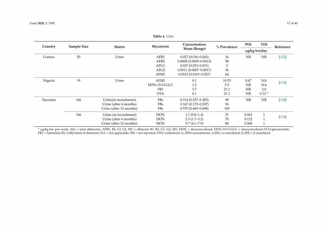
The frequency of AFM1 in breast milk samples was also reported in other countries in SSA,including Tanzania, Cameroon, Kenya, and Sudan [45,93,94,99]. In addition to AFM1, a Tanzanian studyusing HPLC with a fluorescence detector reported FB1 in 44.3% of 131 breast milk samples. This wassucceeded by a confirmation study using a high-resolution liquid chromatographic separation techniqueto validate the FB1 levels in the samples. According to the study, the concentration of FB1 ranged from6570 ng/L to 471,500 ng/L, with about 10.3% of the samples exceeding the FB1 regulatory limits of200 µg/kg set by the EU [93]. Another study using a highly sensitive and specific multi-mycotoxinassay reported the occurrence of OTA, beauvericin (BEA), enniatin B (ENN B), and AFM1 in breast milksamples from Nigeria out of the 28 mycotoxins evaluated [90]. Interestingly, BEA was the dominanttoxin, contaminating 56% of the samples with a concentration up to 19 ng/L. Ochratoxin A, ENN B,and AFM1 were detected in 15%, 9%, and 1% of the samples, respectively, with the concentrationbeing below the limit of quantification (LOQ), except for one sample with ENN B at a concentration of9 ng/L [90]. This is in line with a recent study by Ezekial et al. [75], which detected nine mycotoxins,including AFM1, alternariol methyl ether (AME), BEA, dihydrocitrinone (DCIT), ENN B, ENN B1,OTA, ochratoxin B (OTB), and sterigmatocystin (STG) in breast milk samples from Nigeria. Anotherstudy also reported a 40% co-occurrence rate of AFs and OTA in breast milk samples obtained from113 mothers from Sierra Leone, with the individual toxin concentrations ranging from 3-372,000 ng/Lfor AFs (AFB1, AFM1, AFM2, AFG1, AFG2, and aflatoxicol) and 200–337,000 ng/L for OTA [98].
According to the US Food and Drug Administration, infant formula, which represents a specialdiet, is used solely as infant food by reason of its simulation of human milk or suitability as a completeor partial substitute for human milk (FDA, 2018). Although infant formula serves as complementaryfood for IYC and supplies their daily nutritional requirements, these products may also be a routeof mycotoxin exposure to this age group. The occurrence of mycotoxins in infant formulas has beenreported in different parts of the world. Meucci et al. (2010) investigated the occurrence of AFB1 andOTA in 14 leading brands of infant formulas available in the Italian market. While 133 (72%) sampleswere positive of OTA (range: limit of detection (LOD)–0.690 µg/kg), only two (1%) samples werepositive of AFM1 (range: LOD–0.015 µg/kg) [101]. Another study on the occurrence of mycotoxins ininfant formula marketed in Ankara, Turkey using ELISA detected AFB1, AFM1, and OTA at a rateof 87%, 36.5%, and 40%, and within a range of 0.10–6.04 µg/kg, 0.06–0.32 µg/kg, and 0.27–4.50 µg/kg,respectively [102]. Other countries, such as Pakistan, Canada, Brazil, and South Korea, have alsoreported mycotoxins in infant formulas [103–106].
A few studies from SSA also revealed a high occurrence of various mycotoxins in infant formula(Table 2). Ware et al. reported mycotoxin contamination of infant formulas sampled from the city ofOuagadougou, Burkina Faso [73]. AFB1 was the most dominant mycotoxin, occurring at a rate of 83.9%with a concentration of up to 87.4 µg/kg. Other toxins detected included OTA and FBs (FB1 + FB2),with 7.5% and 1.5% of the samples exceeding the EU regulatory limits and having concentrationsup to 3.2 µg/kg and 672.9 µg/kg, respectively. This study suggests that IYC in this region may behighly exposed to mycotoxins, considering the high occurrence rate and maximum concentrations,which were 900, six, and three times higher than the EU regulatory limits of 0.1 µg/kg (AFB1), 0.5 µg/kg(OTA), and 200 µg/kg (FB) set for foods for IYC, respectively. Equally, Ojuri et al. [74] investigatedthe distribution of mycotoxins in 17 infant formula samples routinely fed to IYC in Nigeria andobserved multiple occurrences of mycotoxins. ZEN was the most prevalent toxin, occurring at arate of 23.5% (range: 0.4–5.4 µg/kg), followed by BEA (17.5%, range 0.1–13.4 µg/kg). Trichothecenes(DON, nivalenol (NIV), and T-2 toxin (T-2)) and moniliformin (MON) were detected at a frequencyof 11.8%, while AFB1 and AFB2 were detected in one sample at a concentration of 4.2 µg/kg and0.5 µg/kg, respectively.
Foods 2020, 9, 1585 12 of 40
2.3. Oilseed- and Fruit-Based Products
In addition to cereals, the utilisation of other plant products such as pulses and oilseeds, fruits,and vegetables in the production of IYC foods has been reported [107–109]. Their uses have beenencouraged, especially in developing regions, such as SSA, to help combat the high rate of malnutritionand its associated health disorders in the region. Soybean and groundnut serve as cheaper alternativesin the absence of animal protein. However, studies have shown that IYC food products processed withthese plant products may also be contaminated with mycotoxins [70,110]. A Nigerian study reportedthe occurrence of Fusarium mycotoxins in processed soybean powder, a major complementary weaningfood in the country [48]. The result revealed the frequency of ZEN (81%), 15-acetyldeoxynivalenol(15-AcDON) (31%), FBs (28%), and HT-2 toxin (HT-2) (25%) in the samples analysed. AF, FB1, and OTAhave also been detected in processed soybean powder samples from Nigeria at incidence rates of 45,100, and 40%, respectively [111]. On the other hand, the susceptibility of groundnut/peanut to fungiand their metabolites has been reported in several countries in SSA [74,77].
Peanut butter is used in weaning foods such as porridges. A study conducted by Ojuri et al. [74]on five samples of peanut butter from Nigeria revealed the occurrence of mycotoxins in 80% of thesamples, including AFs (80%), BEA (80%), MON (60%), and 3-nitropropionic acid (20%) in rangesof 6.5–13.6 µg/kg, 0.6–4.1 µg/kg, 2.2–3.5 µg/kg, and 5.4 µg/kg, respectively (Table 1). Another groupof authors conducted a comprehensive multi-year survey of AF contamination in 954 containers of24 local and imported peanut butter brands collected between 2012 and 2014 from Zambian shops [77].Overall, 73%, 80%, and 53% of the 24 peanut butter brands analysed in 2012, 2013, and 2014 werecontaminated with AFs, with AFB1 concentrations of up to 130 µg/kg, 10,740 µg/kg, and 1000 µg/kg,respectively. It is important to note that some of the brands analysed in this study were manufacturedand imported from South Africa, Malawi, and Zimbabwe, thus suggesting that IYC in these countriesmay also be predisposed to these toxins. The result observed by these authors is in line with the studiesthat reported the occurrence of AFs in peanut butter and groundnut products from Zimbabwe andZambia, respectively [78,112].
Mycotoxins may also contaminate fruit and vegetables, and subsequently be transferred intotheir processed products because of their chemically stable nature, even when exposed to hightemperatures. Studies from EU countries like Germany, Spain, Portugal, and Serbia, and other partsof the world, have reported the occurrence of mycotoxins, especially patulin (PAT), in fruit-basedinfant food products [63,113–116]. Whilst there are data available on mycotoxin contamination of fruit-and vegetable-based IYC foods in different regions of the world, unfortunately, there is little or noinformation in SSA, which suggests the need for more studies in the region to ascertain the extent ofmycotoxin contamination of these products.
3. Mycotoxin Exposure Assessment of Infants and Young Children in Sub-Saharan Africa
In recent years, extensive attention has been channelled to the most vulnerable group regardingthe toxic effects exerted by mycotoxins. This is evident in the continuous monitoring of foods forIYC, assessment of exposure, and characterisation of risk associated with IYC exposure to mycotoxinsand the stringent establishment and enforcement of regulatory limits to protect this subgroup of thepopulation. While these efforts are functional in developed countries, economically developing regionssuch as SSA still face the challenge of high levels of mycotoxin exposure of IYC due to the endemicnature of food contamination in the region. To this effect, only a few studies in SSA have evaluatedand reported the exposure of IYC to mycotoxins, as well as the associated risk.
3.1. Dietary Exposure Assessment
The estimation of dietary exposure can be based on either a numerical point estimation(deterministic) approach or a stochastic (probabilistic) approach, which uses a random probabilitydistribution pattern. Ojuri et al. [74] assessed the mycotoxin (AFs, FBs, OTA, CIT, BEA, and MON)
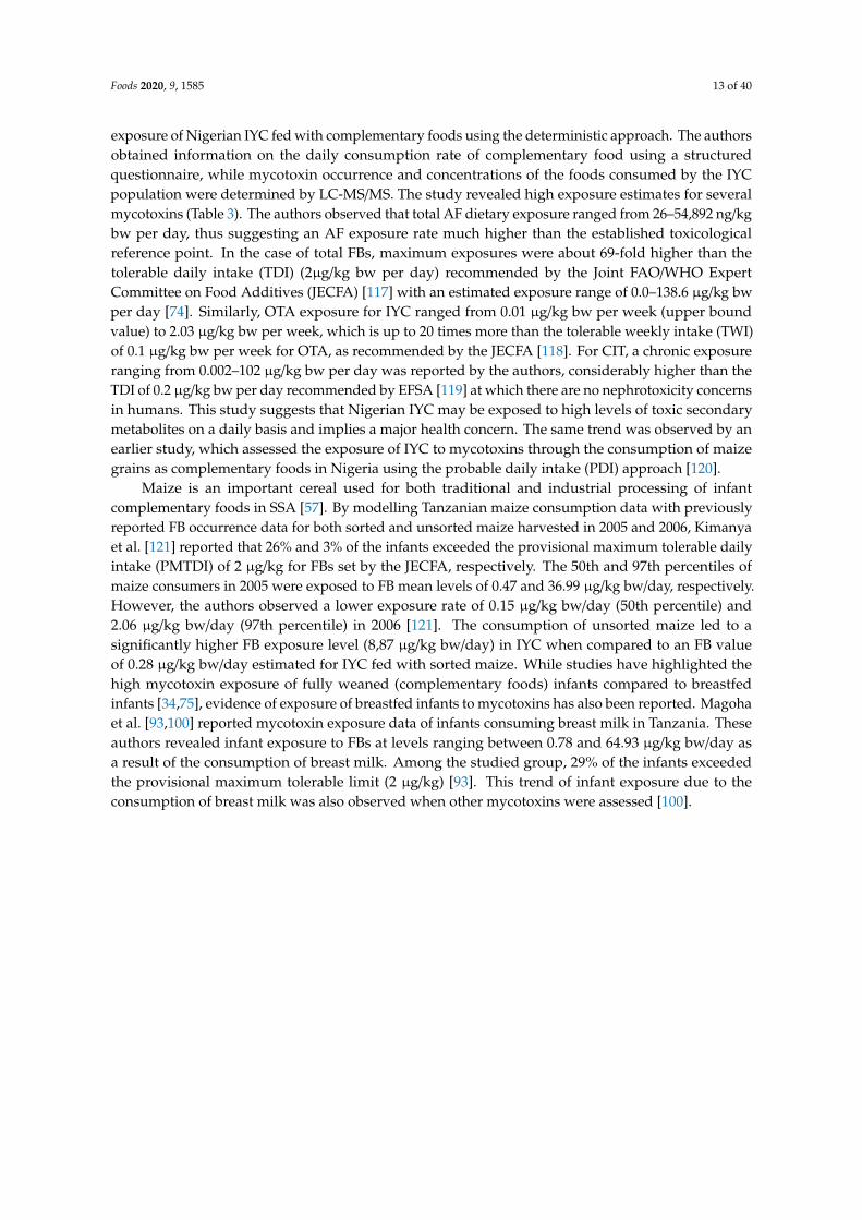
Foods 2020, 9, 1585 13 of 40
exposure of Nigerian IYC fed with complementary foods using the deterministic approach. The authorsobtained information on the daily consumption rate of complementary food using a structuredquestionnaire, while mycotoxin occurrence and concentrations of the foods consumed by the IYCpopulation were determined by LC-MS/MS. The study revealed high exposure estimates for severalmycotoxins (Table 3). The authors observed that total AF dietary exposure ranged from 26–54,892 ng/kgbw per day, thus suggesting an AF exposure rate much higher than the established toxicologicalreference point. In the case of total FBs, maximum exposures were about 69-fold higher than thetolerable daily intake (TDI) (2µg/kg bw per day) recommended by the Joint FAO/WHO ExpertCommittee on Food Additives (JECFA) [117] with an estimated exposure range of 0.0–138.6 µg/kg bwper day [74]. Similarly, OTA exposure for IYC ranged from 0.01 µg/kg bw per week (upper boundvalue) to 2.03 µg/kg bw per week, which is up to 20 times more than the tolerable weekly intake (TWI)of 0.1 µg/kg bw per week for OTA, as recommended by the JECFA [118]. For CIT, a chronic exposureranging from 0.002–102 µg/kg bw per day was reported by the authors, considerably higher than theTDI of 0.2 µg/kg bw per day recommended by EFSA [119] at which there are no nephrotoxicity concernsin humans. This study suggests that Nigerian IYC may be exposed to high levels of toxic secondarymetabolites on a daily basis and implies a major health concern. The same trend was observed by anearlier study, which assessed the exposure of IYC to mycotoxins through the consumption of maizegrains as complementary foods in Nigeria using the probable daily intake (PDI) approach [120].
Maize is an important cereal used for both traditional and industrial processing of infantcomplementary foods in SSA [57]. By modelling Tanzanian maize consumption data with previouslyreported FB occurrence data for both sorted and unsorted maize harvested in 2005 and 2006, Kimanyaet al. [121] reported that 26% and 3% of the infants exceeded the provisional maximum tolerable dailyintake (PMTDI) of 2 µg/kg for FBs set by the JECFA, respectively. The 50th and 97th percentiles ofmaize consumers in 2005 were exposed to FB mean levels of 0.47 and 36.99 µg/kg bw/day, respectively.However, the authors observed a lower exposure rate of 0.15 µg/kg bw/day (50th percentile) and2.06 µg/kg bw/day (97th percentile) in 2006 [121]. The consumption of unsorted maize led to asignificantly higher FB exposure level (8,87 µg/kg bw/day) in IYC when compared to an FB valueof 0.28 µg/kg bw/day estimated for IYC fed with sorted maize. While studies have highlighted thehigh mycotoxin exposure of fully weaned (complementary foods) infants compared to breastfedinfants [34,75], evidence of exposure of breastfed infants to mycotoxins has also been reported. Magohaet al. [93,100] reported mycotoxin exposure data of infants consuming breast milk in Tanzania. Theseauthors revealed infant exposure to FBs at levels ranging between 0.78 and 64.93 µg/kg bw/day asa result of the consumption of breast milk. Among the studied group, 29% of the infants exceededthe provisional maximum tolerable limit (2 µg/kg) [93]. This trend of infant exposure due to theconsumption of breast milk was also observed when other mycotoxins were assessed [100].
Foods 2020, 9, 1585 14 of 40
Table 3. Summary of mycotoxin exposure studies on infants and young children in sub-Saharan Africa using occurrence and consumption data.
Country SampleSize
Mycotoxin Concentration(µg/kg)
Exposure Level, Mean (Range) % PrevalencePDI TDI
Referenceµg/kg bw/day
Nigeria 70 AFB1 (infants) 324.7 * 1850 * ng/kg bw/day NR 1.91 * 0.00017 [120]AFB1 (children) 324.7 * 740 * ng/kg bw/day 0.76 * 0.00017
137 AFB1 0.12–473.8 2.5–51,192 ng/kg bw/day NR NR 0.00017
[74]AFs 0.88–589.8 25.7–54,892 ng/kg bw/day 0.00017FBs 4.6–1,540 0–138.6 ng/kg bw/day 2
OTA 0–26.4 0–2.03 ng/kg bw/day 0.1a
CIT 0.08–1,173 0.002–102 ng/kg bw/day 0.2BEA 0.004–69 0–3.14 ng/kg bw/day 90
MON 0.8–3,450 0.02–156.8 ng/kg bw/day 200
Tanzania 254 FBs (sorted maize) 19–1758 1.99 (0.32–144.29) b µg/kg bw/day 10 c <1.0 2.0 [121]FBs (unsorted maize) 19–21,666 36.80 (10.2–144.29) b µg/kg bw/day
143 AFs 0.33–69.47 0.14–120 ng/kg bw/day 39 0.017 [76]FBs 48–1224 0.005–0.88 µg/kg bw/day 21 <2 2
131 FBs 6.57–471.1 0.78–64.93 µg/kg bw/day 44.3 2.0 [93]
143 AFM1 0.01–0.55 0:81–66.79 ng/kg bw/day 96 d NR NR [100]
41 AFs 0.11–386 1–786 ng/kg bw/day 32 0.017 [72]DON 57–825 0.38–8.87 µg/kg bw/day 44 1FBs 63–2284 0.19–26.37 µg/kg bw/day 83 2
191 FBs 21–3201 0.003–28.84 µg/kg bw/day 69 NR 2 [38]
* Mean concentration; a µg/kg bw per week; b FB exposure at 97th percentile; c exceeded the PMTDI; d exceeded 0.025 ng/mL(EU limit for AFM1 contamination in infant food (EC, 2006));AFs = total aflatoxins; AFB1 = aflatoxin B1; BEA = beauvericin; CIT = citrinin; DON = deoxynivalenol; FBs = total fumonisins; MON = moniliformin; NR = not reported; OTA = ochratoxin A.
Foods 2020, 9, 1585 15 of 40
In addition, it is important to note that co-exposure to multiple mycotoxins may occur, whichcould lead to either synergistic or/and additive effects on the host, though toxicity is dependent onexposure time and dose as well as animal species. A study estimated the co-exposure of rural Tanzanianchildren fed with maize-based complementary foods to multiple mycotoxins, including AFs, DON,and FBs [72]. In the study, out of the 41 children evaluated, 41% were exposed to both DON andFBs, while 29% of the children were exposed to AFs and FBs. Co-exposure of the three mycotoxinsassessed (AFs, DON, and FBs) was observed in 10% of the children. For individual toxins, 32% out ofthe 41 children were exposed to AFs at exposure levels of 1–786 ng/kg bw/day, thus exceeding the AFexposure limit (0.017 ng/kg bw/day). Forty-four percent and 83% of the children had DON and FBexposure, ranging from 0.3–8.87 µg/kg bw/day and 0.19–26.37 µg/kg bw/day, respectively. Among theFB- and DON-exposed children, 56% and 66% exceeded the provisional tolerable intakes of 2 µg/kgbw/day and 1 µg/kg bw/day, respectively [72]. When compared with an earlier exposure study byKimanya et al. [38] from the same country, the researchers observed 15% higher FB exposure rateswith 46% of the children exceeding PMTDI (2 µg/kg bw/day) of FB. They concluded that the variationmay be due to the fact that the study focused on older children who probably consumed more foodscompared to the infant cohort used in their previous study.
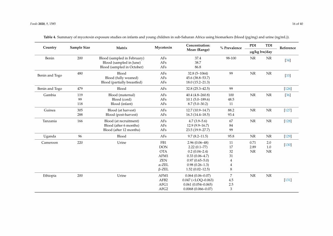
3.2. Mycotoxin Exposure Assessment Using Biomarkers
Beside estimating exposure based on food consumption and mycotoxin concentration in food,the assessment of mycotoxin exposure using biomarker analysis has gained prominence in recentyears. Biomarker analysis covers mycotoxin intake from all dietary sources and exposure routes [122],unlike the use of the occurrence and dietary intake approach which, due to heterogeneous distributionof mycotoxins in food products and limited accuracy in consumption data, often lead to under- oroverestimation of exposure and, consequently, risk. Mycotoxin biomarker analysis in SSA is still inits infancy, albeit available exposure studies using the approach have shown consistently high levelsof exposure in IYC (Table 4) [39]. Mycotoxins such as AFs are lipophilic in nature, have the abilityto cross the placental barrier, and can be bioactivated in utero. It is worrying that a huge number ofinfants in SSA are pre-exposed to mycotoxins early in the uterus through a transplacental pathway as aresult of maternal exposure, thus resulting in developmental issues [36,123–125].
Foods 2020, 9, 1585 16 of 40
Table 4. Summary of mycotoxin exposure studies on infants and young children in sub-Saharan Africa using biomarkers (blood (pg/mg) and urine (ng/mL)).
Country Sample Size Matrix Mycotoxin Concentration:Mean (Range) % Prevalence
PDI TDIReference
µg/kg bw/day
Benin 200 Blood (sampled in February) AFs 37.4 98-100 NR NR [34]Blood (sampled in June) AFs 38.7
Blood (sampled in October) AFs 86.8
Benin and Togo 480 Blood AFs 32.8 (5–1064) 99 NR NR [33]Blood (fully weaned) AFs 45.6 (38.8–53.7)
Blood (partially breastfed) AFs 18.0 (15.2–21.3)
Benin and Togo 479 Blood AFs 32.8 (25.3–42.5) 99 [126]
Gambia 119 Blood (maternal) AFs 40.4 (4.8–260.8) 100 NR NR [36]99 Blood (cord) AFs 10.1 (5.0–189.6) 48.5118 Blood (infant) AFs 8.7 (5.0–30.2) 11
Guinea 305 Blood (at harvest) AFs 12.7 (10.9–14.7) 88.2 NR NR [127]288 Blood (post-harvest) AFs 16.3 (14.4–18.5) 93.4
Tanzania 166 Blood (at recruitment) AFs 4.7 (3.9–5.6) 67 NR NR [128]Blood (after 6 months) AFs 12.9 (9.9–16.7) 84
Blood (after 12 months) AFs 23.5 (19.9–27.7) 99
Uganda 96 Blood AFs 9.7 (8.2–11.5) 95.8 NR NR [129]
Cameroon 220 Urine FB1 2.96 (0.06–48) 11 0.71 2.0 [130]DON 2.22 (0.1–77) 17 2.89 1.0OTA 0.2 (0.04–2.4) 32 NR NR
AFM1 0.33 (0.06–4.7) 31ZEN 0.97 (0.65–5.0) 4α-ZEL 0.98 (0.26–1.3) 4β-ZEL 1.52 (0.02–12.5) 8
Ethiopia 200 Urine AFM1 0.064 (0.06–0.07) 7 NR NR[131]AFB2 0.047 (<LOQ–0.063) 4.5
AFG1 0.061 (0.054–0.065) 2.5AFG2 0.0068 (0.066–0.07) 3
Foods 2020, 9, 1585 17 of 40
Table 4. Cont.
Country Sample Size Matrix Mycotoxin Concentration:Mean (Range) % Prevalence
PDI TDIReference
µg/kg bw/day
Guinea 50 Urine AFB1 0.027 (0.016–0.043) 16 NR NR [132]AFB2 0.0008 (0.0005–0.0013) 58AFG1 0.027 (0.023–0.031) 2AFG2 0.0011 (0.0007–0.0017) 36AFM1 0.0163 (0.0101–0.027) 64
Nigeria 19 Urine AFM1 0.1 10.53 0.67 NA [133]DON-15-O-GLU 1.5 5.3 NR NA
FB1 3.7 21.1 NR 2.0OTA 0.1 21.1 NR 0.12 a
Tanzania 166 Urine(at recruitment) FBs 0.314 (0.257–0.383) 98 NR NR [128]Urine (after 6 months) FBs 0.167 (0.135–0.207) 96Urine (after 12 months) FBs 0.570 (0.465–0.698) 100
166 Urine (at recruitment) DON 1.1 (0.8–1.4) 51 0.063 1 [134]Urine (after 6 months) DON 2.3 (1.7–3.2) 70 0.122 1Urine (after 12 months) DON 5.7 (4.1–7.9) 80 0.268 1
a µg/kg bw per week; AFs = total aflatoxins; AFB1, B2, G1 G2, M1 = aflatoxin B1, B2, G1, G2, M1; DON = deoxynivalenol; DON-15-O-GLU = deoxynivalenol-15-O-glucuronide;FB1 = fumonisin B1; LOQ=limit of detection; NA = not applicable; NR = not reported; OTA=ochratoxin A; ZEN=zearalenone; α-ZEL=α-zearalenol; β-ZEL= β-zearalenol.
Foods 2020, 9, 1585 18 of 40
Gong et al. [34], using serum AF-albumin adducts, reported the high AF exposure of children inBenin, West Africa, with AF-albumin levels as high as >1100 pg AF-lysine equivalents per milligramof albumin. There were strong variations in the levels of AF exposure depending on the village andgeographical region. This is possibly linked to the climatic conditions (temperature and humidity)of the environment, which favours the growth of fungi and subsequently mycotoxin production.The pre- and post-agricultural practices, which differ from region to region, may also be a contributoryfactor [135]. This is evident in the variation of AF-albumin prevalence (February (98%), June (99.5%),and October (100%)) observed by the authors as a result of seasonal changes, even though the variationwas not significant [34]. Earlier studies by this group of authors observed a remarkable increasein IYC exposure to AFs following the introduction of and increase in the consumption of weaningfoods [34,126]. Similar results were reported in a South African study on non-breastfed infants [136].Significant levels of OTA were detected in the plasma of non-breastfed infants at 6 weeks old ascompared to breastfed infants. The authors also highlighted a progressive trend in the exposure upuntil the 8th week. Further studies originating from other countries in SSA using serum and plasmabiomarkers have also indicated the exposure of IYC to AFs and FBs [33,36,126–129,137].
Consistent with these data, exposure studies using urinary biomarker approaches have indicatedhigh concentrations of mycotoxins in IYC [128,130–134,137,138]. A Cameroonian study investigatedthe mycotoxin exposure of 220 children between the age of 1.5–4.5 years using urinary biomarkers [130].Of the children investigated, mycotoxins were detected in 73% of the urine samples, with OTA being themost prevalent (30%) toxin, occurring within a concentration range of 0.04–2.4 ng/mL. Other mycotoxinsdetected in the urine samples included FB1 (11%), DON (17%), AFM1 (14%), ZEN (4%), α-zearalenol(α-ZEL) (4%), and β -zearalenol (β-ZEL) (8%), with up to two, three, and four mycotoxins co-occurringin samples at a rate of 35%, 5%, and 5%, respectively [130]. The detection of multiple mycotoxinsin IYC urine has also been reported by other studies from the region. Ezekiel et al. [133] detectedFB1, AFM1, OTA, and deoxynivalenol-15-O-glucuronide in the urine samples of Nigerian children,with a mycotoxin prevalence rate of 47.3% (9/19). Using an ultra-sensitive stable isotope-assistedquantification method, the same group of authors further confirmed the exposure of Nigerian IYC tomultiple mycotoxins [138].
Ayelign et al. [131] indicated the presence of AFs in 17% of urine samples of Ethiopian children,with AFM1 being the most prevalent (7%) toxin. Other AFs were detected in 4.5% (AFB2), 2.5% (AFG1),and 3% (AFG2) of samples. A similar study in Guinea reported the occurrence of AFs in children’s urinesamples at a higher occurrence rate of 86% [132]. While AFB1, AFB2, AFG1, and AFG2 were detectedin the samples, AFM1 was the dominant mycotoxin, occurring at a frequency and concentrationrange of 64% and 8.0–801 pg/mL, respectively. Using a urinary biomarker, Shirima et al. [128,137]revealed high levels of FBs among Tanzanian IYC, with individual levels as high as 698.2 ng/L.The authors further observed a discrepancy in mycotoxin exposure levels between the samplingtimes, which could be linked to the effects of consumption of contaminated food as well as seasonalvariation of mycotoxin contamination levels [128]. The same trend was observed for DON amongIYC in Tanzania, with concentration ranges of 800–1400 ng/L, 1700–3200 ng/L, and 4100–7900 ng/L atrecruitment, after 6 months, and after 12 months, respectively [134].
4. Health Implications Associated with Mycotoxin Exposure of Infants and Young Children inSub-Saharan Africa
The health significance of human exposure to mycotoxins should not be neglected, especially withregard to infants and young children in SSA. This population group on a daily basis is subjected totoxic metabolites through food consumption, inhalation, and dermal adsorption. Albeit there are nostudies on the exposure to and effect of mycotoxins as a result of inhalation and dermal adsorption inSSA, there are several reports from this region on the detrimental effects of ingestion of mycotoxins,especially AFs and FBs, on the health of IYC.
Foods 2020, 9, 1585 19 of 40
4.1. Child Growth Impairment
Researchers have studied the possible association between mycotoxin exposure, especially to AFsand FBs, and IYC development. The few studies conducted to determine the relationship betweenmycotoxins (AFs and FBs) and IYC growth have been reviewed by Smith et al. [42], Gong et al. [139],and Tesfamariam et al. [140].
4.1.1. Aflatoxin Exposure and Infant Growth
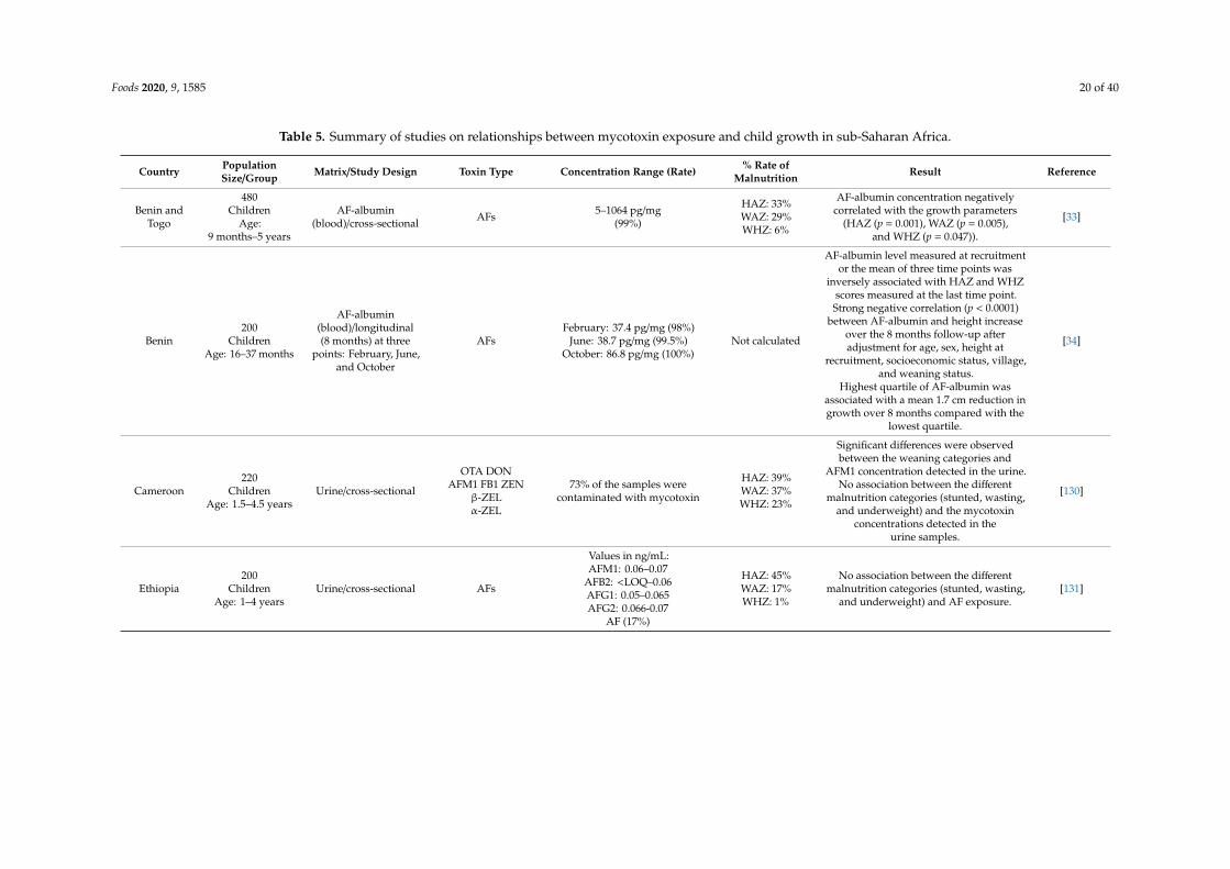
Using a cross-sectional study design, Okoth and Ohingo [37] investigated the possible associationbetween child dietary exposure to AFs and growth impairment among 242 children within the agerange of 3 to 36 months from Kisumu District of Kenya. They revealed a highly significant relationshipbetween the number of children fed with mycotoxin-contaminated flour and the prevalence of wasting(p = 0.002) (Table 5). In another study in Kenya with 204 children (age: 1–3 years), AFM1 exposurewas negatively associated with height-for-age Z-score (HAZ) score (p=0.047), however, there were noassociations between total AFs and HAZ, weight-for-age Z-score (WAZ), or weight-for-height Z-scorescores (WHZ) [44]. The authors found prevalence of stunting, underweight, and wasting of 34%, 30%,and 6%, respectively, of which 53.8% of the children classified as wasted consumed AF-contaminatedflour compared with 27.7% of not wasted children (p=0.002). Sixty percent of the 30.7% of severeprotein energy malnourished children were weaned with AF-contaminated flour compared to 27.4% ofthe normal children (p = 0.004).
Foods 2020, 9, 1585 20 of 40
Table 5. Summary of studies on relationships between mycotoxin exposure and child growth in sub-Saharan Africa.
Country PopulationSize/Group Matrix/Study Design Toxin Type Concentration Range (Rate) % Rate of
Malnutrition Result Reference
Benin andTogo
480Children
Age:9 months–5 years
AF-albumin(blood)/cross-sectional AFs 5–1064 pg/mg
(99%)
HAZ: 33%WAZ: 29%WHZ: 6%
AF-albumin concentration negativelycorrelated with the growth parameters
(HAZ (p = 0.001), WAZ (p = 0.005),and WHZ (p = 0.047)).
[33]
Benin200
ChildrenAge: 16–37 months
AF-albumin(blood)/longitudinal(8 months) at three
points: February, June,and October
AFsFebruary: 37.4 pg/mg (98%)
June: 38.7 pg/mg (99.5%)October: 86.8 pg/mg (100%)
Not calculated
AF-albumin level measured at recruitmentor the mean of three time points was
inversely associated with HAZ and WHZscores measured at the last time point.Strong negative correlation (p < 0.0001)
between AF-albumin and height increaseover the 8 months follow-up afteradjustment for age, sex, height at
recruitment, socioeconomic status, village,and weaning status.
Highest quartile of AF-albumin wasassociated with a mean 1.7 cm reduction ingrowth over 8 months compared with the
lowest quartile.
[34]
Cameroon220
ChildrenAge: 1.5–4.5 years
Urine/cross-sectional
OTA DONAFM1 FB1 ZEN
β-ZELα-ZEL
73% of the samples werecontaminated with mycotoxin
HAZ: 39%WAZ: 37%WHZ: 23%
Significant differences were observedbetween the weaning categories and
AFM1 concentration detected in the urine.No association between the different
malnutrition categories (stunted, wasting,and underweight) and the mycotoxin
concentrations detected in theurine samples.
[130]
Ethiopia200
ChildrenAge: 1–4 years
Urine/cross-sectional AFs
Values in ng/mL:AFM1: 0.06–0.07
AFB2: <LOQ–0.06AFG1: 0.05–0.065AFG2: 0.066-0.07
AF (17%)
HAZ: 45%WAZ: 17%WHZ: 1%
No association between the differentmalnutrition categories (stunted, wasting,
and underweight) and AF exposure.[131]
Foods 2020, 9, 1585 21 of 40
Table 5. Cont.
Country PopulationSize/Group Matrix/Study Design Toxin Type Concentration Range (Rate) % Rate of
Malnutrition Result Reference
Gambia138
ChildrenAge: 0–12 months
AF-albumin(blood)/longitudinal
(14 months follow-upfrom birth until one year
of age)
AFs
Maternal blood: 4.8–260.8 pg/mg(100%)
Cord blood: 5.0–189.6 pg/mg(48.5%)
Not calculated
High maternal AF-albumin was associatedto lower HAZ (p = 0.044) and WAZ
(p = 0.012) scores.Reduction of maternal AF-albumin from110 pg/mg to 10 pg/mg would lead to an
increase of 0.8 kg in weight and 2 cmincrease in height of a child within the first
year of life.
[36]
Infant blood: 5.0–30.2 pg/mg(11%)
AF-albumin measured at Week 16 wasnegatively related to HAZ (p = 0.002).
374Infants
Age: 0–2 years
AF-albumin(blood)/longitudinal AFs
48, 98, and 99% had detectableAF-albumin concentrations(LOD > 3.0 pg/mg at 6, 12,18 months, respectively)
HAZ: 25.9%WAZ: 24.4%WHZ: 12.9%
Inverse relationships between AF-albuminadducts and HAZ, WAZ, and WHZ scores
from the age of 6 to 18 months.Inverse relationship between AF-albuminat 6 months and change in WHZ between
6 and 12 months (p = 0·013).AF-albumin at 12 months was associatedwith changes in HAZ and infant length
between the age of 12 and 18 months.AF-albumin at 6 months was associatedwith IGFBP-3 at 12 months (p = 0·043).
[141]
Kenya242
ChildrenAge: 3–36 months
Weaningflour/cross-sectional AFs 2–82 µg/kg (29%)
HAZ: 34%WAZ: 30%WHZ: 6%
Highly significant association betweenchildren that consumed AF-contaminatedflour and prevalence of wasting (p = 0.002).
[37]
204Children
Age: 1–3 years
Cereals (maize,sorghum) and
milk/cross-sectional
AFsAFM1
AF: 0–194 µg/kg in cerealsAFM1: 0.002–2.56 µg/kg in milk
(98%)
HAZ: 41%WAZ: 17%WHZ: 4%
AFM1 was negatively associated withHAZ (p = 0.047).
No association between total AFs (AFB andAFG) and HAZ, WAZ, and WHZ Scores.
[44]
Kenya881
ChildrenAge: 0–2 years
AFB1-lysineadduct/cluster
randomisedlongitudinal
AFs *18.1 pg/mg albumin Not calculated
The intervention significantly reducedendline serum AFB1-lysine adduct levels
(p = 0.025).No effect on the prevalence of stunting orLAZ, though a significant effect on child
linear growth was found at midline(11–19 months).
[142]
Foods 2020, 9, 1585 22 of 40
Table 5. Cont.
Country PopulationSize/Group Matrix/Study Design Toxin Type Concentration Range (Rate) % Rate of
Malnutrition Result Reference
Nigeria
58Children
Age: 6–48 months(with severe acute
malnutrition)
AFB1-lysine/cross-sectional AFs 0.2–59.2 pg/mgalbumin
Severe acutemalnutrition: 81%
HAZ: 74%
Significantly higher AFB1-lysineconcentrations in stunted children
compared to non-stunted children as wellas in children with severe acute
malnutrition compared to controls.No significant association between
AFB1-lysine and stunting after adjustmentfor malnutrition status (OR = quartile 3,
1.21; 95% CI: 0.086–31.45) and nocorrelation between AFB1-lysine and WAZ.
[143]
Tanzania215
InfantsAge: 6 months
Maize/longitudinal(follow-up at 6 and12 months of age)
FBs 21–3201 µg/kg(69% of 191 maize samples) Not calculated
Infants exposed to FBs above the PMTDI(2 µg/kg) were significantly shorter by
1.3 cm and lighter by 328 g at 12 months(p = 0.002).
[38]
143Infants
Age: 0–6 months143 Lactating
mothers
Breast milk/longitudinal(follow-up at three
points: 1st, 3rd, and 5thmonths of age)
AFM1
1st month:0.01–0.55 ng/mL
3rd month:0.01–0.47 ng/mL
5th month:0.01–0.34 ng/mL
1st month:HAZ: 11%WAZ: 4%WHZ: 4%3rd month:HAZ: 13%WAZ: 9%WHZ: 1%5th monthHAZ: 17%WAZ: 10%WHZ: 3%
Significant inverse association betweenAFM1 exposure levels and WAZ or HAZ
(p < 0.05).[100]
Tanzania
166Children
Age: 6–14 monthsold
AF-albumin and UFB1/longitudinal (12 month
follow-up)
AFsFBs
AF-albumin:at recruitment: *4.7 pg/mg (67%),
6 months: *12.9 pg/mg (84%),12 months: *23.5 pg/mg (99%)
UFB1:at recruitment: *313.9 pg/mg (98%),
6 months: *167.3 pg/mg (96%),12 months: *569.5 pg/mg (100%)
At recruitmentHAZ: 44%WAZ: 8%WHZ: 2%
At 6 months:HAZ: 55%WAZ: 14%WHZ: 2%
At 12 months:HAZ: 56%WAZ: 14%WHZ: 0.7%
No significant negative associationbetween mean AF-albumin levels and
child growth (HAZ, WHZ, or WAZ score).Negative association between meanUFB1 concentrations (at recruitment,
and 6 and 12 months from recruitment)and HAZ at recruitment.
[128]
143Infants
Age: under6 months
Maize flour/longitudinal(follow-up at three
points: 1st, 3rd, and 5thmonths of age)
AFsFBs
AF: 0.33–69.5 µg/kg (58% of67 maize samples)
FB: 48–1224 µg/kg (31% of67 maize samples)
WAZ: 35% of115 infants
HAZ: 43% of115 infants
Insignificant association was observedbetween exposure to AFs or FBs and
stunting or underweight.[76]
Foods 2020, 9, 1585 23 of 40
Table 5. Cont.
Country PopulationSize/Group Matrix/Study Design Toxin Type Concentration Range (Rate) % Rate of
Malnutrition Result Reference
300Children
Maize/clusterrandomised
controlled trial
AFsFBs Not reported Not calculated
AF and FB intakes were inverselyassociated with WAZ. WAZ was 6.7%
lower in the intervention group.Mean WAZ difference between the groups
was 0.57 (p = 0.007).
[144]
Tanzania
114Children
Age: under36 months
AFB1- lysine and UFB1/longitudinal
AFsFBs
AFB1-lysine:0.28–25.1 pg/mg (72%)
UFB1: <LOD–16.6 ng/mL (80%)
At 24 months:HAZ: 61%WAZ: 17%WHZ: 3%
At 36 months:HAZ: 75%WAZ: 21%WHZ: 0%
No associations were found between AFexposure
and growth impairment as measured bystunting, underweight, or wasting.
However, FB exposure was negativelyassociated with underweight.
[41]
Uganda 246Dyads
Maternalserum/longitudinal
(follow-up: pregnancyup to one year)
AFs AFB1: *113.9 pg/mg Not calculated
A negative effect of AF exposure on infantlinear growth (HAZ) in HIV-positive
pregnant women and their infants.Infants of HIV-positive women were inhigh perinatal AF category with lower
HAZ scores (0.460) when compared withthe infants of HIV-negative low-AF
exposed women (p = 0.006).
[145]
220 infantsAge: 0–48 h
Maternalserum/cross-sectional AFs AFB1: 0.71–95.6 pg/mg albumin Not calculated
Maternal AFB-lysine levels weresignificantly associated with lower birth
weight (adj-β: p = 0.040), lower WAZ(adj-β: p = 0.037), smaller HC (adj-β:
p = 0.035), and lower HCZ (adj-β:p = 0.023) at birth. No significant
associations were observed betweenmaternal AFB-lysine levels and infant
length, WHZ, HAZ, or gestational age atbirth.
[146]
* Mean concentrations, AFs = total aflatoxins; AFB1, M1 = aflatoxin B1, M1; FBs = total fumonisin B; FB1 = fumonisin B1; IGFBP-3 = insulin-like growth factor-1 binding protein-3;DON = deoxynivalenol; ZEN = zearalenone; α-ZEL = α-zearalenol; β-ZEL = β-zearalenol; LOD = limit of detection; LOQ = limit of quantification; OTA = ochratoxin A; UFB1 = urinaryfumonisin B1; ZEN = zearalenone; adj-β = adjusted model; HAZ = height-for-age Z-score; HC = head circumference; HCZ = head circumference-for-age Z-score; WAZ = weight-for-ageZ-score; WHZ = weight-for-height Z-score.
Foods 2020, 9, 1585 24 of 40
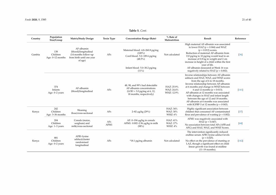
A similar study in Nigeria reported a significantly higher concentration of AF-lysine in stuntedand severe acute malnourished children compared to normal children [143]. This is in agreementwith the study by Hoffmann et al. [142] that examined the effectiveness of reducing AF exposureon child linear growth and AF serum levels of infants and young children in rural communities inEastern Kenya using a cluster randomised longitudinal study design. The study revealed a substantialreduction of endline serum AFs due to the consumption of AF-free maize, however, no effect on childlinear growth of the examined population was observed. Furthermore, the midline analysis suggestedthe possibility of AFs having an effect on linear growth at younger ages [142].
A cross-sectional study by Gong et al. [33] examined the relationship between AF exposure andgrowth in IYC between the ages of 9 months and 5 years from four geographic zones of Benin andTogo. Using anthropometric data and AF-albumin concentrations according to the World HealthOrganization (WHO) Z-score criteria, the authors observed higher AF-albumin in children with stunting,underweight, and wasting with a prevalence rate of 33%, 29%, and 6%, respectively. Notwithstandingthat the AF-albumin concentration negatively correlated with the growth parameters, they were highlysignificant [33]. This observation was supported by a later longitudinal study in the Republic of Benin,which after adjustment for socioeconomic status, anthropometric parameters, agro-ecological zone,and weaning status, showed a negative correlation between AF-albumin and height increase overthe 8-month follow-up period, hence suggesting a strong association between AFs and stunting [34].The same trend was recorded by Watson et al. [141], who reported an inverse relationship betweenAF-albumin adducts and HAZ, WAZ, and WHZ scores in a cohort of 374 Gambian infants between theages of 0 and 2 years.
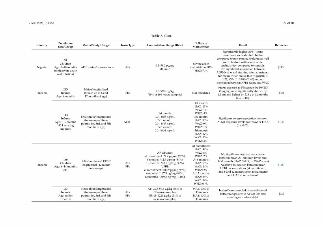
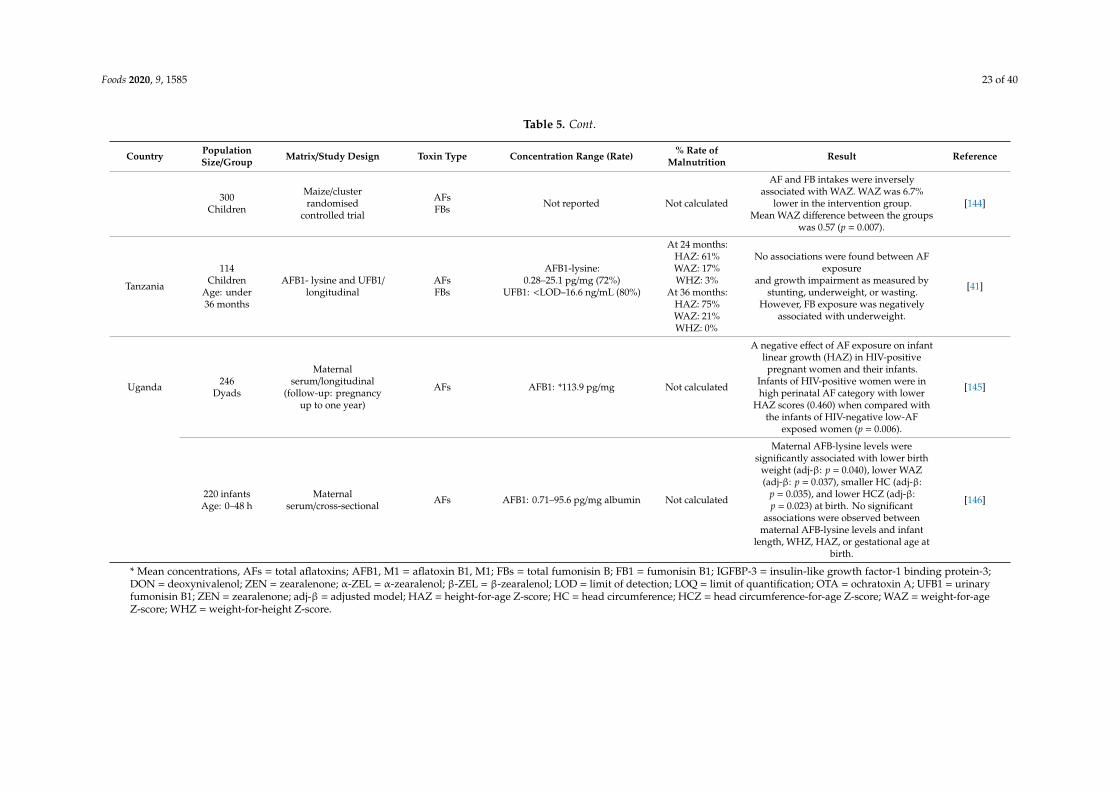
Lauer et al. [146] investigated the association between maternal AF exposure during pregnancyand birth outcomes in Uganda. The authorsobserved significant associations between maternalAFB-lysine levels and lower birth weight, lower WAZ, smaller head circumference (HC), and lowerhead circumference-for-age Z-score (HCZ) at birth. Conversely, no significant associations wereobserved between maternal AFB-lysine levels and infant length, WHZ, HAZ, or gestational age atbirth [146]. Another longitudinal study from Gambia examining the effect of in utero AF exposure oninfant growth using 138 pregnant women and their infants reported a significant association betweenmaternal AF exposure during pregnancy and growth faltering in infants [36]. The same trend wasobserved in a similar study on 143 lactating mothers and their infants in Tanzania, with a significant(p<0.05) inverse association between AFM1 exposure and stunting (HAZ score) and underweight(WAZ score) in infants fed with AFM1-contaminated breast milk [100]. In Uganda, using 246 dyads,Natamba et al. [145] investigated the association of perinatal exposure to AFs with a low rate ofweight gain among human immunodeficiency virus (HIV)-positive pregnant women and reducedlinear growth of HIV-exposed infants. They found a negative effect of AF exposure on infant lineargrowth in HIV-positive pregnant women and their infants, with the infants of HIV-positive women inthe high perinatal AF category having a lower HAZ scores (0.460) when compared with the infantsof HIV-negative low-AF-exposed women (p = 0.006) [145]. Shirima et al. [128] also conducted asimilar longitudinal study in three agro-ecological zones of Tanzania, investigating the effects of AFexposure and co-exposure on the growth of IYC using a cohort of 166 apparently healthy IYC (aged6 to 14 months old). The study revealed no significant association between AFs and child growthirrespective of the sampling time, which the authors attributed to the lower AF-albumin concentrations(geometric mean: 4.7 pg/mg) recorded. This is in line with an Ethiopian study [131], but in contrastto earlier studies reported in West Africa [33,34,36], Tanzania [100], and Kenya [37]. Furthermore,a non-significant association between AFs and infant growth was later reported by a study in Haydom,Tanzania, using a cohort of 114 children under the age of 36 months [41], which is in line with otherstudies from other regions of the world [147]. In addition to the variation in the concentrations of AFs,it is important to highlight that the different methodologies used in these studies may have influencedthe variation in the effects observed.
Foods 2020, 9, 1585 25 of 40
4.1.2. Fumonisin Exposure and Infant Growth
Shirima et al. [128] observed that the concentrations of urinary FBs (0.31, 0.17, and 0.57 ng/mL)detected in a cohort of 166 healthy children were negatively associated with growth impairment(rate: 44%, 55%, and 56%) at recruitment, 6 months, and 12 months, respectively, suggesting apossible association between FBs and child growth (Table 5). Chen et al. [41] also reported a negativeassociation of FBs with stunting, though at a higher incidence rate of 75%, which is attributed tothe higher urinary FB concentration (mean: 1.3 ng/mL) recorded in the study when compared toShirima et al. [128]. This therefore implies a possible dose-dependent effect between FBs and stunting(increase in FB concentration = increase in stunting) [41]. An earlier study originating from the samecountry reported a similar trend [38]. The authors examined the effect of FB exposure on the growthperformance of 215 infants complemented with maize-based complementary foods. They revealedthat infants exposed to FBs above the PMTDI of 2 µg/kg body weight/day established by the JECFAwere significantly shorter (1.3 cm) as compared to the other infants [38]. Another three-time point(1, 3, and 5 months) study investigating the relationship between FB exposure and stunting orunderweight in 143 Tanzanian infants under 6 months of age showed insignificant associations betweenthe variables [76]. Furthermore, it is important to mention that the influence of mycotoxins on infantgrowth is not only an SSA problem, as studies originating from other regions of the world havereported strong correlations between mycotoxin exposure and infant growth impairment [148–150].
4.1.3. Postulated Mechanism for Growth Impairment
Although the mechanism by which mycotoxins influence child growth is yet to be fully understood,Smith et al. [151] suggested two possible pathways by which these toxins may contribute to stunting,including the induction of environmental enteric dysfunction and systemic immune activation,resulting in the interruption of the insulin-like growth factor 1 (IGF1) axis. Castelino et al. [152]investigated the relationship between AF and IGF1 in 199 Kenyan schoolchildren, and observed thatAF-albumin concentrations were inversely associated with both IGF1 and IGF-binding protein-3(IGFBP3). IGF facilitates most of the growth-promoting effects of growth hormone, and thusplays a major role in the growth of a child [153]. AFs may also induce stunting by exerting animmunosuppressive effect, which often increases the susceptibility of infants to infection, loss ofappetite, and reduced nutrient absorption [154]. In addition, AFs may exert infant growth impairmentby mediating intestinal damage through the inhibition of protein synthesis [151]. Other mycotoxinslike DON have also been postulated to exert growth impairment through this mechanism.
On the other hand, FBs may contribute to impaired growth in infants by inhibiting ceramidesynthase, a major enzyme responsible for the biosynthesis of sphingolipids, hence leading to thedisruption of sphingolipid metabolism [155]. This is in line with the study by Semba et al. [156],who reported significantly lower serum concentrations of sphingomyelins in stunted children whencompared with non-stunted children in six villages in rural southern Malawi. Sphingomyelin, localisedin the plasma membrane, is a dominant sphingolipid in the membranes of mammalian cells, and plays amajor role in creating lateral structures in membranes for Toll-like receptors and class A and B scavengerreceptors, as well as insulin receptors [157]. Wu [158] also reported the involvement of sphingomyelinin cell signalling. The influence of FBs on intestinal barrier function by the alteration of the sphingoidbase-1 phosphate signalling pathway has also been reported. Riley et al. [159] revealed a positivecorrelation between the concentrations of urinary FB1 and sphinganine 1-phosphate/sphingosine1-phosphate and sphinganine 1-phosphate ratio in blood. The result was further confirmed by afollow-up study involving 299 Guatemalan women. In addition to ceramide synthase inhibition by FBs,Masching et al. [160] reported the potential of FBs to induce inflammatory responses, thereby promotingdamage to gut barrier function.
Foods 2020, 9, 1585 26 of 40
4.2. Child Immune and Nervous Systems
Exposure to mycotoxins has been shown to hamper immune responses in humans and animals,thus leading to a decrease in resistance to infectious diseases [35]. The mechanism through whichmycotoxins exert immunosuppressive or immunostimulatory effects on humans and animals mayvary depending on toxin type and exposure dose, as well as the investigated parameters highlightedin several reviews [161–163]. Bondy and Peska [162] highlighted the ability of mycotoxins topromote the expression of a diverse array of cytokines through inflammatory responses, with apotential to upregulate and downregulate a wide array of immune functions. While mycotoxinsimpair cell-mediated immunity and phagocytic cell function, exposure to AFs increases the T cellproliferation-inducing capacity of porcine monocyte-derived dendritic cells, therefore enhancing theantigen-presenting capacity of the cell [164]. The inhibition of humoral, cellular, and innate immunityby AFs, OTA, and FBs, leading to a reduced response to vaccines, has been reported in animalspecies [162,165].
Notwithstanding the evidence that IYC, especially in SSA, are most vulnerable to mycotoxinexposure, only a few scientific studies, mostly focusing on AFs, have been conducted to elucidate theeffects of these fungus metabolites on the immunity of IYC. Allen et al. [166] investigated the relationshipbetween AF exposure, hepatitis B infection, and the prevalence of malaria in 391 Gambian childrenbetween the age of 3 and 8 years, and found that a higher AF-albumin adduct concentration wasassociated with increased Plasmodium falciparum parasitaemia (p = 0.01) and hepatitis B surface antigen(p=0.04) carrier status of the children. However, the authors observed no consistent association betweenAF-albumin level, malaria infections, malaria-specific antibody, or lymphoproliferative responses.A later cross-sectional study from the same country, using a cohort of 472 children, studied the effect ofdietary AF exposure on immune parameters, including secretory IgA in saliva (sIgA) and cell-mediatedimmunity, as well as antibody response to rabies and pneumococcal vaccines [35]. A remarkably lowersIgA level of children with detectable AF-albumin in their blood was seen compared to no AF-albumindetectable children (p < 0.0001). However, out of the four pneumococcal serotypes, only one responsewas weakly associated with higher levels of AF-albumin adducts. On the other hand, no associationwas observed between AF-albumin and cell-mediated immunity or rabies [35].
Other West African studies, specifically in Ghana, showed the potential effects of AF exposure onimmune responses of HIV-positive participants [167–170], and tuberculosis patients [171], however,no such study on the infant population in these regions has been reported. Another study investigatedthe effect of OTA on T cell activation using a cohort of South African HIV-exposed infants andHIV-unexposed infants as controls [136]. The authors observed a correlation of OTA plasma levelswith the activation of CD4 T cells (HLADR and CCR5) and a chemokine, CXCL10. Increasedexpression of CXCL10, HLA-DR, and CCR5 has been associated with a variety of human diseases,including HIV, immune dysfunction, chronic inflammation, infectious diseases, tumour development,and encephalopathy, known as a major cause of infant mortality in Africa [136,172–174], thus suggestingan increased risk of morbidity and mortality due to OTA exposure.
Mycotoxin Exposure and Autism Spectrum Disorder
Recent epidemiological studies have revealed possible relationships between mycotoxin exposureand neurodevelopmental disorders in IYC, especially as with regard to autism spectrum disorder(ASD) [23,24], a lifelong neuro-developmental syndrome characterised by deficits in social activities,communication interactions, and unusual restricted and repetitive behaviours. Scientific evidenceindicates the role of multiple interacting genetic factors in the aetiology of ASD. With the recentincrease in the incidence of ASD worldwide [175], at a rate of 60 cases per 10,000 children, there is aquest to identify more possible contributory factors to the disorder. Notwithstanding the attributableresults of conceptual and technological advances in research, the possible role of environmental factors,such as toxic exposure, in triggering ASD should not be underestimated. In a cohort of 110 children,comprising 52 autistic and 58 healthy children (31 siblings and 27 unrelated subjects), De Santis et
Foods 2020, 9, 1585 27 of 40
al. [23] examined the role of mycotoxin exposure in the manifestation of ASD. The authors reported asignificant association between OTA concentrations in the serum (p = 0.0017) and urine (p = 0.0002) ofthe autistic children compared to the unrelated healthy children. In addition, a significant associationwas also found for OTA in urine when comparing ASD patients with healthy children (siblings andunrelated subjects) (p = 0.0081), whereas no significant difference was observed between ASD subjectsand their siblings. The authors highlighted that the average values of mycotoxins detected in ASDchildren were always lower than those of their siblings, despite the comparable diet system and toxinexposure level, which the authors ascribed to a possible alteration in biotransformation and metabolismof the toxins.
Similarly, another cross-sectional study using a cohort of 233 children, comprising 172 autistic and61 healthy (36 siblings and 25 non-parental) children, reported significant levels of mycotoxins in theurine and serum of the ASD children when compared to the controls, regardless of whether they weresiblings or non-parental [24]. The evidence observed in this study revealed mycotoxins as a possiblestress agent involved in the gene–environment interaction, eliciting ASD. In addition, the phenylalaninemolecule of OTA may inhibit phenylalanine hydroxylase, an enzyme responsible for the catalysis ofthe hydroxylation of the aromatic side-chain of phenylalanine to produce tyrosine. In the absenceof tyrosine, the biosynthesis of catecholamines is inhibited. Catecholamines interact with oxytocin,a hormone that plays a major role in social bonding and communication [23], thus contributing to theintellectual ability of an individual. This may explain the possible link of OTA exposure to autism,a syndrome associated with profound and irreversible intellectual disability.
However, whilst these studies showed evidence of an association between mycotoxins and ASD,an earlier study using a total of 54 children (25 ASD and 29 control) reported a contrary observationin spite of the wide range of urinary mycotoxins (87) examined [176]. The disparity in the studies ashighlighted by De Santis et al. [23] may be due to small sample size (54 children) and poor sensitivityof the sample analysis (high limit of quantification). It is also noteworthy to mention that Duringeret al. [176] did not examine the genetic conditions of the patients, and thus may have includedpatients with other genetic syndromes. While there are still limited numbers of studies regarding torelationship between mycotoxin exposure and ASD, no single study was found in SSA on this issue,despite the high frequency and levels of mycotoxins in IYC foods in the region, as revealed in thepresent review. In SSA, research and policy that focus on disease prevalence, risk factors, and clinicalintervention have been geared toward malaria, HIV, communicable diseases such as tuberculosis,and the reduction of infant mortality, a reason that may explain why there is limited information onneuro-developmental diseases in the region. A review by Franz et al. [177] highlighted the dearth ofresource information on ASD in SSA, with the region having the least number of scientific articles,followed by North Africa and South America, notwithstanding the population of the region. It shouldalso be noted that of the limited studies on ASD in SSA, no reliable studies on the prevalence of ASDwere found [177]. Since Africa is predicted to contribute a good percentage to the world’s populationof children, about 40%, by 2050 [178], it is likely that the reported figure of persons living with ASDworldwide is underestimated. This necessitates an urgent need for more studies in the region.
4.3. Causative Agent of Cancer
According to the International Agency for Research on Cancer (IARC), mycotoxins may exhibitcarcinogenic properties on their host depending on the nature of the toxins. Amongst the mostprevalent mycotoxins, AFs (AFB1, AFB2, AFG1, and AFG2) are classified as human carcinogens (Group1) based on sufficient scientific evidence provided by epidemiological data and mechanistic studies.Group 2B includes OTA, AFM1, and FBs (FB1 and FB2) and is classified as possible carcinogens inhumans based on scientific evidence for their carcinogenicity in animals, although data in humans arestill non-conclusive [179].
Several epidemiological studies have reported an association between AFs and liver cancer,highlighting AFs as one of the major causative agents of hepatocellular carcinoma (HCC). AFs induces
Foods 2020, 9, 1585 28 of 40
liver carcinogenesis through DNA adduct formation, leading to AGG to AGT mutations in codon249 of the TP53 tumour suppressor gene (R249S) [180]. This mutation has been recorded in about 75%of HCC in regions of Asia and Africa with high incidence rates of AF contamination, in contrast toHCC cases reported in non-AF-contaminated regions [181,182]. A more difficult dimension of thesetoxins is their combination with the hepatitis virus, including hepatitis B virus (HBV) and hepatitis Cvirus (HCV), thus resulting in synergistic interaction and increasing the risk of individuals developingHCC [180,183]. A review by William et al. [165] and Kew [184] highlighted the high potency of AFsin hepatitis B surface antigen-positive subjects compared to persons without the virus. The risk ofHCC increases from approximately 7% for patients with only HBV infection to approximately 60%for patients with HBV infection in combination with AF exposure [185]. Following the detailed riskassessment performed by the JECFA, a 0.3 cancer potency was estimated per year per 100,000 personsfor an AFB1 exposure of 1 ng/kg bw/day in HBV-positive individuals, whereas a mean estimate of0.017 was reported for HBV-negative individuals [186].
A study from Gambia probed the seasonal variation in R249S and HBV in relation to AFB1 exposure.Their findings showed a seasonal variation of the R249S mutation in circulating cell free DNA inthe serum of subjects, thus reflecting seasonal variations in AF exposure and markers of HBV andthe synergistic interaction between these risk factors. Within adult patients, other environmentalfactors, such as tobacco smoking and alcohol consumption, have also been reported to increase therisk of HCC [187]. HCC is rated as the third leading cause of cancer-related death globally owing tothe inevitably poor prognosis resulting from metastasis and recurrence [188], but is ranked the sixthmost common cancer worldwide [189], exerting an enormous burden in developing regions such asSSA [139]. In 2012, Liu et al. [190] systematically reviewed epidemiological studies on the associationsbetween AF exposure and HBV infection as it relates to HCC in China and Taiwan, as well as SSA.Using a meta-analysis methodology to examine the population attributable risk (PAR) of AF-relatedHCC, their findings suggest that the PAR of AF-related HCC, which is the proportion of disease casesin the population that is avoidable if a risk factor is eliminated, was 17%, while for HBV-positivepopulations it was 21% and for HBV-negative populations it was 8.8% [190]. This signifies that thereduction in AF exposure in high-risk populations could potentially reduce HCC cases, thus preventingbetween 72,800 and 98,800 new cases of HCC. This hypothesis was supported by a Chinese studywhich investigated the impact of agricultural reforms substituting a highly AF-susceptible crop (maize)with rice as well as the implementation of universal HBV immunisation on the prevention of primaryliver cancer [191]. The authors revealed that this approach yielded a remarkable estimated reductionrate of 65% in population mortality, with a decrease in the median concentration of AF-albumin of19.3 pg/mg in 1989 to an undetectable level of <0.5 pg/mg in 2009.
Although childhood HCC is rare, it is the second most common malignant liver tumour afterhepatoblastoma, accounting for less than 1% of childhood abdominal malignancies [192]. Earlierstudies have reported quite a number of childhood HCC cases in SSA [193–195], mostly attributingthe risk to HBV exposure. Admitting there is little or no information on the possible associationbetween child AF exposure and risk of HCC in SSA, early exposure to carcinogens during theperinatal period, as revealed by the transplacental transfer of AFs, detection of AFM1 in breast milk,and frequency of these toxins in IYC food in the region are of concern. This is in line with a Kenyanstudy, which investigated the association between AF exposure and hepatomegaly in 218 children usingan AF-albumin adduct biomarker and observed higher AF-albumin levels in hepatomegaly children ascompared to normal children, suggesting a possible association between AF exposure and childhoodhepatomegaly [196]. While there are limited studies on IYC, a Gambian study, using 494 adultindividuals (97 cirrhosis individuals and 397 controls), examined the role of environmental andinfectious exposure in cirrhosis [197]. The authors found a synergistic interaction between AFs andHBV, thus increasing the risk of cirrhosis. Interestingly, a significantly increased risk of cirrhosiswas associated with lifetime consumption of groundnut, an AF-susceptible crop, as recorded by theauthors [197].
Foods 2020, 9, 1585 29 of 40
In addition to AFs, OTA has been implicated to have a potential carcinogenic effect on humans.Though there is limited evidence of this effect in humans, OTA has demonstrated carcinogenicity,inducing renal tumours (adenomas and carcinomas), in several animal species (rats and mice), [198–200],and is thus classified as a Group 2B human carcinogen by the IARC. Marin-Kuan et al. [201] statedthat the carcinogenicity of OTA may occur through a network of interacting epigenetic mechanisms,such as oxidative stress, protein synthesis inhibition, and the activation of specific cell signallingpathways. Studies have also shown that OTA alters gene expression, which resulted in affectingcalcium homeostasis, thus disrupting the different pathways regulated by hepatocyte nuclear factor4 alpha (HNF4α) and nuclear factor erythroid-2-related factor 2 (Nrf2) in the kidney [201,202]. Using across-sectional study design, Ibrahim et al. [203] investigated the relationship between OTA and HCCin 61 participants (39 HCC patients and 22 healthy controls) in Egypt. Although their findings revealeda strong association between OTA exposure and HCC, the limitations in the study design and smallsample size limited the possible conclusion of OTA as a causative agent of HCC [202].
Furthermore, FBs have been associated with cancer of the oesophagus. Although severalmechanisms have been postulated for their toxicity, FBs’ chemical structural resemblance to sphingosineand sphinganine has been hypothesised as the major molecular mechanism. The structure allows FBsto interfere with sphigolipid biosynthesis via N-acyltransferase, resulting in the accumulation of freesphingoid base, and the inhibition of ceramide synthase and an increase in sphingosine-1-phosphatethat play opposing roles in mammalian cells [204,205]. FBs induce oxidative stress in oesophagealspindle-shaped N-cadherin (+) CD45 (-) osteoblastic (SNO) cancer cells through the generation ofreactive oxygen species (ROS), thus causing damage to biological molecules [204]. In addition,FBs induce global DNA hypomethylation and histone demethylation, a possible alternative mechanismof FB carcinogenesis, as proposed by Chuturgoon et al. [206]. Thus far, there is still limited evidence,though studies from countries such as South Africa, Kenya, China, and Iran, with a high frequency ofFB contamination and exposure, show a higher risk of oesophageal cancer [207].
Nevertheless, the possible co-occurrence and synergistic effects of mycotoxins should not beignored. Fusaric acid, a neglected mycotoxin that often co-occurs with FBs, has also been reportedas a cytotoxic agent to human oesophageal SNO cells. Fusaric acid induces oxidative stress andapoptosis in humans by significantly increasing Bax expression and caspase-8, -9, and -3/7 activities,while decreasing adenosine triphosphate (ATP) levels and Bcl-2 gene expression [208,209]. While canceris rare in children, studies have linked exposures to environmental toxins in the perinatal period as amajor causative agent of genetic diseases, such as cancer, in adulthood [210]. Thus, the frequency ofmycotoxin occurrence in foods for infants and young children and mycotoxin exposure of this group,as highlighted in this review, should not be ignored [74,98,120,130,133,136].
5. Conclusions and Future Direction
The health and economical hazard posed by environmental contaminants such as mycotoxinscannot be overrated globally. However, due to frequent and multiple contaminations of staple diets indeveloping regions like SSA, there is a great concern about the adverse health impacts of mycotoxinson the population of this region. Unfortunately, IYC are not exempted in the surging adverse impactcaused by mycotoxins and are the most vulnerable. A few available studies from SSA, as shown inthe present review, highlight the occurrence and frequency of mycotoxins in IYC foods regardlessof food type (complementary food, breast milk, or infant formula). This is may be linked to healtheffects shown in the exposure studies reported in the present review. Infant exposure to mycotoxinscan occur as early asin utero and continue upon the introduction of complementary foods. While thereported occurrence and exposure level in the region seem to be high, which potentially lead to adversehealth effects on IYC, such as impaired growth rate and immune and nervous system disorders, it isworrisome that only a few epidemiological studies have been conducted in SSA, mostly focusingon AFs and FBs. The scenario is exacerbated by the absence of functional regulatory, surveillance,and control measures to combat the apparent and hidden impacts of these toxins on child health.
Foods 2020, 9, 1585 30 of 40
Although numerous studies on mycotoxins originating from SSA have stated the need for aholistic approach toward protecting the population from the innate effects of mycotoxin exposure,the situation to date remains the same. There is a need to increase advocacy to engage researchers,farmers, food processors, clinicians, government, policy makers, and all stakeholders. Efforts should bemade for detailed evaluations of the occurrence of mycotoxins in foods for infants and young children,as well as the exposure of this group. The utilisation of biomarkers in the exposure assessment shouldalso be encouraged since it assesses the actual levels of exposure, taking into consideration all possibleexposure routes. More epidemiological studies should be conducted to elucidate other possible healthhazards that can occur as a result of exposure to mycotoxins while giving a better understanding of therelationship between mycotoxin exposure and health complications than already reported. Whilstmore studies are required, it is important to emphasise the need to create awareness of mycotoxinoccurrence, exposure, and possible health consequences in all communities. In addition, the need forSSA regional collaborative efforts similar to those in the European Union, the Association of SoutheastAsian Nations, and Australia and New Zealand to develop policies and programmes to combat themenace caused by mycotoxin contamination is of paramount importance.
Author Contributions: Conceptualisation, C.A.C.; writing—original draft preparation, C.A.C.; writing—reviewand editing, C.A.C. and A.M. All authors have read and agreed to the published version of the manuscript.
Funding: This research was funded by the Alexander von Humboldt Foundation.
Conflicts of Interest: The authors declare no conflict of interest.
References
1. Fox, E.M.; Howlett, B.J. Secondary metabolism: Regulation and role in fungal biology. Curr. Opin. Microbiol.2008, 11, 481–487. [CrossRef]
2. I Pitt, J. Toxigenic fungi and mycotoxins. Br. Med Bull. 2000, 56, 184–192. [CrossRef] [PubMed]3. Pitt, J.I.; Hocking, A.D. Fungi and Food Spoilage, 2nd ed.; Springer Science and Business Media LLC: New York,
NY, USA, 1997.4. Eskola, M.; Kos, G.; Elliott, C.T.; HajšLová, J.; Mayar, S.; Krska, R. Worldwide contamination of food-crops
with mycotoxins: Validity of the widely cited ‘FAO estimate’ of 25%. Crit. Rev. Food Sci. Nutr. 2019,60, 2773–2789. [CrossRef] [PubMed]
5. Janzen, D.H. Why fruits rot, seeds mold, and meat spoils. Am. Nat. 1977, 111, 691–713. [CrossRef]6. Tsitsigiannis, D.I.; Mirto, D.; Antoniu, P.P.; Tjamos, E.C. Biological control strategies of mycotoxigenic fungi
and associated mycotoxins in mediterranean basin crops. Phytopathol. Mediterr. 2012, 51, 158–174. [CrossRef]7. Magan, N.; Sanchis, V.; Aldred, D. The Role of Spoilage Fungi in Seed Deterioration. In Fungal Biotechnology
in Agriculture, Food and Environmental Application; Aurora, D.K., Ed.; Marcel Dekker, Inc.: New York, NY,USA, 2003; pp. 311–323.
8. Begum, F.; Samajpati, N. Mycotoxin production on rice, pulses and oilseeds. Naturwissenschaften 2000,87, 275–277. [CrossRef] [PubMed]
9. Chilaka, C.A.; De Boevre, M.; Atanda, O.O.; De Saeger, S. Occurrence of Fusarium mycotoxins in cereal cropsand processed products (ogi) from Nigeria. Toxins 2016, 8, 342. [CrossRef] [PubMed]
10. Gonçalves, B.L.; Coppa, C.F.S.C.; De Neeff, D.V.; Corassin, C.H.; Oliveira, C.A.F. Mycotoxins in fruits andfruit-based products: Occurrence and methods for decontamination. Toxin Rev. 2018, 38, 263–272. [CrossRef]
11. Kabak, B.; Dobson, A.D.W. Mycotoxins in spices and herbs–An update. Crit. Rev. Food Sci. Nutr. 2015,57, 18–34. [CrossRef]
12. Sedova, I.; Kiseleva, M.; Tutelyan, V. Mycotoxins in tea: Occurrence, methods of determination and riskevaluation. Toxins 2018, 10, 444. [CrossRef]
13. Bertero, A.; Moretti, A.; Spicer, L.J.; Caloni, F. Fusarium molds and mycotoxins: Potential species-specificeffects. Toxins 2018, 10, 244. [CrossRef]
14. Fung, F.; Clark, R.F. Health effects of mycotoxins: A toxicological overview. J. Toxicol. Clin. Toxicol. 2004,42, 217–234. [CrossRef] [PubMed]
15. Bennett, J.W.; Klich, M. Mycotoxins. Clin. Microbiol. Rev. 2003, 16, 497–516. [CrossRef]
Foods 2020, 9, 1585 31 of 40
16. Lorenz, N.; Dänicke, S.; Edler, L.; Gottschalk, C.; Lassek, E.; Marko, D.; Rychlik, M.; Mally, A. A criticalevaluation of health risk assessment of modified mycotoxins with a special focus on zearalenone.Mycotoxin Res. 2019, 35, 27–46. [CrossRef] [PubMed]
17. Peraica, M.; Radic, B.; Lucic, A.; Pavlovic, M. Toxic effects of mycotoxins in humans. Bull. World Health Organ.1999, 77, 754–766.
18. Boon, P.E.; Bakker, M.I.; Van Klaveren, J.D.; Van Rossum, C.T.M. Risk assessment of the dietary exposure tocontaminants and pesticide residues in young children in the Netherlands. Eur. J. Nutr. Food Saf. 2014, 4, 8–9.[CrossRef]
19. Peraica, M.; Richter, D.; Rašic, D. Mycotoxicoses in children. Arch. Ind. Hyg. Toxicol. 2014, 65, 347–363.[CrossRef]
20. Alassane-Kpembi, I.; Schatzmayr, G.; Taranu, I.; Marin, D.; Puel, O.; Oswald, I.P. Mycotoxins co-contamination:Methodological aspects and biological relevance of combined toxicity studies. Crit. Rev. Food Sci. Nutr. 2017,57, 3489–3507. [CrossRef] [PubMed]
21. Marin, S.; Ramos, A.; Cano-Sancho, G.; Sanchis, V. Mycotoxins: Occurrence, toxicology, and exposureassessment. Food Chem. Toxicol. 2013, 60, 218–237. [CrossRef]
22. De Ruyck, K.; De Boevre, M.; Huybrechts, I.; De Saeger, S. Dietary mycotoxins, co-exposure, and carcinogenesisin humans: Short review. Mutat. Res. Mutat. Res. 2015, 766, 32–41. [CrossRef]
23. De Santis, B.; Brera, C.; Mezzelani, A.; Soricelli, S.; Ciceri, F.; Moretti, G.; Debegnach, F.; Bonaglia, M.C.;Villa, L.; Molteni, M.; et al. Role of mycotoxins in the pathobiology of autism: A first evidence. Nutr. Neurosci.2019, 22, 132–144. [CrossRef] [PubMed]
24. De Santis, B.; Raggi, M.E.; Moretti, G.; Facchiano, F.; Mezzelani, A.; Villa, L.; Bonfanti, A.; Campioni, A.;Rossi, S.; Camposeo, S.; et al. Study on the association among mycotoxins and other variables in childrenwith autism. Toxins 2017, 9, 203. [CrossRef] [PubMed]
25. Marasas, W.F.O.; Riley, R.T.; Hendricks, K.A.; Stevens, V.L.; Sadler, T.W.; Waes, J.G.-V.; Missmer, S.A.;Cabrera, J.; Torres, O.; Gelderblom, W.C.A.; et al. Fumonisins disrupt sphingolipid metabolism, folatetransport, and neural tube development in embryo culture and in vivo: A potential risk factor for humanneural tube defects among populations consuming fumonisin-contaminated maize. J. Nutr. 2004, 134, 711–716.[CrossRef]
26. Missmer, S.A.; Suarez, L.; Felkner, M.; Wang, E.; Merrill, A.H.; Rothman, K.J.; Hendricks, K.A. Exposureto fumonisins and the occurrence of neural tube defects along the Texas–Mexico border. Environ. HealthPerspect. 2006, 114, 237–241. [CrossRef]
27. Abdulrazzaq, Y.M.; Osman, N.; Ibrahim, A. Fetal exposure to aflatoxins in the United Arab Emirates.Ann. Trop. Paediatr. 2002, 22, 3–9. [CrossRef] [PubMed]
28. Abdulrazzaq, Y.M.; Osman, N.; Yousif, Z.M.; Trad, O. Morbidity in neonates of mothers who have ingestedaflatoxins. Ann. Trop. Paediatr. 2004, 24, 145–151. [CrossRef]
29. Etzel, R.A. What the primary care pediatrician should know about syndromes associated with exposures tomycotoxins. Curr. Probl. Pediatr. Adolesc. Health Care 2006, 36, 282–305. [CrossRef]
30. Katerere, D.R.; Shephard, G.S.; Faber, M. Infant malnutrition and chronic aflatoxicosis in Southern Africa:Is there a link? Int. J. Food Safety, Nutr. Public Health 2008, 1, 127. [CrossRef]
31. De Rodriguez, C.A.S.; Bongiovanni, A.M.; De Borrego, L.C. An epidemic of precocious development inPuerto Rican children. J. Pediatr. 1985, 107, 393–396. [CrossRef]
32. Hsieh, D.P.H. Potential human health hazards of mycotoxins. In Mycotoxins and phytotoxins; Natori, S.,Hashimoto, K., Ueno, Y., Eds.; Elsevier: Amsterdam, The Netherlands, 1989; pp. 69–80.
33. Gong, Y.; Cardwell, K.; Hounsa, A.; Egal, S.; Turner, P.; Hall, A.; Wild, C. Dietary aflatoxin exposure andimpaired growth in young children from Kisumu District, Kenya: Cross sectional study. Br. Med. J. 2002,325, 20–21. [CrossRef]
34. Gong, Y.; Hounsa, A.; Egal, S.; Turner, P.C.; Sutcliffe, A.E.; Hall, A.J.; Cardwell, K.; Wild, C.P. Postweaningexposure to aflatoxin results in impaired child growth: A longitudinal study in Benin, West Africa.Environ. Health Perspect. 2004, 112, 1334–1338. [CrossRef]
35. Turner, P.C.; E Moore, S.; Hall, A.J.; Prentice, A.M.; Wild, C.P. Modification of immune function throughexposure to dietary aflatoxin in Gambian children. Environ. Health Perspect. 2003, 111, 217–220. [CrossRef][PubMed]
Foods 2020, 9, 1585 32 of 40
36. Turner, P.C.; Collinson, A.C.; Cheung, Y.B.; Gong, Y.; Hall, A.J.; Prentice, A.M.; Wild, C.P. Aflatoxin exposurein utero causes growth faltering in Gambian infants. Int. J. Epidemiol. 2007, 36, 1119–1125. [CrossRef]
37. Okoth, S.A.; Ohingo, M. Dietary aflatoxin exposure and impaired growth in young children from KisumuDistrict, Kenya: Cross sectional study. Afr. J. Health Sci. 2004, 11, 43–54. [CrossRef]
38. Kimanya, M.E.; De Meulenaer, B.; Roberfroid, D.; Lachat, C.; Kolsteren, P. Fumonisin exposure throughmaize in complementary foods is inversely associated with linear growth of infants in Tanzania. Mol. Nutr.Food Res. 2010, 54, 1659–1667. [CrossRef] [PubMed]
39. IARC Effects of Aflatoxins and Fumonisins on Child Growth; IARC (International Agency for Research on Cancer)Working Group Report No. 9; Wild, C.P., Miller, J.D., Groopman, J.D., Eds.; IARC: Lyon, France, 2015.
40. Kamala, A.; Kimanya, M.; Lachat, C.; Jacxsens, L.; Haesaert, G.; Kolsteren, P.; Ortiz, J.; Tiisekwa, B.;De Meulenaer, B. Risk of exposure to multiple mycotoxins from maize-based complementary foods inTanzania. J. Agric. Food Chem. 2016, 65, 7106–7114. [CrossRef]
41. Chen, C.; Mitchell, N.J.; Gratz, J.; Houpt, E.R.; Gong, Y.; Egner, P.A.; Groopman, J.D.; Riley, R.T.; Showker, J.L.;Svensen, E.; et al. Exposure to aflatoxin and fumonisin in children at risk for growth impairment in ruralTanzania. Environ. Int. 2018, 115, 29–37. [CrossRef] [PubMed]
42. Smith, L.E.; Prendergast, A.J.; Turner, P.C.; Mbuya, M.N.N.; Mutasa, K.; Kembo, G.; Stoltzfus, R.J. The potentialrole of mycotoxins as a contributor to stunting in the SHINE Trial. Clin. Infect. Dis. 2015, 61, S733–S737.[CrossRef]
43. Adekoya, I.; Njobeh, P.; Obadina, A.; Chilaka, C.; Okoth, S.; De Boevre, M.; De Saeger, S. Awareness andprevalence of mycotoxin contamination in selected Nigerian fermented foods. Toxins 2017, 9, 363. [CrossRef]
44. Kiarie, G. Aflatoxin exposure among young children in urban low-income areas of Nairobi and associationwith child growth. Afr. J. Food, Agric. Nutr. Dev. 2016, 16, 10967–10990. [CrossRef]
45. Oluwafemi, F.T. Aflatoxin M1 levels in lactating mothers in two Nigerian cities. Arch. Clin. Microbiol. 2012,3, 1–6. [CrossRef]
46. Phoku, J.; Dutton, M.; Njobeh, P.; Mwanza, M.; Egbuta, M.; Chilaka, C. Fusarium infection of maize andmaize-based products and exposure of a rural population to fumonisin B1 in Limpopo Province, South Africa.Food Addit. Contam. Part A 2012, 29, 1743–1751. [CrossRef] [PubMed]
47. FAO. Worldwide Regulations for Mycotoxins in Food and Feed in 2003; FAO: Rome, Italy, 2014.48. Chilaka, C.A. Fusarium mycotoxins and their modified forms in Nigerian foods: Occurrence and influence
of traditional processing methods. Doctoral Thesis, Ghent University, Ghent, Belgium, November 2017.49. De Boevre, M.; Di Mavungu, J.D.; Landschoot, S.; Audenaert, K.; Eeckhout, M.; Maene, P.; Haesaert, G.;
De Saeger, S. Natural occurrence of mycotoxins and their masked forms in food and feed products.World Mycotoxin J. 2012, 5, 207–219. [CrossRef]
50. Pereira, C.S.; Cunha, S.C.; Fernandes, J.O. Prevalent Mycotoxins in Animal Feed: Occurrence and AnalyticalMethods. Toxins 2019, 11, 290. [CrossRef]
51. Schenzel, J.; Hungerbühler, K.; Bucheli, T.D. Mycotoxins in the environment: II. occurrence and origin inSwiss river waters. Environ. Sci. Technol. 2012, 46, 13076–13084. [CrossRef]
52. Chilaka, C.A.; De Boevre, M.; Atanda, O.O.; De Saeger, S. The Status of Fusarium mycotoxins in sub-SaharanAfrica: A review of emerging trends and post-harvest mitigation strategies towards Food Control. Toxins2017, 9, 19. [CrossRef]
53. Chilaka, C.A.; De Boevre, M.; Atanda, O.O.; De Saeger, S. Prevalence of Fusarium mycotoxins in cassava andyam products from some selected Nigerian markets. Food Control. 2018, 84, 226–231. [CrossRef]
54. Pereira, V.; Fernandes, J.O.; Cunha, S. Mycotoxins in cereals and related foodstuffs: A review on occurrenceand recent methods of analysis. Trends Food Sci. Technol. 2014, 36, 96–136. [CrossRef]
55. Klerks, M.; Bernal, M.J.; Román, S.; Bodenstab, S.; Gil, A.; Sanchez-Siles, L.M. Infant cereals: Current status,challenges, and future opportunities for whole grains. Nutrients 2019, 11, 473. [CrossRef] [PubMed]
56. Chilaka, C.A.; De Boevre, M.; Atanda, O.O.; De Saeger, S. Quantification of Fusarium mycotoxins in Nigeriantraditional beers and spices using a multi-mycotoxin LC-MS/MS method. Food Control 2018, 87, 203–210.[CrossRef]
57. Chilaka, C.A.; De Boevre, M.; Atanda, O.O.; De Saeger, S. Fate of Fusarium mycotoxins during processing ofNigerian traditional infant foods (ogi and soybean powder). Food Res. Int. 2019, 116, 408–418. [CrossRef]
Foods 2020, 9, 1585 33 of 40
58. Karlovsky, P.; Suman, M.; Berthiller, F.; De Meester, J.; Eisenbrand, G.; Perrin, I.; Oswald, I.P.; Speijers, G.;Chiodini, A.; Recker, T.; et al. Impact of food processing and detoxification treatments on mycotoxincontamination. Mycotoxin Res. 2016, 32, 179–205. [CrossRef]
59. Alvito, P.; Sizoo, E.A.; Almeida, C.M.M.; Van Egmond, H.P. Occurrence of aflatoxins and ochratoxin A inbaby foods in portugal. Food Anal. Methods 2008, 3, 22–30. [CrossRef]
60. Cappozzo, J.; Jackson, L.; Lee, H.J.; Zhou, W.; Al-Taher, F.; Zweigenbaum, J.; Ryu, D. Occurrence of ochratoxinA in infant foods in the United States. J. Food Prot. 2017, 80, 251–256. [CrossRef] [PubMed]
61. Juan, C.; Raiola, A.; Mañes, J.; Ritieni, A. Presence of mycotoxin in commercial infant formulas and babyfoods from Italian market. Food Control. 2014, 39, 227–236. [CrossRef]
62. Rubert, J.; Soler, C.; Mañes, J. Application of an HPLC–MS/MS method for mycotoxin analysis in commercialbaby foods. Food Chem. 2012, 133, 176–183. [CrossRef]
63. Asam, S.; Rychlik, M. Potential health hazards due to the occurrence of the mycotoxin tenuazonic acid ininfant food. Eur. Food Res. Technol. 2013, 236, 491–497. [CrossRef]
64. Bonerba, E.; Ceci, E.; Balzaretti, C.; Vallone, L.; Crescenzo, G.; Di Pinto, A.; Celano, G.; Tantillo, G.; Bozzo, G.Ochratoxin A detection by HPLC-FL in processed baby foods. J. Nephrol. Ther. 2017, 7, 1–6. [CrossRef]
65. Gotthardt, M.; Asam, S.; Gunkel, K.; Moghaddam, A.F.; Baumann, E.; Kietz, R.; Rychlik, M. Quantitation ofsix alternaria toxins in infant foods applying stable isotope labeled standards. Front. Microbiol. 2019, 10, 109.[CrossRef]
66. Hassan, Z.U.; Al Thani, R.; Atia, F.A.; Al Meer, S.; Migheli, Q.; Jaoua, S. Co-occurrence of mycotoxins incommercial formula milk and cereal-based baby food on the Qatar market. Food Addit. Contam. Part B 2018,11, 191–197. [CrossRef]
67. Lee, H.J.; Ryu, D. Worldwide occurrence of mycotoxins in cereals and cereal-derived food products:Public health perspectives of their co-occurrence. J. Agric. Food Chem. 2017, 65, 7034–7051. [CrossRef][PubMed]
68. Rychlik, M.; Lepper, H.; Weidner, C.; Asam, S. Risk evaluation of the Alternaria mycotoxin tenuazonic acidin foods for adults and infants and subsequent risk management. Food Control 2016, 68, 181–185. [CrossRef]
69. Zhang, K.; Flannery, B.M.; Oles, C.J.; Adeuya, A. Mycotoxins in infant/toddler foods and breakfast cereals inthe US retail market. Food Addit. Contam. Part B 2018, 11, 183–190. [CrossRef]
70. Lombaert, G.A.; Pellaers, P.; Roscoe, V.; Mankotia, M.; Neil, R.; Scott, P.M. Mycotoxins in infant cereal foodsfrom the Canadian retail market. Food Addit. Contam. 2003, 20, 494–504. [CrossRef] [PubMed]
71. Scott, P.M.; Zhao, W.; Feng, S.; Lau, B.P.-Y. Alternaria toxins alternariol and alternariol monomethyl ether ingrain foods in Canada. Mycotoxin Res. 2012, 28, 261–266. [CrossRef]
72. Kimanya, M.E.; Shirima, C.P.; Magoha, H.; Shewiyo, D.H.; De Meulenaer, B.; Kolsteren, P.; Gong, Y.Y.Co-exposures of aflatoxins with deoxynivalenol and fumonisins from maize based complementary foods inRombo, Northern Tanzania. Food Control. 2014, 41, 76–81. [CrossRef]
73. Ware, L.Y.; Durand, N.; Nikiema, P.A.; Alter, P.; Fontana, A.; Montet, D.; Barro, N. Occurrence of mycotoxins incommercial infant formulas locally produced in Ouagadougou (Burkina Faso). Food Control 2017, 73, 518–523.[CrossRef]
74. Ojuri, O.T.; Ezekiel, C.N.; Sulyok, M.; Ezeokoli, O.T.; Oyedele, O.A.; I Ayeni, K.; Eskola, M.K.; Šarkanj, B.;HajšLová, J.; Adeleke, R.A.; et al. Assessing the mycotoxicological risk from consumption of complementaryfoods by infants and young children in Nigeria. Food Chem. Toxicol. 2018, 121, 37–50. [CrossRef]
75. Ezekiel, C.N.; A Abia, W.; Braun, D.; Sarkanj, B.; I Ayeni, K.; A Oyedele, O.; Michael-Chikezie, E.C.;Ezekiel, V.C.; Mark, B.; Ahuchaogu, C.P.; et al. Comprehensive Mycotoxin Exposure Biomonitoring inBreastfed and Non-Exclusively Breastfed Nigerian Children. medRxiv 2020, 1–45. [CrossRef]
76. Magoha, H.; Kimanya, M.; De Meulenaer, B.; Roberfroid, D.; Lachat, C.; Kolsteren, P. Risk of dietary exposureto aflatoxins and fumonisins in infants less than 6 months of age in Rombo, Northern Tanzania. Matern. ChildNutr. 2016, 12, 516–527. [CrossRef]
77. Njoroge, S.M.C.; Matumba, L.; Kanenga, K.; Siambi, M.; Waliyar, F.; Maruwo, J.; Monyo, E.S. A case forregular aflatoxin monitoring in peanut butter in Sub-Saharan Africa: Lessons from a 3-year survey in Zambia.J. Food Prot. 2016, 79, 795–800. [CrossRef]
78. Mupunga, I.; Lebelo, S.L.; Mngqawa, P.; Rheeder, J.P. Natural occurrence of aflatoxins in peanuts and peanutbutter from Bulawayo, Zimbabwe. J. Food Prot. 2014, 77, 1814–1818. [CrossRef] [PubMed]
Foods 2020, 9, 1585 34 of 40
79. Horta, B.; Bahl, R.; Martines, J.; Victoria, C. Evidence on the Long-Term Effects of Breastfeeding: Systematic Reviewsand Meta-Analyses; World Health Organization: Geneva, Switzerland, 2007.
80. Ito, S.; Lee, A. Drug excretion into breast milk - Overview. Adv. Drug Deliv. Rev. 2003, 55, 617–627. [CrossRef]81. Warth, B.; Braun, D.; Ezekiel, C.N.; Turner, P.C.; Degen, G.H.; Marko, D. Biomonitoring of mycotoxins in
human breast milk: Current state and future perspectives. Chem. Res. Toxicol. 2016, 29, 1087–1097. [CrossRef][PubMed]
82. Rubert, J.; León, N.; Sáez, C.; Martins, C.P.B.; Godula, M.; Yusà, V.; Mañes, J.; Soriano, J.M.; Soler, C. Evaluationof mycotoxins and their metabolites in human breast milk using liquid chromatography coupled to highresolution mass spectrometry. Anal. Chim. Acta 2014, 820, 39–46. [CrossRef]
83. Bogalho, F.; Duarte, S.; Cardoso, M.; Almeida, A.; Cabeças, R.; Lino, C.; Pena, A. Exposure assessment ofPortuguese infants to Aflatoxin M1 in breast milk and maternal social-demographical and food consumptiondeterminants. Food Control 2018, 90, 140–145. [CrossRef]
84. Radonic, J.R.; Tanackov, S.D.K.; Mihajlovic, I.J.; Grujic, Z.S.; Miloradov, M.B.V.; Škrinjar, M.M.; Sekulic, M.M.T.Occurrence of aflatoxin M1 in human milk samples in Vojvodina, Serbia: Estimation of average daily intakeby babies. J. Environ. Sci. Heal. Part B 2017, 52, 59–63. [CrossRef] [PubMed]
85. Postupolski, J.; Karłowski, K.; Kubik, P. Ochratoxin a in maternal and foetal blood and in maternal milk.Rocz. Panstw. Zakl. Hig. 2006, 57, 23–30.
86. Dostal, A.; Jakusova, L.; Cajdova, J.; Hudeckova, H. Results of the first studies of occurence of ochratoxin Ain human milk in Slovakia. Bratislava Lek List. 2008, 109, 276–278.
87. Muñoz, K.; Wollin, K.-M.; Kalhoff, H.; Degen, G.H. Occurrence of the mycotoxin ochratoxin a in breast milksamples from Germany. Gesundheitswesen 2013, 75, 194–197. [CrossRef]
88. Galvano, F.; Pietri, A.; Bertuzzi, T.; Gagliardi, L.; Ciotti, S.; Luisi, S.; Bognanno, M.; La Fauci, L.; Iacopino, A.M.;Nigro, F.; et al. Maternal dietary habits and mycotoxin occurrence in human mature milk. Mol. Nutr.Food Res. 2008, 52, 496–501. [CrossRef] [PubMed]
89. Barut Uyar, B.; Karaagaoglu, N.; Girgin, G.; Gürbay, A.; Karaagaoglu, E. Aflatoxin M1 and ochratoxin Alevels in breast milk in Ankara, Turkey (1017.8). FASEB J. 2014, 28, 1017.8. [CrossRef]
90. Braun, D.; Ezekiel, C.N.; Abia, W.A.; Wisgrill, L.; Degen, G.H.; Turner, P.C.; Marko, D.; Warth, B. Unravelingearly-life mycotoxin exposures via LC-MS/MS breast milk analysis. ChemRxiv. Prepr. 2018. [CrossRef]
91. Adejumo, O.; Atanda, O.; Raiola, A.; Somorin, Y.; Bandyopadhyay, R.; Ritieni, A. Correlation betweenaflatoxin M1 content of breast milk, dietary exposure to aflatoxin B1 and socioeconomic status of lactatingmothers in Ogun State, Nigeria. Food Chem. Toxicol. 2013, 56, 171–177. [CrossRef]
92. Alegbe, S.D.; Yakubu, S.E.; Olonitola, S.O.; Mukhtar, M.D. Assessing aflatoxin M1 levels among lactatingmothers’ in Damaturu Yobe State, Nigeria. Bayero J. Pure Appl. Sci. 2018, 10, 198. [CrossRef]
93. Magoha, H.; De Meulenaer, B.; Kimanya, M.; Hipolite, D.; Lachat, C.; Kolsteren, P. Fumonisin B1 contaminationin breast milk and its exposure in infants under 6 months of age in Rombo, Northern Tanzania. Food Chem.Toxicol. 2014, 74, 112–116. [CrossRef]
94. Tchana, A.N.; Moundipa, P.F.; Tchouanguep, F.M. Aflatoxin contamination in food and body fluids in relationto malnutrition and cancer status in Cameroon. Int. J. Environ. Res. Public Health 2010, 7, 178–188. [CrossRef]
95. Atanda, O.; Oguntubo, A.; Adejumo, O.; Ikeorah, J.; Akpan, I. Aflatoxin M1 contamination of milk and icecream in Abeokuta and Odeda local governments of Ogun State, Nigeria. Chemosphere 2007, 68, 1455–1458.[CrossRef]
96. Kang ’ethe, E.K.; Gatwiri, M.; Sirma, A.J.; Ouko, E.O.; Mburugu-Musoti, C.K.; Kitala, P.M.; Nduhiu, G.J.;Nderitu, J.G.; Mungatu, J.K.; Hietaniemi, V.; et al. Exposure of Kenyan population to aflatoxins in foodswith special reference to Nandi and Makueni counties ***Ministry of Agriculture Livestock and Fisheries,Veterinary Laboratories Kabete, Private Bag Kabete. Food Qual. Saf. 2017, 1, 131–137. [CrossRef]
97. Gratz, S.W.; Dinesh, R.; Yoshinari, T.; Holtrop, G.; Richardson, A.J.; Duncan, G.; MacDonald, S.; Lloyd, A.;Tarbin, J. Masked trichothecene and zearalenone mycotoxins withstand digestion and absorption in theupper GI tract but are efficiently hydrolyzed by human gut microbiota in vitro. Mol. Nutr. Food Res. 2016,1600680-n/a. [CrossRef]
98. Jonsyn, F.E.; Maxwell, S.M.; Hendrickse, R.G. Ochratoxin A and aflatoxins in breast milk samples from SierraLeone. Mycopathologia 1995, 131, 121–126. [CrossRef]
Foods 2020, 9, 1585 35 of 40
99. Elzupir, A.O.; Abas, A.R.A.; Fadul, M.H.; Modwi, A.K.; Ali, N.M.; Jadian, A.F.F.; Ahmed, N.A.A.; Adam, S.Y.A.;Ahmed, N.A.M.; Khairy, A.A.A.; et al. Aflatoxin M1 in breast milk of nursing Sudanese mothers. Mycotoxin Res.2012, 28, 131–134. [CrossRef]
100. Magoha, H.; Kimanya, M.; De Meulenaer, B.; Roberfroid, D.; Lachat, C.; Kolsteren, P. Association betweenaflatoxin M exposure through breast milk and growth impairment in infants from northern Tanzania.World Mycotoxin J. 2014, 7, 277–284. [CrossRef]
101. Meucci, V.; Razzuoli, E.; Soldani, G.; Massart, F. Mycotoxin detection in infant formula milks in Italy.Food Addit. Contam. Part A 2010, 27, 64–71. [CrossRef]
102. Baydar, T.; Erkekoglu, P.; Sipahi, H.; Sahin, G. Aflatoxin B1, M1 and ochratoxin A levels in infant formulaeand baby foods marketed in Ankara, Turkey. J. Food Drug Anal. 2007, 15, 89–92. [CrossRef]
103. Akhtar, S.; Shahzad, M.A.; Yoo, S.H.; Ismail, A.; Hameed, A.; Ismail, T.; Riaz, M. Determination of aflatoxinM1 and heavy metals in infant formula milk brands available in Pakistani markets. Korean J. Food Sci. Anim.Resour. 2017, 37, 79–86. [CrossRef]
104. Kolakowski, B.; O’rourke, S.M.; Bietlot, H.P.; Kurz, K.; Aweryn, B. Ochratoxin a concentrations in a variety ofgrain-based and non-grain-based foods on the canadian retail market from 2009 to 2014. J. Food Prot. 2016,79, 2143–2159. [CrossRef]
105. Ok, H.E.; Choi, S.-W.; Kim, M.; Chun, H.S. HPLC and UPLC methods for the determination of zearalenonein noodles, cereal snacks and infant formula. Food Chem. 2014, 252–257, 252–257. [CrossRef]
106. Ishikawa, A.T.; Takabayashi-Yamashita, C.R.; Ono, E.Y.S.; Bagatin, A.K.; Rigobello, F.F.; Kawamura, O.;Hirooka, E.Y.; Itano, E.N. Exposure assessment of infants to aflatoxin M1 through consumption of breastmilk and infant powdered milk in Brazil. Toxins (Basel). 2016, 8, 1–11. [CrossRef]
107. Westland, S.; Crawley, H. Fruit and vegetable based purées in pouches for infants and young children; First StepsNutrition Trust: London, UK, 2018.
108. American Academy of Pediatrics Committee on Nutrition Soy Protein-based Formulas: Recommendationsfor use in Infant Feeding. Pediatrics 1998, 101, 148–153. [CrossRef]
109. Kane, N.; Ahmedna, M.; Yu, J. Development of a fortified peanut-based infant formula for recovery ofseverely malnourished children. Int. J. Food Sci. Technol. 2010, 45, 1965–1972. [CrossRef]
110. Tam, J.; Mankotia, M.; Mably, M.; Pantazopoulos, P.; Neil, R.J.; Calway, P.; Scott, P.M. Survey of breakfast andinfant cereals for aflatoxins B1, B2, G1 and G2. Food Addit. Contam. 2006, 23, 693–699. [CrossRef]
111. Egbuta, M.A.; Mwanza, M.; Phoku, J.Z.; Chilaka, C.A.; Dutton, M.F. Comparative analysis of mycotoxigenicfungi and mycotoxins contaminating soya bean seeds and processed soya bean from Nigerian markets.Adv. Microbiol. 2016, 06, 1130–1139. [CrossRef]
112. Njoroge, S.M.C.; Matumba, L.; Kanenga, K.; Siambi, M.; Waliyar, F.; Maruwo, J.; Machinjiri, N.; Monyo, E.S.Aflatoxin B1 levels in groundnut products from local markets in Zambia. Mycotoxin Res. 2017, 33, 113–119.[CrossRef]
113. Cano-Sancho, G.; Marin, S.; Ramos, A.J.; Sanchis, V. Survey of patulin occurrence in apple juice and appleproducts in Catalonia, Spain, and an estimate of dietary intake. Food Addit. Contam. Part. B 2009, 2, 59–65.[CrossRef]
114. Torovic, L.; Dimitrov, N.; Assunção, R.; Alvito, P. Risk assessment of patulin intake through apple-basedfood by infants and preschool children in Serbia. Food Addit. Contam. Part. A 2017, 34, 2023–2032. [CrossRef]
115. Hammami, W.; Al Thani, R.; Fiori, S.; Al-meer, S.; Atia, F.A.; Rabah, D. Patulin and patulin producingPenicillium spp. occurrence in apples and apple-based products including baby food. J. Infect. Dev. Ctries.2017, 11, 343–349. [CrossRef]
116. Darouj, E.; Massouh, L.; Ghanem, I. Investigation of ochratoxin A in Syrian consumed baby foods. Food Control.2016, 62, 97–103. [CrossRef]
117. JECFA Evaluation of Certain Food Additives and Contaminants; World Health Organ. Tech. Rep. Ser. No 884;WHO: Geneva, Switzerland, 2011; Available online: https://apps.who.int/iris/bitstream/handle/10665/44515/
WHO_TRS_960_eng.pdf?sequence=1 (accessed on 15 February 2020).118. JECFA Safety Evaluation of Certain Food Additives and Contaminants. In Prepared by the Sixty-Eighth Meeting
of the Joint FAO/WHO Expert Committee on Food Additives (JECFA); Food Agric. Organ. United Nations,World Health Organ. IPCS-Int. Program. Chem. Safety, WHO Food Addit. Ser. No. 59; World HealthOrgan: Geneva, Switzerland, 2008; Available online: https://apps.who.int/iris/bitstream/handle/10665/198360/
9789240694897_eng.pdf?sequence=1 (accessed on 15 February 2020).
Foods 2020, 9, 1585 36 of 40
119. EFSA Scientific Opinion on the risks for public and animal health related to the presence of citrinin in foodand feed. EFSA J. 2012, 10, 1–82. [CrossRef]
120. Adetunji, M.C.; Atanda, O.O.; Ezekiel, C.N. Risk Assessment of Mycotoxins in Stored Maize Grains Consumedby Infants and Young Children. Children 2017, 4, 58. [CrossRef]
121. Kimanya, M.E.; de Meulenaer, B.; Baert, K.; Tiisekwa, B.; van Camp, J.; Samapundo, S.; Lachat, C.; Kolsteren, P.Exposure of infants to fumonisins in maize-based complementary foods in rural Tanzania. Mol. Nutr.Food Res. 2009, 53, 667–674. [CrossRef]
122. Degen, G. Tools for investigating workplace-related risks from mycotoxin exposure. World Mycotoxin J. 2011,4, 315–327. [CrossRef]
123. Wild, C.P.; Rasheed, F.N.; Jawla, M.F.B.; Hall, A.J.; Jansen, L.A.M.; Montesano, R. In-utero exposure toaflatoxin in West Africa. Lancet 1991, 337, 1602. [CrossRef]
124. Denning, D.W.; Allen, R.; Wilkinson, A.P.; Morgan, M.R.A. Transplacental transfer of aflatoxin in humans.Carcinogenesis 1990, 11, 1033–1035. [CrossRef]
125. Hernandez-Vargas, H.; Castelino, J.; Silver, M.J.; Dominguez-Salas, P.; Cros, M.P.; Durand, G.;Le Calvez-Kelm, F.; Prentice, A.M.; Wild, C.P.; Moore, S.E.; et al. Exposure to aflatoxin B1 in Utero isassociated with DNA methylation in white blood cells of infants in The Gambia. Int. J. Epidemiol. 2015,44, 1238–1248. [CrossRef]
126. Gong, Y.Y.; Egal, S.; Hounsa, A.; Turner, P.C.; Hall, A.J.; Cardwell, K.F.; Wild, C.P. Determinants of aflatoxinexposure in young children from Benin and Togo, West Africa: The critical role of weaning. Int. J. Epidemiol.2003, 32, 556–562. [CrossRef]
127. Watson, S.; Chen, G.; Sylla, A.; Routledge, M.N.; Gong, Y.Y. Dietary exposure to aflatoxin and micronutrientstatus among young children from Guinea. Mol. Nutr. Food Res. 2016, 60, 511–518. [CrossRef]
128. Shirima, C.P.; Kimanya, M.E.; Routledge, M.N.; Srey, C.; Kinabo, J.L.; Humpf, H.-U.; Wild, C.P.; Tu, Y.-K.;Gong, Y.Y. A prospective study of growth and biomarkers of exposure to aflatoxin and fumonisin duringearly childhood in Tanzania. Environ. Health Perspect. 2015, 123, 173–179. [CrossRef]
129. Asiki, G.; Seeley, J.; Srey, C.; Baisley, K.; Lightfoot, T.; Archileo, K.; Agol, D.; Abaasa, A.; Wakeham, K.;Routledge, M.N.; et al. A pilot study to evaluate aflatoxin exposure in a rural Ugandan population. Trop. Med.Int. Health 2014, 19, 592–599. [CrossRef]
130. Ediage, E.N.; Di Mavungu, J.D.; Song, S.; Sioen, I.; De Saeger, S. Multimycotoxin analysis in urines to assessinfant exposure: A case study in Cameroon. Environ. Int. 2013, 57–58, 50–59. [CrossRef]
131. Ayelign, A.; Woldegiorgis, A.Z.; Adish, A.; De Boevre, M.; Heyndrickx, E.; De Saeger, S. Assessment ofaflatoxin exposure among young children in Ethiopia using urinary biomarkers. Food Addit. Contam. Part AChem. Anal. Control. Expo. Risk Assess. 2017, 34, 1606–1616. [CrossRef] [PubMed]
132. Polychronaki, N.; Wild, C.P.; Mykkänen, H.; Amra, H.; Abdel-Wahhab, M.; Sylla, A.; Diallo, M.; El-Nezami, H.;Turner, P.C. Urinary biomarkers of aflatoxin exposure in young children from Egypt and Guinea. Food Chem.Toxicol. 2008, 46, 519–526. [CrossRef] [PubMed]
133. Ezekiel, C.N.; Warth, B.; Ogara, I.M.; Abia, W.A.; Ezekiel, V.C.; Atehnkeng, J.; Sulyok, M.; Turner, P.C.;Tayo, G.O.; Krska, R.; et al. Mycotoxin exposure in rural residents in northern Nigeria: A pilot study usingmulti-urinary biomarkers. Environ. Int. 2014, 66, 138–145. [CrossRef]
134. Srey, C.; Kimanya, M.E.; Routledge, M.N.; Shirima, C.P.; Gong, Y.Y. Deoxynivalenol exposure assessment inyoung children in Tanzania. Mol. Nutr. Food Res. 2014, 58, 1574–1580. [CrossRef]
135. Kotir, J.H. Climate change and variability in sub-Saharan Africa: A review of current and future trends andimpacts on agriculture and food security. Environ. Dev. Sustain. 2011, 13, 587–605. [CrossRef]
136. Wood, L.F.; Wood, M.P.; Fisher, B.S.; Jaspan, H.B.; Sodora, D.L. T cell activation in South African HIV-exposedinfants correlates with ochratoxin a exposure. Front. Immunol. 2017, 8, 1–8. [CrossRef] [PubMed]
137. Shirima, C.P.; Kimanya, M.E.; Kinabo, J.L.; Routledge, M.N.; Srey, C.; Wild, C.P.; Gong, Y.Y. Dietary exposureto aflatoxin and fumonisin among Tanzanian children as determined using biomarkers of exposure Candida.Mol. Nutr. Food Res. 2013, 57, 1874–1881. [CrossRef]
138. Sarkanj, B.; Ezekiel, C.N.; Turner, P.C.; Abia, W.A.; Rychlik, M.; Krska, R.; Warth, B. Ultra-sensitive, stableisotope assisted quantification of multiple urinary mycotoxin exposure biomarkers.pdf. Anal. Chim. Acta2018, 1019, 84–92. [CrossRef]
139. Gong, Y.Y.; Watson, S.; Routledge, M.N. Aflatoxin exposure and associated human health effects, a review ofepidemiological studies. Food Saf. 2016, 4, 14–27. [CrossRef]
Foods 2020, 9, 1585 37 of 40
140. Tesfamariam, K.; De Boevre, M.; Kolsteren, P.; Belachew, T.; Mesfin, A.; De Saeger, S.; Lachat, C. Dietarymycotoxins exposure and child growth, immune system, morbidity, and mortality: A systematic literaturereview. Crit. Rev. Food Sci. Nutr. 2019, 0, 1–21. [CrossRef]
141. Watson, S.; Moore, S.E.; Darboe, M.K.; Chen, G.; Tu, Y.; Huang, Y.; Eriksen, K.G.; Bernstein, R.M.; Prentice, A.M.;Wild, C.P.; et al. Impaired growth in rural Gambian infants exposed to aflatoxin: A prospective cohort study.BMC Public Health 2018, 18, 1247. [CrossRef] [PubMed]
142. Hoffmann, V.; Jones, K.; Leroy, J.L. The impact of reducing dietary aflatoxin exposure on child linear growth:A cluster randomised controlled trial in Kenya. BMJ Glob. Health 2018, 3. [CrossRef]
143. McMillan, A.; Renaud, J.B.; Burgess, K.M.N.; Orimadegun, A.E.; Akinyinka, O.O.; Allen, S.J.; Miller, J.D.;Reid, G.; Sumarah, M.W. Aflatoxin exposure in Nigerian children with severe acute malnutrition. Food Chem.Toxicol. 2018, 111, 356–362. [CrossRef] [PubMed]
144. Kamala, A.; Shirima, C.; Jani, B.; Bakari, M.; Sillo, H.; Rusibamayila, N.; De Saeger, S.; Kimanya, M.; Gong, Y.Y.;Simba, A. Outbreak of an acute aflatoxicosis in Tanzania during 2016. World Mycotoxin J. 2018, 11, 311–320.[CrossRef]
145. Natamba, B.K.; Wang, J.-S.; Young, S.L.; Ghosh, S.; Griffiths, J.K. Perinatal Exposure to Aflatoxins is Associatedwith a Lower Rate of Weight Gain Among HIV-Infected Pregnant Women and Reduced Linear Growth ofHIV-Exposed Infants. FASEB J. 2016, 30, 432.6. [CrossRef]
146. Lauer, J.M.; Duggan, C.P.; Ausman, L.M.; Griffiths, J.K.; Webb, P.; Wang, J.S.; Xue, K.S.; Agaba, E.; Nshakira, N.;Ghosh, S. Maternal aflatoxin exposure during pregnancy and adverse birth outcomes in Uganda. Matern. Child.Nutr. 2019, 15, 1–7. [CrossRef]
147. Mitchell, N.J.; Hsu, H.; Chandyo, R.K.; Shrestha, B.; Bodhidatta, L.; Tu, Y.; Gong, Y.; Egner, P.A.; Ulak, M.;Groopman, J.D.; et al. Aflatoxin exposure during the first 36 months of life was not associated with impairedgrowth in Nepalese children: An extension of the MAL-ED study. PLoS ONE 2017, 12, e0172124. [CrossRef]
148. Shouman, B.O.; El Morsi, D.; Shabaan, S.; Abdel-Hamid, A.H.; Mehrim, A. Aflatoxin B1 level in relation tochild’s feeding and growth. Indian, J. Pediatr. 2012, 79, 56–61. [CrossRef]
149. Mahdavi, R.; Nikniaz, L.; Arefhosseini, S.R.; Vahed Jabbari, M. Determination of aflatoxin M1 in breast milksamples in Tabriz-Iran. Matern. Child. Health J. 2010, 14, 141–145. [CrossRef]
150. Leroy, J.L.; Sununtnasuk, C.; García-Guerra, A.; Wang, J.S. Low level aflatoxin exposure associated withgreater linear growth in southern Mexico: A longitudinal study. Matern. Child. Nutr. 2018, 14, 1–7. [CrossRef]
151. Smith, L.E.; Stoltzfus, R.J.; Prendergast, A. Food chain mycotoxin exposure, gut health, and impaired growth:A conceptual framework. Adv. Nutr. 2012, 3, 526–531. [CrossRef] [PubMed]
152. Castelino, J.; Routledge, M.; Wilson, S.; Dunne, D.; Mwatha, J.; Gachuhi, K.; Wild, C.P.; Gong, Y.Y. Aflatoxinexposure is inversely associated with IGF1 and IGFBP3 levels in vitro and in Kenyan school children.Mol. Nutr. Food Res. 2015, 59, 574–581. [CrossRef] [PubMed]
153. Woods, K.; Camacho-Hubner, C.; Savage, M.; Clark, A. Intrauterine growth retardation and postnatal growthfailure associated with deletion of the insulin-like growth factor I gene. N. Engl. J. Med. 1996, 335, 1363–1367.[CrossRef]
154. Gong, Y.; Turner, P.; Hall, A.; Wild, C. Aflatoxin exposure and impaired child growth in West Africa:An unexplored international public health burden. In Mycotoxins Detection Methods, Management, PublicHealth and Agricultural Trade; Leslie, J., Banerjee, R.A.V., Eds.; Cromwell Press: Trowbridge, UK, 2008;pp. 53–66. ISBN 9781845930820.
155. Yazar, S.; Omurtag, G.Z. Fumonisins, trichothecenes and zearalenone in cereals. Int. J. Mol. Sci. 2008,9, 2062–2090. [CrossRef] [PubMed]
156. Semba, R.D.; Shardell, M.; Ashour, F.A.S.; Moaddel, R.; Trehan, I.; Maleta, K.M.; Ordiz, M.I.; Kraemer, K.;Khadeer, M.A.; Ferrucci, L.; et al. Child stunting is associated with low circulating essential amino acids.EBioMedicine 2016, 6, 246–252. [CrossRef]
157. Slotte, J.P. Biological functions of sphingomyelins. Prog. Lipid Res. 2013, 52, 424–437. [CrossRef]158. Wu, G. Amino acids: Metabolism, functions, and nutrition. Amino Acids 2009, 37, 1–17. [CrossRef]159. Riley, R.T.; Torres, O.; Matute, J.; Gregory, S.G.; Ashley-Koch, A.E.; Showker, J.L.; Mitchell, T.; Voss, K.A.;
Maddox, J.R.; Waes, J.B.G. Evidence for fumonisin inhibition of ceramide synthase in humans consumingmaize-based foods and living in high exposure communities in Guatemala. Mol. Nutr. Food Res. 2016,59, 2209–2224. [CrossRef]
Foods 2020, 9, 1585 38 of 40
160. Masching, S.; Naehrer, K.; Schwartz-Zimmermann, H.-E.; Sarandan, M.; Schaumberger, S.; Dohnal, I.; Nagl, V.;Schatzmayr, D. Gastrointestinal degradation of fumonisin B1 by carboxylesterase FumD prevents fumonisininduced alteration of sphingolipid metabolism in Turkey and Swine. Toxins (Basel) 2016, 8, 84. [CrossRef]
161. Ratnaseelan, A.M.; Tsilioni, I.; Theoharides, T.C. Effects of Mycotoxins on Neuropsychiatric Symptoms andImmune Processes. Clin. Ther. 2018, 40, 903–917. [CrossRef]
162. Bondy, G.S.; Pestka, J.J. Immunomodulation by fungal toxins. J. Toxicol. Environ. HealthPart. B Crit. Rev.2000, 3, 109–143. [CrossRef]
163. Pierron, A.; Alassane-Kpembi, I.; Oswald, I.P. Impact of mycotoxin on immune response and consequencesfor pig health. Anim. Nutr. 2016, 2, 63–68. [CrossRef] [PubMed]
164. Mehrzad, J.; Devriendt, B.; Baert, K.; Cox, E. Aflatoxins of type B and G affect porcine dendritic cell maturationin vitro. J. Immunotoxicol. 2015, 12, 174–180. [CrossRef]
165. Williams, J.H.; Phillips, T.D.; Jolly, P.E.; Stiles, J.K.; Jolly, C.M.; Aggarwal, D. Human aflatoxicosis in developingcountries: A review of toxicology, exposure, potential health consequences, and interventions. Am. J. Clin.Nutr. 2004, 80, 1106–1122. [CrossRef]
166. Allen, S.J.; Wild, C.P.; Wheeler, J.G.; Riley, E.M.; Montesano, R.; Bennett, S.; Whittle, H.C.; Hall, A.J.;Greenwood, B.M. Aflatoxin exposure, malaria and hepatitis B infection in rural Gambian children. Trans. R.Soc. Trop. Med. Hyg. 1992, 86, 426–430. [CrossRef]
167. Jolly, P.E.; Shuaib, F.M.; Jiang, Y.; Preko, P.; Baidoo, J.; Stiles, J.K.; Wang, J.-S.; Phillips, T.D.; Williams, J.H.Association of high viral load and abnormal liver function with high aflatoxin B1–albumin adduct levels inHIV-positive Ghanaians: Preliminary observations. Food Addit Contam Part. A Chem Anal. Control. Expo.Risk Assess. 2011, 28, 1224–1234. [CrossRef] [PubMed]
168. Jiang, Y.; Jolly, P.E.; Ellis, W.O.; Wang, J.S.; Phillips, T.D.; Williams, J.H. Aflatoxin B1 albumin adduct levelsand cellular immune status in Ghanaians. Int. Immunol. 2005, 17, 807–814. [CrossRef]
169. Jiang, Y.; Jolly, P.E.; Preko, P.; Wang, J.S.; Ellis, W.O.; Phillips, T.D.; Williams, J.H. Aflatoxin-related immunedysfunction in health and in human immunodeficiency virus disease. Clin. Dev. Immunol. 2008, 2008.[CrossRef]
170. Obuseh, F.A.; Jolly, P.E.; Kulczycki, A.; Ehiri, J.; Waterbor, J.; Desmond, R.A.; Preko, P.O.; Jiang, Y.;Piyathilake, C.J. Aflatoxin levels, plasma vitamins A and E concentrations, and their association with HIVand hepatitis B virus infections in Ghanaians: A cross-sectional study. J. Int. AIDS Soc. 2011, 14, 1–10.[CrossRef]
171. Keenan, J.; Jolly, P.; Preko, P.; Baidoo, J.; Wang, J.S.; Phillips, T.D.; Williams, J.H.; McGwin, G.J. Associationbetween aflatoxin B1 albumin adduct levels and tuberculosis infection among HIV+ Ghanaians. Arch. Clin.Microbiol. 2011, 2, 1–6. [CrossRef]
172. Liu, M.; Guo, S.; Stiles, J.K. The emerging role of CXCL10 in cancer. Oncol. Lett. 2011, 2, 583–589. [CrossRef]173. Meditz, A.L.; Haas, M.K.; Folkvord, J.M.; Melander, K.; Young, R.; McCarter, M.; MaWhinney, S.;
Campbell, T.B.; Lie, Y.; Coakley, E.; et al. HLA-DR+ CD38+ CD4+ T lymphocytes have elevatedCCR5 expression and roduce the majority of R5-tropic HIV-1 RNA in vivo. J. Virol. 2011, 85, 10189–10200.[CrossRef] [PubMed]
174. Armah, H.B.; Wilson, N.O.; Sarfo, B.Y.; Powell, M.D.; Bond, V.C.; Anderson, W.; Adjei, A.A.; Gyasi, R.K.;Tettey, Y.; Wiredu, E.K.; et al. Cerebrospinal fluid and serum biomarkers of cerebral malaria mortality inGhanaian children. Malar. J. 2007, 6, 1–17. [CrossRef]
175. Levy, S.E.; Mandell, D.S.; Schultz, R.T. Autism. Lancet 2009, 374, 1627–1638. [CrossRef]176. Duringer, J.; Fombonne, E.; Craig, M. No association between mycotoxin exposure and autism: A pilot
case-control study in school-aged children. Toxins (Basel) 2016, 8, 224. [CrossRef] [PubMed]177. Franz, L.; Chambers, N.; von Isenburg, M.; de Vries, P.J. Autism spectrum disorder in sub-Saharan Africa:
A comprehensive scoping review. Autism Res. 2017, 10, 723–749. [CrossRef]178. UNICEF Division of Data, Research, & and Policy. Generation 2030 Africa. 2014. Available online:
https://www.unicef.org/publications/files/Generation_2030_Africa.pdf (accessed on 3 March 2020).179. Ostry, V.; Malir, F.; Toman, J.; Grosse, Y. Mycotoxins as human carcinogens—The IARC Monographs
classification. Mycotoxin Res. 2017, 33, 65–73. [CrossRef]180. Gouas, D.; Shi, H.; Hainaut, P. The aflatoxin-induced TP53 mutation at codon 249 (R249S): Biomarker of
exposure, early detection and target for therapy. Cancer Lett. 2009, 286, 29–37. [CrossRef]
Foods 2020, 9, 1585 39 of 40
181. Hsu, I.C.; Metcalf, R.A.; Sun, T.; Welsh, J.A.; Wang, N.J.; Harris, C.C. Mutational hot spot in the p53 gene inhuman hepatocellular carcinomas. Nature 1991, 350, 427–428. [CrossRef] [PubMed]
182. Bressac, B.; Kew, M.; Wands, J.; Ozturk, M. Selective G to T mutations of p53 gene in hepatocellular carcinomafrom southern Africa. Nature 1991, 350, 429–431. [CrossRef]
183. Wu, F.; Stacy, S.L.; Kensler, T.W. Global risk assessment of aflatoxins in maize and peanuts: Are regulatorystandards adequately protective? Toxicol. Sci. 2013, 135, 251–259. [CrossRef]
184. Kew, M.C. Synergistic interaction between aflatoxin B1 and hepatitis B virus in hepatocarcinogenesis. Liver Int.2003, 23, 405–409. [CrossRef] [PubMed]
185. El-Serag, H.B. Epidemiology of viral hepatitis and hepatocellular carcinoma. Gastroenterology 2012,142, 1264–1273. [CrossRef] [PubMed]
186. FAO/WHO Aflatoxin. Safety evaluation of certain contaminants in food. Prepared by the Sixty-Eighth Meetingof the Joint FAO/WHO Expert Committee on Food Additives (JECFA); WHO Food Additive Series 40.: Geneva,Switzerland, 1999.
187. McGlynn, K.A.; London, W.T. Epidemiology and natural history of hepatocellular carcinoma. Best Pract. Res.Clin. Gastroenterol. 2005, 19, 3–23. [CrossRef] [PubMed]
188. Portolani, N.; Coniglio, A.; Ghidoni, S.; Giovanelli, M.; Benetti, A.; Tiberio, G.A.M.; Giulini, S.M. Early andlate recurrence after liver resection for hepatocellular carcinoma: Prognostic and therapeutic implications.Ann. Surg. 2006, 243, 229–235. [CrossRef]
189. Parkin, D.M.; Bray, F.; Ferlay, J.; Pisani, P. Global Cancer Statistics, 2002. CA Cancer J. Clin. 2005, 55, 74–108.[CrossRef] [PubMed]
190. Liu, Y.; Chang, C.H.; Marsh, G.M.; Wu, F. Hepatocellular carcinoma in infancy and childhood in Ibadan,Western Nigeria. Br J Cancer 1967; Eur. J. Cancer 2012, 48, 2125–2136. [CrossRef]
191. Chen, J.; Egner, P.A.; Jacobson, L.P.; Munoz, A.; Zhu, Y.-R.; Qian, G.-S.; Wu, F.; Yuan, J.-M.; Groopman, J.D.;Kensler, T.W. Reduced aflatoxin exposure presages decline in liver cancer mortality in an endemic region ofChina. Cancer Prev. Res. 2013. [CrossRef]
192. Khanna, R.; Verma, S.K. Pediatric hepatocellular carcinoma. World J. Gastroenterol. 2018, 24, 3980–3999.[CrossRef]
193. Cameron, H.M.; Warwick, G.P. Primary cancer of the liver in Kenyan children. Br. J. Cancer 1977, 36, 793–803.[CrossRef]
194. Kew, M.C.; Hodkinson, J.; Paterson, A.C.; Song, E. Hepatitis-B virus infection in black children withhepatocellular carcinoma. J. Med. Virol. 1982, 9, 201–207. [CrossRef] [PubMed]
195. Williams, A.O.; Edington, G.M.; Obakponovwe, P.C. Hepatocellular carcinoma in infancy and childhood inIbadan, Western Nigeria. Br. J. Cancer 1967, 21, 474–482. [CrossRef] [PubMed]
196. Gong, Y.Y.; Wilson, S.; Mwatha, J.K.; Routledge, M.N.; Castelino, J.M.; Zhao, B.; Kimani, G.; Kariuki, H.C.;Vennervald, B.J.; Dunne, D.W.; et al. Aflatoxin exposure may contribute to chronic hepatomegaly in Kenyanschool children. Environ. Health Perspect. 2012, 120, 893–896. [CrossRef] [PubMed]
197. Kuniholm, M.H.; Lesi, O.A.; Mendy, M.; Akano, A.O.; Sam, O.; Hall, A.J.; Whittle, H.; Bah, E.; Goedert, J.J.;Hainaut, P.; et al. Aflatoxin exposure and viral hepatitis in the etiology of liver cirrhosis in The Gambia,West Africa. Environ. Health Perspect. 2008, 116, 1553–1557. [CrossRef]
198. Adler, M.; Muller, K.; Rached, E.; Dekant, W.; Mally, A. Modulation of key regulators of mitosis linked tochromosomal instability is an early event in ochratoxin A carcinogenicity. Carcinogenesis 2009, 30, 711–719.[CrossRef]
199. Marin-Kuan, M.; Nestler, S.; Verguet, C.; Bezencon, C.; Piguet, D.; Mansourian, R.; Holzwarth, J.; Grigorov, M.;Delatour, T.; Mantle, P.; et al. A toxicogenomics approach to identify new plausible epigenetic mechanismsof ochratoxin A carcinogenicity in rat. Toxicol. Sci. 2006, 89, 120–134. [CrossRef]
200. Boorman, G.A. Toxicology and carcinogenesis studies of ochratoxin a in f344/n rats. Natl. Toxicol. Program.Tech. Rep. Ser. 1989, 5, 1–142.
201. Marin-Kuan, M.; Cavin, C.; Delatour, T.; Schilter, B. Ochratoxin A carcinogenicity involves a complex networkof epigenetic mechanisms. Toxicon 2008, 52, 195–202. [CrossRef]
202. Schrenk, D.; Bodin, L.; Chipman, J.K.; del Mazo, J.; Grasl-Kraupp, B.; Hogstrand, C.; Hoogenboom, L. (Ron);Leblanc, J.-C.; Nebbia, C.S.; Nielsen, E.; et al. Risk assessment of ochratoxin A in food. EFSA J. 2020, 18, 6113.[CrossRef]
Foods 2020, 9, 1585 40 of 40
203. Ibrahim, A.S.; Zaghloul, H.; Badria, F.A. Case report evidence of relationships between hepatocellularcarcinoma and ochratoxicosis. PLoS ONE 2013, 8, 1–7. [CrossRef]
204. Khan, R.B.; Phulukdaree, A.; Chuturgoon, A.A. Fumonisin B1 induces oxidative stress in oesophageal (SNO)cancer cells. Toxicon 2018, 141, 104–111. [CrossRef] [PubMed]
205. Dellafiora, L.; Galaverna, G.; Dall’Asta, C. Mechanisms of fumonisin B1 toxicity: A computational perspectivebeyond the ceramide synthases inhibition. Chem. Res. Toxicol. 2018, 31, 1203–1212. [CrossRef] [PubMed]
206. Chuturgoon, A.; Phulukdaree, A.; Moodley, D. Fumonisin B1 induces global DNA hypomethylation inHepG2 cells–An alternative mechanism of action. Toxicology 2014, 315, 65–69. [CrossRef] [PubMed]
207. Braun, M.S.; Wink, M. Exposure, occurrence, and chemistry of fumonisins and their cryptic derivatives.Compr. Rev. Food Sci. Food Saf. 2018, 17, 769–791. [CrossRef]
208. Devnarain, N.; Tiloke, C.; Nagiah, S.; Chuturgoon, A.A. Fusaric acid induces oxidative stress and apoptosisin human cancerous oesophageal SNO cells. Toxicon 2017, 126, 4–11. [CrossRef]
209. Ye, J.; Montero, M.; Stack, B.C. Effects of fusaric acid treatment on laryngeal squamous cell carcinoma.Chemotherapy 2013, 59, 121–128. [CrossRef]
210. Ekbom, A. Growing evidence that several human cancers may originatein utero. Semin. Cancer Biol. 1998,8, 237–244. [CrossRef]
Publisher’s Note: MDPI stays neutral with regard to jurisdictional claims in published maps and institutionalaffiliations.
© 2020 by the authors. Licensee MDPI, Basel, Switzerland. This article is an open accessarticle distributed under the terms and conditions of the Creative Commons Attribution(CC BY) license (http://creativecommons.org/licenses/by/4.0/).