occurrence of antennal glands in ants
TRANSCRIPT
Occurrence of Antennal Glands in Ants
Robert Renthal1,*, Daniel Velasquez1, David Olmos1, and S. Bradleigh Vinson2
1Department of Biology, University of Texas at San Antonio, San Antonio, TX 782492Department of Entomology, Texas A&M University, College Station, TX 77843
AbstractA previous report of the discovery of exocrine glands in the antennal club of queens and workersof Solenopsis invicta Buren, 1972 (Isidoro et al., Insectes Sociaux 47: 236-240, 2000) left open thequestion of the extent to which similar glands occur in the Formicidae family. We wanted to knowif these antennal glands are unique to Solenopsis, or if they are found in a wider taxonomic group.Using scanning electron microscopy, we examined the antennae of 41 ant species. Presence of theantennal glands was indicated by a characteristic circumferential ring of pores in a distal antennalsegment of workers. Pores were found in the 9th antennal segment of all 26 species of Solenopsisexamined. Pores were absent in the following: Monomorium minimum, M. pharaonis, Pheidolesp., Crematogaster sp., Linepithema humile, Forelius sp., Dorymyrmex sp., Paratrechina sp.,Oecophylla smaragdina, Campanotus sp., Ectatomma ruidum, E. tuberlatum, and Pseudomyrmexferruginea. However, pores were found in the antennal club of Tetramorium bicarinatum workersand queens. After KOH digestion of T. bicarinatum antennae, internal canals were observed inboth workers and queens, and the canals connected to spherical reservoirs in queens. T.bicarinatum was only non-Solenopsis species examined which showed evidence for antennalglands in the distal funiculus.
KeywordsFormicidae; Solenopsis; Tetramorium; glands; antenna
IntroductionThe antennae of insects are considered important sensory structures (Keil, 1999), but glandswere first reported following a light microscopic examination of the antennae of Melittobiaaustralica (Eulophidae) (Dahms, 1984). Bin and Vinson (1986) reported an antennal sexgland in males of another parasitoid, Trissolcus basalis (Scelionidae). Since then antennalglands have been reported in a number of Hymenoptera (Isidoro et al., 1996; Goettler et al.,2006) and beetles (Giglio et al., 2005; Weis et al., 1999). Although glands were reportedfrom the antennal club of female red imported fire ants, Solenopsis invicta Buren, 1972(Isidoro et al. 2000) and the scape of an army ant, Eciton burchelli (Billen 2000), theflagellum of a primitive Ponerinae, Dinoponera lucida (Marques-Silva et al., 2006) and theantennae of a slave-making formicine ant (Romani et al., 2006) little is known about othergenera. The glands of S. invicta are located in the 9th of 10 antennal segments in workersand in the 9th and 10th of 11 segments in queens. The outlets of the gland canals appear as aring of pores in the cuticle near the antennal segment joints. Occasionally, secretions areobserved coming from the glands in queens. The gland cells are penetrated by cuticular
*Correspondence to: Robert Renthal, Department of Biology, University of Texas at San Antonio, 1 UTSA Circle, San Antonio, Texas78249, USA. [email protected].
NIH Public AccessAuthor ManuscriptMicrosc Res Tech. Author manuscript; available in PMC 2010 August 31.
Published in final edited form as:Microsc Res Tech. 2008 November ; 71(11): 787–791. doi:10.1002/jemt.20620.
NIH
-PA Author Manuscript
NIH
-PA Author Manuscript
NIH
-PA Author Manuscript
canals which terminate in external pores (Isidoro et al., 2000 and are thus classified as class3 cells, in contrast to classes 1 and 2, in which the gland cell lacks a cuticular canal (Noirotand Quennedy, 1974). The function of these glands is unknown. There is some evidence forinvolvement of the gland secretion in queen recognition by workers (Kuriachan and Vinson,unpublished). Aside from the observations of Isidoro et al. (2000), there are no otherpublished reports of similar antennal glands in ants, although a thorough survey of antennamorphology from about 50 ant species (not including Solenopsis) was presented byHashimoto (1990). We have now examined a wide range of ant species to determine thedistribution of type 3 antennal glands in the distal funiculus.
Materials and MethodsSolenopsis and Ectatomma specimens were obtained from the collection of the SmithsonianInstitution National Museum of Natural History. Species assignments were made by theMuseum. All other specimens were from laboratory colonies maintained at Texas A&M,with the exception of field-collected specimens of Pogonomyrmex sp., Monomoriumpharaonis, and Oecophylla smaragdina. Antennae from dried museum specimens weretransferred directly to double-sided carbon tape on aluminum stubs. Live specimens werecold-anesthetized, fixed overnight in formalin, dehydrated through an ethanol series, anddried in a desiccator. Field specimens collected in alcohol were dried, rehydrated, fixed informalin, and then dried after dehydration. Specimens for views of the inner surface of theantennal cuticle were treated with KOH by the method of Bin and Vinson (1986). Stubswere coated with gold in vacuo for 40 sec. to 1 min. Scanning electron microscopy (SEM)was done with a JEOL 840 microscope with a LAB-6 filament operated between 5 and 20kV, or a Leo 435VP operated at 15 kV. For Solenopsis invicta and Tetramoriumbicarinatum, both worker and queen antennae were examined. All other specimens in thisstudy were workers. In most cases, six antennae from three ants were examined.
ResultsThe species examined in this study are listed in table 1. For species which displayed externalpores in the antenna viewed by SEM, the name is followed by an asterisk. When externalpores as well as internal canal structures were observed SEM, the species name is followedby two asterisks. Representative electron micrographs are shown in the figures.
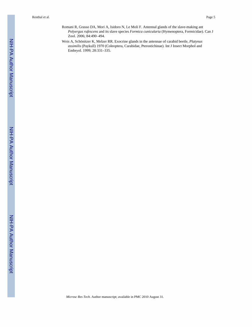
Antennal glands in Solenopsis invictaPores on the external surface are visible where canals from the antennal glands pass throughthe cuticle, as previously shown (Isidoro et al., 2000). Figures 1A and 1B show pores inworkers and queens, respectively, near antennal segment joints. Occasionally secretions areobserved coming from queen antennal glands (figure 1C).
Pores in antennae of other Solenopsis speciesWorker antennae of twenty-five other species of Solenopsis were examined by SEM (table 1and fig. 2). All were dried museum specimens, ranging in age from 46-70 years aftercollection. In every case, at the joint between segments 8 and 9, pores were observed,similar to those seen in S. invicta workers. This suggests that antennal club glands are likelyto be present in all species of Solenopsis.
Antennae of other ant speciesThe antennae of fifteen non-Solenopsis species were examined by SEM. The specimensincluded ants which were field-collected, taken from lab colonies, or from museumcollections. A close examination was made of the segment joints in the club, if present, or
Renthal et al. Page 2
Microsc Res Tech. Author manuscript; available in PMC 2010 August 31.
NIH
-PA Author Manuscript
NIH
-PA Author Manuscript
NIH
-PA Author Manuscript
the distal three segments of the funiculus, in the absence of a club (fig. 3). In all but onespecies, no pores were observed (except for obvious openings of sensilla coeloconica or s.ampulacea.)
Antennal glands in Tetramorium bicarinatumSurprisingly, we observed pores in the antenna of T. bicarinatum workers and queens (fig.4). The pores were observed in the distal segment of the club, at the joint with the proximalsegment (fig. 4D). Some antennae were cut longitudinally and digested with KOH forimaging of the inner surface of the antennae (fig. 4A, B and C). This treatment reveals thegland canals, which have a number of features that are different from the S. invicta glandcanals. The T. bicarinatum canals cross directly through the flat cuticle surface into theexternal pores. By contrast, the S. invicta canals enter an apodeme (fig. 5A) beforeconnecting with the external pores. The T. bicarinatum canals are narrower than the S.invicta canals (approx. 0.2-0.3 μm, compared with 0.5 μm). Examination of the KOH-treated T. bicarinatum antennae revealed that the glands occurred in all club segments,unlike S. invicta, which lacks glands in the distal club segment. The external pores in T.bicarinatum were difficult to observe in the proximal club segments due to their placementin the middle of the segments, where they could be mistaken for broken sensilla. The poresin the T. bicarinatum distal club segment, and the pores in Solenopsis, are more obviousbecause they are located in the sensilla-free region near the joint with adjacent segments. Afurther difference between T. bicarinatum and S. invicta antennal glands is that in T.bicarinatum queens, the pore canals terminate in spherical cuticle-covered reservoirs (fig.4A, B and C). The reservoirs were not observed in S. invicta queens or workers, nor werethey observed in T. bicarinatum workers.
DiscussionCuticular pores were observed in antennal segment 9 near the joint with segment 8 inworkers of all twenty-six species of Solenopsis examined (table 1 and figs. 1 and 2). Thisimplies that type 3 glands (Noirot and Quennedy, 1974) are a characteristic of the antennalclub of all members of this genus. The presence of antennal club glands does not extend toall members of the tribe Solenopsidini. Two species of Monomorium lack this type ofantennal gland. We also failed to find evidence for antennal glands in the distal funiculus ofa wide variety of other ant species (table 1 and fig. 3). However, we did observe antennalclub glands in Tetramorium bicarinatum, a Myrmicine ant in a different tribe fromSolenopsis (fig. 4). The glands differ from those in Solenopsis invicta as follows: i) theyoccur in all club segments; ii) in queens, they contain cuticular reservoirs; iii) the canalspenetrate the antennal wall through a flat surface, whereas S. invicta canals exit through anapodeme (fig. 5). The recently published molecular phylogeny of ants (Moreau et al., 2006)shows a wide separation between Tetramorium and Solenopsis. We found no evidence ofantennal club glands in Crematogaster, which is closer to Tetramorium; and we found noevidence of antennal club glands in Monomorium, which is closer to Solenopsis. Thus, weconclude that antennal club glands have arisen more than once in ants.
In addition to the glands of the distal funiculus described here and previously (Isidoro et al.,2000), glands have been observed in the scape of three ant species: Eciton burchelli (Billen,2000), and Polyergus rufescens and Formica cunicularia (Romani et al., 2006). Also, bothtype 1 and type 3 glands were observed in all antennomers except the scape of the primitiveant Dinoponera lucida (Marques-Silva et al., 2006). We examined antennae of two speciesof Ectatomma and saw no evidence for the external pores of type 3 glands. However,Moreau et al. (2006) raised questions about whether this genus belongs with the Ponerineants. The distribution and variability of ant antennal glands suggests multiple evolutionaryorigins for these glands.
Renthal et al. Page 3
Microsc Res Tech. Author manuscript; available in PMC 2010 August 31.
NIH
-PA Author Manuscript
NIH
-PA Author Manuscript
NIH
-PA Author Manuscript
Our results have some bearing on a long-standing taxonomic controversy. The thief antshave been sometimes classified as a separate genus Diplorhoptrum, or as a subgenus,Solenopsis (Diplorhoptrum). We examined three thief ants, Solenopsis molesta, Solenopsismolesta validiuscula, and Solenopsis fugax. In all three, the gland pores characteristic ofother members of the Solenopsis genus were observed. This could be taken as a piece ofevidence for the similarity of thief ants to the rest of the genus Solenopsis.
We do not yet know the functions of glands in the clubs of female ants. The speciesdistribution suggests multiple evolutionary origins, so there may also be different functionsin different species. It is possible that the glands are vestigial in workers. No secretions havebeen observed from antennal glands in S. invicta workers, in contrast to queens. Also, glandreservoirs are present in T. bicarinatum queen antennae, but not in worker antennae. Furtherunderstanding of antennal gland function awaits more complete behavioral and biochemicalstudies.
AcknowledgmentsSupported by grants from the Texas Imported Fire Ant Research and Management Project and NIH G12 RR13646.We thank Ted Schultz and David Furth for their hospitality at the Smithsonian Institution National Museum ofNatural History, and Marvin Edwards for contributing a specimen of M. pharaonis.
ReferencesBillen J. A novel exocrine gland in the antennal scape of the army ant Eciton burchelli. Belg J Zool.
2000; 130:159–160.Bin F, Vinson SB. Morphology of the antennal sex-gland in male Trissolcus basalis (Woll.) an egg
parasitoid of the green stink bug, Nezara viridula. Int J Insect Morphol and Embryol. 1986; 15:129–138.
Dahms EC. An interpertation of the structure and function of the antennal sense organs of Melittobiaaustralica (Hymenoptera: Eulophidae) with the discovery of large dermal glands in the male scape.Memoirs Queensland Mus. 1984; 21:361–385.
Giglio A, Ferrero EA, Zetto Brandmayr T. Ultrastructural identification of the antennal glandcomplement in Siagona europaea Dejean 1826, a myrmecophagous carabid beetle. Acta Zoologica.2005; 86:195–203.
Goettler W, Kaltenpoth M, Strohm E. Morphology and ultrastructure of a bacteria cultivation organ:the antennal glands of female European beewolves, Philanthus triangulum (Hymenoptera:Crabronidae). Arthropod Struct and Devel. 2006; 36:1–9.
Hashimoto Y. Unique features of sensilla on the antennae of Formicidae (Hymenoptera). ApplEntomol Zool. 1990; 25:491–501.
Isidoro N, Bin F, Colazza S, Vinson SB. Morphology of antennal gustatory sensilla and glands in someparasitoid Hymenoptera with hypothesis on their role in sex and host recognition. J HymenopteraRes. 1996; 5:206–239.
Isidoro N, Romani R, Velasquez D, Renthal R, Bin F, Vinson SB. Antennal glands in queen andworker of the fire ant, Solenopsis invicta Buren: first report in female social Aculeata(Hymenoptera, Formicidae). Insectes Sociaux. 2000; 47:236–240.
Keil, TA. Morphology and development of the peripheral olfactory organs. In: Hansson, BS., editor.Insect Olfaction. Springer-Verlag; Berlin Heidelberg: 1999. p. 5-47.
Marques-Silva S, Matiello-Guss CP, Delabie JHC, Mariano CSF, Zanuncio JC, Serraõ JE. Sensilla andsecretory glands in the antennae of a primitive ant: Dinoponera lucida (Formicidae: Ponerinae).Microscopy Research and Technique. 2006; 69:885–890. [PubMed: 16886227]
Moreau CS, Bell CD, Vila R, Archibald SB, Pierce NE. Phylogeny of the ants: diversification in theage of angiosperms. Science. 2006; 312:101–104. [PubMed: 16601190]
Noirot C, Quennedy A. Fine structure of insect epidermal glands. Annu Rev Entomol. 1974; 19:61–80.
Renthal et al. Page 4
Microsc Res Tech. Author manuscript; available in PMC 2010 August 31.
NIH
-PA Author Manuscript
NIH
-PA Author Manuscript
NIH
-PA Author Manuscript
Romani R, Grasso DA, Mori A, Isidoro N, Le Moli F. Antennal glands of the slave-making antPolyergus rufescens and its slave species Formica cunicularia (Hymenoptera, Formicidae). Can JZool. 2006; 84:490–494.
Weis A, Schönitzer K, Melzer RR. Exocrine glands in the antennae of carabid beetle, Platynusassimilis (Paykull) 1970 (Coleoptera, Carabidae, Pterostichinae). Int J Insect Morphol andEmbryol. 1999; 28:331–335.
Renthal et al. Page 5
Microsc Res Tech. Author manuscript; available in PMC 2010 August 31.
NIH
-PA Author Manuscript
NIH
-PA Author Manuscript
NIH
-PA Author Manuscript
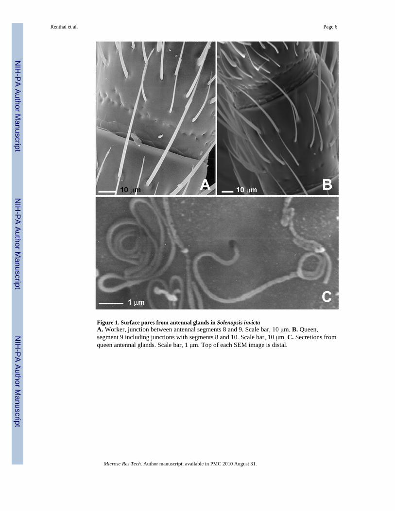
Figure 1. Surface pores from antennal glands in Solenopsis invictaA. Worker, junction between antennal segments 8 and 9. Scale bar, 10 μm. B. Queen,segment 9 including junctions with segments 8 and 10. Scale bar, 10 μm. C. Secretions fromqueen antennal glands. Scale bar, 1 μm. Top of each SEM image is distal.
Renthal et al. Page 6
Microsc Res Tech. Author manuscript; available in PMC 2010 August 31.
NIH
-PA Author Manuscript
NIH
-PA Author Manuscript
NIH
-PA Author Manuscript
Figure 2. Surface pores on antennae from Solenopsis speciesSEM images show the joint between segments 8 and 9 of worker antennae. Left side of eachimage is distal. A. S. aurea; B. S. clytemnestra; C. S. corticalis; D. S. electra; E. S. gayi; F.S. interrupta; G. S. latastei; H. S. molesta validiuscula; I. S. pergandei; J. S. quinquecuspis;K. S. richteri; L. S. wasmannii. One pore is identified by an arrow in each micrograph. Scalebars: A and C-L, 30 μm; B, 10 μm.
Renthal et al. Page 7
Microsc Res Tech. Author manuscript; available in PMC 2010 August 31.
NIH
-PA Author Manuscript
NIH
-PA Author Manuscript
NIH
-PA Author Manuscript

Figure 3. Worker antennae from representative ant speciesSEM images of the joint between segments 9 and 10 worker antennae of A. Forelius sp.; B.Linepithema humile; C. Monomorium pharaonis; and D. the joint between segments 10 and11 of a worker antenna of Oecophylla smaragdina. No gland pores are evident. Left orbottom of each image is proximal. Scale bars: A-C, 20 μm; D, 10 μm.
Renthal et al. Page 8
Microsc Res Tech. Author manuscript; available in PMC 2010 August 31.
NIH
-PA Author Manuscript
NIH
-PA Author Manuscript
NIH
-PA Author Manuscript
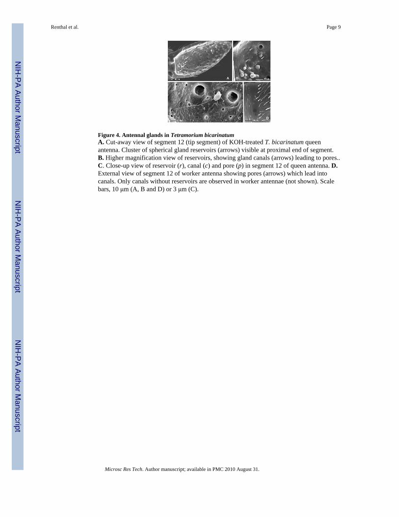
Figure 4. Antennal glands in Tetramorium bicarinatumA. Cut-away view of segment 12 (tip segment) of KOH-treated T. bicarinatum queenantenna. Cluster of spherical gland reservoirs (arrows) visible at proximal end of segment.B. Higher magnification view of reservoirs, showing gland canals (arrows) leading to pores..C. Close-up view of reservoir (r), canal (c) and pore (p) in segment 12 of queen antenna. D.External view of segment 12 of worker antenna showing pores (arrows) which lead intocanals. Only canals without reservoirs are observed in worker antennae (not shown). Scalebars, 10 μm (A, B and D) or 3 μm (C).
Renthal et al. Page 9
Microsc Res Tech. Author manuscript; available in PMC 2010 August 31.
NIH
-PA Author Manuscript
NIH
-PA Author Manuscript
NIH
-PA Author Manuscript

Figure 5. Inside surfaces of Solenopsis and Monomorium antennaeA. Solenopsis invicta worker antennal segment 9. Gland canals (arrows) penetrate the wallof the antenna through an apodeme. B. Monomorium pharaonis worker antennal segment12. No canals are observed. Scale bars, 10 μm.
Renthal et al. Page 10
Microsc Res Tech. Author manuscript; available in PMC 2010 August 31.
NIH
-PA Author Manuscript
NIH
-PA Author Manuscript
NIH
-PA Author Manuscript
NIH
-PA Author Manuscript
NIH
-PA Author Manuscript
NIH
-PA Author Manuscript
Renthal et al. Page 11
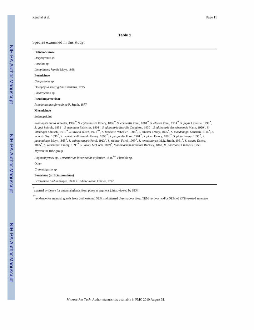
Table 1
Species examined in this study.
Dolichoderinae
Dorymyrmex sp.
Forelius sp.
Linepithema humile Mayr, 1868
Formicinae
Campanotus sp.
Oecophylla smaragdina Fabricius, 1775
Paratrechina sp.
Pseudomyrmecinae
Pseudomyrmex ferruginea F. Smith, 1877
Myrmicinae
Solenopsidini
Solenopsis aurea Wheeler, 1906*, S. clytemnestra Emery, 1896*, S. corticalis Forel, 1881*, S. electra Forel, 1914*, S. fugax Latreille, 1798*,S. gayi Spinola, 1851*, S. geminata Fabricius, 1804*, S. globularia litoralis Creighton, 1930*, S. globularia desecheoensis Mann, 1920*, S.interrupta Santschi, 1916*, S. invicta Buren, 1972**, S. krockowi Wheeler, 1908*, S. latastei Emery, 1895*, S. macdonaghi Santschi, 1916*, S.molesta Say, 1836*, S. molesta validiuscula Emery, 1895*, S. pergandei Forel, 1901*, S. picea Emery, 1896*, S. picta Emery, 1895*, S.punctaticeps Mayr, 1865*, S. quinquecuspis Forel, 1913*, S. richteri Forel, 1909*, S. tennesseensis M.R. Smith, 1951*, S. texana Emery,1895*, S. wasmannii Emery, 1895*, S. xyloni McCook, 1879*, Monomorium minimum Buckley, 1867, M. pharaonis Linnaeus, 1758
Myrmicine tribe group
Pogonomyrmex sp., Tetramorium bicarinatum Nylander, 1846**, Pheidole sp.
Other
Crematogaster sp.
Ponerinae (or Ectatomminae)
Ectatomma ruidum Roger, 1860, E. tuberculatum Olivier, 1792
*external evidence for antennal glands from pores at segment joints, viewed by SEM
**evidence for antennal glands from both external SEM and internal observations from TEM sections and/or SEM of KOH-treated antennae
Microsc Res Tech. Author manuscript; available in PMC 2010 August 31.