mouth development: - dspace@mit
TRANSCRIPT

Mouth development
The MIT Faculty has made this article openly available. Please share how this access benefits you. Your story matters.
Citation Chen, Justin et al. “Mouth Development.” Wiley InterdisciplinaryReviews: Developmental Biology 6, 5 (May 2017): e275 © 2017 TheAuthors
As Published http://dx.doi.org/10.1002/WDEV.275
Publisher Wiley-Blackwell
Version Final published version
Citable link http://hdl.handle.net/1721.1/116900
Terms of Use Creative Commons Attribution-NonCommercial-NoDerivs License
Detailed Terms http://creativecommons.org/licenses/by-nc-nd/4.0/

Advanced Review
Mouth developmentJustin Chen,1,2† Laura A. Jacox,1,3,4†‡ Francesca Saldanha,1§
and Hazel Sive1,2*
A mouth is present in all animals, and comprises an opening from the outsideinto the oral cavity and the beginnings of the digestive tract to allow eating. Thisreview focuses on the earliest steps in mouth formation. In the first half, we con-clude that the mouth arose once during evolution. In all animals, the mouthforms from ectoderm and endoderm. A direct association of oral ectoderm anddigestive endoderm is present even in triploblastic animals, and in chordates,this region is known as the extreme anterior domain (EAD). Further support fora single origin of the mouth is a conserved set of genes that form a ‘mouth geneprogram’ including foxA and otx2. In the second half of this review, we discusssteps involved in vertebrate mouth formation, using the frog Xenopus as a model.The vertebrate mouth derives from oral ectoderm from the anterior neural ridge,pharyngeal endoderm and cranial neural crest (NC). Vertebrates form a mouthby breaking through the body covering in a precise sequence including specifica-tion of EAD ectoderm and endoderm as well as NC, formation of a ‘pre-moutharray,’ basement membrane dissolution, stomodeum formation, and buccophar-yngeal membrane perforation. In Xenopus, the EAD is also a craniofacial organ-izer that guides NC, while reciprocally, the NC signals to the EAD to elicit itsmorphogenesis into a pre-mouth array. Human mouth anomalies are prevalentand are affected by genetic and environmental factors, with understandingguided in part by use of animal models. © 2017 The Authors. WIREs Developmental Biologypublished by Wiley Periodicals, Inc.
How to cite this article:WIREs Dev Biol 2017, 6:e275. doi: 10.1002/wdev.275
INTRODUCTION
Multicellular animals need to eat and a mouth isthe organ that allows food into the digestive sys-
tem. It comprises the opening from the outside of theanimal, the oral cavity that is connected to the openingand the beginning of the digestive system, the pharynx.Even some single-celled organisms like Paramecia havea mouth leading into a subcellular intestine.1 Many ani-mals have accessory structures that assist eating andmouth function, and increase complexity of this organ.We hypothesize that the mouth arose once in evolution,and consider two lines of evidence that support this.These include the understanding that the mouth isalways built from ectodermal and endodermal lineages.The first multicellular animals with a clear mouth werediploblasts (with ectoderm and endoderm).1–3 Interest-ingly, even in triploblastic animals that include
†These authors contributed equally.‡Present address: Orthodontics Department, University of NorthCarolina School of Dentistry, Chapel Hill, NC, USA§Present address: Department of Plastic and Oral Surgery, BostonChildren's Hospital, Harvard Medical School, Boston, MA, USA
*Correspondence to: [email protected] Institute for Biomedical Research, Cambridge,MA, USA2Department of Biology, Massachusetts Institute of Technology,Cambridge, MA, USA3Harvard-MIT Health Sciences and Technology Program, Cam-bridge, MA, USA4Harvard School of Dental Medicine, Boston, MA, USA
Conflict of interest: The authors have declared no conflicts of inter-est for this article.
Volume 6, September/October 2017 1 of 16© 2017 The Authors. WIREs Developmental Biology published by Wiley Periodicals, Inc.This is an open access article under the terms of the Creative Commons Attribution-NonCommercial-NoDerivs License, which permits use and distribution inany medium, provided the original work is properly cited, the use is non-commercial and no modifications or adaptations are made.
mesoderm, the mouth still forms from a region whereectoderm and endoderm directly juxtapose.2,3 In chor-dates, we named this region the extreme anteriordomain (EAD).4,5 Another aspect of the ancient originof the mouth is conservation of gene expression, lead-ing to the proposal of a ‘mouth gene program.’
In initial studies of the Xenopusmouth, we coinedthe term ‘primary mouth’ to indicate the initial orimmature larval mouth, and ‘secondary mouth’ to indi-cate later elaboration and differentiation of structures toform the mature mouth. Although this nomenclaturehas been useful to the community, on consideration, wethink the general term ‘mouth’ is most useful through-out development. We view mouth development as acontinuum, where even at the earliest time after mouthopening, an oral cavity and accessory structures arealready forming. This review focuses on the earlieststages of mouth formation, whereby the initial mouthopening forms and the first steps of differentiation aretaking place, but before the mouth is mature.
Vertebrates have a complex mouth, derived notonly from EAD ectoderm and endoderm that form theoral cavity and pharynx of the digestive tract but alsofrom neural crest (NC) cells that form teeth and jaws.Vertebrates are ‘deuterostomes’ where a mouth breaksthrough the ectodermal covering and connects to theendodermal digestive tract. In the Xenopus model,development of the vertebrate mouth is associated withreciprocal signaling between the EAD and NC.4,5
Mouth formation reflects this precision and involvesmany steps over a long period of development(~2.5 days in Xenopus, 2 weeks in humans). Thesesteps position the mouth-forming oral ectoderm anddigestive endoderm during gastrula and neurula stagesand open the mouth as the tadpole is ready to feed.The complexity of mouth formation is one reason forthe many human anomalies that include this region.
ORAL EVOLUTION: IS MOUTHDEVELOPMENT CONSERVED?
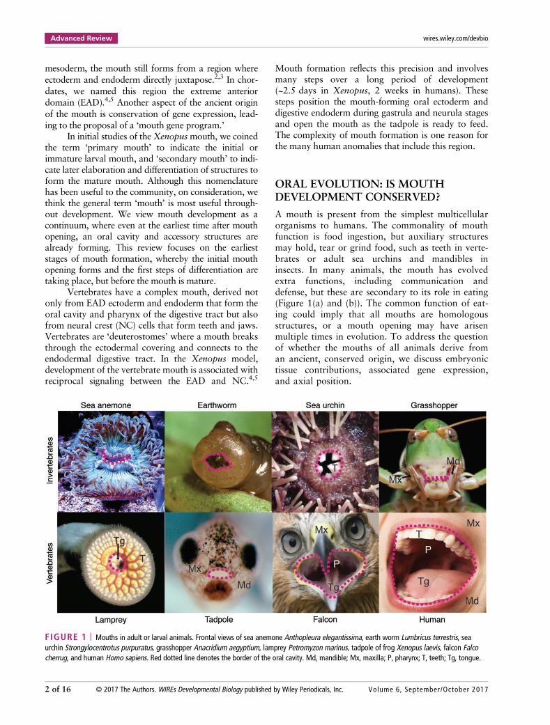
A mouth is present from the simplest multicellularorganisms to humans. The commonality of mouthfunction is food ingestion, but auxiliary structuresmay hold, tear or grind food, such as teeth in verte-brates or adult sea urchins and mandibles ininsects. In many animals, the mouth has evolvedextra functions, including communication anddefense, but these are secondary to its role in eating(Figure 1(a) and (b)). The common function of eat-ing could imply that all mouths are homologousstructures, or a mouth opening may have arisenmultiple times in evolution. To address the questionof whether the mouths of all animals derive froman ancient, conserved origin, we discuss embryonictissue contributions, associated gene expression,and axial position.
FIGURE 1 | Mouths in adult or larval animals. Frontal views of sea anemone Anthopleura elegantissima, earth worm Lumbricus terrestris, seaurchin Strongylocentrotus purpuratus, grasshopper Anacridium aegyptium, lamprey Petromyzon marinus, tadpole of frog Xenopus laevis, falcon Falcocherrug, and human Homo sapiens. Red dotted line denotes the border of the oral cavity. Md, mandible; Mx, maxilla; P, pharynx; T, teeth; Tg, tongue.
Advanced Review wires.wiley.com/devbio
2 of 16 © 2017 The Authors. WIREs Developmental Biology published by Wiley Periodicals, Inc. Volume 6, September/October 2017
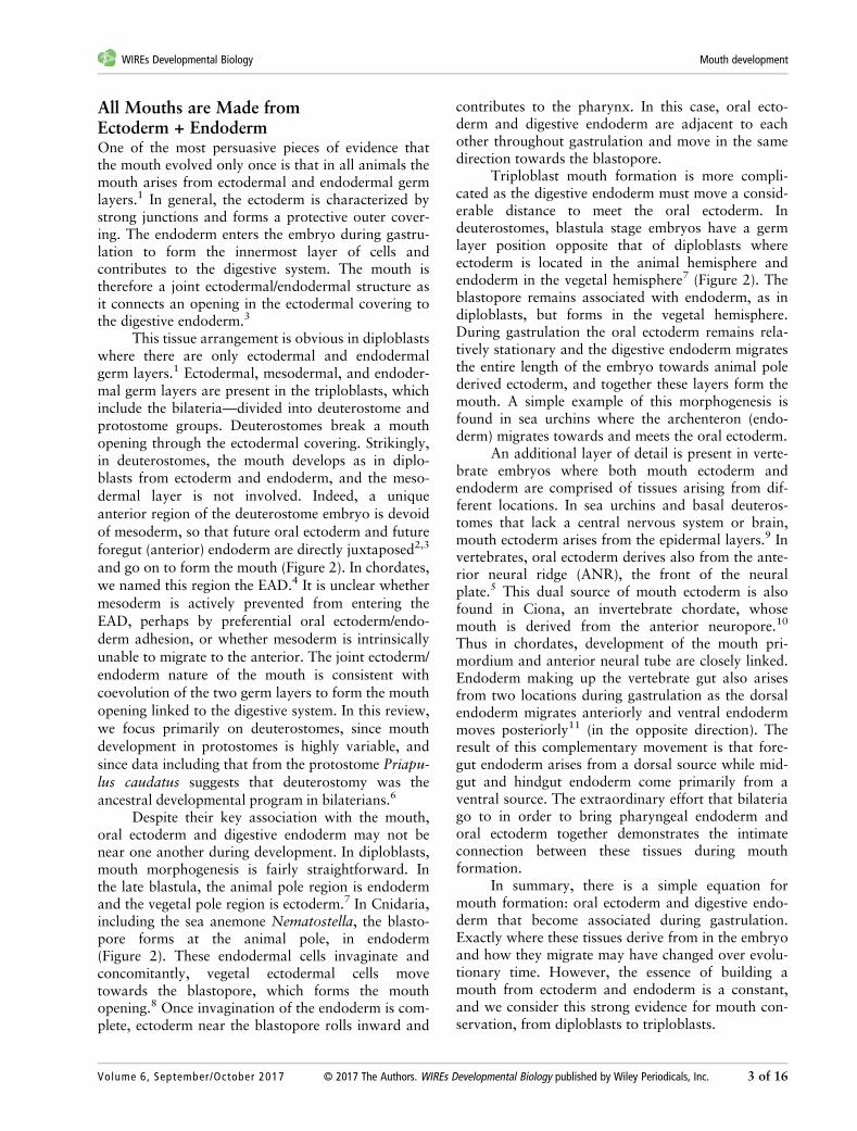
All Mouths are Made fromEctoderm + EndodermOne of the most persuasive pieces of evidence thatthe mouth evolved only once is that in all animals themouth arises from ectodermal and endodermal germlayers.1 In general, the ectoderm is characterized bystrong junctions and forms a protective outer cover-ing. The endoderm enters the embryo during gastru-lation to form the innermost layer of cells andcontributes to the digestive system. The mouth istherefore a joint ectodermal/endodermal structure asit connects an opening in the ectodermal covering tothe digestive endoderm.3
This tissue arrangement is obvious in diploblastswhere there are only ectodermal and endodermalgerm layers.1 Ectodermal, mesodermal, and endoder-mal germ layers are present in the triploblasts, whichinclude the bilateria—divided into deuterostome andprotostome groups. Deuterostomes break a mouthopening through the ectodermal covering. Strikingly,in deuterostomes, the mouth develops as in diplo-blasts from ectoderm and endoderm, and the meso-dermal layer is not involved. Indeed, a uniqueanterior region of the deuterostome embryo is devoidof mesoderm, so that future oral ectoderm and futureforegut (anterior) endoderm are directly juxtaposed2,3
and go on to form the mouth (Figure 2). In chordates,we named this region the EAD.4 It is unclear whethermesoderm is actively prevented from entering theEAD, perhaps by preferential oral ectoderm/endo-derm adhesion, or whether mesoderm is intrinsicallyunable to migrate to the anterior. The joint ectoderm/endoderm nature of the mouth is consistent withcoevolution of the two germ layers to form the mouthopening linked to the digestive system. In this review,we focus primarily on deuterostomes, since mouthdevelopment in protostomes is highly variable, andsince data including that from the protostome Priapu-lus caudatus suggests that deuterostomy was theancestral developmental program in bilaterians.6
Despite their key association with the mouth,oral ectoderm and digestive endoderm may not benear one another during development. In diploblasts,mouth morphogenesis is fairly straightforward. Inthe late blastula, the animal pole region is endodermand the vegetal pole region is ectoderm.7 In Cnidaria,including the sea anemone Nematostella, the blasto-pore forms at the animal pole, in endoderm(Figure 2). These endodermal cells invaginate andconcomitantly, vegetal ectodermal cells movetowards the blastopore, which forms the mouthopening.8 Once invagination of the endoderm is com-plete, ectoderm near the blastopore rolls inward and
contributes to the pharynx. In this case, oral ecto-derm and digestive endoderm are adjacent to eachother throughout gastrulation and move in the samedirection towards the blastopore.
Triploblast mouth formation is more compli-cated as the digestive endoderm must move a consid-erable distance to meet the oral ectoderm. Indeuterostomes, blastula stage embryos have a germlayer position opposite that of diploblasts whereectoderm is located in the animal hemisphere andendoderm in the vegetal hemisphere7 (Figure 2). Theblastopore remains associated with endoderm, as indiploblasts, but forms in the vegetal hemisphere.During gastrulation the oral ectoderm remains rela-tively stationary and the digestive endoderm migratesthe entire length of the embryo towards animal polederived ectoderm, and together these layers form themouth. A simple example of this morphogenesis isfound in sea urchins where the archenteron (endo-derm) migrates towards and meets the oral ectoderm.
An additional layer of detail is present in verte-brate embryos where both mouth ectoderm andendoderm are comprised of tissues arising from dif-ferent locations. In sea urchins and basal deuteros-tomes that lack a central nervous system or brain,mouth ectoderm arises from the epidermal layers.9 Invertebrates, oral ectoderm derives also from the ante-rior neural ridge (ANR), the front of the neuralplate.5 This dual source of mouth ectoderm is alsofound in Ciona, an invertebrate chordate, whosemouth is derived from the anterior neuropore.10
Thus in chordates, development of the mouth pri-mordium and anterior neural tube are closely linked.Endoderm making up the vertebrate gut also arisesfrom two locations during gastrulation as the dorsalendoderm migrates anteriorly and ventral endodermmoves posteriorly11 (in the opposite direction). Theresult of this complementary movement is that fore-gut endoderm arises from a dorsal source while mid-gut and hindgut endoderm come primarily from aventral source. The extraordinary effort that bilateriago to in order to bring pharyngeal endoderm andoral ectoderm together demonstrates the intimateconnection between these tissues during mouthformation.
In summary, there is a simple equation formouth formation: oral ectoderm and digestive endo-derm that become associated during gastrulation.Exactly where these tissues derive from in the embryoand how they migrate may have changed over evolu-tionary time. However, the essence of building amouth from ectoderm and endoderm is a constant,and we consider this strong evidence for mouth con-servation, from diploblasts to triploblasts.
WIREs Developmental Biology Mouth development
Volume 6, September/October 2017 © 2017 The Authors. WIREs Developmental Biology published by Wiley Periodicals, Inc. 3 of 16

(a)
(b)
FIGURE 2 | Mouth forms where ectoderm and endoderm are juxtaposed. (a) Position of future mouth relative to germ layers in embryos ofthree representative animals. Schematics of sagittal sections are shown for the diploblast cnidarian Nematostella vectensis (invertebrate), thetriploblasts and deuterostomes sea urchin Strongylocentrotus purpuratus (invertebrate) and frog Xenopus laevis (vertebrate). The red box outlinesthe mouth-forming region made up of juxtaposed ectoderm and endoderm. In vertebrates, this region is termed the extreme anterior domain.(b) Ancestral mouth embryonic gene expression domains in N. vectensis, S. purpuratus, and X. laevis (purple). The mouth expression domain offoxA and otx but not brachyury is conserved in vertebrates.
Advanced Review wires.wiley.com/devbio
4 of 16 © 2017 The Authors. WIREs Developmental Biology published by Wiley Periodicals, Inc. Volume 6, September/October 2017

A Conserved Mouth Gene ProgramA corollary to the conclusion that oral ectoderm andpharyngeal endoderm are conserved is that similargenes are expressed in these tissues across phyla. Strik-ingly, three genes, otx, brachyury, and foxA, areexpressed throughout the gut endoderm of Cnidaria(Nematostella vectensis), protostomes (Capitella teletaand P. caudatus) and deuterostomes (sea urchins andstarfish).6,12–15 Otx and foxA are also expressed inmouth ectoderm. The overlapping expression of thesethree genes in conserved domains across evolutionsuggests an ancestral gene network regulating mouthand associated gut development16,17 (Figure 3).
Detailed functional evaluation of these geneshas been performed in sea urchins and starfish. otx,brachyury, and foxA are part of the sea urchin endo-dermal gene regulatory network (GRN), which is oneof the most extensively studied GRNs. In general,GRNs comprise ‘kernels’ or evolutionarily inflexiblecircuits responsible for upstream functions in bodypatterning, and ‘plug-ins’ or smaller circuits that havebeen repeatedly co-opted for diverse purposes.
Comparison between GRNs of the distantlyrelated echinoderms sea urchins and starfish, deuter-ostomes of the Echinoderm phylum, reveals an iden-tical core of transcription factors—otx, brachyury,and foxA, as well as two additional genes—blimp1/krox and gataE.18,19 This five-member kernel regu-lates the development of digestive (gut) endodermincluding that associated with the mouth and eachgene is necessary for gut formation. FoxA and otx,independent of their function in endoderm, are alsoactive in oral ectoderm. Transplant experiments insea urchins demonstrate that ectodermal foxA isrequired for mouth formation as embryos withectoderm-specific foxA loss of function had normaldigestive tracts but lacked mouths.17 While func-tional experiments have not been performed in seaurchins, starfish injected with a dominant negativeform of otx formed a truncated archenteron andabnormal mouth ectoderm lacking an invaginationcorresponding to the mouth.16
Tracing to ancestral diploblasts, is the expres-sion of the five-membered GRN discovered in Echi-noderms also present? As stated earlier, Cnidariaexpress otx, Brachyury, and foxA in the mouth andgut. However, they lack such specific expression ofblimp1 and gataE.20 Thus otx, brachyury, and foxAlikely form an ancestral kernel responsible for mouthand gut formation while blimp1 and gataE appearedin later lineages (Figure 3).
Tracing forward, is the five-membered GRN anechinoderm-specific innovation or broadly used by
bilateria? Prostomes (C. teleta and P. caudatus) havemouth and/or gut-specific expression of all fivegenes.6,14 This suggests that the echinoderm GRN isused by basal deuterostomes and protostomes as amouth/gut GRN and that this kernel was present inthe deuterostome–protostome ancestor. Conservationof the kernel in chordates is incomplete—perhapsdue to gene duplication, different body plans, andadded complexity of craniofacial development that
(a)
(b)
FIGURE 3 | Ancestral mouth was present in the commonancestor of cnidaria and triploblasts. (a) Phylogenetic tree. Chordates,echinoderms, and cnidaria are three distantly related phyla that retaincommon mouth characteristics, suggesting that the mouth evolvedonce. (b) Criteria used to evaluate mouth evolution. The mouths ofcnidaria, echinoderms, and chordates are all comprised of ectodermand endoderm and express foxA and otx. There are phylum-specificcharacteristics such as neural ectoderm contributing to the chordatemouth and the expression of blimp1, gataE, and pitx in echinodermand chordates. Analysis of axial positioning genes demonstrates thatthe cnidarian oral–aboral axis is equivalent to the echinoderm andchordate posterior–anterior axis.
WIREs Developmental Biology Mouth development
Volume 6, September/October 2017 © 2017 The Authors. WIREs Developmental Biology published by Wiley Periodicals, Inc. 5 of 16
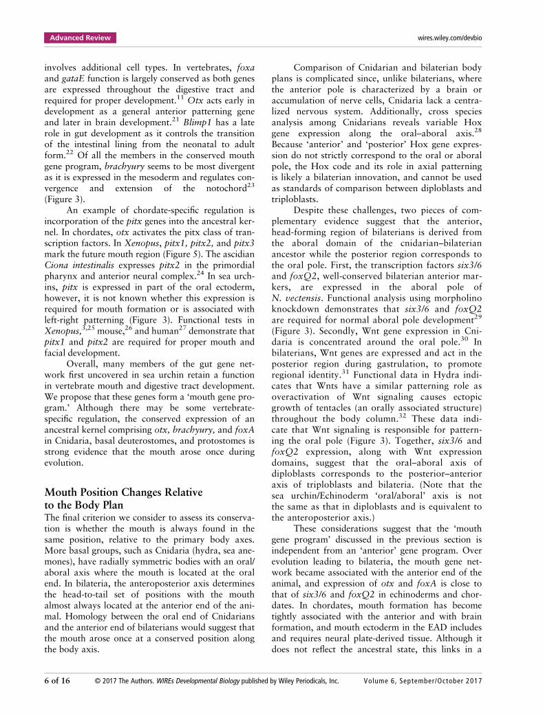
involves additional cell types. In vertebrates, foxaand gataE function is largely conserved as both genesare expressed throughout the digestive tract andrequired for proper development.11 Otx acts early indevelopment as a general anterior patterning geneand later in brain development.21 Blimp1 has a laterole in gut development as it controls the transitionof the intestinal lining from the neonatal to adultform.22 Of all the members in the conserved mouthgene program, brachyury seems to be most divergentas it is expressed in the mesoderm and regulates con-vergence and extension of the notochord23
(Figure 3).An example of chordate-specific regulation is
incorporation of the pitx genes into the ancestral ker-nel. In chordates, otx activates the pitx class of tran-scription factors. In Xenopus, pitx1, pitx2, and pitx3mark the future mouth region (Figure 5). The ascidianCiona intestinalis expresses pitx2 in the primordialpharynx and anterior neural complex.24 In sea urch-ins, pitx is expressed in part of the oral ectoderm,however, it is not known whether this expression isrequired for mouth formation or is associated withleft-right patterning (Figure 3). Functional tests inXenopus,3,25 mouse,26 and human27 demonstrate thatpitx1 and pitx2 are required for proper mouth andfacial development.
Overall, many members of the gut gene net-work first uncovered in sea urchin retain a functionin vertebrate mouth and digestive tract development.We propose that these genes form a ‘mouth gene pro-gram.’ Although there may be some vertebrate-specific regulation, the conserved expression of anancestral kernel comprising otx, brachyury, and foxAin Cnidaria, basal deuterostomes, and protostomes isstrong evidence that the mouth arose once duringevolution.
Mouth Position Changes Relativeto the Body PlanThe final criterion we consider to assess its conserva-tion is whether the mouth is always found in thesame position, relative to the primary body axes.More basal groups, such as Cnidaria (hydra, sea ane-mones), have radially symmetric bodies with an oral/aboral axis where the mouth is located at the oralend. In bilateria, the anteroposterior axis determinesthe head-to-tail set of positions with the mouthalmost always located at the anterior end of the ani-mal. Homology between the oral end of Cnidariansand the anterior end of bilaterians would suggest thatthe mouth arose once at a conserved position alongthe body axis.
Comparison of Cnidarian and bilaterian bodyplans is complicated since, unlike bilaterians, wherethe anterior pole is characterized by a brain oraccumulation of nerve cells, Cnidaria lack a centra-lized nervous system. Additionally, cross speciesanalysis among Cnidarians reveals variable Hoxgene expression along the oral–aboral axis.28
Because ‘anterior’ and ‘posterior’ Hox gene expres-sion do not strictly correspond to the oral or aboralpole, the Hox code and its role in axial patterningis likely a bilaterian innovation, and cannot be usedas standards of comparison between diploblasts andtriploblasts.
Despite these challenges, two pieces of com-plementary evidence suggest that the anterior,head-forming region of bilaterians is derived fromthe aboral domain of the cnidarian–bilaterianancestor while the posterior region corresponds tothe oral pole. First, the transcription factors six3/6and foxQ2, well-conserved bilaterian anterior mar-kers, are expressed in the aboral pole ofN. vectensis. Functional analysis using morpholinoknockdown demonstrates that six3/6 and foxQ2are required for normal aboral pole development29
(Figure 3). Secondly, Wnt gene expression in Cni-daria is concentrated around the oral pole.30 Inbilaterians, Wnt genes are expressed and act in theposterior region during gastrulation, to promoteregional identity.31 Functional data in Hydra indi-cates that Wnts have a similar patterning role asoveractivation of Wnt signaling causes ectopicgrowth of tentacles (an orally associated structure)throughout the body column.32 These data indi-cate that Wnt signaling is responsible for pattern-ing the oral pole (Figure 3). Together, six3/6 andfoxQ2 expression, along with Wnt expressiondomains, suggest that the oral–aboral axis ofdiploblasts corresponds to the posterior–anterioraxis of triploblasts and bilateria. (Note that thesea urchin/Echinoderm ‘oral/aboral’ axis is notthe same as that in diploblasts and is equivalent tothe anteroposterior axis.)
These considerations suggest that the ‘mouthgene program’ discussed in the previous section isindependent from an ‘anterior’ gene program. Overevolution leading to bilateria, the mouth gene net-work became associated with the anterior end of theanimal, and expression of otx and foxA is close tothat of six3/6 and foxQ2 in echinoderms and chor-dates. In chordates, mouth formation has becometightly associated with the anterior and with brainformation, and mouth ectoderm in the EAD includesand requires neural plate-derived tissue. Although itdoes not reflect the ancestral state, this links in a
Advanced Review wires.wiley.com/devbio
6 of 16 © 2017 The Authors. WIREs Developmental Biology published by Wiley Periodicals, Inc. Volume 6, September/October 2017

functional way, the mouth of chordates with theanterior of the body.
The Mouth Is a Conserved StructureIn summary, two criteria indicate that the mouth hasa common evolutionary origin across animals. First,the mouth always forms from oral ectoderm anddigestive endoderm. In triploblasts, mesoderm isnever part of the initial mouth. Second, a conservedset of genes that can be considered a mouth gene pro-gram can be defined in all animals, including otx andfoxA. These considerations indicate that the moutharose once during evolution and that fundamentalaspects of a mouth program have been retainedamongst all animals.
STEPS TO FORM A MOUTH: XENOPUSAS A PARADIGM
Of greatest relevance for human health is develop-ment of the vertebrate mouth (Figure 1(b)). Thisderives from a region of juxtaposed ectoderm andendoderm termed the EAD (Figure 2), in conjunc-tion with cells of the cranial NC.2,4 The EADforms the mouth opening and the oral cavity,including the oropharynx that is the beginning ofthe digestive tract, while the NC forms the jawsand teeth. In this section, we review the earlieststeps in mouth development, including mouthopening, prior to differentiation of tissues found inthe mature mouth.
While retaining the core-conserved aspectsof mouth development: ectodermal plus endoder-mal origins and key mouth regulatory genes, ver-tebrate mouth formation is extremely complex,due to the large number of tissues and cell typespresent, and the involvement of theNC. Vertebrates are deuterostomes where themouth breaks through the ectodermal covering toconnect the outside with the endodermal digestivetract. Formation of the mouth opening must becarefully coordinated with digestive system devel-opment, so that the opening does not form pre-maturely and become a wound.
The frog Xenopus has proven an outstandingvertebrate model for observation of mouth develop-ment, since it undergoes external embryonic develop-ment, allowing all stages to be obtained, and sincethe face is flat owing to the small forebrain. Xenopusmouth development relies on coordinated develop-ment of the EAD and NC, in a carefully orchestratedsequence. The earliest steps in mouth formation,including mouth opening appear similar in Xenopus
and amniotes,2,3 although as discussed later in thisreview, details may differ between species.
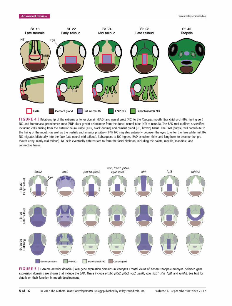
Setting Aside the Ectoderm and Endodermof the EADXenopus mouth development begins at the end ofneurulation. At this time, the EAD is defined byfuture oral ectoderm lying adjacent to pharyngealendoderm, and genes are expressed that indicate thefuture mouth4,5,33 (Figures 2 and 4). Both ectodermand endoderm are essential for mouth formation.3
foxa2 and otx2, genes that are part of the conservedmouth gene kernel, are expressed in the developingmouth region (Figure 5).
EAD ectoderm derives from the ANR, the ante-rior boundary of the neural plate.5,34,35 At the end ofneurulation, a wedge of ectoderm delaminates fromthe ANR to move ventrally and lie between the epi-dermal ectoderm and the pharyngeal endoderm. Thisis EAD ectoderm that will surround the mouth open-ing and contribute to the oral cavity. Subsequently, abasement membrane forms that separates the devel-oping brain from EAD ectoderm.5 Overlying epider-mal ectoderm can be substituted by flank epidermalectoderm and is therefore not specific for mouthdevelopment.2 By early tailbud, EAD ectodermexpresses pitx1, pitx2b, pitx2c, and pitx3 genes,while the underlying pharyngeal endoderm expressespitx1 and pitx2c3 (Figure 5). As noted in the OralEvolution: Is Mouth Development Conserved sec-tion, this class of gene is required for Xenopus mouthdevelopment.36 An expression microarray screen inour group revealed additional genes expressed inEAD ectoderm and in some cases EAD endoderm,including the Wnt-inhibitor frzb-1, kinin–kallikreinpathway factors cpn and kininogen, and the tran-scription factors vgl2, six1, xanf1, xanf2, and goose-coid4,33 (Figure 5). Other signaling factors are alsoexpressed in the EAD or surrounding tissues, includ-ing shh, fgf8, and raldh237–39 (Figure 5).
Pharyngeal endoderm is the inner componentof the EAD that becomes the epithelial lining of thepharynx.40 It derives from dorsal endoderm that hasmoved to the anterior of the embryo by the end ofgastrulation, and lies anterior to head mesoderm.41,42
Pharyngeal endoderm specification involves functionof the tbx1 transcription factor in Xenopus.43 Genesexpressed in EAD endoderm include kininogen, frzb-1, and raldh2,4,33,39 and these are required for mouthdevelopment (next section) (Figure 5). Pharyngealendoderm development is also dependent on retinoicacid (RA) signaling.44
WIREs Developmental Biology Mouth development
Volume 6, September/October 2017 © 2017 The Authors. WIREs Developmental Biology published by Wiley Periodicals, Inc. 7 of 16
foxa2 otx2 pitx1c, pitx2 cpn, frzb1, pitx3,
vgl2, xanf1 shh fgf8 raldh2
FIGURE 5 | Extreme anterior domain (EAD) gene expression domains in Xenopus. Frontal views of Xenopus tadpole embryos. Selected geneexpression domains are shown that include the EAD. These include pitx1c, pitx2, pitx3, vgl2, xanf1, cpn, frzb1, shh, fgf8, and raldh2. See text fordetails on their function in mouth development.
FIGURE 4 | Relationship of the extreme anterior domain (EAD) and neural crest (NC) to the Xenopus mouth. Branchial arch (BA, light green)NC, and frontonasal prominence crest (FNP, dark green) delaminate from the dorsal neural tube (NT) at neurula. The EAD (red outline) is specifiedincluding cells arising from the anterior neural ridge (ANR, black outline) and cement gland (CG, brown) tissue. The EAD (purple) will contribute tothe lining of the mouth (as well as the nostrils and anterior pituitary). FNP NC migrates anteriorly between the eyes to enter the face while first BANC migrates bilaterally into the face (late neural-mid tailbud). Subsequent to NC ingress, EAD ectoderm thins and lengthens to become the ‘pre-mouth array’ (early-mid tailbud). NC cells eventually differentiate to form the facial skeleton, including the palate, maxilla, mandible, andconnective tissue.
Advanced Review wires.wiley.com/devbio
8 of 16 © 2017 The Authors. WIREs Developmental Biology published by Wiley Periodicals, Inc. Volume 6, September/October 2017

Guiding Migratory NC to the Mouth-Forming Region: The EAD as OrganizerIn addition to EAD ectoderm and endoderm, the cra-nial NC makes a key contribution to mouth forma-tion in Xenopus and other vertebrates, eventuallyforming the jaws (maxilla and mandible), palate, andupper lip45 (Figures 4 and 6). The NC is a migratory,multipotent population originating from the lateralborders of the neural plate45 that segregates into fourstreams called branchial arches. First arch crestmigrates to the face to lie on either side of the EAD,while the frontonasal prominence crest migrates overthe top of the head into the face (Figure 4).45 NCmigration is governed by chemotaxis via Sdf1, pro-duced by adjacent regions, together with contact-inhibition of locomotion (CIL) through N-cadherinand Wnt/PCP signaling.46 NC cells express comple-ment receptors and secrete complement ligand thatpromotes cell clustering.47 These dispersion andattraction activities are required for migration of theNC as a group.48 Secreted ephrins and semaphorinspromote branchial arch formation.48,49
Using facial transplants,4 we discovered that inaddition to contributing to the mouth, the EAD is asignaling center that helps direct NC towards thefacial midline. At least two signaling pathways actfrom the EAD. One is the kinin–kallikrein pathway,where the EAD expresses precursors of Kinin ligandsand cpn, encoding a Kinin-processing enzyme. Lossof cpn locally, specifically in the EAD, halts first archcrest migration and leads to failure of mouth forma-tion and an abnormal face.4 Another EAD signal reg-ulates the β-catenin Wnt pathway, where secretedantagonists frzb-1 and crescent act within the EADand also in the developing face, possibly affectingNC.33 EAD pharyngeal endoderm plays a later sig-naling role in NC development, to induce formationof the cartilaginous skeleton of the mouth and phar-ynx.44 Thus, EAD endoderm ablation results in anabnormal mouth and pharyngeal skeleton in Xeno-pus2 and chick.50
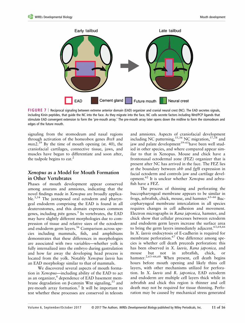
Formation of a Pre-Mouth Array and theStomodeum: the NC Signals to EADEctodermAfter the NC has come to lie on either side of theEAD, it signals back to the EAD to induce morpho-genesis of a ‘pre-mouth array’5 (Figure 7). This sig-naling is via the Wnt/PCP pathway where Wnt11ligand is expressed in the NC and targets the Fzl7receptor in the EAD. Under control of Wnt/PCP sig-naling, EAD ectoderm undergoes convergent
extension to transition from a wide, short 8 × 8block of cells (st. 22) to a narrow, tall 20 × 2 cellarrangement we termed the ‘pre-mouth array’ (st. 28)(Figure 6(a)). Two days later, the pre-mouth arrayopens down the middle to form the ‘stomodeum.’The stomodeum is a highly conserved indentation inbilateria, and indicates the future mouth (tadpolestages, st. 32–40)5 (Figure 6(a) and (b)). The ‘pre-mouth array’ demonstrates that the stomodeum isorganized much earlier than previously understood.Basement membrane breakdown between EAD ecto-derm and endoderm (Figure 6(b)) had been consid-ered the first stage of mouth opening, however, thepre-mouth array precisely sets up the future mouthopening prior to basement membrane breakdown(Figure 6(b)). Our data indicate that mouth develop-ment in Xenopus involves reciprocal signaling: fromEAD to NC and later from NC to EAD, a sequencethat likely coordinates development of tissues andstructures leading to proper mouth development(Figures 6 and 7).
Opening the Mouth: Signals and StepsPre-mouth array formation leads to precisely organ-ized oral ectoderm, juxtaposed to the pharyngealendoderm. Several additional steps complete mouthformation. During pre-mouth array formation, thebasement membrane separating ectoderm from endo-derm is degraded (st. 28) (Figure 6(b)). This isdependent on the β-catenin Wnt antagonists frzb-1and crescent that are expressed in the EAD,33 as wellas Hedgehog signaling.51 Subsequently, the pre-mouth array opens down the midline to form thestomodeum—comprising the borders of the futuremouth opening with a central indentation (st. 35–37)(Figure 6(a)). The signal that causes the array to openis unknown, but we speculate that it derives from theunderlying endoderm that is maturing into a func-tional digestive system. Thus, when the pharyngealendoderm is close to mature, it may signal to the pre-mouth array ectoderm to elicit its opening. Thisoccurs concomitant with appearance of apical mar-kers on the pre-mouth array cells that face oneanother.
EAD ectoderm becomes thinner as cells migrateout of the oral region (st. 32–34) and undergo aburst of apoptosis (st. 34–35).2 The ectoderm andendoderm that form the middle of the stomodeumthin—each becoming a single layer2,5 (Figure 6(b)).These layers intercalate to form a one or two cellthick ‘buccopharyngeal membrane,’ which perforatesto open the mouth (Figure 6(b), st. 40).2 Hedgehogsignaling regulates buccopharyngeal membrane
WIREs Developmental Biology Mouth development
Volume 6, September/October 2017 © 2017 The Authors. WIREs Developmental Biology published by Wiley Periodicals, Inc. 9 of 16

perforation51 (st. 39) and recent, elegant data pointto c-Jun N-terminal kinase (JNK) signaling as a keyplayer in this process, promoting disassembly ofadherens junctions via endocytosis.52 Perforationalso requires adjacent NC that may provide tensionto pull the mouth open.2 Buccopharyngeal membraneperforation is essential, but is more a ‘clean up’ stage,
the culmination of processes such as pre-mouth arrayformation, which precisely set up the future mouth.
While EAD ectoderm and endoderm are com-pleting mouth opening, NC cells form maxillary andfrontonasal ‘prominences’ (cell aggregates) and dif-ferentiate into the jaw cartilages, the palate, and theupper lip (st. 37–39).39,53 Differentiation requires RA
(a)
(b)
FIGURE 6 | Steps in Xenopus mouth formation. (a) Coronal views of steps to mouth opening. Frontal views of the embryo are shown. Theextreme anterior domain (EAD) begins at early tailbud (st. 22) as a wide, short block of cells. By late tailbud (st. 28), the neural crest (NC)migrates to lie on either side of the EAD. Signals from the NC initiate convergent extension in the EAD so that it forms a pre-mouth array. Apico-basal polarity is established in the pre-mouth array, which separates down the midline to form the stomodeum at hatching stages (st. 35/36), thatopens into the mouth at tadpole stage (st. 40). NC is in light green. (b) Sagittal views of steps to mouth opening. The EAD from a tailbud embryoshowing different germ layers is enlarged in schematics below. Epidermal ectoderm is not shown in enlarged schematics. At late tailbud (st. 28),the pre-mouth array forms by convergent extension, and the basement membrane (BM) between EAD ectoderm and endoderm disintegrates. Thepre-mouth array opens to form the stomodeal invagination. Stomodeal ectoderm thins concurrent with a burst of apoptosis and migration ofectoderm out of the region at hatching stages (st. 34–37). Intercalation of ectoderm and endoderm produces the buccopharyngeal membrane(BPM), which perforates to open the mouth at tadpole stages (st. 39–40).
Advanced Review wires.wiley.com/devbio
10 of 16 © 2017 The Authors. WIREs Developmental Biology published by Wiley Periodicals, Inc. Volume 6, September/October 2017
signaling from the stomodeum and nasal regionsthrough activation of the homeobox genes lhx8 andmsx2.39 By the time of mouth opening (st. 40), thecraniofacial cartilages, connective tissue, jaws, andmuscles have begun to differentiate and soon after,the tadpole begins to eat.2
Xenopus as a Model for Mouth Formationin Other VertebratesPhases of mouth development appear conservedamong anurans and amniotes, indicating that thenovel findings made in Xenopus are broadly applica-ble.3,54 The juxtaposed oral ectoderm and pharyn-geal endoderm comprising the EAD is found in alldeuterostomes, and this domain expresses commongenes, including pitx genes.3 In vertebrates, the EADmay have slightly different morphologies due to com-pression of tissue and relative sizes of the ectodermand endoderm germ layers.36 Comparison across spe-cies including mammals, fish, and amphibiansdemonstrates that these differences in morphologiesare associated with two variables—whether yolk isfully internalized into the embryo during gastrulationand how far away the developing head process islocated from the yolk. Notably Xenopus laevis hasan EAD morphology similar to that of mammals.
We discovered several aspects of mouth forma-tion in Xenopus—including ability of the EAD to actas an organizer,4 dependence of EAD basement mem-brane degradation on β-catenin Wnt signaling,33 andpre-mouth array formation.5 It will be important totest whether these processes are conserved in teleosts
and amniotes. Aspects of craniofacial developmentincluding NC patterning,55,56 NC migration,57,58 andjaw and palate development59–61have been well stud-ied in other species, and where compared appear sim-ilar to that in Xenopus. Mouse and chick have afrontonasal ectodermal zone (FEZ) organizer that ispresent after NC has arrived in the face. The FEZ liesat the boundary between shh and fgf8 expression infacial ectoderm and controls jaw and cartilage devel-opment.62 It is unclear whether Xenopus and zebra-fish have a FEZ.
The process of thinning and perforating thebuccopharyngeal membrane appears to be similar infrogs, zebrafish, chick, mouse, and hamster.63–66 Buc-copharyngeal membrane intercalation in all speciesrequires changes in cell adhesion and movement.Electron micrographs in Rana japonica, hamster, andchick show that cellular processes between ectodermand endoderm germ layers increase the surface areato bring the germ layers immediately adjacent.63,64,66
In X. laevis endocytosis of E-cadherin is required formembrane perforation.67 One difference among spe-cies is whether cell death preceeds perforation: thishas been observed in X. laevis, Rana japonica, andmouse but not in zebrafish, chick, orhamster.2,63–66,68 When present, cell death beginshours before mouth opening and likely thins celllayers, with other mechanisms utilized for perfora-tion. In X. laevis and R. japonica, EAD ectodermand endoderm are multiple cell layers thick while inzebrafish and chick this region is thinner and celldeath may not be required for tissue thinning. Perfo-ration may be caused by mechanical stress generated
FIGURE 7 | Reciprocal signaling between extreme anterior domain (EAD) organizer and cranial neural crest (NC). The EAD secretes signals,including Kinin peptides, that guide the NC into the face. As they migrate into the face, NC cells secrete factors including Wnt/PCP ligands thatstimulate EAD convergent extension to form the ‘pre-mouth array.’ The pre-mouth array later opens down the midline to form the stomodeum andedges of the future mouth.
WIREs Developmental Biology Mouth development
Volume 6, September/October 2017 © 2017 The Authors. WIREs Developmental Biology published by Wiley Periodicals, Inc. 11 of 16
by differential growth or movement of tissues sur-rounding the buccopharyngeal membrane. Consistentwith this hypothesis, mouse buccopharyngeal mem-brane has almost no dividing cells, however, cell divi-sion in adjacent areas has not been quantified.65
Inhibition of cell proliferation in X. laevis did notaffect mouth opening suggesting that differential cellproliferation is not required for perforation in frog.67
In addition to cell division, movement of surroundingtissues such as the forebrain or facial prominencesmay generate tension. As embryonic facial morphol-ogy of species varies, the specific location and magni-tude of forces acting on the buccopharyngealmembrane by surrounding tissue may vary betweenspecies.
Human Craniofacial Anomalies Involvingthe MouthCraniofacial anomalies often involve abnormalmouth development, which may go awry frequentlydue to the many steps involved. These steps mayoccur very early during mouth formation, andinvolve the EAD. Later events involving cartilage orbone formation leading to development of the pri-mary or secondary palate may also impact mouthdevelopment. Regulation of human mouth develop-ment is complex, including genetic and environmen-tal factors.69 Mouth anomalies may occur as part ofa ‘syndrome’ if they consistently occur together withphenotypes elsewhere in the face or body,69 or theymay specifically only affect the mouth.
Understanding EAD activity in model organ-isms will lend insight into human craniofacialanomalies, since defects in human EAD signalingmay lead to abnormal NC development later mani-festing as malformed cartilage and bone. Conversely,abnormal NC signaling to the EAD may lead toabnormal mouth morphology and delayed mouthopening. Symptoms of several syndromes such asNager syndrome, craniofacial microsomia, and per-sistent buccopharyngeal membrane may representoutcomes of abnormal EAD function, although theseconnections are yet unexplored.
Human mouth defects have been associatedwith genes and signaling pathways identified in verte-brate models, indicating the utility of these foraddressing human disorders.70 As details of mouthand other facial features may differ between animals,particularly with regard to palate formation, themodel must be chosen carefully. Some of these path-ways identified in vertebrates may affect early events,including those surrounding EAD function, whileothers may impact much later events For example,
tbx1 and fgf8 are implicated in human DiGeorgesyndrome.71 The Shh pathway is associated withmany craniofacial anomalies72 including Pallister–Hall syndrome73 and Grieg cephalopolysyndactlysyndrome.74 Disrupting both SHH and β-cateninWNT signaling promotes facial pathogenesis includ-ing that of palate and mouth.37,75 Genome wideassociation studies analyzing variation in face mor-phology finds associated loci harboring candidategenes important for facial development in vertebratemodels. Examples include gli3, a member of the Shhpathway, and runx2 a gene that interacts with Shhduring bone development, members of the FGF fam-ily, endothelin pathway, and semaphorins.76,77 Genesnot obviously involved in signaling such as the nucle-olar protein TCOF1 in Treacher Collins Syndrome78
may impact human mouth development.Mouth development is sensitive to environmen-
tal factors including pathogens, teratogens in theform of medicines and other chemicals, especiallyduring the first trimester.69 In general it is unclearwhat steps in mouth formation these agents impact.Zika virus and cytomegalovirus are both associatedwith cleft lip and palate.79,80 Antiseizure medicationssuch as valproate81 and phenytoin,82 as well asRA,83 an anti-acne medication, are associated withmouth anomalies. Smoking84 and ethanol85 aretightly associated with facial anomalies. Other terato-gens affecting the mouth have been defined in animalstudies, for example, dioxins86 and dithiocarba-mates.87 Maternal health challenges have also beenassociated with mouth anomalies, including diabe-tes88 and hyperthyroidism.89 Overall, the landscapeof human mouth developmental anomalies is multi-factorial, evolving and incomplete.
CONCLUSION
The mouth is a hallmark of multicellular animals andis essential for survival. In the Oral Evolution: IsMouth Development Conserved section of thisreview, we drew three key conclusions indicating thatthe mouth arose once during metazoan evolution.First, in all animals, the mouth is derived from ecto-derm and endoderm. Indeed, in deuterostomes a spe-cific region, the EAD, devoid of mesoderm, is fatedto form the mouth. Second, we discuss a mouth geneprogram that coordinates ectodermal and endoder-mal lineages to form the mouth. A third point is thatthe chordate mouth has become intimately linked toanterior neural development and includes tissue fromthis region. In the Steps to form a Mouth: Xenopusas a Paradigm section, we addressed the earliest steps
Advanced Review wires.wiley.com/devbio
12 of 16 © 2017 The Authors. WIREs Developmental Biology published by Wiley Periodicals, Inc. Volume 6, September/October 2017
involved in vertebrate mouth formation, using thefrog Xenopus as a model. Xenopus also representsthe deuterostomes, which open the mouth by break-ing through the outer covering of the embryo. A keyaspect of productive mouth opening is its coordina-tion with digestive system development. Mouthdevelopment has been described in Xenopus in great
detail, and comparison with amniotes and teleostswill be important for understanding the universalityof processes involved. Human mouth anomalies areassociated with environmental factors as well asgenes identified directly in affected people and inmodel systems, indicating the usefulness of these sys-tems for addressing human disorders.
ACKNOWLEDGMENTS
We are grateful for support from the NIDCR (RO1 DE021109 to H. Sive and F30 DE022989 to L. Jacox) andHarvard University (Herschel Smith Graduate Fellowship to L. Jacox).
FURTHER READINGCDC: (http://www.cdc.gov/ncbddd/birthdefects/features/craniofacialdefects.html)
NIH: Craniofacial Development Resources (http://www.nidcr.nih.gov/Research/ToolsforResearchers/CDR/)
The Virtual Human Embryo: (http://www.ehd.org/virtual-human-embryo/)
Education section of SDB:(http://www.sdbonline.org/education_resources?STARTROW=1&EDUCATIONRESOURCETYPEID=1)
Specific pages: (http://www.sdbonline.org/resource?ResourceID=2226)
Overviews of development by organism:(http://www.sdbonline.org/education_resources?STARTROW=1&EDUCATIONRESOURCETYPEID=4)
Encyclopedia of Life Sciences: (http://www.els.net/WileyCDA/)
Specific pages: Dev Bio (http://www.els.net/WileyCDA/ElsTopics/L1-DVB.html)
REFERENCES1. Reece JB. Campbell Biology: Concepts & Connections.
San Francisco, CA: Benjamin Cummings; 2012.
2. Dickinson AJ, Sive H. Development of the primarymouth in Xenopus laevis. Dev Biol 2006, 295:700–713.
3. Dickinson A, Sive H. Positioning the extreme anteriorin Xenopus: cement gland, primary mouth and ante-rior pituitary. Semin Cell Dev Biol 2007, 18:525–533.
4. Jacox L, Sindelka R, Chen J, Rothman A,Dickinson A, Sive H. The extreme anterior domain isan essential craniofacial organizer acting throughKinin-kallikrein signaling. Cell Rep 2014, 8:596–609.
5. Jacox L, Chen J, Rothman A, Lathrop-Marshall H,Sive H. Formation of a “pre-mouth array” from theextreme anterior domain is directed by neural crest andWnt/PCP signaling. Cell Rep 2016, 16:1445–1455.
6. Martin-Duran JM, Janssen R, Wennberg S, Budd GE,Hejnol A. Deuterostomic development in the proto-stome Priapulus caudatus. Curr Biol 2012,22:2161–2166.
7. Martindale MQ, Hejnol A. A developmentalperspective: changes in the position of the blastopore
during bilaterian evolution. Dev Cell 2009, 17:162–174.
8. Magie CR, Daly M, Martindale MQ. Gastrulationin the cnidarian Nematostella vectensis occurs viainvagination not ingression. Dev Biol 2007,305:483–497.
9. Yoshikawa S. Oral/aboral ectoderm differentiation ofthe sea urchin embryo depends on a planar or secre-tory signal from the vegetal hemisphere. Dev GrowthDiffer 1997, 39:319–327.
10. Veeman MT, Newman-Smith E, El-Nachef D,Smith WC. The ascidian mouth opening is derivedfrom the anterior neuropore: reassessing the mouth/neural tube relationship in chordate evolution. DevBiol 2010, 344:138–149.
11. Zorn AM, Wells JM. Vertebrate endoderm develop-ment and organ formation. Annu Rev Cell Dev Biol2009, 25:221–251.
12. Fritzenwanker JH, Saina M, Technau U. Analysis offorkhead and snail expression reveals epithelial-mesenchymal transitions during embryonic and larval
WIREs Developmental Biology Mouth development
Volume 6, September/October 2017 © 2017 The Authors. WIREs Developmental Biology published by Wiley Periodicals, Inc. 13 of 16

development of Nematostella vectensis. Dev Biol2004, 275:389–402.
13. Scholz CB, Technau U. The ancestral role of Brachy-ury: expression of NemBra1 in the basal cnidarianNematostella vectensis (Anthozoa). Dev Genes Evol2003, 212:563–570.
14. Boyle MJ, Yamaguchi E, Seaver EC. Molecular conser-vation of metazoan gut formation: evidence fromexpression of endomesoderm genes in Capitella teleta(Annelida). Evodevo 2014, 5:39.
15. Peter IS, Davidson EH. A gene regulatory networkcontrolling the embryonic specification of endoderm.Nature 2011, 474:635–639.
16. Hinman VF, Nguyen AT, Davidson EH. Expressionand function of a starfish Otx ortholog, AmOtx: aconserved role for Otx proteins in endoderm develop-ment that predates divergence of the eleutherozoa.Mech Dev 2003, 120:1165–1176.
17. Oliveri P, Walton KD, Davidson EH, McClay DR.Repression of mesodermal fate by foxa, a key endo-derm regulator of the sea urchin embryo. Development2006, 133:4173–4181.
18. Hinman VF, Davidson EH. Evolutionary plasticity ofdevelopmental gene regulatory network architecture.Proc Natl Acad Sci USA 2007, 104:19404–19409.
19. Peter IS, Davidson EH. The endoderm gene regulatorynetwork in sea urchin embryos up to mid-blastulastage. Dev Biol 2010, 340:188–199.
20. Saudemont A, Haillot E, Mekpoh F, Bessodes N,Quirin M, Lapraz F, Duboc V, Rottinger E, Range R,Oisel A, et al. Ancestral regulatory circuits governingectoderm patterning downstream of Nodal and BMP2/4 revealed by gene regulatory network analysis in anechinoderm. PLoS Genet 2010, 6:e1001259.
21. Boyl PP, Signore M, Annino A, Barbera JP,Acampora D, Simeone A. Otx genes in the develop-ment and evolution of the vertebrate brain. Int J DevNeurosci 2001, 19:353–363.
22. Muncan V, Heijmans J, Krasinski SD, Buller NV,Wildenberg ME, Meisner S, Radonjic M,Stapleton KA, Lamers WH, Biemond I, et al. Blimp1regulates the transition of neonatal to adult intestinalepithelium. Nat Commun 2011, 2:452.
23. Yamada A, Martindale MQ, Fukui A, Tochinai S.Highly conserved functions of the Brachyury gene onmorphogenetic movements: insight from the early-diverging phylum Ctenophora. Dev Biol 2010,339:212–222.
24. Boorman CJ, Shimeld SM. Cloning and expression ofa Pitx homeobox gene from the lamprey, a jawless ver-tebrate. Dev Genes Evol 2002, 212:349–353.
25. Khosrowshahian F, Wolanski M, Chang WY,Fujiki K, Jacobs L, Crawford MJ. Lens and retina for-mation require expression of Pitx3 in Xenopus pre-lensectoderm. Dev Dyn 2005, 234:577–589.
26. Lanctot C, Lamolet B, Drouin J. The bicoid-relatedhomeoprotein Ptx1 defines the most anterior domainof the embryo and differentiates posterior from ante-rior lateral mesoderm. Development 1997,124:2807–2817.
27. Amendt BA, Semina EV, Alward WL. Rieger syn-drome: a clinical, molecular, and biochemical analysis.Cell Mol Life Sci 2000, 57:1652–1666.
28. Chiori R, Jager M, Denker E, Wincker P, Da Silva C,Le Guyader H, Manuel M, Queinnec E. Are Hoxgenes ancestrally involved in axial patterning? Evi-dence from the hydrozoan Clytia hemisphaerica(Cnidaria). PLoS One 2009, 4:e4231.
29. Sinigaglia C, Busengdal H, Leclere L, Technau U,Rentzsch F. The bilaterian head patterning gene six3/6controls aboral domain development in a cnidarian.PLoS Biol 2013, 11:e1001488.
30. Broun M, Gee L, Reinhardt B, Bode HR. Formation ofthe head organizer in hydra involves the canonicalWnt pathway. Development 2005, 132:2907–2916.
31. Petersen CP, Reddien PW. Wnt signaling and thepolarity of the primary body axis. Cell 2009,139:1056–1068.
32. Hobmayer B, Rentzsch F, Kuhn K, Happel CM, vonLaue CC, Snyder P, Rothbacher U, Holstein TW.WNT signalling molecules act in axis formation in thediploblastic metazoan Hydra. Nature 2000,407:186–189.
33. Dickinson AJ, Sive HL. The Wnt antagonists Frzb-1and Crescent locally regulate basement membrane dis-solution in the developing primary mouth. Develop-ment 2009, 136:1071–1081.
34. Couly GF, Le Douarin NM. Mapping of the early neu-ral primordium in quail-chick chimeras.I. Developmental relationships between placodes, facialectoderm, and prosencephalon. Dev Biol 1985,110:422–439.
35. Eagleson G, Ferreiro B, Harris WA. Fate of the ante-rior neural ridge and the morphogenesis of the Xeno-pus forebrain. J Neurobiol 1995, 28:146–158.
36. Soukup V, Horacek I, Cerny R. Development and evo-lution of the vertebrate primary mouth. J Anat 2013,222:79–99.
37. Kurosaka H, Iulianella A, Williams T, Trainor PA.Disrupting hedgehog and WNT signaling interactionspromotes cleft lip pathogenesis. J Clin Invest 2014,124:1660–1671.
38. Crump JG, Maves L, Lawson ND, Weinstein BM,Kimmel CB. An essential role for Fgfs in endodermalpouch formation influences later craniofacial skeletalpatterning. Development 2004, 131:5703–5716.
39. Kennedy AE, Dickinson AJ. Median facial clefts inXenopus laevis: roles of retinoic acid signaling andhomeobox genes. Dev Biol 2012, 365:229–240.
Advanced Review wires.wiley.com/devbio
14 of 16 © 2017 The Authors. WIREs Developmental Biology published by Wiley Periodicals, Inc. Volume 6, September/October 2017
40. Barlow LA. Specification of pharyngeal endoderm isdependent on early signals from axial mesoderm.Development 2001, 128:4573–4583.
41. Keller RE. Vital dye mapping of the gastrula and neur-ula of Xenopus laevis. II. Prospective areas and mor-phogenetic movements of the deep layer. Dev Biol1976, 51:118–137.
42. Keller RE. Vital dye mapping of the gastrula and neur-ula of Xenopus laevis. I. Prospective areas and mor-phogenetic movements of the superficial layer. DevBiol 1975, 42:222–241.
43. Koop D, Chen J, Theodosiou M, Carvalho JE,Alvarez S, de Lera AR, Holland LZ, Schubert M. Rolesof retinoic acid and Tbx1/10 in pharyngeal segmenta-tion: amphioxus and the ancestral chordate condition.Evodevo 2014, 5:36.
44. Graham A, Okabe M, Quinlan R. The role of theendoderm in the development and evolution of thepharyngeal arches. J Anat 2005, 207:479–487.
45. Minoux M, Rijli FM. Molecular mechanisms of cra-nial neural crest cell migration and patterning in crani-ofacial development. Development 2010,137:2605–2621.
46. Carmona-Fontaine C, Matthews HK, Kuriyama S,Moreno M, Dunn GA, Parsons M, Stern CD,Mayor R. Contact inhibition of locomotion in vivocontrols neural crest directional migration. Nature2008, 456:957–961.
47. Carmona-Fontaine C, Theveneau E, Tzekou A,Tada M, Woods M, Page KM, Parsons M,Lambris JD, Mayor R. Complement fragment C3acontrols mutual cell attraction during collective cellmigration. Dev Cell 2011, 21:1026–1037.
48. Mayor R, Theveneau E. The neural crest. Develop-ment 2013, 140:2247–2251.
49. Yu HH, Moens CB. Semaphorin signaling guides cra-nial neural crest cell migration in zebrafish. Dev Biol2005, 280:373–385.
50. Benouaiche L, Gitton Y, Vincent C, Couly G, Levi G.Sonic hedgehog signalling from foregut endoderm pat-terns the avian nasal capsule. Development 2008,135:2221–2225.
51. Tabler JM, Bolger TG, Wallingford J, Liu KJ. Hedge-hog activity controls opening of the primary mouth.Dev Biol 2014, 396:1–7.
52. Houssin NS, Bharathan NK, Turner SD, DickinsonAJG. The role of JNK during buccopharyngeal mem-brane perforation, the last step of embryonic mouthformation. Dev Dyn 2017, 246:100–115.
53. Szabo-Rogers HL, Smithers LE, Yakob W, Liu KJ.New directions in craniofacial morphogenesis. DevBiol 2010, 341:84–94.
54. Young NM, Hu D, Lainoff AJ, Smith FJ, Diaz R,Tucker AS, Trainor PA, Schneider RA,Hallgrimsson B, Marcucio RS. Embryonic bauplans
and the developmental origins of facial diversity andconstraint. Development 2014, 141:1059–1063.
55. Chai Y, Maxson RE Jr. Recent advances in craniofa-cial morphogenesis. Dev Dyn 2006, 235:2353–2375.
56. Couly G, Creuzet S, Bennaceur S, Vincent C, LeDouarin NM. Interactions between Hox-negativecephalic neural crest cells and the foregut endoderm inpatterning the facial skeleton in the vertebrate head.Development 2002, 129:1061–1073.
57. Barriga EH, Trainor PA, Bronner M, Mayor R. Ani-mal models for studying neural crest development: isthe mouse different? Development 2015,142:1555–1560.
58. Theveneau E, Mayor R. Neural crest delamination andmigration: from epithelium-to-mesenchyme transitionto collective cell migration. Dev Biol 2012, 366:34–54.
59. Liu B, Rooker SM, Helms JA. Molecular control offacial morphology. Semin Cell Dev Biol 2010,21:309–313.
60. Medeiros DM, Crump JG. New perspectives on pha-ryngeal dorsoventral patterning in development andevolution of the vertebrate jaw. Dev Biol 2012,371:121–135.
61. Bush JO, Jiang R. Palatogenesis: morphogenetic andmolecular mechanisms of secondary palate develop-ment. Development 2012, 139:231–243.
62. Hu D, Marcucio RS. Unique organization of the fron-tonasal ectodermal zone in birds and mammals. DevBiol 2009, 325:200–210.
63. Watanabe K, Sasaki F, Takahama H. The ultrastruc-ture of oral (buccopharyngeal) membrane formationand rupture in the anuran embryo. Anat Rec 1984,210:513–524.
64. Waterman RE, Schoenwolf GC. The ultrastructure oforal (buccopharyngeal) membrane formation and rup-ture in the chick embryo. Anat Rec 1980,197:441–470.
65. Poelmann RE, Dubois SV, Hermsen C, Smits-vanProoije AE, Vermeij-Keers C. Cell degeneration andmitosis in the buccopharyngeal and branchial mem-branes in the mouse embryo. Anat Embryol (Berl)1985, 171:187–192.
66. Waterman RE. Ultrastructure of oral (buccopharyn-geal) membrane formation and rupture in the hamsterembryo. Dev Biol 1977, 58:219–229.
67. Houssin NS, Bharathan NK, Turner SD, Dickinson AJ.Role of JNK during buccopharyngeal membrane perfo-ration, the last step of embryonic mouth formation.Dev Dyn 2017, 246:100–115.
68. Waterman RE, Kao R. Formation of the mouth open-ing in the zebrafish embryo. Scan Electron Microsc1982, 3:1249–1257.
69. Saal HM. Genetic evaluation for craniofacial condi-tions. Facial Plast Surg Clin North Am 2016,24:405–425.
WIREs Developmental Biology Mouth development
Volume 6, September/October 2017 © 2017 The Authors. WIREs Developmental Biology published by Wiley Periodicals, Inc. 15 of 16
70. Van Otterloo E, Williams T, Artinger KB. The old andnew face of craniofacial research: how animal modelsinform human craniofacial genetic and clinical data.Dev Biol 2016, 415:171–187.
71. Huh SH, Ornitz DM. Beta-catenin deficiency causesDiGeorge syndrome-like phenotypes through regula-tion of Tbx1. Development 2010, 137:1137–1147.
72. Villavicencio EH, Walterhouse DO, Iannaccone PM.The sonic hedgehog-patched-gli pathway in humandevelopment and disease. Am J Hum Genet 2000,67:1047–1054.
73. Hill P, Wang B, Ruther U. The molecular basis of Pall-ister Hall associated polydactyly. Hum Mol Genet2007, 16:2089–2096.
74. Veistinen L, Takatalo M, Tanimoto Y, Kesper DA,Vortkamp A, Rice DP. Loss-of-function of Gli3 in micecauses abnormal frontal bone morphology and prema-ture synostosis of the interfrontal suture. Front Physiol2012, 3:121.
75. Cobourne MT, Xavier GM, Depew M, Hagan L,Sealby J, Webster Z, Sharpe PT. Sonic hedgehog sig-nalling inhibits palatogenesis and arrests tooth devel-opment in a mouse model of the nevoid basal cellcarcinoma syndrome. Dev Biol 2009, 331:38–49.
76. Adhikari K, Fuentes-Guajardo M, Quinto-Sanchez M,Mendoza-Revilla J, Camilo Chacon-Duque J, Acuna-Alonzo V, Jaramillo C, Arias W, Lozano RB,Perez GM, et al. A genome-wide association scanimplicates DCHS2, RUNX2, GLI3, PAX1 and EDARin human facial variation. Nat Commun 2016,7:11616.
77. Zhang YB, Hu J, Zhang J, Zhou X, Li X, Gu C,Liu T, Xie Y, Liu J, Gu M, et al. Genome-wide associa-tion study identifies multiple susceptibility loci for cra-niofacial microsomia. Nat Commun 2016, 7:10605.
78. Valdez BC, Henning D, So RB, Dixon J, Dixon MJ.The Treacher Collins syndrome (TCOF1) gene productis involved in ribosomal DNA gene transcription byinteracting with upstream binding factor. Proc NatlAcad Sci USA 2004, 101:10709–10714.
79. Moura da Silva AA, Ganz JS, Sousa PD, Doriqui MJ,Ribeiro MR, Branco MD, Queiroz RC, Pacheco MJ,Vieira da Costa FR, Silva FS, et al. Early growth andneurologic outcomes of infants with probable
congenital Zika virus syndrome. Emerg Infect Dis2016, 22:1953–1956.
80. Weichert A, Vogt M, Dudenhausen JW, Kalache KD.Evidence in a human fetus of micrognathia and cleftlip as potential effects of early cytomegalovirus infec-tion. Fetal Diagn Ther 2010, 28:225–228.
81. Ornoy A. Valproic acid in pregnancy: how much arewe endangering the embryo and fetus? Reprod Toxicol2009, 28:1–10.
82. Orup HI Jr, Deutsch CK, Holmes LB. Laser light scananalysis of the “anticonvulsant face”. Birth DefectsRes A Clin Mol Teratol 2014, 100:905–911.
83. Malvasi A, Tinelli A, Buia A, De Luca GF. Possiblelong-term teratogenic effect of isotretinoin in preg-nancy. Eur Rev Med Pharmacol Sci 2009,13:393–396.
84. Shi M, Wehby GL, Murray JC. Review on genetic var-iants and maternal smoking in the etiology of oralclefts and other birth defects. Birth Defects Res CEmbryo Today 2008, 84:16–29.
85. Murawski NJ, Moore EM, Thomas JD, Riley EP.Advances in diagnosis and treatment of fetal alcoholspectrum disorders: from animal models to humanstudies. Alcohol Res 2015, 37:97–108.
86. Burns FR, Peterson RE, Heideman W. Dioxin disruptscranial cartilage and dermal bone development in zeb-rafish larvae. Aquat Toxicol 2015, 164:52–60.
87. van Boxtel AL, Pieterse B, Cenijn P, Kamstra JH,Brouwer A, van Wieringen W, de Boer J, Legler J.Dithiocarbamates induce craniofacial abnormalitiesand downregulate sox9a during zebrafish develop-ment. Toxicol Sci 2010, 117:209–217.
88. Liu S, Rouleau J, Leon JA, Sauve R, Joseph KS,Ray JG, Canadian Perinatal Surveillance S. Impact ofpre-pregnancy diabetes mellitus on congenital anoma-lies, Canada, 2002–2012. Health Promot Chronic DisPrev Can 2015, 35:79–84.
89. Carmichael SL, Ma C, Rasmussen SA,Cunningham ML, Browne ML, Dosiou C, Lammer EJ,Shaw GM. Craniosynostosis and risk factors related tothyroid dysfunction. Am J Med Genet A 2015,167A:701–707.
Advanced Review wires.wiley.com/devbio
16 of 16 © 2017 The Authors. WIREs Developmental Biology published by Wiley Periodicals, Inc. Volume 6, September/October 2017