morphological and molecular biological studies on intramuscular myxobolus spp. of cyprinid fish
TRANSCRIPT

Morphological and molecular biological studies
on intramuscular Myxobolus spp. of cyprinid fish
K Moln�r1, E Eszterbauer1, C Sz�kely1, � D�n1,2 and B Harrach1
1 Veterinary Medical Research Institute, Hungarian Academy of Sciences, Budapest, Hungary
2 Central Veterinary Institute, Budapest, Hungary
Abstract
The validity of Myxobolus species infecting the skel-etal muscles of six cyprinid fish species was studied bymorphological and molecular biological methods.Intracellularly developing Myxobolus spores identi-fied as M. cyprini from the common carp, M. musculifrom the barbel, and M. pseudodispar from the roach,rudd, common bream and white bream were verysimilar in their shape and size. Nonetheless, in speciesidentified as M. pseudodispar, the occurrence ofspores with an asymmetrical shape was higher than inM. cyprini, while asymmetrical spores were onlyoccasionally found in M. musculi. The DNAsequence analysis of the polymerase chain reaction(PCR)-amplified 18S rRNA gene of Myxobolusspores from these fish showed a similar phylogeny tothat of their host species. As morphological studiesand DNA sequence analysis demonstrated slight butreal differences in the spores infecting muscles of thesix cyprinid species, it is suggested that M. musculi,M. pseudodispar and M. cyprini are valid species.
Keywords: Myxobolus spp., Myxosporea, morphol-ogy, molecular phylogeny, 18S rDNA, Cyprinidae.
Introduction
Skeletal muscle has been recorded as a location formyxosporean parasites of freshwater fish since theend of the eighteenth century. Amongst others,Myxobolus pfeifferi, M. musculi, M. pseudodispar,Henneguya zschokkei, Thelohanellus pyriformis and
T. fuhrmanni are common parasites in this tissue(Bykhovski 1962; Shulman 1966).
The intracellular location of myxosporeans inmuscle cells was first described by Keysselitz (1908)for M. musculi, a parasite of the barbel. Thepathogenic effect of muscle-dwelling myxosporeansof freshwater fish is best studied in the members ofthe genus Myxobolus. Myxobolus pfeifferi Thelohancausing the boil disease of the barbel, Barbus barbus(L.), M. cyprini Doflein the causative agent ofpernicious anaemia in the common carp, Cyprinuscarpio L., and M. sandrae Reuss producing intensiveinfections in the muscle of the pike perch, Stizos-tedion lucioperca (L.), are regarded as the mostpathogenic species in the muscle of freshwater fish(Markevich 1951; Schaperclaus 1954; Kocylowski& Myaczynski 1960; Shulman 1966), but recentlyOgawa, Delgahapytiya, Furuta & Wakabayashi(1992) reported heavy infections in the muscle ofthe common carp by M. artus Akhmerov.
Besides the above mentioned species, a musclelocation has been described for several other Myxo-bolus spp. In most of these cases, only solitaryscattered spores were observed in the muscle, andsimilar dispersed spores were simultaneously alsofound in other organs. Myxobolus cyprini, one of themost common parasites of the common carp, wasconsidered as a species developing in different organsin small plasmodia, because of the presence ofscattered spores in these organs. However, Molnar &Kovacs-Gayer (1985) proved that this species was atypical intracellular parasite of muscle cells, andspores found in other organs had been carried thereby the blood circulation after the maturation anddisruption of the intramuscular plasmodia. A similarintramuscular development was described by Baska(1986) for M. pseudodispar Gorbunova, a frequent
Journal of Fish Diseases 2002, 25, 643–652
Correspondence Dr K Molnar, Veterinary Medical Research
Institute, Hungarian Academy of Sciences, PO Box 18, H-1581,
Budapest, Hungary
(e-mail: [email protected])
643� 2002
Blackwell Science Ltd
parasite of the roach, Rutilus rutilus (L.). Based on theasymmetrical spores and the different sized polarcapsules, Shulman (1966) and Baska (1986) regar-ded M. pseudodispar as a valid species. On the otherhand, Lom & Dykova (1992) stated that M. cyprinispores may show morphological variability andduring maturation can assume an asymmetricalshape similar to that of M. pseudodispar. Theseauthors supposed that most plasmodia developingintracellularly in muscle cells of different cyprinidspecies belong to M. cyprini, and M. pseudodisparshould merely be regarded as its synonym. Thevalidity of M. musculi has never been questioned, andup to this time no studies have examined itsrelationship to M. cyprini. The taxonomic classifica-tion of Myxosporea, earlier based only on themorphology of myxospores (Lom & Noble 1984;Lom & Arthur 1989), has been refined with theapplication of molecular biological methods(Andree, Gresoviac & Hedrick 1997). Kent, Khattra,Hedrick & Devlin (2000) stated that phylogenetictrees constructed by the comparison of 18S rDNAsequences of different myxosporean species agreedwith Shulman’s (1966) phylogenetic hypothesis inmost respects, and that this sensitive method mayopen new possibilities for the examination of detailedrelationships, such as the phylogenetic distancebetween closely related species. Studying the 18SrDNA of five members of the genus Kudoa, Hervio,Kent, Khattra, Sakanari, Yokoyama & Devlin (1997)found that these species were related more by theirhosts and geographic origin than by spore morphol-ogy. Using 18S rDNA sequences of 10 species,Andree, Szekely, Molnar, Gresoviac & Hedrick(1999) came to the conclusion that members of thegenus Myxobolus tend to cluster according to theirtissue location. In contrast, Salim & Desser (2000)using partial 18S rDNA sequences of seven differentMyxobolus species from cyprinid fish, found thatthese parasites segregate by spore morphology.
The present paper reports on morphological andmolecular biological investigations on the validityand phylogenetic distance of closely related Myxo-bolus species infecting the skeletal muscle of sixcyprinid fish species.
Materials and methods
Collection of spores
Myxobolus spp. spores were collected from 1990during a long-term national research programme
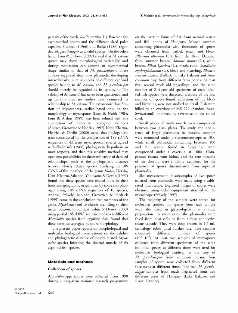
on the parasite fauna of fish from natural watersand fish ponds of Hungary. Muscle samplescontaining plasmodia with thousands of sporeswere obtained from barbel, roach and bleak,Alburnus alburnus (L.), from the River Danube,from common bream, Abramis brama (L.), whitebream, Blicca bjoerkna (L.), roach, rudd, Scardiniuserythrophthalmus (L.), bleak and bitterling, Rhodeussericeus amarus (Pallas), in Lake Balaton and fromcommon carp from different farm ponds. At leastfive, several week old fingerlings, and the samenumber of 2–4-year-old specimens of each infec-ted fish species were dissected. Because of the lownumber of spores found, infections of the bleakand bitterling were not studied in detail. Fish werekilled by an overdose of MS 222 (Sandoz, Basle,Switzerland), followed by severance of the spinalcord.
Small pieces of trunk muscle were compressedbetween two glass plates. To study the occur-rence of larger plasmodia in muscles, sampleswere examined under a stereomicroscope at 10·,while small plasmodia containing between 100and 500 spores, found in fingerlings, werecompressed under a coverslip at 100·. Com-pressed tissues from kidney and the rete mirabileof the choroid were similarly examined for thepresence of spores disseminated from rupturedplasmodia.
Size measurement of subsamples of live sporesisolated from plasmodia were made using a calib-rated microscope. Digitized images of spores wereobtained using video equipment attached to themicroscope (Szekely 1997).
The majority of the samples were stored formolecular studies, but spores from each samplewere also fixed in glycerol-gelatin as a slidepreparation. In most cases, the plasmodia werefreed from host cells or from a host connectivetissue capsule. They were deep frozen in 1.5-mLcentrifuge tubes until further use. The samplescontained different numbers of spores(103)106). At least two samples of myxosporescollected from different specimens of the samefish host species at different times were used formolecular biological studies. In the case ofM. pseudodispar from common bream, foursamples of spores were collected from differentspecimens at different times. The two M. pseudo-dispar samples from roach originated from twodifferent areas of Hungary (Lake Balaton andRiver Danube).
644� 2002
Blackwell Science Ltd
Journal of Fish Diseases 2002, 25, 643–652 K Molnar et al. Intramuscular Myxobolus spp. of cyprinids

DNA extraction
After defrosting the spores, the DNA was extractedas previously described (Eszterbauer, Benko, Dan &Molnar 2001). Briefly, the spores were suspendedin 500 lL lysis buffer [100 mm NaCl, 10 mm Tris,10 mm ethylenediaminetetraacetic acid (EDTA),0.2% sodium dodecyl sulphate (SDS), and0.4 mgmL)1 proteinase K] and incubated at55 �C overnight. The DNA was then extractedwith phenol and chloroform and precipitated inethanol. After centrifugation and washing with 70%ethanol the DNA content was estimated by viewingin a 1.0% agarose gel.
Polymerase chain reaction amplification
The polymerase chain reaction (PCR) was carriedout as described by Eszterbauer et al. (2001).Primers (MX5 and MX3) specific for the familyMyxobolidae (Andree et al. 1999) were used foramplification of an approximately 1600 base pair(bp) fragment of the 18S rRNA gene.
The total volume of the PCR reactions was50 lL, which contained 10–50 ng extracted DNA,1· REDTaq PCR Reaction Buffer (Sigma, St Louis,MO, USA), 0.2 mmol dNTP (MBI Fermentas,Vilnius, Lithuania), 40 pmol of each primer, and2.5 U REDTaq DNA polymerase (Sigma, USA) inMilliQ purified water. A PDR 91 DNA Reproducer(BLS Ltd, Budapest, Hungary) was used for ampli-fication. Amplification conditions were: 95 �C for30 s, 46 �C for 30 s and 72 �C for 60 s for 35 cycles,with a terminal extension at 72 �C for 10 min.
MC5 and MC3, the other primer pair used forPCR amplification, were designed in our laborat-ory. Their sequences are:
MC5 (forward) 5¢-CCTGAGAAACGGCTACCACATCCA-3¢
MC3 (reverse) 5¢-GATTAGCCTGACAGATCACTCCACGA-3¢.
The contents of the PCR reactions and theequipment used for amplification were identicalwith those used for the MX5–MX3 primer pair.The PCR programme was 95 �C for 300 s,followed by 35 cycles of 30 s at 95 �C, 30 s at56 �C and 60 s at 72 �C, and was terminated withan extension at 72 �C for 300 s.
The PCR products were electrophoresed in 1.0%agarose gels (Sigma, St Louis, MO, USA) in TBEbuffer. k phage DNA cut with PstI was used as themolecular weight standard.
Sequencing
For DNA sequencing, six primers were used: theMX5–MX3 and MC5–MC3 primer pairs and alsothe MB5 and MB3 primers designed for sequencingin our laboratory and based on the 18S rDNAsequences of Myxobolus species, available in Gen-Bank. The location of the primers compared withthe M. cerebralis sequence are shown in Table 1.The sequences of the MB5 and MB3 oligonucle-otides are:
MB5 (forward) 5¢-GGTGATGATTAACAGGAGCGGT-3¢
MB3 (reverse) 5¢-CCAACCGCTCCTGTTAATCATC-3¢.
The PCR products were sequenced using thePRISM Ready Reaction Dye Deoxy Cycle Sequen-cing Protocol (Perkin-Elmer, Norwalk, CN, USA)with an ABI 373 A automated DNA sequencer (PEApplied Biosystems, Foster City, CA, USA). Thenucleotide sequences were read using the AppliedBiosystems 373A DNA Sequencer Data AnalysisProgram and assembled by the program packageLasergene (DNASTAR).
In the case of the replicates originating fromdifferent specimens of the same fish host species,only the MX5 and MX3 primers were used forsequencing.
Phylogenetic analysis
Multiple alignment of the nucleotide sequences wasperformed with the MultAlin computer program(Corpet 1998). The highly variable regions wereremoved from the sequences as described byHarrach & Benko (1998), therefore only 972nucleotides were used in the phylogenetic calcula-tions from the approximately 1000 bp long 18SrDNA fragments. The 972 bp long, alignedsequences contained two fragments: a 511 bp longfragment from the 5¢-end of the 18S rRNA geneand a 461 bp long fragment from the 3¢-end.
Phylogenetic calculations were performed by theprograms of the PHYLIP version 3.573c package
Table 1 The location of the 5¢ end of the primers on the 18S
rRNA gene of M. cerebralis
Forward primer Position Reverse primer Position
MX5 66 MX3 1767
MC5 388 MC3 1446
MB5 980 MB3 1004
Journal of Fish Diseases 2002, 25, 643–652 K Molnar et al. Intramuscular Myxobolus spp. of cyprinids
645� 2002
Blackwell Science Ltd

(Felsenstein 1989). The data were analysed withparsimony (DNAPARS) and distance matrixanalyses (DNADIST using Kimura-2 distanceparameter followed by FITCH with global rear-rangements). Gaps of up to three bases were treatedas special (vs. missing) characters. For bootstrapanalysis, the mentioned programs were preceded bySEQBOOT (molecular sequences; 100 data sets)and followed by CONSENSE.
Results
Morphology
Plasmodia containing developmental stages orspores were found in the muscle of all the eightfish species examined. Young plasmodia weredetected intracellularly in muscle cells of the trunkmusculature in all cases. More mature plasmodia, inaddition to the residue of the host cell, weresurrounded by a connective tissue capsule. In somecases, free spores from disrupted plasmodia weredetected in the extracellular space between musclecells. At a more chronic stage scattered spores fromruptured plasmodia could be demonstrated in thegut lumen, in the capillaries of the choroid and thegills, and particularly in the melano-macrophagecentres of the kidney. The most severe and commoninfection of skeletal muscle was in the roach whereencapsulated plasmodia were always detectable inolder fish. Spores found in common carp, barbel,roach, rudd, common bream and white bream wereabout the same size (10–13 lm) and showed a very
similar morphology. The common feature of thespores was the small, indistinct intercapsular processand the polar filaments, which were only looselywound in the polar capsule with not more than fourcoils. In each fish, even in a single plasmodium,three different morphological types of spores werefound. Some spores had a typical elliptical shape andapproximately equal polar capsules at the apex,while other spores had a less regular shape, and polarcapsules of unequal size were lateral to the apex. Inthe third spore type, the opening of the polarcapsules was typically located at the lateral side of thehighly deformed spore (Fig. 1). Although morpho-logically variable spores were found in all fish speciesstudied, in roach, rudd, white bream and commonbream the number of asymmetric spores was muchhigher than in common carp. In common carp,deformed spores were found in encysted plasmodiaand in spores engulfed by macrophages in differentorgans. Deformed spores were found only occasion-ally in barbel (Fig. 2). Morphologically similarspores to those described above were also found inthe muscle of bleak and bitterling, but these werenot characterized further because the quantity ofspores was not sufficient for molecular studies.
The shape and measurements of the spores inevery fish species agreed with the original descrip-tions and therefore in molecular studies we used themost widely accepted terminology and systematics.Based on original descriptions and typical hosts,spores from the common carp were tentativelyidentified as M. cyprini Doflein, spores from barbelas M. musculi Keysselitz, while spores from the
Figure 1 Schematic illustrations of spores of intracellular Myxobolus spp. from the muscle of cyprinid fish. (a) Spore with relatively
regular shape and with only slightly differing polar capsules. (b) Spore with relatively regular shape but with polar capsules of different
size. (c) Spore with irregular shape and polar capsules of different size.
Journal of Fish Diseases 2002, 25, 643–652 K Molnar et al. Intramuscular Myxobolus spp. of cyprinids
646� 2002
Blackwell Science Ltd

roach, rudd, white bream and common bream wereidentified as M. pseudodispar Gorbunova.
PCR and phylogenetic analysis
The specific primer pairs MX5–MX3 and MC5–MC3 successfully amplified approximately 1600and 1000 bp fragments, respectively, of the 18SrRNA gene from every sample of Myxobolus
examined. The replicates of myxospores isolatedfrom different specimens of the same fish hostwere successfully amplified with the MX5–MX3primer pair and were sequenced with these twoprimers. Thus, approximately 500 bp DNAsequences from the 5¢ and 3¢ end of the1600 bp DNA fragment were obtained. TheDNA sequences of the PCR products have beendeposited in GenBank and accession numbers are
Figure 2 Myxospores of intracellular Myxobolus spp. collected from the muscle of cyprinid fish. (a) M. cyprini from common carp, (b)
M. musculi from barbel, (c) M. pseudodispar from roach, (d) M. pseudodispar from white bream, (e) M. pseudodispar from rudd and (f )
M. pseudodispar from common bream (fresh preparations, ·2000).
Journal of Fish Diseases 2002, 25, 643–652 K Molnar et al. Intramuscular Myxobolus spp. of cyprinids
647� 2002
Blackwell Science Ltd

listed in Table 2. In several cases, the partialsequences of the replicates were completely iden-tical with a previously sequenced sample of thegiven parasite species (Table 2).
Distance matrix analysis (Fig. 3) distinguishedsix groups, with very high (99–100) bootstrapconfidence levels. Myxobolus cerebralis, the onlynon-muscular Myxobolus species, was found to be
Table 2 Myxobolus species sequenced from different host species and localities
Species Host Locality GenBank number
Length of
sequence (bp)
M. pseudodispar RO1 Roach Lake Balaton AF380145 1554
M. pseudodispar RO2 Roach River Danube AF466651
AF466656
5¢ end: 560
3¢ end: 500
M. pseudodispar RD1 Rudd Lake Balaton AF380142 1556
M. pseudodispar RD2 Rudd Lake Balaton AF466650
AF466655
5¢ end: 520
3¢ end: 500
M. pseudodispar WB1 White bream Lake Balaton AF380143 1551
M. pseudodispar WB2 White bream Lake Balaton Same as AF380143a
AF466654
5¢ end: 520
3¢ end: 500
M. pseudodispar CB1 Common bream Lake Balaton AF380144 1551
M. pseudodispar CB2 Common bream Lake Balaton AF466648
AF466652
5¢ end: 570
3¢ end: 500
M. pseudodispar CB3 Common bream Lake Balaton AF466649 5¢ end: 570
(Keszthely area) AF466653 3¢ end: 500
M. pseudodispar CB4 Common bream Lake Balaton Same as AF466649a 5¢ end: 520
(Siofok area) Same as AF466653a 3¢ end: 500
M. cyprini 1 Common carp Fish farm AF380140 1554
M. cyprini 2 Common carp Fish farm Same as AF380140a
AF466657
5¢ end: 520
3¢ end: 500
M. musculi 1 Barbel River Danube AF380141 1556
M. musculi 2 Barbel River Danube Same as AF380141a
AF466658
5¢ end: 520
3¢ end: 500
a The partial sequence of the sample was 100% identical with a previously sequenced sample of the given parasite species collected from the given fish-host
species.
Figure 3 Phylogenetic tree generated by distance matrix analysis and showing relationships between Myxobolus spp. collected from the
muscle of cyprinid species. The length of the edited alignment sequences was 972 bp, containing a 511-bp fragment from the 5¢-end of
the 18S rRNA gene and a 461-bp fragment from the 3¢-end. Myxobolus cerebralis (U96492) was chosen as an outgroup. Sequences
published in this study are listed in Table 2. Numbers at nodes indicate bootstrap confidence levels obtained by distance matrix analysis.
Scale bar is equal to 10% difference. *CB: common bream, WB: white bream, RO: roach, RD: rudd.
Journal of Fish Diseases 2002, 25, 643–652 K Molnar et al. Intramuscular Myxobolus spp. of cyprinids
648� 2002
Blackwell Science Ltd

very different, and was a suitable outgroup.Myxobolus musculi and M. cyprini clustered, butwith a very low confidence level.
Similarly, all sequences from the putativeM. pseudodispar collected from four different fishspecies clustered, but with a low bootstrap value.The cluster of the 10 M. pseudodispar sequencesdiverged into two subclusters with high bootstrapconfidence levels. The first subcluster contained thefour samples originating from common bream andthe two from white bream. The other subclusterconsisted of spores collected from roach and rudd.Genetic distances obtained from the distance matrixoutfile are shown in Table 3.
Parsimony analysis confirmed the clusteringaccording to fish species, but not to spore mor-phology. The subcluster containing the M. pseudo-dispar samples originating from roach and ruddjoined to the cluster of samples of M. cyprini andM. musculi, even if only with a low bootstrap value(60). The other parts of the phylogenetic tree(Fig. 4) were congruent to the tree generated by thedistance matrix analysis.
Discussion
Until recently, only spore morphology served as ameans of identification of the more than 500known Myxobolus species. Because of the diffi-culties of experimental studies, little was knownabout host specificity, and morphologically similarspores from genetically different hosts were oftendescribed as the same species. Thus, Shulman(1966) recorded as many as 40 hosts for certainMyxobolus species. To enlarge the criteria forspecies identification, Molnar (1994) suggestedthat the host, organ and tissue specificity shouldalso be considered. Molecular biological methodsoffer great scope for the correct identification ofspecies.
Myxobolus spp. infecting the skeletal muscles ofcyprinids serve as examples of the difficultiesassociated with methods of identification based onspore morphology. It is not surprising thatM. pseudodispar differing from the original descrip-tion of M. cyprini in its asymmetric spore shape anddifferent sized polar capsules was described byGorbunova (1936) as a new species. Similarly, it isunderstandable that Lom & Dykova (1992), whoobserved great morphological variability in thespores of M. cyprini, considered M. pseudodispar ajunior synonym. During the present survey T
able
3G
enet
icd
ista
nce
sob
tain
edfr
omth
ed
ista
nce
mat
rix
outfi
lebas
edon
972
bp
frag
men
tsof
the
18S
rRN
Age
ne
of14
Myx
obol
ussa
mp
les
isol
ated
from
the
mu
scle
ofcy
pri
nid
san
dth
eou
tgro
up
,
M.
cere
bral
is
M.p
.RO
1M
.p.R
O2
M.p
.RD
1M
.p.R
D2
M.p
.WB
1M
.p.W
B2
M.p
.CB
1M
.p.C
B2
M.p
.CB
3M
.p.C
B4
M.m
.2M
.m.1
M.c
.2M
.c.1
M.c
er.
M.p
.RO
10.0
000
0.0
031
0.0
295
0.0
252
0.0
470
0.0
469
0.0
447
0.0
436
0.0
425
0.0
425
0.0
361
0.0
372
0.0
562
0.0
573
0.2
018
M.p
.RO
20.0
000
0.0
306
0.0
284
0.0
481
0.0
481
0.0
469
0.0
458
0.0
447
0.0
447
0.0
394
0.0
405
0.0
596
0.0
608
0.2
033
M.p
.RD
10.0
000
0.0
062
0.0
479
0.0
502
0.0
480
0.0
491
0.0
480
0.0
480
0.0
493
0.0
482
0.0
642
0.0
630
0.2
133
M.p
.RD
20.0
000
0.0
512
0.0
490
0.0
502
0.0
490
0.0
479
0.0
479
0.0
448
0.0
459
0.0
595
0.0
607
0.2
160
M.p
.WB
10.0
000
0.0
021
0.0
114
0.0
125
0.0
114
0.0
114
0.0
525
0.0
514
0.0
629
0.0
618
0.1
974
M.p
.WB
20.0
000
0.0
135
0.0
125
0.0
114
0.0
114
0.0
525
0.0
536
0.0
629
0.0
641
0.2
003
M.p
.CB
10.0
000
0.0
031
0.0
021
0.0
021
0.0
492
0.0
481
0.0
584
0.0
573
0.1
916
M.p
.CB
20.0
000
0.0
010
0.0
010
0.0
481
0.0
492
0.0
550
0.0
561
0.1
931
M.p
.CB
30.0
000
0.0
000
0.0
469
0.0
481
0.0
561
0.0
573
0.1
933
M.p
.CB
40.0
000
0.0
469
0.0
481
0.0
561
0.0
573
0.1
933
M.m
.20.0
000
0.0
010
0.0
460
0.0
471
0.1
887
M.m
.10.0
000
0.0
471
0.0
460
0.1
872
M.c
.20.0
000
0.0
010
0.2
051
M.c
.10.0
000
0.2
036
M.c
er.
0.0
000
M.p
.:M
yxob
olus
pseu
dodi
spar
,M
.c.:
Myx
obol
uscy
prin
i,M
.m.:
Myx
obol
usm
uscu
li,
M.c
er.:
Myx
obol
usce
rebr
alis
;C
B:
com
mon
bre
am,
WB
:w
hit
eb
ream
,R
O:
roac
h,
RD
:ru
dd
.
Journal of Fish Diseases 2002, 25, 643–652 K Molnar et al. Intramuscular Myxobolus spp. of cyprinids
649� 2002
Blackwell Science Ltd

M. pfeifferi, the best known parasite of the muscleof barbel, was not found, but another speciesidentified as M. musculi, commonly infecting themuscle cells of barbel, did occur. Based on sporemorphology, particularly the structure of the polarcapsules, this latter species was erroneously identi-fied as M. ergensi Lom in a previous study(Eszterbauer et al. 2001). The spores found in themuscle of the barbel corresponded to the publisheddiagrams of M. ergensi spores in all respects;however, considering the specific location of plas-modia, we consider that this species should havebeen determined as M. musculi, which was des-cribed earlier but depicted less accurately.
Among the genus Myxobolus, strictly host specificparasites and those with a relatively wide host rangeare found with equal frequency. According toMolnar (1994), M. drjagini and M. pavlovskii,infecting Chinese carp introduced to Europe,represent the strictly host specific species. Myxobolusdrjagini infects only silver carp, Hypophthalmichthysmolitrix (Valenciennes), while M. pavlovskii devel-ops in both silver carp and bighead carp, Aristichthysnobilis (Richardson). Myxobolus cerebralis, the bestknown Myxobolus species, is an example of a specieswith a wider host range, infecting a number ofsalmonid species (Hedrick, El-Matbouli, Adkinson
& MacConnell 1998; Thomson, Nehring, Bowden& Wygant 1999).
Using classical zoological methods it is verydifficult to determine the validity of morphologicallysimilar myxosporeans with identical tissue affinityand developing in taxonomically closely related hostspecies. In such cases, cross-infection experimentsand molecular biological methods can help. In thecase of the muscle parasites M. cyprini, M. musculiand M. pseudodispar, the cyprinid fish hosts can beconsidered relatively closely related species, althoughZardoya & Doadrio (1999) suggest that Cyprinusand Barbus species are more closely related to eachother than to members of the other four cyprinidgenera included in this study, i.e. Rutilus, Scardinius,Abramis and Blicca. Eszterbauer et al. (2001), usinga combined PCR-restriction fragment lengthpolymorphism (RFLP) method, studied muscleinfecting – M. pseudodispar from roach and rudd,M. cyprini from common carp and M. musculi(previously identified as M. ergensi) from barbel. Theresults for the M. pseudodispar samples from differ-ent fish species were identical. The results for M.cyprini and M. musculi were similar to each other,but were markedly different from M. pseudodispar.
In the present study, 14 Myxobolus samplesoriginating from the muscle of six cyprinid species
Figure 4 Phylogenetic tree created by parsimony analysis showing the relationships among the muscle-infecting Myxobolus species
examined. The length of the edited alignment sequences was 972 bp, containing a 511-bp fragment from the 5¢-end of the 18S rRNA
gene and a 461-bp fragment from the 3¢-end. Myxobolus cerebralis (U96492) was chosen as an outgroup. Sequences published in this
study are listed in Table 2. *CB: common bream, WB: white bream, RO: roach, RD: rudd.
Journal of Fish Diseases 2002, 25, 643–652 K Molnar et al. Intramuscular Myxobolus spp. of cyprinids
650� 2002
Blackwell Science Ltd

were compared based on their 18S rDNA sequenc-es. The DNA sequences of Myxobolus samplescollected from different specimens of a given fishspecies were consistently more similar to each otherthan to those of spore samples originating fromother fish hosts. The great similarity of DNAsequences indicates that the Myxobolus spore sam-ples originating from different fish species areclosely related to each other, and suggests that theseparasites could represent populations of the samespecies. If we accept this ‘one-species’ concept thisspecies should be designated M. cyprini based on thepriority of its description.
However, the differences in the DNA sequencesof spores collected from different fish species seemto reflect the phylogenetic relationship of the fishhosts (i.e. there is a similar topology of thephylogenetic tree of the parasites and cyprinidhosts) (Zardoya & Doadrio 1999). Therefore itis possible that M. cyprini, M. musculi andM. pseudodispar are distinct species. In addition, afurther subdivision of the species M. pseudodisparaccording to the host species may be considered.However, this suggestion is difficult to explain inecological terms, i.e. that muscle-parasitic myxos-poreans (species or populations) spread verticallywithin a given biotope. In this case, parasitesoriginating from a given fish species would infect,even after establishing infection in alternate hosts,only that fish species from which they originated.This possible route of development would indicatethat the morphologically similar parasites found incyprinids do not belong to the same species.Although this hypothesis seems extremely unlikely,it should not be ruled out until refuted by cross-infection experiments. Molecular biological meth-ods appear to be excellent tools for studying thisquestion and providing very robust answers; how-ever, only infection experiments and sequence-levelphylogenetic analyses of further spore samples willdefinitively determine whether muscle-parasiticmyxosporeans of cyprinids represent valid speciesor only synonyms. Until further studies give asatisfactory answer to this question, we suggest thatM. cyprini, M. musculi and M. pseudodispar are validspecies in accordance with the original descriptions.
Acknowledgements
This work was supported by the Hungarian Scien-tific Research Fund (OTKA) grants no. T 29200, T31755 and A312. Special thanks are due to Zsuzsa
Kis for making the histological sections, Dr FerencBaska, Dr Gyorgy Csaba, Dr Maria Lang and DrGabor Majoros for kindly providing Danube andpond fish, and Dr Maria Benko for criticallyreading the manuscript.
References
Andree K.B., Gresoviac S.J. & Hedrick R.P. (1997) Small sub-
unit ribosomal sequences unite actinosporean and myxospor-
ean states of Myxobolus cerebralis, the causative agent of
whirling disease in salmonid fishes. Journal of Eukaryotic Mi-crobiology 44, 208–215.
Andree K.B., Szekely C., Molnar K., Gresoviac S.J. & Hedrick
R.P. (1999) Relationships among members of the genus
Myxobolus (Myxozoa: Bivalvidae) based on small subunit
ribosomal DNA sequences. Journal of Parasitology 85, 68–74.
Baska F. (1986) Histological studies on the development of
Myxobolus pseudodispar Gorbunova, 1936 in the roach (Rutilusrutilus). Acta Veterinaria Hungarica 35, 251–257.
Bykhovski B.E. (1962) Key to the Parasites of Freshwater Fish ofthe USSR. Izdatelst’stvo Akademii Nauk USSR, Moscow-
Leningrad (In Russian).
Corpet F. (1998) Multiple sequence alignment with hierarchical
clustering. Nucleid Acids Research 16, 10881–10890.
Eszterbauer E., Benko M., Dan A. & Molnar K. (2001) Iden-
tification of fish parasitic Myxobolus (Myxosporea) species
using a combined PCR-RFLP method. Diseases of AquaticOrganisms 44, 35–39.
Felsenstein J. (1989) PHYLIP – Phylogeny inference package
(version 3.2). Cladistics 5, 164–166.
Gorbunova M.N. (1936) Changes in the parasitic fauna of pike
and roach according to their age. Uchenye Zapiski Lenin-gradskogo Ordena Lenina Gosudarskogo Universiteta. No. 7(Biology Series Fascicule 3), pp. 5–30. Problems of Economic
Parasitology, Leningrad (In Russian).
Harrach B. & Benko M. (1998) Phylogenetic analysis of ade-
novirus sequences. In: Methods in Molecular Medicine, Vol. 21:
Adenovirus Methods and Protocols (ed. by W.S.M. Wold), pp.
309–339. Humana Press Inc., Totowa, NJ, USA.
Hedrick R.P., El-Matbouli M., Adkinson M.A. & MacConnell
E. (1998) Whirling disease: re-emergence among wild trout.
Immunological Reviews 166, 365–376.
Hervio D.M.L., Kent M.L., Khattra J., Sakanari J., Yokoyama
H. & Devlin R.H. (1997) Taxonomy of Kudoa species
(Myxosporea) using small subunit ribosomal DNA sequence.
Canadian Journal of Zoology 75, 2112–2119.
Kent M.L., Khattra J., Hedrick R.P. & Devlin R.H. (2000)
Tetracapsula renicola n. sp. (Myxozoa: Saccosporidae); the
PKX myxozoan – the cause of proliferative kidney disease of
salmonid fishes. Journal of Parasitology 86, 103–111.
Keysselitz G. (1908) Uber durch Sporozoen (Myxosporidien)
hervorgerufene pathologische Veranderungen. Verhandlungender Gesellschaft Deutscher Naturforscher und Arzte 2, 542–543.
Kocylowski B. & Myaczynski T. (1960) Diseases of Fish andCrayfish. PWRiL, Warszawa (In Polish).
Journal of Fish Diseases 2002, 25, 643–652 K Molnar et al. Intramuscular Myxobolus spp. of cyprinids
651� 2002
Blackwell Science Ltd

Lom J. & Arthur J.R. (1989) A guideline for the preparation of
species descriptions in Myxosporea. Journal of Fish Diseases 12,
151–156.
Lom J. & Dykova I. (1992) Protozoan Parasites of Fishes. Elsevier,
New York.
Lom J. & Noble E.R. (1984) Revised classification of the
Myxosporea Butschli, 1881. Folia Parasitologica 31, 193–205.
Markevich A.P. (1951) Parasite Fauna of Fish of the Ukraine SSR.
Kiev, Ukraine (in Russian).
Molnar K. (1994) Comments on the host, organ and tissue
specificity of fish myxosporeans and types of their intrapiscine
development. Parasitologica Hungarica 27, 5–20.
Molnar K. & Kovacs-Gayer E. (1985) The pathogenicity and
development within the host fish of Myxobolus cyprini Doflein,
1898. Parasitology 90, 549–555.
Ogawa K., Delgahapytiya K.P., Furuta T. & Wakabayashi H.
(1992) Histological studies on the host response to Myxobolusartus Akhmerov, 1960 (Myxozoa: Myxobolidae) infection in
the skeletal muscle of carp, Cyprinus carpio L. Journal of FishBiology 41, 363–371.
Salim K.Y. & Desser S.S. (2000) Descriptions and phylogenetic
systematics of Myxobolus spp. from cyprinids in Algonquin
Park, Ontario. Journal of Eukaryotic Microbiology 47, 309–
318.
Schaperclaus W. (1954) Fischkrankheiten. Akademie Verlag,
Berlin.
Shulman S.S. (1966) Myxosporidia of the Fauna of USSR. Izda-
tel’stvo Nauka, Moscow (In Russian).
Szekely Cs. (1997) Possible applications of video technology
and digital image processing in fish parasitology:
morphological examination of the groups Apicomplexa
and Myxosporea-Actinosporea by video technology.
Bulletin of the European Association of Fish Pathologists 17,
81–82.
Thomson K.G., Nehring R.B., Bowden C. & Wygant T.
(1999) Field exposure of seven species or subspecies of
salmonid to Myxobolus cerebralis in the Colorado River,
Middle Park, Colorado. Journal of Aquatic Animal Health 11,
312–329.
Zardoya R. & Doadrio I. (1999) Molecular evidence on the
evolutionary and biogeographical patterns of European
cyprinids. Journal of Molecular Evolution 9, 227–237.
Received: 2 July 2001Accepted: 25 June 2002
Journal of Fish Diseases 2002, 25, 643–652 K Molnar et al. Intramuscular Myxobolus spp. of cyprinids
652� 2002
Blackwell Science Ltd