intramuscular injection of vectorized-scfvmc1 reduces
TRANSCRIPT
RESEARCH Open Access
Intramuscular injection of vectorized-scFvMC1 reduces pathological tau in twodifferent tau transgenic modelsFrancesca Vitale1, Jasmin Ortolan1, Bruce T. Volpe2,3, Philippe Marambaud1,2, Luca Giliberto1,2,4* andCristina d’Abramo1,2*
Abstract
With evidence supporting the prion-like spreading of extracellular tau as a mechanism for the initiation andprogression of Alzheimer’s disease (AD), immunotherapy has emerged as a potential disease-modifying strategy totarget tau. Many studies have proven effective to clear pathological tau species in animal models of AD, and severalclinical trials using conventional immunotherapy with anti-tau native antibodies are currently active. We havepreviously generated a vectorized scFv derived from the conformation-dependent anti-tau antibody MC1, scFvMC1,and demonstrated that its intracranial injection was able to prevent tau pathology in adult tau mice. Here, we showthat, in a prevention paradigm and in two different tau transgenic models (JNPL3 and P301S), a one-timeintramuscular injection of AAV1-scFvMC1 generated a long-lasting peripheral source of anti-tau scFvMC1 andsignificantly reduced insoluble and soluble tau species in the brain. Moreover, our data showed that scFvMC1 wasinternalized by the microglia, in the absence of overt inflammation. This study demonstrates the efficacy ofintramuscular delivery of vectorized scFv to target tau, and suggests a new potential application to treat AD andthe other tauopathies.
Keywords: Vectorized antibodies, AAV, scFv, Tau, Immunotherapy, Intramuscular injection
IntroductionThe microtubule-associated protein tau plays a physio-logical role in microtubule stabilization, axonal growthand cytoskeletal dynamics in neurons, but its aggrega-tion characterizes several neurological diseases classifiedas tauopathies, including Alzheimer’s disease (AD) [1–5]. As increasing evidence supports the existence of tauas an extracellular protein and the concept of its trans-cellular propagation as a mechanism for the initiationand progression of AD, tau has become an attractive tar-get for immunotherapy in animal models of AD and
related tauopathies [6–13]. A conspicuous amount ofdata has been produced by different laboratories, includ-ing ours, showing reduction of tau pathology in trans-genic animal models using tau monoclonal antibodies,with a different degree of success according to whichepitopes were targeted [14–26]. However, passive im-munotherapy using conventional antibodies in humanspresents several potential limitations such as low perme-ability to cross the blood-brain barrier (BBB), detrimen-tal inflammatory reactions and microhemorrhagesassociated to the treatment, requirement for repeateddosing, patients compliance and high costs [27–30]. Toovercome these issues, several groups have engineeredantibodies as fragments, i.e. single chain variable frag-ment (scFv), to be used in combination with gene deliv-ery strategies based on the use of viral vectors:
© The Author(s). 2020 Open Access This article is licensed under a Creative Commons Attribution 4.0 International License,which permits use, sharing, adaptation, distribution and reproduction in any medium or format, as long as you giveappropriate credit to the original author(s) and the source, provide a link to the Creative Commons licence, and indicate ifchanges were made. The images or other third party material in this article are included in the article's Creative Commonslicence, unless indicated otherwise in a credit line to the material. If material is not included in the article's Creative Commonslicence and your intended use is not permitted by statutory regulation or exceeds the permitted use, you will need to obtainpermission directly from the copyright holder. To view a copy of this licence, visit http://creativecommons.org/licenses/by/4.0/.The Creative Commons Public Domain Dedication waiver (http://creativecommons.org/publicdomain/zero/1.0/) applies to thedata made available in this article, unless otherwise stated in a credit line to the data.
* Correspondence: [email protected]; [email protected] of Molecular Medicine, The Litwin-Zucker Center for Alzheimer’sDisease & Memory Disorder, The Feintein Institutes for Medical Research,Manhasset, NY, USAFull list of author information is available at the end of the article
Vitale et al. Acta Neuropathologica Communications (2020) 8:126 https://doi.org/10.1186/s40478-020-01003-7
vectorized anti-Aß (amyloid-beta) and anti-tau scFvshave previously shown benefits in models of AD [31–45]. ScFvs consist of the smallest functional antigen-binding domain of an antibody (Ab) exhibiting compar-able antigen-binding affinity as the parent immuno-globulin, reduced size, improved pharmacokinetic interms of tissue penetration and lack of an Fc receptor-mediated inflammatory response [46–48]. Due to theirshort systemic half-life in vivo [49, 50], in order to reacha sustained and long-lasting expression scFv are gener-ally cloned in adeno associated viral vectors (AAVs) anddelivered by one-time injections [34, 46, 47]. We havepreviously shown [43] that the AAV vectorized-scFvMC1, a recombinant version of the native anti-tauconformational mAb (monoclonal antibody) MC1, sig-nificantly reduces brain pathological tau in adult JNPL3mice, by one-time intracranial injection. In parallel withAAV-based delivery, Spencer et al. [44] have shown thatsystemic injection of a lentiviral vector (LV) carrying ascFv directed to 3Rtau and enhanced for brain penetra-tion (LV-3RT-apoB) was able to reduce tau accumula-tion, neurodegeneration and behavioral deficit in a tautransgenic model. Although using third-generation lenti-viral vectors has emerged as a promising therapeutic op-tion for conditions as primary immunodeficiencies andcancers, it is still necessary to understand the long-termsafety and efficacy of these vectors in humans, especiallyin respect to their potential for insertional oncogenesis[51]. In this context, using AAVs is considered saferthan LVs.Direct delivery of AAV into the brain has been tested
in a number of clinical trials [52–57] and parallel effortshave been done over the past years to develop newbrain-targeted AAVs to treat CNS diseases in vivo usinga systemic delivery approach [58]. In this respect, per-ipheral administration provides obvious advantages toAAV gene therapy: a non-invasive route of injection,lack of surgery-related side effects, improved patientcompliance and costs. In this study we propose to de-velop a novel therapeutic approach for AD and tauopa-thies by using intramuscular (IM) delivery of AAV-vectorized scFv. To date, there has been little effort todevelop peripheral protocols targeting skeletal muscle totackle CNS disorders. IM injection provides a quick,easy, non-invasive and safe route of administration, andcan routinely be performed in virtually any setting. Also,skeletal muscles are an ideal target tissue for AAV trans-duction because individual fibers are large, multinucle-ated and with minimal cellular turnover [59]. Takingadvantage of the practical features and biological proper-ties of this delivery approach, the aim of the presentstudy was to generate a long-lasting peripheral nicheable to produce and release anti-tau scFv in the circula-tion to target cerebral tau, without transducing vital
organs such as liver, kidneys and heart. A similar strat-egy has been previously tested in a mouse model of AD,where intramuscular delivery of AAV1 vectored anti-AßscFv was able to reduce Aß load in brain [35, 45]. Simi-larly, gene therapy for Alpha-1 antitrypsin (AAT) defi-ciency has been developed in humans usingrecombinant AAV1 serotype, demonstrating a continuedstable transgene expression at 5 years after transduction[60, 61].In this study, for the first time, we demonstrate the
in vivo feasibility and efficacy of targeting pathologicaltau in the brain, by employing intramuscular delivery ofvectorized anti-tau scFv. Two different tau transgenicmodels, the JNPL3 and P301S mice, received a single IMinjection of AAV1-scFvMC1, showing a significant re-duction of tau pathology, with some differences betweenstrains. Moreover, no signs of inflammation were ob-served upon AAV1-scFvMC1 immunization, showingthat tau clearance does not involve Fc-receptor-mediated and microglia-associated detrimental inflam-matory response. However, our in vitro and in vivo datapoint to the microglia as a player in the uptake andclearance of scFv-tau, despite the absence of Fc, addingto the potential mechanisms of action of tauimmunotherapy.In summary, our data support the peripheral intramus-
cular route as an effective, feasible and safe delivery ap-proach for AAV-scFv-based anti tau immunotherapy,with relevant translational potential applications to othertauopathies and brain disorders.
MethodsScFv-MC1 design and sub-cloning into AAV1The light and heavy-chain variable domains correspond-ing to the MC1 antibody were sequenced employing theMCLAB antibody service (San Francisco, CA). As previ-ously published [43] the VH and VL chains were joinedtogether by a 15 amino acid residues linker (Gly4Ser)3.5′-terminal signal peptide (SP) and 3′-terminal Myc andHis6X tags were added. The AAV packaging and purifi-cation service was provided by Vector Biolab (Malvern,PA). ScFv-MC1 was sub-cloned into the adeno-associated viral vector serotype 1 (AAV1) under the con-trol of the synthetic strong CAG (CMV-chicken betaactin-rabbit beta globin) promoter. In order to enhanceexpression of the transgene, the WPRE Woodchuckhepatitis virus (WPRE) post-transcriptional regulatoryelement was added 5′ of the Myc and His6X tags.
Tau transgenic miceJNPL3 mice obtained from Taconic (Germantown, NY)express 0N4R human tau with the P301L mutation thatcauses frontotemporal dementia in humans, under themouse prion promoter. JNPL3 mice develop NFTs-like
Vitale et al. Acta Neuropathologica Communications (2020) 8:126 Page 2 of 19
pathology as early as 4.5 months and in later stages pro-gressive deterioration of the motor function [62]. Homo-zygous P301S were obtained from Dr. Michel Goedert(Cambridge, UK) [63]: these mice, on pure C57BL/6background, express 0N4R human tau carrying theP301S mutation, under the control of the neuron-specific murine Thy-1 promoter, and they develop wide-spread tau pathology affecting cerebral cortex, hippo-campus and brain stem as early as 6 months, and partialparalysis of the lower limbs by 8months of age. Animalswere treated according to the current regulations for theproper handling of research animals, following an ap-proved IACUC protocol.
ScFvMC1 purificationScFvMC1 purification was performed as previously pub-lished [43]. Briefly, scFv-MC1 was cloned into the mam-malian expression vector pcDNA3.1 (Genewiz, SouthPlainfield, NJ) and transfected into HEK293T, using Li-pofectamine 2000 (Invitrogen, Carlsbad, CA). After 48 hof transfection, the scFv released into the conditionedmedium was affinity purified using a Ni-Sepharose HighPerformance column (GE Healthcare, Port Washington,NY). The efficiency of purification was tested using animmunosorbent assay employed to assess the antigen–binding specificity of the scFvMC1, as previously de-scribed [43]. Starting material, flow through and elutedfractions were tested to check for proper enrichment ofthe purified material. The purified scFv-MC1 waschecked on Coomassie-stained SDS-PAGE gel for propermolecular weight.
Infrared conjugation and intravenous (IV) injectionsScFv-MC1 and MC1 have been conjugated with IRDye800CW, using IRDye 800CW protein Labeling Kit Low-MW or High-MW respectively (LI-COR Biosciences,Lincoln, NE), according to the manufacturer instruc-tions. Briefly, scFv-MC1 and MC1 were dialyzed in 50mM potassium phosphate buffer pH 8.5 at 4 °C, over-night; the pH was then adjusted with 1M potassiumphosphate buffer to 9. After 2 h incubation, the unconju-gated dye was removed using desalting spin columns(Zeba Desalt Spin Columns, Thermo Scientific).ScFvMC1 and MC1 antigen-binding reactivities weremeasured against an MC1 specific peptide [43] by im-munosorbent assay, in order to exclude loss of activityupon conjugation.To verify the ability of scFvMC1 and MC1 to cross the
blood brain barrier, IV injection was performed in 3-month-old JNPL3 mice using 100 μg of the antibodies:saline, scFvMC1-IRDye, MC1-IRDye, or unlabeled anti-bodies were injected (n = 3 per group). Mice were anes-thetized with isoflurane and the injections performedretro-orbitally. Mice were sacrificed 2 h post injection;
brains were harvested and dissected into cortex (Ctx),hindbrain (HB) and hippocampus (Hip).Homogenization was performed in 1X RIPA buffer(Thermo Fisher Scientific, Waltham, MA) with the Miniprotease inhibitor cocktail (Roche, Indianapolis, IN).Brain samples from each region were spotted on0.45 μm nitrocellulose followed by IR signal acquisitionat 789 nm, using Sapphire Biomolecular Imager (AzureBiosystems, Dublin, CA).
Intra-muscular (IM) injectionsAAV1-CAG-scFvMC1 or AAV1-CAG-eGFP wereinjected at a dose of 2X10¹¹ GC per mouse. Each AAVwas diluted in PBS at a final volume of 50 μl, and a one-time intramuscular injection was administrated in theright tibialis. Injections were performed upon anesthesiawith isoflurane.Twenty-six females JNPL3 (n = 13 per group) mice
were injected at 3 month of age and sacrificed 4 monthslater. The P301S line was injected at 2 month of age andsacrificed 4 months later; we used twelve females P301Sin total (n = 6 per group). Overall, 26 JNPL3 and 12P301S mice were employed in this study.
Brain extracts and tissues preparationMice were sacrificed by isoflurane overdose, decapitatedand processed as described previously [14]. The brainwas removed and divided at the midline so that just onehalf of the brain was dissected for biochemical analysis.Cortex, hippocampus and hindbrain were homogenizedseparately using an appropriate volume of homogenizingbuffer, a solution of Tris-buffered saline (TBS), pH 7.4,containing 10 mM NaF, 1 mM Na3VO4 and 2mMEGTA, plus the complete Mini protease inhibitor cock-tail (Roche). Supernatants were analyzed for protein con-centration using DC Protein Assay (Bio-RadLaboratories, Hercules, CA). Brain homogenates werestored at − 80 °C and used for separate measurement ofsoluble and insoluble tau. Soluble tau was measured asheat-stable preparation (hsp) from brain. Hsp were pre-pared by adding 5% ß-Mercaptoethanol and 200 mMNaCl to the brain homogenates. Samples were thenheated at 100 °C for 10 min and cooled at 4 °C for 30min. After centrifuging at 14,000 g in a table-top micro-centrifuge at 4 °C for 15 min, supernatants were col-lected and 5X sample buffer (Tris-buffered saline, pH 6.8containing 4% SDS, 2% ß-Mercaptoethanol, 40% glyceroland 0.1% bromophenol blue) was added. To obtain in-soluble tau preparations (INS), homogenates werethawed and spun at 14,000 g for 10 min at 4 °C. The col-lected supernatants were centrifuged at 200,000 g for 30min at 4 °C; the pellets were then re-suspended in hom-ogenizing buffer and centrifuged again at 200,000 g for30 min at 4 °C. The final pellets were re-suspended in 1X
Vitale et al. Acta Neuropathologica Communications (2020) 8:126 Page 3 of 19
sample buffer and heated at 100 °C for 10 min to effi-ciently dissociate the insoluble tau fraction.Liver, kidney and heart were harvest and homogenated
using 1X RIPA buffer with the complete Mini proteaseinhibitor cocktail (Roche). Protein concentration wereanalyzed using DC Protein Assay (Bio-Rad Laboratories)and samples were prepared for western blotting. For tibi-alis and gluteus maximum muscles protein were ex-tracted in skeletal muscle homogenizing buffer (20 mMTris, 137 mM NaCl, 2.7 mM KCl, 1 mM MgCl2, 1% Tri-ton X-100, 10% glycerol, 1 mM EDTA and 1mM dithio-threitol) plus the complete Mini protease inhibitorcocktail (Roche). Tissue were mince using a Dounceho-mogenizer, sonicated and then let vortexed overnight at4 °C. The supernatant, containing the protein extract,was collected after 15 min centrifugation at 14,000 g andused in immunoblot, as described later.
Tau ELISALevels of total and phosphorylated tau were assessedusing the Low-tau ELISA (enzyme-linked immunosorb-ent assay) protocol previously published [64, 65]. 96-wellplates were coated for 48 h at 4 °C with specific purifiedmonoclonal tau antibodies (DA31, CP13, PHF1, RZ3) ata concentration of 6 μg/ml. After washing, plates wereblocked for 1 h at RT using StartingBlock buffer(Thermo Fisher Scientific). Brain samples and standardswere diluted in 20% SuperBlock buffer (Thermo FisherScientific) in 1XTBS and loaded on the plates. Once thesamples were added, the total tau detection antibodyDA9-HRP, diluted 1:50 in 20% SuperBlock in 1XTBS,was added to the samples and tapped to combine. Plateswere then incubated overnight at 4 °C. Next day, 1-StepULTRA TMB-ELISA (Thermo Fisher Scientific) wasadded for 30 min at RT, followed by 2 N H2SO4 to stopthe reaction. Plates were read with Infinite m200 platereader (Tecan, San Jose, CA) at 450 nm.
ImmunoblottingAn aliquot of the total lysates was used for western blot-ting (WB). 0.1% SDS was added to the lysates, followedby sonication (3 cycles, 10 s each). Samples were run on4–20% Criterion Tris-HCl gels (Bio-Rad Laboratories)and electrophoretically transferred to a nitrocellulosemembrane (Thermo Fisher Scientific). Residual protein-binding sites were blocked by incubation with 5% non-fat milk in 1XTBST (1X TBS plus 0.1% Tween 20) 1 h atRT, followed by an overnight (O/N) incubation at 4 °Cwith primary antibodies diluted in 20% SuperBlock buf-fer (Thermo Fisher Scientific) in 1XTBST. Mouse anti-tubulin (Thermo Fisher Scientific) were diluted 1:5000;anti-Myc-tag 9B11 (Cell Signaling, Danvers, MA) was di-luted 1:1000. Appropriate isotypes secondary antibodiesHRP-conjugated were diluted 1:2000 or 1:10000 in 5%
non-fat milk 1XTBST, and added for 1 h at RT. Everystep was followed by 3 or 4 washes in 1X TBST. Detec-tion was performed using Pierce ECL Western BlottingSubstrate (Thermo Fisher Scientific) or SuperSignalWest Dura extended duration substrate (Thermo FisherScientific) and exposed to x-ray films.
Immunocytochemistry, immunofluorescence and imageanalysisTau staining and immunofluorescence were performedaccording to standardized protocols [14, 43]. After de-capitation, half of the brain was fixed overnight in 4%paraformaldehyde at 4 °C. Serial sections were cut fromthe fixed brain half on a vibratome, conserved in TBS(50 mM Tris, 150 mM NaCl, pH 7.6)/0.01% NaN3, andstained on 24-well plates with a panel of tau antibodies.Endogenous peroxidases were quenched with 3% H2O2/0.25% Triton X-100/1XTBS for 30 min. Non-specificbinding was blocked with 5% non-fat milk-1XTBS for 1h at RT. Primary antibodies were used as follows: antitau antibodies RZ3 and MC1 (1:500), CP13 and PHF1(1:5000); all antibodies were diluted in 5% non-fat milk-1XTBS, and incubated O/N at 4 °C, shaking. Biotin-conjugated secondary antibodies (SouthernBiotech, Bir-mingham, AL) directed against the specific isotypes werediluted 1:1000 in 20% SuperBlock, left for 2 h at RT, andlately Streptavidin-HRP (SouthernBiotech) was incu-bated for 1 h. Staining was visualized by 3,3′-Diamino-benzidine (Sigma-Aldrich, St. Louis, MO). Images wereacquired using Olympus BH-2 bright field microscope(Waltham, MA); analyzed and processed using ImageJ/Fiji software (NIH). Semi-quantification was done on thehippocampal quadrant CA1 and on the entorhinal cortexby using the measure particles tool, working with 8-bitimages and adjusting the threshold.For immunofluorescence, sections were pre-incubated
5 min at RT in 1XTBS (Gibco, Carlsbad, CA) containing0.2% TritonX100 (Sigma-Aldrich). After blocking 1 h atRT with a solution containing 5% normal goat serum(Sigma-Aldrich) diluted in 1XTBS/0.2% Triton, sectionswere incubated with primary antibodies diluted in 1%normal goat serum in 1XTBS/0.2% Triton: anti-Myc-Alexa Flour555 1:500 (EMD Millipore), Iba-1 1:1000(Wako Chemicals, Richmond, VA), anti-CD68 1:200(Bio-Rad Laboratories) and RZ3 (anti-tau pThr231) 1:500. After washing 3X in 1XTBS/0.2% TritonX100,Alexa Fluor secondary antibodies − 488 and − 568 and −350 (Invitrogen) were added at 1:1000 or 1:2000 dilu-tions for 1 h at RT, in different combinations in order toobtain multiple labeling images. DAPI (Invitrogen) wasused to counterstain. Brains slices were then mountedon slides and let dry 20 min before being coverslippedusing Vectashield hard set anti-fade mounting (VectorLaboratories, Burlingame, CA). Sections incubated
Vitale et al. Acta Neuropathologica Communications (2020) 8:126 Page 4 of 19
without primary antibody were used as negative con-trols. Images were acquired using Zeiss 880 confocallaser microscope (Peabody, MA). Integrated intensitywas quantified using NIH ImageJ (NIH) on raw images,with background fluorescence subtraction on pre-defined ROIs.For Iba-1 VIP-substrate staining (Vector Laboratories,
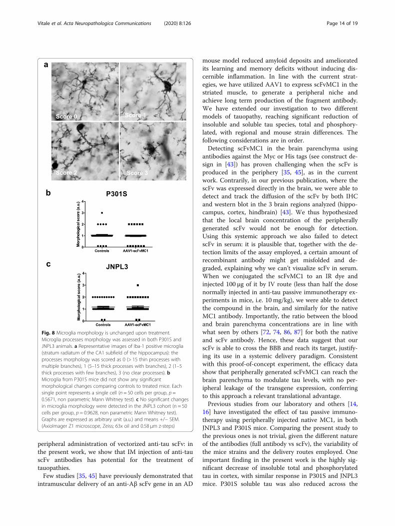
Burlingame, CA), antigen retrieval was performed using1X Dako Target Retrieval solution (Agilent Dako, SantaClara, CA, USA) in distilled water/0.5% Triton, at 95–99 °C for 5 min. After washing, endogenous peroxidaseswere quenched with 3% H2O2/0.25% Triton X-100/1XTBS for 30min. Sections were incubated in 5% normalgoat serum (Sigma Aldrich) in 1XTBS/0.1% Triton. Pri-mary polyclonal antibody, anti Iba-1 (Wako Chemicals,Richmond, VA), was diluted 1:2000 in 1% normal goatserum in 1XTBS/0.1% Triton and let incubate O/N at4 °C. Biotin-conjugated goat anti-rabbit secondary anti-body (SouthernBiotech) were used at 1:2000 in 20% Su-perBlock (ThermoFisher) in 1X TBS/0.05% Triton, left for2 h at RT, and lately Streptavidin-HRP (SouthernBiotech)was incubated for 1 h. Staining was visualized using Vec-tor VIP Substrate (Vector Laboratories) following themanufacture’s specifications. After washing with distilledwater slides were mounted and coverslipped. Microgliawere imaged on AxioImager Z1 microscope (Zeiss) at 63xoil and 0.58 μm z-steps to capture 3 ROIs across thestratum radiatum of the CA1 subfield of the hippocampusfor each animal (5 mice per group, 10 cells per mouse im-aged: 50 cells per treatment group analysed). The micro-glia process morphology was categorized with a scorefrom 0 to 3, following the criteria described by Schaferet al. [66–68]: 0 (> 15 thin processes with multiplebranches), 1 (5–15 thick processes with branches), 2 (1–5thick processes with few branches), 3 (no clear processes).All analyses were performed in blind.On peripheral organs, histology was performed by His-
toWiz Inc. (histowiz.com) using a Standard OperatingProcedure and fully automated workflow. Samples wereprocessed, embedded in paraffin, and sectioned at 4 μm.Immunohistochemistry was performed on a Bond Rxautostainer (Leica Biosystems) with enzyme treatment(1:1000) using standard protocols. Slides were stainedwith hematoxylin and eosin and anti-NFkb. Bond Poly-mer Refine Detection (Leica Biosystems) was used ac-cording to manufacturer’s protocol. After staining,sections were dehydrated and film coverslipped using aTissueTek-Prisma and Coverslipper (Sakura). Wholeslide scanning (40X) was performed on an Aperio AT2(Leica Biosystems).
Primary mouse microglia cultures and uptake experimentCultures were prepared from post-natal C57BL/6 mousepups at 2 days of age. Whole brains were trypsin
digested and made into a cell suspension. Cells wereseeded in flasks pre-coated with 0.1 mg/ml poly-D-lysine(Sigma-Aldrich) and maintained in DMEM supple-mented with 10% heat-inactivated FBS (Gibco) and 1%Pen-Strep (Gibco). Medium was supplemented with 5ng/ml Macrophage Colony Stimulating factor (M-CSF)(Thermo Fisher Scientific) diluted in PBS supplementedwith 0.1% sterile filtered BSA (Sigma-Aldrich). At DIV10microglia were isolated by orbital shaking at 150 RPMfor 1 h and the supernatant was seeded in 12-well plateswith 300,000 cells per well. Experiments were performedon the subsequent day. PHF-tau (paired helical fila-ments) [69] was added to microglia at a concentration of1 μg/ml as determined by total tau ELISA. ScFvMC1 wasadded at a concentration of 10 μg/ml. To allow for im-mune complex formation, PHF-tau and scFvMC1 weremixed in medium and pre-incubated at 37 °C for 30–45min prior to addition to cells. Mixing was performedtwo times during incubation by repeated manual pipet-ting. The 2 h incubation was performed in medium with-out serum. All experiments were performed in triplicate,with each treatment group in quadruplicate. Theamount of PHF-tau in medium at the end of the experi-ments was assessed using the same low-tau ELISA previ-ously described.
Stereotaxic intracranial injectionIntra-hippocampal injections of AAV vectors were per-formed according to a stereotaxic surgery protocol pre-viously published [43]. Briefly, under sterile conditions,3-month-old P301S mice were anesthetized and securedon a stereotaxic frame (David Kopf instruments,Tujunga, CA). Mice received bilateral hippocampal in-jection of AAV preparations using a neuro syringe witha 33 gauge needle (Hamilton, Reno, NV), using the fol-lowing coordinates: AP − 2.1 from bregma, ML +/− 2.0from bregma, DV − 1.8 below dura. Animals weretreated according to the current regulations for theproper handling of research animals, following an ap-proved IACUC protocol.
Flow cytometry on adult mice microgliaMicroglia was isolated from 6-month-old P301S micetreated with AAV5-scFv-MC1 and AAV5-null injectedmice. In this experiment, we used an AAV-null con-struct instead of AAV-eGFP, since our goal was to ascer-tain the uptake of the scFv by microglia and since eGFPmay interfere with flow cytometry analysis. Mice wereanesthetized and cold PBS-perfused. After dissection,the forebrain was minced with a Dounce homogenizerin ice cold HBSS, filtered onto 70 μm cell strainer, andcentrifuged at 300 g for 5 min at 4 °C. Tissue was dissoci-ated using Neural Dissociation Kit P (MACS MiltenyiBiotec, Auburn CA) according to the manufacturer’s
Vitale et al. Acta Neuropathologica Communications (2020) 8:126 Page 5 of 19
instruction. Myelin debris were removed using MyelinRemoval Beads II (MACS Miltenyi Biotec). Briefly, afterneural dissociation, samples were spun at 300 g for 10min at 4 °C and incubated 15 min with Myelin RemovalBeads in 0.5% BSA in 1X PBS. Cells suspension was thenloaded onto a pre-washed MACS LS column and placedin the magnetic field of MACS Separator. The magnetic-ally labeled myelin was retained within the column whileunlabeled cells run through [70]. Cells suspension, mye-lin depleted, was then washed twice with FACS buffer(0.05% BSA, 0.02% sodium azide in 1X PBS) and stainedwith Live/Dead-Pacific Blue (Thermo Fisher Scientific).Surface staining was performed using CD11b-PE andCD45-APC/Cy7 antibodies (BD Biosciences, FranklinLakes, NJ) in order to select microglia from other mono-cytes. After fixation and permeabilization with BD Cyto-fix/Cytoperm Fixation/Permeabilization Solution Kit(BD Biosciences) cells were stained with anti-Myc TagAntibody AlexaFluor-647 (Thermo Fisher Scientific).Debris, doublets and dead cells were excluded using fsc/ssc, fsc-h/fsc-w and Pacific Blue gates, respectively. BDCompBeads (BD Biosciences) were used for calibrationof flow cytometer. Samples were analyzed on a BDLSRFortessa and data processed using FlowJo software(Treestar).
Tau and anti-scFvMC1 antibodies detection in serumA detailed protocol was previously published to detecttotal tau in serum [71]. Upon sacrifice mice were bled,samples collected and allowed to clot for 30 min at RT.After cooling for 15 min, samples were spun at 14,000 gfor 10 min at 4 °C; supernatants were collected and thenre-spun at 14,000 g for 5 min at 4 °C. The final superna-tants correspond to the serum samples. In order to de-tect tau in serum, samples were diluted 1:3 in 0.2MNaOAc, pH 5.0 and heated at 90 °C for 15 min. After theheat treatment, samples were allowed to cool at 4 °C for15 min, and then spun at 15,000 g for 10 min. Superna-tants were collected and 1M Tris buffer was added toneutralize the pH. After diluting 1:2 in 20% Superblock,samples were loaded on the total tau ELISA (DA31capture).In order to detect antibodies directed to the scFvMC1,
96-well plates were coated with purified scFvMC1 at6 μg/ml for at least 24 h. Plates were washed 3X andblocked for 1 h using StartingBlock (Thermo Fisher Sci-entific). Plates were washed 5X and 50 μl of sera addedin triplicate at 1:1000 dilution in 20% SuperBlock(Thermo Fisher Scientific). After 1 h incubation plateswere washed 5X and 50 μl of goat anti-mouse non-specific IgG HRP-conjugated (SoutherBiotech, Birmin-gha, AL) was added and incubated for 1 h. Finally, Bio-Rad HRP Substrate Kit has been used for the detection
and plates were read with Infinite m200 plate reader(Tecan) at 415 nm.
Statistical analysisQuantitative data were analyzed using the dedicatedsoftware GraphPad Prism V.6 (GraphPad software Inc.,CA). Unpaired t test with Welch’s correction was per-formed when the parametric assumption of normality(D’Agostino-Pearson omnibus test) was accomplished.When not, non parametric Mann-Whitney test was per-formed instead. Statistical significance was set at P <0.05. Error bars represent the standard error of the mean(SEM).
ResultsScFvMC1 is detected in the brain homogenates uponintravenous injection of the purified scFvWe first asked whether scFvMC1 crosses the BBB andtargets the brain [72, 73]. After intra-peritoneal (IP) per-ipheral injection, we were not able to detect brainscFvMC1 using antibodies directed against Myc or 6-His(tags present on our scFv construct), via IHC, WB orELISA (not shown). We have thus performed a proof-ofconcept experiment, testing BBB penetration via retro-orbital IV injection of 100 μg of purified scFvMC1, eitherunlabeled (UNL) or infrared-conjugated (IR), in adultJNPL3 and P301S mice; a careful comparison was per-formed between scFvs and native MC1 variants, includ-ing saline as a negative control. Given the scFv’s shorthalf-life, mice were sacrificed 2 h post-injection and per-fused with ice-cold PBS/Heparin. The serum concentra-tion of both scFvMC1-IR and the native MC1-IR werecalculated in the amount of 50 ng/μl. At termination ofthe experiment, scFvMC1 was detected in cortex, HBand hippocampus (Fig. 1a): its amount was in the rangeof 0.1–0.2% of the serum concentration, in line with thecurrent literature [40, 72, 74, 75], confirming the feasi-bility of a peripheral approach that relies on a sustainedrelease of scFv in the circulation.
AAV1-scFvMC1 specifically transduces muscle cells uponone-time intramuscular injectionSince scFvMC1 efficiently crosses the BBB, we have nextfocused on selecting a peripheral tissue to be transducedby the AAV-scFv construct, to generate a stable sourceof antibody, with the goal to circumvent vital organs.Skeletal muscle is considered an ideal target tissue forAAV transduction because individual fibers are large,multinucleated and with minimal cellular turnover [35,59, 76]. Hence, we have selected the AAV1 serotype toproduce a stable muscular niche able to continuouslyproduce anti-tau scFvMC1 and release it into the circu-lation to target cerebral tau. In a prevention protocol,given the different timeline in the development of tau
Vitale et al. Acta Neuropathologica Communications (2020) 8:126 Page 6 of 19
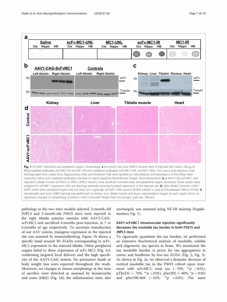
pathology in the two mice models selected, 3-month-oldJNPL3 and 2-month-old P301S mice were injected inthe right tibialis anterior muscles with AAV1-CAG-scFvMC1 and sacrificed 4 months post-injection, at 7 or6 months of age respectively. To ascertain transductionof our AAV system, transgene expression in the injectedsite was assessed by immunoblotting. Figure 1b shows aspecific band around 30–35 kDa corresponding to scFv-MC1 expression in the injected tibialis. Other peripheralorgans failed to show expression of scFv-MC1 (Fig. 1c),confirming targeted local delivery and the high specifi-city of the AAV1-CAG system. No premature death orbody weight loss were reported throughout the study.Moreover, no changes in tissues morphology at the timeof sacrifice were detected as assessed by hematoxylinand eosin (H&E) (Fig. 1d); the inflammatory state, also
unchanged, was assessed using NF-kB staining (Supple-mentary Fig. 1).
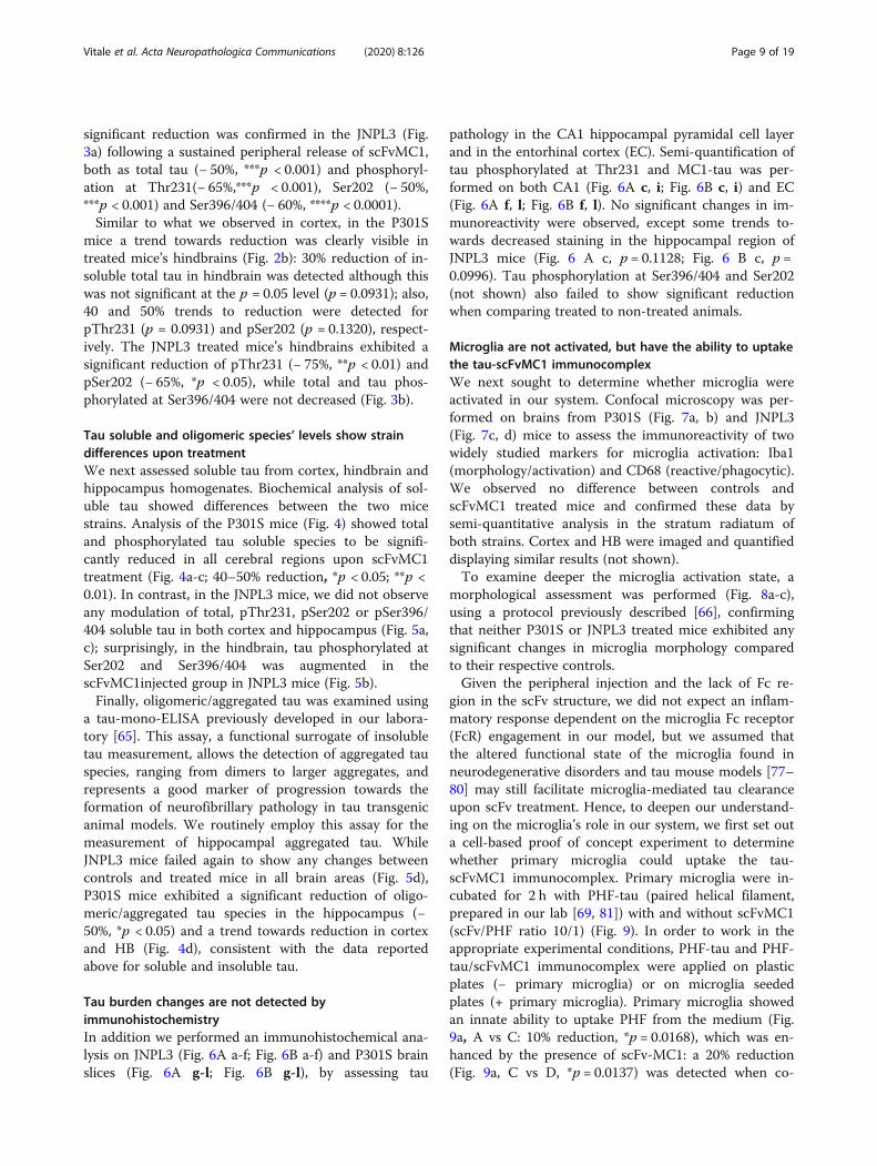
AAV1-scFvMC1 intramuscular injection significantlydecreases the insoluble tau burden in both P301S andJNPL3 miceTo rigorously quantitate the tau burden, we performedan extensive biochemical analysis of insoluble, solubleand oligomeric tau species in brain. We monitored thetau insoluble burden (a proxy for tau aggregation) incortex and hindbrain by low-tau ELISA (Fig. 2, Fig. 3).As shown in Fig. 2a, we observed a dramatic decrease ofcortical insoluble tau in the P301S cohort upon treat-ment with scFvMC1: total tau (− 70%, **p < 0.01),pThr231 (− 70%, **p < 0.01), pSer202 (− 60%, *p < 0.05)and pSer396/404 (− 65%, **p < 0.01). The same
Fig. 1 ScFvMC1 detection and peripheral organs’ morphology. a Six-month old mice (JNPL3 shown) were IV-injected with saline, 100 μg ofIRDye-labelled antibodies (scFvMC1-IR and MC1-IR) and unlabeled antibodies (scFvMC1-UNL and MC1-UNL). Two hours post-injection, brainhomogenates from cortex (Ctx), hippocampus (Hip) and hindbrain (HB) were spotted on nitrocellulose and absorbance of the IRDye labelmeasured. Saline and unlabeled antibodies showed no signal (Sapphire Biomolecular Imager, Azure Biosystems). b, c AAV1-CAG-scFvMC1 wasinjected in tibialis muscle of P301S or JNPL3 (JNPL3 shown), mice sacrificed 4 months later and peripheral organs harvested. Tissue lysates wereanalyzed for scFvMC1 expression with anti-Myc/tag antibody showing localized expression in the injected site (b, right tibialis, Controls = AAV1-eGFP) while other peripheral organs did not show scFv signal (c). ScFvMC1: MW around 30 kDa; tubulin is used as housekeeper: MW at 55 kDa. dHematoxylin and eosin (H&E) staining was performed on kidney, liver, tibialis muscle and heart: representative images of each organs show nosignificant changes in morphology (Controls = AAV1-CAG-eGFP; Bright field microscope, scale bar: 300 μm)
Vitale et al. Acta Neuropathologica Communications (2020) 8:126 Page 7 of 19
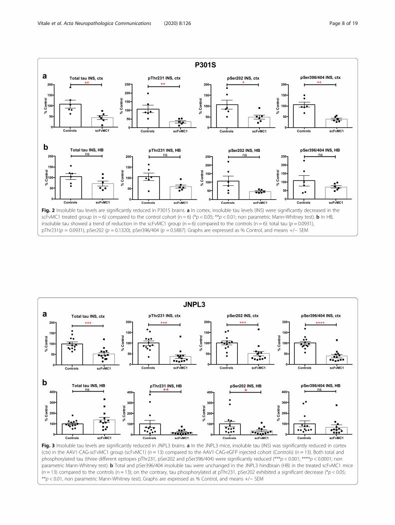
Fig. 2 Insoluble tau levels are significantly reduced in P301S brains. a In cortex, insoluble tau levels (INS) were significantly decreased in thescFvMC1 treated group (n = 6) compared to the control cohort (n = 6) (*p < 0.05; **p < 0.01; non parametric Mann-Whitney test). b In HB,insoluble tau showed a trend of reduction in the scFvMC1 group (n = 6) compared to the controls (n = 6): total tau (p = 0.0931),pThr231(p = 0.0931), pSer202 (p = 0.1320), pSer396/404 (p = 0.5887). Graphs are expressed as % Control, and means +/− SEM
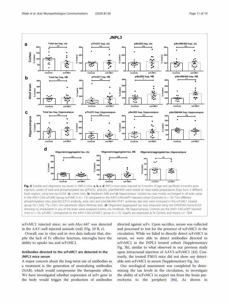
Fig. 3 Insoluble tau levels are significantly reduced in JNPL3 brains. a In the JNPL3 mice, insoluble tau (INS) was significantly reduced in cortex(ctx) in the AAV1-CAG-scFvMC1 group (scFvMC1) (n = 13) compared to the AAV1-CAG-eGFP injected cohort (Controls) (n = 13). Both total andphosphorylated tau (three different epitopes pThr231, pSer202 and pSer396/404) were significantly reduced (***p < 0.001; ****p < 0.0001; nonparametric Mann-Whitney test). b Total and pSer396/404 insoluble tau were unchanged in the JNPL3 hindbrain (HB) in the treated scFvMC1 mice(n = 13) compared to the controls (n = 13); on the contrary, tau phosphorylated at pThr231, pSer202 exhibited a significant decrease (*p < 0.05;**p < 0.01, non parametric Mann-Whitney test). Graphs are expressed as % Control, and means +/− SEM
Vitale et al. Acta Neuropathologica Communications (2020) 8:126 Page 8 of 19
significant reduction was confirmed in the JNPL3 (Fig.3a) following a sustained peripheral release of scFvMC1,both as total tau (− 50%, ***p < 0.001) and phosphoryl-ation at Thr231(− 65%,***p < 0.001), Ser202 (− 50%,***p < 0.001) and Ser396/404 (− 60%, ****p < 0.0001).Similar to what we observed in cortex, in the P301S
mice a trend towards reduction was clearly visible intreated mice’s hindbrains (Fig. 2b): 30% reduction of in-soluble total tau in hindbrain was detected although thiswas not significant at the p = 0.05 level (p = 0.0931); also,40 and 50% trends to reduction were detected forpThr231 (p = 0.0931) and pSer202 (p = 0.1320), respect-ively. The JNPL3 treated mice’s hindbrains exhibited asignificant reduction of pThr231 (− 75%, **p < 0.01) andpSer202 (− 65%, *p < 0.05), while total and tau phos-phorylated at Ser396/404 were not decreased (Fig. 3b).
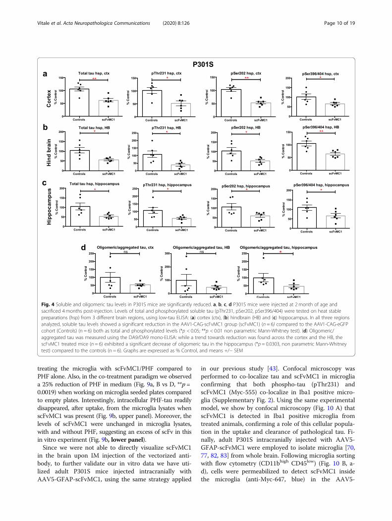
Tau soluble and oligomeric species’ levels show straindifferences upon treatmentWe next assessed soluble tau from cortex, hindbrain andhippocampus homogenates. Biochemical analysis of sol-uble tau showed differences between the two micestrains. Analysis of the P301S mice (Fig. 4) showed totaland phosphorylated tau soluble species to be signifi-cantly reduced in all cerebral regions upon scFvMC1treatment (Fig. 4a-c; 40–50% reduction, *p < 0.05; **p <0.01). In contrast, in the JNPL3 mice, we did not observeany modulation of total, pThr231, pSer202 or pSer396/404 soluble tau in both cortex and hippocampus (Fig. 5a,c); surprisingly, in the hindbrain, tau phosphorylated atSer202 and Ser396/404 was augmented in thescFvMC1injected group in JNPL3 mice (Fig. 5b).Finally, oligomeric/aggregated tau was examined using
a tau-mono-ELISA previously developed in our labora-tory [65]. This assay, a functional surrogate of insolubletau measurement, allows the detection of aggregated tauspecies, ranging from dimers to larger aggregates, andrepresents a good marker of progression towards theformation of neurofibrillary pathology in tau transgenicanimal models. We routinely employ this assay for themeasurement of hippocampal aggregated tau. WhileJNPL3 mice failed again to show any changes betweencontrols and treated mice in all brain areas (Fig. 5d),P301S mice exhibited a significant reduction of oligo-meric/aggregated tau species in the hippocampus (−50%, *p < 0.05) and a trend towards reduction in cortexand HB (Fig. 4d), consistent with the data reportedabove for soluble and insoluble tau.
Tau burden changes are not detected byimmunohistochemistryIn addition we performed an immunohistochemical ana-lysis on JNPL3 (Fig. 6A a-f; Fig. 6B a-f) and P301S brainslices (Fig. 6A g-l; Fig. 6B g-l), by assessing tau
pathology in the CA1 hippocampal pyramidal cell layerand in the entorhinal cortex (EC). Semi-quantification oftau phosphorylated at Thr231 and MC1-tau was per-formed on both CA1 (Fig. 6A c, i; Fig. 6B c, i) and EC(Fig. 6A f, l; Fig. 6B f, l). No significant changes in im-munoreactivity were observed, except some trends to-wards decreased staining in the hippocampal region ofJNPL3 mice (Fig. 6 A c, p = 0.1128; Fig. 6 B c, p =0.0996). Tau phosphorylation at Ser396/404 and Ser202(not shown) also failed to show significant reductionwhen comparing treated to non-treated animals.
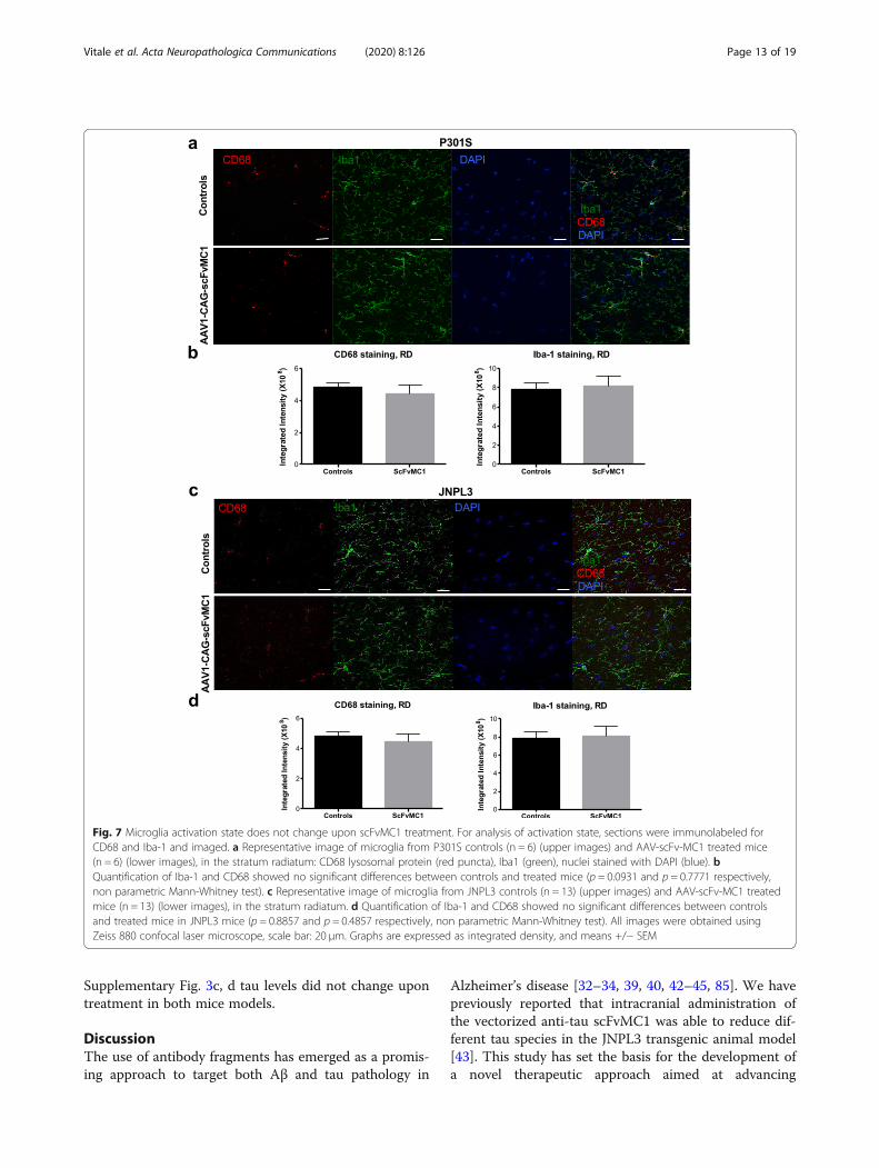
Microglia are not activated, but have the ability to uptakethe tau-scFvMC1 immunocomplexWe next sought to determine whether microglia wereactivated in our system. Confocal microscopy was per-formed on brains from P301S (Fig. 7a, b) and JNPL3(Fig. 7c, d) mice to assess the immunoreactivity of twowidely studied markers for microglia activation: Iba1(morphology/activation) and CD68 (reactive/phagocytic).We observed no difference between controls andscFvMC1 treated mice and confirmed these data bysemi-quantitative analysis in the stratum radiatum ofboth strains. Cortex and HB were imaged and quantifieddisplaying similar results (not shown).To examine deeper the microglia activation state, a
morphological assessment was performed (Fig. 8a-c),using a protocol previously described [66], confirmingthat neither P301S or JNPL3 treated mice exhibited anysignificant changes in microglia morphology comparedto their respective controls.Given the peripheral injection and the lack of Fc re-
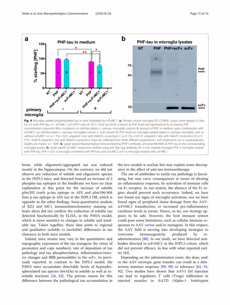
gion in the scFv structure, we did not expect an inflam-matory response dependent on the microglia Fc receptor(FcR) engagement in our model, but we assumed thatthe altered functional state of the microglia found inneurodegenerative disorders and tau mouse models [77–80] may still facilitate microglia-mediated tau clearanceupon scFv treatment. Hence, to deepen our understand-ing on the microglia’s role in our system, we first set outa cell-based proof of concept experiment to determinewhether primary microglia could uptake the tau-scFvMC1 immunocomplex. Primary microglia were in-cubated for 2 h with PHF-tau (paired helical filament,prepared in our lab [69, 81]) with and without scFvMC1(scFv/PHF ratio 10/1) (Fig. 9). In order to work in theappropriate experimental conditions, PHF-tau and PHF-tau/scFvMC1 immunocomplex were applied on plasticplates (− primary microglia) or on microglia seededplates (+ primary microglia). Primary microglia showedan innate ability to uptake PHF from the medium (Fig.9a, A vs C: 10% reduction, *p = 0.0168), which was en-hanced by the presence of scFv-MC1: a 20% reduction(Fig. 9a, C vs D, *p = 0.0137) was detected when co-
Vitale et al. Acta Neuropathologica Communications (2020) 8:126 Page 9 of 19
treating the microglia with scFvMC1/PHF compared toPHF alone. Also, in the co-treatment paradigm we observeda 25% reduction of PHF in medium (Fig. 9a, B vs D, **p =0.0019) when working on microglia seeded plates comparedto empty plates. Interestingly, intracellular PHF-tau readilydisappeared, after uptake, from the microglia lysates whenscFvMC1 was present (Fig. 9b, upper panel). Moreover, thelevels of scFvMC1 were unchanged in microglia lysates,with and without PHF, suggesting an excess of scFv in thisin vitro experiment (Fig. 9b, lower panel).Since we were not able to directly visualize scFvMC1
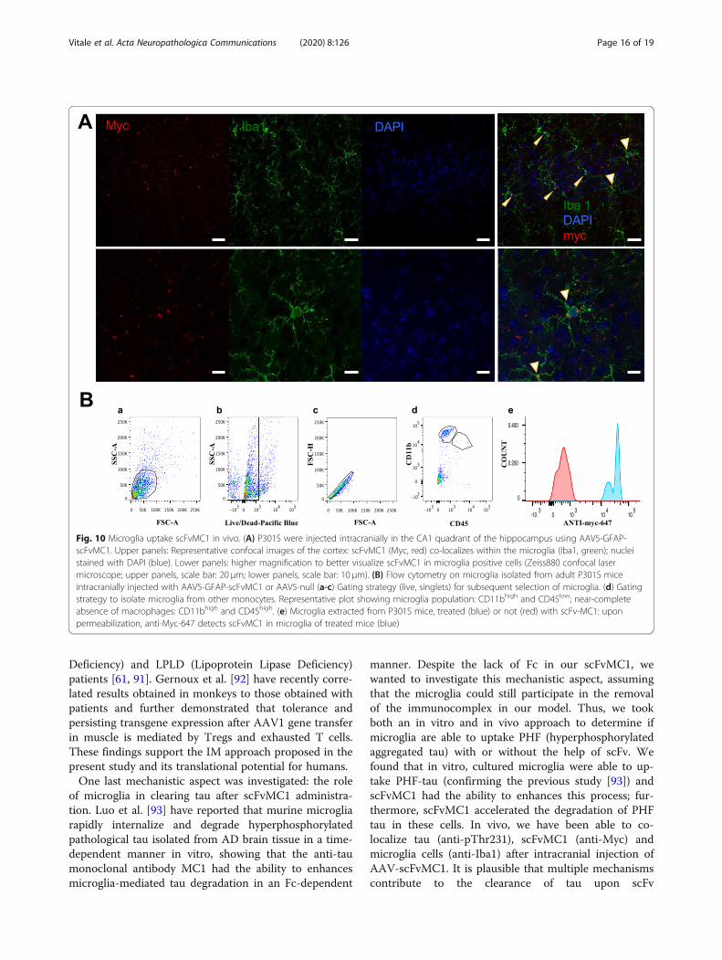
in the brain upon IM injection of the vectorized anti-body, to further validate our in vitro data we have uti-lized adult P301S mice injected intracranially withAAV5-GFAP-scFvMC1, using the same strategy applied
in our previous study [43]. Confocal microscopy wasperformed to co-localize tau and scFvMC1 in microgliaconfirming that both phospho-tau (pThr231) andscFvMC1 (Myc-555) co-localize in Iba1 positive micro-glia (Supplementary Fig. 2). Using the same experimentalmodel, we show by confocal microscopy (Fig. 10 A) thatscFvMC1 is detected in Iba1 positive microglia fromtreated animals, confirming a role of this cellular popula-tion in the uptake and clearance of pathological tau. Fi-nally, adult P301S intracranially injected with AAV5-GFAP-scFvMC1 were employed to isolate microglia [70,77, 82, 83] from whole brain. Following microglia sortingwith flow cytometry (CD11bhigh CD45low) (Fig. 10 B, a-d), cells were permeabilized to detect scFvMC1 insidethe microglia (anti-Myc-647, blue) in the AAV5-
Fig. 4 Soluble and oligomeric tau levels in P301S mice are significantly reduced. a, b, c, d P301S mice were injected at 2 month of age andsacrificed 4 months post-injection. Levels of total and phosphorylated soluble tau (pThr231, pSer202, pSer396/404) were tested on heat stablepreparations (hsp) from 3 different brain regions, using low-tau ELISA: (a) cortex (ctx), (b) hindbrain (HB) and (c) hippocampus. In all three regionsanalyzed, soluble tau levels showed a significant reduction in the AAV1-CAG-scFvMC1 group (scFvMC1) (n = 6) compared to the AAV1-CAG-eGFPcohort (Controls) (n = 6) both as total and phosphorylated levels (*p < 0.05; **p < 0.01 non parametric Mann-Whitney test). (d) Oligomeric/aggregated tau was measured using the DA9/DA9 mono-ELISA: while a trend towards reduction was found across the cortex and the HB, thescFvMC1 treated mice (n = 6) exhibited a significant decrease of oligomeric tau in the hippocampus (*p = 0.0303, non parametric Mann-Whitneytest) compared to the controls (n = 6). Graphs are expressed as % Control, and means +/− SEM
Vitale et al. Acta Neuropathologica Communications (2020) 8:126 Page 10 of 19
scFvMC1 injected mice; no anti-Myc-647 was detectedin the AAV-null injected animals (red) (Fig. 10 B, e).Overall, our in vitro and in vivo data indicate that, des-
pite the lack of Fc effector function, microglia have theability to uptake tau and scFvMC1.
Antibodies directed to the scFvMC1 are detected in theJNPL3 mice serumA major concern about the long-term use of antibodies asa treatment is the generation of neutralizing antibodies(NAB), which would compromise the therapeutic effect.We have investigated whether expression of scFv gene inthe body would trigger the production of antibodies
directed against scFv. Upon sacrifice, serum was collectedand processed to test for the presence of scFvMC1 in thecirculation. While we failed to directly detect scFvMC1 inserum, we were able to detect antibodies directed toscFvMC1 in the JNPL3 treated cohort (SupplementaryFig. 3b), similar to what observed in our previous studyupon intracranial injection of AAV5-scFvMC1 [43]. Con-trarily, the treated P301S mice did not show any detect-able anti-scFvMC1 in serum (Supplementary Fig. 3a).Our serological assessment was completed by deter-
mining the tau levels in the circulation, to investigatethe ability of scFvMC1 to export tau from the brain par-enchyma to the periphery [84]. As shown in
Fig. 5 Soluble and oligomeric tau levels in JNPL3 mice. a, b, c, d JNPL3 mice were injected at 3 months of age and sacrificed 4 months post-injection. Levels of total and phosphorylated tau (pThr231, pSer202, pSer396/404) were tested on heat stable preparations (hsp) from 3 differentbrain regions, using low-tau ELISA: (a) cortex (ctx), (b) hindbrain (HB) and (c) hippocampus. Soluble tau was mostly unchanged in all brain areasin the AAV1-CAG-scFvMC1group (scFvMC1) (n = 13) compared to the AAV1-CAG-eGFP injected cohort (Controls) (n = 13). Two differentphosphorylation sites, pSer202 (CP13 antibody, early site) and pSer396/404 (PHF1 antibody, late site) were increased in the scFvMC1 treatedgroup (*p < 0.05, **p < 0.01, non parametric Mann-Whitney test). (d) Oligomeric/aggregated tau was measured using the DA9/DA9 mono-ELISAshowing no modulation in any of the brain areas analyzed (cortex, ctx; hindbrain, HB; hippocampus). Controls are the AAV1-CAG-eGFP injectedmice (n = 13); scFvMC1 corresponds to the AAV1-CAG-scFvMC1 group (n = 13). Graphs are expressed as % Control, and means +/− SEM
Vitale et al. Acta Neuropathologica Communications (2020) 8:126 Page 11 of 19
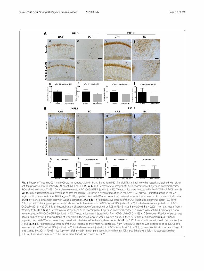
Fig. 6 Phospho-Threonine-231 and MC1-tau immunoreactivity in brain. Brains from P301S and JNPL3 animals were harvested and stained with eitheranti tau phospho-Thr231 antibody (A) or anti-MC1-tau (B). (A): a, b, d, e Representative images of CA1 hippocampal cell layer and entorhinal cortex(EC) stained with anti-pThr231. Control mice received AAV1-CAG-eGFP injection (n = 13). Treated mice were injected with AAV1-CAG-scFvMC1 (n = 13).(A): c,f Semi-quantification of percentage of area stained by RZ3 shows a trend of reduction in the AAV1-CAG-scFvMC1 injected group, in the CA1region of hippocampus in the JNPL3 (c, p = 0.1128, unpaired t test with Welch’s correction); no trend to reduction is detected in the entorhinal cortex(EC) (f, p = 0.3458, unpaired t test with Welch’s correction). (A): g, h, j, k Representative images of the CA1 region and entorhinal cortex (EC) fromP301S; pThr-231 staining was performed as above. Control mice received AAV1-CAG-eGFP injection (n = 6), treated mice were injected with AAV1-CAG-scFvMC1 (n = 6); (A) i, l Semi-quantification of percentage of area stained by RZ3 in P301S mice (i, p = 0.2403; l, p = 0.2251; non parametric Mann-Whitney test). (B): a, b, d, e Representative images of CA1 hippocampal cell layer and entorhinal cortex (EC) stained with anti-MC1 antibody. Controlmice received AAV1-CAG-eGFP injection (n = 13). Treated mice were injected with AAV1-CAG-scFvMC1 (n = 13) (c, f) Semi-quantification of percentageof area stained by MC1 shows a trend of reduction in the AAV1-CAG-scFvMC1 injected group, in the CA1 region of hippocampus (c, p = 0.0996;unpaired t test with Welch’s correction); no reduction is detected in the entorhinal cortex (EC) (f, p = 0.9558; unpaired t test with Welch’s correction) inJNPL3. (g, h, j, k) Representative images of the CA1 region and the entorhinal cortex (EC) from P301S; MC1 staining was performed as above. Controlmice received AAV1-CAG-eGFP injection (n = 6), treated mice were injected with AAV1-CAG-scFvMC1 (n = 6). (i, l) Semi-quantification of percentage ofarea stained by MC1 in P301S mice (i, p = 0.4127; l, p = 0.8413; non parametric Mann-Whitney). (Olympus BH-2 bright field microscope; scale bar:100 μm). Graphs are expressed as % Control area stained, and means +/− SEM
Vitale et al. Acta Neuropathologica Communications (2020) 8:126 Page 12 of 19
Supplementary Fig. 3c, d tau levels did not change upontreatment in both mice models.
DiscussionThe use of antibody fragments has emerged as a promis-ing approach to target both Aβ and tau pathology in
Alzheimer’s disease [32–34, 39, 40, 42–45, 85]. We havepreviously reported that intracranial administration ofthe vectorized anti-tau scFvMC1 was able to reduce dif-ferent tau species in the JNPL3 transgenic animal model[43]. This study has set the basis for the development ofa novel therapeutic approach aimed at advancing
Fig. 7 Microglia activation state does not change upon scFvMC1 treatment. For analysis of activation state, sections were immunolabeled forCD68 and Iba-1 and imaged. a Representative image of microglia from P301S controls (n = 6) (upper images) and AAV-scFv-MC1 treated mice(n = 6) (lower images), in the stratum radiatum: CD68 lysosomal protein (red puncta), Iba1 (green), nuclei stained with DAPI (blue). bQuantification of Iba-1 and CD68 showed no significant differences between controls and treated mice (p = 0.0931 and p = 0.7771 respectively,non parametric Mann-Whitney test). c Representative image of microglia from JNPL3 controls (n = 13) (upper images) and AAV-scFv-MC1 treatedmice (n = 13) (lower images), in the stratum radiatum. d Quantification of Iba-1 and CD68 showed no significant differences between controlsand treated mice in JNPL3 mice (p = 0.8857 and p = 0.4857 respectively, non parametric Mann-Whitney test). All images were obtained usingZeiss 880 confocal laser microscope, scale bar: 20 μm. Graphs are expressed as integrated density, and means +/− SEM
Vitale et al. Acta Neuropathologica Communications (2020) 8:126 Page 13 of 19
peripheral administration of vectorized anti-tau scFv: inthe present work, we show that IM injection of anti-tauscFv antibodies has potential for the treatment oftauopathies.Few studies [35, 45] have previously demonstrated that
intramuscular delivery of an anti-Aβ scFv gene in an AD
mouse model reduced amyloid deposits and amelioratedits learning and memory deficits without inducing dis-cernible inflammation. In line with the current strat-egies, we have utilized AAV1 to express scFvMC1 in thestriated muscle, to generate a peripheral niche andachieve long term production of the fragment antibody.We have extended our investigation to two differentmodels of tauopathy, reaching significant reduction ofinsoluble and soluble tau species, total and phosphory-lated, with regional and mouse strain differences. Thefollowing considerations are in order.Detecting scFvMC1 in the brain parenchyma using
antibodies against the Myc or His tags (see construct de-sign in [43]) has proven challenging when the scFv isproduced in the periphery [35, 45], as in the currentwork. Contrarily, in our previous publication, where thescFv was expressed directly in the brain, we were able todetect and track the diffusion of the scFv by both IHCand western blot in the 3 brain regions analyzed (hippo-campus, cortex, hindbrain) [43]. We thus hypothesizedthat the local brain concentration of the peripherallygenerated scFv would not be enough for detection.Using this systemic approach we also failed to detectscFv in serum: it is plausible that, together with the de-tection limits of the assay employed, a certain amount ofrecombinant antibody might get misfolded and de-graded, explaining why we can’t visualize scFv in serum.When we conjugated the scFvMC1 to an IR dye andinjected 100 μg of it by IV route (less than half the dosenormally injected in anti-tau passive immunotherapy ex-periments in mice, i.e. 10 mg/kg), we were able to detectthe compound in the brain, and similarly for the nativeMC1 antibody. Importantly, the ratio between the bloodand brain parenchyma concentrations are in line withwhat seen by others [72, 74, 86, 87] for both the nativeand scFv antibody. Hence, these data suggest that ourscFv is able to cross the BBB and reach its target, justify-ing its use in a systemic delivery paradigm. Consistentwith this proof-of-concept experiment, the efficacy datashow that peripherally generated scFvMC1 can reach thebrain parenchyma to modulate tau levels, with no per-ipheral leakage of the transgene expression, conferringto this approach a relevant translational advantage.Previous studies from our laboratory and others [14,
16] have investigated the effect of tau passive immuno-therapy using peripherally injected native MC1, in bothJNPL3 and P301S mice. Comparing the present study tothe previous ones is not trivial, given the different natureof the antibodies (full antibody vs scFv), the variability ofthe mice strains and the delivery routes employed. Oneimportant finding in the present work is the highly sig-nificant decrease of insoluble total and phosphorylatedtau in cortex, with similar response in P301S and JNPL3mice. P301S soluble tau was also reduced across the
Fig. 8 Microglia morphology is unchanged upon treatment.Microglia processes morphology was assessed in both P301S andJNPL3 animals. a Representative images of Iba-1 positive microglia(stratum radiatum of the CA1 subfield of the hippocampus): theprocesses morphology was scored as 0 (> 15 thin processes withmultiple branches), 1 (5–15 thick processes with branches), 2 (1–5thick processes with few branches), 3 (no clear processes). bMicroglia from P301S mice did not show any significantmorphological changes comparing controls to treated mice. Eachsingle point represents a single cell (n = 50 cells per group, p =0.5671, non parametric Mann Whitney test). c No significant changesin microglia morphology were detected in the JNPL3 cohort (n = 50cells per group, p = 0.9628, non parametric Mann Whitney test).Graphs are expressed as arbitrary unit (a.u.) and means +/− SEM.(AxioImager Z1 microscope, Zeiss; 63x oil and 0.58 μm z-steps)
Vitale et al. Acta Neuropathologica Communications (2020) 8:126 Page 14 of 19
brain, while oligomeric/aggregated tau was reducedmostly in the hippocampus. On the contrary, we did notobserve any reduction of soluble and oligomeric speciesin the JNPL3 mice, and detected instead an increase of 2phospho-tau epitopes in the hindbrain: we have no clearexplanation at this point for the increase of solublepSer202 (early p-tau epitope in AD) and pSer396/404(late p-tau epitope in AD) tau in the JNPL3 HB, which isopposite to the other findings. Semi-quantitative analysisof RZ3 and MC1 immunohistochemistry staining onbrain slices did not confirm the reduction of soluble taudetected biochemically by ELISA, in the P301S model,which is more sensitive to changes in soluble and insol-uble tau. Taken together, these data point to regionaland qualitative (soluble vs insoluble) differences in tauclearance in both mice models.Indeed, mice strains may vary in the quantitative and
topographic expression of the tau transgene (by virtue ofpromoters and copy numbers), rate of deposition of taupathology and tau phosphorylation, inflammation/react-ive changes and BBB permeability to the scFv. As previ-ously reported, in contrast to the JNPL3 model, theP301S mice accumulate detectable levels of hyperpho-sphorylated tau species (64-kDa) in soluble as well as in-soluble fractions [16, 63]. The precise reason for thisdifference between the pathological tau accumulation in
the two models is unclear but may explain some discrep-ancy in the effect of anti-tau immunotherapy.The use of antibodies to tackle tau pathology is fascin-
ating, but may carry consequences in terms of elicitingan inflammatory response, by activation of immune cellsvia Fc receptor. In our system, the absence of the Fc re-gion should prevent such occurrence. Indeed, we havenot found any signs of microglia activation, nor we havefound signs of peripheral tissue damage from the AAV-scFvMC1 transduction, or increased pro-inflammatorycytokines levels in serum. Hence, so far, our strategy ap-pears to be safe. However, the host immune systemcould pose some limitations, such as cellular immune re-sponses to AAV vector and/or transgene. In this respect,the AAV field is moving into developing strategies toovercome immunogenicity produced by re-administration [88]. In our study, we have detected anti-bodies directed to scFvMC1 in the JNPL3 cohort, whichdid not prevent efficacy, in line with what reported earl-ier [43].Depending on the administration route, the dose, and/
or the AAV serotype, gene transfer can result in a dele-terious immune response [89, 90] or tolerance [61, 91,92]. Two studies have shown that AAV1 IM injectioncan lead to regulatory T cells (Tregs) infiltration ininjected muscles in AATD (Alpha-1 Antitrypsin
Fig. 9 Microglia uptake phosphorylated tau in vitro, facilitated by scFvMC1. (a) Primary mouse microglia (P2 C57Bl/6 J pups) were treated in vitrofor 2 h with PHF-tau +/− scFvMC1 (scFv/PHF ratio of 10/1). Total tau ELISA. Column A: PHF levels are expressed as % of starting PHFconcentration measured after incubation on cell-free plates (− primary microglia); column B: amount of PHF in medium upon combination withscFvMC1, on cell-free plates (− primary microglia); column C and column D: PHF levels on microglia seeded plates (+ primary microglia), with orwithout scFvMC1. A vs C (*p < 0.05, unpaired t test with Welch’s correction); C vs D (*p = 0.0137, unpaired t test with Welch’s correction); B vs D(**p = 0.0019 unpaired t test with Welch’s correction). Data are collected from three different experiments, with treatments run in quadruplicates.Graphs are means +/− SEM. (b, upper panel) Representative immuno-blotting (PHF1 antibody: anti-pSer396/404) of PHF-tau in the correspondingmicroglia lysates; (b, lower panel) scFvMC1 expression verified using anti Myc-tag antibody. NT is non treated microglia; PHF is microglia treatedwith PHF-tau; PHF + scFv is microglia co-treated with PHF-tau and scFvMC1; scFv is microglia treated with scFvMC1
Vitale et al. Acta Neuropathologica Communications (2020) 8:126 Page 15 of 19
Deficiency) and LPLD (Lipoprotein Lipase Deficiency)patients [61, 91]. Gernoux et al. [92] have recently corre-lated results obtained in monkeys to those obtained withpatients and further demonstrated that tolerance andpersisting transgene expression after AAV1 gene transferin muscle is mediated by Tregs and exhausted T cells.These findings support the IM approach proposed in thepresent study and its translational potential for humans.One last mechanistic aspect was investigated: the role
of microglia in clearing tau after scFvMC1 administra-tion. Luo et al. [93] have reported that murine microgliarapidly internalize and degrade hyperphosphorylatedpathological tau isolated from AD brain tissue in a time-dependent manner in vitro, showing that the anti-taumonoclonal antibody MC1 had the ability to enhancesmicroglia-mediated tau degradation in an Fc-dependent
manner. Despite the lack of Fc in our scFvMC1, wewanted to investigate this mechanistic aspect, assumingthat the microglia could still participate in the removalof the immunocomplex in our model. Thus, we tookboth an in vitro and in vivo approach to determine ifmicroglia are able to uptake PHF (hyperphosphorylatedaggregated tau) with or without the help of scFv. Wefound that in vitro, cultured microglia were able to up-take PHF-tau (confirming the previous study [93]) andscFvMC1 had the ability to enhances this process; fur-thermore, scFvMC1 accelerated the degradation of PHFtau in these cells. In vivo, we have been able to co-localize tau (anti-pThr231), scFvMC1 (anti-Myc) andmicroglia cells (anti-Iba1) after intracranial injection ofAAV-scFvMC1. It is plausible that multiple mechanismscontribute to the clearance of tau upon scFv
Fig. 10 Microglia uptake scFvMC1 in vivo. (A) P301S were injected intracranially in the CA1 quadrant of the hippocampus using AAV5-GFAP-scFvMC1. Upper panels: Representative confocal images of the cortex: scFvMC1 (Myc, red) co-localizes within the microglia (Iba1, green); nucleistained with DAPI (blue). Lower panels: higher magnification to better visualize scFvMC1 in microglia positive cells (Zeiss880 confocal lasermicroscope; upper panels, scale bar: 20 μm; lower panels, scale bar: 10 μm). (B) Flow cytometry on microglia isolated from adult P301S miceintracranially injected with AAV5-GFAP-scFvMC1 or AAV5-null (a-c) Gating strategy (live, singlets) for subsequent selection of microglia. (d) Gatingstrategy to isolate microglia from other monocytes. Representative plot showing microglia population: CD11bhigh and CD45low; near-completeabsence of macrophages: CD11bhigh and CD45high. (e) Microglia extracted from P301S mice, treated (blue) or not (red) with scFv-MC1: uponpermeabilization, anti-Myc-647 detects scFvMC1 in microglia of treated mice (blue)
Vitale et al. Acta Neuropathologica Communications (2020) 8:126 Page 16 of 19
immunotherapy and that microglia uptake is one of theroutes the brain uses to remove pathological tau. Futureexperiments will investigate whether microglia or otherCNS or peripheral cell types participate in the uptake ofscFvMC1 and tau using receptors other than the FcR,i.e. toll-like receptors (TLRs), C-X3-C motif chemokinereceptor 1 (CX3CR1), pattern-recognition receptors(PRRs), scavenger receptors, and complement proteinC1q receptor (C1q-R).
ConclusionsTo our knowledge, this work provides the first descrip-tion of vectorized anti-tau scFv exerting an effect in thebrain upon intramuscular injection. More studies areplanned to investigate whether the highly significant re-duction of insoluble tau gained in both animal modelswill also ameliorate their behavioral phenotype. Also,from a clinical perspective, particularly attractive wouldbe the ability to not only express the scFv but to switchit on or off at will, adding a layer of exogenous controlto improve safety. Among the currently available in-ducer/repressor systems permitting control over geneexpression, the tetracycline (Tet)-dependent system is byfar the most advanced and most widely used, and hasbeen already tested in association with AAV1 and locor-egional muscle gene transfer in non-human primatesshowing no humoral or cellular responses against thetransgene [94]: we will certainly explore this avenue inour system.In addition, our work opens the field to future studies
reaching beyond scFv, to test engineered antibodies withmultiple specificities and targets in the brain, using asimple and translatable approach.Overall, given the limitations linked to conventional
immunotherapy in neurodegenerative diseases, this workdemonstrates the efficacy and the advantages of usingintramuscular injection of vectorized scFv to target tau,and its relevant translational features, suggesting poten-tial applications to other brain proteinopathies.
Supplementary informationSupplementary information accompanies this paper at https://doi.org/10.1186/s40478-020-01003-7.
Additional file 1 Supplementary figure 1: Inflammatory status inperipheral organs. NF-kB immunoreactivity, marker of activated proinflam-matory pathways, was evaluated on kidney, liver, tibialis muscle andheart: representative images of each organs do not show differences be-tween controls and AAV1-scFvMC1 treated mice (Controls = AAV1-CAG-eGFP; Bright field microscope, scale bar: 300 μm).
Additional file 2 Supplementary figure 2. Phospho-tau and scFvMC1co-localize in microglia. Representative confocal image of the stratumradiatum from P301S mice injected with AAV5-GFAP-scFvMC1. Astrocytesactively express scFv-MC1 (Myc-red); Iba1 positive microglia (green)shows co-localization of scFvMC1 (myc-red) and pTau (pThr231; blue):
merge purple (white arrows). Zeiss880 confocal laser microscope: mergeimage is 2x crop of 40X magnification; scale bar: 20 μm.
Additional file 3 Supplementary figure 3 Anti-scFvMC1and tau inserum. (a, b) Antibodies anti-scFvMC1 were assessed using a specific im-mune sorbent assay: JNPL3 mice receiving the scFvMC1 exhibited a sig-nificant increase of anti-scFvMC1 in serum (***p = 0.0005, non parametricMann-Whitney test) compared to the controls; no anti-scFvMC1 were de-tected in the P301S animals; (c, d) total tau levels were measured inserum at the end of the 4 month treatment: no significant changes wereidentified in both transgenic models, P301S and JNPL3. Graphs areexpressed as Arbitrary Units (a, b) or % Control (c, d), and means +/−SEM.
AbbreviationsAb: Antibody; Aβ: Amyloid-beta; AAV: Adeno-associated viral vector;AD: Alzheimer’s disease; BBB: Blood-brain barrier; CNS: Central nervoussystem; Ctx: Cortex; EC: Entorhinal cortex; HB: Hind brain;Hippo: Hippocampus; IM: Intramuscular; IV: Intravenous; LV: Lentiviral vector;mAbs: Monoclonal antibodies; PHF: Paired helical filaments; RD: Stratumradiatum; scFv: Single chain variable fragment; scFvMC1: Single chain variablefragment MC1; Tregs: Regulatory T cells; VH: Variable heavy chain; VL: Variablelight chain
AcknowledgementsWe thank Dr. Peter Davies for providing tau antibodies and PHF-tau. We wouldlike to thank Dr. Czeslawa Kowal for helping with retro-orbital injections.
Ethic approval and consent to participateAll experiments were conducted under the institutional guidelines and wereapproved by the Institutional Animal Care and Use Committee at TheFeinstein Institutes for Medical Research, Northwell Health.
Authors’ contributionsFV performed experiments, analysed data and helped preparing themanuscript; JO performed experiments and helped preparing figures; BTVanalysed and interpreted data; PM interpreted experiments and edited themanuscript; LG designed the overall project, analysed data, interpretedresults and prepared the manuscript; CD designed the overall project,performed experiments, analysed data, interpreted results and prepared themanuscript. The authors read and approved the final manuscript.
FundingThis study was supported by the National Institute of Health (NIH) grant1R56AG055479–01 to C. d’Abramo.
Availability of data and materialsThe datasets used and/or analyzed during the current study are availablefrom the corresponding authors upon reasonable request.
Consent for publicationNot applicable.
Competing interestsThe authors declare no conflict of interest.
Author details1Institute of Molecular Medicine, The Litwin-Zucker Center for Alzheimer’sDisease & Memory Disorder, The Feintein Institutes for Medical Research,Manhasset, NY, USA. 2Donald and Barbara Zucker School of Medicine atHofstra/Northwell, Hempstead, NY, USA. 3Institute of Molecular Medicine,Center for Autoimmune and Musculoskeletal Disease, The Feinstein Institutesfor Medical Research, Manhasset, USA. 4Northwell Health NeuroscienceInstitute, Northwell Health System, Manhasset, NY, USA.
Received: 15 May 2020 Accepted: 27 July 2020
References1. Wang Y, Mandelkow E (2016) Tau in physiology and pathology. Nat Rev
Neurosci 17(1):5–21
Vitale et al. Acta Neuropathologica Communications (2020) 8:126 Page 17 of 19
2. Cleveland DW, Hwo SY, Kirschner MW (1977) Physical and chemicalproperties of purified tau factor and the role of tau in microtubuleassembly. J Mol Biol 116(2):227–247
3. Buée L et al (2000) Tau protein isoforms, phosphorylation and role inneurodegenerative disorders. Brain Res Brain Res Rev 33(1):95–130
4. Mandelkow EM, Mandelkow E (2012) Biochemistry and cell biology of tauprotein in neurofibrillary degeneration. Cold Spring Harb Perspect Med 2(7):a006247
5. Rösler TW et al (2019) Four-repeat tauopathies. Prog Neurobiol 180:1016446. Frost B, Jacks RL, Diamond MI (2009) Propagation of tau misfolding from
the outside to the inside of a cell. J Biol Chem 284(19):12845–128527. Clavaguera F et al (2009) Transmission and spreading of tauopathy in
transgenic mouse brain. Nat Cell Biol 11(7):909–9138. Wu JW et al (2013) Small misfolded tau species are internalized via bulk
endocytosis and anterogradely and retrogradely transported in neurons. JBiol Chem 288(3):1856–1870
9. Clavaguera F et al (2014) Peripheral administration of tau aggregatestriggers intracerebral tauopathy in transgenic mice. Acta Neuropathol127(2):299–301
10. Clavaguera F, Grueninger F, Tolnay M (2014) Intercellular transfer of tauaggregates and spreading of tau pathology: implications for therapeuticstrategies. Neuropharmacology 76 Pt A:9–15
11. Clavaguera F et al (2013) Brain homogenates from human tauopathiesinduce tau inclusions in mouse brain. Proc Natl Acad Sci U S A 110(23):9535–9540
12. de Calignon A et al (2012) Propagation of tau pathology in a model of earlyAlzheimer's disease. Neuron 73(4):685–697
13. Colin M et al (2020) From the prion-like propagation hypothesis to therapeuticstrategies of anti-tau immunotherapy. Acta Neuropathol 139(1):3–25
14. d'Abramo C et al (2013) Tau passive immunotherapy in mutant P301L mice:antibody affinity versus specificity. PLoS One 8(4):e62402
15. d'Abramo C et al (2015) Passive immunization in JNPL3 transgenic miceusing an Array of Phospho-tau specific antibodies. PLoS One 10(8):e0135774
16. Chai X et al (2011) Passive immunization with anti-tau antibodies in twotransgenic models: reduction of tau pathology and delay of diseaseprogression. J Biol Chem 286(39):34457–34467
17. Boutajangout A et al (2011) Passive immunization targeting pathologicalphospho-tau protein in a mouse model reduces functional decline andclears tau aggregates from the brain. J Neurochem 118(4):658–667
18. Boutajangout A, Quartermain D, Sigurdsson EM (2010) Immunotherapytargeting pathological tau prevents cognitive decline in a new tanglemouse model. J Neurosci 30(49):16559–16566
19. Asuni AA et al (2007) Immunotherapy targeting pathological tauconformers in a tangle mouse model reduces brain pathology withassociated functional improvements. J Neurosci 27(34):9115–9129
20. Sankaranarayanan S et al (2015) Passive immunization with phospho-tauantibodies reduces tau pathology and functional deficits in two distinctmouse tauopathy models. PLoS One 10(5):e0125614
21. Castillo-Carranza DL et al (2014) Passive immunization with tau oligomermonoclonal antibody reverses tauopathy phenotypes without affectinghyperphosphorylated neurofibrillary tangles. J Neurosci 34(12):4260–4272
22. Albert M et al (2019) Prevention of tau seeding and propagation byimmunotherapy with a central tau epitope antibody. Brain 142(6):1736–1750
23. Yanamandra K et al (2015) Anti-tau antibody reduces insoluble tau anddecreases brain atrophy. Ann Clin Transl Neurol 2(3):278–288
24. Yanamandra K et al (2013) Anti-tau antibodies that block tau aggregateseeding in vitro markedly decrease pathology and improve cognitionin vivo. Neuron 80(2):402–414
25. Courade JP et al (2018) Epitope determines efficacy of therapeutic anti-tauantibodies in a functional assay with human Alzheimer tau. ActaNeuropathol 136(5):729–745
26. Dai CL et al (2018) Tau passive immunization blocks seeding and spread ofAlzheimer hyperphosphorylated tau-induced pathology in 3 × Tg-AD mice.Alzheimers Res Ther 10(1):13
27. Pardridge WM (2012) Drug transport across the blood-brain barrier. J CerebBlood Flow Metab 32(11):1959–1972
28. Pardridge WM (2009) Alzheimer's disease drug development and theproblem of the blood-brain barrier. Alzheimers Dement 5(5):427–432
29. Wilcock DM, Colton CA (2009) Immunotherapy, vascular pathology, andmicrohemorrhages in transgenic mice. CNS Neurol Disord Drug Targets 8(1):50–64
30. Boche D et al (2008) Consequence of Abeta immunization on thevasculature of human Alzheimer's disease brain. Brain 131(Pt 12):3299–3310
31. Frenkel D, Katz O, Solomon B (2000) Immunization against Alzheimer's beta-amyloid plaques via EFRH phage administration. Proc Natl Acad Sci U S A97(21):11455–11459
32. Liu R et al (2004) Single chain variable fragments against beta-amyloid(Abeta) can inhibit Abeta aggregation and prevent abeta-inducedneurotoxicity. Biochemistry 43(22):6959–6967
33. Fukuchi K et al (2006) Anti-Abeta single-chain antibody delivery via adeno-associated virus for treatment of Alzheimer's disease. Neurobiol Dis 23(3):502–511
34. Levites Y et al (2006) Intracranial adeno-associated virus-mediated deliveryof anti-pan amyloid beta, amyloid beta40, and amyloid beta42 single-chainvariable fragments attenuates plaque pathology in amyloid precursorprotein mice. J Neurosci 26(46):11923–11928
35. Wang YJ et al (2009) Intramuscular delivery of a single chain antibody genereduces brain Abeta burden in a mouse model of Alzheimer's disease.Neurobiol Aging 30(3):364–376
36. Ryan DA et al (2010) Abeta-directed single-chain antibody delivery via aserotype-1 AAV vector improves learning behavior and pathology inAlzheimer's disease mice. Mol Ther 18(8):1471–1481
37. Wang XP et al (2009) Conformation-dependent single-chain variablefragment antibodies specifically recognize beta-amyloid oligomers. FEBSLett 583(3):579–584
38. Boado RJ et al (2010) IgG-single chain Fv fusion protein therapeutic forAlzheimer's disease: expression in CHO cells and pharmacokinetics andbrain delivery in the rhesus monkey. Biotechnol Bioeng 105(3):627–635
39. Fernandez-Funez P et al (2015) Anti-Aβ single-chain variable fragmentantibodies exert synergistic neuroprotective activities in drosophila modelsof Alzheimer's disease. Hum Mol Genet 24(21):6093–6105
40. Liu W et al (2016) Vectored intracerebral immunization with the anti-taumonoclonal antibody PHF1 markedly reduces tau pathology in mutant tautransgenic mice. J Neurosci 36(49):12425–12435
41. Gallardo G et al (2019) Targeting tauopathy with engineered tau-degradingintrabodies. Mol Neurodegener 14(1):38
42. Ising C et al (2017) Correction: AAV-mediated expression of anti-tau scFvsdecreases tau accumulation in a mouse model of tauopathy. J Exp Med214(7):2163
43. Vitale F et al (2018) Anti-tau conformational scFv MC1 antibody efficientlyreduces pathological tau species in adult JNPL3 mice. Acta NeuropatholCommun 6(1):82
44. Spencer B et al (2018) Selective targeting of 3 repeat tau with brainpenetrating single chain antibodies for the treatment of neurodegenerativedisorders. Acta Neuropathol 136(1):69–87
45. Yang J et al (2013) Muscle-directed anti-Aβ single-chain antibody deliveryvia AAV1 reduces cerebral Aβ load in an Alzheimer's disease mouse model.J Mol Neurosci 49(2):277–288
46. Bird RE et al (1988) Single-chain antigen-binding proteins. Science242(4877):423–426
47. Huston JS et al (1993) Medical applications of single-chain antibodies. IntRev Immunol 10(2–3):195–217
48. Holliger P, Hudson PJ (2005) Engineered antibody fragments and the rise ofsingle domains. Nat Biotechnol 23(9):1126–1136
49. Wörn A, Plückthun A (2001) Stability engineering of antibody single-chainFv fragments. J Mol Biol 305(5):989–1010
50. Arndt KM, Müller KM, Plückthun A (1998) Factors influencing the dimer tomonomer transition of an antibody single-chain Fv fragment. Biochemistry37(37):12918–12926
51. Milone MC, O'Doherty U (2018) Clinical use of lentiviral vectors. Leukemia32(7):1529–1541
52. Rafii MS et al (2018) Adeno-associated viral vector (serotype 2)-nervegrowth factor for patients with Alzheimer disease: a randomized clinicaltrial. JAMA Neurol 75(7):834-841
53. Herzog CD et al (2011) Gene transfer provides a practical means for safe,long-term, targeted delivery of biologically active neurotrophic factorproteins for neurodegenerative diseases. Drug Deliv Transl Res 1(5):361–382
54. LeWitt PA et al (2011) AAV2-GAD gene therapy for advanced Parkinson'sdisease: a double-blind, sham-surgery controlled, randomised trial. LancetNeurol 10(4):309–319
55. Hitti FL et al (2019) Human gene therapy approaches for the treatment ofParkinson's disease: an overview of current and completed clinical trials.Parkinsonism Relat Disord 66:16–24
Vitale et al. Acta Neuropathologica Communications (2020) 8:126 Page 18 of 19
56. McPhee SW et al (2006) Immune responses to AAV in a phase I study forCanavan disease. J Gene Med 8(5):577–588
57. Christine CW et al (2009) Safety and tolerability of putaminal AADC genetherapy for Parkinson disease. Neurology 73(20):1662–1669
58. Mendell JR et al (2017) Single-dose gene-replacement therapy for spinalmuscular atrophy. N Engl J Med 377(18):1713–1722
59. Rivière C, Danos O, Douar AM (2006) Long-term expression and repeatedadministration of AAV type 1, 2 and 5 vectors in skeletal muscle ofimmunocompetent adult mice. Gene Ther 13(17):1300–1308
60. Mueller C et al (2017) 5 year expression and neutrophil defect repair aftergene therapy in Alpha-1 antitrypsin deficiency. Mol Ther 25(6):1387–1394
61. Mueller C et al (2013) Human Treg responses allow sustained recombinantadeno-associated virus-mediated transgene expression. J Clin Invest 123(12):5310–5318
62. Lewis J et al (2000) Neurofibrillary tangles, amyotrophy and progressivemotor disturbance in mice expressing mutant (P301L) tau protein. NatGenet 25(4):402–405
63. Allen B et al (2002) Abundant tau filaments and nonapoptoticneurodegeneration in transgenic mice expressing human P301S tau protein.J Neurosci 22(21):9340–9351
64. Acker CM et al (2013) Sensitive quantitative assays for tau and phospho-tauin transgenic mouse models. Neurobiol Aging 34(1):338–350
65. Forest SK et al (2013) Methods for measuring tau pathology in transgenicmouse models. J Alzheimers Dis 33(2):463–471
66. Schafer DP et al (2012) Microglia sculpt postnatal neural circuits in anactivity and complement-dependent manner. Neuron 74(4):691–705
67. Nestor J et al (2018) Lupus antibodies induce behavioral changes mediatedby microglia and blocked by ACE inhibitors. J Exp Med 215(10):2554–2566
68. Chan K et al (2020) Lupus autoantibodies act as positive allostericmodulators at GluN2A-containing NMDA receptors and impair spatialmemory. Nat Commun 11(1):1403
69. Greenberg SG, Davies P (1990) A preparation of Alzheimer paired helicalfilaments that displays distinct tau proteins by polyacrylamide gelelectrophoresis. Proc Natl Acad Sci U S A 87(15):5827–5831
70. Bennett ML et al (2016) New tools for studying microglia in the mouse andhuman CNS. Proc Natl Acad Sci U S A 113(12):E1738–E1746
71. d'Abramo C et al (2016) Detecting tau in serum of transgenic animalmodels after tau immunotherapy treatment. Neurobiol Aging 37:58–65
72. Paul SM (2011) Therapeutic antibodies for brain disorders. Sci Transl Med3(84):84ps20
73. Esquerda-Canals G, Martí-Clúa J, Villegas S (2019) Pharmacokineticparameters and mechanism of action of an efficient anti-Aβ single chainantibody fragment. PLoS One 14(5):e0217793
74. Bard F et al (2000) Peripherally administered antibodies against amyloidbeta-peptide enter the central nervous system and reduce pathology in amouse model of Alzheimer disease. Nat Med 6(8):916–919
75. Thompson EJ, Keir G (1990) Laboratory investigation of cerebrospinal fluidproteins. Ann Clin Biochem 27(Pt 5):425–435
76. Haidet AM et al (2008) Long-term enhancement of skeletal muscle massand strength by single gene administration of myostatin inhibitors. ProcNatl Acad Sci U S A 105(11):4318–4322
77. Hammond TR et al (2019) Single-cell RNA sequencing of microgliathroughout the mouse lifespan and in the injured brain reveals complexcell-state changes. Immunity 50(1):253–271.e6
78. Hammond TR, Robinton D, Stevens B (2018) Microglia and the brain:complementary Partners in Development and Disease. Annu Rev Cell DevBiol 34:523–544
79. Shi Y et al (2019) Microglia drive APOE-dependent neurodegeneration in atauopathy mouse model. J Exp Med 216(11):2546-2561
80. Bennett RE et al (2018) Partial reduction of microglia does not affect taupathology in aged mice. J Neuroinflammation 15(1):311
81. Jicha GA et al (1997) Alz-50 and MC-1, a new monoclonal antibody raisedto paired helical filaments, recognize conformational epitopes onrecombinant tau. J Neurosci Res 48(2):128–132
82. Bohlen CJ, Bennett FC, Bennett ML (2019) Isolation and culture of microglia.Curr Protoc Immunol 125(1):e70
83. Bohlen CJ et al (2017) Diverse requirements for microglial survival,specification, and function revealed by defined-medium cultures. Neuron94(4):759–773.e8
84. DeMattos RB et al (2002) Brain to plasma amyloid-beta efflux: a measure ofbrain amyloid burden in a mouse model of Alzheimer's disease. Science295(5563):2264–2267
85. Nisbet RM et al (2017) Combined effects of scanning ultrasound and a tau-specific single chain antibody in a tau transgenic mouse model. Brain140(5):1220–1230
86. Freund J (1930) Accumulation of antibodies in the central nervous system. JExp Med 51(6):889–902
87. Atwal JK et al (2011) A therapeutic antibody targeting BACE1 inhibitsamyloid-β production in vivo. Sci Transl Med 3(84):84ra43
88. Meliani A et al (2018) Antigen-selective modulation of AAV immunogenicitywith tolerogenic rapamycin nanoparticles enables successful vector re-administration. Nat Commun 9(1):4098
89. Mingozzi F et al (2007) CD8(+) T-cell responses to adeno-associated viruscapsid in humans. Nat Med 13(4):419–422
90. Hui DJ et al (2015) AAV capsid CD8+ T-cell epitopes are highly conservedacross AAV serotypes. Mol Ther Methods Clin Dev 2:15029
91. Ferreira V et al (2014) Immune responses to intramuscular administration ofalipogene tiparvovec (AAV1-LPL(S447X)) in a phase II clinical trial oflipoprotein lipase deficiency gene therapy. Hum Gene Ther 25(3):180–188
92. Gernoux G et al (2020) Muscle-directed delivery of an AAV1 vector leads tocapsid-specific T cell exhaustion in nonhuman primates and humans. MolTher 28(3):747–757
93. Luo W et al (2015) Microglial internalization and degradation ofpathological tau is enhanced by an anti-tau monoclonal antibody. Sci Rep5:11161
94. Guilbaud M et al (2019) Five years of successful inducible transgeneexpression following Locoregional adeno-associated virus delivery innonhuman primates with no detectable immunity. Hum Gene Ther 30(7):802–813
Publisher’s NoteSpringer Nature remains neutral with regard to jurisdictional claims inpublished maps and institutional affiliations.
Vitale et al. Acta Neuropathologica Communications (2020) 8:126 Page 19 of 19