monomeric and dimeric coordinatively saturated and
TRANSCRIPT
HAL Id: hal-01624345https://hal.archives-ouvertes.fr/hal-01624345
Submitted on 26 Oct 2017
HAL is a multi-disciplinary open accessarchive for the deposit and dissemination of sci-entific research documents, whether they are pub-lished or not. The documents may come fromteaching and research institutions in France orabroad, or from public or private research centers.
L’archive ouverte pluridisciplinaire HAL, estdestinée au dépôt et à la diffusion de documentsscientifiques de niveau recherche, publiés ou non,émanant des établissements d’enseignement et derecherche français ou étrangers, des laboratoirespublics ou privés.
Monomeric and Dimeric Coordinatively Saturated andSubstitutionally Inert Ru(II) Polypyridyl Complexes as
Anticancer Drug CandidatesAnna Notaro, Gilles Gasser
To cite this version:Anna Notaro, Gilles Gasser. Monomeric and Dimeric Coordinatively Saturated and SubstitutionallyInert Ru(II) Polypyridyl Complexes as Anticancer Drug Candidates. Chemical Society Reviews, RoyalSociety of Chemistry, 2017, 46 (23), pp.7317-7337. �10.1039/C7CS00356K�. �hal-01624345�
Monomeric and Dimeric Coordinatively Saturated and Substitutionally Inert Ru(II) Polypyridyl Complexes as Anticancer Drug Candidates
Anna Notaro,a Gilles Gasser
a,*
Due to the increasing impact of cancer on worldwide mortality, more and more attention is being devoted to the
investigation of novel anticancer strategies. Among these, chemotherapy plays a key role in fighting cancer. This explains
the increasing engagement of both pharmaceutical industry and academia towards the discovery of new
chemotherapeutic agents. Over the recent years, metal-based drugs have attracted much attention due to their atypical
physico-chemical properties compared to organic molecules. After the approval of cisplatin as a chemotherapeutic agent
in 1978, several types of metal-based drugs have been explored. Among them, Ru-based anticancer drug candidates have
become a central subject in this research field. However, most of the Ru-based compounds investigated over the last two
decades express their cytotoxicity with a mechanism of action involving, among others, a ligand-exchange mechanism. In
this Review, we give a complete overview of a specific class of antiproliferative ruthenium complexes, namely
coordinatively saturated and substitutionally inert Ru(II) polypyridyl complexes. This implies that the cytotoxicity observed
comes from the entire complex and not from a ligand-exchange. In this Review, we are presenting monomeric and dimeric
Ru(II) polypyridyl complexes, which have been found to be toxic to cancer cells. More specifically, the monomeric Ru(II)
polypyridyl complexes are analysed considering their direct interaction or not with DNA as cause of cell death, while
dimeric Ru(II) polypyridyl complexes, are classified according to their biological targets. Very importantly, the cellular
targets of these complexes are discussed in detail. Indeed, several targets were identified and different mechanisms of
action were suggested.
1. Introduction
Cancer is a prominent cause of death worldwide. In high-income
countries, it is already the main cause of mortality, a trend which is
becoming more and more similar in low- and middle-income
countries.[1,2]
The latter increase is a consequence of the economic
transition, which intensifies pollutants exposure due to the higher
mechanisation in transports and labor.2 The adoption of cancer-
associated lifestyle choices in these countries (e.g. smoking,
physical inactivity, and ‘‘westernised’’ diets) is another cause of this
rise.1 Furthermore, it has to be noted that other factors playing a
key role in this increasing phenomena are population aging and
growth, which are once again correlated with economic
development.[2,3]
On the other hand, from data estimated by
GLOBOCAN, a project from the World Health Organization to
estimate cancer incidence, mortality and prevalence worldwide,[4,5]
it is interesting to note that even if the incidence rate for all cancers
in developing countries is about half when compared with
developed ones, the mortality rate is quite similar. This disparity
between incidence and mortality can be related with the lack of
prevention as much as with the shortage of appropriate therapeutic
facilities and drug availability occurring in developing countries.1
To date, surgery is the most efficient treatment when used as a
single form of cancer therapy since it can completely remove cancer
from the organism.3 On the other hand, radiotherapy and
chemotherapy are considered complementary techniques since
they are able to kill only part of cancer cells in each treatment.3
However, surgery itself is only working for localised primary
cancers. For all other cases such as metastatic cancers, a
combination therapy is required.3 Due to these considerations, in
recent years, more and more attention has been dedicated to the
discovery of new chemotherapeutic agents.
Among the different agents investigated, metal-based drug
candidates have been intensively studied since they present
atypical physico-chemical properties compared to organic
compounds. Among others, metal complexes offer a greater
structural variety and the possibility of ligand exchange, which
allows for the covalent interaction with biological molecular
targets.[6–12]
Moreover, thanks to their precise three-dimensional
configuration, they can recognise and interact with a defined
molecular target, like, for example, the metal-based kinase
inhibitors of Meggers.13
Since metals have access to different stable
oxidation states, they can also react with the biological redox
chemistry of cells.12
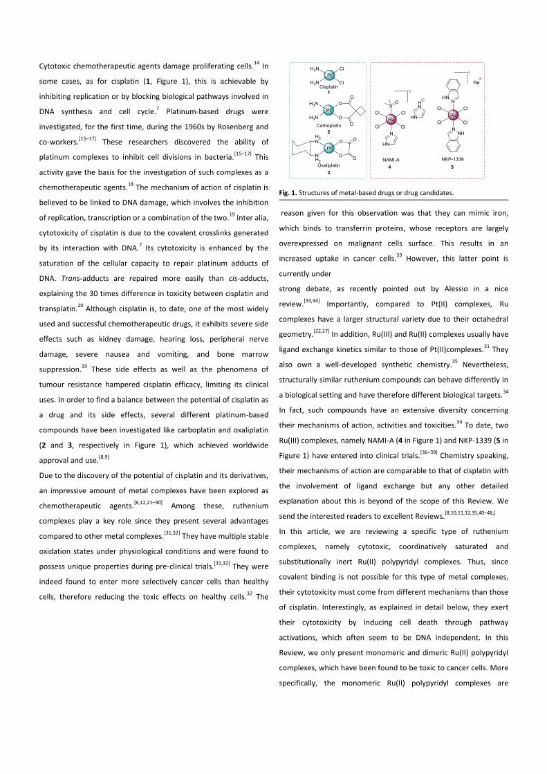
Cytotoxic chemotherapeutic agents damage proliferating cells.14
In
some cases, as for cisplatin (1, Figure 1), this is achievable by
inhibiting replication or by blocking biological pathways involved in
DNA synthesis and cell cycle.7 Platinum-based drugs were
investigated, for the first time, during the 1960s by Rosenberg and
co-workers.[15–17]
These researchers discovered the ability of
platinum complexes to inhibit cell divisions in bacteria.[15–17]
This
activity gave the basis for the investigation of such complexes as a
chemotherapeutic agents.18
The mechanism of action of cisplatin is
believed to be linked to DNA damage, which involves the inhibition
of replication, transcription or a combination of the two.19
Inter alia,
cytotoxicity of cisplatin is due to the covalent crosslinks generated
by its interaction with DNA.7 Its cytotoxicity is enhanced by the
saturation of the cellular capacity to repair platinum adducts of
DNA. Trans-adducts are repaired more easily than cis-adducts,
explaining the 30 times difference in toxicity between cisplatin and
transplatin.20
Although cisplatin is, to date, one of the most widely
used and successful chemotherapeutic drugs, it exhibits severe side
effects such as kidney damage, hearing loss, peripheral nerve
damage, severe nausea and vomiting, and bone marrow
suppression.19
These side effects as well as the phenomena of
tumour resistance hampered cisplatin efficacy, limiting its clinical
uses. In order to find a balance between the potential of cisplatin as
a drug and its side effects, several different platinum-based
compounds have been investigated like carboplatin and oxaliplatin
(2 and 3, respectively in Figure 1), which achieved worldwide
approval and use.[8,9]
Due to the discovery of the potential of cisplatin and its derivatives,
an impressive amount of metal complexes have been explored as
chemotherapeutic agents.[6,12,21–30]
Among these, ruthenium
complexes play a key role since they present several advantages
compared to other metal complexes.[31,32]
They have multiple stable
oxidation states under physiological conditions and were found to
possess unique properties during pre-clinical trials.[31,32]
They were
indeed found to enter more selectively cancer cells than healthy
cells, therefore reducing the toxic effects on healthy cells.32
The
Fig. 1. Structures of metal-based drugs or drug candidates.
reason given for this observation was that they can mimic iron,
which binds to transferrin proteins, whose receptors are largely
overexpressed on malignant cells surface. This results in an
increased uptake in cancer cells.33
However, this latter point is
currently under
strong debate, as recently pointed out by Alessio in a nice
review.[33,34]
Importantly, compared to Pt(II) complexes, Ru
complexes have a larger structural variety due to their octahedral
geometry.[22,27]
In addition, Ru(III) and Ru(II) complexes usually have
ligand exchange kinetics similar to those of Pt(II)complexes.31
They
also own a well-developed synthetic chemistry.35
Nevertheless,
structurally similar ruthenium compounds can behave differently in
a biological setting and have therefore different biological targets.34
In fact, such compounds have an extensive diversity concerning
their mechanisms of action, activities and toxicities.34
To date, two
Ru(III) complexes, namely NAMI-A (4 in Figure 1) and NKP-1339 (5 in
Figure 1) have entered into clinical trials.[36–39]
Chemistry speaking,
their mechanisms of action are comparable to that of cisplatin with
the involvement of ligand exchange but any other detailed
explanation about this is beyond of the scope of this Review. We
send the interested readers to excellent Reviews.[8,10,11,32,35,40–48,]
In this article, we are reviewing a specific type of ruthenium
complexes, namely cytotoxic, coordinatively saturated and
substitutionally inert Ru(II) polypyridyl complexes. Thus, since
covalent binding is not possible for this type of metal complexes,
their cytotoxicity must come from different mechanisms than those
of cisplatin. Interestingly, as explained in detail below, they exert
their cytotoxicity by inducing cell death through pathway
activations, which often seem to be DNA independent. In this
Review, we only present monomeric and dimeric Ru(II) polypyridyl
complexes, which have been found to be toxic to cancer cells. More
specifically, the monomeric Ru(II) polypyridyl complexes are
analysed considering their direct interaction or not with DNA as
cause of cell death. Dimeric Ru(II) polypyridyl complexes, on the
other hand, are classified considering their biological targets.
Decisively, only Ru(II) complexes fully characterised and whose
biological activity was tested on cancer cell lines are reviewed. We
also try to rationalise the antiproliferative activity of the complexes
taking into account the experimental data (i.e. cellular uptake and
cellular localization). However, we would like to highlight that the
role of the counter-anions in the cytotoxicity of the Ru(II)
complexes reported in this Review is not completely clear. It is
known that the counter-anion can influence the solubility of a
complex (i.e. KP1019 vs NKP1339).39
However, to the best of our
knowledge, there has not been a study comparing the cytotoxicity
of a Ru(II) polypyridyl complexes depending on its counter-anion.
Very interestingly, a recent paper by Zhu presented a new method
for delivering DNA ‘light-switching’ Ru(II) polypyridyl complexes into
the nucleus of living cells.49
In this work, Zhu and co-workers
demonstrated that the cell-impermeable Ru(II) polypyridyl cationic
complexes might form novel lipophilic and relatively stable ion-pair
complexes with three structurally unrelated hydrophobic weak
acids (i.e. pentachlorophenol (PCP), carbonyl cyanide p-
(trifluoromethoxy)phenylhydrazone (FCCP) and tolfenamic acid
(TA)) leading to a remarkably enhanced nuclear uptake.49
From this
work, scientists speculate that ion-pair formation might be a
general, promising live-cell delivery method for cell-impermeable
metal complexes and potentially bio-medically important.49
However, it is unclear if this has an impact on the cytotoxicity. In
this Review, we will therefore make sure to mention every time the
counter-anion employed in the biological studies. It is important to
note that most of the studies reported in this Review are based on a
continuation of previous works (i.e. researchers are trying to
improve the cytotoxicity of a complex with the help of a structure-
activity relationship study based on previous results which have
shown that a complex was toxic to cancer cells). However, there are
not, to the best of our knowledge, many examples of inert Ru(II)
polypyridyl complexes, which have been rationally designed to aim
for a specific target, like the Ru-based kinase inhibitors of Meggers,
which are not discussed herein since this field was reviewed in
detail.[50–53]
Recently, Ru(II) cyclometalated complexes have been
also investigated as anticancer agents.[54–57]
However, this topic
together with the Ru(II) polypyridyl complexes activated by light or
the non-coordinatively saturated Ru(II) polypyridyl complexes are
not reviewed here. We send the interested readers to an excellent,
very recent Review of Zeng and co-workers.58
Over the years, many
scientists focused their attention on reviewing the current state in
medicinal chemistry of Ru complexes,[24,26,32,35,45,46,59–61]
in some
cases considering only Ru(II) polypyridyl complexes and the possible
applications.[62–64]
However, to the best of our knowledge, this is
the first Review of the last 6 years focused only on the cytotoxicity
of Ru(II) polypyridyl complexes.
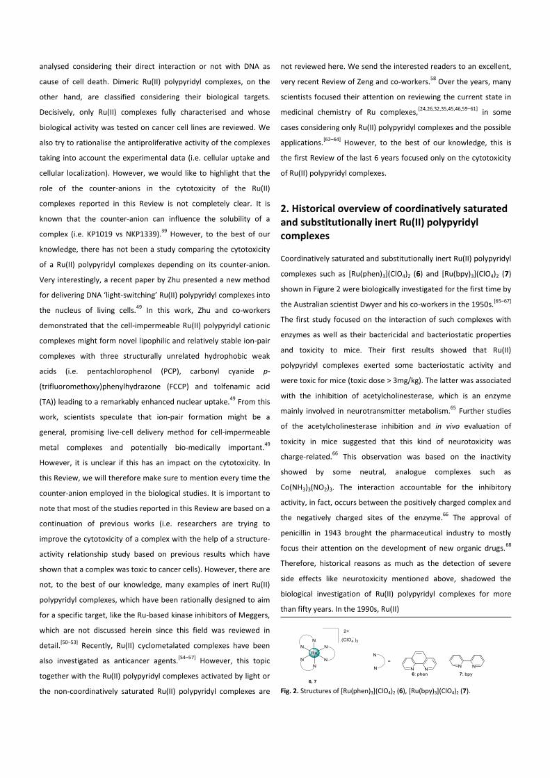
2. Historical overview of coordinatively saturated and substitutionally inert Ru(II) polypyridyl complexes
Coordinatively saturated and substitutionally inert Ru(II) polypyridyl
complexes such as [Ru(phen)3](ClO4)2 (6) and [Ru(bpy)3](ClO4)2 (7)
shown in Figure 2 were biologically investigated for the first time by
the Australian scientist Dwyer and his co-workers in the 1950s.[65–67]
The first study focused on the interaction of such complexes with
enzymes as well as their bactericidal and bacteriostatic properties
and toxicity to mice. Their first results showed that Ru(II)
polypyridyl complexes exerted some bacteriostatic activity and
were toxic for mice (toxic dose > 3mg/kg). The latter was associated
with the inhibition of acetylcholinesterase, which is an enzyme
mainly involved in neurotransmitter metabolism.65
Further studies
of the acetylcholinesterase inhibition and in vivo evaluation of
toxicity in mice suggested that this kind of neurotoxicity was
charge-related.66
This observation was based on the inactivity
showed by some neutral, analogue complexes such as
Co(NH3)3(NO2)3. The interaction accountable for the inhibitory
activity, in fact, occurs between the positively charged complex and
the negatively charged sites of the enzyme.66
The approval of
penicillin in 1943 brought the pharmaceutical industry to mostly
focus their attention on the development of new organic drugs.68
Therefore, historical reasons as much as the detection of severe
side effects like neurotoxicity mentioned above, shadowed the
biological investigation of Ru(II) polypyridyl complexes for more
than fifty years. In the 1990s, Ru(II)
Fig. 2. Structures of [Ru(phen)3](ClO4)2 (6), [Ru(bpy)3](ClO4)2 (7).
polypyridyl complexes once again attracted much attention in the
diagnostic field as DNA probes because of their interaction
ability.[49,69–81]
From these studies, it was concluded that these
complexes may interact with DNA through three different modes,
namely electrostatic, non-intercalative (“surface” interaction) and
intercalative binding mode with a quite consistent enantiomeric
preference for the Δ form respective to the Λ form.[82–88]
On the
other hand, the potential anticancer activity of such compounds
started to be reconsidered in 2000 when Liu and co-workers
reported the in vitro cytotoxicity of some Ru(II) polypyridyl
complexes.89
In this work, the toxicity of several Ru(II)/Co(III)
polypyridyl complexes were tested against HL-60, BEL-7402, KB, and
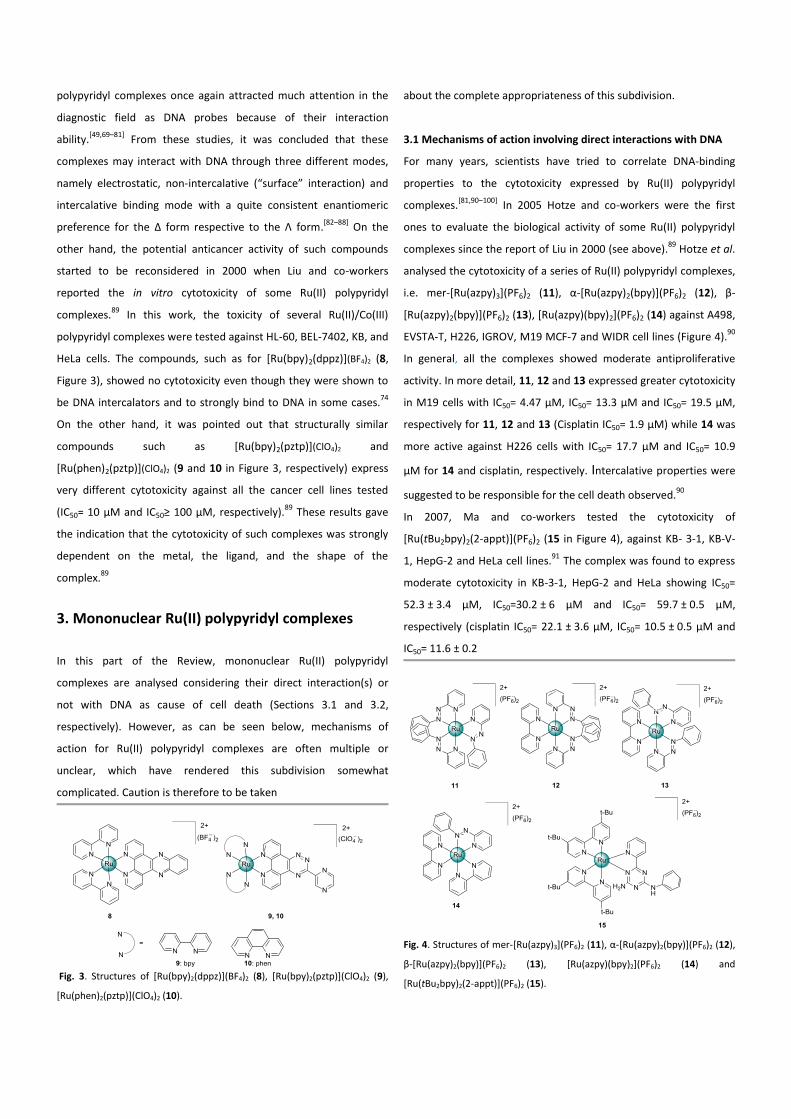
HeLa cells. The compounds, such as for [Ru(bpy)2(dppz)](BF4)2 (8,
Figure 3), showed no cytotoxicity even though they were shown to
be DNA intercalators and to strongly bind to DNA in some cases.74
On the other hand, it was pointed out that structurally similar
compounds such as [Ru(bpy)2(pztp)](ClO4)2 and
[Ru(phen)2(pztp)](ClO4)2 (9 and 10 in Figure 3, respectively) express
very different cytotoxicity against all the cancer cell lines tested
(IC50= 10 µM and IC50≥ 100 µM, respectively).89
These results gave
the indication that the cytotoxicity of such complexes was strongly
dependent on the metal, the ligand, and the shape of the
complex.89
3. Mononuclear Ru(II) polypyridyl complexes
In this part of the Review, mononuclear Ru(II) polypyridyl
complexes are analysed considering their direct interaction(s) or
not with DNA as cause of cell death (Sections 3.1 and 3.2,
respectively). However, as can be seen below, mechanisms of
action for Ru(II) polypyridyl complexes are often multiple or
unclear, which have rendered this subdivision somewhat
complicated. Caution is therefore to be taken
Fig. 3. Structures of [Ru(bpy)2(dppz)](BF4)2 (8), [Ru(bpy)2(pztp)](ClO4)2 (9),
[Ru(phen)2(pztp)](ClO4)2 (10).
about the complete appropriateness of this subdivision.
3.1 Mechanisms of action involving direct interactions with DNA
For many years, scientists have tried to correlate DNA-binding
properties to the cytotoxicity expressed by Ru(II) polypyridyl
complexes.[81,90–100]
In 2005 Hotze and co-workers were the first
ones to evaluate the biological activity of some Ru(II) polypyridyl
complexes since the report of Liu in 2000 (see above).89
Hotze et al.
analysed the cytotoxicity of a series of Ru(II) polypyridyl complexes,
i.e. mer-[Ru(azpy)3](PF6)2 (11), α-[Ru(azpy)2(bpy)](PF6)2 (12), β-
[Ru(azpy)2(bpy)](PF6)2 (13), [Ru(azpy)(bpy)2](PF6)2 (14) against A498,
EVSTA-T, H226, IGROV, M19 MCF-7 and WIDR cell lines (Figure 4).90
In general, all the complexes showed moderate antiproliferative
activity. In more detail, 11, 12 and 13 expressed greater cytotoxicity
in M19 cells with IC50= 4.47 µM, IC50= 13.3 µM and IC50= 19.5 µM,
respectively for 11, 12 and 13 (Cisplatin IC50= 1.9 µM) while 14 was
more active against H226 cells with IC50= 17.7 µM and IC50= 10.9
µM for 14 and cisplatin, respectively. Intercalative properties were
suggested to be responsible for the cell death observed.90
In 2007, Ma and co-workers tested the cytotoxicity of
[Ru(tBu2bpy)2(2-appt)](PF6)2 (15 in Figure 4)‚ against KB- 3-1, KB-V-
1, HepG-2 and HeLa cell lines.91
The complex was found to express
moderate cytotoxicity in KB-3-1, HepG-2 and HeLa showing IC50=
52.3 ± 3.4 µM, IC50=30.2 ± 6 µM and IC50= 59.7 ± 0.5 µM,
respectively (cisplatin IC50= 22.1 ± 3.6 µM, IC50= 10.5 ± 0.5 µM and
IC50= 11.6 ± 0.2
Fig. 4. Structures of mer-[Ru(azpy)3](PF6)2 (11), α-[Ru(azpy)2(bpy)](PF6)2 (12),
β-[Ru(azpy)2(bpy)](PF6)2 (13), [Ru(azpy)(bpy)2](PF6)2 (14) and
[Ru(tBu2bpy)2(2-appt)](PF6)2 (15).
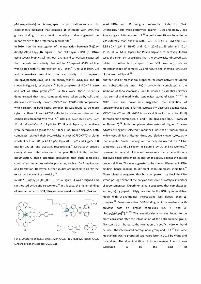
µM, respectively). In this case, spectroscopic titrations and viscosity
experiments indicated that complex 15 interacts with DNA via
groove binding. In more detail, modelling studies suggested the
minor groove as the preferential binding site.91
In 2010, from the investigation of the interaction between [Ru(2,9-
dmp)2PMIP](ClO4)2 (16, Figure 5) and calf thymus DNA (CT DNA)
using several biophysical methods, Zhang and co-workers suggested
that the antitumor activity observed for 16 against A549 cell line
was related with its intercalation in CT DNA.92
One year later, Gill
and co-workers examined the cytotoxicity of complexes
[Ru(bpy)2(tpphz)](ClO4)2 and [Ru(phen)2(tpphz)](ClO4)2 (17 and 18
shown in Figure 5, respectively).81
Both complexes bind DNA in vitro
and act as DNA probes.[81,93]
In this work, these scientists
demonstrated that these compounds were taken up by cells and
displayed cytotoxicity towards MCF-7 and A2780 cells comparable
with cisplatin. In both cases, complex 18 was found to be more
cytotoxic than 17 and A2780 cells to be more sensitive to the
complexes compared with MCF-7.81
Inter alia, IC50= 36 ± 6 µM, IC50=
11 ± 6 µM and IC50=12 ± 2 µM for 17, 18 and cisplatin, respectively
were determined against the A2780 cell line. Unlike cisplatin, both
complexes retained their cytotoxicity against A2780-CP70 cisplatin
resistant cell lines (IC50= 47 ± 6 µM, IC50= 20 ± 5 µM and IC50= 55 ± 8
µM for 17, 18, and cisplatin, respectively).81
Microscopy studies
clearly showed internalisation of complex 18 but limited nuclear
accumulation. These scientists speculated that such complexes
could affect numerous cellular processes, such as DNA replication
and translation. However, further studies are needed to clarify the
exact mechanism of cytotoxicity.81
In 2012, [Ru(bpy)2(H2IIP)](ClO4)2 (19 in Figure 6) was designed and
synthesised by Liu and co-workers.85
In this case, the higher binding
of an enantiomer to DNA/RNA was confirmed for both CT-DNA and
Fig. 5. Structures of [Ru(2,9-dmp)2PMIP](ClO4)2 (16),
[Ru(bpy)2(tpphz)](ClO4)2
(17) and [Ru(phen)2(tpphz)](ClO4)2 (18).
yeast tRNA, with 19 being a preferential binder for tRNA.
Cytotoxicity tests were performed against HL-60 and HepG-2 cell
lines using cisplatin as a control.85
In both cases 19 was found to be
less cytotoxic than cisplatin with IC50= 18.36 ± 1.35 µM and IC50=
5.85 ± 0.44 µM in HL-60 and IC50= 20.45 ± 1.53 µM and IC50=
11.18 ± 0.84 µM in HepG-2 for 19 and cisplatin, respectively. In this
case, the scientists speculated that the cytotoxicity observed was
related to other factors apart from DNA insertion, such as
molecular shape of complex 19 and nature and chemical structure
of the inserted ligand.85
Another kind of mechanism proposed for coordinatively saturated
and substitutionally inert Ru(II) polypyridyl complexes is the
inhibition of topoisomerases I and II, which are essential enzymes
that control and modify the topological states of DNA.[101–103]
In
2012, Kou and co-workers suggested the inhibition of
topoisomerases I and II for the cytotoxicity observed against HeLa,
MCF-7, HepG2 and BEL-7402 tumour cell lines for two chiral Ru(II)
anthraquinone complexes, Δ- and Λ-[Ru(bpy)2(ipad)](ClO4)2 (Δ/Λ-20
in Figure 6).94
Both complexes demonstrated higher in vitro
cytotoxicity against selected tumour cell lines than 5-fluorouracil, a
widely used clinical antitumor drug, but relatively lower cytotoxicity
than cisplatin. Similar findings were already discovered in 2011 for
complexes 21 and 22 shown in Figure 6 by Du and co-workers.95
However, in the work of Kou and co-workers, the two enantiomers
displayed small differences in antitumor activity against the tested
cancer cell lines. This was suggested to be due to differences in DNA
binding, hence leading to different topoisomerases inhibition.94
These scientists suggested that both complexes may block the DNA
strand passage event of the enzyme and serve as catalytic inhibitors
of topoisomerases. Experimental data suggested that complexes Δ-
and Λ-[Ru(bpy)2(ipad)](ClO4)2 may bind to the DNA by intercalative
mode with Λ-enantiomer intercalating less deeply than Δ-
complex.94
Enantioselective DNA-binding is in accordance with
previous data on similar complexes (i.e. Δ- and Λ-
[Ru(bpy)2(dppz]2+
).[84,88]
The enantioselectivity was found to be
more consistent after the introduction of the antraquinone group.
This can be attributed to the formation of specific hydrogen bond
between the intercalated antraquinone group and DNA.94
The same
mechanism was re-proposed two years later in 2014 by Wang and
co-workers. The dual inhibition of topoisomerase I and II was
suggested to be the base of
Fig. 6. Structures of [Ru(bpy)2(H2IIP)](ClO4)2 (19), Δ- and Λ-
[Ru(bpy)2(ipad)](ClO4)2 (20), [Ru(bpy)2(bfipH)](ClO4)2 (21) and
[Ru(phen)2(bfipH)](ClO4)2 (22), Λ/Δ -[Ru(bpy)2(pscl)](ClO4)2 (23) and Δ/Λ-
[Ru(bpy)2(psbr)](ClO4)2 (24).
the cytotoxicity exerted by two new enantiomeric pair of
complexes such as Δ/Λ-[Ru(bpy)2(pscl)](ClO4)2 and Δ/Λ-
[Ru(bpy)2(psbr)](ClO4)2 ((Δ/Λ-23 and Δ/Λ-24, respectively in Figure
6).96
The cytotoxicity of these complexes was tested against HeLa,
A549, HepG2 and BEL-7402 cell lines using NAMI-A and cisplatin as
controls. The two enantiomers display a different cytotoxicity in
agreement with the cellular uptake results obtained, with the Δ
enantiomers being the most active.96
They displayed higher
cytotoxicity when compared to NAMI-A but relatively lower
compared to cisplatin. For example, in the case of HeLa cells (cell
line against which the complexes where found to be the most
active), IC50= 41.8 ± 2.3 µM, IC50= 54.6 ± 2.9 µM, IC50= 515.7 ± 17.4
µM, IC50= 18.3 ± 1.4 µM for Δ-23, Δ-24, NAMI-A and cisplatin,
respectively were determined.96
The V annexin stain method and
flow cytometry analysis showed that complexes Δ-23 and Δ-24
induced apoptosis in HeLa cells. Moreover, topoisomerases
inhibition studies and DNA relegation assays revealed that all four
Ru(II) complexes acted as dual poisons of topoisomerase I and II
and caused DNA damage. This could lead to cell cycle arrest and/or
induction of apoptosis.96
In 2013, Wang and co-workers proposed a new anticancer
mechanism of action for Ru(II) polypyridyl complexes targeting
DNA.97
In this work, the in vitro cytotoxicity of [Ru(bpy)2(tip)](PF6)2
(25, Figure 7) and [Ru(phen)2(tip)](PF6)2 (26, Figure 7) against HeLa
cells was investigated. Experimental data suggested the interaction
of such complexes with G-quadruplex DNA, which is a secondary
structure formed in specific guanine-rich sequences.104
Interestingly, DNA sequences that can form G-quadruplexes have
been found in the upstream promoter of protoncogenes.105
Metal
complexes are usually utilised as gene expression regulators
promoting the formation and/or stabilising G-quadruplexes.[62,106]
Examples of Ru(II) polypyridyl complexes which preferentially target
G-quadruplexes DNA over duplex DNA and are able to stabilise such
structures are already present in the literature.[107–109]
Wang and co-
workers demonstrated through in vitro experiments that both
complexes (25 and 26) could induce and stabilise the formation of
G-quadruplex in promotor of bcl-2 gene (B cell lymphoma gene 2).
Cytotoxicity studies revealed antiproliferative activity on Hela cells,
(IC50= 22.10 ± 1.01 and 18.56 ± 0.67 µM, respectively for 25 and 26).
The highest cytotoxicity of complex 26 is consistent with its DNA-
binding and ability to stabilise G-quadruplexes.97
In order to have
more insights on the mechanism of cell-growth inhibition, further
experiments were performed. Both apoptosis and cell-cycle arrest
were found to be involved. Apoptosis was suggested to be related
to the regulation of the bcl-2 gene expression, dependent on the G-
quadruplex stabilisation.97
However, the apoptotic pathway
activation and the binding mode with G-quadruplex DNA still need
further elucidations. Similar findings were reported the following
year, in 2014, by Zhang and co-workers.98
In this work, they
observed a greater inhibitory activity against MDA-MB-231
expressed by complex 27 (showed in Figure 7) compared with
cisplatin, with IC50= 16.3 ± 2.6 µM and IC50= 36.1 ± 1.9 µM for
complex 27 and cisplatin, respectively. Further studies prompted
these scientists to suggest a correlation between the induced
apoptosis observed and the stabilisation of G-quadruplex
conformation of c-myc oncogene promoter via groove binding
mode which led to the down-regulated expression of c-myc.98
A
detailed mechanism of action is still under investigation but these
findings suggests complex 27 as a potential apoptosis inducer
characterized by a low toxicity against healthy HAcat cells.98
Moreover, during the same year, Srishailam and co-workers
explored three new Ru(II) polypyridyl complexes, namely
[Ru(phen)2ptip](ClO4)2 (28), [Ru(bpy)2ptip](ClO4)2 (29) and
[Ru(dmb)2ptip](ClO4)2 (30).110
These complexes showed
considerable antibacterial activity against S. aureus and E. coli at 1
mg/mL and 0.5 mg/mL concentrations. The in vitro cytotoxicity was
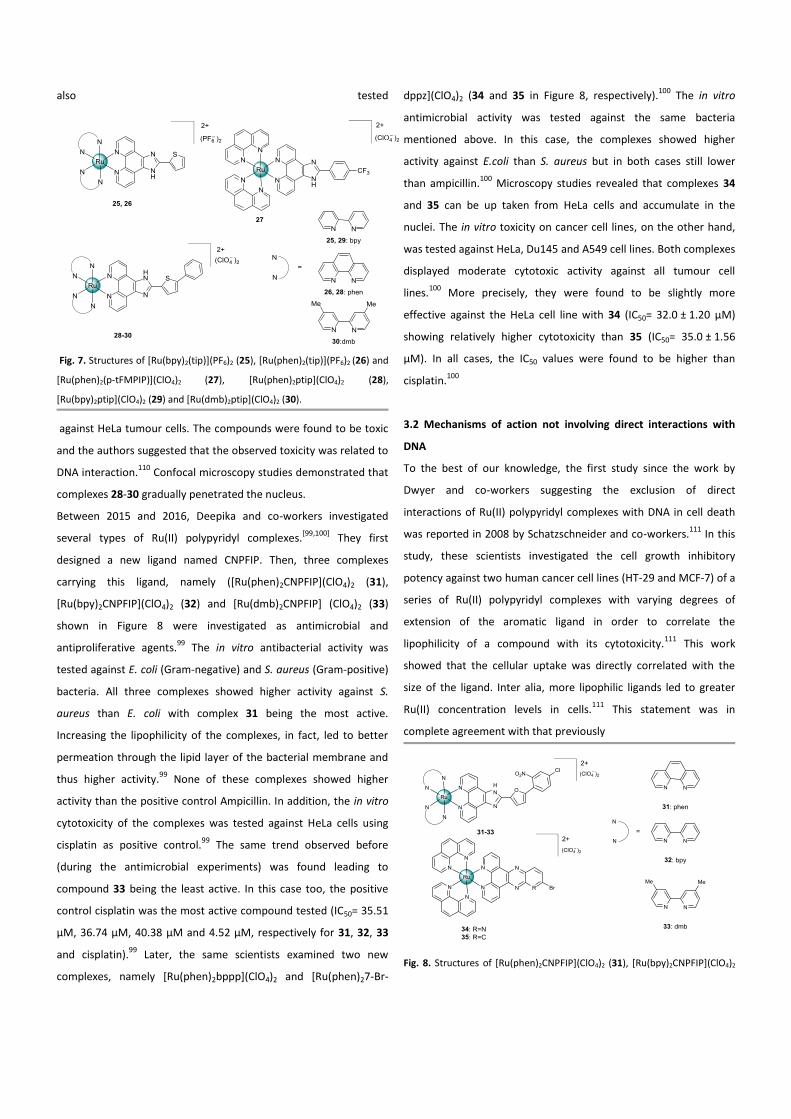
also tested
Fig. 7. Structures of [Ru(bpy)2(tip)](PF6)2 (25), [Ru(phen)2(tip)](PF6)2 (26) and
[Ru(phen)2(p-tFMPIP)](ClO4)2 (27), [Ru(phen)2ptip](ClO4)2 (28),
[Ru(bpy)2ptip](ClO4)2 (29) and [Ru(dmb)2ptip](ClO4)2 (30).
against HeLa tumour cells. The compounds were found to be toxic
and the authors suggested that the observed toxicity was related to
DNA interaction.110
Confocal microscopy studies demonstrated that
complexes 28-30 gradually penetrated the nucleus.
Between 2015 and 2016, Deepika and co-workers investigated
several types of Ru(II) polypyridyl complexes.[99,100]
They first
designed a new ligand named CNPFIP. Then, three complexes
carrying this ligand, namely ([Ru(phen)2CNPFIP](ClO4)2 (31),
[Ru(bpy)2CNPFIP](ClO4)2 (32) and [Ru(dmb)2CNPFIP] (ClO4)2 (33)
shown in Figure 8 were investigated as antimicrobial and
antiproliferative agents.99
The in vitro antibacterial activity was
tested against E. coli (Gram-negative) and S. aureus (Gram-positive)
bacteria. All three complexes showed higher activity against S.
aureus than E. coli with complex 31 being the most active.
Increasing the lipophilicity of the complexes, in fact, led to better
permeation through the lipid layer of the bacterial membrane and
thus higher activity.99
None of these complexes showed higher
activity than the positive control Ampicillin. In addition, the in vitro
cytotoxicity of the complexes was tested against HeLa cells using
cisplatin as positive control.99
The same trend observed before
(during the antimicrobial experiments) was found leading to
compound 33 being the least active. In this case too, the positive
control cisplatin was the most active compound tested (IC50= 35.51
µM, 36.74 µM, 40.38 µM and 4.52 µM, respectively for 31, 32, 33
and cisplatin).99
Later, the same scientists examined two new
complexes, namely [Ru(phen)2bppp](ClO4)2 and [Ru(phen)27-Br-
dppz](ClO4)2 (34 and 35 in Figure 8, respectively).100
The in vitro
antimicrobial activity was tested against the same bacteria
mentioned above. In this case, the complexes showed higher
activity against E.coli than S. aureus but in both cases still lower
than ampicillin.100
Microscopy studies revealed that complexes 34
and 35 can be up taken from HeLa cells and accumulate in the
nuclei. The in vitro toxicity on cancer cell lines, on the other hand,
was tested against HeLa, Du145 and A549 cell lines. Both complexes
displayed moderate cytotoxic activity against all tumour cell
lines.100
More precisely, they were found to be slightly more
effective against the HeLa cell line with 34 (IC50= 32.0 ± 1.20 µM)
showing relatively higher cytotoxicity than 35 (IC50= 35.0 ± 1.56
µM). In all cases, the IC50 values were found to be higher than
cisplatin.100
3.2 Mechanisms of action not involving direct interactions with
DNA
To the best of our knowledge, the first study since the work by
Dwyer and co-workers suggesting the exclusion of direct
interactions of Ru(II) polypyridyl complexes with DNA in cell death
was reported in 2008 by Schatzschneider and co-workers.111
In this
study, these scientists investigated the cell growth inhibitory
potency against two human cancer cell lines (HT-29 and MCF-7) of a
series of Ru(II) polypyridyl complexes with varying degrees of
extension of the aromatic ligand in order to correlate the
lipophilicity of a compound with its cytotoxicity.111
This work
showed that the cellular uptake was directly correlated with the
size of the ligand. Inter alia, more lipophilic ligands led to greater
Ru(II) concentration levels in cells.111
This statement was in
complete agreement with that previously
Fig. 8. Structures of [Ru(phen)2CNPFIP](ClO4)2 (31), [Ru(bpy)2CNPFIP](ClO4)2
(32), [Ru(dmb)2CNPFIP](ClO4)2 (33), [Ru(phen)2bppp](ClO4)2 (34) and
[Ru(phen)27-Br-dppz](ClO4)2 (35).
demonstrated on structurally similar Ru(II) polypyridyl
complexes.112
Moreover, the authors pointed out that greater
cellular accumulation resulted in greater cytotoxicity on both cell
lines. In particular, [Ru(bpy)2(dppn)]Cl2 (36, Figure 9), which was the
complex carrying the ligand with the greater aromatic surface area
considered in this work, showed the most significant
antiproliferative activity with IC50s < 10 µM, values which are
comparable to that of cisplatin under identical conditions.111
The
mechanism of action was suggested to be related to the
modification of cell membrane function and cell adhesion
properties.111
This observation was in contrast with the accustomed
focus on the DNA intercalative properties of such compounds.
One year later, Gao and co-workers showed that the antitumor
activity of Ru(II) polypyridyl complexes must be related to different
mechanisms than only DNA intercalation.113
In this work, these
researchers designed and synthesised different complexes in order
to improve the DNA-binding capability of their Ru(II) complexes by
introducing electropositive pendants to the ancillary ligand or by
increasing their intercalative aromatic surface (complexes 37-40 in
Figure 9). The authors then tested the activity of these compounds
as inhibitors of DNA transcription, which is involved in the
mechanism of action for modifications mentioned above. They
improved the DNA binding ability of their complexes, which led to a
direct, positive correlation with the inhibition of DNA
transcription.113
However, no clear trend was observed between
the DNA binding ability of the complexes and their antitumor
activity,
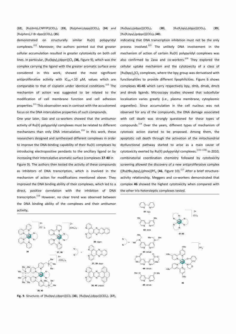
Fig. 9. Structures of [Ru(bpy)2(dppn)](Cl)2 (36), [Ru(bpy)2(dppz)](ClO4)2 (37),
[Ru(bpy)2(pdppz)](ClO4)2 (38), [Ru(R2bpy)2(dppz)](ClO4)6 (39),
[Ru(R2bpy)2(pdppz)](ClO4)6 (40).
indicating that DNA transcription inhibition must not be the only
process involved.113
The unlikely DNA involvement in the
mechanism of action of certain Ru(II) polypyridyl complexes was
also confirmed by Zava and co-workers.114
They explored the
cellular uptake mechanism and the cytotoxicity of a class of
[Ru(bpy)3]Cl2 complexes, where the bpy group was derivatised with
functionalities to provide different lipophilicities. Figure 6 shows
complexes 41-45 which carry respectively bpy, dmb, dmab, dmcb
and dmob ligands. Microscopy studies showed that subcellular
localisation varies greatly (i.e., plasma membrane, cytoplasmic
organelles). Since accumulation in the cell nucleus was not
observed for any of the compounds, the DNA damage associated
with cell death was strongly questioned for these types of
compounds.114
Over the years, different types of mechanism of
cytotoxic action started to be proposed. Among them, the
apoptotic cell death through the activation of the mitochondrial
dysfunctional pathway started to arise as a main cause of
cytotoxicity exerted by Ru(II) polypyridyl complexes.[115–119]
In 2010,
combinatorial coordination chemistry followed by cytotoxicity
screening allowed the discovery of a new antiproliferative complex
([Ru(tBu2bpy)2(phox)]PF6 (46, Figure 10).117
After a brief structure-
activity relationship, Meggers and co-workers demonstrated that
complex 46 showed the highest cytotoxicity when compared with
the other tris-heteroleptic complexes tested.
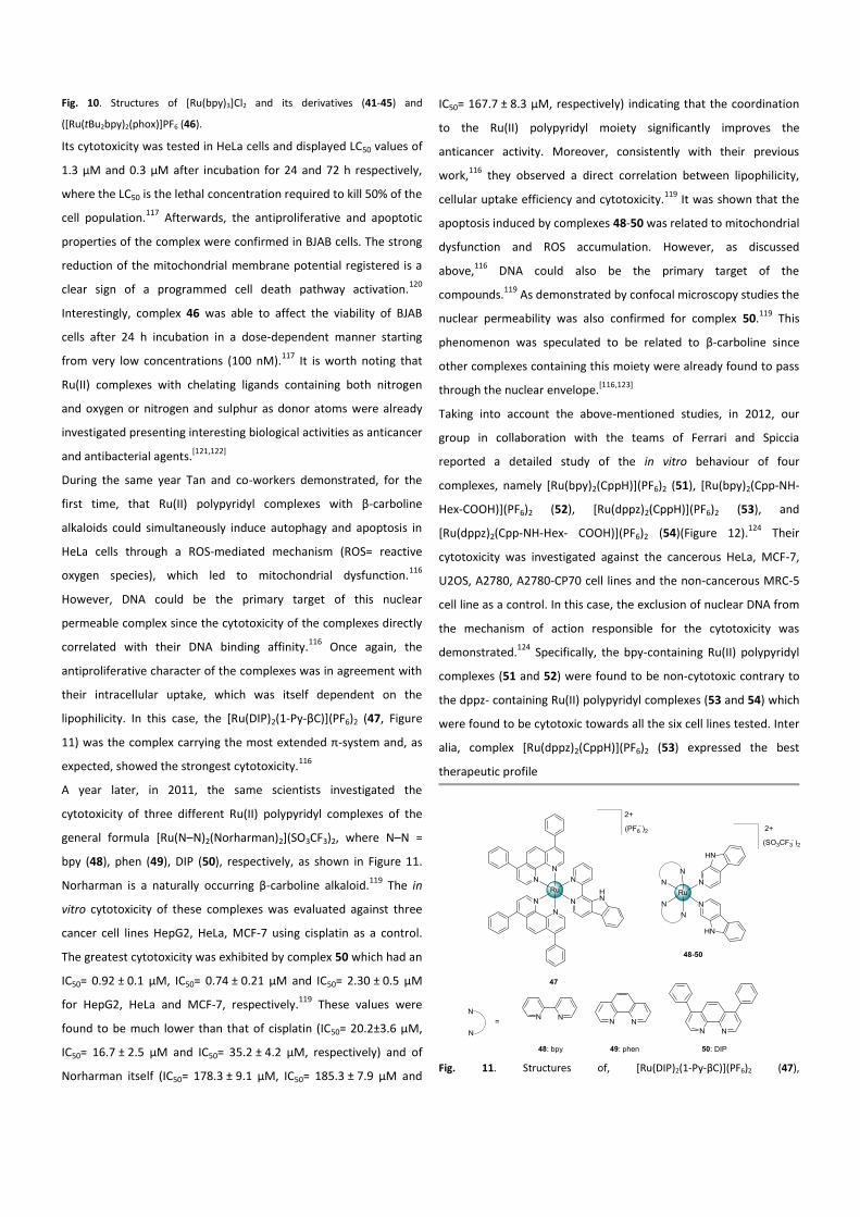
Fig. 10. Structures of [Ru(bpy)3]Cl2 and its derivatives (41-45) and
([Ru(tBu2bpy)2(phox)]PF6 (46).
Its cytotoxicity was tested in HeLa cells and displayed LC50 values of
1.3 µM and 0.3 µM after incubation for 24 and 72 h respectively,
where the LC50 is the lethal concentration required to kill 50% of the
cell population.117
Afterwards, the antiproliferative and apoptotic
properties of the complex were confirmed in BJAB cells. The strong
reduction of the mitochondrial membrane potential registered is a
clear sign of a programmed cell death pathway activation.120
Interestingly, complex 46 was able to affect the viability of BJAB
cells after 24 h incubation in a dose-dependent manner starting
from very low concentrations (100 nM).117
It is worth noting that
Ru(II) complexes with chelating ligands containing both nitrogen
and oxygen or nitrogen and sulphur as donor atoms were already
investigated presenting interesting biological activities as anticancer
and antibacterial agents.[121,122]
During the same year Tan and co-workers demonstrated, for the
first time, that Ru(II) polypyridyl complexes with β-carboline
alkaloids could simultaneously induce autophagy and apoptosis in
HeLa cells through a ROS-mediated mechanism (ROS= reactive
oxygen species), which led to mitochondrial dysfunction.116
However, DNA could be the primary target of this nuclear
permeable complex since the cytotoxicity of the complexes directly
correlated with their DNA binding affinity.116
Once again, the
antiproliferative character of the complexes was in agreement with
their intracellular uptake, which was itself dependent on the
lipophilicity. In this case, the [Ru(DIP)2(1-Py-βC)](PF6)2 (47, Figure
11) was the complex carrying the most extended π-system and, as
expected, showed the strongest cytotoxicity.116
A year later, in 2011, the same scientists investigated the
cytotoxicity of three different Ru(II) polypyridyl complexes of the
general formula [Ru(N–N)2(Norharman)2](SO3CF3)2, where N–N =
bpy (48), phen (49), DIP (50), respectively, as shown in Figure 11.
Norharman is a naturally occurring β-carboline alkaloid.119
The in
vitro cytotoxicity of these complexes was evaluated against three
cancer cell lines HepG2, HeLa, MCF-7 using cisplatin as a control.
The greatest cytotoxicity was exhibited by complex 50 which had an
IC50= 0.92 ± 0.1 µM, IC50= 0.74 ± 0.21 µM and IC50= 2.30 ± 0.5 µM
for HepG2, HeLa and MCF-7, respectively.119
These values were
found to be much lower than that of cisplatin (IC50= 20.2±3.6 µM,
IC50= 16.7 ± 2.5 µM and IC50= 35.2 ± 4.2 µM, respectively) and of
Norharman itself (IC50= 178.3 ± 9.1 µM, IC50= 185.3 ± 7.9 µM and
IC50= 167.7 ± 8.3 µM, respectively) indicating that the coordination
to the Ru(II) polypyridyl moiety significantly improves the
anticancer activity. Moreover, consistently with their previous
work,116
they observed a direct correlation between lipophilicity,
cellular uptake efficiency and cytotoxicity.119
It was shown that the
apoptosis induced by complexes 48-50 was related to mitochondrial
dysfunction and ROS accumulation. However, as discussed
above,116
DNA could also be the primary target of the
compounds.119
As demonstrated by confocal microscopy studies the
nuclear permeability was also confirmed for complex 50.119
This
phenomenon was speculated to be related to β-carboline since
other complexes containing this moiety were already found to pass
through the nuclear envelope.[116,123]
Taking into account the above-mentioned studies, in 2012, our
group in collaboration with the teams of Ferrari and Spiccia
reported a detailed study of the in vitro behaviour of four
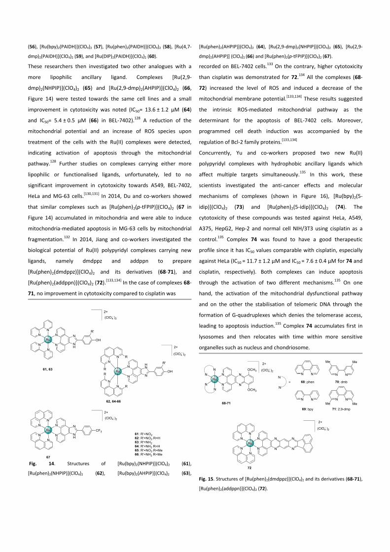
complexes, namely [Ru(bpy)2(CppH)](PF6)2 (51), [Ru(bpy)2(Cpp-NH-
Hex-COOH)](PF6)2 (52), [Ru(dppz)2(CppH)](PF6)2 (53), and
[Ru(dppz)2(Cpp-NH-Hex- COOH)](PF6)2 (54)(Figure 12).124
Their
cytotoxicity was investigated against the cancerous HeLa, MCF-7,
U2OS, A2780, A2780-CP70 cell lines and the non-cancerous MRC-5
cell line as a control. In this case, the exclusion of nuclear DNA from
the mechanism of action responsible for the cytotoxicity was
demonstrated.124
Specifically, the bpy-containing Ru(II) polypyridyl
complexes (51 and 52) were found to be non-cytotoxic contrary to
the dppz- containing Ru(II) polypyridyl complexes (53 and 54) which
were found to be cytotoxic towards all the six cell lines tested. Inter
alia, complex [Ru(dppz)2(CppH)](PF6)2 (53) expressed the best
therapeutic profile
Fig. 11. Structures of, [Ru(DIP)2(1-Py-βC)](PF6)2 (47),
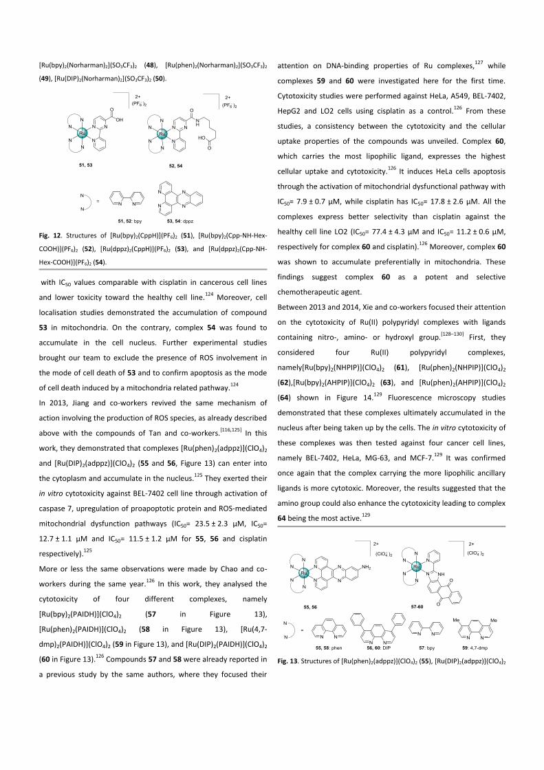
[Ru(bpy)2(Norharman)2](SO3CF3)2 (48), [Ru(phen)2(Norharman)2](SO3CF3)2
(49), [Ru(DIP)2(Norharman)2](SO3CF3)2 (50).
Fig. 12. Structures of [Ru(bpy)2(CppH)](PF6)2 (51), [Ru(bpy)2(Cpp-NH-Hex-
COOH)](PF6)2 (52), [Ru(dppz)2(CppH)](PF6)2 (53), and [Ru(dppz)2(Cpp-NH-
Hex-COOH)](PF6)2 (54).
with IC50 values comparable with cisplatin in cancerous cell lines
and lower toxicity toward the healthy cell line.124
Moreover, cell
localisation studies demonstrated the accumulation of compound
53 in mitochondria. On the contrary, complex 54 was found to
accumulate in the cell nucleus. Further experimental studies
brought our team to exclude the presence of ROS involvement in
the mode of cell death of 53 and to confirm apoptosis as the mode
of cell death induced by a mitochondria related pathway.124
In 2013, Jiang and co-workers revived the same mechanism of
action involving the production of ROS species, as already described
above with the compounds of Tan and co-workers.[116,125]
In this
work, they demonstrated that complexes [Ru(phen)2(adppz)](ClO4)2
and [Ru(DIP)2(adppz)](ClO4)2 (55 and 56, Figure 13) can enter into
the cytoplasm and accumulate in the nucleus.125
They exerted their
in vitro cytotoxicity against BEL-7402 cell line through activation of
caspase 7, upregulation of proapoptotic protein and ROS-mediated
mitochondrial dysfunction pathways (IC50= 23.5 ± 2.3 µM, IC50=
12.7 ± 1.1 µM and IC50= 11.5 ± 1.2 µM for 55, 56 and cisplatin
respectively).125
More or less the same observations were made by Chao and co-
workers during the same year.126
In this work, they analysed the
cytotoxicity of four different complexes, namely
[Ru(bpy)2(PAIDH)](ClO4)2 (57 in Figure 13),
[Ru(phen)2(PAIDH)](ClO4)2 (58 in Figure 13), [Ru(4,7-
dmp)2(PAIDH)](ClO4)2 (59 in Figure 13), and [Ru(DIP)2(PAIDH)](ClO4)2
(60 in Figure 13).126
Compounds 57 and 58 were already reported in
a previous study by the same authors, where they focused their
attention on DNA-binding properties of Ru complexes,127
while
complexes 59 and 60 were investigated here for the first time.
Cytotoxicity studies were performed against HeLa, A549, BEL-7402,
HepG2 and LO2 cells using cisplatin as a control.126
From these
studies, a consistency between the cytotoxicity and the cellular
uptake properties of the compounds was unveiled. Complex 60,
which carries the most lipophilic ligand, expresses the highest
cellular uptake and cytotoxicity.126
It induces HeLa cells apoptosis
through the activation of mitochondrial dysfunctional pathway with
IC50= 7.9 ± 0.7 µM, while cisplatin has IC50= 17.8 ± 2.6 µM. All the
complexes express better selectivity than cisplatin against the
healthy cell line LO2 (IC50= 77.4 ± 4.3 µM and IC50= 11.2 ± 0.6 µM,
respectively for complex 60 and cisplatin).126
Moreover, complex 60
was shown to accumulate preferentially in mitochondria. These
findings suggest complex 60 as a potent and selective
chemotherapeutic agent.
Between 2013 and 2014, Xie and co-workers focused their attention
on the cytotoxicity of Ru(II) polypyridyl complexes with ligands
containing nitro-, amino- or hydroxyl group.[128–130]
First, they
considered four Ru(II) polypyridyl complexes,
namely[Ru(bpy)2(NHPIP)](ClO4)2 (61), [Ru(phen)2(NHPIP)](ClO4)2
(62),[Ru(bpy)2(AHPIP)](ClO4)2 (63), and [Ru(phen)2(AHPIP)](ClO4)2
(64) shown in Figure 14.129
Fluorescence microscopy studies
demonstrated that these complexes ultimately accumulated in the
nucleus after being taken up by the cells. The in vitro cytotoxicity of
these complexes was then tested against four cancer cell lines,
namely BEL-7402, HeLa, MG-63, and MCF-7.129
It was confirmed
once again that the complex carrying the more lipophilic ancillary
ligands is more cytotoxic. Moreover, the results suggested that the
amino group could also enhance the cytotoxicity leading to complex
64 being the most active.129
Fig. 13. Structures of [Ru(phen)2(adppz)](ClO4)2 (55), [Ru(DIP)2(adppz)](ClO4)2
(56), [Ru(bpy)2(PAIDH)](ClO4)2 (57), [Ru(phen)2(PAIDH)](ClO4)2 (58), [Ru(4,7-
dmp)2(PAIDH)](ClO4)2 (59), and [Ru(DIP)2(PAIDH)](ClO4)2 (60).
These researchers then investigated two other analogues with a
more lipophilic ancillary ligand. Complexes [Ru(2,9-
dmp)2(NHPIP)](ClO4)2 (65) and [Ru(2,9-dmp)2(AHPIP)](ClO4)2 (66,
Figure 14) were tested towards the same cell lines and a small
improvement in cytotoxicity was noted (IC50= 13.6 ± 1.2 µM (64)
and IC50= 5.4 ± 0.5 µM (66) in BEL-7402).128
A reduction of the
mitochondrial potential and an increase of ROS species upon
treatment of the cells with the Ru(II) complexes were detected,
indicating activation of apoptosis through the mitochondrial
pathway.128
Further studies on complexes carrying either more
lipophilic or functionalised ligands, unfortunately, led to no
significant improvement in cytotoxicity towards A549, BEL-7402,
HeLa and MG-63 cells.[130,131]
In 2014, Du and co-workers showed
that similar complexes such as [Ru(phen)2(p-tFPIP)](ClO4)2 (67 in
Figure 14) accumulated in mitochondria and were able to induce
mitochondria-mediated apoptosis in MG-63 cells by mitochondrial
fragmentation.132
In 2014, Jiang and co-workers investigated the
biological potential of Ru(II) polypyridyl complexes carrying new
ligands, namely dmdppz and addppn to prepare
[Ru(phen)2(dmdppz)](ClO4)2 and its derivatives (68-71), and
[Ru(phen)2(addppn)](ClO4)2 (72).[133,134]
In the case of complexes 68-
71, no improvement in cytotoxicity compared to cisplatin was
Fig. 14. Structures of [Ru(bpy)2(NHPIP)](ClO4)2 (61),
[Ru(phen)2(NHPIP)](ClO4)2 (62), [Ru(bpy)2(AHPIP)](ClO4)2 (63),
[Ru(phen)2(AHPIP)](ClO4)2 (64), [Ru(2,9-dmp)2(NHPIP)](ClO4)2 (65), [Ru(2,9-
dmp)2(AHPIP)] (ClO4)2 (66) and [Ru(phen)2(p-tFPIP)](ClO4)2 (67).
recorded on BEL-7402 cells.133
On the contrary, higher cytotoxicity
than cisplatin was demonstrated for 72.134
All the complexes (68-
72) increased the level of ROS and induced a decrease of the
mitochondrial membrane potential.[133,134]
These results suggested
the intrinsic ROS-mediated mitochondrial pathway as the
determinant for the apoptosis of BEL-7402 cells. Moreover,
programmed cell death induction was accompanied by the
regulation of Bcl-2 family proteins.[133,134]
Concurrently, Yu and co-workers proposed two new Ru(II)
polypyridyl complexes with hydrophobic ancillary ligands which
affect multiple targets simultaneously.135
In this work, these
scientists investigated the anti-cancer effects and molecular
mechanisms of complexes (shown in Figure 16), [Ru(bpy)2(5-
idip)](ClO4)2 (73) and [Ru(phen)2(5-idip)](ClO4)2 (74). The
cytotoxicity of these compounds was tested against HeLa, A549,
A375, HepG2, Hep-2 and normal cell NIH/3T3 using cisplatin as a
control.135
Complex 74 was found to have a good therapeutic
profile since it has IC50 values comparable with cisplatin, especially
against HeLa (IC50 = 11.7 ± 1.2 µM and IC50 = 7.6 ± 0.4 µM for 74 and
cisplatin, respectively). Both complexes can induce apoptosis
through the activation of two different mechanisms.135
On one
hand, the activation of the mitochondrial dysfunctional pathway
and on the other the stabilisation of telomeric DNA through the
formation of G-quadruplexes which denies the telomerase access,
leading to apoptosis induction.135
Complex 74 accumulates first in
lysosomes and then relocates with time within more sensitive
organelles such as nucleus and chondriosome.
Fig. 15. Structures of [Ru(phen)2(dmdppz)](ClO4)2 and its derivatives (68-71),
[Ru(phen)2(addppn)](ClO4)2 (72).
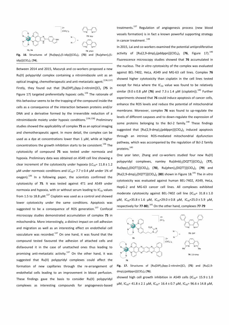
Fig. 16. Structures of [Ru(bpy)2(5-idip)](ClO4)2 (73) and [Ru(phen)2(5-
idip)](ClO4)2 (74).
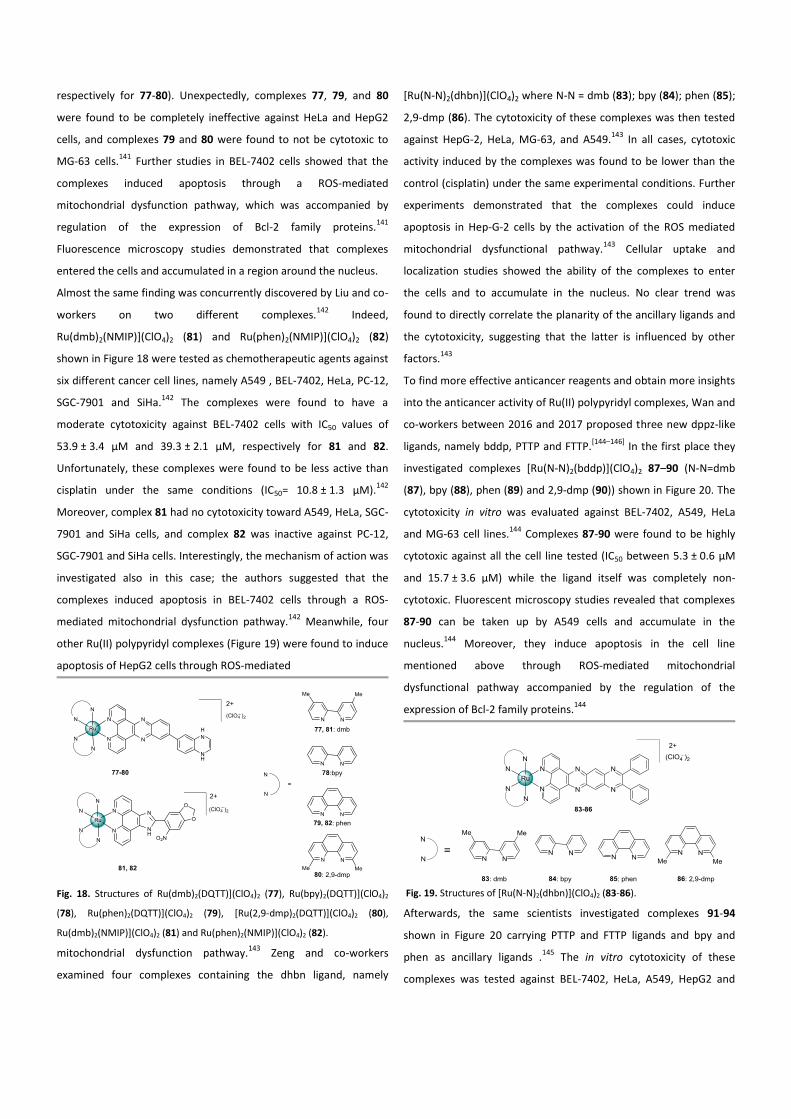
Between 2014 and 2015, Mazuryk and co-workers proposed a new
Ru(II) polypyridyl complex containing a nitroimidazole unit as an
optical imaging, chemotherapeutic and anti-metastatic agent.[136,137]
Firstly, they found out that [Ru(DIP)2(bpy-2-nitroIm)]Cl2 (75 in
Figure 17) targeted preferentially hypoxic cells.136
The rationale of
this behaviour seems to be the trapping of the compound inside the
cells as a consequence of the interaction between proteins and/or
DNA and a derivative formed by the irreversible reduction of a
nitroimidazole moiety under hypoxic conditions.[136,138]
Preliminary
studies showed the applicability of complex 75 as an optical imaging
and chemotherapeutic agent. In more detail, the complex can be
used as a dye at concentrations lower than 1 µM, while at higher
concentrations the growth inhibition starts to be consistent.136
The
cytotoxicity of compound 75 was tested under normoxia and
hypoxia. Preliminary data was obtained on A549 cell line showing a
clear increment of the cytotoxicity under hypoxia (LC50= 11.8 ± 1.2
µM under normoxic conditions and LC50= 7.7 ± 0.4 µM under 1% of
oxygen).136
In a following paper, the scientists confirmed the
cytotoxicity of 75. It was tested against 4T1 and A549 under
normoxia and hypoxia, with or without serum leading to IC50 values
from 1.5 to 18.8 µM.137
Cisplatin was used as a control and showed
lower cytotoxicity under the same conditions. Apoptosis was
suggested to be a consequence of ROS generation.137
Confocal
microscopy studies demonstrated accumulation of complex 75 in
mitochondria. More interestingly, a distinct impact on cell adhesion
and migration as well as an interesting effect on endothelial cell
vasculature was recorded.137
On one hand, it was found that the
compound tested favoured the adhesion of attached cells and
disfavoured it in the case of unattached ones thus leading to
promising anti-metastatic activity.137
On the other hand, it was
suggested that Ru(II) polypyridyl complexes could affect the
formation of new capillaries through the re-arrangement of
endothelial cells leading to an improvement in blood perfusion.
These findings gave the basis to consider Ru(II) polypyridyl
complexes as interesting compounds for angiogenesis-based
treatments.137
Regulation of angiogenesis process (new blood
vessels formation) is in fact a known powerful supporting strategy
in cancer treatment .139
In 2015, Lai and co-workers examined the potential antiproliferative
activity of [Ru(2,9-dmp)2(pddppn)](ClO4)2 (76, Figure 17).140
Fluorescence microscopy studies showed that 76 accumulated in
the nucleus. The in vitro cytotoxicity of the complex was evaluated
against BEL-7402, HeLa, A549 and MG-63 cell lines. Complex 76
showed higher cytotoxicity than cisplatin in the cell lines tested
except for HeLa where the IC50 value was found to be relatively
similar (9.0 ± 0.8 µM (76) and 7.3 ± 1.4 µM (cisplatin)).140
Further
experiments showed that 76 could induce apoptosis of cancer cells,
enhance the ROS levels and reduce the potential of mitochondrial
membrane. Moreover, complex 76 was found to up-regulate the
levels of different caspases and to down-regulate the expression of
some proteins belonging to the Bcl-2 family.140
These findings
suggested that [Ru(2,9-dmp)2(pddppn)](ClO4)2 induced apoptosis
through an intrinsic ROS-mediated mitochondrial dysfunction
pathway, which was accompanied by the regulation of Bcl-2 family
proteins.140
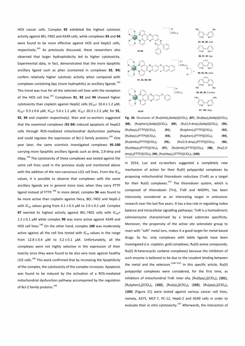
One year later, Zhang and co-workers studied four new Ru(II)
polypyridyl complexes, namley Ru(dmb)2(DQTT)](ClO4)2 (77),
Ru(bpy)2(DQTT)](ClO4)2 (78), Ru(phen)2(DQTT)](ClO4)2 (79) and
[Ru(2,9-dmp)2(DQTT)](ClO4)2 (80) shown in Figure 18.141
The in vitro
cytotoxicity was evaluated against human BEL-7402, A549, HeLa,
HepG-2 and MG-63 cancer cell lines. All complexes exhibited
moderate cytotoxicity against BEL-7402 cell line (IC50= 31.8 ± 1.0
µM, IC50=35.8 ± 1.6 µM, IC50=29.0 ± 0.8 µM, IC50=25.0 ± 5.9 µM,
respectively for 77-80).141
On the other hand, complexes 77-79
Fig. 17. Structures of [Ru(DIP)2(bpy-2-nitroIm)]Cl2 (75) and [Ru(2,9-
dmp)2(pddppn)](ClO4)2 (76).
showed high cell growth inhibition in A549 cells (IC50= 15.9 ± 1.0
µM, IC50= 41.8 ± 2.1 µM, IC50= 16.4 ± 0.7 µM, IC50= 96.6 ± 14.8 µM,
respectively for 77-80). Unexpectedly, complexes 77, 79, and 80
were found to be completely ineffective against HeLa and HepG2
cells, and complexes 79 and 80 were found to not be cytotoxic to
MG-63 cells.141
Further studies in BEL-7402 cells showed that the
complexes induced apoptosis through a ROS-mediated
mitochondrial dysfunction pathway, which was accompanied by
regulation of the expression of Bcl-2 family proteins.141
Fluorescence microscopy studies demonstrated that complexes
entered the cells and accumulated in a region around the nucleus.
Almost the same finding was concurrently discovered by Liu and co-
workers on two different complexes.142
Indeed,
Ru(dmb)2(NMIP)](ClO4)2 (81) and Ru(phen)2(NMIP)](ClO4)2 (82)
shown in Figure 18 were tested as chemotherapeutic agents against
six different cancer cell lines, namely A549 , BEL-7402, HeLa, PC-12,
SGC-7901 and SiHa.142
The complexes were found to have a
moderate cytotoxicity against BEL-7402 cells with IC50 values of
53.9 ± 3.4 µM and 39.3 ± 2.1 µM, respectively for 81 and 82.
Unfortunately, these complexes were found to be less active than
cisplatin under the same conditions (IC50= 10.8 ± 1.3 µM).142
Moreover, complex 81 had no cytotoxicity toward A549, HeLa, SGC-
7901 and SiHa cells, and complex 82 was inactive against PC-12,
SGC-7901 and SiHa cells. Interestingly, the mechanism of action was
investigated also in this case; the authors suggested that the
complexes induced apoptosis in BEL-7402 cells through a ROS-
mediated mitochondrial dysfunction pathway.142
Meanwhile, four
other Ru(II) polypyridyl complexes (Figure 19) were found to induce
apoptosis of HepG2 cells through ROS-mediated
Fig. 18. Structures of Ru(dmb)2(DQTT)](ClO4)2 (77), Ru(bpy)2(DQTT)](ClO4)2
(78), Ru(phen)2(DQTT)](ClO4)2 (79), [Ru(2,9-dmp)2(DQTT)](ClO4)2 (80),
Ru(dmb)2(NMIP)](ClO4)2 (81) and Ru(phen)2(NMIP)](ClO4)2 (82).
mitochondrial dysfunction pathway.143
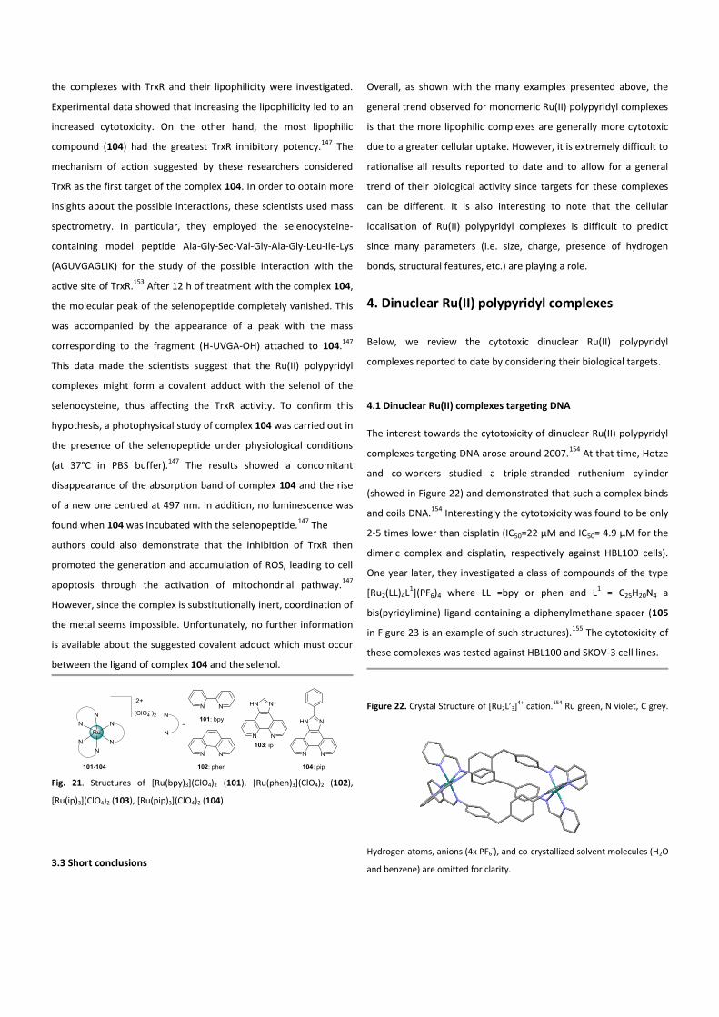
Zeng and co-workers
examined four complexes containing the dhbn ligand, namely
[Ru(N-N)2(dhbn)](ClO4)2 where N-N = dmb (83); bpy (84); phen (85);
2,9-dmp (86). The cytotoxicity of these complexes was then tested
against HepG-2, HeLa, MG-63, and A549.143
In all cases, cytotoxic
activity induced by the complexes was found to be lower than the
control (cisplatin) under the same experimental conditions. Further
experiments demonstrated that the complexes could induce
apoptosis in Hep-G-2 cells by the activation of the ROS mediated
mitochondrial dysfunctional pathway.143
Cellular uptake and
localization studies showed the ability of the complexes to enter
the cells and to accumulate in the nucleus. No clear trend was
found to directly correlate the planarity of the ancillary ligands and
the cytotoxicity, suggesting that the latter is influenced by other
factors.143
To find more effective anticancer reagents and obtain more insights
into the anticancer activity of Ru(II) polypyridyl complexes, Wan and
co-workers between 2016 and 2017 proposed three new dppz-like
ligands, namely bddp, PTTP and FTTP.[144–146]
In the first place they
investigated complexes [Ru(N-N)2(bddp)](ClO4)2 87–90 (N-N=dmb
(87), bpy (88), phen (89) and 2,9-dmp (90)) shown in Figure 20. The
cytotoxicity in vitro was evaluated against BEL-7402, A549, HeLa
and MG-63 cell lines.144
Complexes 87-90 were found to be highly
cytotoxic against all the cell line tested (IC50 between 5.3 ± 0.6 μM
and 15.7 ± 3.6 μM) while the ligand itself was completely non-
cytotoxic. Fluorescent microscopy studies revealed that complexes
87-90 can be taken up by A549 cells and accumulate in the
nucleus.144
Moreover, they induce apoptosis in the cell line
mentioned above through ROS-mediated mitochondrial
dysfunctional pathway accompanied by the regulation of the
expression of Bcl-2 family proteins.144
Fig. 19. Structures of [Ru(N-N)2(dhbn)](ClO4)2 (83-86).
Afterwards, the same scientists investigated complexes 91-94
shown in Figure 20 carrying PTTP and FTTP ligands and bpy and
phen as ancillary ligands .145
The in vitro cytotoxicity of these
complexes was tested against BEL-7402, HeLa, A549, HepG2 and
HOS cancer cells. Complex 92 exhibited the highest cytotoxic
activity against BEL-7402 and A549 cells, while complexes 93 and 94
were found to be more effective against HOS and HepG2 cells,
respectively.145
As previously discussed, these researchers also
observed that larger hydrophobicity led to higher cytotoxicity.
Experimental data, in fact, demonstrated that the more lipophilic
ancillary ligand such as phen (contained in complexes 92, 94)
confers relatively higher cytotoxic activity when compared with
complexes containing bpy (more hydrophilic) as ancillary ligands.145
This trend was true for all the selected cell lines with the exception
of the HOS cell line.145
Complexes 91, 92 and 94 showed higher
cytotoxicity than cisplatin against HepG2 cells (IC50= 10.4 ± 1.2 µM,
IC50= 9.3 ± 0.6 µM, IC50= 5.6 ± 1.2 µM, IC50= 20.3 ± 2.2 µM, for 91,
92, 94 and cisplatin respectively). Wan and co-workers suggested
that the examined complexes (91-94) induced apoptosis of HepG2
cells through ROS-mediated mitochondrial dysfunction pathway
and could regulate the expression of Bcl-2 family proteins.145
One
year later, the same scientists investigated complexes 95-100
carrying more lipophilic ancillary ligands such as dmb, 2,9-dmp and
ttbpy.146
The cytotoxicity of these complexes was tested against the
same cell lines used in the previous study and mentioned above
with the addition of the non-cancerous LO2 cell lines. From the IC50
values, it is possible to observe that complexes with the same
ancillary ligands are in general more toxic when they carry PTTP
ligand instead of FTTP.146
In more detail, complex 96 was found to
be more active than cisplatin against HeLa, BEL-7402 and HepG-2
with IC50 values going from 4.1 ± 0.3 µM to 2.0 ± 0.2 µM. Complex
97 exerted its highest activity against BEL-7402 cells with IC50=
1.5 ± 0.1 µM while complex 99 was more active against A549 and
HOS cell lines.146
On the other hand, complex 100 was moderately
active against all the cell line tested with IC50 values in the range
from 12.8 ± 0.4 µM to 3.2 ± 0.1 µM. Unfortunately, all the
complexes were not highly selective in the expression of their
toxicity since they were found to be also very toxic against healthy
LO2 cells.146
This work confirmed that by increasing the lipophilicity
of the complex, the cytotoxicity of the complex increases. Apoptosis
was found to be induced by the activation of a ROS-mediated
mitochondrial dysfunction pathway accompanied by the regulation
of Bcl-2 family proteins.146
Fig. 20. Structures of [Ru(dmb)2(bddp)](ClO4)2 (87), [Ru(bpy)2(bddp)](ClO4)2
(88), [Ru(phen)2(bddp)](ClO4)2 (89), [Ru(2,9-dmp)2(bddp)](ClO4)2 (90),
[Ru(bpy)2(FTTP)](ClO4)2 (91), [Ru(phen)2(FTTP)](ClO4)2 (92),
[Ru(bpy)2(PTTP)](ClO4)2 (93), [Ru(phen)2(PTTP)](ClO4)2 (94),
[Ru(dmb)2(PTTP)](ClO4)2 (95), [Ru(2,9-dmp)2(PTTP)](ClO4)2 (96),
[Ru(ttbpy)2(PTTP)](ClO4)2 (97), [Ru(dmb)2(FTTP)](ClO4)2 (98), [Ru(2,9-
dmp)2(FTTP)](ClO4)2 (99), [Ru(ttbpy)2(FTTP)](ClO4)2 (100).
In 2014, Luo and co-workers suggested a completely new
mechanism of action for their Ru(II) polypyridyl complexes by
proposing mitochondrial thioredoxin reductase (TrxR) as a target
for their Ru(II) complexes.147
The thioredoxin system, which is
composed of thioredoxin (Trx), TrxR and NADPH, has been
intensively considered as an interesting target in anticancer
research over the last five years. It has a key role in regulating redox
balance and intracellular signalling pathways. TrxR is a homodimeric
selenoenzyme characterised by a broad substrate specificity.
Moreover, the propensity of the active site selenolate group to
react with “soft” metal ions, makes it a good target for metal-based
drugs. So far, only complexes with labile ligands have been
investigated (i.e. cisplatin, gold complexes, Ru(II)-arene compounds,
Ru(II) N-heterocyclic carbene complexes) because the inhibition of
such enzyme is believed to be due to the covalent binding between
the metal and the selenium.[148–152]
In this specific article, Ru(II)
polypyridyl complexes were considered, for the first time, as
inhibitors of mitochondrial TrxR. Inter alia, [Ru(bpy)3](ClO4)2 (101),
[Ru(phen)3](ClO4)2 (102), [Ru(ip)3](ClO4)2 (103), [Ru(pip)3](ClO4)2
(104) (Figure 21) were tested against various cancer cell lines,
namely, A375, MCF-7, PC-12, HepG-2 and A549 cells in order to
evaluate their in vitro cytotoxicity.147
Afterwards, the interaction of
the complexes with TrxR and their lipophilicity were investigated.
Experimental data showed that increasing the lipophilicity led to an
increased cytotoxicity. On the other hand, the most lipophilic
compound (104) had the greatest TrxR inhibitory potency.147
The
mechanism of action suggested by these researchers considered
TrxR as the first target of the complex 104. In order to obtain more
insights about the possible interactions, these scientists used mass
spectrometry. In particular, they employed the selenocysteine-
containing model peptide Ala-Gly-Sec-Val-Gly-Ala-Gly-Leu-Ile-Lys
(AGUVGAGLIK) for the study of the possible interaction with the
active site of TrxR.153
After 12 h of treatment with the complex 104,
the molecular peak of the selenopeptide completely vanished. This
was accompanied by the appearance of a peak with the mass
corresponding to the fragment (H-UVGA-OH) attached to 104.147
This data made the scientists suggest that the Ru(II) polypyridyl
complexes might form a covalent adduct with the selenol of the
selenocysteine, thus affecting the TrxR activity. To confirm this
hypothesis, a photophysical study of complex 104 was carried out in
the presence of the selenopeptide under physiological conditions
(at 37°C in PBS buffer).147
The results showed a concomitant
disappearance of the absorption band of complex 104 and the rise
of a new one centred at 497 nm. In addition, no luminescence was
found when 104 was incubated with the selenopeptide.147
The
authors could also demonstrate that the inhibition of TrxR then
promoted the generation and accumulation of ROS, leading to cell
apoptosis through the activation of mitochondrial pathway.147
However, since the complex is substitutionally inert, coordination of
the metal seems impossible. Unfortunately, no further information
is available about the suggested covalent adduct which must occur
between the ligand of complex 104 and the selenol.
Fig. 21. Structures of [Ru(bpy)3](ClO4)2 (101), [Ru(phen)3](ClO4)2 (102),
[Ru(ip)3](ClO4)2 (103), [Ru(pip)3](ClO4)2 (104).
3.3 Short conclusions
Overall, as shown with the many examples presented above, the
general trend observed for monomeric Ru(II) polypyridyl complexes
is that the more lipophilic complexes are generally more cytotoxic
due to a greater cellular uptake. However, it is extremely difficult to
rationalise all results reported to date and to allow for a general
trend of their biological activity since targets for these complexes
can be different. It is also interesting to note that the cellular
localisation of Ru(II) polypyridyl complexes is difficult to predict
since many parameters (i.e. size, charge, presence of hydrogen
bonds, structural features, etc.) are playing a role.
4. Dinuclear Ru(II) polypyridyl complexes
Below, we review the cytotoxic dinuclear Ru(II) polypyridyl
complexes reported to date by considering their biological targets.
4.1 Dinuclear Ru(II) complexes targeting DNA
The interest towards the cytotoxicity of dinuclear Ru(II) polypyridyl
complexes targeting DNA arose around 2007.154
At that time, Hotze
and co-workers studied a triple-stranded ruthenium cylinder
(showed in Figure 22) and demonstrated that such a complex binds
and coils DNA.154
Interestingly the cytotoxicity was found to be only
2-5 times lower than cisplatin (IC50=22 µM and IC50= 4.9 µM for the
dimeric complex and cisplatin, respectively against HBL100 cells).
One year later, they investigated a class of compounds of the type
[Ru2(LL)4L1](PF6)4 where LL =bpy or phen and L
1 = C25H20N4 a
bis(pyridylimine) ligand containing a diphenylmethane spacer (105
in Figure 23 is an example of such structures).155
The cytotoxicity of
these complexes was tested against HBL100 and SKOV-3 cell lines.
Figure 22. Crystal Structure of [Ru2L’3]4+ cation.154 Ru green, N violet, C grey.
Hydrogen atoms, anions (4x PF6-), and co-crystallized solvent molecules (H2O
and benzene) are omitted for clarity.
Unfortunately, it was found that they hardly inhibited cell
proliferation. The slight activity combined with the low solubility of
the compound avoided the determination of absolute IC50 values.155
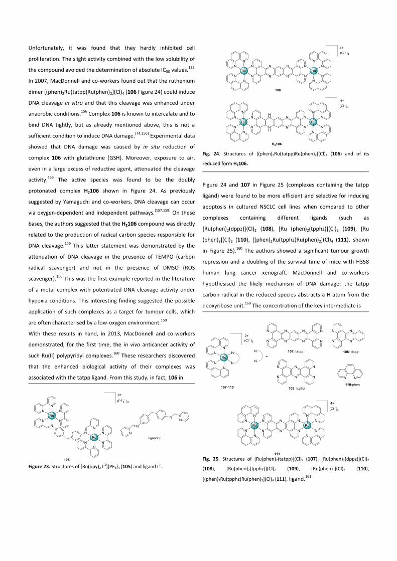
In 2007, MacDonnell and co-workers found out that the ruthenium
dimer [(phen)2Ru(tatpp)Ru(phen)2](Cl)4 (106 Figure 24) could induce
DNA cleavage in vitro and that this cleavage was enhanced under
anaerobic conditions.156
Complex 106 is known to intercalate and to
bind DNA tightly, but as already mentioned above, this is not a
sufficient condition to induce DNA damage.[74,156]
Experimental data
showed that DNA damage was caused by in situ reduction of
complex 106 with glutathione (GSH). Moreover, exposure to air,
even in a large excess of reductive agent, attenuated the cleavage
activity.156
The active species was found to be the doubly
protonated complex H2106 shown in Figure 24. As previously
suggested by Yamaguchi and co-workers, DNA cleavage can occur
via oxygen-dependent and independent pathways.[157,158]
On these
bases, the authors suggested that the H2106 compound was directly
related to the production of radical carbon species responsible for
DNA cleavage.156
This latter statement was demonstrated by the
attenuation of DNA cleavage in the presence of TEMPO (carbon
radical scavenger) and not in the presence of DMSO (ROS
scavenger).156
This was the first example reported in the literature
of a metal complex with potentiated DNA cleavage activity under
hypoxia conditions. This interesting finding suggested the possible
application of such complexes as a target for tumour cells, which
are often characterised by a low-oxygen environment.159
With these results in hand, in 2013, MacDonnell and co-workers
demonstrated, for the first time, the in vivo anticancer activity of
such Ru(II) polypyridyl complexes.160
These researchers discovered
that the enhanced biological activity of their complexes was
associated with the tatpp ligand. From this study, in fact, 106 in
Figure 23. Structures of [Ru(bpy)2 L1](PF6)4 (105) and ligand L’.
Fig. 24. Structures of [(phen)2Ru(tatpp)Ru(phen)2](Cl)4 (106) and of its
reduced form H2106.
Figure 24 and 107 in Figure 25 (complexes containing the tatpp
ligand) were found to be more efficient and selective for inducing
apoptosis in cultured NSCLC cell lines when compared to other
complexes containing different ligands (such as
[Ru(phen)2(dppz)](Cl)2 (108), [Ru (phen)2(tpphz)](Cl)2 (109), [Ru
(phen)3](Cl)2 (110), [(phen)2Ru(tpphz)Ru(phen)2](Cl)4 (111), shown
in Figure 25).160
The authors showed a significant tumour growth
repression and a doubling of the survival time of mice with H358
human lung cancer xenograft. MacDonnell and co-workers
hypothesised the likely mechanism of DNA damage: the tatpp
carbon radical in the reduced species abstracts a H-atom from the
deoxyribose unit.160
The concentration of the key intermediate is
Fig. 25. Structures of [Ru(phen)2(tatpp)](Cl)2 (107), [Ru(phen)2(dppz)](Cl)2
(108), [Ru(phen)2(tpphz)](Cl)2 (109), [Ru(phen)3](Cl)2 (110),
[(phen)2Ru(tpphz)Ru(phen)2](Cl)4 (111). ligand.
161
dependent on the O2 and reducing agent concentrations. Moreover,
since tatpp can be reduced again, the concentration of the key
carbon radical species is controlled by the O2 and reducing agent
concentrations. Interestingly, complexes 106 and 107 showed
significant selectivity against malignant cells. This observation could
be explained by the differences in transport mechanisms.160
Complexes 108, 110, 111 were generally shown to enter cells via
passive diffusion,[162,163]
while it seems reasonable to assume that
complexes 106 and 107 enter cells via active transport as already
demonstrated for Ru(II) polypyridyl complexes containing the tpphz
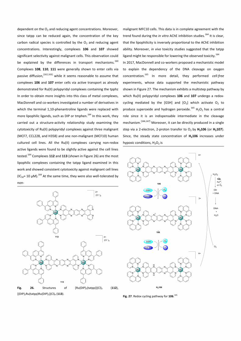
In order to obtain more insights into this class of metal complexes,
MacDonnell and co-workers investigated a number of derivatives in
which the terminal 1,10-phenantroline ligands were replaced with
more lipophilic ligands, such as DIP or tmphen.164
In this work, they
carried out a structure-activity relationship study examining the
cytotoxicity of Ru(II) polypyridyl complexes against three malignant
(MCF7, CCL228, and H358) and one non-malignant (MCF10) human
cultured cell lines. All the Ru(II) complexes carrying non-redox
active ligands were found to be slightly active against the cell lines
tested.164
Complexes 112 and 113 (shown in Figure 26) are the most
lipophilic complexes containing the tatpp ligand examined in this
work and showed consistent cytotoxicity against malignant cell lines
(IC50≈ 10 µM).164
At the same time, they were also well-tolerated by
non-
Fig. 26. Structures of [Ru(DIP)2(tatpp)](Cl)2 (112),
[(DIP)2Ru(tatpp)Ru(DIP)2](Cl)4 (113).
malignant MFC10 cells. This data is in complete agreement with the
trend found during the in vitro AChE inhibition studies.164
It is clear,
that the lipophilicity is inversely proportional to the AChE inhibition
ability. Moreover, in vivo toxicity studies suggested that the tatpp
ligand might be responsible for lowering the observed toxicity.164
In 2017, MacDonnell and co-workers proposed a mechanistic model
to explain the dependency of the DNA cleavage on oxygen
concentration.165
In more detail, they performed cell-free
experiments, whose data supported the mechanistic pathway
shown in Figure 27. The mechanism exhibits a multistep pathway by
which Ru(II) polypyridyl complexes 106 and 107 undergo a redox-
cycling mediated by the [GSH] and [O2] which activate O2 to
produce superoxide and hydrogen peroxide.165
H2O2 has a central
role since it is an indispensable intermediate in the cleavage
mechanism.[166,167]
Moreover, it can be directly produced in a single
step via a 2-electron, 2-proton transfer to O2 by H2106 (or H2107).
Since, the steady state concentration of H2106 increases under
hypoxic conditions, H2O2 is
Fig. 27. Redox cycling pathway for 106.165
produced more efficiently and DNA cleavage is enhanced.165
Furthermore, the in vitro cytotoxicity of 106 and 107 was examined
against H358, HCC2998, HOP-62, Hs766t cell lines under normoxia
(18 % O2) and hypoxia (1.1 % O2). Unlike many O2 activating drugs
which are less effective under hypoxic conditions, 106 showed no
differences in cytotoxicity going from normoxia to hypoxia.165
Complex 107 is two-times more cytotoxic under hypoxic conditions
in HOP-62, Hs766t when compared to normoxia. From the results
discussed above, it seems that the role of ruthenium is mostly
structural, so it would be very interesting to investigate analogues
of these complexes containing different metals (e.g. osmium).
4.2 Dinuclear Ru(II) complexes targeting mitochondria
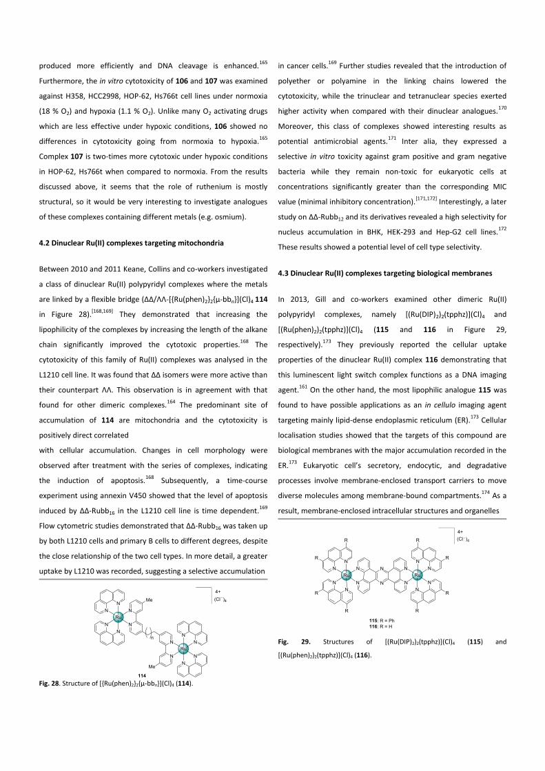
Between 2010 and 2011 Keane, Collins and co-workers investigated
a class of dinuclear Ru(II) polypyridyl complexes where the metals
are linked by a flexible bridge (ΔΔ/ΛΛ-[{Ru(phen)2}2{µ-bbn}](Cl)4 114
in Figure 28).[168,169]
They demonstrated that increasing the
lipophilicity of the complexes by increasing the length of the alkane
chain significantly improved the cytotoxic properties.168
The
cytotoxicity of this family of Ru(II) complexes was analysed in the
L1210 cell line. It was found that ΔΔ isomers were more active than
their counterpart ΛΛ. This observation is in agreement with that
found for other dimeric complexes.164
The predominant site of
accumulation of 114 are mitochondria and the cytotoxicity is
positively direct correlated
with cellular accumulation. Changes in cell morphology were
observed after treatment with the series of complexes, indicating
the induction of apoptosis.168
Subsequently, a time-course
experiment using annexin V450 showed that the level of apoptosis
induced by ∆∆-Rubb16 in the L1210 cell line is time dependent.169
Flow cytometric studies demonstrated that ∆∆-Rubb16 was taken up
by both L1210 cells and primary B cells to different degrees, despite
the close relationship of the two cell types. In more detail, a greater
uptake by L1210 was recorded, suggesting a selective accumulation
Fig. 28. Structure of [{Ru(phen)2}2{µ-bbn}](Cl)4 (114).
in cancer cells.169
Further studies revealed that the introduction of
polyether or polyamine in the linking chains lowered the
cytotoxicity, while the trinuclear and tetranuclear species exerted
higher activity when compared with their dinuclear analogues.170
Moreover, this class of complexes showed interesting results as
potential antimicrobial agents.171
Inter alia, they expressed a
selective in vitro toxicity against gram positive and gram negative
bacteria while they remain non-toxic for eukaryotic cells at
concentrations significantly greater than the corresponding MIC
value (minimal inhibitory concentration).[171,172]
Interestingly, a later
study on ∆∆-Rubb12 and its derivatives revealed a high selectivity for
nucleus accumulation in BHK, HEK-293 and Hep-G2 cell lines.172
These results showed a potential level of cell type selectivity.
4.3 Dinuclear Ru(II) complexes targeting biological membranes
In 2013, Gill and co-workers examined other dimeric Ru(II)
polypyridyl complexes, namely [(Ru(DIP)2)2(tpphz)](Cl)4 and
[(Ru(phen)2)2(tpphz)](Cl)4 (115 and 116 in Figure 29,
respectively).173
They previously reported the cellular uptake
properties of the dinuclear Ru(II) complex 116 demonstrating that
this luminescent light switch complex functions as a DNA imaging
agent.161
On the other hand, the most lipophilic analogue 115 was
found to have possible applications as an in cellulo imaging agent
targeting mainly lipid-dense endoplasmic reticulum (ER).173
Cellular
localisation studies showed that the targets of this compound are
biological membranes with the major accumulation recorded in the
ER.173
Eukaryotic cell’s secretory, endocytic, and degradative
processes involve membrane-enclosed transport carriers to move
diverse molecules among membrane-bound compartments.174
As a
result, membrane-enclosed intracellular structures and organelles
Fig. 29. Structures of [(Ru(DIP)2)2(tpphz)](Cl)4 (115) and
[(Ru(phen)2)2(tpphz)](Cl)4 (116).
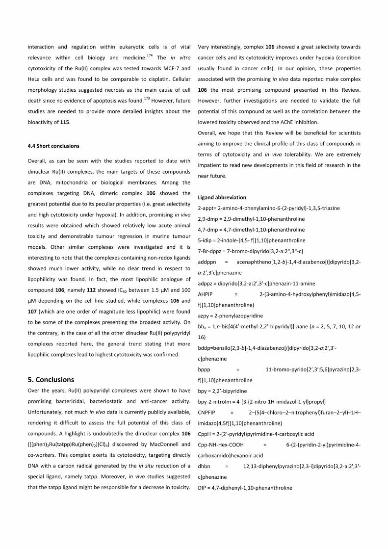
interaction and regulation within eukaryotic cells is of vital
relevance within cell biology and medicine.174
The in vitro
cytotoxicity of the Ru(II) complex was tested towards MCF-7 and
HeLa cells and was found to be comparable to cisplatin. Cellular
morphology studies suggested necrosis as the main cause of cell
death since no evidence of apoptosis was found.173
However, future
studies are needed to provide more detailed insights about the
bioactivity of 115.
4.4 Short conclusions
Overall, as can be seen with the studies reported to date with
dinuclear Ru(II) complexes, the main targets of these compounds
are DNA, mitochondria or biological membranes. Among the
complexes targeting DNA, dimeric complex 106 showed the
greatest potential due to its peculiar properties (i.e. great selectivity
and high cytotoxicity under hypoxia). In addition, promising in vivo
results were obtained which showed relatively low acute animal
toxicity and demonstrable tumour regression in murine tumour
models. Other similar complexes were investigated and it is
interesting to note that the complexes containing non-redox ligands
showed much lower activity, while no clear trend in respect to
lipophilicity was found. In fact, the most lipophilic analogue of
compound 106, namely 112 showed IC50 between 1.5 µM and 100
µM depending on the cell line studied, while complexes 106 and
107 (which are one order of magnitude less lipophilic) were found
to be some of the complexes presenting the broadest activity. On
the contrary, in the case of all the other dinuclear Ru(II) polypyridyl
complexes reported here, the general trend stating that more
lipophilic complexes lead to highest cytotoxicity was confirmed.
5. Conclusions
Over the years, Ru(II) polypyridyl complexes were shown to have
promising bactericidal, bacteriostatic and anti-cancer activity.
Unfortunately, not much in vivo data is currently publicly available,
rendering it difficult to assess the full potential of this class of
compounds. A highlight is undoubtedly the dinuclear complex 106
([(phen)2Ru(tatpp)Ru(phen)2](Cl)4) discovered by MacDonnell and
co-workers. This complex exerts its cytotoxicity, targeting directly
DNA with a carbon radical generated by the in situ reduction of a
special ligand, namely tatpp. Moreover, in vivo studies suggested
that the tatpp ligand might be responsible for a decrease in toxicity.
Very interestingly, complex 106 showed a great selectivity towards
cancer cells and its cytotoxicity improves under hypoxia (condition
usually found in cancer cells). In our opinion, these properties
associated with the promising in vivo data reported make complex
106 the most promising compound presented in this Review.
However, further investigations are needed to validate the full
potential of this compound as well as the correlation between the
lowered toxicity observed and the AChE inhibition.
Overall, we hope that this Review will be beneficial for scientists
aiming to improve the clinical profile of this class of compounds in
terms of cytotoxicity and in vivo tolerability. We are extremely
impatient to read new developments in this field of research in the
near future.
Ligand abbreviation
2-appt= 2-amino-4-phenylamino-6-(2-pyridyl)-1,3,5-triazine
2,9-dmp = 2,9-dimethyl-1,10-phenanthroline
4,7-dmp = 4,7-dimethyl-1,10-phenanthroline
5-idip = 2-indole-[4,5- f][1,10]phenanthroline
7-Br-dppz = 7-bromo-dipyrido[3,2-a:2‟,3‟-c]
addppn = acenaphtheno[1,2-b]-1,4-diazabenzo[i]dipyrido[3,2-
a:2’,3’c]phenazine
adppz = dipyrido[3,2-a:2',3'-c]phenazin-11-amine
AHPIP = 2-(3-amino-4-hydroxylphenyl)imidazo[4,5-
f][1,10]phenanthroline)
azpy = 2-phenylazopyridine
bbn = 1,n-bis[4(4’-methyl-2,2’-bipyridyl)]-nane (n = 2, 5, 7, 10, 12 or
16)
bddp=benzilo[2,3-b]-1,4-diazabenzo[i]dipyrido[3,2-a:2′,3′-
c]phenazine
bppp = 11-bromo-pyrido[2′,3′:5,6]pyrazino[2,3-
f][1,10]phenanthroline
bpy = 2,2’-bipyridine
bpy-2-nitroIm = 4-[3-(2-nitro-1H-imidazol-1-yl)propyl]
CNPFIP = 2–(5(4–chloro–2–nitrophenyl)furan–2–yl)–1H–
imidazo[4,5f][1,10]phenanthroline)
CppH = 2-(2′-pyridyl)pyrimidine-4-carboxylic acid
Cpp-NH-Hex-COOH = 6-(2-(pyridin-2-yl)pyrimidine-4-
carboxamido)hexanoic acid
dhbn = 12,13-diphenylpyrazino[2,3-i]dipyrido[3,2-a:2',3'-
c]phenazine
DIP = 4,7-diphenyl-1,10-phenanthroline
dmab = 4,4’-bis(N,N-dimethylamino)-2,2’-bipyridine
dmb = 4,4’-dimethyl-2,2’-bipyridine
dmcb = 4,4’-dimethoxycarbonyl-2,2’-bipyridine
dmdppz = 5,8-dimethoxylpyrido[3,2-a:2′,3′-c]phenazine
dmob = 4,4’-dimethoxy-2,2’-bipyridine
dppz = dipyrido[3,2-a:2',3'-c]phenazine
DQTT = 12-(1,4-dihydroquinoxalin-6-yl)-4,5,9,14-
tetraazabenzo[b]triphenylene, dmb = 4,4'-dimethyl-2,2'-bipyridine
FTTP = 2-(3-fluoronaphthalen-2- yloxy)-1,4,8,9-tetraazatriphenylene
H2IIP = 2-(indole-3-yl)-imidazo[4,5-f][1,10]phenanthroline
Hphox = 2-(2’-hydroxyphenyl)oxazoline
ip = imidazole[4,5-f ][1,10]phenanthroline,
ipad = 2-(anthracene-9,10-dione-2-yl)imidazo[4,5-
f][1,10]phenanthroline)
NHPIP = 2-(3-nitro- 4- hydroxylphenyl)imidazo[4,5-
f][1,10]phenanthroline)
NMIP = 2'-(2"-nitro-3",4"-methylenedioxyphenyl)imidazo[4',5'-
f][1,10]-phenanthroline
Norharman = 9H-pyrido[3,4-b]indole
PAIDH= 2-Pyridyl-1H-anthra[1,2-d]imidazole-6,11-dione
pddppn: phenantheno[1,2-b]-1,4-diazabenzo[i]dipyrido[3,2-a:2',3'-
c]phenazine
phen = 1,10 phenantroline
pip = 2-phenylimidazo[4,5-f][1,10]phenanthroline)
psbr = 2-(5-bromothiophen-2-yl) imidazo[4,5-
f][1,10]phenanthroline
pscl = 2-(5-chlorothiophen-2-yl)imidazo[4,5-f][1,10]phenanthroline
ptip = (2-(5-phenylthiophen-2-yl)-1H- imidazo[4, 5-f][1,10
phenanthroline
PTTP = 2-phenoxy-1,4,8,9- tetraazatriphenylene
pztp = 3-(pyrazin-2-yl)-[1,2,4]triazino[5,6-f][1,10]phenanthroline
R2bpy = 5,5’-di[1-(triethylammonio)methyl]-2,2’-dipyridine
tatpp = 9,11,20,22-tetraazatetrapyrido[3,2-a:2′,3′-c:3′′,2′′-1:2′′′,3′′′-
n]pentacene
tBu2bpy = 4,4′-di-tert -butyl-2,2′-bipyridine
tFPIP = 2-(2-trifluoromethane phenyl)imidazole[4,5-
f][1,10]phenanthroline)
tip = 2-(thiophen-2-yl)-1H-imidazo[4,5-f][1,10]phenanthroline)
tmphen = 3,4,7,8,-tetramethyl-1,10-phenantroline
tpphz = tetrapyrido[3,2-a:2',3'-c:3'',2''-h:2''',3'''-j]phenazine
ttbpy = 4,4′-ditertiary butyl-2,2′-bipyridine
Cell lines explanation
4T1 = mouse breast cancer cell line
A2780 = human ovarian carcinoma cell line
A2780-CP70 = cisplatin-resistant ovarian carcinoma cell line
A375 = human melanoma cell line
A549 = human lung adenocarcinoma cell line
B = B lymphocytes cell line
BEL-7402 = human hepatocellular carcinoma cell line
BHK = baby hamster kidney cell line
BJAB = Burkitt-like lymphoma cell line
CCD-19Lu = human noncancerous normal lung fibroblasts cell line
Du145 = human prostate cancer cell line
EVSA-T = human breast carcinoma cell line
HBL100 = human breast cancer cell line
HCC2998 = human colon adenocarcinoma cell line
HaCaT = aneuploid immortal keratinocyte cell line
HeLa = human adenocarcinoma of the cervix cell line
HEK-293 = human embryonic kidney cell line
HepG-2 = human hepatocarcinoma cell line
HL-60 = human leucocytoma cell line
HOP-62 = human lung adenocarcinoma cell line
Hs766t = human pancreatic adenocarcinoma cell line
HT-29 = human colon cancer cell line
H226 = human squamous cell lung carcinoma cell line
H358 = human non-small cell lung cancer cell line
IGROV = human ovarian endometrioid adenocarcinoma cell line
LO2 = human hepatic cell lines
KB = human cervical adenocarcinoma cell line
KB-3-1 = human cervical adenocarcinoma cell line
KB-V-1 = human cervical adenocarcinoma cell line
MCF-7 = human breast carcinoma cell line
MDA-MB-231= human breast adenocarcinoma
MG-63 = human osteosarcoma cell line
MRC-5 = human lung fibroblasts
M19 = human mesothelioma cell line
NIH/3T3 = Mouse fibroblast
NSCLC = human non-small cell lung cancer cell line
PC12 = rat pheochromocytoma cell line
SGC-7901 = human gastric cancer cell line
SiHa = human cervical cancer cell line
U2OS = human osteosarcoma cell line
WIDR = human rectosigmoid adenocarcinoma cell line
Biographies
Anna Notaro
Anna Notaro (left) is from a small town near Naples (Italy). She
studied at the Università Degli Studi di Napoli “Federico II” were she
obtained her master degree in chemical sciences in 2016. During
her master thesis, she worked on carbohydrate chemistry under the
supervision of Dr. Annalisa Guaragna and Dr. Daniele D’Alonzo. Part
of this work was performed at the National University of Ireland
(Galway, Ireland) in the frame of an Erasmus for traineeship
program under the supervision of Prof. V. Paul Murphy. At the
beginning of 2017, Anna started a PhD thesis in the Gasser Group at
Chimie ParisTech, PSL Research University (France). She is currently
working on the preparation of novel Ru(II) polypyridyl complexes as
chemotherapeutic agents.
Gilles Gasser
Gilles Gasser (right) was born, raised and educated in Switzerland.
After a PhD thesis in supramolecular chemistry with Prof. Helen
Stoeckli-Evans (University of Neuchâtel, Switzerland), Gilles
performed two post-docs, first with the late Prof. Leone Spiccia
(Monash University, Australia) in bioinorganic chemisty and then as
an Alexander von Humboldt fellow with Prof. Nils Metzler Nolte
(Ruhr-University Bochum, Germany) in bioorganometallic
chemistry. Gilles then started his independent scientific career at
the University of Zurich in 2010 first as a Swiss National Science
Foundation (SNSF) Ambizione Fellow and then as a SNSF Assistant
Professor in 2011. In 2016, Gilles moved to Chimie ParisTech, PSL
Research University (France), thanks among other to a PSL Chair of
Excellence Program Grant. Gilles was the recipient of several
fellowships and awards including the Alferd Werner Award from the
Swiss Chemical Society, an ERC Consolidator Grant, the Thieme
Chemistry Journal Award and recently the Jucker Award for his
contribution to cancer research. Gilles’s research group aims at
utilizing the specific physico-chemical properties of metal
complexes in different areas of medicinal chemistry.
Acknowledgements
We thank Patrick Felder (Chimie ParisTech) for his essential
contribution to the graphics. This work was financially
supported by an ERC Consolidator Grant PhotoMedMet to
G.G. (GA 681679) and has received support under the program
Investissements d’Avenir launched by the French Government
and implemented by the ANR with the reference ANR-10-IDEX-
0001-02 PSL (G.G.).
Notes
The authors declare no competing financial interest. References 1 A. Jemal, F. Bray and J. Ferlay, CA. Cancer J. Clin., 2011, 61,
69–90.
2 L. A. Torre, R. L. Siegel, E. M. Ward and A. Jemal, Cancer
Epidemiol. Biomarkers Prev., 2015, 25, 16–27.
3 A. Urruticoechea, R. Alemany, J. Balart, A. Villanueva, F.
Viñals and G. Capellá, Curr. Pharm. Des., 2010, 16, 3–10.
4 J. Ferlay, H.-R. Shin, F. Bray, D. Forman, C. Mathers and D.
M. Parkin, Int. J. Cancer, 2010, 127, 2893–2917.
5 L. A. Torre, F. Bray, R. L. Siegel, J. Ferlay, J. Lortet-tieulent
and A. Jemal, CA Cancer J. Clin., 2015, 65, 87–108.
6 C. X. Zhang and S. J. Lippard, Curr. Opin. Chem. Biol., 2003,
7, 481–489.
7 E. Jamieson and S. Lippard, Chem. Rev., 1999, 99, 2467–
2498.
8 E. Wong and C. M. Giandomenico, Chem. Rev., 1999, 99,
2451–2466.
9 D. Lebwohl and R. Canetta, Eur. J. Cancer, 1998, 34, 1522–
1534.
10 E. Alessio, G. Mestroni, A. Bergamo and G. Sava, Curr. Top.
Med. Chem., 2004, 4, 1525–35.
11 V. Singh, G. K. Azad, A. Reddy M., S. Baranwal and R. S.
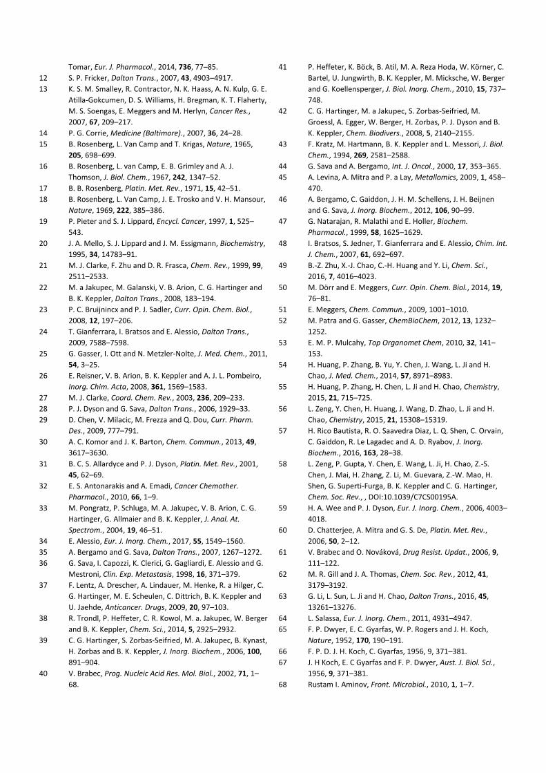
Tomar, Eur. J. Pharmacol., 2014, 736, 77–85.
12 S. P. Fricker, Dalton Trans., 2007, 43, 4903–4917.
13 K. S. M. Smalley, R. Contractor, N. K. Haass, A. N. Kulp, G. E.
Atilla-Gokcumen, D. S. Williams, H. Bregman, K. T. Flaherty,
M. S. Soengas, E. Meggers and M. Herlyn, Cancer Res.,
2007, 67, 209–217.
14 P. G. Corrie, Medicine (Baltimore)., 2007, 36, 24–28.
15 B. Rosenberg, L. Van Camp and T. Krigas, Nature, 1965,
205, 698–699.
16 B. Rosenberg, L. van Camp, E. B. Grimley and A. J.
Thomson, J. Biol. Chem., 1967, 242, 1347–52.
17 B. B. Rosenberg, Platin. Met. Rev., 1971, 15, 42–51.
18 B. Rosenberg, L. Van Camp, J. E. Trosko and V. H. Mansour,
Nature, 1969, 222, 385–386.
19 P. Pieter and S. J. Lippard, Encycl. Cancer, 1997, 1, 525–
543.
20 J. A. Mello, S. J. Lippard and J. M. Essigmann, Biochemistry,
1995, 34, 14783–91.
21 M. J. Clarke, F. Zhu and D. R. Frasca, Chem. Rev., 1999, 99,
2511–2533.
22 M. a Jakupec, M. Galanski, V. B. Arion, C. G. Hartinger and
B. K. Keppler, Dalton Trans., 2008, 183–194.
23 P. C. Bruijnincx and P. J. Sadler, Curr. Opin. Chem. Biol.,
2008, 12, 197–206.
24 T. Gianferrara, I. Bratsos and E. Alessio, Dalton Trans.,
2009, 7588–7598.
25 G. Gasser, I. Ott and N. Metzler-Nolte, J. Med. Chem., 2011,
54, 3–25.
26 E. Reisner, V. B. Arion, B. K. Keppler and A. J. L. Pombeiro,
Inorg. Chim. Acta, 2008, 361, 1569–1583.
27 M. J. Clarke, Coord. Chem. Rev., 2003, 236, 209–233.
28 P. J. Dyson and G. Sava, Dalton Trans., 2006, 1929–33.
29 D. Chen, V. Milacic, M. Frezza and Q. Dou, Curr. Pharm.
Des., 2009, 777–791.
30 A. C. Komor and J. K. Barton, Chem. Commun., 2013, 49,
3617–3630.
31 B. C. S. Allardyce and P. J. Dyson, Platin. Met. Rev., 2001,
45, 62–69.
32 E. S. Antonarakis and A. Emadi, Cancer Chemother.
Pharmacol., 2010, 66, 1–9.
33 M. Pongratz, P. Schluga, M. A. Jakupec, V. B. Arion, C. G.
Hartinger, G. Allmaier and B. K. Keppler, J. Anal. At.
Spectrom., 2004, 19, 46–51.
34 E. Alessio, Eur. J. Inorg. Chem., 2017, 55, 1549–1560.
35 A. Bergamo and G. Sava, Dalton Trans., 2007, 1267–1272.
36 G. Sava, I. Capozzi, K. Clerici, G. Gagliardi, E. Alessio and G.
Mestroni, Clin. Exp. Metastasis, 1998, 16, 371–379.
37 F. Lentz, A. Drescher, A. Lindauer, M. Henke, R. a Hilger, C.
G. Hartinger, M. E. Scheulen, C. Dittrich, B. K. Keppler and
U. Jaehde, Anticancer. Drugs, 2009, 20, 97–103.
38 R. Trondl, P. Heffeter, C. R. Kowol, M. a. Jakupec, W. Berger
and B. K. Keppler, Chem. Sci., 2014, 5, 2925–2932.
39 C. G. Hartinger, S. Zorbas-Seifried, M. A. Jakupec, B. Kynast,
H. Zorbas and B. K. Keppler, J. Inorg. Biochem., 2006, 100,
891–904.
40 V. Brabec, Prog. Nucleic Acid Res. Mol. Biol., 2002, 71, 1–
68.
41 P. Heffeter, K. Böck, B. Atil, M. A. Reza Hoda, W. Körner, C.
Bartel, U. Jungwirth, B. K. Keppler, M. Micksche, W. Berger
and G. Koellensperger, J. Biol. Inorg. Chem., 2010, 15, 737–
748.
42 C. G. Hartinger, M. a Jakupec, S. Zorbas-Seifried, M.
Groessl, A. Egger, W. Berger, H. Zorbas, P. J. Dyson and B.
K. Keppler, Chem. Biodivers., 2008, 5, 2140–2155.
43 F. Kratz, M. Hartmann, B. K. Keppler and L. Messori, J. Biol.
Chem., 1994, 269, 2581–2588.
44 G. Sava and A. Bergamo, Int. J. Oncol., 2000, 17, 353–365.
45 A. Levina, A. Mitra and P. a Lay, Metallomics, 2009, 1, 458–
470.
46 A. Bergamo, C. Gaiddon, J. H. M. Schellens, J. H. Beijnen
and G. Sava, J. Inorg. Biochem., 2012, 106, 90–99.
47 G. Natarajan, R. Malathi and E. Holler, Biochem.
Pharmacol., 1999, 58, 1625–1629.
48 I. Bratsos, S. Jedner, T. Gianferrara and E. Alessio, Chim. Int.
J. Chem., 2007, 61, 692–697.
49 B.-Z. Zhu, X.-J. Chao, C.-H. Huang and Y. Li, Chem. Sci.,
2016, 7, 4016–4023.
50 M. Dörr and E. Meggers, Curr. Opin. Chem. Biol., 2014, 19,
76–81.
51 E. Meggers, Chem. Commun., 2009, 1001–1010.
52 M. Patra and G. Gasser, ChemBioChem, 2012, 13, 1232–
1252.
53 E. M. P. Mulcahy, Top Organomet Chem, 2010, 32, 141–
153.
54 H. Huang, P. Zhang, B. Yu, Y. Chen, J. Wang, L. Ji and H.
Chao, J. Med. Chem., 2014, 57, 8971–8983.
55 H. Huang, P. Zhang, H. Chen, L. Ji and H. Chao, Chemistry,
2015, 21, 715–725.
56 L. Zeng, Y. Chen, H. Huang, J. Wang, D. Zhao, L. Ji and H.
Chao, Chemistry, 2015, 21, 15308–15319.
57 H. Rico Bautista, R. O. Saavedra Diaz, L. Q. Shen, C. Orvain,
C. Gaiddon, R. Le Lagadec and A. D. Ryabov, J. Inorg.
Biochem., 2016, 163, 28–38.
58 L. Zeng, P. Gupta, Y. Chen, E. Wang, L. Ji, H. Chao, Z.-S.
Chen, J. Mai, H. Zhang, Z. Li, M. Guevara, Z.-W. Mao, H.
Shen, G. Superti-Furga, B. K. Keppler and C. G. Hartinger,
Chem. Soc. Rev., , DOI:10.1039/C7CS00195A.
59 H. A. Wee and P. J. Dyson, Eur. J. Inorg. Chem., 2006, 4003–
4018.
60 D. Chatterjee, A. Mitra and G. S. De, Platin. Met. Rev.,
2006, 50, 2–12.
61 V. Brabec and O. Nováková, Drug Resist. Updat., 2006, 9,
111–122.
62 M. R. Gill and J. A. Thomas, Chem. Soc. Rev., 2012, 41,
3179–3192.
63 G. Li, L. Sun, L. Ji and H. Chao, Dalton Trans., 2016, 45,
13261–13276.
64 L. Salassa, Eur. J. Inorg. Chem., 2011, 4931–4947.
65 F. P. Dwyer, E. C. Gyarfas, W. P. Rogers and J. H. Koch,
Nature, 1952, 170, 190–191.
66 F. P. D. J. H. Koch, C. Gyarfas, 1956, 9, 371–381.
67 J. H Koch, E. C Gyarfas and F. P. Dwyer, Aust. J. Biol. Sci.,
1956, 9, 371–381.
68 Rustam I. Aminov, Front. Microbiol., 2010, 1, 1–7.
69 Y. Jenkins, A. E. Friedman, N. J. Turro and J. K. Barton,
Biochemistry, 1992, 31, 10809–10816.
70 J.-G. Liu, B.-H. Ye, H. Li, Q.-X. Zhen, L.-N. Ji and Y.-H. Fu, J.
Inorg. Biochem., 1999, 76, 265–271.
71 K. E. Erkkila, D. T. Odom and J. K. Barton, Chem. Rev., 1999,
99, 2777–2796.
72 Y. Xiong, X. He, X. Zou, J. Wu, X. Chen, L. Ji, R. Li, J. Zhou
and K. Yu, J. Chem. Soc. Dalt. Trans., 1999, 19–23.
73 L. N. Ji, X. H. Zou and J. G. Liu, Coord. Chem. Rev., 2001,
216–217, 513–536.
74 A. E. Friedman, J.-C. Chambron, J.-P. Sauvage, N. J. Turro
and J. K. Barton, J. Am. Chem. Soc., 1990, 112, 4960–4962.
75 E. J. C. Olson, D. Hu, a Ho, a M. Jonkman, M. R. Arkin, E. D.
a Stemp, J. K. Barton and P. F. Barbara, J. Am. Chem. Soc.,
1997, 7863, 11458–11467.
76 C. A. Puckett and J. K. Barton, J. Am. Chem. Soc., 2009, 131,
8738–8739.
77 J. K. Barton, E. D. Olmon and P. A. Sontz, Coord. Chem.
Rev., 2011, 255, 619–634.
78 H. Song, J. T. Kaiser and J. K. Barton, Nat. Chem., 2012, 4,
615–20.
79 C. R. Cardoso, M. V. S. Lima, J. Cheleski, E. J. Peterson, T.
Venâncio, N. P. Farrell and R. M. Carlos, J. Med. Chem.,
2014, 57, 4906–4915.
80 Z.-P. Zeng, Q. Wu, F.-Y. Sun, K.-D. Zheng and W.-J. Mei,
Inorg. Chem., 2016, 55, 5710–5718.
81 M. R. Gill, H. Derrat, C. G. W. Smythe, G. Battaglia and J. A.
Thomas, ChemBioChem, 2011, 12, 877–880.
82 M. Eriksson, M. Leijon, C. Hiort, B. Norden and A.
Graeslund, Biochemistry, 1994, 33, 5031–5040.
83 G. Yang, J. Z. Wu, L. Wang, L. N. Ji and X. Tian, J. Inorg.
Biochem., 1997, 66, 141–144.
84 J. P. Hall, P. M. Keane, H. Beer, K. Buchner, G. Winter, T. L.
Sorensen, D. J. Cardin, J. A. Brazier and C. J. Cardin, Nucleic
Acids Res., 2016, 44, 9472–9482.
85 J. Liu, L. F. Tan, L. H. Jin and F. Luan, DNA Cell Biol., 2012,
31, 250–258.
86 C. J. Cardin, J. M. Kelly and S. J. Quinn, Chem. Sci., 2017, 8,
4705–4723.
87 J. K. Barton, A. T. Danishefsky and J. M. Goldberg, J. Am.
Chem. Soc., 1984, 106, 2172–2176.
88 P. Lincoln, A. Broo and B. Norden, J. Am. Chem. Soc., 1996,
2644–2653.
89 J. Liu, X.-H. Zou, Q.-L. Zhang, W.-J. Mei, J.-Z. Liu and L.-N. Ji,
Met. Based Drugs, 2000, 7, 343–348.
90 A. C. G. Hotze, E. P. L. Van Der Geer, H. Kooijman, A. L.
Spek, J. G. Haasnoot and J. Reedijk, Eur. J. Inorg. Chem.,
2005, 2, 2648–2657.
91 D. Ma, C. Che, F. Siu, M. Yang and K. Wong, Inorg. Chem.,
2007, 46, 740–749.
92 P. Zhang, J. Chen and Y. Liang, Acta Biochim. Biophys. Sin.,
2010, 42, 440–449.
93 D. A. Lutterman, A. Chouai, Y. Liu, Y. Sun, C. D. Stewart, K.
R. Dunbar and C. Turro, J. Am. Chem. Soc., 2008, 130,
1163–1170.
94 J. F. Kou, C. Qian, J. Q. Wang, X. Chen, L. L. Wang, H. Chao
and L. N. Ji, J. Biol. Inorg. Chem., 2012, 17, 81–96.
95 K. J. Du, J. Q. Wang, J. F. Kou, G. Y. Li, L. L. Wang, H. Chao
and L. N. Ji, Eur. J. Med. Chem., 2011, 46, 1056–1065.
96 Y. C. Wang, C. Qian, Z. L. Peng, X. J. Hou, L. L. Wang, H.
Chao and L. N. Ji, J. Inorg. Biochem., 2014, 130, 15–27.
97 C. Wang, Q. Yu, L. Yang, Y. Liu, D. Sun, Y. Huang, Y. Zhou, Q.
Zhang and J. Liu, BioMetals, 2013, 26, 387–402.
98 Z. Zhang, Q. Wu, X. H. Wu, F. Y. Sun, L. M. Chen, J. C. Chen,
S. L. Yang and W. J. Mei, Eur. J. Med. Chem., 2014, 80, 316–
324.
99 P. Venkat Reddy, M. Rajender Reddy, S. Avudoddi, Y.
Praveen Kumar, C. Nagamani, N. Deepika, K.
Nagasuryaprasad, S. Satyanarayana Singh and S.
Satyanarayana, Anal. Biochem., 2015, 485, 49–58.
100 N. Deepika, C. Shobha Devi, Y. Praveen Kumar, K. Laxma
Reddy, P. Venkat Reddy, D. Anil Kumar, S. S. Singh and S.
Satyanarayana, J. Photochem. Photobiol. B Biol., 2016, 160,
142–153.
101 J. C. Wang, Annu. Rev. Biochem., 1996, 65, 635–692.
102 J. J. Champoux, Annu. Rev. Biochem., 2001, 70, 369–413.
103 J. C. Wang, Nat. Rev. Mol. Cell. Biol., 2002, 3, 430–440.
104 T. Simonsson, Biol. Chem., 2001, 382, 621–628.
105 L. H. Hurley, Biochem. Soc. Trans., 2001, 29, 692–696.
106 C. Metcalfe and J. a Thomas, Chem. Soc. Rev., 2003, 32,
215–224.
107 X. Chen, J.-H. Wu, Y.-W. Lai, R. Zhao, H. Chao and L.-N. Ji,
Dalton Trans., 2013, 42, 4386–4397.
108 Q. Yu, Y. Liu, C. Wang, D. Sun, X. Yang, Y. Liu and J. Liu, PLoS
One, 2012, 7, 1–13.
109 S. Shi, J. Xu, X. Gao, H. Huang and T. Yao, Chem. - A Eur. J.,
2015, 21, 11435–11445.
110 A. Srishailam, Y. P. Kumar, P. Venkat Reddy, N. Nambigari,
U. Vuruputuri, S. S. Singh and S. Satyanarayana, J.
Photochem. Photobiol. B Biol., 2014, 132, 111–123.
111 U. Schatzschneider, J. Niesel, I. Ott, R. Gust, H. Alborzinia
and S. Wölfl, ChemMedChem, 2008, 3, 1104–1109.
112 S. Schäfer, I. Ott, R. Gust and W. S. Sheldrick, Eur. J. Inorg.
Chem., 2007, 3034–3046.
113 F. Gao, X. Chen, J. Q. Wang, Y. Chen, H. Chao and L. N. Ji,
Inorg. Chem., 2009, 48, 5599–5601.
114 O. Zava, S. M. Zakeeruddin, C. Danelon, H. Vogel, M.
Grätzel and P. J. Dyson, ChemBioChem, 2009, 10, 1796–
1800.
115 T. Chen, Y. Liu, W. J. Zheng, J. Liu and Y. S. Wong, Inorg.
Chem., 2010, 49, 6366–6368.
116 C. Tan, S. Lai, S. Wu, S. Hu, L. Zhou, Y. Chen, M. Wang, Y.
Zhu, W. Lian, W. Peng, L. Ji and A. Xu, J. Med. Chem., 2010,
53, 7613–7624.
117 S. P. Mulcahy, K. Gründler, C. Frias, L. Wagner, A. Prokop
and E. Meggers, Dalton Trans., 2010, 39, 8177–82.
118 T. Chen, W.-J. Mei, Y.-S. Wong, J. Liu, Y. Liu, H.-S. Xie and
W.-J. Zheng, Med. Chem. Comm., 2010, 1, 73–75.
119 C. Tan, S. Wu, S. Lai, M. Wang, Y. Chen, L. Zhou, Y. Zhu, W.
Lian, W. Peng, L. Ji and A. Xu, Dalton Trans., 2011, 40,
8611–8621.
120 K. M. Debatin, Toxicol. Lett., 2000, 112–113, 41–48.
121 U. K. Mazumder, M. Gupta, S. S. Karki, S. Bhattacharya, S.
Rathinasamy and S. Thangavel, Chem. Pharm. Bull.
(Tokyo)., 2004, 52, 178–185.
122 S. S. Karki, S. Thota, S. Y. Darj, J. Balzarini and E. De Clercq,
J. Enzym. Inhib. Med. Chem., 2010, 25, 513–519.
123 L. C. Tu, C. S. Chen, I. C. Hsiao, J. W. Chern, C. H. Lin, Y. C.
Shen and S. F. Yeh, Chem. Biol., 2005, 12, 1317–1324.
124 V. Pierroz, T. Joshi, A. Leonidova, C. Mari, J. Schur, I. Ott, L.
Spiccia, S. Ferrari and G. Gasser, J. Am. Chem. Soc., 2012,
134, 20376–20387.
125 G.-B. Jiang, Y.-Y. Xie, G.-J. Lin, H.-L. Huang, Z.-H. Liang and
Y.-J. Liu, J. Photochem. Photobiol. B., 2013, 129, 48–56.
126 C. Qian, J.-Q. Wang, C.-L. Song, L.-L. Wang, L.-N. Ji and H.
Chao, Metallomics, 2013, 5, 844–854.
127 Y. Yuan, A. Y. Wang, A. L. Jiang, A. F. Gao, A. S. Liang, C. Su,
A. H. Chao and L. J. A, Aust. J. Chem., 2008, 61, 732–739.
128 Y. Y. Xie, Z. Z. Li, G. J. Lin, H. L. Huang, X. Z. Wang, Z. H.
Liang, G. Bin Jiang and Y. J. Liu, Inorganica Chim. Acta,
2013, 405, 228–234.
129 G. J. Lin, Z. Z. Li, J. H. Yao, H. L. Huang, Y. Y. Xie and Y. J. Liu,
Aust. J. Chem., 2013, 66, 555–563.
130 B.-J. Han, G.-B. Jiang, J. Wang, W. Li, H.-L. Huang and Y.-J.
Liu, RSC Adv., 2014, 4, 40899–40906.
131 W. Li, G.-B. Jiang, J.-H. Yao, X.-Z. Wang, J. Wang, B.-J. Han,
Y.-Y. Xie, G.-J. Lin, H.-L. Huang and Y.-J. Liu, J. Photochem.
Photobiol. B., 2014, 140, 94–104.
132 Y. Du, X. Fu, H. Li, B. Chen, Y. Guo, G. Su, H. Zhang, F. Ning,
Y. Lin, W. Mei and T. Chen, ChemMedChem, 2014, 9, 714–
718.
133 G. Bin Jiang, X. Zheng, J. H. Yao, B. J. Han, W. Li, J. Wang, H.
L. Huang and Y. J. Liu, J. Inorg. Biochem., 2014, 141, 170–
179.
134 G.-B. Jiang, J.-H. Yao, J. Wang, W. Li, B.-J. Han, Y.-Y. Xie, G.-
J. Lin, H.-L. Huang and Y.-J. Liu, New J. Chem., 2014, 38,
2554–2563.
135 Q. Yu, Y. Liu, L. Xu, C. Zheng, F. Le, X. Qin, Y. Liu and J. Liu,
Eur. J. Med. Chem., 2014, 82, 82–95.
136 O. Mazuryk, M. Maciuszek, G. Stochel, F. Suzenet and M.
Brindell, J. Inorg. Biochem., 2014, 134, 83–91.
137 O. Mazuryk, F. Suzenet, C. Kieda and M. Brindell,
Metallomics, 2015, 7, 553–566.
138 S. Kizaka-Kondoh and H. Konse-Nagasawa, Cancer Sci.,
2009, 100, 1366–1373.
139 C. Kieda, B. El Hafny-Rahbi, G. Collet, N. Lamerant-Fayel, C.
Grillon, A. Guichard, J. Dulak, A. Jozkowicz, J. Kotlinowski,
K. C. Fylaktakidou, A. Vidal, P. Auzeloux, E. Miot-Noirault, J.
C. Beloeil, J. M. Lehn and C. Nicolau, J. Mol. Med., 2013, 91,
883–899.
140 S. H. Lai, G. Bin Jiang, J. H. Yao, W. Li, B. J. Han, C. Zhang, C.
C. Zeng and Y. J. Liu, J. Inorg. Biochem., 2015, 152, 1–9.
141 C. Zhang, B.-J. Han, C.-C. Zeng, S.-H. Lai, W. Li, B. Tang, D.
Wan, G.-B. Jiang and Y.-J. Liu, J. Inorg. Biochem., 2016, 157,
62–72.
142 S. Liu, J. Zhu, H. Xu, Y. Wang, Y. Liu, J. Liang, G. Zhang, D.
Cao, Y. Lin, Y. Wu and Q. Guo, Spectrochim. Acta Part A
Mol. Biomol. Spectrosc., 2016, 161, 77–82.
143 C.-C. Zeng, S.-H. Lai, J.-H. Yao, C. Zhang, H. Yin, W. Li, B.-J.
Han and Y.-J. Liu, Eur. J. Med. Chem., 2016, 122, 118–126.
144 D. Wan, S.-H. Lai, H.-H. Yang, B. Tang, C. Zhang, H. Yin, C.-C.
Zeng and Y.-J. Liu, J. Photochem. Photobiol. B Biol., 2016,
161, 295–303.
145 D. Wan, S.-H. Lai, H.-H. Yang, B. Tang, C. Zhang, H. Yin, C.-C.
Zeng and Y.-J. Liu, J. Photochem. Photobiol. B Biol., 2016,
165, 246–255.
146 D. Wan, S.-H. Lai, C.-C. Zeng, C. Zhang, B. Tang and Y.-J. Liu,
J. Inorg. Biochem., 2017, 173, 1–11.
147 Z. Luo, L. Yu, F. Yang, Z. Zhao, B. Yu, H. Lai, K.-H. Wong, S.-
M. Ngai, W. Zheng and T. Chen, Metallomics, 2014, 6,
1480–1490.
148 I. Ott, X. Qian, Y. Xu, D. H. W. Vlecken, I. J. Marques, D.
Kubutat, J. Will, W. S. Sheldrick, P. Jesse, A. Prokop and C.
P. Bagowski, J. Med. Chem., 2009, 52, 763–770.
149 M. Yamada, A. Tomida, H. Yoshikawa, Y. Taketani and T.
Tsuruo, Clin. Cancer Res., 1996, 2, 427–432.
150 S. Prast-Nielsen, M. Cebula, I. Pader and E. S. J. Arnér, Free
Radic. Biol. Med., 2010, 49, 1765–1778.
151 A. Casini, C. Gabbiani, F. Sorrentino, M. P. Rigobello, A.
Bindoli, T. J. Geldbach, A. Marrone, N. Re, C. G. Hartinger,
P. J. Dyson and L. Messori, J. Med. Chem., 2008, 51, 6773–
6781.
152 L. Oehninger, M. Stefanopoulou, H. Alborzinia, J. Schur, S.
Ludewig, K. Namikawa, A. Muñoz-Castro, R. W. Köster, K.
Baumann, S. Wölfl, W. S. Sheldrick and I. Ott, Dalton
Trans., 2013, 42, 1657–66.
153 A. Meyer, C. P. Bagowski, M. Kokoschka, M. Stefanopoulou,
H. Alborzinia, S. Can, D. H. Vlecken, W. S. Sheldrick, S. Wölfl
and I. Ott, Angew. Chemie Int. Ed., 2012, 51, 8895–8899.
154 G. I. Pascu, A. C. G. Hotze, C. Sanchez-Cano, B. M. Kariuki
and M. J. Hannon, Angew. Chemie Int. Ed., 2007, 46, 4374–
4378.
155 U. Mcdonnell, J. M. C. A. Kerchoffs, R. P. M. Castineiras, M.
R. Hicks, A. C. G. Hotze, J. Hannon and A. Rodger, Dalton
Trans., 2008, 667–675.
156 T. K. Janaratne, A. Yadav, F. Ongeri and F. M. MacDonnell,
Inorg. Chem., 2007, 46, 3420–3422.
157 T. Yamaguchi, H. Nomura, K. Matsunaga, S. Ito, J. Takata
and Y. Karube, Biol. Pharm. Bull., 2003, 26, 1523–1527.
158 T. Yamaguchi, S. Matsumoto and K. Watanabe,
Tetrahedron Lett., 1998, 39, 8311–8312.
159 G. L. Semenza, J. Mol. Med., 2007, 85, 1301–1307.
160 A. Yadav, T. Janaratne, A. Krishnan, S. S. Singhal, S. Yadav,
A. S. Dayoub, D. L. Hawkins, S. Awasthi and F. M.
Macdonnell, Mol. Cancer Ther., 2013, 12, 643–653.
161 M. R. Gill, J. Garcia-Lara, S. J. Foster, C. Smythe, G. Battaglia
and J. a Thomas, Nat. Chem., 2009, 1, 662–667.
162 C. A. Puckett and J. K. Barton, J. Am. Chem. Soc., 2007, 129,
46–47.
163 C. A. Puckett and J. K. Barton, Biochemistry, 2008, 47,
11711–11716.
164 N. Alatrash, E. S. Narh, A. Yadav, M.-J. Kim, T. Janaratne, J.
Gabriel and F. M. MacDonnell, ChemMedChem, 2017, 12,
1055–1069.
165 C. A. Griffith, A. Dayoub, T. Janaratne, N. Alatrash, K.
Abayan, A. Mohamedi, Z. Breitbach, D. W. Armstrong and
F. M. MacDonnell, Chem. Sci., 2017, 8, 3726–3740.
166 R. Franco, M. I. Panayiotidis and J. A. Cidlowski, J. Biol.
Chem., 2007, 282, 30452–30465.
167 A. R. Giandomenico, G. E. Cerniglia, J. E. Biaglow, C. W.
Stevens and C. J. Koch, Free Radic. Biol. Med., 1997, 23,
426–434.
168 M. J. Pisani, D. K. Weber, K. Heimann, J. G. Collins and F. R.
Keene, Metallomics, 2010, 2, 357–432.
169 M. J. Pisani, P. D. Fromm, Y. Mulyana, R. J. Clarke, H.
Körner, K. Heimann, J. G. Collins and F. R. Keene,
ChemMedChem, 2011, 6, 848–858.
170 Y. Mulyana, D. K. Weber, D. P. Buck, C. A. Motti, J. G.
Collins and F. R. Keene, Dalton Trans., 2011, 40, 1510.
171 F. Li, Y. Mulyana, M. Feterl, J. M. Warner, J. G. Collins and
F. R. Keene, Dalton Trans., 2011, 40, 5032.
172 X. Li, A. K. Gorle, T. D. Ainsworth, K. Heimann, C. E.
Woodward, J. Grant Collins and F. Richard Keene, Dalt.
Trans., 2014, 44, 3594–3603.
173 M. R. Gill, D. Cecchin, M. G. Walker, R. S. Mulla, G.
Battaglia, C. Smythe and J. a Thomas, Chem. Sci., 2013, 4,
4512–4519.
174 J. Lippincott-Schwartz and R. D. Phair, Annu. Rev. Biophys.,
2010, 39, 559–578.