monomeric solution structure of the prototypical ‘c' chemokine lymphotactin † , ‡
TRANSCRIPT

Monomeric Solution Structure of the Prototypical ‘C’ Chemokine Lymphotactin†,‡
E. Sonay Kulogˇlu,§ Darrell R. McCaslin,| Moiz Kitabwalla,⊥ C. David Pauza,⊥ John L. Markley,# andBrian F. Volkman*,1
Department of Biochemistry, Biophysics Instrumentation Facility, and National Magnetic Resonance Facility at Madison,UniVersity of Wisconsin-Madison, 433 Babcock DriVe, Madison, Wisconsin 53706, Department of Biochemistry,
Medical College of Wisconsin, 8701 Watertown Plank Road, Milwaukee, Wisconsin 53226, and Institute of Human Virology,UniVersity of Maryland, 725 West Lombard Street, Baltimore, Maryland 21201
ReceiVed May 30, 2001; ReVised Manuscript ReceiVed July 23, 2001
ABSTRACT: Lymphotactin, the sole identified member of the C class of chemokines, specifically attractsT lymphocytes and natural killer cells. This 93-residue protein lacks 2 of the 4 conserved cysteine residuescharacteristic of the other 3 classes of chemokines and possesses an extended carboxyl terminus, whichis required for chemotactic activity. We have determined the three-dimensional solution structure ofrecombinant human lymphotactin by NMR spectroscopy. Under the conditions used for the structuredetermination, lymphotactin was predominantly monomeric; however, pulsed field gradient NMR self-diffusion measurements and analytical ultracentrifugation revealed evidence of dimer formation. Sequence-specific chemical shift assignments were determined through analysis of two- and three-dimensional NMRspectra of15N- and13C/15N-enriched protein samples. Input for the torsion angle dynamics calculationsused in determining the structure included 1258 unique NOE-derived distance constraints and 60 dihedralangle constraints obtained from chemical-shift-based searching of a protein conformational database. Theensemble of 20 structures chosen to represent the structure had backbone and heavy atom rms deviationsof 0.46( 0.11 and 1.02( 0.14 Å, respectively. The results revealed that human lymphotactin adopts theconserved chemokine fold, which is characterized by a three-stranded antiparallelâ-sheet and a C-terminalR-helix. Two regions are dynamically disordered as evidenced by1H and13C chemical shifts and{15N}-1H NOEs: residues 1-9 of the amino terminus and residues 69-93 of the C-terminal extension. Afunctional role for the C-terminal extension, which is unique to lymphotactin, remains to be elucidated.
Chemokines (chemotactic cytokines) are small, solubleproteins that play important roles in regulating leukocytetrafficking. They act primarily through high-affinity interac-tions with seven-transmembrane G-protein coupled receptors(1, 2). To stimulate migration of target cells, a gradient ofincreasing chemokine concentration is thought to arise
through the binding of chemokines to cell-surface glycos-aminoglycans (GAGs)1 (3, 4). These interactions may alsopromote chemokine oligomerization in some cases (5). Thegeneral degree to which GAG binding and chemokineaggregation are involved in receptor activation is unclear.A subset of chemokine receptors has been implicated as co-receptors for HIV entry into cells, and the cognate chemokineligands have been found to inhibit HIV infection (6-12).
On the basis of the number and relative positions of theirconserved cysteine residues, chemokines are divided into fourclasses: CXC, CC, C, and CX3C. Each class shows distinctspecificity for the general cell types that they chemoattract.CXC chemokines containing the ELR sequence motif specif-ically chemoattract neutrophils, whereas those without theELR motif are chemotactic for T and B cells. Among thecytokines, CC chemokines exhibit the widest range of targetcell specificity, although they act preferentially on mono-cytes. This class includes chemotactic effectors for mostleukocyte cell types except neutrophils (1). The single known
† The study was supported by NIH Grant R01 AI45843. For allmultidimensional NMR experiments, this study made use of theNational Magnetic Resonance Facility at Madison (NMRFAM), usingequipment purchased with funds from the University of Wisconsin,the NSF Biological Instrumentation Program (DMB-8415048), the NIHBiomedical Research Technology Program (RR02301), the NSFAcademic Research Instrumentation Program (BIR-9214394), the NIHShared Instrumentation Program (RR02781 and RR08438), and the U.S.Department of Agriculture. Sedimentation equilibrium data wereobtained at the University of Wisconsin-Madison Biophysics Instru-mentation Facility, which is supported by the University of Wisconsin-Madison and Grants BIR-9512577 (NSF) and S10 RR13790 (NIH).
‡ Coordinates for the minimized average structure and the family of20 structures have been deposited in the RCSB Protein Data Bank undercodes 1J9O and 1J8I, respectively. Chemical shifts have been depositedin the BioMagResBank under accession number 5042.
* To whom correspondence should be addressed. Phone: (414) 456-8400, fax: (414) 456-6510, email: [email protected].
§ Department of Biochemistry, University of Wisconsin-Madison.| Department of Biochemistry and Biophysics Instrumentation Facil-
ity, University of Wisconsin-Madison.⊥ Institute of Human Virology, University of Maryland.# Department of Biochemistry and National Magnetic Resonance
Facility at Madison, University of Wisconsin-Madison.1 Department of Biochemistry, Medical College of Wisconsin.
1 Abbreviations:E. coli, Escherichia coli; GAGs, glycosaminogly-cans; hLtn, human lymphotactin; IL8, interleukin 8; RANTES, regulatedupon activation, normal T-cell expressed and secreted; FRCD, fracta-lkine chemokine domain; IPTG, isopropylâ-D-thiogalactopyranoside;LB, Luria broth; Ltn, lymphotactin; PCR, polymerase chain reaction;PFG, pulsed field gradient; rmsd, root-mean-square deviation; TAD,torsion angle dynamics; EDTA, ethylenediaminetetraacetic acid; PMSF,phenylmethylsulfonyl fluoride; HIV, human immunodefficiency virus;NK cells, natural killer cells; DTT, dithiothreitol.
12486 Biochemistry2001,40, 12486-12496
10.1021/bi011106p CCC: $20.00 © 2001 American Chemical SocietyPublished on Web 09/27/2001

member of the CX3C class, fractalkine, has potent chemoat-tractant activities for T cells, monocytes, and natural killer(NK) cells (13).
The structures of more than 20 chemokine have beensolved (14-29); these reveal a highly conserved chemokinefold, which consists of a 3-stranded, antiparallelâ-sheet anda C-terminalR-helix. Most CXC and CC chemokines formdimers, but the dimer interfaces of these two classes aredistinctly different (30). Fractalkine and a few of the CXCand CC chemokines are monomeric (22, 26, 27, 29).
Lymphotactin (Ltn), the sole member of the C class ofchemokines, is specific for T lymphocytes and NK cells (31-33). In contrast to all other chemokines, Ltn contains only 1disulfide bond, and its sequence contains an extension ofabout 25 amino acid residues at the carboxyl terminus. TheC-terminal extension of Ltn is conserved across species(mouse, human, rat, and rhesus), as shown in Figure 1 (34),and truncation of the C-terminus by 21 residues abolishesthe Ca2+-flux and chemotaxis activities of hLtn (33, 35).
To provide a basis for understanding the functionalsignificance of its unique sequence elements, we haveinvestigated the structure and association state of humanlymphotactin (hLtn) by NMR spectroscopy and analyticalultracentrifugation. We present the complete nuclear mag-netic resonance assignments and solution structure of hLtn.Pulsed field gradient (PFG) self-diffusion and sedimentationequilibrium measurements showed that hLtn forms dimers.Like all other chemokines, hLtn adopts a fold consisting ofa three-strandedâ-sheet and C-terminalR-helix. The uniqueC-terminal extension is completely unstructured. Examinationof this structure reveals differences that are related to thedisulfide bridge that Ltn lacks compared to chemokines ofthe other classes.
MATERIALS AND METHODS
Plasmids, Enzymes, and Chemicals.Commercial sourcesfor reagents were the following: Fisher Scientific (Pittsburgh,PA) for HPLC-grade acetonitrile; Isotec (Miamisburg, OH)for 15NH4Cl and [13C]glucose; Novagen (Madison, WI) forplasmids (pET-3a, pET-9a, pET-16a, pET-22b), bacterialstrains [HMS174 and BL21(DE3)pLysS], and the factor Xacleavage and arrest kit; Promega (Madison, WI) for restric-tion enzymes (NdeI andBamHI) and T4 DNA ligase; Sigma-Aldrich (St. Louis, MO) for dithiothreitol and guanidiniumchloride; and Stratagene (La Jolla, CA) for Pfu DNA
polymerase. Oligonucleotides coding for the hLtn genesequence were chemically synthesized at the University ofUtah; shorter DNA oligonucleotides used as primers incloning reactions were synthesized at the University ofWisconsin-Madison Biotechnology Center.
Cloning.The GCG program [Wisconsin Package Version9.0, Genetics Computer Group (GCG), Madison, WI] wasused to design an artificial gene sequence, based on thepublished sequence for the mature hLtn gene (GenBankaccession no. U23772), optimized for expression inEscheri-chia coli (E. coli). This gene was synthesized as two, 150-base oligonucleotides with a central overlapping sequenceof 27 base pairs. PCR amplification with secondary primerswas used to generate the 300 base pair full-length hLtn genewith NdeI andBamHI sites at the 5′ and 3′ ends, respectively,to facilitate insertion of the gene into the pET vector system.The gene was subcloned into the various pET bacterialexpression vectors, either by itself or as a fusion with a (His)6
tag or signal peptidase sequence. All plasmids were verifiedby DNA sequencing. However, none of these plasmidsexpressed detectable amounts of hLtn (by SDS-PAGEanalysis). An alternative strategy, which proved successful,was to clone the synthetic hLtn gene into a pET-3a vectoras a fusion with staphylococcal nuclease (SNase) (36); afactor Xa cleavage site was inserted between the SNase andhLtn genes to provide a means for cleaving the SNase-hLtnfusion protein. The expression plasmid, which was calledpET-3a SNase-Xa-hLtn, was then transformed intoE. colistrain BL21(DE3)pLysS.
Protein Expression, Purification, and Refolding.Cells weregrown at 37 °C in LB medium containing 100µg/mLampicillin and 34µg/mL chloramphenicol until the celldensity reached OD600 ) 0.8-1.0. At that point, proteinexpression was induced by the addition of isopropylâ-D-thiogalactopyranoside (IPTG) to a final concentration of 1mM. Following induction, cells were grown for another 3-5h, harvested, and stored at-20 °C until processed further.For uniform labeling with15N and13C, cells were grown inM9 minimal media containing [13C]glucose (2 g/L) and [15N]-ammonium chloride (1 g/L) as the respective sole carbonand nitrogen sources.
Analysis of the cell lysate by SDS-PAGE on 16% Tris-glycine precast gels (Novex, San Diego, CA) showed thatmore than 80% of the fusion protein was expressed in theform of inclusion bodies. Initial attempts to purify hLtn from
FIGURE 1: Multiple sequence alignment for lymphotactins from various species. Sequences for human (GenBank accession no. AAC50164),rhesus (M. Kitabwalla and C. D. Pauza, unpublished results), mouse (AAA56752), rat (AAA69478), and chicken (AAB99904) were alignedwith the program GCG. The N-terminal signal sequence which is removed posttranslationally is shown in italics. Residue numbering is forthe mature extracellular species. Residues which are absolutely conserved across the five known species are in boldface type, and conservedbasic residues are underlined in the human sequence.
NMR Structure of Human Lymphotactin Biochemistry, Vol. 40, No. 42, 200112487

the soluble fraction were unsuccessful. The followingprocedure was used to isolate and purify the protein frominclusion bodies: Cell paste from a 1-2 L culture wassuspended in 50 mL of lysis buffer (50 mM Tris-HCl, pH8.0, 50 mM sodium chloride, 50 mM PMSF, 2 mM EDTA)and lysed by multiple freeze/thaw cycles. The SNase in thefusion protein was activated by the addition of CaCl2 to afinal concentration of 10 mM; this catalyzed the digestionof DNA and RNA in the lysate and reduced its viscosity.The lysate was then stirred at room temperature for about20 min or until no longer viscous. Inclusion bodies wereisolated by centrifugation at 20000g for 15 min in a JLA-16.250 rotor (Beckman, Palo Atlo, CA). The supernatant wasdecanted and discarded, and the pellet, which contained theinclusion bodies, was washed once with 50 mL of lysisbuffer, once with 50 mL of lysis buffer containing 0.5%Triton-X100, and again with 50 mL of lysis buffer. Theinclusion bodies were solubilized, and disulfide bonds werereduced by the addition of 30-50 mL of solubilization buffer(7 M guanidinium chloride, 40 mM Tris-HCl, pH 7.4, 20mM EDTA, 50 mM DTT) followed by stirring at roomtemperature for 2-3 h. The denaturant and reducing agentwere removed by dialysis against 12 L of 20 mM acetic acidsolution, repeated twice for 12 h. The resulting solution wasclarified by centrifugation at 20000g for 20 min. Thesupernatant was added dropwise into oxidation buffer (200mM Tris-acetate, pH 8.0, 25µM 2-mercaptoethanol). Toprevent aggregation due to intermolecular disulfide forma-tion, the protein concentration was kept below 0.3 mg/mL.Oxidation was carried out with stirring at 4°C for about 16h. The oxidized fusion protein was concentrated to∼10 mLin an Amicon (Beverly, MA) stirred-cell ultrafiltrationconcentrator using a 3000 Da molecular mass cutoff mem-brane (Millipore, Bedford, MA). To remove the oxidationbuffer, the concentrated fusion protein was washed 3 timeswith 50 mL of deionized water. An appropriate amount of10× cleavage buffer stock solution (0.5 M Tris-HCl, pH 8.0,1 M NaCl, 10 mM CaCl2) was then mixed with theconcentrated fusion protein solution. The factor Xa cleavagereaction was optimized by treating varying amounts of fusionprotein (5-10 mg/mL) with varying amounts of factor Xa(1-2 units). Although cleavage proceeded faster at higherconcentrations (2 units of factor Xa/10 mg fusion at 10 mg/mL concentration) at room temperature, lower overall yieldsof hLtn were obtained due to nonspecific digestion of fusionprotein. Subsequent cleavage reactions were carried out at4 °C using 1 unit of enzyme for 10 mg of fusion protein at5 mg/mL concentration for 1-2 days, or until the reactionwas nearly complete. The hLtn product was separated fromSNase by reversed-phase HPLC on a C18 column (Vydac,Hesperia, CA).
Sedimentation Equilibrium Studies.Sedimentation equi-librium studies were conducted in a Beckman XL-A Analyti-cal Ultracentrifuge. Samples with initial concentrations of235, 100, and 36µM were prepared by dissolving appropriateamounts of hLtn in a buffer consisting of 20 mM sodiumphosphate, 200 mM NaCl, pH 6.0. The samples were placedin separate Biodialyzers (Sialomed Inc., Columbia, MD) with1000 Da cutoff membrane, and dialyzed simultaneouslyagainst 500 mL of the buffer overnight at 4°C. Thecentrifuge cell centerpieces were double-sector charcoal-filledEpon (12 mm path length for the lowest concentration, 3
mm for the others), and dialysate was used in the referencesector. Equilibrium data were collected at 20 000, 26 000,36 000, and 42 000 rpm at 10°C. Concentration gradientswere monitored by the absorbance at 280 nm with super-imposable gradients recorded 2-4 h apart used as thecriterion that equilibrium had been reached. At 26 000 rpm,the gradient was monitored for more than 12 h afterequilibration and found to be stable. Integration of theconcentration gradients showed no significant loss of proteinduring the experiment. High-speed sedimentation was usedto deplete protein from the centrifuge cell and to measurethe amount of nonsedimenting absorbance in each cell(<0.006).
The molecular weight of hLtn was calculated from thesequence to be 10 254. The partial specific volume wascalculated to be 0.735 mL/g. The extinction coefficient wascalculated as 7115 M-1 cm-1 using the extinctions for Trp,Tyr, and cystine in Pace and Schmid (37). The solventdensity was measured at 10°C with an Anton PaarDMA5000 to be 1.01 g/mL.
Data were analyzed using programs written by Darrell R.McCaslin in the Igor Pro Data Analysis Program (Wavem-etrics Inc., Lake Oswego, OR). Prior to analysis, all datasets were corrected for the amount of nonsedimentingabsorbance measured after high-speed depletion of theprotein, and the data from the lowest concentration of hLtnwere normalized to a 3 mmpath length for global fittingpurposes. All data sets (three concentrations each at fourspeeds) were fit simultaneously to various models in amanner analogous to that discussed by Laue (38). Modelsincluded single species, two and three species in equilibrium,and two independent noninteracting species.
Diffusion Coefficient Measurements.A water-suppressedlongitudinal encode-decode (Water-SLED) experiment (39)was used for measurements of translational self-diffusioncoefficients at 600 MHz. All proteins were dissolved in 20mM sodium phosphate buffer, pH 6.0, containing 0.05%sodium azide and 200 mM sodium chloride. All measure-ments utilized a 30.5 G/cm Z-gradient and were carried outat 10°C. The gradient duration was incremented by 0.5 msfrom 0.5 to 6.5 ms. One percent cyclodextrin dissolved in90% H2O/10% D2O, which has a diffusion coefficient of3.239 × 106 cm2/s at 25°C (40), served as the standardsample for calibration of gradient strength. Self-diffusioncoefficients were obtained by nonlinear least-squares fits tothe following equation:
whereγ is the magnetogyric ratio of1H, G is the gradientstrength (G/cm),δ is the pulsed field gradient (PFG) duration(s), and∆ is the time between the gradient pulses.
NMR Spectroscopy.NMR experiments were carried outat the National Magnetic Resonance Facility at Madison(NMRFAM) on Bruker DMX600 and Bruker DMX750spectrometers equipped with three-axis gradient triple-resonance probes. All NMR samples were prepared eitherin 90% H2O/10% D2O or in 100% D2O containing 20 mMsodium phoshate (pH 6.0), 0.05% sodium azide, 200 mMsodium chloride. Samples for NMR spectroscopy contained0.5-1.3 mM hLtn and were placed in Shigemi (Tokyo,Japan) microcells. Heteronuclear NOEs were measured from
A(2τ) ) A(0) exp[(-γδG)2(∆ - δ/3)Ds] (1)
12488 Biochemistry, Vol. 40, No. 42, 2001 Kuloglu et al.

an interleaved pair of 2D15N-1H gradient sensitivity en-hanced correlation spectra of hLtn (41). NOEs were calcu-lated as the ratios of the peak heights in spectra recordedwith and without a 3 sproton saturation period. The totalrecycle delay was 5 s.
The following experiments were used for obtainingcomplete sequence-specific resonance assignments and theNOEs used in the structure calculations: 3D SE HNCA (42-44), 3D SE HN(CO)CA (44), 3D SE C(CO)NH (45), 3DSE HNCO (43, 44), 3D 15N NOESY-HSQC (46), 3D 15NTOCSY-HSQC (46), 3D HCCH-TOCSY (47), 2D 15NHSQC (46), 2D 13C constant-time SE HSQC (48), 2DNOESY, and 3D13C SE NOESY-HSQC. A pair of 2D13C-1H spin-echo difference CT-HSQC experiments (JCC andJNC) were used to obtain stereospecific assignments of ValCγ methyl groups (49, 50). All NOESY mixing times were80 ms. NMR data were processed with the NMRPipesoftware package (51) on personal computers running theLinux operating system. Chemical shifts were referenced tointernal DSS (taken as 0 ppm) as described by Markley etal. (52). The XEASY software package (53) was used forresonance assignments and analysis of NOE spectra. The CSIprogram was used for determining the secondary structurefrom the chemical shift index (54, 55).
Structure Calculation and Analysis.The simulated an-nealing protocol of the torsion angle dynamics programDYANA ( 56) was used in calculating all structures. Inpreliminary rounds of structure calculations, 8000 dynamicssteps were used in calculating each of 50 conformers; in thefinal round of calculations, 10 000 dynamics steps were used.NOE intensities were converted into upper distance boundswith the CALIBA function of DYANA. To account fordisparities in line widths and peak intensities, separate NOEcalibrations were performed for residues of the structuredand unstructured regions (residues 1-9 and 69-93). TheASSIGN function of DYANA was utilized for automatedassignment of NOEs on the basis of chemical shifts andinitial structures. Additional NOE constraints were added ineach round of calculations, and restraints that were consis-tently violated were removed. In addition to NOE restraints,a total of 60 φ and ψ dihedral angle constraints weregenerated from1HR, 13CR, 13Câ, 13C′, and15N secondary shiftsby the program TALOS and included in the later rounds ofrefinement (57). Of the final 50 structures calculated, the20 conformers with the lowest target function values wereselected for analysis. The mean structure, calculated fromthis ensemble of 20 structures in MOLMOL (58), wasminimized in DYANA using 300 steps of variable targetfunction minimization.
RESULTS
Protein Expression and Purification.Human lymphotactinwas produced successfully fromE. coli as a fusion proteinwith SNase. SDS-PAGE analysis indicated that more than80% of the fusion protein was localized in the insolublefraction. Protein isolated from inclusion bodies was purifiedas described above. The natural precursor form of lympho-tactin contains an N-terminal signal sequence, which isremoved in the mature secreted protein (32). A sequenceidentical to this mature 93 amino acid species (hLtn) wasproduced by proteolysis of the recombinant fusion protein
catalyzed by factor Xa. The hLtn product was separated fromintact fusion protein and SNase by reverse-phase HPLC. Theidentity of the purified protein (>95% pure) was confirmedby mass spectrometry. Biological activity was confirmed ina Ca2+-flux assay performed as described previously (datanot shown) (35). Typical yields from a 1 L culture rangedfrom 3 to 6 mg. Purified hLtn was lyophilized and dissolveddirectly in NMR buffer.
Choice of Conditions for Structural Analysis.Two-dimensional1H-15N HSQC spectra were acquired at varioustemperatures and ionic strengths to determine appropriateconditions for structure determination. These studies indi-cated that hLtn exhibits conformational heterogeneity at somecombinations of temperature and ionic strength. A similartemperature effect on NMR spectra of hLtn has been reportedby others (35). The1H-15N HSQC spectrum of hLtn in 200mM NaCl at pH 6.0 at 10°C (Figure 2) is well dispersed,and the number of resolved signals indicates that hLtn adoptsa single folded conformation under these conditions. Thesesolution conditions were therefore chosen for the structuredetermination.
Self-Association of hLtn.A plot of the logarithm ofabsorbance vs squared radial position would be linear for asingle species in sedimentation equilibrium. Plots for hLtnexhibited increasing slope with radial position requiring thepresence of two or more species. This was confirmed byglobal fitting of the data to a single species model. This fityielded a molecular weight of 12 950( 30, correspondingto neither the monomer nor the dimer species of hLtn.Moreover, the quality of the single species fit is poor,exhibiting systematic deviations with radial position. Anexample of a fit constrained to the monomer molecular
FIGURE 2: 2D 1H-15N HSQC spectrum of15N-hLtn at 10 °C.Backbone NH assignments are indicated by the one-letter aminoacid code and residue number. The inset box shows peaks for tworesidues with1H chemical shifts upfield of the others due to ring-current effects from the side chain of Trp55.
NMR Structure of Human Lymphotactin Biochemistry, Vol. 40, No. 42, 200112489

weight is shown in Figure 3. Using a model for a monomerto oligomer equilibrium and allowing the values for molec-ular weight, aggregation state, and equilibrium constant tovary, a mass consistent with the monomer was obtained aswell as an aggregation number near 2 (2.10( 0.08). A fitwith the aggregation number fixed at 2 resulted in amolecular weight of 10 550( 140 and an associationconstant of 660( 80 M-1. Fitted curves for this model areshown in Figure 3. Close examination of the fit residualsshowed some systematic deviations at high concentrations,but attempts to include an additional association step did notyield consistent results. It is possible that higher molecularweight species are formed at higher concentration, but thepresent study has not permitted their enumeration. Therefore,hLtn is best described as existing in a monomer-dimerequilibrium under these solution conditions. A final fit withthe aggregation number fixed at 2 and the mass constrainedto that calculated from the hLtn sequence (10 254 Da) gavean association constant for dimer formation of 850( 10M-1.
Self-Diffusion Measurements.The results of diffusionmeasurements on hLtn and two other proteins are sum-marized in Table 1. A series of 1D1H spectra with PFGpulses of increasing duration (0.5-6.5 ms) were collectedfor each sample. The diffusion delay between gradient pulseswas adjusted to obtain signal attenuation of at least 10-foldfor accurate fitting of the data to eq 1. Each diffusioncoefficient (Ds) in Table 1 reflects an average obtained fromfits of the intensities of at least four peaks in the proteinspectrum.
Values ofDs obtained for hLtn at 10°C in 200 mM NaClwere nearly constant over the concentration range 0.1-1.0
mM (data not shown). Comparison of the diffusion coef-ficients obtained for Ltn with those for ribonuclease A (13.7kDa) and ubiquitin (8.6 kDa) showed that theDs for hLtn isnearer to that of the larger protein.
Resonance Assignments.Sequential backbone assignmentswere deduced from HNCO, HNCA, HN(CO)CA, and15NNOESY-HSQC data. CCONH,15N TOCSY-HSQC, andHCCH-TOCSY spectra were used to assign aliphatic sidechain 13C and 1H resonances. Aromatic side chain protonresonances were assigned from the 2D NOESY spectrum ofa sample dissolved in 100% D2O. The existence of a disulfidebridge linking Cys11 and Cys48 was confirmed by analysisof the cysteine13CR and 13Câ chemical shifts, which areindicative of the oxidation states of cysteine pairs (59). Allchemical shift assignments have been deposited in theBioMagResBank (accession number 5042).
Secondary Structure Elements from CSI and NOE Data.Secondary structure elements were identified on the basisof NOE connectivities and the chemical shift index (CSI).The consensus CSI values calculated from experimental1HR,13CR, 13Câ, and13C′ chemical shifts, along with the summaryof sequential and medium-range NOEs, are shown in Figure4. The CSI identified fourâ-strands and oneR-helix.Interstrand NOEs connect three of theâ-strands (24-30, 36-41, 46-50) in an antiparallel sheet. AnR-helix from residues54 to 66 was confirmed by characteristici, i+3 NOEs (Figure4). No long-range NOEs were observed for the 9 N-terminaland 26 C-terminal residues.
The internal mobility of the polypeptide chain was probedby measuring{1H}15N NOE values for all backbone amides;these are shown as a function of residue number in Figure5A. A steady decline is seen in the heteronuclear NOE valuesfor the unstructured residues approaching the ends of theN- and C-termini with negative NOE values observed forresidues near each terminus; these values are consistent withlarge-amplitude motions on the picosecond to nanosecondtime scale and a complete lack of stable secondary or tertiarystructure. Residues connecting the first twoâ-strands (30’s
FIGURE 3: Sedimentation equilibrium gradient of hLtn. Absorbance data at 280 nm (open circles) are shown for each of the initialconcentrations used after equilibrium has been reached at 26 000 rpm and 10°C. Solid lines are from a global fit to all 12 sets of data usinga monomer-dimer equilibrium model resulting in a mass of 10 550 Da and an equilibrium constant of 660 M-1. The additional curve(monomer) is the result of fitting the data to a single species model where the mass was constrained to that calculated from the hLtnsequence.
Table 1: Self-Diffusion Coefficients for Various Proteins at 10°C
protein Mr Ds (×10-6 cm2/s) concn (mM) ∆ (ms)
hLtn 10254 0.78( 0.03 0.5 150ubiquitin 8565 1.09( 0.01 0.5 120ribonuclease A 13690 0.81( 0.01 0.5 150
12490 Biochemistry, Vol. 40, No. 42, 2001 Kuloglu et al.

loop) also exhibit low heteronuclear NOE values and fewNOE constraints.
Structure Determination.A total of 3270 NOE peaks wereassigned in 3 different NOESY spectra (15N NOESY-HSQC,13C NOESY-HSQC, and 2D NOESY); these yielded1258 unique, nontrivial distance constraints, which were usedin the structure calculations. In addition, two upper boundand two lower bound distance constraints served to definethe disulfide bond between Cys11 and Cys48. Backbonedihedral angle restraints (φ andψ) from TALOS were usedonly in regions of secondary structures and in the N-loop,not in terminal regions or in the loops connecting theâ-strands. Stereospecific assignments for valine Cγ methylgroups of residues 37, 56, 59, and 60 were obtained fromquantitativeJ-correlation experiments (JCC and JNC) andincluded in structure calculations. No other stereospecificassignments were determined. The distribution of distanceconstraints along the sequence is shown in Figure 5B.Structural statistics are summarized in Table 2. Although theaverage number of constraints per residue is 13.5 for theentire molecule, the structured region of hLtn (residues9-68) is defined by an average of 16.8 constraints perresidue. Superposition of residues 9-68 for the finalensemble of 20 TAD structures yielded a root-mean-squaredeviation (rmsd) of 0.46 and 1.02 Å from the mean structurefor backbone (N, CR, and, C′) and heavy atoms, respectively.Average backbone and heavy atom rmsd values relative to
the mean structure are shown as a function of residue numberin Figure 5C. The atomic rms deviations for terminal residuesare much higher, reaching 10.7 Å for residue Val1 and 57Å for Gly93. None of the 20 structures had NOE violationsgreater than 0.42 Å or van der Waals violations greater than0.22 Å (Table 2).
Figure 6 shows a superposition of the ensemble of 20structures for the well-ordered residues (9-68) of hLtn,together with a ribbon representation of the minimized meanstructure. The hLtn molecule adopts the domain foldobserved in all other chemokine structures, with a C-terminalR-helix packing against a three-stranded antiparallelâ-sheet.Residues 1-8 and 69-93 are highly disordered. Residues14-20 form the characteristic ‘N-loop’ lying above theâ-sheet. A helical turn connects the N-loop to strandâ1.Strandâ1 contains aâ-bulge involving residues 24 and 25.Strandsâ1 (24-30) andâ2 (36-41) are joined by the 30’sloop consisting of residues 31-35. A hydrogen bond isobserved between side chain Oγ1 of Thr30 and backboneHN of Ala36. A type I turn involving residues 41-44connectsâ2 andâ3 (46-50) with Leu45 bulged out. AnR-helix (residues 54-66) follows â3 and lies atop theâ-sheet, nearly parallel to the N-loop.
A number of hydrophobic interactions position the helixwith respect to theâ-sheet. Side chains of Ile24, Tyr27,Val37, and Phe39 from theâ-sheet and Trp55, Val56, Val60,and Met63 from the helix contribute to the hydrophobic core.
FIGURE 4: Summary of NOE connectivities and consensus chemical shift index for hLtn. Relative intensities of sequential NOEs areindicated by the bar thickness. Consensus CSI values are indicated by+1, 0, and-1 values for sheet, no prediction, and helix, respectively.Secondary structure elements deduced from CSI and NOE patterns are shown above the amino acid sequence. While the CSI predicts sheetfor residues 10-16, no interstrand NOEs were observed. Torsion angle constraints obtained from the program TALOS are also indicated.Circles, up triangles, and down triangles indicate torsion angle restraints in the random coil, helix, and sheet regions, respectively, of theRamachandran plot.
NMR Structure of Human Lymphotactin Biochemistry, Vol. 40, No. 42, 200112491

Residues of the N-loop also contribute to the core, includingLeu19 and Val21. In addition to NOEs between hydrophobicside chains, unusual ring-current effects on chemical shiftalso provide evidence of key tertiary interactions. Owing totheir proximity to the Trp55 side chain, the backbone1HN
resonances of Val56 and Leu19 are shifted upfield of thenormal range for backbone amide protons (Figure 2, inset).
DISCUSSION
Association State.Curvature in logarithmic plots of datafrom sedimentation equilibrium requires the presence of twoor more species in the sample undergoing centrifugation.hLtn exhibited such curvature, and analysis by direct, globalcurve-fitting to specific models results in the simplest modelbeing that of a monomer-dimer equilibrium. The bestestimate for the association constant was found to be 850(10 M-1.
The measured self-diffusion coefficient constant for hLtn,0.78× 10-6 cm2 s-1, is very close to that obtained for the
13.7 kDa protein ribonuclease A. Given aKa of 850 M-1, a0.5 mM solution of hLtn would contain 0.32 mM monomerand 0.09 mM dimer, with an average mass near that ofribonuclease A. The unstructured C-terminus of hLtn mayreduce the reliability of molecular weight estimates from self-diffusion measurements, but these results are generallyconsistent with the sedimentation analysis. Taken together,ultracentrifugation and NMR self-diffusion results indicatethat hLtn exists in a monomer-dimer equilibrium.
NMR Structure of hLtn.Most CXC and CC chemokinesform dimers, although with two very distinct mechanismsfor oligomerization. Sedimentation and self-diffusion resultsclearly indicated that hLtn is able to form dimers, with atypical 1 mM NMR sample predicted to contain up to 50%of the total protein in the dimeric form (∼0.53 mM monomer,∼0.23 mM dimer). Throughout the refinement process, wesearched unsuccessfully for NOEs that might identify a dimerinterface. Moreover, NMR spectra of hLtn acquired as afunction of protein concentration showed no shifting or
FIGURE 5: Evidence for dynamic disorder in the 30’s loop and N- and C-terminal residues. (A) Heteronuclear15N-1H NOE values areplotted as a function of amino acid sequence. Lower values are an indication of greater backbone mobility. (B) Distribution of NOEconstraints along the primary sequence (intraresidue, short, medium, and long range NOEs are indicated by white, light gray, dark gray,and black bars, respectively). (C) Average backbone (solid lines) and heavy atom (dashed lines) rmsd values for the family of 20 structuresto the mean structure. Residues of the N- and C-terminus reach maximal rmsd values of 10.7 and 57 Å, respectively.
12492 Biochemistry, Vol. 40, No. 42, 2001 Kuloglu et al.
broadening of peaks that might be taken as evidence ofexchange between monomer and dimer forms (data notshown). Only one set of resonances was observed in all thespectra, indicating that exchange between the monomer anddimer species is rapid on the NMR time scale. We concludethat the relatively low abundance and short lifetime of thedimer combine to prevent detection of NOEs betweenmonomers. In the absence of specific information about thedimer interface, the NMR structure of hLtn was refined asa monomer. Whether hLtn forms a dimer analogous to eitherthe typical CXC or the CC type remains to be determined.While unusual sequence features (missing disulfide, extendedC-terminus) distinguish lymphotactin from the other classes,it adopts the same features of secondary and tertiary structureseen in all chemokines to date. Despite their role inlymphotactin function, residues of the unique C-terminalextension are dynamically disordered (Figure 5). While theabsence of unique structural elements fails to explain thefunctional role of the hLtn C-terminal tail, it is still reasonableto assume that these residues, along with the mobile 30’s
loop and N-terminus, can play a role in receptor binding oractivation, as discussed below.
Glycosaminoglycan Binding.Virtually all chemokines,including lymphotactin, bind glycosaminoglycans such asheparin, and this interaction is thought to be important informing chemokine concentration gradients directing cellmigration to the site of injury or infection (3-5). A number
Table 2: Structural Statistics for hLtn Structures
NOE constraints number
long 286medium 184short 387intraresidue 401constraints/residue 13.5
Ramachandran statistics(residues 9-68)
most favored 76.8%additionally allowed 19.6%generously allowed 3.3%disallowed 0.3%
parameter family minimized average
target function (Å2) 0.70( 0.23 0.29upper limit violations
number>0.1 Å 9( 3 0sum of violations (Å) 4.5( 0.8 1.9maximum violation (Å) 0.42 0.13
van der Waals violationsnumber>0.2 Å 0( 0 0sum of violations (Å) 2.5( 0.5 1.7maximum violation (Å) 0.22 0.11
atomic rmsd’s (residues 9-68) (Å)family of 20 structures vs mean
minimizedaverage
backbone 0.46( 0.11 0.53( 0.14heavy atom 1.02( 0.14 1.21( 0.20
FIGURE 6: NMR structure of hLtn. (A) The ensemble of 20 TADconformers with lowest target function, from a total of 50 structures,is superimposed on the mean structure using backbone atoms ofresidues 9-68. (B) The minimized average structure of hLtn isshown in a ribbon representation. The disulfide bridge is shown inyellow, â-strands in green, and helices in red.
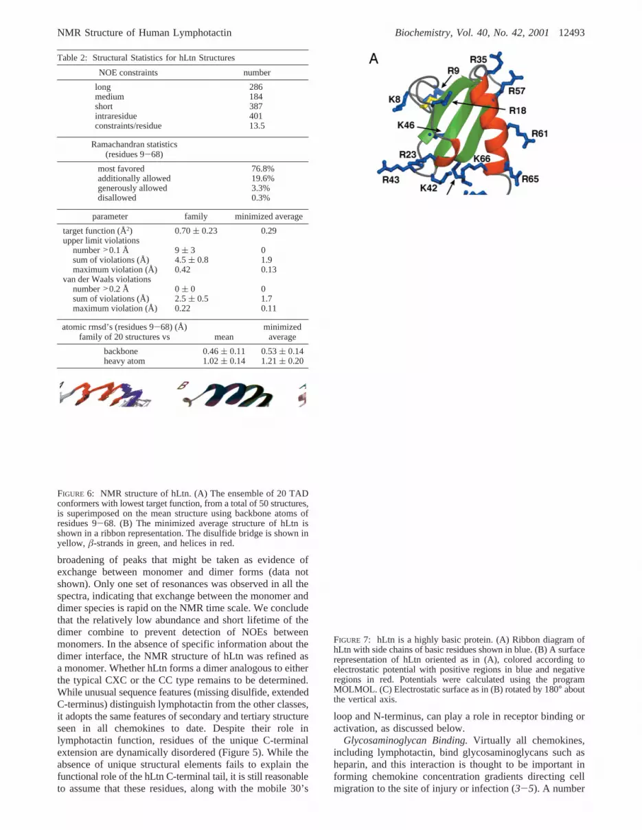
FIGURE 7: hLtn is a highly basic protein. (A) Ribbon diagram ofhLtn with side chains of basic residues shown in blue. (B) A surfacerepresentation of hLtn oriented as in (A), colored according toelectrostatic potential with positive regions in blue and negativeregions in red. Potentials were calculated using the programMOLMOL. (C) Electrostatic surface as in (B) rotated by 180° aboutthe vertical axis.
NMR Structure of Human Lymphotactin Biochemistry, Vol. 40, No. 42, 200112493

of different heparin binding residues have been identified inchemokines involving basic residues in the N-terminal loop,strandâ1, the C-terminal helix, and, most commonly, the40’s loop connectingâ2 andâ3 (29, 60-63). Ltn is a verybasic protein, with an estimated isoelectric point of 10.6,and it binds to a heparin-Sepharose column with a highaffinity (4). The electrostatic surface map of hLtn shown inFigure 7 reveals several clusters of positively chargedresidues that could participate in GAG-hLtn interactions.Two basic residues in the 40’s loop (Lys42 and Arg43) areproximal to Arg23, Lys25, Lys 66, and Arg70; these createa large positively charged surface area (Figure 7A,B).Another cluster is located on the solvent-exposed surface oftheR-helix. Three equally spaced arginine residues (Arg57,Arg61, and Arg65) of theR-helix together with Arg35 fromthe 30’s loop make another possible binding site for GAGs.
Comparison of hLtn with Other Chemokines.The struc-tures of all known chemokines share the same conservedchemokine fold. We have compared hLtn, the sole exampleof the C class, with representative members from each ofthe other chemokine classes (CXC, CC, and CX3C). Figure8 shows the sequence alignment and superposition of thebackbone of the structured regions of hLtn and IL-8 (CXC),RANTES (CC), and fractalkine (CX3C). Despite its lack ofone of the two conserved disulfides, hLtn adopts the samefolded structure as the other proteins (Figure 8B). Thesimilarity of hLtn to the CC and CX3C chemokines issomewhat greater than to this CXC chemokine in terms ofboth sequence and structure. hLtn shares 37.5%, 31.9%, and20% sequence identity with RANTES, fractalkine, and IL8,
respectively (Figure 8A). Backbone rms deviations from hLtnfor structured regions of RANTES, fractalkine, and IL8,excluding the N-terminal residues and 30’s loop, werecalculated to be 0.9, 0.9, and 1.3 Å, respectively. Residuesof the N-terminus preceding the first cysteine are highlymobile in hLtn as well as in other chemokine structures (22,27, 29). The three-stranded, antiparallelâ-sheet andR-helixsuperimpose very closely in all four structures, with onlyminor differences in the lengths of the strands and the helices.Positioning of the N-loop and 310 helical turn relative to thesheet are also similar (Figure 8B).
Significant structural differences between hLtn and theother chemokines arise from the absence of a second disulfideand a shortened 30’s loop in hLtn and are localized to theseregions (Figure 8C). Rotation of theø1 angle of Cys11 by∼150° in hLtn relative to the analogous residue in IL-8,RANTES, and fractalkine results in a completely differentorientation for this disulfide. As a result, the backboneconformation for residues Cys11-Leu14 of hLtn divergesfrom the other three structures, which are quite similar toeach other. Interestingly, these residues are predicted by theconsensus CSI to form aâ-strand, though no evidence forthis was found in the NOESY data. In all other chemokines,the 30’s loop is tethered to the N-terminal region by anotherdisulfide bond. This interaction is completely absent in hLtnowing to the missing disulfide. Increased backbone flexibilityfor these residues, as detected by{1H}15N heteronuclear NOEmeasurements, is not unique to hLtn, however, as the 30’sloop is known to be highly mobile in a number of otherchemokines (20, 64).
FIGURE 8: Comparison of hLtn structure with IL-8, RANTES, and FRCD. (A) Sequence alignment of hLtn with human interleukin 8(IL-8), human RANTES, and human fractalkine chemokine domain (FRCD). Sequences were aligned based on structural alignments obtainedwith the program SwissPDBViewer. (B) Structural overlay for hLtn (green), IL-8 (yellow), RANTES (red), and FRCD (blue). All disulfidesare shown with sulfur atoms in yellow. Residue ranges and backbone rmsds for the superimpositions were as follows: hLtn(13-15,18-32,35-64), IL-8(11-13,16-30,37-66) 1.3 Å; hLtn(14-15,18-29,34-62), RANTES(14-15,18-29,36-64) 0.9 Å; hLtn(13-15,18-28,-35-65), FRCD(14-16,18-28,37-67) 0.9 Å. (C) Detailed view of the 30’s loop and N-terminal cysteines, with structures colored andsuperimposed as in (B).
12494 Biochemistry, Vol. 40, No. 42, 2001 Kuloglu et al.

The positioning of the C-terminal helix relative to the sheetalso varies throughout the chemokine family. Hydrophobicinteractions of the helix and sheet in hLtn are similar to thosein CC chemokines and fractalkine. This results from con-servation in hLtn of apolar residues that play key roles inthe hydrophobic core of CC chemokines (65), includingLeu19, Tyr27, Val37, Phe39, Trp55, Val60, and Met63.
Receptor Binding and the Functional Role of C-TerminalExtension.Structural characterization of the mode of actionof chemokines upon their cognate G-protein-coupled recep-tors (GPCR) is hampered by the insoluble nature of theseintegral membrane proteins. Other investigators have studiedthe interactions of chemokines and peptides derived fromthe N-terminal extracellular domain of the correspondingGPCR. Residues of the unstructured N-terminus, N-loop, and30’s loop as well as residues adjacent to the conserveddisulfide motifs and in theR-helix have been implicated invarious receptor-derived peptide-chemokine interactions (22,66, 67). The unique C-terminal tail of Ltn has been shownto be necessary for its function by several groups (33, 35).It is clear from the structure presented here that theC-terminal extension of hLtn is highly dynamic and doesnot adopt any stable conformation in solution. Since theydo not play a key structural role, it seems likely that residuesof the C-terminus are involved in direct interactions withthe receptor. However, our current studies do not predictwhich residues participate in binding or the relevant con-formation.
As mentioned above, the 30’s loop, often implicated inreceptor-chemokine interactions, is decoupled from theN-terminal region of hLtn and is shorter by at least tworesidues in hLtn than in other chemokines (Figure 8C). It ispossible that the 30’s loop in Ltn interacts differently withits receptor than the corresponding loops in other chemokinesdo with their cognate receptors. In a commonly suggestedtwo-part mechanism for GPCR signaling by chemokines,binding to the receptor N-terminus is followed by activationthrough interactions with the mobile loops and termini ofthe chemokine. Specific receptor-activating interactionsprovided by the 30’s loop in other classes of chemokinesmight be augmented in hLtn by interactions with unstructuredterminal residues.
Summary.Lymphotactin is an unusual member of thechemokine family owing to its single disulfide and extendedC-terminal sequence (Figure 1). A synthetic gene coding forhLtn was constructed and used for expression of recombinantprotein. We concluded from analytical ultracentrifugation andNMR self-diffusion measurements that at the conditionsstudied (10°C, 200 mM NaCl, 20 mM phosphate buffer,pH 6.0) hLtn exists in a monomer-dimer equilibrium withan association constant of approximately 850 M-1 (Table 1,Figure 3). All1H, 15N, and13C chemical shifts were assigned(Figure 2), and the solution structure was refined using NOEdistance constraints and dihedral angle constraints generatedwith the program TALOS (Table 2). Residues comprisingthe unique C-terminal sequence of hLtn are entirely disor-dered in solution (Figures 4 and 5), but residues 9-68 adoptthe conserved fold observed for all other chemokines (Figure6). Our studies provide a structural basis for furtherinvestigations into the binding reactions of hLtn with cell-surface GAGs (Figure 7) and its specific G-protein-coupledreceptor, XCR1 (Figure 8).
ACKNOWLEDGMENT
We thank Dr. Ed Mooberry for assistance with NMRinstrumentation and gratefully acknowledge Dr. Steve L.Alam and Ed Meenen for synthesis of DNA oligonucleotides.
REFERENCES
1. Gale, L. M., and McColl, S. R. (1999)BioEssays 21, 17-28.2. Murphy, P. M., Baggiolini, M., Charo, I. F., Hebert, C. A.,
Horuk, R., Matsushima, K., Miller, L. H., Oppenheim, J. J.,and Power, C. A. (2000)Pharmacol. ReV. 52, 145-176.
3. McFadden, G., and Kelvin, D. (1997)Biochem. Pharmacol.54, 1271-1280.
4. Kuschert, G. S. V., Coulin, F., Power, C. A., Proudfoot, A. E.I., Hubbard, R. E., Hoogewerf, A. J., and Wells, T. N. C.(1999)Biochemistry 38, 12959-12968.
5. Hoogewerf, A. J., Kuschert, G. S. V., Proudfoot, A. E., Borlat,F., Clark-Lewis, I., Power, C. A., and Wells, T. N. C. (1997)Biochemistry 36, 13570-13578.
6. Cocchi, F., DeVico, A. L., Garzinio-Demo, A., Arya, S. K.,and Gallo, R. C. (1995)Science 270, 1811.
7. Feng, Y., Broder, C. C., Kennedy, P. E., and Berger, E. A.(1996)Science 272, 872-877.
8. Bates, P. (1996)Cell 86, 1-3.9. D’Souza, M. P., and Harden, V. A. (1996)Nat. Med. 2, 1293-
1300.10. He, J., Chen, Y., Farzan, M., Choe, H., Ohagen, A., Gartner,
S., Busciglio, J., Yang, X., Hofmann, W., Newman, W.,Mackay, C., Sodroski, J., and Gabuzda, D. (1997)Nature 384,645-649.
11. Greco, G., Mackewicz, C., and Levy, J. A. (1999)J. Gen.Virol. 80, 2369-2373.
12. Berger, E. A., Murphy, P. M., and Farber, J. M. (1999)Annu.ReV. Immunol. 17, 657-700.
13. Bazan, J. F., Bacon, K. B., Hardiman, G., Wang, W., Rossi,D., Greaves, D. R., Zlotnik, A., and Schall, T. J. (1997)Nature385, 640-644.
14. Clore, G. M., Appella, E., Yamada, M., Matsushima, K., andGronenborn, A. M. (1990)Biochemistry 29, 1689-1696.
15. St. Charles, R., Walz, D. A., and Edwards, B. F. P. (1989)J.Biol. Chem. 264, 2092-2099.
16. Lodi, P. J., Garrett, D. S., Kuszewski, J., Tsang, M. L.-S.,Weatherbee, J. A., Leonard, W. J., Gronenborn, A. M., andClore, G. M. (1994)Science 263, 1762-1767.
17. Kim, K. S., Clark-Lewis, I., and Sykes, B. D. (1994)J. Biol.Chem. 269, 32909-32915.
18. Chung, C.-W., Cooke, R. M., Proudfoot, A. E. I., and Wells,T. N. C. (1995)Biochemistry 34, 9307-9314.
19. Handel, T. M., and Domaille, P. J. (1996)Biochemistry 35,6569-6584.
20. Young, H., Roongta, V., Daly, T. J., and Mayo, K. H. (1999)Biochem. J. 338, 591-598.
21. Meunier, S., Bernassau, J. M., Guillemot, J. C., Ferrara, P.,and Darbon, H. (1997)Biochemistry 36, 4412-4422.
22. Crump, M. P., Gong, J.-H., Loetscher, P., Rajarathnam, K.,Amara, A., Arenzana-Seisdedos, F., Virelizier, J.-L., Baggio-lini, M., Sykes, B. D., and Clark-Lewis, I. (1997)EMBO J.16, 6996-7007.
23. Crump, M. P., Rajarathnam, K., Kim, K.-S., Clark-Lewis, I.,and Sykes, B. D. (1998)J. Biol. Chem. 273, 22471-22479.
24. LiWang, A. C., Wang, Z.-X., Sun, Y., Peiper, S. C., andLiWang, P. J. (1999)Protein Sci. 8, 2270-2280.
25. Qian, Y. Q., Johanson, K. O., and McDevitt, P. (1999)J. Mol.Biol. 294, 1065-1072.
26. Sticht, H., Escher, S. E., Schweimer, K., Forssmann, W.-G.,Rosch, P., and Aderman, K. (1999)Biochemistry 38, 5995-6002.
27. Mizoue, L. S., Bazan, J. F., Johnson, E. C., and Handel, T.M. (1999)Biochemistry 38, 1402-1414.
28. Mayer, K. L., and Stone, M. J. (2000)Biochemistry 39, 8382-8395.
29. Rajarathnam, K., Li, Y., Rohrer, T., and Gentz, R. (2001)J.Biol. Chem. 276, 4909-4916.
NMR Structure of Human Lymphotactin Biochemistry, Vol. 40, No. 42, 200112495

30. Clore, G. M., and Gronenborn, A. M. (1995)FASEB J. 9,57-62.
31. Kelner, G., Kennedy, J., Bacon, K., Kleyensteuber, S.,Largaespada, D., Jenkins, N., Copeland, N., Bazan, J., Moore,K., Schall, T., and Zlotnik, A. (1994)Science 266, 1395-1399.
32. Kennedy, J., Kelner, G., Kleyensteuber, S., Schall, T., Weiss,M., Yssel, H., Schneider, P., Cocks, B., Bacon, K., and Zlotnik,A. (1995)J. Immunol. 155, 203-209.
33. Hedrick, J. A., Saylor, V., Figueroa, D., Mizoue, L., Xu, Y.,Menon, S., Abrams, J., Handel, T., and Zlotnik, A. (1997)J.Immunol. 158, 1533-1540.
34. Hedrick, J. A., and Zlotnik, A. (1997)Methods Enzymol. 287,206-215.
35. Marcaurelle, L. A., Mizoue, L. S., Wilken, J., Oldham, L.,Kent, S. B. H., Handel, T. M., and Bertozzi, C. R. (2001)Chem. Eur. J. 7, 1129-1132.
36. Hinck, A., Walkenhorst, W., Westler, W., Choe, S., andMarkley, J. (1993)Protein Eng. 6, 221-227.
37. Pace, N., and Schmid, F. X. (1997)Protein Structure: APractical Approach, IRL Press, Oxford.
38. Laue, T. M. (1995)Methods Enzymol. 259, 427-452.39. Altieri, A. S., Hinton, D. P., and Byrd, R. A. (1995)J. Am.
Chem. Soc. 117, 7566-7567.40. Uedaira, H., and Uedaira, H. (1970)J. Phys. Chem. 74, 2211-
2214.41. Farrow, N. A., Muhandiram, R., Singer, A. U., Pascal, S. M.,
Kay, C. M., Gish, G., Shoelson, S. E., Pawson, T., Forman-Kay, J. D., and Kay, L. E. (1994)Biochemistry 33, 5984-6003.
42. Kay, L. E., Xu, G. Y., and Yamazaki, T. (1994)J. Magn.Reson., Ser. A 109, 129-133.
43. Muhandiram, D. R., and Kay, L. E. (1994)J. Magn. Reson.,Ser. B 103, 203-216.
44. Grzesiek, S., and Bax, A. (1992)J. Magn. Reson. 96, 432-440.
45. Grzesiek, S., Anglister, J., and Bax, A. (1993)J. Magn. Reson.,Ser. B 101, 114-119.
46. Zhang, O., Kay, L. E., Olivier, J. P., and Forman-Kay, J. D.(1994)J. Biomol. NMR 4, 845-858.
47. Kay, L. E., Xu, G.-Y., Singer, A. U., Muhandiram, D. R., andForman-Kay, J. D. (1993)J. Magn. Reson., Ser. B 101, 333-337.
48. Santoro, J., and King, G. C. (1992)J. Magn. Reson. 97, 202-207.
49. Grzesiek, S., Vuister, G. W., and Bax, A. (1993)J. Biomol.NMR 3, 487-493.
50. Vuister, G. W., Wang, A. C., and Bax, A. (1993)J. Am. Chem.Soc. 115, 5334-5335.
51. Delaglio, F., Grzesiek, S., Vuister, G. W., Zhu, G., Pfeifer, J.,and Bax, A. (1995)J. Biomol. NMR 6, 277-293.
52. Markley, J., Bax, A., Arata, Y., Hilbers, C., Kaptein, R., Sykes,B., Wright, P., and Wu¨thrich, K. (1998)Eur. J. Biochem. 256,1-15.
53. Bartels, C. H., Xia, T. H., Billeter, M., Gu¨nter, P., andWuthrich, K. (1995)J. Biomol. NMR 5, 1-10.
54. Wishart, D. S., and Sykes, B. D. (1994)J. Biomol. NMR 4,171-180.
55. Wishart, D. S., Sykes, B. D., and Richards, F. M. (1992)Biochemistry 31, 1647-1651.
56. Guntert, P., Mumenthaler, C., and Wu¨thrich, K. (1997)J. Mol.Biol. 273, 283-298.
57. Cornilescu, G., Delaglio, F., and Bax, A. (1999)J. Biomol.NMR 13, 289-302.
58. Koradi, R., Billeter, M., and Wuthrich, K. (1996)J. Mol.Graph. 14, 51-55.
59. Sharma, D., and Rajarathnam, K. (2000)J. Biomol. NMR 18,165-171.
60. Koopman, W., and Krangel, M. S. (1997)J. Biol. Chem. 272,10103-10109.
61. Amara, A., Lorthioir, O., Valenzuela, A., Magerus, A., Thelen,M., Montes, M., Virelizier, J. L., Delepierre, M., Baleux, F.,Lortat-Jacob, H., and Arenzana-Seisdedos, F. (1999)J. Biol.Chem. 274, 23916-23925.
62. Proudfoot, A. E. I., Fritchley, S., Borlat, F., Shaw, J. P.,Vilbois, F., Zwahlen, C., Trkola, A., Marchant, D., Clapham,P. R., and Wells, T. N. C. (2001)J. Biol. Chem. 276, 10620-10626.
63. Laurence, J. S., Blanpain, C., Leener, A. D., Parmentier, M.,and LiWang, P. J. (2001)Biochemistry 40, 4990-4999.
64. Ye, J., Mayer, K. L., and Stone, M. J. (1999)J. Biomol. NMR15, 115-124.
65. Fernandez, E. J., Wilken, J., Thompson, D. A., Peiper, S. C.,and Lolis, E. (2000)Biochemistry 39, 12837-12844.
66. Clubb, R. T., Omichinski, J. G., Clore, G. M., and Gronenborn,A. M. (1994) FEBS Lett. 338, 93-97.
67. Laurence, J. S., Blanpain, C., Burgner, J. W., Parmentier, M.,and LiWang, P. J. (2000)Biochemistry 39, 3401-3409.
BI011106P
12496 Biochemistry, Vol. 40, No. 42, 2001 Kuloglu et al.