the human cxcr3 chemokine system in early differentiated
TRANSCRIPT

Aus dem Institut für Medizinische Immunologie
der Medizinischen Fakultät Charité – Universitätsmedizin Berlin
DISSERTATION
The human CXCR3 chemokine system in early differentiated CD8+ T cell function and solid tumors
Das humane CXCR3 Chemokinsystem in der früh differenzierten
CD8+ T-Zellfunktion und in soliden Tumoren
zur Erlangung des akademischen Grades
Medical Doctor - Doctor of Philosophy (MD/PhD)
vorgelegt der Medizinischen Fakultät Charité – Universitätsmedizin Berlin
von
Tino Vollmer
aus Freiburg im Breisgau
Datum der Promotion: 04.03.2022

Tino Vollmer The human CXCR3 chemokine system in early differentiated CD8+ T cell function and solid tumors
1
Für meine Eltern.
Tino Vollmer The human CXCR3 chemokine system in early differentiated CD8+ T cell function and solid tumors
2
1 Abstracts ................................................................................................................... 5
1.1 Zusammenfassung (Deutsch) ............................................................................... 5 1.2 Abstract (English) .................................................................................................. 7 2 Current State of Immune Research on CD8+ T cells in Cancer ................................ 9 2.1 The immunoediting concept describes the immune-cancer cell interplay ............. 9 2.2 T cell infiltration as prognostic and predictive variable for solid cancer patients . 10 2.3 Cell migration and differentiation state shape anti-tumor memory CD8+ T cell
function ................................................................................................................ 12 2.4 Bladder cancer as a solid tumor disease modulated by T cell responses ........... 15 2.5 Hypotheses and aims of this thesis ..................................................................... 17 3 In-depth Methods .................................................................................................... 18
3.1 Isolation of lymphocytes from human blood and lymph nodes ............................ 18 3.2 Flow cytometry .................................................................................................... 18 3.3 Fluorescently activated cell sorting (FACS) ........................................................ 19 3.4 Functional T cell assays ...................................................................................... 19 3.5 Real time-quantitative polymerase chain reaction (RT-qPCR) ............................ 20 3.6 Patients included in this study ............................................................................. 21 3.7 Analysis of the inflammatory tumor microenvironment ........................................ 22 3.8 Second bladder cancer cohort and single-cell analysis of the CXCR3
chemokine components ...................................................................................... 23 4 Essential Results .................................................................................................... 24
4.1 Identification and characterization of early differentiated CD8+ T cells in healthy humans and bladder cancer patients ...................................................... 25
4.2 Functional investigation of the CXCR3 chemokine system in human early differentiated CD8+ T cells ................................................................................... 26
4.3 Characterization of intratumoral T cell infiltration and the CXCR3 chemokine system in bladder cancer .................................................................................... 28
4.4 Second biomarker testing, identification of the cellular origin of intratumoral CXCR3/CXCL11 and analysis of early differentiated CD8+ T cell states in MIBC, melanoma and renal cell carcinoma ......................................................... 29
4.5 Establishment steps towards CD8+CXCR3high TSCM based T cell therapy .......... 29 5 Clinical Applications and Outlook ........................................................................... 34 5.1 Human analysis unveils a stimulatory role of the CXCR3 chemokine system in
early differentiated CD8+ T cell function and solid tumors ................................... 34 5.2 Biomarker validation in bladder cancer and transferability to other solid tumor
diseases .............................................................................................................. 35 5.3 Therapeutic modulation of the tumor microenvironment by CXCR3-based
approaches .......................................................................................................... 37

Tino Vollmer The human CXCR3 chemokine system in early differentiated CD8+ T cell function and solid tumors
3
6 References ............................................................................................................. 39
7 Statutory Declaration .............................................................................................. 52 8 Detailed Description of Own Achievements ............................................................ 53
9 Journal Summary List ............................................................................................. 55 10 Publication .............................................................................................................. 56
11 Curriculum Vitae ..................................................................................................... 73 12 Complete List of Publications ................................................................................. 76
13 Danksagung ............................................................................................................ 77
Tino Vollmer The human CXCR3 chemokine system in early differentiated CD8+ T cell function and solid tumors
4
This MD/PhD thesis presents immunological and clinical literature, the applied methods,
the gained results and putative clinical applications (diagnostics and therapeutics) based
on the publication “The intra-tumoral CXCR3 chemokine system is predictive of chemotherapy response in human bladder cancer” published in Science Translational
Medicine (2021). In addition, first establishment steps for an adoptive T cell therapy
approach were developed, which exploit early differentiated CD8+ T cell characteristics
studied in this thesis.
Tino Vollmer The human CXCR3 chemokine system in early differentiated CD8+ T cell function and solid tumors
5
1 Abstracts
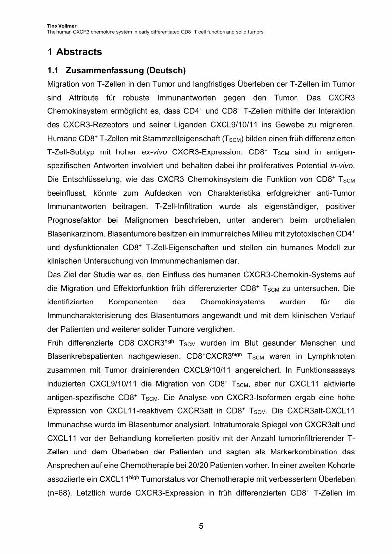
1.1 Zusammenfassung (Deutsch) Migration von T-Zellen in den Tumor und langfristiges Überleben der T-Zellen im Tumor
sind Attribute für robuste Immunantworten gegen den Tumor. Das CXCR3
Chemokinsystem ermöglicht es, dass CD4+ und CD8+ T-Zellen mithilfe der Interaktion
des CXCR3-Rezeptors und seiner Liganden CXCL9/10/11 ins Gewebe zu migrieren.
Humane CD8+ T-Zellen mit Stammzelleigenschaft (TSCM) bilden einen früh differenzierten
T-Zell-Subtyp mit hoher ex-vivo CXCR3-Expression. CD8+ TSCM sind in antigen-
spezifischen Antworten involviert und behalten dabei ihr proliferatives Potential in-vivo.
Die Entschlüsselung, wie das CXCR3 Chemokinsystem die Funktion von CD8+ TSCM
beeinflusst, könnte zum Aufdecken von Charakteristika erfolgreicher anti-Tumor
Immunantworten beitragen. T-Zell-Infiltration wurde als eigenständiger, positiver
Prognosefaktor bei Malignomen beschrieben, unter anderem beim urothelialen
Blasenkarzinom. Blasentumore besitzen ein immunreiches Milieu mit zytotoxischen CD4+
und dysfunktionalen CD8+ T-Zell-Eigenschaften und stellen ein humanes Modell zur
klinischen Untersuchung von Immunmechanismen dar.
Das Ziel der Studie war es, den Einfluss des humanen CXCR3-Chemokin-Systems auf
die Migration und Effektorfunktion früh differenzierter CD8+ TSCM zu untersuchen. Die
identifizierten Komponenten des Chemokinsystems wurden für die
Immuncharakterisierung des Blasentumors angewandt und mit dem klinischen Verlauf
der Patienten und weiterer solider Tumore verglichen.
Früh differenzierte CD8+CXCR3high TSCM wurden im Blut gesunder Menschen und
Blasenkrebspatienten nachgewiesen. CD8+CXCR3high TSCM waren in Lymphknoten
zusammen mit Tumor drainierenden CXCL9/10/11 angereichert. In Funktionsassays
induzierten CXCL9/10/11 die Migration von CD8+ TSCM, aber nur CXCL11 aktivierte
antigen-spezifische CD8+ TSCM. Die Analyse von CXCR3-Isoformen ergab eine hohe
Expression von CXCL11-reaktivem CXCR3alt in CD8+ TSCM. Die CXCR3alt-CXCL11
Immunachse wurde im Blasentumor analysiert. Intratumorale Spiegel von CXCR3alt und
CXCL11 vor der Behandlung korrelierten positiv mit der Anzahl tumorinfiltrierender T-
Zellen und dem Überleben der Patienten und sagten als Markerkombination das
Ansprechen auf eine Chemotherapie bei 20/20 Patienten vorher. In einer zweiten Kohorte
assoziierte ein CXCL11high Tumorstatus vor Chemotherapie mit verbessertem Überleben
(n=68). Letztlich wurde CXCR3-Expression in früh differenzierten CD8+ T-Zellen im

Tino Vollmer The human CXCR3 chemokine system in early differentiated CD8+ T cell function and solid tumors
6
Blasentumor, Melanom und Nierenzellkarzinom bestätigt und erste Schritte für eine
CD8+CXCR3high TSCM-basierte adoptive T-Zell-Therapie entwickelt.
Zusammenfassend zeigen die experimentellen Daten, dass das CXCR3
Chemokinsystem die Migration und die Aktivierung früh differenzierter CD8+ T-Zellen
moduliert. Die klinischen Daten implizieren, dass das CXCR3 Chemokinsystem das
Ansprechen auf eine Chemotherapie beim Blasenkrebs prädiziert und früh differenzierte
CD8+CXCR3high T-Zellen in soliden Tumoren eine protektive Relevanz besitzen, was
neue therapeutische Optionen eröffnet.
Tino Vollmer The human CXCR3 chemokine system in early differentiated CD8+ T cell function and solid tumors
7
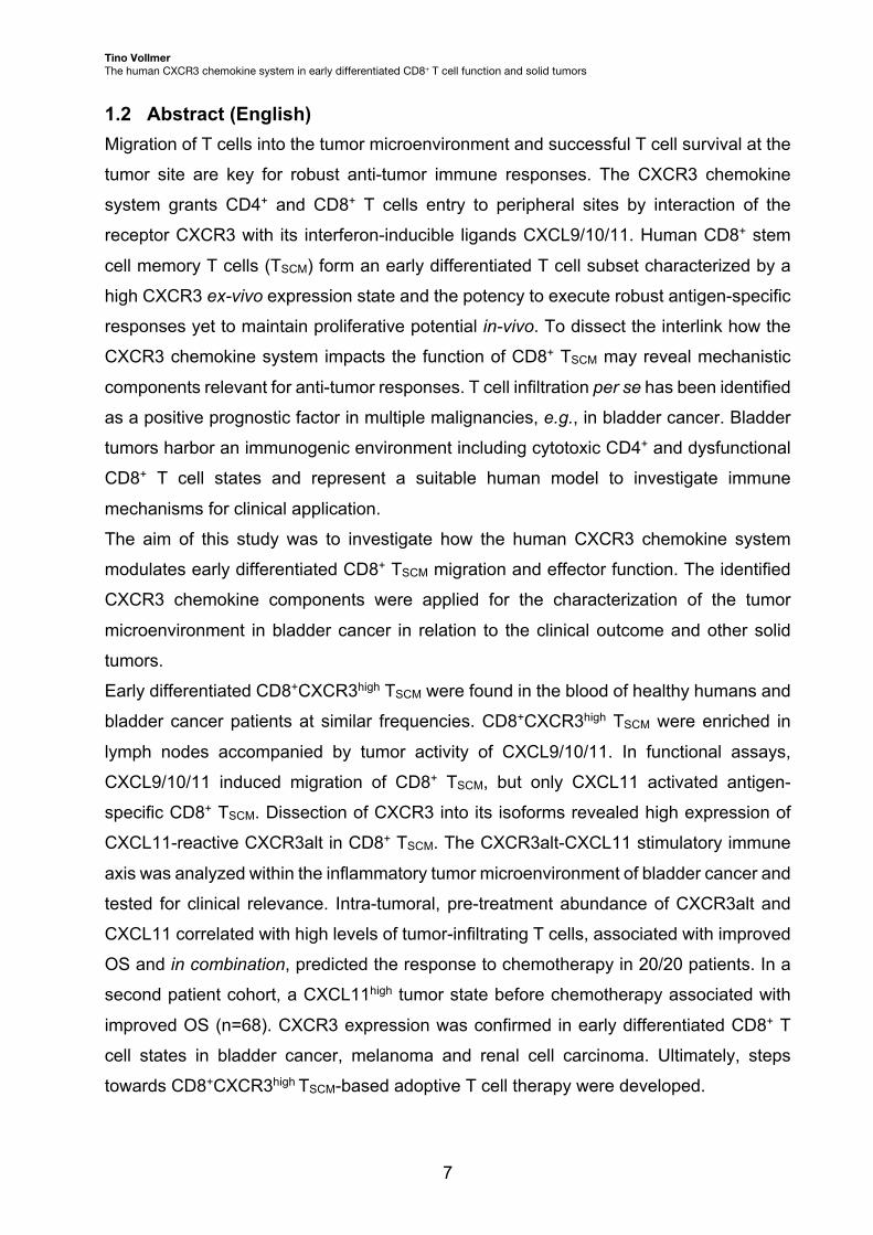
1.2 Abstract (English) Migration of T cells into the tumor microenvironment and successful T cell survival at the
tumor site are key for robust anti-tumor immune responses. The CXCR3 chemokine
system grants CD4+ and CD8+ T cells entry to peripheral sites by interaction of the
receptor CXCR3 with its interferon-inducible ligands CXCL9/10/11. Human CD8+ stem
cell memory T cells (TSCM) form an early differentiated T cell subset characterized by a
high CXCR3 ex-vivo expression state and the potency to execute robust antigen-specific
responses yet to maintain proliferative potential in-vivo. To dissect the interlink how the
CXCR3 chemokine system impacts the function of CD8+ TSCM may reveal mechanistic
components relevant for anti-tumor responses. T cell infiltration per se has been identified
as a positive prognostic factor in multiple malignancies, e.g., in bladder cancer. Bladder
tumors harbor an immunogenic environment including cytotoxic CD4+ and dysfunctional
CD8+ T cell states and represent a suitable human model to investigate immune
mechanisms for clinical application.
The aim of this study was to investigate how the human CXCR3 chemokine system
modulates early differentiated CD8+ TSCM migration and effector function. The identified
CXCR3 chemokine components were applied for the characterization of the tumor
microenvironment in bladder cancer in relation to the clinical outcome and other solid
tumors.
Early differentiated CD8+CXCR3high TSCM were found in the blood of healthy humans and
bladder cancer patients at similar frequencies. CD8+CXCR3high TSCM were enriched in
lymph nodes accompanied by tumor activity of CXCL9/10/11. In functional assays,
CXCL9/10/11 induced migration of CD8+ TSCM, but only CXCL11 activated antigen-
specific CD8+ TSCM. Dissection of CXCR3 into its isoforms revealed high expression of
CXCL11-reactive CXCR3alt in CD8+ TSCM. The CXCR3alt-CXCL11 stimulatory immune
axis was analyzed within the inflammatory tumor microenvironment of bladder cancer and
tested for clinical relevance. Intra-tumoral, pre-treatment abundance of CXCR3alt and
CXCL11 correlated with high levels of tumor-infiltrating T cells, associated with improved
OS and in combination, predicted the response to chemotherapy in 20/20 patients. In a
second patient cohort, a CXCL11high tumor state before chemotherapy associated with
improved OS (n=68). CXCR3 expression was confirmed in early differentiated CD8+ T
cell states in bladder cancer, melanoma and renal cell carcinoma. Ultimately, steps
towards CD8+CXCR3high TSCM-based adoptive T cell therapy were developed.

Tino Vollmer The human CXCR3 chemokine system in early differentiated CD8+ T cell function and solid tumors
8
In conclusion, the results indicate a migratory but also stimulatory role of the human
CXCR3 chemokine system for the function of early differentiated CD8+ T cells. The clinical
data imply the CXCR3 chemokine system to be decisive for response to chemotherapy
in bladder cancer and suggest protective pan-cancer relevance of intra-tumoral early
differentiated CD8+CXCR3high T cells, opening novel therapeutic options.
Tino Vollmer The human CXCR3 chemokine system in early differentiated CD8+ T cell function and solid tumors
9
2 Current State of Immune Research on CD8+ T cells in Cancer
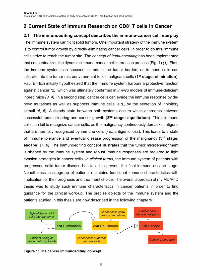
2.1 The immunoediting concept describes the immune-cancer cell interplay The immune system can fight solid tumors. One important strategy of the immune system
is to control tumor growth by directly eliminating cancer cells. In order to do this, immune
cells strive to reach the tumor site. The concept of immunoediting has been implemented
that conceptualizes the dynamic immune-cancer cell interaction process (Fig. 1) (1). First,
the immune system can succeed to reduce the tumor burden, as immune cells can
infiltrate into the tumor microenvironment to kill malignant cells (1st stage: elimination).
Paul Ehrlich initially hypothesized that the immune system harbors a protective function
against cancer (2), which was ultimately confirmed in in-vivo models of immune-deficient
inbred mice (3, 4). In a second step, cancer cells can evade the immune response by de-
novo mutations as well as suppress immune cells, e.g., by the secretion of inhibitory
stimuli (5, 6). A steady state between both systems occurs which alternates between
successful tumor clearing and cancer growth (2nd stage: equilibrium). Third, immune
cells can fail to recognize cancer cells, as the malignancy continuously demasks antigens
that are normally recognized by immune cells (i.e., antigenic loss). This leads to a state
of immune tolerance and eventual disease progression of the malignancy (3rd stage: escape) (7, 8). The immunoediting concept illustrates that the tumor microenvironment
is shaped by the immune system and robust immune responses are required to fight
evasion strategies in cancer cells. In clinical terms, the immune system of patients with
progressed solid tumor disease has failed to prevent the final immune escape stage.
Nonetheless, a subgroup of patients maintains functional immune characteristics with
implication for their prognosis and treatment choice. The overall approach of my MD/PhD
thesis was to study such immune characteristics in cancer patients in order to find
guidance for the clinical work-up. The precise objects of the immune system and the
patients studied in this thesis are now described in the following chapters.
Figure 1: The cancer immunoediting concept.
�VW�(OLPLQDWLRQ �QG�(TXLOLEULXP �UG�(VFDSH
+LJK�LQILOWUDWLRQ�RI�7�FHOOV�LQWR�WKH�WXPRU
(IILFLHQW�NLOOLQJ�RI�FDQFHU�FHOOV�E\�7�FHOOV
&DQFHU�FHOOV�VKRZ�GH��QRYR�PXWDWLRQV
&DQFHU�FHOOV�VXSSUHVV�LPPXQH�FHOOV
&DQFHU�FHOOV�GHPDVN�DQWLJHQV
&DQFHU�SURJUHVVLRQ
Tino Vollmer The human CXCR3 chemokine system in early differentiated CD8+ T cell function and solid tumors
10
2.2 T cell infiltration as prognostic and predictive variable for solid cancer patients
In my MD/PhD thesis, I focused on the human adaptive immune system as part of the
tumor elimination machinery. CD3+ T lymphocytes (T cells) are central cellular players of
the adaptive immune system and are directly involved in anti-tumor responses. Herein,
CD8+ T cells are regarded as cytotoxic T cells, as they have the ability to kill pathogen
infected and malignant cells. Their T cell receptor (TCR) can recognize tumor-related
peptides presented on major histocompatibility complex I (MHC-I) molecules of the
malignant cell (9). When recognized, CD8+ T cells can induce apoptosis in the malignant
cell by the expression of death receptors (e.g., Fas ligand) and the secretion of granules
that include cytotoxic granzymes and perforins (10). In order to elicit killing and contribute
to tumor elimination, CD8+ T cells have to overcome the immunosuppressive tumor
microenvironment and infiltrate into the tumor.
The immune infiltrate in solid tumors has been investigated for its prognostic role
in cancer patients by a myriad of studies. An early report in 2006 demonstrated that tumor
infiltration of CD8+ T cells associated with diseases-free survival of patients with
colorectal cancer. Interestingly, not only a high abundance of CD3 and CD8, but also their
location at multiple sites within the tumor associated with a positive outcome in these
patients (11). In addition, the intra-tumoral presence of the T cell memory marker, and
splice variant of the tyrosin kinase CD45, CD45RO correlated with a similar beneficial
outcome in colorectal cancer (12). Colorectal cancer has remained the pilot tumor entity
for implementing immune criteria for the clinical evaluation of solid cancer patients. A
recent multi-center validation study suggests an “Immunoscore” (derived from an
automated intratumoral CD3/CD8 immunohistochemistry analysis) as a potent predictor
for colorectal cancer recurrence (13). The high accuracy for predicting colorectal cancer
recurrence has initiated discussions to apply the “Immunoscore” in addition to
standardized staging procedure (e.g., TNM-score) (14, 15). Beyond colorectal cancer, a
cytotoxic, CD8-dominated immune infiltrate positively correlated with overall survival in
15 out of 17 investigated solid tumor entities, which included multiple types of
adenocarcinoma and squamous cell cancers (16). To translate prognostic immune
markers for clinical application, treatment-specific immune signatures need to be defined
that harbor predictive capacity for the therapeutic outcome.
Immunotherapies have been in the translational focus for testing T cell related
signatures for treatment stratification approaches. Recently introduced immunotherapies,
Tino Vollmer The human CXCR3 chemokine system in early differentiated CD8+ T cell function and solid tumors
11
e.g., immune checkpoint blockade (ICB) therapies (e.g., anti-programmed cell death
protein-1; anti-PD-1 and anti-PD-1 ligand; anti-PD-L1) underline that in-vivo T cell
activation can lead to durable cancer remission. ICB therapies are now clinically approved
in multiple tumor entities including melanoma and muscle-invasive bladder cancer (17).
Based on the T cell activating mechanism, a pre-existing T cell response was
hypothesized to be relevant for response to immune treatment (18). Multiple studies have
now confirmed that the intra-tumoral abundance of CD8+ T cells (expressing PD-1)
associates with response to immune checkpoint blockade (19–22).
Classical cancer therapies - such as chemotherapy – are poorly investigated on
immune markers for treatment success. Chemotherapeutic agents elicit unspecific toxic
effects, e.g., by inhibition of cell proliferation, but can also induce strong immune-
stimulatory effects, which has only recently been recently appreciated (23, 24).
Exemplary, platinum-based chemotherapeutics (e.g., cisplatin) bind to DNA and initiate
crosslinking within one DNA strand that leads to insufficient DNA repair (25). As a
consequence, cancer cells die (causing tumor rejection), but also proliferating healthy
cells die (causing treatment side effects: myelosuppression, nephrotoxicity, neurotoxicity)
(26). Mechanistic studies unveiled that platinum-based chemotherapy has distinct
immunomodulatory effects. Platinum-based agents were shown to induce the up-
regulation of PD-L1 in cancer cells of tumor patients (27) characterized as part of an
immunogenic cell death program after platinum exposure to cancer cell lines in-vitro (28–
31), synergize with anti-PD-L1 treatment (32, 33) and promote co-stimulation of CD8+ T
cells by activating dendritic cells (34). This indicates an activation of CD8+ T cells, which
follows chemotherapy treatment and may support successful tumor rejection. In analogy
to immunotherapies, it remains to be determined whether a pre-existing T cell infiltration
state is pre-requisite for chemotherapy response and applicable for treatment
stratification.
The identification of a T cell state in solid tumors before platinum-based
chemotherapy was one primary clinical subject of my MD/PhD thesis. In the following
chapter, I will outline CD8+ T cell migration and CD8+ T cell differentiation, two aspects of
CD8+ T cell immunity investigated in this thesis.
Tino Vollmer The human CXCR3 chemokine system in early differentiated CD8+ T cell function and solid tumors
12
2.3 Cell migration and differentiation state shape anti-tumor memory CD8+ T cell function
T cell infiltration is a positive prognostic factor in multiple tumor entities and can be
interpreted as a sign of immune elimination (see 2.1). A significant role can be attributed
to effector and memory CD8+ T cells that were shown to elicit (tumor related) antigen-
specific responses in multiple solid tumor entities (35). Beyond antigen recognition, two
components are of importance for robust anti-tumor CD8+ effector and memory T cell
function: i) productive T cell migration into the tumor and ii) successful T cell survival at
the tumor site despite the immunosuppressive environment.
T cell migration is mediated by external signals, yet T cells harbor a receptor
repertoire that can modulate their migratory response. Important external signals are
chemotactic proteins (chemokines). Chemokines can be found in the tumor
microenvironment and bind to chemokine receptors, which are expressed on immune
cells inducing cell migration (36, 37). Chemotaxis (i.e., cell migration towards a
chemokine gradient) is supposedly the primary mechanism how T cells migrate towards
dynamic inflammatory environments such as the tumor site (38). CXCR3 is a G-protein
coupled chemokine receptor expressed on Th1 CD4+ and CD8+ T cells that mediates
chemotaxis to inflamed tumors (39, 40), inflamed peripheral sites (41) and the secondary
lymphoid compartment (42). The three interferon-inducible ligands CXCL9 (MIG),
CXCL10 (IP-10) and CXCL11 (I-TAC) are the principal chemokines that bind to CXCR3
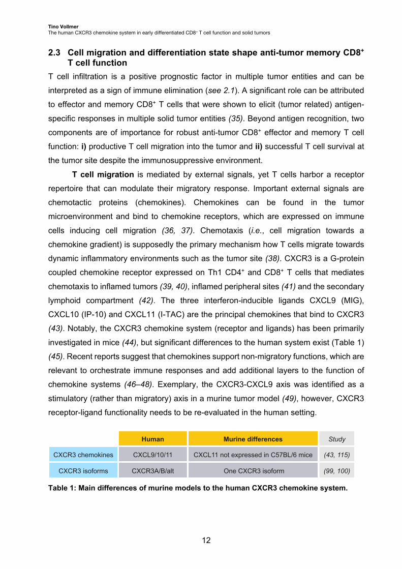
(43). Notably, the CXCR3 chemokine system (receptor and ligands) has been primarily
investigated in mice (44), but significant differences to the human system exist (Table 1)
(45). Recent reports suggest that chemokines support non-migratory functions, which are
relevant to orchestrate immune responses and add additional layers to the function of
chemokine systems (46–48). Exemplary, the CXCR3-CXCL9 axis was identified as a
stimulatory (rather than migratory) axis in a murine tumor model (49), however, CXCR3
receptor-ligand functionality needs to be re-evaluated in the human setting.
Table 1: Main differences of murine models to the human CXCR3 chemokine system.
&;&/������� ���������
&;&5�$�%�DOW ���������
&;&/���QRW�H[SUHVVHG�LQ�&��%/���PLFH
�2QH�&;&5��LVRIRUP
&;&5��FKHPRNLQHV
&;&5��LVRIRUPV
+XPDQ 6WXG\0XULQH�GLIIHUHQFHV
Tino Vollmer The human CXCR3 chemokine system in early differentiated CD8+ T cell function and solid tumors
13
T cell survival is substantially guided by intrinsic cell programs that allow subtypes
of T cells (e.g., CD8+ memory T cell subsets) to endure in non-stimulatory or even
immune-suppressive environments. Specifically, Rafi Ahmed`s group has established the
concept that antigen-specific CD8+ T cell memory sustains longevity guided by intrinsic
transcriptional and epigenetic differentiation programs (50–52). In general, cell
differentiation is an important concept in biology that describes the specialization of cells,
i.e., daughter cells are in a more professionalized state compared to their cellular origin,
the adult stem cell (53). In tumor-infiltrating T cells, single-cell analyses have likewise
revealed distinct T cell differentiation states (54–56). T cell differentiation states can be
divided into i) early differentiated states that are endowed with a high self-renewing
capacity and ii) late differentiated states that execute immediate T cell effector function.
Interestingly, early differentiated CD8+ T cells were shown to be pivotal for maintaining T
cell survival at the tumor site by providing a local “stem-like” T cell source (57–59), actively
responding to anti-PD-1 ICB therapies (21, 60, 61) and boosting the efficacy of adoptive
T cell therapy approaches against cancer (62, 63). However, there is limited research on
how the early differentiated T cell state impacts the migratory capacity of human CD8+ T
cells. To study this interlink may reveal functional insights into human signals that mediate
homing of early differentiated CD8+ T cells to the secondary lymphoid compartment in
cancer, but also directly to the tumor microenvironment.
CD8+ stem cell memory T cells (TSCM) form a T cell subset with early
differentiated cell properties and were the primary cellular object of this thesis. TSCM were
identified in the (originally defined) naïve T cell compartment characterized as
CCR7+CD45RA+CD45RO-CD62L+CD27+CD28+CD127+, which associated with naïve-
like functional attributes such as longevity and high proliferative capacity. In addition, TSCM
were characterized as CD95+CD122highCXCR3high, which associated with memory-like
functional attributes such as effector function on antigenic rechallenge and tumor clearing
in in-vivo models (64). This initial report is now confirmed by multiple studies underlining
that TSCM can exhibit potent antigen-specific responses (65, 66), maintain longevity (67,
68) and execute high proliferative potential in-vivo (69, 70). All of these characteristics
are preferable for immunotherapeutic use and already exploited for T cell product
generation approaches (71, 72). This makes TSCM an excellent representative of the early
differentiated T cell state and a therapeutically relevant T cell subset to study in solid
cancer disease (see 3.2 methods for overview on all CD8+ T cell subsets investigated
including the flow cytometric gating strategy).

Tino Vollmer The human CXCR3 chemokine system in early differentiated CD8+ T cell function and solid tumors
14
Our proof-of-concept study was performed in the disease of bladder cancer, which
will be introduced in chapter (2.4) followed by a summary on the hypotheses and aims of
my thesis (2.5).
Tino Vollmer The human CXCR3 chemokine system in early differentiated CD8+ T cell function and solid tumors
15
2.4 Bladder cancer as a solid tumor disease modulated by T cell responses The introduction of ICB treatment has successfully complemented the treatment arsenal
against solid cancer disease (see 2.2). Mechanistic and clinical studies in the
“frontrunner” disease of melanoma were pivotal for the implementation of ICB into clinical
practice and have contributed to a thorough understanding of immune mechanisms in
this skin cancer disease (19, 73). Noteworthy, other immunotherapeutic strategies were
already in place before ICB, but have gained less scientific attention. One of the first ideas
to stimulate the immune system against cancer goes back to “Coley`s toxin”, an approach
to apply bacteria (Streptococcus) against sarcoma in 1891 (74). A similar treatment
approach was applied for 9 patients with bladder cancer in 1976 by the vesical installation
of the attenuated bacteria Mycobacterium bovis, called Bacillus Calmette-Guérin (BCG),
which showed clinical successes (75). Nowadays, BCG installation is a standard
treatment for patients with high-risk non-muscle invasive bladder cancer (NMIBC)
inducing complete tumor rejection in more than half of the patients (76). Despite
widespread BCG application, mechanisms contributing to BCG response still need to be
fully resolved. Primary studies report the significance of a pre-treatment BCG-specific T
cell response for success and tumoral PD-L1 expression for failure of treatment (77, 78).
In addition, increased T cell infiltration and chemokine release were observed after BCG
treatment (79, 80). This links the induction of adaptive immune responses as a potent
anti-bladder tumor treatment strategy and ultimately, bladder cancer has been
acknowledged in literature as a human model disease to study anti-tumor T cell
responses (81, 82).
The bladder tumor has been characterized as an active immune landscape by
the identification of multiple immune cell types in the tumor microenvironment (83).
Bladder tumors were among the highest immune infiltrated tumors in a comparative
transcriptomic signature analysis between multiple entities (84). Immune infiltration was
observed both in the NMIBC as well as the advanced muscle-invasive bladder cancer
disease (MIBC) stage (85–87). In MIBC, T cell analyses revealed clonal expansion of T
cells in conjunction with a high neoantigen load as positive predictor of patient survival
(88). Neoantigen-reactive T cell responses were reported in MIBC and tumor-infiltrating
T cells were characterized for the development of autologous T cell therapy against
bladder cancer (89, 90). Moreover, cytotoxic CD4+ T cell states were identified in human
MIBC tumors that were activated following ICB and contributed to treatment response in
murine models (91, 92). A dysfunctional CD8+ T cell state was likewise reported to

Tino Vollmer The human CXCR3 chemokine system in early differentiated CD8+ T cell function and solid tumors
16
associate with poor clinical outcome in MIBC (93). However, the precise characteristics
of a functional and robust CD8+ T cell response in bladder cancer remains unknown.
Above all, immune state analyses need to be set in relation to the clinical situation of
patients with MIBC to identify relevant prognostic and predictive biomarkers that may
guide treatment decision and improve the outcome of patients with this disease.
Chemotherapy has remained a corner stone in the treatment for MIBC patients.
Guidelines recommend the application of platinum-based chemotherapy to clinically fit
MIBC patients prior to surgery, i.e., neoadjuvant chemotherapy (NAC) (94). These
recommendations are based on prospective randomized trials that reported a NAC-
related absolute increase of the 5-year overall survival (OS) by 5-8 % compared to
surgery alone (95, 96). However, only 25-40 % patients respond by histopathological
downstaging after NAC treatment (97). A pilot study on immune mechanisms identified
an improved effector T cell response in MIBC after NAC treatment (98), which confirms
the observed immune-stimulatory effects of platinum-based agents in other entities (see
2.3). It remains unknown, however, whether responder patients harbor an immune state
before treatment that associates with responsiveness to platinum-based NAC and allows
treatment stratification.
To conclude, bladder cancer is an immunogenic tumor disease, and the clinical
outcome can be modulated by effective adaptive immune responses. In this thesis, the
distinct role of the CXCR3 chemokine system was studied as potential part of a beneficial
intratumoral immune signature before NAC and tumor-infiltrating early differentiated CD8+
T cells were studied in MIBC compared to other solid tumors.
Tino Vollmer The human CXCR3 chemokine system in early differentiated CD8+ T cell function and solid tumors
17
2.5 Hypotheses and aims of this thesis The presented literature describes that migration of T cells into the tumor
microenvironment (e.g., by the CXCR3 chemokine system) and successful T cell survival
at the tumor site (e.g., of early differentiated CD8+ T cells) are key for robust anti-tumor
immune responses. However, mechanistical studies have been mostly conducted in
murine models. Based on these premises, the hypothesis was laid out that the human
CXCR3 chemokine system modulates early differentiated CD8+ T cell function and its
characterization may reveal novel and significant components for effective tumor
eradication by the immune system. Moreover, the literature describes bladder cancer as
an immunogenic solid tumor disease and a suitable model to examine immune
mechanisms for clinical application. Based on these hypotheses, I pursued three principal
aims during my MD/PhD studies:
i) to investigate how the CXCR3 chemokine system modulates early differentiated
CD8+ TSCM function relevant for anti-tumor immunity,
ii) to translate these findings for the characterization of the bladder tumor
microenvironment in relation to the clinical outcome and
iii) to establish first T cell culture steps towards CD8+ TSCM based adoptive T cell
therapy.

Tino Vollmer The human CXCR3 chemokine system in early differentiated CD8+ T cell function and solid tumors
18
3 In-depth Methods This chapter gives a schematic overview on the methods that were established and
applied in my MD/PhD thesis. Detailed descriptions of the methods are included the
Material & Methods section in the study of this thesis (see 10. Publication “The intra-
tumoral CXCR3 chemokine system is predictive of chemotherapy response in human
bladder cancer”).
3.1 Isolation of lymphocytes from human blood and lymph nodes Cell isolation procedures were applied to access lymphocytes including T cells from the
human veinous blood system and lymph nodes. i) From venous blood samples,
peripheral blood mononuclear cells (PBMC) were isolated by density gradient
centrifugation in presence of a polymer substance (Biochrom). PBMC separated in a
transferable cell layer (pellet) of macrophages/monocytes and lymphocytes including
peripheral T cells. ii) Lymph node samples of patients were accessed after surgery, kept
on ice, prepared and cut by a scalpel followed by filtering using a cell strainer. Lymph
node derived cells including T cells were then applied to the subsequent analyses and
experiments.
3.2 Flow cytometry Flow cytometry was performed to investigate protein expression on CD8+ T cells for
analysis of CD8+ T cell subsets, chemokine receptor and intracellular cytokine
expression. Prior to analysis, fluorochrome-conjugated monoclonal antibodies were
applied for T cell staining. Flow cytometry uses a fluidic system that aligns stained cells
by a laminar flow. An optical system includes lasers that activate fluorochromes bound to
antibodies and filters that direct emitted signals to the detectors. Light signals are
converted into electronic signals for flow cytometric analysis. The stained cells were
analyzed by the LSR II Fortessa flow (BD) or Cytoflex LX cytometer (Beckmann Coulter).
Extracellular staining was performed to discriminate the CD3+CD8+ T cell subsets:
CCR7+CD45RA+CD95- naïve T cells (Tnaïve), CCR7+CD45RA+CD95+ stem cell memory T
cells (TSCM), CCR7+CD45RA- central memory T cells (TCM), CCR7-CD45RA- effector
memory T cells (TEM), and CCR7-CD45RA+ terminally differentiated effector-memory T
cells (TEMRA). This gating strategy was likewise used to study chemokine receptor
expression (CXCR1/3/4 and CCR3/5/6/7) on CD8+ T cell subsets. After antigenic
Tino Vollmer The human CXCR3 chemokine system in early differentiated CD8+ T cell function and solid tumors
19
stimulation, fixation/permeabilization procedure was performed to identify intracellular
expression of cytokines (IFN-g, TNF-a, IL-2) and activation markers (e.g., CD137). This
allowed the identification and characterization of cytomegalovirus (CMV)-specific and
tumor-reactive T cells after an overnight stimulation set-up.
3.3 Fluorescently activated cell sorting (FACS) FACS was performed to isolate human CD8+ T cell subsets prior to the experimental or
analytical setting (e.g., TSCM expansion assay or analysis of CXCR3 isoforms, see below).
FACS is based on flow cytometric analysis of cells stained by fluorochrome-conjugated
antibodies (see above). In addition, FACS includes electric charging to single cell
containing droplets for polarization. Polarized droplets can be subsequently
electromagnetically separated based on the single-cell marker expression selected by
the operator. The Aria II Calliope (BD) was used for cell sorting of the distinct human
CD8+ T cell subsets.
3.4 Functional T cell assays T cell assays were applied to test for effects of chemokines on CD8+ T cell migration, T
cell effector function and chemokine receptor expression.
The T cell chemotaxis assay was performed to investigate CD8+ T cell
chemotaxis directed to chemokines. Short term culture was performed using transwell
assays (Corning) with a porous membrane between the lower chamber (including
chemokines) and the upper chamber (including PBMC). Flow cytometry was applied to
analyze transmigrated CD8+ T cells.
The TSCM expansion assay was performed to investigate stimulatory effects of
the chemokines on early differentiated CD8+ T cell function. Long term culture was
performed using a TSCM-based expansion set-up, as previously published by our group
(65). PBMC were enriched for a CD3+CCR7+CD45RA+ T cell population by FACS
followed by stimulation with CD3-depleted and CMV peptide pool (JPT) pulsed PBMC
and IL-7 and IL-15 (CellGenix) supplemented complete medium. At day 7, restimulation
was performed by peptide-pool pulsed, CD3-depleted PBMC. At day 14 and 21, antigen-
specificity was assessed by restimulation with peptide-pool pulsed antigen presenting
cells.
Tino Vollmer The human CXCR3 chemokine system in early differentiated CD8+ T cell function and solid tumors
20
The T cell proliferation assay was performed to investigate the proliferative effect
of the chemokine CXCL11 on early differentiated CD8+ T cell function. TSCM were labeled
with Carboxyfluorescein succinimidyl ester (CFSE) and spiked into the Tnaïve population
at the initial ex-vivo frequency. CMV stimulation was conducted analogous to the TSCM
expansion assay set-up (see above). CFSE dilution in CD8+ T cells after 96 hours
indicated T cell proliferation and was analyzed by flow cytometry.
The chemokine receptor ligation assay was performed to investigate
chemokine mediated downregulation of CXCR3 on CD8+ TSCM. Chemokines were
incubated with PBMC in different concentrations. Acidic washing was applied to exclude
receptor occupancy prior to flow cytometric analysis of CXCR3 expression.
3.5 Real time-quantitative polymerase chain reaction (RT-qPCR) RT-qPCR was performed to analyze mRNA abundance of the alternatively spliced
isoforms of CXCR3 in CD8+ T cell subtypes and in bladder tumors. Isolated mRNA was
transcribed into complementary DNA (cDNA) by reverse transcriptase (Quiagen).
Taqman RT-qPCR uses probes with attached fluorochrome (5' end) and quencher (3'
end) that hybridize with the target sequence during PCR amplification. The polymerase
replicates the amplicon and thereby, the polymerase cleaves the fluorescent probe
leading to decoupling of the quencher from the fluorochrome. A fluorescence signal is
emitted by the fluorochrome and detected by the thermal cycler at real time, which is
proportional to the cDNA amount of the target. For analysis of the CXCR3 isoforms, an
RT-qPCR panel was established that discriminates the CXCR3B and CXCR3alt isoform
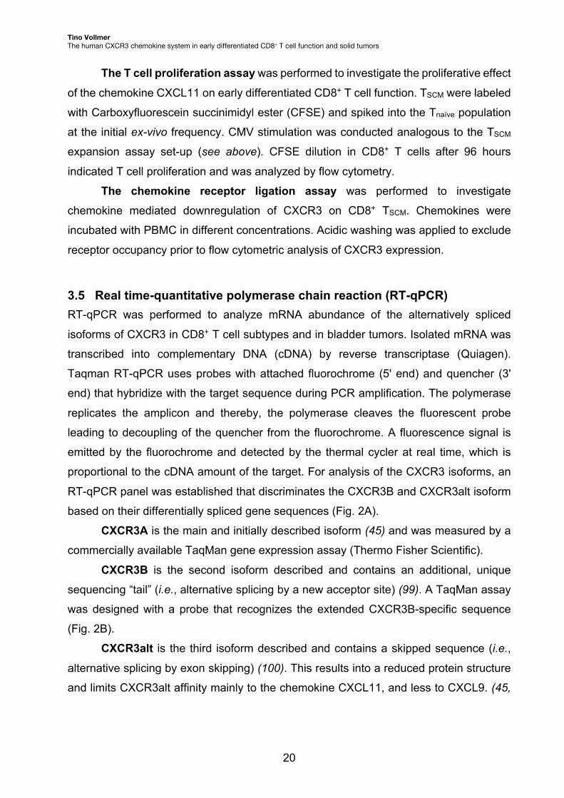
based on their differentially spliced gene sequences (Fig. 2A).
CXCR3A is the main and initially described isoform (45) and was measured by a
commercially available TaqMan gene expression assay (Thermo Fisher Scientific).
CXCR3B is the second isoform described and contains an additional, unique
sequencing “tail” (i.e., alternative splicing by a new acceptor site) (99). A TaqMan assay
was designed with a probe that recognizes the extended CXCR3B-specific sequence
(Fig. 2B).
CXCR3alt is the third isoform described and contains a skipped sequence (i.e.,
alternative splicing by exon skipping) (100). This results into a reduced protein structure
and limits CXCR3alt affinity mainly to the chemokine CXCL11, and less to CXCL9. (45,
Tino Vollmer The human CXCR3 chemokine system in early differentiated CD8+ T cell function and solid tumors
21
100, 101). A TaqMan assay was designed with a probe that spans the CXCR3alt-specific
sequencing gap (Fig. 2C).
Figure 2: Established RT-qPCR panel to analyze the isoforms CXCR3B and CXCR3alt. The figure is derived from the supplementary material of the study by Vollmer et al., 2021. (A) Scheme of all CXCR3 isoforms. Red area indicates location for the designed CXCR3B and CXCR3alt probes. (B) Sequence of primers and probe to analyze the CXCR3B isoform. (C) Sequence of primers and probe to analyze the CXCR3alt isoform.
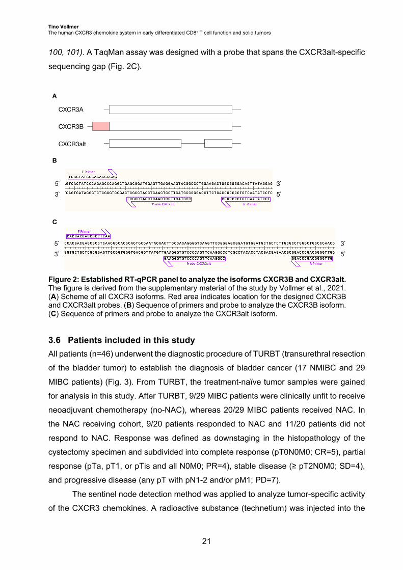
3.6 Patients included in this study All patients (n=46) underwent the diagnostic procedure of TURBT (transurethral resection
of the bladder tumor) to establish the diagnosis of bladder cancer (17 NMIBC and 29
MIBC patients) (Fig. 3). From TURBT, the treatment-naïve tumor samples were gained
for analysis in this study. After TURBT, 9/29 MIBC patients were clinically unfit to receive
neoadjuvant chemotherapy (no-NAC), whereas 20/29 MIBC patients received NAC. In
the NAC receiving cohort, 9/20 patients responded to NAC and 11/20 patients did not
respond to NAC. Response was defined as downstaging in the histopathology of the
cystectomy specimen and subdivided into complete response (pT0N0M0; CR=5), partial
response (pTa, pT1, or pTis and all N0M0; PR=4), stable disease (≥ pT2N0M0; SD=4),
and progressive disease (any pT with pN1-2 and/or pM1; PD=7).
The sentinel node detection method was applied to analyze tumor-specific activity
of the CXCR3 chemokines. A radioactive substance (technetium) was injected into the
CXCR3A
CXCR3B
CXCR3alt
A
C
B
5`3`
5`3`
3`5`
3`5`
Tino Vollmer The human CXCR3 chemokine system in early differentiated CD8+ T cell function and solid tumors
22
tumor base before surgery by a transurethral access and sentinel node detection was
performed after surgery by means of a Geiger meter.
Figure 3: Diagnostics and treatment of patients with bladder cancer investigated in this study. The figure is derived from the supplementary material of the study by Vollmer et al., 2021.
3.7 Analysis of the inflammatory tumor microenvironment The tumor microenvironment was analyzed for the components of the CXCR3 chemokine
system (chemokines and CXCR3 isoforms) and compared to inflammatory mediators
(cytokines) and tumor-infiltrating T cells (CD3). A tissue method was established that
homogenizes the tumor material by chemical and mechanical lysis procedure. DNA/RNA
extraction was performed by the AllPrep DNA/RNA Micro kit (Qiagen) and protein
isolation by use of protein extraction buffer (Thermo Fisher Scientific).
Protein was used for multiplex analysis to measure intra-tumoral cytokines
including the CXCR3 chemokines (CXCL9/10/11). The ELISA-based Luminex platform
was applied that uses cytokine-specific antibodies conjugated to magnetic beads. The
beads or microspheres harbor differential fluorescent signatures and can be detectable
individually by the system. This enabled the simultaneous measurement of multiple
analytes (cytokines) in one well of tumor lysate.
mRNA was used for RT-qPCR to measure intra-tumoral abundance of CXCR3
isoform expression (see 3.5) and T cell infiltration (CD3 mRNA). 34 house keeping genes
(HKG) were tested in bladder tumors and a statistical algorithm applied to select the most
20 NAC-receiving MIBC 9 no-NAC MIBC
17 NMIBC
11 non-resp.9 resp.
7 PD5 CR 4 PR 4 SD
biopsy via TURBT
NAC-treatment
muscle
XX
46 bladder cancer patients
29 MIBC
eligible to NAC?
local treatment
XX
X XX
X
Tino Vollmer The human CXCR3 chemokine system in early differentiated CD8+ T cell function and solid tumors
23
stable HKG candidates. CDKN1B and IPO8 were identified as the most stable HKG and
the geometric mean of CDKN1B and IPO8 was used for normalization of the target genes.
DNA was used for T cell receptor (TCR) sequencing to analyze intratumoral
TCR diversity. The complementary determining region 3 (CDR3) within the TCR-b chain
was amplified. The CDR3 contains regions of all three TCR gene segments (V/J/D) that
are recombined (i.e., during somatic recombination). Thus, CDR3 represents an
extremely variable part of the TCR representing suitable to discriminate TCRs. Amplified
CDR3 loci were processed by the Illumina NGS platform. TCR diversity was estimated
by the Shannon and Berger-Parker index.
3.8 Second bladder cancer cohort and single-cell analysis of the CXCR3 chemokine components
Sequencing data of the intra-tumoral CXCR3 chemokine components were accessed for
two purposes: i) testing the prognostic relevance of the CXCR3 chemokine system in a
second patient cohort and ii) elucidating the single-cell origin of the CXCR3 chemokine
components in different tumor entities.
Bulk RNA-sequencing data was accessed from The Cancer Genome Atlas
(TCG) and gene expression values were processed for normalization. Gene expression
was analyzed in treatment-naïve tumor samples of 68 MIBC patients who received
chemotherapy in the follow-up and compared to 292 MIBC patients who did not receive
chemotherapy.
Single-cell RNA-sequencing (scRNAseq) data were accessed under the
respective Gene Expression Omnibus (GEO) of the National Center for Biotechnology
Information (NCBI). scRNAseq data was derived from tissue samples of healthy bladder
and tumor samples of MIBC, melanoma and renal cell carcinoma. Cells with low quality
and genes with low abundance were removed. For analysis of CD8+ T cells, pre-sorting
was applied by filtering on CD4- and CD8+ T cells.
Tino Vollmer The human CXCR3 chemokine system in early differentiated CD8+ T cell function and solid tumors
24
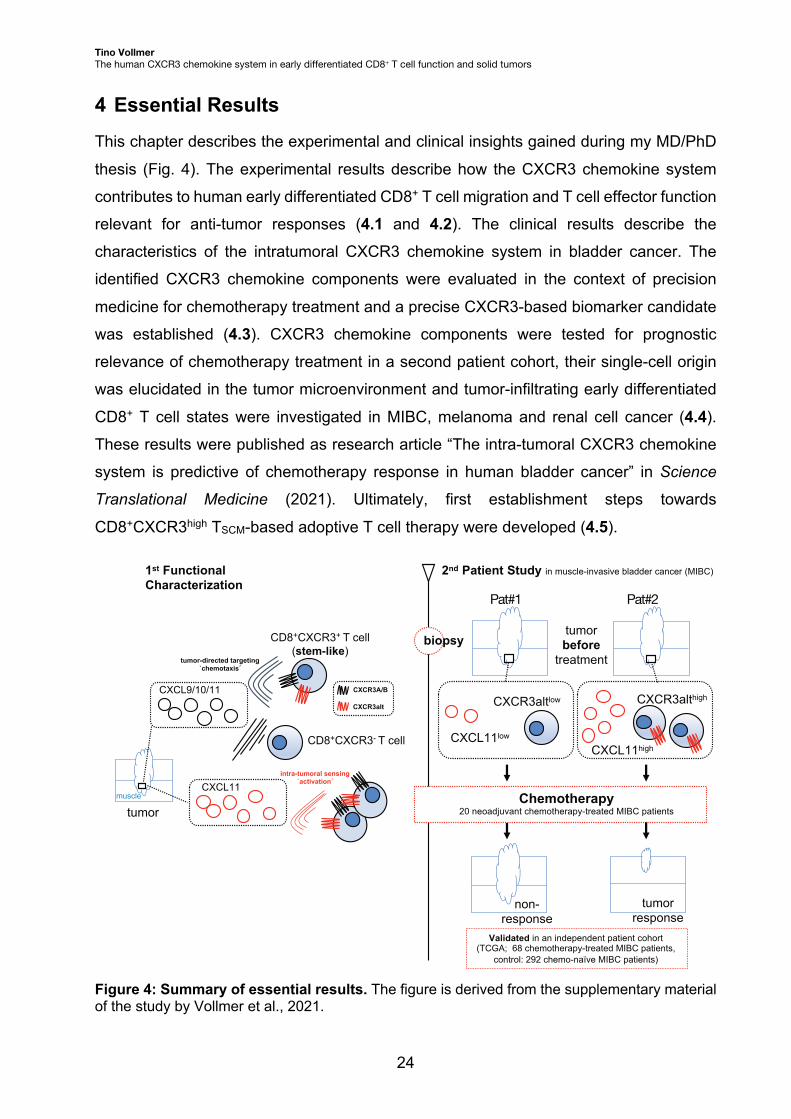
4 Essential Results This chapter describes the experimental and clinical insights gained during my MD/PhD
thesis (Fig. 4). The experimental results describe how the CXCR3 chemokine system
contributes to human early differentiated CD8+ T cell migration and T cell effector function
relevant for anti-tumor responses (4.1 and 4.2). The clinical results describe the
characteristics of the intratumoral CXCR3 chemokine system in bladder cancer. The
identified CXCR3 chemokine components were evaluated in the context of precision
medicine for chemotherapy treatment and a precise CXCR3-based biomarker candidate
was established (4.3). CXCR3 chemokine components were tested for prognostic
relevance of chemotherapy treatment in a second patient cohort, their single-cell origin
was elucidated in the tumor microenvironment and tumor-infiltrating early differentiated
CD8+ T cell states were investigated in MIBC, melanoma and renal cell cancer (4.4).
These results were published as research article “The intra-tumoral CXCR3 chemokine
system is predictive of chemotherapy response in human bladder cancer” in Science
Translational Medicine (2021). Ultimately, first establishment steps towards
CD8+CXCR3high TSCM-based adoptive T cell therapy were developed (4.5).
Figure 4: Summary of essential results. The figure is derived from the supplementary material of the study by Vollmer et al., 2021.
CXCL9/10/11
CXCL11high
tumorbefore
treatment
1st FunctionalCharacterization
CXCR3A/B
CXCR3alt
tumorresponse
CXCL11low
CXCR3altlow CXCR3althigh
non-response
tumor-directed targeting`chemotaxis´
intra-tumoral sensing`activation´
CD8+CXCR3+ T cell(stem-like)
CD8+CXCR3- T cell
CXCL11
tumor
Pat#1 Pat#2
2nd Patient Study in muscle-invasive bladder cancer (MIBC)
biopsy
Chemotherapy20 neoadjuvant chemotherapy-treated MIBC patients
Validated in an independent patient cohort(TCGA; 68 chemotherapy-treated MIBC patients,
control: 292 chemo-naïve MIBC patients)
muscle
Tino Vollmer The human CXCR3 chemokine system in early differentiated CD8+ T cell function and solid tumors
25
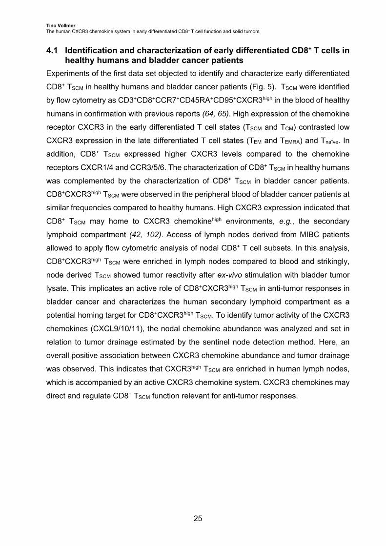
4.1 Identification and characterization of early differentiated CD8+ T cells in healthy humans and bladder cancer patients
Experiments of the first data set objected to identify and characterize early differentiated
CD8+ TSCM in healthy humans and bladder cancer patients (Fig. 5). TSCM were identified
by flow cytometry as CD3+CD8+CCR7+CD45RA+CD95+CXCR3high in the blood of healthy
humans in confirmation with previous reports (64, 65). High expression of the chemokine
receptor CXCR3 in the early differentiated T cell states (TSCM and TCM) contrasted low
CXCR3 expression in the late differentiated T cell states (TEM and TEMRA) and Tnaïve. In
addition, CD8+ TSCM expressed higher CXCR3 levels compared to the chemokine
receptors CXCR1/4 and CCR3/5/6. The characterization of CD8+ TSCM in healthy humans
was complemented by the characterization of CD8+ TSCM in bladder cancer patients.
CD8+CXCR3high TSCM were observed in the peripheral blood of bladder cancer patients at
similar frequencies compared to healthy humans. High CXCR3 expression indicated that
CD8+ TSCM may home to CXCR3 chemokinehigh environments, e.g., the secondary
lymphoid compartment (42, 102). Access of lymph nodes derived from MIBC patients
allowed to apply flow cytometric analysis of nodal CD8+ T cell subsets. In this analysis,
CD8+CXCR3high TSCM were enriched in lymph nodes compared to blood and strikingly,
node derived TSCM showed tumor reactivity after ex-vivo stimulation with bladder tumor
lysate. This implicates an active role of CD8+CXCR3high TSCM in anti-tumor responses in
bladder cancer and characterizes the human secondary lymphoid compartment as a
potential homing target for CD8+CXCR3high TSCM. To identify tumor activity of the CXCR3
chemokines (CXCL9/10/11), the nodal chemokine abundance was analyzed and set in
relation to tumor drainage estimated by the sentinel node detection method. Here, an
overall positive association between CXCR3 chemokine abundance and tumor drainage
was observed. This indicates that CXCR3high TSCM are enriched in human lymph nodes,
which is accompanied by an active CXCR3 chemokine system. CXCR3 chemokines may
direct and regulate CD8+ TSCM function relevant for anti-tumor responses.
Tino Vollmer The human CXCR3 chemokine system in early differentiated CD8+ T cell function and solid tumors
26
Figure 5: Identification and characterization of early differentiated CD8+ T cells.
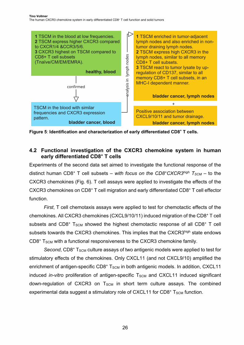
4.2 Functional investigation of the CXCR3 chemokine system in human early differentiated CD8+ T cells
Experiments of the second data set aimed to investigate the functional response of the
distinct human CD8+ T cell subsets – with focus on the CD8+CXCR3high TSCM – to the
CXCR3 chemokines (Fig. 6). T cell assays were applied to investigate the effects of the
CXCR3 chemokines on CD8+ T cell migration and early differentiated CD8+ T cell effector
function.
First, T cell chemotaxis assays were applied to test for chemotactic effects of the
chemokines. All CXCR3 chemokines (CXCL9/10/11) induced migration of the CD8+ T cell
subsets and CD8+ TSCM showed the highest chemotactic response of all CD8+ T cell
subsets towards the CXCR3 chemokines. This implies that the CXCR3high state endows
CD8+ TSCM with a functional responsiveness to the CXCR3 chemokine family.
Second, CD8+ TSCM culture assays of two antigenic models were applied to test for
stimulatory effects of the chemokines. Only CXCL11 (and not CXCL9/10) amplified the
enrichment of antigen-specific CD8+ TSCM in both antigenic models. In addition, CXCL11
induced in-vitro proliferation of antigen-specific TSCM and CXCL11 induced significant
down-regulation of CXCR3 on TSCM in short term culture assays. The combined
experimental data suggest a stimulatory role of CXCL11 for CD8+ TSCM function.
KHDOWK\��EORRG
�
��76&0�LQ�WKH�EORRG�DW�ORZ�IUHTXHQFLHV���76&0�H[SUHVV�KLJKHU�&;&5��FRPSDUHGWR�&;&5����&&5��������&;&5��KLJKHVW�RQ�76&0�FRPSDUHG�WR�&'���7�FHOO�VXEVHWV�7QDwYH�&0�(0�(05$��
76&0�LQ�WKH�EORRG�ZLWK�VLPLODU�IUHTXHQFLHV�DQG�&;&5��H[SUHVVLRQ�SDWWHUQ�
EODGGHU�FDQFHU��EORRG
��76&0�HQULFKHG�LQ�WXPRU��DGMDFHQW�O\PSK�QRGHV�DQG�DOVR�HQULFKHG�LQ�QRQ��WXPRU�GUDLQLQJ�O\PSK�QRGHV���76&0�H[SUHVV�KLJK�&;&5��LQ�WKH�O\PSK�QRGHV��VLPLODU�WR�DOO�PHPRU\�&'���7�FHOO�VXEVHWV���76&0�UHDFW�WR�WXPRU�O\VDWH�E\�XS��UHJXODWLRQ�RI�&'�����VLPLODU�WR�DOO�PHPRU\�&'���7�FHOO�VXEVHWV��LQ�DQ�0+&��,�GHSHQGHQW�PDQQHU�
EODGGHU�FDQFHU��O\PSK�QRGHV
3RVLWLYH�DVVRFLDWLRQ�EHWZHHQ�&;&/��������DQG�WXPRU�GUDLQDJH�
DQDO\VLV�LQ
��O\P
SK�QRG
HVEODGGHU�FDQFHU��O\PSK�QRGHV
FRQȴUPHG
Tino Vollmer The human CXCR3 chemokine system in early differentiated CD8+ T cell function and solid tumors
27
Third, screening for a CXCL11-reactive receptor in CD8+ TSCM was performed. A
CXCL11-reactive receptor expressed in CD8+ TSCM may mediate the CXCL11-specific
functional response. The expression of the (non-CXCR3) CXCL11-receptors CXCR7 and
DARC on CD8+ TSCM was excluded by flow cytometry. Next, CXCL11 reactivity was
screened within the CXCR3 isoform repertoire. Due to unavailable CXCR3 isoform-
specific antibodies, an RT-qPCR panel was established that detects all human CXCR3
isoforms (CXCR3A/B/alt) (Fig. 2). FACS was applied to sort on the CD8+ T cell subsets,
which were analyzed for CXCR3 isoform expression. Strikingly, CD8+ TSCM were
confirmed to express the highest transcript amounts of the CXCL11-reactive CXCR3alt
of all CD8+ T cell subsets.
In conclusion, CXCL11 attracted and stimulated CD8+CXCR3high TSCM, and
associated with high expression of the CXCL11-reactive isoform CXCR3alt in TSCM.
Ultimately, CXCR3alt-CXCL11 was identified as a potential stimulatory immune axis
relevant for CD8+ T cell function.
Figure 6: Functional investigation of the human CXCR3 chemokine system.
0LJUDWLRQ�&;&/��������DWWUDFW�&'���7�FHOOV�LQFOXGLQJ�76&0�
��'RVH��GHSHQGHQW�FKHPRWD[LV�E\�&;&/��������RI�DOO�&'���7�FHOO�VXEVHWV��KLJKHVW�DW�����QJ�PO����&;&/��!&;&/�!&;&/���FKHPRWDFWLF�SRWHQF\���76&0�VKRZHG�KLJKHVW�FKHPRWD[LV�DPRQJ�&'���7�FHOO�VXEVHWV���1RQ��FODVVLFDO�&;&5���OLJDQG�&;&/��GLG�QRW�LQGXFH�FKHPRWD[LV�
$FWLYDWLRQ�&;&/���DFWLYDWHV�76&0�
��&;&/���EXW�QRW�&;&/�����VXSSRUWHG�WKH�HQULFKPHQW�RI�&'���76&0�LQ�WZR�DQWLJHQ��VSHFLILF�H[SDQVLRQ�PRGHOV���&;&/���LQGXFHG�LQ��YLWUR�SUROLIHUDWLRQ�RI�76&0���&;&/��!&;&/�����LQGXFHG�UREXVW�&;&5��GRZQUHJXODWLRQ�RQ�76&0���1RQ��FODVVLFDO�&;&5���OLJDQG�&;&/��GLG�QRW�DFWLYDWH�76&0�
&;&/��������LQGXFH�FKHPRWD[LV�LQ�&'���7�FHOOV�LQ�D�VLPLODU�IDVKLRQ�
FRQȴUPHG
&;&/��!&;&/�����LQGXFHG�UREXVW�&;&5��GRZQUHJXODWLRQ�RQ�76&0�
��&'���76&0�GR�QRW�H[SUHVV�&;&5��'$5&���&'���76&0�H[SUHVV�KLJK�&;&5��LVRIRUPV�LQFOXGLQJ�&;&5�DOW�DV�&;&/����UHDFWLYH�LVRIRUP�
&;&/����VSHFLILF�UHFHSWRU�UHSHUWRLUH�RQ�&'���76&0"
&'���76&0�H[SUHVV�KLJK�&;&5��LVRIRUPV�LQFOXGLQJ�&;&5�DOW�DV�&;&/����UHDFWLYH�LVRIRUP�FRQȴUPHG
FRQȴUPHG
KHDOWK\��EORRG KHDOWK\��EORRG
EODGGHU�FDQFHU��EORRG EODGGHU�FDQFHU��EORRG
KHDOWK\��EORRG EODGGHU�FDQFHU��EORRG
Tino Vollmer The human CXCR3 chemokine system in early differentiated CD8+ T cell function and solid tumors
28
4.3 Characterization of intratumoral T cell infiltration and the CXCR3 chemokine system in bladder cancer
Experiments of the third data set investigated the role of the CXCR3 chemokine system
in the tumor microenvironment within inflammatory networks and for clinical relevance.
46 patients with bladder cancer were recruited in this thesis and treatment-naïve tumor
samples were studied (see 3.6 for detailed patient description). For analysis, a tissue
method was established to mechanically disrupt and chemically lyse the tumor sample,
which was followed by i) protein extraction to analyze intratumoral cytokines including the
CXCR3 chemokines by multiplex-based ELISA technique, ii) RNA extraction to analyze
intratumoral mRNA transcripts of the CXCR3 isoforms and CD3 by RT-qPCR and iii) DNA extraction to analyze TCR diversity by TCR sequencing. As control, tumor biopsy
matched patient sera were analyzed for protein cytokine levels.
First, analysis on the abundance of tumor infiltrating T cells (CD3) revealed no
differences between the three main patient groups: NMIBC, no-NAC MIBC and NAC-
receiving MIBC. However, within the NAC-receiving MIBC group responder patients had
significantly higher intratumoral T cell levels compared to non-responder. The increased
infiltration of T cells in the responder tumors did not associate with changes in the diversity
of TCR repertoire compared to non-responder.
Second, a comprehensive screen on intratumoral cytokines revealed that CXCL11
was most significantly associated with NAC response and harbored the highest sensitivity
for response prediction. In detail, CXCL11 clustered with CXCL9/10, IFN-y, CCL2/3/4/19,
CXCL12/13, IL-16 and correlated significantly with the abundance of tumor-infiltrating T
cells. Ultimately, intratumoral CXCL11 concentrations were significantly higher in
responder compared to non-responder NAC-receiving MIBC, no-NAC and NMIBC
patients, and a CXCL11high tumor state associated with improved OS in MIBC.
Third, the assessment of intratumoral CXCR3 isoforms revealed that CXCR3A/alt
but not CXCR3B significantly correlated with the abundance of tumor-infiltrating T cells,
CXCR3alt most rigorously predicted NAC response and interestingly, non-responder
showed a significant decrease in intratumoral CXCR3A/alt concentrations compared to
responder NAC-receiving MIBC, no-NAC and NMIBC patients. Last, a CXCR3althigh
tumor state associated with improved OS in MIBC.
Fourth, the interactions and dependencies between the ligands (CXCL9/10/11)
and the CXCR3 isoforms (A/B/alt) were analyzed within an intra-tumoral inflammatory
network analysis that included the response to NAC. Here, a correlation between
Tino Vollmer The human CXCR3 chemokine system in early differentiated CD8+ T cell function and solid tumors
29
CXCL11 and the response and CXCR3alt and the response was observed,
independently. Application of combined CXCL11 and CXCR3alt as pre-treatment
biomarkers within a logistic regression model resulted into complete discrimination
between the 9 responder and the 11 non-responder MIBC patients.
4.4 Second biomarker testing, identification of the cellular origin of intratumoral CXCR3/CXCL11 and analysis of early differentiated CD8+ T cell states in MIBC, melanoma and renal cell carcinoma
Analysis of openly accessible data sets were conducted for testing purpose of the
CXCR3-based biomarker and to gain improved mechanistic insights of the intra-tumoral
CXCR3 chemokine system in solid tumors.
First, components of the CXCR3-biomarker were investigated in a second MIBC
cohort of the TCGA data bank, which comprises bulk RNA sequencing data of human
tumors. Treatment-naïve tumor samples of 68 MIBC patients who in the follow-up
received adjuvant chemotherapy were compared to 292 MIBC patients who did not
receive chemotherapy. Interestingly, a CXCL9/10/11high and CXCR3high state associated
with improved OS only in the chemotherapy receiving cohort, but not in the cohort that
did not receive chemotherapy.
Second, single-cell RNA sequencing data of healthy bladder samples and MIBC
tumors samples were analyzed. Here, CXCR3 expression associated to tissue-resident
and tumor-infiltrating CD8+ T cells in the bladder, but not to non-immune healthy bladder
cells or cancer cells. In addition, CXCR3 expression was confirmed in tumor-infiltrating
CD8+ T cells in the disease melanoma and on the ligand site, CXCL9/10/11 expression
was detected in tumor-infiltrating CD14+ macrophages.
Third, single-cell analysis was exploited to decipher CXCR3 expression in
differentiation states of tumor infiltrating CD8+T cells in human MIBC, melanoma and
renal cell carcinoma. Strikingly, CXCR3 expression could be confirmed in early and in
late CD8+ T cell differentiation states in all three tumor entities investigated.
4.5 Establishment steps towards CD8+CXCR3high TSCM based T cell therapy Adoptive T cell therapy, e.g., the transfer of CAR T cells, has shown limited clinical
successes against solid tumors, which associated with reduced infiltration and
persistence of transferred T cells in the tumor microenvironment (103–109). Early
differentiated CD8+CXCR3high TSCM are a promising T cell source for use in adoptive T

Tino Vollmer The human CXCR3 chemokine system in early differentiated CD8+ T cell function and solid tumors
30
cell therapy due to their high survival and migratory capacity, as demonstrated in this
thesis. Until now, characterization of tumor-specific TSCM has been mostly performed in
murine models (110, 111). Human CMV-specific TSCM can be found in the peripheral
blood at low frequencies (Fig. 7A). Notably, human CMV-specific CD8+ T cells share
exhaustion signatures with tumor-reactive CD8+ T cells (112) and exploit the CXCR3
chemokine system for infiltration into the inflamed tissue (113). Hence, CMV-specific TSCM
are a reasonable and accessible human model to establish a good manufacturing
practice (GMP)-compatible T cell culture process that yields high amounts of antigen-
specific TSCM. To increase TSCM frequencies, TSCM were initially pre-enriched on a
CCR7+CD45RA+ phenotype followed by CMV-specific T cell expansion in accordance
with a protocol previously published by our group (Fig. 7B) (65). To increase the
robustness of the protocol, i) the influence of GMP-grade cytokine regimes (IL-7/IL-15,
IL-2, IL-4/IL-7) (114), ii) the role of CD4+ T cells (i.e., the depletion of CD4+ regulatory T
cells (TREG) and addition of bulk CD4+ T cells) and iii) post-stimulation selection strategies
were tested on antigen-specific CD8+ TSCM expansion.
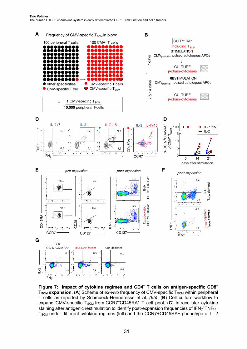
First, both IL-7/IL-15 and IL-2 supplementation enriched antigen-specific TSCM
during cell culture, whereas IL-4/IL-7 did not. Moreover, solely IL-7/IL-15 maintained
proportions of the initial CCR7+CD45RA+ naïve-like T cell phenotype, whereas IL-2-
cultured antigen-specific CD8+ TSCM vastly lost CCR7 and CD45RA expression during the
expansion process (Fig. 7C, D).
Second, initial depletion of TREG from pre-enriched CCR7+CD45RA+ T cells did not
alter CD8+ TSCM expansion (Fig. 7E). Surprisingly, the depletion of TREG strongly reduced
CD8+ TSCM expansion, when the starting cells were initially sorted on the TSCM phenotype
(Fig. 7F). The addition of irradiated CD4+ T cells to the starting T cell pool increased,
whereas CD4-depletion decreased the frequency of antigen-specific CD8+ TSCM from pre-
enriched CCR7+CD45RA+ during cell culture (Fig. 7G).
Tino Vollmer The human CXCR3 chemokine system in early differentiated CD8+ T cell function and solid tumors
31
Figure 7: Impact of cytokine regimes and CD4+ T cells on antigen-specific CD8+ TSCM expansion. (A) Scheme of ex-vivo frequency of CMV-specific TSCM within peripheral T cells as reported by Schmueck-Henneresse et al. (65). (B) Cell culture workflow to expand CMV-specific TSCM from CCR7+CD45RA+ T cell pool. (C) Intracellular cytokine staining after antigenic restimulation to identify post-expansion frequencies of IFNg+TNFa+ TSCM under different cytokine regimes (left) and the CCR7+CD45RA+ phenotype of IL-2
A100 peripheral T cells
other specifitiesCMV-specific T cells
other specifitiesCMV-specific T cells
other specificitiesCMV-specific T cell
100 CMV+ T cells
CMV-specific T cellsCMV-specific TSCMother specifities
CMV-specific T cells
1 CMV-specific TSCM
10.000 peripheral T-cells=
Frequency of CMV-specific TSCM in bloodCCR7+ RA+
STIMULATION CMVpp65/IE-1 pulsed autologous APCs
CULTUREg-chain-cytokines
RESTIMULATION CMVpp65/IE-1 pulsed autologous APCs
CULTUREg-chain-cytokines
including TSCM
7 da
ys
B
C
7 &
14 d
ays
F
14
2,4
IFNg
TNFa
1,8
0,6
Bulk
T SCM
deriv
edT R
EGde
pleted
T SCM
deriv
ed
post-expansion
plus CD4+ feeder CD4-depleted
0,1 0,5 0,1
1,1 4,2 0,6
IFNg
IL-2
BulkCCR7+CD45RA+
IL-4+7 IL-20,3
0,9
12,1
6,1
TNFa
IFNg
9,2
8,3
CCR7
CD4
5RA
IL-7+15 IL-2 IL-7+15
other specifitiesCMV-specific T cells
T memory cells
Tscm
0 14 210
5
10
15
100
days after stimulation%
CC
R7+ C
D45
RA
+ of
CM
V+
T SC
M
IL-2IL-7+15
D
G
E pre-expansion post-expansion
CCR7
CD45RA
97,8
98,9
0,4
5,6
CD127
CD25
7,2
CD137
IFNg
6,5
Bulk
CCR7
+ CD4
5RA+
T REG
depleted
CCR7
+ CD4
5RA+

Tino Vollmer The human CXCR3 chemokine system in early differentiated CD8+ T cell function and solid tumors
32
and IL-7/IL-15-expanded antigen-specific TSCM as overlay plot (right) at day 14. (D) Quantified data of CCR7+CD45RA+ TSCM-phenotype based on identification strategy presented in (C) at day 14, n=4 healthy donors. (E) Ex-vivo CD127-CD25+ TREG depletion of CCR7+CD45RA+ T cell pool by FACS before expansion (left) and intracellular cytokine staining after antigenic restimulation to identify frequencies of CD137+IFNg+ CD8+ TSCM derived from CCR7+CD45RA+ T cell pool in the presence or absence of TREG at day 21 (right). (F) Intracellular cytokine staining after antigenic restimulation to identify frequencies of IFNg+TNFa+ CD8+ TSCM derived from TSCM pool in the presence or absence of TREG at day 14. (G) Intracellular cytokine staining after antigenic restimulation to identify frequencies of IFNg+IL-2+ CD8+ TSCM derived from a CCR7+CD45RA+ T cell pool in the presence or absence of CD4+ irradiated feeder T cells compared to a CD4-depleted CCR7+CD45RA+ T cell pool at day 14.
Third, post-expansion selection on activated CD45RO+ TSCM (after activation-
induced CD45RAàCD45RO switch) significantly increased the frequency of antigen-
specific CD8+ TSCM from pre-enriched CCR7+CD45RA+ during cell culture (Fig. 8A-C).
In summary, IL-7/IL-15 cytokine culture supplementation, bulk untouched CD4+ T
cell support and CD45RO-based selection were established as robust and GMP-
compatible culture strategies to purify an antigen-specific CD8+ TSCM product (Fig. 8D).
Figure 8: CD45RO selection for improved CD8+ TSCM expansion. Cell culture protocol based on workflow presented in Fig. 7B. (A) Kinetics of CD45RO expression on CD8+ T cells derived from a TSCM included CCR7+CD45RA+ T cell pool (bulk) versus a TSCM depleted
pre-enrichment CD45RO-enr. CD45RA-enr.
CD45RO
CD4
5RA
A
B
C
bulk RO RA0
10203040
% C
MV-
spec
ific
T scm
of
CD
8+ T
cel
ls
✱✱
CD45RO selection
purifies TSCM
product
Additional(TREG-incl.)
CD4 support increases
enrichment
IL-7 and IL-15preserve
CCR7+CD45RA+
TSCM and promote expansion
D
0h 2h 16h 24h 48h 72h 96h05
1015202530
hours after stimulation
% C
D45R
O+ o
f CD
8+ T
cells
TSCM depleted
TSCM included
Tino Vollmer The human CXCR3 chemokine system in early differentiated CD8+ T cell function and solid tumors
33
CCR7+CD45RA+ T cell pool after antigenic stimulation, n=9 healthy donors. (B) CD45RO selection (and CD45RA selection as control) by FACS of a CCR7+CD45RA+ T cell derived pool after antigenic stimulation at day 4. (C) Frequency of antigen-specific CD8+ TSCM derived from a CCR7+CD45RA+ T cell pool, bulk versus selected on CD45RO/RA according to (B) and identified by antigenic restimulation at day 14. Wilcoxon rank-sum test applied, *p < 0.05, n=9 healthy donors. (D) Cell culture steps towards application of antigen-specific TSCM therapy.
Tino Vollmer The human CXCR3 chemokine system in early differentiated CD8+ T cell function and solid tumors
34
5 Clinical Applications and Outlook The mechanistic data reveal that the CXCR3 chemokine system modulates the function
of human early differentiated CD8+ T cells in a migratory but also stimulatory manner.
The clinical data reveal that the CXCR3 chemokine system is decisive for the response
to chemotherapy in bladder cancer. Ultimately, early differentiated CD8+CXCR3high T cells
were identified in the tumor microenvironment of MIBC, melanoma and renal cell
carcinoma patients. The data suggest pan-cancer relevance of the intra-tumoral CXCR3
chemokine system for early differentiated CD8+ T cells and offer an experimental strategy
to exploit CD8+CXCR3high TSCM for adoptive T cell therapy approaches.
5.1 Human analysis unveils a stimulatory role of the CXCR3 chemokine system in early differentiated CD8+ T cell function and solid tumors
The approach of this thesis included a thorough functional investigation of the CXCR3
chemokine system in the human setting. Most murine models are markedly reduced in
the complexity of the CXCR3 chemokine system (45, 115). Notably, CXCR3 isoform
expression and CXCL11 abundance showed high relevance within the experimental and
clinical analyses of this thesis: i) the CXCR3 isoforms CXCR3A/alt positively and
CXCR3B negatively associated with chemotherapy outcome and ii) CXCL11 was defined
as a stimulatory regulator of T cell function. Hence, these data offer strong experimental
and clinical arguments in favor of human studies that facilitate the dissection of the non-
redundant function of the CXCR3 isoforms and CXCL9/10/11 chemokines within the
versatile CXCR3 chemokine system.
Novel insights were gained in this thesis for the functional requirements of early
differentiated CD8+ T cells in cancer disease. Their identified intratumoral niche in
proximity to antigen presenting cells (58) and their pivotal role for response to anti-PD-1
ICB (21) is now complemented by their functional dependency on the CXCR3 chemokine
system. First, the presented data indicate that early differentiated CD8+CXCR3high T cells
preferentially locate in tumor-adjacent lymph nodes and are attracted by all three
chemokines CXCL9/10/11. Second, early differentiated CD8+CXCR3high T cells are
present in the tumor microenvironment and harbor a repertoire of CXCR3 isoforms, which
defines their functional outcome to the chemokines. Third, the selective (tissue)
abundance of CXCR3 chemokines may stimulate early differentiated CD8+ T cell function
(e.g., by CXCL11 expressed and presented by tumor infiltrating CD14+ macrophages). In
summary, these data imply that early differentiated CD8+ T cell migration and T cell
Tino Vollmer The human CXCR3 chemokine system in early differentiated CD8+ T cell function and solid tumors
35
survival can be regulated within one chemokine system based on two modalities, i.e.,
chemokine receptor isoform expression and ligand abundance.
The functional dependency between receptor (CXCR3 isoforms) and ligands
(CXCL9/10/11) was clinically relevant for MIBC patients, i.e., utilizing intratumoral co-
abundance of CXCR3alt and CXCL11 resulted into perfect separation of responder and
non-responder patients. Hence, clinical benefits can be observed, when the CXCR3
chemokine system is complete, which facilitates an active immune axis between receptor
and ligand. The pivotal cellular players within this immune axis are likely tumor infiltrating
CD8+CXCR3alt+ T cells stimulated by CD14+CXCL11+ macrophages.
To broaden the mechanistic understanding, one important next step will be to
assess single-cell expression of the CXCR3 isoforms in tumor infiltrating CD8+ T cells. In
this thesis, the CXCR3 isoform repertoire of early differentiated CD8+ T cells was defined
as CXCR3A/B/althigh in peripheral blood of healthy humans and bladder cancer patients.
Yet, the precise CXCR3 isoform state in tumor infiltrating early differentiated CD8+ T cells
remains to be determined. This analysis could not be performed due to the snap freezing
procedure of the tumor material that was necessary for biomarker evaluation.
This thesis lays the groundwork for clinical application of the CXCR3 chemokine
system for cancer diagnostics, i.e., predictive and prognostic strategies in solid tumors
(5.2) and for therapeutic application to modulate the tumor microenvironment (5.3).
5.2 Biomarker validation in bladder cancer and transferability to other solid tumor diseases
A plethora of studies has pursued to identify biomarkers that predict NAC response in
MIBC, however, robust stratification systems have not been translated for clinical use
(116–131). The primary clinical benefit of NAC stratification will be to exclude non-
responder MIBC patients from ineffective chemotherapy. Non-responder patients have a
high tendency to progress and metastasize which cannot be prevented by chemotherapy.
The identification of non-response at the diagnostic stage could initiate immediate radical
surgery that halts disease progression (132). In this study, intratumoral
CXCR3alt/CXCL11high segregated all 9 responder patients from the 11 non-responder
MIBC patients before NAC. In the TCGA cohort, an intratumoral CXCL11high state before
adjuvant chemotherapy associated with improved OS compared to a CXCL11low state in
68 MIBC patients. These clinical data strongly support the utility of a CXCR3-based
biomarker for NAC stratification and identification of non-responder patients. However, a
Tino Vollmer The human CXCR3 chemokine system in early differentiated CD8+ T cell function and solid tumors
36
thorough validation study is required that applies the same study conditions to a second
MIBC patient cohort including tumor sample preparation, ELISA-based analysis of
CXCL11 protein and RT-qPCR analysis of CXCR3 isoform mRNA abundance. A
validation study is already ongoing in joined work with Umea University, Sweden.
The transferability of a CXCR3-based biomarker to other tumor diseases is
supported by the data within this thesis: melanoma tumors were confirmed to be strongly
infiltrated by CD8+CXCR3high T cells and most importantly, protective early differentiated
CD8+ T cells were identified to express CXCR3 within the melanoma immune infiltrate.
Previous studies confirm a positive prognostic role of CD8+CXCR3high T cells for the
outcome of melanoma patients and describe melanoma-directed recruitment of CD8+ T
cells by the CXCR3 chemokine system (39, 133, 134). Hence, combined analysis of intra-
tumoral CXCR3 isoform and ligand abundance may be a promising biomarker candidate,
e.g., for patient stratification of routinely applied ICB treatment in progressed melanoma.
Early differentiated CD8+CXCR3high T cells were also identified in the tumor
microenvironment of renal cell carcinoma patients within this study. Previous clinical
studies unveiled an ambiguous prognostic role of the CXCR3 chemokine system in this
disease, which may be explained by the additional involvement of CXCR3 in renal cancer
cell function (135, 136). In detail, renal cancer cell derived CXCR3A associates to
metastatic function, whereas CXCR3B mediates apoptotic renal cancer cell signaling
(137, 138). Hence, cancer cell derived CXCR3 isoforms may impede the clinical utility of
a biomarker that is based on the absolute abundance of the CXCR3 isoforms. Single-cell
strategies are warranted to untangle renal cancer and immune cell related origin of the
CXCR3 isoforms, which needs to set in relation to the clinical outcome, respectively. In
addition, the role and cellular source of CXCR3alt remains to be elucidated in renal cell
carcinoma.
Ultimately, gained mechanistic insights behind the CXCR3-based biomarker allow
a hypothesis driven, yet adjustable approach for testing in other solid tumor diseases. A
“copy and paste” approach (i.e., analysis of intra-tumoral CXCR3alt-CXCL11) may be
applicable in the disease of melanoma, whereas cell source characterization of the
CXCR3 isoforms may result into a redefined CXCR3-based biomarker for patients with
renal cell carcinoma. Moreover, patients with solid tumor diseases that receive analogous
platinum-based NAC (e.g., cohorts of breast cancer and rectal carcinoma patients) are of
concrete interest to study for transferability of the CXCR3-based biomarker.
Tino Vollmer The human CXCR3 chemokine system in early differentiated CD8+ T cell function and solid tumors
37
5.3 Therapeutic modulation of the tumor microenvironment by CXCR3-based approaches
The study describes the beneficial role of a T cell-inflamed tumor microenvironment
equipped with an active state of the IFN-g-associated CXCR3 chemokine system in
treatment responding patients. In general, this supports an accepted immunotherapeutic
concept that aims to induce a favorable “immune hot” tumor microenvironment (139). In
detail, the presented data confirm the role of stimulatory and regulatory CXCR3
components and strikingly, add new functional layers to the CXCR3 chemokine system.
The dissection of the distinct CXCR3 components allows targeted strategies to modulate
the expression and abundance of single CXCR3 isoforms and ligands. On the receptor
side, CXCR3A-mediated stimulatory signaling (inducing cell chemotaxis) should be
favored over CXCR3B-mediated inhibitory signaling (inducing apoptosis) in CD8+ T cells
(45). This antagonistic function was supported in this study by the observed CXCR3A-
related good prognostic versus a CXCR3B-related poor predictive outcome in MIBC
patients. In addition, CXCR3alt-related immune stimulatory effects were identified as anti-
cancer objectives. On the ligand side, this study describes CXCL9/10/11 as potential T
cell migratory targets in confirmation with previous results (140), and identified CXCL11
as a stimulatory therapeutic target for human CD8+ T cell function. Hence, therapeutic
interventions of the CXCR3 chemokine system can be categorized into i) CXCR3 isoform
modulation and ii) CXCR3 ligand modulation.
CXCR3A and CXCR3alt are highly expressed in peripheral blood derived CD8+
TSCM of healthy humans and cancer patients. Thus, CXCR3A/althigh TSCM display a direct
native source for the stimulatory CXCR3 isoforms accessible by the veinous blood
system. This makes CD8+ TSCM an attractive candidate for adoptive T cell therapy
approaches in the solid tumor setting, i.e., the transfer of antigen-specific T cells to the
patient. In this thesis, first establishment steps were developed that seek to exploit the
CXCR3A/althigh ex-vivo state in CD8+ TSCM for an autologous cell transfer set-up of
antigen-specific T cell products. The presented data confirm the previously described
stimulatory role of the common gamma-chain cytokines IL-7 and IL-15 for antigen-specific
TSCM function (141). Further, the data in this thesis highlight the relevance of a complete
CD4+ helper T cell pool for sufficient CD8+ TSCM expansion and define a selection method
based on activation induced CD45RO expression to accelerate TSCM enrichment. The
parallel application of the identified culture requirements allows a GMP-compatible and
robust enrichment process of antigen-specific CD8+ TSCM from peripheral blood. The
Tino Vollmer The human CXCR3 chemokine system in early differentiated CD8+ T cell function and solid tumors
38
established protocol is suitable for transfer to a neoantigen or tumor-associated antigen
TSCM expansion set-up. In a next step, TSCM enrichment may be further enhanced by
supplementation of the stimulatory chemokine CXCL11, as characterized in this thesis.
However, a GMP-compatible production process of recombinant CXCL11 protein will be
required for ultimate clinical translation. Moreover, TSCM enriched populations may be
used for genetic incorporation of, e.g., chimeric antigen receptors (CAR) that ensure
anticancer recognition. Further, genetical engineering by (viral or non-viral) knock-in
strategies of CXCR3A/alt may be applied to enhance the migratory and survival function
of established T cell products. Last, CXCR3A/alt may be directly targeted by agonizing
antibodies that recognize these CXCR3 isoforms.
CXCR3B represents the opposing immune inhibitory CXCR3 molecule. In analogy
to ICB, CXCR3B may be a reasonable blocking target by CXCR3B-specific antibodies in
order to enhance CD8+ T cell function. In this study, the additionally investigated non-
classical CXCR3B-ligand CXCL4 (142–145) did not interfere with CD8+ TSCM function;
neither in a stimulatory nor inhibitory manner. Hence, thorough mechanistical analysis of
CXCR3B-ligand interactions will be pre-requisite before the initiation of blocking
approaches to fully clarify the role of CXCR3B for early differentiated CD8+ T cell function.
CXCL9/10/11 were identified as potential targeting candidates to enhance tumor
infiltration of early differentiated CD8+ T cells in humans. Future mechanistic studies that
dissect non-redundant relevance for migratory function of CXCL9/10 or 11 in distinct
tumor entities and tissue compartments (tumor versus lymph node) are pivotal to prioritize
modulatory strategies on one CXCR3 chemokine. This study suggests that increased
CXCL11 expression in the tumor microenvironment may favor both CD8+ T cell migration
and CD8+ T cell survival. In addition, increased CXCL11 gene expression was observed
in tumor-infiltrating CD14+ macrophages in this study. Notably, CXCL11 gene expression
was previously reported to be STAT3-dependent (146), to be induced by IFNg and IFNb
(but not by IFNa) (43, 147) and enhanced by TNFa (148). Cell-type specific modulation
of CXCL11 expression (e.g., by ex-vivo cell culture or genetical manipulation) may benefit
ongoing antigen presenting cell based immunotherapeutic approaches (149).
In summary, this thesis contributes to the mechanistic understanding how the
human CXCR3 chemokine system modulates early differentiated CD8+ T cell function.
Beyond diagnostic application, this opens new therapeutical paths for targeted
modulatory strategies of the distinct human CXCR3 components in solid tumors.
Tino Vollmer The human CXCR3 chemokine system in early differentiated CD8+ T cell function and solid tumors
39
6 References 1. G. P. Dunn, A. T. Bruce, H. Ikeda, L. J. Old, R. D. Schreiber, Cancer immunoediting: from immunosurveillance to tumor escape, Nat Immunol 3, 991–998 (2002).
2. P. Ehrlich, Über den jetzigen Stand der Karzinomforschung, Beiträge zur experimentellen Pathologie und Chemotherapie (1909).
3. Y. Shinkai, 0Gary Rathbun, K.-P. Lam, E. M. Oltz, V. Stewart, M. Mendelsohn, J. Charron, M. Datta, F. Young, A. M. Stall, F. W. Alt, RAG-2-deficient mice lack mature lymphocytes owing to inability to initiate V(D)J rearrangement, Cell 68, 855–867 (1992).
4. V. Shankaran, H. Ikeda, A. T. Bruce, J. M. White, P. E. Swanson, L. J. Old, R. D. Schreiber, IFNγ and lymphocytes prevent primary tumour development and shape tumour immunogenicity, Nature 410, 1107–1111 (2001).
5. R. D. Schreiber, L. J. Old, M. J. Smyth, Cancer Immunoediting: Integrating Immunity’s Roles in Cancer Suppression and Promotion, Science 331, 1565–1570 (2011).
6. D. Hanahan, R. A. Weinberg, Hallmarks of Cancer: The Next Generation, Cell 144, 646–674 (2011).
7. B. M. Olson, D. G. McNeel, Antigen loss and tumor-mediated immunosuppression facilitate tumor recurrence, Expert Rev Vaccines 11, 1315–1317 (2014).
8. J. A. Joyce, D. T. Fearon, T cell exclusion, immune privilege, and the tumor microenvironment, Science 348, 74–80 (2015).
9. A. K. Palucka, L. M. Coussens, The Basis of Oncoimmunology, Cell 164, 1233–1247 (2016).
10. L. Martínez-Lostao, A. Anel, J. Pardo, How Do Cytotoxic Lymphocytes Kill Cancer Cells?, Clin Cancer Res 21, 5047–5056 (2015).
11. J. Galon, A. Costes, F. Sanchez-Cabo, A. Kirilovsky, B. Mlecnik, C. Lagorce-Pagès, M. Tosolini, M. Camus, A. Berger, P. Wind, F. Zinzindohoué, P. Bruneval, P.-H. Cugnenc, Z. Trajanoski, W.-H. Fridman, F. Pagès, Type, Density, and Location of Immune Cells Within Human Colorectal Tumors Predict Clinical Outcome, Science 313, 1960–1964 (2006).
12. F. Pagès, A. Kirilovsky, B. Mlecnik, M. Asslaber, M. Tosolini, G. Bindea, C. Lagorce, P. Wind, F. Marliot, P. Bruneval, K. Zatloukal, Z. Trajanoski, A. Berger, W.-H. Fridman, J. Galon, In Situ Cytotoxic and Memory T Cells Predict Outcome in Patients With Early-Stage Colorectal Cancer, J Clin Oncol 27, 5944–5951 (2009).
13. F. Pagès, B. Mlecnik, F. Marliot, G. Bindea, F.-S. Ou, C. Bifulco, A. Lugli, I. Zlobec, T. T. Rau, M. D. Berger, I. D. Nagtegaal, E. Vink-Börger, A. Hartmann, C. Geppert, J. Kolwelter, S. Merkel, R. Grützmann, M. V. den Eynde, A. Jouret-Mourin, A. Kartheuser, D. Léonard, C. Remue, J. Y. Wang, P. Bavi, M. H. A. Roehrl, P. S. Ohashi, L. T. Nguyen, S. Han, H. L. MacGregor, S. Hafezi-Bakhtiari, B. G. Wouters, G. V. Masucci, E. K. Andersson, E. Zavadova, M. Vocka, J. Spacek, L. Petruzelka, B. Konopasek, P. Dundr, H. Skalova, K. Nemejcova, G. Botti, F. Tatangelo, P. Delrio, G. Ciliberto, M. Maio, L. Laghi, F. Grizzi, T. Fredriksen, B. Buttard, M. Angelova, A. Vasaturo, P. Maby, S. E. Church, H. K. Angell, L. Lafontaine, D. Bruni, C. E. Sissy, N. Haicheur, A. Kirilovsky, A. Berger, C. Lagorce, J. P. Meyers, C. Paustian, Z. Feng, C. Ballesteros-Merino, J. Dijkstra, C. van de Water, S. van L. Vliet, N. Knijn, A.-M. Mușină, D.-V. Scripcariu, B. Popivanova, M. Xu, T. Fujita, S. Hazama, N. Suzuki, H. Nagano, K. Okuno, T. Torigoe, N. Sato, T. Furuhata, I. Takemasa, K. Itoh, P. S. Patel, H. H. Vora, B. Shah, J. B. Patel, K. N. Rajvik, S. J. Pandya, S. N. Shukla, Y. Wang, G. Zhang, Y. Kawakami, F. M. Marincola, P. A. Ascierto, D. J. Sargent, B. A. Fox, J. Galon, International validation of the consensus Immunoscore for the classification of colon cancer: a prognostic and accuracy study, Lancet 391 (2018), doi:10.1016/s0140-6736(18)30789-x.

Tino Vollmer The human CXCR3 chemokine system in early differentiated CD8+ T cell function and solid tumors
40
14. W. H. Fridman, F. Pagès, C. Sautès-Fridman, J. Galon, The immune contexture in human tumours: impact on clinical outcome, Nat Rev Cancer 12, 298 (2012).
15. J. Galon, A. Lanzi, Immunoscore and its introduction in clinical practice, Q J Nucl Medicine Mol Imaging 64, 152–161 (2020).
16. D. Bruni, H. K. Angell, J. Galon, The immune contexture and Immunoscore in cancer prognosis and therapeutic efficacy, Nat Rev Cancer 20, 662–680 (2020).
17. R. K. Vaddepally, P. Kharel, R. Pandey, R. Garje, A. B. Chandra, Review of Indications of FDA-Approved Immune Checkpoint Inhibitors per NCCN Guidelines with the Level of Evidence, Cancers 12, 738 (2020).
18. S. L. Topalian, J. M. Taube, R. A. Anders, D. M. Pardoll, Mechanism-driven biomarkers to guide immune checkpoint blockade in cancer therapy, Nat Rev Cancer 16, 275–287 (2016).
19. P. C. Tumeh, C. L. Harview, J. H. Yearley, I. P. Shintaku, E. J. M. Taylor, L. Robert, B. Chmielowski, M. Spasic, G. Henry, V. Ciobanu, A. N. West, M. Carmona, C. Kivork, E. Seja, G. Cherry, A. J. Gutierrez, T. R. Grogan, C. Mateus, G. Tomasic, J. A. Glaspy, R. O. Emerson, H. Robins, R. H. Pierce, D. A. Elashoff, C. Robert, A. Ribas, PD-1 blockade induces responses by inhibiting adaptive immune resistance, Nature 515, 568 (2014).
20. S. F. Ngiow, A. Young, N. Jacquelot, T. Yamazaki, D. Enot, L. Zitvogel, M. J. Smyth, A Threshold Level of Intratumor CD8+ T-cell PD1 Expression Dictates Therapeutic Response to Anti-PD1, Cancer Res 75, 3800–3811 (2015).
21. M. Sade-Feldman, K. Yizhak, S. L. Bjorgaard, J. P. Ray, C. G. de Boer, R. W. Jenkins, D. J. Lieb, J. H. Chen, D. T. Frederick, M. Barzily-Rokni, S. S. Freeman, A. Reuben, P. J. Hoover, A.-C. Villani, E. Ivanova, A. Portell, P. H. Lizotte, A. R. Aref, J.-P. Eliane, M. R. Hammond, H. Vitzthum, S. M. Blackmon, B. Li, V. Gopalakrishnan, S. M. Reddy, Z. A. Cooper, C. P. Paweletz, D. A. Barbie, A. Stemmer-Rachamimov, K. T. Flaherty, J. A. Wargo, G. M. Boland, R. J. Sullivan, G. Getz, N. Hacohen, Defining T Cell States Associated with Response to Checkpoint Immunotherapy in Melanoma, Cell 175, 998-1013.e20 (2018).
22. J. J. Havel, D. Chowell, T. A. Chan, The evolving landscape of biomarkers for checkpoint inhibitor immunotherapy, Nat Rev Cancer 19, 1 (2019).
23. L. Galluzzi, A. Buqué, O. Kepp, L. Zitvogel, G. Kroemer, Immunological Effects of Conventional Chemotherapy and Targeted Anticancer Agents, Cancer Cell 28, 690–714 (2015).
24. L. Galluzzi, J. Humeau, A. Buqué, L. Zitvogel, G. Kroemer, Immunostimulation with chemotherapy in the era of immune checkpoint inhibitors, Nat Rev Clin Oncol, 1–17 (2020).
25. N. Poklar, D. S. Pilch, S. J. Lippard, E. A. Redding, S. U. Dunham, K. J. Breslauer, Influence of cisplatin intrastrand crosslinking on the conformation, thermal stability, and energetics of a 20-mer DNA duplex, Proc National Acad Sci 93, 7606–7611 (1996).
26. J. Mikuła-Pietrasik, A. Witucka, M. Pakuła, P. Uruski, B. Begier-Krasińska, A. Niklas, A. Tykarski, K. Książek, Comprehensive review on how platinum- and taxane-based chemotherapy of ovarian cancer affects biology of normal cells, Cell Mol Life Sci 76, 681–697 (2019).
27. J. Shin, J.-H. Chung, S. H. Kim, K. S. Lee, K. J. Suh, J. Y. Lee, J.-W. Kim, J.-O. Lee, J.-W. Kim, Y.-J. Kim, K.-W. Lee, J. H. Kim, S.-M. Bang, J.-S. Lee, Effect of Platinum-Based Chemotherapy on PD-L1 Expression on Tumor Cells in Non-small Cell Lung Cancer, Cancer Res Treat 51, 1086–1097 (2019).
28. S.-J. Park, W. Ye, R. Xiao, C. Silvin, M. Padget, J. W. Hodge, C. V. Waes, N. C. Schmitt, Cisplatin and oxaliplatin induce similar immunogenic changes in preclinical models of head and neck cancer, Oral Oncol 95, 127–135 (2019).
Tino Vollmer The human CXCR3 chemokine system in early differentiated CD8+ T cell function and solid tumors
41
29. D. Y. Q. Wong, W. W. F. Ong, W. H. Ang, Induction of Immunogenic Cell Death by Chemotherapeutic Platinum Complexes, Angewandte Chemie Int Ed 54, 6483–6487 (2015).
30. A. Tesniere, F. Schlemmer, V. Boige, O. Kepp, I. Martins, F. Ghiringhelli, L. Aymeric, M. Michaud, L. Apetoh, L. Barault, J. Mendiboure, J.-P. Pignon, V. Jooste, P. van Endert, M. Ducreux, L. Zitvogel, F. Piard, G. Kroemer, Immunogenic death of colon cancer cells treated with oxaliplatin, Oncogene 29, 482–491 (2010).
31. I. Martins, O. Kepp, F. Schlemmer, S. Adjemian, M. Tailler, S. Shen, M. Michaud, L. Menger, A. Gdoura, N. Tajeddine, A. Tesniere, L. Zitvogel, G. Kroemer, Restoration of the immunogenicity of cisplatin-induced cancer cell death by endoplasmic reticulum stress, Oncogene 30, 1147–1158 (2011).
32. L. Fournel, Z. Wu, N. Stadler, D. Damotte, F. Lococo, G. Boulle, E. Ségal-Bendirdjian, A. Bobbio, P. Icard, J. Trédaniel, M. Alifano, P. Forgez, Cisplatin increases PD-L1 expression and optimizes immune check-point blockade in non-small cell lung cancer, Cancer Lett 464, 5–14 (2019).
33. X. Zhu, J. Xu, H. Cai, J. Lang, Carboplatin and programmed death-ligand 1 blockade synergistically produce a similar antitumor effect to carboplatin alone in murine ID8 ovarian cancer model, J Obstet Gynaecol Re 44, 303–311 (2018).
34. E. B. Nejad, T. C. van der Sluis, S. van Duikeren, H. Yagita, G. M. Janssen, P. A. van Veelen, C. J. M. Melief, S. H. van der Burg, R. Arens, Tumor Eradication by Cisplatin Is Sustained by CD80/86-Mediated Costimulation of CD8+ T Cells, Cancer Res 76, 6017–6029 (2016).
35. J. Han, N. Khatwani, T. G. Searles, M. J. Turk, C. V. Angeles, Memory CD8+ T cell responses to cancer, Semin Immunol 49, 101435 (2020).
36. N. Nagarsheth, M. S. Wicha, W. Zou, Chemokines in the cancer microenvironment and their relevance in cancer immunotherapy, Nat Rev Immunol 17, 559–572 (2017).
37. S. K. Bromley, T. R. Mempel, A. D. Luster, Orchestrating the orchestrators: chemokines in control of T cell traffic, Nat Immunol 9, 970–980 (2008).
38. T. Lämmermann, W. Kastenmüller, Concepts of GPCR-controlled navigation in the immune system., Immunol Rev 289, 205–231 (2019).
39. M. E. Mikucki, D. T. Fisher, J. Matsuzaki, J. J. Skitzki, N. B. Gaulin, J. B. Muhitch, A. W. Ku, J. G. Frelinger, K. Odunsi, T. F. Gajewski, A. D. Luster, S. S. Evans, Non-redundant requirement for CXCR3 signalling during tumoricidal T-cell trafficking across tumour vascular checkpoints, Nat Commun 6, 7458 (2015).
40. D. Dangaj, M. Bruand, A. J. Grimm, C. Ronet, D. Barras, P. A. Duttagupta, E. Lanitis, J. Duraiswamy, J. L. Tanyi, F. Benencia, J. Conejo-Garcia, H. R. Ramay, K. T. Montone, D. J. Powell, P. A. Gimotty, A. Facciabene, D. G. Jackson, J. S. Weber, S. J. Rodig, S. F. Hodi, L. E. Kandalaft, M. Irving, L. Zhang, P. Foukas, S. Rusakiewicz, M. Delorenzi, G. Coukos, Cooperation between Constitutive and Inducible Chemokines Enables T Cell Engraftment and Immune Attack in Solid Tumors, Cancer Cell 35, 885-900.e10 (2019).
41. H. D. Hickman, G. V. Reynoso, B. F. Ngudiankama, S. S. Cush, J. Gibbs, J. R. Bennink, J. W. Yewdell, CXCR3 Chemokine Receptor Enables Local CD8+ T Cell Migration for the Destruction of Virus-Infected Cells, Immunity 42, 524–537 (2015).
42. W. Kastenmüller, M. Brandes, Z. Wang, J. Herz, J. G. Egen, R. N. Germain, Peripheral Prepositioning and Local CXCL9 Chemokine-Mediated Guidance Orchestrate Rapid Memory CD8+ T Cell Responses in the Lymph Node, Immunity 38, 502–513 (2013).
Tino Vollmer The human CXCR3 chemokine system in early differentiated CD8+ T cell function and solid tumors
42
43. J. R. Groom, A. D. Luster, CXCR3 ligands: redundant, collaborative and antagonistic functions, Immunol Cell Biol 89, 207–215 (2011).
44. J. R. Groom, A. D. Luster, CXCR3 in T cell function, Exp Cell Res 317, 620–631 (2011).
45. M. Metzemaekers, V. Vanheule, R. Janssens, S. Struyf, P. Proost, Overview of the Mechanisms that May Contribute to the Non-Redundant Activities of Interferon-Inducible CXC Chemokine Receptor 3 Ligands, Front Immunol 8, 1970 (2018).
46. J. M. Laufer, D. F. Legler, Beyond migration—Chemokines in lymphocyte priming, differentiation, and modulating effector functions, J Leukocyte Biol 104, 301–312 (2018).
47. R. A. Rahimi, A. D. Luster, Chemokines: Critical Regulators of Memory T Cell Development, Maintenance, and Function, Adv Immunol 138, 71–98 (2018).
48. J. R. Groom, Regulators of T-cell fate: Integration of cell migration, differentiation and function., Immunol Rev 289, 101–114 (2019).
49. M. T. Chow, A. J. Ozga, R. L. Servis, D. T. Frederick, J. A. Lo, D. E. Fisher, G. J. Freeman, G. M. Boland, A. D. Luster, Intratumoral Activity of the CXCR3 Chemokine System Is Required for the Efficacy of Anti-PD-1 Therapy, Immunity 50, 1498-1512 (2019).
50. R. S. Akondy, M. Fitch, S. Edupuganti, S. Yang, H. T. Kissick, K. W. Li, B. A. Youngblood, H. A. Abdelsamed, D. J. McGuire, K. W. Cohen, G. Alexe, S. Nagar, M. M. McCausland, S. Gupta, P. Tata, W. N. Haining, M. J. McElrath, D. Zhang, B. Hu, W. J. Greenleaf, J. J. Goronzy, M. J. Mulligan, M. Hellerstein, R. Ahmed, Origin and differentiation of human memory CD8 T cells after vaccination, Nature 552, 362 (2017).
51. B. Youngblood, J. S. Hale, H. T. Kissick, E. Ahn, X. Xu, A. Wieland, K. Araki, E. E. West, H. E. Ghoneim, Y. Fan, P. Dogra, C. W. Davis, B. T. Konieczny, R. Antia, X. Cheng, R. Ahmed, Effector CD8 T cells dedifferentiate into long-lived memory cells, Nature 552, 404 (2017).
52. R. R. Jadhav, S. J. Im, B. Hu, M. Hashimoto, P. Li, J.-X. Lin, W. J. Leonard, W. J. Greenleaf, R. Ahmed, J. J. Goronzy, Epigenetic signature of PD-1+ TCF1+ CD8 T cells that act as resource cells during chronic viral infection and respond to PD-1 blockade., P Natl Acad Sci Usa 116, 14113–14118 (2019).
53. A. J. Wagers, I. L. Weissman, Plasticity of Adult Stem Cells, Cell 116, 639–648 (2004).
54. I. Tirosh, B. Izar, S. M. Prakadan, M. H. Wadsworth, D. Treacy, J. J. Trombetta, A. Rotem, C. Rodman, C. Lian, G. Murphy, M. Fallahi-Sichani, K. Dutton-Regester, J.-R. Lin, O. Cohen, P. Shah, D. Lu, A. S. Genshaft, T. K. Hughes, C. G. K. Ziegler, S. W. Kazer, A. Gaillard, K. E. Kolb, A.-C. Villani, C. M. Johannessen, A. Y. Andreev, E. M. V. Allen, M. Bertagnolli, P. K. Sorger, R. J. Sullivan, K. T. Flaherty, D. T. Frederick, J. Jané-Valbuena, C. H. Yoon, O. Rozenblatt-Rosen, A. K. Shalek, A. Regev, L. A. Garraway, Dissecting the multicellular ecosystem of metastatic melanoma by single-cell RNA-seq, Science 352, 189–196 (2016).
55. A. M. van der Leun, D. S. Thommen, T. N. Schumacher, CD8+ T cell states in human cancer: insights from single-cell analysis, Nat Rev Cancer 20, 218–232 (2020).
56. P. Gueguen, C. Metoikidou, T. Dupic, M. Lawand, C. Goudot, S. Baulande, S. Lameiras, O. Lantz, N. Girard, A. Seguin-Givelet, M. Lefevre, T. Mora, A. M. Walczak, J. J. Waterfall, S. Amigorena, Contribution of resident and circulating precursors to tumor-infiltrating CD8+ T cell populations in lung cancer, Sci Immunol 6, eabd5778 (2021).
57. J. Brummelman, E. M. C. Mazza, G. Alvisi, F. S. Colombo, A. Grilli, J. Mikulak, D. Mavilio, M. Alloisio, F. Ferrari, E. Lopci, P. Novellis, G. Veronesi, E. Lugli, High-dimensional single cell analysis identifies stem-like cytotoxic CD8+ T cells infiltrating human tumors, J Exp Med 215, jem.20180684 (2018).

Tino Vollmer The human CXCR3 chemokine system in early differentiated CD8+ T cell function and solid tumors
43
58. C. S. Jansen, N. Prokhnevska, V. A. Master, M. G. Sanda, J. W. Carlisle, M. A. Bilen, M. Cardenas, S. Wilkinson, R. Lake, A. G. Sowalsky, R. M. Valanparambil, W. H. Hudson, D. McGuire, K. Melnick, A. I. Khan, K. Kim, Y. M. Chang, A. Kim, C. P. Filson, M. Alemozaffar, A. O. Osunkoya, P. Mullane, C. Ellis, R. Akondy, S. J. Im, A. O. Kamphorst, A. Reyes, Y. Liu, H. Kissick, An intra-tumoral niche maintains and differentiates stem-like CD8 T cells., Nature , 1–6 (2019).
59. S. K. Vodnala, R. Eil, R. J. Kishton, M. Sukumar, T. N. Yamamoto, N.-H. Ha, P.-H. Lee, M. Shin, S. J. Patel, Z. Yu, D. C. Palmer, M. J. Kruhlak, X. Liu, J. W. Locasale, J. Huang, R. Roychoudhuri, T. Finkel, C. A. Klebanoff, N. P. Restifo, T cell stemness and dysfunction in tumors are triggered by a common mechanism, Science 363, eaau0135 (2019).
60. S. J. Im, M. Hashimoto, M. Y. Gerner, J. Lee, H. T. Kissick, M. C. Burger, Q. Shan, J. S. Hale, J. Lee, T. H. Nasti, A. H. Sharpe, G. J. Freeman, R. N. Germain, H. I. Nakaya, H.-H. Xue, R. Ahmed, Defining CD8+ T cells that provide the proliferative burst after PD-1 therapy, Nature 537, 417 (2016).
61. I. Siddiqui, K. Schaeuble, V. Chennupati, S. A. F. Marraco, S. Calderon-Copete, D. P. Ferreira, S. J. Carmona, L. Scarpellino, D. Gfeller, S. Pradervand, S. A. Luther, D. E. Speiser, W. Held, Intratumoral Tcf1+PD-1+CD8+ T Cells with Stem-like Properties Promote Tumor Control in Response to Vaccination and Checkpoint Blockade Immunotherapy, Immunity 50, 195-211.e10 (2019).
62. W. Held, I. Siddiqui, K. Schaeuble, D. E. Speiser, Intratumoral CD8+ T cells with stem cell–like properties: Implications for cancer immunotherapy, Sci Transl Med 11, eaay6863 (2019).
63. S. Krishna, F. J. Lowery, A. R. Copeland, E. Bahadiroglu, R. Mukherjee, L. Jia, J. T. Anibal, A. Sachs, S. O. Adebola, D. Gurusamy, Z. Yu, V. Hill, J. J. Gartner, Y. F. Li, M. Parkhurst, B. Paria, P. Kvistborg, M. C. Kelly, S. L. Goff, G. Altan-Bonnet, P. F. Robbins, S. A. Rosenberg, Stem-like CD8 T cells mediate response of adoptive cell immunotherapy against human cancer, Science 370, 1328–1334 (2020).
64. L. Gattinoni, E. Lugli, Y. Ji, Z. Pos, C. M. Paulos, M. F. Quigley, J. R. Almeida, E. Gostick, Z. Yu, C. Carpenito, E. Wang, D. C. Douek, D. A. Price, C. H. June, F. M. Marincola, M. Roederer, N. P. Restifo, A human memory T cell subset with stem cell–like properties, Nat Med 17, 1290 (2011).
65. M. Schmueck-Henneresse, R. Sharaf, K. Vogt, B. J. D. Weist, S. Landwehr-Kenzel, H. Fuehrer, A. Jurisch, N. Babel, C. M. Rooney, P. Reinke, H.-D. Volk, Peripheral Blood–Derived Virus-Specific Memory Stem T Cells Mature to Functional Effector Memory Subsets with Self-Renewal Potency, J Immunol 194, 5559–5567 (2015).
66. S. A. F. Marraco, C. Soneson, L. Cagnon, P. O. Gannon, M. Allard, S. A. Maillard, N. Montandon, N. Rufer, S. Waldvogel, M. Delorenzi, D. E. Speiser, Long-lasting stem cell–like memory CD8+ T cells with a naïve-like profile upon yellow fever vaccination, Sci Transl Med 7, 282ra48-282ra48 (2015).
67. E. Lugli, M. H. Dominguez, L. Gattinoni, P. K. Chattopadhyay, D. L. Bolton, K. Song, N. R. Klatt, J. M. Brenchley, M. Vaccari, E. Gostick, D. A. Price, T. A. Waldmann, N. P. Restifo, G. Franchini, M. Roederer, Superior T memory stem cell persistence supports long-lived T cell memory, J Clin Invest 123, 594–599 (2013).
68. L. Biasco, S. Scala, L. B. Ricci, F. Dionisio, C. Baricordi, A. Calabria, S. Giannelli, N. Cieri, F. Barzaghi, R. Pajno, H. Al-Mousa, A. Scarselli, C. Cancrini, C. Bordignon, M. G. Roncarolo, E. Montini, C. Bonini, A. Aiuti, In vivo tracking of T cells in humans unveils decade-long survival and activity of genetically modified T memory stem cells, Sci Transl Med 7, 273ra13-273ra13 (2015).
69. R. Ahmed, L. Roger, P. C. del Amo, K. L. Miners, R. E. Jones, L. Boelen, T. Fali, M. Elemans, Y. Zhang, V. Appay, D. M. Baird, B. Asquith, D. A. Price, D. C. Macallan, K. Ladell, Human Stem Cell-like Memory T Cells Are Maintained in a State of Dynamic Flux, Cell Reports 17, 2811–2818 (2016).
70. Y. Nagai, M. Kawahara, M. Hishizawa, Y. Shimazu, N. Sugino, S. Fujii, N. Kadowaki, A. Takaori-Kondo, T memory stem cells are the hierarchical apex of adult T-cell leukemia, Blood 125, 3527–3535 (2015).
Tino Vollmer The human CXCR3 chemokine system in early differentiated CD8+ T cell function and solid tumors
44
71. M. Sabatino, J. Hu, M. Sommariva, S. Gautam, V. Fellowes, J. D. Hocker, S. Dougherty, H. Qin, C. A. Klebanoff, T. J. Fry, R. E. Gress, J. N. Kochenderfer, D. F. Stroncek, Y. Ji, L. Gattinoni, Generation of clinical-grade CD19-specific CAR-modified CD8+ memory stem cells for the treatment of human B-cell malignancies., Blood 128, 519–28 (2016).
72. L. Gattinoni, D. E. Speiser, M. Lichterfeld, C. Bonini, T memory stem cells in health and disease, Nat Med 23, 18–27 (2017).
73. J. Larkin, V. Chiarion-Sileni, R. Gonzalez, J. J. Grob, C. L. Cowey, C. D. Lao, D. Schadendorf, R. Dummer, M. Smylie, P. Rutkowski, P. F. Ferrucci, A. Hill, J. Wagstaff, M. S. Carlino, J. B. Haanen, M. Maio, I. Marquez-Rodas, G. A. McArthur, P. A. Ascierto, G. V. Long, M. K. Callahan, M. A. Postow, K. Grossmann, M. Sznol, B. Dreno, L. Bastholt, A. Yang, L. M. Rollin, C. Horak, F. S. Hodi, J. D. Wolchok, Combined Nivolumab and Ipilimumab or Monotherapy in Untreated Melanoma, New Engl J Medicine 373, 23–34 (2015).
74. W. B. Coley, Contribution to the Knowledge of Sarcoma, Ann Surg 14, 199–220 (1891).
75. A. Morales, D. Eidinger, A. W. Bruce, Intracavitary Bacillus Calmette-guerin in the Treatment of Superficial Bladder Tumors, J Urology 116, 180–182 (1976).
76. C. Pettenati, M. A. Ingersoll, Mechanisms of BCG immunotherapy and its outlook for bladder cancer, Nat Rev Urol 15, 615–625 (2018).
77. C. Biot, C. A. Rentsch, J. R. Gsponer, F. D. Birkhäuser, H. Jusforgues-Saklani, F. Lemaître, C. Auriau, A. Bachmann, P. Bousso, C. Demangel, L. Peduto, G. N. Thalmann, M. L. Albert, Preexisting BCG-Specific T Cells Improve Intravesical Immunotherapy for Bladder Cancer, Sci Transl Med 4, (2012).
78. M. Kates, A. Matoso, W. Choi, A. S. Baras, M. J. Daniels, K. Lombardo, A. Brant, N. Mikkilineni, D. J. McConkey, A. M. Kamat, R. S. Svatek, S. P. Porten, J. J. Meeks, S. P. Lerner, C. P. Dinney, P. C. Black, J. M. McKiernan, C. Anderson, C. G. Drake, T. J. Bivalacqua, Adaptive Immune Resistance to Intravesical BCG in Non–Muscle Invasive Bladder Cancer: Implications for Prospective BCG-Unresponsive Trials, Clin Cancer Res 26, 882–891 (2020).
79. G. F. Stefanini, E. Bercovich, V. Mazzeo, W. F. Grigioni, E. Emili, A. D’errico, M. L. Cigno, N. Tamagnini, M. Mazzetti, Class I and Class II HLA Antigen Expression by Transitional Cell Carcinoma of the Bladder: Correlation with T-Cell Infiltration and BCG Treatment, J Urology 141, 1449–1453 (1989).
80. O. Ashiru, G. Esteso, E. M. García-Cuesta, E. Castellano, C. Samba, E. Escudero-López, S. López-Cobo, M. Álvarez-Maestro, A. Linares, M. M. Ho, A. Leibar, L. Martínez-Piñeiro, M. Valés-Gómez, BCG Therapy of Bladder Cancer Stimulates a Prolonged Release of the Chemoattractant CXCL10 (IP10) in Patient Urine, Cancers 11, 940 (2019).
81. D. Song, T. Powles, L. Shi, L. Zhang, M. A. Ingersoll, Y. Lu, Bladder cancer, a unique model to understand cancer immunity and develop immunotherapy approaches, J Pathology (2019).
82. J. H. van Puffelen, S. T. Keating, E. Oosterwijk, A. G. van der Heijden, M. G. Netea, L. A. B. Joosten, S. H. Vermeulen, Trained immunity as a molecular mechanism for BCG immunotherapy in bladder cancer, Nat Rev Urol 17, 513–525 (2020).
83. M. Joseph, D. Enting, Immune Responses in Bladder Cancer-Role of Immune Cell Populations, Prognostic Factors and Therapeutic Implications, Frontiers Oncol 9, 1270 (2019).
84. V. Thorsson, D. L. Gibbs, S. D. Brown, D. Wolf, D. S. Bortone, T.-H. O. Yang, E. Porta-Pardo, G. F. Gao, C. L. Plaisier, J. A. Eddy, E. Ziv, A. C. Culhane, E. O. Paull, I. K. A. Sivakumar, A. J. Gentles, R. Malhotra, F. Farshidfar, A. Colaprico, J. S. Parker, L. E. Mose, N. S. Vo, J. Liu, Y. Liu, J. Rader, V. Dhankani, S. M. Reynolds, R. Bowlby, A. Califano, A. D. Cherniack, D. Anastassiou, D. Bedognetti, A. Rao, K. Chen, A. Krasnitz, H. Hu, T. M. Malta, H. Noushmehr, C. S. Pedamallu, S. Bullman, A. I. Ojesina, A. Lamb, W. Zhou, H. Shen, T. K. Choueiri, J. N. Weinstein, J. Guinney, J. Saltz, R. A. Holt, C. E. Rabkin, T. C. G. A. R.

Tino Vollmer The human CXCR3 chemokine system in early differentiated CD8+ T cell function and solid tumors
45
Network, S. J. Caesar-Johnson, J. A. Demchok, I. Felau, M. Kasapi, M. L. Ferguson, C. M. Hutter, H. J. Sofia, R. Tarnuzzer, Z. Wang, L. Yang, J. C. Zenklusen, J. (Julia) Zhang, S. Chudamani, J. Liu, L. Lolla, R. Naresh, T. Pihl, Q. Sun, Y. Wan, Y. Wu, J. Cho, T. DeFreitas, S. Frazer, N. Gehlenborg, G. Getz, D. I. Heiman, J. Kim, M. S. Lawrence, P. Lin, S. Meier, M. S. Noble, G. Saksena, D. Voet, H. Zhang, B. Bernard, N. Chambwe, V. Dhankani, T. Knijnenburg, R. Kramer, K. Leinonen, Y. Liu, M. Miller, S. Reynolds, I. Shmulevich, V. Thorsson, W. Zhang, R. Akbani, B. M. Broom, A. M. Hegde, Z. Ju, R. S. Kanchi, A. Korkut, J. Li, H. Liang, S. Ling, W. Liu, Y. Lu, G. B. Mills, K.-S. Ng, A. Rao, M. Ryan, J. Wang, J. N. Weinstein, J. Zhang, A. Abeshouse, J. Armenia, D. Chakravarty, W. K. Chatila, I. de Bruijn, J. Gao, B. E. Gross, Z. J. Heins, R. Kundra, K. La, M. Ladanyi, A. Luna, M. G. Nissan, A. Ochoa, S. M. Phillips, E. Reznik, F. Sanchez-Vega, C. Sander, N. Schultz, R. Sheridan, S. O. Sumer, Y. Sun, B. S. Taylor, J. Wang, H. Zhang, P. Anur, M. Peto, P. Spellman, C. Benz, J. M. Stuart, C. K. Wong, C. Yau, D. N. Hayes, J. S. Parker, M. D. Wilkerson, A. Ally, M. Balasundaram, R. Bowlby, D. Brooks, R. Carlsen, E. Chuah, N. Dhalla, R. Holt, S. J. M. Jones, K. Kasaian, D. Lee, Y. Ma, M. A. Marra, M. Mayo, R. A. Moore, A. J. Mungall, K. Mungall, A. G. Robertson, S. Sadeghi, J. E. Schein, P. Sipahimalani, A. Tam, N. Thiessen, K. Tse, T. Wong, A. C. Berger, R. Beroukhim, A. D. Cherniack, C. Cibulskis, S. B. Gabriel, G. F. Gao, G. Ha, M. Meyerson, S. E. Schumacher, J. Shih, M. H. Kucherlapati, R. S. Kucherlapati, S. Baylin, L. Cope, L. Danilova, M. S. Bootwalla, P. H. Lai, D. T. Maglinte, D. J. V. D. Berg, D. J. Weisenberger, J. T. Auman, S. Balu, T. Bodenheimer, C. Fan, K. A. Hoadley, A. P. Hoyle, S. R. Jefferys, C. D. Jones, S. Meng, P. A. Mieczkowski, L. E. Mose, A. H. Perou, C. M. Perou, J. Roach, Y. Shi, J. V. Simons, T. Skelly, M. G. Soloway, D. Tan, U. Veluvolu, H. Fan, T. Hinoue, P. W. Laird, H. Shen, W. Zhou, M. Bellair, K. Chang, K. Covington, C. J. Creighton, H. Dinh, H. Doddapaneni, L. A. Donehower, J. Drummond, R. A. Gibbs, R. Glenn, W. Hale, Y. Han, J. Hu, V. Korchina, S. Lee, L. Lewis, W. Li, X. Liu, M. Morgan, D. Morton, D. Muzny, J. Santibanez, M. Sheth, E. Shinbrot, L. Wang, M. Wang, D. A. Wheeler, L. Xi, F. Zhao, J. Hess, E. L. Appelbaum, M. Bailey, M. G. Cordes, L. Ding, C. C. Fronick, L. A. Fulton, R. S. Fulton, C. Kandoth, E. R. Mardis, M. D. McLellan, C. A. Miller, H. K. Schmidt, R. K. Wilson, D. Crain, E. Curley, J. Gardner, K. Lau, D. Mallery, S. Morris, J. Paulauskis, R. Penny, C. Shelton, T. Shelton, M. Sherman, E. Thompson, P. Yena, J. Bowen, J. M. Gastier-Foster, M. Gerken, K. M. Leraas, T. M. Lichtenberg, N. C. Ramirez, L. Wise, E. Zmuda, N. Corcoran, T. Costello, C. Hovens, A. L. Carvalho, A. C. de Carvalho, J. H. Fregnani, A. Longatto-Filho, R. M. Reis, C. Scapulatempo-Neto, H. C. S. Silveira, D. O. Vidal, A. Burnette, J. Eschbacher, B. Hermes, A. Noss, R. Singh, M. L. Anderson, P. D. Castro, M. Ittmann, D. Huntsman, B. Kohl, X. Le, R. Thorp, C. Andry, E. R. Duffy, V. Lyadov, O. Paklina, G. Setdikova, A. Shabunin, M. Tavobilov, C. McPherson, R. Warnick, R. Berkowitz, D. Cramer, C. Feltmate, N. Horowitz, A. Kibel, M. Muto, C. P. Raut, A. Malykh, J. S. Barnholtz-Sloan, W. Barrett, K. Devine, J. Fulop, Q. T. Ostrom, K. Shimmel, Y. Wolinsky, A. E. Sloan, A. D. Rose, F. Giuliante, M. Goodman, B. Y. Karlan, C. H. Hagedorn, J. Eckman, J. Harr, J. Myers, K. Tucker, L. A. Zach, B. Deyarmin, H. Hu, L. Kvecher, C. Larson, R. J. Mural, S. Somiari, A. Vicha, T. Zelinka, J. Bennett, M. Iacocca, B. Rabeno, P. Swanson, M. Latour, L. Lacombe, B. Têtu, A. Bergeron, M. McGraw, S. M. Staugaitis, J. Chabot, H. Hibshoosh, A. Sepulveda, T. Su, T. Wang, O. Potapova, O. Voronina, L. Desjardins, O. Mariani, S. Roman-Roman, X. Sastre, M.-H. Stern, F. Cheng, S. Signoretti, A. Berchuck, D. Bigner, E. Lipp, J. Marks, S. McCall, R. McLendon, A. Secord, A. Sharp, M. Behera, D. J. Brat, A. Chen, K. Delman, S. Force, F. Khuri, K. Magliocca, S. Maithel, J. J. Olson, T. Owonikoko, A. Pickens, S. Ramalingam, D. M. Shin, G. Sica, E. G. V. Meir, H. Zhang, W. Eijckenboom, A. Gillis, E. Korpershoek, L. Looijenga, W. Oosterhuis, H. Stoop, K. E. van Kessel, E. C. Zwarthoff, C. Calatozzolo, L. Cuppini, S. Cuzzubbo, F. DiMeco, G. Finocchiaro, L. Mattei, A. Perin, B. Pollo, C. Chen, J. Houck, P. Lohavanichbutr, A. Hartmann, C. Stoehr, R. Stoehr, H. Taubert, S. Wach, B. Wullich, W. Kycler, D. Murawa, M. Wiznerowicz, K. Chung, W. J. Edenfield, J. Martin, E. Baudin, G. Bubley, R. Bueno, A. D. Rienzo, W. G. Richards, S. Kalkanis, T. Mikkelsen, H. Noushmehr, L. Scarpace, N. Girard, M. Aymerich, E. Campo, E. Giné, A. L. Guillermo, N. V. Bang, P. T. Hanh, B. D. Phu, Y. Tang, H. Colman, K. Evason, P. R. Dottino, J. A. Martignetti, H. Gabra, H. Juhl, T. Akeredolu, S. Stepa, D. Hoon, K. Ahn, K. J. Kang, F. Beuschlein, A. Breggia, M. Birrer, D. Bell, M. Borad, A. H. Bryce, E. Castle, V. Chandan, J. Cheville, J. A. Copland, M. Farnell, T. Flotte, N. Giama, T. Ho, M. Kendrick, J.-P. Kocher, K. Kopp, C. Moser, D. Nagorney, D. O’Brien, B. P. O’Neill, T. Patel, G. Petersen, F. Que, M. Rivera, L. Roberts, R. Smallridge, T. Smyrk, M. Stanton, R. H. Thompson, M. Torbenson, J. D. Yang, L. Zhang, F. Brimo, J. A. Ajani, A. M. A. Gonzalez, C. Behrens, J. Bondaruk, R. Broaddus, B. Czerniak, B. Esmaeli, J. Fujimoto, J. Gershenwald, C. Guo, A. J. Lazar, C. Logothetis, F. Meric-Bernstam, C. Moran, L. Ramondetta, D. Rice, A. Sood, P. Tamboli, T. Thompson, P. Troncoso, A. Tsao, I. Wistuba, C. Carter, L. Haydu, P. Hersey, V. Jakrot, H. Kakavand, R. Kefford, K. Lee, G. Long, G. Mann, M. Quinn, R. Saw, R. Scolyer, K. Shannon, A. Spillane, onathan Stretch, M. Synott, J. Thompson, J. Wilmott, H. Al-Ahmadie, T. A. Chan, R. Ghossein, A. Gopalan, D. A. Levine, V. Reuter, S. Singer, B. Singh, N. V. Tien, T. Broudy, C. Mirsaidi, P. Nair, P. Drwiega, J. Miller, J. Smith, H. Zaren, J.-W. Park, N. P. Hung, E. Kebebew, W. M. Linehan, A. R. Metwalli, K. Pacak, P. A. Pinto, M. Schiffman, L. S. Schmidt, C. D. Vocke, N. Wentzensen, R. Worrell, H. Yang, M. Moncrieff, C. Goparaju, J. Melamed, H.
Tino Vollmer The human CXCR3 chemokine system in early differentiated CD8+ T cell function and solid tumors
46
Pass, N. Botnariuc, I. Caraman, M. Cernat, I. Chemencedji, A. Clipca, S. Doruc, G. Gorincioi, S. Mura, M. Pirtac, I. Stancul, D. Tcaciuc, M. Albert, I. Alexopoulou, A. Arnaout, J. Bartlett, J. Engel, S. Gilbert, J. Parfitt, H. Sekhon, G. Thomas, D. M. Rassl, R. C. Rintoul, C. Bifulco, R. Tamakawa, W. Urba, N. Hayward, H. Timmers, A. Antenucci, F. Facciolo, G. Grazi, M. Marino, R. Merola, R. de Krijger, A.-P. Gimenez-Roqueplo, A. Piché, S. Chevalier, G. McKercher, K. Birsoy, G. Barnett, C. Brewer, C. Farver, T. Naska, N. A. Pennell, D. Raymond, C. Schilero, K. Smolenski, F. Williams, C. Morrison, J. A. Borgia, M. J. Liptay, M. Pool, C. W. Seder, K. Junker, L. Omberg, M. Dinkin, G. Manikhas, D. Alvaro, M. C. Bragazzi, V. Cardinale, G. Carpino, E. Gaudio, D. Chesla, S. Cottingham, M. Dubina, F. Moiseenko, R. Dhanasekaran, K.-F. Becker, K.-P. Janssen, J. Slotta-Huspenina, M. H. Abdel-Rahman, D. Aziz, S. Bell, C. M. Cebulla, A. Davis, R. Duell, J. B. Elder, J. Hilty, B. Kumar, J. Lang, N. L. Lehman, R. Mandt, P. Nguyen, R. Pilarski, K. Rai, L. Schoenfield, K. Senecal, P. Wakely, P. Hansen, R. Lechan, J. Powers, A. Tischler, W. E. Grizzle, K. C. Sexton, A. Kastl, J. Henderson, S. Porten, J. Waldmann, M. Fassnacht, S. L. Asa, D. Schadendorf, M. Couce, M. Graefen, H. Huland, G. Sauter, T. Schlomm, R. Simon, P. Tennstedt, O. Olabode, M. Nelson, O. Bathe, P. R. Carroll, J. M. Chan, P. Disaia, P. Glenn, R. K. Kelley, C. N. Landen, J. Phillips, M. Prados, J. Simko, K. Smith-McCune, S. VandenBerg, K. Roggin, A. Fehrenbach, A. Kendler, S. Sifri, R. Steele, A. Jimeno, F. Carey, I. Forgie, M. Mannelli, M. Carney, B. Hernandez, B. Campos, C. Herold-Mende, C. Jungk, A. Unterberg, A. von Deimling, A. Bossler, J. Galbraith, L. Jacobus, M. Knudson, T. Knutson, D. Ma, M. Milhem, R. Sigmund, A. K. Godwin, R. Madan, H. G. Rosenthal, C. Adebamowo, S. N. Adebamowo, A. Boussioutas, D. Beer, T. Giordano, A.-M. Mes-Masson, F. Saad, T. Bocklage, L. Landrum, R. Mannel, K. Moore, K. Moxley, R. Postier, J. Walker, R. Zuna, M. Feldman, F. Valdivieso, R. Dhir, J. Luketich, E. M. M. Pinero, M. Quintero-Aguilo, C. G. Carlotti, J. S. D. Santos, R. Kemp, A. Sankarankuty, D. Tirapelli, J. Catto, K. Agnew, E. Swisher, J. Creaney, B. Robinson, C. S. Shelley, E. M. Godwin, S. Kendall, C. Shipman, C. Bradford, T. Carey, A. Haddad, J. Moyer, L. Peterson, M. Prince, L. Rozek, G. Wolf, R. Bowman, K. M. Fong, I. Yang, R. Korst, W. K. Rathmell, J. L. Fantacone-Campbell, J. A. Hooke, A. J. Kovatich, C. D. Shriver, J. DiPersio, B. Drake, R. Govindan, S. Heath, T. Ley, B. V. Tine, P. Westervelt, M. A. Rubin, J. I. Lee, N. D. Aredes, A. Mariamidze, A. J. Lazar, J. S. Serody, E. G. Demicco, M. L. Disis, B. G. Vincent, llya Shmulevich, The Immune Landscape of Cancer, Immunity 48, 812-830 (2018).
85. A. Kamoun, A. de Reyniès, Y. Allory, G. Sjödahl, A. G. Robertson, R. Seiler, K. A. Hoadley, C. S. Groeneveld, H. Al-Ahmadie, W. Choi, M. A. A. Castro, J. Fontugne, P. Eriksson, Q. Mo, J. Kardos, A. Zlotta, A. Hartmann, C. P. Dinney, J. Bellmunt, T. Powles, N. Malats, K. S. Chan, W. Y. Kim, D. J. McConkey, P. C. Black, L. Dyrskjøt, M. Höglund, S. P. Lerner, F. X. Real, F. Radvanyi, M. Aine, I. Bernard-Pierrot, B. Czerniak, E. A. Gibb, J. Kim, D. J. Kwiatkowski, T. Lebret, F. Liedberg, A. Siefker-Radtke, N. Sirab, A. Taber, J. Weinstein, A Consensus Molecular Classification of Muscle-invasive Bladder Cancer, Eur Urol 77, 420–433 (2019).
86. A. G. Robertson, J. Kim, H. Al-Ahmadie, J. Bellmunt, G. Guo, A. D. Cherniack, T. Hinoue, P. W. Laird, K. A. Hoadley, R. Akbani, M. A. A. Castro, E. A. Gibb, R. S. Kanchi, D. A. Gordenin, S. A. Shukla, F. Sanchez-Vega, D. E. Hansel, B. A. Czerniak, V. E. Reuter, X. Su, B. de S. Carvalho, V. S. Chagas, K. L. Mungall, S. Sadeghi, C. S. Pedamallu, Y. Lu, L. J. Klimczak, J. Zhang, C. Choo, A. I. Ojesina, S. Bullman, K. M. Leraas, T. M. Lichtenberg, C. J. Wu, N. Schultz, G. Getz, M. Meyerson, G. B. Mills, D. J. McConkey, T. R. Network, R. Akbani, H. Al-Ahmadie, M. Albert, I. Alexopoulou, A. Ally, T. Antic, M. Aron, M. Balasundaram, J. Bartlett, S. B. Baylin, A. Beaver, J. Bellmunt, I. Birol, L. Boice, M. S. Bootwalla, J. Bowen, R. Bowlby, D. Brooks, B. M. Broom, W. Bshara, S. Bullman, E. Burks, F. M. Cárcano, R. Carlsen, B. S. Carvalho, A. L. Carvalho, E. P. Castle, M. A. A. Castro, P. Castro, J. W. Catto, V. S. Chagas, A. D. Cherniack, D. W. Chesla, C. Choo, E. Chuah, S. Chudamani, V. K. Cortessis, S. L. Cottingham, D. Crain, E. Curley, B. A. Czerniak, S. Daneshmand, J. A. Demchok, N. Dhalla, H. Djaladat, J. Eckman, S. C. Egea, J. Engel, I. Felau, M. L. Ferguson, J. Gardner, J. M. Gastier-Foster, M. Gerken, G. Getz, E. A. Gibb, C. R. Gomez-Fernandez, D. A. Gordenin, G. Guo, D. E. Hansel, J. Harr, A. Hartmann, L. M. Herbert, T. Hinoue, T. H. Ho, K. A. Hoadley, R. A. Holt, C. M. Hutter, S. J. M. Jones, M. Jorda, R. J. Kahnoski, R. S. Kanchi, K. Kasaian, J. Kim, L. J. Klimczak, D. J. Kwiatkowski, P. H. Lai, P. W. Laird, B. R. Lane, K. M. Leraas, S. P. Lerner, T. M. Lichtenberg, J. Liu, L. Lolla, Y. Lotan, Y. Lu, F. R. Lucchesi, Y. Ma, R. D. Machado, D. T. Maglinte, D. Mallery, M. A. Marra, S. E. Martin, M. Mayo, D. J. McConkey, A. Meraney, M. Meyerson, G. B. Mills, A. Moinzadeh, R. A. Moore, E. M. M. Pinero, S. Morris, C. Morrison, K. L. Mungall, A. J. Mungall, J. B. Myers, R. Naresh, P. H. O’Donnell, A. I. Ojesina, D. J. Parekh, J. Parfitt, J. D. Paulauskis, C. S. Pedamallu, R. J. Penny, T. Pihl, S. Porten, M. E. Quintero-Aguilo, N. C. Ramirez, W. K. Rathmell, V. E. Reuter, K. Rieger-Christ, A. G. Robertson, S. Sadeghi, C. Saller, A. Salner, F. Sanchez-Vega, G. Sandusky, C. Scapulatempo-Neto, J. E. Schein, A. K. Schuckman, N. Schultz, C. Shelton, T. Shelton, S. A. Shukla, J. Simko, P. Singh, P. Sipahimalani, N. D. Smith, H. J. Sofia, A. Sorcini, M. L. Stanton, G. D. Steinberg, R. Stoehr, X. Su, T. Sullivan, Q. Sun, A. Tam, R. Tarnuzzer, K. Tarvin, H. Taubert, N. Thiessen, L. Thorne, K.

Tino Vollmer The human CXCR3 chemokine system in early differentiated CD8+ T cell function and solid tumors
47
Tse, K. Tucker, D. J. V. D. Berg, K. E. van Kessel, S. Wach, Y. Wan, Z. Wang, J. N. Weinstein, D. J. Weisenberger, L. Wise, T. Wong, Y. Wu, C. J. Wu, L. Yang, L. A. Zach, J. C. Zenklusen, J. (Julia) Zhang, J. Zhang, E. Zmuda, E. C. Zwarthoff, J. N. Weinstein, D. J. Kwiatkowski, S. P. Lerner, Comprehensive Molecular Characterization of Muscle-Invasive Bladder Cancer, Cell 171, 540-556 (2017).
87. J. Hedegaard, P. Lamy, I. Nordentoft, F. Algaba, S. Høyer, B. P. Ulhøi, S. Vang, T. Reinert, G. G. Hermann, K. Mogensen, M. B. H. Thomsen, M. M. Nielsen, M. Marquez, U. Segersten, M. Aine, M. Höglund, K. Birkenkamp-Demtröder, N. Fristrup, M. Borre, A. Hartmann, R. Stöhr, S. Wach, B. Keck, A. K. Seitz, R. Nawroth, T. Maurer, C. Tulic, T. Simic, K. Junker, M. Horstmann, N. Harving, A. C. Petersen, M. L. Calle, E. W. Steyerberg, W. Beukers, K. E. M. van Kessel, J. B. Jensen, J. S. Pedersen, P.-U. Malmström, N. Malats, F. X. Real, E. C. Zwarthoff, T. F. Ørntoft, L. Dyrskjøt, Comprehensive Transcriptional Analysis of Early-Stage Urothelial Carcinoma, Cancer Cell 30, 27–42 (2016).
88. N. J. Choudhury, K. Kiyotani, K. L. Yap, A. Campanile, T. Antic, P. Y. Yew, G. Steinberg, J. H. Park, Y. Nakamura, P. H. O’Donnell, Low T-cell Receptor Diversity, High Somatic Mutation Burden, and High Neoantigen Load as Predictors of Clinical Outcome in Muscle-invasive Bladder Cancer, European Urology Focus 2 (2016).
89. M. Poch, M. Hall, A. Joerger, K. Kodumudi, M. Beatty, P. P. Innamarato, B. L. Bunch, M. N. Fishman, J. Zhang, W. J. Sexton, J. M. Pow-Sang, S. M. Gilbert, P. E. Spiess, J. Dhillon, L. Kelley, J. Mullinax, A. A. Sarnaik, S. Pilon-Thomas, Expansion of tumor infiltrating lymphocytes (TIL) from bladder cancer, Oncoimmunology, 1–7 (2018).
90. V. Leko, L. A. McDuffie, Z. Zheng, J. J. Gartner, T. D. Prickett, A. B. Apolo, P. K. Agarwal, S. A. Rosenberg, Y.-C. Lu, Identification of Neoantigen-Reactive Tumor-Infiltrating Lymphocytes in Primary Bladder Cancer, J Immunol, (2019).
91. Y. Sato, J. K. Bolzenius, A. M. Eteleeb, X. Su, C. A. Maher, J. K. Sehn, V. K. Arora, CD4+ T cells induce rejection of urothelial tumors after immune checkpoint blockade, Jci Insight 3, (2018).
92. D. Y. Oh, S. S. Kwek, S. S. Raju, T. Li, E. McCarthy, E. Chow, D. Aran, A. Ilano, C.-C. S. Pai, C. Rancan, K. Allaire, A. Burra, Y. Sun, M. H. Spitzer, S. Mangul, S. Porten, M. V. Meng, T. W. Friedlander, C. J. Ye, L. Fong, Intratumoral CD4+ T Cells Mediate Anti-tumor Cytotoxicity in Human Bladder Cancer, Cell (2020).
93. Z. Liu, Q. Zhou, Z. Wang, H. Zhang, H. Zeng, Q. Huang, Y. Chen, W. Jiang, Z. Lin, Y. Qu, Y. Xiong, Q. Bai, Y. Xia, Y. Wang, L. Liu, Y. Zhu, L. Xu, B. Dai, J. Guo, J. Wang, Y. Chang, W. Zhang, Intratumoral TIGIT+ CD8+ T-cell infiltration determines poor prognosis and immune evasion in patients with muscle-invasive bladder cancer, J Immunother Cancer 8, (2020).
94. J. A. Witjes, E. Compérat, N. C. Cowan, M. D. Santis, G. Gakis, T. Lebret, M. J. Ribal, A. G. V. der Heijden, A. Sherif, EAU Guidelines on Muscle-invasive and Metastatic Bladder Cancer: Summary of the 2013 Guidelines, Eur Urol 65, 778–792 (2014).
95. H. B. Grossman, R. B. Natale, C. M. Tangen, V. O. Speights, N. J. Vogelzang, D. L. Trump, R. W. deVere White, M. F. Sarosdy, D. P. Wood, D. Raghavan, E. D. Crawford, Neoadjuvant Chemotherapy plus Cystectomy Compared with Cystectomy Alone for Locally Advanced Bladder Cancer, New Engl J Medicine 349, 859–866 (2003).
96. A. Sherif, L. Holmberg, E. Rintala, O. Mestad, J. Nilsson, S. Nilsson, P.-U. Malmström, other co-workers in the N. U. C. Group, Neoadjuvant Cisplatinum Based Combination Chemotherapy in Patients with Invasive Bladder Cancer: A Combined Analysis of Two Nordic Studies, Eur Urol 45, 297–303 (2004).
97. H. Zargar, P. N. Espiritu, A. S. Fairey, L. S. Mertens, C. P. Dinney, M. C. Mir, L.-M. Krabbe, M. S. Cookson, N.-E. Jacobsen, N. M. Gandhi, J. Griffin, J. S. Montgomery, N. Vasdev, E. Y. Yu, D. Youssef, E. Xylinas, N. J. Campain, W. Kassouf, M. A. Dall’Era, J.-A. Seah, C. E. Ercole, S. Horenblas, S. S. Sridhar, J. S. McGrath, J. Aning, S. F. Shariat, J. L. Wright, A. C. Thorpe, T. M. Morgan, J. M. Holzbeierlein, T. J. Bivalacqua, S. North, D. A. Barocas, Y. Lotan, J. A. Garcia, A. J. Stephenson, J. B. Shah, B. W. van Rhijn,
Tino Vollmer The human CXCR3 chemokine system in early differentiated CD8+ T cell function and solid tumors
48
S. Daneshmand, P. E. Spiess, P. C. Black, Multicenter Assessment of Neoadjuvant Chemotherapy for Muscle-invasive Bladder Cancer, Eur Urol 67, 241–249 (2015).
98. D. Krantz, C. A. Hartana, M. E. Winerdal, M. Johansson, F. Alamdari, T. Jakubczyk, Y. Huge, F. Aljabery, K. Palmqvist, A. A. Zirakzadeh, B. Holmström, K. Riklund, A. Sherif, O. Winqvist, Neoadjuvant Chemotherapy Reinforces Antitumour T cell Response in Urothelial Urinary Bladder Cancer, Eur Urol 74 (2018).
99. L. Lasagni, M. Francalanci, F. Annunziato, E. Lazzeri, S. Giannini, L. Cosmi, C. Sagrinati, B. Mazzinghi, C. Orlando, E. Maggi, F. Marra, S. Romagnani, M. Serio, P. Romagnani, An Alternatively Spliced Variant of CXCR3 Mediates the Inhibition of Endothelial Cell Growth Induced by IP-10, Mig, and I-TAC, and Acts as Functional Receptor for Platelet Factor 4, J Exp Medicine 197, 1537–1549 (2003).
100. J. E. Ehlert, C. A. Addison, M. D. Burdick, S. L. Kunkel, R. M. Strieter, Identification and Partial Characterization of a Variant of Human CXCR3 Generated by Posttranscriptional Exon Skipping, J Immunol 173, 6234–6240 (2004).
101. Y. A. Berchiche, T. P. Sakmar, CXC Chemokine Receptor 3 Alternative Splice Variants Selectively Activate Different Signaling Pathways, Mol Pharmacol 90, 483–495 (2016).
102. J. R. Groom, J. Richmond, T. T. Murooka, E. W. Sorensen, J. H. Sung, K. Bankert, U. H. von Andrian, J. J. Moon, T. R. Mempel, A. D. Luster, CXCR3 Chemokine Receptor-Ligand Interactions in the Lymph Node Optimize CD4+ T Helper 1 Cell Differentiation, Immunity 37, 1091–1103 (2012).
103. M. Martinez, E. K. Moon, CAR T Cells for Solid Tumors: New Strategies for Finding, Infiltrating, and Surviving in the Tumor Microenvironment, Front Immunol 10, 128 (2019).
104. H. M. Knochelmann, A. S. Smith, C. J. Dwyer, M. M. Wyatt, S. Mehrotra, C. M. Paulos, CAR T Cells in Solid Tumors: Blueprints for Building Effective Therapies, Front Immunol 9, 1740 (2018).
105. M. M. D’Aloia, I. G. Zizzari, B. Sacchetti, L. Pierelli, M. Alimandi, CAR-T cells: the long and winding road to solid tumors, Cell Death Dis 9, 282 (2018).
106. C. Zhang, Z. Wang, Z. Yang, M. Wang, S. Li, Y. Li, R. Zhang, Z. Xiong, Z. Wei, J. Shen, Y. Luo, Q. Zhang, L. Liu, H. Qin, W. Liu, F. Wu, W. Chen, F. Pan, X. Zhang, P. Bie, H. Liang, G. Pecher, C. Qian, Phase I Escalating-Dose Trial of CAR-T Therapy Targeting CEA + Metastatic Colorectal Cancers, Mol Ther 25, 1248–1258 (2017).
107. M. H. Brown, M. L. Dustin, Steering CAR T Cells into Solid Tumors, New Engl J Medicine 380, 289–291 (2019).
108. S. S. Chandran, C. A. Klebanoff, T cell receptor-based cancer immunotherapy: Emerging efficacy and pathways of resistance., Immunol Rev 290, 127–147 (2019).
109. R. G. Majzner, C. L. Mackall, Clinical lessons learned from the first leg of the CAR T cell journey, Nat Med 25, 1341–1355 (2019).
110. L. Guan, X. Li, J. Wei, Z. Liang, J. Yang, X. Weng, X. Wu, Antigen-specific CD8+ memory stem T cells generated from human peripheral blood effectively eradicate allogeneic targets in mice, Stem Cell Res Ther 9, 337 (2018).
111. K. Wu, Y. Li, S. Zhang, N. Zhou, B. Liu, T. Pan, X. Zhang, H. Luo, Z. Huang, X. Li, H. Zhang, J. Zhang, Preferential Homing of Tumor-specific and Functional CD8+ Stem Cell-like Memory T Cells to the Bone Marrow, J Immunother 42, 197–207 (2019).
Tino Vollmer The human CXCR3 chemokine system in early differentiated CD8+ T cell function and solid tumors
49
112. L. Baitsch, P. Baumgaertner, E. Devêvre, S. K. Raghav, A. Legat, L. Barba, S. Wieckowski, H. Bouzourene, B. Deplancke, P. Romero, N. Rufer, D. E. Speiser, Exhaustion of tumor-specific CD8+ T cells in metastases from melanoma patients, J Clin Invest 121, 2350–2360 (2011).
113. P. J. E. J. van de Berg, S.-L. Yong, E. B. M. Remmerswaal, R. A. W. van Lier, I. J. M. ten Berge, Cytomegalovirus-Induced Effector T Cells Cause Endothelial Cell Damage, Clin Vaccine Immunol 19, 772–779 (2012).
114. M. Shourian, J.-C. Beltra, B. Bourdin, H. Decaluwe, Common gamma chain cytokines and CD8 T cells in cancer, Semin Immunol 42, 101307 (2019).
115. F. Sierro, C. Biben, L. Martínez-Muñoz, M. Mellado, R. M. Ransohoff, M. Li, B. Woehl, H. Leung, J. Groom, M. Batten, R. P. Harvey, C. Martínez-A, C. R. Mackay, F. Mackay, Disrupted cardiac development but normal hematopoiesis in mice deficient in the second CXCL12/SDF-1 receptor, CXCR7, Proc National Acad Sci 104, 14759–14764 (2007).
116. G. Marcq, E. Jarry, I. Ouzaid, J.-F. Hermieu, F. Henon, J.-C. Fantoni, E. Xylinas, Contemporary best practice in the use of neoadjuvant chemotherapy in muscle-invasive bladder cancer, Ther Adv Urology 11, 175628721882367 (2019).
117. P. D. Williams, S. Cheon, D. M. Havaleshko, H. Jeong, F. Cheng, D. Theodorescu, J. K. Lee, Concordant Gene Expression Signatures Predict Clinical Outcomes of Cancer Patients Undergoing Systemic Therapy, Cancer Res 69, 8302–8309 (2009).
118. D. P. Nguyen, G. N. Thalmann, Contemporary update on neoadjuvant therapy for bladder cancer, Nat Rev Urol 14, 348–358 (2017).
119. F. Wezel, S. Vallo, F. Roghmann, Y. A. U. U. C. G. of the E. A. of Urology, Do we have biomarkers to predict response to neoadjuvant and adjuvant chemotherapy and immunotherapy in bladder cancer?, Transl Androl Urology 6, 1067–1080 (2017).
120. J. J. Meeks, H. Al-Ahmadie, B. M. Faltas, J. A. Taylor, T. W. Flaig, D. J. DeGraff, E. Christensen, B. L. Woolbright, D. J. McConkey, L. Dyrskjøt, Genomic heterogeneity in bladder cancer: challenges and possible solutions to improve outcomes, Nat Rev Urol 17, 259–270 (2020).
121. A. S. Baras, N. Gandhi, E. Munari, S. Faraj, L. Shultz, L. Marchionni, M. Schoenberg, N. Hahn, M. O. Hoque, M. Hoque, D. Berman, T. J. Bivalacqua, G. Netto, Identification and Validation of Protein Biomarkers of Response to Neoadjuvant Platinum Chemotherapy in Muscle Invasive Urothelial Carcinoma, Plos One 10, (2015).
122. W. Choi, S. Porten, S. Kim, D. Willis, E. R. Plimack, J. Hoffman-Censits, B. Roth, T. Cheng, M. Tran, I.-L. Lee, J. Melquist, J. Bondaruk, T. Majewski, S. Zhang, S. Pretzsch, K. Baggerly, A. Siefker-Radtke, B. Czerniak, C. P. N. Dinney, D. J. McConkey, Identification of Distinct Basal and Luminal Subtypes of Muscle-Invasive Bladder Cancer with Different Sensitivities to Frontline Chemotherapy, Cancer Cell 25, 152–165 (2014).
123. M. Tervahartiala, P. Taimen, T. Mirtti, I. Koskinen, T. Ecke, S. Jalkanen, P. J. Boström, Immunological tumor status may predict response to neoadjuvant chemotherapy and outcome after radical cystectomy in bladder cancer, Sci Rep 7, 12682 (2017).
124. R. Seiler, H. A. D. Ashab, N. Erho, B. W. G. van Rhijn, B. Winters, J. Douglas, K. E. V. Kessel, E. E. F. van de Putte, M. Sommerlad, N. Q. Wang, V. Choeurng, E. A. Gibb, B. Palmer-Aronsten, L. L. Lam, C. Buerki, E. Davicioni, G. Sjödahl, J. Kardos, K. A. Hoadley, S. P. Lerner, D. J. McConkey, W. Choi, W. Y. Kim, B. Kiss, G. N. Thalmann, T. Todenhöfer, S. J. Crabb, S. North, E. C. Zwarthoff, J. L. Boormans, J. Wright, M. Dall’Era, M. S. van der Heijden, P. C. Black, Impact of Molecular Subtypes in Muscle-invasive Bladder Cancer on Predicting Response and Survival after Neoadjuvant Chemotherapy, Eur Urol 72, 544–554 (2017).
Tino Vollmer The human CXCR3 chemokine system in early differentiated CD8+ T cell function and solid tumors
50
125. P. Turker, U. Segersten, P.-U. Malmström, T. Hemdan, Is Bcl-2 a predictive marker of neoadjuvant chemotherapy response in patients with urothelial bladder cancer undergoing radical cystectomy?, Scand J Urol , 1–6 (2019).
126. C. Buttigliero, M. Tucci, F. Vignani, G. V. Scagliotti, M. D. Maio, Molecular biomarkers to predict response to neoadjuvant chemotherapy for bladder cancer, Cancer Treat Rev 54, 1–9 (2017).
127. J. Tse, R. Ghandour, N. Singla, Y. Lotan, Molecular Predictors of Complete Response Following Neoadjuvant Chemotherapy in Urothelial Carcinoma of the Bladder and Upper Tracts, Int J Mol Sci 20, 793 (2019).
128. D. Liu, P. Abbosh, D. Keliher, B. Reardon, D. Miao, K. Mouw, A. Weiner-Taylor, S. Wankowicz, G. Han, M. Y. Teo, C. Cipolla, J. Kim, G. Iyer, H. Al-Ahmadie, E. Dulaimi, D. Y. T. Chen, R. K. Alpaugh, J. Hoffman-Censits, L. A. Garraway, G. Getz, S. L. Carter, J. Bellmunt, E. R. Plimack, J. E. Rosenberg, E. M. V. Allen, Mutational patterns in chemotherapy resistant muscle-invasive bladder cancer, Nat Commun 8, 2193 (2017).
129. R. Takata, T. Katagiri, M. Kanehira, T. Tsunoda, T. Shuin, T. Miki, M. Namiki, K. Kohri, Y. Matsushita, T. Fujioka, Y. Nakamura, Predicting Response to Methotrexate, Vinblastine, Doxorubicin, and Cisplatin Neoadjuvant Chemotherapy for Bladder Cancers through Genome-Wide Gene Expression Profiling, Clin Cancer Res 11, 2625–2636 (2005).
130. A. Sherif, The long perspective in emergence of neoadjuvant chemotherapy for bladder cancer in Ontario, Canada—space for improvement with regular and organized multidisciplinary team meetings, Transl Androl Urology 7, 508–510 (2018).
131. R. Takata, T. Katagiri, M. Kanehira, T. Shuin, T. Miki, M. Namiki, K. Kohri, T. Tsunoda, T. Fujioka, Y. Nakamura, Validation study of the prediction system for clinical response of M-VAC neoadjuvant chemotherapy, Cancer Sci 98, 113–117 (2007).
132. A. T. Chu, S. K. Holt, J. L. Wright, J. D. Ramos, P. Grivas, E. Y. Yu, J. L. Gore, Delays in radical cystectomy for muscle-invasive bladder cancer, Cancer 125 (2019).
133. I. M. Mullins, C. L. Slingluff, J. K. Lee, C. F. Garbee, J. Shu, S. G. Anderson, M. E. Mayer, W. A. Knaus, D. W. Mullins, CXC Chemokine Receptor 3 Expression by Activated CD8+ T cells Is Associated with Survival in Melanoma Patients with Stage III Disease, Cancer Res 64, 7697–7701 (2004).
134. J. Wenzel, B. Bekisch, M. Uerlich, O. Haller, T. Bieber, T. Tüting, Type I Interferon–Associated Recruitment of Cytotoxic LymphocytesA Common Mechanism in Regressive Melanocytic Lesions, Am J Clin Pathol 124, 37–48 (2005).
135. C. Billottet, C. Quemener, A. Bikfalvi, CXCR3, a double-edged sword in tumor progression and angiogenesis, Biochimica Et Biophysica Acta Bba - Rev Cancer 1836, 287–295 (2013).
136. M. Gudowska-Sawczuk, J. Kudelski, B. Mroczko, The Role of Chemokine Receptor CXCR3 and Its Ligands in Renal Cell Carcinoma, Int J Mol Sci 21, 8582 (2020).
137. D. Datta, P. Banerjee, M. Gasser, A. M. Waaga-Gasser, S. Pal, CXCR3-B Can Mediate Growth-inhibitory Signals in Human Renal Cancer Cells by Down-regulating the Expression of Heme Oxygenase-1, J Biol Chem 285, 36842–36848 (2010).
138. T. Utsumi, T. Suyama, Y. Imamura, M. Fuse, S. Sakamoto, N. Nihei, T. Ueda, H. Suzuki, N. Seki, T. Ichikawa, The Association of CXCR3 and Renal Cell Carcinoma Metastasis, J Urology 192, 567–574 (2014).
139. J. Galon, D. Bruni, Approaches to treat immune hot, altered and cold tumours with combination immunotherapies, Nat Rev Drug Discov 18, 197–218 (2019).
Tino Vollmer The human CXCR3 chemokine system in early differentiated CD8+ T cell function and solid tumors
51
140. K. V. Raemdonck, P. E. V. den Steen, S. Liekens, J. V. Damme, S. Struyf, CXCR3 ligands in disease and therapy, Cytokine Growth F R 26, 311–327 (2015).
141. N. Cieri, B. Camisa, F. Cocchiarella, M. Forcato, G. Oliveira, E. Provasi, A. Bondanza, C. Bordignon, J. Peccatori, F. Ciceri, M. T. Lupo-Stanghellini, F. Mavilio, A. Mondino, S. Bicciato, A. Recchia, C. Bonini, IL-7 and IL-15 instruct the generation of human memory stem T cells from naive precursors, Blood 121, 573–584 (2013).
142. S. Deng, Q. Deng, Y. Zhang, H. Ye, X. Yu, Y. Zhang, G. YQ. Han, P. Luo, M. Wu, Y. Yu, W. Han, Non-platelet-derived CXCL4 differentially regulates cytotoxic and regulatory T cells through CXCR3 to suppress the immune response to colon cancer, Cancer Lett 443, 1–12 (2018).
143. B. Kasper, F. Petersen, Molecular pathways of platelet factor 4/CXCL4 signaling., Eur J Cell Biol 90, 521–6 (2011).
144. A. Mueller, A. Meiser, E. M. McDonagh, J. M. Fox, S. J. Petit, G. Xanthou, T. J. Williams, J. E. Pease, CXCL4-induced migration of activated T lymphocytes is mediated by the chemokine receptor CXCR3, J Leukocyte Biol 83, 875–882 (2008).
145. P. Ruytinx, P. Proost, S. Struyf, CXCL4 and CXCL4L1 in cancer, Cytokine 109, 65–71 (2018).
146. C. H. Yang, L. Wei, S. R. Pfeffer, Z. Du, A. Murti, W. J. Valentine, Y. Zheng, L. M. Pfeffer, Identification of CXCL11 as a STAT3-Dependent Gene Induced by IFN, J Immunol 178, 986–992 (2007).
147. M. R. S. Rani, G. R. Foster, S. Leung, D. Leaman, G. R. Stark, R. M. Ransohoff, Characterization of β-R1, a Gene That Is Selectively Induced by Interferon β (IFN-β) Compared with IFN-α*, J Biol Chem 271, 22878–22884 (1996).
148. Y. Ohmori, L. Wyner, S. Narumi, D. Armstrong, M. Stoler, T. A. Hamilton, Tumor necrosis factor-alpha induces cell type and tissue-specific expression of chemoattractant cytokines in vivo., Am J Pathology 142, 861–70 (1993).
149. L. J. Eggermont, L. E. Paulis, J. Tel, C. G. Figdor, Towards efficient cancer immunotherapy: advances in developing artificial antigen-presenting cells, Trends Biotechnol 32, 456–465 (2014).
Tino Vollmer The human CXCR3 chemokine system in early differentiated CD8+ T cell function and solid tumors
52
7 Statutory Declaration “I, Tino Vollmer, by personally signing this document in lieu of an oath, hereby affirm that I prepared the
submitted dissertation on the topic “The human CXCR3 chemokine system in early differentiated CD8+ T
cell function and solid tumors”/“Das humane CXCR3 Chemokinsystem in der früh differenzierten CD8+ T-
Zellfunktion und in soliden Tumoren”, independently and without the support of third parties, and that I used
no other sources and aids than those stated.
All parts which are based on the publications or presentations of other authors, either in letter or in spirit, are specified as such in accordance with the citing guidelines. The sections on methodology (in particular
regarding practical work, laboratory regulations, statistical processing) and results (in particular regarding
figures, charts and tables) are exclusively my responsibility.
[In the case of having conducted your doctoral research project completely or in part within a working
group:] Furthermore, I declare that I have correctly marked all of the data, the analyses, and the conclusions
generated from data obtained in collaboration with other persons, and that I have correctly marked my own
contribution and the contributions of other persons (cf. declaration of contribution). I have correctly marked all texts or parts of texts that were generated in collaboration with other persons.
My contributions to any publications to this dissertation correspond to those stated in the below joint
declaration made together with the supervisor. All publications created within the scope of the dissertation
comply with the guidelines of the ICMJE (International Committee of Medical Journal Editors;
www.icmje.org) on authorship. In addition, I declare that I shall comply with the regulations of Charité –
Universitätsmedizin Berlin on ensuring good scientific practice.
I declare that I have not yet submitted this dissertation in identical or similar form to another Faculty.
The significance of this statutory declaration and the consequences of a false statutory declaration under
criminal law (Sections 156, 161 of the German Criminal Code) are known to me.”
---------------------------- ---------------------------- Date Signature
Tino Vollmer The human CXCR3 chemokine system in early differentiated CD8+ T cell function and solid tumors
53
8 Detailed Description of Own Achievements The publication: Vollmer T, Schlickeiser S, Amini L, Schulenberg S, Wendering DJ,
Banday V, Jurisch A, Noster R, Kunkel D, Brindle N, Savidis I, Akyüz L, Hecht J, Stervbo
U, Roch T, Babel N, Reinke P, Winqvist O, Sherif A, Volk HD, Schmueck-Henneresse M.
The intra-tumoral CXCR3 chemokine system is predictive of chemotherapy response in human bladder cancer. Sci Trans Med, 2021.
Overall, I contributed with conceptualization of the study, planning and execution of
experiments, selection and inclusion of the patients, analysis of data, composition of the
figures and writing the manuscript.
Initially, I executed thorough literature search to identify new hypotheses and
distinct methods for the investigation of the human CXCR3 chemokine system in CD8+ T
cells and solid tumors. In accordance and discussions with Dr. Michael Schmueck-
Henneresse, Prof. Dr. Hans-Dieter Volk and Prof. Dr. Petra Reinke, I set up the principal
hypotheses, aims, experimental design and methods for this study.
Establishments: To complement existing T cell-based techniques of our group for
this study, I established the setups for flow cytometry panels of chemokine receptors, T
cell chemotaxis assay and the chemokine receptor ligation assay. In addition, and with
support of Katrin Vogt, I designed the RT-qPCR panel for analysis of the CXCR3 isoforms
including the blasting of the isoform-specific probes/primers, selection of HKG in bladder
tumors, and the experimental establishment of the qPCR assays. To analyze protein and
DNA/RNA from the inflammatory tumor environment, I established a tumor lysis method
(including mechanical and chemical lysis steps) followed by a protein and DNA/RNA
extraction method. For the tissue methods, I benefitted from the knowledge of Dr.
Janosch Schoon in handling of murine tumors. In accordance with Prof. Winqvist, I
established a set-up for ex-vivo stimulation of node-derived CD8+ T cells with tumor lysate
to estimate tumor reactivity. All of these establishments were the basis for the majority of
the data in the publication (Fig. 1-7 and Supp. Fig. 1-11, 15).
Experimental data (Fig. 1,2 and Supp. Fig. 1, 3, 4, 5, 6): I conducted all T cell-
based experiments, e.g., flow cytometry and functional T cell assays including long-term
cell culture in this study. Occasionally, Dr. Leila Amini, Sarah Schulenberg, Dr. Desiree
J. Wendering, Anke Jurisch and Dr. Schmueck-Henneresse supported with, e.g., medium
change during cell culture. I prepared T cell samples for FACS, which was conducted by
the Core Facility for Cell Sorting of the Berlin-Brandenburg Center for Regenerative
Tino Vollmer The human CXCR3 chemokine system in early differentiated CD8+ T cell function and solid tumors
54
Therapies. I performed all analyses from T cell-based readouts (e.g., flow cytometric
gating/compensation/analysis and qPCR normalization/analysis) in accordance with Dr.
Schmueck-Henneresse.
Clinical data bladder cancer, 1st cohort (Fig. 3-7 and Supp. Fig. 2, 3, 8, 9): In the
primary clinical study, I conducted most of the experiments and was supported by my
colleagues in handling multiple patient specimens. Initially and in accordance with Dr.
Amir Sherif, I selected the patients and set-up the chemotherapy response criteria for this
study. I lysed and processed the tumor samples with support of Anke Jurisch, extracted
DNA/RNA with support of Sarina Richter and performed qPCR with support of Rebecca
Noster. The Luminex analysis was performed at the Immunological Study Lab of the
BCRT and Dr. Sherif and Kerstin Almroth helped with the initial clinical sampling of
bladder tumors after surgery. I contributed to the application of TCR sequencing for this
study together with Dr. Ulrik Stervbo who analyzed the data. Alltogether, I analyzed the
clinical data in accordance with by Dr. Schlickeiser and Dr. Schmueck-Henneresse.
Further, I supported Dr. Schlickeiser who applied in-silico methods to analyze the protein
data (clustering in heatmap) and established the logistic regression model including the
biomarkers (CXCR3alt and CXCL11) challenged by cross validation techniques.
Clinical analysis 2nd bladder cancer cohort and melanoma/renal cell carcinoma
(Supp. Fig. 10-14): I selected the appropriate patient cohort in the TCGA data bank and
the scRNA sequencing data from recent studies on intra-tumoral T cell states and tumor-
infiltrating immune cells based on our hypotheses. I supported Dr. Schlickeiser who
accessed the data and I supported him throughout the analysis (spectral seriation and
dimensionality reduction techniques).
Eventually, I interpreted and reviewed all data from the initial manuscript
submission, data generated during the revision process and data from the CD8+ TSCM-
based adoptive T cell therapy approaches and discussed it together with all co-authors,
mainly with Dr. Schmueck-Henneresse, Prof. Dr. Volk and Prof. Dr. Reinke. I designed
the graphs for the publication and wrote the initial version of the manuscript, which was
optimized mainly by Dr. Michael Schmueck-Henneresse, and by all co-authors. During
the revision process, I designed new data graphs and improved the manuscript together
with Dr. Michael Schmueck-Henneresse based on the reviewers` comments. ---------------------------- ---------------------------- Datum Unterschrift

Tino Vollmer The human CXCR3 chemokine system in early differentiated CD8+ T cell function and solid tumors
55
9 Journal Summary List
1
Selected JCR Year: 2018; Selected Categories: “CELL BIOLOGY”
Journal Data Filtered By: Selected JCR Year: 2018 Selected Editions: SCIE,SSCI Selected Categories: “CELL BIOLOGY” Selected Category Scheme: WoS
Gesamtanzahl: 193 Journale Rank Full Journal Title Total Cites Journal Impact
Factor Eigenfactor Score
1 NATURE REVIEWS MOLECULAR CELL
BIOLOGY 45,869 43.351 0.091360
2 CELL 242,829 36.216 0.571850
3 NATURE MEDICINE 79,243 30.641 0.162840
4 CANCER CELL 36,056 23.916 0.091050
5 Cell Metabolism 34,829 22.415 0.099550
6 Cell Stem Cell 24,628 21.464 0.087030
7 CELL RESEARCH 15,131 17.848 0.038680
8 NATURE CELL BIOLOGY 40,615 17.728 0.082430
9 Science Translational Medicine 30,485 17.161 0.121980
10 TRENDS IN CELL BIOLOGY 14,380 16.588 0.034120
11 MOLECULAR CELL 62,812 14.548 0.170680
12
NATURE STRUCTURAL &
MOLECULAR BIOLOGY
27,166 12.109 0.069440
13 EMBO JOURNAL 65,212 11.227 0.067930
14 Autophagy 16,161 11.059 0.032630
15 TRENDS IN
MOLECULAR MEDICINE
9,946 11.028 0.018900
16 Journal of Extracellular Vesicles 3,675 11.000 0.012110
17 Annual Review of Cell
and Developmental Biology
9,734 10.833 0.016750
18 AGEING RESEARCH REVIEWS 6,539 10.390 0.015890
19 CURRENT BIOLOGY 60,772 9.193 0.135820
20 DEVELOPMENTAL CELL 28,572 9.190 0.068550
Tino Vollmer The human CXCR3 chemokine system in early differentiated CD8+ T cell function and solid tumors
56
10 Publication DOI: http://doi.org/10.1126/scitranslmed.abb3735
Vollmer et al., Sci. Transl. Med. 13, eabb3735 (2021) 13 January 2021
MS no: RAabb3735/MEDICINE
S C I E N C E T R A N S L A T I O N A L M E D I C I N E | R E S E A R C H A R T I C L E
1 of 16
C A N C E R
The intratumoral CXCR3 chemokine system is predictive of chemotherapy response in human bladder cancerTino Vollmer1,2,3, Stephan Schlickeiser1,2, Leila Amini1,2,3, Sarah Schulenberg2, Desiree J. Wendering1,2,3, Viqar Banday4,5, Anke Jurisch1,2,3, Rebecca Noster2, Desiree Kunkel2, Nicola R. Brindle2, Ioannis Savidis2, Levent Akyüz1,2, Jochen Hecht6,7, Ulrik Stervbo8, Toralf Roch2,8, Nina Babel2,8, Petra Reinke2,3, Ola Winqvist9, Amir Sherif4, Hans-Dieter Volk1,2, Michael Schmueck-Henneresse1,2,3*
Chemotherapy has direct toxic effects on cancer cells; however, long-term cancer control and complete remission are likely to involve CD8+ T cell immune responses. To study the role of CD8+ T cell infiltration in the success of chemotherapy, we examined patients with muscle invasive bladder cancer (MIBC) who were categorized on the basis of the response to neoadjuvant chemotherapy (NAC). We identified the intratumoral CXCR3 chemokine system (ligands and receptor splice variants) as a critical component for tumor eradication upon NAC in MIBC. Through characterization of CD8+ T cells, we found that stem-like T cell subpopulations with abundant CXCR3alt, a variant form of the CXCL11 receptor, responded to CXCL11 in culture as demonstrated by migration and en-hanced effector function. In tumor biopsies of patients with MIBC accessed before treatment, CXCL11 abundance correlated with high numbers of tumor-infiltrating T cells and response to NAC. The presence of CXCR3alt and CXCL11 was associated with improved overall survival in MIBC. Evaluation of both CXCR3alt and CXCL11 enabled discrimination between responder and nonresponder patients with MIBC before treatment. We validated the prognostic role of the CXCR3-CXCL11 chemokine system in an independent cohort of chemotherapy-treated and chemotherapy-naïve patients with MIBC from data in TCGA. In summary, our data revealed stimulatory activity of the CXCR3alt-CXCL11 chemokine system on CD8+ T cells that is predictive of chemotherapy responsiveness in MIBC. This may offer immunotherapeutic options for targeted activation of intratumoral stem-like T cells in solid tumors.
INTRODUCTIONCD8+ T cells play an essential role in tumor eradication (1). Abun-dant intratumoral CD8+ T cells localized at the invasive tumor mar-gin have favorable prognostic relevance in multiple tumor entities (2). However, the immunosuppressive tumor microenvironment can generate dysfunctional CD8+ T cell states that promote cancer pro-gression (3). The clinical response to checkpoint blockade, such as programmed cell death protein-1 (PD-1) or the ligand (PD-L1) in-hibiting antibodies, relies on the functional activation of preexisting CD8+ T cell responses in the tumor microenvironment (4,!5), ex-emplified by a tumor interferon-g (IFN-g)–related signature (6). In the same manner, a robust native CD8+ T cell response against the malignancy can be reinforced by routinely applied chemotherapy treatment (7).
Bladder cancer (BC) ranks among the 10 most frequent malig-nancies in Europe and the United States (8,!9). In a subset of patients,
tumor-eradicating T cell responses can be induced by intravesical Bacillus Calmette-Guérin (BCG) therapy in limited, non–muscle- invasive BC (NMIBC) (10) and by PD-1 or PD-L1 inhibitors in pro-gressed, muscle-invasive BC (MIBC) (11). Patients with MIBC receive platinum-based neoadjuvant chemotherapy (NAC) before radical cystectomy that improves overall survival (OS) compared to radical cystectomy alone (12–14). Between 25 and 40% of patients with MIBC respond to NAC, as defined by the pathoanatomical down-staging in the tumor specimen (15,!16). Nonresponder patients with MIBC do not exhibit tumor reduction, have generally poor OS, and remain in a muscle-invasive disease state until radical cystectomy is initiated. Hence, timely radical cystectomy may improve OS in non-responder patients with MIBC (17). Stratification systems for pre-dicting the response to chemotherapy are needed to improve the treatment of nonresponder patients with MIBC.
Murine models have revealed stimulatory effects of chemotherapy on immune cells leading to tumor control. Specifically, platinum- based chemotherapy regimens act through the induction of apoptosis and immunogenicity of malignant cells (18). These changes in the cancer cells activate proinflammatory pathways in the tumor micro-environment that cause the release of cytokines (19,!20). Cytokines play essential roles in directing the function of T cells. Chemotactic cytokines (chemokines) orchestrate the homing of tumor-specific T cells to inflamed tissues and the cancer microenvironment (21,!22). The chemokine receptor CXCR3, which is activated by the IFN-g–inducible ligands CXCL9 (also known as MIG), CXCL10 (IP-10), and CXCL11 (I-TAC), is heterogeneously distributed within the CD8+ T cell compartment (23,!24). CXCR3 mediates the localization and
1Institute of Medical Immunology, Charité – Universitätsmedizin Berlin, D-13353 Berlin, Germany. 2Berlin Institute of Health Center for Regenerative Therapies (BCRT), Charité – Universitätsmedizin Berlin, D-13353 Berlin, Germany. 3Berlin Center for Advanced Therapies (BeCAT), Charité – Universitätsmedizin Berlin, D-13353 Berlin, Germany. 4Department of Surgical and Perioperative Sciences, Urology and Androl-ogy, Umea University, 901 85 Umea, Sweden. 5Department of Clinical Microbiology, Immunology, Umea University, 901 85 Umea, Sweden. 6Centre for Genomic Regu-lation, Barcelona Institute of Science and Technology, 08003 Barcelona, Spain. 7Universitat Pompeu Fabra (UPF), 08002 Barcelona, Spain. 8Center for Translational Medicine, Medical Clinic I, Marien Hospital Herne, University Hospital of the Ruhr- University Bochum, D-44623 Herne, Germany. 9Department of Clinical Immunology, Karolinska University Hospital, 17 176 Stockholm, Sweden.*Corresponding author. Email: [email protected]
Copyright © 2021 The Authors, some rights reserved; exclusive licensee American Association for the Advancement of Science. No claim to original U.S. Government Works
at Charite-U
niversitatsmedizin Berlin on January 13, 2021
http://stm.sciencem
ag.org/D
ownloaded from

Tino Vollmer The human CXCR3 chemokine system in early differentiated CD8+ T cell function and solid tumors
57

Tino Vollmer The human CXCR3 chemokine system in early differentiated CD8+ T cell function and solid tumors
58

Tino Vollmer The human CXCR3 chemokine system in early differentiated CD8+ T cell function and solid tumors
59

Tino Vollmer The human CXCR3 chemokine system in early differentiated CD8+ T cell function and solid tumors
60

Tino Vollmer The human CXCR3 chemokine system in early differentiated CD8+ T cell function and solid tumors
61

Tino Vollmer The human CXCR3 chemokine system in early differentiated CD8+ T cell function and solid tumors
62

Tino Vollmer The human CXCR3 chemokine system in early differentiated CD8+ T cell function and solid tumors
63

Tino Vollmer The human CXCR3 chemokine system in early differentiated CD8+ T cell function and solid tumors
64

Tino Vollmer The human CXCR3 chemokine system in early differentiated CD8+ T cell function and solid tumors
65

Tino Vollmer The human CXCR3 chemokine system in early differentiated CD8+ T cell function and solid tumors
66

Tino Vollmer The human CXCR3 chemokine system in early differentiated CD8+ T cell function and solid tumors
67

Tino Vollmer The human CXCR3 chemokine system in early differentiated CD8+ T cell function and solid tumors
68

Tino Vollmer The human CXCR3 chemokine system in early differentiated CD8+ T cell function and solid tumors
69

Tino Vollmer The human CXCR3 chemokine system in early differentiated CD8+ T cell function and solid tumors
70

Tino Vollmer The human CXCR3 chemokine system in early differentiated CD8+ T cell function and solid tumors
71

Tino Vollmer The human CXCR3 chemokine system in early differentiated CD8+ T cell function and solid tumors
72

Tino Vollmer The human CXCR3 chemokine system in early differentiated CD8+ T cell function and solid tumors
73
11 Curriculum Vitae Mein Lebenslauf wird aus datenschutzrechtlichen Gründen in der elektronischen Version meiner Arbeit nicht veröffentlicht.

Tino Vollmer The human CXCR3 chemokine system in early differentiated CD8+ T cell function and solid tumors
74

Tino Vollmer The human CXCR3 chemokine system in early differentiated CD8+ T cell function and solid tumors
75
Tino Vollmer The human CXCR3 chemokine system in early differentiated CD8+ T cell function and solid tumors
76
12 Complete List of Publications
T cell-based system for stratified application of chemotherapy (thesis)
Cell model for toxicity testing in
uremic patients
T cell product for adoptive
immunotherapy, characterization
T cell product for adoptive
immunotherapy, knockout for specific
application
Vollmer T, Schlickeiser S, Amini L, Schulenberg S, Wendering DJ, Banday V, Jurisch A, Noster R, Kunkel D, Brindle N, Savidis I, Akyüz L, Hecht J, Stervbo U, Roch T, Babel N, Reinke P, Winqvist O, Sherif A, Volk HD, Schmueck-Henneresse M The intra-tumoral CXCR3 chemokine system is predictive of chemotherapy response in human bladder cancer Science Translational Medicine, 2021 Vollmer T, Ljungberg B, Jankowski V, Jankowski J, Glorieux G, Stegmayr BG An in-vitro assay using human spermatozoa to detect toxicity of biologically active substances Scientific Reports, 2019 Amini L, Vollmer T, Wendering DJ, Jurisch A, Landwehr-Kenzel S, Jürchott K, Volk HD, Reinke P, Schmueck-Henneresse M Comprehensive characterization of a next-generation antiviral T-cell product and feasibility for application in immunosuppressed transplant patients Frontiers in Immunology, 2019 Amini L, Wagner DL, Uta Rössler, Zarrinrad G, Wagner LF, Vollmer T, Kornak U, Volk HD, Reinke P, Schmueck-Henneresse M CRISPR-Cas9-Edited Tacrolimus-Resistant Antiviral T Cells for Advanced Adoptive Immunotherapy. Molecular Therapy, 2020
Tino Vollmer The human CXCR3 chemokine system in early differentiated CD8+ T cell function and solid tumors
77
13 Danksagung Ich möchte mich bei meinem Doktorvater Prof. Dr. Hans-Dieter Volk und meiner
Doktormutter Prof. Dr. Petra Reinke für meine Promotionsausbildung bedanken, die mich
zu einem begeisterten Immunologen gemacht hat und mich für meine weitere
wissenschaftliche Laufbahn prägt. Dabei bin ich für die persönliche Unterstützung
dankbar, die mich hat wachsen lassen und ohne die meine Dissertation nicht möglich
gewesen wäre. Ich möchte mich bei meinem Mentor Dr. Michael Schmueck-Henneresse
bedanken, der mich in die methodische wissenschaftliche Arbeit eingeführt hat, mich
stets gefördert und mit seiner nicht endenden Ausdauer bei meiner Arbeit unterstützt hat.
Auch möchte ich mich bei Dr. Stephan Schlickeiser für die Unterstützung in der
Schlussphase meiner Doktorarbeit und für den wissenschaftlichen Austausch bedanken.
Ich bin meinen Kolleginnen und Kollegen (Andrea, Anke, Jacky, Leila, Mathias, Mikalai,
Onur, Rebecca, Regina, Sarah, Sarina, Toralf; alphabetisch) für die gemeinsam erlebte
Zeit sehr dankbar; sei es im Labor oder auf Konferenzen, in Arbeitsgruppentreffen oder
während Kaffeepausen. Auch möchte ich mich bei meiner Graduiertenschule (Berlin-
Brandenburg School for Regenerative Therapies) und Dr. Sabine Bartosch, Bianca Kühn
und Rosa Macht bedanken, die stets mit Rat und Tat beiseite standen und mit denen das
Ausrichten des BSRT Symposiums 2019 viel Spaß gemacht hat. Für die klinische
Unterstützung möchte ich mich bei Dr. Amir Sherif und Prof. Winqvist bedanken, die
meine Doktorarbeit durch eine Kollaboration vorangebracht und mich bei meinem
internationalen wissenschaftlichen Aufenthalt betreut haben. Den Patientinnen und
Patienten der Studie bin ich größtem Dank verpflichtet, da sie die Bearbeitung
onkologischer Fragestellungen in meiner Doktorarbeit ermöglicht haben. Schließlich bin
ich Marlen, meinen Eltern Maria & Urs, meinen Geschwistern Nicola & Silvan unendlich
dankbar, dass sie in der Zeit meiner Doktorarbeit immer für mich da waren.