molecular and functional characterization of an endoglucanase in the phytopathogenic fungus...
TRANSCRIPT
BioOne sees sustainable scholarly publishing as an inherently collaborative enterprise connecting authors, nonprofit publishers, academic institutions,research libraries, and research funders in the common goal of maximizing access to critical research.
Molecular and Functional Characterization of a Unique Rab Protein, RABRP1,Containing the WDIAGQE Sequence in a GTPase MotifAuthor(s): Kazuyo Fujikawa, Akiko K. Satoh, Satoru Kawamura, and Koichi OzakiSource: Zoological Science, 19(9):981-993. 2002.Published By: Zoological Society of JapanDOI: http://dx.doi.org/10.2108/zsj.19.981URL: http://www.bioone.org/doi/full/10.2108/zsj.19.981
BioOne (www.bioone.org) is a nonprofit, online aggregation of core research in the biological, ecological,and environmental sciences. BioOne provides a sustainable online platform for over 170 journals and bookspublished by nonprofit societies, associations, museums, institutions, and presses.
Your use of this PDF, the BioOne Web site, and all posted and associated content indicates your acceptance ofBioOne’s Terms of Use, available at www.bioone.org/page/terms_of_use.
Usage of BioOne content is strictly limited to personal, educational, and non-commercial use. Commercialinquiries or rights and permissions requests should be directed to the individual publisher as copyright holder.
2002 Zoological Society of JapanZOOLOGICAL SCIENCE
19
: 981–993 (2002)
Molecular and Functional Characterization of a Unique RabProtein, RABRP1, Containing the WDIAGQE
Sequence in a GTPase Motif
Kazuyo Fujikawa
1
, Akiko K. Satoh
1†
, Satoru Kawamura
2
and Koichi Ozaki
2
*
1
Department of Biology, Graduate School of Science and
2
Graduate School of Frontier Biosciences, Osaka University,Toyonaka, Osaka 5600043, Japan
ABSTRACT
—Rab proteins of the small G-protein superfamily are known to be involved in intracellular ves-icle transport. Here, we describe the unique characteristics of a novel Rab protein, RABRP1 (Rab-RelatedProtein 1). The
Drosophila
RabRP1 gene is mainly transcribed in the eyes and testes, where the 3-kb and1.5-kb mRNAs, respectively, are the predominant gene products. The amino-acid sequence deduced fromthe longer cDNA indicated that the C-terminal 1/3 of the sequence shares homology with Rab proteins,whereas the rest of the peptide shows no significant homology with any other proteins. Immunoblot anal-ysis using antiserum against the Rab-domain indicated that the multiple translates (94 k, 53 k, 30 k, 29 kand 27 k) were expressed in the eyes. In contrast, only smaller peptides (30 k, 29 k and 27 k) were iden-tified in the testes. Molecular phylogenetic analysis revealed that RABRP1 forms a subgroup with
Dictios-telium
RabE and mammalian Rab29, Rab32, Rab38 proteins, whose functions have not been identifiedyet. RABRP1 and its relatives were characterized by the amino acid substitution occurring in the conservedGTP-binding motifs. Immunohistochemical studies demonstrated that RABRP1 was localized on the sub-rhabdomeric cisternae of photoreceptor cells and on the pigment granules in photoreceptor and pigmentcells in the retina. The expression of the dominant negative RABRP1 caused the abnormal accumulationof autophagosome-like vesicles. These data suggest that RABRP1 is involved in the lysosomal vesicletransport pathway, including the biogenesis or degradation of pigment granules.
Key words
: Rab, photoreceptor cell, pigment cell, pigment granule,
Drosophila melanogaster
INTRODUCTION
Proteins newly synthesized in the rER are transportedto their functioning sites through several intracellular com-partments (exocytosis). Likewise, extracellular nutrients andprostrate proteins on the plasma membrane are incorpo-rated into the intracellular organelles (endocytosis). In both,transport of the molecules is usually mediated by vesicles,which bud from donor membranes and then recognize andfuse precisely with the target membranes. Rab proteins,members of a small GTP-binding protein superfamily, play acrucial role in regulation of this vesicle transport pathway(Novick and Brennwald, 1993). In mammals, more than 60Rab proteins have been identified (Pereira-Leal and Seabra,2000, 2001). Each Rab protein is located in a distinct sub-
cellular compartment and is believed to regulate a particularstage of vesicle transport (Novick and Zerial, 1997; Simonsand Zerial, 1993). For example, RAB1 and RAB8 areengaged in transport from the rER to the
cis
-Golgi (Tisdale
et al.
, 1992) and from the
trans
-Golgi to the basolateralplasma membrane (Huber
et al.
, 1993), respectively. On theother hand, RAB5 is involved in the endocytic pathway fromthe coated pit of the plasma membrane to the early endo-some (Bucci
et al.
, 1992).Molecular cloning and sequencing of various Rab pro-
teins from yeast and mammalian cells revealed that 5 spe-cific motifs of amino-acid sequences are conserved in mostRab proteins. Four of them are essential for the proteins tobind and hydrolyze GTP, and are also conserved in othersubfamilies of small GTP-binding proteins. The other motif,often referred to as the “effector region”, is conserved withinthe Rab protein family, and is implicated in binding to a tar-get protein. Since the pioneering study of Satoh et al. (Satoh
et al.
, 1997b), 14 cDNA clones of Rab family proteins havebeen identified from
Drosophila melanogaster
. Among them,
* Corresponding author: Tel. +81-6-6850-5439;FAX. +81-6-6850-5439.E-mail: [email protected]
†
Present address: Department of Biological Sciences, Lilly Hall,Purdue University, West Lafayette, IN 47907, USA

K. Fujikawa
et al
.982
10 kinds of Rab proteins share high degrees (> 80%) ofsequence homology with the corresponding mammalianRab proteins, and would most likely be
Drosophila
homologsof those Rab proteins. In contrast, the other 4 Rab proteins,which are referred to as Rab-related proteins (RABRPs),showed low degrees of homology with the already-knownRab proteins, although they also contained the conservedmotifs mentioned above (Satoh
et al.
, 1997b). One of them,RABRP1, has particularly unique features not observed inother Rab proteins. A recent
Drosophila
genome projectsuggested that the transcript of RabRP1 gene possibly hasan unusually long open reading frame encoding 686 aminoacids (Adams
et al.
, 2000). The molecular weight ofRABRP1 estimated from the sequence is 74.7k, which ismuch higher than those of other Rab proteins (20–30 k).However, the size of the protein actually expressed andfunctioning
in vivo
remains unclear. Also, RABRP1 carriesan amino acid substitution of isoleucine for threonine thatlocates in the second motif for GTP binding and hydrolysis(WD
T
AGQE → WD I AGQE). This threonine residue is highlyconserved in most small G-proteins, but a few members ofRab protein (
Dictiostelium
RabE, mammalian Rab29, Rab32and Rab38), whose functions
in vivo
have not been eluci-dated, also show this unique substitution (Jager
et al.
, 2000;Norian
et al.
, 1999). Because these Rab proteins including
Drosophila
RABRP1 are clustered in a molecular phyloge-netic tree, detailed characterization of RABRP1 will certainlybe helpful to elucidate the common features and functionsamong these unique Rab proteins.
If a novel Rab protein showed some tissue-specific dis-tribution, it could provide valuable information for elucidatingthe role of the protein. For example, RAB3A is distributed inneurons, and specifically functions in the docking and fusionof synaptic vesicles (Fischer von Mollard
et al.
, 1990; Gep-pert
et al.
, 1994). RAB17 exists exclusively in the epithelialcells, and is suggested to function in the formation of the cellpolarity (Lutcke
et al.
, 1993; Zacchi
et al.
, 1998). Further-more, the analysis of the morphological and physiologicaleffects induced by the inhibition of a Rab protein activity
invivo
would provide precise knowledge of the function of theprotein (Satoh
et al.
, 1997a). In the present study, we there-fore examined the distribution of RABRP1 using immu-nochemical and immunohistochemical techniques. In addition,we investigated the effect of the expression of a dominantnegative RabRP1 protein that can interfere with the nativeRABRP1 function.
MATERIALS AND METHODS
Fly stocks
All experiments were carried out on white-eyed (w)
Drosophilamelanogaster
(
Oregon R, A35 or w1118
). For immunohistochemis-try, we also used wild-type flies (
Canton S
). Flies were raised in aroom kept at 25
°
C with a 12 hr light / 12 hr dark cycle of fluorescentlighting at an intensity of 50 lux.
Cloning and sequencing of RabRP1 cDNA
Cloning and sequencing of RabRP1 cDNA were performedaccording to a method described previously (Satoh
et al.
, 1997b).Briefly, cDNA fragments partially encoding RabRP1 were amplifiedwith polymerase chain reaction (PCR) from a pool of single-stranded DNA reverse-transcribed from
Drosophila
retinal mRNA.Two oligonucleotide primers for amplification were designed at theeffector region and the GTP-binding region III of Rab protein. Theamplified cDNA fragments of ca. 240 bp were subcloned in pUC18vector DNA, and sequenced by the dideoxy chain terminationmethod using a Taq Dye Primer Cycle Sequencing Kit and a 373ADNA sequencer (Applied Biosystems, USA). Using the clonedRabRP1 cDNA fragment as a probe, we screened a
Drosophila
head cDNA library (Satoh
et al.
, 1997b). We obtained several pos-itive clones in pBluescript II SK+ vector DNA (Stratagene, USA),and sequenced as described above. Although the longest cDNAencoded the whole region of the Rab-domain of RabRP1, it is stillinsufficient to cover the complete coding region of RabRP1expected from the genomic sequence provided by the
Drosophila
genome project. Therefore, we carried out the 5’RACE methodusing a single stranded cDNA pool as a template in order to obtainthe 5’-end sequence of RabRP1.
Northern hybridization Approximately 900 flies (0–7 days after eclosion) were frozen
in liquid nitrogen and shaken vigorously in a plastic tube to sepa-rated heads from bodies. After dehydration in cold acetone (–30
°
C)for 10 days, retinas, brains, ovaries and testes were dissected outunder a dissection microscope. Poly(A)
+
RNA was extracted sepa-rately from each tissue or organ using a QuickPrep Micro mRNAPurification Kit (Amersham Pharmacia Biotech, UK), and roughlyquantified spectrophotometrically. Poly(A)
+
RNA was then sepa-rated on a 1.4% agarose gel containing 6% formaldehyde, vacuumtransferred onto a nylon membrane (Hybond-N, Amersham Phar-macia Biotech, UK) and fixed on the membrane by u.v. irradiation.We used a cDNA fragment of RabRP1 (ca. 1 kb) as a probe. Thefragment consists of cDNA encoding the Rab-domain of RabRP1and a part of the 3’-noncoding region. Hybridization was carried outat 53
°
C for 12hr, and the membrane was washed with 2
×
SSC atroom temperature, followed by washing with 0.2
×
SSC containing0.1% SDS at 60
°
C. Signals were detected with an X-ray film (X-Omat AR, Kodak, USA). After removing the signals by severewashing, we reused the filter for another hybridization with a controlprobe (histone cDNA probe).
Preparation of anti-RABRP1 antiserum
The cDNA fragment encoding the Rab-domain of RABRP1 wasrecloned into the pQE60 expression vector (Qiagen, Germany). Thefusion protein carrying a 6
×
histidine tag at the C-terminal ofRABRP1 was expressed in
E. coli
(JM109) cells. N-terminal 17amino-acid residues of Rab-domain were eliminated to increase theyield of the fusion protein. The fusion protein was purified with Ni-NTA agarose resin (Qiagen, Germany) and injected into mice. Anti-sera against RABRP1 were raised and collected as has beendescribed (Satoh
et al.
, 1997a).
SDS-PAGE and immunoblotting
Flies (0–7 days after eclosion) were rapidly frozen in liquidnitrogen and dehydrated in cold acetone (–30
°
C) for 10 days. Dehy-drated eyes, brains, thoraxes, and testis were isolated as describedabove. Each tissue was dissolved with SDS-containing buffer, andsolubilized proteins were separated by SDS-polyacrylamide gelelectrophoresis (SDS-PAGE) according to the method of Laemmli(Laemmli, 1970). The concentration of acrylamide was 5% in thestacking gel and 12.5% in the separating gel. After electrophoresis,proteins were electrophoretically blotted onto polyvinylidene difluo-ride (PVDF) membrane (Millipore, USA), and incubated with anti-
Characterization of RABRP1 in
Drosophila
983
RABRP1 antiserum at 4
°
C for overnight. Immunoreactive proteinswere detected using an avidin-biotin amplification system (VectorLaboratories, USA).
Immunofluorescence microscopy
Compound eyes were dissected out of flies with a razor blade.The eyes were immediately fixed with 4% paraformaldehyde and0.1% glutaraldehyde in 0.1 M phosphate-buffered saline (PBS) atpH7.4 (on ice, 2 hr), and washed with a graded series of sucrose-containing phosphate buffers (SPB) (5, 10, 15, 20% (W/V) sucrosein 0.1 M PBS), each step taking 4–12 hr at 4
°
C. The fixed eyeswere embedded in a mixture of Tissue-Tek OCT Compound(Sakura, Japan) and 20% SPB (1:1), and frozen in liquid nitrogenmediated with isopentane. Cryosections 15-mm thick were mountedon albumin-coated glass slides (Matsunami, Japan), and air-dried.The sections were incubated overnight at 4
°
C with anti-RABRP1antiserum diluted 1:100 in TTBS (0.1M Tris-Cl, 0.9% NaCl, 0.1%Tween20, pH7.5), and rinsed with TTBS at 20
°
C (10 min
×
6). Sec-tions were then incubated with biotinylated horse anti-mouse IgGfor 4 hours at 4
°
C, and rinsed as above. The specimens were fur-ther incubated with FITC-conjugated streptavidin for 1 hr at roomtemperature, and green fluorescence of FITC was observed with aconfocal scanning microscope (MRC-1024; Bio-Rad Laboratories,USA).
Conventional electron and immunoelectron microscopy
Conventional electronmicroscopy was carried out using amethod described previously (Satoh
et al.
, 1997a). In brief, com-pound eyes were dissected out of the flies (3-days after eclosion),and immediately fixed with 2% paraformaldehyde and 2% glutaral-dehyde in 0.1M PBS at pH7.4 (on ice, 2 hr). Specimens were thenpost-fixed with 2% OsO
4
in 0.1M PBS at pH7.4, dehydrated througha graded series of ethanol, and embedded in epoxy resin (Quetol-812, Nisshin EM Co. Ltd., Japan). Ultrathin sections were stainedwith uranyl acetate and lead hydroxide, and observed with aJEM1010 electron microscope (JEOL Ltd., Japan).
We used wild-type (
Canton S
) and transgenic (
RabRP1(N601I) / rh1-gal4
) flies for immunoelectron microscopy. Compoundeyes of the flies (3 days after eclosion) were fixed with 4%paraformaldehyde and 0.1% glutaraldehyde in 0.1 M Na-Cacody-late buffer (CB, pH7.4) containing 0.05% CaCl
2
(on ice, 2hr). Afterseveral rinses with CB, specimens were dehydrated through agraded series of ethanol, and then embedded in medium gradeLRWhite resin (London Resin Co. Ltd., UK). Ultrathin sections 70nm thick were picked up on formvar-coated nickel grids, and treatedwith saturated aqueous solution of sodium meta-periodate for 30min. After several rinses with distilled water, the specimens wereincubated with PBSG (4% BSA, 0.25% gelatin in 0.1 M PBS) at20
°
C for 30 min to eliminate the non-specific binding of antibodies.The sections were then incubated overnight with anti-RABRP1 anti-serum diluted 1:30 in PBSG at 4
°
C. After several rinses with PBSG,the sections were incubated with goat anti-mouse IgG conjugatedwith 15 nm colloidal gold particles (British Biocell, UK) at 20
°
C for30 min. Following several rinses with PBSG and subsequently withdistilled water, specimens were stained with 4% uranyl acetate andobserved with a JEM1010 electron microscope (JEOL Ltd., Japan).For negative controls, anti-RABRP1 antiserum was replaced withpreimmune mouse serum.
Generation of transgenic flies
We generated transgenic fries that expressed mutatedRabRP1 protein having a dominant negative effect to nativeRABRP1. The mutant protein was composed solely of the Rab-domain of RABRP1, who’s Asn-601 in the third GTP-binding /hydrolysis motif is replaced by isoleucine. In order to substitute Ilefor Asn-601 and introduce preferable restriction endonuclease sitesat both ends of the cDNA fragment, we performed PCR. We
designed a pair of oligonucleotide primers (RP1M-F2, ATCCT-GCTCGCGATTAAATGCGAC; RP1M-R1, ACATGAACCTCGAGTG-GACGTAG). In the forward primer (RP1M-F2), 3 nucleotide substi-tutions, TGGCCA → TCGCGA and AAT → ATT, were introduced.Two of them (TGGCCA→ TCGCGA) introduced a novel restrictionendonuclease (
Nru
I) site without any amino acid substitutions,
whereas another (AAT→ ATT) caused the substitution of Ile for Asn-601. The reverse RP1M-R1 primer included two substitutions (CTC-CAT→ CTCGAG), which introduced a Xho I site in the 3’-noncodingregion of the cDNA. Using these primers, the mutant cDNA frag-ment was amplified, and digested with
Nru
I and
Xho
I. The wild-typeRabRP1 cDNA in pBluescript was digested with
Bal
I and
Xho
I, andthe mutant cDNA fragment was inserted into the position. The sec-ond PCR was carried out to remove the 5’-noncoding region ofmutant RabRP1 cDNA that possibly interferes with the expressionof mutant RabRP1. The forward primer (RP1E-F1, CGGAATTCG-CATGCCGGCCTTCGGTGACC) was designed at the positionincluding the start point of Rab-domain (Met-456). The primerincludes 5 nucleotide substitutions, 4 of them (CCAAGC → GAATTC) generating an
EcoR
I site immediately upstream to the
start point of Rab-domain. The last substitution (CCATGC → GCATGC) introduced the
Sph
I site (not utilized in the present
study) without any amino-acid substitution. Using the mutantRabRP1 cDNA as a template, we carried out the second PCR withRP1E-F1 and RP1M-R1 primers. The amplified cDNA was thendigested with
EcoR
I and
Xho
I, and inserted into pUAST transforma-tion vector DNA.
For transformation, pUAST-RabRP1(N601I) plasmid was puri-fied with a Qiagen plasmid midi kit (Qiagen, Germany), and injectedinto the eggs of the fly,
B-#1610 (w*; Dr1/TMS, Sb1 P{ry
+t7.2
=Delta2-3}99B)
. After crossing with wild-type flies (
w1118
), 9 het-erozygous insertion lines, each containing a
UAS-RabRP1(N601I)
,were isolated. Females of the transgenic flies were crossed withmales carrying the Gal4 gene under control of the
rh1
(
ninaE
) opsinpromoter. Then heterozygote flies carrying both
UAS-RabRP1(N601I)
and
rh1-Gal4 genes were isolated and used for experi-ments.
In order to suppress the expression of RABRP1, we generatedtransgenic flies that expressed the anti-sense RNA of RabRP1using the Gal4-UAS expression system (Deng et al., 1999). cDNAencoding the whole part of the Rab-domain of RABRP1 wasinserted into pUAST in the reverse direction. The plasmid wasamplified in E. coli cells and purified with a Qiagen plasmid midi kit(Qiagen, Germany). Seven heterozygous insertion lines, each con-taining a UAS-RabRP1(antisense), were isolated using the methoddescribed above. Females of the transgenic flies were crossed withmales containing the Gal4 gene under the control of the GMR reg-ulatory element.
RESULTS
cDNA and deduced amino acid sequence of DrosophilaRabRP1
A Drosophila head cDNA library of 6×10⁵ primaryclones was screened with the PCR fragment of RabRP1.Several positive clones, each of which carried a differentlength RabRP1 cDNA fragment, were isolated and sequ-enced. By comparison with the genomic sequence deter-mined by the Drosophila genome project, it was revealedthat the longest cDNA fragment (1450bp; GenBank acces-sion number, AB035646) was still incomplete, and com-posed of the sequence encoding the C-terminal half ofRabRP1 and the 3’-noncoding sequence. Therefore, we car-ried out the 5’-RACE protocol, and determined the full-
K. Fujikawa et al.984
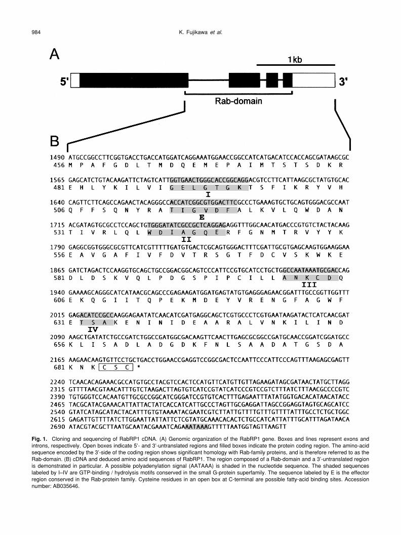
Fig. 1. Cloning and sequencing of RabRP1 cDNA. (A) Genomic organization of the RabRP1 gene. Boxes and lines represent exons andintrons, respectively. Open boxes indicate 5’- and 3’-untranslated regions and filled boxes indicate the protein coding region. The amino-acidsequence encoded by the 3’-side of the coding region shows significant homology with Rab-family proteins, and is therefore referred to as theRab-domain. (B) cDNA and deduced amino acid sequences of RabRP1. The region composed of a Rab-domain and a 3’-untranslated regionis demonstrated in particular. A possible polyadenylation signal (AATAAA) is shaded in the nucleotide sequence. The shaded sequenceslabeled by I–IV are GTP-binding / hydrolysis motifs conserved in the small G-protein superfamily. The sequence labeled by E is the effectorregion conserved in the Rab-protein family. Cysteine residues in an open box at C-terminal are possible fatty-acid binding sites. Accessionnumber: AB035646.
Characterization of RABRP1 in Drosophila 985
length sequence of RabRP1 cDNA. Fig. 1A schematicallyshows the genomic organization of the RabRP1 gene. Thegene is composed of 4 exons and 3 introns, and the cDNAcontains a long open reading frame encoding a protein com-posed of 686 amino acids. The relative molecular mass (Mr)of the protein is calculated to be 74.7 k. A possible polyade-nylation signal (AATAAA) locates near the 3’-end of thecDNA (Fig. 1B). Because the C-terminal 1/3 of the deducedamino acid sequence (Met-456 - Cys-686, Mr = 25.9 k) obvi-ously showed similarity to that of the Rab small G-proteins(see below), we referred to this region as “Rab-domain”. In
contrast, a larger N-terminal domain of the deducedsequence shared no apparent homology with any other pro-tein so far reported.
The nucleotide and the deduced amino acid sequencesof the Rab-domain are shown in Fig. 1B. The Rab-domaincontains all four motifs for GTP-binding and hydrolysis con-served in the small G-protein superfamily. In addition, thedomain possesses a consensus sequence (TIGXXF) ofRab-specific effector region. However, a unique amino-acidsubstitution of isoleucine for threonine conserved in mostsmall G-proteins was found in the second motif for GTP-
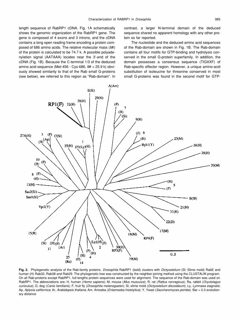
Fig. 2. Phylogenetic analysis of the Rab-family proteins. Drosophila RabRP1 (bold) clusters with Dictyostelium (Sl; Slime mold) RabE andhuman (H) Rab32, Rab38 and Rab29. The phylogenetic tree was constructed by the neighbor-joining method using the CLUSTALW program.On all Rab-proteins except RabRP1, full-lengths protein sequences were used for alignment. The sequence of the Rab-domain was used onRabRP1. The abbreviations are; H, human (Homo sapiens); M, mouse (Mus musculus); R, rat (Rattus norvegicus); Ra, rabbit (Oryctolaguscuniculus); D, dog (Canis familiaris); F, fruit fly (Drosophila melanogaster); Sl, slime mold (Dictyostelium discoideum); Ly, Lymnaea stagnalis;Ap, Aplysia californica; Ar, Arabidopsis thaliana; Am, Amoeba (Entamoeba histolytica); Y, Yeast (Saccharomyces pombe). Bar = 0.3 evolution-ary distance

K. Fujikawa et al.986
binding and hydrolysis (WDIAGQE). In the carboxyl end ofthe protein, two cysteine residues were found with a motif of–CXC. This motif, together with –CC and –CCXX, allowslipid modification of both the cysteine residues most likely tohave geranylgeranyl prenylation, which is commonly foundin the small G-proteins.
We next constructed a molecular phylogenetic tree ofRab family proteins using the Rab-domain of RABRP1. Asshown in Fig. 2, RABRP1 is clustered with DictyosteliumRabE (AF116859), and mammalian RAB29 (D84488),RAB32 (Q13637) and RAB38 (NM022337). The amino acididentities of RABRP1 with the members of the cluster werebetween 43–62%, whereas those with other Rab proteinswere below 30%.
Fig. 3 shows the sequence alignment of RABRP1 (Rab-domain) together with RabE, RAB29, RAB32, and RAB38.Sequences of some other Rab proteins are also aligned forcomparison. In addition to the five conserved motifs, several
amino acids widely conserved in the Rab family proteinswere also found in RABRP1 (K-485, Y/F-553, K/R-555, D-565, W-578, N-639). Interestingly, the amino acid substitu-tion of isoleucine for threonine in the second motif for GTPbinding and hydrolysis (WDTAGQE → WD I AGQE) alsooccurred in other members of the RabRP1 cluster (RabE,Rab29, Rab32, Rab38). Furthermore, we found anotheramino acid substitution specific to the members of theRabRP1 cluster; the substitution replacing glycine in thethird motif for GTP binding and hydrolysis by alanine(GNKCD → ANKCD). In spite of these substitutions in theconserved motifs, GTP-blot analysis demonstrated thatRABRP1 still retained the ability to bind GTP (data notshown).
Tissue-specific distribution of RABRP1
In order to investigate the tissue specificity of RabRP1gene expression, we carried out northern hybridization for
Fig. 3.
Comparison of RabRP1 amino acid sequence with other members in the RabRP1 cluster. For comparison, sequences of some otherhuman (h),
Drosophila
(D) and yeast (Yst) Rab proteins are aligned together. The amino acid residues identical to that of RabRP1 are shaded.Asterisks indicate the amino acid residues conserved in most Rab proteins. Arrowheads indicate the positions where amino acid substitutionspecific to RabRP1 and its relatives are occurring within the conserved motifs I–IV and E.
Characterization of RABRP1 in
Drosophila
987
mRNAs extracted from several kinds of
Drosophila
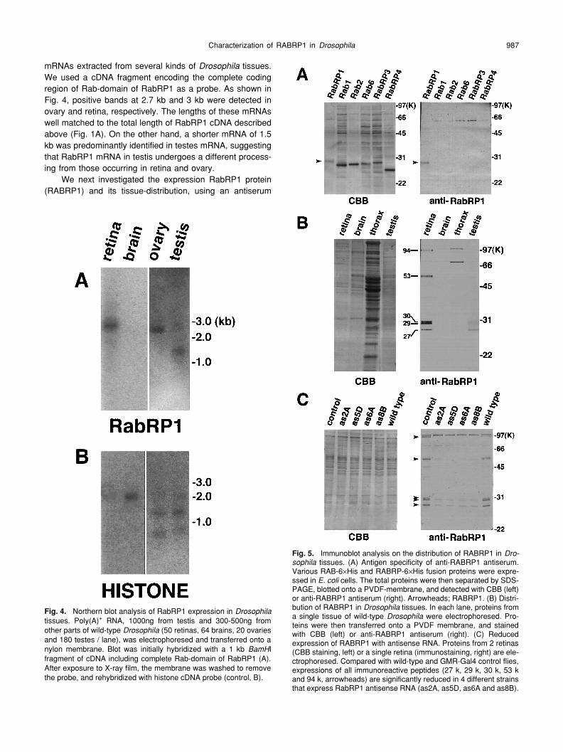
tissues.We used a cDNA fragment encoding the complete codingregion of Rab-domain of RabRP1 as a probe. As shown inFig. 4, positive bands at 2.7 kb and 3 kb were detected inovary and retina, respectively. The lengths of these mRNAswell matched to the total length of RabRP1 cDNA describedabove (Fig. 1A). On the other hand, a shorter mRNA of 1.5kb was predominantly identified in testes mRNA, suggestingthat RabRP1 mRNA in testis undergoes a different process-ing from those occurring in retina and ovary.
We next investigated the expression RabRP1 protein(RABRP1) and its tissue-distribution, using an antiserum
Fig. 4.
Northern blot analysis of RabRP1 expression in
Drosophila
tissues. Poly(A)
+
RNA, 1000ng from testis and 300-500ng fromother parts of wild-type
Drosophila
(50 retinas, 64 brains, 20 ovariesand 180 testes / lane), was electrophoresed and transferred onto anylon membrane. Blot was initially hybridized with a 1 kb
BamH
Ifragment of cDNA including complete Rab-domain of RabRP1 (A).After exposure to X-ray film, the membrane was washed to removethe probe, and rehybridized with histone cDNA probe (control, B).
Fig. 5.
Immunoblot analysis on the distribution of RABRP1 in
Dro-sophila
tissues. (A) Antigen specificity of anti-RABRP1 antiserum.Various RAB-6
×
His and RABRP-6
×
His fusion proteins were expre-ssed in
E. coli
cells. The total proteins were then separated by SDS-PAGE, blotted onto a PVDF-membrane, and detected with CBB (left)or anti-RABRP1 antiserum (right). Arrowheads; RABRP1. (B) Distri-bution of RABRP1 in
Drosophila
tissues. In each lane, proteins froma single tissue of wild-type
Drosophila
were electrophoresed. Pro-teins were then transferred onto a PVDF membrane, and stainedwith CBB (left) or anti-RABRP1 antiserum (right). (C) Reducedexpression of RABRP1 with antisense RNA. Proteins from 2 retinas(CBB staining, left) or a single retina (immunostaining, right) are ele-ctrophoresed. Compared with wild-type and GMR-Gal4 control flies,expressions of all immunoreactive peptides (27 k, 29 k, 30 k, 53 kand 94 k, arrowheads) are significantly reduced in 4 different strainsthat express RabRP1 antisense RNA (as2A, as5D, as6A and as8B).

K. Fujikawa
et al
.988
against the Rab-domain of RABRP1. Immunoblot analysisdemonstrated that the antiserum specifically recognizedRABRP1 without any crossreaction with other
Drosophila
Rab proteins (Fig. 5A). Using the antiserum, we examinedthe expression of RABRP1 in retina, brain, thorax and testis(Fig. 5B). In the retina, the antiserum recognized five clearlyseparated bands of peptides (27 k, 29 k, 30 k, 53 k and 94k), among which the 29 k band was most predominant. Inaddition, all kinds of these peptides were principally recov-ered in the membrane fraction, when retinas were separatedinto soluble and membrane fractions (data not shown). Thisresult suggests that the peptides mainly exist in a mem-brane-bound form. In testis, only smaller bands (27 k, 29 kand 30 k) were detected, and the 27 k band was predomi-nant. No specific signals of anti-RABRP1 were identified inbrain and thorax (signals at ~70 k and ~100 k were due tothe nonspecific immunoreactions of secondary antibodies),indicating that RABRP1 was exclusively expressed in theretinal and the gonadal organs.
To examine whether the five immunoreactive bands inretina are actual products of the RabRP1 gene, we performedimmunoblot analysis on the flies expressing antisenseRabRP1 RNA. In these flies, the hybridization of antisenseRNA with RabRP1 mRNA suppressed the activity of mRNA,and specifically reduced the yields of the RabRP1 geneproducts. By germline transformation, we obtained 7 strainsof transgenic flies carrying
UAS-RabRP1(antisense)
het-erozygously. Four strains of them were used for experi-ments. As shown in Fig. 5C, the yields of 5 immunoreactiveproducts in the retina were concomitantly reduced, whenantisense RNA was expressed with GAL4 under control ofthe
GMR
regulatory element. This result demonstrated thatthese 5 peptides were apparently produced from theRabRP1 mRNA. The
M
r of RABRP1 calculated from thecomplete sequence of cDNA was 74.7 k, which was signifi-cantly smaller than 94 k of the largest immunoreactive pep-tide. A possible interpretation was that the largest bandmight arise from oligomerization or post-translational modifi-cation of the smaller products.
Cellular and subcellular Localization of RABRP1 in
Drosophila
retina
We immunohistochemically investigated the localizationof RABRP1 in
Drosophila
retina. Fig. 6 shows the immuno-fluorescent staining of a fly head cryosection with anti-RABRP1 antiserum. A fluorescent signal was observed overthe entire region of the retina, whereas no signal wasdetected in the brain (Fig. 6B). This result agreed with theimmunoblot analysis (Fig. 5B). Further observation of theretina revealed that strong fluorescence was located imme-diately beneath the corneal cuticle (Fig. 6B). In detail, thefluorescence exhibited a cup-shaped profile surrounding apseudocone (Fig. 6C). This pattern of fluorescence stainingoverlapped with distribution of the retinal pigment cells, sug-gesting that RABRP1 was predominantly distributed in thecells in the retina.
Fig. 6.
Immunofluorescence localization of RABRP1 in compoundeyes of
Drosophila
. Frozen sections of compound eyes werestained with anti-RABRP1 antiserum, visualized with biotinylatedsecondary antibody and FITC-conjugated streptavidin, andobserved with a confocal laser microscope. (A) Control experimentusing preimmune serum for anti-RABRP1 antiserum. Bar, 50
µ
m. (B)A longitudinal section of the retina was stained with anti-RABRP1antiserum. Bar, 50
µ
m. (C) Higher-power magnification image of theretina in (B). Bar, 20
µ
m. r; retina, b; brain, c; corneal cuticle.
Characterization of RABRP1 in
Drosophila
989
In the photoreceptor cell layer, many fibrous signals ranalong the longitudinal axes of retinal cells, in addition to adiffuse staining over the cell cytoplasm (Fig. 6B). The fibroussignals were localized at the borders between rhabdomeresand photoreceptor cell bodies (Fig. 6C). This staining pat-tern was reminiscent of the distribution of the pigmentgranules in photoreceptor cells, as well as that of the sub-rhabdomeric cisternae (SRC) derived from the smoothendoplasmic reticulum (Matsumoto-Suzuki
et al.
, 1989). Theresults thus suggest that membrane-bound form of RABRP1was localized at one or both of these organelles in photore-ceptor cells.
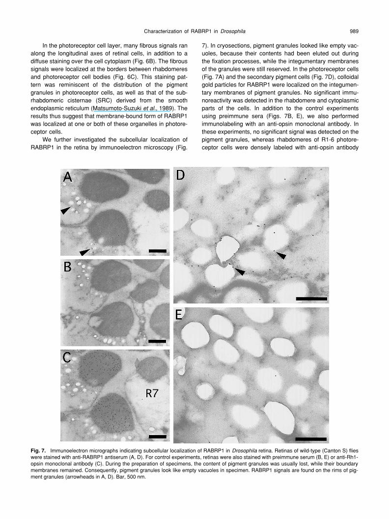
We further investigated the subcellular localization ofRABRP1 in the retina by immunoelectron microscopy (Fig.
7). In cryosections, pigment granules looked like empty vac-uoles, because their contents had been eluted out duringthe fixation processes, while the integumentary membranesof the granules were still reserved. In the photoreceptor cells(Fig. 7A) and the secondary pigment cells (Fig. 7D), colloidalgold particles for RABRP1 were localized on the integumen-tary membranes of pigment granules. No significant immu-noreactivity was detected in the rhabdomere and cytoplasmicparts of the cells. In addition to the control experimentsusing preimmune sera (Figs. 7B, E), we also performedimmunolabeling with an anti-opsin monoclonal antibody. Inthese experiments, no significant signal was detected on thepigment granules, whereas rhabdomeres of R1-6 photore-ceptor cells were densely labeled with anti-opsin antibody
Fig. 7.
Immunoelectron micrographs indicating subcellular localization of RABRP1 in
Drosophila
retina. Retinas of wild-type (Canton S) flieswere stained with anti-RABRP1 antiserum (A, D). For control experiments, retinas were also stained with preimmune serum (B, E) or anti-Rh1-opsin monoclonal antibody (C). During the preparation of specimens, the content of pigment granules was usually lost, while their boundarymembranes remained. Consequently, pigment granules look like empty vacuoles in specimen. RABRP1 signals are found on the rims of pig-ment granules (arrowheads in A, D). Bar, 500 nm.
K. Fujikawa
et al
.990
(Fig. 7C). These results thus indicated that RABRP1 wasassociated with the pigment granules both in photoreceptorand secondary pigment cells.
Accumulation of the autophagosome-like structures inthe dominant negative RabRP1 mutant
In order to address the function of RABRP1
in vivo
, wegenerated flies that expressed mutant RabRP1 proteinexhibiting the dominant inhibitory effect against nativeRABRP1. In the mutant protein (RABRP1(N601I)), isoleu-cine was substituted for asparagine in the third GTP-bindingand hydrolysis motif. It has been demonstrated that theequivalent substitution of Ile for Asn transforms native RABsto their dominant inhibitors in various kinds of Rab proteins
including Rab1 (Satoh
et al.
, 1997a; Tisdale
et al.
, 1992),Rab5 (Bucci
et al.
, 1992) and Rab7 (Feng
et al.
, 1995).The transgenic
GalUAS-RabRP1(N601I)
fly was cro-ssed with the
rh1-Gal4
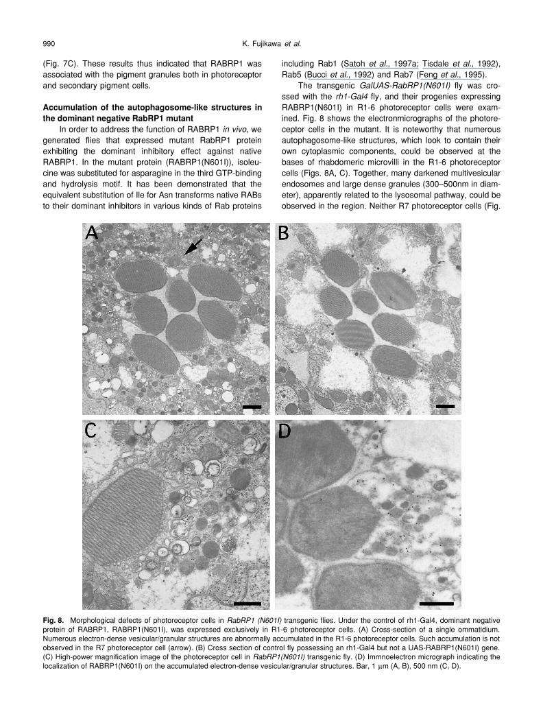
fly, and their progenies expressingRABRP1(N601I) in R1-6 photoreceptor cells were exam-ined. Fig. 8 shows the electronmicrographs of the photore-ceptor cells in the mutant. It is noteworthy that numerousautophagosome-like structures, which look to contain theirown cytoplasmic components, could be observed at thebases of rhabdomeric microvilli in the R1-6 photoreceptorcells (Figs. 8A, C). Together, many darkened multivesicularendosomes and large dense granules (300–500nm in diam-eter), apparently related to the lysosomal pathway, could beobserved in the region. Neither R7 photoreceptor cells (Fig.
Fig. 8.
Morphological defects of photoreceptor cells in
RabRP1 (N601I)
transgenic flies. Under the control of rh1-Gal4, dominant negativeprotein of RABRP1, RABRP1(N601I), was expressed exclusively in R1-6 photoreceptor cells. (A) Cross-section of a single ommatidium.Numerous electron-dense vesicular/granular structures are abnormally accumulated in the R1-6 photoreceptor cells. Such accumulation is notobserved in the R7 photoreceptor cell (arrow). (B) Cross section of control fly possessing an rh1-Gal4 but not a UAS-RABRP1(N601I) gene.(C) High-power magnification image of the photoreceptor cell in
RabRP1(N601I)
transgenic fly. (D) Immnoelectron micrograph indicating thelocalization of RABRP1(N601I) on the accumulated electron-dense vesicular/granular structures. Bar, 1
µ
m (A, B), 500 nm (C, D).
Characterization of RABRP1 in Drosophila 991
8A, arrow), in which the rh1 promoter is silent, nor R1-6 cellsof the control fly carrying only rh1-Gal4 gene (Fig. 8B) accu-mulated such autophagosome-like structures, indicating thatthe phenotype of the mutant actually resulted from theexpression of RABRP1(N601I). Furthermore, immuno-elec-tronmicroscopy revealed that RABRP1(N601I) specificallylocalized on the autophagosome-like or lysosome-relatedstructures (Fig. 8D). These results thus suggested thatRABRP1(N601I) interfered with the degradation of endo-somes and autophagosome-like structures through the lyso-somal pathway.
DISCUSSION
Unique characteristics of RABRP1In the present study, we identified a novel Rab protein,
RabRP1, in Drosophila and investigated its molecular andfunctional characteristics. Although the primary structure ofthe protein has been deduced from the genome sequence,the features of the protein actually expressed in vivo stillremain to be clarified. Here, we demonstrated several dis-tinctive features of RABRP1, which are different from thoseof other Rab proteins. First, multiple kinds of translationalproducts having different relative molecular masses (94 k,53 k, 30 k, 29 k and 27 k) were produced from the RabRP1gene in the eye. This result was confirmed by two differenttechniques; the inhibition of RABRP1 expression with anti-sense RNA and immunoblot analysis using antibodiesagainst the Rab-domain of RABRP1. Among these pep-tides, the 94 k and 53 k peptides were significantly largerthan any other Rab proteins so far found whose relativemolecular masses are between 20 k and 30 k. However,these large peptides cannot be detected in testes. In thisstudy, we also demonstrated that the large mRNA forRabRP1 (3 kb) having an extraordinarily long open readingframe (ORF) was expressed in the eye, whereas only amuch smaller mRNA (1.5 kb) was detected in testes. Fur-thermore, the Rab-like sequence (Rab-domain, 25.9 k) ofRABRP1 was encoded in the 3’-end of 3 kb mRNA ofRabRP1. These results suggested that the large peptides inthe eye are translational products of the long ORF and thuscontain additional sequences to the N-terminal of the Rab-domain. In contrast, 27–30 k peptides would be composedof the Rab-domain alone. Although multiple peptides areexpressed in the eye, the major product of the RabRP1gene was the 29 k peptide. Furthermore, no large peptideswere detectable in testes. These results suggest that theRab-domain is the actually functional domain of RABRP1 invivo. This assumption was strongly supported by the factthat amino-acid substitution in the Rab-domain (RABRP1-N601I) dominantly inhibited the function of RABRP1.
Second, RABRP1 was characterized by its tissue-spe-cific distribution in fly. A lot of the members of the Rab-pro-tein family are involved in general pathways for endocytosisand exocytosis, and are thus distributed ubiquitously inevery tissue and organ in the body. For example, RAB1 and
RAB5 are involved in the exocytotic process from rER toGolgi body (Satoh et al., 1997a) and the endocytic processfor early endosome, respectively, and are found ubiquitouslyin the body (Satoh et al., 1998). On the other hand, somekinds of Rab proteins are distributed in a tissue-specificmanner. For example, it has been reported that RAB3A andRAB17 are specifically distributed in neural (Fischer vonMollard et al., 1990) and epithelial (Lutcke et al., 1993) tis-sues, respectively. It should be noted that such tissue-spe-cific distributions of Rab proteins are usually associated withtheir specific functions in cells. For example, RAB3A func-tions in the fusion process of synaptic vesicles. In this study,we demonstrated that RABRP1 was predominantly expre-ssed in the eye and testis unlike any other Rab proteins sofar reported. This result therefore suggested that RABRP1possibly plays a unique role in these tissues. Details on theRABRP1 function are discussed later.
Third, the Rab-domain of RABRP1 included uniqueamino-acid substitutions within the conserved GTP-binding/hydrolysis motifs of the protein (Ile-540 and Ala-600).Despite these substitutions in the conserved motifs,RABRP1 still retained the ability to bind GTP. However, thisresult do not exclude the possibility that there may exist sig-nificant difference in enzyme kinetics between RABRP1 andconventional Rab proteins. Furthermore, the equivalent sub-stitutions were also found in mammalian Rab32, Rab38,Rab29 and Dictyostelium RabE proteins whose functionsremain to be elucidated. The phylogenetic analysis of Rabproteins indicated that RabRP1, together with these 4 pro-teins, formed a cluster in the phylogenetic tree. Interestingly,the recently issued human genomic DNA sequence sug-gests that human RAB32 also has an extended N-terminalregion to give a long peptide with a relative molecular massof 53 k, although its deduced N-terminal sequence showsno apparent similarity to that of RABRP1. These resultssuggest that RABRP1 shares functional similarities withmammalian and Dictyostelium Rab proteins, and possiblyregulates a common process of vesicle transport.
A possible function of RABRP1In order to elucidate the function of RABRP1, we inves-
tigated the cellular and subcellular localizations of the pro-tein and the effects of functional inhibition of RABRP1 byexpressing the dominant negative form of the protein(RABRP1N601I). Immunofluorescence microscopy of thecompound eye demonstrated that RABRP1 was predomi-nantly localized in the pigment cells just beneath the cornealcuticle. In Drosophila, pigment cells having pigment-contain-ing organelles (pigment granules) in the cytoplasm existedexclusively in compound eyes, ocelli and testes. The resultindicating the histochemical localization of RABRP1 in thepigment cells thus coincided with that of the immunoblotanalysis that demonstrated RABRP1 localizing in the eyeand testes of fly. We also investigated the subcellular local-ization of RABRP1 using immunoelectron microscopy. Theresults revealed that RABRP1 was localized on the bound-
K. Fujikawa et al.992
ary membranes of pigment granules in the retinal pigmentcells and the photoreceptor cells, suggesting that RABRP1possibly functioned in the vesicle transport pathway involvedin the formation or degradation of pigment granules. Forfunctional analysis of RABRP1, we further investigated themorphological defects of photoreceptor cells caused by theoverexpression of RABRP1N601I. The results indicated thatfunctional inhibition of RABRP1 with RABRP1N601I inducedthe abnormal accumulation of autophagosome-like struc-tures in the cells. In addition, many electron-dense multive-sicular endosomes and large dense granules, which areapparently related to lysosomal pathway, were alsoobserved in cells. The autophagosome is a cell organelleparticipating in the degradation of the endogenous proteinsand organelles through the fusion with lysosome. Thesedata therefore suggest that RABRP1 functions in the vesicletransport pathway toward lysosomes and/or its related struc-tures. Recently, in mammalian pigment cells (melanocytes),it has been suggested that melanosomes, organelles con-taining the melanin pigment, are specialized forms of lateendosomes or lysosomes, and are formed through a similarpathway to that of lysosomes (Jimbow et al., 1997; Schraer-meyer and Stieve, 1994; Vijayasaradhi et al., 1995). Fur-thermore, in Drosophila, several kinds of proteins requiredfor vesicle traffic to lysosomes are also essential for biogen-esis of pigment granules: garnet gene product, which func-tions in the delivery of proteins to pigment granules, wasidentified as the δ-subunit of the AP-3 adaptor complex thatwas involved in the Golgi-to-lysosome pathway (Cowles etal., 1997; Le Borgne et al., 1998; Ooi et al., 1997; Simpsonet al., 1997). The deep orange and light, genes required forthe biogenesis of pigment granules, encode Drosophilahomologues of the yeast VPS18 and VPS41 gene products,respectively. In the yeast, these proteins are essential forthe normal delivery of proteins to the vacuole, the yeastequivalent of the mammalian lysosome (Sevrioukov et al.,1999; Warner et al., 1998). Based on these data, it is nowassumed that the biogenesis of pigment containingorganelles (melanosomes and pigment granules) sharescommon molecular machineries with the protein transport tolysosomes. In the present study, we demonstrated thatRABRP1 was localized on pigment granules, and was pos-sibly involved in the lysosomal pathway, too. This resulttherefore supports the above assumption and suggests thatRABRP1 participates in the vesicle traffics for pigment gran-ules using the endosomal / lysosomal pathway. From thispoint of view, it is very interesting that Rab38, which belongsto the RabRP1 cluster in the Rab-phylogenetic tree, is exclu-sively localized in cultured melanocytes (Jager et al., 2000).In addition, Rab7, a member of the subfamily neighboring onthe RabRP1 cluster, is also localized on melanosomes(Gomez et al., 2001). These results suggest that RABRP1and its relatives function in the vesicle transport pathwaysfor pigment-containing organelles, both in insects and mam-mals.
ACKNOWLEDGMENTS
We thank C. Hama for providing Drosophila stocks carryingrh1-Gal4 insertion. This work was supported in part by a Grant-in-Aid for Scientific Research to K. O., and the JSPS Research for theFuture Program to S. K. K. F. was supported by the JSPS ResearchFellowships for Young Scientists.
REFERENCES
Adams M D, Celniker S E, Holt R A, Evans C A, Gocayne J D et al.(2000) The genome sequence of Drosophila melanogaster.Science 287: 2185–2195
Bucci C, Parton R G, Mather I H, Stunnenberg H, Simons K et al.(1992) The small GTPase rab5 functions as a regulatory factorin the early endocytic pathway. Cell 70: 715–728
Cowles C R, Odorizzi G, Payne G S, and Emr S D (1997) The AP-3adaptor complex is essential for cargo-selective transport to theyeast vacuole. Cell 91: 109–118
Deng W, Leaper K, and Bownes M (1999) A targeted gene silencingtechnique shows that Drosophila myosin VI is required for eggchamber and imaginal disc morphogenesis. J Cell Sci 112:3677–3690
Feng Y, Press B, and Wandinger-Ness A (1995) Rab 7: an impor-tant regulator of late endocytic membrane traffic. J Cell Biol131: 1435–1452
Fischer von Mollard G, Mignery G A, Baumert M, Perin M S, Han-son T J et al. (1990) Rab3 is a small GTP-binding protein exclu-sively localized to synaptic vesicles. Proc Natl Acad Sci U S A87: 1988–1992
Geppert M, Bolshakov V Y, Siegelbaum S A, Takei K, De Camilli Pet al. (1994) The role of Rab3A in neurotransmitter release.Nature 369: 493–497
Gomez P F, Luo D, Hirosaki K, Shinoda K, Yamashita T et al.(2001) Identification of rab7 as a melanosome-associated pro-tein involved in the intracellular transport of tyrosinase-relatedprotein 1. J Invest Dermatol 117: 81–90
Huber L A, Pimplikar S, Parton R G, Virta H, Zerial M et al. (1993)Rab8, a small GTPase involved in vesicular traffic between theTGN and the basolateral plasma membrane. J Cell Biol 123:35–45
Jager D, Stockert E, Jager E, Gure A O, Scanlan M J et al. (2000)Serological cloning of a melanocyte rab guanosine 5'-triphos-phate- binding protein and a chromosome condensation proteinfrom a melanoma complementary DNA library. Cancer Res 60:3584–3591
Jimbow K, Gomez P F, Toyofuku K, Chang D, Miura S et al (1997)Biological role of tyrosinase related protein and its biosynthesisand transport from TGN to stage I melanosome, late endo-some, through gene transfection study. Pigment Cell Res 10:206–213
Laemmli U K (1970) Cleavage of structural proteins during theassembly of the head of bacteriophage T4. Nature 227: 680–685
Le Borgne R, Alconada A, Bauer U, and Hoflack B (1998) Themammalian AP-3 adaptor-like complex mediates the intracellu-lar transport of lysosomal membrane glycoproteins. J BiolChem 273: 29451–29461
Lutcke A, Jansson S, Parton R G, Chavrier P, Valencia A et al(1993) Rab17, a novel small GTPase, is specific for epithelialcells and is induced during cell polarization. J Cell Biol 121:553–564
Matsumoto-Suzuki E, Hirosawa K, and Hotta Y (1989) Structure ofthe subrhabdomeric cisternae in the photoreceptor cells ofDrosophila melanogaster. J Neurocytol 18: 87–93
Norian L, Dragoi I A, and O’Halloran T (1999) Molecular character-
Characterization of RABRP1 in Drosophila 993
ization of rabE, a developmentally regulated Dictyosteliumhomolog of mammalian rab GTPases. DNA Cell Biol 18: 59–64
Novick P, and Brennwald P (1993) Friends and family: the role ofthe Rab GTPases in vesicular traffic. Cell 75: 597–601
Novick P, and Zerial M (1997) The diversity of Rab proteins in vesi-cle transport. Curr Opin Cell Biol 9: 496–504
Ooi C E, Moreira J E, Dell’Angelica E C, Poy G, Wassarman D A etal (1997) Altered expression of a novel adaptin leads to defec-tive pigment granule biogenesis in the Drosophila eye colormutant garnet. Embo J 16: 4508–4518
Pereira-Leal J B, and Seabra M C (2000) The mammalian Rabfamily of small GTPases: definition of family and subfamilysequence motifs suggests a mechanism for functional specific-ity in the Ras superfamily. J Mol Biol 301: 1077–1087
Pereira-Leal J B, and Seabra M C (2001) Evolution of the Rab fam-ily of small GTP-binding proteins. J Mol Biol 313: 889–901
Satoh A, Tokunaga F, Kawamura S, and Ozaki K (1997a) In situinhibition of vesicle transport and protein processing in thedominant negative Rab1 mutant of Drosophila. J Cell Sci 110:2943–2953
Satoh A K, Nagatani H, Tokunaga F, Kawamura S, and Ozaki K(1998) Rhodopsin transport and Rab expression in the caro-tenoid-deprived Drosophila melanogaster. Zool Sci 15: 651–659
Satoh A K, Tokunaga F, and Ozaki K (1997b) Rab proteins ofDrosophila melanogaster: novel members of the Rab- proteinfamily. FEBS Lett 404: 65–69
Schraermeyer U, and Stieve H (1994) A newly discovered pathwayof melanin formation in cultured retinal pigment epithelium ofcattle. Cell Tissue Res 276: 273–279
Sevrioukov E A, He J P, Moghrabi N, Sunio A, and Kramer H (1999)A role for the deep orange and carnation eye color genes inlysosomal delivery in Drosophila. Mol Cell 4: 479–486
Simons K, and Zerial M (1993) Rab proteins and the road maps forintracellular transport. Neuron 11: 789–799
Simpson F, Peden A A, Christopoulou L, and Robinson M S (1997)Characterization of the adaptor-related protein complex, AP-3.J Cell Biol 137: 835–845
Tisdale E J, Bourne J R, Khosravi-Far R, Der C J, and Balch W E(1992) GTP-binding mutants of rab1 and rab2 are potent inhibi-tors of vesicular transport from the endoplasmic reticulum to theGolgi complex. J Cell Biol 119: 749–761
Vijayasaradhi S, Xu Y, Bouchard B, and Houghton A N (1995) Intra-cellular sorting and targeting of melanosomal membrane pro-teins: identification of signals for sorting of the human brownlocus protein, gp75. J Cell Biol 130: 807–820
Warner T S, Sinclair D A, Fitzpatrick K A, Singh M, Devlin R H et al.(1998) The light gene of Drosophila melanogaster encodes ahomologue of VPS41, a yeast gene involved in cellular-proteintrafficking. Genome 41: 236–243
Zacchi P, Stenmark H, Parton R G, Orioli D, Lim F et al. (1998)Rab17 regulates membrane trafficking through apical recyclingendosomes in polarized epithelial cells. J Cell Biol 140: 1039–1053
(Received April 16, 2002 / Accepted June 13, 2002)