antibacterial and antifungal potential of some medicinal plants against certain phytopathogenic...
TRANSCRIPT
This article was downloaded by: [Maulana Azad Library]On: 13 December 2013, At: 21:31Publisher: Taylor & FrancisInforma Ltd Registered in England and Wales Registered Number: 1072954 Registeredoffice: Mortimer House, 37-41 Mortimer Street, London W1T 3JH, UK
Archives Of Phytopathology And PlantProtectionPublication details, including instructions for authors andsubscription information:http://www.tandfonline.com/loi/gapp20
Antibacterial and antifungal potentialof some medicinal plants againstcertain phytopathogenic micro-organismsMuzafar Sheikh a , A. Safiuddin a , Zehra Khan a , Rose Rizvi a &Irshad Mahmood aa Plant Pathology Laboratory, Department of Botany , AligarhMuslim University , Aligarh , IndiaPublished online: 30 Jan 2013.
To cite this article: Muzafar Sheikh , A. Safiuddin , Zehra Khan , Rose Rizvi & Irshad Mahmood(2013) Antibacterial and antifungal potential of some medicinal plants against certainphytopathogenic micro-organisms, Archives Of Phytopathology And Plant Protection, 46:9,1070-1080, DOI: 10.1080/03235408.2012.757859
To link to this article: http://dx.doi.org/10.1080/03235408.2012.757859
PLEASE SCROLL DOWN FOR ARTICLE
Taylor & Francis makes every effort to ensure the accuracy of all the information (the“Content”) contained in the publications on our platform. However, Taylor & Francis,our agents, and our licensors make no representations or warranties whatsoever as tothe accuracy, completeness, or suitability for any purpose of the Content. Any opinionsand views expressed in this publication are the opinions and views of the authors,and are not the views of or endorsed by Taylor & Francis. The accuracy of the Contentshould not be relied upon and should be independently verified with primary sourcesof information. Taylor and Francis shall not be liable for any losses, actions, claims,proceedings, demands, costs, expenses, damages, and other liabilities whatsoever orhowsoever caused arising directly or indirectly in connection with, in relation to or arisingout of the use of the Content.
This article may be used for research, teaching, and private study purposes. Anysubstantial or systematic reproduction, redistribution, reselling, loan, sub-licensing,systematic supply, or distribution in any form to anyone is expressly forbidden. Terms &
Conditions of access and use can be found at http://www.tandfonline.com/page/terms-and-conditions
Dow
nloa
ded
by [
Mau
lana
Aza
d L
ibra
ry]
at 2
1:31
13
Dec
embe
r 20
13
Antibacterial and antifungal potential of some medicinal plantsagainst certain phytopathogenic micro-organisms
Muzafar Sheikh*, A. Safiuddin, Zehra Khan, Rose Rizvi and Irshad Mahmood
Plant Pathology Laboratory, Department of Botany, Aligarh Muslim University, Aligarh, India
(Received 6 December 2012; final version received 7 December 2012)
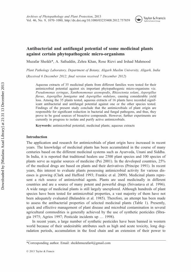
Aqueous extracts of 35 medicinal plants from different families were tested for theirantimicrobial potential against six important phytopathogenic micro-organisms viz.Pseudomonas syringae, Xanthonomonas axonopodis, Rhizoctonia solani, Aspergillusflavus, Aspergillus fumigatus and Aspergillus nidulans, causing considerable yieldloss. Among the 35 plants tested, aqueous extracts of 16 plants have recorded signif-icant antibacterial and antifungal potential against one or the other species tested.Findings of the present study conclude that the antimicrobials of plant origin areresponsible for significant reduction in bacterial and fungal pathogens, and thus, theyprove to be good sources of bioactive compounds. However, further experiments arecurrently in progress to isolate and purify active antimicrobials.
Keywords: antimicrobial potential; medicinal plants; aqueous extracts
Introduction
The application and research for antimicrobials of plant origin have increased in recentyears. The knowledge of medicinal plants has been accumulated in the course of manycenturies based on the different medicinal systems such as Ayurveda, Unani and Siddha.In India, it is reported that traditional healers use 2500 plant species and 100 species ofplants serve as regular sources of medicine (Pei 2001). In the developed countries, 25%of the medical drugs are based on plants and their derivatives (Principe 1991). In recentyears, this interest to evaluate plants possessing antimicrobial activity for various dis-eases is growing (Clark and Hufford 1993; Frankic et al. 2009). Medicinal plants repre-sent a rich source of antimicrobial agents. Plants are used medicinally in differentcountries and are a source of many potent and powerful drugs (Srivastava et al. 1996).A wide range of medicinal plants is still largely unexplored. Although hundreds of plantspecies have been tested for antimicrobial properties, a vast majority of them have notbeen adequately evaluated (Balandrin et al. 1985). Therefore, an attempt has been madeto assess the antibacterial properties of selected medicinal plants (Table 1). Presently,quick and effective management of plant disease and microbial contamination in severalagricultural commodities is generally achieved by the use of synthetic pesticides (Sbra-gia 1975; Agrios 1997; Pesticide incidents up … 1998).
In recent years, a large number of synthetic pesticides have been banned in westernworld because of their undesirable attributes such as high and acute toxicity, long deg-radation periods, accumulation in the food chain and an extension of their power to
*Corresponding author. Email: [email protected]
Archives of Phytopathology and Plant Protection, 2013Vol. 46, No. 9, 1070–1080, http://dx.doi.org/10.1080/03235408.2012.757859
� 2013 Taylor & Francis
Dow
nloa
ded
by [
Mau
lana
Aza
d L
ibra
ry]
at 2
1:31
13
Dec
embe
r 20
13
destroy both useful and harmful pests. In developing countries such as India, they arestill being used despite their harmful effects (Pharmacopoeia 1996; Wodageneh andWulp 1997). Many pathogenic micro-organisms and insect pests have developed resis-tance against chemical pesticides (May 1985; Urech et al. 1997; Williams and Heymann1998; Witte 1998). This seriously hinders the management of diseases of crops andagricultural products (Dekker 1987). Considering the deleterious effects of syntheticpesticides on life supporting systems, there is an urgent need for alternative agents formanagement of phytopathogenic micro-organisms (Bolkan and Reinert 1994; Rice et al.1998). However, sometimes even very strong antagonists become in effective in naturalsoil, from which they were not isolated. The major constraint in the direct use of antag-onists is ensuring sufficient quantities for application on field scale. This is not onlytedious, but uneconomical also; and the mere identification of antagonistic bacteriain vitro need not necessarily correlate with antagonism in the field. Under field condi-tions, the biocontrol agent (bacterium/fungus) has to face competition with other soilmicrobes, which may have negative effect on its efficacy. Green plants represent areservoir of effective chemotherapeutants and can provide valuable sources of naturalpesticides (Balandrin et al. 1985; Hostettmann and Wolfender 1997). Reports are
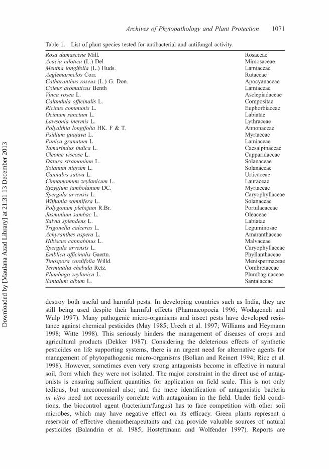
Table 1. List of plant species tested for antibacterial and antifungal activity.
Rosa damascene Mill. RosaceaeAcacia nilotica (L.) Del MimosaceaeMentha longifolia (L.) Huds. LamiaceaeAeglemarmelos Corr. RutaceaeCatharanthus roseus (L.) G. Don. ApocyanaceaeColeus aromaticus Benth LamiaceaeVinca rosea L. AsclepiadaceaeCalandula officinalis L. CompositaeRicinus communis L. EuphorbiaceaeOcimum sanctum L. LabiataeLawsonia inermis L. LythraceaePolyalthia longifolia HK. F & T. AnnonaceaePsidium guajava L. MyrtaceaePunica granatum L LamiaceaeTamarindus indica L. CaesalpinaceaeCleome viscose L. CapparidaceaeDatura stramonium L. SolanaceaeSolanum nigrum L. SolanaceaeCannabis sativa L. UrticaceaeCinnamomum zeylanicum L. LauraceaeSyzygium jambolanum DC. MyrtaceaeSpergula arvensis L. CaryophyllaceaeWithania somnifera L. SolanaceaePolygonum plebejum R.Br. PortulacaceaeJasminium sambac L. OleaceaeSalvia splendens L. LabiataeTrigonella calceras L. LeguminosaeAchyranthes aspera L. AmaranthaceaeHibiscus cannabinus L. MalvaceaeSpergula arvensis L. CaryophyllaceaeEmblica officinalis Gaertn. PhyllanthaceaeTinospora cordifolia Willd. MenispermaceaeTerminalia chebula Retz. CombretaceaePlumbago zeylanica L. PlumbaginaceaeSantalum album L. Santalaceae
Archives of Phytopathology and Plant Protection 1071
Dow
nloa
ded
by [
Mau
lana
Aza
d L
ibra
ry]
at 2
1:31
13
Dec
embe
r 20
13
available on the use of several plant byproducts, which possess antimicrobial propertiesagainst several pathogenic bacteria and fungi (Deans and Svoboda 1990; Heisey andGorham 1992; De Pooter et al. 1995; Lis-Balchin and Deans 1996; Hili et al. 1997;Iqbal and Arina 2000; Bassam et al. 2006; Doughari et al. 2008; Gandhiraja et al.2009; Bhattacharjee et al. 2011; Olukayode et al. 2011).
Necessity of the present work
Being aware of the problems of environmental degradation caused by chemical pesti-cides used in agriculture, we should switch to sources of botanical biocides as they arecomponents of the ecosystem. There are no environmental problems unlike what is seenwith broad-spectrum pesticides. Besides the targets, they also harm non-target organ-isms. Botanical biocides are environmentally safe because they do not cause socio-eco-nomic and environmental problems that encountered very often with chemicalpesticides.
The objective of this work was to evaluate and compare the antimicrobial potentialof aqueous extracts of the different medicinal plants commonly used by the people ofwestern Uttar Pradesh, India.
Materials and methods
Sample preparation
Fresh leaves of plants mentioned in Table 1 were collected together and shade dried.Since certain compounds get denatured in sunlight, they were dried under shade toavoid decomposition. The dried leaves were then pulverised well in an UDYcyclone mill. About 20 g of the powdered leaves were soaked in 100ml of distilledwater. It was left for 24 h so that alkaloids, terpenoids and other constituents, ifpresent, get dissolved. The aqueous extract was then filtered using Whatmann 41 fil-ter paper. It was again filtered through sodium sulphate in order to remove thetraces of moisture.
Microbial samples
Authentic pure culture of phytopathogenic Pseudomonas syringae, Xanthonomonasaxonopodis, Rhizoctonia solani were obtained from Microbial Type Culture CollectionCentre (MTCC), Institute of Microbial Technology (IMTECH), Chandigarh in lyophi-lised vials under MTCC NOs. 7620, 7444 and 4633.
The cultures were first revived in nutrient broth using standard protocol as suppliedby the MTCC, IMTECH along with the culture. Revived culture was maintained onAgar slants and preserved in refrigerator.
Composition of Nutrient Agar (Cappuccino and Sherman 1992)
Peptone 5 g/lBeef extract 3 g/lAgar 15 g/l
Composition of SDA medium (Sabouroud 1892)Dextrose 40 g/lPeptone 10 g/lAgar 15 g/l
1072 M. Sheikh et al.
Dow
nloa
ded
by [
Mau
lana
Aza
d L
ibra
ry]
at 2
1:31
13
Dec
embe
r 20
13
The cultures of fungi Aspergillus fumigatus, Aspergillus flavus and Aspergillus nidulanswere obtained from IMTECH, Chandigarh in lyophilised vial under MTCC NOs. 7132,7992 and 10584, respectively. The culture was maintained on the sabouraud dextroseagar (SDA) medium using standard protocols.
Antimicrobial assay
Media preparation
Thirty-six grams of Muller–Hinton Media (Hi-Media) was mixed with distilled waterand then sterilised in autoclave at 15 lb pressure for 15min. The sterilised media werepoured into Petri dishes. The solidified plates were bored with 5mm diameter corkbearer. The plates with wells were used for the antimicrobial studies.
The paper disc diffusion method was used to screen the antibacterial activity ofplant extracts and performed by using Mueller–Hinton agar (MHA). The experimentwas carried out according to the National Committee for Clinical Laboratory StandardsGuidelines (NCCLS 1999). Test bacteria were grown in Mueller–Hinton broth. Bacterialsuspension was diluted with sterile physiological solution to 108 cfuml�1 (turbid-ity =McFarland standard 0.5). One hundred microlitres of bacterial suspension wereswabbed uniformly on surface of MHA and the inocula were allowed to dry for 5min.Sterilised filter paper discs (Whatman, 6mm in diameter) were placed on the surface ofthe MHA and soaked with 20 μl of a solution of each plant extract (500 μg (Merck-Darmstadt, Germany). The inoculated plates were stored at 4 °C for 2 h and then incu-bated at 37 °C for 24 h in the inverted position. The aqueous extract of 500 μg wastested against the bacteria under study for antimicrobial activity. It was demonstrated bywell diffusion method. Three replicates were produced for each extract and the resultsare expressed as mean along with the standard deviation of three parallel measurements(Dhigra and James 1995).
Two hundred grams of potato slices were boiled with distilled water. The potatoinfusion was used as water source of media preparation. Twenty grams of dextrose wasmixed with potato infusion. Twenty grams of agar was added as a solidifying agent.These constituents were mixed and autoclaved. The solidified plates were bored with6mm diameter cork borer. The plates with wells were used for antifungal studies.
Antifungal activity of the plant extract
The aqueous extract of 100, 200 and 500 μg were tested against different fungal patho-gens for their antifungal activity. It was demonstrated by well diffusion assay.
Antifungal activities of the plant extract were tested using Well diffusion method(Baur et al. 1996). The prepared culture plates were inoculated with different selectedstrains of bacteria and fungi using streak plate method. Wells were made on the agarsurface with 6mm cork borer. The extracts were poured into the wells using sterile syr-inge. The plates were incubated at 37 ± 2 °C for 24 h for bacterial and 25 ± 2 °C for 48 hfor fungal activity. The plates were observed for the zone clearance around the wells.
Results
Data on antimicrobial activity measured as a zone of inhibition (ZOI) of aqueousextracts of 35 medicinal plants and two antibiotics against P. syringae, X. axonopodis,
Archives of Phytopathology and Plant Protection 1073
Dow
nloa
ded
by [
Mau
lana
Aza
d L
ibra
ry]
at 2
1:31
13
Dec
embe
r 20
13
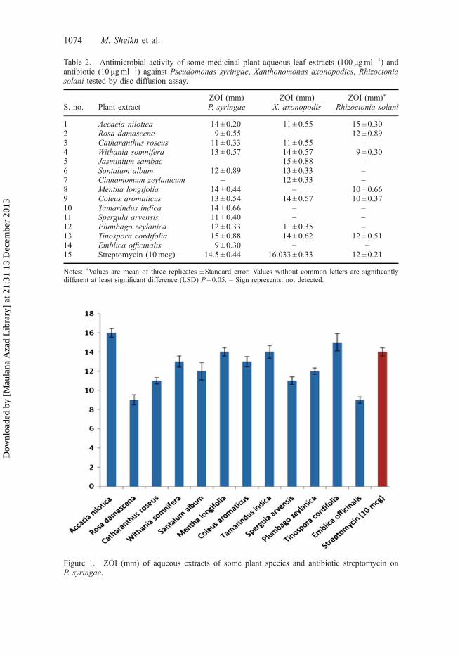
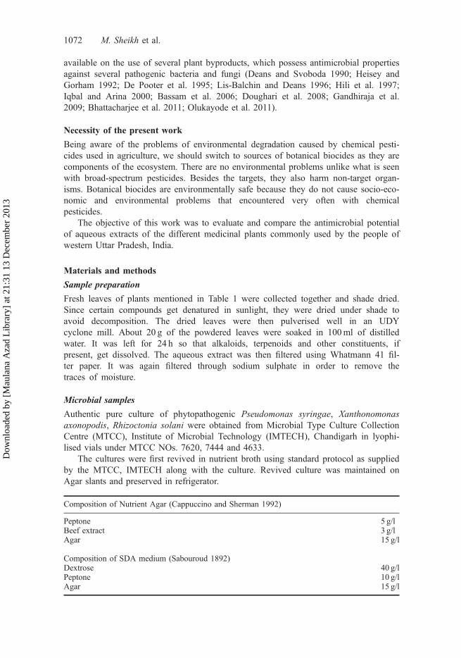
Table 2. Antimicrobial activity of some medicinal plant aqueous leaf extracts (100 μgml�1) andantibiotic (10 μgml�1) against Pseudomonas syringae, Xanthonomonas axonopodies, Rhizoctoniasolani tested by disc diffusion assay.
S. no. Plant extractZOI (mm) ZOI (mm) ZOI (mm)⁄
P. syringae X. axonopodis Rhizoctonia solani
1 Accacia nilotica 14 ± 0.20 11 ± 0.55 15 ± 0.302 Rosa damascene 9 ± 0.55 – 12 ± 0.893 Catharanthus roseus 11 ± 0.33 11 ± 0.55 –4 Withania somnifera 13 ± 0.57 14 ± 0.57 9 ± 0.305 Jasminium sambac – 15 ± 0.88 –6 Santalum album 12 ± 0.89 13 ± 0.33 –7 Cinnamomum zeylanicum – 12 ± 0.33 –8 Mentha longifolia 14 ± 0.44 – 10 ± 0.669 Coleus aromaticus 13 ± 0.54 14 ± 0.57 10 ± 0.3710 Tamarindus indica 14 ± 0.66 – –11 Spergula arvensis 11 ± 0.40 – –12 Plumbago zeylanica 12 ± 0.33 11 ± 0.35 –13 Tinospora cordifolia 15 ± 0.88 14 ± 0.62 12 ± 0.5114 Emblica officinalis 9 ± 0.30 – –15 Streptomycin (10mcg) 14.5 ± 0.44 16.033 ± 0.33 12 ± 0.21
Notes: ⁄Values are mean of three replicates ± Standard error. Values without common letters are significantlydifferent at least significant difference (LSD) P= 0.05. – Sign represents: not detected.
Figure 1. ZOI (mm) of aqueous extracts of some plant species and antibiotic streptomycin onP. syringae.
1074 M. Sheikh et al.
Dow
nloa
ded
by [
Mau
lana
Aza
d L
ibra
ry]
at 2
1:31
13
Dec
embe
r 20
13
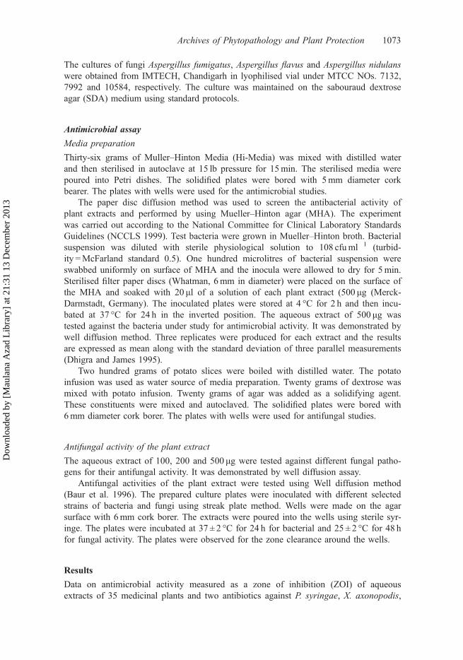
R. solani (Table 2, Figures 1–3) and antifungal activity against, A. fumigatus, A. flavusand A. nidulans (Table 3, Figures 4–6) is explained below.
As is evident from Table 2 leaf extracts of Acacia nilotica, Rosa damascena,Catharanthus roseus, Withania somnifera, Jasminium sambac, Santalum album,Cinnamomum zeylanicum, Mentha longifolia, Coleus aromaticus, Tamarindus indica,Spergula arvensis, Plumbago zeylanica, Tinospora cordifolia, Emblica officinalis formedsignificant ZOI around the well in nutrient agar Petri dishes. Aqueous extract of leaves ofother plants listed in Table 1 did not form any zone. When tested by the disc diffusion
Figure 2. ZOI (mm) of aqueous extracts of some plant species and antibiotic streptomycin onX. axonopodis.
Figure 3. ZOI (mm) of aqueous extracts of some plant species and antibiotic streptomycin onR. solani.
Archives of Phytopathology and Plant Protection 1075
Dow
nloa
ded
by [
Mau
lana
Aza
d L
ibra
ry]
at 2
1:31
13
Dec
embe
r 20
13
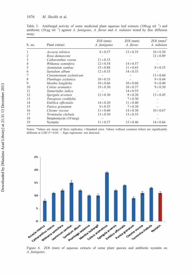
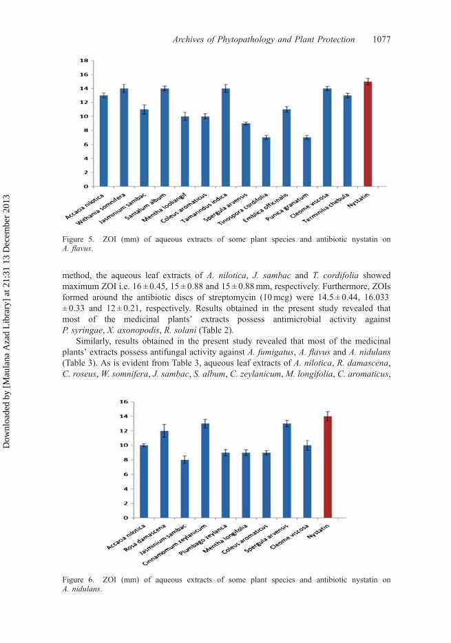
Table 3. Antifungal activity of some medicinal plant aqueous leaf extracts (100 μg ml�1) andantibiotic (10 μg ml�1) against A. fumigatus, A. flavus and A. nidulans tested by disc diffusionassay.
S. no. Plant extractZOI (mm) ZOI (mm) ZOI (mm)⁄
A. fumigatus A. flavus A. nidulans
1 Accacia nilotica 8 ± 0.57 13 ± 0.35 10 ± 0.202 Rosa damascene – – 12 ± 0.893 Catharanthus roseus 11 ± 0.33 – –4 Withania somnifera 12 ± 0.54 14 ± 0.57 –5 Jasminium sambac 15 ± 0.88 11 ± 0.65 8 ± 0.556 Santalum album 12 ± 0.33 14 ± 0.33 –7 Cinnamomum zeylanicum – – 13 ± 0.608 Plumbago zeylanica 10 ± 0.33 – 9 ± 0.449 Mentha longifolia 10 ± 0.66 10 ± 0.60 9 ± 0.4010 Coleus aromatics 19 ± 0.30 10 ± 0.37 9 ± 0.3011 Tamarindus indica – 14 ± 0.55 –12 Spergula arvensis 12 ± 0.30 9 ± 0.20 13 ± 0.4513 Tinospora cordifolia – 7 ± 0.30 –14 Emblica officinalis 14 ± 0.20 11 ± 0.40 –15 Punica granatum 9 ± 0.55 7 ± 0.30 –16 Cleome viscose 11 ± 0.60 14 ± 0.30 10 ± 0.6717 Terminalia chebula 13 ± 0.30 13 ± 0.33 –18 Streptomycin (10mcg) – – –19 Nystatin 11 ± 0.57 15 ± 0.46 14 ± 0.66
Notes: ⁄Values are mean of three replicates. ± Standard error. Values without common letters are significantlydifferent at LSD P= 0.05. – Sign represents: not detected.
Figure 4. ZOI (mm) of aqueous extracts of some plant species and antibiotic nystatin onA. fumigatus.
1076 M. Sheikh et al.
Dow
nloa
ded
by [
Mau
lana
Aza
d L
ibra
ry]
at 2
1:31
13
Dec
embe
r 20
13
method, the aqueous leaf extracts of A. nilotica, J. sambac and T. cordifolia showedmaximum ZOI i.e. 16 ± 0.45, 15 ± 0.88 and 15 ± 0.88mm, respectively. Furthermore, ZOIsformed around the antibiotic discs of streptomycin (10mcg) were 14.5 ± 0.44, 16.033± 0.33 and 12 ± 0.21, respectively. Results obtained in the present study revealed thatmost of the medicinal plants’ extracts possess antimicrobial activity againstP. syringae, X. axonopodis, R. solani (Table 2).
Similarly, results obtained in the present study revealed that most of the medicinalplants’ extracts possess antifungal activity against A. fumigatus, A. flavus and A. nidulans(Table 3). As is evident from Table 3, aqueous leaf extracts of A. nilotica, R. damascena,C. roseus,W. somnifera, J. sambac, S. album, C. zeylanicum,M. longifolia, C. aromaticus,
Figure 5. ZOI (mm) of aqueous extracts of some plant species and antibiotic nystatin onA. flavus.
Figure 6. ZOI (mm) of aqueous extracts of some plant species and antibiotic nystatin onA. nidulans.
Archives of Phytopathology and Plant Protection 1077
Dow
nloa
ded
by [
Mau
lana
Aza
d L
ibra
ry]
at 2
1:31
13
Dec
embe
r 20
13
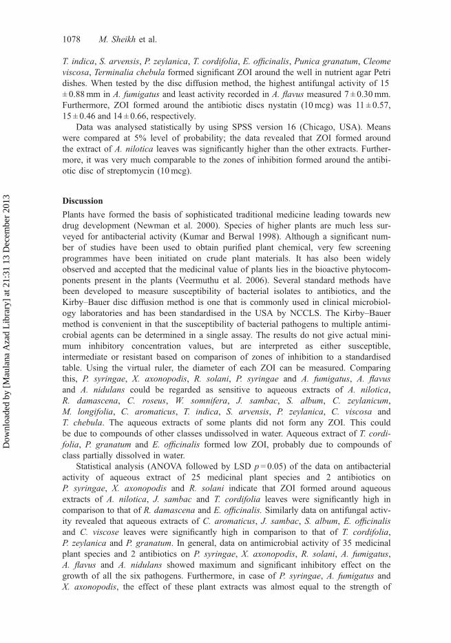
T. indica, S. arvensis, P. zeylanica, T. cordifolia, E. officinalis, Punica granatum, Cleomeviscosa, Terminalia chebula formed significant ZOI around the well in nutrient agar Petridishes. When tested by the disc diffusion method, the highest antifungal activity of 15± 0.88mm in A. fumigatus and least activity recorded in A. flavus measured 7 ± 0.30mm.Furthermore, ZOI formed around the antibiotic discs nystatin (10mcg) was 11 ± 0.57,15 ± 0.46 and 14 ± 0.66, respectively.
Data was analysed statistically by using SPSS version 16 (Chicago, USA). Meanswere compared at 5% level of probability; the data revealed that ZOI formed aroundthe extract of A. nilotica leaves was significantly higher than the other extracts. Further-more, it was very much comparable to the zones of inhibition formed around the antibi-otic disc of streptomycin (10mcg).
Discussion
Plants have formed the basis of sophisticated traditional medicine leading towards newdrug development (Newman et al. 2000). Species of higher plants are much less sur-veyed for antibacterial activity (Kumar and Berwal 1998). Although a significant num-ber of studies have been used to obtain purified plant chemical, very few screeningprogrammes have been initiated on crude plant materials. It has also been widelyobserved and accepted that the medicinal value of plants lies in the bioactive phytocom-ponents present in the plants (Veermuthu et al. 2006). Several standard methods havebeen developed to measure susceptibility of bacterial isolates to antibiotics, and theKirby–Bauer disc diffusion method is one that is commonly used in clinical microbiol-ogy laboratories and has been standardised in the USA by NCCLS. The Kirby–Bauermethod is convenient in that the susceptibility of bacterial pathogens to multiple antimi-crobial agents can be determined in a single assay. The results do not give actual mini-mum inhibitory concentration values, but are interpreted as either susceptible,intermediate or resistant based on comparison of zones of inhibition to a standardisedtable. Using the virtual ruler, the diameter of each ZOI can be measured. Comparingthis, P. syringae, X. axonopodis, R. solani, P. syringae and A. fumigatus, A. flavusand A. nidulans could be regarded as sensitive to aqueous extracts of A. nilotica,R. damascena, C. roseus, W. somnifera, J. sambac, S. album, C. zeylanicum,M. longifolia, C. aromaticus, T. indica, S. arvensis, P. zeylanica, C. viscosa andT. chebula. The aqueous extracts of some plants did not form any ZOI. This couldbe due to compounds of other classes undissolved in water. Aqueous extract of T. cordi-folia, P. granatum and E. officinalis formed low ZOI, probably due to compounds ofclass partially dissolved in water.
Statistical analysis (ANOVA followed by LSD p= 0.05) of the data on antibacterialactivity of aqueous extract of 25 medicinal plant species and 2 antibiotics onP. syringae, X. axonopodis and R. solani indicate that ZOI formed around aqueousextracts of A. nilotica, J. sambac and T. cordifolia leaves were significantly high incomparison to that of R. damascena and E. officinalis. Similarly data on antifungal activ-ity revealed that aqueous extracts of C. aromaticus, J. sambac, S. album, E. officinalisand C. viscose leaves were significantly high in comparison to that of T. cordifolia,P. zeylanica and P. granatum. In general, data on antimicrobial activity of 35 medicinalplant species and 2 antibiotics on P. syringae, X. axonopodis, R. solani, A. fumigatus,A. flavus and A. nidulans showed maximum and significant inhibitory effect on thegrowth of all the six pathogens. Furthermore, in case of P. syringae, A. fumigatus andX. axonopodis, the effect of these plant extracts was almost equal to the strength of
1078 M. Sheikh et al.
Dow
nloa
ded
by [
Mau
lana
Aza
d L
ibra
ry]
at 2
1:31
13
Dec
embe
r 20
13
streptomycin and nystatin (10mcg). The present study evaluated the antibacterial andantifungal potential of leaf extract of 35 medicinal plant species. So, the use of studiedplants as anti-infective agents in the ayurvedic medicine has been justified. Theantibacterial and antifungal potential of leaf extracts of medicinal plants is probably dueto their high terpene content. Terpenes are biologically active molecules and areconsidered to be part of plants’ defence systems.
AcknowledgementsGrateful thanks are expressed to Prof. Irshad Mahmood for his helpful suggestions and criticalreading of the manuscript. The authors are thankful to the Chairman, Department of Botany, forproviding necessary research facilities.
ReferencesAgrios GN. 1997. Control of plant diseases. In: Plant pathology. 4th ed. San Diego, CA:
Academic Press. p. 200–216.Balandrin MF, Klocke JA, Wurtele ES, Bollinger WH. 1985. Natural plant chemicals: sources of
industrial and medicinal materials. Science. 228(4704):1154–1160.Bassam AS, Ghaleb A, Naser J, Awni AH, Kamel A. 2006. Antibacterial activity of four plant
extracts used in palestine in folkloric medicine against methicillin-resistant Staphylococcusaureus. Turk J Biol. 30:195–198.
Baur AW, Kirby WM, Sherris JS, Turck M. 1996. Antibiotic susceptibility testing by a standardsingle disk method. Am J Clin Pathol. 45:493–496.
Bhattacharjee I, Chatterjee SK, Ghosh A, Chandra G. 2011. Antibacterial activities of some plantextracts used in Indian traditional folk medicine. Asian Pac J Trop Biomed. S165–S169.
Bolkan HA, Reinert WR. 1994. Developing and implementing IPM strategies to assist farmers:an industry approach. J Plant Dis. 78:545–550.
Cappuccino JG, Sherman N. 1992. Biochemical activities of microorganisms. In: Microbiology, Alaboratory manual. San Francisco, CA: The Benjamin/Cummings Publishing Co.
Clark AM, Hufford CD. 1993. Discco and development of novel prototype antibiotics foropportunistic infections related to the acquired immunodeficiency syndrome. In: Humanmedical agents from plants. Washington (DC): American Chemical Society (ACS Symposiumseries 534). p. 228–241.
De Pooter HL, Aboutabl EA, Shabrawy AO. 1995. Chemical composition and antimicrobialactivity of essential oil of leaf, stem and rhizome of Alpinia speciosa (J.C. Wendl.) K. Schum.grown in Egypt. Flavour Frag J. 10:63–67.
Deans SG, Svoboda KP. 1990. Biotechnology and bioactivity of culinary and medicinal plants.AgBiotech News Inf. 2:211–216.
Dekker J. 1987. The risks for development of fungicide resistance a worldwide problem. In: Mag-aIlona, editor. Proceedings of the 11th International Congress of Plant Protection; Oct 5–7;Manila, Philippines. p. 318–321.
Dhigra DO, James B. 1995. Basic plant pathology methods. London: CRC Press, Vol. 1, p. 287–305.
Doughari JH, EL-mahmood AM, Tyoyina I. 2008. Antimicrobial activity of leaf extracts of Sennaobtusifolia (L). Afr J Pharm Pharmacol. 2(1):7–13.
Frankic T, Voljc M, Salobir J, Rezar V. 2009. Use of herbs and spices and their extracts in animalnutrition. Acta Agri Scand. 94(2):95–102.
Gandhiraja N, Sriram S, Meena V, Kavita Srilakshmi J, Sasikumar C, Rajeshwari R. 2009.Phytochemical screening and antimicrobial activity of the plant extracts of Mimosa pudica L.against selected microbes. Ethnobot. leaflets. 13:618–624.
Heisey RM, Gorham BK. 1992. Antimicrobial effects of plant extracts on Streptococcus mutans,Candida albicans, Trichophyton rubrum and other micro-organisms. Lett Appl Microbiol.14:136–139.
Hili P, Evans CS, Veness RG. 1997. Antimicrobial action of essential oils: the effect of dimethylsulphoxide on the activity of cinnamon oil. Lett Appl Microbiol. 24:269–275.
Archives of Phytopathology and Plant Protection 1079
Dow
nloa
ded
by [
Mau
lana
Aza
d L
ibra
ry]
at 2
1:31
13
Dec
embe
r 20
13
Hostettmann K, Wolfender J. 1997. The search for biological active secondary metabolities. PesticSci. 51:471–482.
Iqbal A, Arina ZB. 2000. Antimicrobial and phytochemical studies on 45 Indian medicinal plantsagainst multi-drug resistant human pathogens. J Ethnopharmacol. 74:113–123.
Kumar M, Berwal JS. 1998. Sensitivity of food pathogens to garlic (Allium sativum). J ApplMicrobiol. 84:213–215.
Lis-Balchin M, Deans SG. 1996. Antimicrobial effects of hydrophilic extracts of Pelargoniumspecies (Geraniaceae). Lett Appl Microbiol. 23:205–207.
May RM. 1985. Evolution of pesticide resistance. Nature. 15:12–13.National Committee for Clinical Laboratory Standards (NCCLS). 1999. Performance standards for
antimicrobial susceptibility testing; ninth informational supplement. Wayne, Pensilvaniadocument M100-S9, Vol. 19. No. 1, Table 2I.
Newman DJ, Cragg GM, Snader KM. 2000. The influence of natural products upon drugdiscovery. Nat Prod Res. 17:215–234.
Olukayode MO, Adebola OO, Andry RO. 2011. Antimicrobial activity of Berkheya bergianaleaves extracts. Afr J Biotechnol. 10(24):4941–4946.
Pei SJ. 2001. Ethnobotanical approaches of traditional medicine studies: some experiences fromAsia. Pharm Biol. 39:74–79.
Pesticide incidents up for 1996/97 compared with previous year. 1998. International Pest Control.40:1–8.
Pharmacopoeia of India (The India Pharmacopoeia). 3rd ed. Government of India, New Delhi:Ministry of Health and Family Welfare.
Principe P. 1991. Monetising the pharmacological benefits of plants. Washington (DC): USEnvironmental protection Agency.
Rice MJ, Legg M, Powell KA. 1998. Natural products inagriculture-a view from the industry.Pestic Sci. 52:184–188.
Sabouroud R. 1892. Annales de dermatologie et de syphiligraphie. 3:1061.Sbragia RJ. 1975. Chemical control of plant diseases, an exciting future. Annu Rev Phytopathol.
13:257–269.Srivastava J, Lambert J, Vietmeyer N. 1996. Medicinal plants: an expanding role in develop-
mentWorld Bank Technical Paper, No. 320.Urech PA, Staub T, Voss G. 1997. Resistance as a concomitant of modern crop protection. Pestic
Sci. 51:227–234.Veermuthu D, Muniappan A, Savarimuthu I. 2006. Antimicrobial activity of some ethnomedicinal
plants used by paliyar tribe from Tamilnadu, India. BMC Compl Alternative Med. 6(35).Williams RJ, Heymann DL. 1998. Containment of antibiotic resistance. Science. 279:1153–1154.Witte W. 1998. Medical consequences of antibiotic use in agriculture. Science. 279:996–997.Wodageneh A, Wulp HVD. 1997. Obsolete pesticides in developing countries. Pest Inf.
23:33–36.
1080 M. Sheikh et al.
Dow
nloa
ded
by [
Mau
lana
Aza
d L
ibra
ry]
at 2
1:31
13
Dec
embe
r 20
13