microorganisms in the deterioration and preservation of
TRANSCRIPT
Edith Joseph Editor
Microorganisms in the Deterioration and Preservation of Cultural Heritage

Microorganisms in the Deterioration andPreservation of Cultural Heritage
Edith JosephEditor
Microorganisms in theDeterioration andPreservation of CulturalHeritage
EditorEdith JosephInstitute of ChemistryUniversity of NeuchâtelNeuchâtel, Switzerland
Haute Ecole Arc Conservation RestaurationUniversity of Applied Sciences and Arts HES-SONeuchâtel, Switzerland
ISBN 978-3-030-69410-4 ISBN 978-3-030-69411-1 (eBook)https://doi.org/10.1007/978-3-030-69411-1
© The Editors(s) (if applicable) and The Author(s) 2021. This book is an open access publication.Open Access This book is licensed under the terms of the Creative Commons Attribution 4.0 InternationalLicense (http://creativecommons.org/licenses/by/4.0/), which permits use, sharing, adaptation,distribution and reproduction in any medium or format, as long as you give appropriate credit to theoriginal author(s) and the source, provide a link to the Creative Commons license and indicate if changeswere made.The images or other third party material in this book are included in the book's Creative Commons license,unless indicated otherwise in a credit line to the material. If material is not included in the book's CreativeCommons license and your intended use is not permitted by statutory regulation or exceeds the permitteduse, you will need to obtain permission directly from the copyright holder.The use of general descriptive names, registered names, trademarks, service marks, etc. in this publicationdoes not imply, even in the absence of a specific statement, that such names are exempt from the relevantprotective laws and regulations and therefore free for general use.The publisher, the authors, and the editors are safe to assume that the advice and information in this bookare believed to be true and accurate at the date of publication. Neither the publisher nor the authors or theeditors give a warranty, expressed or implied, with respect to the material contained herein or for anyerrors or omissions that may have been made. The publisher remains neutral with regard to jurisdictionalclaims in published maps and institutional affiliations.
This Springer imprint is published by the registered company Springer Nature Switzerland AG.The registered company address is: Gewerbestrasse 11, 6330 Cham, Switzerland

Preface
Awareness regarding our environmental impact is greater than before. In recentyears, the development of environmentally friendly methods has become a signifi-cant alternative towards more sustainable practices. This has been encouraged byenvironmental and socio-economic policies for land development and tourism. In theheritage field as well, there is an increased research interest towards greenapproaches. Different initiatives related to this matter developed such as the inter-national conferences in Green Conservation of Cultural Heritage (Roma 2015;Palermo 2017; Porto 2019), Sustainability in Conservation founded in 2016 or KiCulture, a non-profit organization founded in 2019 and that provides sustainablesolutions for cultural heritage. Confronted with the transmission of heritage to thenext generations, stakeholders in the field have also a major role to assume towards aglobal societal change. In direct link to this topic, this book gives a comprehensiveoverview of sustainable conservation and in particular on biotechnology applied tothe preservation of cultural heritage. Using microorganisms offers both opportunitiesand challenges and potential of microorganism’s pro- and against- deterioration ofcultural materials (e.g. stones, metals, graphic documents, textiles, paintings) ispresented. The chapters are organized into three main sections: (1) examples ofmicroorganisms involved in biodeterioration, (2) green control methods in theheritage field and (3) microorganisms involved in the preservation and protectionof heritage using green materials.
Microorganisms are often considered harmful for the preservation of culturalheritage. Indeed, microorganisms are a major cause of deterioration on culturalartefacts, both in the case of outdoor monuments and archaeological finds. Microbialprocesses, such as bioweathering (rocks and minerals), biodeterioration (organicsubstrates) or biocorrosion (metals), thus contribute to irreversible changes and lossof valuable heritage. In the first section of this book “Occurrence of Microorganismsin Heritage Materials”, emphasize was given to stone (Chaps. 1–3) as one of themost representative inorganic substrates but also includes inputs to wall paintings,subterranean environments, stained glass and metals (Chapter 1). As well graphicdocuments (paper and parchment) were chosen as examples of organic materials
v
(Chap. 4). Nonetheless, Chap. 9 addresses biodeterioration of textiles with its section“Microbial Growth and Metabolism as Degradative and Deterioration Agents”.Chapter 6 briefly refers to the biodeterioration of wood, textiles and easel paintings.It is worth mentioning that biodeterioration affects a large variety of substrates andthat due to space constraints not all the tremendous work done on wood, ceramics(e.g. deterioration of Chinese terracotta statues), underground cultural heritage (e.g. tombs, rock art) could be considered for a contribution to this book.
To reduce the impact of microbial activity on heritage, conservation strategies aremainly devoted to control microbial development. Hence, preventive or remedialmethods, such as controlled environmental conditions, mechanical removal, appli-cation of biocides, fumigation or ultraviolet radiation, are commonly adopted. Theuse of biocides is controverted nowadays as this can be detrimental to non-targetpopulations as well as lead to resistance development and green alternatives tobiocides are presented in the section entitled “Green Methods Again Biodeteriora-tion”, in particular Chap. 5. In addition, the design of mitigation strategies with thehelp of bacterial secondary metabolites is developed in Chap. 6. Chapter 7 illustrateshow an appropriate use of public lighting can control biological colonization.
Over the last decades, a completely opposite perspective has emerged: microbescan be used to safeguard heritage. This creates new opportunities for the develop-ment of methods and materials for the conservation of cultural artefacts, with realprogress in terms of sustainability, effectiveness and toxicity. In particular, microbialmechanisms are exploited aiming to consolidate, clean, stabilize or even protectsurfaces of cultural items. For instance, biological methods using different soilbacteria and resulting in carbonate mineralization or sulphate reduction have beenused as alternative treatments for stone conservation. Another example is thedevelopment of biological cleaning agents that use microorganisms and enzymes,conferring significant advantages in terms of efficiency, impact on the surfacetexture, environmental safety and risk for operators. These cleaning agents havebeen applied for the removal of undesirable organic substances or inorganic depositson stone, paintings, ceramic, paper and even concrete substrates. Some additionalmethods include the formation of passivating biogenic layers that can be applied forpreserving copper- and iron-based heritage, in particular sculptures but also archae-ological objects, as well as the development of bacterial extraction methods of ironspecies from waterlogged wood. Such examples are showed in the section entitled“Biocleaning and Bio-based Conservation Methods”. Interestingly, Chap. 11 pro-vides a comprehensive proof of concept for the biocleaning of organic substancesand inorganic compounds using microorganisms and plant extracts of renewableorigin. In addition, biocleaning is demonstrated on different substrates with thebioremoval of graffiti (Chap. 8), residual organic matter on ancient textiles(Chap. 9) and on wall paintings (Chap. 10), efflorescence salts on stone (Chap. 12)as well as iron staining from wood and textiles (Chap. 14). Two examples ofprotection on stone and metals and achieved by bacterial carbonatogenesis andbio-based corrosion inhibitors, respectively, are discussed in Chaps. 13 and 15.
At the same time, a large-scale transfer into real practice faces different chal-lenges, such as the negative perception of heritage stakeholders towards
vi Preface

microorganisms, the eventual additional cost and prolonged time required in the caseof biological treatments, the safety and potential risks of undesired microbial growthand regulatory barriers. There are however encouraging signs of alternativeapproaches to address these issues. For instance, alternative modes of applicationare currently been explored, which include the identification of active extracellularmetabolites (i.e. enzymes) to be applied directly on the substrate, the use of deadcells or cellular fractions or the enhancement of the activity of indigenous microor-ganisms. By dealing with the challenges cited above, significant steps are beingmade towards unsealing the unexploited potential of microorganisms as miniaturechemical factories. The contributions collected here attend to illustrate but also toinspire green and sustainable strategies in heritage conservation.
Neuchâtel, Switzerland Edith Joseph
Preface vii
Acknowledgement
This book was published as an open-access resource, thanks to the financial supportprovided by The Swiss National Science Foundation, grant number10BP12_200221/1.
ix
Contents
Part I Occurrence of Microorganisms in Heritage Materials
1 Microbial Growth and its Effects on Inorganic HeritageMaterials . . . . . . . . . . . . . . . . . . . . . . . . . . . . . . . . . . . . . . . . . . . . . 3Daniela Pinna
2 Microbiota and Biochemical Processes Involved inBiodeterioration of Cultural Heritage and Protection . . . . . . . . . . . 37Ji-Dong Gu and Yoko Katayama
3 Molecular-Based Techniques for the Study of MicrobialCommunities in Artworks . . . . . . . . . . . . . . . . . . . . . . . . . . . . . . . . 59Katja Sterflinger and Guadalupe Piñar
4 Extreme Colonizers and Rapid Profiteers: The Challenging Worldof Microorganisms That Attack Paper and Parchment . . . . . . . . . . 79Flavia Pinzari and Beata Gutarowska
Part II Green Methods Again Biodeterioration
5 Novel Antibiofilm Non-Biocidal Strategies . . . . . . . . . . . . . . . . . . . . 117Francesca Cappitelli and Federica Villa
6 Green Mitigation Strategy for Cultural Heritage Using BacterialBiocides . . . . . . . . . . . . . . . . . . . . . . . . . . . . . . . . . . . . . . . . . . . . . . 137Ana Teresa Caldeira
7 New Perspectives Against Biodeterioration Through PublicLighting . . . . . . . . . . . . . . . . . . . . . . . . . . . . . . . . . . . . . . . . . . . . . 155Patricia Sanmartín
xi

Part III Biocleaning and Bio-Based Conservation Methods
8 Bioremoval of Graffiti in the Context of Current BiocleaningResearch . . . . . . . . . . . . . . . . . . . . . . . . . . . . . . . . . . . . . . . . . . . . . 175Pilar Bosch-Roig and Patricia Sanmartín
9 Ancient Textile Deterioration and Restoration: Bio-Cleaningof an Egyptian Shroud Held in the Torino Museum . . . . . . . . . . . . 199Roberto Mazzoli and Enrica Pessione
10 Advanced Biocleaning System for Historical Wall Paintings . . . . . . 217Giancarlo Ranalli and Elisabetta Zanardini
11 Sustainable Restoration Through Biotechnological Processes:A Proof of Concept . . . . . . . . . . . . . . . . . . . . . . . . . . . . . . . . . . . . . 235Anna Rosa Sprocati, Chiara Alisi, Giada Migliore, Paola Marconi,and Flavia Tasso
12 The Role of Microorganisms in the Removal of Nitratesand Sulfates on Artistic Stoneworks . . . . . . . . . . . . . . . . . . . . . . . . 263Giancarlo Ranalli and Elisabetta Zanardini
13 Protection and Consolidation of Stone Heritage by BacterialCarbonatogenesis . . . . . . . . . . . . . . . . . . . . . . . . . . . . . . . . . . . . . . 281Fadwa Jroundi, Maria Teresa Gonzalez-Muñoz,and Carlos Rodriguez-Navarro
14 Siderophores and their Applications in Wood, Textile,and Paper Conservation . . . . . . . . . . . . . . . . . . . . . . . . . . . . . . . . . 301Stavroula Rapti, Stamatis C. Boyatzis, Shayne Rivers,and Anastasia Pournou
15 Organic Green Corrosion Inhibitors Derived from Naturaland/or Biological Sources for Conservation of MetalsCultural Heritage . . . . . . . . . . . . . . . . . . . . . . . . . . . . . . . . . . . . . . 341Vasilike Argyropoulos, Stamatis C. Boyatzis, Maria Giannoulaki,Elodie Guilminot, and Aggeliki Zacharopoulou
xii Contents

Part IOccurrence of Microorganisms
in Heritage Materials
Chapter 1Microbial Growth and its Effectson Inorganic Heritage Materials
Daniela Pinna
Abstract Cultural heritage objects composed of inorganic materials, such as metalsand stones, support microbial life. Many factors affect the growth of microorgan-isms: moisture, pH, light, temperature, nutrients. Their colonization relates closely tothe nature of the substrata as well as to the characteristic of the surroundingenvironment. This chapter contains an overview of the complex relationshipsamong microbial growth, materials, and the environment. It emphasizes issues onbioreceptivity of stones and the factors influencing biological colonization, focusingon the biological alteration of inorganic heritage objects and on the agents ofbiodeterioration. It outlines the effect of biofilms and lichens in terms of degradationof substrata and includes a discussion on an important topic, the bioprotection ofstones by biofilms and lichens. In summary, this chapter aims to discuss these issuesand review the recent literature on (i) biofilms and lichens colonizing inorganicmaterials, (ii) the limiting factors of this colonization, (iii) the deteriorative aspects,and (iv) the protective effects of the colonization.
Keywords Biofilms · Lichens · Stone bioreceptivity · Substratum pH ·Eutrophication · Environmental factors · Biodeteriorative processes of stones, metalsand stained-glass windows · Bioprotection of stones by biofilms and lichens
1 Introduction
Cultural heritage objects that are composed of inorganic materials, such as metalsand stones, support microbial life. Bacteria, algae, fungi, lichens colonize thesurfaces of historic buildings, archaeological sites, stone and metal sculptures,rock art sites, caves, catacombs. Microbial growth has been detected on wallpaintings, ceramics, mosaics, glass, mortars, concrete. Many factors affect the
D. Pinna (*)Department of Chemistry, University of Bologna, Bologna, Italye-mail: [email protected]
© The Author(s) 2021E. Joseph (ed.), Microorganisms in the Deterioration and Preservation of CulturalHeritage, https://doi.org/10.1007/978-3-030-69411-1_1
3
colonization of microorganisms: moisture, pH, light, temperature, nutrients. Vari-able ecological spatial patterns can occur on monuments because of changes in theseenvironmental factors. When a microbial colonization is evident, its relevance todegradation and weathering of inorganic materials should be carefully evaluated asbiotic and abiotic agents interact in quantitatively variable relations (Siegesmundand Snethlage 2014). The detection of organisms on cultural objects does notindicate necessarily that they are modifying the chemical composition or physicalproperties of the materials (Pinna 2017). Only in particular conditions and incombination with other factors they can initiate, facilitate, or accelerate deteriorationprocesses. Moreover, the growth of some organisms is very slow, and the damagebecomes visible only after years or even decades.
At present, the importance of biodeterioration processes on historical objects ofart has reached growing attention of people in charge of the conservation of culturalheritage. A large set of relevant studies have documented and discussed the interac-tion between biological colonization and cultural heritage objects. Despite consid-erable research efforts, there are still general issues that need to be addressed.Regarding stones, many aspects of the interaction between microbial communities,lichens and these materials are still unknown (Di Martino 2016). Not surprisingly, inrecent times many papers report that the biological colonization of outdoor stonesmay act as a protective layer shielding the materials from other factors that causedecay, such as wind and rainwater. In addition, the species and their amount withinbiofilm-forming microbial communities can change over time. Progress in microbialecology and genomics, in parallel with developments in biological imaging andanalytical surface techniques, can promote a comprehensive insight into the dynam-ics of the structured microbial community within biofilms. This evidence willcontribute to foresee their responses after short- and long-term disturbances and infuture changes of environmental conditions.
Regarding metals, the importance of microbial ecology in microbiologicallyinfluenced corrosion (i.e. the differences between actively corroding andnon-corroding microbes present on metals exposed in the same environment) andthe effect of the biofilm matrix on the electrochemical behavior of metals are issuesthat need to be further examined. Elucidating them will also facilitate the develop-ment of more efficient prevention and protection measures.
This chapter aims to discuss these issues and review the recent literature on(i) biofilms and lichens colonizing inorganic materials, (ii) the limiting factors ofthis colonization, (iii) the deteriorative aspects, and (iv) the protective effects of thecolonization.
2 Biofilms and Lichens
Scientific observations of a wide variety of natural habitats have established that themajority of microbes predominantly live in complex sessile communities known asbiofilms (Bridier et al. 2017). Biofilms are highly structured assemblages of
4 D. Pinna
microbial cells attached to a surface and entwined in a matrix of self-producedextracellular polymeric substances (EPS) (López et al. 2010). The structured biofilmecosystem enables microbial cells to resist stress and increase their tolerance tostressors, both at the level of individual microorganisms and collectively as acommunity (Flemming et al. 2016). The microorganisms within a biofilm cansurvive and thrive in harsh environments characterized by desiccation,low-nutrient concentrations, large temperature variations, and high exposure towind, UV radiation, and physical damage (Jacob et al. 2018). Biofilms are presentin natural, industrial, medical, household environments and, from the human point ofview, they can be either beneficial or detrimental.
The characteristics of microbial cells forming biofilms are distinct from those oftheir planktonic counterparts; their higher resistance to antimicrobial agents andultraviolet radiation (UV), the development of physical and social interactions, anenhanced rate of gene exchange, and the selection for phenotypic variants are traitsrelated to the structural characteristics of the community (Hentzer et al. 2003;Flemming et al. 2016; Di Martino 2016; Bridier et al. 2017; Mittelmann 2018).Indeed, both the microbial development and the matrix lead to the growth of acooperative consortium that offers a protective structure able to hinder diffusion andaction of antimicrobials (Bridier et al. 2017).
The process of biofilm development is coordinated by molecular pathways whilethe spatial structure of the biofilms is dependent on the species as well as on theenvironmental conditions (Tolker-Nielsen 2015). Cell-to-cell biochemical signalsinfluence each step in the process of biofilm formation and enhance the persistenceof both individual species and the biofilm (Katharios-Lanwermeyer et al. 2014; Vegaet al. 2014). Multispecies biofilm is a result of cell–cell and cell–environmentinteractions such as cooperation, competition or exploitation (Liu et al. 2016). Thespatial organization of biofilms is driven by the specific interactions between species.
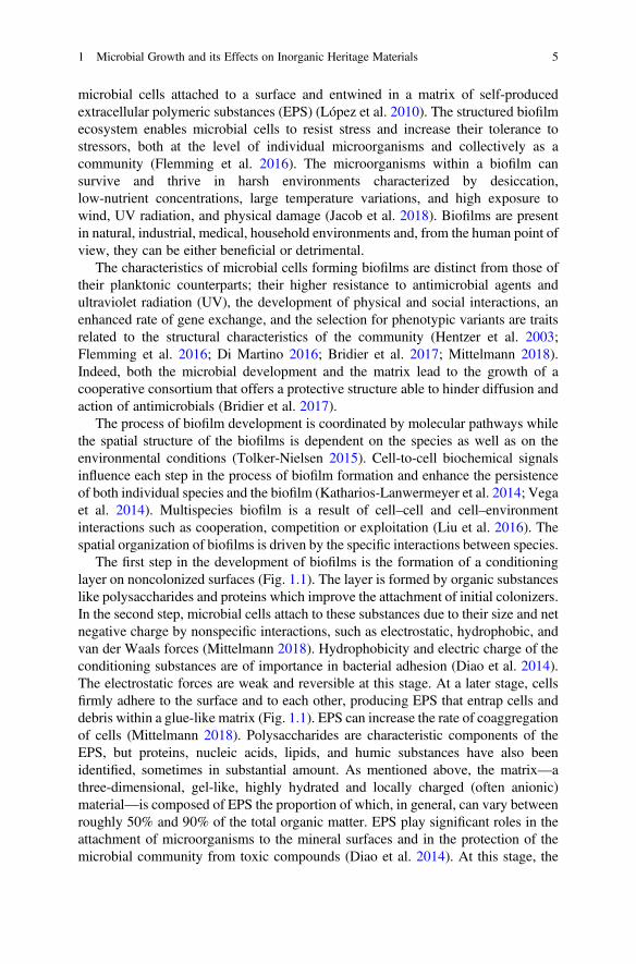
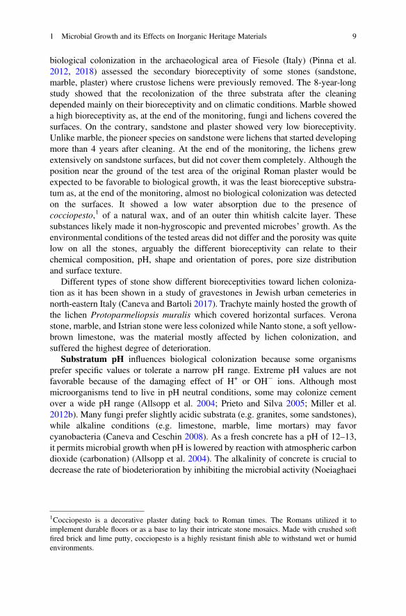
The first step in the development of biofilms is the formation of a conditioninglayer on noncolonized surfaces (Fig. 1.1). The layer is formed by organic substanceslike polysaccharides and proteins which improve the attachment of initial colonizers.In the second step, microbial cells attach to these substances due to their size and netnegative charge by nonspecific interactions, such as electrostatic, hydrophobic, andvan der Waals forces (Mittelmann 2018). Hydrophobicity and electric charge of theconditioning substances are of importance in bacterial adhesion (Diao et al. 2014).The electrostatic forces are weak and reversible at this stage. At a later stage, cellsfirmly adhere to the surface and to each other, producing EPS that entrap cells anddebris within a glue-like matrix (Fig. 1.1). EPS can increase the rate of coaggregationof cells (Mittelmann 2018). Polysaccharides are characteristic components of theEPS, but proteins, nucleic acids, lipids, and humic substances have also beenidentified, sometimes in substantial amount. As mentioned above, the matrix—athree-dimensional, gel-like, highly hydrated and locally charged (often anionic)material—is composed of EPS the proportion of which, in general, can vary betweenroughly 50% and 90% of the total organic matter. EPS play significant roles in theattachment of microorganisms to the mineral surfaces and in the protection of themicrobial community from toxic compounds (Diao et al. 2014). At this stage, the
1 Microbial Growth and its Effects on Inorganic Heritage Materials 5
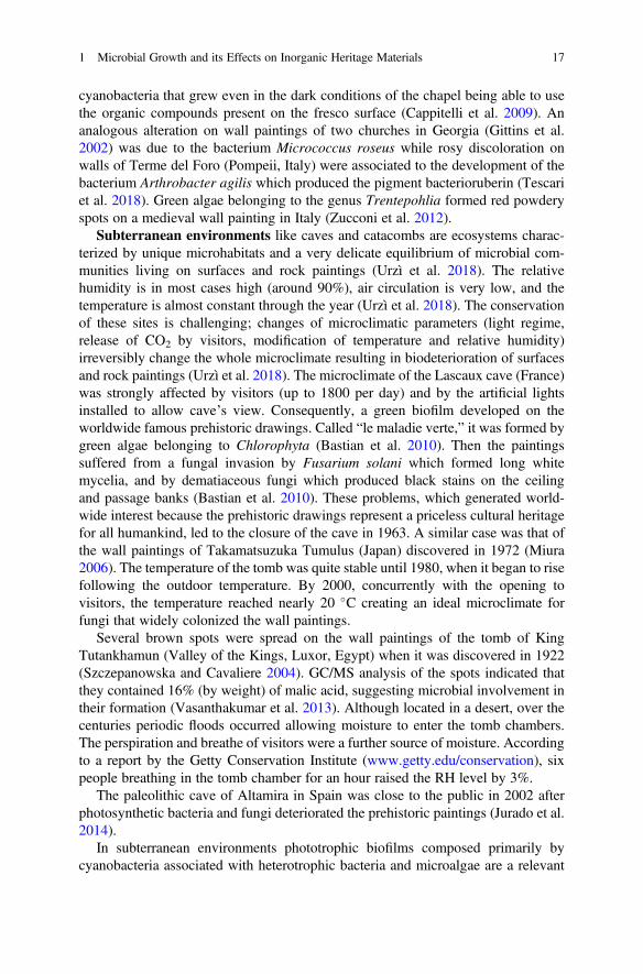
biofilm environment is a rich layer of nutrients that can support rapid growth of themicroorganisms within it. Biofilms are thus accumulations of microorganisms, EPS,multivalent cations, inorganic particles, biogenic materials, colloidal, and dissolvedcompounds. As the microcolonies grow, additional species, the so-called secondarycolonizers, are recruited through coaggregation and nonspecific aggregation inter-actions, increasing the biofilm biomass and species complexity (Katharios-Lanwermeyer et al. 2014). This process allows species that cannot adhere to thenoncolonized surfaces to become part of the biofilm (Gorbushina 2007). In themature biofilm, complex diffusion channels transport nutrients, oxygen, and othernecessary elements for cells, and carry away metabolic waste products. The laststage of biofilm lifestyle is the dispersal of cells from the biofilm colony, whichenables it to spread and colonize new surfaces (Fig. 1.1).
Exposed heritage surfaces are mostly low-nutrient environments. The formationof biofilms is a survival mechanism adopted by microorganisms. Other survivalmechanisms are nutritional versatility, cellular morphological changes, and forma-tion of spores (Mittelmann 2018). Moreover, microbial cells can enter a viable butnon-culturable state reducing their respiration rates and densities yet retaining theiressential metabolic activities in response to nutrient deficiency (Mittelmann 2018).Associations of phototrophic and heterotrophic microorganisms often compose thebiofilms that grow on heritage objects. Photosynthetic microorganisms sustain thedevelopment of heterotrophs through the production of EPS such as polysaccharides
Fig. 1.1 Stages leading to microbial formation on a surface. Once cells attach to surfaces irrevers-ibly, they replicate and grow forming microcolonies. The microbial cells secrete extracellularpolymeric substances (EPS) and become encapsulated in a hydrogel layer, which forms a physicalbarrier between the community and the extracellular environment. The community forms a three-dimensional structure and matures into a biofilm. Cells in an established biofilm are “glued”together by the EPS, which resist mechanical stresses and detachment of the community from thesurface of the substratum. Some cells detach from regions of the biofilm and disperse to formbiofilms in new environmental niches
6 D. Pinna
(Villa et al. 2015). The heterotrophs, in turn, can promote the growth of autotrophsby supplying key substances and other products.
A lichen is a mutualistic symbiotic association between green algae orcyanobacteria and fungi. The mutual life form has properties quite different fromthose of its component organisms growing separately. The fungal component of thelichen is called mycobiont, while the photosynthesizing organism is calledphotobiont. Photobionts are green algae and cyanobacteria. Most lichens are asso-ciated to green algae. The photobiont produces the nutrients needed by the entirelichen, while the mycobiont produces large numbers of secondary metabolites, thelichen acids, which play a role in the biogeochemical weathering of rocks and in soilformation. The nonreproductive tissue, or vegetative body part, is called thallus.Nearly all lichens have an outer cortex, which is a dense, protective layer of hyphae.Below it, there is the photosynthetic layer. Then, a layer of loose hyphae, themedulla, is present. Foliose lichens have a lower cortex with rhizines, which anchorthem to the substratum, while crustose lichens attach to the substratum through themedulla. Lichens come in many colors (white, black, red, orange, brown, yellow,and green), shapes, and forms. Three major growth forms are recognized: crustosethat is flattened and adheres tightly to the substratum; foliose with flat leaf-like lobesthat lift from the surface; fruticose that has tiny, multiple leafless branches three-dimensional growing.
The mycobiont produces the fruiting bodies, which are spore-producing struc-tures. The most common sexual fruiting bodies are the apothecium and the perithe-cium. Apothecia typically are structures cup or disc-shaped, often with a distinct rimaround the edge. The hymenium, the tissue containing the asci, forms the disc, theupper surface of the apothecium. Two basic types of apothecium are recognized inlichens, differing in their margins and underside (together named “the exciple”). Inlecanorine apothecia, the thallus tissue extends up the outside of the apothecium toform the exciple and the rim. This margin generally retains the color of the thallusand normally contains algal cells. In lecideine apothecia, the exciple is part of thetrue apothecial tissue and does not contain algal cells. Discs that become verycontorted and appear as line segments on the surface of the thallus are calledlyrellae.
Perithecia are usually flask-shaped fruiting bodies enclosing the asci. At maturity,an apical opening, the ostiole, releases the ascospores. Perithecia are usually partiallyembedded in the thallus or in the substratum and are relatively small, rarely morethan 1 mm in diameter and commonly much less.
3 Factors Influencing Microbial Colonization
Inorganic cultural heritage materials differ in surface texture, hardness, porosity, pH,and chemical composition, characteristics that make them favorable or unfavorableto microbial colonization.
1 Microbial Growth and its Effects on Inorganic Heritage Materials 7
The susceptibility of stones to hold organisms is called bioreceptivity, a termcoined by Guillitte (1985). In other words, it is the aptitude of a stone to bevulnerable to organisms’ colonization. Guillitte further defined this concept. Theprimary bioreceptivity indicates the potential of a healthy material to be colonized;the secondary bioreceptivity is the result of the deterioration caused by abiotic andbiotic factors; finally, the tertiary bioreceptivity is prompted by nutrients containedin the substratum (e.g. dead biomass, dust particles, animal feces, water repellentsand consolidants, biocides, etc.).
There are several reports in the literature on studies conducted either in situ orunder laboratory conditions on the assessment of bioreceptivity. Highly porousmaterials are more susceptible to microbial colonization because of their capacityto absorb more water and retain it in for longer periods of time. Surface roughnessaffects the trapping of moisture and concentrates it in micro-fissures where growth isusually more abundant. It also enables the accumulation of particles - soot, organicand inorganic debris, pollens, spores, and salts (Jacob et al. 2018). In addition,rougher surfaces can be a preferential site for colonization because they provideshelter from wind, desiccation, and excessive solar radiation (Miller et al. 2012b;Jacob et al. 2018). Evidence of the importance of these physical characteristics isgiven by the resistance to microbial colonization of smooth and impermeablesurfaces of glazed ceramics unless fissures are present.
Studies on primary and secondary bioreceptivity of granites showed that thegrowth of phototrophic biofilms is strongly enhanced by high open porosity, capil-lary water content, surface roughness, and abrasion pH rather than by differences inthe chemical and mineralogical composition of the rocks (Prieto and Silva 2005;Vázquez-Nion et al. 2018a, b). Abrasion pH, measured after grinding the rock indistilled water, relates to the number of basic cations released by the rock when incontact with aqueous solutions. Open porosity is correlated with void spaces in therock; thus, it refers to the capacity of rock to absorb water. Capillary water absorp-tion, providing information about the pattern of the pore network, is connected to thetime the rocks remain wet. Despite the above-mentioned stone properties areassumed as the most important to determine bioreceptivity of natural and artificialstones, some studies showed that the chemical composition and petrography appearto be crucial factors as well (Miller et al. 2012b). A study (Miller et al. 2006) onlimestone, granite, and marble samples artificially inoculated with two photosyn-thetic microorganisms (the cyanobacterium Gloeocapsa alpina and the greenmicroalga Stichococcus bacillaris) showed that limestone and marble, which hadthe highest (>17%) and the lowest (�1%) porosity, respectively, supported thegreatest microorganisms’ colonization while granite showed just a limited growth.The result apparently depends mainly on the chemical composition rather than on thephysical characteristics of the stones. Similarly, the importance of stone chemicalcomposition emerged from a laboratory experiment (Olsson-Francis et al. 2012) thatshowed basalts have higher rates of cyanobacterial growth and dissolution thanrhyolitic rocks. According to the authors, the difference is due to a higher contentof quartz, which has a low rate of weathering, and to lower concentrations ofbio-essential elements, such as Ca, Fe, and Mg. A research on the prevention of
8 D. Pinna
biological colonization in the archaeological area of Fiesole (Italy) (Pinna et al.2012, 2018) assessed the secondary bioreceptivity of some stones (sandstone,marble, plaster) where crustose lichens were previously removed. The 8-year-longstudy showed that the recolonization of the three substrata after the cleaningdepended mainly on their bioreceptivity and on climatic conditions. Marble showeda high bioreceptivity as, at the end of the monitoring, fungi and lichens covered thesurfaces. On the contrary, sandstone and plaster showed very low bioreceptivity.Unlike marble, the pioneer species on sandstone were lichens that started developingmore than 4 years after cleaning. At the end of the monitoring, the lichens grewextensively on sandstone surfaces, but did not cover them completely. Although theposition near the ground of the test area of the original Roman plaster would beexpected to be favorable to biological growth, it was the least bioreceptive substra-tum as, at the end of the monitoring, almost no biological colonization was detectedon the surfaces. It showed a low water absorption due to the presence ofcocciopesto,1 of a natural wax, and of an outer thin whitish calcite layer. Thesesubstances likely made it non-hygroscopic and prevented microbes’ growth. As theenvironmental conditions of the tested areas did not differ and the porosity was quitelow on all the stones, arguably the different bioreceptivity can relate to theirchemical composition, pH, shape and orientation of pores, pore size distributionand surface texture.
Different types of stone show different bioreceptivities toward lichen coloniza-tion as it has been shown in a study of gravestones in Jewish urban cemeteries innorth-eastern Italy (Caneva and Bartoli 2017). Trachyte mainly hosted the growth ofthe lichen Protoparmeliopsis muralis which covered horizontal surfaces. Veronastone, marble, and Istrian stone were less colonized while Nanto stone, a soft yellow-brown limestone, was the material mostly affected by lichen colonization, andsuffered the highest degree of deterioration.
Substratum pH influences biological colonization because some organismsprefer specific values or tolerate a narrow pH range. Extreme pH values are notfavorable because of the damaging effect of H+ or OH� ions. Although mostmicroorganisms tend to live in pH neutral conditions, some may colonize cementover a wide pH range (Allsopp et al. 2004; Prieto and Silva 2005; Miller et al.2012b). Many fungi prefer slightly acidic substrata (e.g. granites, some sandstones),while alkaline conditions (e.g. limestone, marble, lime mortars) may favorcyanobacteria (Caneva and Ceschin 2008). As a fresh concrete has a pH of 12–13,it permits microbial growth when pH is lowered by reaction with atmospheric carbondioxide (carbonation) (Allsopp et al. 2004). The alkalinity of concrete is crucial todecrease the rate of biodeterioration by inhibiting the microbial activity (Noeiaghaei
1Cocciopesto is a decorative plaster dating back to Roman times. The Romans utilized it toimplement durable floors or as a base to lay their intricate stone mosaics. Made with crushed softfired brick and lime putty, cocciopesto is a highly resistant finish able to withstand wet or humidenvironments.
1 Microbial Growth and its Effects on Inorganic Heritage Materials 9
et al. 2017). Thus, the degree of carbonation and pH values play a key role in thereceptivity of concrete and mortars to microbial colonization.
Differences in abundance of colonization on stones can be due to tertiarybioreceptivity related to nutrients contained on the surfaces or in the stones(Salvadori and Charola 2011). They can derive from existing biological growth onthe surface, bird droppings, organic compounds used in restoration practice, pollut-ants. Air and rain carry nutrients in the form of dust and soil particles. Soilfertilization leads to an accumulation of nitrates and phosphates (eutrophication)that are contained in bird droppings too. Some lichens (nitrophilic species) have beenadapted to high nitrogen levels. Generally, they have an orange thallus, easilyobserved on roofs, architectural moldings, horizontal surfaces of statues where theeutrophication derived from bird droppings and/or fertilizers transported by airand rain.
Along with substrata’ bioreceptivity and capacity to retain water, the environ-ment surrounding the monument and the monument itself act as limiting factors ofbiological growth. Local microclimate, macroclimate, wind-driven rain, pollution,geographical location, architectural design and details of monuments or sculpturesare remarkable factors influencing organisms’ colonization (Tonon et al. 2019).Tropical climate conditions in Far East, for example, enhance the establishmentand widespread colonization of microflora on cultural heritage objects due to highwater availability during monsoon season (Zhang et al. 2019).
The importance of wind-driven rain to the abundance of microbial colonization ofsurfaces emerged from a five-year-long study in the archaeological site of Pompeii,Italy (Traversetti et al. 2018). The extent of microbial growth was much broader onnorthern and western exposures respect to south-exposed walls. Comparison ofclimatic parameters, data of the dominant winds and microorganisms’ occurrence(cyanobacteria, algae, and lichens) showed that wind-driven rain along with lowertemperatures and poor ventilation of northern exposure strongly affected stones’wetness and played a key role in the promotion of microbial development. A similarstudy documented that the moisture contents in the walls of a brick tower was highernear the edges of the walls than at the center just for wind-driven precipitation,inducing fungal mold colonization especially at this position (Abuku et al. 2009). Inthese studies, the orientation of the object towards the light was an aspect affectingmicrobial colonization as well. In wet climates and northern latitudes, north-exposedsurfaces get less direct sunlight. Therefore, once wet, they remain damp for muchlonger than other stones. This slow-drying condition is much more favorable tobiological colonization than the hostile condition of rapid wetting and drying cyclesexperienced on the south faces (Adamson et al. 2013). Similarly, in a differentclimate and latitude (south of Brazil) painted surfaces showed higher fungal coloni-zation on south-facing sides that received less solar radiation than north-facing ones.If the surface temperature fell below dew point at night, they remained moist forlonger periods of time after wetting (Shirakawa et al. 2010).
Temperature and relative humidity are key parameters influencing the develop-ment of fungi (Jain et al. 2009). Most of them may be grouped into three categorieson a thermal basis. Psychrophiles thrive at temperatures between 0 �C and 5 �C,
10 D. Pinna
mesophiles between 20 �C and 45 �C, while thermophiles at or above 55 �C.Members of the Mucoraceae and Deuteromycetes grow on substrata at relativehumidity of 90–100% and not below, while members of the Aspergillus glaucusgroup grow at relative humidity as low as 65% (Jain et al. 2009). Temperature mayalso affect the stone water content. The influence of surface temperature showed asignificant positive correlation with green algae biofilms in a survey of four sand-stone heritage structures in central Belfast (Ireland) exposed for around 100 years(Cutler et al. 2013). Areas with lower temperatures were, on average, greener thanwarmer areas. As reported by the authors, it is possible to model algal greening ofsandstones from the scaled-down outputs of regional climate models as it mostlyrelates to climate and atmospheric particulates.
In hot climates sunny surfaces are more hostile and variable than those shaded,thus the production of extra cellular polymeric substances tends to be higher toprotect the cells from the adverse conditions (Scheerer 2008). Moreover, microbialbiomass and species diversity are usually much higher in shaded areas (Ramírezet al. 2010).
4 Biodeterioration Processes Caused by Biofilmsand Lichens
Microorganisms that play a potential role in biodeteriorative processes of inorganicmaterials are autotrophic and heterotrophic bacteria, fungi, algae and lichens.
Microbial biofilms interact with inorganic materials in several ways: physicaldeterioration, where materials structure is affected by microbial growth(e.g. physical or mechanical breaking); aesthetic deterioration due to fouling; chem-ical deterioration, mineral and metal transformations due to excretion of metabolitesor other substances such as acids that adversely affect the structural properties of thematerial (e.g. increasing of porosity, weakening of the mineral matrix, dissolution ofminerals and formation of biominerals, biocorrosion of metals and alloys).
The physical and chemical weathering of rocks by lichens—mainly epilithic andendolithic crustose species - encompasses the following mechanisms: hyphaegrowth in intergranular voids and mineral cleavage planes; expansion and contrac-tion of the thallus by wetting and drying, and freeze–thaw cycles; incorporation ofmineral fragments into the thallus; swelling and deteriorative action of organic andinorganic acids; dissolution/etching of mineral grains and precipitation of amor-phous and crystalline compounds.
In addition, the dissolution of respiratory CO2 contained in water held by biofilmsand lichen thalli can lower the pH at the substratum–thallus interface, acceleratingthe chemical weathering of the rocks (Di Martino 2016).
Effective physical weathering requires chemical weathering since the growth inthe stone of biofilms and lichens is facilitated by the dissolution of minerals alonggrain boundaries, cleavages, and cracks. The overall effect increases porosity and
1 Microbial Growth and its Effects on Inorganic Heritage Materials 11
permeability. Since these weathering processes interact and enhance each otheraction, it is impossible to separately quantify their role (Bjelland and Thorseth2002; de Los Rios et al. 2002).
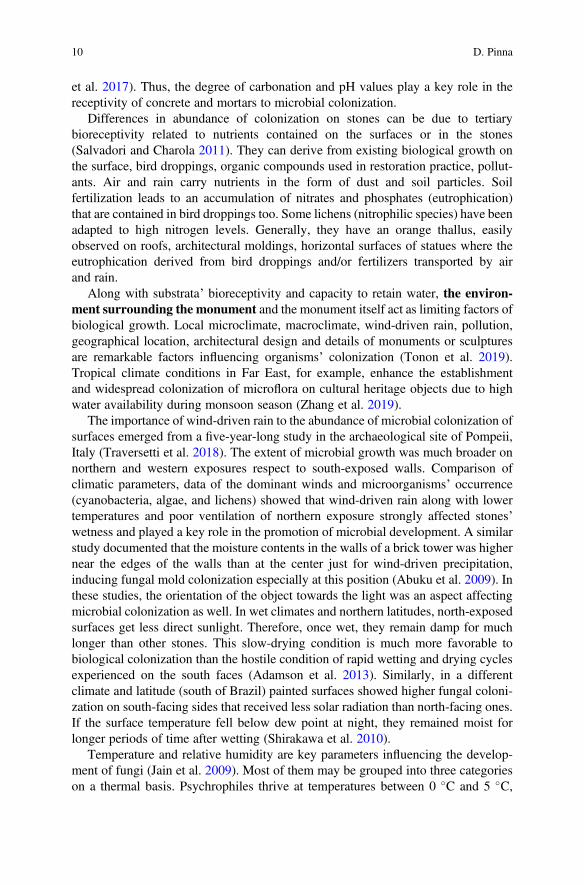
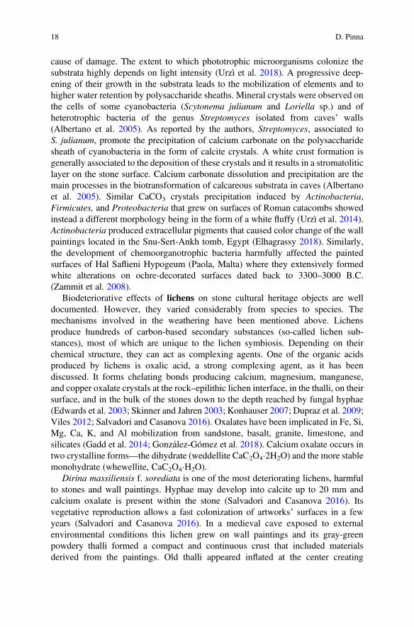
As for natural and artificial stones, the aesthetic aspects assume an important rolebecause biofilms’ and lichens’ development is an impediment to the site’s presen-tation to the audience when it compromises the legibility of a monument (Ashbee2010) (Fig. 1.2).
4.1 Stone
The so-called biomineralization or biologically induced mineralization is the processby which biological activity induces the precipitation and accumulation of minerals.It is the result of the metabolism of the organisms and is present in all five kingdomsof life. Minerals produced by biofilms and lichens include iron hydroxides, magne-tite, manganese oxides, clays, amorphous silica, carbonates, phosphates, oxalates(Konhauser 2007; Dupraz et al. 2009; Miller et al. 2012a; Urzì et al. 2014).Biomineralization produced by fungi occurs when they modify the local microen-vironment creating conditions that favor extracellular precipitation of mineral phases(Fomina et al. 2010; Gadd et al. 2014).
Stone-inhabiting microorganisms may grow on the surface (epiliths) or somemillimeters or even centimeters in the substratum (endoliths) (Gadd et al. 2014). The
Fig. 1.2 Statue in the parkof Nympheburg, Munich,Germany. The statue’ssurface is almost completelycovered by biofilms andlichens that stronglycompromise its legibility
12 D. Pinna
endolithic colonization is an adaptive strategy developed by cyanobacteria, algae,fungi, and lichens. The use of the substratum as a shield against external stressproves to be a decisive evolutionary selection advantage (Pohl and Scheider 2002).The endolithic mode of life includes different ecological niches: chasmoendolithsand cryptoendoliths occupy preexisting fissures and structural cavities in the rocks,whereas euendoliths grow in soluble carbonatic and phosphatic substrata dissolvingthe stone immediately below the surface. The first form of growth leads to aco-responsibility in the detachment of scales of material due to the pressure exertedby increasing biomass. This process can occur repeatedly, involving areas increas-ingly in depth (Pinna and Salvadori 2008). The light that reaches the bulk of thestones limits the growth of phototrophic microorganisms. The presence of water inmicropores, especially those with translucent walls, may enhance light penetration,increasing the light available for photosynthesis in the cryptoendolithic habitats(Cámara et al. 2014). Euendoliths form microcavities of varying morphologiesaccording to the species.
Stones subjected to extreme sun irradiation in hyper-arid Atacama Desert (Chile)are colonized by endolithic algae and cyanobacteria. In order to endure the harshenvironment, they synthesize carotenoids and scytonemin, respectively. The pro-duction of these pigments is interpreted as an adaptation strategy against high dosesof solar irradiation as they are passive UV-screening pigments (Vítek et al. 2016).
Cyanobacteria and microalgae are often the first colonizers of stone surfaceswhere they develop phototrophic biofilms. Algae form colored (green, gray, black,brown, and orange) powdery patinas, and gelatinous layers. They usually dominatesurfaces in wet and rainy areas. Cyanobacteria typically form dark brown and blackpatinas but also pink discolorations. Besides the aesthetic disturbance caused by thecolored patinas, algae and cyanobacteria cause water retention and damage due tofreeze–thaw cycles. Areas colonized by dark biofilms formed by cyanobacteria mayabsorb more sunlight. Temperature changes increase mechanical stress by expansionand contraction of the biofilms (Scheerer et al. 2009).
The main groups of fungi isolated from stone monuments are Hyphomycetes andblack meristematic fungi (Salvadori and Casanova 2016). Hyphomycetes, com-monly known as mold, are a class of asexual or imperfect fungi. They lack fruitingbodies, the sexual structures used to classify other fungi. The production of conidia(spores) occurs by fragmentation of vegetative hyphae or from specialized hyphaecalled conidiophores. Since the attachment of airborne spores to surfaces is the firststep of fungal colonization, the species diversity of fungi present on stones is similarto the diversity of common airborne spores (Sterflinger and Piñar 2013). ManyHyphomycetes, notably Aspergillus, Fusarium, and Penicillium genera, producetoxic metabolites (mycotoxins). Several Hyphomycetes growing on stone heritageobjects are dematiaceous. The term refers to the characteristic dark appearance ofthese fungi that form dark gray, brown or black colonies. Fungi excrete organic acids(oxalic, citric, acetic, formic, gluconic, glyoxylic, fumaric, malic, succinic, andpyruvic) that can act as chelators. Particularly, oxalic acid has a high capacity ofdegrading minerals for its complexing and acid properties. Its importance relates tothe ability of the oxalate anion to complex and/or precipitate metals as secondary
1 Microbial Growth and its Effects on Inorganic Heritage Materials 13
biominerals. The reaction results both in mineral dissolution and in mineral forma-tion (Gadd et al. 2014). Calcium oxalate precipitation has an important influence onbiogeochemical processes in soils, acting as a calcium reservoir (Gadd et al. 2014).The precipitation of secondary minerals (carbonates and oxalates) on and withinstones, and the mineral dissolution connected to fungal growth lead to the formationof crusts on the surface and around the hyphae that can progressively cement fissuresand cracks (Fomina et al. 2010).
An investigation into a thin dark rock coating at the Ngaut Ngaut heritagecomplex in South Australia showed that it contained a mixture of calcite, quartz,gypsum, and weddellite (Roberts et al. 2015). The dark coating covered petroglyphsengraved in the rock shelter and dated back around 3000 years B.C. According to theauthors, the weddellite was likely formed from the reaction of calcite with oxalicacid excreted by surface microflora.
Metal mobilization can also be achieved by chelation ability of siderophores.When living in environments of reduced iron content, fungi produce iron(III)-binding ligands, commonly of a hydroxamate nature, termed siderophores(Salvadori and Casanova 2016).
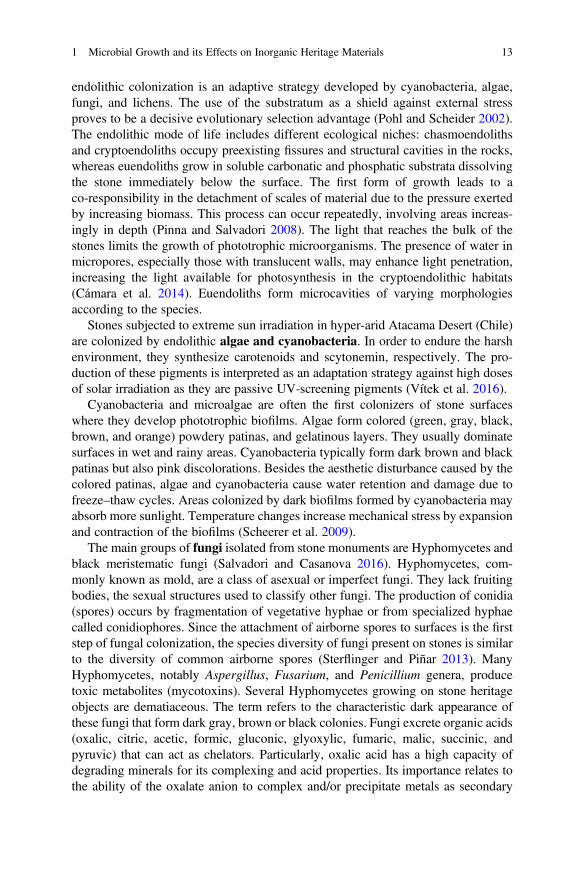
Black meristematic fungi belong to the genera Hortaea, Sarcinomyces,Coniosporium, Capnobotryella, Exophiala, Knufia, and Trimmatostroma(Sterflinger and Piñar 2013). Their cells have thick pigmented walls. They formslowly expanding, cauliflower-like colonies that grow by isodiametric enlargementof the cells (Sterflinger 2010) (Fig. 1.3). In addition to the meristematic growth,many of the black fungi can exhibit a yeast-like growth (De Leo et al. 2003). Theyabandoned the hyphal phase adopting the microcolonial or yeast phase characterizedby an extremely slow growth, in response to the lack of organic nutrients and tostresses of outdoor substrata. The microcolonial phase enhances the survival andpersistence of these fungi in the biofilms. They produce various pigments includingcarotenoids, mycosporines, and melanins that protect them from UV irradiation.Moreover, melanin provides them with extra-mechanical strength making hyphaeable to grow better in fissures. Microcolonial fungi, as well as cyanobacteria, algae
Fig. 1.3 Early developmentof black meristematic fungion the wealthy surface of amarble model sampleexposed outdoor.Stereomicroscope, 50�
14 D. Pinna
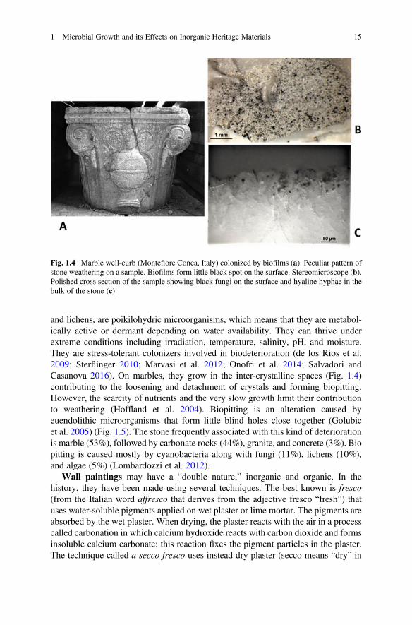
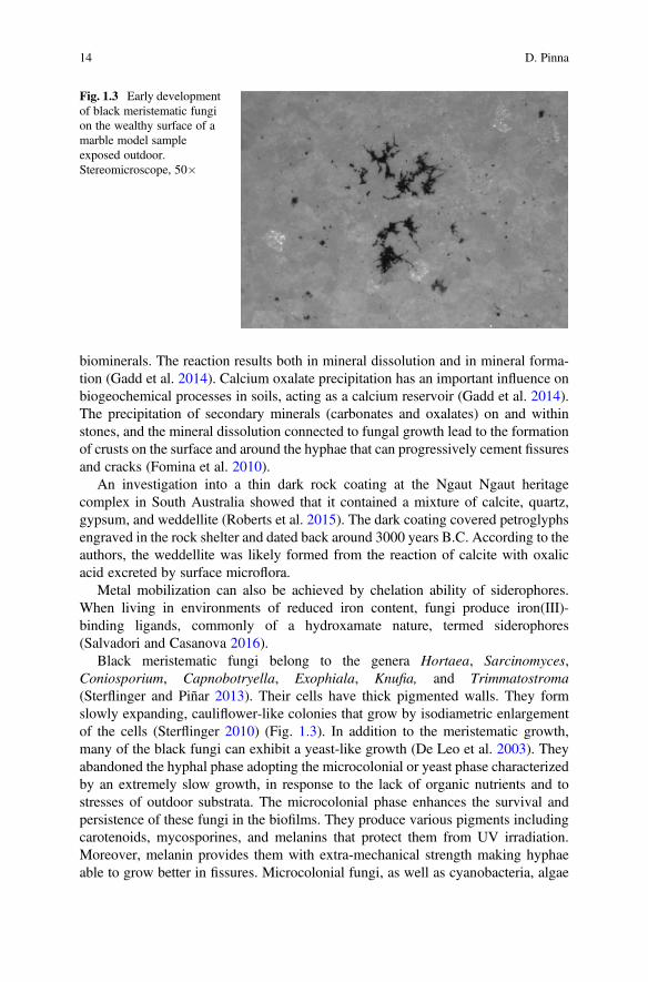
and lichens, are poikilohydric microorganisms, which means that they are metabol-ically active or dormant depending on water availability. They can thrive underextreme conditions including irradiation, temperature, salinity, pH, and moisture.They are stress-tolerant colonizers involved in biodeterioration (de los Rios et al.2009; Sterflinger 2010; Marvasi et al. 2012; Onofri et al. 2014; Salvadori andCasanova 2016). On marbles, they grow in the inter-crystalline spaces (Fig. 1.4)contributing to the loosening and detachment of crystals and forming biopitting.However, the scarcity of nutrients and the very slow growth limit their contributionto weathering (Hoffland et al. 2004). Biopitting is an alteration caused byeuendolithic microorganisms that form little blind holes close together (Golubicet al. 2005) (Fig. 1.5). The stone frequently associated with this kind of deteriorationis marble (53%), followed by carbonate rocks (44%), granite, and concrete (3%). Biopitting is caused mostly by cyanobacteria along with fungi (11%), lichens (10%),and algae (5%) (Lombardozzi et al. 2012).
Wall paintings may have a “double nature,” inorganic and organic. In thehistory, they have been made using several techniques. The best known is fresco(from the Italian word affresco that derives from the adjective fresco “fresh”) thatuses water-soluble pigments applied on wet plaster or lime mortar. The pigments areabsorbed by the wet plaster. When drying, the plaster reacts with the air in a processcalled carbonation in which calcium hydroxide reacts with carbon dioxide and formsinsoluble calcium carbonate; this reaction fixes the pigment particles in the plaster.The technique called a secco fresco uses instead dry plaster (secco means “dry” in
Fig. 1.4 Marble well-curb (Montefiore Conca, Italy) colonized by biofilms (a). Peculiar pattern ofstone weathering on a sample. Biofilms form little black spot on the surface. Stereomicroscope (b).Polished cross section of the sample showing black fungi on the surface and hyaline hyphae in thebulk of the stone (c)
1 Microbial Growth and its Effects on Inorganic Heritage Materials 15
Italian). An organic binding medium, like egg, glue, or oil, is needed to fix thepigment into the plaster. In Classical Greco-Roman times, the encaustic paintingtechnique was in use. Pigments were ground in a molten beeswax binder (or resinbinder) and applied to the surface while hot. Today, murals are mostly painted inoils, tempera, or acrylic colors. Microbial colonies may deteriorate either the inor-ganic component or the organic one causing efflorescence, expansion cracking,peeling and spalling of outer layers, color changes, and stains. Oxalates may beproduced from the calcite or from metal-containing pigments of the painted layers(Gadd et al. 2014; Unković et al. 2017).
The deteriorative potential of fungi isolated from wall paintings was evaluated byin vitro studies to assess the risk of deterioration and formulate appropriate conser-vation treatments. Some biodegradative properties—calcite dissolution, caseinhydrolysis, pigment secretion, acid and alkali production—were tested using specificagar culture media (Pangallo et al. 2012; Unković et al. 2018). Many species of thegenera Aspergillus and Penicillium demonstrated the ability to dissolve calcite asthey produced and secreted acids; oxalic acid was the main cause of deterioration inmost instances (Ortega-Morales et al. 2016). Species of the genus Cladosporiumhydrolyzed casein showing high proteolytic activity (Unković et al. 2018). They arethought as the primary degraders of protein components of painted layers of wallpaintings. Applying the mentioned method, the authors detected also species show-ing no deterioration capabilities.
Pink patinas and rosy discolorations occurring on stones and wall paintings maybe caused by bacteria and algae. In the crypt of the Original Sin (Matera, Italy), thepink color resulted from the production of a ruberin-type carotenoid pigmentproduced by actinobacteria (Nugari et al. 2009). Similarly, carotenoids causedpink and yellow discolorations on wall paintings of St. Botolph’s Church (Hardham,UK) (Kyi 2006). According to the authors, the pigments provide microorganismswith a protective mechanism against damage caused by photooxidation reactions.Rosy-powdered areas on the frescoes of St. Brizio Chapel (Orvieto Cathedral, Italy)contained instead phycoerythrin, a pigment produced by capsulated coccoid
Fig. 1.5 Biopitting on a stone statue, Uffizi Gallery (Firenze, Italy). Close-up of a detail of thesurface (right). The circular micro-holes were likely produced by endolithic lichens that grew whenthe statue was located outdoors. The fruiting bodies were present inside the holes. After the lichensdied, they left just the empty holes
16 D. Pinna
cyanobacteria that grew even in the dark conditions of the chapel being able to usethe organic compounds present on the fresco surface (Cappitelli et al. 2009). Ananalogous alteration on wall paintings of two churches in Georgia (Gittins et al.2002) was due to the bacterium Micrococcus roseus while rosy discoloration onwalls of Terme del Foro (Pompeii, Italy) were associated to the development of thebacterium Arthrobacter agilis which produced the pigment bacterioruberin (Tescariet al. 2018). Green algae belonging to the genus Trentepohlia formed red powderyspots on a medieval wall painting in Italy (Zucconi et al. 2012).
Subterranean environments like caves and catacombs are ecosystems charac-terized by unique microhabitats and a very delicate equilibrium of microbial com-munities living on surfaces and rock paintings (Urzì et al. 2018). The relativehumidity is in most cases high (around 90%), air circulation is very low, and thetemperature is almost constant through the year (Urzì et al. 2018). The conservationof these sites is challenging; changes of microclimatic parameters (light regime,release of CO2 by visitors, modification of temperature and relative humidity)irreversibly change the whole microclimate resulting in biodeterioration of surfacesand rock paintings (Urzì et al. 2018). The microclimate of the Lascaux cave (France)was strongly affected by visitors (up to 1800 per day) and by the artificial lightsinstalled to allow cave’s view. Consequently, a green biofilm developed on theworldwide famous prehistoric drawings. Called “le maladie verte,” it was formed bygreen algae belonging to Chlorophyta (Bastian et al. 2010). Then the paintingssuffered from a fungal invasion by Fusarium solani which formed long whitemycelia, and by dematiaceous fungi which produced black stains on the ceilingand passage banks (Bastian et al. 2010). These problems, which generated world-wide interest because the prehistoric drawings represent a priceless cultural heritagefor all humankind, led to the closure of the cave in 1963. A similar case was that ofthe wall paintings of Takamatsuzuka Tumulus (Japan) discovered in 1972 (Miura2006). The temperature of the tomb was quite stable until 1980, when it began to risefollowing the outdoor temperature. By 2000, concurrently with the opening tovisitors, the temperature reached nearly 20 �C creating an ideal microclimate forfungi that widely colonized the wall paintings.
Several brown spots were spread on the wall paintings of the tomb of KingTutankhamun (Valley of the Kings, Luxor, Egypt) when it was discovered in 1922(Szczepanowska and Cavaliere 2004). GC/MS analysis of the spots indicated thatthey contained 16% (by weight) of malic acid, suggesting microbial involvement intheir formation (Vasanthakumar et al. 2013). Although located in a desert, over thecenturies periodic floods occurred allowing moisture to enter the tomb chambers.The perspiration and breathe of visitors were a further source of moisture. Accordingto a report by the Getty Conservation Institute (www.getty.edu/conservation), sixpeople breathing in the tomb chamber for an hour raised the RH level by 3%.
The paleolithic cave of Altamira in Spain was close to the public in 2002 afterphotosynthetic bacteria and fungi deteriorated the prehistoric paintings (Jurado et al.2014).
In subterranean environments phototrophic biofilms composed primarily bycyanobacteria associated with heterotrophic bacteria and microalgae are a relevant
1 Microbial Growth and its Effects on Inorganic Heritage Materials 17
cause of damage. The extent to which phototrophic microorganisms colonize thesubstrata highly depends on light intensity (Urzì et al. 2018). A progressive deep-ening of their growth in the substrata leads to the mobilization of elements and tohigher water retention by polysaccharide sheaths. Mineral crystals were observed onthe cells of some cyanobacteria (Scytonema julianum and Loriella sp.) and ofheterotrophic bacteria of the genus Streptomyces isolated from caves’ walls(Albertano et al. 2005). As reported by the authors, Streptomyces, associated toS. julianum, promote the precipitation of calcium carbonate on the polysaccharidesheath of cyanobacteria in the form of calcite crystals. A white crust formation isgenerally associated to the deposition of these crystals and it results in a stromatoliticlayer on the stone surface. Calcium carbonate dissolution and precipitation are themain processes in the biotransformation of calcareous substrata in caves (Albertanoet al. 2005). Similar CaCO3 crystals precipitation induced by Actinobacteria,Firmicutes, and Proteobacteria that grew on surfaces of Roman catacombs showedinstead a different morphology being in the form of a white fluffy (Urzì et al. 2014).Actinobacteria produced extracellular pigments that caused color change of the wallpaintings located in the Snu-Sert-Ankh tomb, Egypt (Elhagrassy 2018). Similarly,the development of chemoorganotrophic bacteria harmfully affected the paintedsurfaces of Hal Saflieni Hypogeum (Paola, Malta) where they extensively formedwhite alterations on ochre-decorated surfaces dated back to 3300–3000 B.C.(Zammit et al. 2008).
Biodeteriorative effects of lichens on stone cultural heritage objects are welldocumented. However, they varied considerably from species to species. Themechanisms involved in the weathering have been mentioned above. Lichensproduce hundreds of carbon-based secondary substances (so-called lichen sub-stances), most of which are unique to the lichen symbiosis. Depending on theirchemical structure, they can act as complexing agents. One of the organic acidsproduced by lichens is oxalic acid, a strong complexing agent, as it has beendiscussed. It forms chelating bonds producing calcium, magnesium, manganese,and copper oxalate crystals at the rock–epilithic lichen interface, in the thalli, on theirsurface, and in the bulk of the stones down to the depth reached by fungal hyphae(Edwards et al. 2003; Skinner and Jahren 2003; Konhauser 2007; Dupraz et al. 2009;Viles 2012; Salvadori and Casanova 2016). Oxalates have been implicated in Fe, Si,Mg, Ca, K, and Al mobilization from sandstone, basalt, granite, limestone, andsilicates (Gadd et al. 2014; González-Gómez et al. 2018). Calcium oxalate occurs intwo crystalline forms—the dihydrate (weddellite CaC2O4�2H2O) and the more stablemonohydrate (whewellite, CaC2O4�H2O).
Dirina massiliensis f. sorediata is one of the most deteriorating lichens, harmfulto stones and wall paintings. Hyphae may develop into calcite up to 20 mm andcalcium oxalate is present within the stone (Salvadori and Casanova 2016). Itsvegetative reproduction allows a fast colonization of artworks’ surfaces in a fewyears (Salvadori and Casanova 2016). In a medieval cave exposed to externalenvironmental conditions this lichen grew on wall paintings and its gray-greenpowdery thalli formed a compact and continuous crust that included materialsderived from the paintings. Old thalli appeared inflated at the center creating
18 D. Pinna
blister-like structures and causing the detachment of painting layers (Nugari et al.2009). The decay and detachment of the center part of the thallus occur in otherlichen species and open the underlying area to further weathering, resulting incratered mounts on the rock surface (Mottershead and Lucas 2000).
Endolithic lichens develop their biomass in the bulk of the stones, with coloni-zation patterns varying among species. A study on the anatomy of five calcicolousendolithic lichens showed that the photobiont layer is located at the same depth,100–180 μm, from the surface, but the arrangement and depth of diffusion of hyphaeinto the stones vary considerably among species (Pinna et al. 1998). A different,atypical pattern of growth is that of Pyrenodesmia erodens, an euendolithic lichenable to dissolve limestones to a depth of several millimeters. It forms, with aging,confluent, moniliform depressions (Tretiach et al. 2003). Its thallus is composed byclusters of green algae surrounded by inflated, appressed hyphae. The clusters arearranged in bores formed by dissolution of the rock. This lichen has been found indry sites of the Mediterranean region and of the southern Alps on exposed,subvertical faces of limestone and dolomite rocks (Tretiach et al. 2003). Its occur-rence has been reported on the calcareous rocks of monumental remains in Pasar-gadae, UNESCO world heritage site in Iran (Sohrabi et al. 2017). The study focusedon the lichen colonization and deterioration patterns in the semi-arid conditions ofthe area. Besides the effects of Pyrenodesmia erodens, other epilithic and endolitihiclichens showed damaging action such as pitting and granular disaggregation. There-fore, lichen communities are a potential threat for the conservation of the archaeo-logical site. Moreover, lichen colonization and deterioration patterns do not appearpeculiar respect to what has been previously described by the literature aboutcolonized limestones in temperate and semi-arid areas around the Mediterraneanbasin.
The chemical deterioration of silicate and carbonate rocks by endolithic lichensrelates to the secretion of siderophore-like compounds and of carbonic anhydrase(Favero-Longo et al. 2011). This enzyme increases the speed of the reactionCO2 + H2O$HCO3
� + H+, thus it accelerates the dissolution of calcium carbonate.However, the study of calcium carbonate deterioration by endolithic lichens needsfurther experimental evidence and it is an interesting goal for further research. Thepresent results, yet relevant, enlighten the complexity of the phenomenon.
Endolithic lichens form biopitting on limestones, with pits’ diameter rangingfrom 0.2 to 1–2 mm depending on the size of fruiting bodies (Salvadori andCasanova 2016) (Fig. 1.5). When lichen death occurs, the empty pits are progres-sively enlarged by water (rainfall, water runoff, water accumulation). Then they cancoalesce forming larger interconnected depressions. Unlike epilithic species, endo-lithic ones are not characterized by the production of calcium oxalates.
A one-year long laboratory experiment on lichen–rock interactions wasconducted using mycobionts and photobionts of the endolithic lichens Bagliettoabaldensis and B. marmorea (Favero-Longo et al. 2009). They were isolated and theninoculated on marble and limestone samples. The same species growing on lime-stone outcrops and abandoned marble quarries showed penetration pathways similarto those reproduced in vitro. The study highlighted that erosion processes caused by
1 Microbial Growth and its Effects on Inorganic Heritage Materials 19
lichen development increased the availability of hyphae passageways only afterlong-term colonization. The differences in hyphae growth depended on the mineralcomposition and structure of the lithotypes.
Different climates affect the endolithic growth of lichens (Pohl and Scheider2002). In the humid Northern Alps (Austrian glacier), the bulk of the calcareousrock, just under the surface, showed three layers. At a depth of 0–150 μm, thesubstratum is mostly intact, just partly interlocked with the lichen litho cortex. At150–300 μm, photobionts (green algae or cyanobacteria) are capable of activelydissolving the substratum, thus creating habitable cavities. Then several mm indepth, there is the mycobiont with hyphae actively solubilizing the substratum andoften forming dense networks. Most biomass of endoliths on carbonate rocks in thearid Mediterranean-Maritime Alps (Provence, France) was instead confined justunder the surface. The average colonization intensity and growth depth are markedlydeeper in the more humid substratum of the Austrian Alps.
4.2 Stained-Glass Windows
Glass is a non-crystalline, amorphous inorganic solid. The most familiar types ofglass are prepared by melting and rapidly cooling quartz sand and other ingredients -sodium carbonate (Na2CO3, “soda”), lime (calcium oxide—CaO), magnesium oxide(MgO), and aluminum oxide (Al2O3). The resulting glass contains about 70–74%silica by weight and is called a soda–lime glass (Na-rich glass) which accounts forabout 90% of manufactured glass. To color the glass, powdered metals are added tothe mixture while the glass is still molten. The medieval stained-glass windows thatdecorate many European churches were made using, besides sand, a differentingredient, the so-called potash that is wood ash (K2O, K-rich glass). These glassesare more easily decayed than Na-rich ones.
Biofilms’ growth on glass is correlated to tertiary bioreceptivity. Neither theinorganic composition nor the physical features of glass favor microbial coloniza-tion, but the organic residues of various origins, as dust deposits, dead microbialmaterial, and bird droppings, can be a source of nutrients. The deteriorative action ofmicroorganisms on glass is a modest, slow, yet continuous process that can accel-erate its weathering. Research focused mainly on medieval stained-glass windows ofEuropean churches where corrosion, patinas, pitting, cracks, and mineral crustsoccur. Microorganisms may contribute to all these decay forms (Carmona et al.2006; Marvasi et al. 2009; Piñar et al. 2013). Analyses revealed complex bacterialcommunities consisting of members of the phyla Proteobacteria, Bacteroidetes,Firmicutes and Actinobacteria (Piñar et al. 2013). Fungi showed less diversitythan bacteria, and species of the genera Aspergillus, Cladosporium, and Phomawere dominant (Carmona et al. 2006; Piñar et al. 2013). Thus, historical glasswindows are a habitat in which both fungi and bacteria form complex microbialconsortia of high diversity. The bacteria are genetically similar to those that causemineral precipitation on stones. Regarding the detected fungi, they are ubiquitous
20 D. Pinna
airborne species. According to Piñar and coauthors (2013), the pitting present on thesurfaces of glass windows could relate to other more specialized fungi that grew inthe past and are not detectable now.
The chemical composition of the glass affects microbial development; coppercontained in green glasses acts as an inhibitor (Marvasi et al. 2009; Piñar et al. 2013).In a laboratory test, historically accurate reconstructions of glass windows (fifteenthand seventeenth centuries, Sintra, Portugal) were inoculated with fungi of the generaCladosporium and Penicillium previously isolated from the original stained-glasswindows. The fungi produced clear damage on glass surfaces in form of stains,erosion, pitting, crystals, and leaching (Rodrigues et al. 2014).
Biofilms on modern glasses may alter their transparency (Shirakawa et al. 2016)and smoothness (Corrêa Pinto et al. 2019). However, Na-rich glass samples inocu-lated with a mixture of four fungal species showed high resistance against themicroorganisms’ growth (Corrêa Pinto et al. 2019).
4.3 Metals
Physicochemical interactions between ametallic material and its environment canlead to electrochemical corrosion. It is a chemical reaction that occurs when elec-trons from metal are transferred to an external electron acceptor, causing release ofthe metal ions into the surrounding medium and deterioration of the metal (Beechand Sunner 2004). This process consists of a series of oxidation (anodic) andreduction (cathodic) reactions of compounds present in direct contact with or inproximity to the metallic surface (Beech and Sunner 2004). The rate of the metaldeterioration decreases gradually with time, because the oxidation products (forexample, oxidation of metallic iron to Fe2+ ion) adhere to the surface forming aprotective layer that provides a diffusion barrier to the reactants.
Biocorrosion or microbiologically influenced corrosion (MIC) is caused bythe interaction between the metal surface, abiotic corrosion products, and microbialcells and their metabolites. MIC is an electrochemical process in which microor-ganisms initiate, facilitate, or accelerate a corrosion reaction on a metal surfacethrough the utilization and release of electrons (Kadukova and Pristas 2018). Micro-bial activity of biofilms present on surfaces of metallic objects may affect the kineticsof cathodic and/or anodic reactions and may also considerably modify the chemistryof any protective layers (Beech and Sunner 2004). Recent evidence indicates thatbacteria can transport electrons via organic molecules of the surrounding environ-ment (Gu 2019).
Most investigations have addressed the impact of biofilms on corrosion behaviorof iron, copper, aluminum, and their alloys. The effects of microbial activity on ironor steel range from pitting, crevice formation, under-deposit corrosion to stresscorrosion cracking (Aramendia et al. 2015).
Microorganisms mostly involved in the biocorrosion of metals are aerobic andanaerobic bacteria (Zanardini et al. 2008). In open air systems, anaerobic corrosion
1 Microbial Growth and its Effects on Inorganic Heritage Materials 21
still occurs if a corrosive anaerobic biofilm grows underneath a top aerobic biofilm(Xu et al. 2016). While in oxic environments metal corrosion is a fast process thatcan be enhanced by microorganisms, under anoxic conditions it would take verylong time without the help of microorganisms (Kadukova and Pristas 2018).
Anaerobic sulfate reducing bacteria (SRB) have been considered key culprits inMIC of a wide range of industrial structures because the sulfur cycle is linked tomicrobial metabolism, affecting the integrity of metals (Beech et al. 2014;Rémazeilles et al. 2010). Indeed, one of the most known phenomena is the produc-tion of sulfides induced in anoxic environments by SRB (Aramendia et al. 2015).However, also other types of bacteria have been associated with metals in terrestrialand aquatic habitats—sulfur oxidizing bacteria, iron oxidizing/reducing bacteria,manganese-oxidizing bacteria, nitrate reducing bacteria and bacteria secretingorganic acids and slime (Beech and Coutinho 2003; Xu et al. 2016; Kadukova andPristas 2018). Regarding the influence of SRB in the corrosion of iron, it has beendocumented that not only different genera of these organisms but also species withinthe same genus vary in their ability to deteriorate iron (Beech et al. 2014).
Extracellular polymeric substances are able to bind metal ions. This property isimportant to MIC and depends both on bacterial species and on the type of metal ion(Kinzler et al. 2003). Metal binding by EPS involves interaction between the metalions and anionic functional groups (e.g. carboxyl, phosphate, sulfate, glycerate,pyruvate, and succinate groups) that are commonly present on the protein andcarbohydrate components of EPS. In particular, the affinity of multidentate anionicligands for multivalent ions, such as Ca2+, Cu2+, Mg2+, and Fe3+, can be very strong(Beech and Sunner 2004).
Biocorrosion is recognized as an important category of corrosion leading toimportant economic loss in many industries and services including oil and gaspipelines, water utilities and the power generation industry (Xu et al. 2016; Eidet al. 2018). Therefore, MIC has been the subject of extensive studies on thesesystems for the past decades and several models have been proposed to explainmechanisms governing biocorrosion (Beech and Sunner 2004). Regarding culturalheritage objects, many papers documented the role of microorganisms in corrosionof metal artifacts (Brown and Masters 1980; Dhawan 1987; Gilbert 1987; McNeilet al. 1991; Sánchez del Junco et al. 1992; Little et al. 1998; Rémazeilles et al. 2010;Aramendia et al. 2015). MIC is associated to the presence of anaerobic SRBs on thesurfaces of archaeological objects recovered from terrestrial and aquatic environ-ments (Sánchez del Junco et al. 1992). Regarding buried metal objects, it is worthmentioning that the degree of preservation of them in soil is specific to the type ofmetal (Kibblewhite et al. 2015). Ag is less resistant to corrosion than Au but morethan Cu while Zn corrodes faster. Cu artefacts may contain As, and this element isalso commonly a minor constituent of bronze (an alloy of Cu and Sn, which is moreresistant to corrosion than pure Cu). Fe is much more easily corroded than Cu, whilePb is resistant to corrosion in most aqueous environments. Aluminum forms aprotective surface oxide coating that gives it some resistance to oxidative corrosion(Kibblewhite et al. 2015). Degradation rates of these materials (e.g. Fe) can beinfluenced by the soil type. Sulfide and disulfide ions generated by SRB in soils
22 D. Pinna
enhance the corrosive activity of chloride ions. Moreover, biogenic sulfides may beused as alternative substrates in the cathodic reaction at low oxygen concentration(Sánchez del Junco et al. 1992). Biocorrosion of objects in soil can be markedlyaccelerated by alternate anaerobic and aerobic conditions. Sulfides formed by SRBin anaerobic conditions are transformed to polysulfides, and sulfur and ferric sulfateswhen soil pH decreases.
The formation of black corrosion stains containing sulfides on copper alloy buriedobjects was attributed to SRB, prevalent in those soils, through the production ofhydrogen sulfide (Sánchez del Junco et al. 1992).
Some studies have focused on the involvement of fungi in the MIC. The devel-opment of Aspergillus niger accelerated the corrosion process (severe pitting corro-sion and aluminum depletion accompanied with copper enrichment) of aluminumalloy 2024 samples in a laboratory experiment (Dai et al. 2016). The corrosion ratewas four times greater than that of samples exposed to 3.5 wt% NaCl solution.Oxalic acid, identified as the dominant metabolite of Aspergillus niger, caused thecorrosion. The same compound formed iron oxalates and calcium oxalates on thesurface of parts of a weathering steel sculpture (Aramendia et al. 2015). Irregularitiesand discolorations of the object were thus caused by oxalic acid excreted bymicroorganisms growing on the steel surface.
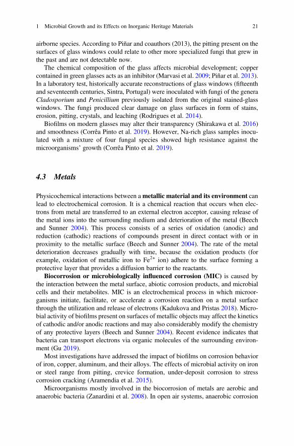
To the best of my knowledge, no studies have been carried out yet on theinteraction of lichens with metallic works of art. As illustrated in Fig. 1.6, crustoselichens completely cover a metal object. Thus, carrying out these studies would bebeneficial.
While most studies have documented the negative aspects of the biofilms’development on metallic materials, an emerging topic deals with a positive aspectof this interaction, that is corrosion inhibition. The mechanisms involved are sup-posed to be the neutralization of the action of environmental corrosive substances(e.g. acidic compounds), and the formation of protective films or the stabilization ofpreexisting protective films on a metal (e.g. EPS cohesive effects) (Videla andHerrera 2009). Both mechanisms are linked to a marked modification of the
Fig. 1.6 Widespread lichen colonization on metals. Ancient cannon, Ioannina, Greece (left). Aclose-up of the colonized metal surface (right)
1 Microbial Growth and its Effects on Inorganic Heritage Materials 23
environmental conditions at the metal/solution interface due to biological activity.However, the inhibitory action of bacteria may change to the contrary, that is to acorrosive action by other bacteria located within the biofilm (Videla and Herrera2009). Therefore, the corrosive and the inhibitory actions of bacteria can coexist atthe metal surfaces where complex biofilm/protective films interactions occur.
5 Bioprotection of Stones by Biofilms and Lichens
The damaging capability of biofilms and lichens on rock art and monuments hasbeen documented, as previously mentioned. Nonetheless, recent research has pro-vided increasing evidence of a minor effect and even of protection. Therefore, thecorrelation among biofilms, lichens and stone deterioration is no longer beingconsidered an axiom (Pinna 2014). Their presence on works of art does not auto-matically indicate a change in the physicochemical properties of the materials. Astudy on petroglyphs colonized by lichens demonstrated, for example, that theorganisms were not one of the key factors to define their state (Chiari and Cossio2004). Lichens neither acted damaging the outermost layer of the sandstones norprotecting them from rain, sun, etc. Lichens filled the micro-fissures between thegrains, which were large enough to hold them without exerting relevant pressure. Inthat way lichens filled the pores so that the porosity of the outer layer was the same asin the bulk of the rock. The deterioration of the sandstone related mainly to the natureof the sandstone itself, particularly to the dimension of the quartz grains: the largerthe grains, the greater the porosity, and consequently water absorption, fragility, anddecohesion of the rock.
Phototrophic biofilms colonizing marbles of the cathedral of Monza (Italy) didnot affect the stones that were instead altered by meteoric waters, wind, solarradiation, and large thermal excursions (Gulotta et al. 2018). The environmentalexposure was the main cause of damage of the marbles. Moreover, two types ofmarble showed the most intense surface decay in the uncolonized areas.
Lithobionts, mainly lichens, have been suggested to temporarily stabilize porousand loosely to moderately cemented stones. Case hardened surfaces that developover tuff outcrops in Frijoles Canyon (Bandelier National Monument, New Mexico,USA) are a combination of biotic and abiotic crusts that contribute to the preserva-tion of ancient rock-cut caves called cavates (derived from the words “cave” and“excavate”), which were used as dwelling, storage, and special-purpose rooms bythe Native American Pueblos (Porter et al. 2016). Lichen cover improved theweather resistance of the tuff and provided some degree of protection to archaeo-logical resources carved in the rock. The lichen cover formed a barrier layershielding rock surfaces from water flow, wind abrasion, and temperature variation,resulting in reduced weathering rates (Porter et al. 2016). The results were supportedby in situ measures of the volume of water absorbed by the rock through time and ofsurface gas permeability. According to the authors, surface improvements occurredas the result of two different processes: (1) colonization of outcrop surfaces by
24 D. Pinna
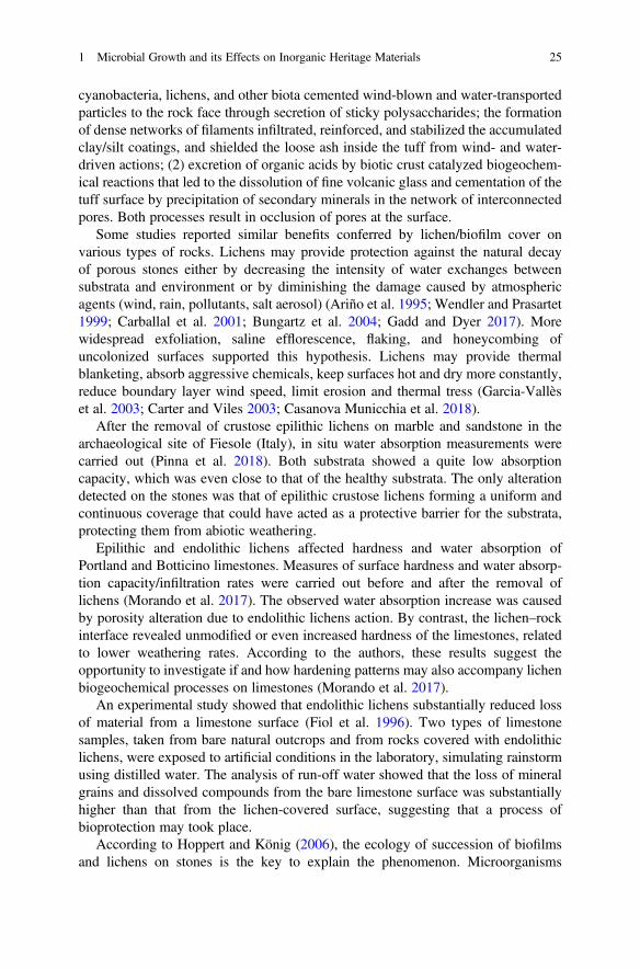
cyanobacteria, lichens, and other biota cemented wind-blown and water-transportedparticles to the rock face through secretion of sticky polysaccharides; the formationof dense networks of filaments infiltrated, reinforced, and stabilized the accumulatedclay/silt coatings, and shielded the loose ash inside the tuff from wind- and water-driven actions; (2) excretion of organic acids by biotic crust catalyzed biogeochem-ical reactions that led to the dissolution of fine volcanic glass and cementation of thetuff surface by precipitation of secondary minerals in the network of interconnectedpores. Both processes result in occlusion of pores at the surface.
Some studies reported similar benefits conferred by lichen/biofilm cover onvarious types of rocks. Lichens may provide protection against the natural decayof porous stones either by decreasing the intensity of water exchanges betweensubstrata and environment or by diminishing the damage caused by atmosphericagents (wind, rain, pollutants, salt aerosol) (Ariño et al. 1995; Wendler and Prasartet1999; Carballal et al. 2001; Bungartz et al. 2004; Gadd and Dyer 2017). Morewidespread exfoliation, saline efflorescence, flaking, and honeycombing ofuncolonized surfaces supported this hypothesis. Lichens may provide thermalblanketing, absorb aggressive chemicals, keep surfaces hot and dry more constantly,reduce boundary layer wind speed, limit erosion and thermal tress (Garcia-Vallèset al. 2003; Carter and Viles 2003; Casanova Municchia et al. 2018).
After the removal of crustose epilithic lichens on marble and sandstone in thearchaeological site of Fiesole (Italy), in situ water absorption measurements werecarried out (Pinna et al. 2018). Both substrata showed a quite low absorptioncapacity, which was even close to that of the healthy substrata. The only alterationdetected on the stones was that of epilithic crustose lichens forming a uniform andcontinuous coverage that could have acted as a protective barrier for the substrata,protecting them from abiotic weathering.
Epilithic and endolithic lichens affected hardness and water absorption ofPortland and Botticino limestones. Measures of surface hardness and water absorp-tion capacity/infiltration rates were carried out before and after the removal oflichens (Morando et al. 2017). The observed water absorption increase was causedby porosity alteration due to endolithic lichens action. By contrast, the lichen–rockinterface revealed unmodified or even increased hardness of the limestones, relatedto lower weathering rates. According to the authors, these results suggest theopportunity to investigate if and how hardening patterns may also accompany lichenbiogeochemical processes on limestones (Morando et al. 2017).
An experimental study showed that endolithic lichens substantially reduced lossof material from a limestone surface (Fiol et al. 1996). Two types of limestonesamples, taken from bare natural outcrops and from rocks covered with endolithiclichens, were exposed to artificial conditions in the laboratory, simulating rainstormusing distilled water. The analysis of run-off water showed that the loss of mineralgrains and dissolved compounds from the bare limestone surface was substantiallyhigher than that from the lichen-covered surface, suggesting that a process ofbioprotection may took place.
According to Hoppert and König (2006), the ecology of succession of biofilmsand lichens on stones is the key to explain the phenomenon. Microorganisms
1 Microbial Growth and its Effects on Inorganic Heritage Materials 25
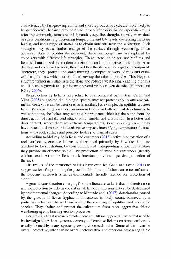
characterized by fast-growing ability and short reproductive cycle are more likely tobe deteriorative, because they colonize rapidly after disturbance (sporadic eventsaffecting community structure and dynamics, e.g., fire, drought, storms, or erosion)or stress conditions (e.g. increasing temperature and UV levels, decreasing moisturelevels), and use a range of strategies to obtain nutrients from the substratum. Suchstrategies may cause further change of the surface through weathering. In anadvanced state of biofilm development, these microorganisms are replaced bycolonizers with different life strategies. These “new” colonizers are biofilms andlichens characterized by moderate metabolic and reproductive rates. In order todevelop and colonize the rock, they need that the stone is not rapidly decomposed.Therefore, they “protect” the stone forming a compact network of cells and extra-cellular polymers, which surround and enwrap the mineral particles. This biogenicstructure temporarily stabilizes the stone and reduces weathering, enabling biofilmsand lichens to growth and persist over several years or even decades (Hoppert andKönig 2006).
Bioprotection by lichens may relate to environmental parameters. Carter andViles (2005) suggested that a single species may act protectively in one environ-mental context but can be deteriorative in another. For example, the epilithic crustoselichen Verrucaria nigrescens is common in Europe in both wet and dry climates. Inwet conditions, the lichen may act as a bioprotector, shielding the stone from thedirect action of rainfall, acid attack, wind, runoff, and dissolution. In a hotter anddrier context, where there are extreme temperatures, Verrucaria nigrescens mayhave instead a dominant biodeteriorative impact, intensifying temperature fluctua-tions at the rock surface and possibly leading to thermal stress.
According to McIlroy de la Rosa and coauthors (2013), active bioprotection of arock surface by crustose lichens is determined primarily by how the thalli areattached to the substratum, by their binding and waterproofing action and whetherthey provide an effective shield. The production of insoluble substances (usuallycalcium oxalates) at the lichen–rock interface provides a passive protection ofthe rock.
The results of the mentioned studies have even led Gadd and Dyer (2017) tosuggest actions for promoting the growth of biofilms and lichens on stone surfaces asthe biogenic approach is an environmentally friendly method for protection ofstones.
A general consideration emerging from the literature so far is that biodeteriorationand bioprotection by lichens coexist in a delicate equilibrium that can be destabilizedby environmental changes. According to Morando et al. (2017), deterioration causedby the growth of lichen hyphae in limestones is likely counterbalanced by aprotective effect on the rock surface by the covering of epilithic and endolithicspecies. They shelter and protect the substratum from more aggressive abioticweathering agents limiting erosion processes.
Despite significant research efforts, there are still many general issues that need tobe investigated. A homogeneous coverage of crustose lichens on stone surfaces isusually formed by many species growing close each other. Some of them can beoverall protective, other can be overall deteriorative and other can have a negligible
26 D. Pinna
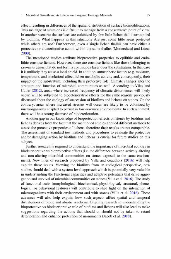
effect, resulting in differences of the spatial distribution of surface biomodifications.This mélange of situations is difficult to manage from a conservative point of view.In another scenario the surfaces are colonized by few little lichen thalli surroundedby biofilms. What happens in this situation? Are just some little areas protectedwhile others are not? Furthermore, even a single lichen thallus can have either aprotective or a deteriorative action within the same thallus (Mottershead and Lucas2000).
The mentioned studies attribute bioprotective properties to epilithic and endo-lithic crustose lichens. However, there are crustose lichens like those belonging toLepraria genus that do not form a continuous layer over the substratum. In that caseit is unlikely they act as a local shield. In addition, atmospheric factors (e.g. moisture,temperature, and insolation) affect lichen metabolic activity and, consequently, theirimpact on the substratum, including their protective role. Climate changes alter thestructure and function of microbial communities as well. According to Viles andCutler (2012), areas where increased frequency of climatic disturbances will likelyoccur, will be subjected to biodeteriorative effects for the same reasons previouslydiscussed about the ecology of succession of biofilms and lichens on stones. On thecontrary, areas where increased stresses will occur are likely to be colonized bymicroorganisms adapted to persist in low-resource environments. In such a context,there will be a strong decrease of biodeterioration.
Another gap in our knowledge of bioprotection effects on stones by biofilms andlichens derives from the fact that the mentioned studies applied different methods toassess the protective properties of lichens, therefore their results are not comparable.The assessment of standard test methods and procedures to evaluate the protectiveand/or damaging action by biofilms and lichens is crucial for future studies on thissubject.
Further research is required to understand the importance of microbial ecology inbiodeteriorative vs bioprotective effects (i.e. the difference between actively alteringand non-altering microbial communities on stones exposed to the same environ-ment). New lines of research proposed by Villa and coauthors (2016) will helpexplain these issues. Viewing the biofilms from an ecological perspective, newstudies should deal with a system-level approach which is potentially very valuablein understanding the functional capacities and adaptive potentials that drive aggre-gation and survival of microbial communities on stones (Villa et al. 2016). The studyof functional traits (morphological, biochemical, physiological, structural, pheno-logical, or behavioral features) will contribute to shed light on the interaction ofmicroorganisms with their environment and with stones (Villa et al. 2016). Theseadvances will also help explain how such aspects affect spatial and temporaldistributions of biotic and abiotic reactions. Ongoing research in understanding thebioprotective vs biodeteriorative role of biofilms and lichens will also lead to makesuggestions regarding the actions that should or should not be taken to retarddeterioration and enhance protection of monuments (Jacob et al. 2018).
1 Microbial Growth and its Effects on Inorganic Heritage Materials 27
References
Abuku M, Janssen H, Roels S (2009) Impact of wind-driven rain on historic brick wall buildings ina moderately cold and humid climate: numerical analyses of mould growth risk, indoor climateand energy consumption. Energ Buildings 41:101–110
Adamson C, McCabe S, Warke PA, McAllister D, Snith BJ (2013) The influence of aspect on thebiological bolonization of stone in Northern Ireland. Int Biodeterior Biodegradation 84:357–366
Albertano P, Bruno L, Bellezza S (2005) New strategies for the monitoring and control ofcyanobacterial films on valuable lithic faces. Plant Biosyst 139:311–322
Allsopp D, Seal K, Gaylarde C (2004) Introduction to biodeterioration. Cambridge UniversityPress, Cambridge
Aramendia J, Gomez-Nubla L, Bellot-Gurlet L, Castro K, Arana G, Madariaga JM (2015)Bioimpact on weathering steel surfaces: oxalates formation and the elucidation of their origin.Int Biodeterior Biodegradation 104:59–66
Ariño X, Ortega-Calvo JJ, Gomez-Bolea A, Saiz-Jimenez C (1995) Lichen colonization of theRoman pavement at Baelo Claudia (Cadiz, Spain): biodeterioration vs bioprotection. Sci TotalEnviron 67:353–363
Ashbee J (2010) Ivy and the presentation of ancient monuments and building. In: Sternberg T(ed) Ivy on Walls, Seminar Report, English Heritage, pp 9–15
Bastian F, Jurado V, Nováková A, Alabouvette C, Saiz-Jimenez C (2010) The microbiology ofLascaux Cave. Microbiology 156:644–652
Beech IB, Coutinho CLM (2003) Biofilms on corroding materials. In: Lens P, Moran AP,Mahony T, Stoodly P, O’Flaherty V (eds) Biofilms in medicine, industry and environmentalbiotechnology - characteristics, analysis and control. IWA Publishing of Alliance House,London, pp 115–131
Beech IB, Sunner J (2004) Biocorrosion: towards understanding interactions between biofilms andmetals. Curr Opin Biotechnol 15:181–186
Beech IB, Sztyler M, Gaylarde CC, Smith WL, Sunner J (2014) Biofilms and biocorrosion. In:Liengen T, Feron D, Basseguy R, Beech IB (eds) Understanding biocorrosion: fundamentalsand applications. Woodhead Publishing, Cambridge, pp 33–50
Bjelland TH, Thorseth IH (2002) Comparative studies of the lichen–rock interface of four lichens inVingen, Western Norway. Chem Geol 192:81–98
Bridier A, Piard J-C, Pandin C, Labarthe S, Dubois-Brissonnet F, Briandet R (2017) Spatialorganization plasticity as an adaptive driver of surface microbial communities. Front Microbiol8:1364
Brown PW, Masters LW (1980) Factors affecting the corrosion of metals in the atmosphere. In:Ailor WH (ed) Atmospheric corrosion. John Wiley and Sons, New York, pp 31–49
Bungartz F, Garvie LAJ, Nash TH (2004) Anatomy of the endolithic Sonoran Desert lichenVerrucaria rubrocincta Breuss: implications for biodeterioration and biomineralization. Lichen-ologist 36:55–73
Cámara B, Suzuki S, Nealson KH, Wierzchos J, Ascaso C, Artieda O, de los Ríos A (2014)Ignimbrite textural properties as determinants of endolithic colonization patterns from hyper-arid Atacama Desert. Int Microbiol 17:235–247
Caneva G, Bartoli F (2017) Botanical planning and lichen control for the conservation of grave-stones in Jewish urban cemeteries in North-Eastern Italy. Israel J Plant Sci 64:1–14
Caneva G, Ceschin S (2008) Ecology of biodeterioration. In: Caneva G, Nugari MP, Salvadori O(eds) Plant biology for cultural heritage: biodeterioration and conservation. Getty ConservationInstitute, Los Angeles, pp 35–58
Cappitelli F, Abbruscato P, Foladori P, Zanardini E, Ranalli G, Principi P, Villa F, Polo A, Sorlini C(2009) Detection and elimination of cyanobacteria from frescoes: the case of the St. BrizioChapel (Orvieto Cathedral, Italy). Microbial Ecol 57:633–639
28 D. Pinna
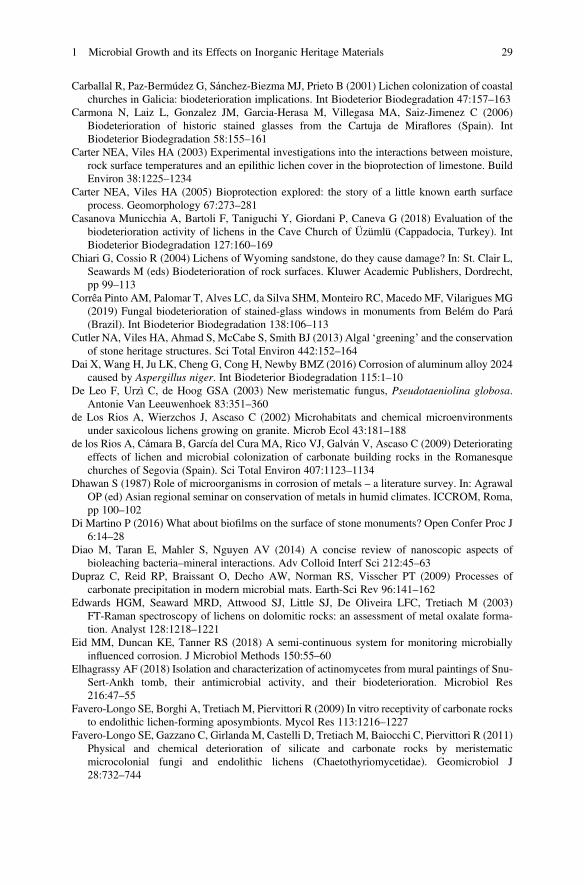
Carballal R, Paz-Bermúdez G, Sánchez-Biezma MJ, Prieto B (2001) Lichen colonization of coastalchurches in Galicia: biodeterioration implications. Int Biodeterior Biodegradation 47:157–163
Carmona N, Laiz L, Gonzalez JM, Garcia-Herasa M, Villegasa MA, Saiz-Jimenez C (2006)Biodeterioration of historic stained glasses from the Cartuja de Miraflores (Spain). IntBiodeterior Biodegradation 58:155–161
Carter NEA, Viles HA (2003) Experimental investigations into the interactions between moisture,rock surface temperatures and an epilithic lichen cover in the bioprotection of limestone. BuildEnviron 38:1225–1234
Carter NEA, Viles HA (2005) Bioprotection explored: the story of a little known earth surfaceprocess. Geomorphology 67:273–281
Casanova Municchia A, Bartoli F, Taniguchi Y, Giordani P, Caneva G (2018) Evaluation of thebiodeterioration activity of lichens in the Cave Church of Üzümlü (Cappadocia, Turkey). IntBiodeterior Biodegradation 127:160–169
Chiari G, Cossio R (2004) Lichens of Wyoming sandstone, do they cause damage? In: St. Clair L,Seawards M (eds) Biodeterioration of rock surfaces. Kluwer Academic Publishers, Dordrecht,pp 99–113
Corrêa Pinto AM, Palomar T, Alves LC, da Silva SHM, Monteiro RC, Macedo MF, Vilarigues MG(2019) Fungal biodeterioration of stained-glass windows in monuments from Belém do Pará(Brazil). Int Biodeterior Biodegradation 138:106–113
Cutler NA, Viles HA, Ahmad S, McCabe S, Smith BJ (2013) Algal ‘greening’ and the conservationof stone heritage structures. Sci Total Environ 442:152–164
Dai X, Wang H, Ju LK, Cheng G, Cong H, Newby BMZ (2016) Corrosion of aluminum alloy 2024caused by Aspergillus niger. Int Biodeterior Biodegradation 115:1–10
De Leo F, Urzì C, de Hoog GSA (2003) New meristematic fungus, Pseudotaeniolina globosa.Antonie Van Leeuwenhoek 83:351–360
de Los Rios A, Wierzchos J, Ascaso C (2002) Microhabitats and chemical microenvironmentsunder saxicolous lichens growing on granite. Microb Ecol 43:181–188
de los Rios A, Cámara B, García del Cura MA, Rico VJ, Galván V, Ascaso C (2009) Deterioratingeffects of lichen and microbial colonization of carbonate building rocks in the Romanesquechurches of Segovia (Spain). Sci Total Environ 407:1123–1134
Dhawan S (1987) Role of microorganisms in corrosion of metals – a literature survey. In: AgrawalOP (ed) Asian regional seminar on conservation of metals in humid climates. ICCROM, Roma,pp 100–102
Di Martino P (2016) What about biofilms on the surface of stone monuments? Open Confer Proc J6:14–28
Diao M, Taran E, Mahler S, Nguyen AV (2014) A concise review of nanoscopic aspects ofbioleaching bacteria–mineral interactions. Adv Colloid Interf Sci 212:45–63
Dupraz C, Reid RP, Braissant O, Decho AW, Norman RS, Visscher PT (2009) Processes ofcarbonate precipitation in modern microbial mats. Earth-Sci Rev 96:141–162
Edwards HGM, Seaward MRD, Attwood SJ, Little SJ, De Oliveira LFC, Tretiach M (2003)FT-Raman spectroscopy of lichens on dolomitic rocks: an assessment of metal oxalate forma-tion. Analyst 128:1218–1221
Eid MM, Duncan KE, Tanner RS (2018) A semi-continuous system for monitoring microbiallyinfluenced corrosion. J Microbiol Methods 150:55–60
Elhagrassy AF (2018) Isolation and characterization of actinomycetes from mural paintings of Snu-Sert-Ankh tomb, their antimicrobial activity, and their biodeterioration. Microbiol Res216:47–55
Favero-Longo SE, Borghi A, Tretiach M, Piervittori R (2009) In vitro receptivity of carbonate rocksto endolithic lichen-forming aposymbionts. Mycol Res 113:1216–1227
Favero-Longo SE, Gazzano C, Girlanda M, Castelli D, Tretiach M, Baiocchi C, Piervittori R (2011)Physical and chemical deterioration of silicate and carbonate rocks by meristematicmicrocolonial fungi and endolithic lichens (Chaetothyriomycetidae). Geomicrobiol J28:732–744
1 Microbial Growth and its Effects on Inorganic Heritage Materials 29
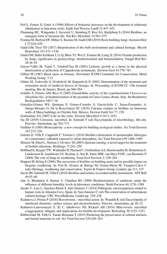
Fiol L, Fornós JJ, Ginés A (1996) Effects of biokarstic processes on the development of solutionalrillenkarren in limestone rocks. Earth Surf Process Landf 21:447–452
Flemming HC, Wingender J, Szewzyk U, Steinberg P, Rice SA, Kjelleberg S (2016) Biofilms: anemergent form of bacterial life. Nat Rev Microbiol 14:563–575
Fomina M, Burford EP, Hillier S, Kierans M, Gadd GM (2010) Rock-building fungi. GeomicrobiolJ 27:624–629
Gadd GM, Dyer TD (2017) Bioprotection of the built environment and cultural heritage. MicrobBiotechnol 10:1152–1156
Gadd GM, Bahri-Esfahani J, Li Q, Rhee YJ, Wei Z, Fomina M, Liang X (2014) Oxalate productionby fungi: significance in geomycology, biodeterioration and bioremediation. Fungal Biol Rev28:36–55
Garcia-Vallès M, Topal T, Vendrell-Saz M (2003) Lichenic growth as a factor in the physicaldeterioration or protection of Cappadocian monuments. Environ Geol 43:776–781
Gilbert M (1987) Black spots on bronzes. Newsletter ICOM Committee for Conservation, MetalWorking Group, 3:12
Gittins M, Vedovello S, Dvalishvili M, Kuprashvili N (2002) Determination of the treatment andrestoration needs of medieval frescos in Georgia. In: Proceeding of ICOM-CC 13th triennialmeeting, Rio de Janeiro, Brazil, pp 560–564
Golubic S, Pietrini AM, Ricci S (2005) Euendolithic activity of the cyanobacterium Chroococcuslithophilus Erc. in biodeterioration of the pyramid of Caius Cestius, Rome, Italy. Int BiodeteriorBiodegradation 100:7–16
González-Gómez WS, Quintana P, Gómez-Cornelio S, García-Solis C, Sierra-Fernandez A,Ortega-Morales O, De la Rosa-García SC (2018) Calcium oxalates in biofilms on limestonewalls of Maya buildings in Chichén Itzá, Mexico. Environ Earth Sci 77: 230
Gorbushina AA (2007) Life on the rocks. Environ Microbiol 9:1613–1631Gu JD (2019) Corrosion, microbial. In: Schmidt T (ed) Encyclopedia of microbiology, 4th edn.
Elsevier, Amsterdam, pp 762–771Guillitte O (1985) Bioreceptivity: a new concept for building ecological studies. Sci Total Environ
167:215–220Gulotta D, Villa F, Cappitelli F, Toniolo L (2018) Biofilm colonization of metamorphic lithotypes
of a renaissance cathedral exposed to urban atmosphere. Sci Total Environ 639:1480–1490Hentzer M, Eberl L, Nielsen J, Givskov M (2003) Quorum sensing: a novel target for the treatment
of biofilm infections. BioDrugs 17:241–250Hoffland E, Kuyper TW,Wallander H, Plassard C, Gorbushina AA, Haselwandter H, Holmstrom S,
Landeweert R, Lundstrom US, Rosling A, Sen R, Smits MM, van Hees PAW, van Breemen N(2004) The role of fungi in weathering. Front Ecol Environ 2: 258–264
Hoppert M, König S (2006) The succession of biofilms on building stone and its possible impact onbiogenic weathering. In: Fort R, Alvarez de Buergo M, Gomez-Heras M, Vazquez-Calvo C(eds) Heritage, weathering and conservation. Taylor & Francis Group, London, pp 311–315
Jacob JM, Schmull M, Villa F (2018) Biofilms and lichens on eroded marble monuments. APT Bull49:55–60
Jain A, Bhadauria S, Kumar V, Chauhan RS (2009) Biodeterioration of sandstone under theinfluence of different humidity levels in laboratory conditions. Build Environ 44:1276–1284
Jurado V, Laiz L, Sanchez-Moral S, Saiz-Jimenez C (2014) Pathogenic microorganisms related tohuman visits in Altamira Cave, Spain. In: Saiz-Jimenez C (ed) The conservation of subterraneancultural heritage. CRC Press/Balkema, Leiden pp. 229–238
Kadukova J, Pristas P (2018) Biocorrosion – microbial action. In: Wandelt K (ed) Encyclopedia ofinterfacial chemistry: surface science and electrochemistry. Elsevier, Amsterdam, pp 20–22
Katharios-Lanwermeyer S, Xi C, Jakubovics NS, Rickard AH (2014) Mini-review: microbialcoaggregation: ubiquity and implications for biofilm development. Biofouling 30:1235–1251
Kibblewhite M, Tóth G, Tamás Hermann T (2015) Predicting the preservation of cultural artefactsand buried materials in soil. Sci Total Environ 529:249–263
30 D. Pinna
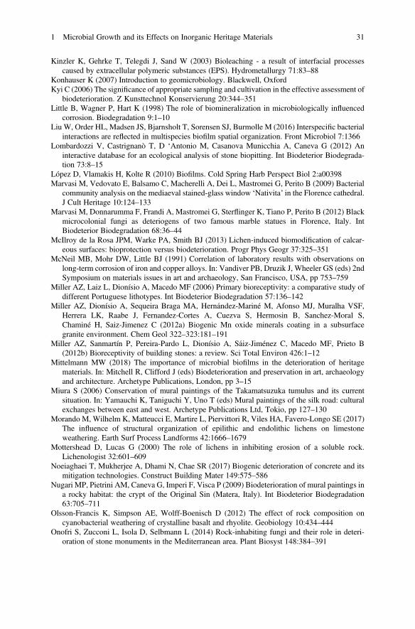
Kinzler K, Gehrke T, Telegdi J, Sand W (2003) Bioleaching - a result of interfacial processescaused by extracellular polymeric substances (EPS). Hydrometallurgy 71:83–88
Konhauser K (2007) Introduction to geomicrobiology. Blackwell, OxfordKyi C (2006) The significance of appropriate sampling and cultivation in the effective assessment of
biodeterioration. Z Kunsttechnol Konservierung 20:344–351Little B, Wagner P, Hart K (1998) The role of biomineralization in microbiologically influenced
corrosion. Biodegradation 9:1–10Liu W, Order HL, Madsen JS, Bjarnsholt T, Sorensen SJ, Burmolle M (2016) Interspecific bacterial
interactions are reflected in multispecies biofilm spatial organization. Front Microbiol 7:1366Lombardozzi V, Castrignanò T, D ‘Antonio M, Casanova Municchia A, Caneva G (2012) An
interactive database for an ecological analysis of stone biopitting. Int Biodeterior Biodegrada-tion 73:8–15
López D, Vlamakis H, Kolte R (2010) Biofilms. Cold Spring Harb Perspect Biol 2:a00398Marvasi M, Vedovato E, Balsamo C, Macherelli A, Dei L, Mastromei G, Perito B (2009) Bacterial
community analysis on the mediaeval stained-glass window ‘Nativita’ in the Florence cathedral.J Cult Heritage 10:124–133
Marvasi M, Donnarumma F, Frandi A, Mastromei G, Sterflinger K, Tiano P, Perito B (2012) Blackmicrocolonial fungi as deteriogens of two famous marble statues in Florence, Italy. IntBiodeterior Biodegradation 68:36–44
McIlroy de la Rosa JPM, Warke PA, Smith BJ (2013) Lichen-induced biomodification of calcar-eous surfaces: bioprotection versus biodeterioration. Progr Phys Geogr 37:325–351
McNeil MB, Mohr DW, Little BJ (1991) Correlation of laboratory results with observations onlong-term corrosion of iron and copper alloys. In: Vandiver PB, Druzik J, Wheeler GS (eds) 2ndSymposium on materials issues in art and archaeology, San Francisco, USA, pp 753–759
Miller AZ, Laiz L, Dionísio A, Macedo MF (2006) Primary bioreceptivity: a comparative study ofdifferent Portuguese lithotypes. Int Biodeterior Biodegradation 57:136–142
Miller AZ, Dionísio A, Sequeira Braga MA, Hernández-Mariné M, Afonso MJ, Muralha VSF,Herrera LK, Raabe J, Fernandez-Cortes A, Cuezva S, Hermosin B, Sanchez-Moral S,Chaminé H, Saiz-Jimenez C (2012a) Biogenic Mn oxide minerals coating in a subsurfacegranite environment. Chem Geol 322–323:181–191
Miller AZ, Sanmartín P, Pereira-Pardo L, Dionísio A, Sáiz-Jiménez C, Macedo MF, Prieto B(2012b) Bioreceptivity of building stones: a review. Sci Total Environ 426:1–12
Mittelmann MW (2018) The importance of microbial biofilms in the deterioration of heritagematerials. In: Mitchell R, Clifford J (eds) Biodeterioration and preservation in art, archaeologyand architecture. Archetype Publications, London, pp 3–15
Miura S (2006) Conservation of mural paintings of the Takamatsuzuka tumulus and its currentsituation. In: Yamauchi K, Taniguchi Y, Uno T (eds) Mural paintings of the silk road: culturalexchanges between east and west. Archetype Publications Ltd, Tokio, pp 127–130
Morando M, Wilhelm K, Matteucci E, Martire L, Piervittori R, Viles HA, Favero-Longo SE (2017)The influence of structural organization of epilithic and endolithic lichens on limestoneweathering. Earth Surf Process Landforms 42:1666–1679
Mottershead D, Lucas G (2000) The role of lichens in inhibiting erosion of a soluble rock.Lichenologist 32:601–609
Noeiaghaei T, Mukherjee A, Dhami N, Chae SR (2017) Biogenic deterioration of concrete and itsmitigation technologies. Construct Building Mater 149:575–586
Nugari MP, Pietrini AM, Caneva G, Imperi F, Visca P (2009) Biodeterioration of mural paintings ina rocky habitat: the crypt of the Original Sin (Matera, Italy). Int Biodeterior Biodegradation63:705–711
Olsson-Francis K, Simpson AE, Wolff-Boenisch D (2012) The effect of rock composition oncyanobacterial weathering of crystalline basalt and rhyolite. Geobiology 10:434–444
Onofri S, Zucconi L, Isola D, Selbmann L (2014) Rock-inhabiting fungi and their role in deteri-oration of stone monuments in the Mediterranean area. Plant Biosyst 148:384–391
1 Microbial Growth and its Effects on Inorganic Heritage Materials 31
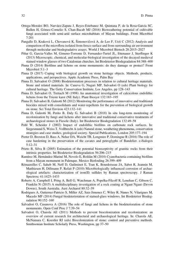
Ortega-Morales BO, Narváez-Zapata J, Reyes-Estebanez M, Quintana P, de la Rosa-García SC,Bullen H, Gómez-Cornelio S, Chan-Bacab MJ (2016) Bioweathering potential of cultivablefungi associated with semi-arid surface microhabitats of Mayan buildings. Front Microbiol7:201
Pangallo D, Kraková L, Chovanová K, Šimonovičová A, de Leo F, Urzì C (2012) Analysis andcomparison of the microflora isolated from fresco surface and from surrounding air environmentthrough molecular and biodegradative assays. World J Microbiol Biotech 28:2015–2027
Piñar G, Garcia-Valles M, Gimeno-Torrente D, Fernandez-Turiel JL, Ettenauer J, Sterflinger K(2013) Microscopic, chemical, and molecular-biological investigation of the decayed medievalstained window glasses of two Catalonian churches. Int Biodeterior Biodegradation 84:388–400
Pinna D (2014) Biofilms and lichens on stone monuments: do they damage or protect? FrontMicrobiol 5:1–3
Pinna D (2017) Coping with biological growth on stone heritage objects. Methods, products,applications, and perspectives. Apple Academic Press, Palm Bay
Pinna D, Salvadori O (2008) Biodeterioration processes in relation to cultural heritage materials.Stone and related materials. In: Caneva G, Nugari MP, Salvadori O (eds) Plant biology forcultural heritage. The Getty Conservation Institute, Los Angeles, pp 128–143
Pinna D, Salvadori O, Tretiach M (1998) An anatomical investigation of calcicolous endolithiclichens from the Trieste karst (NE Italy). Plant Biosyst 132:183–195
Pinna D, Salvadori B, Galeotti M (2012) Monitoring the performance of innovative and traditionalbiocides mixed with consolidants and water-repellents for the prevention of biological growthon stone. Sci Total Environ 423:132–141
Pinna D, Galeotti M, Perito B, Daly G, Salvadori B (2018) In situ long-term monitoring ofrecolonization by fungi and lichens after innovative and traditional conservative treatments ofarchaeological stones in Fiesole (Italy). Int Biodeterior Biodegradation 132:49–58
Pohl W, Scheider J (2002) Impact of endolithic biofilms on carbonate rock surfaces. In:Siegesmund S, Weiss T, Vollbrecht A (eds) Natural stone, weathering phenomena, conservationstrategies and case studies, geological society. Special Publications, London 205:177–194
Porter D, Broxton D, Bass A, Neher DA, Weicht TR, Longmire P, Domingue R (2016) The role ofcase hardening in the preservation of the cavates and petroglyphs of Bandelier. e-δialogos5:12–31
Prieto B, Silva B (2005) Estimation of the potential bioreceptivity of granitic rocks from theirintrinsic properties. Int Biodeterior Biodegradation 56:206–215
Ramírez M, Hernández-Mariné M, Novelo E, Roldán M (2010) Cyanobacteria containing biofilmsfrom a Mayan monument in Palenque. Mexico Biofouling 26:399–409
Rémazeilles C, Saheb M, Neff D, Guilminot E, Tran K, Bourdoiseau JA, Sabot R, Jeannin M,Matthiesen H, Dillmann P, Refait P (2010) Microbiologically influenced corrosion of archae-ological artefacts: characterization of iron(II) sulfides by Raman spectroscopy. J RamanSpectrosc 41:1425–1433
Roberts A, Campbell I, Pring A, Bell G, Watchman A, Popelka-Filcoff R, Lenehan C, Gibson C,Franklin N (2015) A multidisciplinary investigation of a rock coating at Ngaut Ngaut (DevonDowns), South Australia. Aust Archaeol 80:32–39
Rodrigues A, Gutierrez-Patricio S, Miller AZ, Saiz-Jimenez C, Wiley R, Nunes N, Vilarigues M,Macedo MF (2014) Fungal biodeterioration of stained-glass windows. Int Biodeterior Biodeg-radation 90:152–160
Salvadori O, Casanova A (2016) The role of fungi and lichens in the biodeterioration of stonemonuments. Open Conf Proc J 7:39–54
Salvadori O, Charola AE (2011) Methods to prevent biocolonization and recolonization: anoverview of current research for architectural and archaeological heritage. In: Charola AE,McNamara C, Koestler RJ (eds) Biocolonization of stone: control and preventive methods.Smithsonian Institute Scholarly Press, Washington, pp 37–50
32 D. Pinna
Sánchez del Junco A, Moreno DA, Ranninger C, Ortega-Calvo JJ, Saiz-Jimenez C (1992) Micro-bial induced corrosion of metallic antiquities and works of art: a critical review. Int BiodeteriorBiodegradation 29:367–375
Scheerer S (2008) Microbial biodeterioration of outdoor stone monuments. Assessment methodsand control strategies. PhD Thesis, Cardiff University, UK
Scheerer S, Ortega-Morales O, Gaylarde C (2009) Microbial deterioration of stone monuments - anupdated overview. Adv Appl Microbiol 66:97–139
Shirakawa MA, Tavares RG, Gaylarde CC, Taqueda ME, Loh K, John VM (2010) Climate as themost important factor determining anti-fungal biocide performance in paint films. Sci TotalEnviron 408:5878–5886
Shirakawa MA, Vanderley MJ, Mocelin A, Zilles R, Toma SH, Araki K, Toma HE, Thomaz AC,Gaylarde CC (2016) Effect of silver nanoparticle and TiO2 coatings on biofilm formation onfour types of modern glass. Int Biodeterior Biodegradation 108:175–180
Siegesmund S, Snethlage RE (2014) Stone in architecture. Properties, durability. Springer, BerlinHeidelberg, Germany
Skinner HCW, Jahren AH (2003) Biomineralization. In: Schlesinger WH (ed) Treatise on geo-chemistry, vol 8. Elsevier, Amsterdam, pp 117–184
Sohrabi M, Favero-Longo SE, Perez-Ortega S, Ascaso C, Haghighat Z, Talebian MH, Fadaei H, delos Ríos A (2017) Lichen colonization and associated deterioration processes in Pasargadae,UNESCO world heritage site, Iran. Int Biodeterior Biodegradation 117:171–182
Sterflinger K (2010) Fungi: their role in deterioration of cultural heritage. Fungal Biol Rev24:47–55
Sterflinger K, Piñar G (2013) Microbial deterioration of cultural heritage and works of art - tilting atwindmills? Appl Microbiol Biotechnol 97:9637–9646
Szczepanowska HM, Cavaliere AR (2004) Tutankhamun tomb, a closer look at biodeterioration:preliminary report. In: Rauch A, Miklin-Kniefacz S, Harmssen A (eds) Schimmel: Gefahr fürMensch und Kulturgut durch Mikroorganismen. Konrad Theiss Verlag GmbH & Co, Stuttgart,pp 42–47
Tescari M, Frangipani E, Caneva G, Casanova Municchia A, Sodo A, Visca P (2018) Arthrobacteragilis and rosy discoloration in “Terme del Foro” (Pompeii, Italy). Int Biodeterior Biodegrada-tion 130:48–54
Tolker-Nielsen T (2015) Biofilm development. Microbiol Spectr 3: MB-0001-2014Tonon C, Favero-Longo SE, Matteucci E, Piervittori R, Croveri P, Appolonia L, Meirano V,
Serino M, Elia D (2019) Microenvironmental features drive the distribution of lichens in thehouse of the Ancient Hunt, Pompeii, Italy. Int Biodeterior Biodegradation 136:71–81
Traversetti L, Bartoli F, Caneva G (2018) Wind-driven rain as a bioclimatic factor affecting thebiological colonization at the archaeological site of Pompeii, Italy. Int Biodeterior Biodegrada-tion 134:31–38
Tretiach M, Pinna D, Grube M (2003) Caloplaca erodens [sect. Pyrenodesmia], a new lichenspecies from Italy with an unusual thallus type. Mycolog Prog 2:127–136
Unković N, Erić S, Šarić K, Stupar M, Savković Ž, Stanković S, Stanojević O, Dimkić I,Vukojević J, Ljaljević Grbić M (2017) Biogenesis of secondary mycogenic minerals relatedto wall paintings deterioration process. Micron 100:1–9
Unković N, Dimkić I, Stupar M, Stanković S, Vukojević J, Ljaljević Grbić M (2018)Biodegradative potential of fungal isolates from sacral ambient: in vitro study as risk assessmentimplication for the conservation of wall paintings. PLoS One 13:e0190922
Urzì C, De Leo F, Bruno L, Pangallo D (2014) New species description, biomineralizationprocesses and biocleaning applications of Roman catacombs-living bacteria. In: Saiz-JimenezC (ed) The conservation of subterranean cultural heritage. CRC Press/Balkema, Leiden/Neth-erlands, pp 65–72
Urzì C, Bruno L, De Leo F (2018) Biodeterioration of paintings in caves, catacombs and otherhypogean sites. In: Mitchell R, Clifford J (eds) Biodeterioration and preservation in art,archaeology and architecture. Archetype Publications Ltd, London, pp 114–129
1 Microbial Growth and its Effects on Inorganic Heritage Materials 33
Vasanthakumar A, DeAraujo A, Mazurek J, Schilling M, Mitchell R (2013) Microbiological surveyfor analysis of the brown spots on the walls of the tomb of King Tutankhamun. Int BiodeteriorBiodegradation 79:56–63
Vázquez-Nion D, Silva B, Prieto B (2018a) Influence of the properties of granitic rocks on theirbioreceptivity to subaerial phototrophic biofilms. Sci Total Environ 610–611:44–54
Vázquez-Nion D, Troiano F, Sanmartín P, Valagussa C, Cappitelli F, Prieto B (2018b) Secondarybioreceptivity of granite: effect of salt weathering on subaerial biofilm growth. Mater Struct51:158
Vega LM, Mathieu J, Yang Y, Pyle BH, McLean RJC, Alvarez PJJ (2014) Nickel and cadmiumions inhibit quorum sensing and biofilm formation without affecting viability in Burkholderiamultivorans. Int Biodeterior Biodegradation 91:82–87
Videla HA, Herrera LK (2009) Understanding microbial inhibition of corrosion. A comprehensiveoverview. Int Biodeterior Biodegradation 63:896–900
Viles HA (2012) Microbial geomorphology: a neglected link between life and landscape. Geomor-phology 157–158:6–16
Viles HA, Cutler NA (2012) Global environmental change and the biology of heritage structures.Glob Chang Biol 18:2406–2418
Villa F, Pitts B, Lauchnor E, Cappitelli F, Stewart PS (2015) Development of a laboratory model ofa phototroph-heterotroph mixed-species biofilm at the stone/air interface. Front Microbiol6:1251
Villa F, Stewart PS, Klapper I, Jacob JM, Cappitelli F (2016) Subaerial biofilms on outdoor stonemonuments: changing the perspective towards an ecological framework. Bioscience66:285–294
Vítek P, Ascaso C, Artieda O, Wierzchos J (2016) Raman imaging in geomicrobiology: endolithicphototrophic microorganisms in gypsum from the extreme sun irradiation area in the AtacamaDesert. Anal Bioanal Chem 408:4083–4092
Wendler E, Prasartet C (1999) Lichen growth on old Khmer-style sandstone monuments inThailand: damage factor or shelter? In: Bridgland J, Brown J (eds) Proceedings of the 12thtriennial meeting of the ICOM Committee for Conservation. James and James, London, pp750–754
Xu D, Li Y, Gu T (2016) Mechanistic modeling of biocorrosion caused by biofilms of sulfatereducing bacteria and acid producing bacteria. Bioelectrochemistry 110:52–58
Zammit G, De Leo F, Albertano P, Urzì C (2008) A preliminary study of microbial communitiescolonizing ochre-decorated chambers at the Hal Saflieni Hypogeum at Paola, Malta. In:Lukaszewicz JW, Niemcewicz P (eds) Proceedings of the 11th international congress ondeterioration and conservation of stone. Torun, Poland, pp 555–562
Zanardini E, Cappitelli F, Ranalli G, Sorlini C (2008) Biodeterioration processes in relation tocultural heritage materials - metals. In: Caneva G, Nugari MP, Salvadori O (eds) Plant biologyfor cultural heritage: biodeterioration and conservation. Getty Conservation Institute, LosAngeles, pp 153–156
Zhang G, Gong C, Gu J, Katayama Y, Someya T, Gu JD (2019) Biochemical reactions andmechanisms involved in the biodeterioration of stone world cultural heritage under the tropicalclimate conditions. Int Biodeterior Biodegradation 143:104723
Zucconi L, Gagliardi M, Isola D, Onofri S, Andaloro MC, Pelosi C, Pogliani P, Selbmann L (2012)Biodeterioration agents dwelling in or on the wall paintings of the Holy Saviour’s cave(Vallerano, Italy). Int Biodeterior Biodegradation 70:40–46
34 D. Pinna

Open Access This chapter is licensed under the terms of the Creative Commons Attribution 4.0International License (http://creativecommons.org/licenses/by/4.0/), which permits use, sharing,adaptation, distribution and reproduction in any medium or format, as long as you give appropriatecredit to the original author(s) and the source, provide a link to the Creative Commons license andindicate if changes were made.
The images or other third party material in this chapter are included in the chapter's CreativeCommons license, unless indicated otherwise in a credit line to the material. If material is notincluded in the chapter's Creative Commons license and your intended use is not permitted bystatutory regulation or exceeds the permitted use, you will need to obtain permission directly fromthe copyright holder.
1 Microbial Growth and its Effects on Inorganic Heritage Materials 35
Chapter 2Microbiota and Biochemical ProcessesInvolved in Biodeterioration of CulturalHeritage and Protection
Ji-Dong Gu and Yoko Katayama
Abstract The world cultural heritage sites face new challenges for an effectiveprotection and management because of destruction and damage initiated by bothnatural and anthropogenic causes. Fresh rock and sandstone surfaces of buildings arequickly colonized and covered by a layer of microorganisms, including phototrophs,lithotrophs, and heterotrophs to form a biofilm that alters the local conditions of thestone surfaces, especially under the favorable tropical climate conditions for auto-trophic microorganisms and plants. Biofilms had been studied with indigenous orpure cultures of isolated microorganisms, but the selective ones that contribute todeterioration of the cultural heritage cannot be confirmed easily. Currently, high-throughput sequencing and metegenomics analyses are capable of obtaining micro-bial community and composition in great depth, but they also suffer from similarweakness unable to identify the culprits in the community. With these as back-ground, this article presents a different approach by focusing on the biochemicalprocesses and the responsible microorganisms involved to reveal the destructionprocesses for management and protection. Among these different functional groupsof microorganisms, lichens are known as pioneering rock-decomposing microor-ganisms, and both sulfur-oxidizing bacteria and fungi participate in the decomposi-tion of sandstone via sulfur cycling and initiation of salt attack of the stone afterward,resulting in defoliation and cracking of stone. Other microorganisms includingammonia-oxidizing bacteria and archaea, especially the latter, have been recentlydetected on sandstone monuments providing evidence on the new organismsinvolved in the deterioration of cultural heritage and buildings. In addition, fungican colonize the surfaces of the matured biofilms and play a new role in the removal
J.-D. Gu (*)Environmental Engineering, Guangdong Technion Israel Institute of Technology, Shantou,Guangdong, People’s Republic of China
Laboratory of Environmental Microbiology and Toxicology, School of Biological Sciences,The University of Hong Kong, Hong Kong SAR, People’s Republic of Chinae-mail: [email protected]
Y. KatayamaTokyo National Research Institute for Cultural Properties, Tokyo, Japan
© The Author(s) 2021E. Joseph (ed.), Microorganisms in the Deterioration and Preservation of CulturalHeritage, https://doi.org/10.1007/978-3-030-69411-1_2
37
of them, which has a potential biotechnological application in conservation ofcultural heritage. The new proposed approach by focusing the microorganismswith identified biochemical function is more productive than a description of thecommunity composition and assembly when assessing cultural heritage biodeterio-ration, and this provides basic and useful information for effective protectionstrategies and management.
Keywords Biodeterioration · Sandstone · Biofilms · Defoliation · Monuments ·Sulfur-oxidizing · Ammonia-oxidizing · Salt attack
1 Introduction
Deterioration of the world cultural heritage and historic buildings is a result of bothnatural and/or anthropogenic contributors involving flora, fauna, and microorgan-isms as well as pollutants. Early investigations on cultural heritage biodeteriorationwere mainly focused on the isolation and identification of the cultural microorgan-isms for a description of them without specific function to the biodeterioration (May2000). Such practice had been persisted before the application of polymerase chainreaction (PCR) to cultural heritage microbiology research to reveal the complexmicrobial community without culturing and isolation (Rölleke et al. 1998). The latesthigh-throughput sequencing and metagenomics provide a much deeper descriptionof the microbial community and composition without culturing or isolation (May2000; McNamara et al. 2006; Sterflinger and Pinar 2013; Zhang et al. 2019; Ma et al.2020; Meng et al. 2020). This new approach also has some major weakness in itsinability to identify the active microbes from the genomic DNA-based communityanalysis and, in addition, the active deteriorating ones are not identified from thecommunity. Because of these, the advances made on knowing the community betterhave not translated to useful results on the deteriorating processes for a moreeffective management and prevention (Liu et al. 2020).
Microorganisms colonizing surfaces of cultural heritage can destruct the under-lying materials through their influences on the physical, chemical, andbioreceptibility of the substratum materials, especially metabolic activities andbiochemical reaction. Biofilms as a physical layer on the outer surface of culturalheritage alter the thermal property and moisture contents, which in turn have theirimpact on the biofilms and the activities of the microorganisms. The mechanismsinvolved in biodeterioration of natural sandstone and man-made materials includeboth abiotic and biological ones (Mansch and Bock 1998; Mitchell and Gu 2000;Warscheid and Braams 2000; Gu 2003; Liu et al. 2018a; Zhang et al. 2019; Dinget al. 2020; Liu et al. 2020). On the basis of microbiology and biochemistry, theinitial colonization by the pioneering microorganisms to form biofilms on rock andstone surfaces can initiate a number of subsequent physical and chemical changesof the substratum materials, including the discoloration in appearance, alteration ofporosity and vapor/moisture diffusivity in and out of the stone, accumulation of
38 J.-D. Gu and Y. Katayama
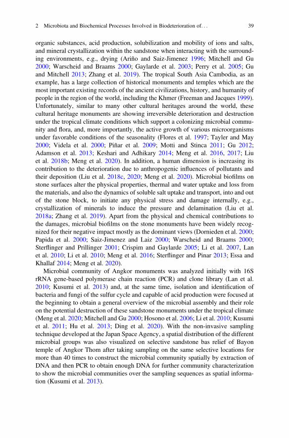
organic substances, acid production, solubilization and mobility of ions and salts,and mineral crystallization within the sandstone when interacting with the surround-ing environments, e.g., drying (Ariño and Saiz-Jimenez 1996; Mitchell and Gu2000; Warscheid and Braams 2000; Gaylarde et al. 2003; Perry et al. 2005; Guand Mitchell 2013; Zhang et al. 2019). The tropical South Asia Cambodia, as anexample, has a large collection of historical monuments and temples which are themost important existing records of the ancient civilizations, history, and humanity ofpeople in the region of the world, including the Khmer (Freeman and Jacques 1999).Unfortunately, similar to many other cultural heritages around the world, thesecultural heritage monuments are showing irreversible deterioration and destructionunder the tropical climate conditions which support a colonizing microbial commu-nity and flora, and, more importantly, the active growth of various microorganismsunder favorable conditions of the seasonality (Flores et al. 1997; Tayler and May2000; Videla et al. 2000; Piñar et al. 2009; Motti and Stinca 2011; Gu 2012;Adamson et al. 2013; Keshari and Adhikary 2014; Meng et al. 2016, 2017; Liuet al. 2018b; Meng et al. 2020). In addition, a human dimension is increasing itscontribution to the deterioration due to anthropogenic influences of pollutants andtheir deposition (Liu et al. 2018c, 2020; Meng et al. 2020). Microbial biofilms onstone surfaces alter the physical properties, thermal and water uptake and loss fromthe materials, and also the dynamics of soluble salt uptake and transport, into and outof the stone block, to initiate any physical stress and damage internally, e.g.,crystallization of minerals to induce the pressure and delamination (Liu et al.2018a; Zhang et al. 2019). Apart from the physical and chemical contributions tothe damages, microbial biofilms on the stone monuments have been widely recog-nized for their negative impact mostly as the dominant views (Dornieden et al. 2000;Papida et al. 2000; Saiz-Jimenez and Laiz 2000; Warscheid and Braams 2000;Sterflinger and Prillinger 2001; Crispim and Gaylarde 2005; Li et al. 2007, Lanet al. 2010; Li et al. 2010; Meng et al. 2016; Sterflinger and Pinar 2013; Essa andKhallaf 2014; Meng et al. 2020).
Microbial community of Angkor monuments was analyzed initially with 16SrRNA gene-based polymerase chain reaction (PCR) and clone library (Lan et al.2010; Kusumi et al. 2013) and, at the same time, isolation and identification ofbacteria and fungi of the sulfur cycle and capable of acid production were focused atthe beginning to obtain a general overview of the microbial assembly and their roleon the potential destruction of these sandstone monuments under the tropical climate(Meng et al. 2020; Mitchell and Gu 2000; Hosono et al. 2006; Li et al. 2010; Kusumiet al. 2011; Hu et al. 2013; Ding et al. 2020). With the non-invasive samplingtechnique developed at the Japan Space Agency, a spatial distribution of the differentmicrobial groups was also visualized on selective sandstone bas relief of Bayontemple of Angkor Thom after taking sampling on the same selective locations formore than 40 times to construct the microbial community spatially by extraction ofDNA and then PCR to obtain enough DNA for further community characterizationto show the microbial communities over the sampling sequences as spatial informa-tion (Kusumi et al. 2013).
2 Microbiota and Biochemical Processes Involved in Biodeterioration of. . . 39
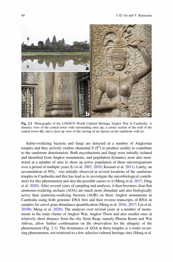
Sulfur-oxidizing bacteria and fungi are detected at a number of Angkoriantemples and they actively oxidize elemental S (S0) to produce acidity to contributeto the sandstone deterioration. Both mycobacteria and fungi were initially isolatedand identified from Angkor monuments, and population dynamics were also mon-itored at a number of sites to show an active population of these microorganismsover a period of multiple years (Li et al. 2007, 2010; Kusumi et al. 2011). Lately, anaccumulation of NO3
� was initially observed at several locations of the sandstonetemples in Cambodia and this has lead us to investigate the microbiological contrib-utors for this phenomenon and also the possible causes to it (Meng et al. 2017; Dinget al. 2020). After several years of sampling and analyses, it then becomes clear thatammonia-oxidizing archaea (AOA) are much more abundant and also biologicallyactive than ammonia-oxidizing bacteria (AOB) on these Angkor monuments inCambodia using both genomic DNA first and then reverse transcripts of RNA insamples for amoA gene abundance quantification (Meng et al. 2016, 2017; Liu et al.2018b; Meng et al. 2020). The analyses over several years at a number of monu-ments in the main cluster of Angkor Wat, Angkor Thom and also smaller ones atrelatively short distance from the city Siem Reap, namely Phnom Krom and WatAthvea, allow further confirmation on the observation for the ubiquity of thephenomenon (Fig. 2.1). The dominance of AOA at these temples is a wider occur-ring phenomenon, not restricted to a few selective cultural heritage sites (Meng et al.
Fig. 2.1 Photographs of the UNESCO World Cultural Heritage Angkor Wat in Cambodia. Adistance view of the central tower with surrounding ones (a); a corner section of the wall of thecentral tower (b); and a close up view of the carving of an Apsara on the sandstone wall (c)
40 J.-D. Gu and Y. Katayama
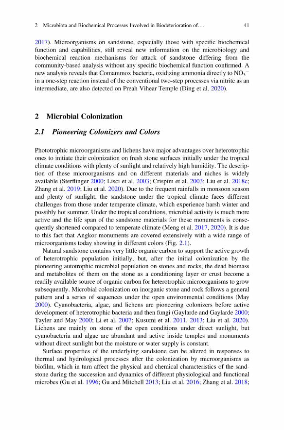
2017). Microorganisms on sandstone, especially those with specific biochemicalfunction and capabilities, still reveal new information on the microbiology andbiochemical reaction mechanisms for attack of sandstone differing from thecommunity-based analysis without any specific biochemical function confirmed. Anew analysis reveals that Comammox bacteria, oxidizing ammonia directly to NO3
�
in a one-step reaction instead of the conventional two-step processes via nitrite as anintermediate, are also detected on Preah Vihear Temple (Ding et al. 2020).
2 Microbial Colonization
2.1 Pioneering Colonizers and Colors
Phototrophic microorganisms and lichens have major advantages over heterotrophicones to initiate their colonization on fresh stone surfaces initially under the tropicalclimate conditions with plenty of sunlight and relatively high humidity. The descrip-tion of these microorganisms and on different materials and niches is widelyavailable (Sterflinger 2000; Lisci et al. 2003; Crispim et al. 2003; Liu et al. 2018c;Zhang et al. 2019; Liu et al. 2020). Due to the frequent rainfalls in monsoon seasonand plenty of sunlight, the sandstone under the tropical climate faces differentchallenges from those under temperate climate, which experience harsh winter andpossibly hot summer. Under the tropical conditions, microbial activity is much moreactive and the life span of the sandstone materials for these monuments is conse-quently shortened compared to temperate climate (Meng et al. 2017, 2020). It is dueto this fact that Angkor monuments are covered extensively with a wide range ofmicroorganisms today showing in different colors (Fig. 2.1).
Natural sandstone contains very little organic carbon to support the active growthof heterotrophic population initially, but, after the initial colonization by thepioneering autotrophic microbial population on stones and rocks, the dead biomassand metabolites of them on the stone as a conditioning layer or crust become areadily available source of organic carbon for heterotrophic microorganisms to growsubsequently. Microbial colonization on inorganic stone and rock follows a generalpattern and a series of sequences under the open environmental conditions (May2000). Cyanobacteria, algae, and lichens are pioneering colonizers before activedevelopment of heterotrophic bacteria and then fungi (Gaylarde and Gaylarde 2000;Tayler and May 2000; Li et al. 2007; Kusumi et al. 2011, 2013; Liu et al. 2020).Lichens are mainly on stone of the open conditions under direct sunlight, butcyanobacteria and algae are abundant and active inside temples and monumentswithout direct sunlight but the moisture or water supply is constant.
Surface properties of the underlying sandstone can be altered in responses tothermal and hydrological processes after the colonization by microorganisms asbiofilm, which in turn affect the physical and chemical characteristics of the sand-stone during the succession and dynamics of different physiological and functionalmicrobes (Gu et al. 1996; Gu and Mitchell 2013; Liu et al. 2016; Zhang et al. 2018;
2 Microbiota and Biochemical Processes Involved in Biodeterioration of. . . 41
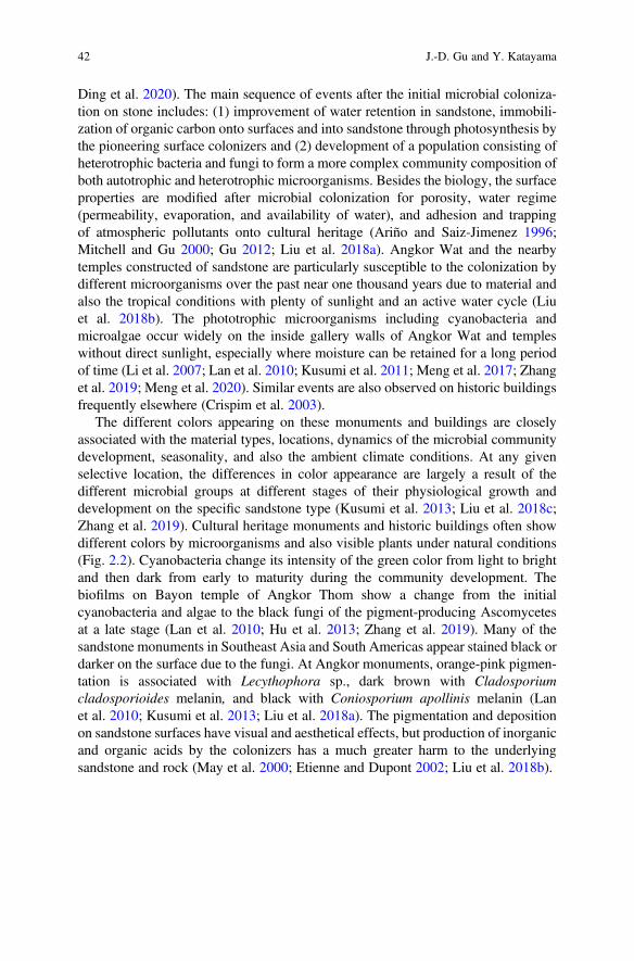
Ding et al. 2020). The main sequence of events after the initial microbial coloniza-tion on stone includes: (1) improvement of water retention in sandstone, immobili-zation of organic carbon onto surfaces and into sandstone through photosynthesis bythe pioneering surface colonizers and (2) development of a population consisting ofheterotrophic bacteria and fungi to form a more complex community composition ofboth autotrophic and heterotrophic microorganisms. Besides the biology, the surfaceproperties are modified after microbial colonization for porosity, water regime(permeability, evaporation, and availability of water), and adhesion and trappingof atmospheric pollutants onto cultural heritage (Ariño and Saiz-Jimenez 1996;Mitchell and Gu 2000; Gu 2012; Liu et al. 2018a). Angkor Wat and the nearbytemples constructed of sandstone are particularly susceptible to the colonization bydifferent microorganisms over the past near one thousand years due to material andalso the tropical conditions with plenty of sunlight and an active water cycle (Liuet al. 2018b). The phototrophic microorganisms including cyanobacteria andmicroalgae occur widely on the inside gallery walls of Angkor Wat and templeswithout direct sunlight, especially where moisture can be retained for a long periodof time (Li et al. 2007; Lan et al. 2010; Kusumi et al. 2011; Meng et al. 2017; Zhanget al. 2019; Meng et al. 2020). Similar events are also observed on historic buildingsfrequently elsewhere (Crispim et al. 2003).
The different colors appearing on these monuments and buildings are closelyassociated with the material types, locations, dynamics of the microbial communitydevelopment, seasonality, and also the ambient climate conditions. At any givenselective location, the differences in color appearance are largely a result of thedifferent microbial groups at different stages of their physiological growth anddevelopment on the specific sandstone type (Kusumi et al. 2013; Liu et al. 2018c;Zhang et al. 2019). Cultural heritage monuments and historic buildings often showdifferent colors by microorganisms and also visible plants under natural conditions(Fig. 2.2). Cyanobacteria change its intensity of the green color from light to brightand then dark from early to maturity during the community development. Thebiofilms on Bayon temple of Angkor Thom show a change from the initialcyanobacteria and algae to the black fungi of the pigment-producing Ascomycetesat a late stage (Lan et al. 2010; Hu et al. 2013; Zhang et al. 2019). Many of thesandstone monuments in Southeast Asia and South Americas appear stained black ordarker on the surface due to the fungi. At Angkor monuments, orange-pink pigmen-tation is associated with Lecythophora sp., dark brown with Cladosporiumcladosporioides melanin, and black with Coniosporium apollinis melanin (Lanet al. 2010; Kusumi et al. 2013; Liu et al. 2018a). The pigmentation and depositionon sandstone surfaces have visual and aesthetical effects, but production of inorganicand organic acids by the colonizers has a much greater harm to the underlyingsandstone and rock (May et al. 2000; Etienne and Dupont 2002; Liu et al. 2018b).
42 J.-D. Gu and Y. Katayama
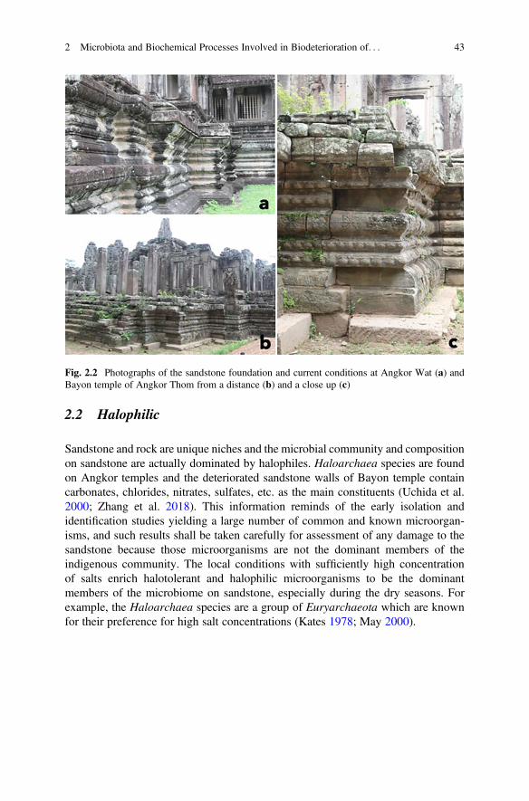
2.2 Halophilic
Sandstone and rock are unique niches and the microbial community and compositionon sandstone are actually dominated by halophiles. Haloarchaea species are foundon Angkor temples and the deteriorated sandstone walls of Bayon temple containcarbonates, chlorides, nitrates, sulfates, etc. as the main constituents (Uchida et al.2000; Zhang et al. 2018). This information reminds of the early isolation andidentification studies yielding a large number of common and known microorgan-isms, and such results shall be taken carefully for assessment of any damage to thesandstone because those microorganisms are not the dominant members of theindigenous community. The local conditions with sufficiently high concentrationof salts enrich halotolerant and halophilic microorganisms to be the dominantmembers of the microbiome on sandstone, especially during the dry seasons. Forexample, the Haloarchaea species are a group of Euryarchaeota which are knownfor their preference for high salt concentrations (Kates 1978; May 2000).
Fig. 2.2 Photographs of the sandstone foundation and current conditions at Angkor Wat (a) andBayon temple of Angkor Thom from a distance (b) and a close up (c)
2 Microbiota and Biochemical Processes Involved in Biodeterioration of. . . 43
2.3 Archaea
Archaea as an independent domain of life is a significant milestone discovery inunderstanding of the biological world (Woese 1978). The non-methanogenicarchaea have been expanded rapidly due to the many new phyla and superphyla ofarchaea discovered with the available high-throughput sequencing technology,metagenomics, and bioinformatics tools available (Liu et al. 2018a, b; Zhou et al.2018, 2019). The information has illuminated on new lineages of the archaea, butalmost all of these new archaea are discovered mainly based on 16S rRNA genesequences and metagenomes available. To further advance the physiology andbiochemistry of these archaea, enrichment and isolation in pure cultures are thekey step to advance the understanding on their ecological contribution and signifi-cance. As an example, AOA has only been recently detected as the dominant groupresponsible for oxidation of ammonia on sandstone monuments in Cambodia afterobserving accumulation of NO3
� in sandstone (Meng et al. 2016, 2017). Afteranalyzing samples from three different temples in Siem Reap of Cambodia, namelyBayon temple of Angkor Thom, Wat Athevea, and Phnom Krom, AOA was thedominant ones in the genomic DNA and also transcribed cDNA of the same set ofsamples, while AOB could only be detected at lower abundance and in a smallfraction of the samples (Meng et al. 2016, 2017). Three PCR amplified sequences ofBayon temple were closely related to Nitrosopumilus maritimus, the first pureculture isolated from a tropical marine aquarium (Könneke et al. 2005; Walkeret al. 2010), which is designated as Group I.1a. Major sequences of Phnom Kromsamples fell into Group I.1a-associated closely affiliated with Ca. Nitrosotaleadevanaterra (Tourna et al. 2008, 2011) and into Group I.1b with Ca. Nitrososphaera(Meng et al. 2016, 2017). The occurrence of these two groups is in good agreementwith the fact that they are tolerant to low pH values (Hatzenpichler et al. 2008;Hatzenpichler 2012). Nitrosotalea and Nitrososphaera can grow when urea isavailable under acidic conditions (Kowalchuk et al. 2000; Laverman et al. 2001;Lu et al. 2012). In contrast, AOB were less abundant significantly compared withAOA on cultural heritage monuments under the tropical climate (Meng et al. 2016,2017).
3 Key Biochemical Processes of Biodeterioration
3.1 Carbon Sequestration
As discussed above, phototrophic microorganisms are pioneer colonizers on freshsandstone surfaces of cultural heritage under the tropical climate conditions and thenthe subsequent impacts from the microorganisms can be also variable due to thedifferent species and the specific biochemical processes carried out by them (Gu andMitchell 2013; Liu et al. 2018a). The most significant event is to sequestrate carbon
44 J.-D. Gu and Y. Katayama
dioxide from atmosphere initially onto sandstone to alter the exterior surface prop-erties of the sandstone materials for an active dynamic advance of microorganismswith different physiological capabilities to provide chances for heterotrophic micro-organisms to colonize over time. In addition to the natural cycle of carbon frominorganic to organic, anthropogenic pollution and pollutants cannot be ignored fortheir contribution to the development of a different microbial community that utilizethe deposition to grow and also cause damage of the underlying materials (Mitchelland Gu 2000).
3.2 Nitrogen Transformation
Among the different biochemical reactions of N cycle, N2-fixation and nitrificationare two of the most significant ones on sandstone monuments of cultural heritage andhistoric buildings. At the same time as CO2 is sequestrated onto sandstone inbiomass, N2 fixation is also taking place when cyanobacteria and algae are thedominant ones in the biofilm community (Zhang et al. 2019; Liu et al. 2020). Forthe nitrification or ammonia oxidation, the amoA genes of bacteria and archaea arephylogenetically distinct possessing different evolution and phenotypic characteris-tics (De Boer and Kowalchuk 2001; Nicol and Schleper 2006; Dodsworth et al.2011). Abundance of the AOA amoA gene is much higher than that of AOB inselective habitat, including open ocean (Wuchter et al. 2006; Nakagawa et al. 2007;Beman et al. 2008; Cao et al. 2013), oil reservoirs (Li et al. 2011), acidic soils(Leininger et al. 2006; Nicol and Schleper 2006; Adair and Schwartz 2008; Nicolet al. 2008; Onodera et al. 2010), and estuaries and wetlands (Caffrey et al. 2007;Wang and Gu 2013).
Deteriorated sandstone samples are mainly comprised of decomposition productsof minerals and clays from sandstone and components of biofilms at differentdevelopment stages. Intrinsic sandstone properties dictating the colonization ofmicroorganisms to form biofilms include mineral composition, porosity and perme-ability and retaining of water by the materials and susceptibility to dissolution (Ariñoand Saiz-Jimenez 1996; Essa and Khallaf 2014). Ammonium released from decom-position of organic biomass and deposition from atmosphere is oxidized throughmicrobial metabolism and AOA is the dominant group over AOB at several templesincluding Bayon temple of Angkor Thom, Wat Athevea, and Phnom Krom inCambodia (Meng et al. 2016). AOA are easily detected at all sandstone monumentsand the pH values of these samples are also generally low. Coupling the relative highconcentration of NO3
� detected with the low pH condition and the more abundantAOA than AOB, an active biochemical process of ammonia oxidation or nitrificationis operative on these cultural heritage to result in an accumulation of biogenic nitricacid, which contributes to the biodeterioration of sandstone. At the same time, theaccumulation of NO3
� in sandstone of Angkor temples is also related to the inactiveremoval of the NO3
� by denitrification and anammox of the relevant microorgan-isms (Ding et al. 2020).
2 Microbiota and Biochemical Processes Involved in Biodeterioration of. . . 45
3.3 Sulfur Transformation
The four most important elements to life are carbon, nitrogen, phosphorus, andsulfur. Both nitrogen and sulfur metabolizing microorganisms are responsible forproduction of inorganic acids that can erode the sandstone and rock of culturalheritage. Previously, emphasis has been put more on corrosion initiated by sulfate/sulfur-reducing bacteria and sulfur-oxidizing bacteria (Li et al. 2007; Li et al. 2010;Kusumi et al. 2013) and AOB (Sand and Bock 1991; Mansch and Bock 1998). Afterthe discovery of ecological role of AOA, this group of archaea has also been detectedon sandstone monuments using both DNA- and also RNA-based quantification ofthe amoA gene of this functional group of organisms (Meng et al. 2016, 2017). Theabundance of AOA was higher than that of AOB, implying that AOA is a majorcontributor to the deterioration of sandstone (discussed above). In addition, sulfur-oxidizing bacteria and fungi are also detected on sandstone cultural heritage and theirpopulation is proportional to the sampling site where the damage is the most severe(Li et al. 2010; Kusumi et al. 2011). Based on these results, special attention shall begiven to the microorganisms involved in sulfur cycling to effectively protect culturalheritage. Sulfate was detected in the liquid medium inoculated with deterioratedsandstone samples, and the chemolithoautotrophic Mycobacterium spp. (Kusumiet al. 2011) and 19 sulfur-oxidizing fungal strains (Li et al. 2010) capable of sulfuroxidation were isolated from the deteriorated sandstones in Angkor Wat, Bayon, andPhnom Krom temples to indicate their contribution.
Sources of sulfur may be from the biofilm and preformed biomass and also fecalmaterials of bats and wild animals living in these temples. In the early part of theprevious century, heterotrophic fungi were confirmed for sulfur oxidation andvarious fungi can oxidize S0 for energy to grow, including Trichoderma harzianumand Aspergillus niger. A number of fungal genera are capable of oxidizing inorganicsulfur compounds, but the biochemical sulfur oxidation pathway and enzymesinvolved are still not known clearly. Fusarium solani strain THIF01, isolated fromAngkor monuments, is capable of utilizing S0 chemolithoautotrophically as theenergy source (Li et al. 2010). This fungus contains an endobacteriumBradyrhizobium sp. which has sulfur oxidation capability, but the bacterium-freeculture (strain THIF01BF) that was obtained by treatments with antibiotics of strainTHIF01 showed chemolithoautotrophic growth with S0 (Xu et al. 2018).
3.4 Other Elements
Other elements are also important to life and the cycling of nutrients by microor-ganisms in different ways. They can serve in dissimilatory and assimilatory bio-chemical processes depending on the element types and the specific microorganismsinvolved producing transformation products and biomass, respectively (Ehrlich2002). As an example, iron in the environment can play a role to participate in the
46 J.-D. Gu and Y. Katayama
corrosion and biodeterioration of stone materials (Warscheid and Braams 2000), butthe relative significance of this biochemical process compared with majorelements C, N, and S shall be much smaller or insignificant. Because of this,involvement and contribution of microorganisms utilizing Fe and Mn and otherelements to deterioration of stone are not further discussed here, but their signifi-cance in revealing new biochemical processes shall not be underestimated in futureresearch as a focus area.
4 Interactions Among Sandstone, Microbiota,and the Local Environment
4.1 Material Types
Sandstone is a porous material of natural origin and is not strong enough comparedto other stone and rock from igneous formation. Such property of this material makesit to be very susceptible to attack by weathering through water absorption and freeze-and-thaw cycle. Colonization by microflora and plants adds another dimension to thestability challenge to sandstone in open climate conditions (Liu et al. 2018b, 2020).Under tropical conditions of Southeast Asia and South America, both sunlight andwater are plenty to provide the basic requirements for phototrophic life forms ofplants and microorganisms to colonize and grow on surfaces. As soon as the initialcolonization on surfaces is established, e.g., lichens either the initial protection orfurther damage of the materials can take place through the specific microorganismsactively metabolizing on surfaces of stone (Liu et al. 2018c, 2019). Both autotrophicand heterotrophic ones can actively grow in the surface colonizing community andalter the materials through their metabolic processes and the metabolites. Suchprogress and development of the community over time on sandstone surfacesmaintain an active and dynamic community on the sandstone surfaces and allowsequestration of inorganic CO2 from atmosphere and cycling of nutrients through thelife cycle of different microorganisms and their biomass coupling with theseasonality.
Sandstone is interacting with the surrounding environment, including water, air,microbiota, and anthropogenic contributors. Among all of them, temperature andavailability of water play a crucial role in limiting or promoting microbial coloniza-tion and biofilm formation on sandstone under favorable conditions of the tropics(Liu et al. 2018a). Because of the porosity of sandstone and biofilm formation toprevent more quick exchanges of water between the surrounding and those retainedinside sandstone, increasing retention of water holding in sandstone supports moreactive microbial colonization and growth to result in more damage of stone frombiological activities (Zhang et al. 2019). At the same time, movement of watercontaining soluble salts by siphon effects promotes formation of mineral crystalsupon drying, which initiates external surface defoliation progressively to lead to
2 Microbiota and Biochemical Processes Involved in Biodeterioration of. . . 47
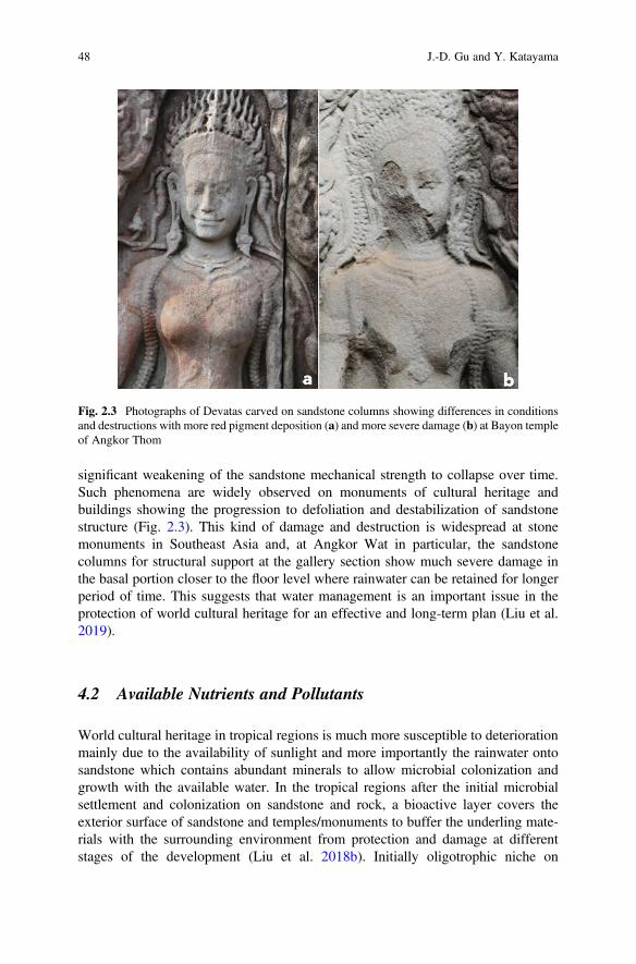
significant weakening of the sandstone mechanical strength to collapse over time.Such phenomena are widely observed on monuments of cultural heritage andbuildings showing the progression to defoliation and destabilization of sandstonestructure (Fig. 2.3). This kind of damage and destruction is widespread at stonemonuments in Southeast Asia and, at Angkor Wat in particular, the sandstonecolumns for structural support at the gallery section show much severe damage inthe basal portion closer to the floor level where rainwater can be retained for longerperiod of time. This suggests that water management is an important issue in theprotection of world cultural heritage for an effective and long-term plan (Liu et al.2019).
4.2 Available Nutrients and Pollutants
World cultural heritage in tropical regions is much more susceptible to deteriorationmainly due to the availability of sunlight and more importantly the rainwater ontosandstone which contains abundant minerals to allow microbial colonization andgrowth with the available water. In the tropical regions after the initial microbialsettlement and colonization on sandstone and rock, a bioactive layer covers theexterior surface of sandstone and temples/monuments to buffer the underling mate-rials with the surrounding environment from protection and damage at differentstages of the development (Liu et al. 2018b). Initially oligotrophic niche on
Fig. 2.3 Photographs of Devatas carved on sandstone columns showing differences in conditionsand destructions with more red pigment deposition (a) and more severe damage (b) at Bayon templeof Angkor Thom
48 J.-D. Gu and Y. Katayama
sandstone becomes enriched through phototrophic growth to sequestrate organicsubstances onto and into the surface layer of sandstone, in doing so the localconditions are altered from the oligotrophic to organic carbon rich one. After thistransformation, heterotrophic microorganisms become the dominant populationincluding both bacteria and fungi to change the color of sandstone from its originalgray or light brown to dark and black. Fresh microbial biofilm consisting of algaeand cyanobacteria is reported (Lan et al. 2010) and the different colors and pigmen-tations on sandstone wall are associated with different microbial groups at Bayonand Angkor Wat (Kusumi et al. 2013). These microorganisms need to be furtherevaluated for their specific role in the bioprotection and/or biodeterioration atdifferent development stages considered to advance our comprehensive understand-ing of their specific role over time to provide more accurate information for futuremanagement of cultural heritage effectively. Subsequent results indicate the acidattack initiated by microbial oxidation of S� from reduction product of H2S or sulfate(Li et al. 2007, 2010). More recent research information showed that ammoniaoxidation is an active biochemical reaction mechanism carried out mainly by AOAat several temples in Cambodia (Meng et al. 2016, 2017). This is a new developmentfor further assessment of its flux and activities so that future management can bemade considering such results in the conservation plan. It is certain that morecontributors are involved in the biodeterioration of sandstone monuments anddestruction of valuable galleries and bas relief, key factors shall be identified andranked according to the extent of destruction by each of them so that managementcan be focused on a few most important ones first to monitor the effectiveness overtime to verify the conservation results.
Cultural heritage of monuments and temples is in constant exchange with thesurrounding environmental conditions, both water and biological attacks may takeplace simultaneously. In addition, the increasing anthropogenic pollution cannot beignored in the current and future investigation and protection. With the pre-existingbiofilms and modified surfaces of sandstone to increase the roughness of the surfacesand porosity, the color change enhances thermal property of the underlying sand-stone to absorb solar radiation, but emits less due to the biofilm layer especially whenwater contents in the surface layer are high. In addition, the pollutants in the air canbe deposited and trapped by the porous biofilms on surfaces to be potential sourcesof organic carbon to selective biofilm microorganisms capable of utilizing them togrow. The active surface layer coated on sandstone is of significant research interestto investigate the physical and biochemical processes actively involved and also thedynamics of them spatially and temporally to gain further insights of this micro-niche and system for new discoveries.
2 Microbiota and Biochemical Processes Involved in Biodeterioration of. . . 49
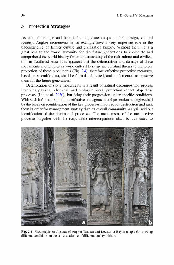
5 Protection Strategies
As cultural heritage and historic buildings are unique in their design, culturalidentity, Angkor monuments as an example have a very important role in theunderstanding of Khmer culture and civilization history. Without them, it is agreat loss to the world humanity for the future generations to appreciate andcomprehend the world history for an understanding of the rich culture and civiliza-tion in Southeast Asia. It is apparent that the deterioration and damage of thesemonuments and temples as world cultural heritage are constant threats to the futureprotection of these monuments (Fig. 2.4), therefore effective protective measures,based on scientific data, shall be formulated, tested, and implemented to preservethem for the future generations.
Deterioration of stone monuments is a result of natural decomposition processinvolving physical, chemical, and biological ones, protection cannot stop theseprocesses (Liu et al. 2020), but delay their progression under specific conditions.With such information in mind, effective management and protection strategies shallbe the focus on identification of the key processes involved for destruction and rankthem in order for management strategy than an overall community analysis withoutidentification of the detrimental processes. The mechanisms of the most activeprocesses together with the responsible microorganisms shall be delineated to
Fig. 2.4 Photographs of Apsaras of Angkor Wat (a) and Devatas at Bayon temple (b) showingdifferent conditions on the same sandstone of different quality initially
50 J.-D. Gu and Y. Katayama
advance the knowledge for protection and management. Biofilms on sandstone havedifferent roles at different stages of the colonization and biofilm development, suchunderstanding is vitally important to a deeper understanding of biofilm dynamicsand its relationship with the integrity of sandstone (Warscheid and Braams 2000; Guet al. 2011; Liu et al. 2018c; Zhang et al. 2019). As a matter of fact, physical removalof biofilms is not going to solve the deterioration issue from microbial activity, butaccelerate the damage because of removing the protective biofilms to expose theunderlying surfaces for another cycle of colonization and biofilm development.Microorganisms can also play a role in control of biofilms. Fungi appearing onmatured biofilms are correlated with the peeling off of old biofilms, which haspotential to be further developed into biotechnology for biological control andcleaning of colonized surfaces (Gleeson et al. 2005; Hu et al. 2013).
The biochemical processes should be identified when evaluating the susceptibil-ity of sandstone to microorganisms for colonization and biodeterioration (Menget al. 2017; Zhang et al. 2019). Phototrophic microorganisms initially establish onthe sandstone surfaces to be the pioneering biofilms when water and mineralnutrients are readily available from the substratum. Apart from the external factorsof the environment, sandstone has reasonable porosity to allow water retention andalso promotion of microbial colonization. At the same time, the small pores also trapnutrients from atmospheric deposition containing dust and microbes to promotebiosusceptibility and attack of sandstone. Currently, major efforts have been givento the analysis of the microbial community with the latest available sequencingtechnology independent of culturing and isolation, but the biochemical functions bythe selective members of the community need to be delineated so that initiation fordamage of the cultural heritage can be prevented and limited with means available.The interfaces between sandstone surface and biofilms remain a hotspot for biolog-ical activity and biochemical processes when analysis of cultural heritage is thetarget (Ma et al. 2015; Wu et al. 2017; Kakakhel et al. 2019). Biofilm and plants canestablish the cultural heritage over time when routine maintenance is not carried outeffectively. A close monitoring and clearance can achieve a good protection fromweeds and plants, which otherwise would grow into the sandstone to cause disloca-tion of the stone blocks and collapse of the structure (Liu et al. 2018a; 2019). Toprotect them, maintenance work shall be conducted to remove newly grown plantsand weeds regularly so that no large plants can be established and remain on thesestructures.
Maintenance work also involves repair of the damaged and destructed structureand stone pieces. This may involve the use of chemical adhesive and consolidants, sothat the repaired stone may last longer when exposed to both the ambient climateconditions and invasive growth of microorganisms, particularly fungi (Gu et al.2011; Wu et al. 2017; Zhang et al. 2018). At the same time, water repellants are alsoused to seal the stone from natural rainwater and keep dry to avoid water-associateddamage. But such practice has a serious potential drawback in that trapped waterinside the sandstone is more destructive than those without such protections used sonature regulates the water movement (Sterflinger and Pinar 2013; Liu et al. 2018b;Sterflinger et al. 2018). Considering these factors, sandstone monuments and
2 Microbiota and Biochemical Processes Involved in Biodeterioration of. . . 51
temples are better kept away from water, especially stagnant water, if possible or letnatural rainwater to uptake and drain freely from the structures when raining ordrying accordingly. However, after the formation of biofilm on surfaces, theseexchange processes for water are being slowed down for both into and out of thestone, resulting in less efficient movement of water to initiate damage of the stonethrough both biofilm development and also salting effects.
Polymeric materials are increasingly used in applications including conservationand protection of cultural heritage. Some of the successful cases are usuallyrestricted to specific environmental conditions of museums, but such informationis not widely reported for the specificity of the materials to the selective outdoorenvironmental conditions. Misuse of such information has resulted in disaster inconservation work due to outbreaks of microbial growth after application ofconsolidants or even antimicrobials to cultural heritage exposed to outdoor condi-tions (Gu 2003; Gu et al. 2011; Wu et al. 2017; Liu et al. 2018c; Kakakhel et al.2019). Polymeric materials may perform well under harsh conditions including highhumidity and temperature, but such polymeric materials are generally very difficultto apply as consolidants because of their high viscosity, insolubility in even solvents,and associated colors. In contrast, those polymers that have been used successfully inmuseums are generally not resistant to metabolism by indigenous microorganismsbecause fungi are airborne and particularly capable of attacking high strength andengineering polymers due to fillers, additives, and plasticizers (Gu 2003; Mitchelland Gu 2000; Gu et al. 2011; Gu 2012).
Inorganic materials have many good applications in construction and protectionof building. Such practice has been proven successful when the candidate materialsare used to appropriate niche and conditions. For example, lime is a commonmaterial to apply to surfaces of building to protect the biomaterials from insectattack. Such materials are also available now on the market, but the composition ofthe commercially available products contains not only lime but also additives,including polymeric materials, cellulose, and cellulose esters, which are biodegrad-able and cause serious biofilm establishment and growth on surfaces of structure(Gu and Mitchell 2013).
New materials and technologies are also becoming available through research andinnovation, and some of the promising ones include nanomaterials and technologyfor effective protection against fast growth of colonized biofilm microorganisms(Liu et al. 2018a). At the same time, corroding chemical and biochemical processesmay be inhibited through intervention of chemical treatment or approaches toadvance the protection more effectively.
Antimicrobial chemicals are commercially available for inhibiting the growth ofmicroorganisms under selective or specific conditions, e.g., disease pathogen controland eradication, but the available information indicates that successive applicationspromote the development of resistance in microorganisms, which is a major publichealth concern when the resistant genes are enclosed on plasmids that are morereadily transmitted among the same species and also between different species. It isunlikely to achieve an effective long-term control using a single or few chemicals.Conversely, when selecting candidate chemicals, the current testing protocols for
52 J.-D. Gu and Y. Katayama
screening are problematic because the testing conditions are mainly based oninhibitory testing in liquid cultures, not the application surface or the environmentalniche conditions. The very little resemblance between the target conditions and thelaboratory testing ones results in non-effectiveness after applications in situ oncultural heritage (Gu 2003; Essa and Khallaf 2014; Liu et al. 2018b, 2020).
6 Summary
Stone cultural heritage and historic buildings are susceptible to colonization bymicroorganisms of a wide range of physiological characteristics depending on theenvironmental conditions and also the stage of the development. Both the specificactive microorganisms and also their physiological functions shall be identified andelucidated for a better and more specific understanding on the deterioration bio-chemical processes and mechanisms involved for effective protection of the sand-stone temples/monuments. Microorganisms involved in carbon, nitrogen, and sulfurcycles are the most important ones, especially the biochemical processes thatgenerate acidic or corrosive products to erode sandstone and dissolve the minerals.On Angkor temples/monuments, sulfur-oxidizing bacteria and fungi, ammonia-oxidizing archaea, and also phototrophic cyanobacteria and algae are identified aspriority members for investigations of their biochemical contribution to the deteri-oration. Related processes, e.g., denitrification, anammox, and comammox, are alsoimportant for the comprehensive knowledge on the accumulation of nitrate in manysandstone monuments.
Physical and environmental factors of the materials and hydrological dynamicsshall not be ignored from the biology for protection and management of culturalheritage sites because they affect the biology on stone fundamentally and then thestability of the stone. Equipped with the information on the materials of culturalheritage and buildings, biology, and biochemistry, preservation strategies can beformulated for protection of sandstone by taking advantage of the latest developmentand technologies in chemistry, nanomaterials, and molecular biology.
Acknowledgements This project was supported by Safeguarding of Bayon Temple of AngkorThom (JASA, Japan), APSARA Authority of Cambodian Government, the UNESCO/JapaneseFunds-in-Trust for the Preservation of the World Cultural Heritage, and partially by a Hong KongRGC GRF Grant (No. 17302119).
Ethical Statement Conflict of Interest:All authors declare that they have no conflict of interest inthis study.
Ethical Approval: This article does not contain any studies with human participants or animalsperformed by any of the authors of this investigation.
Sampling at temples was granted by APSARA Authority of Cambodian Government, theKingdom of Cambodia.
2 Microbiota and Biochemical Processes Involved in Biodeterioration of. . . 53
References
Aair KL, Schwartz E (2008) Evidence that ammonia-oxidizing archaea are more abundant thanammonia-oxidizing bacteria in semiarid soils of northern Arizona, USA. Microbiol Ecol56:420–426
Adamson C, McCabe S, Warke PA, McAllister D, Smith BJ (2013) The influence of aspect on thebiological colonization of stone in Northern Ireland. Int Biodeterior Biodegradation 84:357–366
Ariño X, Saiz-Jimenez C (1996) Factors affecting the colonization and distribution ofcyanobacteria, algae and lichens in ancient mortars. In: Riederer J (ed) 8th InternationalCongress on Deterioration and Conservation of Stone. Möller Druck und Verlag, Berlin, pp725–731
Beman JM, Popp BN, Francis CA (2008) Molecular and biogeochemical evidence for ammoniaoxidation by marine Crenarchaeota in the Gulf of California. ISME J 2:429–441
Caffrey JM, Bano N, Kalanetra K, Hollibaugh JT (2007) Ammonia oxidation and ammonia-oxidizing bacteria and archaea from estuaries with differing histories of hypoxia. ISME J1:660–662
Cao H, Auguet JC, Gu J-D (2013) Global ecological pattern of ammonia-oxidizing archaea. PLoSOne 8:e52853
Crispim C, Gaylarde C (2005) Cyanobacteria and biodeterioration of cultural heritage: a review.Microbiol Ecol 49:1–9
Crispim CA, Gaylarde CC, Gaylarde PM, Copp J, Neilan BA (2003) Molecular biology forinvestigation of cyanobacterial populations on historic buildings in Brazil. In: Saiz-Jimenez C(ed) Molecular biology and cultural heritage. Balkema, Lisse, pp 141–143
De Boer W, Kowalchuk G (2001) Nitrification in acid soils: micro-organisms and mechanisms. SoilBiol Biochem 33:853–866
Ding X, Lan W, Li Y, Wu J, Hong Y, Urzi C, Katayama Y, Ge Q, Gu J-D (2020) Microbiome andnitrate removal by denitrifying and anammox on the sandstone Preah Vihear temple in Cam-bodia revealed by metagenomics and N-15 isotope. Appl Microbiol Biotechnol 104(22):9823–9837. https://doi.org/10.1007/s00253-020-10886-4
Dodsworth JA, Hungate BA, Hedlund BP (2011) Ammonia oxidation, denitrification and dissim-ilatory nitrate reduction to ammonium in two US Great Basin hot springs with abundantammonia-oxidizing archaea. Environ Microbiol 13:2371–2386
Dornieden T, Gorbushina AA, Krumbein WE (2000) Biodecay of cultural heritage as a space/time-related ecological situation – an evaluation of a series of studies. Int Biodeterior Biodegradation46:261–270
Ehrlich HL (2002) Geomicrobiology. Marcel Dekker, New York, p 768Essa AM, Khallaf MK (2014) Biological nanosilver particles for the protection of archaeological
stones against microbial colonization. Int Biodeterior Biodegradation 94:31–37Etienne S, Dupont J (2002) Fungal weathering of basaltic rocks in a cold oceanic environment
(Iceland): comparison between experimental and field observations. Earth Surf Proc Land27:737–748
Flores M, Lorenzo J, Gómez-Alarcón G (1997) Algae and bacteria on historic monuments at Alcalade Henares, Spain. Int Biodeterior Biodegradation 40:241–246
Freeman M, Jacques C (1999) Ancient Angkor. River Books, Ltd., BangkokGaylarde C, Silva MR, Warscheid T (2003) Microbial impact on building materials: an overview.
Mater Struct 36:342–352Gaylarde PM, Gaylarde CC (2000) Algae and cyanobacteria on painted buildings in Latin America.
Int Biodeterior Biodegradation 46:93–97Gleeson DB, Clipson N, Melville K, Gadd GM, McDermott FP (2005) Characterization of fungal
community structure on a weathered pegmatitic granite. Microb Ecol 50:360–368Gu J-D (2003) Microbiological deterioration and degradation of synthetic polymeric materials:
recent research advances. Int Biodeterior Biodegrad 52:69–91. https://doi.org/10.1016/S0964-8305(02)00177-4
54 J.-D. Gu and Y. Katayama
Gu J-D (2012) Biofouling and prevention: corrosion, biodeterioration and biodegradation ofmaterials. In: Kultz M (ed) Handbook of environmental degradation of materials, 2nd edn.Elsevier, Waltham, pp 243–282
Gu J-D, Mitchell R (2013) Biodeterioration. In: Rosenberg E, DeLong EF, Lory S, Stackebrandt E,Thompson F (eds) The prokaryotes: applied bacteriology and biotechnology. Springer,New York, pp 309–341
Gu J-D, Ford TE, Mitchell R (1996) Fungal degradation of concrete. In: Sand W (ed) DECHEMAmonographs. Biodeterioration and biodegradation. VCH, Frankfurt, pp 135–142
Gu J-D, Ford TE, Mitton B, Mitchell R (2011) Microbial degradation of materials: generalprocesses. In: Revie W (ed) The Uhlig corrosion handbook, 3rd edn. John Wiley & Sons,New York, pp 351–363
Hatzenpichler R (2012) Diversity, physiology, and niche differentiation of ammonia-oxidizingarchaea. Appl Environ Microbiol 78:7501–7510
Hatzenpichler R, Lebedeva EV, Spieck E, Stoecker K, Richter A, Daims H, Wagner M (2008) Amoderately thermophilic ammonia-oxidizing crenarchaeote from a hot spring. Proc Natl AcadSci U S A 105:2134–2139
Hosono T, Uchida E, Suda C, Ueno A, Nakagawa T (2006) Salt weathering of sandstone at theAngkor monuments, Cambodia: identification of the origins of salts using sulfur and strontiumisotopes. J Archaeol Sci 33:1541–1551
Hu H, Katayama Y, Kusumi A, Li SX, Wang J, de Vries RP, Gu J-D (2013) Occurrence ofAspergillus allahabadii on sandstone at Bayon Temple, Angkor Thom, Cambodia. IntBiodeterior Biodegradation 76:112–117
Kakakhel MA, Wu F, Gu J-D, Feng H, Shah K, Wang W (2019) Controlling biodeterioration ofcultural heritage objects with biocides: a review. Int Biodeterior Biodegrad 143:104721. https://doi.org/10.1016/j.ibiod.2019.104721
Kates M (1978) The phytanyl ether-linked polar lipids and isoprenoid neutral lipids of extremelyhalophilic bacteria. Prog Chem Fats Other Lipids 15:301–342
Keshari N, Adhikary SP (2014) Diversity of cyanobacteria on stone monuments and buildingfacades of India and their phylogenetic analysis. Int Biodeterior Biodegradation 90:45–51
Könneke M, Bernhard AE, José R, Walker CB, Waterbury JB, Stahl DA (2005) Isolation of anautotrophic ammonia-oxidizing marine archaeon. Nature 437:543–546
Kowalchuk GA, Stienstra AW, Heilig GHJ, Stephen JR, Woldendorp JW (2000) Molecularanalysis of ammonia-oxidising bacteria in soil of successional grasslands of the Drentsche A(The Netherlands). FEMS Microbiol Ecol 31:207–215
Kusumi A, Li XS, Katayama Y (2011) Mycobacteria isolated from Angkor monument sandstonesgrow chemolithoautotrophically by oxidizing elemental sulfur. Front Microbiol 2:104
Kusumi A, Li X, Osuga Y, Kawashima A, Gu J-D, Nasu M, Katayama Y (2013) Bacterialcommunities in pigmented biofilms formed on the sandstone bas-relief walls of the BayonTemple, Angkor Thom, Cambodia. Microbes Environ 28:422
Lan W, Li H, Wang WD, Katayama Y, Gu J-D (2010) Microbial community analysis of fresh andold microbial biofilms on Bayon temple sandstone of Angkor Thom, Cambodia. Microb Ecol60:105–115
Laverman A, Speksnijder A, Braster M, Kowalchuk G, Verhoef H, van Verseveld H (2001)Spatiotemporal stability of an ammonia-oxidizing community in a nitrogen-saturated forestsoil. Microb Ecol 42:35–45
Leininger S, Urich T, Schloter M, Schwark L, Qi J, Nicol G, Prosser J, Schuster S, Schleper C(2006) Archaea predominate among ammonia-oxidizing prokaryotes in soils. Nature442:806–809
Li H, Mu B-Z, Jiang Y, Gu J-D (2011) Production processes affected prokaryotic amoA geneabundance and distribution in high-temperature petroleum reservoirs. Geomicrobiol J28:692–704
2 Microbiota and Biochemical Processes Involved in Biodeterioration of. . . 55
Li X, Arai H, Shimoda I, Kuraishi H, Katayama Y (2007) Enumeration of sulfur-oxidizingmicroorganisms on deteriorating stone of the Angkor monuments, Cambodia. Microbes Environ23:293–298
Li XS, Sato T, Ooiwa Y, Kusumi A, Gu J-D, Katayama Y (2010) Oxidation of elemental sulfur byFusarium solani strain THIF01 harboring endobacterium Bradyrhizobium sp. Microb Ecol60:96–104
Lisci M, Monte M, Pacini E (2003) Lichens and higher plants on stone: a review. Int BiodeteriorBiodegradation 51:1–17
Liu J-F, Wu W-L, Yao F, Wang B, Zhang B-L, Mbadinga SM, Gu J-D, Mu B-Z (2016) Athermophilic nitrate-reducing bacterium isolated from production water of a high temperatureoil reservoir and its inhibition on sulfate-reducing bacteria. Appl Environ Biotechnol 1(2):35–42. https://doi.org/10.18063/AEB.2016.02.004
Liu X, Meng H, Wang Y, Katayama Y, Gu J-D (2018a) Water is the critical factor to establishmentbiological and stability of Angkor temple sandstone in Southeast Asia. Int BiodeteriorBiodegrad 133:9–16. https://doi.org/10.1016/j.ibiod.2018.05.011
Liu X, Li M, Castelle CJ, Probst AJ, Zhou Z, Pan J, Liu Y, Banfield JF, Gu J-D (2018b) Insightsinto the ecology, evolution and metabolism of the widespread Wosearchaeotal lineage.Microbiome 6:102
Liu X, Pan J, Liu Y, Li M, Gu J-D (2018c) Diversity and distribution of Archaea in global estuarineecosystems. Sci Total Environ 637:349–358
Liu Y-F, Qi Z-Z, Shou L-B, Liu J-F, Yang S-Z, Gu J-D, Mu B-Z (2019) Anaerobic hydrocarbondegradation in candidate phylum ‘Atribacteria’ (JS1) inferred from genomics. ISME J 13:2377–2390. https://doi.org/10.1038/s41396-019-0448-2
Liu X, Koestler RJ, Warscheid T, Katayama Y, Gu J-D (2020) Microbial biodeterioration andsustainable conservation of stone monuments and buildings. Nature Sustainability (accepted)
Lu L, Han W, Zhang J, Wu Y, Wang B, Lin X, Zhu J, Cai Z, Jia Z (2012) Nitrification of archaealammonia oxidizers in acid soils is supported by hydrolysis of urea. ISME J 6:1978–1984
Ma Y, Zhang H, Du Y, Tian T, Xiang T, Liu X, Wu F, An L, Wang W, Gu J-D (2015) Thecommunity distribution of bacteria and fungi on ancient wall paintings of the Mogao Grottoes.Sci Rep 5:7752
Ma W, Wu F, Tian T, He D, Zhang Q, Gu J-D, Duan Y, Wang W, Feng H (2020) Fungal diversityand potential biodeterioration of mural paintings on bricks in two 1700-year-old tombs of China.Int Biodeterior Biodegrad 152:104972. https://doi.org/10.1016/j.ibiod.2020.104972
Mansch R, Bock E (1998) Biodeterioration of natural stone with special reference to nitrifyingbacteria. Biodegradation 9:47–64
May E (2000) Stone biodeterioration. In: Mitchell R, McNamara CJ (eds) Cultural heritagemicrobiology: fundamental studies in conservation science. American Society for Microbiol-ogy, Washington, DC, pp 221–234
May E, Papida S, Abdulla H, Tayler S, Dewedar A (2000) Comparative studies of microbialcommunities on stone monuments in temperate and semi-arid climates. In: Ciferri O, Tiano P,Mastromei G (eds) Of microbes and art: the role of microbial communities in the degradationand protection of cultural heritage. Kluwer Academic Publishers, Dordrecht, pp 49–62
McNamara CJ, Perry TD, Bearce KA, Hernandez-Duque G, Mitchell R (2006) Epilithic andendolithic bacterial communities in limestone from a Maya archaeological site. Microb Ecol51:51–64
Meng H, Luo L, Chan HW, Katayama Y, Gu J-D (2016) Higher diversity and abundance ofammonia-oxidizing archaea than bacteria detected at the Bayon Temple of Angkor Thom inCambodia. Int Biodeterior Biodegradation 115:234–243
Meng H, Katayama Y, Gu J-D (2017) Wide occurrence and dominance of ammonia-oxidizingarchaea than bacteria at three Angkor sandstone temples Bayon, Phnom Krom and wat Athveain Cambodia. Int Biodeterior Biodegradation 117:78–88
56 J.-D. Gu and Y. Katayama
Meng H, Zhang X, Katayama Y, Ge Q, Gu J-D (2020) Microbial diversity and composition of thePreah Vihear temple in Cambodia by high-throughput sequencing based on both genomic DNAand RNA. Int Biodeterior Biodegrad 149:104936
Mitchell R, Gu J-D (2000) Changes in the biofilm microflora of limestone caused by atmosphericpollutants. Int Biodeterior Biodegrad 46:299–303
Motti R, Stinca A (2011) Analysis of the biodeteriogenic vascular flora at the Royal Palace ofPortici in southern Italy. Int Biodeterior Biodegradation 65:1256–1265
Nakagawa T, Mori K, Kato C, Takahashi R, Tokuyama T (2007) Distribution of cold-adaptedammonia-oxidizing microorganisms in the deep-ocean of the northeastern Japan Sea. MicrobesEnviron 22:365–372
Nicol GW, Schleper C (2006) Ammonia-oxidising Crenarchaeota: important players in the nitrogencycle? Trends Microbiol 14:207–212
Nicol GW, Leininger S, Schleper C, Prosser JI (2008) The influence of soil pH on the diversity,abundance and transcriptional activity of ammonia oxidizing archaea and bacteria. EnvironMicrobiol 10:2966–2978
Onodera Y, Nakagawa T, Takahashi R, Tokuyama T (2010) Seasonal change in vertical distributionof ammonia-oxidizing archaea and bacteria and their nitrification in temperate forest soil.Microbes Environ 25:28–35
Papida S, Murphy W, May E (2000) Enhancement of physical weathering of building stone bymicrobial population. Int Biodeterior Biodegradation 64:305–317
Perry IV TD, McNamara CJ, Mitchell R (2005) Biodeterioration of stone, scientific examination ofart: modern techniques in conservation and analysis. Sackler National Academy of SciencesColloquium, pp 72–86
Piñar G, Ripka K, Weber J, Sterflinger K (2009) The microbiota of a sub-surface monument themedieval chapel of St. Virgil (Vienna, Austria). Int Biodeterior Biodegradation 63:851–859
Rölleke S, Witte A, Wanner G, Lubitz W (1998) Medieval wall paintings-a habitat for archaea:identification of archaea by denaturing gradient gel electrophoresis (DGGE) of PCR-amplifiedgene fragments coding for 16S rRNA in a medieval wall painting. Int Biodeterior Biodegrada-tion 41:85–92
Saiz-Jimenez C, Laiz L (2000) Occurrence of halotolerant/halophilic bacterial communities indeteriorated monuments. Int Biodeterior Biodegradation 46:319–326
Sand W, Bock E (1991) Biodeterioration of mineral materials by microorganisms – biogenicsulfuric and nitric acid corrosion of concrete and natural stone. Geomicrobiol J 9:129–138
Sterflinger K (2000) Fungi as geologic agents. Geomicrobiol J 17:97–124Sterflinger K, Pinar G (2013) Microbial deterioration of cultural heritage and works of art – tilting at
windmills? Appl Microbiol Biotechnol 97:9637–9646Sterflinger K, Prillinger H (2001) Molecular taxonomy and biodiversity of rock fungal communities
in an urban environment (Vienna, Austria). Anton Leeuw 80:275–286Sterflinger K, Little B, Pinar G, Pinzari F, de los Rios A, Gu J-D (2018) Future directions and
challenges in biodeterioration research on historic materials and cultural properties. IntBiodeterior Biodegradation 129:10–12
Tayler S, May E (2000) Investigations of the localisation of bacterial activity on sandstone fromancient monuments. Int Biodeterior Biodegradation 46:327–333
Tourna M, Freitag TE, Nicol GW, Prosser JI (2008) Growth, activity and temperature responses ofammonia-oxidizing archaea and bacteria in soil microcosms. Environ Microbiol 10:1357–1364
Tourna M, Stieglmeier M, Spang A, Könneke M, Schintlmeister A, Urich T, Engel M, Schloter M,Wagner M, Richter A (2011) Nitrososphaera viennensis, an ammonia oxidizing archaeon fromsoil. Proc Natl Acad Sci U S A 108:8420–8425
Uchida E, Ogawa Y, Maeda N, Nakagawa T (2000) Deterioration of stone materials in the Angkormonuments, Cambodia. Eng Geol 55:101–112
Videla HA, Guiamet PS, Gomez de Saravia S (2000) Biodeterioration of Mayan archaeologicalsites in the Yucatan peninsula, Mexico. Int Biodeterior Biodegradation 46:335–341
2 Microbiota and Biochemical Processes Involved in Biodeterioration of. . . 57
Walker C, de La Torre J, Klotz M, Urakawa H, Pinel N, Arp D, Brochier-Armanet C, Chain P,Chan P, Gollabgir A (2010) Nitrosopumilus maritimus genome reveals unique mechanisms fornitrification and autotrophy in globally distributed marine crenarchaea. Proc Natl Acad Sci U SA 107:8818–8823
Wang YF, Gu J-D (2013) Higher diversity of ammonia/ammonium-oxidizing prokaryotes inconstructed freshwater wetland than natural coastal marine wetland. Appl Microbiol Biotechnol97:7015–7033
Warscheid T, Braams J (2000) Biodeterioration of stone: a review. Int Biodeterior Biodegrad46:343–368
Woese CR (1978) Bacterial evolution. Microbiol Rev 51:221–271Wu F, Duan Y, Wang W, He D, Gu J-D, Feng H, Chen T, Liu G, An L (2017) The microbial
community characteristics and biodeterioration assessment of ancient wall paintings inMaijishan Grottoes, China. PLoS One 5:12, e0179718
Wuchter C, Abbas B, Coolen MJL, Herfort L, van Bleijswijk J, Timmers P, Strous M, Teira E,Herndl GJ, Middelburg JJ, Schouten S, Damste JSS (2006) Archaeal nitrification in the ocean.Proc Natl Acad Sci U S A 103:12317–12322
Xu H-B, Tsukuda M, Takahara Y, Sato T, Gu J-D, Katayama Y (2018) Lithoautotrophicaloxidation of elemental sulfur by fungi including Fusarium solani isolated from sandstoneAngkor temples. Int Biodeterior Biodegrad 126:95–102. https://doi.org/10.1016/j.ibiod.2017.10.005
Zhang G, Gong C, Gu J, Katamaya Y, Ji-Dong G (2019) Biodeterioration and the mechanismsinvolved of sandstone monuments of World Cultural Heritage sites in tropical regions. IntBiodeter Biodegr 143:104723
Zhang X, Ge Q, Zhu X, Deng M, Gu J-D (2018) Microbiological community analysis of the RoyalPalace in Angkor Thom and Beng Mealea of Cambodia by Illumina sequencing based on 16SrRNA gene. Int Biodeterior Biodegradation 134:127–135
Zhou Z, Liu Y, Li M, Gu J-D (2018) Two or three domains: a new view of the tree of life in thegenomics era. Appl Microbiol Biotechnol 102:3049–3058
Zhou Z, Liu Y, Lloyd KG, Pan J, Yang Y, Gu J-D, Li M (2019) Genomic and transcriptomicinsights into the ecology and metabolism of benthic archael cosmopolitan, Thermoprofundales(MBG-D archaea). ISME J 13:885–901
Open Access This chapter is licensed under the terms of the Creative Commons Attribution 4.0International License (http://creativecommons.org/licenses/by/4.0/), which permits use, sharing,adaptation, distribution and reproduction in any medium or format, as long as you give appropriatecredit to the original author(s) and the source, provide a link to the Creative Commons license andindicate if changes were made.
The images or other third party material in this chapter are included in the chapter's CreativeCommons license, unless indicated otherwise in a credit line to the material. If material is notincluded in the chapter's Creative Commons license and your intended use is not permitted bystatutory regulation or exceeds the permitted use, you will need to obtain permission directly fromthe copyright holder.
58 J.-D. Gu and Y. Katayama
Chapter 3Molecular-Based Techniques for the Studyof Microbial Communities in Artworks
Katja Sterflinger and Guadalupe Piñar
Abstract Thanks to the revolutionary invention of the polymerase chain reactionand the sequencing of DNA and RNA by means of “Sanger sequencing” in the1970th and 1980th, it became possible to detect microorganisms in art and culturalassets that do not grow on culture media or that are non-viable. The followinggeneration of sequencing systems (next generation sequencing, NGS) alreadyallowed the detection of microbial communities on objects without the intermediatestep of cloning, but still most of the NGS technologies used for the study ofmicrobial communities in objects of art rely on “target sequencing” linked to theselectivity of the primers used for amplification. Today, with the third generation ofsequencing technology, whole genome and metagenome sequencing is possible,allowing the detection of taxonomic units of all domains and kingdoms as well asfunctional genes in the produced metagenome. Currently, Nanopore sequencingtechnology is a good, affordable, and simple way to characterize microbial commu-nities, especially in the field of Heritage Science. It also has the advantage that abioinformatic analysis can be performed automatically. In addition to genomics andmetagenomics, other “-omics” techniques such as transcriptomics, proteomics, andmetabolomics have a great potential for the study of processes in art and culturalheritage, but are still in their infancy as far as their application in this field isconcerned.
Keywords Biodeterioration · Cultural heritage · Microbiology · Next generationsequencing · Metagenomics · Microbial pedigree · -Omic technologies · Nanoporesequencing
K. Sterflinger (*) · G. PiñarInstitute of Natural Sciences and Technology in the Arts, Academy of Fine Arts Vienna, Vienna,Austriae-mail: [email protected]; [email protected]
© The Author(s) 2021E. Joseph (ed.), Microorganisms in the Deterioration and Preservation of CulturalHeritage, https://doi.org/10.1007/978-3-030-69411-1_3
59
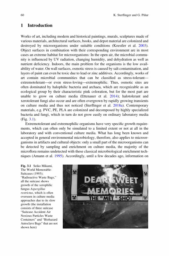
1 Introduction
Works of art, including modern and historical paintings, murals, sculptures made ofvarious materials, architectural surfaces, books, and depot material are colonized anddestroyed by microorganisms under suitable conditions (Koestler et al. 2003).Object surfaces in combination with their corresponding environment are in mostcases an extreme habitat for microorganisms: In the open air, the microbial commu-nity is influenced by UV radiation, changing humidity, and dehydration as well asnutrient deficiency. Indoors, the main problem for the organisms is the low avail-ability of water. On wall surfaces, osmotic stress is caused by salt contamination, andlayers of paint can even be toxic due to lead or zinc additives. Accordingly, works ofart contain microbial communities that can be classified as stress-tolerant—extremotolerant—or even stress-loving—extremophilic. Thus, osmotic sites areoften dominated by halophilic bacteria and archaea, which are recognizable as anecological group by their characteristic pink coloration, but for the most part areunable to grow on culture media (Ettenauer et al. 2014); halotolerant andxerotolerant fungi also occur and are often overgrown by rapidly growing transientson culture media and thus not noticed (Sterflinger et al. 2018a). Contemporarymaterials, e.g. PVC, PE, PLA are colonized and decomposed by highly specializedbacteria and fungi, which in turn do not grow easily on ordinary laboratory media(Fig. 3.1).
Extremotolerant and extremophilic organisms have very specific growth require-ments, which can often only be simulated to a limited extent or not at all in thelaboratory and with conventional culture media. What has long been known andaccepted in general environmental microbiology, therefore, also applies to microor-ganisms in artifacts and cultural objects: only a small part of the microorganisms canbe detected by sampling and enrichment on culture media, the majority of themicroflora remains undetected with these classical microbiological enrichment tech-niques (Amann et al. 1995). Accordingly, until a few decades ago, information on
Fig. 3.1 Seiko Mikami,The World Memorable:Suitcases (1993);“Radioactive Waste Bags,”all the suitcase showsgrowth of the xerophilicfungus Aspergillusrestrictus, which is oftenoverseen in culture mediaapproaches due to its slowgrowth (the installationconsists of three suitcase“Suitcase Accident AirNoxious Particles WasteContainers” and “BiohazardAutoclave Bags” that are notshown here)
60 K. Sterflinger and G. Piñar
microbial biodiversity was limited, but also the knowledge about the ecology oforganisms and the processes of damage they cause.
Only the routine use of PCR in the 1980s and 1990s made it possible to usemicrobial DNA for research on biogenic damage processes on art and cultural assetsin order to overcome the hurdle of cultivation and enrichment and to complete andexpand the data obtained from this. Initially, in addition to cloning and Sangersequencing (which is considered a first-generation sequencing technology developedby Frederick Sanger and colleagues in 1977), DNA-based fingerprinting techniqueswere successfully used to study the biological agents that cause the biodeteriorationof cultural artifacts.
Denaturing gradient gel electrophoresis (DGGE) in combination with clonelibraries and first-generation sequencing of amplified DNA fragments has beensuccessfully used to characterize microbial communities on walls and murals,paper, parchment, glass, stone, textiles, human remains, and other materials ofcultural heritage (Rölleke et al. 1999; Schabereiter-Gurtner et al. 2001, 2002a, b;Laiz et al. 2003; Di Bonaventura et al. 2003; Saiz-Jimenez 2003). The application ofthis molecular strategy, together with imaging and chemical analyses (Pinzari et al.2010; Wiesinger 2018), has provided relevant information on the microbial com-munities associated with different materials and the microbiological risk they presenton the surface of the substrates studied (Sterflinger and Piñar 2013). In addition, suchstudies have enabled the implementation of preservative and disinfectant treatmentsas well as the monitoring during and after the conservation treatment of valuableobjects (Jroundi et al. 2015).
Although first-generation sequencing technology provided only limited data, thefirst studies opened a new door into the world of biodiversity of artifacts and culturalassets and into the phylogeny of the organisms involved. These studies also led to thedescription of new species involved in biodeterioration (Sterflinger et al. 1997;Wieser et al. 2005). The studies were also an important basis—and justification—for the further development and application of technologies in the field of HeritageScience. Nevertheless, the work based on DNA-fingerprints, clone libraries, andfirst-generation sequencing was rather time-consuming and not free of errors, sincethe selection of suitable primers for DNA amplification already resulted in a strongselection of the initial data. Since Sanger sequencing was only able to sequence shortparts of the total DNA contained in a cell in a reasonable time and with reasonableeffort, the studies were limited to taxonomically and phylogenetically relevant DNAsections such as 16S-, 18S-rDNA, and ITS sequences. This provided information onbiodiversity, but not on the potential of functional genes in potentially harmfulorganisms. To establish a link between the identity of an organism and its functionwas, once again, only possible by means of experiments on pure cultures, ifavailable.
3 Molecular-Based Techniques for the Study of Microbial Communities in Artworks 61
2 High-Throughput Sequencing Methods: Next GenerationSequencing (NGS)
“The commercial launch of the first massively parallel pyrosequencing platform in2005 ushered in the new era of high-throughput genomic analysis now referred to asnext-generation sequencing (NGS)” (Völkerding et al. 2009). Although these tech-nologies generate shorter reads (between 25 and 500 bp), they are capable ofdelivering many hundreds of thousands or millions of reads in a relatively shorttime (a few hours). This leads to a high coverage, but the assembly process is muchmore computationally intensive. These technologies are far superior to Sangersequencing due to the high data volume and the relatively short time required tosequence an entire genome (Metzker 2010). In recent years, the so-called nextgeneration sequencing technologies (NGS) have been developed and have been arevolutionary innovation for deciphering the complexity of genomes andmetagenomes. NGS methods have enabled significant advances, such as the prepa-ration of NGS libraries in a cell-free system without cloning; the thousands to manymillions of sequencing reactions produced in parallel; and direct sequencing outputthat is performed cyclically and in parallel. The large number of reads generated byNGS enables the sequencing of entire genomes and metagenomes at an unprece-dented speed. Today, there are several sequencing platforms based on differenttechnologies (Goodwin et al. 2016). Therefore, the choice of the sequencing plat-form to be used for a particular study depends on the requirements and resources,which vary depending on the type of sample, study finality, cost/runtime, dataoutput, tolerated amplicon size, data storage capacities, and error rates.
Different methods available on the market until 2009 are explained in detail byVölkerding et al. (2009); the most relevant ones are only briefly described here:
– The Roche 454 technology was derived from the technological convergence ofpyrosequencing and emulsion PCR. The system has been efficiently used both forsequencing the entire genome and for amplicon sequencing in environmental andmedical studies. However, the technology was no longer competitive and wasdiscontinued in 2013.
– Illumina/Solexa: In 2006, the Solexa Genome Analyzer, the first “short-read”sequencing platform, was commercially launched. Acquired by Illumina in 2006,the Genome Analyzer uses a flow-through cell consisting of an optically trans-parent slide with individual lanes on whose surface oligonucleotide anchors arebound. The template DNA is fragmented into lengths of several hundred basepairs and finally repaired to produce 50-phosphorylated blunt ends. The polymer-ase activity of the Klenow fragment is used to attach a single A base to the 30 endof the blunt phosphorylated DNA fragments. This addition prepares the DNAfragments for ligation to oligonucleotide adapters that have a single T-baseoverhang at their 30 end to increase ligation efficiency. The adapter oligonucle-otides are complementary to the flow cell anchors. Under boundary dilutionconditions, adapter-modified single-stranded template DNA is added to the
62 K. Sterflinger and G. Piñar
flow cell and immobilized on the anchors by hybridization. Currently, Illumina isone of the most widely used sequencing systems both in science and clinicalapplications.
– The SOLiD (Supported Oligonucleotide Ligation and Detection) System 2.0platform, marketed by Applied Biosystems, is a ligation-based short-readsequencing technology. Applied Biosystems has refined the technology and in2007 launched the SOLiD instrumentation. Sample preparation is similar to the454 technology in that DNA fragments are ligated to oligonucleotide adapters,bound to beads, and clonally amplified using emulsion PCR.
Recent technologies developed during the last decade are:
– The next generation sequencing technology of Ion Torrent (ThermoFisher) is notbased on an optical detection system. The technology was first published inNature in 2011 (Rothberg et al. 2011) and was launched on the market in 2012.It exploits the fact that the addition of a dNTP to a DNA polymer releases ahydrogen ion. The pH change resulting from these hydrogen ions is measuredusing semiconductors, with the simultaneous measurement of millions of suchchanges determining the sequence of each fragment.
– Single Molecule, Real-Time (SMRT) sequencing (Pacific Biosciences) is the coretechnology that enables sequencing long reads with resulting reads of several tensof kilobases in length. Due to the long fragments, the technology is particularlyuseful for de novo sequencing and the assembly of entire genomes (Eid et al.2009).
– Nanopore sequencing technology (Oxford Nanopore Technologies) is based onreading the inserted nucleotides while the DNA sequence passes through ananopore with an inner diameter of 1 nm (Branton et al. 2008). The electricalconductivity of the protein nanopore is changed as the DNA passes through, and asignal is detected in this way. Sequencing with this innovative technology doesnot necessarily require intermediate PCR amplification or chemical labeling. Inaddition, this technology offers a pocket-sized sensor device (Fig. 3.2), theMinION, which was made commercially available in 2015 and whose perfor-mance has been further optimized in recent years, resulting in an increasingnumber of publications in the field of metagenomics.
3 Metagenomics in Cultural Heritage
Metagenomics can be performed by the “Sequence it all” approach (simple readingof all DNA sequences present in a sample) or by the “Target sequencing” approach,concentrating on certain conserved sequences, such as only ribosomal RNA genes.The latter procedure has several advantages, such as reducing the complexity of thedata obtained and the possibility of assigning more sequences to a particularorganism or a group of related organisms. This approach facilitates some
3 Molecular-Based Techniques for the Study of Microbial Communities in Artworks 63
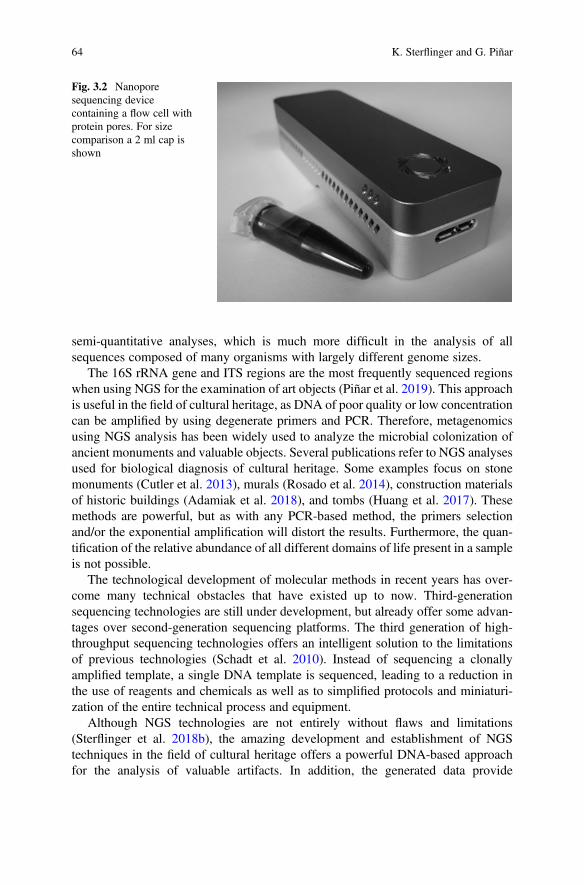
semi-quantitative analyses, which is much more difficult in the analysis of allsequences composed of many organisms with largely different genome sizes.
The 16S rRNA gene and ITS regions are the most frequently sequenced regionswhen using NGS for the examination of art objects (Piñar et al. 2019). This approachis useful in the field of cultural heritage, as DNA of poor quality or low concentrationcan be amplified by using degenerate primers and PCR. Therefore, metagenomicsusing NGS analysis has been widely used to analyze the microbial colonization ofancient monuments and valuable objects. Several publications refer to NGS analysesused for biological diagnosis of cultural heritage. Some examples focus on stonemonuments (Cutler et al. 2013), murals (Rosado et al. 2014), construction materialsof historic buildings (Adamiak et al. 2018), and tombs (Huang et al. 2017). Thesemethods are powerful, but as with any PCR-based method, the primers selectionand/or the exponential amplification will distort the results. Furthermore, the quan-tification of the relative abundance of all different domains of life present in a sampleis not possible.
The technological development of molecular methods in recent years has over-come many technical obstacles that have existed up to now. Third-generationsequencing technologies are still under development, but already offer some advan-tages over second-generation sequencing platforms. The third generation of high-throughput sequencing technologies offers an intelligent solution to the limitationsof previous technologies (Schadt et al. 2010). Instead of sequencing a clonallyamplified template, a single DNA template is sequenced, leading to a reduction inthe use of reagents and chemicals as well as to simplified protocols and miniaturi-zation of the entire technical process and equipment.
Although NGS technologies are not entirely without flaws and limitations(Sterflinger et al. 2018b), the amazing development and establishment of NGStechniques in the field of cultural heritage offers a powerful DNA-based approachfor the analysis of valuable artifacts. In addition, the generated data provide
Fig. 3.2 Nanoporesequencing devicecontaining a flow cell withprotein pores. For sizecomparison a 2 ml cap isshown
64 K. Sterflinger and G. Piñar
information to answer many interesting questions that can arise when handlingvaluable art objects and might help to:
– provide insight into the selection of materials at the time of manufacture, such asthe composition of the materials,
– gain knowledge about the storage conditions of materials and their geographicalorigin,
– trace the history of use of the object, and– group objects of unknown origin on the basis of similarities in their microbiomes.
The information enables to understand many open questions in a variety of fields,such as archeology, history, restoration, philology, or criminology, but also cancontribute to give a historical benefit to the investigated objects (Piñar et al. 2019).
As described above, there are different sequencing platforms based on differenttechnologies. All of them present advantages and disadvantages; therefore, thechoice of the sequencing platform should be decided carefully depending on thepurpose of the study. Also, the bioinformatics tools and pipelines necessary for dataanalysis are manifold and have to be chosen according to the respective data sets andthe scientific question to be answered. In recent years, different methods have beentested, their protocols optimized and used to perform metagenomic analyses onartworks at the first time. The following three case studies describe the applicationof three different sequencing technologies for the study of cultural assets, but withdifferent finalities: (1) Ion Torrent technology, (2) Illumina technology, and(3) Nanopore technology.
(1) Ion Torrent technology. In a metagenome study performed on three ancientmarble statues, the main question was whether they might have been smuggledand/or stored together and whether biological traces could help to elucidate theirgeographical origin (Piñar et al. 2019). Two statues were human torsi, onefemale and one male, and the third one was a representation of a small girl’shead (Fig. 3.3). The finality of this study was to collect as much biologicalinformation as possible from the surface of the statues to answer the aforemen-tioned questions. To this end, metagenomic analyses were performed using theIon Torrent sequencing technology by means of the Ion Personal GenomeMachine (PGM), which allows a very good target barcoding sequencing ofdifferent life domains. Especially for the screening of prokaryotes, this technol-ogy offers a very smart solution thanks to the commercialization of the Ion 16SrDNA Metagenomics Kit that enables the simultaneous targeting of seven of thenine variable regions of the 16S rDNA. This eliminates the bias committed byother NGS technologies that tend to focus only on the V4 region of the 16SrDNA, which even if the most widely used does not cover all phylogeneticgroups (Piñar et al. 2019). To approach this study, the deposits and dirt visible inthe spaces (e.g., folds, curls of hair) defined by the shape of the sculptures wereremoved with the help of sterile scalpels and needles without damaging the rockitself. The material contained was mainly powder, dust, dirt, and even sometextile fibers.
3 Molecular-Based Techniques for the Study of Microbial Communities in Artworks 65
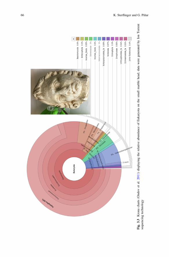
Fig.3.3
Krona
charts
(Ond
ovet
al.20
11)displaying
therelativ
eabun
danceof
Euk
aryo
taon
thesm
allmarblehead;data
weregeneratedby
IonTorrent
sequ
encing
techno
logy
66 K. Sterflinger and G. Piñar
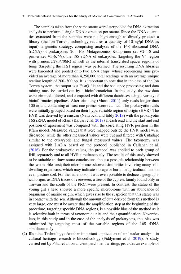
The samples taken from the same statue were later pooled for DNA extractionanalysis to perform a single DNA extraction per statue. Since the DNA quanti-ties extracted from the samples were not high enough to directly produce alibrary (the Ion Torrent technology requires a quantity of 10 ng/μl DNA asinput), a genetic strategy, comprising analyses of the 16S ribosomal DNA(rDNA) of prokaryotes (Ion 16S Metagenomics Kit: primer set V2-4-8 andprimer set V3-6,7-9), the 18S rDNA of eukaryotes (targeting the V4 regionwith primers 528F/706R) as well as the internal transcribed spacer regions offungi (targeting the ITS1 region) was performed. The resulting DNA librarieswere barcoded and pooled onto two DNA chips, whose sequencing runs pro-vided an average of more than 4,250,000 total readings with an average uniquereading length of 200–300 bp. It is important to note that in the case of the IonTorren system, the output is a FastQ file and the sequence processing and datamining must be carried out by a bioinformatician. In this study, the raw datawere trimmed, filtered, and compared with different databases using a variety ofbioinformatics pipelines. After trimming (Martin 2011) only reads longer than100 nt and containing at least one primer were retained. The prokaryotic readswere initially grouped based on their hypervariable region of origin (HVR). TheHVR was derived by a cmscan (Nawrocki and Eddy 2013) with the prokaryotic16S rRNA model of Rfam (Kalvari et al. 2018) at each read and the start and endposition of agreement was compared with the correlating HVR position in theRfam model. Measured values that were mapped outside the HVR model werediscarded, while the other measured values were cut and filtered with Cutadaptsimilar to the eukaryotic and fungal measured values. The taxonomy wasassigned with DADA based on the protocol published in Callahan et al.(2016). For the prokaryotic values, the protocol was applied to each group ofIHR separately and to all HVR simultaneously. The results of this study showedto be suitable to draw some conclusions about a possible relationship betweenthe two marble torsi; their microbiomes showed similarities involving many soil-dwelling organisms, which may indicate storage or burial in agricultural land oreven pasture soil. For the male torso, it was even possible to deduce a geograph-ical origin, as DNA traces of Taiwania, a tree of the cypress family found only inTaiwan and the south of the PRC, were present. In contrast, the statue of theyoung girl’s head showed a more specific microbiome with an abundance oforganisms of marine origin, which gives rise to the suspicion that this statue wasin contact with the sea. Although the amount of data derived from this method isvery large, one must be aware that the amplification step at the beginning of theprocedure, targeting specific DNA regions, is a possible bias of the method as itis selective both in terms of taxonomic units and their quantification. Neverthe-less, in this study and in the case of the analysis of prokaryotes, this bias wasminimized by targeting most of the variable regions of the 16S rDNAsimultaneously.
(2) Illumina Technology: Another important application of molecular analysis incultural heritage research is biocodicology (Fiddyment et al. 2019). A studycarried out by Piñar et al. on ancient parchment writings provides an example of
3 Molecular-Based Techniques for the Study of Microbial Communities in Artworks 67
the emerging field of research in biocodicology and shows how metagenomicscan help to answer relevant questions that can contribute to a better understand-ing of the history of ancient manuscripts (Miklas et al. 2019; Piñar et al. 2020c).For this purpose, two Slavonic codices from the eleventh century were investi-gated by shotgun metagenomics using the Illumina sequencing platform.
The analysis of nucleic acids contained in historical parchments is a difficultmatter due to the poor preservation of ancient DNA (aDNA) and the frequentexternal contamination with modern DNA (Vuissoz et al. 2007). A crucial stepin such studies is the extraction of DNA from very little sample material (a fewfibers or 1 to 2 millimeters of material) and even from eraser fragments loosenedafter rubbing the parchment surface (Fiddyment et al. 2019). In the case-studypresented here, a combination of two commercial kits was used for the success-ful extraction of total DNA (ancient and modern), which aim at a silicamembrane-based purification of very short DNA fragments (~ 70 bp), i.e. theexpected size of the ancient DNA. This approach further enables the elution invery small volumes allowing the concentration of very low DNA yield, as it wasthe case of the DNA extracted from these ancient objects. The DNA extractswere used to create DNA libraries with the NEBNext Ultra II DNA Library PrepKit for Illumina and sequenced by means of the Illumina HiSeq 2500 platformusing 100 bp single reads. The total number of reads processed ranged from43,150,589 to 50,416,907, with reads being significantly shorter than whenusing Ion Torrent or Nanopore. However, the strategy followed in this studyshowed to be adequate for analyzing the ancient DNA of the parchments. Theapplication of Illumina shotgun sequencing proved to be very effective for thesequencing of short DNA fragments (aDNA) with a high sensitivity, generatinga huge amount of data and being less susceptible to chimeric artifacts that canresult from PCR-based analyses. Quality control of the reads was done withfastqc (Andrews 2010). Cutadapt was used to trim adapter sequences (Martin2011). FastQ Screen was used to assess the origin of the parchment probes(Wingett and Andrews 2018). The parchment metagenomes were assessed byfirst removing human and animal reads for each dataset. This was done for eachexperiment by filtering out the reads to the human and corresponding animalgenomes with STAR (Dobin et al. 2013). The remaining reads were thenprocessed with MetaphlAn2 in order to describe and represent the microbiomesof the parchment probes (Truong et al. 2015). The endogenous DNA enabled toinfer the animal origin of the skins used in the production of the two codices,while the DNA obtained from viruses provided insights into the plant origin ofthe inks used in one of the two codices. In addition, the microbiomes colonizingthe surface of the parchments helped to determine their state of preservation andtheir latent risk of deterioration. The advantage of using this molecular strategywas the detection of extremely small DNA fragments that originate from alreadystrongly degraded ancient DNA, which could not be detected by technologieswith longer reading lengths, such as Ion Torrent or Nanopore. Similar to theNanopore amplification of the entire genome described below, shotgun
68 K. Sterflinger and G. Piñar
sequencing enables to obtain the entire genomic information in a given sample:taxonomic tracers from all areas and realms of life as well as functional genes.
(3) Nanopore sequencing technology: Nanopore has been little used in the field ofcultural heritage and only two published studies have reported on the benefits ofapplying this state-of-the-art technology to valuable works of art. In the firststudy (Šoltys et al. 2019), Nanopore sequencing technology was used to studythe microbiome of an eighteenth century wax seal stained with minium. Theauthors reported on the advantages of sequencing long DNA fragments, whichallows sequencing of the almost complete 16S rRNA gene as well as relativelylong fragments (more than 600 bp) from the fungal ITS regions and 28S rRNA.However, the authors used target sequencing technology that did not allow forthe relative proportion of different phylogenetic groups. In the second study(Piñar et al. 2020a), Nanopore sequencing technology was used for the first timetogether with a whole genome amplification (WGA) protocol to provide a rapiddiagnosis of the biological infection in cultural artifacts. All advantages that thisnew technology can offer for metagenomic analyses starting from very lowDNA concentrations were combined. To test the feasibility of this protocol oncultural assets, samples were taken from two eighteenth and nineteenth centuryoil paintings on canvas, one of which showed active fungal colonization, whilethe other had a cracked varnish surface (Fig. 3.4). The total number of readsobtained with Nanopore exceeded the 2,000,000 reads with an average readlength of more than 4 Kb in both analyzed paintings. The resulting fast 5 datafiles were basecalled using the Nanopore GPU basecalling with GUPPY 3.0.3 onUBUNTU 16.04 (Nanopore Community Platform). Once the Fastq files weregenerated, the data were compared with databases using one of the availablepipelines for data analyses of the Nanopore Community Platform, following thesteps recommended by manufacturers. The selected workflow chosen was“What’s in my pot” (WIMP). It is important to note that this platform offers aseries of bioinformatics pipelines that are accessible to the end user and are user-friendly, allowing basic data analysis without the need for a bioinformaticsexpert. The results obtained with the WGA metagenomic approach proved tobe successful for a rapid diagnosis of the microbial colonization of the two testedpaintings showing different deterioration phenomena. Their microbiomesshowed to be directly related to the conservation status of the paintings as wellas with their restoration history (Piñar et al. 2020a).
Until now, the power of metagenomics has been largely tied to expensive androom-sized sequencing technologies, a large number of laboratory ancillary equip-ment and time-consuming workflows, making analysis far too complex and expen-sive for routine analysis by museums, collections, and restorers. Nanoporetechnology opens up new possibilities in this area of research and appliedrestoration:
– The basic equipment for this technology is the pocket-sized sequencer, combinedwith a laptop.
3 Molecular-Based Techniques for the Study of Microbial Communities in Artworks 69
Fig.3
.4Partof
theph
ylog
enetic
tree
createdby
theOxfordNanop
oresoftware“Whatisin
mypo
t”afterWGSfrom
theoilpaintin
g“Boy
holdingado
g”show
non
theleft
70 K. Sterflinger and G. Piñar
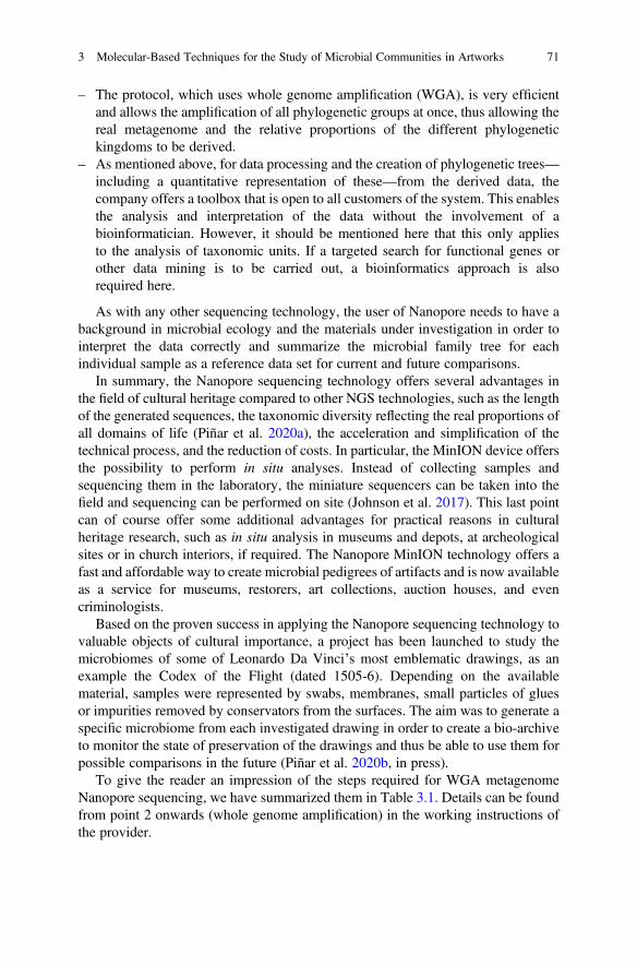
– The protocol, which uses whole genome amplification (WGA), is very efficientand allows the amplification of all phylogenetic groups at once, thus allowing thereal metagenome and the relative proportions of the different phylogenetickingdoms to be derived.
– As mentioned above, for data processing and the creation of phylogenetic trees—including a quantitative representation of these—from the derived data, thecompany offers a toolbox that is open to all customers of the system. This enablesthe analysis and interpretation of the data without the involvement of abioinformatician. However, it should be mentioned here that this only appliesto the analysis of taxonomic units. If a targeted search for functional genes orother data mining is to be carried out, a bioinformatics approach is alsorequired here.
As with any other sequencing technology, the user of Nanopore needs to have abackground in microbial ecology and the materials under investigation in order tointerpret the data correctly and summarize the microbial family tree for eachindividual sample as a reference data set for current and future comparisons.
In summary, the Nanopore sequencing technology offers several advantages inthe field of cultural heritage compared to other NGS technologies, such as the lengthof the generated sequences, the taxonomic diversity reflecting the real proportions ofall domains of life (Piñar et al. 2020a), the acceleration and simplification of thetechnical process, and the reduction of costs. In particular, the MinION device offersthe possibility to perform in situ analyses. Instead of collecting samples andsequencing them in the laboratory, the miniature sequencers can be taken into thefield and sequencing can be performed on site (Johnson et al. 2017). This last pointcan of course offer some additional advantages for practical reasons in culturalheritage research, such as in situ analysis in museums and depots, at archeologicalsites or in church interiors, if required. The Nanopore MinION technology offers afast and affordable way to create microbial pedigrees of artifacts and is now availableas a service for museums, restorers, art collections, auction houses, and evencriminologists.
Based on the proven success in applying the Nanopore sequencing technology tovaluable objects of cultural importance, a project has been launched to study themicrobiomes of some of Leonardo Da Vinci’s most emblematic drawings, as anexample the Codex of the Flight (dated 1505-6). Depending on the availablematerial, samples were represented by swabs, membranes, small particles of gluesor impurities removed by conservators from the surfaces. The aim was to generate aspecific microbiome from each investigated drawing in order to create a bio-archiveto monitor the state of preservation of the drawings and thus be able to use them forpossible comparisons in the future (Piñar et al. 2020b, in press).
To give the reader an impression of the steps required for WGA metagenomeNanopore sequencing, we have summarized them in Table 3.1. Details can be foundfrom point 2 onwards (whole genome amplification) in the working instructions ofthe provider.
3 Molecular-Based Techniques for the Study of Microbial Communities in Artworks 71
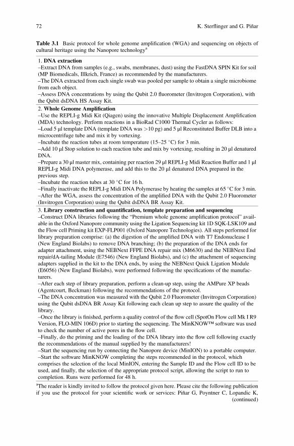
Table 3.1 Basic protocol for whole genome amplification (WGA) and sequencing on objects ofcultural heritage using the Nanopore technologya
1. DNA extraction–Extract DNA from samples (e.g., swabs, membranes, dust) using the FastDNA SPIN Kit for soil(MP Biomedicals, Illkrich, France) as recommended by the manufacturers.–The DNA extracted from each single swab was pooled per sample to obtain a single microbiomefrom each object.–Assess DNA concentrations by using the Qubit 2.0 fluorometer (Invitrogen Corporation), withthe Qubit dsDNA HS Assay Kit.
2. Whole Genome Amplification–Use the REPLI-g Midi Kit (Qiagen) using the innovative Multiple Displacement Amplification(MDA) technology. Perform reactions in a BioRad C1000 Thermal Cycler as follows:–Load 5 μl template DNA (template DNA was >10 pg) and 5 μl Reconstituted Buffer DLB into amicrocentrifuge tube and mix it by vortexing.–Incubate the reaction tubes at room temperature (15–25 �C) for 3 min.–Add 10 μl Stop solution to each reaction tube and mix by vortexing, resulting in 20 μl denaturedDNA.–Prepare a 30 μl master mix, containing per reaction 29 μl REPLI-g Midi Reaction Buffer and 1 μlREPLI-g Midi DNA polymerase, and add this to the 20 μl denatured DNA prepared in theprevious step.–Incubate the reaction tubes at 30 �C for 16 h.–Finally inactivate the REPLI-g Midi DNA Polymerase by heating the samples at 65 �C for 3 min.–After the WGA, assess the concentration of the amplified DNA with the Qubit 2.0 Fluorometer(Invitrogen Corporation) using the Qubit dsDNA BR Assay Kit.
3. Library construction and quantification, template preparation and sequencing–Construct DNA libraries following the “Premium whole genome amplification protocol” avail-able in the Oxford Nanopore community using the Ligation Sequencing kit 1D SQK-LSK109 andthe Flow cell Priming kit EXP-FLP001 (Oxford Nanopore Technologies). All steps performed forlibrary preparation comprise: (a) the digestion of the amplified DNA with T7 Endonuclease I(New England Biolabs) to remove DNA branching; (b) the preparation of the DNA ends foradapter attachment, using the NEBNext FFPE DNA repair mix (M6630) and the NEBNext Endrepair/dA-tailing Module (E7546) (New England Biolabs), and (c) the attachment of sequencingadapters supplied in the kit to the DNA ends, by using the NEBNext Quick Ligation Module(E6056) (New England Biolabs), were performed following the specifications of the manufac-turers.–After each step of library preparation, perform a clean-up step, using the AMPure XP beads(Agentcourt, Beckman) following the recommendations of the protocol.–The DNA concentration was measured with the Qubit 2.0 Fluorometer (Invitrogen Corporation)using the Qubit dsDNA BR Assay Kit following each clean up step to assure the quality of thelibrary.–Once the library is finished, perform a quality control of the flow cell (SpotOn Flow cell Mk I R9Version, FLO-MIN 106D) prior to starting the sequencing. The MinKNOW™ software was usedto check the number of active pores in the flow cell.–Finally, do the priming and the loading of the DNA library into the flow cell following exactlythe recommendations of the manual supplied by the manufacturers!–Start the sequencing run by connecting the Nanopore device (MinION) to a portable computer.–Start the software MinKNOW completing the steps recommended in the protocol, whichcomprises the selection of the local MinION, entering the Sample ID and the Flow cell ID to beused, and finally, the selection of the appropriate protocol script, allowing the script to run tocompletion. Runs were performed for 48 h.aThe reader is kindly invited to follow the protocol given here. Please cite the following publicationif you use the protocol for your scientific work or services: Piñar G, Poyntner C, Lopandic K,
(continued)
72 K. Sterflinger and G. Piñar
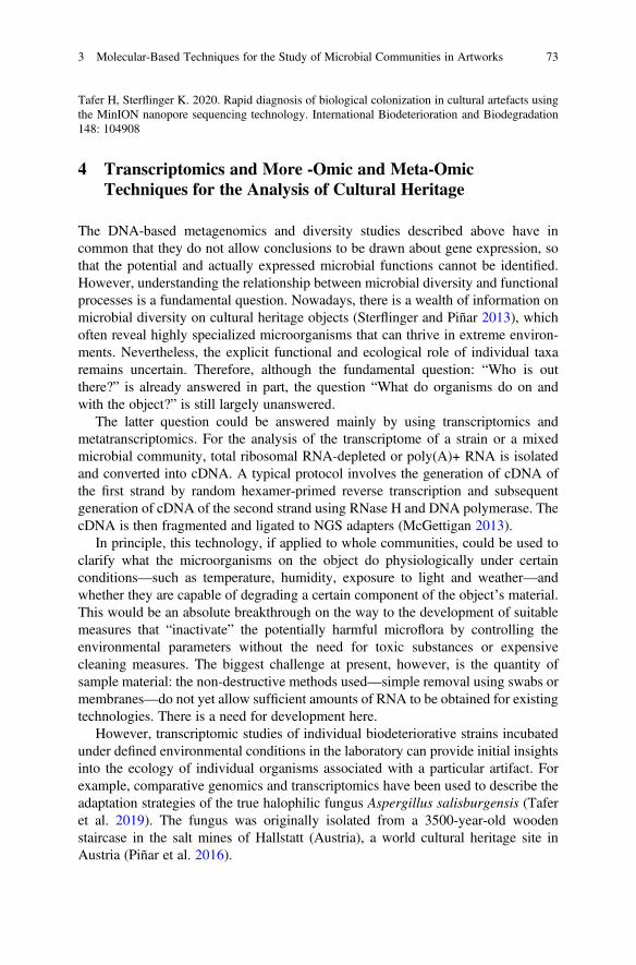
4 Transcriptomics and More -Omic and Meta-OmicTechniques for the Analysis of Cultural Heritage
The DNA-based metagenomics and diversity studies described above have incommon that they do not allow conclusions to be drawn about gene expression, sothat the potential and actually expressed microbial functions cannot be identified.However, understanding the relationship between microbial diversity and functionalprocesses is a fundamental question. Nowadays, there is a wealth of information onmicrobial diversity on cultural heritage objects (Sterflinger and Piñar 2013), whichoften reveal highly specialized microorganisms that can thrive in extreme environ-ments. Nevertheless, the explicit functional and ecological role of individual taxaremains uncertain. Therefore, although the fundamental question: “Who is outthere?” is already answered in part, the question “What do organisms do on andwith the object?” is still largely unanswered.
The latter question could be answered mainly by using transcriptomics andmetatranscriptomics. For the analysis of the transcriptome of a strain or a mixedmicrobial community, total ribosomal RNA-depleted or poly(A)+ RNA is isolatedand converted into cDNA. A typical protocol involves the generation of cDNA ofthe first strand by random hexamer-primed reverse transcription and subsequentgeneration of cDNA of the second strand using RNase H and DNA polymerase. ThecDNA is then fragmented and ligated to NGS adapters (McGettigan 2013).
In principle, this technology, if applied to whole communities, could be used toclarify what the microorganisms on the object do physiologically under certainconditions—such as temperature, humidity, exposure to light and weather—andwhether they are capable of degrading a certain component of the object’s material.This would be an absolute breakthrough on the way to the development of suitablemeasures that “inactivate” the potentially harmful microflora by controlling theenvironmental parameters without the need for toxic substances or expensivecleaning measures. The biggest challenge at present, however, is the quantity ofsample material: the non-destructive methods used—simple removal using swabs ormembranes—do not yet allow sufficient amounts of RNA to be obtained for existingtechnologies. There is a need for development here.
However, transcriptomic studies of individual biodeteriorative strains incubatedunder defined environmental conditions in the laboratory can provide initial insightsinto the ecology of individual organisms associated with a particular artifact. Forexample, comparative genomics and transcriptomics have been used to describe theadaptation strategies of the true halophilic fungus Aspergillus salisburgensis (Taferet al. 2019). The fungus was originally isolated from a 3500-year-old woodenstaircase in the salt mines of Hallstatt (Austria), a world cultural heritage site inAustria (Piñar et al. 2016).
Tafer H, Sterflinger K. 2020. Rapid diagnosis of biological colonization in cultural artefacts usingthe MinION nanopore sequencing technology. International Biodeterioration and Biodegradation148: 104908
3 Molecular-Based Techniques for the Study of Microbial Communities in Artworks 73
The development of high-throughput “omics” methods enables systemapproaches in which mixed microbial communities are studied as a single unit.Metagenomics, metatranscriptomics, metaproteomics, and metabolomics are used todetermine the DNA sequences of the mixed community under investigation, thecollectively transcribed RNA, the translated proteins, and the metabolites resultingfrom cellular processes. All data obtained can be used to identify metabolic path-ways and cellular processes within an ecosystem (Kulski 2016). Therefore, theapplication of meta-omics analyses not only allows the identification of microbialgroups in complex communities, but also allows the correlation of taxonomic groupswith their ecological function and the observation of structural and functional shiftsin communities caused by environmental changes. Meta-omics have the potential toperform in-depth studies on cultural assets in order to gain a deep understanding ofthe interactions between microorganisms and their substrates. In contrast to thesuccessful application of metagenomics to cultural heritage, there are very fewstudies to date that demonstrate the success of applying proteomics in this field(Marvasi et al. 2019 and bibliography therein). However, proteomics has beensuccessfully applied to cultural heritage to characterize protein-containing materialsin ancient samples (Fiddyment et al. 2015; Vinciguerra et al. 2016). In the future,proteomics could help to analyze degrading enzymes such as proteases, cellulases,lipases, and other proteins with respect to their biodeterioration potential directlyfrom the object of study. Also, the metabolome can provide information aboutdestruction processes (Gutarowska et al. 2015). Conversely, the metabolome couldpossibly also be a key to the biological control of harmful microbes, since among thenumerous primary and secondary metabolites, some of which are volatile organiccompounds, there may also be those with antimicrobial effects (Campos andZampieri 2019).
In conclusion, this review shows the potential of the molecular techniquesavailable to date and the need for development to adapt many of these methodologiesto the delicate field of cultural heritage. The further development of technologicalprocesses in molecular biology applied to heritage sciences will provide an in-depthunderstanding of the biodiversity and metabolic functions of the microbial commu-nities that colonize culturally valuable objects. This will help in the first place tounderstand and mitigate biodeterioration, but also to answer many other interestingquestions that may arise when handling valuable art objects.
References
Adamiak J, Otlewska A, Tafer H, Lopandic K, Gutarowska B, Sterflinger K, Piñar G (2018) Firstevaluation of the microbiome of built cultural heritage by using the Ion Torrent next generationsequencing platform. Int Biodeter Biodegr 131:11–18
Amann RI, Ludwig W, Schleifer KH (1995) Phylogenetic identification and in situ detection ofindividual microbial cells without cultivation. Microbiol Rev 59:143–169
Andrews S (2010) FastQC: a quality control tool for high throughput sequence data. Availableonline at: http://www.bioinformatics.babraham.ac.uk/projects/fastqc
74 K. Sterflinger and G. Piñar
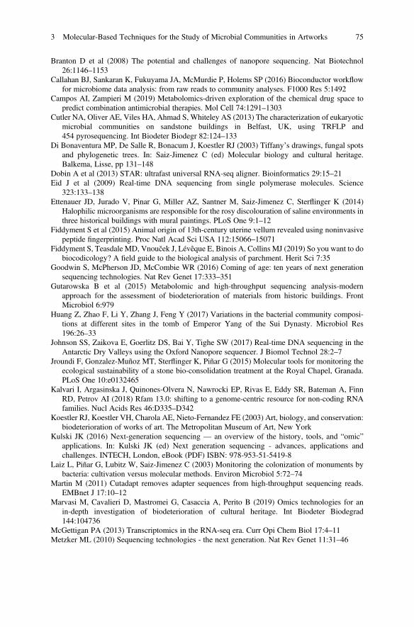
Branton D et al (2008) The potential and challenges of nanopore sequencing. Nat Biotechnol26:1146–1153
Callahan BJ, Sankaran K, Fukuyama JA, McMurdie P, Holems SP (2016) Bioconductor workflowfor microbiome data analysis: from raw reads to community analyses. F1000 Res 5:1492
Campos AI, Zampieri M (2019) Metabolomics-driven exploration of the chemical drug space topredict combination antimicrobial therapies. Mol Cell 74:1291–1303
Cutler NA, Oliver AE, Viles HA, Ahmad S, Whiteley AS (2013) The characterization of eukaryoticmicrobial communities on sandstone buildings in Belfast, UK, using TRFLP and454 pyrosequencing. Int Biodeter Biodegr 82:124–133
Di Bonaventura MP, De Salle R, Bonacum J, Koestler RJ (2003) Tiffany’s drawings, fungal spotsand phylogenetic trees. In: Saiz-Jimenez C (ed) Molecular biology and cultural heritage.Balkema, Lisse, pp 131–148
Dobin A et al (2013) STAR: ultrafast universal RNA-seq aligner. Bioinformatics 29:15–21Eid J et al (2009) Real-time DNA sequencing from single polymerase molecules. Science
323:133–138Ettenauer JD, Jurado V, Pinar G, Miller AZ, Santner M, Saiz-Jimenez C, Sterflinger K (2014)
Halophilic microorganisms are responsible for the rosy discolouration of saline environments inthree historical buildings with mural paintings. PLoS One 9:1–12
Fiddyment S et al (2015) Animal origin of 13th-century uterine vellum revealed using noninvasivepeptide fingerprinting. Proc Natl Acad Sci USA 112:15066–15071
Fiddyment S, Teasdale MD, Vnouček J, Lévêque E, Binois A, Collins MJ (2019) So you want to dobiocodicology? A field guide to the biological analysis of parchment. Herit Sci 7:35
Goodwin S, McPherson JD, McCombie WR (2016) Coming of age: ten years of next generationsequencing technologies. Nat Rev Genet 17:333–351
Gutarowska B et al (2015) Metabolomic and high-throughput sequencing analysis-modernapproach for the assessment of biodeterioration of materials from historic buildings. FrontMicrobiol 6:979
Huang Z, Zhao F, Li Y, Zhang J, Feng Y (2017) Variations in the bacterial community composi-tions at different sites in the tomb of Emperor Yang of the Sui Dynasty. Microbiol Res196:26–33
Johnson SS, Zaikova E, Goerlitz DS, Bai Y, Tighe SW (2017) Real-time DNA sequencing in theAntarctic Dry Valleys using the Oxford Nanopore sequencer. J Biomol Technol 28:2–7
Jroundi F, Gonzalez-Muñoz MT, Sterflinger K, Piñar G (2015) Molecular tools for monitoring theecological sustainability of a stone bio-consolidation treatment at the Royal Chapel, Granada.PLoS One 10:e0132465
Kalvari I, Argasinska J, Quinones-Olvera N, Nawrocki EP, Rivas E, Eddy SR, Bateman A, FinnRD, Petrov AI (2018) Rfam 13.0: shifting to a genome-centric resource for non-coding RNAfamilies. Nucl Acids Res 46:D335–D342
Koestler RJ, Koestler VH, Charola AE, Nieto-Fernandez FE (2003) Art, biology, and conservation:biodeterioration of works of art. The Metropolitan Museum of Art, New York
Kulski JK (2016) Next-generation sequencing — an overview of the history, tools, and “omic”applications. In: Kulski JK (ed) Next generation sequencing - advances, applications andchallenges. INTECH, London, eBook (PDF) ISBN: 978-953-51-5419-8
Laiz L, Piñar G, Lubitz W, Saiz-Jimenez C (2003) Monitoring the colonization of monuments bybacteria: cultivation versus molecular methods. Environ Microbiol 5:72–74
Martin M (2011) Cutadapt removes adapter sequences from high-throughput sequencing reads.EMBnet J 17:10–12
Marvasi M, Cavalieri D, Mastromei G, Casaccia A, Perito B (2019) Omics technologies for anin-depth investigation of biodeterioration of cultural heritage. Int Biodeter Biodegrad144:104736
McGettigan PA (2013) Transcriptomics in the RNA-seq era. Curr Opi Chem Biol 17:4–11Metzker ML (2010) Sequencing technologies - the next generation. Nat Rev Genet 11:31–46
3 Molecular-Based Techniques for the Study of Microbial Communities in Artworks 75
Miklas H, Sablatnig R, Brenner S, Schreiner M, Cappa F, Piñar G, Sterflinger K (2019) The Viennaglagolotic projects: past and present. In: Stankovska P, Derganc A, Sivic-Dular A, RajkkoNahtigal in 100 let slavistike na Univerzi v Ljubljani - Monografija ob 100. oblenici nastankaOddelka za slavistiko Filozofske fakultete UL, 63-80; Slavica Slovenica, Ljubljana, pp 63–80.Please, clarify this reference. Editor, publisher, city, etc
Nawrocki EP, Eddy SR (2013) Infernal 1.1: 100-fold faster RNA homology searches. Bioinfor-matics 29:2933–2935
Ondov BD, Bergman NH, Phillippy AM (2011) Interactive metagenomic visualization in a Webbrowser. BMC Bioinformatics 12:385
Piñar G, Dalnodar D, Voitl C, Reschreiter H, Sterflinger K (2016) Biodeterioration risk threatensthe 3100 year old staircase of Hallstatt (Austria): possible involvement of halophilic microor-ganisms. PLoS One 11:e0148279
Piñar G, Poyntner C, Tafer H, Sterflinger K (2019) A time travel story: metagenomic analysesdecipher the unknown geographical shift and the storage history of possibly smuggled antiquemarble statues. Ann Microbiol 69:1001–1021
Piñar G, Poyntner C, Lopandic K, Tafer H, Sterflinger K (2020a) Rapid diagnosis of biologicalcolonization in cultural artefacts using the MinION nanopore sequencing technology. IntBiodeter Biodegr 148:104908
Piñar G, Sclocchi MC, Sebastiani ML, Sterflinger K (2020b) The “Biological Pedigrees” of someemblematic Leonardo Da Vinci’s objects. Gangemi Editore, Rome. in press
Piñar G, Tafer H, Schreiner M, Miklas H, Sterflinger K (2020c) Decoding the biological informa-tion contained in two ancient Slavonic parchment codices: an added historical value. EnvironMicrobiol. In press. https://doi.org/10.1111/1462-2920.15064
Pinzari F, Montanari M, Michaelsen A, Piñar G (2010) Analytical protocols for the assessment ofbiological dam-age in historical documents. Coalit Newslett 19:6–13
Rölleke S, Muyzer WG,Wanner C, Lubitz W (1999) Identification of bacteria in a biodegraded wallpainting by denaturing gradient gel electrophoresis. Appl Environ Microbiol 62:2059–2065
Rosado T, Mirão J, Candeias A, Caldeira AT (2014) Microbial communities analysis assessed bypyrosequening—a new approach applied to conservation state studies of mural paintings. AnalBioanal Chem 406:887–895
Rothberg J et al (2011) An integrated semiconductor device enabling non-optical genome sequenc-ing. Nature 475:348–352
Saiz-Jimenez C (ed) (2003) Molecular biology and cultural heritage. Balkema, LisseSchabereiter-Gurtner C, Piñar G, Lubitz W, Rölleke S (2001) An advanced molecular strategy to
identify bacterial communities on art objects. J Microbiol Methods 45:77–87Schabereiter-Gurtner C, Saiz-Jimenez C, Piñar G, Lubitz W, Rölleke S (2002a) Phylogenetic 16S
rRNA analysis reveals the presence of complex and partly unknown bacterial communities inTito Bustillo cave, Spain, and on its Palaeolithic paintings. Environ Microbiol 4:392–400
Schabereiter-Gurtner C, Saiz-Jimenez C, Piñar G, Lubitz W, Rölleke S (2002b) Altamira cavePaleolithic paintings harbor partly unknown bacterial communities. FEMS Microbiol Lett211:7–11
Schadt EE, Turner S, Kasarkskis A (2010) A window into third-generation sequencing. HumanMolGenet 19:227–240
Šoltys K, Planý M, Biocca P, Vianello V, Bučková M, Puškárov A, Sclocchi MC, Colaizzi P,Bicchieri M, Pangallo D, Pinzari F (2019) Lead soaps formation and biodiversity in a XVIIICentury wax seal coloured with minium. Environ Microbiol 22(4):1517–1534
Sterflinger K, Piñar G (2013) Microbial deterioration of cultural heritage and works of art— tiltingat windmills? Appl Microbiol Biotechnol 97:9637–9646
Sterflinger K, De Baere R, de Hoog GS, De Wachter R, Krumbein WE, Haase G (1997)Coniosporium perforans and C. apollinis, two new rock-inhabiting fungi isolated from marblein the Sanctuary of Delos (Cyclades, Greece). Anton Leeuw 2: 349-363
76 K. Sterflinger and G. Piñar
Sterflinger K, Little B, Piñar G, Pinzari F, De los Rios A, Gu JD (2018a) Future directions andchallenges in biodeterioration research on historic materials and cultural properties. Int BiodeterBiodegr 129:10–12
Sterflinger K, Voitl C, Lopandic K, Piñar G, Tafer H (2018b) Big sound and extreme fungi-xerophilic, halotolerant Aspergilli and Penicillia with low optimal temperature as invaders ofhistoric pipe organs. Life 8:22
Tafer H, Poyntner C, Lopandic K, Sterflinger K, Piñar G (2019) Back to the salt mines: Genome andtranscriptome comparisons of the halophilic fungus Aspergillus salisburgensis and itshalotolerant relative Aspergillus sclerotialis. Genes 10:381
Truong DT, Franzosa E, Tickle TL, Scholz M, Weingart G, Pasolli E et al (2015) MetaPhlAn2 forenhanced metagenomic taxonomic profiling. Nat Methods 12:902–903
Vinciguerra R, De Chiaro A, Pucci P, Marino G, Biroloa L (2016) Proteomic strategies for culturalheritage: From bones to paintings. Microchem J 126:341–348
Völkerding KV, Dames SA, Durtschi JD (2009) Next-generation sequencing: from basic researchto diagnostics. Clin Chem 55:641–658
Vuissoz A, Worobeya M, Odegaard N, Bunce M, Machadoa CA, Lynner N, Peacocke E,Thomas M, Gilbert P (2007) The survival of PCR-amplifiable DNA in cow leather. J ArchaeolSci 34:823–829
Wieser M, Worliczek HL, Kämpfer P, Busse HJ (2005) Bacillus herbersteinensis sp. nov. Int J SystEvol Microbiol 55:2119–2123
Wiesinger R (2018) Material stability of art and cultural heritage, Habilitation Thesis, Institut fürNaturwissenschaften und Technologie in der Kunst, Akademie der Bildenden Künste Wien
Wingett SW, Andrews S (2018) FastQ Screen: a tool for multi-genome mapping and qualitycontrol. F1000 Res 7:1338
Open Access This chapter is licensed under the terms of the Creative Commons Attribution 4.0International License (http://creativecommons.org/licenses/by/4.0/), which permits use, sharing,adaptation, distribution and reproduction in any medium or format, as long as you give appropriatecredit to the original author(s) and the source, provide a link to the Creative Commons license andindicate if changes were made.
The images or other third party material in this chapter are included in the chapter's CreativeCommons license, unless indicated otherwise in a credit line to the material. If material is notincluded in the chapter's Creative Commons license and your intended use is not permitted bystatutory regulation or exceeds the permitted use, you will need to obtain permission directly fromthe copyright holder.
3 Molecular-Based Techniques for the Study of Microbial Communities in Artworks 77
Chapter 4Extreme Colonizers and Rapid Profiteers:The Challenging World of MicroorganismsThat Attack Paper and Parchment
Flavia Pinzari and Beata Gutarowska
Abstract Microorganisms form the backbone of life on Earth. Over billions ofyears, they have colonized and shaped every possible niche on the planet. Microbeshave modelled both the land and the sea, and have created favourable conditions formulticellular organisms to thrive in. Our understanding of how microbial diversity isdistributed across natural environments and how microbes affect ecosystems isconstantly evolving as public databases are set up and new techniques based onmassive sequencing are developed. The microbiome found in a particular anthropo-genic environment is generally much less complex than those found in natural ones:there is less competition and the main actors are often linked to survival mechanismsregulated by a few limiting factors. Despite this simplicity, it is very difficult to linkcause and effect when seeking to identify the role of individual organisms. In thecase of biodeterioration of paper and parchment, even when analysing the individualcomponents of a simple phenomenon, it is not always easy to understand themechanisms at play. Works of art are unique objects and the elements that determinethe arrival and establishment of one or more microorganisms and the direction thatthe biodeterioration process takes are always different. In some cases, however, thereare common denominators and predictable mechanisms. The variables that comeinto play are examined below.
Keywords Ecology · Metabolomics · Niche · Colonization · Diagnostics ·Mechanisms · Biodeterioration · Substrate · Succession · Metabolites · Enzymes ·Indoor environment
F. Pinzari (*)Institute for Biological Systems, National Research Council of Italy (CNR), Rome, Italy
Life Sciences Department, Natural History Museum, London, UKe-mail: [email protected]; [email protected]
B. GutarowskaDepartment of Environmental Biotechnology, Lodz University of Technology, Łódź, Polande-mail: [email protected]
© The Author(s) 2021E. Joseph (ed.), Microorganisms in the Deterioration and Preservation of CulturalHeritage, https://doi.org/10.1007/978-3-030-69411-1_4
79
1 Paper and Parchment as Food Sources for Microbes
1.1 Paper
Paper serves as an excellent substrate for various heterotrophic organisms. It is, infact, a source of organic carbon for many microorganisms, and when it is used in themanufacture of objects such as books, documents, prints, etc., it also contains severalorganic substances such as animal or vegetable glues, inks, pigments and fillers thatcan augment its biodegradability. These compounds can represent a nutritionalsource for many non-specialized microorganisms which, as they grow, prepare thesubstrate for a more specific and therefore more harmful attack by typically cellu-lolytic strains—that is, those capable of degrading the cellulose that constitutes thechief component of most papers.
The biodegradability of paper varies depending on different factors, such as thedegree of polymerization of cellulose, the percentage ratio between the two types ofcellulose (crystalline or amorphous), and the presence of chemical or mechanicalpulp (lignin, for example, makes paper unattractive for many fungal species).Additionally, the type of glue used in the pulp or on the paper’s surface, whichcan be of animal or plant origin (starch, gelatin, casein, rosin, etc.), has a significanteffect because it can to a greater or lesser extent be either palatable or toxic tomicroorganisms. The nature of the fillers (e.g. talc, kaolin, calcium carbonate, etc.)used in the manufacture of paper, and possible surface treatments (e.g. colouration,satin-effects, etc.), can have a substantial effect on the outcome of an infectioncaused by microorganisms (Florian 2002; Pinzari et al. 2006).
Cellulose itself is a long linear chain of glucose. The polymer consists ofanhydrous glucopyranose units joined by ß-1,4 glycosidic bonds. A cellulosechain contains approximately 10,000 units of glucose with a molecular weight ofapproximately 1.5–2 million. The polymer chains are arranged parallel to each otherand bind together by means of hydrogen bonds, forming fibrils. These fibrils canform a hydrophobic and very resistant crystalline structure. The enzymatic hydro-lysis of crystalline cellulose typically requires the participation of three differentenzymes: (1) an endo-1,4-ß-glucanase (EC3.2.1.4) which breaks the molecule,randomly producing chains with free ends; (2) an eso-1,4-ß-glucanase(EC3.2.1.91) that detaches cellobiose units, ß-glucose dimer, from thenon-reducing ends of the linear chains; and (3) a ß-glucosidase (EC3.2.1.21) thathydrolyses cellobiose to form glucose (Jayasekara and Ratnayake 2019).
The complete biodegradation of cellulose is carried out by microorganisms thatpossess all the above-mentioned enzymes; the enzymes operate in succession as a“cellulase complex” capable of breaking down the cellulose polymer into glucosemonomers. There are, however, only a few organisms that show real cellulolyticactivity: most of the species use simple, easily hydrolysable sugars. The variouscellulolytic species produce a wide variety of isoenzymes that attack cellulose indifferent ways (Sterflinger and Pinzari 2012; El Bergadi et al. 2014; Oetari et al.2016; Coronado-Ruiz et al. 2018; Jayasekara and Ratnayake 2019). For example,
80 F. Pinzari and B. Gutarowska
some fungal species do not possess all the enzymes that form the cellulase complex,and therefore exert an incomplete depolymerization effect on cellulose. Many fungido not possess exoglucanase and act only on amorphous cellulose, leaving thecrystalline component intact (Jayasekara and Ratnayake 2019). Bacteria possesscellulolytic enzyme systems that are just as effective and even more diversifiedthan fungi.
The superiority of fungi in cellulose breakdown in comparison to unicellularorganisms derives from their growth mode and ecology. Mycelium makes it possiblefor fungi to colonize a substrate more rapidly and to penetrate it by branching outwithin. In addition, fungal hyphae can readily move and mobilize the products ofpolymer degradation over considerable distances through the mycelial network(Szczepanowska et al. 2015). An important factor that influences the degree ofcellulose decomposition is a relative lack of nitrogen. Natural cellulosic materialsoften have a C:N ratio of 100:1 or greater, so the addition of nitrogen is necessary fortheir decomposition. Consequently, the presence of glues containing proteins, suchas gelatin or other nitrogen-rich molecules, increases the biodegradability of paper(Florian 2002).
Fungi that damage paper belong to a few genera that share some ecological traitswhich characterize them as species that are well suited to colonizing “indoor”environments. Such environments, although artificial, offer sufficient trophic nichesto accommodate real trophic successions (Zyska 1997; Pinzari 2011, 2018; Malloet al. 2017; Karakasidou et al. 2018; Koul and Upadhyay 2018; Lax et al. 2019). Thespecies that attack books and archives are fungi that, in natural settings, are mainlyfound in soil and on decomposing plant and animal matter (saprophytic fungi)(Treseder and Lennon 2015; Hyde et al. 2019). They primarily feed on cellulose,starch and sugars, but some can also degrade chemically more complex substancessuch as lignin, keratin, glues and synthetic polymers (Ayu and Teja 2016; Brunneret al. 2018; Hyde et al. 2019). They are organisms that disperse through conidia andspores, which are easily transportable through the air or by insects.
Fungi can damage paper in different ways depending on the species concerned,growth conditions and the type of paper involved. The fungal hyphae can alsopenetrate cellulose fibrils, fragmenting them through mechanical action (Figs. 4.1,4.2, 4.3). They can also exploit cellulose as a substrate and cause sheets of paper tobecome fragile and cracked. Sometimes they release coloured metabolic substancesof various kinds between the fibres that can stain sheets or induce chemical alter-ations, such as oxidation.
Melo et al. (2019) reviewed the known mechanisms behind the formation ofstains on paper, the colourants commonly present in the stains and the main fungalspecies associated with both the production of coloured compounds and their abilityto develop in paper. Among the most widespread and staining fungal pigments,melanins are particularly detrimental to books and objects of heritage. Melanins areoften contained within the walls of fungi and typically accompany the growth of thehyphae across the cellulose network, infiltrating the structure of paper and causingdark stains that can remain deep in the body of the material (Szczepanowska et al.
4 Extreme Colonizers and Rapid Profiteers: The Challenging World of. . . 81

2015). Melanins are natural polymers characterized by a strong negative charge,high molecular weight and hydrophobic nature (Nosanchuk et al. 2015). They areproduced through the oxidation and polymerization of phenolic/indolic precursorsand in fungi they form insoluble pigments (eumelanins) that confer dark colourationsand have several functional roles (for example: photoprotection, energy harvesting,heat gain, thermoregulation, and metal scavenging) (Cordero and Casadevall 2017).Colour and concentration of melanins can differ in one and the same microorganismdepending on growth conditions, the availability of nutrients and the properties ofthe substrate. Accordingly, the same fungus can produce very different stains ondifferent paper stocks (Pinzari et al. 2006). This wide variability in colour productionalso occurs in pigments other than melanin. According to Melo et al. (2019), thecolourants predominantly produced by paper-colonizing fungi arehydroxyanthraquinoid (HAQN) pigments. These can be yellow and red or orange,and when widely distributed in paper can change in shade and intensity dependingon the local pH and the presence of salts (Figs. 4.1, 4.2, 4.3). In the literature, the
Fig. 4.1 Scanning Electron Microscopy images of parchment and paper samples affected bybiodeterioration: (a) surface of a parchment leaf attacked by a fungus. The collagen fibres stillappear intact, although fungal hyphae are attached to them. The fungus, in this case, is probably notusing collagen as a carbon source, but it is feeding on other components of the material (i.e. waxes,glues, oils); (b) the surface of a parchment sample attacked by bacteria. Here there is visibleevidence of the formation of cracks between the bundles of fibrils, and disintegration of the matrixthat holds the fibrils together; (c) surface of a paper sample attacked by a fungus. Despite the factthat the organism has developed among the cellulose fibres producing abundant structures, thefibres still appear intact, indicating that the fungus is not using cellulose as a carbon source; (d)surface of a paper sample deeply infiltrated by a fungus that produced abundant acidic compounds,probably oxalic acids. The cellulose fibres appear broken and dissolved. These acidic compoundsprecipitate in the presence of calcium used in the manufacture of the paper; the precipitated crystalsof calcium oxalate are visible as lighter structures in the image (they are brighter than cellulosebecause of the higher atomic mass of calcium, as revealed by a backscattered electron detector)
82 F. Pinzari and B. Gutarowska
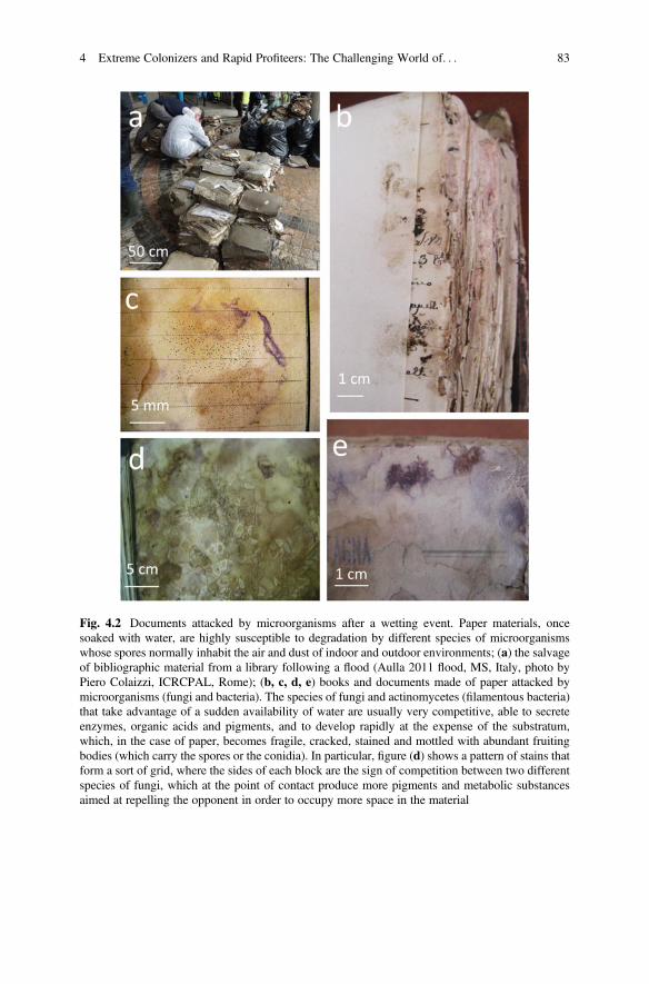
Fig. 4.2 Documents attacked by microorganisms after a wetting event. Paper materials, oncesoaked with water, are highly susceptible to degradation by different species of microorganismswhose spores normally inhabit the air and dust of indoor and outdoor environments; (a) the salvageof bibliographic material from a library following a flood (Aulla 2011 flood, MS, Italy, photo byPiero Colaizzi, ICRCPAL, Rome); (b, c, d, e) books and documents made of paper attacked bymicroorganisms (fungi and bacteria). The species of fungi and actinomycetes (filamentous bacteria)that take advantage of a sudden availability of water are usually very competitive, able to secreteenzymes, organic acids and pigments, and to develop rapidly at the expense of the substratum,which, in the case of paper, becomes fragile, cracked, stained and mottled with abundant fruitingbodies (which carry the spores or the conidia). In particular, figure (d) shows a pattern of stains thatform a sort of grid, where the sides of each block are the sign of competition between two differentspecies of fungi, which at the point of contact produce more pigments and metabolic substancesaimed at repelling the opponent in order to occupy more space in the material
4 Extreme Colonizers and Rapid Profiteers: The Challenging World of. . . 83
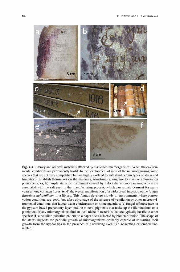
Fig. 4.3 Library and archival materials attacked by s-selected microorganisms. When the environ-mental conditions are permanently hostile to the development of most of the microorganisms, somespecies that are not very competitive but are highly evolved to withstand certain types of stress andlimitations, establish themselves on the materials, sometimes giving rise to massive colonizationphenomena; (a, b) purple stains on parchment caused by halophilic microorganisms, which areassociated with the salt used in the manufacturing process, which can remain dormant for manyyears among collagen fibres; (c, d) the typical manifestation of a widespread infection of the fungusEurotium halophilicum in a library. This fungus develops slowly in environments where conser-vation conditions are good, but takes advantage of the absence of ventilation or other microenvi-ronmental conditions that favour water condensation on some materials; (e) fungal efflorescence onthe gypsum-based preparatory layer and the mineral pigments that make up the illuminations on aparchment. Many microorganisms find an ideal niche in materials that are typically hostile to otherspecies; (f) a peculiar oxidation pattern on a paper sheet affected by biodeterioration. The shape ofthe stains suggests the periodic growth of microorganisms probably capable of re-starting theirgrowth from the hyphal tips in the presence of a recurring event (i.e. re-wetting or temperature-related)
84 F. Pinzari and B. Gutarowska
production of these colourants is often associated with the genera Aspergillus andPenicillium (Melo et al. 2019), which are also frequently found on stained paper.
1.2 Parchment
Parchment was the most commonly used writing material from the second centuryBC to the end of the Middle Ages, when it was substituted by paper in Europe. It is athin material made from the skin of animals such as sheep, goats, lambs, pigs andcalves. It differs from leather in that it is mainly limed rather than tanned. There wereconsiderable differences in parchment manufacturing methods in different countriesand historical periods. Even so, the preparation of parchment involved always thecleaning and mechanical removal of hair by scraping the flayed skin, followed bydesiccation of any remaining flesh using sodium or potassium chloride, adjustmentof the pH by treatment with ammonium chloride or sulphate with lime, and theapplication of potash alum (Larsen 2002). Parchment books, depending on their dateof manufacture and country of origin, were written using iron gall or carbonpigmented inks, or printed using resinous printing inks; they were frequentlyilluminated with a wide range of mineral and organic pigments, and embellishedwith metals such as gold or silver and their alloys. In maedieval times, egg white,natural oils and other materials were applied to the parchment to form a very smooth,highly polished surface for writing and illumination. Parchments were also used inthe making of official and private documents (e.g. contracts, wills, genealogies,maps, etc.), often collected in composite volumes bound together by sewing withthread or ribbons, and sometimes with adhesives. The main chemical component ofparchment is collagen, a natural biopolymer with a relative molecular mass of350 kDa. Collagen is formed from a triple helix with random coil telo-peptides.Degradation of parchment is a complex process which involves the oxidativechemical deterioration of the amino acid chains and hydrolytic cleavage of thepeptide backbone (Florian 2007).
Parchment is usually a very robust and durable material; it is particularly resistantto abrasion, tearing and other mechanical damage. Parchment is more resistant thanpaper to corrosive inks and acid degradation in general, owing to the alkaline agentsused in the manufacturing process, such as lime and chalk. However, parchment isextremely hygroscopic, which makes it highly susceptible to infection by bacteriaand moulds when exposed to elevated relative humidity.
Microorganisms with a proteolytic capacity include bacteria and fungi, even if theformer possess more specific collagenases capable of digesting the proteins of theparchment through hydrolysis (Figs. 4.1, 4.3). Fungi, on the other hand, developparticularly at the expense of oils and waxes added during the manufacturing process(to make the parchment more flexible), although some species of proteolytic fungican also utilize collagen as their sole carbon source (Florian 2002, 2007; Pinzariet al. 2012a, b; Lech 2016; Pinzari 2018).
4 Extreme Colonizers and Rapid Profiteers: The Challenging World of. . . 85
The microorganism species involved in the degradation of parchment can varyconsiderably with the chemical characteristics of the material, which can be verydifferent depending on the origin of the animal, the part of it used and themanufacturing processes employed. The presence of fats lends parchment a certaindegree of water repellency which largely speaking slows down biodeteriorationprocesses. Lipid substances are rather resistant to biological degradation but becomemore susceptible if they are found as water supported emulsions or in the presence ofadditional nutritional sources that can easily be metabolized, such as simple sugars(Florian 2002, 2007; Pinzari et al. 2012a, b; Lech 2016; Pinzari 2018).
It has been observed that biodegradation typically starts on the side of theparchment corresponding to the “flesh” side of the skin. In the more advanced stagesof degradation, the side bearing the grain (i.e. the “hair” side) is also attacked, wherethe collagen and elastin fibres form a denser network.
Initial studies on parchment deterioration indicated the Actinomycetes as thebacterial group that plays a major role in the deterioration of many kinds of historicaldocuments and books supported on parchment. Actinomycetes are known to pro-duce many types of enzyme, collagenases (proteases) in particular that are capable ofdestroying collagen through their hydrolytic activity (Blyskal et al. 2017). Manyspecies are alkaliphiles and therefore develop on parchments (prepared with chalk).However, some more recent studies (Piñar et al. 2015a, b; Migliore et al. 2017, 2019)have highlighted some rather complex mechanisms driving parchment biodegrada-tion, in which actinomycetes only represent some of the actors.
2 The Fungal and Bacterial Species That Attack Paperand Parchment
During their storage and utilization, paper and parchment can be colonized bynumerous microorganisms, especially fungi and bacteria, including actinomycetes.Examples of microorganisms isolated from paper and parchment are presented inTables 4.1 and 4.2.
In total, 85 fungal genera and 71 bacterial genera were identified in the book’sconstituent materials. Paper is a material that is susceptible to a larger number ofcolonizing microorganisms (80 genera of fungi and 59 genera of bacteria) thanparchment (29 genera of fungi, 23 genera of bacteria). The development of modernmolecular methods, chiefly NGS (next-generation sequencing), is greatly contribut-ing to the discovery of new genera of microorganisms that colonize these materials.Molecular methods have identified an additional 40 genera of fungi (out of the85 recognized so far) that inhabit paper but were not previously detected, since theycannot be cultured under laboratory conditions, and 58 genera of bacteria out of the71 known up to the present. In the case of parchment, thanks to the use of molecularmethods, an additional 15 genera of fungi and several taxa of unculturable bacteriahave been discovered as frequent colonizers and potential spoilers of the material. It
86 F. Pinzari and B. Gutarowska
Table 4.1 Book materials (paper and parchment) contaminated with fungi, identified by means ofculture-dependent and independent methods
Isolated from book materials identified by cultural dependenta and culture independentb methods
Genera of fungi Paper Parchment References
Acremonium a, b a, b Arai (2000), Borrego et al. (2012), El Bergadi et al.(2014), Florian and Manning (2000), Gutarowska(2016), Karakasidou et al. (2018), Karbowska-Berent et al. (2014), Kraková et al. (2012, 2018),Lech (2016), Michaelsen et al. (2009, 2010), Nolet al. (2001), Nunes et al. (2015), Oetari et al.(2016), Paiva de Carvalho et al. (2016), Piñar et al.(2015a, b), Principi et al. (2011), Rakotonirainyet al. (2007), Saada et al. (2018), Sato et al. (2014),Szczepanowska et al. (2014), Szulc et al. (2018),Tanney et al. (2015), Zotti et al. (2008, 2011), Zyska(1997)
Acrothecium a
Alternaria a, b a, b
Aspergillus a, b a, b
Aureobasidium a, b b
Beauveria b
Bjerkandera b
Blumeria b
Botryotrichum a
Botrytis a
Byssochlamys b
Capnobotryella b
Ceratocystis a
Chaetomium a, b a, b
Chalastospora a, b
Cladonia b
Chloridium a
Chrysosporium a
Cladobotryum a
Cladosporium a, b a, b
Coniosporium a b
Curvularia a
Diaporthe b
Diploöspora a, b
Doratomyces a
Engyodontium b
Epicoccum a, b a, b
Erysiphales b
Eurotium a, b b
Fusarium a a
Geomyces a
Geosmithia a
Geotrichum a
Gleotinia b
Gliocladium a
Graphiopsis b
Gymnoascus a
Haplographium a
Helicostylum a
(continued)
4 Extreme Colonizers and Rapid Profiteers: The Challenging World of. . . 87
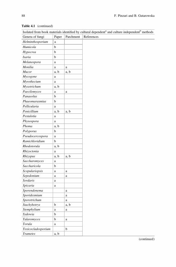
Table 4.1 (continued)
Isolated from book materials identified by cultural dependenta and culture independentb methods
Genera of fungi Paper Parchment References
Helminthosporium a
Humicola b
Hypocrea b
Isaria b
Melanospora a
Monilia a a
Mucor a, b a, b
Mycogone a
Myrothecium a
Myxotrichum a, b
Paecilomyces a a
Panaeolus b
Phaeomarasmius b
Pellicularia a
Penicillium a, b a, b
Pestalotia a
Physospora a
Phoma a, b
Polyporus b
Pseudocercospora a
Ramichloridium b
Rhodotorula a, b
Rhizoctonia a
Rhizopus a, b a, b
Saccharomyces a
Saccharicola b
Scopulariopsis a a
Sepedonium a a
Sordaris a
Spicaria a
Sporendonema a
Sporidesmium a
Sporotrichum a
Stachybotrys b a, b
Stemphylium a a
Sydowia b
Talaromyces b a
Torula a
Toxicocladosporium b
Trametes a, b
(continued)
88 F. Pinzari and B. Gutarowska
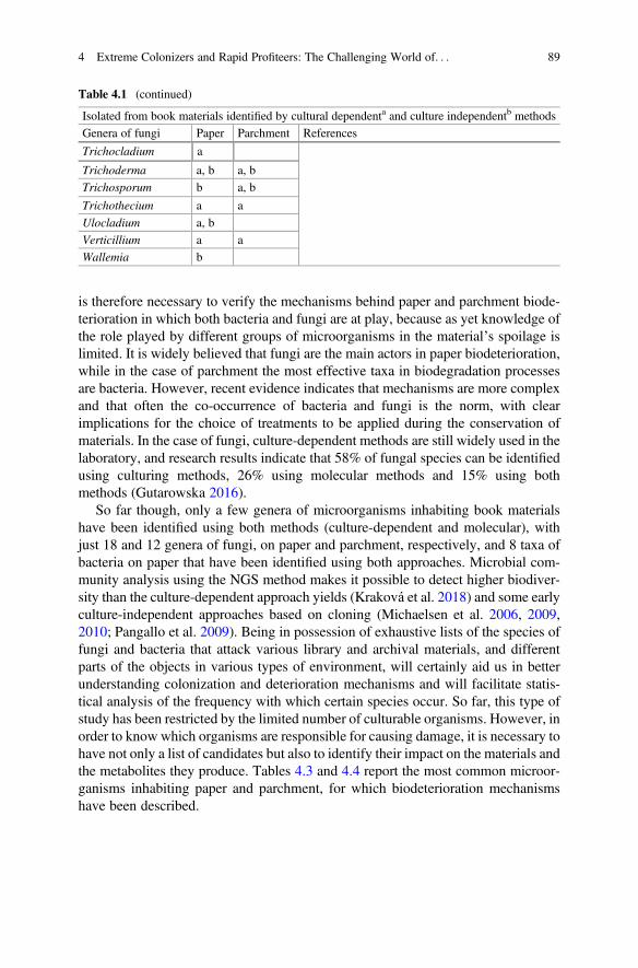
is therefore necessary to verify the mechanisms behind paper and parchment biode-terioration in which both bacteria and fungi are at play, because as yet knowledge ofthe role played by different groups of microorganisms in the material’s spoilage islimited. It is widely believed that fungi are the main actors in paper biodeterioration,while in the case of parchment the most effective taxa in biodegradation processesare bacteria. However, recent evidence indicates that mechanisms are more complexand that often the co-occurrence of bacteria and fungi is the norm, with clearimplications for the choice of treatments to be applied during the conservation ofmaterials. In the case of fungi, culture-dependent methods are still widely used in thelaboratory, and research results indicate that 58% of fungal species can be identifiedusing culturing methods, 26% using molecular methods and 15% using bothmethods (Gutarowska 2016).
So far though, only a few genera of microorganisms inhabiting book materialshave been identified using both methods (culture-dependent and molecular), withjust 18 and 12 genera of fungi, on paper and parchment, respectively, and 8 taxa ofbacteria on paper that have been identified using both approaches. Microbial com-munity analysis using the NGS method makes it possible to detect higher biodiver-sity than the culture-dependent approach yields (Kraková et al. 2018) and some earlyculture-independent approaches based on cloning (Michaelsen et al. 2006, 2009,2010; Pangallo et al. 2009). Being in possession of exhaustive lists of the species offungi and bacteria that attack various library and archival materials, and differentparts of the objects in various types of environment, will certainly aid us in betterunderstanding colonization and deterioration mechanisms and will facilitate statis-tical analysis of the frequency with which certain species occur. So far, this type ofstudy has been restricted by the limited number of culturable organisms. However, inorder to know which organisms are responsible for causing damage, it is necessary tohave not only a list of candidates but also to identify their impact on the materials andthe metabolites they produce. Tables 4.3 and 4.4 report the most common microor-ganisms inhabiting paper and parchment, for which biodeterioration mechanismshave been described.
Table 4.1 (continued)
Isolated from book materials identified by cultural dependenta and culture independentb methods
Genera of fungi Paper Parchment References
Trichocladium a
Trichoderma a, b a, b
Trichosporum b a, b
Trichothecium a a
Ulocladium a, b
Verticillium a a
Wallemia b
4 Extreme Colonizers and Rapid Profiteers: The Challenging World of. . . 89
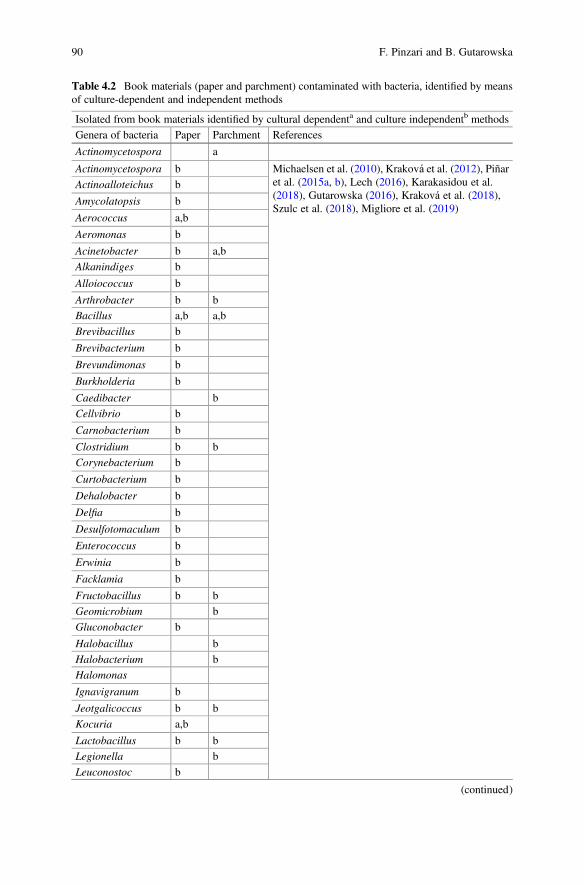
Table 4.2 Book materials (paper and parchment) contaminated with bacteria, identified by meansof culture-dependent and independent methods
Isolated from book materials identified by cultural dependenta and culture independentb methods
Genera of bacteria Paper Parchment References
Actinomycetospora a
Actinomycetospora b Michaelsen et al. (2010), Kraková et al. (2012), Piñaret al. (2015a, b), Lech (2016), Karakasidou et al.(2018), Gutarowska (2016), Kraková et al. (2018),Szulc et al. (2018), Migliore et al. (2019)
Actinoalloteichus b
Amycolatopsis b
Aerococcus a,b
Aeromonas b
Acinetobacter b a,b
Alkanindiges b
Alloiococcus b
Arthrobacter b b
Bacillus a,b a,b
Brevibacillus b
Brevibacterium b
Brevundimonas b
Burkholderia b
Caedibacter b
Cellvibrio b
Carnobacterium b
Clostridium b b
Corynebacterium b
Curtobacterium b
Dehalobacter b
Delfia b
Desulfotomaculum b
Enterococcus b
Erwinia b
Facklamia b
Fructobacillus b b
Geomicrobium b
Gluconobacter b
Halobacillus b
Halobacterium b
Halomonas
Ignavigranum b
Jeotgalicoccus b b
Kocuria a,b
Lactobacillus b b
Legionella b
Leuconostoc b
(continued)
90 F. Pinzari and B. Gutarowska
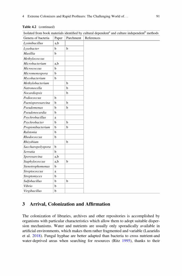
3 Arrival, Colonization and Affirmation
The colonization of libraries, archives and other repositories is accomplished byorganisms with particular characteristics which allow them to adopt suitable disper-sion mechanisms. Water and nutrients are usually only sporadically available inartificial environments, which makes them rather fragmented and variable (Lazaridiset al. 2018). Fungal hyphae are better adapted than bacteria to cross nutrient-andwater-deprived areas when searching for resources (Ritz 1995), thanks to their
Table 4.2 (continued)
Isolated from book materials identified by cultural dependenta and culture independentb methods
Genera of bacteria Paper Parchment References
Lysinibacillus a,b
Lysobacter b b
Masillia b
Methylococcus
Microbacterium a,b
Micrococcus b
Micromonospora b
Mycobacterium b
Methylobacterium b
Natronocella b
Nocardiopsis b
Pediococcus b
Paenisporosarcina b b
Pseudomonas b b
Pseudonocardia b
Psychrobacillus a
Psychrobacter b b
Propionibacterium b b
Ralstonia b
Rhodococcus b
Rhizobium b
Saccharopolyspora b
Serratia b
Sporosarcina a,b
Staphylococcus a,b b
Stenotrophomonas b
Streptococcus a
Streptomyces b
Sulfobacillus b b
Vibrio b
Virgibacillus b
4 Extreme Colonizers and Rapid Profiteers: The Challenging World of. . . 91
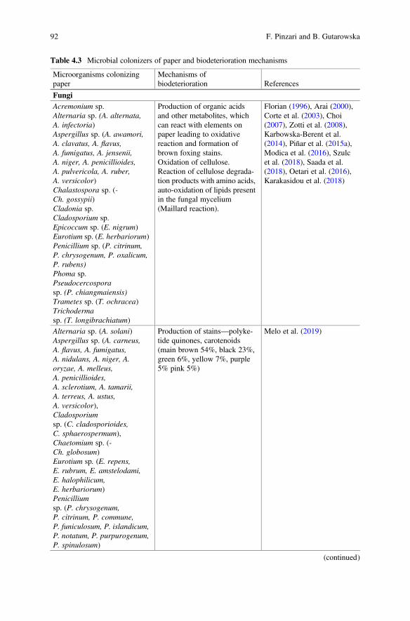
Table 4.3 Microbial colonizers of paper and biodeterioration mechanisms
Microorganisms colonizingpaper
Mechanisms ofbiodeterioration References
FungiAcremonium sp.Alternaria sp. (A. alternata,A. infectoria)Aspergillus sp. (A. awamori,A. clavatus, A. flavus,A. fumigatus, A. jensenii,A. niger, A. penicillioides,A. pulvericola, A. ruber,A. versicolor)Chalastospora sp. (-Ch. gossypii)Cladonia sp.Cladosporium sp.Epicoccum sp. (E. nigrum)Eurotium sp. (E. herbariorum)Penicillium sp. (P. citrinum,P. chrysogenum, P. oxalicum,P. rubens)Phoma sp.Pseudocercosporasp. (P. chiangmaiensis)Trametes sp. (T. ochracea)Trichodermasp. (T. longibrachiatum)
Production of organic acidsand other metabolites, whichcan react with elements onpaper leading to oxidativereaction and formation ofbrown foxing stains.Oxidation of cellulose.Reaction of cellulose degrada-tion products with amino acids,auto-oxidation of lipids presentin the fungal mycelium(Maillard reaction).
Florian (1996), Arai (2000),Corte et al. (2003), Choi(2007), Zotti et al. (2008),Karbowska-Berent et al.(2014), Piñar et al. (2015a),Modica et al. (2016), Szulcet al. (2018), Saada et al.(2018), Oetari et al. (2016),Karakasidou et al. (2018)
Alternaria sp. (A. solani)Aspergillus sp. (A. carneus,A. flavus, A. fumigatus,A. nidulans, A. niger, A.oryzae, A. melleus,A. penicillioides,A. sclerotium, A. tamarii,A. terreus, A. ustus,A. versicolor),Cladosporiumsp. (C. cladosporioides,C. sphaerospermum),Chaetomium sp. (-Ch. globosum)Eurotium sp. (E. repens,E. rubrum, E. amstelodami,E. halophilicum,E. herbariorum)Penicilliumsp. (P. chrysogenum,P. citrinum, P. commune,P. funiculosum, P. islandicum,P. notatum, P. purpurogenum,P. spinulosum)
Production of stains—polyke-tide quinones, carotenoids(main brown 54%, black 23%,green 6%, yellow 7%, purple5% pink 5%)
Melo et al. (2019)
(continued)
92 F. Pinzari and B. Gutarowska
ability to mechanically penetrate substrates and translocate nutrients. Active andpassive dispersal mechanisms can produce very different results in indoor environ-ments. Fungi and bacteria populate the airborne dust of indoor environments asresting spores or living propagules. These organisms can be introduced to buildingsby different sources or be generated from “amplification sites”. Damp walls, venti-lation systems, spoiled food or rotting materials can host bacterial or fungal biofilmsand act as rich sources of airborne contamination (Wang et al. 2016; Liu et al. 2018).The number of spores deriving from a contaminated material can vary considerablywith the biology and ecology of individual species, and can also be influenced bychance events. However, not all microbes have evolved for air dispersion; there areseveral other mechanisms which favour the microbial colonization of new “niches”.Some of the pests that attack organic materials in libraries and archives, such aswood, paper, leather and parchment, can feed on moulds or cohabit with them, andact as active or passive means of dispersal. Insect-mediated fungal dispersion hasbeen widely documented in natural environments (Jacobsen et al. 2018), but it alsooccurs in indoor environments. Some indoor species such as silverfish (familyLepismatidae), booklice (family Liposcelididae) and mites (i.e. familyPyroglyphidae, genus Dermatophagoides) are often directly correlated to themoulds infecting materials (Green and Farman 2015) in mutually beneficial circum-stances. In fact, the metabolic water produced by insects, their droppings and debrisconstitute an ideal substrate for fungal development (Pinzari and Montanari 2008,2011). Humans constitute another factor in the contamination of materials and thedispersion of microorganisms.
Traces of the human microbiome are typically present on materials and thanks tomolecular analysis this is becoming increasingly apparent, as the list of
Table 4.3 (continued)
Microorganisms colonizingpaper
Mechanisms ofbiodeterioration References
Trichoderma spp. (T. koningii,T. pseudokoningii,T. citrinoviride)Others
BacteriaRalstonia sp.Delfia sp.
Enzymatic degradation ofchemicals used in papermak-ing, production of colouredslimes, ability to produce dif-fusible brown pigments.
Väisänen et al. (1998), Szulcet al. (2018)
Pseudomonas sp. (P. stutzeri)Burkholderia sp. (B. cepacia)
Enzymatic degradation ofstarch, casein, carboxymethylcellulose, ability to producediffusible yellow pigments.
Szulc et al. (2018)
Bacillus sp.Brevibacillus sp.Lysinibacillus sp.
Enzymatic degradation ofstarch, casein, cellulose.
De Paolis and Lippi (2008),Szulc et al. (2018)
4 Extreme Colonizers and Rapid Profiteers: The Challenging World of. . . 93
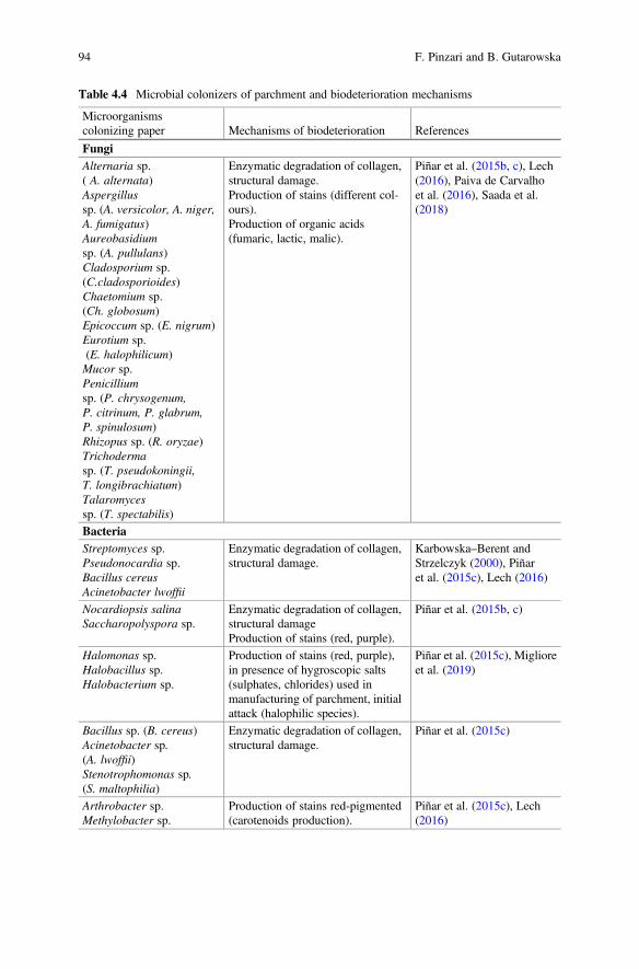
Table 4.4 Microbial colonizers of parchment and biodeterioration mechanisms
Microorganismscolonizing paper Mechanisms of biodeterioration References
FungiAlternaria sp.( A. alternata)Aspergillussp. (A. versicolor, A. niger,A. fumigatus)Aureobasidiumsp. (A. pullulans)Cladosporium sp.(C.cladosporioides)Chaetomium sp.(Ch. globosum)Epicoccum sp. (E. nigrum)Eurotium sp.(E. halophilicum)Mucor sp.Penicilliumsp. (P. chrysogenum,P. citrinum, P. glabrum,P. spinulosum)Rhizopus sp. (R. oryzae)Trichodermasp. (T. pseudokoningii,T. longibrachiatum)Talaromycessp. (T. spectabilis)
Enzymatic degradation of collagen,structural damage.Production of stains (different col-ours).Production of organic acids(fumaric, lactic, malic).
Piñar et al. (2015b, c), Lech(2016), Paiva de Carvalhoet al. (2016), Saada et al.(2018)
BacteriaStreptomyces sp.Pseudonocardia sp.Bacillus cereusAcinetobacter lwoffii
Enzymatic degradation of collagen,structural damage.
Karbowska–Berent andStrzelczyk (2000), Piñaret al. (2015c), Lech (2016)
Nocardiopsis salinaSaccharopolyspora sp.
Enzymatic degradation of collagen,structural damageProduction of stains (red, purple).
Piñar et al. (2015b, c)
Halomonas sp.Halobacillus sp.Halobacterium sp.
Production of stains (red, purple),in presence of hygroscopic salts(sulphates, chlorides) used inmanufacturing of parchment, initialattack (halophilic species).
Piñar et al. (2015c), Miglioreet al. (2019)
Bacillus sp. (B. cereus)Acinetobacter sp.(A. lwoffii)Stenotrophomonas sp.(S. maltophilia)
Enzymatic degradation of collagen,structural damage.
Piñar et al. (2015c)
Arthrobacter sp.Methylobacter sp.
Production of stains red-pigmented(carotenoids production).
Piñar et al. (2015c), Lech(2016)
94 F. Pinzari and B. Gutarowska
contaminants, even those that do not grow in culture, is growing longer and moreaccurate with the advent of new investigative techniques based on the extraction andsequencing of DNA.
The microorganisms that are identified in the air and on materials in libraries,archives and book repositories using massive sequencing methods include thosetypically found growing in indoor environments as well as those introduced fromoutdoors (Liao et al. 2004; Borrego et al. 2012; Nunes et al. 2013). Fungi that areobviously “outdoor” additions to the indoor environment often include ecto- andarbuscular-mycorrhizal genera that are obligate associates of host plants. The pres-ence of mammalian mycobionts such as Candida albicans, Malassezia spp. andbacterial species belonging to the group Propionibacterium/Cutibacterium showsthat humans contribute directly to the microbiome of indoor dust. Other bacteria ofcutaneous origin, belonging to the genera Corynebacterium and Staphylococcus(Ramsey et al. 2016), have often been detected on archival documents in the past(Puškárová et al. 2016; Kraková et al. 2012, 2018).
The main limiting factor that permits fungal development on paper and parch-ment is water. Spore germination only occurs when some water is available. From anecological standpoint, it is possible to imagine at least two different scenarios inwhich a microbiological attack on books and documents is possible (Figs. 4.2, 4.3,4.4): (1) a sudden abundance of water, such as in the case of flooding or pipe ruptureand (2) a situation in which a microclimate is established that encourages thedevelopment of particular organisms possessing the essential characteristics whichmake them eminently suited to occupying a particular ecological niche (Gu et al.2013; Sato et al. 2014). These are very different situations that can lead to conflictingbiodeterioration mechanisms as well as non-comparable effects on materials.
The microbial community that inhabits the indoor environment depends on thelevel of abiotic stress (e.g. low water availability, poor resources). The fungal andbacterial species that take advantage of a sudden availability of free water in a libraryare usually r-selected organisms capable of arriving, establishing themselves andpersisting until reproduction and dissemination have been accomplished (Boddy andHiscox 2016). Community change occurs when the initial colonizers are substitutedby organisms that are either better able to tolerate conditions within the resource, orcapable of using the resource modified by the initial colonizers and their residue.Competition for space and food may involve specialized species-specific interac-tions such as mycoparasitism, or be more generalized, but in any case will involvemetabolic changes, reactive oxygen species production, acidic compounds andantibiotics release into the immediate environment, with the result that communitydevelopment on materials is not a predictable and ordered sequence, but instead acomplex, ever-changing and rather haphazard process (Boddy and Hiscox 2016).
The fast-growing fungal species that develop on paper and other organic mate-rials stored in libraries and archives can generate strong odours (Pinzari andMontanari 2011; Micheluz et al. 2016), coloured stains (Melo et al. 2019) andtoxic compounds (Micheluz et al. 2016). Substrate composition and the availabilityof degradable carbon sources such as sugars and starch or mineral nutrients likenitrogen, phosphorus and potassium can be determining factors in the growth of the
4 Extreme Colonizers and Rapid Profiteers: The Challenging World of. . . 95
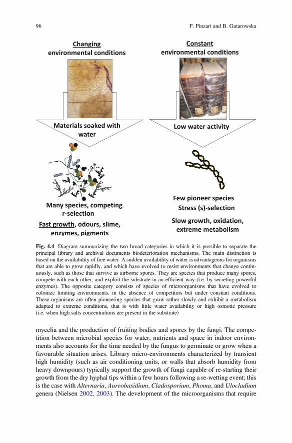
mycelia and the production of fruiting bodies and spores by the fungi. The compe-tition between microbial species for water, nutrients and space in indoor environ-ments also accounts for the time needed by the fungus to germinate or grow when afavourable situation arises. Library micro-environments characterized by transienthigh humidity (such as air conditioning units, or walls that absorb humidity fromheavy downpours) typically support the growth of fungi capable of re-starting theirgrowth from the dry hyphal tips within a few hours following a re-wetting event; thisis the case with Alternaria, Aureobasidium, Cladosporium, Phoma, and Ulocladiumgenera (Nielsen 2002, 2003). The development of the microorganisms that require
Fig. 4.4 Diagram summarizing the two broad categories in which it is possible to separate theprincipal library and archival documents biodeterioration mechanisms. The main distinction isbased on the availability of free water. A sudden availability of water is advantageous for organismsthat are able to grow rapidly, and which have evolved to resist environments that change contin-uously, such as those that survive as airborne spores. They are species that produce many spores,compete with each other, and exploit the substrate in an efficient way (i.e. by secreting powerfulenzymes). The opposite category consists of species of microorganisms that have evolved tocolonize limiting environments, in the absence of competitors but under constant conditions.These organisms are often pioneering species that grow rather slowly and exhibit a metabolismadapted to extreme conditions, that is with little water availability or high osmotic pressure(i.e. when high salts concentrations are present in the substrate)
96 F. Pinzari and B. Gutarowska
high water activity typically takes longer, subsequent to a sudden wetting event. Thegrowth of mycelium and production of spores directly in the materials after theyhave been soaked with water usually takes a couple of days and depends on theambient temperature. In fact, after flooding, librarians and conservators know thatwater-impregnated materials must be refrigerated or dried hastily so as to arrest thedevelopment of fungi and bacteria.
By contrast with what has been described so far for organisms capable of takingadvantage of randomly and suddenly favourable situations, it is also possible toencounter very different species living on book materials and in libraries. Themicroorganisms that establish themselves on materials when environmentalconditions are extreme, but constant, are typically slow-growing species that areparticularly resistant to stress (Fig. 4.4). These species are defined as “stress-toler-ant” or “s-selected”; they are specialized in occupying specific niches and theirparticular advantage is the ability to survive and disperse in a very hostile environ-ment. The “s-selected” fungal species that inhabit indoor environments can toleratedesiccation and grow in restrictive conditions (Boddy 2000).
4 Random Events, Common Denominators, ModelMechanisms
4.1 Actors and Mechanisms
The wide diffusion and frequency of some bacterial and fungal species as confirmedspoilers of archival and library materials have only recently begun to emerge thanksto an increase in the number of diagnostic investigations based on massive DNAsequencing techniques that extend beyond the limits of culture-dependent investi-gations. Over the last ten years or so, various articles have been published whichhave clarified some biodeterioration mechanisms and the ecology of particularspecies responsible for phenomena that, although widespread, were previouslyunknown (Montanari et al. 2012; Piñar et al. 2015a, b, c; Pinzari et al. 2012a,2018, Melo et al. 2019; Migliore et al. 2019).
Even if it is impossible to imagine perfectly repeatable patterns of materialbiodeterioration caused by the different groups of microorganisms, we can describerecurrent situations where phenomena can be parameterized, so as to model andforecast the risks posed by one or another biodeterioration mechanism, based on thepresence of specific environmental conditions or the assemblage of microbial species(Fig. 4.4).
The initial creation of two broad categories, as mentioned above, is based on theavailability of free water. If there is plenty of water available, such as in a sudden andunexpected event (a flood, for example), the microorganisms that attack the paper orthe parchment grow rapidly, produce pigmented stains and strong odours, abundantexopolymeric material, and exhibit significant enzymatic activity that results in the
4 Extreme Colonizers and Rapid Profiteers: The Challenging World of. . . 97
destruction of cellulose fibres in the case of paper and of collagen in the case ofparchment (Figs. 4.2, 4.3, 4.4). In this biodeterioration scenario the species, bothbacterial and fungal, can be numerous and multifarious. They are often the same asthose found in dust, although sometimes unusual saprophytes are identified that aretransported by chance on the materials and suddenly placed in a situation where theycan germinate and grow at the expense of large quantities of organic matter (Fig. 4.5)(Pinzari and Montanari 2008). During the slow drying of the sodden materials asuccession of organisms occurs that gradually take over from each other by takingadvantage of the partially degraded matter and the remains of the first settlers. Thisrepresents a natural succession mechanism that can produce very similar results,albeit depending (in each instance) on a different assortment of species, all of whichare capable of rapid growth, in addition to being endowed with enzymes andmetabolites diffusible in abundant water and different competition mechanisms(e.g. the production of antimicrobial compounds or volatile products with allopathicfunctions) (Pinzari et al. 2004; Canhoto et al. 2004; Sawoszczuk et al. 2015;Micheluz et al. 2015, 2016).
Cellulolytic and proteolytic enzymes cause structural damage by means of aprecisely targeted attack of the covalent chemical bonds of macromolecules(i.e. cellulose and collagen), whilst organic acids promote oxidation mechanismsand chelate microelements, such as calcium and iron (Fig. 4.1). Metals and salts canprecipitate as secondary compounds between the fibres, thereby increasing spatialheterogeneity, which in turn facilitates the coexistence of multiple species of spoil-ing microorganisms, each linked to a particular microenvironment and capable ofcausing a different type of damage to the material in question (Pinzari 2018).
Fig. 4.5 Diagram showing the different conditions in which microorganisms can be found in amaterial such as paper. Some can be active and grow, and produce pigments, whereas others can beviable but dormant; others still can be dead remains. Finally, it is possible to have many microor-ganisms represented by individual spores, deposited from the air, viable or otherwise. All theorganisms present in the various physiological states are detectable using massive DNA sequencinganalysis. In order to be able to distinguish among a long list of candidates that are responsible fordamage, it is necessary to add to the genetic analysis further diagnostic techniques, capable ofdistinguishing the viable species from the dead ones or to describe a causative mechanism behindthe damage
98 F. Pinzari and B. Gutarowska
Table 4.2 lists some of the mechanisms involved in paper and parchment biode-terioration caused by bacteria and fungi. Aspergillus and Penicillium fungal speciesare among those most frequently associated with the production of organic acids andother metabolites, which can react with various components of paper (Fig. 4.2). Infact, not only cellulose can be affected by oxidative reactions and enzymaticdegradation. The presence of starch, gelatin, rosin and other glues, or that of saltsand mineral compounds can exert an influence on fungal metabolism and determine,for example, the kind of stains produced (Fig. 4.2). The fungal pigments that affectpaper have been associated to polyketide quinones, carotenoids and other com-pounds whose synthesis or colour can depend on the availability of nitrogen, theprevailing pH and the presence of other limiting nutrients and enzyme cofactors suchas metals and cations (i.e. Fe, K, Ca, Mg, Mn) (Melo et al. 2019). However, in orderfor fungi to be able to produce abundant enzymes and synthesize complex metab-olites, there must be no limiting factors, and therefore water must be availabletogether with the organic matter.
When free water is scarce, the microbiological attack on the materials takes placedifferently (Figs. 4.3, 4.4). It is slower, and there are only a few or single extrem-ophile species that can germinate and grow. In such situations, generally linked tospecific microenvironmental conditions, the secretion of enzymes, organic acids andpigments also occurs, albeit to a lesser extent and with limited diffusion, and oftenproducing different effects depending on the species concerned. The appearance ofdamage on the material, alterations, and also identifying the presence of a biodete-rioration phenomenon in progress in such cases may not be immediate, but insteadoccur over a long period of time. While in the case of a flood the development ofmicroorganisms is a virtual inevitability, the outcome of other crises is far lesspredictable, owing to local phenomena, micro-environments or extremophile speciesthat involve mechanisms that are more complex and difficult to predict.
An example of slow-forming damage that gradually makes itself apparent isfoxing (Choi 2007; Nunes et al. 2015; Modica et al. 2019). The formation of foxingstains on paper and other materials can be caused by fungi as a result of limited wateravailability. Absolute tonophilic fungi germinate on paper in the presence of verylow water availability (aw < 0.80) and release cello-oligosaccharides, aminobutyricacid and amino acids. These compounds condensate in a spontaneous chemicalreaction (Maillard condensation) to form brown-coloured compounds known as“melanoidins” (Arai 1987, 2000; Arai et al. 1990), which in many cases areresponsible for foxing stains.
Pinzari (2018) described three types of biodeterioration mechanisms with increas-ing levels of complexity: (1) the damage is caused by the enzymatic activity ofmicroorganisms and is focused on the main chemical component of the material, asin the case of cellulolytic fungi that attack paper soaked with water; (2) the damage iscaused by the presence of peculiar conditions that trigger species with very particularrequirements, e.g. extremophilic species that grow at the presence of salts; (3) thedamage is the result of several related events such as the succession of species thatfeed on each other remains, with a primary colonizer which is often able todrastically modify the initial conditions of the material (as in mechanisms 1 and
4 Extreme Colonizers and Rapid Profiteers: The Challenging World of. . . 99
2). Understanding what causes damage, which species are involved, the resultingmetabolites and the chemical changes undergone by the materials are not a merespeculative exercise. The microorganisms’ ecology, their growth requirements andthe overall biodeterioration mechanism can all have a very significant effect on theoutcomes of the conservation treatments. Here we describe three examples where thebiodeterioration mechanisms have been examined in detail (Figs. 4.3, 4.6): (1) a rarefungal species, linked to the droppings of mites and the presence of salts, whichattacked the preparatory layer of an illuminated parchment and caused its detach-ment; (2) a fungus, a solitary colonizer with extremophile characteristics, whichfound its ideal niche in libraries; (3) a complex turnover of species that recurs inancient parchments all over the world, consistently causing the same type of damage(Figs 4.3 a, b).
4.2 A Rare Fungal Species
The halophilic fungus Diploöspora rosea (Tanney et al. 2015) was observed grow-ing copiously on a twentieth-century illuminated parchment (Fig. 4.6). The framedparchment leaf was on display in a museum, hung on a wall affected by rising damp.The fungus was documented on the thick preparative layer of gypsum and calciumcarbonate, and was seen to be capable of developing on the green pigments used forthe illumination. The pigments were prepared using chromium, iron and arsenicsalts. D. rosea grew not only in the presence of toxic metals but also showed theability to produce fruiting structures on very low water activity media, includingmalt extract agar amended with 60% sucrose. Despite the extreme xerophilic natureof this fungus, its structures caused the detachment of large parts of the artwork’spreparative layer and the overlying illumination. D. rosea is an onygenalean fungus,of uncertain taxonomic position, basal or sister to the Gymnoascaceae that seems tooccur in archival and domestic environments subject to periodic wetting. Thisfungus was previously identified in 1913, on damp cardboard covered with severalfungi. The dominant fungus was unknown and was described as the type species ofthe new hyphomycete genus Diploöspora. In the 100 years following its firstcollection, it was reported exclusively in association with illuminated parchmentsand in indoor settled dust in Micronesia. In the biodeteriorated parchment samples,D. rosea grew with other unidentified fungi and house mite exuvia and faecespellets. Mite faeces were also observed on the holotype (i.e. the 1913 cardboard).This hints at an insect/mite-mediated dispersal mechanism for this fungus. The highsalt and nitrogen concentrations that characterize mite droppings may represent aselective niche for osmophilic fungal species. The vast geographical distancebetween specimens suggested that D. rosea may have a broad distribution. Itsapparent rarity may reflect its xerophilic nature and slow growth rate rather thanits actual prevalence. However, its rarity in indoor settled dust samples was con-firmed by the detection of only two similar sequences using pyrosequencing. Thiscase study shows how some fungal and bacterial species, rarely found in nature, can
100 F. Pinzari and B. Gutarowska
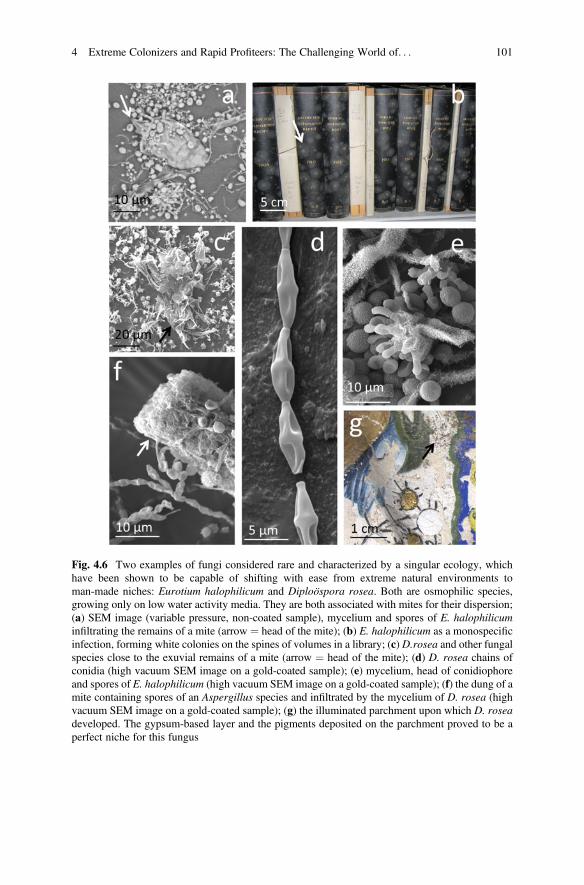
Fig. 4.6 Two examples of fungi considered rare and characterized by a singular ecology, whichhave been shown to be capable of shifting with ease from extreme natural environments toman-made niches: Eurotium halophilicum and Diploöspora rosea. Both are osmophilic species,growing only on low water activity media. They are both associated with mites for their dispersion;(a) SEM image (variable pressure, non-coated sample), mycelium and spores of E. halophilicuminfiltrating the remains of a mite (arrow ¼ head of the mite); (b) E. halophilicum as a monospecificinfection, forming white colonies on the spines of volumes in a library; (c) D.rosea and other fungalspecies close to the exuvial remains of a mite (arrow ¼ head of the mite); (d) D. rosea chains ofconidia (high vacuum SEM image on a gold-coated sample); (e) mycelium, head of conidiophoreand spores of E. halophilicum (high vacuum SEM image on a gold-coated sample); (f) the dung of amite containing spores of an Aspergillus species and infiltrated by the mycelium of D. rosea (highvacuum SEM image on a gold-coated sample); (g) the illuminated parchment upon which D. roseadeveloped. The gypsum-based layer and the pigments deposited on the parchment proved to be aperfect niche for this fungus
4 Extreme Colonizers and Rapid Profiteers: The Challenging World of. . . 101
find in the niches created by man their ideal environment where they can develop andproliferate, sometimes without competition. In the case of the D. rosea, however,several factors seem to have played a role, such as the possible repeated wettingevents suffered by the material, the presence of mites and their droppings, themineral nature of the substrate that allowed the fungus to thrive, despite being anosmophilic species, probably at the expense of the glues used to consolidate thepreparatory layer, subsequently causing the detachment of the illuminations.
4.3 A Solitary Colonizer
Occasionally, some fungi considered rare and of singular ecology, not only occupy aniche of the anthropic environment, but also find effective means of diffusion, andtherefore become particularly invasive. They are often intrusive species, capable ofshifting with alarming ease from extreme natural environments to man-made niches.A good example of this behaviour is provided by the spread of the fungus Eurotiumhalophilicum in Italian libraries and archives (Pinzari and Montanari 2011;Montanari et al. 2012) (Fig. 4.6). Eurotium halophilicum is a xerophilic fungus,with high tolerance to water stress; it was first isolated from dry food and indoor dustin association with other xerophilic fungal species and dust mites (Montanari et al.2012; Micheluz et al. 2015) (Fig. 4.6). Since the first reports of it as a bookcontaminant, it has been identified in dozens of libraries and archives in Italy asthe main, if not the sole fungus flourishing on books and archival materials (PinzariandMontanari 2011; Montanari et al. 2012; Micheluz et al. 2015; Polo et al. 2017). Ithas also been linked to the appearance of foxing-like stains on materials (Piñar et al.2015c; Sclocchi et al. 2016). The minimum water activity value observed for itssuccessful germination and growth is 0.675 (Christensen et al. 1959). It has repeat-edly been found in association with the covers of books and other surfaces withinmuseums, libraries and archives, even when the overall environmental conditions arein line with those recommended for the conservation of the materials (i.e. relativehumidity values ranging from 50 to 60%, and a temperature ranging from 20 to22�C). The niches preferred by this fungus are characterized by infrequent ventila-tion and water condensation events after a drop in temperature or night/day thermiccycles. The inadequacy of sampling procedures and the very slow growth of thefungus on most media probably account for previous underestimations. Anotherxerophilic fungus, Aspergillus penicillioides, has often been isolated from books inassociation with E. halophilicum, probably due to its similar ecology. Other fungihave frequently been isolated from E. halophilicum mycelium: Aspergillus creber,A. protuberus, Penicillium chrysogenum and P. brevicompactum (Micheluz et al.2015). These species grow on the dead mycelium of E. halophilicum. It has beenobserved that their presence is associated with historic infections of the fungus. Aslong as the mycelium is alive, the fungus remains the only organism on materials,and therefore represents the primary colonizer. Once established and mature, itsmycelium serves as a substrate for other xerophilic organisms. Dead structures of the
102 F. Pinzari and B. Gutarowska
fungus E. halophilicum have also been found in foxing spots on paper and othermaterials. Metal oxalates have often been documented in association with fungalhyphae (i.e. calcium oxalates) (Pinzari et al. 2010). These findings are consistentwith the hypothesis that absolute tonophilic fungi germinate on paper, releasingorganic acids, oligosaccharides and amino acids. The damage that this fungal speciescauses to materials consists of pale brown to dull grey stains and other forms ofdecolouration (Figs. 4.3c, 4.6). When decomposition occurs, it is limited to smallareas and consists of a sort of superficial erosion. This fungus also produces volatileorganic compounds (Micheluz et al. 2016) and represents a potential hazard forworkers who manage infected materials. GC-MS analysis showed thatE. halophilicum produces at least 20 different volatile compounds, with acetoneand 2-butanone being the main products. A total of eight secondary metabolites weredetected through LC/MS-MS, e.g. deoxybrevianamid E, neoechinulin A andtryprostatin B (Micheluz et al. 2015).
4.4 A Complex Turnover
In the two examples of the microbiological attack described above, the agentsresponsible for the colonization of the material and the main spoiling effects itresults in have been identified. The other organisms associated with them havebeen attributed to secondary phases that only played a marginal role in the mecha-nism. The situation in which all or most of the organisms present in the alterationsplay a role and are part of a complex mechanism whose phases are difficult toreconstruct is quite different. An example of a complex mechanism is described inthe works of Gallo and Strzelczyk (1971), Petushkova and Koestler (1996),Karbowska–Berent and Strzelczyk (2000), Piñar et al. (2015a, b, c), Migliore et al.(2017, 2019) (to cite only the main ones), where more than once and by means ofdifferent and progressively more sophisticated methods the cause of the formation ofpurple stains on parchment leaves was sought (Fig. 4.3a, b).
The parchment alteration that has long intrigued many experts consists of red orpurple nucleated maculae with peripheral halos, isolate or coalescent, often associ-ated with perforations and a loss of material which is more severe on the “flesh side”of a leaf. Karbowska–Berent and Strzelczyk (2000) isolated species of Streptomycesfrom the purple stains marring several documents of different ages and origins. Theyfound that many of the strains had proteolytic enzymes that are capable of destroyingcollagen through their hydrolytic activity and were alkaliphiles, hence able todevelop on skins prepared with lime and chalk (Figs. 4.1b, 4.3a, b). The isolatedbacteria, however, did not reproduce the purple stains when inoculated on modernparchment.
Piñar et al. (2015a) analysed five documents with different histories and origins,all marred by purple spots. Both scanning electron microscopy and molecularanalysis, based on culture-independent techniques (total DNA extraction, cloningand sequencing of bacterial 16S and fungal ITS rRNA genes), detected the presence
4 Extreme Colonizers and Rapid Profiteers: The Challenging World of. . . 103
of bacteria and fungi in the damaged areas. Halophilic, proteolytic bacterial specieswere found in all the documents. Moreover, as common microbial denominators,members of the Actinobacteria, mainly Saccharopolyspora spp. and species of thefungal genus Aspergillus, were detected in all investigated cases. Piñar et al. (2015a)proposed that a relationship exists between the phenomenon of purple spotsappearing on ancient parchments and that of so-called red heat, known to affectsome leather and animal skin products manufactured with marine salt and associatedwith the presence in the salt of extremophilic living bacteria.
Migliore et al. (2017) used 16S rRNA gene analysis in conjunction with454-pyrosequencing to study the purple spots on a parchment roll dated to 1244A.D. They hypothesized a two-phase model mechanism of parchment colonization,where halophilic Archaea colonize parchment as pioneers, followed byGammaproteobacteria, and fungi as the last colonizers. In a second study (Miglioreet al. 2019), the microbiomes of three parchment manuscripts affected by purplespots were analysed by means of next-generation sequencing (Illumina platform). Inthis instance, the authors attributed to Halobacterium salinarum the role of commondenominator in the purple stains, along with that of the primary, triggering cause ofthe phenomenon. They identified as haloarchaeal bacterioruberin and bacteriorho-dopsin the pigment within the purple spots using RAMAN spectroscopy. Miglioreet al. (2019) proposed a further multi-phase microbial succession model leading toprogressive degradation of the parchment’s collagen. In the suggested mechanism,the first phase “pioneer” colonizers are the halophilic and halotolerant microorgan-isms inhabiting the brines used to cure the skins. The second phase “late” colonizersconsist of other bacteria and fungi that can vary among different environments andaccording to the life history of each document. The halophilic and halotolerantmicrobes from the marine salt enter the hides during the manufacturing process,forming the core of the purple spot damage (Perini et al. 2019). They develop insidethe parchment and release proteolytic and lipolytic enzymes which attack anddegrade the collagen’s fibres. The formation of the nucleated purple stains wasexplained as the result of the lysis of halobacterial cells, with the subsequent releaseof bacteriorhodopsin and nutrients that can trigger colonization by other halotolerantorganisms of the proteobacterial taxa (mainly Gammaproteobacteria) andFirmicutes. In the model formulated by Migliore et al. (2019), both actinobacteriaand fungi are involved in the damage caused to skins because of their ability to attacknot only the collagen but other compounds too, although they only participate in thelast phases of the succession. Confirmation of a late role played by fungi in thecolonization of parchment in areas already affected by purple spots came fromdifferent studies. Pinzari et al. (2012a) quantified both the adenosine triphosphate(ATP) and the fungal β(1-4)-N-acetyl-D-glucosaminidase activity in damaged andundamaged samples, concluding that the viable fungal mycelium was present, albeitnot strictly associated with the purple stains. Additionally, Piñar et al. (2015b)quantified the β-actin gene through real-time polymerase chain reaction analyses(qPCR) in parchment samples affected by purple spots (taken from the ArchimedesPalimpsest). They identified a greater abundance of fungi on degraded areas incomparison to healthy ones. A broader statistic on the communities of
104 F. Pinzari and B. Gutarowska
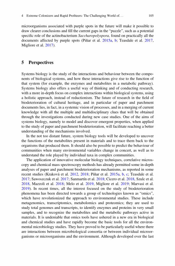
microorganisms associated with purple spots in the future will make it possible todraw clearer conclusions and fill the current gaps in the “puzzle”, such as a potentialspecific role of the actinobacterium Saccharopolyspora, found on practically all thedocuments affected by purple spots (Piñar et al. 2015a, b; Teasdale et al. 2017,Migliore et al. 2017).
5 Perspectives
Systems biology is the study of the interactions and behaviour between the compo-nents of biological systems, and how these interactions give rise to the function ofthat system (for example, the enzymes and metabolites in a metabolic pathway).Systems biology also offers a useful way of thinking and of conducting research,with a more in-depth focus on complex interactions within biological systems, usinga holistic approach, instead of reductionism. The future of research in the field ofbiodeterioration of cultural heritage, and in particular of paper and parchmentdocuments lies, in fact, in a systemic vision of processes, and in a merging of currentknowledge with all the multiple and multidisciplinary clues that will be obtainedthrough the investigations conducted during new case studies. One of the aims ofsystems biology, namely to model and discover emergent properties, when appliedto the study of paper and parchment biodeterioration, will facilitate reaching a betterunderstanding of the mechanisms involved.
In the not too distant future, system biology tools will be developed to uncoverthe functions of the metabolites present in materials and to trace them back to theorganisms that produced them. It should also be possible to predict the behaviour ofcommunities when many environmental variables change in concert, as well as tounderstand the role played by individual taxa in complex communities.
The application of innovative molecular biology techniques, correlative micros-copy and chemical mass spectroscopy methods has already permitted some in-depthanalyses of paper and parchment biodeterioration mechanisms, as reported in somerecent studies (Kraková et al. 2012, 2018; Piñar et al. 2015a, b, c; Teasdale et al.2017; Sawoszczuk et al. 2017; Sanmartín et al. 2018; Cicero et al. 2018, Szulc et al.2018, Mazzoli et al. 2018; Melo et al. 2019, Migliore et al. 2019; Marvasi et al.2019). In recent times, all the interest focused on the study of biodeteriorationphenomena has been directed towards a group of technologies known as “omics”,which have revolutionized the approach to environmental studies. These includemetagenomics, transcriptomics, metabolomics and proteomics; they are used tostudy total genomes and transcripts, to identify enzymes and proteins in very smallsamples, and to recognize the metabolites and the metabolic pathways active inmaterials. It is undeniable that omics tools have ushered in a new era in biologicaland chemical studies and have rapidly become the basic tools for all the environ-mental microbiology studies. They have proved to be particularly useful where thereare interactions between microbiological consortia or between individual microor-ganisms or microorganisms and the environment. Although developed over the last
4 Extreme Colonizers and Rapid Profiteers: The Challenging World of. . . 105
ten years or so, the application of omics to the study of cultural heritage and, inparticular, to archival documents and materials is only now beginning to gainground, as are the concepts of systemic biology. Indeed, very recently IlluminaMiSeq was used for massive DNA sequencing to analyse the microbiologicalbiodiversity in historical samples of paper and parchment. The technique wasapplied in order to better understand phenomena which up till now have remainedcontroversial. In fact, it was applied for the purpose of comparing brown foxingspots on paper and purple spots on parchment (Szulc et al. 2018, Migliore et al.2019). The study identified the contribution of new microorganisms (Phoma,Cladonia moulds and Gluconobacter, Ralstonia bacteria) to the foxing phenomenaobserved on 19th-century paper (Szulc et al. 2018); it also helped to establish themechanism underlying the microbial succession that occurs in purple stains onparchment, with haloarchaea establishing initially, followed by halotolerant bacteria,then actinobacteria, and finally fungi arriving at the end of the deterioration process(Migliore et al. 2019). In addition to massive DNA sequencing techniques,metabolomic techniques have been shown to provide useful elements for under-standing the mechanisms underlying the biodeterioration of materials (Fig. 4.5).Metabolomics encompasses a comprehensive qualitative and quantitative analysis ofsmall molecule substances with various properties that contribute to the metabolicpathways in the system under study. Metabolomics and proteomics provide infor-mation on biological mechanisms and potential biomarkers in samples. Recently,metabolomic analysis based on the AuNPET SALDI-ToF-MS method was appliedin the study of foxing stains marring some 19th-century papers. This techniqueenabled the authors (Szulc et al. 2018) to demonstrate the occurrence in the stainedareas of several metabolic pathways, including sugar degradation, amino acid andprotein metabolism, ubiquinone and other terpenoid-quinone biosynthesis,2-methyl-6-phytylquinol and delta-, gamma-, beta-tocopherols (responsible for theyellowish-brown colour of foxing spots) and 3-hydroxy-L-kynurenine(a fluorescent, yellow compound). These pigments can all contribute to the mecha-nism underlying the appearance of foxing caused by microorganisms (Szulc et al.2018).
Notwithstanding the considerable body of knowledge with respect to the meta-bolic processes occurring in organisms, the number of identified metabolites indatabases, particularly for microorganisms involved in cultural heritage deteriora-tion, remains limited. Having a complete database that also includes the metabolismof taxa of non-biomedical or food interest is a goal yearned for by all microbiologistsworking in the field of environmental metabolomics (Marvasi et al. 2019). Similarly,public databases that facilitate the alignment of diagnostic sequences for the identi-fication of fungi and bacteria are often lacking for taxa whose species are poorlystudied, of little interest, and troublesome to cultivate in vitro, and hence poorlyrepresented in living culture collections. This is a common situation for microor-ganisms that attack cultural heritage. Molecular ecology studies are very effective incapturing biodiversity, but the fungal and bacterial isolates conserved as livingcollections allow for studies of growth, morphology, secondary metabolism, geno-mics and other traits (Paiva de Carvalho et al. 2016). Without the existence of a
106 F. Pinzari and B. Gutarowska
living culture that makes it possible to couple morphological observations and DNAextraction, many molecular sequences are of little use since they often result in poormatches with sequences stored in public databases. This state of affairs underscoresthe importance of actively collecting and accessioning sequences of describedspecies that are currently unrepresented in sequence databases (new or unsequencedspecies). Populating databases, such as GenBank, with verified sequences obtainedfrom organisms isolated in cultural heritage biodeterioration studies will improvefuture amplicon-based metagenomics studies. In addition to meagre databases, theenvironmental sector and especially the cultural heritage sector suffer from a paucityof computational biology tools that would make it possible to combine genetic andchemical data, or informatics tools that would be of help in modelling biodeteriora-tion mechanisms, so as to be able to predict the processes before they happen(Sterflinger et al. 2018).
If we had adequate and specifically earmarked funds for research in the field ofbiodeterioration of cultural heritage, we could envisage the development, in the nottoo distant future, of bioinformatics tools capable of utilizing the masses of dataalready available and those that are rapidly being generated, so as to enable us toidentify unique chemical or genetic markers associated with the deterioration ofmaterials (e.g. early diagnostic systems that predict harmful mechanisms based on afew metabolomic or genomic clues).
The quantity of data obtained during omics analyses is enormous: a single,relatively simple experiment aimed at analysing a microbial metabolome generatestens of thousands of spectra. These data, as well as those obtained from studies ofmetagenomics and transcriptomics, which can be directly associated with the bio-deterioration mechanisms of materials and chemical data on the materials them-selves, should by rights already be collected within a single public database. Thiswould permit the development of appropriate tools for bioinformatics and networksanalysis, and therefore promote and stimulate further studies on the mechanisms andpreventative methods, as well as early diagnosis of cultural heritage biodeterioration.
Acknowledgements We would like to thank Mark Livesey for his helpful suggestions during thepreparation of the English text, and the anonymous reviewer for the meaningful comments.
References
Arai H (1987) Microbiological studies on the conservation of paper and related cultural properties.Part 5. Physiological and morphological characteristics of fungi isolated from foxing, formationmechanisms and countermeasures. Sci Conserv 26:43–52
Arai H (2000) Foxing caused by fungi: twenty–five years of study. Int Biodeter Biodegr46:181–188. https://doi.org/10.1016/S0964-8305(00)00063-9
Arai H, Matsumura N, Murakita H (1990) Microbiological studies on the conservation of paper andrelated cultural properties, part 9, induction of artificial foxing. Hozon Kagaku 29:25–34.(in Japanese)
4 Extreme Colonizers and Rapid Profiteers: The Challenging World of. . . 107
Ayu DC, Teja TP (2016) Occurrence of fungi on deteriorated old dluwang manuscripts fromIndonesia. Int Biodeter Biodegrad 114:94–103. https://doi.org/10.1016/j.ibiod.2016.05.025
Blyskal B, Lenart–Borod A, Borod P (2017) Approaches to taxonomic studies of actinomycetesisolated from historic and contemporary materials. J Pure Appl Microbiol 11:637–648
Boddy L (2000) Interspecific combative interactions between wood decaying basidiomycetes.FEMS Microbiol Ecol 31:185–194
Boddy L, Hiscox J (2016) Fungal ecology: principles and mechanisms of colonization andcompetition by saprotrophic fungi. Microbiol Spectrum 4: FUNK-0019-2016
Borrego S, Lavin P, Perdomo I, Gómez de Saravia S, Guiamet P (2012) Determination of indoor airquality in archives and biodeterioration of the documentary heritage. ISRN Microbiol2012:680598
Brunner I, Fischer M, Rüthi J, Stierli B, Frey B (2018) Ability of fungi isolated from plastic debrisfloating in the shoreline of a lake to degrade plastics. PLoS ONE 13:e0202047. https://doi.org/10.1371/journal.pone.0202047
Canhoto O, Pinzari F, Fanelli C, Magan N (2004) Application of electronic nose technology for thedetection of fungal contamination in library paper. Int Biodeter Biodegr 54:303–309. https://doi.org/10.1016/j.ibiod.2004.04.001
Choi S (2007) Foxing on paper: a literature review. J Am Inst Conserv 46:137–152. https://doi.org/10.1179/019713607806112378
Christensen C, Papavizas GC, Benjamin CR (1959) A new halophilic species of Eurotium.Mycologia 51:636–640
Cicero C, Pinzari F, Mercuri F (2018) 18th Century knowledge on microbial attacks on parchment:analytical and historical evidence. Int Biodeter Biodegr 134:76–82. https://doi.org/10.1016/j.ibiod.2018.08.007
Cordero RJB, Casadevall A (2017) Functions of fungal melanin beyond virulence. Fungal Biol Rev31:99–112
Coronado-Ruiz C, Avendaño R, Escudero–Leyva E, Conejo-Barboza G, Chaverri P, Chavarría M(2018) Two new cellulolytic fungal species isolated from a 19th–century art collection. Sci Rep8:1–9. https://doi.org/10.1038/s41598-018-24934-7
Corte AM, Ferroni A, Salvo VS (2003) Isolation of fungal species from test samples and mapsdamaged by foxing, and correlation between these species and the environment. Int BiodeteriorBiodegradation 51:167–173. https://doi.org/10.1016/S0964-8305(02)00137-3
De Paolis MR, Lippi D (2008) Use of metabolic and molecular methods for the identification of aBacillus strain isolated from paper affected by foxing. Microbiol Res 163:121–131. https://doi.org/10.1016/j.micres.2007.06.002
El Bergadi F, Laachari F, Elabed S, Mohammed IH, Ibnsouda SK (2014) Cellulolytic potential andfilter paper activity of fungi isolated from ancients manuscripts from the Medina of Fez. AnnMicrobiol 64:815–822. https://doi.org/10.1007/s13213-013-0718-6
Florian ML (1996) The role of the conidia of fungi in fox spots + rusty irregularly shaped areas onrag paper in 16th-century to 19th-century books. Stud Conserv 41:65–67. https://doi.org/10.2307/1506518
Florian M–LE, Manning L (2000) SEM analysis of irregular fungal spot in an 1854 book:population dynamics and species identification. Int Biodeterior Biodegrad 46:205–220
Florian M–LE (2002) Fungal facts: solving fungal problems in heritage collections. Archetypepublications. London
Florian M–LE (2007) Protein facts. Fibrous proteins in cultural and natural history artifacts.Archetype Publications, London
Gallo F, Strzelczyk A (1971) Indagine preliminare sulle alterazioni microbiche della pergamena.Boll Ist Patol Libro 30:71–87
Green PWC, Farman DI (2015) Can paper and glue alone sustain damaging populations ofbooklice, Liposcelis bostrychophila? J Conserv Museum Stud 13:3
108 F. Pinzari and B. Gutarowska
Gu JD, Kigawa R, Sato Y, Katayama Y (2013) Addressing the microbiological problems of culturalproperty and archive documents after earthquake and tsunami. Int Biodeter Biodegr85:345–346. https://doi.org/10.1016/j.ibiod.2013.08.018
Gutarowska B (2016) A modern approach to biodeterioration assessment and the disinfection ofhistorical book collections. Lodz University of Technology
Hyde KD, Xu J, Rapior S et al (2019) The amazing potential of fungi: 50 ways we can exploit fungiindustrially. Fungal Divers 97:1. https://doi.org/10.1007/s13225-019-00430-9
Jacobsen RM, Sverdrup–Thygeson A, Kauserud H, Birkemoe T (2018) Revealing hidden insect–fungus interactions; moderately specialized, modular and anti–nested detritivore networks. ProcR Soc B 285:20172833. https://doi.org/10.1098/rspb.2017.2833
Jayasekara S, Ratnayake R (2019) Microbial cellulases: an overview and applications. In:Rodríguez Pascual A, Eugenio Martín ME (eds) Cellulose. IntechOpen, London. https://doi.org/10.5772/intechopen.84531.
Karakasidou K, Nikolouli K, Amoutzias GD, Pournou A, Manassis C, Tsiamis G, Mossialos D(2018) Microbial diversity in biodeteriorated Greek historical documents dating back to the 19thand 20th century: a case study. Microbiol Open 7:e00596. https://doi.org/10.1002/mbo3.596
Karbowska–Berent J, Strzelczyk A (2000) The role of streptomycetes in the biodeterioration ofhistoric parchment. Nicolaus Copernicus University, Torun
Karbowska-Berent J, Jarmiłko J, Czuczko J (2014) Fungi in fox spots of a drawing by LeonWyczółkowski. Restaurator 35:159–179. https://doi.org/10.1515/res-2014-1000
Koul B, Upadhyay H (2018) Fungi–mediated biodeterioration of household materials, libraries,cultural heritage and its control. In: Gehlot P, Singh J (eds) Fungi and their role in sustainabledevelopment: current perspective. Springer, Singapore, pp 597–615
Kraková L, Chovanová K, Selim SA, Šimonovičová A, Puškarová A, Maková A, Pangallo D(2012) A multiphasic approach for investigation of the microbial diversity and itsbiodegradative abilities in historical paper and parchment documents. Int Biodeter Biodegr70:117–125. https://doi.org/10.1016/j.ibiod.2012.01.011
Kraková L, Šoltys K, Otlewska A, Pietrzak K, Purkrtová S, Savická D, Puškárová A, Bučková M,Szemes T, Budiš J, Demnerová K, Gutarowska B, Pangallo D (2018) Comparison of methodsfor identification of microbial communities in book collections: culture–dependent (sequencingand MALDI–TOF-MS) and culture–independent (Illumina MiSeq). Int Biodeter Biodegr131:51–59. https://doi.org/10.1016/j.ibiod.2017.02.015
Larsen R (2002) Microanalysis of parchment. Archetype Publications, LondonLax S, Cardona C, Zhao D, Winton VJ, Goodney G, Gao P, Gottel N, Hartmann EM, Henry C,
Thomas PM, Kelley ST, Stephens B, Gilbert JA (2019) Microbial and metabolic succession oncommon building materials under high humidity conditions. Nat Commun 10:1767. https://doi.org/10.1038/s41467-019-09764-z
Lazaridis M, Katsivela E, Kopanakis I, Raisi L, Mihalopoulos N, Panagiaris G (2018) Character-ization of airborne particulate matter and microbes inside cultural heritage collections. J CultHerit 30:136–146. https://doi.org/10.1016/j.culher.2017.09.018
Lech T (2016) Evaluation of a parchment document, the 13th century incorporation charter for thecity of Krakow, Poland, for microbial hazards. Appl Environ Microbiol 82:2620–2631. https://doi.org/10.1128/AEM.03851-15
Liao C-M, Luo W-C, Chen S-C, Chen J-W, Liang H-M (2004) Temporal/seasonal variations ofsize–dependent airborne fungi indoor/outdoor relationships for a wind–induced naturally ven-tilated airspace. Atmos Environ 38:4415–4419. https://doi.org/10.1016/j.atmosenv.2004.04.029
Liu Z, Zhang Y, Zhang F, Hu C, Liu G, Pan J (2018) Microbial community analyses of thedeteriorated storeroom objects in the Tianjin Museum using culture–independent and culture–dependent approaches. Front Microbiol 9:802. https://doi.org/10.3389/fmicb.2018.00802
Mallo AC, Nitiu DS, Elíades LA, Saparrat MCN (2017) Fungal degradation of cellulosic materialsused as support for cultural heritage. Int J Conserv Sci 8:619–632
4 Extreme Colonizers and Rapid Profiteers: The Challenging World of. . . 109
Marvasi M, Cavalieri D, Mastromei G, Casaccia A, Perito B (2019) Omics technologies for an in–depth investigation of biodeterioration of cultural heritage. Int Biodeter Biodegr 144:104736.https://doi.org/10.1016/j.ibiod.2019.104736
Mazzoli R, Giuffrida MG, Pessione E (2018) Back to the past: “find the guilty bug—microorgan-isms involved in the biodeterioration of archeological and historical artifacts”. Appl MicrobiolBiotechnol 102:6393–6407. https://doi.org/10.1007/s00253-018-9113-3
Melo D, Sequeira SO, Lopes JA, Macedo MF (2019) Stains versus colourants produced by fungicolonising paper cultural heritage: a review. J Cult Herit 35:161–182. https://doi.org/10.1016/j.culher.2018.05.013
Michaelsen A, Pinzari F, Ripka K, Lubitz W, Piñar G (2006) Application of molecular techniquesfor identification of fungal communities colonising paper material. Int Biodeter Biodegr58:133–141. https://doi.org/10.1016/j.ibiod.2006.06.019
Michaelsen A, Piñar G, Montanari M, Pinzari F (2009) Biodeterioration and restoration of a 16th–century book using a combination of conventional and molecular techniques: a case–study. IntBiodeter Biodegr 63:161–168. https://doi.org/10.1016/j.ibiod.2008.08.007
Michaelsen A, Piñar G, Pinzari F (2010) Molecular and microscopical investigation of the micro-flora inhabiting a deteriorated Italian manuscript dated from the 13th–century. Microb Ecol60:69–80. https://doi.org/10.1007/s00248-010-9667-9
Micheluz A, Manente S, Tigini V, Prigione V, Pinzari F, Ravagnan G, Varese GC (2015) Theextreme environment of a library: xerophilic fungi inhabiting indoor niches. Int BiodeterBiodegr 99:1–7. https://doi.org/10.1016/j.ibiod.2014.12.012
Micheluz A, Manente S, Rovea M, Slanzi D, Varese GC, Ravagnan G, Formenton G (2016)Detection of volatile metabolites of moulds isolated from a contaminated library. J MicrobiolMethods 128:34–41. https://doi.org/10.1016/j.mimet.2016.07.004
Migliore L, Thaller MC, Vendittozzi G, Mejia AY, Mercuri F, Orlanducci S, Rubechini A (2017)Purple spot damage dynamics investigated by an integrated approach on a 1244 A.D. parchmentroll from the secret Vatican archive. Sci Rep 7:9521. https://doi.org/10.1038/s41598-017-05398-7
Migliore L, Perini N, Mercuri F, Orlanducci S, Rubechini A, Thaller MC (2019) Three ancientdocuments solve the jigsaw of the parchment purple spot deterioration and validate the micro-bial succession model. Sci Rep 9:1623. https://doi.org/10.1038/s41598-018-37651-y
Modica AMB, Di Bella M, Alberghina MF, Brai MDF, Tranchina L (2016) Characterization offoxing stains in early twentieth century photographic and paper materials. Nat Prod Res 33:987–996. https://doi.org/10.1080/14786419.2016.1180600
Modica A, Bruno M, Di Bella M, Alberghina MF, Brai M, Fontana D, Tranchina L (2019)Characterization of foxing stains in early twentieth century photographic and paper materials.Nat Prod Res 33:987–996. https://doi.org/10.1080/14786419.2016.1180600
Montanari M, Melloni V, Pinzari F, Innocenti G (2012) Fungal biodeterioration of historical librarymaterials stored in compactus movable shelves. Int Biodeter Biodegr 75:83–88. https://doi.org/10.1016/j.ibiod.2012.03.011
Nielsen KF (2002) Mould growth on building materials. Secondary metabolites, mycotoxins andbiomarkers. PhD Thesis. BioCentrum–DTU, Technical University of Denmark.
Nielsen KF (2003) Review: Mycotoxin production by indoor molds. Fungal Genet Biol39:103–117
Nol L, Henis Y, Kenneth RG (2001) Biological factors of foxing in postage stamp paper(Reprinted). Int Biodeterior Biodegradation 48:98–104. https://doi.org/10.1016/S0964-8305(01)00072-5
Nosanchuk JD, Stark RE, Casadevall A (2015) Fungal Melanin: what do we know about structure?Front Microbiol 6:1463. https://doi.org/10.3389/fmicb.2015.01463
Nunes I, Mesquita N, Cabo Verde S, Leitao Bandeira AM, Carolino MM, Portugal A, Botelho ML(2013) Characterization of an airborne microbial community: a case study in the archive of theUniversity of Coimbra, Portugal. Int Biodeter Biodegr 79:36–41. https://doi.org/10.1016/j.ibiod.2013.01.013
110 F. Pinzari and B. Gutarowska
Nunes M, Relvas C, Figueira F, Campelo J, Candeias A, Caldeira AT, Ferreira T (2015) Analyticaland microbiological characterization of paper samples exhibiting foxing stains. MicroscMicroanal 21:63–77. https://doi.org/10.1017/S143192761500001X
Oetari A, Susetyo–Salim T, Sjamsuridzal W, Suherman EA, Monica M, Wongso R, Fitri R, NurlailiDG, Ayu DC, Teja TP (2016) Occurrence of fungi on deteriorated old dluwang manuscriptsfrom Indonesia. Int Biodeter Biodegr 114:94–103. https://doi.org/10.1016/j.ibiod.2016.05.025
Paiva de Carvalho H, Mesquita N, Trovão J, Peixoto da Silva J, Rosa B, Martins R, Bandeira AML,Portugal A (2016) Diversity of fungal species in ancient parchments collections of the archive ofthe University of Coimbra. Int Biodeter Biodegr 108:57–66. https://doi.org/10.1016/j.ibiod.2015.12.001
Pangallo D, Chovanova K, Simonovicova A, Ferianc P (2009) Investigation of microbica commu-nity isolated from indoor artworks and their environment: identification, biodegradative abili-ties, and DNA typing. Can J Microbiol 55:277–287. https://doi.org/10.1139/W08-136
Perini N, Mercuri F, Thaller MC, Orlanducci S, Castiello D, Talarico V, Migliore L (2019) Thestain of the original salt: red heats on chrome tanned leathers and purple spots on ancientparchments are two sides of the same ecological coin. Front Microbiol 10:2459. https://doi.org/10.3389/fmicb.2019.02459
Petushkova JP, Koestler R (1996) Biodeterioration studies on parchment and leather attacked bybacteria in the Commonwealth of Socialist States. In: Federici C, Munafò PF (eds) Internationalconference on conservation and restoration of archival and library materials. Erice, 22nd-29thApril 1996. G.P. Palumbo, Palermo, pp 195–211
Piñar G, Sterflinger K, Pinzari F (2015a) Unmasking the measles–like parchment discoloration:molecular and microanalytical approach. Environ Microbiol 17:427–443. https://doi.org/10.1111/1462-2920.12471
Piñar G, Sterflinger K, Ettenauer J, Quandt A, Pinzari F (2015b) A combined approach to assess themicrobial contamination of the Archimedes Palimpsest. Microb Ecol 69:118–134. https://doi.org/10.1007/s00248-014-0481-7
Piñar G, Tafer H, Sterflinger K, Pinzari F (2015c) Amid the possible causes of a very famousfoxing: molecular and microscopic insight into Leonardo da Vinci’s self–portrait. EnvironMicrobiol Rep 7:849–859. https://doi.org/10.1111/1758-2229.12313
Pinzari F (2011) Microbial ecology of indoor environments. The ecological and applied aspects ofmicrobial contamination in archives, libraries and conservation environments. In: Abdul-WahabSA (ed) Sick building syndrome in public buildings and workplaces. Springer, Berlin, pp153–178
Pinzari F (2018) Microbial processes involved in the deterioration of paper and parchment. In:Mitchell R, Clifford J (eds) Biodeterioration and preservation in art, archaeology and architec-ture. Archetype Publications, London, pp 33–56
Pinzari F, Montanari M (2008) A substrate utilisation pattern (SUP) method for evaluating thebiodeterioration potential of micro -flora affecting libraries and archival materials. In: Joice H,Townsend L, Toniolo F, Cappitelli F (eds) Conservation science. Archetype Publications,London, pp 236–241
Pinzari F, Montanari M (2011) Mould growth on library materials stored in compactus–typeshelving units. In: Abdul-Wahab SA (ed) Sick building syndrome in public buildings andworkplaces. Springer, Berlin, pp 193–206
Pinzari F, Canhoto O, Fanelli C, Magan N (2004) Electronic nose for the early detection of mouldsin libraries and archives. Indoor Built Environ 13:387–395. https://doi.org/10.1177/1420326X04046948
Pinzari F, Pasquariello G, De Mico A (2006) Biodeterioration of paper: a SEM study of fungalspoilage reproduced under controlled conditions. Macromol Symp 238:57–66. https://doi.org/10.1002/masy.200650609
Pinzari F, Zotti M, De Mico A, Calvini P (2010) Biodegradation of inorganic components in paperdocuments: formation of calcium oxalate crystals as a consequence of Aspergillus terreus Thomgrowth. Int Biodeter Biodegr 64:499–505. https://doi.org/10.1016/j.ibiod.2010.06.001
4 Extreme Colonizers and Rapid Profiteers: The Challenging World of. . . 111
Pinzari F, Troiano F, Piñar G, Sterflinger K, Montanari M (2011) The contribution of microbio-logical research in the field of book, paper and parchment conservation. In: Engel P, Schirò J,Larsen R, Moussakova E, Kecskeméti I (eds) New approaches to book and paper conservation–restoration. Verlag Berger, Horn/Wien, pp 575–594
Pinzari F, Cialei V, Piñar G (2012a) A case study of ancient parchment biodeterioration usingvariable pressure and high vacuum scanning electron microscopy. In: Meeks N, Cartwright C,Meek A, Mongiatti A (eds) Historical technology, materials and conservation: SEM andmicroanalysis. Archetype Publications, London, pp 93–99
Pinzari F, Colaizzi P, Maggi O, Persiani AM, Schütz R, Rabin I (2012b) Fungal bioleaching ofmineral components in a twentieth–century illuminated parchment. Anal Bioanal Chem402:1541–1550. https://doi.org/10.1007/s00216-011-5263-1
Polo A, Cappitelli F, Villa F, Pinzari F (2017) Biological invasion in the indoor environment: thespread of Eurotium halophilicum on library materials. Int Biodeter Biodegr 118:34–44. https://doi.org/10.1016/j.ibiod.2016.12.010
Principi P, Villa F, Sorlini C, Cappitelli F (2011) Molecular studies of microbial communitystructure on stained pages of Leonardo da Vinci’s Atlantic Codex. Microb Ecol 61:214–222.https://doi.org/10.1007/s00248-010-9741-3
Puškárová A, Bučková M, Habalová B, Kraková L, Maková A, Pangallo D (2016) Microbialcommunities affecting albumen photography heritage: a methodological survey. Sci Rep6:20810. https://doi.org/10.1038/srep20810
Ramsey MM, Freire MO, Gabrilska RA, Rumbaugh KP, Lemon KP (2016) Staphylococcus aureusshifts toward commensalism in response to Corynebacterium species. Front Microbiol 7:1230.https://doi.org/10.3389/fmicb.2016.01230
Rakotonirainy MS, Heude E, Lavedrine B (2007) Isolation and attempts of biomolecular charac-terization of fungal strains associated to foxing on a 19th century book. J Cult Herit 8:126–133.https://doi.org/10.1016/j.culher.2007.01.003
Ritz K (1995) Growth responses of some soil fungi to spatially heterogeneous nutrients. FEMSMicrobiol Ecol 16:269–280. https://doi.org/10.1016/0168-6496(94)00090-J
Saada NS, Abdel-Maksound G, Youssef AM, Abdel-Aziz MS (2018) The hydrolytic activities oftwo fungal species isolated from historical quranic parchment manuscript. J Soc Leath Tech Ch102:141–148
Sanmartín P, DeAraujo A, Vasanthakumar A (2018) Melding the old with the new: trends inmethods used to identify, monitor, and control microorganisms on cultural heritage materials.Microb Ecol 76:64–80. https://doi.org/10.1007/s00248-016-0770-4
Sato YY, Aoki M, Kigawa R (2014) Microbial deterioration of tsunami–affected paper–basedobjects: a case study. Int Biodeter Biodegr 88:142–149. https://doi.org/10.1016/j.ibiod.2013.12.007
Sawoszczuk T, Syguła–Cholewińska J, del Hoyo-Meléndez JM (2015) Optimization of headspacesolid phase microextraction for the analysis of microbial volatile organic compounds emitted byfungi: application to historical objects. J Chromatogr A 1409: 30-45. doi:https://doi.org/10.1016/j.chroma.2015.07.059
Sawoszczuk T, Syguła-Cholewińska J, del Hoyo-Meléndez JM (2017) Application of HS–SPME–GC–MSmethod for the detection of active moulds on historical parchment. Anal Bioanal Chem409:2297–2307. https://doi.org/10.1007/s00216-016-0173-x
Sclocchi MC, Kraková L, Pinzari F, Colaizzi P, Bicchieri M, Šaková N (2016) Microbial life anddeath in a foxing stain: a suggested mechanism of photographic prints defacement. Microb Ecol73:1–12. https://doi.org/10.1007/s00248-016-0913-7
Sterflinger K, Pinzari F (2012) The revenge of time: Fungal deterioration of cultural heritage withparticular reference to books, paper and parchment. Environ Microbiol 14:559–566. https://doi.org/10.1111/j.1462-2920.2011.02584.x
Sterflinger K, Little B, Piñar G, Pinzari F, de los Rios A, Gu JD (2018) Future directions andchallenges in biodeterioration research on historical materials and cultural properties. IntBiodeter Biodegr 129:10–12. https://doi.org/10.1016/j.ibiod.2017.12.007
112 F. Pinzari and B. Gutarowska
Szczepanowska H, Mathia TG, Belin P (2014) Morphology of fungal stains on paper characterizedwith multi-scale and multi-sensory surface metrology. Scanning 36:76–85. https://doi.org/10.1002/sca.21095
Szczepanowska HM, Jha D, Mathia TG (2015) Morphology and characterization of Dematiaceousfungi on a cellulose paper substrate using synchrotron X–ray microtomography, scanningelectron microscopy and confocal laser scanning microscopy in the context of cultural heritage.J Anal Atom Spectrom 30:651–657. https://doi.org/10.1039/c4ja00337c
Szulc J, Otlewska A, Ruman T, Kubiak K, Karbowska-Berent J, Kozielec T, Gutarowska B (2018)Analysis of paper foxing by newly available omics techniques. Int Biodeter Biodegr132:157–165. https://doi.org/10.1016/j.ibiod.2018.03.005
Tanney JB, Nguyen HDT, Pinzari F, Seifert KA (2015) A century later: rediscovery, culturing andphylogenetic analysis ofDiploöspora rosea, a rare onygenalean hyphomycete. Anton Leeuw IntJ G 108:1023–1035. https://doi.org/10.1007/s10482-015-0555-7
Teasdale MD, Fiddyment S, Vnouček J, Mattiangeli V, Speller C, Binois A, Carver M, Dand C,Newfield TP, Webb CC, Bradley DG, Collins MJ (2017) The York Gospels: a 1000–yearbiological palimpsest. R Soc Open Sci 4:170988. https://doi.org/10.1098/rsos.170988
Treseder KK, Lennon JT (2015) Fungal traits that drive ecosystem dynamics on land. MicrobiolMol Biol Rev 79:243–262. https://doi.org/10.1128/MMBR.00001-15
Vaisanen OM, Weber A, Bennasar A, Rainey FA, Busse HJ, Salkinoja-Salonen MS (1998)Microbial communities of printing paper machines. J Appl Microbiol 84:1069–1084
Wang XW, Houbraken J, Groenewald JZ, Meijer M, Andersen B, Nielsen KF, Crous PW, SamsonRA (2016) Diversity and taxonomy of Chaetomium and Chaetomium–like fungi from indoorenvironments. Stud Mycol 84:145–224. https://doi.org/10.1016/j.simyco.2016.11.005
Zotti M, Ferroni A, Calvini P (2008) Microfungal biodeterioration of historic paper: preliminaryFTIR and microfungal analyses. Int Biodeterior Biodegradation 62:186–194. https://doi.org/10.1016/j.ibiod.2008.01.005
Zotti M, Ferroni A, Calvini P (2011) Mycological and FTIR analysis of biotic foxing on papersubstrates. Int Biodeterior Biodegradation 65:569–578. https://doi.org/10.1016/j.ibiod.2010.01.011
Zyska B (1997) Fungi isolated from library materials: a review of the literature. Int BiodeterBiodegr 40:43–51. https://doi.org/10.1016/S0964-8305(97)00061-9
Open Access This chapter is licensed under the terms of the Creative Commons Attribution 4.0International License (http://creativecommons.org/licenses/by/4.0/), which permits use, sharing,adaptation, distribution and reproduction in any medium or format, as long as you give appropriatecredit to the original author(s) and the source, provide a link to the Creative Commons license andindicate if changes were made.
The images or other third party material in this chapter are included in the chapter's CreativeCommons license, unless indicated otherwise in a credit line to the material. If material is notincluded in the chapter's Creative Commons license and your intended use is not permitted bystatutory regulation or exceeds the permitted use, you will need to obtain permission directly fromthe copyright holder.
4 Extreme Colonizers and Rapid Profiteers: The Challenging World of. . . 113

Part IIGreen Methods Again Biodeterioration
Chapter 5Novel Antibiofilm Non-Biocidal Strategies
Francesca Cappitelli and Federica Villa
Abstract Subaerial biofilm (SAB) formation on cultural heritage objects is oftenconsidered an undesirable process in which microorganisms and their by-products,e.g., enzymes and pigments, cause damage or alteration to a surface. Since biofilmsare widespread phenomena, there has been a high demand for preventive and controlstrategies that resist their formation or reduce their negative effects once formed. Upto date, the main strategy to control biofilms has been the use of biocides. Because oftheir intrinsic properties, biocidal products can pose risks to humans, animals, andthe environment. In this chapter, the authors call “green” only those alternativestrategies to biocides able to prevent/control biofilms but that do not kill microor-ganisms, i.e., irrespective of the use of natural compounds. Here, we describe someof the methods that are most commonly used to test the effectiveness of antibiofilmcompounds with multiple-species biofilm model systems. A unified terminology andwell described protocols and guidelines are still required to compare and test theeffectiveness of traditional or novel compounds against biofilms retrieved on heri-tage surfaces.
Keywords Antibiofilm · Sublethal · Green alternatives · Prevention · Control · Labmodels
1 Biocides
Biofilms are highly structured communities of microbial cells that adhere to a surfaceand are embedded in extracellular polymeric substances (EPS) consisting of poly-saccharides, nucleic acids, proteins, and lipids. Biofilm is a natural and ubiquitousform of bacterial and fungal growth. The fact that EPS are commonly observed closeto their cells also suggests that lichens are biofilms (Banfield et al. 1999; De Los Rios
F. Cappitelli (*) · F. VillaDepartment of Food, Environmental and Nutritional Sciences, Università degli Studi di Milano,Milano, Italye-mail: [email protected]; [email protected]
© The Author(s) 2021E. Joseph (ed.), Microorganisms in the Deterioration and Preservation of CulturalHeritage, https://doi.org/10.1007/978-3-030-69411-1_5
117
et al. 2005; Grube and Berg 2009; Casano et al. 2015). The concept of the biofilmimplies a need to take into consideration the interactions established among micro-organisms, and of the microorganisms with the heritage substratum and the imme-diate environment, a view that overcomes the value-laden identification ofmicroorganisms and their activities as single taxon.
In the scientific literature, biofilms have been mainly investigated when growingon a solid surface and exposed to a liquid phase. Indeed, most studies have focusedon human infecting biofilms, typically at the solid–liquid interface of the inner bodyand in marine biofouling environments. Medical literature addressing the solid–airinterface is considerably less substantial and concerns (partially) only skin woundbiofilms. Despite many similarities between biofilms grown at a liquid–solid inter-face and at an air–solid interface, there are marked differences that have been notedonly relatively recently. In contrast to the solid–air interface, Bacillus subtilis formssessile pellicles at liquid–air and solid–liquid interfaces (Vlamakis et al. 2013). Inthis scenario, the work of cultural heritage microbiologists that mainly addressessessile life at solid–air interfaces is pivotal to study a biofilm aspect that is lessinvestigated.
Subaerial biofilm (SAB) formation on cultural heritage objects is often consid-ered an undesirable process in which microorganisms and their by-products, e.g.,enzymes and pigments, cause damage or alteration to a surface. Since biofilms arewidespread phenomena, there has been a high demand for preventive and controlstrategies that resist their formation or reduce their negative effects once formed. Upto date, the main strategy to control biofilms has been the use of biocides. Accordingto the European Regulation 528/2012, “biocidal product” means “any substance ormixture, in the form in which it is supplied to the user, consisting of, containing orgenerating one or more active substances, with the intention of destroying, deter-ring, rendering harmless, preventing the action of, or otherwise exerting a control-ling effect on, any harmful organism by any means other than mere physical ormechanical action” (Regulation (EU) No 528/2012 of the European Parliament andof the Council of 22May 2012). In the past, biocides have been freely used to controlbiofilms on cultural heritage surfaces. However, the use of biocides has been provento have major drawbacks: biocides harm non-target populations of the surroundingenvironment and they carry a risk of the development of resistance to themselves aswell as cross-resistance to antibiotics (Villa et al. 2020). Thus, because of theirintrinsic properties, biocidal products can pose risks to humans, animals, and theenvironment. As a result, the EU has set up strict rules and procedures to minimizethese risks and all biocidal products have to be authorized by a competent authoritybefore they are placed on the market (https://ec.europa.eu/health/biocides/overview_en).
Moreover, specifically in relation to conservation, before deciding whetherremoval of microbial colonization with biocides is actually beneficial, we shouldconsider the effects of this so-called biodeterioration in comparison with thephysico-chemical deterioration induced by the use of the biocidal agent. In outdoorconditions, once structures and objects have been cleaned, recolonization is inevi-table and may begin shortly after treatment. In many cases no well-defined
118 F. Cappitelli and F. Villa
differences in the microbial community between the untreated and the treatedsurfaces are seen as only a biofilm component is affected (Urzì et al. 2016). Inother cases, subsequent colonization might be more aggressive or disfiguring thanthe one eradicated (Martin-Sanchez et al. 2012).
In conservation as well as in all other fields, the new approach is to find moresustainable ways to use biocides for the removal of deleterious biofilms fromrelevant abiotic surfaces and, at the same time, to search for feasible soundalternatives.
2 What Do We Mean by Green Alternatives?
In this chapter we define green alternatives those able to prevent/control biofilmswithout affecting cell growth. One of the many advantages is that, if the antibiofilmeffect is not due to a biocidal effect, it is possible to prevent the generation ofmicrobial drug-resistance.
At first glance, physical techniques seem a good green alternative for the removalof biodeteriogens. These methods dislodge or cause the lysis of some microorgan-isms with the help of tools, such as brushes (Caneva et al. 1991; Borderie et al.2015). In contrast to biocides, the killing does not lead to resistance but the materialcan be discolored or damaged (López et al. 2010; Vujcic et al. 2019). Additionally,the effect is mainly on the surface, e.g., endolithic growth is generally not affected.Therefore, physical cleaning is not the focus of this chapter even if its use incombination with other methods discussed here can be considered.
Antibiofilm agents target various stages of biofilm growth, namely adhesion,maturation, and dispersion. Any action that inhibits adhesion is considered a pre-ventive approach, whereas any action that interferes with maturation and dispersionis included in a control strategy. Prevention can be operated with two strategies:either through the employment of specific substances in the repository environmentor by modifying the surface of the heritage object. For instance, essential oils in thevapor phase or blended with some solvent have the potential to be used asantibiofilm substances and can be considered an interesting alternative (Borregoet al. 2012).
From an ecological point of view, the selection of plants as source materials ofantimicrobial compounds is a good approach, since plants produce a wide range ofsecondary metabolites that naturally defend them against microorganisms. Silvaet al. (2016) reported a comprehensive list of plant metabolites that have beenproven effective against microorganisms and their products, including pigments.We can argue about whether this is the green alternative we need. “Natural,”including plant-derived compounds, is not a synonym of non-toxic ornon-biocidal. In the past, thymol vapor was used extensively by a number ofconservators in “thymol cabinets.” However, in respect to heritage materials thymolsoftens varnishes and resins, renders parchment brittle, degrades some paper sup-ports, watercolor binders, and iron gall ink, and, with respect to human health,
5 Novel Antibiofilm Non-Biocidal Strategies 119
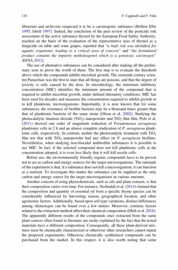
librarians and archivists suspected it to be a carcinogenic substance (Holben Ellis1995; Isbell 1997). Indeed, the conclusion of the peer review of the pesticide riskassessment of the active substance thymol by the European Food Safety Authority,reached on the basis of the evaluation of the representative uses of thymol as afungicide on table and wine grapes, reported that “a high risk was identified foraquatic organisms, leading to a critical area of concern” and “the formulatedproduct contains the impurity methyleugenol which is a genotoxic carcinogen”(EFSA 2012).
The use of alternative substances can be considered after making all the prelim-inary tests to prove the worth of them. The first step is to evaluate the thresholdabove which the compounds inhibit microbial growth. The sixteenth century scien-tist Paracelsus was the first to state that all things are poisons, and that the degree oftoxicity is only caused by the dose. In microbiology, the minimum inhibitoryconcentration (MIC) identifies the minimum amount of the compound that isrequired to inhibit microbial growth, under defined laboratory conditions. MIC hasbeen used for decades and measures the concentration required to inhibit growth orto kill planktonic microorganisms. Importantly, it is now known that for somesubstances, the resistance of biofilm bacteria may be a thousand times greater thanthat of planktonic bacteria of the same strain (Olson et al. 2002). Studying thephotocatalytic titanium dioxide (TiO2) nanopowder and TiO2 thin film, Polo et al.(2011) showed one order of magnitude reduction of Pseudomonas aeruginosaplanktonic cells in 2 h and an almost complete eradication of P. aeruginosa plank-tonic cells, respectively. In contrast, neither the photocatalytic treatment with TiO2
film nor that with TiO2 nanopowder had any effect on P. aeruginosa biofilms.Nevertheless, when studying non-biocidal antibiofilm substances it is possible touse MIC. In fact, if the selected compound does not kill planktonic cells at theconcentration adopted, it is even less likely that it will kill sessile cells.
Before use, the environmentally friendly organic compounds have to be provennot to act as carbon and energy sources for the target microorganisms. The rationaleof the experiment is that, if a substance does not kill a microorganism, it can functionas a nutrient. To investigate this matter the substance can be supplied as the onlycarbon and energy source for the target microorganisms in various amounts.
Another concern of using phytochemicals, such as oils and plant extracts, is thattheir composition varies over time. For instance, Nezhadali et al. (2014) claimed thatthe composition and quantity of essential oil from a specific thyme species can beconsiderably influenced by harvesting season, geographical location, and otheragronomic factors. Additionally, based upon soil type variations, distinct differencesamong chemotypes can be found over a few meters. Moreover, extrinsic factorsrelated to the extraction method affect their chemical composition (Dhifi et al. 2016).The apparently different results of the compounds once extracted from the sameplant sources often found in literature are easily explained by the fact that the testedmaterials have a different composition. Consequently, all these plant-derived mix-tures must be chemically characterized or otherwise other researchers cannot repeatthe proposed experiments. Otherwise chemically synthetized compounds can bepurchased from the market. In this respect, it is also worth noting that some
120 F. Cappitelli and F. Villa
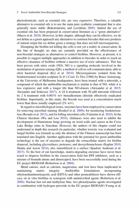
phytochemicals, such as essential oils, are very expensive. Therefore, a valuablealternative to essential oils is to use the main pure synthetic counterpart that is alsogenerally more stable (Rakotonirainy and Lavédrine 2005). Indeed the use ofessential oils has been proposed in conservation literature as a “green alternative”(Macro et al. 2018). However, in this chapter, although they can be effective, we donot define as a green approach any alternative to common biocides that uses productsof natural origin but we define green a strategy that does not kill microorganisms.
Disrupting the biofilm not killing the cells is not yet a reality in conservation. Inthis line of thought, no data are currently provided on the effectiveness ofnon-biocidal strategies as alternatives to control biofilms. However, at present, it ispossible to suggest multiple approaches in addition to biocides in order to reach aneffective clearance of biofilms without a massive use of toxic substances. This hasbeen proven with nitric oxide (NO). NO is a signaling molecule involved in themodulation of quorum sensing (QS), a method of cell-to-cell communication, able toelicit bacterial dispersal (Kyi et al. 2014). Microorganisms isolated from thebiodeteriorated wooden sculpture So It’s Come To This (1986) by Bruce Armstrong,at the University of Melbourne headquarters, have been treated with a nitroxide, acompound of which the antibiofilm mechanism is similar to that of NO, while it isless expensive and with a longer life than NO-donors (Alexander et al. 2015;Alexander and Schiesser 2017). A 24 h treatment with 50 μM nitroxide followedby 2 h treatment with 0.001% w/v benzalkonium chloride effectively eradicatedbiofilms. Importantly, in this study, the biocide was used at a concentration muchlower than those usually employed (2% w/v).
In regard to microbiological issues, enzymes have been employed in conservationfor removing microbial staining (Konkol et al. 2009), for monitoring biodeteriora-tion (Rosado et al. 2013), and for killing microbial cells (Valentini et al. 2010). In theChinese literature (Wu and Lou 2016), chitinases were also used to inhibit thedevelopment of filamentous fungi growing on word walls and canoes at the CrossLake Bridge ruins in Xiaoshan. However, the authors of this chapter could notunderstand in depth this research (in particular, whether toxicity was evaluated andfungal biofilm was formed) as only the abstract of the Chinese manuscript has beentranslated into English. Another application with the potential for green antibiofilmtechnology is the use of enzymes to degrade the extracellular matrix for biofilmdispersal, including glycosidases, proteases, and deoxyribonucleases (Kaplan 2010;Shadia and Aeron 2014), also immobilized to a surface (Spadoni Andreani et al.2017). To the best of our knowledge, matrix-degrading enzymes have never beentested to disperse biofilms in the conservation field but EPS inhibitors, such as amixture of bismuth nitrate and dimercaprol, have been successfully used during theEU project BIODAM (Robertson et al. 2004).
Metal cations, such as calcium, magnesium, and iron have been implicated inmaintaining matrix integrity. Antibiofilm formulations incorporatingethylenediaminetetraacetic acid (EDTA) and other permeabilizers have shown effi-cacy on in vitro biofilms in synergism with antimicrobial agents (Robertson et al.2004). Nuclear fast red and methylene blue, two photodynamic agents investigatedin combination with hydrogen peroxide in the EU project BIODAM (Young et al.
5 Novel Antibiofilm Non-Biocidal Strategies 121
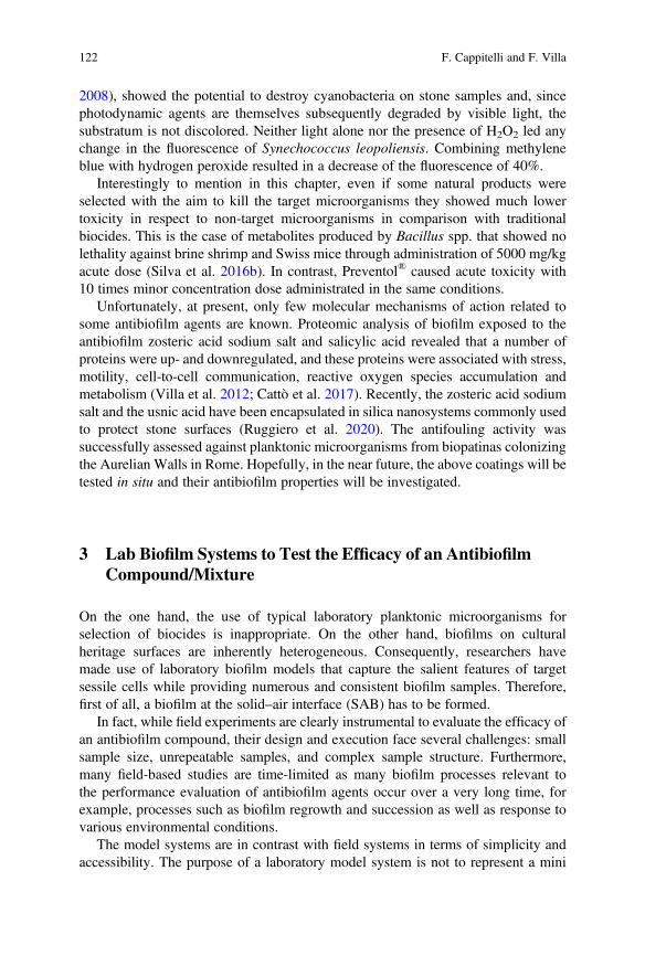
2008), showed the potential to destroy cyanobacteria on stone samples and, sincephotodynamic agents are themselves subsequently degraded by visible light, thesubstratum is not discolored. Neither light alone nor the presence of H2O2 led anychange in the fluorescence of Synechococcus leopoliensis. Combining methyleneblue with hydrogen peroxide resulted in a decrease of the fluorescence of 40%.
Interestingly to mention in this chapter, even if some natural products wereselected with the aim to kill the target microorganisms they showed much lowertoxicity in respect to non-target microorganisms in comparison with traditionalbiocides. This is the case of metabolites produced by Bacillus spp. that showed nolethality against brine shrimp and Swiss mice through administration of 5000 mg/kgacute dose (Silva et al. 2016b). In contrast, Preventol® caused acute toxicity with10 times minor concentration dose administrated in the same conditions.
Unfortunately, at present, only few molecular mechanisms of action related tosome antibiofilm agents are known. Proteomic analysis of biofilm exposed to theantibiofilm zosteric acid sodium salt and salicylic acid revealed that a number ofproteins were up- and downregulated, and these proteins were associated with stress,motility, cell-to-cell communication, reactive oxygen species accumulation andmetabolism (Villa et al. 2012; Cattò et al. 2017). Recently, the zosteric acid sodiumsalt and the usnic acid have been encapsulated in silica nanosystems commonly usedto protect stone surfaces (Ruggiero et al. 2020). The antifouling activity wassuccessfully assessed against planktonic microorganisms from biopatinas colonizingthe Aurelian Walls in Rome. Hopefully, in the near future, the above coatings will betested in situ and their antibiofilm properties will be investigated.
3 Lab Biofilm Systems to Test the Efficacy of an AntibiofilmCompound/Mixture
On the one hand, the use of typical laboratory planktonic microorganisms forselection of biocides is inappropriate. On the other hand, biofilms on culturalheritage surfaces are inherently heterogeneous. Consequently, researchers havemade use of laboratory biofilm models that capture the salient features of targetsessile cells while providing numerous and consistent biofilm samples. Therefore,first of all, a biofilm at the solid–air interface (SAB) has to be formed.
In fact, while field experiments are clearly instrumental to evaluate the efficacy ofan antibiofilm compound, their design and execution face several challenges: smallsample size, unrepeatable samples, and complex sample structure. Furthermore,many field-based studies are time-limited as many biofilm processes relevant tothe performance evaluation of antibiofilm agents occur over a very long time, forexample, processes such as biofilm regrowth and succession as well as response tovarious environmental conditions.
The model systems are in contrast with field systems in terms of simplicity andaccessibility. The purpose of a laboratory model system is not to represent a mini
122 F. Cappitelli and F. Villa
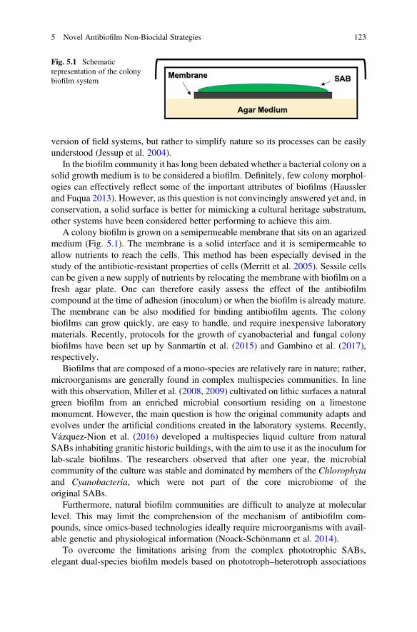
version of field systems, but rather to simplify nature so its processes can be easilyunderstood (Jessup et al. 2004).
In the biofilm community it has long been debated whether a bacterial colony on asolid growth medium is to be considered a biofilm. Definitely, few colony morphol-ogies can effectively reflect some of the important attributes of biofilms (Hausslerand Fuqua 2013). However, as this question is not convincingly answered yet and, inconservation, a solid surface is better for mimicking a cultural heritage substratum,other systems have been considered better performing to achieve this aim.
A colony biofilm is grown on a semipermeable membrane that sits on an agarizedmedium (Fig. 5.1). The membrane is a solid interface and it is semipermeable toallow nutrients to reach the cells. This method has been especially devised in thestudy of the antibiotic-resistant properties of cells (Merritt et al. 2005). Sessile cellscan be given a new supply of nutrients by relocating the membrane with biofilm on afresh agar plate. One can therefore easily assess the effect of the antibiofilmcompound at the time of adhesion (inoculum) or when the biofilm is already mature.The membrane can be also modified for binding antibiofilm agents. The colonybiofilms can grow quickly, are easy to handle, and require inexpensive laboratorymaterials. Recently, protocols for the growth of cyanobacterial and fungal colonybiofilms have been set up by Sanmartín et al. (2015) and Gambino et al. (2017),respectively.
Biofilms that are composed of a mono-species are relatively rare in nature; rather,microorganisms are generally found in complex multispecies communities. In linewith this observation, Miller et al. (2008, 2009) cultivated on lithic surfaces a naturalgreen biofilm from an enriched microbial consortium residing on a limestonemonument. However, the main question is how the original community adapts andevolves under the artificial conditions created in the laboratory systems. Recently,Vázquez-Nion et al. (2016) developed a multispecies liquid culture from naturalSABs inhabiting granitic historic buildings, with the aim to use it as the inoculum forlab-scale biofilms. The researchers observed that after one year, the microbialcommunity of the culture was stable and dominated by members of the Chlorophytaand Cyanobacteria, which were not part of the core microbiome of theoriginal SABs.
Furthermore, natural biofilm communities are difficult to analyze at molecularlevel. This may limit the comprehension of the mechanism of antibiofilm com-pounds, since omics-based technologies ideally require microorganisms with avail-able genetic and physiological information (Noack-Schönmann et al. 2014).
To overcome the limitations arising from the complex phototrophic SABs,elegant dual-species biofilm models based on phototroph–heterotroph associations
Fig. 5.1 Schematicrepresentation of the colonybiofilm system
5 Novel Antibiofilm Non-Biocidal Strategies 123
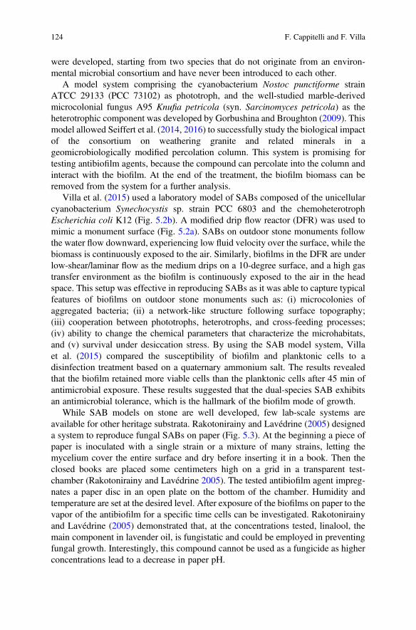
were developed, starting from two species that do not originate from an environ-mental microbial consortium and have never been introduced to each other.
A model system comprising the cyanobacterium Nostoc punctiforme strainATCC 29133 (PCC 73102) as phototroph, and the well-studied marble-derivedmicrocolonial fungus A95 Knufia petricola (syn. Sarcinomyces petricola) as theheterotrophic component was developed by Gorbushina and Broughton (2009). Thismodel allowed Seiffert et al. (2014, 2016) to successfully study the biological impactof the consortium on weathering granite and related minerals in ageomicrobiologically modified percolation column. This system is promising fortesting antibiofilm agents, because the compound can percolate into the column andinteract with the biofilm. At the end of the treatment, the biofilm biomass can beremoved from the system for a further analysis.
Villa et al. (2015) used a laboratory model of SABs composed of the unicellularcyanobacterium Synechocystis sp. strain PCC 6803 and the chemoheterotrophEscherichia coli K12 (Fig. 5.2b). A modified drip flow reactor (DFR) was used tomimic a monument surface (Fig. 5.2a). SABs on outdoor stone monuments followthe water flow downward, experiencing low fluid velocity over the surface, while thebiomass is continuously exposed to the air. Similarly, biofilms in the DFR are underlow-shear/laminar flow as the medium drips on a 10-degree surface, and a high gastransfer environment as the biofilm is continuously exposed to the air in the headspace. This setup was effective in reproducing SABs as it was able to capture typicalfeatures of biofilms on outdoor stone monuments such as: (i) microcolonies ofaggregated bacteria; (ii) a network-like structure following surface topography;(iii) cooperation between phototrophs, heterotrophs, and cross-feeding processes;(iv) ability to change the chemical parameters that characterize the microhabitats,and (v) survival under desiccation stress. By using the SAB model system, Villaet al. (2015) compared the susceptibility of biofilm and planktonic cells to adisinfection treatment based on a quaternary ammonium salt. The results revealedthat the biofilm retained more viable cells than the planktonic cells after 45 min ofantimicrobial exposure. These results suggested that the dual-species SAB exhibitsan antimicrobial tolerance, which is the hallmark of the biofilm mode of growth.
While SAB models on stone are well developed, few lab-scale systems areavailable for other heritage substrata. Rakotonirainy and Lavédrine (2005) designeda system to reproduce fungal SABs on paper (Fig. 5.3). At the beginning a piece ofpaper is inoculated with a single strain or a mixture of many strains, letting themycelium cover the entire surface and dry before inserting it in a book. Then theclosed books are placed some centimeters high on a grid in a transparent test-chamber (Rakotonirainy and Lavédrine 2005). The tested antibiofilm agent impreg-nates a paper disc in an open plate on the bottom of the chamber. Humidity andtemperature are set at the desired level. After exposure of the biofilms on paper to thevapor of the antibiofilm for a specific time cells can be investigated. Rakotonirainyand Lavédrine (2005) demonstrated that, at the concentrations tested, linalool, themain component in lavender oil, is fungistatic and could be employed in preventingfungal growth. Interestingly, this compound cannot be used as a fungicide as higherconcentrations lead to a decrease in paper pH.
124 F. Cappitelli and F. Villa
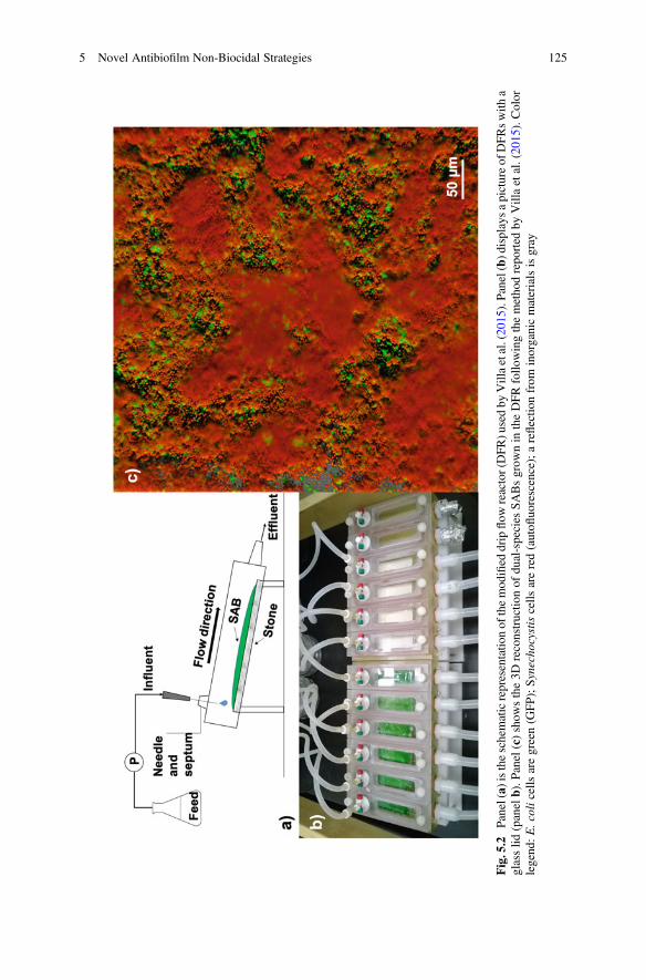
Fig.5.2
Panel(a)istheschematicrepresentatio
nof
themod
ified
dripflow
reactor(DFR)u
sedby
Villaetal.(20
15).Panel(b)d
isplaysapictureof
DFRswith
aglasslid
(panelb).P
anel(c)show
sthe3D
reconstructio
nof
dual-species
SABsgrow
nin
theDFRfollo
wingthemetho
drepo
rted
byVillaetal.(20
15).Color
legend
:E.colicells
aregreen(G
FP);Synecho
cystiscells
arered(autofl
uorescence);areflectio
nfrom
inorganicmaterialsisgray
5 Novel Antibiofilm Non-Biocidal Strategies 125
4 Methods for Testing the Effectiveness of AntibiofilmSubstances
Despite the vast use of traditional and novel antibiofilm agents in conservation ofcultural heritage, these practices are being increasingly questioned because of thelack of correlation between results obtained with conventional susceptibility tests atlaboratory-scale and those obtained from in-field applications. The reason for thisdiscrepancy is based on the use of planktonic cells for testing antibiofilm atlaboratory-scale rather than sessile cells. As previously mentioned, it is now wellknown that biofilms are�1000-fold more resistant than their planktonic counterparts(Hall and Mah 2017). Furthermore, the lack of simple reliable and standard assayshas remained a major limitation in selecting adequate antibiofilm treatments.
In view of the previous considerations, biofilm model systems are instrumentalfor testing the effectiveness of antibiofilm substances before an application in-field.Once the biofilm is grown, it can be exposed to different treatments and environ-mental conditions. Furthermore, biofilm model systems can be used to study biofilmregrowth when the treatment is concluded.
The antibiofilm agents can affect biofilm biomass, impair the biofilm matrix, oraffect biofilm thickness and morphology (e.g., inability of fungi to form filamentousstructures), while cells remain metabolically active. Thus, several methods can beapplied to evaluate the impacts of a compound against the biofilm. Here, we brieflydescribe some of the methods that are most commonly used to test the effectivenessof antibiofilm compounds.
4.1 Plate Count Assay
To be able to assess the effectiveness of antibiofilm compounds, it is essential todetermine the amount of viable cells in the biofilm, or the relative reduction in viable
Fig. 5.3 Schematic representation of the system designed by Rakotonirainy and Lavédrine (2005)to reproduce biofilms on paper
126 F. Cappitelli and F. Villa
cells after the treatment. The most widely used technique to determining bacterialviability is counting the number of colony forming units (CFU) after plating cultureson agar media. This method to assess biofilm viability requires the dislodgment ofthe biomass from the surface and the disaggregation of cell clusters due to thepresence of the biofilm matrix. Procedures such as vortexing, sonication, ormatrix-dissolving enzymes can be applied to separate bacterial cells from the matrix.However, these procedures may affect bacterial viability, leading to significant errorsin the quantification of the biomass (Azeredo et al. 2017). Furthermore, many strainscannot be cultured, as well as metabolically inactive cells (such as dormant cells andspores) commonly present within the biofilm (Sebestyen et al. 2016). Finally, whileCFU counting can be a valuable tool for quantifying a bacterial biofilm, it is notsuitable for fungal biofilms that develop filamentous structures and spores.
4.2 XTT Cell Viability Assay
The tetrazolium salt assay (XTT assay) is another useful technique for assessingthe viability of biofilm cells in vitro, as well as their susceptibility to antimicrobials.The number of viable biofilm cells can be deduced by measuring the absorbance ofthe supernatant after the metabolic reduction of XTT into the water-soluble formazan(Pantanella et al. 2013). Since the formazan product is water soluble and easilymeasured in cellular supernatants, the XTT assay allows the investigation of biofilmagent susceptibility without disruption of the biofilm structure (Kuhn et al. 2003).The main limitation of this method is related to the biofilm heterogeneity withgradients of substrata and products, which lead to cells with different metabolicstates. Furthermore, the biofilm matrix might slow down the reduction of XTT orpartially retain and release the formazan.
4.3 ATP-Bioluminescence Assay
The bioluminescence assay, based on the use of the North American firefly lucifer-ase, sees the conversion of intracellular ATP, the main energy carrier in cells of allliving organisms, into light. The light generated is recorded by a luminometer andquantified in relative light units (Nante et al. 2017). Intracellular ATP content is agood indicator of cell viability. In fact, upon cell death, ATP synthesis is immedi-ately arrested, while its hydrolysis can continue for some time. The main drawbackof this method is related to the variation in ATP production among diverse microbialtaxa (Ivanova et al. 2006).
5 Novel Antibiofilm Non-Biocidal Strategies 127
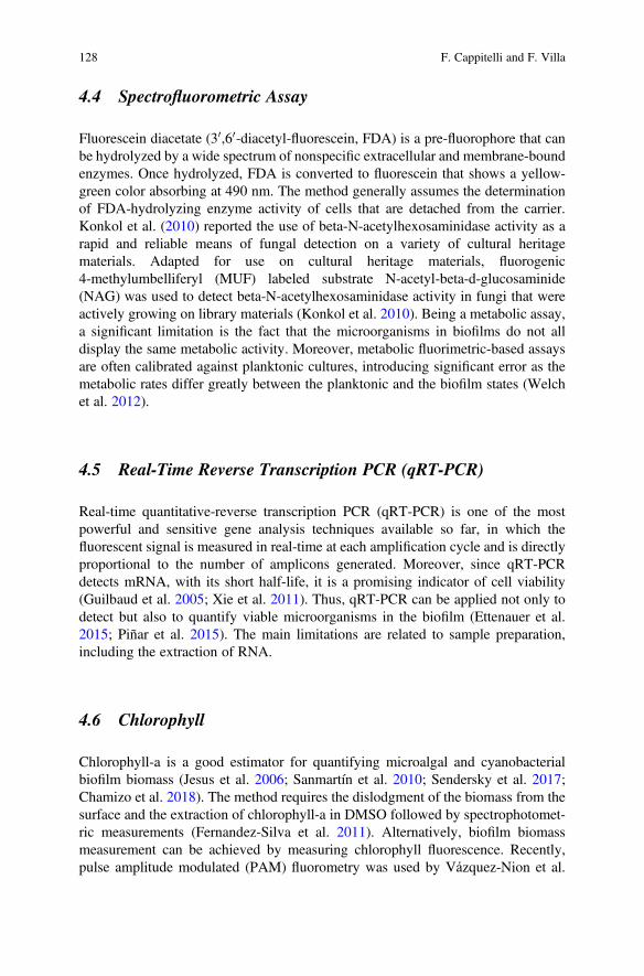
4.4 Spectrofluorometric Assay
Fluorescein diacetate (30,60-diacetyl-fluorescein, FDA) is a pre-fluorophore that canbe hydrolyzed by a wide spectrum of nonspecific extracellular and membrane-boundenzymes. Once hydrolyzed, FDA is converted to fluorescein that shows a yellow-green color absorbing at 490 nm. The method generally assumes the determinationof FDA-hydrolyzing enzyme activity of cells that are detached from the carrier.Konkol et al. (2010) reported the use of beta-N-acetylhexosaminidase activity as arapid and reliable means of fungal detection on a variety of cultural heritagematerials. Adapted for use on cultural heritage materials, fluorogenic4-methylumbelliferyl (MUF) labeled substrate N-acetyl-beta-d-glucosaminide(NAG) was used to detect beta-N-acetylhexosaminidase activity in fungi that wereactively growing on library materials (Konkol et al. 2010). Being a metabolic assay,a significant limitation is the fact that the microorganisms in biofilms do not alldisplay the same metabolic activity. Moreover, metabolic fluorimetric-based assaysare often calibrated against planktonic cultures, introducing significant error as themetabolic rates differ greatly between the planktonic and the biofilm states (Welchet al. 2012).
4.5 Real-Time Reverse Transcription PCR (qRT-PCR)
Real-time quantitative-reverse transcription PCR (qRT-PCR) is one of the mostpowerful and sensitive gene analysis techniques available so far, in which thefluorescent signal is measured in real-time at each amplification cycle and is directlyproportional to the number of amplicons generated. Moreover, since qRT-PCRdetects mRNA, with its short half-life, it is a promising indicator of cell viability(Guilbaud et al. 2005; Xie et al. 2011). Thus, qRT-PCR can be applied not only todetect but also to quantify viable microorganisms in the biofilm (Ettenauer et al.2015; Piñar et al. 2015). The main limitations are related to sample preparation,including the extraction of RNA.
4.6 Chlorophyll
Chlorophyll-a is a good estimator for quantifying microalgal and cyanobacterialbiofilm biomass (Jesus et al. 2006; Sanmartín et al. 2010; Sendersky et al. 2017;Chamizo et al. 2018). The method requires the dislodgment of the biomass from thesurface and the extraction of chlorophyll-a in DMSO followed by spectrophotomet-ric measurements (Fernandez-Silva et al. 2011). Alternatively, biofilm biomassmeasurement can be achieved by measuring chlorophyll fluorescence. Recently,pulse amplitude modulated (PAM) fluorometry was used by Vázquez-Nion et al.
128 F. Cappitelli and F. Villa
(2018) to measure in vivo the fluorescence signal in a non-destructive way. Theauthors developed an indicator of fitness for photosynthetic organisms by quantify-ing the maximum photochemical efficiency of photosystem II. To this end, theymeasured the minimal fluorescence signal of dark-adapted cells and the maximalfluorescence signal after a saturating light pulse in dark-adapted cells and theydeveloped a standard curve that allowed the correlation of the minimal fluorescencesignal of dark-adapted cells with the amount of chl-a content as a biofilm biomassestimator.
4.7 Proteins Quantification
Protein cellular content can be used to estimate the total biofilm biomass. Aftercellular lysis, the released proteins can be quantified by colorimetric assays such asquantification, including the Bradford, Lowry, and bicinchoninic acid (BCA)methods. The most used method is the Bradford assay, which consists of addingan acidic Bradford reagent containing Coomassie Brilliant Blue G-250 dye to thelysed sample. Once the protein binds to the dye, changing the color from brown toblue occurs. The change in absorbance at 595 nm is recorded and converted to a totalprotein concentration through a standard curve (Bradford 1976).
4.8 Biofilm Staining and Microscopy
One of the most important advances in the study of biofilm has been the ability tovisualize the effects of antibiofilm compounds on hydrated living biofilms in threedimensions, over time, using confocal laser scanning microscopy (CLSM).
The use of fluorescent probes allows imaging by multiple fluorochromes forevaluating the impacts of a compound on individual biofilm components simulta-neously. Thus, specific fluorescent stains are also used in combination with CLSMfor imaging biofilm cellular and extracellular matrix material. Furthermore, theintense development of specialized image processing software with user-friendlyinterfaces and the implementation of advanced CLSM techniques such as fluores-cence lifetime imaging (FLIM), fluorescence correlation spectroscopy (FCS), andfluorescence recovery after photobleaching (FRAP) in commercially available con-focal microscopes have improved the development of quantitative methods for theanalyses of biofilm images.
The fluorescent stains are generally designed to bind a specific cellular compo-nent, such as DNA (e.g., propidium iodide, SYBR-green, and ToTo-1) or protein(e.g., Sypro-Ruby, 3-(4-carboxybenzoyl) quinoline-2-carboxaldehyde (CBQCA) orthe NanoOrange) (Cattò and Cappitelli 2019). Furthermore, the intrinsic naturalautofluorescence of some phototrophic microorganisms can be exploited for imaging
5 Novel Antibiofilm Non-Biocidal Strategies 129
differentiation. A comprehensive list of fluorescent stains commonly used in biofilmresearch has been provided by Neu and Lawrence (2014).
The LIVE/DEAD BacLight assay is a dual staining kit composed of twofluorophores, namely SYTO9 and propidium iodide (PI). The green-fluorescentnucleic acid stain SYTO9 enters live and dead bacterial cells and binds to DNA ofboth Gram-positive and Gram-negative bacteria. The red-fluorescent nucleic acidstain PI intercalates to DNA and it is commonly used for identifying dead cells in apopulation and as a counterstain in multicolor fluorescent techniques because it issupposed to penetrate only cells with disrupted membranes and it is generallyexcluded from viable cells. Thus, the SYTO9/PI combination allows one to detectbacterial viability based on the detection of membrane integrity. The main advan-tages of using the LIVE/DEAD BacLight assay are related to the rapid procedure,the possibility to perform quantitative analyses, as well as to measure the fluorescentsignal using various instruments such as flow cytometers and microplate readers.The principal drawback of this method is the need to observe a statistically relevantportion of the sample to obtain representative information on the total population.
The coupling of CLSM with fluorescence in situ hybridization (FISH) probesleads to more information about the identification and localization of specificmicrobial taxa before and after a biocide treatment. In fact, FISH-CLSM allowsmicroorganisms to be specifically labeled within the 3D extracellular matrix byusing non-invasive imaging of fully hydrated biofilms. In short, biofilm cells arefixed, permeabilized to facilitate access of the probe to the target site and thenhybridized with nucleic acid probes labeled with a fluorochrome. The classic FISHtechnique relied on ribosomal RNA as probe target, and thus it is traditionallyapplied for the phylogenetic identification of microorganisms in mixed assemblageswithout prior cultivation. Since the FISH signal is related to the cellular rRNAcontent, which reflects the cell activity, this technique can be used to evaluate thebiocidal effect of a compound or a treatment towards target microorganisms.
Despite these promising features, the classic FISH protocol might generate weakfluorescent signals in metabolically active cells. This drawback can be attributed tothe biofilm matrix that hinders the access of the probes into the cell, as well as to thelow ribosome content in slowly growing or metabolically inactive biofilm cells. Toovercome these limitations, different ways for increasing the FISH signal have beendeveloped, including the use of multiple probes for one target microorganism, theuse of peptide nucleic acid probes (PNA-FISH), and the use of catalyzed reporterdeposition (CARD-FISH), just to name a few. Recently, a method based on combi-natorial labeling and spectral imaging FISH (CLASI-FISH) has been developed todetect hundreds of different microbial taxa in single microscopy imaging (Valmet al. 2011; Behnam et al. 2012; Valm et al. 2012). Another disadvantage of FISHapproaches is that the samples have to be processed with several treatments prior tothe probe hybridization, which may disturb the structure of the biofilm and makestime-course studies difficult.
Most fluorescent dyes used in biofilm studies stain cellular components. Visual-izing the extracellular matrix by CSLM has been more challenging. A few fluores-cent stains are becoming available for CSLM studies of biofilm matrix components.
130 F. Cappitelli and F. Villa
In particular, fluorescent lectins bind to specific sugars and can be used to staincertain extracellular polysaccharides. Since lectins are large molecules, they do notpenetrate biofilms well. Ideally, small molecule stains will become available forstaining the polysaccharide component of biofilms, such as Calcofluor, which is usedto stain biofilms of strains that produce cellulose (Spiers et al. 2003). Anothercomponent of the biofilm extracellular matrix material is extracellular DNA. Variousstains are available that bind DNA, with the TOTO-1 iodide stain providing excel-lent contrast between the biofilm eDNA component and the biofilm cells (Gloaget al. 2013). By comparing the biovolumes of the matrix components before andafter the treatments, it is possible to evaluate the effectiveness of an antibiofilmcompound.
In addition to staining biofilm components, fluorescent stains are available forstudying metabolic activities within biofilms. For example, tetrazolium salts precip-itate when reduced by the biofilm cells, forming a zone of fluorescence around theactive cells (Franklin et al. 2015). Calcein AM is a fluorogenic, cell-permeantfluorescent probe that indicates cellular viability in biofilms. The probe isnonfluorescent until acted upon by nonspecific esterases present in live cells.Thus, cleaving the AM ester allows the probe to emit a fluorescent signal that isproportional to cell vitality.
Calcein AM green stain has also been used to observe the permeabilization ofbiofilm cells by a biocide with time-lapse CLSM (Davison et al. 2010). Thetechnique allowed for the simultaneous imaging of changes in biofilm structureand disruption of cellular membrane integrity through the loss of the intracellulargreen signal, generated by the cleavage of the AM ester prior to the antimicrobialexposure.
Daddi Oubekka et al. (2012) used a set of advanced fluorescence microscopictools such as fluorescence recovery after photobleaching, fluorescence correlationspectroscopy, and fluorescence lifetime imaging, to characterize the dynamics offluorescently labeled vancomycin in biofilms.
Thus, imaging techniques can be applied to elucidate the effectiveness of abiocide treatment by watching, through a microscope, the antimicrobial attack.
5 Conclusion
Biofilms are the dominant lifestyle of microorganisms in all environments, eithernatural or manmade, including heritage. The development of effective strategies tocombat biofilms is a challenging task.
These emerging novel antibiofilm strategies are still in the nascent phase ofdevelopment, and more research is urgently needed to validate these approaches,which may eventually lead to effective prevention and control of biofilms. Untilnow, the research and application of antibiofilm compounds have often beenquestioned owing to the diversity of the testing methods available and the variationsof the results reported in the literature vs those obtained in-field. Thus, numerous
5 Novel Antibiofilm Non-Biocidal Strategies 131
innovative antibiofilm approaches have been published, but it is difficult to reliablycompare all these strategies.
Some factors still hamper the testing and screening of antibiofilm compound suchas, among others, the scarcity of homogenized testing protocols, the lack of normal-ized vocabulary, the difficulty of testing repeatability and reproducibly. Thus, a keyaspect of future antibiofilm research is the need for standards: a unified terminologyand well described protocols and guidelines are required to test the effectiveness oftraditional or novel compounds against biofilms retrieved on heritage surfaces.These protocols and guidelines should be a preliminary step in the direction of apotential code of green practices.
References
Alexander S-A, Schiesser CH (2017) Heteroorganic molecules and bacterial biofilms: controllingbiodeterioration of cultural heritage. Arkivoc part ii: 180–222
Alexander S-A, Rouse EM, White JM, Tse N, Kyi C, Schiesser CH (2015) Controlling biofilms oncultural materials: the role of 3-(dodecane-1-thiyl)-4-(hydroxymethyl)-2, 2, 5, 5-tetramethyl-1-pyrrolinoxyl. Chem Commun 51:3355–3358
Azeredo J, Azevedo NF, Briandet R, Cerca N, Coenye T, Costa AR, Desvaux M, DiBonaventura G, Hébraud M, Jaglic Z, Kačániová M, Knøchel S, Lourenço A, Mergulhão F,Meyer RL, Nychas G, Simões M, Tresse O, Sternberg C (2017) Critical review on biofilmmethods. Crit Rev Microbiol 43:313–351
Banfield JF, Barker WW, Welch SA, Taunton A (1999) Biological impact on mineral dissolution:application of the lichen model to understanding mineral weathering in the rhizosphere. ProcNatl Acad Sci USA 96:3404–3411
Behnam F, Vilcinskas A, Wagner M, Stoecker K (2012) A straightforward DOPE (double labelingof oligonucleotide probes)-FISH (fluorescence in situ hybridization) method for simultaneousmulticolor detection of six microbial populations. Appl Environ Microbiol 78:5138–5142
Borderie F, Alaoui-Sossé B, Aleya L (2015) Heritage materials and biofouling mitigation throughUV-C irradiation in show caves: state-of-the-art practices and future challenges. Environ SciPollut Res 22:4144–4172
Borrego S, Valdes O, Vivar I et al (2012) Essential oils of plants as biocides against microorganismsisolated from Cuban and Argentine documentary heritage. ISRN Microbiol 2012:826786
Bradford MM (1976) A rapid and sensitive method for the quantitation of microgram quantities ofprotein utilizing the principle of protein-dye binding. Anal Biochem 72:248–254
Caneva G, Nugari MP, Salvadori O (1991) Biology in the conservation of works of art. ICCROM,Rome
Casano LM, Braga MR, Álvarez R, del Campo EM, Barreno E (2015) Differences in the cell wallsand extracellular polymers of the two Trebouxia microalgae coexisting in the lichen Ramalinafarinacea are consistent with their distinct capacity to immobilize extracellular Pb. Plant Sci236:195–204
Cattò C, Cappitelli F (2019) Testing anti-biofilm polymeric surfaces: where to start? Int J Mol Sci20:3794
Cattò C, Grazioso G, Dell’Orto SC, Gelain A, Villa S, Marzano V, Vitali A, Villa F, Cappitelli F,Forlani F (2017) The response of Escherichia coli biofilm to salicylic acid. Biofouling33:235–251
Chamizo S, Adessi A, Mugnai G, Simiani A, De Philippis R (2018) Soil type and cyanobacteriaspecies influence the macromolecular and chemical characteristics of the polysaccharidic matrixin induced biocrusts. Microb Ecol 78:482–493
132 F. Cappitelli and F. Villa
Daddi Oubekka S, Briandet R, Fontaine-Aupart MP, Steenkeste K (2012) Correlative time-resolvedfluorescence microscopy to assess antibiotic diffusion-reaction in biofilms. Antimicrob AgentsChemother 56:3349–3358
Davison WM, Pitts B, Stewart PS (2010) Spatial and temporal patterns of biocide action againstStaphylococcus epidermidis biofilms. Antimicrob Agents Chemother 54:2920–2927
De Los Ríos A, Wierzchos J, Sancho LG, Green TGA, Ascaso C (2005) Ecology of endolithiclichens colonizing granite in continental Antarctica. Lichenologist 37:383–395
Dhifi W, Bellili S, Jazi S, Bahloul N, Mnif W (2016) Essential oils’ chemical characterization andinvestigation of some biological activities: a critical review. Medicines (Basel) 3:25
EFSA (2012) Conclusion on the peer review of the pesticide risk assessment of the active substancethymol. EFSA J 10:2916
Ettenauer J, Piñar G, Tafer H, Sterflinger K (2015) Quantification of fungal abundance on culturalheritage using real time PCR targeting the β-actin gene. Front Microbiol 5:262
Fernandez-Silva I, Sanmartín P, Silva B, Moldes A, Prieto B (2011) Quantification of phototrophicbiomass on rocks: optimization of chlorophyll-a extraction by response surface methodology. JInd Microbiol Biotechnol 38:179–188
Franklin MJ, Chang C, Akiyama T, Bothner B (2015) New technologies for studying biofilms.Microbiol Spectrum 3. https://doi.org/10.1128/microbiolspec
Gambino M, Ahmed MAA, Villa F, Cappitelli F (2017) Zinc oxide nanoparticles hinder fungalbiofilm development in an ancient Egyptian tomb. Int Biodeter Biodegr 122:92–99
Gloag ES, Turnbull L, Huang A, Vallotton P, Wang H, Nolan LM, Mililli L, Hunt C, Lu J, OsvathSR, Monahan LG, Cavaliere R, Charles IG, Wand MP, Gee ML, Prabhakar R, Whitchurch CB(2013) Self-organization of bacterial biofilms is facilitated by extracellular DNA. Proc NatlAcad Sci USA 110:11541–11546
Gorbushina AA, Broughton WJ (2009) Microbiology of the atmosphere–rock interface: howbiological interactions and physical stresses modulate a sophisticated microbial ecosystem.Ann Rev Microbiol 63:431–450
Grube M, Berg G (2009) Microbial consortia of bacteria and fungi with focus on the lichensymbiosis. Fungal Biol Rev 23:72–85
Guilbaud M, de Coppet P, Bourion F, Rachman C, Prévost H, Dousset X (2005) Quantitativedetection of Listeria monocytogenes in biofilms by realtime PCR. Appl Environ Microbiol71:2190–2194
Hall CW, Mah TF (2017) Molecular mechanisms of biofilm-based antibiotic resistance andtolerance in pathogenic bacteria. FEMS Microbiol Rev 41:276–301
Haussler S, Fuqua C (2013) Biofilms 2012: new discoveries and significant wrinkles in a dynamicfield. J Bacteriol 195:2947–2958
Holben Ellis M (1995) The care of prints and drawings. Altamira Press, Walnut CreekIsbell LH (1997) The effects of thymol on paper, pigments, and media. Abbey Newsletter 21:39–43Ivanova EP, Alexeeva YV, Pham DK, Wright JP, Nicolau DV (2006) ATP level variations in
heterotrophic bacteria during attachment on hydrophilic and hydrophobic surfaces. IntMicrobiol 9:37–46
Jessup CM, Kassen R, Forde SE, Kerr B, Buckling A, Rainey PB, Bohannan BJM (2004) Bigquestions, small worlds: microbial model systems in ecology. Trends Ecol Evol 19:189–197
Jesus B, Perkins RG, Mendes CR, Brotas V, Paterson DM (2006) Chlorophyll fluorescence as aproxy for microphytobenthic biomass: Alternatives to the current methodology. Mar Biol150:17–28
Kaplan JB (2010) Biofilm dispersal: mechanisms, clinical implications, and potential therapeuticuses. J Dent Res 89:205–218
Konkol N, McNamara C, Sembrat J, Rabinowitz M, Mitchell R (2009) Enzymatic decolorization ofbacterial pigments from culturally significant marble. J Cult Herit 10:362–366
Konkol N, McNamara CJ, Mitchell R (2010) Fluorometric detection and estimation of fungalbiomass on cultural heritage materials. J Microbiol Methods 80:178–182
5 Novel Antibiofilm Non-Biocidal Strategies 133
Kuhn DM, Balkis M, Chandra J, Mukherjee PK, GhannoumMA (2003) Uses and limitations of theXTT assay in studies of Candida growth and metabolism. J Clin Microbiol 41:506–508
Kyi C, Sloggett R, Schiesser CH (2014) Preliminary investigations into the action of nitric oxide inthe control of biodeterioration. AICCM Bull 35:60–68
López AJ, Rivas T, Lamas J, Ramil A, Yáñez A (2010) Optimisation of laser removal of biologicalcrusts in granites. Appl Phys A-Mater Sci Process 100:733–739
Macro N, Sbrana C, Legnaioli S., Galli E (2018) Antimicrobial activity of essential oils: a greenalternative to treat cultural heritage. In: Mosquera MJ, Almoraima Gil ML (eds) Conservingcultural heritage. Proceedings of the 3rd International Congress on Science and Technology forthe (TechnoHeritage 2017), May 21–24, 2017, Cadiz, Spain, pp 291–293
Martin-Sanchez PM, Nováková A, Bastian F, Alabouvette C, Saiz-Jimenez C (2012) Use ofbiocides for the control of fungal outbreaks in subterranean environments: the case of theLascaux Cave in France. Environ Sci Technol 46:3762–3770
Merritt JH, Kadouri DE, O’Toole GA (2005) Growing and analyzing static biofilms. Curr ProtocMicrobiol Chapter 1, Unit–1B.1, 1B.1.1–1B.1.17
Miller AZ, Laiz L, Gonzalez JM, Dionísio A, Macedo MF, Saiz-Jimenez C (2008) Reproducingstone monument photosynthetic–based colonization under laboratory conditions. Sci TotalEnviron 405:278–285
Miller AZ, Liaz L, Dionisio A, Macedo MF, Saiz-Jimenez C (2009) Growth of phototrophicbiofilms from limestone monuments under laboratory conditions. Int Biodeter Biodegr63:860–867
Nante N, Ceriale E, Messina G, Lenzi D, Manzi P (2017) Effectiveness of ATP bioluminescence toassess hospital cleaning: a review. J Prev Med Hyg 58:E177–E183
Neu TR, Lawrence JR (2014) Investigation of microbial biofilm structure by laser scanningmicroscopy. Adv Biochem Eng Biotechnol 146:1–51
Nezhadali A, Nabavi M, Rajabian M, Akbarpour M, Pourali P, Amini F (2014) Chemical variationof leaf essential oil at different stages of plant growth and in vitro antibacterial activity ofThymus vulgaris Lamiaceae, from Iran. Beni-Suef Univ J Basic Appl Sci 3:87–92
Noack-Schönmann S, Bus T, Banasiak R, Knabe N, Broughton WJ, Dulk-Ras HD, Hooykaas PJJ,Gorbushina AA (2014) Genetic transformation of Knufia petricola A95 – a model organism forbiofilm–material interactions. AMB Express 4:80
Olson ME, Ceri H, Morck DW, Buret AG, Read RR (2002) Read biofilm bacteria: formation andcomparative susceptibility to antibiotics. Can J Vet Res 66:86–92
Pantanella F, Valenti P, Natalizi T, Passeri D, Berlutti F (2013) Analytical techniques to studymicrobial biofilm on abiotic surfaces: pros and cons of the main techniques currently in use. AnnIg 25:31–42
Piñar G, Sterflinger K, Ettenaueret J, Quandt A, Pinzari F (2015) A combined approach to assess themicrobial contamination of the archimedes palimpsest. Microb Ecol 69:118–134
Polo A, Diamanti MV, Bjarnsholt T, Høiby N, Villa F, Pedeferri MP, Cappitelli F (2011) Effects ofphotoactivated titanium dioxide nanopowders and coating on planktonic and biofilm growth ofPseudomonas aeruginosa. Photochem Photobiol 87:1387–1394
Rakotonirainy MS, Lavédrine B (2005) Screening for antifungal activity of essential oils and relatedcompounds to control the biocontamination in libraries and archives storage areas. Int BiodeterBiodegr 55:141–147
Robertson PKJ, Alakomi HL, Arrien N, Gorbushina AA, Krumbein WE, Maxwell I, McCullaghC. Ross N, Saarela M, Valero J, Vendrell M, Young ME (2004) Inhibitors of biofilm damage onmineral materials (Biodam). In: Kwiatkowski D, Löfvendahl R (eds) 10th International Con-gress on Deterioration and Conservation of Stone, Stockholm, Sweden, 27 June-2 July 2004.ICOMOS Sweden, pp 399–406
Rosado T, Martins MR, Pires M, Mirão J, Candeias A, Caldeira AT (2013) Enzymaticmonitorization of mural paintings biodegradation and biodeterioration. Int J Conserv Sci4:603–612
134 F. Cappitelli and F. Villa
Ruggiero L, Bartoli F, Fidanza MR, Zurlo F, Marconi E, Gasperi T, Tuti S, Crociani L, DiBartolomeo E, Caneva G, Ricci MA, Sodo A (2020) Encapsulation of environmentally-friendlybiocides in silica nanosystems for multifunctional coatings. Appl Surf Sci 514:145908
Sanmartín P, Aira N, Devesa-Rey R, Silva B, Prieto B (2010) Relationship between color andpigment production in two stone biofilm-forming cyanobacteria (Nostoc sp PCC 9104 andNostoc sp PCC 9025). Biofouling 26:499–509
Sanmartín P, Villa F, Polo A, Silva B, Prieto B, Cappitelli F (2015) Rapid evaluation of threebiocide treatments against the cyanobacterium Nostoc sp. PCC 9104 by color changes. AnnMicrobiol 65:1153–1158
Sebestyen P, Blanken W, Bacsa I, Tóth G, Martin A, Bhaiji T, Dergez Á, Kesseru P, Koós Á, Kiss I(2016) Upscale of a laboratory rotating disk biofilm reactor and evaluation of its performanceover a half-year operation period in outdoor conditions. Algal Res 18:266–272
Seiffert F, Bandow N, Bouchez J, von Blanckenburg F (2014) Microbial colonization of bare rocks:laboratory biofilm enhances mineral weathering. Procedia Earth Planet Sci 10:123–129
Seiffert F, Bandow N, Kalbe U, Milke R, Gorbushina AA (2016) Laboratory tools to quantifybiogenic dissolution of rocks and minerals: a model rock biofilm growing in percolationcolumns. Front Earth Sci 4:31
Sendersky E, Simkovsky R, Golden SS, Schwarz R (2017) Quantification of chlorophyll as a proxyfor biofilm formation in the cyanobacterium Synechococcus elongatus. Bio-Protoc 7:14
Shadia MAA, Aeron A (2014) Bacterial biofilm: dispersal and inhibition strategies. SAJ Biotechnol1:105
Silva LN, Zimmer KR, Macedo AJ, Silva Trentin D (2016a) Plant natural products targetingbacterial virulence factors. Chem Rev 116:9162–9236
Silva M, Salvador C, Candeias MF, Teixeira D (2016b) Toxicological assessment of novel greenbiocides for cultural heritage. Int J Conserv Sci 7:265–272
Spadoni Andreani E, Villa F, Cappitelli F, Krasowska A, Biniarz P, Łukaszewicz M, Secundo F(2017) Coating polypropylene surfaces with protease weakens the adhesion and increases thedispersion of Candida albicans cells. Biotechnol Lett 39:423–428
Spiers AJ, Bohannon J, Gehrig SM, Rainey PB (2003) Biofilm formation at the air-liquid interfaceby the Pseudomonas fluorescens SBW25 wrinkly spreader requires an acetylated form ofcellulose. Mol Microbiol 50:15–27
Urzì C, De Leo F, Krakova L, Pangallo D, Bruno L (2016) Effects of biocide treatments on thebiofilm community in Domitilla’s catacombs in Rome. Sci Total Environ 572:252–262
Valentini F, Diamanti A, Palleschi G (2010) New bio-cleaning strategies on porous buildingmaterials affected by biodeterioration event. Appl Surf Sci 256:6550–6563
Valm AM, Mark Welch JL, Rieken CW, Hasegawa Y, Sogin ML, Oldenbourg R, Dewhirst FE,Borisy GG (2011) Systems-level analysis of microbial community organization through com-binatorial labeling and spectral imaging. Proc Natl Acad Sci USA 108:4152–4157
Valm AM, Mark Welch JL, Borisy GG (2012) CLASI-FISH: principles of combinatorial labelingand spectral imaging. Syst Appl Microbiol 35:496–502
Vázquez-Nion D, Rodríguez-Castro J, López-Rodríguez MC, Fernández-Silva I, Prieto B (2016)Subaerial biofilms on granitic historic buildings: microbial diversity and development ofphototrophic multi–species cultures. Biofouling 32:657–669
Vazquez-Nion D, Silva B, Prieto B (2018) Bioreceptivity index for granitic rocks used as construc-tion material. Sci Total Environ 633:112–121
Villa F, Remelli W, Forlani F, Vitali A, Cappitelli F (2012) Altered expression level of Escherichiacoli proteins in response to treatment with the antifouling agent zosteric acid sodium salt.Environ Microbiol 14:1753–1761
Villa F, Pitts B, Lauchnor E, Cappitelli F, Stewart PS (2015) Development of a laboratory model ofa phototroph–heterotroph mixed–species biofilm at the stone/air interface. Front Microbiol6:1251
Villa F, Gulotta D, Toniolo L, Borruso L, Cattò C, Cappitelli F (2020) Aesthetic alteration of marblesurfaces caused by biofilm formation: effects of chemical cleaning. Coatings 10:122
5 Novel Antibiofilm Non-Biocidal Strategies 135
Vlamakis H, Chai Y, Beauregard P, Losick R, Kolter R (2013) Sticking together: building a biofilmthe Bacillus subtilis way. Nat Rev Microbiol 11:157–168
Vujcic I, Masic S, Medic M (2019) The influence of gamma irradiation on the color change of wool,linen, silk, and cotton fabrics used in cultural heritage artifacts. Radiat Phys Chem 156:307–313
Welch K, Cai Y, Strømme M (2012) A method for quantitative determination of biofilm viability. JFunct Biomater 3:418–431
Wu J, Lou W (2016) Study of biological enzymes’ inhibition of filamentous fungi on the CrosslakeBridge ruins. Sci Archaeol Conserv:25–29
Xie Z, Thompson A, Kashleva H, Dongari-Bagtzoglou A (2011) A quantitative real-time RT-PCRassay for mature C. albicans biofilms. BMC Microbiol 11:93
Young ME, Alakomi H-L, Fortune I, Gorbushina AA, Krumbein WE, Maxwell I, McCullagh C,Robertson P, Saarela M, Valero J, Vendrell M (2008) Development of a biocidal treatmentregime to inhibit biological growths on cultural heritage: BIODAM. Environ Geol 56:631–641
Open Access This chapter is licensed under the terms of the Creative Commons Attribution 4.0International License (http://creativecommons.org/licenses/by/4.0/), which permits use, sharing,adaptation, distribution and reproduction in any medium or format, as long as you give appropriatecredit to the original author(s) and the source, provide a link to the Creative Commons license andindicate if changes were made.
The images or other third party material in this chapter are included in the chapter's CreativeCommons license, unless indicated otherwise in a credit line to the material. If material is notincluded in the chapter's Creative Commons license and your intended use is not permitted bystatutory regulation or exceeds the permitted use, you will need to obtain permission directly fromthe copyright holder.
136 F. Cappitelli and F. Villa
Chapter 6Green Mitigation Strategy for CulturalHeritage Using Bacterial Biocides
Ana Teresa Caldeira
Abstract The microbiota present in cultural heritage objects, made by diverseinorganic and organic materials and inserted into particular environment, representsa complex and dynamic ecosystem composed by bacteria, cyanobacteria, fungi,algae and lichens, which can induce decay by biological mechanisms. To controlthe microbial growth several methods are being applied such as mechanical andphysical processes and chemical biocides. However, these methods have severalweaknesses like be dangerous to handle, material incompatibility or produce envi-ronmental and health hazards. Therefore, the identification of effectivelybiodeteriogenic agents and the design of mitigation strategies directed to theseagents without prejudice to historical materials, to the environment and to operators,taking into account the microbial community’s dynamics, is an important challengeto control biodeterioration of cultural heritage. Bacteria, in particular Bacillus spp.are worth for the creation of new green biocides solutions because they produce agreat variety of secondary metabolites including ribosomally and non-ribosomallysynthesized antimicrobial peptides, known to possess antagonistic activities againstmany biodeteriogenic fungi and bacteria. The discovery of new safe active com-pounds and green nanotechnology for direct application in cultural heritage safe-guard can in a close future contribute to potentiate a new generation of biocides andsafe sustainable methods for cultural heritage.
Keywords Biodegradation/biodeterioration · Bacterial biocides · Cultural heritage ·Green biocides · Sustainability
A. T. Caldeira (*)HERCULES Laboratory, Évora University, Évora, Portugal
Chemistry Department, School of Sciences and Technology, Évora University, Évora, Portugale-mail: [email protected]
© The Author(s) 2021E. Joseph (ed.), Microorganisms in the Deterioration and Preservation of CulturalHeritage, https://doi.org/10.1007/978-3-030-69411-1_6
137
1 Biodeterioration of Cultural Heritage Materials
Biodeterioration of Cultural Heritage materials has been neglected for a long timesince it was previously believed that detriment was mainly due to chemical andphysical processes. Over the last decades, it has been recognized that the action ofmicroorganisms is a critical factor in the deterioration of Cultural Heritage that needsto be considered (Caldeira et al. 2015). To fully understand the role of biodeterio-ration/biodegradation processes and the deleterious effects on cultural assets it isfundamental to characterize the microbiota and to identify the microorganismspresent. Furthermore, it is a key step to define efficient preventive conservationapproaches and strategies to protect monuments and artworks from microbialre/colonization (Barresi et al. 2017; Rosado et al. 2013a, 2017; Piñar and Sterflinger2009; Salvador et al. 2016, 2017).
The characterization of microbial communities, through detection and identifica-tion of microorganisms present on cultural assets can be carried out by means ofspecific complementary methods and new processes and strategies are constantlybeing developed. In the near past, research carried out in this field relied mainly onclassical culture-based methodologies which are time consuming and omit slowgrowing and uncultivable microorganisms that may account for more than 90% ofthe microflora present (Amann et al. 2001; Moter and Göbel 2020). Recently, thismajor drawback has been tackled by the introduction of culture-independentmethods based in molecular approaches to study microorganisms which are moresensitive and require smaller amounts of samples than the former (Rosado et al.2013a, 2014a) including Random Amplified Polymorphic DNA (RAPD), Micro-Satellite Primer-Polymeric Chain Reaction (MSP-PCR), restriction fragment lengthpolymorphism (RFLP), Denaturing Gradient Gel Electrophoresis (DGGE)(González and Saiz-Jiménez 2005), RNA-FISH (Gonzalez et al. 2014, 2017; Vieiraet al. 2014, 2018) and Next Generation Sequencing (NGS) (Caldeira et al. 2015;Dias et al. 2018; Rosado et al. 2014a, 2015, 2020). Furthermore, one has to take intoaccount that the colonization and proliferation of microorganisms (like bacteria,fungi, algae and lichens) on heritage materials are influenced by microclimaticconditions such as relative humidity, temperature and light and by the intrinsicchemical nature of the support material (Pangallo et al. 2009).
Figure 6.1 shows several examples of microbial presence in some heritagematerials with obvious signs of biodegradation and aesthetic damage.
Although nowadays biodeterioration is well recognized in the overall deteriora-tion process of cultural heritage, the specific role of the different microbial speciesthat compose the most biodeteriogenic agents in communities is not yet wellunderstood. Many microorganisms produce serious damage in historic materials,which are decomposed by the action of specific enzymes and organic acids (Urzì andDe Leo 2007; Rosado et al. 2013a, 2014b, 2015). Fungi are especially dangerous dueto the fact their hyphae can easily proliferate inside heritage materials and theirspores, in a dormant state, are usually present and may germinate. Furthermore,fungi can produce carboxylic acids (e.g., oxalic, citric, malic, acetic, gluconic and
138 A. T. Caldeira
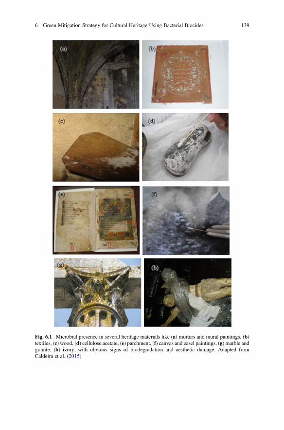
Fig. 6.1 Microbial presence in several heritage materials like (a) mortars and mural paintings, (b)textiles, (c) wood, (d) cellulose acetate, (e) parchment, (f) canvas and easel paintings, (g) marble andgranite, (h) ivory, with obvious signs of biodegradation and aesthetic damage. Adapted fromCaldeira et al. (2015)
6 Green Mitigation Strategy for Cultural Heritage Using Bacterial Biocides 139
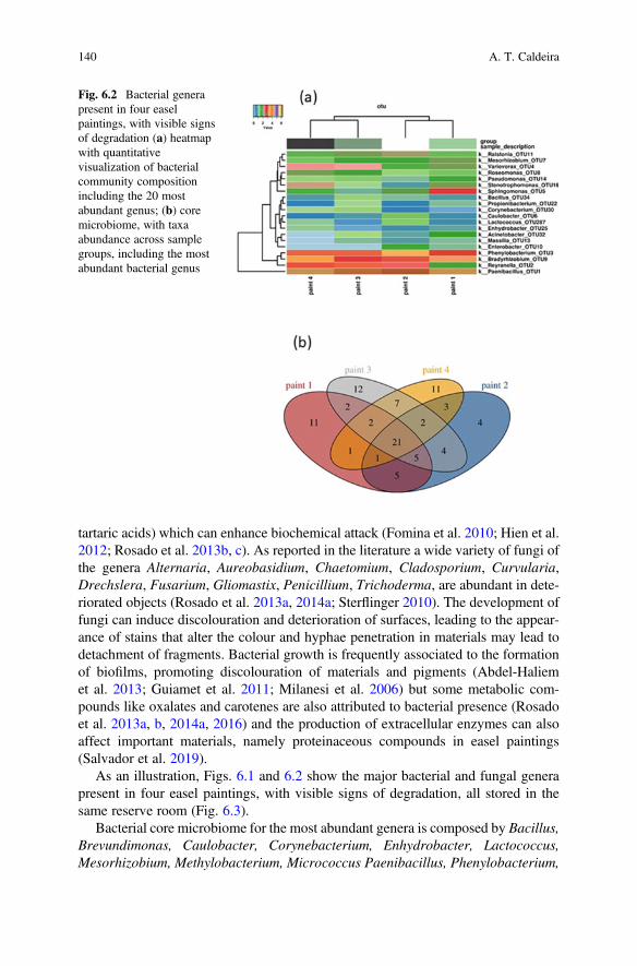
tartaric acids) which can enhance biochemical attack (Fomina et al. 2010; Hien et al.2012; Rosado et al. 2013b, c). As reported in the literature a wide variety of fungi ofthe genera Alternaria, Aureobasidium, Chaetomium, Cladosporium, Curvularia,Drechslera, Fusarium, Gliomastix, Penicillium, Trichoderma, are abundant in dete-riorated objects (Rosado et al. 2013a, 2014a; Sterflinger 2010). The development offungi can induce discolouration and deterioration of surfaces, leading to the appear-ance of stains that alter the colour and hyphae penetration in materials may lead todetachment of fragments. Bacterial growth is frequently associated to the formationof biofilms, promoting discolouration of materials and pigments (Abdel-Haliemet al. 2013; Guiamet et al. 2011; Milanesi et al. 2006) but some metabolic com-pounds like oxalates and carotenes are also attributed to bacterial presence (Rosadoet al. 2013a, b, 2014a, 2016) and the production of extracellular enzymes can alsoaffect important materials, namely proteinaceous compounds in easel paintings(Salvador et al. 2019).
As an illustration, Figs. 6.1 and 6.2 show the major bacterial and fungal generapresent in four easel paintings, with visible signs of degradation, all stored in thesame reserve room (Fig. 6.3).
Bacterial core microbiome for the most abundant genera is composed by Bacillus,Brevundimonas, Caulobacter, Corynebacterium, Enhydrobacter, Lactococcus,Mesorhizobium, Methylobacterium, Micrococcus Paenibacillus, Phenylobacterium,
Fig. 6.2 Bacterial generapresent in four easelpaintings, with visible signsof degradation (a) heatmapwith quantitativevisualization of bacterialcommunity compositionincluding the 20 mostabundant genus; (b) coremicrobiome, with taxaabundance across samplegroups, including the mostabundant bacterial genus
140 A. T. Caldeira
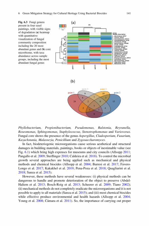
Phyllobacterium, Propionibacterium, Pseudomonas, Ralstonia, Reyranella,Roseomonas, Sphingomonas, Staphylococcus, Stenotrophomonas and Variovorax.Fungal core shows the presence of the genus Aspergillus, Cladosporium, Fusarium,Kazachstania, Malassezia, Penicillium and Zygosaccharomyces.
In fact, biodeteriogenic microorganisms cause serious aesthetical and structuraldamages in building materials, paintings, books or objects of inestimable value (seeFig. 6.1) which bring high expenses for museums and city councils (Allsopp 2011;Pangallo et al. 2009; Sterflinger 2010; Caldeira et al. 2018). To control the microbialgrowth several approaches are being applied such as mechanical and physicalmethods and chemical biocides (Allsopp et al. 2004; Barresi et al. 2017; Favero-Longo et al. 2017; Kakakhel et al. 2019; Pena-Poza et al. 2018; Quagliarini et al.2018; Sanza et al. 2015).
However, these methods have several weaknesses: (i) physical methods can bedangerous to handle and promote deterioration of the object to preserve (Abdel-Haliem et al. 2013; Bosch-Roig et al. 2013; Scheerer et al. 2009; Tiano 2002);(ii) mechanical methods do not completely eradicate the microorganisms and it is notpossible to apply to all materials (Sanza et al. 2015); and (iii) most chemical biocideswhile effective produce environmental and health hazards (Allsopp et al. 2004;Young et al. 2008; Cámara et al. 2011). So, the importance of carrying out proper
Fig. 6.3 Fungi generapresent in four easelpaintings, with visible signsof degradation (a) heatmapwith quantitativevisualization of fungalcommunity compositionincluding the 20 mostabundant genus and (b) coremicrobiome, with taxaabundance across samplegroups, including the mostabundant fungal genus
6 Green Mitigation Strategy for Cultural Heritage Using Bacterial Biocides 141
remediation action for microbiologically contaminated historic materials is of vitalimportance.
The identification of effectively biodeteriogenic agents and the design of mitiga-tion strategies directed to these agents without bias to historical materials, to theenvironment and to operators, taking into account the perspective of the microbialcommunities dynamics is a major challenge to control the biodeterioration ofcultural heritage and where much remains to be done.
2 Bacterial Green Biocides for Cultural Heritage
According to the European legislation, a biocide can be defined as “a chemicalsubstance or microorganism intended to destroy, deter, render harmless or exert acontrolling effect on any harmful organism by chemical or biological means”.Unfortunately, many traditional biocides used in cultural heritage may be dangerousfor human health and the environment, while they differ in their toxicologicalprofile, as EU databases indicate (https://echa.europa.eu). The protective solutionsbased on the use of these toxic chemicals face increasing restrictions due to thepossibility of accumulation in animal tissues (Leifert et al. 1995).
Many commercial biocides are synthetic, mainly composed by mixtures with arange of formulation additives, generally formed by quaternary ammonium com-pounds or other nitrogen-containing compounds, urea, benzalkonium chloride andphenol derivatives (Barresi et al. 2017; Coors et al. 2018; Favero-Longo et al. 2017;Fernandes 2006). Copper, gold, platinum, silver, titanium, zinc metal nanoparticlesare also applied in the last few years (Barresi et al. 2017; Ruggiero et al. 2020).
There are also naturally occurring biocides classified as natural biocides, derivedfrom, e.g., bacteria (Caldeira et al. 2011a; Chena et al. 2020; Colombo et al. 2019;Junier and Joseph 2017; Mariana et al. 2020; Silva et al. 2014), fungi (Hooker et al.1994; Massart and Jijakli 2007; Suárez et al. 2007) and plants (Arantes et al.2019a, b; Chien et al. 2019; Jeong et al. 2018; Silva et al. 2019 Jeong et al. 2018;Palla et al. 2020). A review by Fidanza and Caneva (2019) gives a description ofimportant natural biocides for the conservation of stone in cultural heritage. Con-trolled release systems of biocides by encapsulation in silica were also developed forseveral biocides, both commercial and natural, like Biotin T and New Des50 (Dresler et al. 2017), zosteric acid sodium salt, usnic acid (Ruggiero et al.2020), among others.
In heritage context, biocides are intended to limit the microbial growth and beuseful in extending the life of heritage objects but material compatibility is a criticalfactor to be considered. Moreover, green and non-toxic biocides are eco-friendly andpromising alternatives to chemical industrial biocides that allow to avoid unneces-sary costs and risks. Their production and application do not imply the implemen-tation of a risk-management protocol for toxic compounds which is an advantage inthe field of conservation and restoration.
142 A. T. Caldeira
Therefore, it is crucial to develop green biocides compatible with historic mate-rials that allow the elimination, inhibition and prevention of microbiologicalcontamination.
Over the last years, some bioactive metabolites with potential applications inagriculture and pharmaceutical industries (Lowes et al. 2000; Walker et al. 1995),with a large spectrum of antimicrobial action have been reported. In agriculture thebiological control of some pests has been carried out for decades through theintroduction of bacteria in the soil or directly in the cultures. In recent years, interestin the biological control of phytopathogenic fungi has increased considerablybecause of the public concern to decrease the use of chemical pesticides and becausethere are some diseases that are hardly controllable by other strategies (Caldeira et al.2011a). Besides the biocide effect, surfactins and fengycins of Bacillus subtilisprevented also the adhesion of competitive organisms on the surface of plant roots(Falardeau et al. 2013).
Mechanism and metabolite studies involved in this biological control oftenchoose the use of bacteria (Beltran-Gracia et al. 2017), namely Bacillus spp. Thesemicroorganisms produce a variety of secondary metabolites, specifically antibioticsof peptide nature with antimicrobial properties (Caldeira et al. 2006, 2007, 2008,2011a, b). The antimicrobial, fungicidal, insecticidal, anticarcinogenic andimmunomodulating properties of lipopeptides can help to solve problems in medi-cine and agriculture, and the creation of an ecologically balanced environment(Maksimova et al. 2020).
A successful methodology to produce new green biocide solutions was recentlydeveloped (Silva et al. 2015, 2016a, b, 2017, 2019; Salvador et al. 2016; Rosadoet al. 2019) exploring the potentialities of bioactive metabolites produced by Bacil-lus spp. in the cultural heritage safeguard field.
The Bacillus species are rich in biodiversity, constituting important repositorybioresources, being worth for the creation of green biocides because they produce agreat variety of secondary metabolites including ribosomally and non-ribosomallysynthesized antimicrobial peptides, known to possess antagonistic activities againstmany biodeteriogenic fungi and bacteria (Berezhnayaa et al. 2019; Leifert et al.1995; Jin et al. 2020; Medeot et al. 2020; Mikkola et al. 2004; Yazgan et al. 2001).Bacillus subtilis; B. amyloliquefaciens, B. pumilus, B. licheniformis,B. methylotrophicus, B. atrophaeus, B. laterosporus, B. paralicheniformis,B. lehensis, Paenibacillus terrae, P. peoriae, P. polymyxa, P. larvae,P. mucilaginosus, and P. bovis are probably the main producers of lipopeptides,which are known for their antifungal properties (Berezhnayaa et al. 2019; Caldeiraet al. 2006, 2007, 2011a; Cawoy et al. 2015; Meena and Kanwar 2015; Moyne et al.2001; Ongena and Jacques 2008; Tsuge et al. 2001; Zhao and Kuipers 2016).
Usually, three different classes of bioactive peptides can be distinguished: anti-microbial lanthipeptides ribosomally synthesized, such as subtilin, entianin, ericins,clausin, subtilomycin, thuricins, mersacidin, amylolysin, haloduracin, lichenicidinand cerecidins (Barbosa et al. 2015); and non-ribosomally synthesized antifungalpeptides, such as bacilysin and rhizocticin or antifungal lipopeptides, such assurfactins, iturins and fengycins, bacillomycin, bacilysin, ericin, mersacidin,
6 Green Mitigation Strategy for Cultural Heritage Using Bacterial Biocides 143
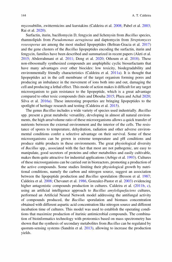
mycosubtilin, zwittermicins and kurstakins (Caldeira et al. 2008; Pabel et al. 2003;Rai et al. 2020).
Surfactin, iturin, bacillomycin D, fengycin and lichenysin from Bacillus species,rhamnolipids from Pseudomonas aeruginosa and daptomycin from Streptomycesroseosporus are among the most studied lipopeptides (Beltran-Gracia et al. 2017)and the gene clusters of the Bacillus lipopeptides encoding the surfactin, iturin andfengycin, families have been described and summarized in recent papers (Aleti et al.2015; Abderrahmani et al. 2011; Deng et al. 2020; Othoum et al. 2018). Thesenon-ribosomally synthesized compounds are amphiphilic cyclic biosurfactants thathave many advantages over other biocides: low toxicity, biodegradability andenvironmentally friendly characteristics (Caldeira et al. 2011a). It is thought thatlipopeptides act in the cell membrane of the target organism forming pores andproducing an imbalance in the movement of ions both into and out, damaging thecell and producing a lethal effect. This mode of action makes it difficult for any targetmicroorganism to gain resistance to the lipopeptide, which is a great advantagecompared to other toxic compounds (Inès and Dhouha 2015; Płaza and Achal 2020;Silva et al. 2016a). These interesting properties are bringing lipopeptides to thespotlight of heritage research and testing (Caldeira et al. 2015).
The genus Bacillus includes a wide variety of species used industrially. Bacillusspp. present a great metabolic versatility, developing in almost all natural environ-ments, the high area/volume ratio of these microorganisms allows a quick transfer ofnutrients between the external environment and the interior of the cells. The resis-tance of spores to temperature, dehydration, radiation and other adverse environ-mental conditions confer a selective advantage on their survival. Some of thesemicroorganisms can be grown in extreme temperature and pH conditions andproduce stable products in these environments. The great physiological diversityof Bacillus spp., associated with the fact that most are not pathogenic, are easy tomanipulate, good secretors of proteins and other metabolites and easily cultivable,makes them quite attractive for industrial applications (Arbige et al. 1993). Culturesof these microorganisms can be carried out in bioreactors, promoting a production ofthe active compounds. Some studies limiting their physiological growth by nutri-tional conditions, namely the carbon and nitrogen source, suggest an associationbetween the lipopeptide production and Bacillus sporulation (Besson et al. 1987;Caldeira et al. 2008; Chevanet et al. 1986, Gonzalez-Pastor et al. 2003) evidencinghigher antagonistic compounds production in cultures. Caldeira et al. (2011b, c),using an artificial intelligence approach to Bacillus amyloliquefaciens cultures,performed an Artificial Neural Network model addressing the antifungal activityof compounds produced, the Bacillus sporulation and biomass concentrationobtained with different aspartic acid concentration like nitrogen source and differentincubation time of cultures. This model was used to establish the operating condi-tions that maximize production of iturinic antimicrobial compounds. The combina-tion of bioinformatics technology with proteomics based on mass spectrometry hasshown that the synthesis of secondary metabolites from Bacillus can be regulated byquorum-sensing systems (Sandrin et al. 2013), allowing to increase the productionyields.
144 A. T. Caldeira
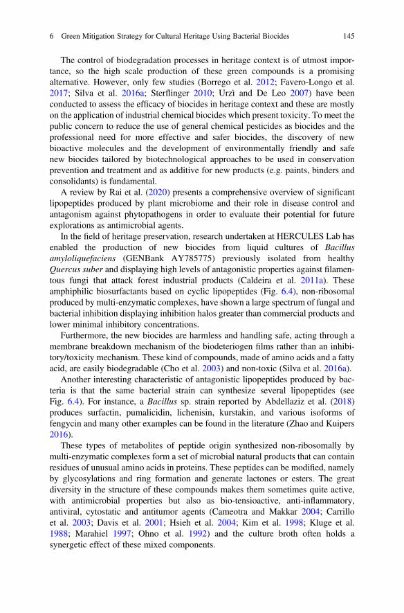
The control of biodegradation processes in heritage context is of utmost impor-tance, so the high scale production of these green compounds is a promisingalternative. However, only few studies (Borrego et al. 2012; Favero-Longo et al.2017; Silva et al. 2016a; Sterflinger 2010; Urzì and De Leo 2007) have beenconducted to assess the efficacy of biocides in heritage context and these are mostlyon the application of industrial chemical biocides which present toxicity. To meet thepublic concern to reduce the use of general chemical pesticides as biocides and theprofessional need for more effective and safer biocides, the discovery of newbioactive molecules and the development of environmentally friendly and safenew biocides tailored by biotechnological approaches to be used in conservationprevention and treatment and as additive for new products (e.g. paints, binders andconsolidants) is fundamental.
A review by Rai et al. (2020) presents a comprehensive overview of significantlipopeptides produced by plant microbiome and their role in disease control andantagonism against phytopathogens in order to evaluate their potential for futureexplorations as antimicrobial agents.
In the field of heritage preservation, research undertaken at HERCULES Lab hasenabled the production of new biocides from liquid cultures of Bacillusamyloliquefaciens (GENBank AY785775) previously isolated from healthyQuercus suber and displaying high levels of antagonistic properties against filamen-tous fungi that attack forest industrial products (Caldeira et al. 2011a). Theseamphiphilic biosurfactants based on cyclic lipopeptides (Fig. 6.4), non-ribosomalproduced by multi-enzymatic complexes, have shown a large spectrum of fungal andbacterial inhibition displaying inhibition halos greater than commercial products andlower minimal inhibitory concentrations.
Furthermore, the new biocides are harmless and handling safe, acting through amembrane breakdown mechanism of the biodeteriogen films rather than an inhibi-tory/toxicity mechanism. These kind of compounds, made of amino acids and a fattyacid, are easily biodegradable (Cho et al. 2003) and non-toxic (Silva et al. 2016a).
Another interesting characteristic of antagonistic lipopeptides produced by bac-teria is that the same bacterial strain can synthesize several lipopeptides (seeFig. 6.4). For instance, a Bacillus sp. strain reported by Abdellaziz et al. (2018)produces surfactin, pumalicidin, lichenisin, kurstakin, and various isoforms offengycin and many other examples can be found in the literature (Zhao and Kuipers2016).
These types of metabolites of peptide origin synthesized non-ribosomally bymulti-enzymatic complexes form a set of microbial natural products that can containresidues of unusual amino acids in proteins. These peptides can be modified, namelyby glycosylations and ring formation and generate lactones or esters. The greatdiversity in the structure of these compounds makes them sometimes quite active,with antimicrobial properties but also as bio-tensioactive, anti-inflammatory,antiviral, cytostatic and antitumor agents (Cameotra and Makkar 2004; Carrilloet al. 2003; Davis et al. 2001; Hsieh et al. 2004; Kim et al. 1998; Kluge et al.1988; Marahiel 1997; Ohno et al. 1992) and the culture broth often holds asynergetic effect of these mixed components.
6 Green Mitigation Strategy for Cultural Heritage Using Bacterial Biocides 145
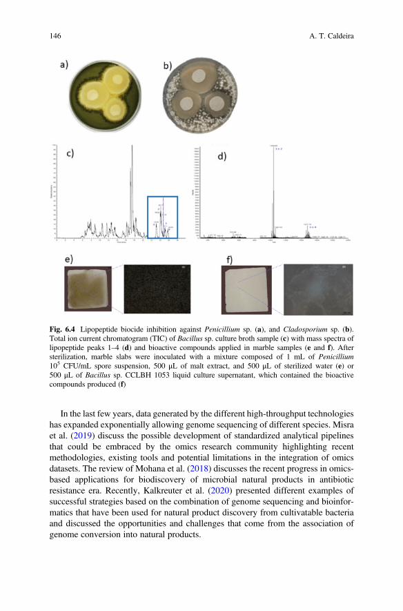
In the last few years, data generated by the different high-throughput technologieshas expanded exponentially allowing genome sequencing of different species. Misraet al. (2019) discuss the possible development of standardized analytical pipelinesthat could be embraced by the omics research community highlighting recentmethodologies, existing tools and potential limitations in the integration of omicsdatasets. The review of Mohana et al. (2018) discusses the recent progress in omics-based applications for biodiscovery of microbial natural products in antibioticresistance era. Recently, Kalkreuter et al. (2020) presented different examples ofsuccessful strategies based on the combination of genome sequencing and bioinfor-matics that have been used for natural product discovery from cultivatable bacteriaand discussed the opportunities and challenges that come from the association ofgenome conversion into natural products.
Fig. 6.4 Lipopeptide biocide inhibition against Penicillium sp. (a), and Cladosporium sp. (b).Total ion current chromatogram (TIC) of Bacillus sp. culture broth sample (c) with mass spectra oflipopeptide peaks 1–4 (d) and bioactive compounds applied in marble samples (e and f). Aftersterilization, marble slabs were inoculated with a mixture composed of 1 mL of Penicillium105 CFU/mL spore suspension, 500 μL of malt extract, and 500 μL of sterilized water (e) or500 μL of Bacillus sp. CCLBH 1053 liquid culture supernatant, which contained the bioactivecompounds produced (f)
146 A. T. Caldeira
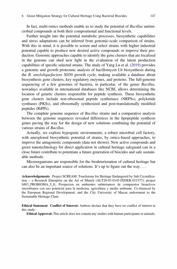
In fact, multi-omics methods enable us to study the potential of Bacillus antimi-crobial compounds at both their compositional and functional levels.
Further insight into the potential metabolic processes, biosynthetic capabilitiesand stress adaptations can be inferred from genomic-scale comparison of strains.With this in mind, it is possible to screen and select strains with higher industrialpotential capable to produce new desired active compounds or improve their pro-duction. Genomic approaches capable to identify the gene clusters that are localizedin the genome can shed new light in the evaluation of the latent productioncapabilities of specific selected strains. The study of Yang Lu et al. (2019) providesa genomic and growth proteomic analysis of bacillomycin Lb biosynthesis duringthe B. amyloliquefaciens X030 growth cycle, making available a database aboutbiosynthesis gene clusters, key regulatory enzymes, and proteins. The full-genomesequencing of a few genomes of bacteria, in particular, of the genus Bacillus,nowadays available in international databases like NCBI, allows determining thelocation of genetic clusters responsible for peptide synthesis. These biosyntheticgene clusters include non-ribosomal peptide synthetases (NRPSs), polyketidesynthases (PKSs), and ribosomally synthesized and post-translationally modifiedpeptides (RiPPs).
The complete genome sequence of Bacillus strains and a comparative analysisbetween the genome sequences revealed differences in the lipopeptide synthesisgenes paving the way for the design of new solutions combining the potential ofvarious strains of Bacillus.
Actually, we explore hypogenic environments, a robust microbial cell factory,with unexplored biosynthetic potential of strains, by omics-based approaches, toimprove the antagonistic compounds (data not shown). New active compounds andgreen nanotechnology for direct application in cultural heritage safeguard can in aclose future contribute to potentiate a future generation of biocides and safe sustain-able methods.
Microorganisms are responsible for the biodeterioration of cultural heritage butcan also be an important source of solutions. It’s up to figure out the way. . . .
Acknowledgements Project SCREAM: Touchstone for Heritage Endangered by Salt Crystalliza-tion – a Research Enterprise on the Art of Munch (ALT20-03-0145-FEDER-031577); project0483_PROBIOMA_5_E, Prospecion en ambientes subterraneos de compuestos bioactivosmicrobianos con uso potencial para la medicina, agricultura y medio ambiente. Co-financed bythe European Regional Development; and the City University of Macau endowment to theSustainable Heritage Chair.
Ethical Statement Conflict of Interest: Authors declare that they have no conflict of interest inthis study.
Ethical Approval: This article does not contain any studies with human participants or animals.
6 Green Mitigation Strategy for Cultural Heritage Using Bacterial Biocides 147
References
Abdel-Haliem ME, Sakr AA, Ali MF, Ghaly MF, Sohlenkamp C (2013) Characterization ofStreptomyces isolates causing colour changes of mural paintings in ancient Egyptian tombs.Microbiol Res 168:428–437
Abdellaziz L, Chollet M, Abderrahmani A, Bechet M, Yaici L, Chataigne G, Arias A, Leclere V,Jacques P (2018) Lipopeptide biodiversity in antifungal Bacillus strains isolated from Algeria.Arch Microbiol 200:1205–1216
Abderrahmani A, Tapi A, Nateche F, Chollet M, Leclere V, Wathelet B, Hacene H, Jacques P(2011) Bioinformatics and molecular approaches to detect NRPS genes involved in the biosyn-thesis of kurstakin from Bacillus thuringiensis. Appl Microbiol Biotechnol 92:571–581
Aleti G, Sessitsch A, Brader G (2015) Genome mining: prediction of lipopeptides and polyketidesfrom Bacillus and related Firmicutes. Comput Struct Biotechnol J 13:192–203
Allsopp D (2011) Worldwide wastage: the economics of biodeterioration. Microbiol Today38:150–153
Allsopp D, Swal K, Gaylande C (2004) The control of biodeterioration. In: Introduction tobiodeterioration. Cambridge University Press, Cambridge, pp 203–223
Amann R, Fuchs B, Behrens S (2001) The identification of microorganisms by fluorescence in situhybridisation. Curr Opin Biotechnol 12:231–236
Arantes S, Caldeira AT, Candeias A, Martins MR (2019a) Antimicrobial potential of Lamiaceaeessential oils against heritage biodeteriogenic strains. Congress of Microbiology and Biotech-nology, Book of abstracts Microbiotec’ 19:97, Coimbra, Portugal (http://microbiotec19.net/test/wpcontent/uploads/2019/07/Book_abstract_online.pdf)
Arantes S, Guerreiro M, Piçarra A, Caldeira AT, Martins RM (2019b). Antimicrobial potential ofessential oils of Portuguese autochthonous Lavandula against resistant bacteria. Congress ofMicrobiology and Biotechnology, Book of abstracts Microbiotec’19, pp 313, Coimbra, Portugal(http://microbiotec19.net/test/wp-content/uploads/2019/07/Book_abstract_online.pdf)
Arbige MV, Bulthuis BA, Schultz J, Crabb D (1993) Fermentation of Bacillus. In: Sonenshein AL,Hoch JA, Losick R (eds) Bacillussubtilis and other gram-positive bacteria: biochemistry,physiology, and molecular genetics. American Society for Microbiology, Washington, DC,pp 871–895
Barbosa J, Caetano T, Mendo S (2015) Class I and class II lanthipeptides produced by Bacillus spp.J Nat Prod 78:2850–2866
Barresi G, Cammarata M, Palla F (2017) Biocide. In: Palla F, Barresi G (eds) Biotechnology andconservation of cultural heritage. Springer, Switzerland, pp 49–66
Beltran-Gracia E, Macedo-Raygoza G, Villafaña-Rojas J, Martinez-Rodriguez A, Chavez-Castrillon Y, Espinosa-Escalante F, Di Mascio P, Ogura T, Beltran-Garcia M (2017) Productionof lipopeptides by fermentation processes: Endophytic bacteria, fermentation strategies and easymethods for bacterial Selection. In: Angela Faustino J (ed) Fermentation Processes, IntechOpen,London. https://doi.org/10.5772/64236
Berezhnayaa A, Evdokimovaa O, Valentovicha L, Sverchkovaa N, Titoka M, Kolomiyets E (2019)Molecular genetic and functional analysis of the genome of bacteria Bacillus velezensis BIMB-439D. Appl Biochem Microbiol 55:386–396
Besson F, Chevanet C, Michel G (1987) Influence of the culture medium on the production of iturinA by Bacillus subtilis. J Gen Microbiol 3:767–772
Borrego S, Valdés O, Vivar I, Lavin P, Guiamet P, Battistoni P, Gómez de Saravia S, Borges P(2012) Essential oils of plants as biocides against microorganisms isolated from cuban andargentine documentary heritage. Int Sch Res Notices 2012:7., Article ID 826786. https://doi.org/10.5402/2012/826786
Bosch-Roig P, Regidor-Ros JL, Montes-Estelles R (2013) Biocleaning of nitrate alterations on wallpaintings by Pseudomonas stutzeri. Int Biodeter Biodegr 84:266–274
148 A. T. Caldeira
Britta K, Joachim V, Johann S, Klaus E (1988) Studies on the biosynthesis of surfactin, alipopeptide antibiotic from Bacillus subtilis ATCC 21332. FEBS Lett:231. https://doi.org/10.1016/0014-5793(88)80712-9
Caldeira AT, Savluchinske-Feio S, Arteiro JM, Roseiro J (2006) Antimicrobial activity of steady-state cultures of Bacillus sp. CCMI 1051 against wood contaminant fungi. Biochem Eng J30:231–236
Caldeira AT, Savluchinske-Feio S, Arteiro JM, Roseiro J (2007) Bacillus amyloliquefaciens CCMI1051 in vitro activity against wood contaminant fungi. Ann Microbiol 57:29–34
Caldeira AT, Savluchinske-Feio S, Arteiro JM, Coelho A, Roseiro J (2008) Environmental dynam-ics of Bacillus amyloliquefaciens CCMI1051 antifungal activity under different nitrogen pat-terns. J Appl Microbiol 104:808–816
Caldeira AT, Arteiro JM, Coelho A, Roseiro J (2011a) Combined use of LC–ESI-MS andantifungal tests for rapid identification of bioactive lipopeptides produced by Bacillusamyloliquefaciens CCMI 1051. Process Biochem 46(9):1738–1746
Caldeira AT, Vicente H, Arteiro JM, Roseiro J, Neves J (2011b) An artificial intelligence approachto Bacillus amyloliquefaciens CCMI 1051 cultures: application to the production of antifungalcompounds. Bioresour Technol 102:1496–1502
Caldeira AT, Vicente H, Arteiro JM, Roseiro J, Neves J (2011c) Production of bioactive com-pounds against wood contaminant fungi: an artificial intelligence approach. In: Caldeira F(ed) Minimizing the environmental impact of the forest products industries. UFP Editions,Porto, pp 131–137
Caldeira AT, Rosado T, Silva M, Gonzalez M, Candeias A (2015) Microrganismos e Património -Novas abordagens. Magazine da Sociedade Portuguesa de Microbiologia 4:1–7
Caldeira AT, Salvador C, Rosado T, Teixeira D (2018) Biodeterioration of easel paintings – anoverview. In: Mitchell R, Clifford J (eds) Biodeterioration and preservation in art, archaeologyand architecture. Archetype Publications, London, pp 71–78
Cámara B, De los Ríos A, Urizal M, Buergo M, Varas MJ, Fort R, Ascaso C (2011) Characterizingthe microbial colonization of a dolostone quarry: implications for stone biodeterioration andresponse to biocide treatments. Microb Ecol 62:299–313
Cameotra SS, Makkar RS (2004) Recent applications of biosurfactants as biological and immuno-logical molecules. Curr Opin Microbiol 7:262–266
Carrillo PM, Robinson HS, Anumba CJ, Al-Ghassani AM (2003) IMPaKT: a framework for linkingknowledge management to business performance. Electron J Knowl Manag 1(1):1–12
Cawoy H, Debois D, Franzil L, De Pauw E, Thonart P, Ongena M (2015) Lipopeptides as mainingredients for inhibition of fungal phytopathogens by Bacillus subtilis/amyloliquefaciens.Microb Biotechnol 8:281–295
Chena J, Xue Q, Mac CL, Tan X (2020) Streptomyces pactummay control Phelipanche aegyptiacain tomato. Appl Soil Ecol 146:103369
Chevanet C, Besson F, Michel G (1986) Effect of various growth conditions on spore formation andbacillomycin L production in Bacillus subtilis. Can J Microbiol 32:254–258
Chien H, Kuo C, Kao L, Lin G, Chen P (2019) Polysaccharidic spent coffee grounds for silvernanoparticle immobilization as a green and highly efficient biocide. Int J Biol Macromol140:168–176
Cho SJ, Lee SK, Cha BJ, Kim YH, Shin KS (2003) Detection and characterization of theGloeosporium gloeosporioides growth inhibitory compound iturin A from Bacillus subtilisstrain KS03. FEMS Microbiol Lett 223:47–51
Colombo EM, Kunova A, Pizzatti C, Saracchi M, Cortesi P, Pasquali M (2019) Selection of anendophytic streptomyces sp. strain DEF09 from wheat roots as a biocontrol agent againstFusarium graminearum. Front Microbiol 10:1–12
Coors A, Vollmar P, Heim J, Sacher F, Kehrer A (2018) Environmental risk assessment of biocidalproducts: identification of relevant components and reliability of a component based mixtureassessment. Environ Sci Eur 30(3):1–15
6 Green Mitigation Strategy for Cultural Heritage Using Bacterial Biocides 149
Davis DA, Lynch HC, Varley J (2001) The application of foaming for the recovery of Surfactinfrom B. subtilis ATCC 21332 cultures. Enzym Microb Technol 28:346–354. https://doi.org/10.1016/S0141-0229(00)00327-6
Deng Q, Wang R, Sun D, Sun L, Wang Y, Pu Y, Fang Z, Xu D, Liu Y, Ye R, Yin S, Xie S,Gooneratne R (2020) Complete genome of Bacillus velezensis CMT-6 and comparative genomeanalysis reveals lipopeptide diversity. Biochem Genet 58:1–15
Dias L, Rosado T, Coelho A, Barrulas P, Lopes L, Moita P, Candeias A, Mirão J, Caldeira AT(2018) Natural limestone discolouration triggered by microbial activity—a contribution. AIMSMicrobiol 4:594–607
Dresler C, Saladino M, Demirbag C, Caponetti E, Martino D, Alduina R (2017) Development ofcontrolled release systems of biocides for the conservation of cultural heritage. Int BiodeteriorBiodegrad 125:150–156
Falardeau J, Wise C, Novitsky L, Avis TJ (2013) Ecological and mechanistic insights into the directand indirect antimicrobial properties of Bacillus subtilis Lipopeptides on plant pathogens. JChem Ecol 39(7):869–878
Favero-Longo S, Benesperi R, Bertuzzi S, Bianchi E, Buffa G, Giordani P, Loppi S, Malaspina P,Matteucci E, Paoli L, Ravera S, Roccardi A, Segimiro A, Vannini A (2017) Species- and site-specific efficacy of commercial biocides and application solvents against lichens. Int BiodeteriorBiodegrad 123:127–137
Fernandes P (2006) Applied microbiology and biotechnology in the conservation of culturalheritage materials. Appl Microbiol Biotechnol 73:291–296
Fidanza M, Caneva G (2019) Natural biocides for the conservation of stone cultural heritage: areview. J Cult Herit 38:271–286
Fomina M, Burford P, Hillier S, Kierans M, Gadd M (2010) Rock-building fungi. Geomicrobiol J27:624–629
González JM, Saiz-Jiménez C (2005) Application of molecular nucleic acid-based techniques forthe study of microbial communities in monuments and artworks. Int Microbiol 8:189–194
Gonzalez M, Vieira R, Nunes P, Rosado T, Martins S, Candeias A, Pereira A, Caldeira AT (2014)Fluorescence In Situ hybridization: a potentially useful technique for detection of microorgan-isms on mortars. e-conser J 2:44–52
Gonzalez M, Brinco C, Vieira R, Rosado T, Mauran G, Pereira A, Candeias A, Caldeira AT (2017)Dual phylogenetic staining protocol for simultaneous analysis of yeast and bacteria in artworks.Appl Phys A:123–142
Gonzalez-Pastor J, Hobbs E, Losick R (2003) Cannibalism by sporulating bacteria. Science301:510–513
Guiamet P, Borrego S, Lavin P, Perdomo I, Saravia SGD (2011) Biofouling and biodeterioration inmaterials stored at the historical archive of the Museum of La Plata, Argentine and at theNational Archive of the Republic of Cuba. Colloids Surf B Biointerf 85:229–234
Hien TH, Govin A, Guyonnet R, Grosseau P, Lors C, Garcia-Diaz E, Damidot D, Devès O, Ruot B(2012) Influence of the intrinsic characteristics of mortars on biofouling by Klebsormidiumflaccidum. Int Biodeterior Biodegrad 70:31–39
Hooker JE, Jaizme-Vega M, Atkinson D (1994) Biocontrol of plant pathogens using arbuscularmycorrhizal fungi. In: Gianinazzi S, Schüepp H (eds) Impact of Arbuscular Mycorrhizas onsustainable agriculture and natural ecosystems. ALS Advances in Life Sciences. Birkhäuser,Basel, pp 191–200
Hsieh FC, Li MC, Lin TC, Kao SS (2004) Rapid detection and characterization of surfactin-producing Bacillus subtilis and closely related species based on PCR. Curr Microbiol 49:186–191. https://doi.org/10.1007/s00284-004-4314-7
Inès M, Dhouha G (2015) Lipopeptide surfactants: production, recovery and pore forming capacity.Peptides 71:100–112
Jeong S, Lee H, Kim DW, Chung Y (2018) New biocide for eco-friendly biofilm removal onoutdoor stone monuments. Int Biodeterior Biodegrad 131:19–28
150 A. T. Caldeira
Jin P, Wang H, Tan Z, Xuan Z, Dahar GY, Li QX, MiaoW, Liu W (2020) Antifungal mechanism ofbacillomycin D from Bacillus velezensis HN-2 against Colletotrichum gloeosporioides Penz.Pestic Biochem Phys 163:102–107
Junier P, Joseph E (2017) Microbial biotechnology approaches to mitigating the deterioration ofconstruction and heritage materials. Microb Biotechnol 10(5):1145–1148
Kakakhel I, Wu F, Gu JD, Feng H, Shah K, WangW (2019) Controlling biodeterioration of culturalheritage objects with biocides: a review. Int Biodeterior Biodegrad 143(104721):1–10
Kalkreuter E, Pan G, Cepeda A, Shen B (2020) Targeting bacterial genomes for natural product.Discovery Trends Pharmacol Sci 41:13–26
Kim K, Jung SY, Lee DK, Jung JK, Park JK, Kim DK, Lee CH (1998) Suppression of inflammatoryresponses by surfactin, a selective inhibitor of platelet cytosolic phospholipase A(2). BiochemPharmacol 55(7):975–985. https://doi.org/10.1016/S0006-2952(97)00613-8
Leifert C, Li H, Chidburee S, Hampson S, Workman S, Sigee D, Epton H, Harbour A (1995)Antibiotic production and biocontrol activity by Bacillus subtilis CL27 and Bacillus pumilusCL45. J Appl Bacteriol 78:97–108
Lowes KF, Shearman CA, Payne J, MacKenzie D, Archer DB, Merry RJ, Gasson MJ (2000)Prevention of yeast spoilage in feed and food by the yeast mycocin HMK. Appl EnvironMicrobiol 66(3):1066–1076. https://doi.org/10.1128/aem.66.3.1066-1076.2000
Lu JY, Zhou K, Huang WT, Zhou P, Yang S, Zhao X, Xie J, Xia L, Ding X (2019) A comprehen-sive genomic and growth proteomic analysis of antitumor lipopeptide bacillomycin Lb biosyn-thesis in Bacillus amyloliquefaciens X030. Appl Microbiol Biotechnol 103:7647–7662
Maksimova I, Singhb B, Cherepanovaa E, Burkhanovaa G, Khairullin R (2020) Prospects andapplications of lipopeptide-producing bacteria for plant protection. Appl Biochem Microbiol 56(1):15–28
Marahiel MA, Stachelhaus T, Mootz HD (1997) Modular peptide synthetases involved innonribosomal peptide synthesis. Chem Rev 97:2651–2673
Mariana M, Ohnob T, Suzukic H, Kitamurac H, Kurodac K, Shimizua M (2020) A novel strain ofendophytic Streptomyces for the biocontrol of strawberry anthracnose caused by Glomerellacingulate. Microbiol Res 234(126428):1–9
Massart S, Jijakli H (2007) Use of molecular techniques to elucidate the mechanisms of action offungal biocontrol agents: a review. J Microbiol Methods 69(2):229–241
Medeot D, Fernandez M, Morales G, Jofré E (2020) Fengycins from Bacillus amyloliquefaciensMEP218 exhibit antibacterial activity by producing alterations on the cell surface of thepathogens Xanthomonas axonopodis pv. vesicatoria and Pseudomonas aeruginosa PA01.Front Microbiol 10(3107):1–12
Meena KR, Kanwar SS (2015) Lipopeptides as the antifungal and antibacterial agents: applicationsin food safety and therapeutics. Biomed Res Int 2015:473050
Mikkola R, Andersson M, Grigoriev P, Teplova V, Saris N, Rainey F, Salonen M (2004) Bacillusamyloliquefaciens strains isolated from moisture-damaged buildings produced surfactin and asubstance toxic to mammalian cells. Arch Microbiol 181:314–323
Milanesi C, Baldi F, Borin S, Vignani R, Ciampolini F, Faleri C, Cresti M (2006) Biodeteriorationof a fresco by biofilm forming bacteria. Int Biodeterior Biodegrad 57:168–173
Misra B, Langefeld C, Olivier M, Cox L (2019) Integrated omics: tools, advances and futureapproaches. J Mol Endocrinol 62:21–45
Mohana N, Rao H, Rakshith D, Mithun PR, Nuthan BR, Satish S (2018) Omics based approach forbiodiscovery of microbial natural products in antibiotic resistance era. J Genet Eng Biotechnol16(1):1–8
Moter A, Göbel U (2020) Fluorescence in situ hybridization (FISH) for direct visualization ofmicroorganisms. J Microbiol Methods 41(2):85–112
Moyne A, Shelby R, Cleveland T, Tuzun S (2001) Bacillomycin D: an iturin with antifungalactivity against Aspergillus flavus. J Appl Microbiol 90:622–629
Ongena M, Jacques P (2008) Bacillus lipopeptides: versatile weapons for plant disease biocontrol.Trends Microbiol 16:115–125
6 Green Mitigation Strategy for Cultural Heritage Using Bacterial Biocides 151
Ohno A, Takashi A, Shoda M (1992) Production of a lipopeptideantibiotic surfactin with recom-binant Bacillus subtilis. Biotechnol Lett 14:1165–1168
Othoum G, Bougouffa S, Razali R et al (2018) In silico exploration of Red Sea Bacillus genomesfor natural product biosynthetic gene clusters. BMC Genomics 19:382. https://doi.org/10.1186/s12864-018-4796-5
Pabel C, Vater J, Wilde C, Franke P, Hofemeisrer J, Adler B, Bringmann G, Hacker J, Hentschel U(2003) Antimicrobial activities and matrix-assisted laser desorption/ionization mass spectrom-etry of Bacillus isolates from the marine sponge Aplysina aerophoba. Mar Biotechnol5:424–434
Palla F, Bruno M, Mercurio F, Tantillo A, Rotolo V (2020) Essential oils as natural biocides inconservation of cultural heritage. Molecules 25(730):1–11
Pangallo D, Chovanová K, Simonovicova A, Ferianc P (2009) Investigation of microbial commu-nity isolated from indoor artworks and air environment: identification, biodegradative abilities,and DNA typing. Can J Microbiol 55:277–287
Pena-Poza J, Ascaso C, Sanz M, Pérez-Ortega S, Oujja M, Wierzchos J, Souza-Egipsy V,Cañamares MV, Urizal M, Castillejo M, García-Heras M (2018) Effect of biological coloniza-tion on ceramic roofing tiles by lichens and a combined laser and biocide procedure for itsremoval. Int Biodeterior Biodegrad 126:86–94
Piñar G, Sterflinger K (2009) Microbes and building materials. In: Cornejo DN, Haro JL (eds)Building materials: properties, performance and applications. Nova Science Publishers,New York, pp 163–188
Płaza G, Achal V (2020) Biosurfactants: eco-friendly and innovative biocidesagainst biocorrosion.Int J Mol Sci 21:2152
Quagliarini E, Graziani L, Diso D, Licciulli A, D’Orazio M (2018) Is nano-TiO2 alone an effectivestrategy for the maintenance of stones in cultural heritage? J Cult Heritage 30:81–91
Rai J, Singh H, Brahmaprakash G (2020) Lesson from ecotoxicity: revisiting the microbiallipopeptides for the management of emerging diseases for crop protection. Int J Environ ResPublic Health 17(1434):1–27
Rosado T, Martins MR, Pires M, Mirão J, Candeias A, Caldeira AT (2013a) Enzymaticmonitorization of mural paintings biodeterioration. Int J Cons Sci 4:603–612
Rosado T, Gil M, Mirão J, Candeias A, Caldeira AT (2013b) Oxalate biofilm formation in muralpaintings due to microorganisms - a comprehensive study. Int Biodeterior Biodegrad 85:1–7
Rosado T, Mirão J, Gil M, Candeias A, Caldeira AT (2013c) Evaluation of mural paintingsbiodeterioration by oxalate formation. In: Rogerio-Candelera MA, Lazzari M, Cano E (eds)Science and technology for the conservation of cultural heritage. CRC Press/Balkema, London,pp 147–150
Rosado T, Mirão J, Candeias A, Caldeira AT (2014a) Microbial communities analysis assessed bypyrosequencing - a new approach applied to conservation state studies of mural paintings. AnalBioanal Chem 406:887–895
Rosado T, Reis A, Mirão J, Candeias A, Vandenabeeled P, Caldeira AT (2014b) Pink! Why not?On the unusual colour of Évora Cathedral. Int Biodeterior Biodegrad 94:121–127
Rosado T, Mirão J, Candeias A, Caldeira AT (2015) Characterizing microbial diversity and damagein mural paintings. Microsc Microanal 21(1):78–83
Rosado T, Gil M, Mirão J, Candeias A, Caldeira AT (2016) Darkening on lead based pigments:microbiological contribution. Color Res Appl 41(3):294–298
Rosado T, Silva M, Dias L, Candeias A, Gil M, Mirão J, Pestana J, Caldeira AT (2017) Microor-ganisms and the integrated conservation-intervention process of the renaissance mural paintingsfrom Casas Pintadas in Évora - know to act, act to preserve. J King Sau Univ – Sci 29(4):478–486
Rosado T, Santos R, Silva M, Galvão A, Mirão J, Candeias A, Caldeira AT (2019) Mitigationapproach to avoid fungal colonisation of porous limestone. Int J conser sci 10(1):3–14
Rosado T, Dias L, Lança M, Nogueira C, Santos R, Martins MR, Candeias A, Mirão J, Caldeira AT(2020) Assessment of microbiota present on a Portuguese historical stone convent using high
152 A. T. Caldeira
throughput sequencing approaches. Microbiol Open e1030:1–18. https://doi.org/10.1002/mbo3.1030
Ruggiero L, Bartoli F, Fidanza MR, Zurlo F, Marconi E, Gasperi T, Tuti S, Crociani L, DiBartolomeo E, Caneva G, Ricci MA, Sodo A (2020) Encapsulation of environmentally-friendlybiocides in silica nanosystems for multifunctional coatings. Appl Surf Sci 514(145908):1–9
Salvador C, Silva M, Rosado T, Vaz Freire R, Bordalo R, Candeias A, Caldeira AT (2016)Biodeterioration of easel paintings: development of new mitigation strategies. ConservarPatrimónio 23:119–124
Salvador C, Borlado R, Silva M, Rosado T, Candeias A, Caldeira AT (2017) On the conservation ofeasel paintings - evaluation of microbial contamination and artist materials. Appl Phys A 123(80):1–13
Salvador C, Medeiros R, Candeias A, Caldeira AT (2019) Biodeterioration of easel paintings: a casestudy of Munch’s paintings. In: Congress of microbiology and biotechnology, book of abstractsMicrobiotec’19. Portugal, Coimbra, p 90. http://microbiotec19.net/test/wp-content/uploads/2019/07/Book_abstract_online.pdf
Sandrin TR, Goldstein GE, Schumaker S (2013) MALDI TOF MS profiling of bacteria at the strainlevel: a review. Mass Spectrom Rev 32:188–217
Sanza M, Oujja M, Ascaso C, Ríos A, Pérez-Ortega S, Souza-Egipsy V, Wierzchos J, Speranza M,Canamares M, Castillejo M (2015) Infrared and ultraviolet laser removal of crustose lichens ondolomite heritage stone. Appl Surf Sci 346:248–255
Scheerer S, Ortega-Morales O, Gaylarde C (2009) Microbial deterioration of stone monuments – anupdated overview. Adv Appl Microbiol 66:97–139
Silva M, Teixeira D, Silva S, Candeias A, Caldeira AT (2014) Production of novel biocides forcultural heritage from Bacillus sp. In: Rogerio-Candelera MA (ed) Science, technology andcultural heritage. CRC Press/Balkema, London, pp 223–228
Silva M, Rosado T, Teixeira D, Candeias A, Caldeira AT (2015) Production of Green biocides forcultural heritage- novel biotechnological solutions. Int J Conerv Sci 6:519–530
Silva M, Vador C, Candeias MF, Teixeira D, Candeias A, Caldeira AT (2016a) Toxicologicalassessment of novel green biocides for cultural heritage. Int J Conserv Sci 7:265–272
Silva M, Pereira A, Teixeira D, Candeias A, Caldeira AT (2016b) Combined use of NMR,LC-ESIMS and antifungal tests for rapid detection of bioactive lipopeptides produced byBacillus. Adv Microbiol 6:788–796
Silva M, Rosado T, Teixeira D, Candeias A, Caldeira AT (2017) Green mitigation strategy forcultural heritage: bacterial potential for biocide production. Environ Sci Pollut Res24:4871–4881
Silva M, Rosado T, Lopes da Silva Z, Nóbrega Y, Silveira D, Candeias A, Caldeira AT (2019)Green bioactive compounds: mitigation strategies for cultural heritage. Conserv Sci CultureHeritage 19:133–142
Sterflinger K (2010) Fungi: their role in deterioration of cultural heritage. Fungal Biol Rev24:47–55
Suárez MB, Vizcaíno JA, Llobell A, Monte E (2007) Characterization of genes encoding novelpeptidases in the biocontrol fungus Trichoderma harzianum CECT 2413 using the Tricho ESTfunctional genomics approach. Curr Genet 51:331–342
Tiano P (2002) Biodegradation of cultural heritage: decay, mechanisms and control methods.Seminar article, New University of Lisbon, Department of Conservation and Restoration,7–12 (http://www.arcchip.cz/w09/w09_tiano.pdf)
Tsuge K, Akiyama T, Shoda M (2001) Isolation of a gene essential for biosynthesis of thelipopeptide antibiotics plipastatin B1 and surfactin in Bacillus subtilis YB8. J Bacteriol183:6265–6273
Urzì C, De Leo F (2007) Evaluation of the efficiency of water-repellent and biocide compoundsagainst microbial colonization of mortars. Int Biodeterior Biodegrad 60:25–34
Vieira R, Nunes P, Martins S, González M, Rosado T, Pereira A, Candeias A, Caldeira AT (2014)Fluorescence in situ hybridisation for microbiological detection in mortars. In: Rogerio-
6 Green Mitigation Strategy for Cultural Heritage Using Bacterial Biocides 153
Candelera (ed) Science, technology and cultural heritage. CRC Press/Balkema Taylor & FrancisGroup, London, pp 257–262
Vieira R, Pazian M, González-Pérez M, Pereira A, Candeias A, Caldeira AT (2018) Detecting cellswith low RNA content colonizing artworks non-invasively: RNA-FISH. In: Mosquera MJ, GilA (eds) Conserving cultural heritage. CRC Press/Balkema, London, pp 303–305
Walker GM, Mcleod AH, Hodgson VJ (1995) Interactions between killer yeasts and pathogenicfungi. FEMS Microbiol Lett 127(3):213–222. https://doi.org/10.1111/j.1574-6968.1995.tb07476.x
Yazgan A, Ozcengiz G, Marahiel MA (2001) Tn10 insertional mutations of Bacillus subtilis thatblock the biosynthesis of bacilysin. Biochim Biophys Acta 1518:87–94
Young ME, Alakomi H, Fortune I, Gorbushina A, Krumbein W, Maxwell I, McCullagh C,Robertson P, Saarela M, Valero J, Vendrell M (2008) Development of a biocidal treatmentregime to inhibit biological growths on cultural heritage: BIODAM. Environ Geol 56(3):631–641
Zhao X, Kuipers O (2016) Identification and classification of known and putative antimicrobialcompounds produced by a wide variety of Bacillales species. BMC Genomics 17(882):1–18
Open Access This chapter is licensed under the terms of the Creative Commons Attribution 4.0International License (http://creativecommons.org/licenses/by/4.0/), which permits use, sharing,adaptation, distribution and reproduction in any medium or format, as long as you give appropriatecredit to the original author(s) and the source, provide a link to the Creative Commons license andindicate if changes were made.
The images or other third party material in this chapter are included in the chapter's CreativeCommons license, unless indicated otherwise in a credit line to the material. If material is notincluded in the chapter's Creative Commons license and your intended use is not permitted bystatutory regulation or exceeds the permitted use, you will need to obtain permission directly fromthe copyright holder.
154 A. T. Caldeira
Chapter 7New Perspectives Against BiodeteriorationThrough Public Lighting
Patricia Sanmartín
Abstract There is currently an increasing trend in urban centres towards the use ofpublic outdoor lighting systems to illuminate historic and architecturally importantbuildings during evening hours, but for which there is no specific regulatoryframework. Considering that the light is a key factor involved in regulating growthand physiological processes in photosynthetic organisms, it seems appropriate toaddress the effects that artificial light has on the organisms growing on the facadesaffected by public lighting. In this sense, despite scientific research in the fields ofbiological colonization of buildings surfaces and light technology has advancedgreatly in recent years, the combination of both disciplines aimed at the correcthandling of city public lighting remains uncharted territory with huge potential toprovide innovative solutions for smart cities. Recent studies have examined howurban monuments are affected by night-time outdoor illumination in combinationwith natural sunlight and demonstrated that the use of suitable lighting can inhibit thedevelopment of biological colonization. In this frame, this chapter will look at waysof contribute to the long-term management of public illumination on monuments andother structures, while reducing negative impacts caused by night lighting.
Keywords Organism’s control strategies · Cultural and natural heritage ·Biodiversity · Light pollution · Public lighting · Smart cities · Stone ·Biodeterioration · Urban fabric · GLOBAL change
1 Introduction
Light is a type of electromagnetic radiation that, via photochemical interactions,provides the main source of energy for the metabolism of photoautotrophic organ-isms (i.e. photosynthesis) and that is directly involved in fixing C, and in N and S
P. Sanmartín (*)Departamento de Edafoloxía e Química Agrícola. Facultade de Farmacia, Universidade deSantiago de Compostela, Santiago de Compostela, Spaine-mail: [email protected]
© The Author(s) 2021E. Joseph (ed.), Microorganisms in the Deterioration and Preservation of CulturalHeritage, https://doi.org/10.1007/978-3-030-69411-1_7
155
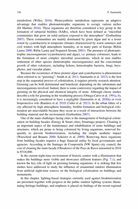
metabolism (Willey 2016). Photosynthetic metabolism represents an adaptiveadvantage that enables photoautotrophic organisms to occupy various niches(Di Martino 2016). These organisms are therefore considered a key group in theformation of subaerial biofilms (SABs), which have been defined as “microbialcommunities that grow on solid surfaces exposed to the atmosphere” (Gorbushina2007). These communities are mainly dominated by green algae and to a lesserextent by cyanobacteria in temperate climates characterized by warm summers andcool winters with high atmospheric humidity, as in many parts of Europe (RifónLastra 2000; Rifón Lastra and Noguerol Seoane 2001). The presence of photoauto-trophic microorganisms (cyanobacteria and algae), i.e. primary colonizers, leads tothe formation of more complex microbial associations, which in turn enablessettlement of other species (heterotrophic microorganisms) and the concomitantgrowth of other colonizers, including lichens, heterotrophic bacteria, fungi, bryo-phytes, and vascular plants.
Because the occurrence of these pioneer algae and cyanobacteria (a phenomenonoften referred to as “greening”: Smith et al. 2011; Sanmartín et al. 2012) is the firststep in the sequential process of colonization, it requires priority research attention.Greening can be both detrimental and beneficial, depending on the substratum andmicroorganisms involved. Indeed, there is some controversy regarding the impact ofgreening on the physical and chemical integrity of stone. Although classic studiesdescribe a role for greening in the weathering process (Ortega-Calvo et al. 1991a, b),it is increasingly considered to have a negligible (Sanmartín et al. 2020a) or evenbioprotective role (Ramírez et al. 2010; Cutler et al. 2013). In the urban fabric of acity affected by high atmospheric humidity, biofilm formation and biological colo-nization are unavoidable because they occur as a result of interactions between thebuilding material and the environment (Gorbushina 2007).
One of the main challenges facing cities is the management of biological coloni-zation on building facades (Energy & Smart cities; Smartiago project). Cleaning isan important aspect of the maintenance and rehabilitation of stone buildings andstructures, which are prone to being colonized by living organisms, removed fre-quently to prevent biodeterioration, including the simple aesthetic impact(Warscheid and Braams 2000; Scheerer et al. 2009). Removing these organismsfrom building facades requires a huge financial outlay by local governments andagencies. According to the Santiago de Compostela (NW Spain) city council, thecost of cleaning the main facade (Obradoiro) of the Pazo de Raxoi amounted in 2016to €318,000.
In the current night-time environment of historic centres with a illumination thatmakes the buildings more visible and showcases different features (Fig. 7.1), andknown the key role of light on greening-forming organisms; it is striking that fewstudies have addressed to study the influence of ornamental outdoor illuminationfrom artificial night-time sources on the biological colonization on buildings andmonuments.
In this chapter, lighting-based strategies currently used against biodeteriorationare presented together with progress in the public outdoor lighting systems illumi-nating heritage buildings, and emphasis is placed on findings of the recent regional
156 P. Sanmartín
2-year Light4Heritage project (2016–2018), focused on developing lighting-basedstrategies to control biological colonization and to manage the chromatic integrationof biofouling at laboratory scale. Finally, a critical analysis about in what formpublic lighting can be turned into a green method is also reported.
2 Lighting-Based Strategies Currently Used AgainstBiodeterioration
Lighting-based strategies that are currently used to control primary colonizers (algaeand cyanobacteria) on the built heritage are described below (Borderie et al. 2012;Figueroa et al. 2017; Sanmartín et al. 2018a):
Restricting the Duration of Lighting The simplest way to restrict lampenflora,defined as “massive growth of cyanobacterial-based microbiota and of algae, mossesand other plants located near light sources in caves accessible to tourists” (Dobat1998; Mulec and Kosi 2009; Mulec 2012), is to limit the time that caves areilluminated (during visiting hours) thus increasing the length of the dark period(Figueroa et al. 2017).
Changing the Amount of Light The use of low-intensity lamps reduces biologicalcolonization. The use of low levels of illumination for short periods helps to preventand control the growth of phototrophic biofilms (Albertano and Bruno 2003). Thelight must be strong enough to cause photoinhibition, considered a reduction inphotosynthesis in response to increasing irradiance, although the term may refermore generally to inhibition of growth due to a change in lighting (Sanmartín et al.2018a).
Using Different Wavelengths of Monochromatic LED Lights The use of LEDswith light qualities enriched in the wavelengths with low photosynthetic quantumefficiency for the target phototrophs has a biostatic effect (Sanmartín et al. 2018b).However, when LED lighting is used to control phototrophs, the ability of theseorganisms to photoacclimatize (MacIntyre et al. 2002) and adapt to light (Tandeau
Fig. 7.1 Night-time view of the Hércules Tower (A Coruña, Galicia, Spain) illuminated withpurple, green, blue, and orange light in 2017. Source: Elena López
7 New Perspectives Against Biodeterioration Through Public Lighting 157
de Marsac 1977) must be taken into account, along with the presence of differentpigments in photosynthetic organisms and other organisms in the same biofilm.Nonetheless, light quality is one of the most important and well-studied lightparameters, and recent studies suggest that it is the main regulator of growthresponses in algae and cyanobacteria (Kehoe 2010; Bussell and Kehoe 2013;Wiltbank and Kehoe 2016). Table 7.1 summarizes the most important contributionsfrom previous studies regarding the responses of algae and cyanobacteria to LEDlight treatments.
Using UV (A, B, or C) Irradiation UV treatment is widely used to disinfectsurfaces. Ultraviolet light has a short wavelength and high energy and becomesmore energetic as the wavelength decreases, according to Planck’s quantum theory,so that the energy increases in the order UV-A (315–400 nm) < UV-B(280–315 nm) < UV-C (190–280 nm). The sun emits radiation in all three UVranges, but the UV radiation that reaches the Earth’s surface comprises around 95%UV-A and 5% UV-B. As a physical and germicidal treatment, UV-C irradiation is asuitable alternative method of controlling or limiting biofouling, and its potential todamage phototrophic biofilms in caves has been demonstrated (Borderie et al. 2012,2014, 2015). Although many studies have addressed the effects of UV rays notfiltered by the ozone layer, i.e. UV-A and UV-B radiation, on algal andcyanobacterial cultures (Sinha et al. 1996; Wingard et al. 1997; Yakovleva andTitlyanov 2001; Mullineaux 2001; Bischof et al. 2006; Castenholz and Garcia-Pichel 2012; Moon et al. 2012; Shang et al. 2018), few studies have focused onlonger-wave radiation applied to biological colonization in built heritage. Table 7.2summarizes the most important contributions from previous studies regarding theresponses of algae and cyanobacteria to UV light treatments.
All these aforementioned lighting studies are scarce in relation to poikilohydricorganisms, such as lichens and bryophytes, and even more so in relation to vascularplants on cultural heritage. To the best of my knowledge, only the EU-fundedECOLIGHT (Ecological effects of light pollution) project has made major advancesin discovering how terrestrial plants respond to artificial light at night. The projectfindings show that artificial light can prolong the retention of leaves in urbanenvironments and initiate early onset of bud burst in the spring, thus increasing therisk of exposure to frost and pathogens.
3 Public Outdoor Lighting Systems Illuminating HeritageBuildings
Historic centres are tourist attractions of enormous importance to the economy ofcities and regions, which today are faced with the important challenge of applyingnew technological and management strategies within the smart city concept. Thismodel aspires to use technological solutions to improve the management and
158 P. Sanmartín
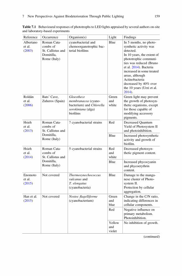
Table 7.1 Behavioural responses of phototrophs to LED lights appraised by several authors on-siteand laboratory-based experiments
Reference Occurrence Organism(s) Light Findings
Albertanoet al.(2003)
Roman Cata-combs ofSt. Callistus andDomitilla,Rome (Italy)
cyanobacterial andchemoorganotrophic bac-terial biofilms
Blue In 5 months, no photo-synthetic activity wasdetected.In 10 years, the extent ofphototrophic communi-ties was reduced (Brunoet al. 2014). Bacteriaincreased in some treatedareas, althoughActinobacteriadecreased by 40% overthe 10 years (Urzi et al.2014).
Roldánet al.(2006)
Bats’ Cave,Zuheros (Spain)
Gloeothecemembranacea (cyano-bacterium) and Chlorellasorokiniana (alga)biofilms
Greenandwhite
Green light may preventthe growth of photosyn-thetic organisms, exceptfor those capable ofmodifying accessorypigments.
Hsiehet al.(2013)
Roman Cata-combs ofSt. Callistus andDomitilla,Rome (Italy)
7 cyanobacterial strains Red Decreased QuantumYield of Photosystem IIand photoinhibition.
Blue Increased photosyntheticactivity and growth ofbiofilm.
Hsiehet al.(2014)
Roman Cata-combs ofSt. Callistus andDomitilla,Rome (Italy)
5 cyanobacterial strains Redandwhite
Decreased photosyn-thetic pigment content.
Blue Increased phycocyaninand phycoerythrincontent.
Enomotoet al.(2015)
Not covered Thermosynechococcusvulcanus andT. elongatus(cyanobacteria)
Blue Damage to the manga-nese cluster of Photo-system II.Protection by cellularaggregation.
Han et al.(2015)
Not covered Nostoc flagelliforme(cyanobacterium)
Greenandblue
Change in the C/N ratio,indicating differences incellular components.
Red Negative influence onprimary metabolism.Photoinhibition.
Yellowandviolet
No inhibition of growth.
(continued)
7 New Perspectives Against Biodeterioration Through Public Lighting 159
efficiency of the urban environment, with the ultimate aim of increasing urbansustainability (Energy & Smart cities).
There is currently an increasing trend for cities to install external lighting systemsto illuminate buildings and monuments. Lighting components, as floodlights andspotlights, equipped with lamps are placed on the building itself, on its facade,lighting the architectural surroundings. This trend is favoured by technologicalimprovements in lighting installations, specifically light emitting diode (LED)-based lighting. LED lighting has rapidly become sufficiently effective for use inexterior lighting systems and has several advantages over traditional lighting (lessenergy demanding, longer lifetime and mercury free). LED lamps not only increasethe temperature and reduce humidity on building facades, but the artificial light alsogenerates a specific physiological response in the colonizing organisms, interruptingtheir natural lighting cycle. This could favour the further development of biologicalcolonization (Fig. 7.2), as previously observed in the subterranean cultural heritage(caves, catacombs, necropolis, etc.) by Albertano and Bruno (2003) and Albertanoet al. (2003), and most recently by del Rosal and colleagues (del Rosal Padial et al.2016; Jurado et al. 2020). Indeed, the term lampenflora defined above was coined to
Table 7.1 (continued)
Reference Occurrence Organism(s) Light Findings
de Mooijet al.(2016)
Not covered Chlamydomonasreinhardtii CC-1690(green alga)
Red Low biomass.High specificgrowth rate.
Blue Low biomass.High specific growthrate. Increased QuantumYield of Photosystem II.
Yellow High biomass.Improved physiologicalstate of cells when com-bined with a small pro-portion of blue light.
Del RosalPadial(2016)
Nerja Cave,Malaga (Spain)
Biofilms containingChroococcidiopsis sp.,other cyanobacteria taxa,red alga Cyanidium sp.,and green algae
Green Reduced growth of pho-tosyntheticmicroorganisms.
Bruno andValle(2017)
Roman Cata-combs ofDomitilla,Rome (Italy)
3 cyanobacterial strainsand biofilms
Red,orangeandblue
In 2 months, improvedbiofilm growthHigh electrontransport rate.High chlorophyll-a andcarotenoids content.
Whiteandgreen
In 2 months, growthlimited.
160 P. Sanmartín
Table 7.2 Behavioural responses of phototrophs to UV lights appraised by several authors on-siteand laboratory-based experiments
Reference Occurrence Organism(s) Light Findings
Sinhaet al.(1996)
rice paddy fields near Vara-nasi (India)
4 cyanobacterialcultures
UV-BandUV-A
30’ to 5 Wm–2 causedthe death of 50% pop-ulation.Decrease in the uptakeof 14CO2 and proteincontent with increasedexposure time.
Wingardet al.(1997)
Alkaline hot spring in thecity of Rotorua(New Zealand)
4 cyanobacterialcultures
UV-B Increased negativeeffect on 14C incorpo-ration rate relative tothe control.Chlorophyll-a contentdecreased with increas-ing light intensity l.
Moonet al.(2012)
Not covered Synechocystissp. PCC 6803(cyanobacterium)
UV-A High-dose: negativephototactic response.Low-dose:photoregulation. Nega-tive phototaxisaffected.
Borderieet al.(2012)
3 caves of south west ofFrance (Domme, Laugerie-Haute, Combarelles,Department of Dordogne,France) and 1 cave ofnortheastern of France,Moidons Cave (Departmentof Jura, France)
mixed algaecultures
UV-C Chlorophyll-a, -b andcarotenoids contentwas affected.Cell viability decreasedgreatly.Programmed cell deathoccurred in unicellularalgae.Greater effectiveness incombination with highlight.
Borderieet al.(2014)
Moidons Cave (Departmentof Jura, France)
5 biofilms(mainly algae)
UV-C In 12 months, cleaningdue to light was effec-tive.In 16 months, biologi-cal colonization con-tinued to increase.Treatment effective-ness depended on bio-film thickness.
Borderieet al.(2015)
Moidons Cave (Departmentof Jura, France)
Green algae UV-C Damage was greater inthe planktonic than inthe biofilm mode ofgrowth.
Pfendleret al.(2017)
La Glacière Cave, Chaux-lès-Passavant (France)
Chlorellasp. (green alga)
UV-C In situ, discoloration ofbiofilm.In 21 months, no pho-tosynthetic
(continued)
7 New Perspectives Against Biodeterioration Through Public Lighting 161
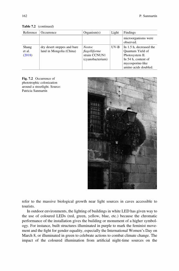
refer to the massive biological growth near light sources in caves accessible totourists.
In outdoor environments, the lighting of buildings in white LED has given way tothe use of coloured LEDs (red, green, yellow, blue, etc.) because the chromaticperformance of the installation gives the building or monument of a higher symbol-ogy. For instance, built structures illuminated in purple to mark the feminist move-ment and the fight for gender equality, especially the International Women’s Day onMarch 8, or illuminated in green to celebrate actions to combat climate change. Theimpact of the coloured illumination from artificial night-time sources on the
Table 7.2 (continued)
Reference Occurrence Organism(s) Light Findings
microorganisms wereobserved.
Shanget al.(2018)
dry desert steppes and bareland in Mongolia (China)
Nostocflagelliformestrain CCNUN1(cyanobacterium)
UV-B In 1.5 h, decreased theQuantum Yield ofPhotosystem II.In 54 h, content ofmycosporine-likeamino acids doubled.
Fig. 7.2 Occurrence ofphototrophic colonizationaround a streetlight. Source:Patricia Sanmartín
162 P. Sanmartín
biological colonization on buildings and monuments largely depends on the com-position and diversity of community, particularly its pigment content. Phototrophs(algae and cyanobacteria) need to live, to a greater or lesser extent, light fromdifferent parts of visible spectrum depending on the pigments that they contain. Ifthey are rich in chlorophyll-a and -b, which absorb in the red and blue regions ofspectrum, they should be more vulnerable to the effect of red and blue monochro-matic lights. If their higher pigment content is in phycobiliproteins phycocyanin andallophycocyanin, which absorb in the red region, red light will have a greater effect.Finally, if total carotenoids and phycobiliprotein phycoerythrin (absorbing in thegreenish-yellow and green regions respectively) are the main pigments, yellow andgreen lights will present the greater effect on organisms. However, this is anoversimplification. Microorganisms have several protective mechanisms that canbe triggered by certain qualities of light. Account should also be taken of the lightwith long wavelengths (such as red and orange light) are more penetrating.
Artificial lighting on outdoor constructions cannot be considered without alsoconsidering the effects of daylight, as monuments and structures are always exposedto natural light. As indicated above, very few studies have been published regardinghow urban monuments are affected by night-time outdoor illumination in combina-tion with natural sunlight. The Light4Heritage project (2016–2018) has focused ondeveloping lighting-based strategies to control biological colonization and to man-age the chromatic integration of biofouling at laboratory scale. The first study carriedout within the project involved the use of coloured cellophane films to generatedifferent types of light (by cancelling out the spectral components in certain bands ofthe visible electromagnetic spectrum, thus emulating monochromatic LED lights) atdifferent photon flux densities. The cellophane films were used to cover phototrophiccultures, derived from natural biofilm growing on a historic granitic building andmainly comprising green algae and cyanobacteria, in order to promote specificphysiological responses. The blue cellophane inhibited growth of the test culture,while the yellow cellophane did not significantly decrease the biomass, pigment orEPS content, relative to uncovered, control cultures. The different coloured cello-phane covers also generated colour changes in the cultures; e.g. the red cellophaneproduced notable greening, whereas the green cellophane enhanced the redness ofthe cultures (Sanmartín et al. 2017). Further experiments were carried out usingphototrophs in biofilm mode of growth and LED lights. In these studies,phototrophic biofilms thrived well under blue LEDs, whereas green and red LEDshad biostatic effects (Sanmartín et al. 2018b). Phototrophs responded differently toexposure to different coloured light: the biofilms developed under blue light pre-dominantly comprised algae, and those exposed to red and green light mainlycomprised cyanobacteria (P. Sanmartín, unpublished results). Regarding the dura-tion (number of hours daily) of LED illumination, a period of 4 hours provedsufficient to reduce colonization under red and green LED lights, while a period of8 h proved optimal for further growth of organisms under blue LED light(P. Sanmartín, unpublished results). Finally, among cross-sectional studies, fromproject Light4Heritage, on the effects of UV-A and UV-B on biological
7 New Perspectives Against Biodeterioration Through Public Lighting 163
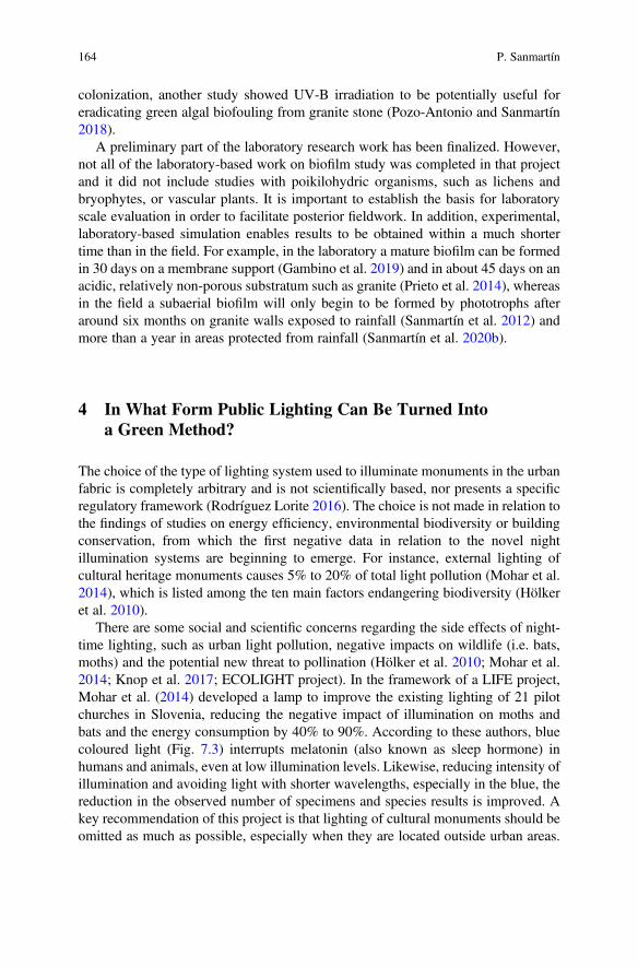
colonization, another study showed UV-B irradiation to be potentially useful foreradicating green algal biofouling from granite stone (Pozo-Antonio and Sanmartín2018).
A preliminary part of the laboratory research work has been finalized. However,not all of the laboratory-based work on biofilm study was completed in that projectand it did not include studies with poikilohydric organisms, such as lichens andbryophytes, or vascular plants. It is important to establish the basis for laboratoryscale evaluation in order to facilitate posterior fieldwork. In addition, experimental,laboratory-based simulation enables results to be obtained within a much shortertime than in the field. For example, in the laboratory a mature biofilm can be formedin 30 days on a membrane support (Gambino et al. 2019) and in about 45 days on anacidic, relatively non-porous substratum such as granite (Prieto et al. 2014), whereasin the field a subaerial biofilm will only begin to be formed by phototrophs afteraround six months on granite walls exposed to rainfall (Sanmartín et al. 2012) andmore than a year in areas protected from rainfall (Sanmartín et al. 2020b).
4 In What Form Public Lighting Can Be Turned Intoa Green Method?
The choice of the type of lighting system used to illuminate monuments in the urbanfabric is completely arbitrary and is not scientifically based, nor presents a specificregulatory framework (Rodríguez Lorite 2016). The choice is not made in relation tothe findings of studies on energy efficiency, environmental biodiversity or buildingconservation, from which the first negative data in relation to the novel nightillumination systems are beginning to emerge. For instance, external lighting ofcultural heritage monuments causes 5% to 20% of total light pollution (Mohar et al.2014), which is listed among the ten main factors endangering biodiversity (Hölkeret al. 2010).
There are some social and scientific concerns regarding the side effects of night-time lighting, such as urban light pollution, negative impacts on wildlife (i.e. bats,moths) and the potential new threat to pollination (Hölker et al. 2010; Mohar et al.2014; Knop et al. 2017; ECOLIGHT project). In the framework of a LIFE project,Mohar et al. (2014) developed a lamp to improve the existing lighting of 21 pilotchurches in Slovenia, reducing the negative impact of illumination on moths andbats and the energy consumption by 40% to 90%. According to these authors, bluecoloured light (Fig. 7.3) interrupts melatonin (also known as sleep hormone) inhumans and animals, even at low illumination levels. Likewise, reducing intensity ofillumination and avoiding light with shorter wavelengths, especially in the blue, thereduction in the observed number of specimens and species results is improved. Akey recommendation of this project is that lighting of cultural monuments should beomitted as much as possible, especially when they are located outside urban areas.
164 P. Sanmartín
Another important recommendation is that after 23.00 hours, lighting should beswitched off in order to attract fewer moths.
Furthermore, the energy consumed by the lighting systems increases the level ofCO2 in the atmosphere. Although the simple change of incandescence (e.g. metalhalide and sodium vapour lamps) to LED technology in public lighting would bereduced by up to 90% this emission. An example is the lighting of the Puerta deAlcalá in Madrid, which until 2014 had a total consumption of 41,470 W and atpresent uses 4660 W of energy (Rodríguez Lorite 2016). In Spain, consumption of1 kWh of energy is equivalent to emitting 430 g of CO2 into the atmosphere (SanMartín Páramo and Ferrero Andreu 2008 apud Rodríguez Lorite 2016), so that theillumination of this monument every night causes the release of 3440 g of CO2 to theatmosphere, one tenth of what was issued until 6 years ago.
Regarding the biodiversity of higher plants, in the ECOLIGHT project mentionedabove, constant artificial light was also found to increase the rates of foliar damagedue to ozone in three clover species and in Lotus pedunculatus, which produced 10%to 25% fewer flower heads under simulated street lighting, which in turn led toreduced numbers of the aphid Acyrthosiphon pisum (Bennie et al. 2016, 2018).These researchers also value positively the implementation of LED technology inpublic lighting to achieve an environmentally sustainable system.
Fig. 7.3 Façade of the San Fiz de Solovio Church in Santiago de Compostela (Galicia, Spain)illuminated in blue light in 2018. Source: Justo Arines
7 New Perspectives Against Biodeterioration Through Public Lighting 165
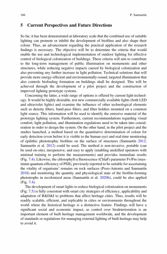
5 Current Perspectives and Future Directions
So far, it has been demonstrated at laboratory scale that the combined use of suitablelighting can promote or inhibit the development of biofilms and also shape theircolour. Thus, an advancement regarding the practical application of the researchfindings is necessary. The objective will be to determine the criteria that wouldenable the use and technological implementation of outdoor lighting for effectivecontrol of biological colonization of buildings. These criteria will aim to contributeto the long-term management of public illumination on monuments and otherstructures, while reducing negative impacts caused by biological colonization andalso preventing any further increase in light pollution. Technical solutions that willprovide more energy-efficient and environmentally-sound, targeted illumination thatalso controls biofouling formation on buildings shall be designed. This will beachieved through the development of a pilot project and the construction ofimproved lighting prototype systems.
Concerning the latter, a wide range of options is offered by current light technol-ogy. It would be highly desirable, test new commercially available lights (both LEDand ultraviolet lights) and examine the influence of other technological elementssuch as density filters, band-pass filters, and filter holders mounted on a commonlight source. This information will be used to identify the emissive material of theprototype lighting system. Furthermore, current recommendations regarding visualcomfort, light pollution, and illumination regulations need to be taken into consid-eration in order to design the system. On the other hand, in the pilot project and thestudies launched, a method based on the quantitative determination of colour forearly detection (even before it is visible to the human eye) and real-time monitoringof epilithic phototrophic biofilms on the surface of structures (Sanmartín 2012;Sanmartín et al. 2012) could be used. The method is non-invasive, portable (canbe used on-site), inexpensive, and easy to apply (enabling unskilled operators withminimal training to perform the measurements) and provides immediate results(Fig. 7.4). Likewise, the chlorophyll-a fluorescence (ChlaF) parameter Fv/Fm (max-imum quantum efficiency of PSII), previously reported to be suitable for ascertainingthe vitality of organisms’ remains on rock surfaces (Pozo-Antonio and Sanmartín2018) and monitoring the quantity and physiological state of the biofilm-formingphototrophs in recolonized areas (Sanmartín et al. 2020b), could be also applied(Fig. 7.4).
The development of smart lights to reduce biological colonization on monuments(Fig. 7.5) is fully consistent with smart city strategies of efficiency, applicability andadaptation of R&D&I to problems that affect heritage cities. Thus, results will bereadily scalable, efficient, and replicable in cities or environments throughout theworld where the historical heritage is a distinctive feature. Findings will have asignificant social and economic impact, as control over biodeterioration is animportant element of built heritage management worldwide, and the developmentof standards or regulations for managing external lighting of built heritage may helpto avoid it.
166 P. Sanmartín
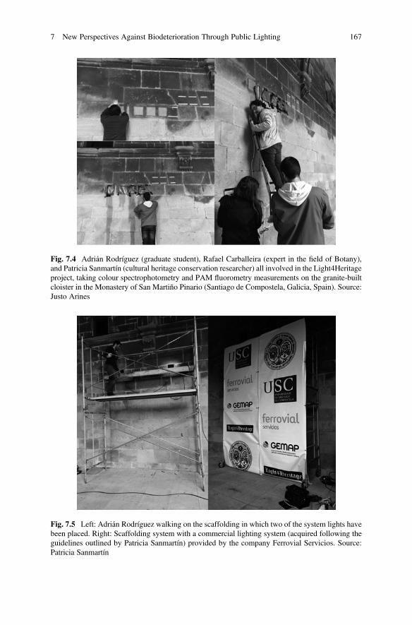
Fig. 7.4 Adrián Rodríguez (graduate student), Rafael Carballeira (expert in the field of Botany),and Patricia Sanmartín (cultural heritage conservation researcher) all involved in the Light4Heritageproject, taking colour spectrophotometry and PAM fluorometry measurements on the granite-builtcloister in the Monastery of San Martiño Pinario (Santiago de Compostela, Galicia, Spain). Source:Justo Arines
Fig. 7.5 Left: Adrián Rodríguez walking on the scaffolding in which two of the system lights havebeen placed. Right: Scaffolding system with a commercial lighting system (acquired following theguidelines outlined by Patricia Sanmartín) provided by the company Ferrovial Servicios. Source:Patricia Sanmartín
7 New Perspectives Against Biodeterioration Through Public Lighting 167
Finally, the public response to the new lighting developed and installed should betaken into account to enable evaluation of the lighting systems from a perceptual,and not only procedural, viewpoint.
Acknowledgements The author is grateful to Elena López and Adrián Rodríguez, who carried outa master thesis (MSc) and a final year (BSc) research project on this subject under her supervision.She also thanks Dr Justo Arines (Universidade de Santiago de Compostela, Spain) and Dr RafaelCarballeira (Universidade da Coruña, Spain) their collaboration on the Light4Heritage projectstudies. Finally, she thanks the financial support of Xunta de Galicia grant ED431C 2018/32.
References
Albertano P, Bruno L (2003) The importance of light in the conservation of hypogean monuments.In: Saiz-Jiménez C (ed) Molecular biology and cultural heritage. Balkema, Lisse, Rotterdam,The Netherlands, pp 171–177
Albertano P, Moscone D, Palleschi G, Hermosin B, Saiz-Jiménez C, Sánchez-Moral S, Hernández-Mariné M, Urzí C, Groth I, Schroeckh V, Gallon JR, Graziottin F, Bisconti F, Giuliani R (2003)Cyanobacteria attack rocks (CATS): control and preventive strategies to avoid damage causedby cyanobacteria and associated microorganisms in Roman hypogean monuments. In: Saiz-Jiménez C (ed) Molecular biology and cultural heritage. Balkema, Lisse, Rotterdam, TheNetherlands, pp 151–162
Bennie J, Davies TW, Cruse D, Gaston KJ (2016) Ecological effects of artificial light at night onwild plants. J. Ecol 104:611–620
Bennie J, Davies TW, Cruse D, Bell F, Gaston KJ (2018) Artificial light at night alters grasslandvegetation species composition and phenology. J Appl Ecol 55:442–450
Bischof K, Gomez I, Molis M, Hanelt D, Karsten U, Lüder U, Roleda MY, Zacher K, Wiencke C(2006) Ultraviolet radiation shapes seaweed communities. Environ Sci Biotechnol 5:141–166
Borderie F, Alaoui-Sehmer L, Bousta F, Orial G, Rieffel D, Richard H, Alaoui-Sossé B (2012) UVirradiation as an alternative to chemical treatments: a new approach against algal biofilmsproliferation contaminating building facades, historical monuments and touristic subterraneanenvironments. In: Krueger D, Meyer H (eds) Algae: ecology, economic uses and environmentalimpact. Nova Science Publishers, Inc. Hauppauge, New York, pp 1–28
Borderie F, Tête N, Cailhol D, Alaoui-Sehmer L, Bousta F, Rieffel D, Aleya L, Alaoui-Sossé B(2014) Factors driving epilithic algal colonization in show caves and new insights into combat-ing biofilm development with UV-C treatments. Sci Total Environ 484:43–52
Borderie F, Alaoui-Sossé B, Aleya L (2015) Heritage materials and biofouling mitigation throughUV-C irradiation in show caves: states-of-the-art practices and future challenges. Environ SciPollut Res 22:4144–4172
Bruno L, Valle V (2017) Effect of white and monochromatic lights on cyanobacteria and biofilmsfrom Roman Catacombs. Int Biodeteri Biodegr 123:286–295
Bruno L, Belleza S, Urzì C, De Leo F (2014) A study for monitoring and conservation in the RomanCatacombs of St. Callistus and Domitilla, Rome (Italy). In: Saiz-Jimenez C (ed) The conserva-tion of subterranean cultural heritage. CRC Press/Bakelma/Taylor & Francis Group, Leiden,The Netherlands, pp 37–44
Bussell AN, Kehoe DM (2013) Control of a four-color sensing photoreceptor by a two color sensingphotoreceptor reveals complex light regulation in cyanobacteria. Proc Natl Acad Sci USA110:12834–12839
Castenholz RW, Garcia-Pichel F (2012) Cyanobacterial responses to UV-radiation. In: Whitton B(ed) Ecology of cyanobacteria II. Springer, Dordrecht, The Netherlands, pp 591–611
168 P. Sanmartín
Cutler NA, Viles HA, Ahmad S, McCabe S, Smith BJ (2013) Algal “greening” and the conservationof stone heritage structures. Sci Total Environ 442:152–164
de Mooij T, de Vries G, Latsos C, Wijffels RH, Janssen M (2016) Impact of light color onphotobioreactor productivity. Algal Res 15:32–42
del Rosal Padial Y (2016) Análisis, impacto y evolución de los biofilms fotosintéticos enespeleotemas, El caso de la Cueva de Nerja, Málaga. PhD thesis. Universidad de Málaga, Spain.
del Rosal Padial Y, Jurado Lobo V, Hernández Mariné M, Roldán Molina M, Sáiz Jiménez C(2016) Biofilms en cuevas turísticas: La Cueva de Nerja y la Cueva del Tesoro. In: Andreo B,Durán JJ (eds) El Karst y el Hombre: Las Cuevas como Patrimonio Mundial. Asociación deCuevas Turísticas Españolas, Madrid, Spain, pp 103–114
Di Martino P (2016) What about biofilms on the surface of stone monuments. Open Conf Proc J7:14–28
Dobat K (1998) Flore de la lumière artificielle (Lampenflora - Maladie verte). In: Juberthie C, DecuV (eds) Encyclopaedia Biospeleologica, Société Internationale de Biospéologie - InternationalSociety for Subterranean Biology. Moulis-Bucarest, France-,Romania, vol II, pp 1325–1335
ECOLIGHT Project Available online: https://www.exeter.ac.uk/esi/research/currentresearch/ecolight/ Accessed Dic 2019
Energy & Smart cities – European Comission’s priority policies. Available online: https://ec.europa.eu/info/eu-regional-and-urbandevelopment/topics/cities-and-urban-development/city-initiatives/smart-cities_en Accessed Dic 2019
Enomoto G, Win NN, Narikawa RR, Ikeuchi M (2015) Three cyanobacteriochromes work togetherto form a light color-sensitive input system for c-di-GMP signaling of cell aggregation. ProcNatl Acad Sci USA 112:8082–8087
Figueroa FL, Álvarez-Gómez F, del Rosal Y, Celis-Plá PSM, González G, Hernández M, Korbee N(2017) In situ photosynthetic yields of cave photoautotrophic biofilms using two different pulseamplitude modulated fluorometers. Algal Res 22:104–115
Gambino M, Sanmartín P, Longoni M, Villa F, Mitchell R, Cappitelli F (2019) Surface colour: anoverlooked aspect in the study of cyanobacterial biofilm formation. Sci Total Environ659:342–353
Gorbushina AA (2007) Life on the rocks. Environ Microbiol 9:1613–1631Han PP, Shen SG, Wang HY, Sun Y, Dai YJ, Jia SR (2015) Comparative metabolomic analysis of
the effects of light quality on polysaccharide production of cyanobacterium Nostoc flageliforme.Algal Res 9:143–150
Hölker F, Wolter C, Perkin EK, Tockner K (2010) Light pollution as a biodiversity threat. TrendsEcol Evol 25:681–682
Hsieh P, Pedersen JZ, Albertano P (2013) Generation of reactive oxygen species upon red lightexposure of cyanobacteria from Roman hypogea. Int Biodeter Biodegr 84:258–265
Hsieh P, Pedersen JZ, Bruno L (2014) Photoinhibition of cyanobacteria and its application incultural heritage conservation. Photochem Photobiol 90:533–543
Jurado V, del Rosal Y, Gonzalez-Pimentel JL, Hermosin B, Saiz-Jimenez C (2020) Biologicalcontrol of phototrophic biofilms in a show cave: the case of Nerja Cave. Appl Sci 10:3448
Kehoe DM (2010) Chromatic adaptation and the evolution of light color sensing in cyanobacteria.Proc Natl Acad Sci USA 107:9029–9030
Knop E, Zoller L, Ryser R, Gerpe C, Hörler M, Fontaine C (2017) Artificial light at night as a newthreat to pollination. Nature 548:206–209
MacIntyre HL, Kana TM, Anning T, Geider RJ (2002) Photo-acclimation of photosynthesisirradiance response curves and photosynthetic pigments in microalgae and cyano- bacteria. JPhycol 38:17–38
Mohar A, Zagmajster M, Verovnik R, Skaberne BB (2014) Nature-friendlier lighting of objects ofcultural heritage (churches): Recommendations. Dark-Sky Slovenia 2014, Available online:http://temnonebo.com/images/pdf/nature_friendler_lightning_churces_booklet_web.pdf.Accessed June 2020
7 New Perspectives Against Biodeterioration Through Public Lighting 169
Moon Y, Kim S, Chung Y (2012) Sensing and responding to UV-A in cyanobacteria. Int J Mol Sci13:16303–16332
Mulec J (2012) Lampenflora. In: White WB, Culver DC (eds) Encyclopedia of caves. Elsevier/Academic Press, Amsterdam, The Netherlands, pp 451–456
Mulec J, Kosi G (2009) Lampenflora algae and methods of growth control. J Cave Karst Stud71:109–115
Mullineaux CW (2001) How do cyanobacteria sense and respond to light? Mol Microbiol41:965–971
Ortega-Calvo JJ, Hernández-Mariné M, Saiz-Jimenez C (1991a) Biodeterioration of buildingsmaterials by cyanobacteria and algae. Int Biodeter 28:165–185
Ortega-Calvo JJ, Hernández-Mariné M, Saiz-Jimenez C (1991b) Mechanical deterioration ofbuilding stones by cyanobacteria and algae. In: Roossmoore KW (ed) Biodeterioration andbiodegradation. Elsevier, London, pp 392–394
Pfendler S, Einhorn O, Karimi B, Bousta F, Cailhol D, Alaoui-Sosse L, Alaoui-Sosse B, Aleya L(2017) UV-C as an efficient means to combat biofilm formation in show caves: evidence fromthe La Glaciere Cave (France) and laboratory experiments. Environ Sci Pollut Res24:24611–24623
Pozo-Antonio JS, Sanmartín P (2018) Exposure to artificial daylight or UV-irradiation (A, B or C)prior to chemical cleaning: an effective combination for removing phototrophs from granite.Biofouling 34:851–869
Prieto B, Sanmartín P, Silva C, Vázquez-Nion D, Silva B (2014) Deleterious effect plastic-basedbiocides on back-ventilated granite facades. Int Biodeter Biodegr 86:19–24
Ramírez M, Hernández-Mariné M, Novelo E, Roldán M (2010) Cyanobacteria-containing biofilmsfrom a Mayan monument in Palenque, Mexico. Biofouling 26:399–409
Rifón Lastra A (2000) Algas epilíticas en monumentos de interés histórico de Galicia. PhD thesis.Universidade da Coruña, Spain
Rifón Lastra A, Noguerol Seoane A (2001) Green algae associated with the granite walls ofmonuments in Galicia (NW Spain). Cryptogamie Algol 22:305–326
Rodríguez Lorite MA (2016) Guía de Iluminación Eficiente de Monumentos. Dirección General deIndustria, Energía y Minas. DL: M-21749-2016. Available online: http://www.madrid.org/bvirtual/BVCM015700.pdf Accessed Dic 2019
Roldán M, Oliva F, Gónzales del Valle MA, Saiz-Jimenez C, Hernández-Mariné M (2006) Doesgreen light influence the fluorescence properties and structure of phototrophic biofilms? ApplEnviron Microbiol 72:3026–3031
San Martín Páramo R, Ferrero Andreu L (2008) Los costos de la implantación y el mantenimientode las instalaciones de alumbrado exterior. CONAMA 9. Congreso Nacional del MedioAmbiente. Cumbre del Desarrollo Sostenible. Madrid, Spain. December 1–5, 2008
Sanmartín P (2012) Color quantification in the study of biofilm formation on granite stone inhistorical and artistic heritage. PhD thesis. Universidade de Santiago de Compostela, Spain
Sanmartín P, Vázquez-Nion D, Silva B, Prieto B (2012) Spectrophotometric color measurement forearly detection and monitoring of greening on granite buildings. Biofouling 28:329–338
Sanmartín P, Vázquez-Nion D, Arines J, Cabo-Domínguez L, Prieto B (2017) Controlling growthand colour of phototrophs by using simple and inexpensive coloured lighting: a preliminarystudy in the Light4Heritage project towards future strategies for outdoor illumination. IntBiodeter Biodegr 122:107–115
Sanmartín P, DeAraujo A, Vasanthakumar A (2018a) Melding the old with the new: trends inmethods used to identify, monitor and control microorganisms on cultural heritage materials.Microb Ecol 76:64–80
Sanmartín P, Vázquez-Nion D, Silva B, Prieto B, Arines J (2018b) Assessing the effect of differentcoloured lighting in controlling biological colonization. In: Mosquera MJ, Gil A (eds) Conserv-ing cultural heritage. CRC Press/Bakelma/Taylor & Francis Group, Leiden, The Netherlands, pp313–318
170 P. Sanmartín
Sanmartín P, Villa F, Cappitelli F, Balboa S, Carballeira R (2020a) Characterization of a biofilm andthe pattern outlined by its growth on a granite-built cloister in the Monastery of San MartiñoPinario (Santiago de Compostela, NW Spain). Int Biodeter Biodegr 147:104871
Sanmartín P, Rodríguez A, Aguiar U (2020b) Medium-term field evaluation of several widely usedcleaning-restoration techniques applied to phototrophic algal biofilm formed on a granite-builthistorical monument. Int Biodeter Biodegr 147:104870
Scheerer S, Ortega-Morales O, Gaylarde C (2009) Chapter 5 - Microbial deterioration of stonemonuments - an updated overview. In: Laskin AI, Sariaslani S, Gadd GM (eds) Adv ApplMicrobiol, Elsevier Inc., Academic Press, Cambridge, vol. 66:97–139
Shang JL, Zhang ZC, Yin XY, Chen M, Hao FH, Wang K, Feng JL, Xu HF, Yin YC, Tang HR, QiuBS (2018) UV-B induced biosynthesis of a novel sunscreen compound in solar radiation anddesiccation tolerant cyanobacteria. Environ Microbiol 20:200–213
Sinha RP, Singh N, Kumar A, Kumar HD, Häder M, Häder DP (1996) Effects of UV irradiation oncertain physiological and biochemical processes in cyanobacteria. J Photochem Photobiol B:Biol 32:107–113
Smartiago Project, Available online: https://smartiago.santiagodecompostela.gal Accessed Dic2019
Smith BJ, McCabe S, McAllister D, Adamson C, Viles HA, Curran JM (2011) A commentary onclimate change, stone decay dynamics and the ‘greening’ of natural stone buildings: newperspectives on ‘deep wetting’. Environ Earth Sci 63:1691–1700
Tandeau de Marsac N (1977) Occurrence and nature of chromatic adaptation in cyanobacteria. JBacteriol 130:82–91
Urzi C, De Leo F, Bruno L, Pangallo D, Krakova L (2014) New species description, biomineral-ization processes and biocleaning applications of Roman catacombs-living bacteria. In: Saiz-Jimenez C (ed) The conservation of subterranean cultural heritage. CRC Press/Bakelma, Taylor& Francis Group, Leiden, The Netherlands, pp 65–72
Warscheid T, Braams J (2000) Biodeterioration of stone: a review. Int Biodeterior Biodegrad46:343–368
Willey N (2016) Environmental plant physiology. Taylor & Francis, Garland Science, New YorkWiltbank LB, Kehoe DM (2016) Two cyanobacterial photoreceptors regulate photosynthetic light
harvesting by sensing teal, green, yellow, and red light. MBio 7:e02130–e02115Wingard CE, Schiller JR, Casnnholz RW (1997) Evidence regarding the possible role of
c-phycoerythrin in ultraviolet-B tolerance in a thermophilic cyanobacterium. PhotochemPhotobiol 65:833–842
Yakovleva IM, Titlyanov EA (2001) Effect of visible and UV irradiance on subtidal Chondruscrispus: Stress, photoinhibition and protective mechanisms. Aquat Bot 71:47–61
Open Access This chapter is licensed under the terms of the Creative Commons Attribution 4.0International License (http://creativecommons.org/licenses/by/4.0/), which permits use, sharing,adaptation, distribution and reproduction in any medium or format, as long as you give appropriatecredit to the original author(s) and the source, provide a link to the Creative Commons license andindicate if changes were made.
The images or other third party material in this chapter are included in the chapter's CreativeCommons license, unless indicated otherwise in a credit line to the material. If material is notincluded in the chapter's Creative Commons license and your intended use is not permitted bystatutory regulation or exceeds the permitted use, you will need to obtain permission directly fromthe copyright holder.
7 New Perspectives Against Biodeterioration Through Public Lighting 171

Part IIIBiocleaning and Bio-Based
Conservation Methods
Chapter 8Bioremoval of Graffiti in the Contextof Current Biocleaning Research
Pilar Bosch-Roig and Patricia Sanmartín
Abstract Some microorganisms can be used as bioremediation agents, inbiocleaning treatments, to remove undesired sulphates, nitrates and organic matterfrom cultural heritage surfaces. Graffiti materials (mainly spray paints) are nowincluded in the list of materials that can be biocleaned, with studies on this topicbeing initiated just over 5 years ago. Research on the bioremoval of graffiti iscontinuing and on a promising track. This chapter reports a critical analysis ofstudies of the bioremoval of graffiti carried out in recent years, which are comparedwith similar studies of the removal of salts (mainly nitrates and sulphates) andorganic matter conducted in the last thirty years. Likewise, the present challengesand ways of overcoming them are addressed towards developing a complete proto-col for the use of bioremediation to remove graffiti, with particular emphasis on theuse of the method for cleaning facades and buildings.
Keywords Bacterial strains · Biocleaning · Cultural heritage · Microbial agents ·Safe methods · Stone
1 Introduction
The use of naturally occurring bacteria, and to lesser extent fungi, in biocleaningtreatments for cultural heritage buildings and monuments is currently popular andprovides an effective, non-invasive, relatively ecologically safe and inexpensiveapproach to cultural heritage conservation (Bosch-Roig and Ranalli 2014; Sanmartín
P. Bosch-Roig (*)Instituto Universitario de Restauración del Patrimonio, Universitat Politècnica de València,Valencia, Spaine-mail: [email protected]
P. SanmartínDepartamento de Edafoloxía e Química Agrícola. Facultade de Farmacia, Universidade deSantiago de Compostela, Santiago de Compostela, Spaine-mail: [email protected]
© The Author(s) 2021E. Joseph (ed.), Microorganisms in the Deterioration and Preservation of CulturalHeritage, https://doi.org/10.1007/978-3-030-69411-1_8
175
et al. 2018). From the beginning of the 1990s until the present, viable(non-pathogenic) cells and selected enzymes have been identified and used for thebiological removal of undesired substances, such as salts (mainly nitrates andsulphates) and organic matter from stone substrates (Webster and May 2006). Inthe last few years graffiti paint has been added to this list as an undesired, potentiallybioremovable material. The inclusion of graffiti paint in this list arose as a result ofthe drawbacks and environmental impacts of the methods then available (chemicaland physical, including laser) for graffiti removal (Sanmartín et al. 2014). A need todevelop a new inexpensive, risk-free method, especially designed for use in porousmaterials (such as natural stone, cement and mortar lime) was also recognized(Sanmartín et al. 2014; Sanmartín and Bosch-Roig 2019).
Every year large amounts of money and effort are expended in removingdisfiguring graffiti from urban fabric surfaces (Sanmartín et al. 2014, 2016). Forexample, in the early 2000s about 20,000 private buildings and more than 400 munic-ipal buildings in the city of Milan (Italy) were estimated to be disfigured every year(DDL 1607 2002). However, extrapolating the findings on bioremoval of salts andorganic matter to graffiti is not an easy or direct task, and it is necessary to firstidentify microorganisms as potential candidates. In relation to the occurrence ofmicroorganisms on paints, an earlier review paper concerning the biodeterioration ofarchitectural paint by bacteria, fungi and algae indicated that studies on the microbialecology of paints are scarce (Gaylarde et al. 2011). One study of painted walls inLatin America described microbial populations mainly formed by bacteria (includ-ing actinobacteria and cyanobacteria) as well as by populations of algae, protozoa,rotifers, nematodes and a huge variety of fungi (Gaylarde and Gaylarde 2000). Thesame authors also reported that biomass on external painted walls of buildings inEurope and Latin America is generally composed of fungi and to a lesser extentactinobacteria, which are more commonly found on building surfaces in Europe thanin Latin America (Gaylarde and Gaylarde 2005). However, these results do not applyto other types of surfaces. Hence, some authors have reported that actinobacteria arecommonly found on different types of damaged materials and buildings(e.g. ceramics and sandstone monuments) in Europe and on floor dust in damagedbuildings in the USA (Hyvärinen et al. 2002; Palla et al. 2002; Macedo et al. 2009;Park et al. 2017). Allsopp et al. (2004) listed the fungi and phototrophs detected onpaint films comprising refined and processed materials. Furthermore, in a review ofseveral published studies on microbial colonization of frescoes and in regard to thepresence of microorganisms and their role in damage to painted surfaces, Ciferri(1999) concluded that bacteria may be the first colonizers. After bacterial coloniza-tion chemically modifies some of the components of the paint and renders themutilizable, fungi may be able to become established and often become the principalmicroflora on painted surfaces (Ciferri 1999; Gaylarde et al. 2011). Whether or notthey are the most abundant organisms, fungi have long been considered the majorbiodeteriogens in painted surfaces, producing paint-degrading metabolites (princi-pally organic acids) that weaken and then penetrate the film (Grant et al. 1986). Forexample, during the 1950s, some researchers considered Aureobasidium pullulans(formerly Pullularia pullulans) to be the main biological agent involved in paint
176 P. Bosch-Roig and P. Sanmartín
deterioration (Reynolds 1950; Klens and Lang 1956). Filamentous algae andactinobacteria can also alter painted surfaces by penetrating the paint film andreleasing reactive metabolites that reduce the effectiveness and lifespan of thecoating (Gaylarde et al. 2011). However, more recently, a study involving bioreme-diation processes showed that black fungi are not suitable for biocleaning stoneartefacts, as they can damage stone treated with synthetic polymers (Troiano et al.2014).
Most microorganisms found on paint films are actually aerial contaminants andare not involved in degradation processes (Gaylarde et al. 2011). More specifically,very few microorganisms have been shown to degrade graffiti. Indeed, in the onlystudy to date of the microbial ecology of graffiti wall paintings, of a total of fifty-fourdifferent isolates, only nine bacterial isolates and one fungal isolate showed somecapacity to degrade spray paint graffiti; of these, only three were isolated from recentor old graffiti (Sanmartín et al. 2016).
2 Why Can Graffiti Paint Be Removed by BiocleaningTreatments?
Graffiti spray paint contains the following main ingredients: (i) pigment, whichprovides colour, (ii) binding medium, in which the pigment particles are dispersed,and which hardens and binds the pigments on the painted surface and (iii) solvent,which allows the pigment/binder mixture to flow. Acrylic- and alkyd-based bindersare common in current non-metallic graffiti formulations, while polyethylene-typebinders predominate in metallic formulations. Semi-synthetic binders are also rela-tively common, although increasingly less so. Thus, although a study conductedabout fifteen years ago showed that almost half of the varieties of red graffiti spraypaint contain alkyd-nitrocellulose based binders (Govaert and Bernard 2004), thesepaints have been displaced by red alkyd paints.
Graffiti spray paints contain man-made compounds both as binders and aspigments, and they can therefore be considered xenobiotic (synthetic) substances.Although it is commonly thought that synthetic substances are more resistant tomicrobial attack than natural components, several studies have demonstrated thecontrary (Cappitelli and Sorlini 2008; Troiano et al. 2014). Moreover, paint containsa wide variety of organic and inorganic biodegradable components, includingadditives such as emulsifiers, thickeners, etc., which may be utilized for growth bya large variety of microbial species (Ciferri 1999; Sanmartín et al. 2014). Mostbiodegradable substances, including graffiti paints, contain a mixture of carbon andelements such as oxygen, nitrogen, sulphur and phosphorus, which create chargeimbalances that enzyme-containing bacteria can exploit. Thus, in previous studies,aged synthetic resins in outdoor environments (Cappitelli et al. 2007) and syntheticpolymers in indoor environments (Lustrato et al. 2012) have been utilized bymicroorganisms as growth substrates. Likewise, cases have been reported wherein
8 Bioremoval of Graffiti in the Context of Current Biocleaning Research 177
some microorganisms, such as fungi and particularly bacteria, can modify spraypaint, even inside the cans (Bentley and Turner 1998). Microorganisms can enter thepaint via infected raw materials (including water) or unsterile equipment. Forexample, the presence of bacteria has been found to cause reduced viscosity, gassingand colour drift in latex paint (Bentley and Turner 1998). In this regard, thecomponents of spray paint affect microorganisms, either by inhibiting or stimulatingtheir development. Thus, cellulose derivatives can act as nutrients for fungal cells(Allsopp et al. 2004), whereas organic solvents and heavy metals in pigments canadversely affect fungi (Gaylarde et al. 2011). Higher proportions of resin in glosspaint may also yield greater bioresistance (Gaylarde et al. 2011).
Paint pigments may also affect the bioremoval process. In a study of differentlycoloured acrylic paint, red paint was colonized more rapidly by microbes than bluepaint, probably due to the presence of copper in the blue paint (� 0.4%), which actsas a microbiocide (Breitbach et al. 2011). Zinc oxide can also act as an antimicrobialagent and has been found to exert fungicidal activity at a concentration of 2% inoil-based paint (Turner 1967). Antimicrobial compounds are added to liquid paintboth to protect the paint in-can and to reduce fouling of the dry film. However, thereis no universally active antimicrobial ingredient that is compatible with all paintformulations and that meets the requirements of the coating manufacturers. Thus,mixtures of antimicrobials are often used, among which isothiazolinone-basedcompounds are widely used for conserving paint (Gaylarde et al. 2011). In a studyof 29 freshly dried synthetic resins used as paint binding media, almost all the acrylicresins proved to be resistant to fungal attack, while all alkyd resins and somepolyvinyl acetates (PVAs) were found to be biodegradable (Cappitelli et al. 2005).These findings indicate that under favourable environmental conditions, and con-sidering the same paint components, alkyd-based paint would be more readilybiodegraded than acrylic-based paint. The authors of the aforementioned studyalso noted that knowledge of the microbial susceptibility of the binding mediumunder certain conditions does not necessarily enable prediction of the potential of aspecific paint to undergo biodegradation. In fact, pigments and other componentsmay be readily available sources of carbon in paint formulations.
3 Methodological Advances in Bioremoval of Salts, OrganicMatter and Graffiti
Microorganisms have mainly been used to remove undesired inorganic salts (nitratesand sulphates) and organic substances (animal glue, casein, hydrocarbons, lipids,etc.) from inorganic surfaces (stone sculptures, building façades and frescoes). Fewstudies have been conducted in relation to the use of microorganisms to removepaints (included graffiti paints) produced in the twentieth and twenty-first centuriesand which contain synthetic polymers.
178 P. Bosch-Roig and P. Sanmartín
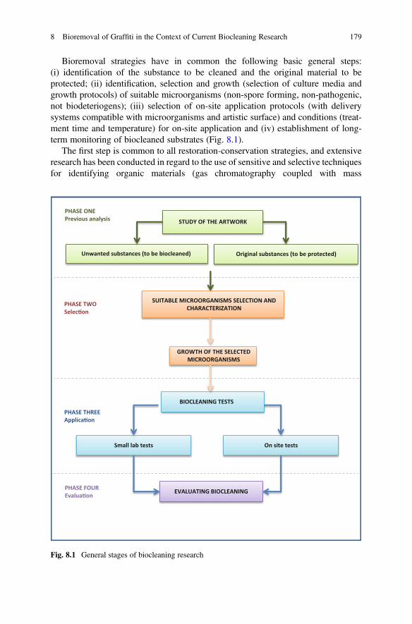
Bioremoval strategies have in common the following basic general steps:(i) identification of the substance to be cleaned and the original material to beprotected; (ii) identification, selection and growth (selection of culture media andgrowth protocols) of suitable microorganisms (non-spore forming, non-pathogenic,not biodeteriogens); (iii) selection of on-site application protocols (with deliverysystems compatible with microorganisms and artistic surface) and conditions (treat-ment time and temperature) for on-site application and (iv) establishment of long-term monitoring of biocleaned substrates (Fig. 8.1).
The first step is common to all restoration-conservation strategies, and extensiveresearch has been conducted in regard to the use of sensitive and selective techniquesfor identifying organic materials (gas chromatography coupled with mass
PHASE ONEPrevious analysis STUDY OF THE ARTWORK
Unwanted substances (to be biocleaned) Original substances (to be protected)
SUITABLE MICROORGANISMS SELECTION ANDCHARACTERIZATION
PHASE TWOSelec�on
GROWTH OF THE SELECTEDMICROORGANISMS
PHASE THREEApplica�on
Small lab tests On site tests
BIOCLEANING TESTS
EVALUATING BIOCLEANINGPHASE FOUREvalua�on
Fig. 8.1 General stages of biocleaning research
8 Bioremoval of Graffiti in the Context of Current Biocleaning Research 179
spectrometry (GC-MS), pyrolysis-GC-MS and inorganic materials (FTIR, SEM,Ion-chromatography)) (Colombini et al. 1999, 2003; Ranalli et al. 2005; Bosch-Roig et al. 2013). As this first step is well known and used in the field of restoration,it is not considered in this chapter, which includes a comparison of the methodo-logical analysis of the other three general steps involved in the bioremoval of salt andorganic matter and of graffiti. The chapter also includes a critical analysis of themethodological advances made by the scientific community and the applicability ofthese in the field of graffiti removal (see Section 3 and Table 8.1).
3.1 Selection of Suitable Microorganisms for the Bioremovalof Graffiti
As relatively few studies have addressed the biodeterioration and biocleaning ofgraffiti, few microorganisms with the potential to degrade graffiti paint have beenidentified (Table 8.2). Therefore, focusing on the selection of suitable microorgan-isms is an important area of study. Candidate microorganisms can be found ininternational collections of microorganisms (DSMZ, ATCC, CECT, etc.) or innatural environments.
If researchers wish to use registered microorganisms, these should be selected fortheir particular characteristics and be purchased and assayed in the laboratory inorder to evaluate their capacity to grow and degrade graffiti. Although this is a fasterand probably cheaper selection method, some studies have shown that it is moredifficult to obtain the most suitable microorganisms. Ranalli et al. (2005) conducteda comparative enzymatic analysis of the bioremoval capacity of a bacterium (Pseu-domonas stutzeri A29) directly isolated from the substance to be removed (agedanimal glue from frescoes) and compared with six other commercial strains of thesame genera (Pseudomonas) and even species (P. stutzeri). This study revealed thatall strains tested were able to synthesize inducible specific enzymes after beinggrown with the target substance (animal glue). However, the isolated strain(P. stutzeri A29) grew optimally and displayed the highest enzymatic (protease)activity (Ranalli et al. 2005). A study of the molecular mechanisms of action in thisparticular strain revealed a unique enzymatic profile when the bacterium was grownwith the glue isolated from the fresco. The enzymes may be a mixture of particularproteases whose combined action enables the complete digestion of the hardenedglue present in the frescoes (Antoniolli et al. 2005).
A logical starting point for obtaining microorganisms that can act as biocleaningagents from natural sources is to isolate them from the substance to be removed. Thiswas done, for example, by analysing black crust biofilms, from which differentresearchers have isolated sulphate reducing bacteria (SRB), hydrocarbon degradingbacteria (HDB) and other microorganisms with hydrolytic activities able to degradepolycyclic aromatic hydrocarbons and fatty acids, among others (Saiz-Jimenez1993, 1997; Fernandes 2006; Soffitti et al. 2019). In the case of graffiti paint, outdoor
180 P. Bosch-Roig and P. Sanmartín
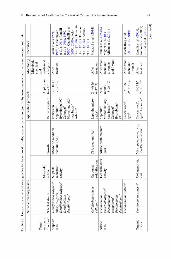
Tab
le8.1
Com
parisonof
generalstrategies
forthebiorem
ovalof
salts,o
rganicmatterandgraffitiby
usingmicroorganism
sfrom
inorganicsubstrate
Suitablemicroorganism
sApp
licationprotocols
Mon
itoring
References
Target
substance
toremov
eSelectedstrains
Metabolic
actio
nGrowth
Deliverysystem
App
lication
time/T�
Microbial,
chem
ical
and
aesthetical
changes
Sulph
ate
salts
Desulfovibrio
vulgarisa
subsp.
vulgaris
Desulfovibrio
vulgarisa
Desulfovibrio
desulfu
ricans
a
Sulph
ate
redu
ction
activ
ity
DSMZ63
mod
ified
medium
(An)
Immersion
Sepiolitea
Hyd
robiog
el.97a
Carbo
gela
Mortarandalgi-
natebeadsa
Arbocela
12–11
0h/
17–30
� CAfter
treatm
ent
Gaurietal.(19
89,
1992
);Heselmeyer
etal.(19
91);Ranalli
etal.(19
96a,19
97,
2000
);Cappitelli
etal.
(200
5,20
06);Polo
etal.(20
10);Gioventù
etal.(20
11);Troiano
etal.(20
14);Alfano
etal.(20
11)
Cellulosimicrobium
cellu
lans
aCarbo
nate
solubilization
TSAmedium
(Ae)
Lapon
itemicro-
packsa
24–48
h/6–
37� C
After
treatm
ent
Mazzoni
etal.(20
14)
Nitrate
salts
Pseud
omon
aspseudo
alcalig
enes
a
Pseud
omon
asaerugino
saa
Pseud
omon
asdesnitrificans
a
Denitrificatio
nactiv
ityNitratebrothmedium
(Ae)
Sepiolitea
Mortarandalgi-
natebeadsa
Carbo
gela
Hyd
robiog
el-
97a
24h-1
mon
th/
18–26
� C
After
treat-
mentand
8mon
ths
and6years
Ranallietal.(19
96b)
May
etal.(20
08)
Alfanoetal.(20
11)
Pseud
omon
asstutzeria
Cottonwoo
la,
agar
a1.5–
3h/
25�
4� C
After
treat-
mentand
1mon
th
Bosch-Roigetal.
(201
0,20
13,2
019)
Organic
matter
Pseud
omon
asstutzeria
Collageno
lytic
and
M9supp
lementedwith
0.5–1%
anim
alglue
Cottonwoo
la,
agar
a ,Lapon
itea
1.5–
24h/
28�
3� C
After
treatm
ent
Ranallietal.(20
05);
Anton
iolietal.(20
05)
Lustratoetal.(20
12)
(con
tinued)
8 Bioremoval of Graffiti in the Context of Current Biocleaning Research 181
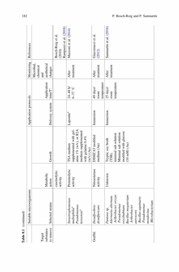
Tab
le8.1
(con
tinued)
Suitablemicroorganism
sApp
licationprotocols
Mon
itoring
References
Target
substance
toremov
eSelectedstrains
Metabolic
actio
nGrowth
Deliverysystem
App
lication
time/T�
Microbial,
chem
ical
and
aesthetical
changes
caseinolytic
activ
ityBosch-Roigetal.
(201
0)Ram
pazzietal.(20
18)
Stenotroph
omon
asmaltoph
iliaa
Pseud
omon
askoreensisa
Proteinolytic
activ
ityTSAmedium
supp
lementedwith
gel-
atine1%
(w/v),or
R2A
medium
supp
lemented
with
gelatin
e0.4%
(w/v)(A
e)
Lapon
itea
24–48
h/6–
37� C
After
treatm
ent
Mazzoni
etal.(20
14)
Graffiti
Desulfovibrio
desulfu
ricans
Nitroesterase
activ
ityDSMZ63
mod
ified
medium
(An)
Immersion
49days/
room
temperature
After
treatm
ent
Giacomuccietal.
(201
2)
Pan
toea
sp.
Alternaria
alternata
Arthrob
acteroryzae
Pseud
omon
asoryzihab
itans
Bacillus
megaterium
Arthrob
acter
aurescens
Bacillus
aquimaris
Pseud
omon
asmendo
cina
Microba
cterium
Unk
nown
Trypticsoybroth
(TSB),
minim
alsaltsolutio
nMinim
alsaltsolutio
nmod
ified
with
glucose
(10mM)(A
e)
Immersion
25days/
room
temperature
After
treatm
ent
Sanmartín
etal.(20
16)
182 P. Bosch-Roig and P. Sanmartín
oleivorans
Gordo
niaalkanivorans
Pseud
omon
asstutzeria
Aerob
acteraerogenesa
Com
amon
assp.a
M9enriched
with
graf-
fitipo
wder(A
e)Cottonwoo
la,
agar
a20
days/
room
temperature
After
treatm
ent
Sanmartín
andBosch-
Roig(201
9)
Aerob
acteraerogenes,
Bacillus
subtilis
Amixture
ofBacillus
sp.,Delftialacustris,
Sphing
obacterium
caeniand
Ochroba
ctruman
thropi
Com
amon
assp.
Rub
ellim
icrobium
thermop
hilum,
Chelatococcus
daeguensis,
Escherichia
coli
Marinospirillu
msp.
Unk
nown
Sequentially
(4steps):
(1)Trypticsoybroth
(TSB)du
ring
2days
(2)1/10
streng
thtryp
ticsoybroth(TSB)du
ring
5days
3)Com
pletemineral
(CM)du
ring
10days
4)Com
pletemineral
(CM)du
ring
10days
Immersion
27days/
30� C
After
each
step
Cattò
etal.(20
21)
Aerob
acteraerogenesa
Com
amon
assp.a
Unk
nown
CM
medium
andM9
enriched
with
graffiti
powdera(A
e)
Agara
14days/
room
temperature
After
treatm
ent
Sanmartín
andBosch-
Roigetal.
(unp
ublishedresults)
DSMZ63
medium:mod
ified
byelim
inatinganyiron
source;AeAerob
icmetabolism,A
nAnaerob
icmetabolism
a Sub
aerealapplication
8 Bioremoval of Graffiti in the Context of Current Biocleaning Research 183
Tab
le8.2
Com
parisonof
generalstrategies
fortheliv
emicroorganism
s’biorem
ovalof
graffiti
Bacterialselection
App
licationmaterials
andstrategy
Analysisprotocols
References
Collection
sources/registered
microorganism
s
Naturesources/
isolated
microorganism
s
Glass
slide/
immersion
strategy
Stone/
subaerial
strategy
Visual
alteratio
nColou
rvariants
Surface
changesa
Chemical
mod
ificatio
nsb
Enzym
atic
assays
Giacomuccietal.
(201
2)•
••
••
••
Sanmartín
etal.
(201
6)•
••
Sanmartín
andBosch-
Roig(201
9)•
••
••
•
Cattò
etal.(20
21)
••
••
•
Sanmartín
andBosch-
Roigetal.
(unp
ublishedresults)
••
••
••
a Refersto
microscop
icob
servationanalysis
bRefersto
FTIR
analysis
184 P. Bosch-Roig and P. Sanmartín
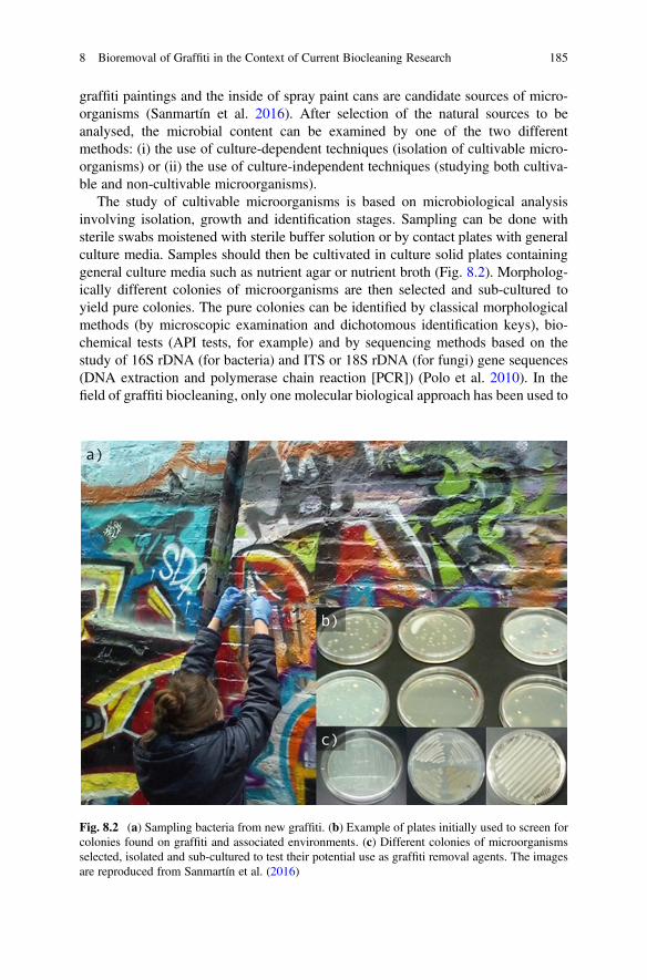
graffiti paintings and the inside of spray paint cans are candidate sources of micro-organisms (Sanmartín et al. 2016). After selection of the natural sources to beanalysed, the microbial content can be examined by one of the two differentmethods: (i) the use of culture-dependent techniques (isolation of cultivable micro-organisms) or (ii) the use of culture-independent techniques (studying both cultiva-ble and non-cultivable microorganisms).
The study of cultivable microorganisms is based on microbiological analysisinvolving isolation, growth and identification stages. Sampling can be done withsterile swabs moistened with sterile buffer solution or by contact plates with generalculture media. Samples should then be cultivated in culture solid plates containinggeneral culture media such as nutrient agar or nutrient broth (Fig. 8.2). Morpholog-ically different colonies of microorganisms are then selected and sub-cultured toyield pure colonies. The pure colonies can be identified by classical morphologicalmethods (by microscopic examination and dichotomous identification keys), bio-chemical tests (API tests, for example) and by sequencing methods based on thestudy of 16S rDNA (for bacteria) and ITS or 18S rDNA (for fungi) gene sequences(DNA extraction and polymerase chain reaction [PCR]) (Polo et al. 2010). In thefield of graffiti biocleaning, only one molecular biological approach has been used to
Fig. 8.2 (a) Sampling bacteria from new graffiti. (b) Example of plates initially used to screen forcolonies found on graffiti and associated environments. (c) Different colonies of microorganismsselected, isolated and sub-cultured to test their potential use as graffiti removal agents. The imagesare reproduced from Sanmartín et al. (2016)
8 Bioremoval of Graffiti in the Context of Current Biocleaning Research 185
identify microorganisms from natural sources: nine cultivable bacteria and onefungus were identified as candidates for biocleaning purposes by sequencing of16S rDNA and fungal ITS regions (Sanmartín et al. 2016).
Traditional identification techniques are sometimes tedious and time consuming.Researchers are currently focusing on new methodological advances such as matrix-assisted laser desorption ionization-time of flight mass spectrometry (MALDI-TOF-MS) to identify cultivable microorganisms. This technique captures the moleculespresent in the microorganisms by detecting proteins, peptides and lipid ions. Itidentifies a bacterial spectrum, rather than specific DNA sequences. As the samplesdo not require pretreatment, the method is therefore more rapid than traditionalidentification techniques; in addition, no reagents are required, and more than 1000samples can be analysed per day. For identification, the bacterial spectrum obtainedmust then be compared with the existing databases. The main limitation of thistechnique is that few of the databases available include environmental microorgan-isms, and that many of the existing ones are costly to access (Shingal et al. 2015).
Massive DNA sequencing represents another important methodological advancein the identification of microorganisms. This approach can be used to study cultiva-ble and non-cultivable microorganisms isolated from natural samples because itenables direct sequencing of native DNA/RNA. This next generation sequencing(NGS) technique can provide a more complete view of the microbial communitiesassociated with an object or substrate (Vilanova and Porcar 2020). New generationsingle molecule nanopore technology sequencing (MinION Oxford Nanopore Tech-nologies) is emerging and can be used for the rapid, simple identification ofmicroorganisms as potential biocleaning agents. The huge amounts of nucleotidesequence data must be analysed and compared with existing databases in order toidentify the microorganisms. The advantages of this method are that the analysis oflong sequences enables more precise identification, little material is needed and theexperimental process is simplified. There is no need to grow the microorganisms orto extract or amplify DNA for sequencing (Feng et al. 2015). This method providesmore comprehensive information about the microbial populations colonizing aparticular substrate (e.g. graffiti) because it produces data on all the microorganismspresent in the sample.
A combination of culture-dependent and culture-independent techniques shouldbe used to identify and study microorganisms as potential graffiti biocleaning agents.After identification, the microorganisms must be grown/cultivated in a particularmedium supplied with the target substance as a sole carbon source. Thebiodegradative capacities should then be analysed, among other factors, in order tofinally apply them in practical biocleaning actions (Vilanova and Porcar 2020).
186 P. Bosch-Roig and P. Sanmartín
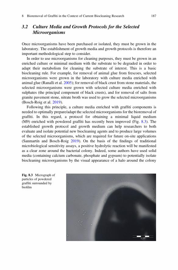
3.2 Culture Media and Growth Protocols for the SelectedMicroorganisms
Once microorganisms have been purchased or isolated, they must be grown in thelaboratory. The establishment of growth media and growth protocols is therefore animportant methodological step to consider.
In order to use microorganisms for cleaning purposes, they must be grown in anenriched culture or minimal medium with the substrate to be degraded in order toadapt their metabolism for cleaning the substrate of interest. This is a basicbiocleaning rule. For example, for removal of animal glue from frescoes, selectedmicroorganisms were grown in the laboratory with culture media enriched withanimal glue (Ranalli et al. 2005); for removal of black crust from stone materials, theselected microorganisms were grown with selected culture media enriched withsulphates (the principal component of black crusts), and for removal of salts fromgranite pavement stone, nitrate broth was used to grow the selected microorganisms(Bosch-Roig et al. 2019).
Following this principle, a culture media enriched with graffiti components isneeded to optimally prepare/adapt the selected microorganisms for the bioremoval ofgraffiti. In this regard, a protocol for obtaining a minimal liquid medium(M9) enriched with powdered graffiti has recently been improved (Fig. 8.3). Theestablished growth protocol and growth medium can help researchers to bothevaluate and isolate potential new biocleaning agents and to produce large volumesof the selected microorganisms, which are required for future on-site applications(Sanmartín and Bosch-Roig 2019). On the basis of the findings of traditionalmicrobiological sensitivity assays, a positive hydrolytic reaction will be manifestedas a clear zone around the bacterial colony. Indeed, some authors have used solidmedia (containing calcium carbonate, phosphate and gypsum) to potentially isolatebiocleaning microorganisms by the visual appearance of a halo around the colony
Fig. 8.3 Micrograph ofparticles of powderedgraffiti surrounded bybiofilm
8 Bioremoval of Graffiti in the Context of Current Biocleaning Research 187
(Mazzoni et al. 2014). We are now focussing on adapting this isolation method inorder to apply it to the isolation of microorganisms that could potentially be used forbiocleaning graffiti. A new graffiti solid media rather than the liquid media currentlyused will facilitate the bacterial selection and evaluation process with visual resultsand prevent the need for a complementary analysis of graffiti degradation.
3.3 Microorganism Application Protocols
Another important aspect that should be considered in biocleaning strategies is thatmicroorganisms require particular conditions (humidity and temperature) forgrowth. Therefore, different strategies have been used in the biocleaning field inorder to improve application protocols (Bosch-Roig et al. 2015).
The first studies of bioremoval of graffiti used glass microscope slides paintedwith a graffiti layer. The microscope slides were placed in liquid cultures with theselected bacteria. Finally, the samples were incubated for long periods (25–49 days)(Giacomucci et al. 2012; Sanmartín et al. 2016). These initial studies were importantbecause they showed that graffiti could be removed by biological means, althoughimprovement of the application protocols was needed. The immersion protocolcannot be used for on-site graffiti biocleaning and therefore methodologicaladvances were needed in this area. Based on the different delivery systems used inother biocleaning strategies, a more appropriate application protocol was developedby Sanmartín and Bosch-Roig (2019). This research has addressed this problem bystudying two delivery systems commonly used in other biocleaning strategies:cotton wool and 2% agar gel (Bosch-Roig et al. 2015). This research has producedinteresting results for bioapplication on stone, contributing to the future transfer ofthe biocleaning technology to real, on-site stone graffiti removal. However, furtherwork is required, because long treatment times are required, and the graffiti layer isnot completely removed.
Another important factor associated with the application protocols is the temper-ature required for growth of the microorganisms. Most microorganisms used inbiocleaning strategies are mesophilic, i.e. they grow optimally at moderate temper-atures (between 20 and 45 �C). If biocleaning is carried out at temperatures lowerthan 20 �C (very common in winter, for example), the effects could be altered orhampered. Different strategies (using air conditioning or heat lamps) could be usedin order to reach the ideal temperature of the treatments (Ranalli et al. 2005; Bosch-Roig et al. 2013). However, some researches are focusing on the selection ofmicroorganisms that can grow at higher temperatures. For example, Mazzoni et al.(2014) isolated biocleaning microorganisms (Stenotrophomonas maltophilia, Pseu-domonas koreensis and Cellulosimicrobium cellulans), which have an optimaltemperature range of 6–37 �C.
188 P. Bosch-Roig and P. Sanmartín
3.4 Protocols for Evaluating Graffiti Cleaning
Once microorganisms have been purchased or isolated and grown under laboratoryconditions, their hydrolytic abilities and metabolic capacities must be studied inorder to determine how they will contribute to graffiti removal. Most biocleaningstudies use one of the two approaches: (i) an indirect approach based on study of thedegradation of the target substance (graffiti) or (ii) a more direct approach based onthe study of the enzymes or proteins produced by the microorganisms.
Most studies have used the indirect strategy. Few studies have investigated theenzymes and proteins produced by the microorganisms. Physico-chemical analysisis usually used for this purpose.
Considering the field of graffiti biocleaning, all the studies conducted to date haveused the indirect strategy, in which microorganisms are grown with the graffiti andthe graffiti is then examined in terms of visual alteration, colour variations(by reflectance colour measurements), surface change (by stereomicroscope,epifluorescence microscope and electron microscope observations) and chemicalmodifications, using Fourier transform infrared spectroscopy (FTIR) (Giacomucciet al. 2012; Sanmartín et al. 2016; Sanmartín and Bosch-Roig 2019) (see Table 8.2).In particular, preliminary FTIR findings on the potential biodegradation pathwaysinduced by Desulfovibrio desulfuricans in nitrocellulose-based paints have beenreported (Giacomucci et al. 2012). These findings include a depletion of signalrelated to the N-O bond, suggesting that the bacteria induce degradation of thepaint via ammonification. In addition, Sanmartín and Bosch-Roig (2019)) havedemonstrated bacterially induced chemical changes in graffiti paints, by usingFTIR analysis. In the resulting spectra a decreasing intensity of peak bands relatedto alkanes, carbonyl, carbon-carbon triple bound and esters was observed. Thesetypes of indirect analysis could be complemented with other analytical strategiesused in other graffiti cleaning strategies such as advanced laser-based techniques(laser-induced breakdown spectroscopy: LIBS), X-ray diffraction or RAMAN anal-ysis (Gómez et al. 2010; Siano et al. 2012; Penide et al. 2013, Sanmartín et al. 2016).
More direct approaches based on enzymatic and proteomic analysis can also beused. In these approaches, microorganisms are grown with the target substance(graffiti) and the enzymatic activity or enzyme-protein production is then deter-mined. In graffiti biocleaning, only one of these more direct approaches has beenreported. In this study, the nitroesterase activity (nitrate and nitrite reductase activity)of a particular bacterial culture (Desulfovibrio desulfuricans) was evaluated byanalysis of the presence of nitrate, nitrite and ammonia by use of a particular reagentthat produces a colorimetric reaction (when the enzyme is expressed), which is thenmeasured by spectrophotometry. This analysis enabled the researchers to concludethat D. desulfuricans was able to degrade the nitrocellulose present in paint binders,possibly by an ammonification pathway (Giacomucci et al. 2012).
In the field of biocleaning research, this type of direct analysis should inspirefurther studies on graffiti bioremoval. Enzymatic activity has been analysed by usingbiochemical (API-ZYM™ Systems BioMérieux, Rome, Italy) and colorimetric tests
8 Bioremoval of Graffiti in the Context of Current Biocleaning Research 189
(based on azo dye impregnated casein) and (casein) zymograms (spectrophotometricassay) to evaluate and observe the enzymatic (protease) activity, in particular thespecific caseinolytic and collagenolytic activity of the bacterium P. stutzeri A29(Ranalli et al. 2005; Antonioli et al. 2005). Sulphate reducing activity has beenevaluated in liquid culture medium by determining the residual sulphate content inthe cultural broth medium by spectrophotometry (absorbance at 515 nm) (Ranalliet al. 1997). Some authors have used colorimetric test strips that enable the rapidsemi-quantitative evaluation of bacterial enzymatic activities. For example, nitrateand nitrite test strips can be used to analyse the optimal effectiveness of differentbacterial strains for nitrate removal (Bosch-Roig et al. 2013).
The protein content of bacteria can be extracted and analysed by differentmethods. For example, Giacomucci et al. (2012) used ultrasound-assisted extractionand the Bradford method (based on the protein interaction to Coomassie dye underacidic conditions resulting in a colour change from brown to blue) to determine thetotal protein content of D. desulfuricans.
Advanced phenotypic technologies, such as phenotype microarray systems(PM Biolog system), can also be used to obtain information about the propertiesof the selected strains. These systems enable rapid screening of an extensive range ofgrowth conditions (Borglin et al. 2012). Analysis of the use of almost hundreddifferent carbon sources by a selected microorganism can be done simultaneously(96 well microplate), to produce a “metabolic fingerprint”. This type of technologyhas been used to study carbon and nitrogen metabolism, chemical sensitivity(osmolyte tolerance, pH tolerance and toxic compound tolerance) and the metabolicpathways sustaining the biocleaning capacity of strain P. stutzeri DSMZ 5190,which has been widely used for biocleaning purposes (Bosch-Roig et al. 2016).
The so-called omic techniques are increasingly used in the field of conservationand restoration research. These techniques enable complete analysis of the total poolof DNA (genomics), the total proteins (proteomics) and their metabolites(metabolomics) (Vilanova and Porcar 2020).
3.5 Long-Term Monitoring to Evaluate Graffiti Cleaning
The final step that should be considered in any biocleaning process, especially in thetreatment of decorative surfaces, is to monitor the changes on the cleaned surfaceover time (Bosch-Roig and Ranalli 2014). Monitoring must focus on at least twoaspects: (i) the absence of microbial growth on the surface (microbial monitoring)and (ii) the absence of aesthetic alteration of the cleaned surface over time.
All graffiti biocleaning studies include both of these monitoring strategies afterthe cleaning, but further long-term changes must be still determined. However,diverse biocleaning studies have focused on the changes on the cleaned surfaceover time. For example, in Matera cathedral, the effects of nitrate bioremoval weremonitored 8 months and 6 years after the treatment. In the areas treated with bacteria,the nitrate concentration remained stable, and no notable differences in microflora or
190 P. Bosch-Roig and P. Sanmartín
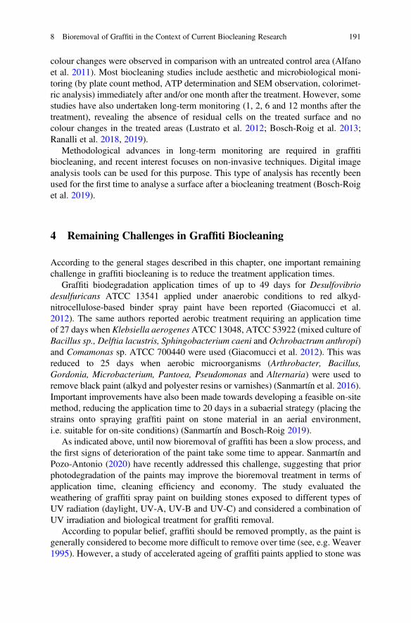
colour changes were observed in comparison with an untreated control area (Alfanoet al. 2011). Most biocleaning studies include aesthetic and microbiological moni-toring (by plate count method, ATP determination and SEM observation, colorimet-ric analysis) immediately after and/or one month after the treatment. However, somestudies have also undertaken long-term monitoring (1, 2, 6 and 12 months after thetreatment), revealing the absence of residual cells on the treated surface and nocolour changes in the treated areas (Lustrato et al. 2012; Bosch-Roig et al. 2013;Ranalli et al. 2018, 2019).
Methodological advances in long-term monitoring are required in graffitibiocleaning, and recent interest focuses on non-invasive techniques. Digital imageanalysis tools can be used for this purpose. This type of analysis has recently beenused for the first time to analyse a surface after a biocleaning treatment (Bosch-Roiget al. 2019).
4 Remaining Challenges in Graffiti Biocleaning
According to the general stages described in this chapter, one important remainingchallenge in graffiti biocleaning is to reduce the treatment application times.
Graffiti biodegradation application times of up to 49 days for Desulfovibriodesulfuricans ATCC 13541 applied under anaerobic conditions to red alkyd-nitrocellulose-based binder spray paint have been reported (Giacomucci et al.2012). The same authors reported aerobic treatment requiring an application timeof 27 days when Klebsiella aerogenesATCC 13048, ATCC 53922 (mixed culture ofBacillus sp., Delftia lacustris, Sphingobacterium caeni and Ochrobactrum anthropi)and Comamonas sp. ATCC 700440 were used (Giacomucci et al. 2012). This wasreduced to 25 days when aerobic microorganisms (Arthrobacter, Bacillus,Gordonia, Microbacterium, Pantoea, Pseudomonas and Alternaria) were used toremove black paint (alkyd and polyester resins or varnishes) (Sanmartín et al. 2016).Important improvements have also been made towards developing a feasible on-sitemethod, reducing the application time to 20 days in a subaerial strategy (placing thestrains onto spraying graffiti paint on stone material in an aerial environment,i.e. suitable for on-site conditions) (Sanmartín and Bosch-Roig 2019).
As indicated above, until now bioremoval of graffiti has been a slow process, andthe first signs of deterioration of the paint take some time to appear. Sanmartín andPozo-Antonio (2020) have recently addressed this challenge, suggesting that priorphotodegradation of the paints may improve the bioremoval treatment in terms ofapplication time, cleaning efficiency and economy. The study evaluated theweathering of graffiti spray paint on building stones exposed to different types ofUV radiation (daylight, UV-A, UV-B and UV-C) and considered a combination ofUV irradiation and biological treatment for graffiti removal.
According to popular belief, graffiti should be removed promptly, as the paint isgenerally considered to become more difficult to remove over time (see, e.g. Weaver1995). However, a study of accelerated ageing of graffiti paints applied to stone was
8 Bioremoval of Graffiti in the Context of Current Biocleaning Research 191
the first to demonstrate that the age of graffiti (i.e. the time that the graffiti hasremained on the lithic substrate) enhances the effectiveness of the cleaning process(Sanmartín and Cappitelli 2017). This finding was strengthened in subsequentstudies involving the removal of graffiti from anti-graffiti surfaces, in which thesame conclusion was reached, in contrast to conventional wisdom (Pozo-Antonioet al. 2018).
Another (parallel) strategy is to identify the microorganisms that are the mostefficient graffiti bioremoval agents. This approach should be accompanied by studyof the enzymatic reactions involved in graffiti bioremoval to better elucidate themetabolic pathways. On-site, large-scale production of microorganisms is anotherremaining challenge that modern biotechnologies could help to solve (Philip andAtlas 2017).
Another challenge is the study of biocleaning strategies for diverse materials andgraffiti colours and types. To date, only four substrates have been analysed: glass,natural granite stone, man-made concrete stone and metal (Fig. 8.4). In addition,only three types of graffiti have been tested: red spray paint (Motip–DupliÒ
Fig. 8.4 Laboratory-based graffiti bioremoval from diverse materials and involving colouredpaints. (a) Remains of black and silver graffiti on glass after immersion biocleaning. (b) Naturalgranite stone and man-made concrete stone painted with black and silver graffiti paint during agar-assisted biocleaning. (c) Vertical metal sheet painted with black and silver graffiti paint during agar-assisted biocleaning. The image is reproduced from Sanmartín and Bosch-Roig (2019)
192 P. Bosch-Roig and P. Sanmartín
Autocolor, colour 5-0200), black non-metallic spray paint (R-9011 from MontanaColors, Barcelona, Spain) and a silver metallic spray paint (Silver Chrome fromMontana Colors).
Several authors have suggested that future research should focus on the follow-ing: (i) selection/identification of new biological formulation to effectively removeall kinds of paints and colours; (ii) the biocleaning potential of viable yeast cells(which produce few toxic metabolites); (iii) evaluation of the effectiveness of thecleaning of aged graffiti from different substrates; (iv) incomplete graffiti cleaning;(v) testing diverse delivery systems to shorten the biocleaning treatment times andresolve difficulties in application (Troiano et al. 2014; Gomes et al. 2017; Sanmartínand Bosch-Roig 2019).
5 Conclusion and Final Remarks
The bioremoval of graffiti is a promising green approach and risk-free method basedon the use of selected microorganisms as “probiotics” (Cappitelli 2016; Sanmartínet al. 2016; Vilanova and Porcar 2020). However, it remains an emerging field inwhich there are still many challenges to be addressed and solved.
In this chapter, a comprehensive analysis has been conducted by focusing oncomparing existing studies on the bioremoval of salts and organic matter by the useof live microorganisms in laboratory simulations or applied to real artwork. Specialemphasis has been placed on the methodological advances made and on existingmethodological tools and approaches used in related fields that could be applied inthis particular area in order to help researchers address the remaining challenges.
Acknowledgements P. Sanmartín is grateful for financial support from the Xunta de Galicia (grantED431C 2018/32).
References
Alfano G, Lustrato G, Belli C, Zanardini E, Cappitelli F, Mello E, Sorlini C, Ranalli G (2011) Thebioremoval of nitrate and sulfate alterations on artistic stonework: the case study of MateraCathedral after six years from the treatment. Int Biodeterior Biodegrad 65(7):1004–1011
Allsopp D, Seal K, Gaylarde C (2004) Introduction to biodeterioration. Chapter 3. Biodeteriorationof refined and processed materials: paints, 2nd edn. Cambridge University Press, Cambridge, pp78–85
Antonioli P, Zapparoli G, Abbruscato P, Sorlini C, Ranalli G, Righetti PG (2005) Art-loving bugs:the resurrection of Spinello Aretino from Pisa’s cemetery. Proteomics 5:2453–2459
Bentley J, Turner GPA (1998) Introduction to paint chemistry and principles of paint technology,4th edn. Chapman & Hall, London
Borglin S, Joyner D, DeAngelis KM, Khudyakov J, D’haeseleer P, Joachimiak MP, Hazen T (2012)Application of phenotypic microarrays to environmental microbiology. Curr Opin Biotechnol23:41–48
8 Bioremoval of Graffiti in the Context of Current Biocleaning Research 193
Bosch-Roig P, Regidor-Ros JL, Soriano-Sancho P, Doménech-Carbó MT, Montes-Estelles RM(2010) Ensayos de biolimpieza con bacterias en pinturas murales. Arché 4–5:115–124
Bosch-Roig P, Regidor-Rosi JL, Montes-Estellés RM (2013) Biocleaning of nitrate alterations onwall paintings by Pseudomonas stutzeri. Int Biodeter Biodegr 84:266–274
Bosch-Roig P, Ranalli G (2014) The safety of biocleaning technologies for cultural heritage. FrontMicrobiol 5:155
Bosch-Roig P, Lustrato G, Zanardini E, Ranalli G (2015) Biocleaning of cultural heritage stonesurfaces and frescoes: which delivery system can be the most appropriate? Ann Microbiol65:1227–1241
Bosch-Roig P, Decorosi F, Giovannetti L, Ranalli G, Viti C (2016) Connecting phenome to genomein Pseudomonas stutzeri 5190: an artwork biocleaning bacterium. Res Microbiol 167:757–765
Bosch-Roig P, Allegue H, Bosch I (2019) Granite pavement nitrate desalination: traditionalmethods vs. biocleaning methods. Sustainability 19:4227
Breitbach AM, Rocha JC, Gaylarde CC (2011) Influence of pigments on biodeterioration of paintfilms. J Coating Technol Res 8:619–628
Cappitelli F (2016) Biocleaning of cultural heritage surfaces. Open Conf Proc J 7(suppl 1:M6):65–69
Cappitelli F, Sorlini C (2008) Microorganisms attack synthetic polymers in items representing ourcultural heritage. Appl Environ Microbiol 74:564–569
Cappitelli F, Vicini S, Piaggio P, Abbruscato P, Princi E, Casadevall A, Nosanchuk JD, Zanardini E(2005) Investigation of fungal deterioration of synthetic paint binders using vibrational spec-troscopic techniques. Macromol Biosci 5:49–57
Cappitelli F, Zanardini E, Ranalli G, Mello E, Daffonchio D, Sorlini C (2006) Improved method-ology for bioremoval of black crusts on historical stone artworks by use of sulfate-reducingbacteria. Appl Environ Microbiol 72:3733–3737
Cappitelli F, Nosanchuk JD, Casadevall A, Toniolo L, Brusetti L, Florio S, Principi P, Borin S,Sorlini C (2007) Synthetic consolidants attacked by melanine producing fungi: case study of thebiodeterioration of Milan (Italy) Cathedral marble treated with acrylics. Appl Environ Microbiol73:271–277
Cattò C, Sanmartín P, Gulotta D, Troiano F, Cappitelli F (2021) Bioremoval of graffiti using novelcommercial strains of bacteria. Sci Total Environ 756:144075
Colombini MP, Modugno F, Giacomelli A (1999) Two procedures for suppressing interferencefrom inorganic pigments in analysis by gas chromatography-mass spectrometry of proteina-ceous binders in painting. J Chromatogr Acta 846:101–111
Colombini MP, Bonaduce I (2003) Gas chromatography/mass spectrometry for the characterizationof organic materials in frescoes of the Monumental Cemetery of Pisa (Italy). Rapid CommunMass Spectrom 17:2523–2527
Ciferri O (1999) Microbial degradation of paintings. Appl Environ Microbiol 65:879–885DDL 1607 del 16 luglio (2002) Disciplina del fenomeno del “graffitismo”. Annunciato nella seduta
ant. n. 214 del 17 luglio 2002. (In Italian)Feng Y, Zhang Y, Ying C, Wang D, Du C (2015) Nanopore-based Fourth-generation DNA
sequencing technology. Genom Proteom Bioinf 13:4–16Fernandes P (2006) Applied microbiology and biotechnology in the conservation of stone cultural
heritage materials. Appl Microbiol Biotechnol 73:291–296Gauri KL, Chowdhury AN, Kulshreshtha NP, Punuru AR (1989) The sulfation of marble and the
treatment of gypsum crusts. Stud Conserv 34:201–206Gauri KL, Parks L, Jaynes J, Atlas R (1992) Removal of sulfated-crusts frommarble using sulphate-
reducing bacteria. In: Webster RGM (ed) Proceedings of the International Conference on StoneCleaning and the Nature, soiling and decay mechanisms of stone. Donhead, Edinburgh, pp 160–165
Gaylarde PM, Gaylarde CC (2000) Algae and cyanobacteria on painted buildings in Latin America.Int Biodeter Biodegr 46:93–97
194 P. Bosch-Roig and P. Sanmartín
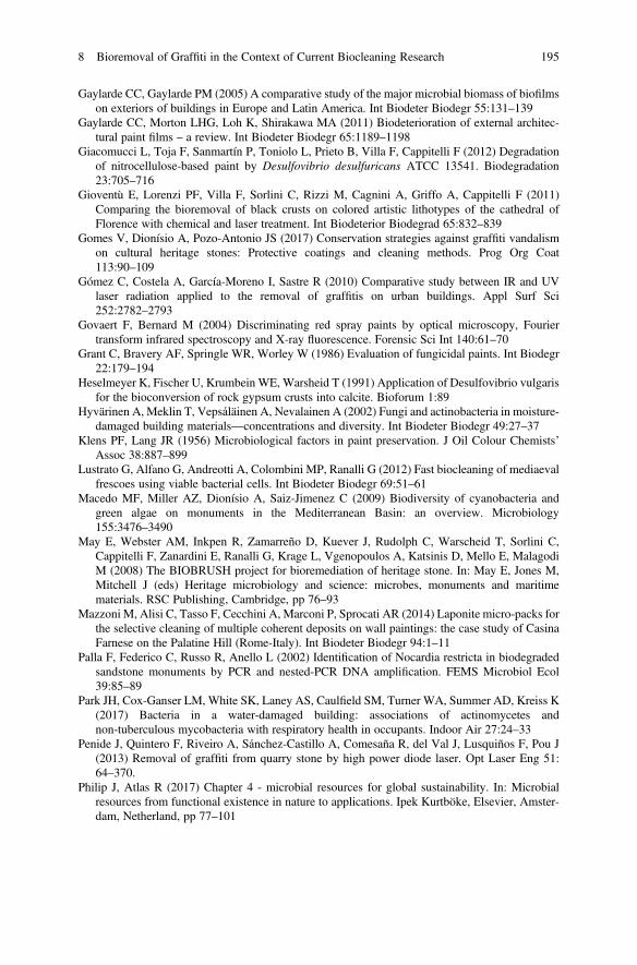
Gaylarde CC, Gaylarde PM (2005) A comparative study of the major microbial biomass of biofilmson exteriors of buildings in Europe and Latin America. Int Biodeter Biodegr 55:131–139
Gaylarde CC, Morton LHG, Loh K, Shirakawa MA (2011) Biodeterioration of external architec-tural paint films – a review. Int Biodeter Biodegr 65:1189–1198
Giacomucci L, Toja F, Sanmartín P, Toniolo L, Prieto B, Villa F, Cappitelli F (2012) Degradationof nitrocellulose-based paint by Desulfovibrio desulfuricans ATCC 13541. Biodegradation23:705–716
Gioventù E, Lorenzi PF, Villa F, Sorlini C, Rizzi M, Cagnini A, Griffo A, Cappitelli F (2011)Comparing the bioremoval of black crusts on colored artistic lithotypes of the cathedral ofFlorence with chemical and laser treatment. Int Biodeterior Biodegrad 65:832–839
Gomes V, Dionísio A, Pozo-Antonio JS (2017) Conservation strategies against graffiti vandalismon cultural heritage stones: Protective coatings and cleaning methods. Prog Org Coat113:90–109
Gómez C, Costela A, García-Moreno I, Sastre R (2010) Comparative study between IR and UVlaser radiation applied to the removal of graffitis on urban buildings. Appl Surf Sci252:2782–2793
Govaert F, Bernard M (2004) Discriminating red spray paints by optical microscopy, Fouriertransform infrared spectroscopy and X-ray fluorescence. Forensic Sci Int 140:61–70
Grant C, Bravery AF, Springle WR, Worley W (1986) Evaluation of fungicidal paints. Int Biodegr22:179–194
Heselmeyer K, Fischer U, Krumbein WE, Warsheid T (1991) Application of Desulfovibrio vulgarisfor the bioconversion of rock gypsum crusts into calcite. Bioforum 1:89
Hyvärinen A, Meklin T, Vepsáläinen A, Nevalainen A (2002) Fungi and actinobacteria in moisture-damaged building materials—concentrations and diversity. Int Biodeter Biodegr 49:27–37
Klens PF, Lang JR (1956) Microbiological factors in paint preservation. J Oil Colour Chemists’Assoc 38:887–899
Lustrato G, Alfano G, Andreotti A, Colombini MP, Ranalli G (2012) Fast biocleaning of mediaevalfrescoes using viable bacterial cells. Int Biodeter Biodegr 69:51–61
Macedo MF, Miller AZ, Dionísio A, Saiz-Jimenez C (2009) Biodiversity of cyanobacteria andgreen algae on monuments in the Mediterranean Basin: an overview. Microbiology155:3476–3490
May E, Webster AM, Inkpen R, Zamarreño D, Kuever J, Rudolph C, Warscheid T, Sorlini C,Cappitelli F, Zanardini E, Ranalli G, Krage L, Vgenopoulos A, Katsinis D, Mello E, MalagodiM (2008) The BIOBRUSH project for bioremediation of heritage stone. In: May E, Jones M,Mitchell J (eds) Heritage microbiology and science: microbes, monuments and maritimematerials. RSC Publishing, Cambridge, pp 76–93
Mazzoni M, Alisi C, Tasso F, Cecchini A, Marconi P, Sprocati AR (2014) Laponite micro-packs forthe selective cleaning of multiple coherent deposits on wall paintings: the case study of CasinaFarnese on the Palatine Hill (Rome-Italy). Int Biodeter Biodegr 94:1–11
Palla F, Federico C, Russo R, Anello L (2002) Identification of Nocardia restricta in biodegradedsandstone monuments by PCR and nested-PCR DNA amplification. FEMS Microbiol Ecol39:85–89
Park JH, Cox-Ganser LM, White SK, Laney AS, Caulfield SM, Turner WA, Summer AD, Kreiss K(2017) Bacteria in a water-damaged building: associations of actinomycetes andnon-tuberculous mycobacteria with respiratory health in occupants. Indoor Air 27:24–33
Penide J, Quintero F, Riveiro A, Sánchez-Castillo A, Comesaña R, del Val J, Lusquiños F, Pou J(2013) Removal of graffiti from quarry stone by high power diode laser. Opt Laser Eng 51:64–370.
Philip J, Atlas R (2017) Chapter 4 - microbial resources for global sustainability. In: Microbialresources from functional existence in nature to applications. Ipek Kurtböke, Elsevier, Amster-dam, Netherland, pp 77–101
8 Bioremoval of Graffiti in the Context of Current Biocleaning Research 195
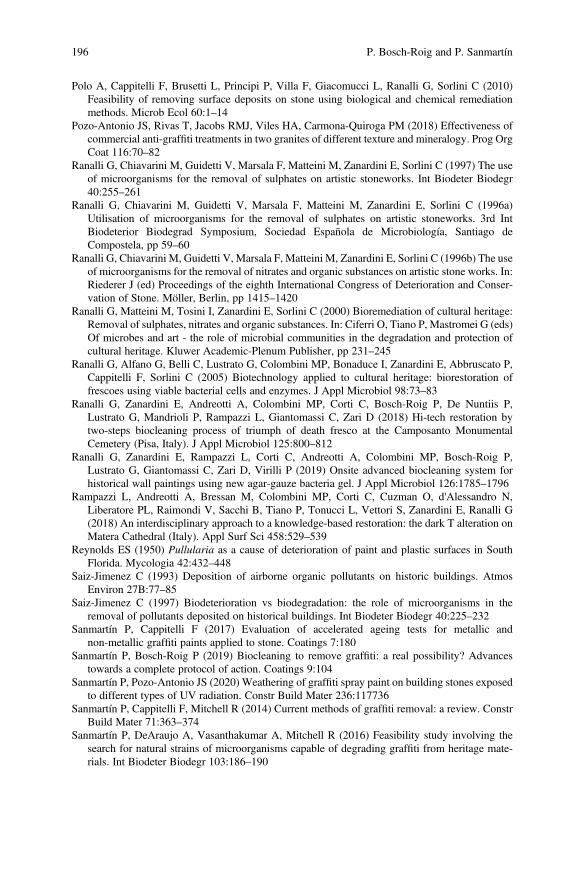
Polo A, Cappitelli F, Brusetti L, Principi P, Villa F, Giacomucci L, Ranalli G, Sorlini C (2010)Feasibility of removing surface deposits on stone using biological and chemical remediationmethods. Microb Ecol 60:1–14
Pozo-Antonio JS, Rivas T, Jacobs RMJ, Viles HA, Carmona-Quiroga PM (2018) Effectiveness ofcommercial anti-graffiti treatments in two granites of different texture and mineralogy. Prog OrgCoat 116:70–82
Ranalli G, Chiavarini M, Guidetti V, Marsala F, Matteini M, Zanardini E, Sorlini C (1997) The useof microorganisms for the removal of sulphates on artistic stoneworks. Int Biodeter Biodegr40:255–261
Ranalli G, Chiavarini M, Guidetti V, Marsala F, Matteini M, Zanardini E, Sorlini C (1996a)Utilisation of microorganisms for the removal of sulphates on artistic stoneworks. 3rd IntBiodeterior Biodegrad Symposium, Sociedad Española de Microbiología, Santiago deCompostela, pp 59–60
Ranalli G, Chiavarini M, Guidetti V, Marsala F, Matteini M, Zanardini E, Sorlini C (1996b) The useof microorganisms for the removal of nitrates and organic substances on artistic stone works. In:Riederer J (ed) Proceedings of the eighth International Congress of Deterioration and Conser-vation of Stone. Möller, Berlin, pp 1415–1420
Ranalli G, Matteini M, Tosini I, Zanardini E, Sorlini C (2000) Bioremediation of cultural heritage:Removal of sulphates, nitrates and organic substances. In: Ciferri O, Tiano P, Mastromei G (eds)Of microbes and art - the role of microbial communities in the degradation and protection ofcultural heritage. Kluwer Academic-Plenum Publisher, pp 231–245
Ranalli G, Alfano G, Belli C, Lustrato G, Colombini MP, Bonaduce I, Zanardini E, Abbruscato P,Cappitelli F, Sorlini C (2005) Biotechnology applied to cultural heritage: biorestoration offrescoes using viable bacterial cells and enzymes. J Appl Microbiol 98:73–83
Ranalli G, Zanardini E, Andreotti A, Colombini MP, Corti C, Bosch-Roig P, De Nuntiis P,Lustrato G, Mandrioli P, Rampazzi L, Giantomassi C, Zari D (2018) Hi-tech restoration bytwo-steps biocleaning process of triumph of death fresco at the Camposanto MonumentalCemetery (Pisa, Italy). J Appl Microbiol 125:800–812
Ranalli G, Zanardini E, Rampazzi L, Corti C, Andreotti A, Colombini MP, Bosch-Roig P,Lustrato G, Giantomassi C, Zari D, Virilli P (2019) Onsite advanced biocleaning system forhistorical wall paintings using new agar-gauze bacteria gel. J Appl Microbiol 126:1785–1796
Rampazzi L, Andreotti A, Bressan M, Colombini MP, Corti C, Cuzman O, d'Alessandro N,Liberatore PL, Raimondi V, Sacchi B, Tiano P, Tonucci L, Vettori S, Zanardini E, Ranalli G(2018) An interdisciplinary approach to a knowledge-based restoration: the dark T alteration onMatera Cathedral (Italy). Appl Surf Sci 458:529–539
Reynolds ES (1950) Pullularia as a cause of deterioration of paint and plastic surfaces in SouthFlorida. Mycologia 42:432–448
Saiz-Jimenez C (1993) Deposition of airborne organic pollutants on historic buildings. AtmosEnviron 27B:77–85
Saiz-Jimenez C (1997) Biodeterioration vs biodegradation: the role of microorganisms in theremoval of pollutants deposited on historical buildings. Int Biodeter Biodegr 40:225–232
Sanmartín P, Cappitelli F (2017) Evaluation of accelerated ageing tests for metallic andnon-metallic graffiti paints applied to stone. Coatings 7:180
Sanmartín P, Bosch-Roig P (2019) Biocleaning to remove graffiti: a real possibility? Advancestowards a complete protocol of action. Coatings 9:104
Sanmartín P, Pozo-Antonio JS (2020) Weathering of graffiti spray paint on building stones exposedto different types of UV radiation. Constr Build Mater 236:117736
Sanmartín P, Cappitelli F, Mitchell R (2014) Current methods of graffiti removal: a review. ConstrBuild Mater 71:363–374
Sanmartín P, DeAraujo A, Vasanthakumar A, Mitchell R (2016) Feasibility study involving thesearch for natural strains of microorganisms capable of degrading graffiti from heritage mate-rials. Int Biodeter Biodegr 103:186–190
196 P. Bosch-Roig and P. Sanmartín
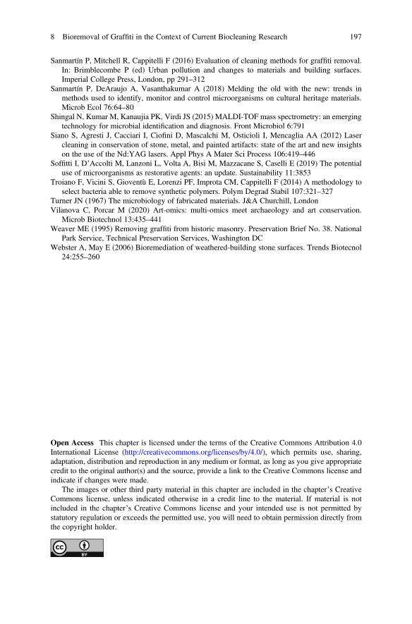
Sanmartín P, Mitchell R, Cappitelli F (2016) Evaluation of cleaning methods for graffiti removal.In: Brimblecombe P (ed) Urban pollution and changes to materials and building surfaces.Imperial College Press, London, pp 291–312
Sanmartín P, DeAraujo A, Vasanthakumar A (2018) Melding the old with the new: trends inmethods used to identify, monitor and control microorganisms on cultural heritage materials.Microb Ecol 76:64–80
Shingal N, Kumar M, Kanaujia PK, Virdi JS (2015) MALDI-TOF mass spectrometry: an emergingtechnology for microbial identification and diagnosis. Front Microbiol 6:791
Siano S, Agresti J, Cacciari I, Ciofini D, Mascalchi M, Osticioli I, Mencaglia AA (2012) Lasercleaning in conservation of stone, metal, and painted artifacts: state of the art and new insightson the use of the Nd:YAG lasers. Appl Phys A Mater Sci Process 106:419–446
Soffitti I, D’Accolti M, Lanzoni L, Volta A, Bisi M, Mazzacane S, Caselli E (2019) The potentialuse of microorganisms as restorative agents: an update. Sustainability 11:3853
Troiano F, Vicini S, Gioventù E, Lorenzi PF, Improta CM, Cappitelli F (2014) A methodology toselect bacteria able to remove synthetic polymers. Polym Degrad Stabil 107:321–327
Turner JN (1967) The microbiology of fabricated materials. J&A Churchill, LondonVilanova C, Porcar M (2020) Art-omics: multi-omics meet archaeology and art conservation.
Microb Biotechnol 13:435–441Weaver ME (1995) Removing graffiti from historic masonry. Preservation Brief No. 38. National
Park Service, Technical Preservation Services, Washington DCWebster A, May E (2006) Bioremediation of weathered-building stone surfaces. Trends Biotecnol
24:255–260
Open Access This chapter is licensed under the terms of the Creative Commons Attribution 4.0International License (http://creativecommons.org/licenses/by/4.0/), which permits use, sharing,adaptation, distribution and reproduction in any medium or format, as long as you give appropriatecredit to the original author(s) and the source, provide a link to the Creative Commons license andindicate if changes were made.
The images or other third party material in this chapter are included in the chapter’s CreativeCommons license, unless indicated otherwise in a credit line to the material. If material is notincluded in the chapter’s Creative Commons license and your intended use is not permitted bystatutory regulation or exceeds the permitted use, you will need to obtain permission directly fromthe copyright holder.
8 Bioremoval of Graffiti in the Context of Current Biocleaning Research 197
Chapter 9Ancient Textile Deteriorationand Restoration: Bio-Cleaning of anEgyptian Shroud Held in the TorinoMuseum
Roberto Mazzoli and Enrica Pessione
Abstract Ancient textiles are fragile and several factors can affect their integrity. Inthe present chapter, the main agents of deterioration of old and new textiles, namelyphysical-chemical (light, oxygen, heat, and humidity) and biological factors as wellas human erroneous interventions will be explored. As far as the biological deteri-oration is considered, the effects of microbial growth, primary and secondarymetabolites (acids, solvents, surfactants, pigments) and enzymes (lipases, proteases,and glycosidases) on textile strength and cleanliness will be described in details. Themain fungal and bacterial species involved in the damage (textile discoloration,black and green spots, cuts) will be reported. Adhesive application during restorationprocedures is discussed to highlight the risk of glue thickening giving rise to dullprecipitates on the fabric.
The main strategies for oil-stain and glue removal (both animal glue, such as fishcollagen, and vegetal glue, i.e. starch) will be described in the paragraph devoted tobiorestoration. Finally, a case study concerning an ancient Coptic tunic housed in theEgyptian Museum of Torino, Italy, and biocleaned by means of gellan-immobilizedalpha-amylase from Bacillus sp. will be largely discussed by reporting historicaldata, adhesive characterization, methods for artificial aging of simulated sample andglue removal from the artwork.
Keywords Microbial and physical-chemical deterioration · Human restoration ·Starch glue · Immobilized enzymes · Amylase · Wool artificial aging · Gellan ·Coptic period
R. Mazzoli · E. Pessione (*)Structural and Functional Biochemistry, Laboratory of Proteomics and Metabolic Engineeringof Prokaryotes, Department of Life Sciences and Systems Biology, University of Torino,Torino, Italye-mail: [email protected]
© The Author(s) 2021E. Joseph (ed.), Microorganisms in the Deterioration and Preservation of CulturalHeritage, https://doi.org/10.1007/978-3-030-69411-1_9
199
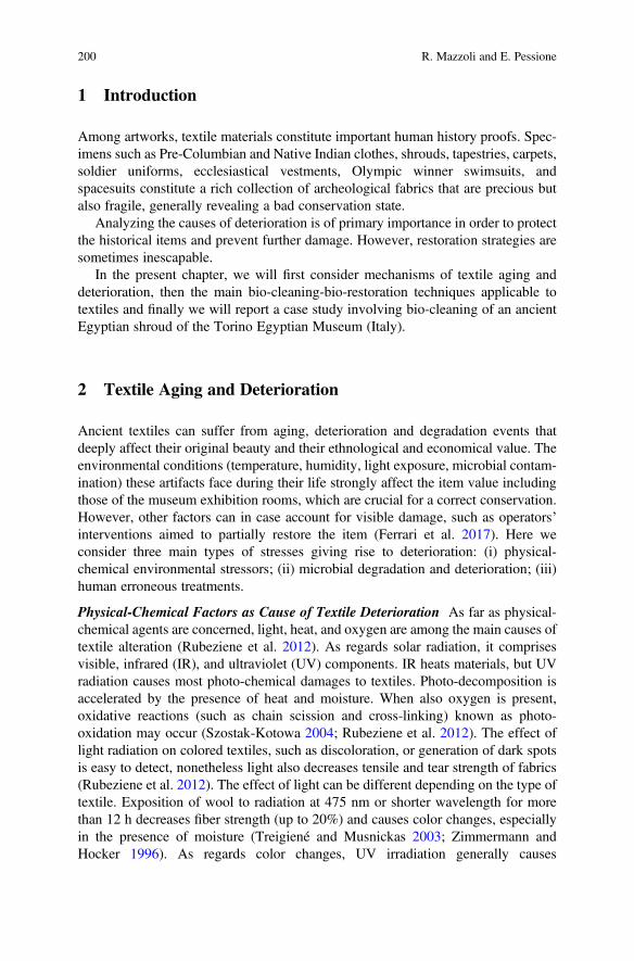
1 Introduction
Among artworks, textile materials constitute important human history proofs. Spec-imens such as Pre-Columbian and Native Indian clothes, shrouds, tapestries, carpets,soldier uniforms, ecclesiastical vestments, Olympic winner swimsuits, andspacesuits constitute a rich collection of archeological fabrics that are precious butalso fragile, generally revealing a bad conservation state.
Analyzing the causes of deterioration is of primary importance in order to protectthe historical items and prevent further damage. However, restoration strategies aresometimes inescapable.
In the present chapter, we will first consider mechanisms of textile aging anddeterioration, then the main bio-cleaning-bio-restoration techniques applicable totextiles and finally we will report a case study involving bio-cleaning of an ancientEgyptian shroud of the Torino Egyptian Museum (Italy).
2 Textile Aging and Deterioration
Ancient textiles can suffer from aging, deterioration and degradation events thatdeeply affect their original beauty and their ethnological and economical value. Theenvironmental conditions (temperature, humidity, light exposure, microbial contam-ination) these artifacts face during their life strongly affect the item value includingthose of the museum exhibition rooms, which are crucial for a correct conservation.However, other factors can in case account for visible damage, such as operators’interventions aimed to partially restore the item (Ferrari et al. 2017). Here weconsider three main types of stresses giving rise to deterioration: (i) physical-chemical environmental stressors; (ii) microbial degradation and deterioration; (iii)human erroneous treatments.
Physical-Chemical Factors as Cause of Textile Deterioration As far as physical-chemical agents are concerned, light, heat, and oxygen are among the main causes oftextile alteration (Rubeziene et al. 2012). As regards solar radiation, it comprisesvisible, infrared (IR), and ultraviolet (UV) components. IR heats materials, but UVradiation causes most photo-chemical damages to textiles. Photo-decomposition isaccelerated by the presence of heat and moisture. When also oxygen is present,oxidative reactions (such as chain scission and cross-linking) known as photo-oxidation may occur (Szostak-Kotowa 2004; Rubeziene et al. 2012). The effect oflight radiation on colored textiles, such as discoloration, or generation of dark spotsis easy to detect, nonetheless light also decreases tensile and tear strength of fabrics(Rubeziene et al. 2012). The effect of light can be different depending on the type oftextile. Exposition of wool to radiation at 475 nm or shorter wavelength for morethan 12 h decreases fiber strength (up to 20%) and causes color changes, especiallyin the presence of moisture (Treigiené and Musnickas 2003; Zimmermann andHocker 1996). As regards color changes, UV irradiation generally causes
200 R. Mazzoli and E. Pessione
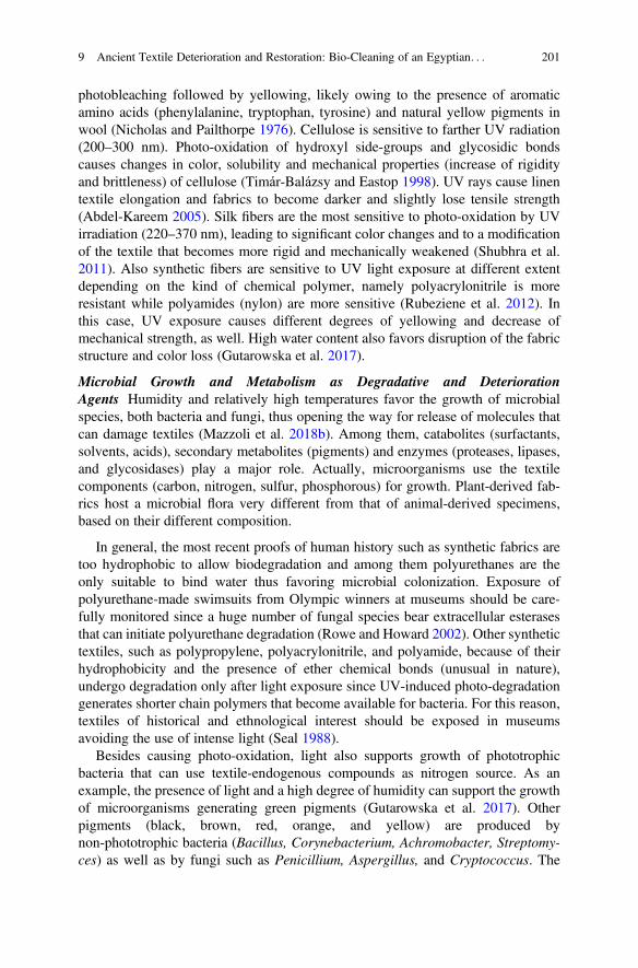
photobleaching followed by yellowing, likely owing to the presence of aromaticamino acids (phenylalanine, tryptophan, tyrosine) and natural yellow pigments inwool (Nicholas and Pailthorpe 1976). Cellulose is sensitive to farther UV radiation(200–300 nm). Photo-oxidation of hydroxyl side-groups and glycosidic bondscauses changes in color, solubility and mechanical properties (increase of rigidityand brittleness) of cellulose (Timár-Balázsy and Eastop 1998). UV rays cause linentextile elongation and fabrics to become darker and slightly lose tensile strength(Abdel-Kareem 2005). Silk fibers are the most sensitive to photo-oxidation by UVirradiation (220–370 nm), leading to significant color changes and to a modificationof the textile that becomes more rigid and mechanically weakened (Shubhra et al.2011). Also synthetic fibers are sensitive to UV light exposure at different extentdepending on the kind of chemical polymer, namely polyacrylonitrile is moreresistant while polyamides (nylon) are more sensitive (Rubeziene et al. 2012). Inthis case, UV exposure causes different degrees of yellowing and decrease ofmechanical strength, as well. High water content also favors disruption of the fabricstructure and color loss (Gutarowska et al. 2017).
Microbial Growth and Metabolism as Degradative and DeteriorationAgents Humidity and relatively high temperatures favor the growth of microbialspecies, both bacteria and fungi, thus opening the way for release of molecules thatcan damage textiles (Mazzoli et al. 2018b). Among them, catabolites (surfactants,solvents, acids), secondary metabolites (pigments) and enzymes (proteases, lipases,and glycosidases) play a major role. Actually, microorganisms use the textilecomponents (carbon, nitrogen, sulfur, phosphorous) for growth. Plant-derived fab-rics host a microbial flora very different from that of animal-derived specimens,based on their different composition.
In general, the most recent proofs of human history such as synthetic fabrics aretoo hydrophobic to allow biodegradation and among them polyurethanes are theonly suitable to bind water thus favoring microbial colonization. Exposure ofpolyurethane-made swimsuits from Olympic winners at museums should be care-fully monitored since a huge number of fungal species bear extracellular esterasesthat can initiate polyurethane degradation (Rowe and Howard 2002). Other synthetictextiles, such as polypropylene, polyacrylonitrile, and polyamide, because of theirhydrophobicity and the presence of ether chemical bonds (unusual in nature),undergo degradation only after light exposure since UV-induced photo-degradationgenerates shorter chain polymers that become available for bacteria. For this reason,textiles of historical and ethnological interest should be exposed in museumsavoiding the use of intense light (Seal 1988).
Besides causing photo-oxidation, light also supports growth of phototrophicbacteria that can use textile-endogenous compounds as nitrogen source. As anexample, the presence of light and a high degree of humidity can support the growthof microorganisms generating green pigments (Gutarowska et al. 2017). Otherpigments (black, brown, red, orange, and yellow) are produced bynon-phototrophic bacteria (Bacillus, Corynebacterium, Achromobacter, Streptomy-ces) as well as by fungi such as Penicillium, Aspergillus, and Cryptococcus. The
9 Ancient Textile Deterioration and Restoration: Bio-Cleaning of an Egyptian. . . 201
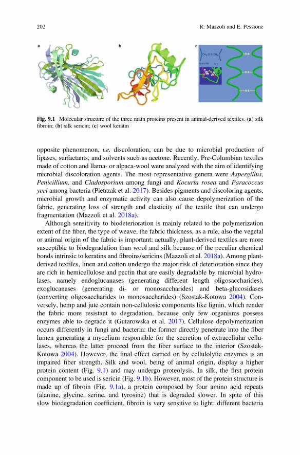
opposite phenomenon, i.e. discoloration, can be due to microbial production oflipases, surfactants, and solvents such as acetone. Recently, Pre-Columbian textilesmade of cotton and llama- or alpaca-wool were analyzed with the aim of identifyingmicrobial discoloration agents. The most representative genera were Aspergillus,Penicillium, and Cladosporium among fungi and Kocuria rosea and Paracoccusyeei among bacteria (Pietrzak et al. 2017). Besides pigments and discoloring agents,microbial growth and enzymatic activity can also cause depolymerization of thefabric, generating loss of strength and elasticity of the textile that can undergofragmentation (Mazzoli et al. 2018a).
Although sensitivity to biodeterioration is mainly related to the polymerizationextent of the fiber, the type of weave, the fabric thickness, as a rule, also the vegetalor animal origin of the fabric is important: actually, plant-derived textiles are moresusceptible to biodegradation than wool and silk because of the peculiar chemicalbonds intrinsic to keratins and fibroins/sericins (Mazzoli et al. 2018a). Among plant-derived textiles, linen and cotton undergo the major risk of deterioration since theyare rich in hemicellulose and pectin that are easily degradable by microbial hydro-lases, namely endoglucanases (generating different length oligosaccharides),exoglucanases (generating di- or monosaccharides) and beta-glucosidases(converting oligosaccharides to monosaccharides) (Szostak-Kotowa 2004). Con-versely, hemp and jute contain non-cellulosic components like lignin, which renderthe fabric more resistant to degradation, because only few organisms possessenzymes able to degrade it (Gutarowska et al. 2017). Cellulose depolymerizationoccurs differently in fungi and bacteria: the former directly penetrate into the fiberlumen generating a mycelium responsible for the secretion of extracellular cellu-lases, whereas the latter proceed from the fiber surface to the interior (Szostak-Kotowa 2004). However, the final effect carried on by cellulolytic enzymes is animpaired fiber strength. Silk and wool, being of animal origin, display a higherprotein content (Fig. 9.1) and may undergo proteolysis. In silk, the first proteincomponent to be used is sericin (Fig. 9.1b). However, most of the protein structure ismade up of fibroin (Fig. 9.1a), a protein composed by four amino acid repeats(alanine, glycine, serine, and tyrosine) that is degraded slower. In spite of thisslow biodegradation coefficient, fibroin is very sensitive to light: different bacteria
Fig. 9.1 Molecular structure of the three main proteins present in animal-derived textiles. (a) silkfibroin; (b) silk sericin; (c) wool keratin
202 R. Mazzoli and E. Pessione
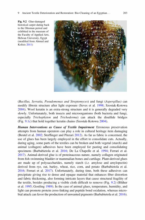
(Bacillus, Serratia, Pseudomonas and Streptomyces) and fungi (Aspergillus) canmodify fibroin structure after light exposure (Seves et al. 1998; Szostak-Kotowa2004). Wool keratin is an extra-strong structure and it is generally degraded veryslowly. Unfortunately, both insects and microorganisms (both bacteria and fungi,especially Trichophyton and Trichoderma) can attack the disulfide bridges(Fig. 9.1c) that hold together keratin chains (Szostak-Kotowa 2004).
Human Interventions as Cause of Textile Impairment Erroneous preservationattempts from human operators can play a role in cultural heritage item damaging(Beutel et al. 2002; Sterflinger and Pinzari 2012). As far as fabric is concerned, theuse of glues has been largely employed in the effort to consolidate cuts. Actually,during aging, some parts of the textiles can be broken and both vegetal (starch) andanimal (collagen) adhesives have been employed for pasting and consolidatingspecimens (Barbabietola et al. 2016; De La Chapelle et al. 1994; Ferrari et al.2017). Animal-derived glue is of proteinaceous nature, namely collagen originatedfrom fish swimming bladder or mammalian bones and cartilage. Plant-derived gluesare made up of polysaccharides, namely starch (i.e. amylose and amylopectin)derived from rye, oat, barley, wheat, rice, corn, and potato (Barbabietola et al.2016; Ferrari et al. 2017). Unfortunately, during time, both these adhesives canprecipitate giving rise to dense and opaque material that enhances fiber distortionand fabric thickening, also forming intricate layers that cause structural fragility ofthe textile, besides producing a visible cloth difficult to remove (Fig. 9.2) (Blüheret al. 1995; Gostling 1989). In the case of animal glues, temperature, humidity, andlight can promote protein cross-linking and peptide bond oxidation, whereas micro-bial attack can favor the production of unwanted pigments (Barbabietola et al. 2016).
Fig. 9.2 Glue-damagedhistorical carpet dating backto the Ottoman period andexhibited in the museum ofthe Faculty of Applied Arts,Helwan University, Egypt(modified from Ahmed andKolisis 2011)
9 Ancient Textile Deterioration and Restoration: Bio-Cleaning of an Egyptian. . . 203
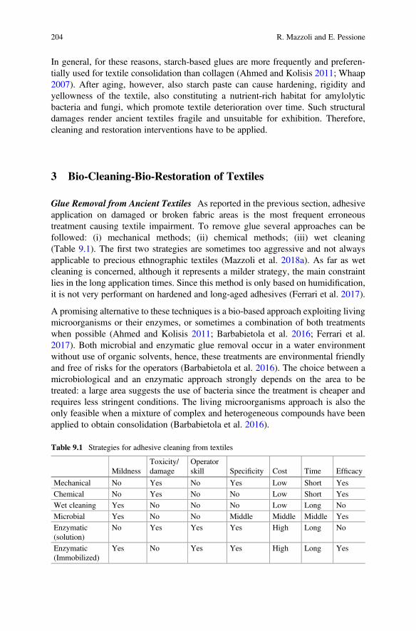
In general, for these reasons, starch-based glues are more frequently and preferen-tially used for textile consolidation than collagen (Ahmed and Kolisis 2011; Whaap2007). After aging, however, also starch paste can cause hardening, rigidity andyellowness of the textile, also constituting a nutrient-rich habitat for amylolyticbacteria and fungi, which promote textile deterioration over time. Such structuraldamages render ancient textiles fragile and unsuitable for exhibition. Therefore,cleaning and restoration interventions have to be applied.
3 Bio-Cleaning-Bio-Restoration of Textiles
Glue Removal from Ancient Textiles As reported in the previous section, adhesiveapplication on damaged or broken fabric areas is the most frequent erroneoustreatment causing textile impairment. To remove glue several approaches can befollowed: (i) mechanical methods; (ii) chemical methods; (iii) wet cleaning(Table 9.1). The first two strategies are sometimes too aggressive and not alwaysapplicable to precious ethnographic textiles (Mazzoli et al. 2018a). As far as wetcleaning is concerned, although it represents a milder strategy, the main constraintlies in the long application times. Since this method is only based on humidification,it is not very performant on hardened and long-aged adhesives (Ferrari et al. 2017).
A promising alternative to these techniques is a bio-based approach exploiting livingmicroorganisms or their enzymes, or sometimes a combination of both treatmentswhen possible (Ahmed and Kolisis 2011; Barbabietola et al. 2016; Ferrari et al.2017). Both microbial and enzymatic glue removal occur in a water environmentwithout use of organic solvents, hence, these treatments are environmental friendlyand free of risks for the operators (Barbabietola et al. 2016). The choice between amicrobiological and an enzymatic approach strongly depends on the area to betreated: a large area suggests the use of bacteria since the treatment is cheaper andrequires less stringent conditions. The living microorganisms approach is also theonly feasible when a mixture of complex and heterogeneous compounds have beenapplied to obtain consolidation (Barbabietola et al. 2016).
Table 9.1 Strategies for adhesive cleaning from textiles
MildnessToxicity/damage
Operatorskill Specificity Cost Time Efficacy
Mechanical No Yes No Yes Low Short Yes
Chemical No Yes No No Low Short Yes
Wet cleaning Yes No No No Low Long No
Microbial Yes No No Middle Middle Middle Yes
Enzymatic(solution)
No Yes Yes Yes High Long No
Enzymatic(Immobilized)
Yes No Yes Yes High Long Yes
204 R. Mazzoli and E. Pessione
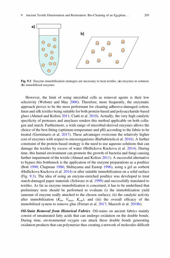
However, the limit of using microbial cells as removal agents is their lowselectivity (Webster and May 2006). Therefore, more frequently, the enzymaticapproach proves to be the most performant for cleaning adhesive-damaged cotton,linen and silk textiles being suitable for both protein-based and polysaccharide-basedglues (Ahmed and Kolisis 2011; Ciatti et al. 2010). Actually, the very high catalyticspecificity of proteases and amylases renders this method applicable on both colla-gen and starch. Furthermore, a wide range of microbial-derived enzymes allows thechoice of the best fitting (optimum temperature and pH) according to the fabric to betreated (Germinario et al. 2017). These advantages overcome the relatively highercost of enzymes with respect to microorganisms (Barbabietola et al. 2016). A furtherconstraint of the protein-based strategy is the need to use aqueous solutions that candamage the textiles by excess of water (Hrdlickova Kuckova et al. 2014). Duringtime, this humid environment can promote the growth of bacteria and fungi causingfurther impairment of the textile (Ahmed and Kolisis 2011). A successful alternativeto bypass this bottleneck is the application of the enzyme preparations as a poultice(Bott 1990; Chapman 1986; Shibayama and Eastop 1996), using a gel as sorbent(Hrdlickova Kuckova et al. 2014) or after suitable immobilization on a solid surface(Fig. 9.3). The idea of using an enzyme-enriched poultice was developed to treatstarch-damaged paper materials (Schwarz et al. 1999) and successfully translated totextiles. As far as enzyme immobilization is concerned, it has to be underlined thatpreliminary tests should be performed to evaluate (i) the immobilization yield(amount of enzyme really attached to the chosen surface); (ii) the catalytic activityafter immobilization (Km, Vmax, Kcat); and (iii) the overall efficacy of theimmobilized system to remove glue (Ferrari et al. 2017; Mazzoli et al. 2018b).
Oil-Stain Removal from Historical Fabrics Oil-stains on ancient fabrics mainlyconsist of unsaturated fatty acids that can undergo oxidation on the double bonds.During time, environmental oxygen can attack these double bonds generatingoxidation products that can polymerize thus creating a network of molecules difficult
Fig. 9.3 Enzyme immobilization strategies are necessary to treat textiles. (a) enzymes in solution;(b) immobilized enzymes
9 Ancient Textile Deterioration and Restoration: Bio-Cleaning of an Egyptian. . . 205
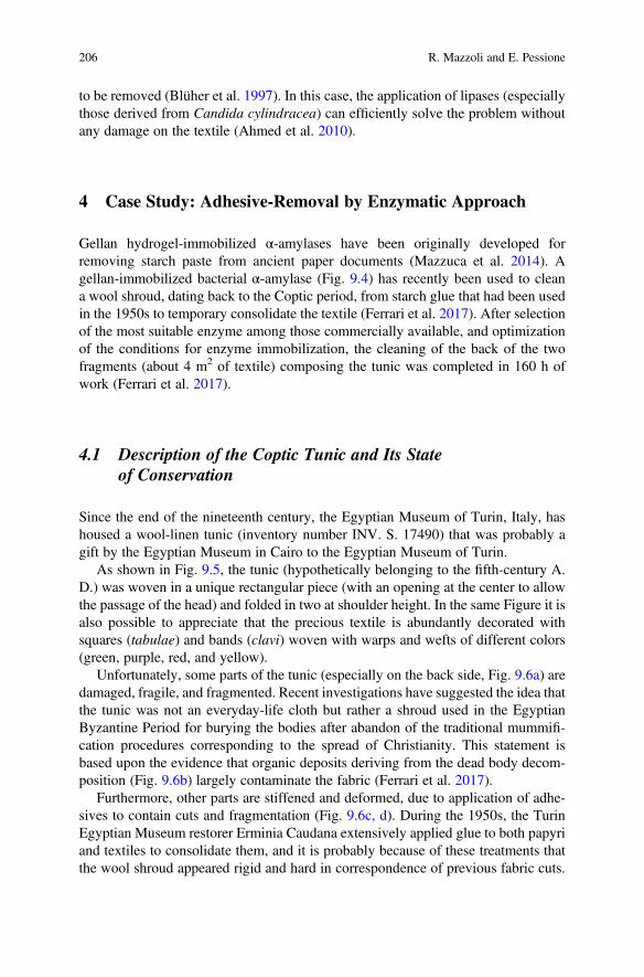
to be removed (Blüher et al. 1997). In this case, the application of lipases (especiallythose derived from Candida cylindracea) can efficiently solve the problem withoutany damage on the textile (Ahmed et al. 2010).
4 Case Study: Adhesive-Removal by Enzymatic Approach
Gellan hydrogel-immobilized α-amylases have been originally developed forremoving starch paste from ancient paper documents (Mazzuca et al. 2014). Agellan-immobilized bacterial α-amylase (Fig. 9.4) has recently been used to cleana wool shroud, dating back to the Coptic period, from starch glue that had been usedin the 1950s to temporary consolidate the textile (Ferrari et al. 2017). After selectionof the most suitable enzyme among those commercially available, and optimizationof the conditions for enzyme immobilization, the cleaning of the back of the twofragments (about 4 m2 of textile) composing the tunic was completed in 160 h ofwork (Ferrari et al. 2017).
4.1 Description of the Coptic Tunic and Its Stateof Conservation
Since the end of the nineteenth century, the Egyptian Museum of Turin, Italy, hashoused a wool-linen tunic (inventory number INV. S. 17490) that was probably agift by the Egyptian Museum in Cairo to the Egyptian Museum of Turin.
As shown in Fig. 9.5, the tunic (hypothetically belonging to the fifth-century A.D.) was woven in a unique rectangular piece (with an opening at the center to allowthe passage of the head) and folded in two at shoulder height. In the same Figure it isalso possible to appreciate that the precious textile is abundantly decorated withsquares (tabulae) and bands (clavi) woven with warps and wefts of different colors(green, purple, red, and yellow).
Unfortunately, some parts of the tunic (especially on the back side, Fig. 9.6a) aredamaged, fragile, and fragmented. Recent investigations have suggested the idea thatthe tunic was not an everyday-life cloth but rather a shroud used in the EgyptianByzantine Period for burying the bodies after abandon of the traditional mummifi-cation procedures corresponding to the spread of Christianity. This statement isbased upon the evidence that organic deposits deriving from the dead body decom-position (Fig. 9.6b) largely contaminate the fabric (Ferrari et al. 2017).
Furthermore, other parts are stiffened and deformed, due to application of adhe-sives to contain cuts and fragmentation (Fig. 9.6c, d). During the 1950s, the TurinEgyptian Museum restorer Erminia Caudana extensively applied glue to both papyriand textiles to consolidate them, and it is probably because of these treatments thatthe wool shroud appeared rigid and hard in correspondence of previous fabric cuts.
206 R. Mazzoli and E. Pessione
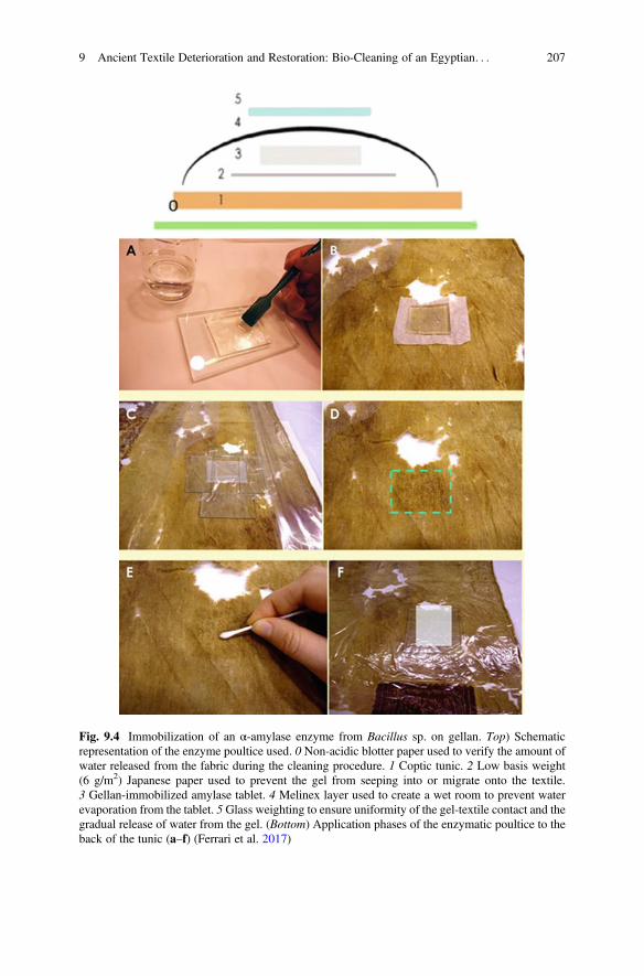
Fig. 9.4 Immobilization of an α-amylase enzyme from Bacillus sp. on gellan. Top) Schematicrepresentation of the enzyme poultice used. 0 Non-acidic blotter paper used to verify the amount ofwater released from the fabric during the cleaning procedure. 1 Coptic tunic. 2 Low basis weight(6 g/m2) Japanese paper used to prevent the gel from seeping into or migrate onto the textile.3 Gellan-immobilized amylase tablet. 4 Melinex layer used to create a wet room to prevent waterevaporation from the tablet. 5Glass weighting to ensure uniformity of the gel-textile contact and thegradual release of water from the gel. (Bottom) Application phases of the enzymatic poultice to theback of the tunic (a–f) (Ferrari et al. 2017)
9 Ancient Textile Deterioration and Restoration: Bio-Cleaning of an Egyptian. . . 207
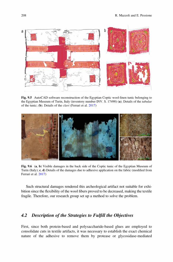
Such structural damages rendered this archeological artifact not suitable for exhi-bition since the flexibility of the wool fibers proved to be decreased, making the textilefragile. Therefore, our research group set up a method to solve the problem.
4.2 Description of the Strategies to Fulfill the Objectives
First, since both protein-based and polysaccharide-based glues are employed toconsolidate cuts in textile artifacts, it was necessary to establish the exact chemicalnature of the adhesive to remove them by protease or glycosidase-mediated
Fig. 9.5 AutoCAD software reconstruction of the Egyptian Coptic wool-linen tunic belonging tothe Egyptian Museum of Turin, Italy (inventory number INV. S. 17490) (a). Details of the tabulaeof the tunic; (b). Details of the clavi (Ferrari et al. 2017)
Fig. 9.6 (a, b) Visible damages in the back side of the Coptic tunic of the Egyptian Museum ofTurin (Italy); c, d) Details of the damages due to adhesive application on the fabric (modified fromFerrari et al. 2017)
208 R. Mazzoli and E. Pessione
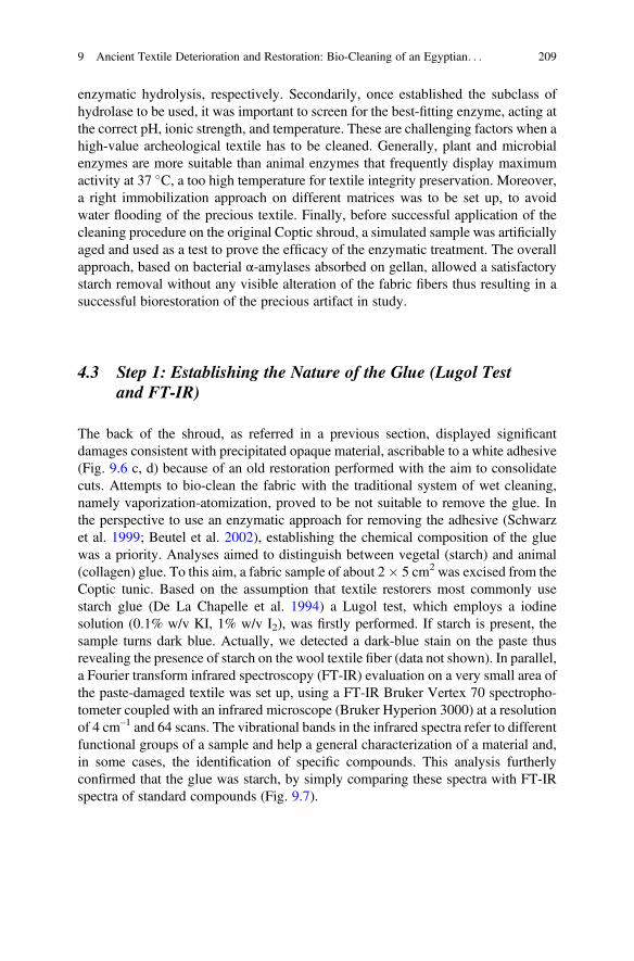
enzymatic hydrolysis, respectively. Secondarily, once established the subclass ofhydrolase to be used, it was important to screen for the best-fitting enzyme, acting atthe correct pH, ionic strength, and temperature. These are challenging factors when ahigh-value archeological textile has to be cleaned. Generally, plant and microbialenzymes are more suitable than animal enzymes that frequently display maximumactivity at 37 �C, a too high temperature for textile integrity preservation. Moreover,a right immobilization approach on different matrices was to be set up, to avoidwater flooding of the precious textile. Finally, before successful application of thecleaning procedure on the original Coptic shroud, a simulated sample was artificiallyaged and used as a test to prove the efficacy of the enzymatic treatment. The overallapproach, based on bacterial α-amylases absorbed on gellan, allowed a satisfactorystarch removal without any visible alteration of the fabric fibers thus resulting in asuccessful biorestoration of the precious artifact in study.
4.3 Step 1: Establishing the Nature of the Glue (Lugol Testand FT-IR)
The back of the shroud, as referred in a previous section, displayed significantdamages consistent with precipitated opaque material, ascribable to a white adhesive(Fig. 9.6 c, d) because of an old restoration performed with the aim to consolidatecuts. Attempts to bio-clean the fabric with the traditional system of wet cleaning,namely vaporization-atomization, proved to be not suitable to remove the glue. Inthe perspective to use an enzymatic approach for removing the adhesive (Schwarzet al. 1999; Beutel et al. 2002), establishing the chemical composition of the gluewas a priority. Analyses aimed to distinguish between vegetal (starch) and animal(collagen) glue. To this aim, a fabric sample of about 2� 5 cm2 was excised from theCoptic tunic. Based on the assumption that textile restorers most commonly usestarch glue (De La Chapelle et al. 1994) a Lugol test, which employs a iodinesolution (0.1% w/v KI, 1% w/v I2), was firstly performed. If starch is present, thesample turns dark blue. Actually, we detected a dark-blue stain on the paste thusrevealing the presence of starch on the wool textile fiber (data not shown). In parallel,a Fourier transform infrared spectroscopy (FT-IR) evaluation on a very small area ofthe paste-damaged textile was set up, using a FT-IR Bruker Vertex 70 spectropho-tometer coupled with an infrared microscope (Bruker Hyperion 3000) at a resolutionof 4 cm–1 and 64 scans. The vibrational bands in the infrared spectra refer to differentfunctional groups of a sample and help a general characterization of a material and,in some cases, the identification of specific compounds. This analysis furtherlyconfirmed that the glue was starch, by simply comparing these spectra with FT-IRspectra of standard compounds (Fig. 9.7).
9 Ancient Textile Deterioration and Restoration: Bio-Cleaning of an Egyptian. . . 209
4.4 Step 2: Selecting the Most Suitable Enzymeand Immobilization Strategy
Once established the starch nature of the adhesive, amylases proved to be the mostsuitable hydrolytic enzymes for bio-cleaning. Starch consists of two α-glucans, i.e.amylose (linear glucose polymer) and amylopectin (branched-chain glucose poly-mer), but amylopectin is generally considered the main responsible for paste-features. Both α- and β-amylases exist which hydrolyze amylose and amylopectinthrough different catalytic mechanisms. α-Amylases hydrolyze internal α-1-4 gly-cosidic bonds at random generating dextrins. β-Amylases are exoglucanases thatcleave amylose and amylopectin starting from their non-reducing terminals andrelease maltose (Ray and Nanda 1996). Choice was addressed to microbialα-amylases because they are maltodextrin-generating endo-enzymes able to quicklyeliminate the adhesive properties of starch (Schwarz et al. 1999) whereas β-amylasesdisplay a slower catalytic mechanism (Ray and Nanda 1996). Furthermore,prokaryote-derived enzymes display intrinsic higher stability in a wider pH andtemperature range than animal-derived amylases (Mehta and Satyanarayana 2016).Among commercial α-amylases, that from Bacillus sp. was selected for starch glueremoval, owing to its very high specific activity (Chand et al. 2014). More in details,lyophilized α-amylase from Bacillus sp. Type IIA (Sigma-Aldrich) with specificactivity >1500 units/mg protein was finally chosen. A 0.1% w/v solution of the
3500
1.71.61.51.41.31.21.11.0
Abs
orba
nce
3301
2956
2926
2872
2855
1651
1549
1454
1409
1380
1323
1232
1151
1077
1039
699
933
958
0.90.80.70.60.50.40.30.20.1
3000 2500
Wavenumbers (cm–1)
2000 1500 1000
Fig. 9.7 Fourier Transform Infrared Spectroscopy Analysis of the glue reveals the starch nature ofthe adhesive found on the Coptic tunic of the Egyptian Museum of Turin (Italy) (Ferrari et al. 2017)
210 R. Mazzoli and E. Pessione
enzyme in 20 mM sodium-phosphate buffer pH 6.9 was prepared (De La Chapelleet al. 1994; Cremonesi 2009) for tunic restoration. It is worth noting that pH 6.9corresponds to the optimal pH for Bacillus sp. Type IIA alpha-amylase activity.
However, as referred above, the use of water solutions of enzymes is not suitablefor precious archeological artifacts since this treatment can flood the textile with atoo high water load, favoring successive microbial contamination. Therefore, wedesigned an enzyme immobilization approach to restrict the amount of water neededfor catalysis. Two immobilization matrices were tested: agar and gellan. Both arepolysaccharides displaying high pH stability and highly hygroscopic properties thatlimit liquid transfer to the fibers, allowing a gradual release of the enzyme into thefabric. Furthermore, their un-adhesive nature and facility to form films render themsuitable for application on textiles (Iannuccelli and Sotgiu 2007). Hence, one cmthick low-viscosity “soft” gels (2% w/v) agar (polygalactose andsulfurpolygalactose, Bresciani SRL) and 2% w/v Gellan gum (Phytagel,heteropolysaccharide, Bresciani SRL) in 20 mM sodium-phosphate buffer pH 6.9were used for this purpose. Since a very good efficiency in glue removal can beobtained by adsorbing enzyme solutions at the interface between textile and support,the solution of α-amylase from Bacillus sp. was brush and immobilized onto thesolid gel surfaces so as to have the maximum enzyme concentration at the interfaceand, hence, maximize efficiency of glue removal (Fig. 9.4a). However, the optimumcatalytic activity for Bacillus sp. α-amylases is 60 �C and this temperature cannot bereached because of the risk of gel melting at temperatures higher than 40 �C. Theoverall enzyme immobilization procedure is illustrated in Fig. 9.4.
4.5 Step 3: Simulated Sample Preparation, Aging,and Damaging
To prevent any risk of damage to the original archeological tunic, a goat woolsimulated sample, with similar chemical-physical properties to the original Copticshroud fabric, was woven to compare the enzymatic cleaning and the cleaning effectof the immobilization gels alone, prior to application of the system on the realartwork. To have a simulated sample as similar as possible to the original textile,also aging should be considered. Reproducing the original aging of artifacts is achallenging procedure. In the present investigation, we tested an incubation temper-ature of 90 �C (maximum allowed for wool) (Quaglierini 1985) for 60 days to induceaccelerated artificial aging (it has previously been established that 25 years of naturalaging can be obtained by 3 days at 120 �C in silk) (Ahmed and Kolisis 2011). Dryincubation was chosen and applied here because this condition attempted to repro-duce the climatic conditions characterizing the ground where the Coptic shroud wasburied in Egypt. However, some events occurring in the natural aging process(contamination by burial ground and dead body decomposition liquids) cannot bereproduced. After this time, a treatment with starch glue was performed to mimic the
9 Ancient Textile Deterioration and Restoration: Bio-Cleaning of an Egyptian. . . 211
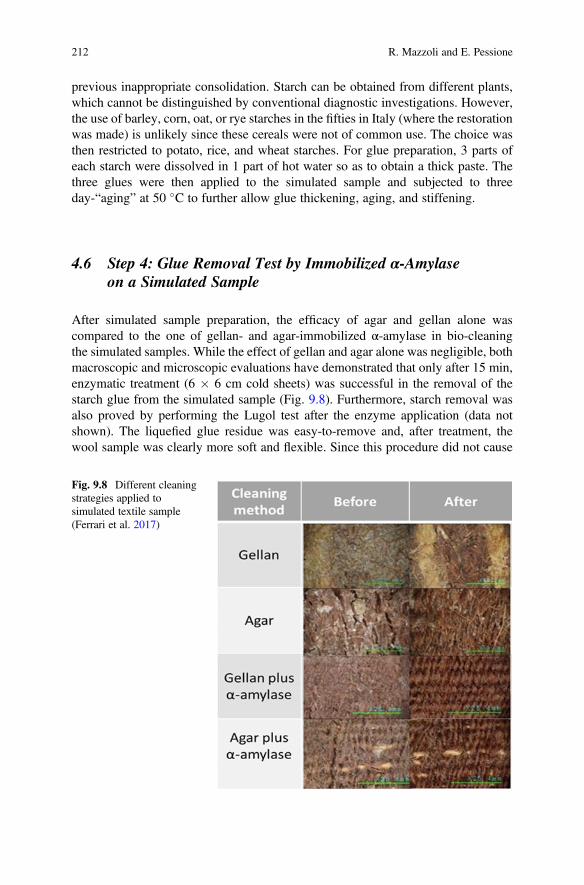
previous inappropriate consolidation. Starch can be obtained from different plants,which cannot be distinguished by conventional diagnostic investigations. However,the use of barley, corn, oat, or rye starches in the fifties in Italy (where the restorationwas made) is unlikely since these cereals were not of common use. The choice wasthen restricted to potato, rice, and wheat starches. For glue preparation, 3 parts ofeach starch were dissolved in 1 part of hot water so as to obtain a thick paste. Thethree glues were then applied to the simulated sample and subjected to threeday-“aging” at 50 �C to further allow glue thickening, aging, and stiffening.
4.6 Step 4: Glue Removal Test by Immobilized α-Amylaseon a Simulated Sample
After simulated sample preparation, the efficacy of agar and gellan alone wascompared to the one of gellan- and agar-immobilized α-amylase in bio-cleaningthe simulated samples. While the effect of gellan and agar alone was negligible, bothmacroscopic and microscopic evaluations have demonstrated that only after 15 min,enzymatic treatment (6 � 6 cm cold sheets) was successful in the removal of thestarch glue from the simulated sample (Fig. 9.8). Furthermore, starch removal wasalso proved by performing the Lugol test after the enzyme application (data notshown). The liquefied glue residue was easy-to-remove and, after treatment, thewool sample was clearly more soft and flexible. Since this procedure did not cause
Fig. 9.8 Different cleaningstrategies applied tosimulated textile sample(Ferrari et al. 2017)
212 R. Mazzoli and E. Pessione
any damage to the simulated textile, it was suitable to be employed on the originalCoptic tunic. However, the gellan matrix was better performing than agar also formonitoring adhesive paste removal since it is more transparent. Furthermore, thelower rigidity of gellan with respect to agar allowed a better adhesion to thediscontinuity of the textile fibers. For these reasons, application of gellan-immobilized α-amylase on the original Coptic shroud was preferred.
4.7 Step 5: Glue Removal by Immobilized α-Amylaseon the Original Archeological Shroud
Finally, the same enzymatic treatment described in the preliminary test on thesimulated sample was performed on the archeological Coptic tunic. α-Amylase,immobilized on gellan sheets (that were those showing the best performances) wasthen placed on a thin Japanese paper sheet to avoid any seeping of the gel into thefabric (Fig. 9.4a, b). Afterwards, a “wet room” (Fig. 9.4c) was created bysuperimposing a Melinex sheet and a glass weight to prevent dehydration.
As shown in Fig. 9.4 e, f, 15 min were sufficient to swollen and liquefy the glue. Itworth remembering that such a high efficiency was obtained although the temper-ature used during bio-cleaning procedure was lower than that for amylase optimumactivity (60 �C) (Schwarz et al. 1999). The adhesive residue was then removed bymeans of a moistened cotton swab and a spatula (Fig. 9.4e). No more adhesive pastewas detectable on the fabric (dried by using an absorbent paper (Fig. 9.4f), that alsodisplayed improved elasticity and flexibility. After this bio-cleaning treatment, theneedle consolidation of the textile was finally performed. The overall bio-cleaning ofboth sides of the textile (about 4 m2) was achieved in 160 h of work.
Other advantages of immobilized amylases are the very quick time of applicationand the overall milder mechanical treatment required as compared to traditionalcleaning methods (i.e. wet cleaning). It is useful mentioning that α-amylases havebeen already used to restore wool/linen tapestry after suitable immobilization onmethylcellulose matrices (Whaap 2007) but also (both in solution and as a poultice)
Fig. 9.9 The restoredCoptic tunic-shroudexhibited at the EgyptianMuseum of Turin, Italy(Ferrari et al. 2017)
9 Ancient Textile Deterioration and Restoration: Bio-Cleaning of an Egyptian. . . 213
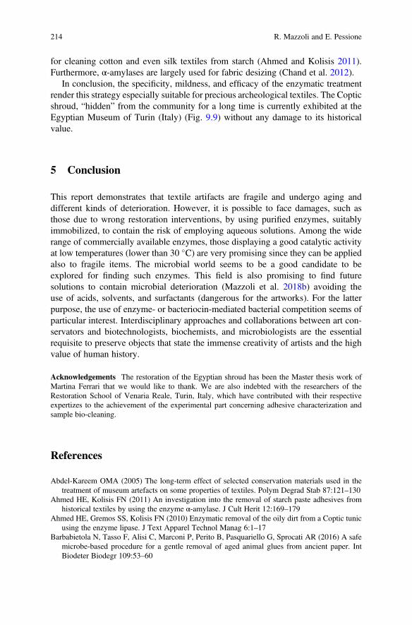
for cleaning cotton and even silk textiles from starch (Ahmed and Kolisis 2011).Furthermore, α-amylases are largely used for fabric desizing (Chand et al. 2012).
In conclusion, the specificity, mildness, and efficacy of the enzymatic treatmentrender this strategy especially suitable for precious archeological textiles. The Copticshroud, “hidden” from the community for a long time is currently exhibited at theEgyptian Museum of Turin (Italy) (Fig. 9.9) without any damage to its historicalvalue.
5 Conclusion
This report demonstrates that textile artifacts are fragile and undergo aging anddifferent kinds of deterioration. However, it is possible to face damages, such asthose due to wrong restoration interventions, by using purified enzymes, suitablyimmobilized, to contain the risk of employing aqueous solutions. Among the widerange of commercially available enzymes, those displaying a good catalytic activityat low temperatures (lower than 30 �C) are very promising since they can be appliedalso to fragile items. The microbial world seems to be a good candidate to beexplored for finding such enzymes. This field is also promising to find futuresolutions to contain microbial deterioration (Mazzoli et al. 2018b) avoiding theuse of acids, solvents, and surfactants (dangerous for the artworks). For the latterpurpose, the use of enzyme- or bacteriocin-mediated bacterial competition seems ofparticular interest. Interdisciplinary approaches and collaborations between art con-servators and biotechnologists, biochemists, and microbiologists are the essentialrequisite to preserve objects that state the immense creativity of artists and the highvalue of human history.
Acknowledgements The restoration of the Egyptian shroud has been the Master thesis work ofMartina Ferrari that we would like to thank. We are also indebted with the researchers of theRestoration School of Venaria Reale, Turin, Italy, which have contributed with their respectiveexpertizes to the achievement of the experimental part concerning adhesive characterization andsample bio-cleaning.
References
Abdel-Kareem OMA (2005) The long-term effect of selected conservation materials used in thetreatment of museum artefacts on some properties of textiles. Polym Degrad Stab 87:121–130
Ahmed HE, Kolisis FN (2011) An investigation into the removal of starch paste adhesives fromhistorical textiles by using the enzyme α-amylase. J Cult Herit 12:169–179
Ahmed HE, Gremos SS, Kolisis FN (2010) Enzymatic removal of the oily dirt from a Coptic tunicusing the enzyme lipase. J Text Apparel Technol Manag 6:1–17
Barbabietola N, Tasso F, Alisi C, Marconi P, Perito B, Pasquariello G, Sprocati AR (2016) A safemicrobe-based procedure for a gentle removal of aged animal glues from ancient paper. IntBiodeter Biodegr 109:53–60
214 R. Mazzoli and E. Pessione
Beutel S, Klein K, Knobbe G, Königfeld P, Petersen K, Ulber R, Scheper T (2002) Controlledenzymatic removal of damaging casein layers on medieval wall paintings. Biotechnol Bioeng80:13–21
Blüher A, Haller U, Banik G, Thobois E (1995) The application of carbopol poultices on paperobjects. Restaur Int J Preserv Libr Arch Mater 16:234–247
Blüher A, Grube A, Bornscheuer U, Banik G (1997) A reappraisal of the enzyme lipase forremoving drying-oil stains on paper. Pap Conserv J Inst Pap Conserv 1997:37–46
Bott G (1990) Amylase for starch removal from a set of 17th century embroidered panels. Conserv14:23–29
Chand N, Nateri AS, Sajedi RH, Mahdavi A, Rassa M (2012) Enzymatic desizing of cotton fabricusing a Ca 2+-independent α-amylase with acidic pH profile. J Mol Catal B Enzym 83:46–50
Chand N, Sajedi RH, Nateri AS, Khajeh K, Rassa M (2014) Fermentative desizing of cotton fabricusing an α-amylase-producing Bacillus strain: Optimization of simultaneous enzyme productionand desizing. Process Biochem 49:1884–1888
Chapman V (1986) Amylase in a viscous medium—textile applications. The Conservator 10:7–11Ciatti M, Conti S, Fineschi C, Nelson JK, Pini S (2010) Il ricamo in or nué su disegno di Raffaellino
del Garbo. Aspetti storico-stilistici, tecnici, minimo intervento e conservazione preventiva. OPDRestauro 22:81–116
Cremonesi P (2009) L’uso degli enzimi nella pulitura di opere policrome. Il Prato, Padova, ItalyDe La Chapelle A, Choisy F, Gallo M, Legoy M (1994) Emploi d’amylases et de protéases pour la
restauration d’arts graphiques. In: Environnement et Conservation de l’Ecrit, de l’Image et duSon: Actes des Deuxièmes Journées Internationales d’Etudes de l’ARSAG, 16 au 20 mai 1994.Association pour la recherche scientifique sur les arts graphiques, Paris, France, pp 217–229
Ferrari M, Mazzoli R, Morales S, Fedi M, Liccioli L, Piccirillo A, Cavaleri T, Oliva C, Gallo P,Borla M, Cardinali M, Pessione E (2017) Enzymatic laundry for old clothes: immobilized alpha-amylase from Bacillus sp. for the biocleaning of an ancient Coptic tunic. Appl MicrobiolBiotechnol 101:7041–7052
Germinario G, van der Werf ID, Palazzo G, Regidor Ros JL, Montes-Estelles RM, Sabbatini L(2017) Bioremoval of marker pen inks by exploiting lipase hydrolysis. Prog Org Coatings110:162–171
Gostling K (1989) Bookbinders and adhesives: part 2. New Bookbind 9:30–39Gutarowska B, Pietrzak K, Machnowski W, Milczarek JM (2017) Historical textiles - a review of
microbial deterioration analysis and disinfection methods. Text Res J 87:2388–2406Hrdlickova Kuckova S, Crhova Krizkova M, Pereira CLC, Hynek R, Lavrova O, Busani T, Branco
LC, Sandu ICA (2014) Assessment of green cleaning effectiveness on polychrome surfaces byMALDI-TOF mass spectrometry and microscopic imaging. Microsc Res Technol 77:574–585
Iannuccelli S, Sotgiu S (2007) La pulitura ad umido di opere d’arte su carta con gel rigidi di gellano:presupposti teorici, metodologia, applicazioni e verifica analitica. In: Quaderni del Cesmar7,Vol. 11. Il Prato, Padova, Italy
Mazzoli R, Giuffrida MG, Pessione E (2018a) Back to the past: “find the guilty bug—microorgan-isms involved in the biodeterioration of archeological and historical artifacts”. Appl MicrobiolBiotechnol 102:6393–6407
Mazzoli R, Giuffrida MG, Pessione E (2018b) Back to the past—forever young: cutting-edgebiochemical and microbiological tools for cultural heritage conservation. Appl MicrobiolBiotechnol 102:6815–6825
Mazzuca C, Micheli L, Cervelli E, Basoli F, Cencetti C, Coviello T, Iannuccelli S, Sotgiu S,Palleschi A (2014) Cleaning of paper artworks: development of an efficient gel-based materialable to remove starch paste. ACS Appl Mater Interfaces 6:16519–16528
Mehta D, Satyanarayana T (2016) Bacterial and archaeal α-amylases: diversity and amelioration ofthe desirable characteristics for industrial applications. Front Microbiol 7:1129
Nicholas CH, Pailthorpe MT (1976) 50—primary reactions in the photoyellowing of wool keratin. JText Inst 67:397–403
9 Ancient Textile Deterioration and Restoration: Bio-Cleaning of an Egyptian. . . 215
Pietrzak K, Puchalski M, Otlewska A, Wrzosek H, Guiamet P, Piotrowska M, Gutarowska B (2017)Microbial diversity of pre-Columbian archaeological textiles and the effect of silvernanoparticles misting disinfection. J Cult Herit 23:138–147
Quaglierini C (1985) Manuale di merceologia tessile. Zanichelli, Bologna, ItalyRay RR, Nanda G (1996) Microbial β-amylases: biosynthesis, characteristics, and industrial
applications. Crit Rev Microbiol 22:181–199Rowe L, Howard GT (2002) Growth of Bacillus subtilis on polyurethane and the purification and
characterization of a polyurethanase-lipase enzyme. Int Biodeter Biodegr 50:33–40Rubeziene V, Varnaite S, Baltusnikaite J, Padleckiene I (2012) Effects of light exposure on textile
durability. In: Annis PA (ed) Understanding and improving the durability of textiles. WoodheadPublishing, Oxford, pp 104–125
Schwarz I, Blüher A, Banik G, Thobois E, Maurer KH (1999) The development of a ready-for-usepoultice for local removal of starch paste by enzymatic action. Restaurator 20:225–244
Seal KJ (1988) The biodegradation of naturally occurring and synthetic plastic polymers. BiodeterAbstr 2:296–317
Seves A, Romanò M, Maifreni T, Sora S, Ciferri O (1998) The microbial degradation of silk: alaboratory investigation. Int Biodeter Biodegr 42:203–211
Shibayama N, Eastop D (1996) Removal of flour paste residues from a painted banner with alpha-amylase. The Conservator 20:53–64
Shubhra QTH, Alam AKMM, Beg MDH (2011) Mechanical and degradation characteristics ofnatural silk fiber reinforced gelatin composites. Mater Lett 65:333–336
Sterflinger K, Pinzari F (2012) The revenge of time: fungal deterioration of cultural heritage withparticular reference to books, paper and parchment. Environ Microbiol 14:559–566
Szostak-Kotowa J (2004) Biodeterioration of textiles. Int Biodeter Biodegr 53:165–170Timár-Balázsy Á, Eastop D (1998) Chemical principles of textile conservation. Routledge, Taylor
and Francis Group, London and New YorkTreigiené R, Musnickas J (2003) Solvent pre-treated wool fabric permanent set and physical
properties. Fibres Text East Eur 11:37–40Webster A, May E (2006) Bioremediation of weathered-building stone surfaces. Trends Biotechnol
24:255–260Whaap F (2007) The treatment of two Coptic tapestry fragments. V A Conserv J 55:11–13Zimmermann M, Hocker H (1996) Typical fracture appearance of broken wool fibers after
simulated sunlight irradiation. Text Res J 66:657–660
Open Access This chapter is licensed under the terms of the Creative Commons Attribution 4.0International License (http://creativecommons.org/licenses/by/4.0/), which permits use, sharing,adaptation, distribution and reproduction in any medium or format, as long as you give appropriatecredit to the original author(s) and the source, provide a link to the Creative Commons license andindicate if changes were made.
The images or other third party material in this chapter are included in the chapter's CreativeCommons license, unless indicated otherwise in a credit line to the material. If material is notincluded in the chapter's Creative Commons license and your intended use is not permitted bystatutory regulation or exceeds the permitted use, you will need to obtain permission directly fromthe copyright holder.
216 R. Mazzoli and E. Pessione
Chapter 10Advanced Biocleaning System for HistoricalWall Paintings
Giancarlo Ranalli and Elisabetta Zanardini
Abstract This chapter will focus on the potential role of safe microorganisms asbiocleaning agents in the removal of altered or undesirable organic substances onhistorical wall paintings. Selected microbes can be adopted as biological cleaners toreduce and remove deterioration ageing phenomena, environmental pollutants andaltered by-products of past intervention of restorations. The aim is to offer acomprehensive view on the role and potentiality of virtuous microorganisms pro-biocleaning of altered historical wall paintings. We also report four case studies inthe CH restoration field, carried out in the last 25 years, with the innovative use ofbacteria and different delivery systems, focusing the attention on the preliminarydiagnosis and the monitoring of the whole process. The CH field represents a greatchallenge and Science and Art link together the work of conservator scientists andhistorians with researchers and scientists, sharing their diverse expertises and joiningthe knowledges to the preservation and the conservation of our artistic patrimony.
Keywords Conservation-Restoration · Cultural heritage · Biotechnology ·Biocleaning · Organic matters · Bacteria
Cultural Heritage (CH) artworks are one of the most important elements of theidentity in large part of the World, especially in the old European Continent andrepresent the cultural legacy from past generations defined as tangible (buildings,monuments, books, etc.) and intangible (as folklore, traditions, language, knowledgeand natural as landscapes, and flora-fauna biodiversity).
The knowledge of our history has been subject to a continuous evolution over thetime; that is true if we consider the modern potentialities of communications and themobility of scientists and conservation specialists across the World. However, the
G. Ranalli (*)Department of Bioscience and Territory, University of Molise, Pesche, Italye-mail: [email protected]
E. ZanardiniDepartment of Sciences and High Technology, University of Insubria, Como, Italy
© The Author(s) 2021E. Joseph (ed.), Microorganisms in the Deterioration and Preservation of CulturalHeritage, https://doi.org/10.1007/978-3-030-69411-1_10
217
knowledge in the conservation field, being relatively young, remains difficult bothfor the differences in terminology and for the lacks in the standardization of pro-cedures, especially considering national-international multidisciplinary sectors(trades, professionals, administrative authorities, academic lectures, conservationstudents, workers involved in built environment, etc.).
Three examples are here cited: the terms restoration, wall painting and fresco.The first still linked to possible negative aspects originated by the restoration-reconstruction methods applied in the past centuries. The second defines wallpaintings as paintings on inorganic substrates (plaster, stone), but in other cases,with painting on the wall are included paintings on wooden wall (EWAGLOS2016). The third, the term fresco is limited to paintings on fresh lime plaster(Italy), but used in France for all kinds of wall paintings, including combination offresco and secco techniques.
Artworks related to CH are exposed to weather and submitted to the influence ofthe environmental conditions: physicals, chemicals and biological factors interactwith the constitutive materials, inducing changes both in its compositional andstructural characteristics. In some cases the material transformation is due to themetabolic activity associated with the growth and the activity of living organisms,from microorganisms to plants and animals.
In the last decades, an important role of CH conservation has been mostly playedby physics, chemistry and material science, but actually, the scientific scenery alsoincludes biotechnology and applied microbiology, which can contribute to thedevelopment of new methods for the detection and identification of microorganismsaltering stoneworks and for the bioremediation of weathered artworks (Fernandes2006). In the last twenty years, the advanced methodologies have contributed to theknowledge of the structure (species composition and abundance) of the microbialcommunities colonizing artworks and their biodeterioration potentialities(Daffonchio et al. 2000; Gurtner et al. 2000; Piñar et al. 2001; Urzì et al. 2001;Crispim and Gaylarde 2005; McNamara and Mitchell 2005; Gorbushina 2007;Abdulla et al. 2008; Portillo et al. 2009; Ranalli et al. 2009; Scheerer et al. 2009;May 2010; Alonso-Vega et al. 2011; Giacomucci et al. 2011; Saiz-Jimenez et al.2011; Zanardini et al. 2011, 2016, 2019; Cappitelli et al. 2012).
The biodeterioration mechanism and the role played by several biological agents“biodeteriogens” in the decay of stone monuments are well known. Different kindsof organisms (primary colonization) can grow using the stone mineral componentsand its superficial deposits. The main consequence of their metabolic activity, suchas the excretion of enzymes, inorganic and organic acids and of complex formingsubstances, is the dissolution of minerals of the substratum. Moreover, the growthand the swelling of some vegetative structures (e.g. roots and lichenic thalli) inducephysical stresses and mechanical breaks. The growth process and vegetative devel-opment of the living species of organisms can interfere on the conservation of CHs.More, the intensity of these damages is strictly correlated with: type and dimensionof the organism involved; kind of material and state of its conservation; environ-mental conditions, micro-climatic exposure; level and types of air pollutants (Tiano2002).
218 G. Ranalli and E. Zanardini
In the last decades, the traditional microbiological culture-dependent methodsbased on the use of specific media for the cultivation and the identification ofmicroorganisms have been coupled to the culture-independent methods. Theseallow a better understanding of the complexity of the microbial communities alteringstoneworks, the detection of novel microorganisms (bacteria, fungi, algae, etc.) andtheir activity (Schabereiter-Gurtner et al. 2001; Portillo et al. 2009; Saiz-Jimenezet al. 2011; Villa et al. 2015; Zanardini et al. 2016, 2019).
Moreover, the molecular methods can provide an early detection of the biodete-rioration process, even when the deterioration is not visible yet. So, the use ofmolecular techniques for a preventive diagnosis and in the conservation perspectivesbecomes necessary both for the scientific approach and the economical point ofview. These new techniques combined with the traditional microbiological methodsgive a much better understanding of the number, activity, function of isolated oraggregate microorganisms, not only in the “barbarian” biodeterioration, but also inbio-restoration and bio-conservation process, for their “virtuosos” functions.
In addition, these new approaches can be useful to set up alternative biocleaningrestoration methodologies and to control during the treatment the microbial growthand activity and after the treatment to prevent further colonizations (Palla 2004,2013).
1 Biotechnologies Applied to CH
For these reasons, the use of biological systems, as living organisms or derivatives(e.g. enzymes), goes under the name of biotechnology in many fields including therecovery of CH artwork.
In natural environments (air, water, soil) microorganisms contribute to veryimportant aspect of life in the Earth: the equilibrium of the “matter transformationcycle” or “carbon cycle”. Some examples are the transformation of rocks in soils(pedogenesis) or the transformation of the complex substances (polymers) intosimple components (monomers) until the complete degradation (mineralization).As we mentioned above, micro- and macroorganisms, ranging from microscopicalbacterial cells to higher plants and animals (eukaryotes), can find as suitable habitatsfor their growth, the CH surfaces such as ancient archaeological remains, monu-mental buildings, works of arts made with different inorganic and organic materials(stone, wall paintings, metals, wood, textiles, paper, etc.).
In conservation and restoration practices, different procedures can be used andsome of them require the use and application of substances (i.e. animal and vegetalglues, re-lining paste, varnishes, temperas and other materials for cleaning andsoaking). These substances can contribute to the biological risk being nutrients forthe microbial growth especially when the environmental conditions are favourable(high water content, air humidity, temperature, etc.).
Indeed, when in past restoration, organic matter has been applied to stone surfacesfor various purposes, often stoneworks show alteration over time and, in these cases,
10 Advanced Biocleaning System for Historical Wall Paintings 219
their preservation poses serious questions (Alfano et al. 2011; Ranalli et al. 1996,2005). The residual compounds, under certain environmental conditions, can act asan adequate growth substrate for microorganisms, as bacteria and fungi and they candeteriorate surfaces (Ranalli et al. 1997; Tiano et al. 2006).
Moreover all outdoor artworks surfaces are exposed to atmospheric inorganic andorganic pollutants giving the formation of surface deposits and alterations (patinas)and these materials over the time tend to accumulate and interact with the stonesurfaces.
Restoration practices, aiming the cleaning of altered surfaces, need to previouslydefine and characterize the organic and inorganic components, the mineralogicalproperties and the level and extend of decay in order to understand the causes andmechanisms of the processes actually in place. In particular, CH wall paintingspreservation and restoration can be complex and depends on the nature, state ofconservation, environmental conditions (indoor and outdoor); therefore, an interdis-ciplinary technical and scientific approach is imprescindible.
Based on the chemical characterization results and their distribution on theartwork surfaces, the cleaning methods should be selective and specific in order toremove the altered materials and to respect the original materials avoiding irrevers-ible damages (Cremonesi 2004).
During the last 20 years, the biological cleaning seems to satisfy this need ofaccuracy, since both cultural-dependent and molecular biology methods allow theselection of microbial strains with specific metabolic activities and safe to use. Forthe removal of patinas and deposits, which are the main form of alteration present inwall paintings, two approaches have been used until today: the application ofpurified enzymes and the use of viable microbial cells (especially bacterial strains),chosen for their selective action towards specific substrates.
Microorganisms are extremely versatile and show great potentialities to inducechemical transformations both using different substrates as energy and carbonsources and producing a large variety of enzymes. The enzymes specifically accel-erate the chemical reactions, working with high selectivity; for example, amylases,proteases and lipases are widely used in CH field, for their ability to hydrolyzesubstrates such as starch, casein, animal glues, wax, oil, synthetic resins frequentlyused in past restoration practices.
In 1970, trypsin was used by Wendelbo, for the first time, for the detachment ofbook pages stuck with animal glue (Wendelbo 1976). Later, amylases and proteaseswere used by Segal and Cooper (1977) and Makes (1988) for the removal of animalglue from parchment and for the treatment of painted canvas, respectively.
The use of enzymes, pure or in mixture, has been reported for the cleaning ofmural paintings, sculptures, paper, etc., evidencing the importance of the type ofenzyme, operative conditions (pH, temperature, etc.) and the way of applications(Cremonesi 1999; Bonomi 1994; Wolbers 2000; Banik 2003; De la Chapelle 2003;Iannuccelli and Sotgiu 2009).
The use of the enzymes in CH requires the correct and deep detection of thematerials to remove; in fact, proteases can work for the removal of proteic substances
220 G. Ranalli and E. Zanardini
(e.g. casein and egg, animal glues and gelatines), amylases for starch and gum, andlipases for oils, waxes and fat.
If the substances to remove are complex, it can be useful to use a mixture ofenzymes; so it necessary to individualize, case by case if the best choice is the use ofpurified enzymes or a crude mixture.
PH and temperature are important factors for the optimal enzyme activity.In the CH restoration field, usually the enzymes are used at an optimal temper-
ature ranging 30–37 �C, offer the possibility of a soft heating of the artworks surface,but only after a previous adequate evaluation. The pH values normally rangebetween 4 and 9, excluding, for example, some proteolytic enzymes that requirean optimal pH around 1 and obviously difficult to use.
Another important aspect is the application of the enzyme and the maintenance ofthe best conditions for its optimal activity, for example, the use of buffer solutionsable to keep the pH stable and to guarantee the activity of purified enzymes, but theaddition of some additives and/or gelling reagents could induce reduction in theeffectiveness of the enzyme (Bellucci and Cremonesi 1994).
The same authors highlighted the importance of evaluating the economic impactof the use of the enzymes in the CH field, i.e. the cost depends on the grade of purityand the quantity needed. The formation of operators is also important being the useof the enzymes still not so widely accessible and optimized.
Among the biocleaning procedures, the innovative approach based on the use ofviable bacterial cells has been proposed by Ranalli et al. (2005), especially when thetraditional methods (solvents) cannot work for the removal of weathered andinsoluble compounds.
Viable bacterial cells can produce both constitutive and inducible enzymes able toattack and to degrade several types of undesired organic substances.
The versatility and efficiency of specific groups of bacteria are well known sincethey are able to degrade a wide range of substrates resulting more effective comparedto the use of a single enzyme that can work only specifically in the attack of certainsubstrates (Ranalli et al. 2005).
In the following section, different case studies are illustrated showing how in thelast 20 years the use of viable bacterial cells has become an imprescindible step in therestoration practices aimed to the recover altered ancient frescoes (Table 10.1).
2 Case Study of the Conversion of St. Efisio and Battle(Conversione di San Efisio e Battaglia), Pisa, Italy
The restoration of one of the most important Pisa Monumental Cemetery’s frescoes,Conversione di S. Efisio e Battaglia, a fourteenth century fresco at MonumentalCemetery at Pisa, Italy, painted by Spinello Aretino was the first pioneeristic studybased on the synergic combination of bacterial metabolism with the hydrolytic
10 Advanced Biocleaning System for Historical Wall Paintings 221
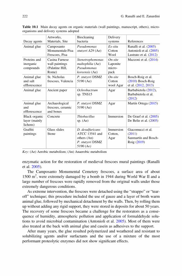
enzymatic action for the restoration of medieval frescoes mural paintings (Ranalliet al. 2005).
The Camposanto Monumental Cemetery frescoes, a surface area of about1500 m2, were extremely damaged by a bomb in 1944 during World War II and alarge number of frescoes were rapidly removed from the original walls under thoseextremely dangerous conditions.
As extreme intervention, the frescoes were detached using the “strappo” or “tear-off” technique; this procedure included the use of gauze and a layer of broth warmanimal glue, followed by mechanical detachment by the walls. Then, by rolling themup without adding any rigid support, they were stored in deposits for about 50 years.The recovery of some frescoes became a challenge for the restorators as a conse-quence of humidity, atmospheric pollution and application of formaldehyde solu-tions to avoid microbial contamination (Antonioli et al. 2005). Most of them werealso treated at the back with animal glue and casein as adhesives to the support.
After many years, the glue resulted polymerized and weathered and resistant tosolubilizing agents and/or surfactants and the use of a mixture of the mostperformant proteolytic enzymes did not show significant effects.
Table 10.1 Main decay agents on organic materials (wall paintings, manuscript, others), micro-organisms and delivery systems adopted
Decay agentsArtworks,Materials, Site
Biocleaningbacteria
Deliverysystems References
Animal glue CamposantoMonumentale PisaFrescoes, Pisa
Pseudomonasstutzeri A29 (Ae)
Ex-situCottonWool
Ranalli et al. (2005)Antonioli et al. (2005)Lustrato et al. (2012)
Proteins andinorganiccompounds
Casina Farnesewall paintings(Palatine Hill,Rome)
Stenotrophomonasmaltophilia (Ae)Pseudomonaskoreensis (Ae)
On-siteLaponitemicro-pack
Mazzoni et al. (2014)
Animal glueand saltefflorescence
St. Nicholasfrescoes, Valencia
P. stutzeri DSMZ5190 (Ae)
On-siteCottonwool Agar
Bosch-Roig et al.(2010) Bosch-Roiget al. (2012, 2013)
Animal glue Ancient paper Ochrobactrumsp. TNS15
Agar Barbabietola (2012),Barbabietola et al.(2012)
Animal glueandefflorescence
Archaeologicalfrescoes, ceramicand bones
P. stutzeri DSMZ5190 (Ae)
Agar Martín Ortega (2015)
Black organiclayer (mainlylichens)
Concrete Thiobacillussp. (Ae)
Immersion De Graef et al. (2005)De Belie et al. (2005)
Graffitipaintings
Glass slidesStone
D. desulfuricansATCC 13541 andothers (An)P. stutzeri DSMZ5190 (Ae)
ImmersionCotton,Agar
Giacomucci et al.(2011)Sanmartín and Bosch-Roig (2019)
Key: (Ae) Aerobic metabolism; (An) Anaerobic metabolism
222 G. Ranalli and E. Zanardini
In order to solve these problems and to identify suitable restoration practices andmethodology to recover the frescoes, the approach required specific analyses toindividualize the organic substances originally applied, as well as with thoseapported later in the restoration works.
For this aim, the use of sensitive, selective, and, if possible, non-invasive tech-niques that are minimally destructive, such as calorimetry, gas chromatographycoupled with mass spectrometry (GC/MS) and pyrolysis/GC-MS, are suggested(Stassi et al. 1998; Colombini et al. 1999, 2002, 2003; Bonaduce and Andreotti2009).
Once identified the substances to be removed, we decided that the use of viablecells could be the only way to restore the ancient frescoes.
The selection of microbial strains was a very important step, because the successof a “biocleaning” treatment depends on the efficiency of the used microorganisms.Both culture-dependent and -independent methods can help in the selection of themost effective microbial strain.
In our case, bacteria belonging to the genus Pseudomonas, Gram negative,asporigen, ubiquitary in nature and with extensive metabolic versatility, able infact of using a large variety of different compounds as source of carbon and energy.Among the five species tested in vitro, Pseudomonas stutzeriA29 strain was selectedbecause is not pathogenic to man and the environment and capable to grow usinganimal glue when supplied with it as sole organic carbon.
A direct application was carried out onto the artwork surface using viableP. stutzeri A29 bacterial cells in broth culture suspension (106–107 UFC/ml) embed-ded in sterile cotton wool, for 10–12 h at 30 �C temperature (Ranalli et al. 2005;Antonioli et al. 2005; Cappitelli et al. 2005, 2006, 2007). The costs of the bacterialsuspension were quantified in about 50 euro/litre, and 100 euro/m2 of fresco surface.At the end, a commercial Protease Type XIX was adopted to eliminate residues ofglue on the surface.
3 Case Study of Stories of the Holy Fathers (Storie dei SantiPadri), Pisa, Italy
A second pioneeristic protocol based on the synergic combination of bacterialmetabolism with the hydrolytic enzymatic action was performed for the restorationof medieval frescoes “Stories of the Holy Fathers”, (size 6.10 15.65 m), painted byBuonamico Buffalmacco, in the fourteenth century (Lustrato et al. 2012).
In addition, in this case, the fresco damaged by a bomb during World War II wasquickly removed from the original walls under extremely dangerous conditions.
In the 1960s, during the first restoration the fresco was cut into 17 sections(ranging from 1.3–2.0 m to 3.5–2.0 m) and attached onto asbestos-cement supports.The gauze and a large portion of the animal glue layer on the surface of the fresco
10 Advanced Biocleaning System for Historical Wall Paintings 223
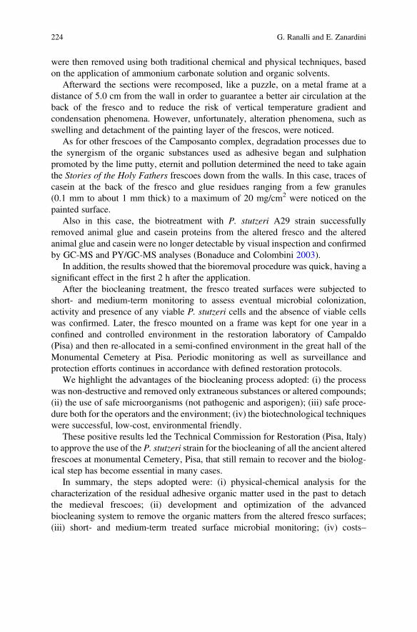
were then removed using both traditional chemical and physical techniques, basedon the application of ammonium carbonate solution and organic solvents.
Afterward the sections were recomposed, like a puzzle, on a metal frame at adistance of 5.0 cm from the wall in order to guarantee a better air circulation at theback of the fresco and to reduce the risk of vertical temperature gradient andcondensation phenomena. However, unfortunately, alteration phenomena, such asswelling and detachment of the painting layer of the frescos, were noticed.
As for other frescoes of the Camposanto complex, degradation processes due tothe synergism of the organic substances used as adhesive began and sulphationpromoted by the lime putty, eternit and pollution determined the need to take againthe Stories of the Holy Fathers frescoes down from the walls. In this case, traces ofcasein at the back of the fresco and glue residues ranging from a few granules(0.1 mm to about 1 mm thick) to a maximum of 20 mg/cm2 were noticed on thepainted surface.
Also in this case, the biotreatment with P. stutzeri A29 strain successfullyremoved animal glue and casein proteins from the altered fresco and the alteredanimal glue and casein were no longer detectable by visual inspection and confirmedby GC-MS and PY/GC-MS analyses (Bonaduce and Colombini 2003).
In addition, the results showed that the bioremoval procedure was quick, having asignificant effect in the first 2 h after the application.
After the biocleaning treatment, the fresco treated surfaces were subjected toshort- and medium-term monitoring to assess eventual microbial colonization,activity and presence of any viable P. stutzeri cells and the absence of viable cellswas confirmed. Later, the fresco mounted on a frame was kept for one year in aconfined and controlled environment in the restoration laboratory of Campaldo(Pisa) and then re-allocated in a semi-confined environment in the great hall of theMonumental Cemetery at Pisa. Periodic monitoring as well as surveillance andprotection efforts continues in accordance with defined restoration protocols.
We highlight the advantages of the biocleaning process adopted: (i) the processwas non-destructive and removed only extraneous substances or altered compounds;(ii) the use of safe microorganisms (not pathogenic and asporigen); (iii) safe proce-dure both for the operators and the environment; (iv) the biotechnological techniqueswere successful, low-cost, environmental friendly.
These positive results led the Technical Commission for Restoration (Pisa, Italy)to approve the use of the P. stutzeri strain for the biocleaning of all the ancient alteredfrescoes at monumental Cemetery, Pisa, that still remain to recover and the biolog-ical step has become essential in many cases.
In summary, the steps adopted were: (i) physical-chemical analysis for thecharacterization of the residual adhesive organic matter used in the past to detachthe medieval frescoes; (ii) development and optimization of the advancedbiocleaning system to remove the organic matters from the altered fresco surfaces;(iii) short- and medium-term treated surface microbial monitoring; (iv) costs–
224 G. Ranalli and E. Zanardini
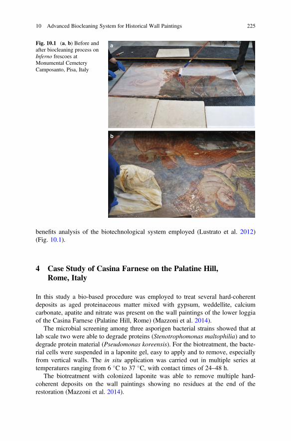
benefits analysis of the biotechnological system employed (Lustrato et al. 2012)(Fig. 10.1).
4 Case Study of Casina Farnese on the Palatine Hill,Rome, Italy
In this study a bio-based procedure was employed to treat several hard-coherentdeposits as aged proteinaceous matter mixed with gypsum, weddellite, calciumcarbonate, apatite and nitrate was present on the wall paintings of the lower loggiaof the Casina Farnese (Palatine Hill, Rome) (Mazzoni et al. 2014).
The microbial screening among three asporigen bacterial strains showed that atlab scale two were able to degrade proteins (Stenotrophomonas maltophilia) and todegrade protein material (Pseudomonas koreensis). For the biotreatment, the bacte-rial cells were suspended in a laponite gel, easy to apply and to remove, especiallyfrom vertical walls. The in situ application was carried out in multiple series attemperatures ranging from 6 �C to 37 �C, with contact times of 24–48 h.
The biotreatment with colonized laponite was able to remove multiple hard-coherent deposits on the wall paintings showing no residues at the end of therestoration (Mazzoni et al. 2014).
a
b
Fig. 10.1 (a, b) Before andafter biocleaning process onInferno frescoes atMonumental CemeteryCamposanto, Pisa, Italy
10 Advanced Biocleaning System for Historical Wall Paintings 225
5 Case Study of Animal Glue on Frescoes on the SantosJuanes Church, Valencia, Spain
Other studies showed the use of P. stutzeri DSMZ 5190 viable cells for the onsiteremoval of altered animal glue residues on frescoes of the central vault inside theSantos Juanes Church in Valencia, Spain (Bosch-Roig et al. 2010, 2012, 2013,2015). The authors adopted in their experimental conditions, agar-gel colonized bybacteria as improved delivery systems. Agar showed to be the most appropriatecarrier, both at lab scale and at onsite experiments. It was observed a good adhesiveproperty when applied onto vertical and oblique surfaces, and an adequate waterretention for bacteria without interfering with wall surfaces. Large amount ofbacterial cell suspension (109 UFC/ml) was applied in an aqueous solution directlyonto the fresco surface by soft brush, with short time of contact (less than 2 h) foreach application. The positive results showed that the animal glue was completelyremoved, confirmed also by analytical pyrolysis and gas-chromatography–massspectrometry analyses. The bacteria were able to degrade the organic matter presenton the fresco compared with area of control treatments (only water).
The authors highlighted the positive results evidencing that the biotechnology isrisk-free for the restorers since only non-pathogenic bacteria were used, and is notinvasive for the artworks and easily monitored since these bacteria are unable toproduce spores (Bosch-Roig et al. 2013).
In conclusion, the different case studies here illustrated show that the biocleaningsteps are:
• Diagnosis and characterisation of the stone alteration.• Laboratory selection of microbial strains able to remove the alteration.• Evaluation of the degradation rate in order to individualize the most promising
microbial strain (safe, efficiency, adaption to the in situ treatment, etc.).• Selection of the appropriate delivery system to vehicular microbial viable cells.• Delivery system colonization and evaluation of the better condition for the
microbial metabolisms.• Lab-scale application on specimens artificially enriched and on real altered
fragments.• Optimization of the ex situ, onsite application conditions.• Evaluation of the environmental conditions at the time of the application.• Monitoring during the application.• Removal of the bioapplication and cleaning of the treated surfaces.• Short– and long-term surveillance and monitoring after the biotreatment.
226 G. Ranalli and E. Zanardini
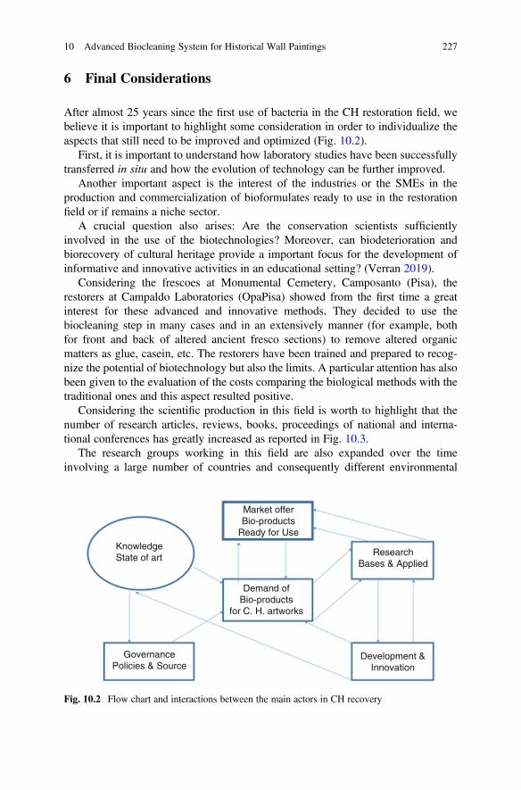
6 Final Considerations
After almost 25 years since the first use of bacteria in the CH restoration field, webelieve it is important to highlight some consideration in order to individualize theaspects that still need to be improved and optimized (Fig. 10.2).
First, it is important to understand how laboratory studies have been successfullytransferred in situ and how the evolution of technology can be further improved.
Another important aspect is the interest of the industries or the SMEs in theproduction and commercialization of bioformulates ready to use in the restorationfield or if remains a niche sector.
A crucial question also arises: Are the conservation scientists sufficientlyinvolved in the use of the biotechnologies? Moreover, can biodeterioration andbiorecovery of cultural heritage provide a important focus for the development ofinformative and innovative activities in an educational setting? (Verran 2019).
Considering the frescoes at Monumental Cemetery, Camposanto (Pisa), therestorers at Campaldo Laboratories (OpaPisa) showed from the first time a greatinterest for these advanced and innovative methods. They decided to use thebiocleaning step in many cases and in an extensively manner (for example, bothfor front and back of altered ancient fresco sections) to remove altered organicmatters as glue, casein, etc. The restorers have been trained and prepared to recog-nize the potential of biotechnology but also the limits. A particular attention has alsobeen given to the evaluation of the costs comparing the biological methods with thetraditional ones and this aspect resulted positive.
Considering the scientific production in this field is worth to highlight that thenumber of research articles, reviews, books, proceedings of national and interna-tional conferences has greatly increased as reported in Fig. 10.3.
The research groups working in this field are also expanded over the timeinvolving a large number of countries and consequently different environmental
KnowledgeState of art
Demand ofBio-products
for C. H. artworks
Development &Innovation
GovernancePolicies & Source
Market offerBio-products
Ready for Use
ResearchBases & Applied
Fig. 10.2 Flow chart and interactions between the main actors in CH recovery
10 Advanced Biocleaning System for Historical Wall Paintings 227
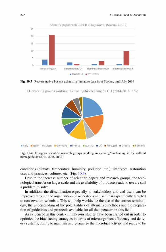
conditions (climate, temperature, humidity, pollution, etc.), lithotypes, restorationuses and practices, cultures, etc. (Fig. 10.4).
Despite the increase number of scientific papers and research groups, the tech-nological transfer on larger scale and the availability of products ready to use are stilla problem to solve.
In addition, the dissemination especially to stakeholders and end users can beimproved through the organization of workshops and seminars specifically targetedto conservation scientists. This will help worldwide the use of the correct terminol-ogy, the understanding of the potentialities of alternative methods and the prepara-tion of guidelines and protocols available for all the operators in this field.
As evidenced in this context, numerous studies have been carried out in order tooptimize the biocleaning strategies in terms of microorganism efficiency and deliv-ery systems, ability to maintain and guarantee the microbial activity and ready to be
0
5
10
15
20
25
biocleaning/CH biorestoration/CH biomineralization/CH bioprecipitation/CH
Scientific papers with Bio/CH as key-words (Scopus, 7-2019)
2000-2010 2011-2019
Fig. 10.3 Representative but not exhaustive literature data from Scopus, until July 2019
EU working groups working in cleaning/biocleaning on CH (2014-2018 in %)
Italy Spain Suisse Germany France Austria UK Portugal Greece Romania
Fig. 10.4 European scientific research groups working in cleaning/biocleaning in the culturalheritage fields (2014–2018, in %)
228 G. Ranalli and E. Zanardini
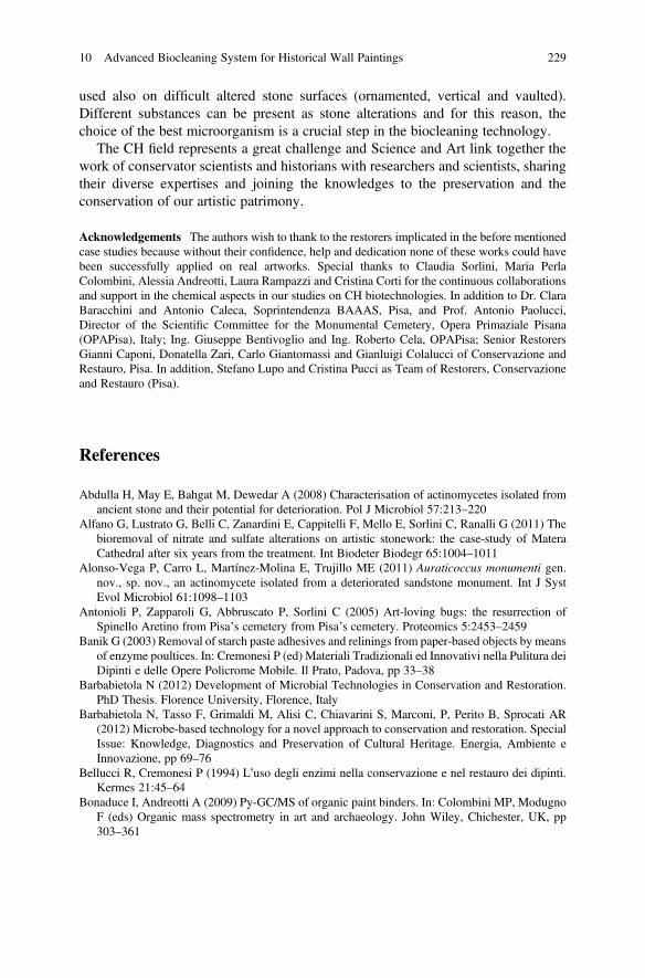
used also on difficult altered stone surfaces (ornamented, vertical and vaulted).Different substances can be present as stone alterations and for this reason, thechoice of the best microorganism is a crucial step in the biocleaning technology.
The CH field represents a great challenge and Science and Art link together thework of conservator scientists and historians with researchers and scientists, sharingtheir diverse expertises and joining the knowledges to the preservation and theconservation of our artistic patrimony.
Acknowledgements The authors wish to thank to the restorers implicated in the before mentionedcase studies because without their confidence, help and dedication none of these works could havebeen successfully applied on real artworks. Special thanks to Claudia Sorlini, Maria PerlaColombini, Alessia Andreotti, Laura Rampazzi and Cristina Corti for the continuous collaborationsand support in the chemical aspects in our studies on CH biotechnologies. In addition to Dr. ClaraBaracchini and Antonio Caleca, Soprintendenza BAAAS, Pisa, and Prof. Antonio Paolucci,Director of the Scientific Committee for the Monumental Cemetery, Opera Primaziale Pisana(OPAPisa), Italy; Ing. Giuseppe Bentivoglio and Ing. Roberto Cela, OPAPisa; Senior RestorersGianni Caponi, Donatella Zari, Carlo Giantomassi and Gianluigi Colalucci of Conservazione andRestauro, Pisa. In addition, Stefano Lupo and Cristina Pucci as Team of Restorers, Conservazioneand Restauro (Pisa).
References
Abdulla H, May E, Bahgat M, Dewedar A (2008) Characterisation of actinomycetes isolated fromancient stone and their potential for deterioration. Pol J Microbiol 57:213–220
Alfano G, Lustrato G, Belli C, Zanardini E, Cappitelli F, Mello E, Sorlini C, Ranalli G (2011) Thebioremoval of nitrate and sulfate alterations on artistic stonework: the case-study of MateraCathedral after six years from the treatment. Int Biodeter Biodegr 65:1004–1011
Alonso-Vega P, Carro L, Martínez-Molina E, Trujillo ME (2011) Auraticoccus monumenti gen.nov., sp. nov., an actinomycete isolated from a deteriorated sandstone monument. Int J SystEvol Microbiol 61:1098–1103
Antonioli P, Zapparoli G, Abbruscato P, Sorlini C (2005) Art-loving bugs: the resurrection ofSpinello Aretino from Pisa’s cemetery from Pisa’s cemetery. Proteomics 5:2453–2459
Banik G (2003) Removal of starch paste adhesives and relinings from paper-based objects by meansof enzyme poultices. In: Cremonesi P (ed) Materiali Tradizionali ed Innovativi nella Pulitura deiDipinti e delle Opere Policrome Mobile. Il Prato, Padova, pp 33–38
Barbabietola N (2012) Development of Microbial Technologies in Conservation and Restoration.PhD Thesis. Florence University, Florence, Italy
Barbabietola N, Tasso F, Grimaldi M, Alisi C, Chiavarini S, Marconi, P, Perito B, Sprocati AR(2012) Microbe-based technology for a novel approach to conservation and restoration. SpecialIssue: Knowledge, Diagnostics and Preservation of Cultural Heritage. Energia, Ambiente eInnovazione, pp 69–76
Bellucci R, Cremonesi P (1994) L’uso degli enzimi nella conservazione e nel restauro dei dipinti.Kermes 21:45–64
Bonaduce I, Andreotti A (2009) Py-GC/MS of organic paint binders. In: Colombini MP, ModugnoF (eds) Organic mass spectrometry in art and archaeology. John Wiley, Chichester, UK, pp303–361
10 Advanced Biocleaning System for Historical Wall Paintings 229

Bonaduce I, Colombini MP (2003) Gas chromatography/mass spectrometry for the characterizationof organic materials in frescoes of the Monumental Cemetery of Pisa (Italy). Rapid CommMassSpectrom 17:2523–2527
Bonomi R (1994) Utilizzo degli enzimi per il restauro di una scultura in terracotta policroma. OPDRestauro 6:101–107
Bosch-Roig P, Regidor-Ros JL, Soriano-Sancho P, Domenech-Carbo MT, Montes-Estellés RM(2010) Ensayos de biolimpieza con bacterias en pinturas murales. Arché 4–5:115–122
Bosch-Roig P, Montes-Estellés RM, Regidor-Ros JL, Roig Picazo P, Ranalli G (2012) Newfrontiers in the microbial bio-cleaning of artworks. Picture Restorer 41:37–41
Bosch-Roig P, Regidor-Ros JL, Soriano-Sancho P, Montes-Estellés R (2013) Biocleaning ofanimal glue on wall paintings by Pseudomonas stutzeri. Chim Oggi 31:50–53
Bosch-Roig P, Lustrato G, Zanardini E, Ranalli G (2015) Biocleaning of cultural heritage stonesurfaces and frescoes: which delivery system can be the most appropriate? Ann Microbiol65:1227–1241
Cappitelli F, Zanardini E, Toniolo L, Abbruscato P, Ranalli G, Sorlini C (2005) Bioconservation ofthe marble base of the Pietà Rondanini by Michelangelo Buonarroti. Geophys Res Abstr7:06675–06676
Cappitelli F, Zanardini E, Ranalli G, Mello E, Daffonchio D, Sorlini C (2006) Improved method-ology for bioremoval of black crusts on historical stone artworks by use of sulfate-reducingbacteria. Appl Environ Microbiol 72:3733–3737
Cappitelli F, Toniolo L, Sansonetti A, Gulotta D, Ranalli G, Zanardini E, Sorlini C (2007)Advantages of using microbial technology over traditional chemical technology in removal ofblack crusts from stone surfaces of historical monuments. Appl Environ Microbiol73:5671–5675
Cappitelli F, Salvadori O, Albanese D, Villa F, Sorlini C (2012) Cyanobacteria cause black stainingof the National Museum of the American Indian Building, Washington DC. Biofouling28:257–266
Colombini MP, Modugno F, Giacomelli A (1999) Two procedures for suppressing interferencefrom inorganic pigments in analysis by gas chromatography-mass spectrometry of proteina-ceous binders in painting. J Chromatogr A 846:101–111
Colombini MP, Modugno F, Fuoco R, Tognazzi A (2002) A GC-MS study on the deterioration oflipidic paint binders. Microchem J 73:175–185
Colombini MP, Bonaduce I, Gautier G (2003) Molecular pattern recognition of fresh and agedshellac. Chromatographia 58:1–8
Cremonesi P (1999) L’uso degli enzimi nella pulitura di opere policrome. In: Il Prato (ed) I Talenti,Padova, pp 5–11
Cremonesi P (2004) L’uso dei solventi organici nella pulitura di opere policrome. In: Il Prato (ed) ITalenti, Padova, pp 2–15
Crispim CA, Gaylarde CC (2005) Cyanobacteria and biodeterioration of cultural heritage: a review.Microb Ecol 49:1–9
Daffonchio D, Cherif A, Borin S (2000) Homoduplex and heteroduplex polymorphisms of theamplified ribosomal 16S-23S internal transcribed spacers describe genetic relationships in the“Bacillus cereus group”. Appl Environ Microbiol 66:5460–5468
De Belie N, De Graef B, De Muynck W, Dick J, De Windt W, Verstraete W (2005) Biocatalyticprocesses on concrete: bacterial cleaning and repair. In 10th DBMC Int. Conference onDurability of Building Materials and Components, April, Lyon, pp 17–20
De Graef B, De Windt W, Dick J, Verstraete W, De Belie N (2005) Cleaning of concrete fouled bylichens with the aid of Thiobacilli. Mater Struct 38:875–882
De La Chapelle A (2003) Utilizzo degli enzimi nel restauro delle opere grafiche policrome. In:Cremonesi P (ed) Materiali tradizionali ed innovativi nella pulitura dei dipinti e delle operepolicrome mobile. Il Prato, Padova, pp 51–53
230 G. Ranalli and E. Zanardini
EWAGLOS (2016) In: Weyer A, Roig Picazo P, Pop D, Carras J, Ozkose A, Vallet JM, Srsa I (eds)EwaGlos-European Illustrated Glossary of Conservation Terms for Wall Paintings and Archi-tectural Surfaces. English definitions with translations into Bulgarian, Croatian, French, Ger-man, Hungarian, Italian, Polish, Romanian, Spanish and Turkish, vol. 17. Michael ImhofVerlag, Petersberg, Germany, pp 304–307
Fernandes P (2006) Applied microbiology and biotechnology in the conservation of stone culturalheritage materials. Appl Microb Biotechnol 73:291–296
Giacomucci L, Bertoncello R, Salvadori O, Martini I, Favaro M, Villa F, Sorlini C, Cappitelli F(2011) Microbial deterioration of artistic tiles from the Façade of the Grande Albergo Ausonia &Hungaria (Venice, Italy). Microb Ecol 62:287–298
Gorbushina AA (2007) Life on the rocks. Environ Microbiol 9:1613–1631Gurtner C, Heyrman J, Piñar G, Lubitz W, Swings J, Rölleke S (2000) Comparative analyses of the
bacterial diversity on two different biodeteriorated wall painting by DGGE and 16s rRNAsequence analysis. Int Biodeter Biodegrad 46:229–239
Iannuccelli S, Sotgiu S (2009) La pulitura superficiale di opera grafiche a stampa con gel rigidi. In:Il Prato (ed) Progetto Restauro, Padova, pp 15–24
Lustrato G, Alfano G, Andreotti A, Colombini MP, Ranalli G (2012) Fast biocleaning of mediaevalfrescoes using viable bacterial cells. Int Biodeter Biodegr 69:51–61
Makes F (1988) Enzymatic consolidation of the portrait of Rudolf II as “Vertumnus” by GiuseppeArcimboldo with a new multi-enzyme preparation isolated from Antarctic Krill (Euphausiasuperba). Acta Universitatis Gotheburg 23:98–110
Martín Ortega S (2015) Neteges raonades: Experimentació amb nou tractaments de neteja aplicatssobre 6 peces de material arqueologic divers. Treball final de grau ESCRBCC, Barcelona
May E (2010) Stone biodeterioration. In: Mitchell R, McNamara CJ (eds) Cultural heritagemicrobiology: fundamental studies in conservation science. ASM Press, Washington DC, pp1–39
Mazzoni M, Alisi C, Tasso F, Cecchini A, Marconi P, Sprocati AR (2014) Laponite micro-packs forthe selective cleaning of multiple coherent deposits on wall paintings: the case study of CasinaFarnese on the Palatine Hill (Rome-Italy). Int Biodeter Biodegr 94:1–11
McNamara CJ, Mitchell R (2005) Microbial deterioration of historic stone. Front Ecol Environ3:445–451
Palla F (2004) Le biotecnologie molecolari per la caratterizzazione e la valutazione del ruolo deimicrorganismi nei processi di degrado di manufatti di interesse storico artistico. In: Lorusso S(ed) Quaderni di Scienza della Conservazione. Univerità di Bologna, Ravenna, pp 183–194
Palla F (2013) Bioactive molecules: innovative contribution of biotechnology to the restoration ofcultural heritage. J Conserv Sci 13:369–373
Piñar G, Ramos C, Rölleke S, Schabereiter-Gurtner C, Vybiral D, Lubitz W, Denner EB (2001)Detection of indigenous Halobacillus populations in damaged ancient wall paintings andbuilding materials: Molecular monitoring and cultivation. Appl Environ Microbiol67:4891–4895
Portillo C, Saiz-Jimenez C, Gonzalez JM (2009) Molecular characterization of total and metabol-ically active bacterial communities of “white colonizations” in the Altamira Cave. Spain. ResMicrobiol 160:41–47
Ranalli G, Chiavarini M, Guidetti V, Marsala F, Matteini M, Zanardini E, Sorlini C (1996) The useof microorganisms for the removal of nitrates and organic substances on artistic stoneworks. In:Riederer J (ed) Proceedings of the 8th Int. Congress on Deterioration and Conservation of Stone,Berlin, 30 September–4 October 1996, pp 1415–1420
Ranalli G, Chiavarini M, Guidetti V, Marsala F, Matteini M, Zanardini E, Sorlini C (1997) The useof microorganisms for the removal of sulphates on artistic stoneworks. Int Biodeter Biodegr40:255–261
10 Advanced Biocleaning System for Historical Wall Paintings 231
Ranalli G, Alfano G, Belli C, Lustrato G, Colombini MP, Bonaduce I, Zanardini E, Abbruscato P,Cappitelli F, Sorlini C (2005) Biotechnology applied to cultural heritage: biorestoration offrescoes using viable bacterial cells and enzymes. J Appl Microbiol 98:73–83
Ranalli G, Zanardini E, Sorlini C (2009) Biodeterioration e including cultural heritage. In:Schaechter M (ed) Encyclopedia of microbiology. Elsevier, Oxford, pp 191–205
Saiz-Jimenez C, Cuezva S, Jurado V, Fernandez-Cortes A, Porca E, Benavente D, Canaveras JC,Sanchez-Moral S (2011) Paleolithic art in peril: policy and science collide at Altamira Cave.Science 334:42–43
Sanmartín P, Bosch-Roig P (2019) Biocleaning to remove graffiti: a real possibility? Advancestowards a complete protocol of action. Coatings 9:104–119
Schabereiter-Gurtner C, Gurtner G, Piñar G, Lubitz W, Rölleke S (2001) An advanced molecularstrategy to identify bacterial communities on art objects. J Microbiol Methods 45:77–87
Scheerer S, Ortega-Morales O, Gaylarde C (2009) Microbial deterioration of stone monuments-anupdated overview. Adv Appl Microb 66:97–139
Segal J, Cooper D (1977) The use of enzymes to release adhesives. Paper Conservator 2:47–50Stassi A, Zanardini E, Cappitelli F, Schiraldi A, Sorlini C (1998) Calorimetric investigations on the
metabolism of Bacillus strains isolated from artistic stoneworks. Ann Microbiol Enzim48:111–120
Tiano P (2002) Biodegradation of cultural heritage: decay mechanisms and control methods. TheProceedings of the ARIADNE 9 WS. Institute of Theoretical and Applied Mechanics of theAcademy of Sciences of the Czech Republic, Prague 4-10 February, pp 1–37
Tiano P, Cantisani E, Sutherland I, Paget JM (2006) Bioremediated reinforcement of weatheredcalcareous stones. J Cult Herit 7:49–55
Urzì C, Brusetti L, Salamone P, Sorlini C, Stackebrandt E, Daffonchio D (2001) Biodiversity ofGeodermatophilaceae isolated from altered stones and monuments in the Mediterranean basin.Environ Microbiol 3:471–479
Verran J (2019) Mixed culture: encouraging cross-disciplinary collaboration and communication toenhance learning. Ann Microbiol 69:1107–1111
Villa F, Vasanthakumar A, Mitchell R, Cappitelli F (2015) RNA-based molecular survey ofbiodiversity of limestone tombstone microbiota in response to atmospheric sulphur pollution.Lett Appl Microbiol 60:92–102
Wendelbo O (1976) The use of proteolytic enzymes in the restoration of paper and papyrus.University Library of Bergen
Wolbers R (2000) Cleaning painted surface: aqueous methods. Archetype Publication Ltd, LondonZanardini E, Purdy K, May E, Moussard H, Williams S, Oakley B, Murrell C (2011) Investigation
of archaeal and bacterial community structure and functional gene diversity in exfoliatedsandstone at Portchester Castle (UK). In: Sterflinger K, Piñar G (eds) International Symposiumon Biodeterioration and Biodegradation (IBBS-15), Vienna, September 19–24, p 90.
Zanardini E, May E, Inkpen R, Cappitelli F, Murrell JC, Purdy KJ (2016) Diversity of archaeal andbacterial communities on exfoliated sandstone from Portchester Castle (UK). Int BiodeterBiodegrad 109:78–87
Zanardini E, May E, Purdy KJ, Murrell JC (2019) Nutrient cycling potential within microbialcommunities on culturally important stoneworks. Environ Microbiol Rep 11:147–154
232 G. Ranalli and E. Zanardini

Open Access This chapter is licensed under the terms of the Creative Commons Attribution 4.0International License (http://creativecommons.org/licenses/by/4.0/), which permits use, sharing,adaptation, distribution and reproduction in any medium or format, as long as you give appropriatecredit to the original author(s) and the source, provide a link to the Creative Commons license andindicate if changes were made.
The images or other third party material in this chapter are included in the chapter's CreativeCommons license, unless indicated otherwise in a credit line to the material. If material is notincluded in the chapter's Creative Commons license and your intended use is not permitted bystatutory regulation or exceeds the permitted use, you will need to obtain permission directly fromthe copyright holder.
10 Advanced Biocleaning System for Historical Wall Paintings 233
Chapter 11Sustainable Restoration ThroughBiotechnological Processes: A Proofof Concept
Anna Rosa Sprocati, Chiara Alisi, Giada Migliore, Paola Marconi, andFlavia Tasso
Abstract An understanding of the different microbial constellations ormicrobiomes, which every habitat and every organism harbor, will be the key toaddressing many of the challenges humanity will face in the twenty-first century.Such comprehension could launch several innovations relating to natural and cul-tural capital, including historical and artistic heritage. In relation to cultural heritage,microorganisms are mainly known through their role as deteriogens, but the featurescreating damage can be exploited positively, attaining more sustainable restorationstrategies, in accordance with the principles of compatibility and retreatabilityderiving from reflections on the Cultural Heritage inspired by the Charter of Venice(International charter for the Conservation and restoration of monument and sites(the Venice Chart 1964). In: ICOMOS, IInd International Congress of Architects andTechnicians of Historic Monuments, 1964) onwards. In this article, we show a seriesof case studies, using both wild-type microorganisms and plant-based extracts,providing a comprehensive proof of concept of the feasibility of biotechnologicalsolutions for a more sustainable restoration strategy, to replace the products in usewhich are often dangerous for operators, aggressive for works of art and no longercompatible with the environment. The overview of the case studies presented, manyof which are still unpublished, responds to the need to go beyond the state of the artand has entirely sprung from suggestions by restorers, interested in learning aboutpotential innovations and strongly determined to introduce non-toxic products intheir daily work. In this perspective, the case studies dealt with two topics:bio-cleaning and disinfection. Noteworthy results were obtained on a platform ofdifferent types of artworks and different materials with compatible, harmless andselective products.
Keywords Bio-cleaning · Disinfection · Bio-restoration · Microbial biotechnology
A. R. Sprocati (*) · C. Alisi · G. Migliore · P. Marconi · F. TassoENEA, Department for Sustainability, Rome, Italye-mail: [email protected]; [email protected]; [email protected];[email protected]; [email protected]
© The Author(s) 2021E. Joseph (ed.), Microorganisms in the Deterioration and Preservation of CulturalHeritage, https://doi.org/10.1007/978-3-030-69411-1_11
235
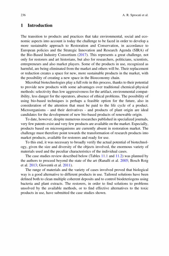
1 Introduction
The transition to products and practices that take environmental, social and eco-nomic aspects into account is today the challenge to be faced in order to develop amore sustainable approach to Restoration and Conservation, in accordance toEuropean policies and the Strategic Innovation and Research Agenda (SIRA) ofthe Bio-Based Industry Consortium (2017). This represents a great challenge, notonly for restorers and art historians, but also for researchers, politicians, scientists,entrepreneurs and also market players. Some of the products in use, recognized asharmful, are being eliminated from the market and others will be. Their replacementor reduction creates a space for new, more sustainable products in the market, withthe possibility of creating a new space in the Bioeconomy chain.
Microbial biotechnologies play a full role in this process, thanks to their potentialto provide new products with some advantages over traditional chemical-physicalmethods: selectivity thus low aggressiveness for the artifact, environmental compat-ibility, less danger for the operators, absence of ethical problems. The possibility ofusing bio-based techniques is perhaps a feasible option for the future, also inconsideration of the attention that must be paid to the life cycle of a product.Microorganisms - and their derivatives - and products of plant origin are idealcandidates for the development of new bio-based products of renewable origin.
To date, however, despite numerous researches published in specialized journals,very few patents exist and very few products are available on the market. Especially,products based on microorganisms are currently absent in restoration market. Thechallenge must therefore point towards the transformation of research products intomarket products, available for restorers and ready for use.
To this end, it was necessary to broadly verify the actual potential of biotechnol-ogy, given the size and diversity of the objects involved, the enormous variety ofmaterials used and the peculiar characteristics of the individual cases.
The case studies review described below (Tables 11.1 and 11.2) was planned bythe authors to proceed beyond the state of the art (Ranalli et al. 2005; Bosch Roiget al. 2013; Gioventù et al. 2011).
The range of materials and the variety of cases involved proved that biologicalway is a good alternative to different products in use. Tailored solutions have beendefined both to clean multiple coherent deposits and to control biodeteriogens usingbacteria and plant extracts. The restorers, in order to find solutions to problemsunsolved by the available methods, or to find effective alternatives to the toxicproducts in use, have submitted the case studies shown.
236 A. R. Sprocati et al.
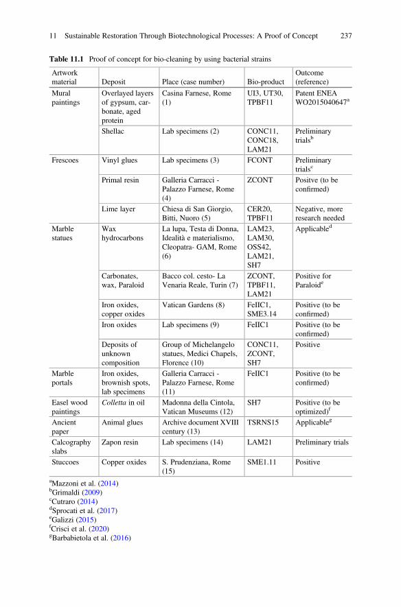
Table 11.1 Proof of concept for bio-cleaning by using bacterial strains
Artworkmaterial Deposit Place (case number) Bio-product
Outcome(reference)
Muralpaintings
Overlayed layersof gypsum, car-bonate, agedprotein
Casina Farnese, Rome(1)
UI3, UT30,TPBF11
Patent ENEAWO2015040647a
Shellac Lab specimens (2) CONC11,CONC18,LAM21
Preliminarytrialsb
Frescoes Vinyl glues Lab specimens (3) FCONT Preliminarytrialsc
Primal resin Galleria Carracci -Palazzo Farnese, Rome(4)
ZCONT Positve (to beconfirmed)
Lime layer Chiesa di San Giorgio,Bitti, Nuoro (5)
CER20,TPBF11
Negative, moreresearch needed
Marblestatues
Waxhydrocarbons
La lupa, Testa di Donna,Idealità e materialismo,Cleopatra- GAM, Rome(6)
LAM23,LAM30,OSS42,LAM21,SH7
Applicabled
Carbonates,wax, Paraloid
Bacco col. cesto- LaVenaria Reale, Turin (7)
ZCONT,TPBF11,LAM21
Positive forParaloide
Iron oxides,copper oxides
Vatican Gardens (8) FeIIC1,SME3.14
Positive (to beconfirmed)
Iron oxides Lab specimens (9) FeIIC1 Positive (to beconfirmed)
Deposits ofunknowncomposition
Group of Michelangelostatues, Medici Chapels,Florence (10)
CONC11,ZCONT,SH7
Positive
Marbleportals
Iron oxides,brownish spots,lab specimens
Galleria Carracci -Palazzo Farnese, Rome(11)
FeIIC1 Positive (to beconfirmed)
Easel woodpaintings
Colletta in oil Madonna della Cintola,Vatican Museums (12)
SH7 Positive (to beoptimized)f
Ancientpaper
Animal glues Archive document XVIIIcentury (13)
TSRNS15 Applicableg
Calcographyslabs
Zapon resin Lab specimens (14) LAM21 Preliminary trials
Stuccoes Copper oxides S. Prudenziana, Rome(15)
SME1.11 Positive
aMazzoni et al. (2014)bGrimaldi (2009)cCutraro (2014)dSprocati et al. (2017)eGalizzi (2015)fCrisci et al. (2020)gBarbabietola et al. (2016)
11 Sustainable Restoration Through Biotechnological Processes: A Proof of Concept 237
2 Bio-Cleaning Procedures Developed in the Case Studies
The procedure involves the use of living bacteria immobilized at high concentration(108 CFU/mL) in support agents with different viscosities, to create a micro-pack,easy to apply and remove without leaving residues on the artifact.
The micro-packs are easy to apply and to remove, without dripping and leavingresidues. They can be applied on vertical surfaces and ceilings; do not require strictoperating conditions, in terms of temperature and pH and with no disposal problems.The micro-pack can be conveniently prepared on a support table or prepared directlyon the object to be treated. In the first case a standard micro-pack is organizedbackwards: a plastic film, which preserves the humidity during the treatment, is firstplaced followed by a slightly moistened veil of paper (i.e. Japanese or Englishpaper), a layer of bacteria in their support agent and another veil of paper. Theinterposition of the paper facilitates the application and the removal of the micro-pack and allows inspections to check the progress of the process. However, theopportunity to interpose paper can be evaluated from time to time, based on thespecific situation. Direct contact with the original material can sometimes be moresuitable. After removal, a damp swab or a sponge can easily clean the artefact.
The time of contact of the micro-pack depends on various factors, such as thenature of the substrate to be removed, the thickness of the layer; the metabolic rate ofthe microorganism, the nature and the state of conservation of the artifacts. Theprocedures developed in the case studies (Tables 11.1 and 11.2) are fairly
Table 11.2 Proof of concept for biodeteriogens removal and control by using bio-products andbacterial strains
Artwork material Deposit Place Bio-productOutcome(reference)
Mobile paintingon canvas
Biofilm Private painting Liq Positive
Frescoes Biofilm Chapel XIII –Orta,Novara
Liq Positive
Hypogeum Biofilm Domus Aurea, Rome Liq, Underwaya
Stone material Biofilm Vatican gardens Liq, BioZ, SME1.11 Positive
Diocleziano termae,Rome
Liq, BioZ,SME1.11, NopalCap
Positiveb
Roman wall, Rome Liq, BioZ,SME1.11, NopalCap
Underway
Outdoor statues-Vicenza
Liq Underway
Mortar,bio-mortar
Bio-receptivity
Lab specimens Nopal Positivec
Biofilm Outdoor statue“Beethoven”- Naples
NopalCap Positive
aRugnini et al. (2019)bLedda (2019)cPersia et al. (2016)
238 A. R. Sprocati et al.
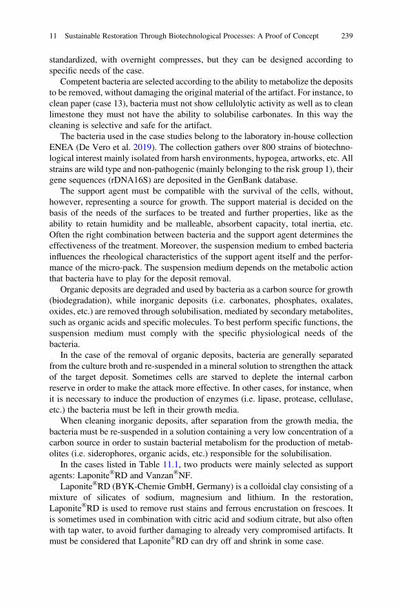
standardized, with overnight compresses, but they can be designed according tospecific needs of the case.
Competent bacteria are selected according to the ability to metabolize the depositsto be removed, without damaging the original material of the artifact. For instance, toclean paper (case 13), bacteria must not show cellulolytic activity as well as to cleanlimestone they must not have the ability to solubilise carbonates. In this way thecleaning is selective and safe for the artifact.
The bacteria used in the case studies belong to the laboratory in-house collectionENEA (De Vero et al. 2019). The collection gathers over 800 strains of biotechno-logical interest mainly isolated from harsh environments, hypogea, artworks, etc. Allstrains are wild type and non-pathogenic (mainly belonging to the risk group 1), theirgene sequences (rDNA16S) are deposited in the GenBank database.
The support agent must be compatible with the survival of the cells, without,however, representing a source for growth. The support material is decided on thebasis of the needs of the surfaces to be treated and further properties, like as theability to retain humidity and be malleable, absorbent capacity, total inertia, etc.Often the right combination between bacteria and the support agent determines theeffectiveness of the treatment. Moreover, the suspension medium to embed bacteriainfluences the rheological characteristics of the support agent itself and the perfor-mance of the micro-pack. The suspension medium depends on the metabolic actionthat bacteria have to play for the deposit removal.
Organic deposits are degraded and used by bacteria as a carbon source for growth(biodegradation), while inorganic deposits (i.e. carbonates, phosphates, oxalates,oxides, etc.) are removed through solubilisation, mediated by secondary metabolites,such as organic acids and specific molecules. To best perform specific functions, thesuspension medium must comply with the specific physiological needs of thebacteria.
In the case of the removal of organic deposits, bacteria are generally separatedfrom the culture broth and re-suspended in a mineral solution to strengthen the attackof the target deposit. Sometimes cells are starved to deplete the internal carbonreserve in order to make the attack more effective. In other cases, for instance, whenit is necessary to induce the production of enzymes (i.e. lipase, protease, cellulase,etc.) the bacteria must be left in their growth media.
When cleaning inorganic deposits, after separation from the growth media, thebacteria must be re-suspended in a solution containing a very low concentration of acarbon source in order to sustain bacterial metabolism for the production of metab-olites (i.e. siderophores, organic acids, etc.) responsible for the solubilisation.
In the cases listed in Table 11.1, two products were mainly selected as supportagents: Laponite®RD and Vanzan®NF.
Laponite®RD (BYK-Chemie GmbH, Germany) is a colloidal clay consisting of amixture of silicates of sodium, magnesium and lithium. In the restoration,Laponite®RD is used to remove rust stains and ferrous encrustation on frescoes. Itis sometimes used in combination with citric acid and sodium citrate, but also oftenwith tap water, to avoid further damaging to already very compromised artifacts. Itmust be considered that Laponite®RD can dry off and shrink in some case.
11 Sustainable Restoration Through Biotechnological Processes: A Proof of Concept 239
Vanzan®NF (Vanderbilt Minerals LLC, Norwolk, CT) is the commercial name ofthe Xanthan gum, a high molecular weight exocellular polysaccharide derived fromthe bacterium Xanthomonas campestris. Xanthan gum is widely used as a rheologycontrol agent for aqueous systems. It increases viscosity, helps to stabilize emulsionsand prevents the settling of solids in a wide variety of applications.
Vanzan®NF shows a great compatibility with cells viability, perfect adherenceand plasticity also on vertical surfaces, and overall the capacity to maintain themoisture without drying off or shrinking, up to 24 h of application.
Laponite®RD and Vanzan®NF were used for the first time as support agent formicroorganisms in the bio-cleaning case 1 and 12, respectively (Mazzoni et al. 2014;Crisci et al. 2020). The most appropriate concentration for a correct consistency andproperty of the micro-pack was 9% w/v for Laponite®RD and 6% w/v forVanzan®NF.
3 Bio-Cleaning Case Studies: Review and Discussion
The series of case studies (Table 11.1) springs from the European patent EP 3046779extended to Italy, Switzerland, Germany, Spain, France, GB (Sprocati et al. 2014)that derived from the bio-cleaning of mural painting of the external loggias in theCasina Farnese (case 1). The cases that followed (2–15) expressly ranged betweendifferent types of artifacts, materials and substrates to be removed, experimentingnumerous microbial species available in the in-house collection in order to create aproof of concept of the feasibility of microbial biotechnology to contribute to a moresustainable restoration strategy.
The Casina Farnese (case 1) is situated at the top of the Palatine Hill, in Rome, thedouble order of open loggias representing the legend of Ercole and Caco wascompleted by 1593. The decoration of the lower loggia, object of the bio-cleaningtests, was ascribed to a pupil of Taddeo Zuccari and represents Ercole killing Caco.Coherent deposits composed of gypsum, weddellite, calcium carbonate, apatite,nitrate, and aged proteinaceous matter (FTIR-ATR analysis) were deemed difficultto remove from the wall paintings, especially in areas where the deposits weresuperimposed in layers. Following a laboratory screening, three bacterial nonspore-forming strains were selected to solubilise calcium sulphate and carbonate,to degrade protein, to solubilise inorganic compounds and degrade protein material.Micro-packs with single strains were used in situ, from July 2012 to February 2013,in temperatures ranging from 6 �C to 37 �C. After experiencing some supports in usefor wet compresses without satisfactory results, the living bacterial cells wereeventually immobilized in a Laponite®RD gel. In this occasion, Laponite®RD wasused for the first time to immobilize microbial cells, proving to be optimal both forcompatibility with the vitality of the cells and for perfectly retaining the material ofcompress, without leaks. The strain Cellulosimicrobium cellulans TBF11E DSM27224 (GenBank accession number: EU249577) removed the inorganic darkerlayer, Stenotrophomonas maltophilia UI3E DSM 27225 (JX133197) dissolved the
240 A. R. Sprocati et al.
brownish layer (probably aged casein) and Pseudomonas koreensis UT30E DSM27226 (JX133187) removed the mixed deposits. In areas with overlaid deposits,micro-packs were applied in succession, containing the strain able to remove eachlayer.
Another critical point threating the survival and the performance of bacteria wasthe low temperature in the open loggia on winter days. No significant differenceswere detected in the efficacy of the bio-cleaning treatment, probably the micro-packcontributes to maintain a microclimate suitable for the bacterial metabolic activity.After the bio-cleaning, the restorers successfully completed the restoration(Fig. 11.1). The microbial monitoring performed on treated surfaces up to 4 monthsdetected only transitory population, but none of the used bacterial strains.
The procedure established by this study proved to be selective, safe for the muralpainting, uncaring of the seasonal temperature changes. As a result, the cleaning wasgradual, controllable and able to safeguard the surface of the painting. Laponite®RDmay have facilitated the cleaning process by softening the layer of deposit to beremoved, as seen in Rozeik (2009), with a synergistic effect (Mazzoni et al. 2014).
This case faced three challenges: coherent overlaid deposits of different nature;choice of the right support, suitable for application on vertical surfaces and ceilingswithout releasing water and bacteria, and low temperatures. The identified procedurepaved the way for the case studies that were addressed later.
A few cases are preliminary studies carried out only at laboratory scale onspecimens reproducing the artwork. The cases 2 (shellac) and 3 (vinyl glues) dealswith the removal of fixatives (natural and synthetic resins and adhesives based onproteins and hydrocarbons) from mural paintings and frescoes, used to repair theflaking or disintegration of the pigment layer; loss of cohesion or adhesion to thesupport.
Case 2 was focused on shellac degradation. The use of shellac as a fixative formural paintings was very widespread in India being used for the restoration ofpaintings kept in the caves of Ajanta, Ellora, Bagh or Kancheepuram (Sharmaet al. 1980; Singh and Balasaheb 2013). Although shellac shows good penetrationfeatures, adhesive properties and resistance to biological attacks, the resin has atendency to yellowing and darkening, becoming hard, brittle and insoluble, becauseof polyesterification reactions which are accelerated by high temperatures. This
Fig. 11.1 Aged-casein layer covering the architrave over the door at Casina Farnese, Rome. (a)before cleaning, (b) after bio-cleaning with a micro-pack containing UI3 strain, (c) after the pictorialretouching (photo from Mazzoni et al. 2014)
11 Sustainable Restoration Through Biotechnological Processes: A Proof of Concept 241
makes shellac one among the undesired deposits which can be the object of cleaning,necessary to re-establish the visual perception of the paintings (Mora et al. 1984).
In the last thirty years different cleaning procedures, which have involved the useof various solvents, have been carried out: starting from aqueous solutions of sodiumor potassium borate, carbonate or hydroxide, to mixtures of different solvents, suchas butyl lactate, morpholine, alcohol, turpentine oil and ligroin, applied in successionand for the final cleaning. In exceptional cases, in order to make the deteriorated andharden, superficial layer of shellac susceptible to the solvent attack, a solution offormic acid 1:10 in ethyl alcohol have been used. As drawbacks, colour loss, whitecrusts and surfacing of soluble salts appeared after each treatment. Case 2 representedthe first attempt of removing shellac through a biological approach (Barbabietola2013).
On the molecular level, shellac is a complex mixture made of mono-and poly-esters of hydroxyl-aliphatic and sesquiterpenoid acids (Colombini et al. 2003).Comparing fresh with naturally aged shellac analysis showed that the only stablecompound under pyrolysis condition was the butolic acid that is used as a marker forthe molecular pattern recognition of the molecule.
In order to carry out in vivo bio-cleaning trials, specimens of wall paintings werepurposely prepared. The specimens were made up of bricks covered with a plasterlayer. Three different pigments, cinnabar, (red), kaolin (white) and indigo (blue),were diluted into three different media, rabbit skin glue, arabic gum and linseed oiland then were spread by brush over the plaster layer. When the paint layer wascompletely dried, a solution of shellac in ethanol 10% (w/v) was spread by brushover the upper surface of the wall painting specimen. In order to simulate theclimatic conditions of the Ajanta caves paintings, a climatic chamber was set at34 �C and 96% R.H, which represents the annual average values of the Maharashtraregion where Ajanta caves are situated. Colorimetric measurements with the spec-trophotometer Techkon sp-820λ were carried out on the samples, before they weresubjected to artificial weathering. In order to monitor shellac transformation as aresult of the microbial growth, gas chromatography coupled with mass spectrometrywas employed.
Among the many strains tested, only three, Pseudomonas stutzeri CONC11(EU275358), Achromobacter xylosoxidans CONC 18 (EU275351) andAcinetobacter calcoaceticus LAM21 (JK534248) were able to grow on shellac0.2% as sole carbon source. A new procedure to supply shellac on a paper diskwas developed, in order to achieve reproducible chromatograms for gaschromatography-mass spectrometry analysis, aimed to determine which componentsof shellac were able to sustain the bacterial growth. Butolic acid was reduced by 60%after 24 h of incubation and by 80% in the plateau phase and seemed to be the onlycomponent that undergoes a transformation after microbial growth. Work is actuallyin progress in order to tailor a microbial formula by varying the successions of thebacterial strains over time, so that to strengthen the attack to more complex com-pounds, once assimilated the more soluble portion.
Case 3 deals with the removal of vinyl glues from the frescoes of the Chapels ofSaint Martial and Saint Jean of the Pope’s Palace in Avignon, the most important
242 A. R. Sprocati et al.
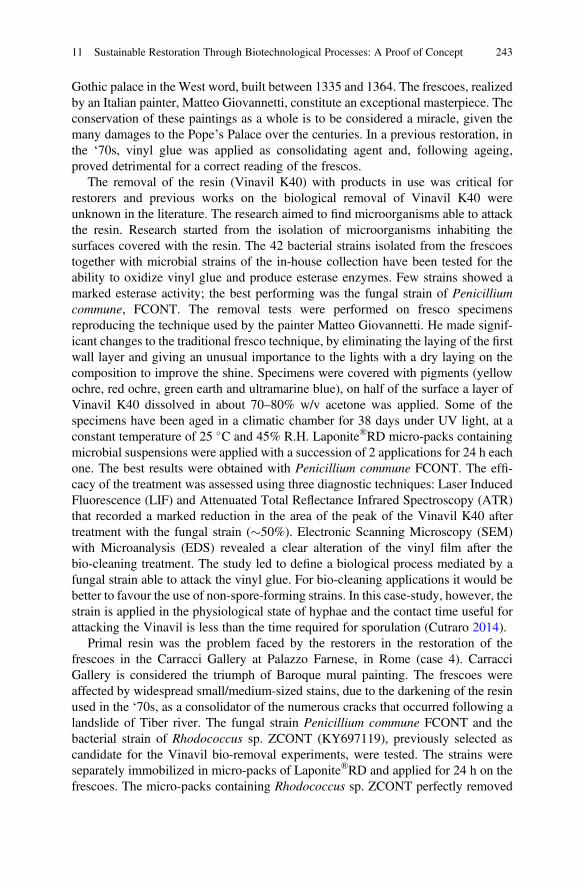
Gothic palace in the West word, built between 1335 and 1364. The frescoes, realizedby an Italian painter, Matteo Giovannetti, constitute an exceptional masterpiece. Theconservation of these paintings as a whole is to be considered a miracle, given themany damages to the Pope’s Palace over the centuries. In a previous restoration, inthe ‘70s, vinyl glue was applied as consolidating agent and, following ageing,proved detrimental for a correct reading of the frescos.
The removal of the resin (Vinavil K40) with products in use was critical forrestorers and previous works on the biological removal of Vinavil K40 wereunknown in the literature. The research aimed to find microorganisms able to attackthe resin. Research started from the isolation of microorganisms inhabiting thesurfaces covered with the resin. The 42 bacterial strains isolated from the frescoestogether with microbial strains of the in-house collection have been tested for theability to oxidize vinyl glue and produce esterase enzymes. Few strains showed amarked esterase activity; the best performing was the fungal strain of Penicilliumcommune, FCONT. The removal tests were performed on fresco specimensreproducing the technique used by the painter Matteo Giovannetti. He made signif-icant changes to the traditional fresco technique, by eliminating the laying of the firstwall layer and giving an unusual importance to the lights with a dry laying on thecomposition to improve the shine. Specimens were covered with pigments (yellowochre, red ochre, green earth and ultramarine blue), on half of the surface a layer ofVinavil K40 dissolved in about 70–80% w/v acetone was applied. Some of thespecimens have been aged in a climatic chamber for 38 days under UV light, at aconstant temperature of 25 �C and 45% R.H. Laponite®RD micro-packs containingmicrobial suspensions were applied with a succession of 2 applications for 24 h eachone. The best results were obtained with Penicillium commune FCONT. The effi-cacy of the treatment was assessed using three diagnostic techniques: Laser InducedFluorescence (LIF) and Attenuated Total Reflectance Infrared Spectroscopy (ATR)that recorded a marked reduction in the area of the peak of the Vinavil K40 aftertreatment with the fungal strain (�50%). Electronic Scanning Microscopy (SEM)with Microanalysis (EDS) revealed a clear alteration of the vinyl film after thebio-cleaning treatment. The study led to define a biological process mediated by afungal strain able to attack the vinyl glue. For bio-cleaning applications it would bebetter to favour the use of non-spore-forming strains. In this case-study, however, thestrain is applied in the physiological state of hyphae and the contact time useful forattacking the Vinavil is less than the time required for sporulation (Cutraro 2014).
Primal resin was the problem faced by the restorers in the restoration of thefrescoes in the Carracci Gallery at Palazzo Farnese, in Rome (case 4). CarracciGallery is considered the triumph of Baroque mural painting. The frescoes wereaffected by widespread small/medium-sized stains, due to the darkening of the resinused in the ‘70s, as a consolidator of the numerous cracks that occurred following alandslide of Tiber river. The fungal strain Penicillium commune FCONT and thebacterial strain of Rhodococcus sp. ZCONT (KY697119), previously selected ascandidate for the Vinavil bio-removal experiments, were tested. The strains wereseparately immobilized in micro-packs of Laponite®RD and applied for 24 h on thefrescoes. The micro-packs containing Rhodococcus sp. ZCONT perfectly removed
11 Sustainable Restoration Through Biotechnological Processes: A Proof of Concept 243
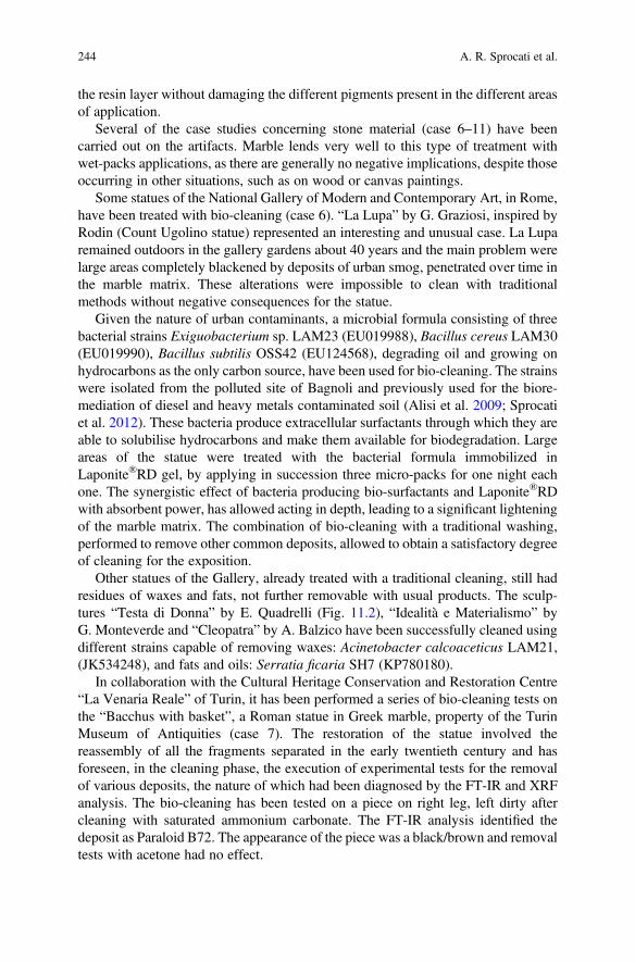
the resin layer without damaging the different pigments present in the different areasof application.
Several of the case studies concerning stone material (case 6–11) have beencarried out on the artifacts. Marble lends very well to this type of treatment withwet-packs applications, as there are generally no negative implications, despite thoseoccurring in other situations, such as on wood or canvas paintings.
Some statues of the National Gallery of Modern and Contemporary Art, in Rome,have been treated with bio-cleaning (case 6). “La Lupa” by G. Graziosi, inspired byRodin (Count Ugolino statue) represented an interesting and unusual case. La Luparemained outdoors in the gallery gardens about 40 years and the main problem werelarge areas completely blackened by deposits of urban smog, penetrated over time inthe marble matrix. These alterations were impossible to clean with traditionalmethods without negative consequences for the statue.
Given the nature of urban contaminants, a microbial formula consisting of threebacterial strains Exiguobacterium sp. LAM23 (EU019988), Bacillus cereus LAM30(EU019990), Bacillus subtilis OSS42 (EU124568), degrading oil and growing onhydrocarbons as the only carbon source, have been used for bio-cleaning. The strainswere isolated from the polluted site of Bagnoli and previously used for the biore-mediation of diesel and heavy metals contaminated soil (Alisi et al. 2009; Sprocatiet al. 2012). These bacteria produce extracellular surfactants through which they areable to solubilise hydrocarbons and make them available for biodegradation. Largeareas of the statue were treated with the bacterial formula immobilized inLaponite®RD gel, by applying in succession three micro-packs for one night eachone. The synergistic effect of bacteria producing bio-surfactants and Laponite®RDwith absorbent power, has allowed acting in depth, leading to a significant lighteningof the marble matrix. The combination of bio-cleaning with a traditional washing,performed to remove other common deposits, allowed to obtain a satisfactory degreeof cleaning for the exposition.
Other statues of the Gallery, already treated with a traditional cleaning, still hadresidues of waxes and fats, not further removable with usual products. The sculp-tures “Testa di Donna” by E. Quadrelli (Fig. 11.2), “Idealità e Materialismo” byG. Monteverde and “Cleopatra” by A. Balzico have been successfully cleaned usingdifferent strains capable of removing waxes: Acinetobacter calcoaceticus LAM21,(JK534248), and fats and oils: Serratia ficaria SH7 (KP780180).
In collaboration with the Cultural Heritage Conservation and Restoration Centre“La Venaria Reale” of Turin, it has been performed a series of bio-cleaning tests onthe “Bacchus with basket”, a Roman statue in Greek marble, property of the TurinMuseum of Antiquities (case 7). The restoration of the statue involved thereassembly of all the fragments separated in the early twentieth century and hasforeseen, in the cleaning phase, the execution of experimental tests for the removalof various deposits, the nature of which had been diagnosed by the FT-IR and XRFanalysis. The bio-cleaning has been tested on a piece on right leg, left dirty aftercleaning with saturated ammonium carbonate. The FT-IR analysis identified thedeposit as Paraloid B72. The appearance of the piece was a black/brown and removaltests with acetone had no effect.
244 A. R. Sprocati et al.
A single micro-pack containing the bacterial strain Rhodococcus sp. ZCONTimmobilized in Vanzan®NF was applied overnight on the half of the dowel coveredby Paraloid B72. After removal of the pack, a slight mechanical action with a dilutedacetone-soaked swab was sufficient to completely remove the Paraloid residues,discovering the completely browned surface. FTIR spectra, before and after thetreatment, confirmed the effectiveness of the treatment. Restorers reported that thenoble patina of the original material was preserved unlike other more aggressiveproducts (Galizzi 2015).
In the case of the pedestal of a statue in the Vatican gardens (case 8) copper oxideswere responsible of stains. The patches have been treated with micro-packscontaining the bacterial strain Sphingopyxis macrogoltabida SME 3.14(MT023684) immobilized in Vanzan®NF. The strain was isolated from a mine inSardinia and it has been selected for its capacity to produce siderophores, to have ahigh esterase activity and to not be able to solubilise carbonates.
The case 11 faces with brown stains on marble. During restoration of the CarracciGallery (Palazzo Farnese, Rome), the sixteenth century portals of Carrara marblepresented brown and other rust-coloured stains due to pins or brackets used for therepair of cracks due, as well as for the frescoes, to the past landslide of Tiber river. Inthis case the restorers put forward the hypothesis that the brown stains present on thesixteenth century portals of the Gallery may be due to an emergence of the ironcontained in the marble, due to maintenance or cleaning treatments previouslycarried out, probably with acids and waxes.
Fig. 11.2 Testa di Donna- Quadrelli. The portion treated with a micro-pack containing living cellsof LAM21 strain was cleaned in an overnight application
11 Sustainable Restoration Through Biotechnological Processes: A Proof of Concept 245
Staining of the stone can be attributed to corrosion of inclusions of iron mineralspresent in the same stone or oxidation of metal parts used as elements purelyaesthetic or for structural strength. The chemical removal of stains of iron com-pounds on marble shows the drawbacks encountered using phosphates (aggressionof the limestone), oxalates (not very effective), fluorides (good solubility of iron in asaturated solution of ammonium fluoride), salicylates (unusable as it has a strong redcolour). Alternatives could be phosphoric acid, phosphoric acid and EDTA disodiumsalt in water, glycerine and gypsum, reagents that reduce Fe3+ to Fe2+, and thenre-oxidize producing new iron hydroxides or hydrated salts more easily removable.It has experimented the use of another complexing agent, the thioglycolic acidneutralized with ammonia, which has produced good results but it is restricted inthe use because of the toxicity for the operators. Even cysteine has been proposed totreat the iron stains on marble. It is an amino acid with a high structural similaritywith thioglycolic acid, but not toxic, and it has an amino group, which gives it threecomplexing groups. Cysteine can act both as bidentate and tridentate binder, thelatter in particular with Fe3+through the thiol and carboxylic groups. Having threedonor groups and being the reactions variable according to the pH and the redoxpotential, it has a chemical very complex, and its chemical and kinetic behaviour isvery difficult to understand. All the reactions, however, give a dark colour to the endproduct, which can vanish with the time.
A biological approach is based on the exploitation of bacteria producingsiderophores, water-soluble molecules with low molecular weight, which can bindin a specific manner Fe3+. Bacteria have acquired the ability to synthesize and secretesiderophores to overcome the shortage of bioavailable iron in nature. Siderophoresare usually released into the extracellular environment, where they act as a captor ofiron. Specific cell receptors allow the entrance into the cell of the complexFe-siderophore. Once in the cytoplasm of the cell, the complex dissociates, withreduction of ferric iron to ferrous iron. The siderophore empty of the reduced ironcan be degraded or reused releasing it into the extracellular medium. This topic hasbeen addressed in a preliminary work with a series of tests performed on laboratoryspecimens prepared ad hoc, reproducing rusts on different stone materials(Pieragostini 2015).
On the Carracci Gallery portals we experimented the bacterial strain Pseudomo-nas protegens FeIIC1 (KY765342), isolated from mine drainage waters and selectedfor its high siderophores production.
After preliminary test on small portions, Vanzan®NF micro-packs containing thebacterial strain FeIIC1, added with a very low concentration of cysteine (0.1% w/v),were applied overnight on almost the entire surface of the four portals. The rustdisappeared and the brown stains inside the crystalline matrix had lost their browncolour or were largely lightened. After the treatment, a very slight dark nuance due tocysteine, vanished with the heat of the day without leaving a trace.
The most recent study on marble was carried out on Tombs of Giuliano andLorenzo de’ Medici sculptured by Michelangelo in the Medici Chapel in Florence(case 10). It was not available a precise diagnosis of the composition of the depositsto be cleaned, as merely non-invasive analyses were performed. Based on the
246 A. R. Sprocati et al.
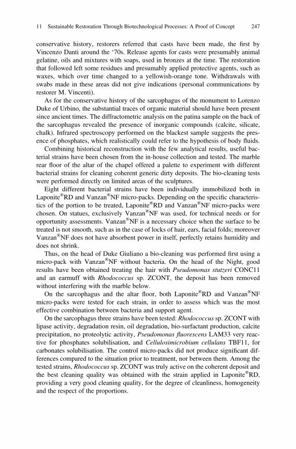
conservative history, restorers referred that casts have been made, the first byVincenzo Danti around the ‘70s. Release agents for casts were presumably animalgelatine, oils and mixtures with soaps, used in bronzes at the time. The restorationthat followed left some residues and presumably applied protective agents, such aswaxes, which over time changed to a yellowish-orange tone. Withdrawals withswabs made in these areas did not give indications (personal communications byrestorer M. Vincenti).
As for the conservative history of the sarcophagus of the monument to LorenzoDuke of Urbino, the substantial traces of organic material should have been presentsince ancient times. The diffractometric analysis on the patina sample on the back ofthe sarcophagus revealed the presence of inorganic compounds (calcite, silicate,chalk). Infrared spectroscopy performed on the blackest sample suggests the pres-ence of phosphates, which realistically could refer to the hypothesis of body fluids.
Combining historical reconstruction with the few analytical results, useful bac-terial strains have been chosen from the in-house collection and tested. The marblerear floor of the altar of the chapel offered a palette to experiment with differentbacterial strains for cleaning coherent generic dirty deposits. The bio-cleaning testswere performed directly on limited areas of the sculptures.
Eight different bacterial strains have been individually immobilized both inLaponite®RD and Vanzan®NF micro-packs. Depending on the specific characteris-tics of the portion to be treated, Laponite®RD and Vanzan®NF micro-packs werechosen. On statues, exclusively Vanzan®NF was used, for technical needs or foropportunity assessments. Vanzan®NF is a necessary choice when the surface to betreated is not smooth, such as in the case of locks of hair, ears, facial folds; moreoverVanzan®NF does not have absorbent power in itself, perfectly retains humidity anddoes not shrink.
Thus, on the head of Duke Giuliano a bio-cleaning was performed first using amicro-pack with Vanzan®NF without bacteria. On the head of the Night, goodresults have been obtained treating the hair with Pseudomonas stutzeri CONC11and an earmuff with Rhodococcus sp. ZCONT, the deposit has been removedwithout interfering with the marble below.
On the sarcophagus and the altar floor, both Laponite®RD and Vanzan®NFmicro-packs were tested for each strain, in order to assess which was the mosteffective combination between bacteria and support agent.
On the sarcophagus three strains have been tested: Rhodococcus sp. ZCONT withlipase activity, degradation resin, oil degradation, bio-surfactant production, calciteprecipitation, no proteolytic activity, Pseudomonas fluorescens LAM33 very reac-tive for phosphates solubilisation, and Cellulosimicrobium cellulans TBF11, forcarbonates solubilisation. The control micro-packs did not produce significant dif-ferences compared to the situation prior to treatment, nor between them. Among thetested strains, Rhodococcus sp. ZCONT was truly active on the coherent deposit andthe best cleaning quality was obtained with the strain applied in Laponite®RD,providing a very good cleaning quality, for the degree of cleanliness, homogeneityand the respect of the proportions.
11 Sustainable Restoration Through Biotechnological Processes: A Proof of Concept 247
The back of the sarcophagus, where very dark, sometimes black stains werepresent and phosphates had been detected, was treated with the Serratia ficaria SH7strain in Laponite®RD, obtaining a total removal of the deposit, except in thethickest points, where it was necessary to proceed with several applications.
By the literature, the artworks undergone bio-cleaning are mainly frescoes, muralpaintings and stone (Bosch-Roig et al. 2014; Mazzoni et al. 2014). Case 12 faced achallenge, it concerned the removal of an aged darkened organic deposit from afragile surface of a sixteenth century wood painting. The experiment was carried outin collaboration with the laboratory of paintings and the Scientific Research Cabinetof the Vatican Museums, where the painting was under restoration.
Main challenge was to meet the conflicting requirements of microorganisms -needing aqueous solutions- and painting - refractory to a long exposure to moisture.The final strategy was to couple the bio-cleaning with a chemical pre-treatment asprotective of the fragile painted surfaces (Crisci et al. 2020).
The deposit consisted of an animal glue with oil, called “colletta con olio”, verytenacious, altered and resistant to the usually effective chemical treatments withoutfurther damaging the painting. The original recipe includes different oils, gall andmolasses.
Two original bacterial strains, Serratia ficaria SH7 and Pseudomonas protegensFeIIC1 non-pathogenic and no spore-forming, were selected following a metabolicscreening, where different bacterial strains have been tested on the individualsubstrate composing the original recipe.
Artificial wood specimens, simulating the artistic technique and covered by alayer of “colletta con olio”, were weathered and used as laboratory tests, to simulatethe application on the artefact. After tests on the specimens, using different supportagents and different application times (from 8 to 24 h), the best procedure resulted inapplying the strains entrapped in Vanzan®NF, used for the first time to this purpose.Micro-packs allowed a complete removal of the “colletta” without altering thecolours of pigments, nor raise the pictorial film and without causing swelling ofthe wood. The results allowed applying the procedure directly on the wood painting,where this effective combination led to the selective removal of the deposit, througha long-lasting contact of bacteria with the sensitive and hygroscopic material of thepainting, without damage for the artwork.
This experiment was significant, as it demonstrated the possibility of degradingvery tenacious and refractory deposits without the use of toxic products; the possi-bility of proceeding with the bio-cleaning method even on materials sensitive tohumidity and on very fragile surfaces. In perspective, an optimisation of the proce-dure can be feasible by acting on microbial metabolism, to reduce the applicationtime needed to a complete removal of tenacious deposits.
Animal glues were historically used both in paper manufacturing, during thesizing process, and in conservation as adhesive for the lining of prints or graphicsand for the creation of passe-partout. Animal glue films are highly hygroscopic andgo through degradation by ageing. The development of internal stresses affects theglue’s elasticity, strength and physical stability and may lead to significant damageto the substrate. Humidity, temperature, UV radiation and pollutants can induce
248 A. R. Sprocati et al.
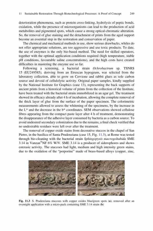
deterioration phenomena, such as protein cross-linking, hydrolysis of peptic bonds,oxidation, while the presence of microorganisms can lead to the production of acidmetabolites and pigmented spots, which cause a strong optical-chromatic alteration.So, the removal of glue staining and the detachment of prints from the aged supportbecome an essential step in the restoration and conservation of paper.
The chemical and mechanical methods in use, show serious drawbacks, whiles donot offer appropriate solutions, are too aggressive and use toxic products. To date,the use of enzymes is the only bio-based method. The need for skilled operators,together with the optimal application conditions required (high temperature, stablepH conditions, favourable saline concentrations), and the high costs have createddifficulties in mastering the enzyme use so far.
Following a screening, a bacterial strain Ochrobactrum sp. TSNRS15 (EU249585), deriving from an Etruscan hypogeum, was selected from thelaboratory collection, able to grow on Cervione and rabbit glues as sole carbonsource and devoid of cellulolytic activity. Original paper samples, kindly suppliedby the National Institute for Graphics (case 13), representing the back supports ofancient prints from a historical volume of prints from the collection of the Institute,have been treated with the bacterial strain immobilized in an agar gel. The treatmentshowed its efficacy already after 4 h of incubation, allowing the complete removal ofthe thick layer of glue from the surface of the paper specimen. The colorimetricmeasurements allowed to assess the whitening of the specimens, by the increase inthe L* and the decrease in the b* coordinates. SEM observations showed cellulosefibres appearing from the compact paste layer after 4 h of treatment, demonstratingthe disappearance of the adhesive layer consumed by bacteria as a carbon source. Toavoid undesired secondary colonization due to the remains, a final check verified thatno undesirable residues were left over after the treatment.
The removal of copper oxide stains from decorative stuccos in the chapel of SanPietro, in the basilica of Santa Prudenziana (case 15, Fig. 11.3), at Rome was testedthrough bio-cleaning with the bacterial strain Sphingopyxis macrogoltabida SME3.14 in Vanzan®NF 6% W/V. SME 3.14 is a producer of siderophores and showsesterasic activity. The stuccoes had light, medium and high intensity green stains,due to the oxidation of the “porporine” made of brass-based alloys (copper, zinc,
Fig. 11.3 S. Prudenziana stuccoes with copper oxides blue/green spots (a), removed after anovernight application with a micro-pack containing SME 3.14 strain (b)
11 Sustainable Restoration Through Biotechnological Processes: A Proof of Concept 249
iron) used in previous restoration works to replace the original gilding. Nine pointswith different intensities were treated. The bio-cleaning tests were carried out bothby applying the micro-packs by direct contact and by interposing a tissue paper.Each application was in contact for 24 h. The most intense spots were cleaned withtwo consecutive applications. Dino-Lite digital micro-images taken for each pointbefore and after the treatment demonstrated the fading or an attenuation of the stains.
The application of Vanzan®NF micro-pack in direct contact with the surface ofthe stucco, without the interposition of paper, led to a greater adhesion of thesupports to the stucco, with greater risk that the product could not be completelyremoved; in fact, in the portions where the product was left for 48 h the stuccoabsorbed part of the water of the support, making it considerably more adherent tothe substrate.
As far as efficacy is concerned, we do not yet have enough data to establish whichapplication method is preferable (by placing an interface or directly in contact) orwhich application time can be recommended.
Cleaning of stucco from copper oxides is therefore biologically viable, but theprocedure requires further tests to be optimized.
4 Biodeteriogens Removal and Control
The term biodeterioration was defined about 50 years ago by Hueck (1965) as “anyunwanted change in the properties of a material, caused by the vital activity of livingorganisms”. In many natural environments, the physical and chemical transforma-tions of materials are considered necessary and positive conditions; however, onartistic material the transformations of the substrate induced by the colonizingmicroflora, in association with various environmental factors, are seen as a destruc-tive and negative phenomenon, both from a cultural and economic point of view. Fora long time, abiotic factors were thought to affect physicochemical properties,facilitating subsequent microbial colonization. Furthermore, it was believed thatthe phenomena of biodeterioration would modify the substrate only from an aes-thetic point of view, that is, inducing the appearance of patinas and coloured spots onthe surface, following the release of biogenic pigments. Recently, several analyticalapproaches have demonstrated that microorganisms are also responsible for physi-cal, chemical and aesthetic modifications of the colonized surface (Sterflinger andPiñar 2013). They can use the surface as growth support, using mineral componentsor surface deposits as metabolites necessary for their development. The speed andextent of microbial colonization are influenced not only by environmental parame-ters, such as the availability of water, but also by the physical-chemical properties ofthe material, such as the mineralogical composition. In fact, although microorgan-isms colonize a certain environment permanently, their harmful activity is notconstant, but periodic.
Today the concept of biodeterioration is intended with a broader meaning: it isknown that the phenomenon is controlled by synergistic and antagonistic
250 A. R. Sprocati et al.
relationships between colonizing microflora and environmental factors; therefore, itcannot be considered as an isolated phenomenon, as it occurs in conjunction withother phenomena of physical and chemical degradation, and it is therefore difficult toattribute any specific damage to a single cause.
Currently, to control or eliminate the phenomena of biodeterioration, restorerscan intervene with mechanical (chisels, spatulas, scalpels), physical (UV rays) andchemical methods (biocides).
The biocidal products have been classified by the EU in four categories andtwenty-two product-types and their use is regulated under the Biocidal ProductsRegulation (BPR, Regulation (EU) 528/2012, Biocidal Products Regulation(EU) 2012) (replacing the Biocidal Products Directive 98/8/EC 1998). Nowadays,many of the broad-spectrum biocides have severe limitations in their use for thetoxicity and persistence in the environment. In conservation and restoration, onlyfew of the existing biocides can be used, due to the lack of information about theirinteraction with the historic materials of the artworks.
Benzalkonium chloride, one of the most used substances in disinfection as part ofthe formulation of many commercial biocides used by restorers is being reviewed foruse as a biocide in the EEA and/or Switzerland according to the ECHA site (2020).
Another aspect not to be underestimated in the use of biocides in the restoration isthe onset of resistant microbial strains. As it happens in clinical microbiology, wherethe thoughtless use of antibiotics determines the development of resistant microor-ganisms, the repeated use of the same biocide or the choice of incorrect concentra-tions can determine the selection, in the microflora present on the artwork, of strainsresistant to the biocide used. This phenomenon is very serious because it candetermine the onset of attacks by biodeteriogens that become difficult to eradicate,considering that often the resistance acquired extends to the whole class of biocidesto which the product used belongs. This was exemplified in Lascaux Cave, France,where the indiscriminate use of benzalkonium chloride resulted in an explosivebacterial and fungal attack (Bastian et al. 2009; Martin-Sanchez et al. 2012).
4.1 “Green Biocides”
There is an increasing interest in the use of naturally-produced compounds, and theneed of alternative solutions led the researchers to explore the world of the naturalsubstances such as plant extracts or bio-based products, that will be more easilydegraded and environment friendly. Plants extracts are already used as biocides infood, medicine and different fields of the pharmaceutical industry. In the last yearsthey have been tested for other applications, such as in the biodeterioration control ofCultural Heritage as a valid alternative to the traditional biocides (Rotolo et al.2016). Many of these compounds are phenols, polyphenols, terpenoids, essentialoils, alkaloids, lectins and mixtures of polypeptides derived from plants (Guiametet al. 2006).
11 Sustainable Restoration Through Biotechnological Processes: A Proof of Concept 251
Oregano, thyme, clove and arborvitae essential oil have been tested for assessingthe antimicrobial potential and might be used as broad-spectrum antimicrobialagents for decontaminating an indoor environment (Puškárová et al. 2017). A pilotstudy was carried out at the Vatican Gardens (Devreux et al. 2015), the applicationsof essential oil of thyme and oregano have been compared with chemical productsbased on quaternary ammonium salts such as benzalkonium chloride, on stonesaffected by biodegradation. The first results obtained showed that the synergistic useof the two essential oils allows to obtain the best biocidal efficacy. Recently an anti-musk product based on essential oils from plant became available on the market(Essenzio, Ibix Biocare). Anyway, essential oils use is not free of concern, sincelavender derivatives, including essential oil, are listed in REACH registrationsamong the substances that cause “serious eye irritation, are harmful to aquatic lifewith long-lasting effects and may cause an allergic skin reaction”.
Many bio-based substances have been investigated till now, as reported in arecent extensive review (Fidanza and Caneva 2019) that lists 61 natural substances,among essential oils and substances of plant and microorganism origin. Theirapplication produces highly variable results due to a lack of a coherent assessmentof the best practices, showing the need of a standard methodology in the use ofnatural biocides for controlling biodeterioration.
For these reasons, our effort was to develop some alternative ways to remove thebiological patinas and possibly control the further microbial development, all ofthem based on natural products of plant and bacterial origin that are harmless for thehealth and the environment, using a standard procedure involving the pack applica-tion. The bio-products have been applied in the real-case studies listed in Table 11.2;here we describe briefly their origin, characteristics and range of applicationsperformed until now.
4.2 BioZ
Surface active agents or “surfactants”, are a group of molecules that lower thesurface tension between two liquids or between a liquid and a solid. In the practicalsense, surfactants may act as wetting agents, emulsifiers, foaming agents anddispersants. Bio-surfactants are synthetized by microorganisms and they are beingstudied for antimicrobial properties and their potential use as biocides on artefacts(Grimaldi 2009; Rivardo et al. 2009; Desai and Banat 1997). Their action is stillunclear; they may inhibit infections through signals that interact with the host and/orbacterial cells or prevent the microbial adhesion and growth on various substrates(Rodrigues et al. 2006). Some bio-surfactants may show antimicrobial activitieswhich could be applied to conservation of cultural heritage.
BioZ is a crude extract containing extracellular glycoproteins, produced by thestrain MCC-Z (JF279930) from the ENEA-Lilith collection. The strain was identi-fied by 16SRNA sequencing and belongs to the Sphingobacteriaceae family andPedobacter genus.
252 A. R. Sprocati et al.
The product BioZ has a good surfactant and emulsifying action which is pre-served after freeze-drying: reduces the surface tension by 40 mN/m and generates anemulsion stable over time, temperature, pH and salinity, with almost constant valuesup to 4 months, ensuring the effectiveness in multiple application conditions(Beltrani et al. 2015).
BioZ product does not contain live bacterial cells, since its preparation involvesan autoclave treatment at 121 �C for 20 min, it is applied incorporated in a suitablesupport to the specific situation to facilitate its application and removal withoutleaving residues on the surface.
The emulsifying capacity of BioZ has been tested on many hydrocarbon com-pounds (hexadecane, toluene, xylene, isooctane, cyclohexane and diesel) whichrepresent common environmental contaminants and are often included in the super-ficial deposits of artistic artefacts. BioZ could also facilitate the removal of unwanteddeposits of dust and smog particles, which often cover the surface of artifactsexposed to atmospheric agents. Its emulsifying activity was tested in the cleaningof laboratory samples from fumes of fuel oil, fats and kerosene.
BioZ antimicrobial activity was tested in-vitro on microorganisms known to beresponsible for biodeterioration of Cultural Heritage, such as Cellulomonas sp.,Bacillus cereus, Bacillus pumilus, Bacillus megaterium, Acinetobactercalcoaceticus, Rhodococcus erythropolis, Rhodococcus sp., Paenibacillus sp.,showing an inhibitory effect on all the Bacillus species and on Cellulomonassp. The different effectiveness was due to the different sensitivity of the singlemicrobial strain tested. It was effective at very low concentrations (0.05% w/v)and showed an inhibitory effect on Gram-positive strains but not on Gram-negativebacteria.
4.3 LIQ
Licorice (Glycyrrhiza glabra) root extract is rich in different classes ofphytocompounds, such as phenols (liquiritina, isoliquiritin, liquiritigenina,isoliquiritigenina, glabridina and glabrol), terpenoids and saponins (β-amyrin,glycyrrhizin, glycyrretol, galabrolide, licorice acid), volatiles compounds (benzal-dehyde, fenchone, linalool, anethole, estragole, eugenol and hexanoic acid), vita-mins (B1, B2, B3, B6, C, E, biotin, folic acid and pantothenic acid), coumarins(glycyrine, umbelliferone, ligcoumarin and herniarin) and mineral content (Öztürket al. 2018). These are bioactive compounds which anti-bacterial, anti-fungal, anti-viral, anti-tumour, anti-inflammatory, anti-oxidant, anti-allergic, expectorant, anti-malarial and anti-convulsive activities have been widely demonstrated (Abbas et al.2015). In addition to the innumerable properties of the root extract, phytochemicalstudies have recently been carried out on the extract of licorice leaves which haveshown the presence of classes of phenols and acid compounds such as glabranin,licoflavanon and pinocembrin (Scherf et al. 2012). These compounds are present
11 Sustainable Restoration Through Biotechnological Processes: A Proof of Concept 253
only in traces or completely absent in the roots, but which are also known for theirantimicrobial and anti-fungal action in plants and men.
The company Trifolio-M GmbH (Lahnau, Germany) has created a formulationfrom the extract of licorice leaves (LIQ), commonly considered an agriculturalwaste. The alcoholic LIQ extract, prepared from dried and finely ground licoriceleaves, is still in experimental phase and not yet commercialized. Trifolio-M isstudying LIQ for its antimicrobial activity against plants pathogenic fungi forapplication in agriculture. In parallel, LIQ extract is under study by ENEA to verifythe possibility of exploiting the antimicrobial properties of this extract towards thebiodeteriogens affecting artworks and monuments. The antimicrobial effect of theextract was demonstrated in-vitro towards about 20 bacterial strains (belonging tothe phyla Firmicutes, Actinobacteria and Gammaproteobacteria) and 10 fungalstrains (belonging to the genera Aspergillus, Fusarium, Epicoccum, Penicilliumand Cladosporium), isolated from hypogeal environments, frescoes, wall and canvaspainting, and included in the ENEA collection of microbial strains. Fungal strainswere less sensitive to the extract than bacterial strains; however, the intermediatesensitivity level shown by Cladosporium spp. and Epicoccum nigrum is a significantresult, given that these fungal genera, widespread in hypogeal environments, arecharacterized by the production of melanins and other deleterious pigments.
4.4 SME 1.11
Arthrobacter xylosoxidans SME1.11 (SUB4060614) is a bacterial strain isolatedfrom the soil of Ingurtosu mining site (Sardinia, Italy). Its cells are non-motile,non-spore forming, Gram-positive, aerobic and rod-shaped. The optimal laboratorygrowth conditions include 4% of salinity and pH 6 and 28 �C; SME1.11 shows noreducing power, no proteolytic and lipase activities but high pectinase activity,produces siderophores and calcite crystals, and lactate tolerance. This bacterialspecies belongs to the risk Group 1 that contains non-pathogenic organisms,i.e. do not cause disease in healthy adult humans.
The capacity to degrade pectin and produce siderophores are two characteristicsthat made this strain a good candidate for biodeterioration control. Further biochem-ical characterization of SME1.11 properties and its antimicrobial activity are underinvestigation.
4.5 NopalCap
A natural plant product obtained from the mucilage of Opuntia ficus-indica com-bined with the alcoholic chili extract - NopalCap- is under investigation for itspotential application as additive in mortars to reduce biofilm colonization. Thisproduct has been studied and carried out in the frame of a bilateral cooperation
254 A. R. Sprocati et al.
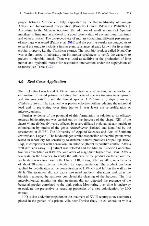
project between Mexico and Italy, supported by the Italian Ministry of ForeignAffairs and International Cooperation (Progetto Grande Rilevanza PGR00971).According to the Mexican tradition, the addition of small amounts of Opuntiamucilage to lime mortar allowed to a good preservation of ancient mural paintingsand other artworks. The bio-receptivity of mortars containing different percentagesof mucilage was tested (Persia et al. 2016) and the positive results encouraged us toexpand the study to include a further plant substance, already known for its antimi-crobial property, i.e. the Capsicum extract. The new bio-product called NopalCapwas at first tested in laboratory on bio-mortar specimens to verify the capacity toprevent a microbial attack. Then was used as additive in the production of limemortar and hydraulic mortar for restoration intervention under the supervision ofrestorers (see Table 11.2).
4.6 Real Cases Application
The LIQ extract was tested at 3% v/v concentration on a painting on canvas for theelimination of mixed patinas including the bacterial species Bacillus licheniformisand Bacillus subtilis, and the fungal species Arthrinium sp., Aspergillus sp.,Cladosporium sp. The treatment was proven effective both in reducing the microbialload and in preventing over time (up to 1 year later) the re-proliferation ofmicroorganisms.
Further evidence of the potential of this formulation in relation to its efficacytowards biodeteriogens was carried out on the frescoes of the chapel XIII of theSacro Monte di Orta (Novara), affected by a very diffused pink patina, attributable tocolonization by strains of the genus Arthrobacter (isolated and identified by theresearchers at SUPSI, The University of Applied Sciences and Arts of SouthernSwitzerland, Lugano). The biodeteriogen strains responsible of the pink patina weretested in laboratory for sensitivity to different natural products (NopalCap, BioZ,Liq), in comparison with benzalkonium chloride (Benz) as positive control. After awell diffusion assay LIQ extract was selected and the Minimal Biocide Concentra-tion was quantified as 0.4% v/v, one order of magnitude higher than Benz. After afew tests on the frescoes, to verify the influence of the product on the colour, theapplication was carried out in the Chapel XIII, during February 2019, on a test areaof about 20 square metres, intended for experimentation. The product has beenapplied by nebulization at the concentration of 1.5% v/v and left on the wall up to48 h. The treatment did not cause unwanted aesthetic alterations and, after thebiocide treatment, the restorers completed the cleaning of the frescoes. The firstmicrobiological monitoring after treatment did not detected the presence of thebacterial species correlated to the pink patina. Monitoring over time is underwayto evaluate the preventive or retarding properties of a new colonization, by LIQextract.
LIQ is also under investigation in the treatment of XVIII century stone sculpturesplaced in the garden of a private villa near Treviso (Italy) in collaboration with a
11 Sustainable Restoration Through Biotechnological Processes: A Proof of Concept 255
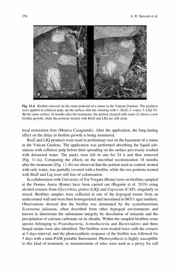
local restoration firm (Monica Casagrande). After the application, the long-lastingeffect on the delay in biofilm growth is being monitored.
BioZ and LIQ products were used in preliminary test on the basement of a statuein the Vatican Gardens. The application was performed absorbing the liquid sub-stances with cellulose pulp before their spreading on the surface previously washedwith deionized water. The packs were left in situ for 24 h and then removed(Fig. 11.4a). Comparing the effects on the microbial recolonization 18 monthsafter the treatments (Fig. 11.4b) we observed that the portion used as control, treatedwith only water, was partially covered with a biofilm, while the two portions treatedwith BioZ and Liq were still free of colonization.
In collaboration with University of Tor Vergata (Rome) tests on biofilms sampledat the Domus Aurea (Rome) have been carried out (Rugnini et al. 2019) usingalcohol extracts from Glycyrrhiza glabra (LIQ) and Capsicum (CAP), singularly ormixed. Biofilms samples were collected in one of the hypogeal rooms from anundecorated wall and were then homogenized and inoculated in BG11 agar medium.Observations showed that the biofilm was dominated by the cyanobacteriumScytonema julianum, often described from other hypogeal environments andknown to deteriorate the substratum integrity by dissolution of minerals and theprecipitation of calcium carbonate on its sheaths. Within the sampled biofilms somespecies belonging to Proteobacteria, Actinobacteria and Bacteroidetes and threefungal strains were also identified. The biofilms were treated twice with the extractsat 5 days-interval, and the photosynthetic response of the biofilm was followed for5 days with a mini-PAM portable fluorometer. Photosynthesis is highly susceptibleto this kind of treatment, so measurements of rates were used as a proxy for cell
Fig. 11.4 Biofilm removal on the stone pedestal of a statue in the Vatican Gardens. The productswere applied in cellulose pulp, (a) the surface after the cleaning with 1- BioZ; 2- water; 3- LIQ 3%.(b) the same surface 18 months after the treatments: the portion cleaned with water (2) shows a newbiofilm growth, while the portions treated with BioZ and LIQ are still clean
256 A. R. Sprocati et al.
health. Changes in photosynthetic activity of the samples treated with the extractswere compared to control biofilms receiving no treatment. Results showed that LIQ30% v/v had the highest photosynthesis inhibition potential, followed by LIQ extract10% v/v. Capsicum extract was the least efficient. These initial results will befollowed by applications on test areas on the wall and roof surfaces within theDomus Aurea site.
NopalCap was used in preparing a hydraulic mortar to fill the voids in the base ofa statue, placed in the patio of the Conservatory of Naples (Italy). A year after therestoration, the mortar is free from biofilm attack, while the neighbouring areas showrecolonization of the surface. This experiment had been preceded by laboratory testson hydraulic mortar specimens also subjected to artificial ageing which gave excel-lent results and will be included in the Dissertation of a thesis on the restoration ofthe whole statue (Lorenza Cardone, Accademia delle Belle Arti di Napoli).
In collaboration with the University of Tuscia (Viterbo) and Superintendency forRoman Museum, a broad comparison of the different bio-products under investiga-tion in our laboratory was carried out at Diocleziano’s termae (Rome, Italy), on amarble artefact placed outdoor, showing a diffuse biofilm (Fig. 11.5). The surfacewas examined using the digital microscope Dino-Lite and the biofilm was sampledto study the microbial patina. High-throughput molecular analyses were performedon the biofilm samples: by a preliminary valuation of the data, the biofilm collectedbefore the treatment was dominated by Cyanobacteria, mostly belonging toNostocales (76% of the total bacterial diversity) and by fungi belonging toAscomycota (90% of the total fungal diversity).
Particular attention was paid to the application process: the liquid products wereimmobilized in both cellulose pulp and in Vanzan®NF to test the best procedure for
Fig. 11.5 Biofilm removalon marble artifact atDiocleziano Termae, Rome.(a) shows the scheme of thetrial, with the productapplied in cellulose pulp(P) and in Vanzan gel (V).From left to right: BioZ incellulose pulp and Vanzan,LIQ in cellulose pulp andVanzan, NopalCap incellulose pulp and Vanzan,SME1.11 in Vanzan, C-(water in cellulose pulp) andC+ (benzalkonium chloride1% in cellulose pulp). (b)the result soon after the packremoval
11 Sustainable Restoration Through Biotechnological Processes: A Proof of Concept 257
application. BioZ, LIQ 3%, NopalCap 10% and SME1.11 (only in Vanzan®NF) plusa negative control (water in cellulose pulp) and a positive control (benzalkoniumchloride 1% in cellulose pulp) were kept in place for 2 weeks, covered with a plasticand aluminium foil to delay the dry out. As for the application procedure, it wasevident that cellulose pulp is not suitable for this kind of products: during the layingof the mixture on the vertical surface, the cellulose pulp did not retain the liquidphase, while the Vanzan®NF gel was able to keep the product into its matrix. Afterthe packs removal, the surface was washed with deionized water, let dry and theresults were recorded (Fig. 11.5b): the best biofilm removal and surface cleaning wasobtained with SME 1.11 in Vanzan®NF, followed by the pack with water incellulose pulp. The other bio-products gave less effective results but better whenapplied in Vanzan®NF; the positive control showed a transient chromatic alteration.Bioluminometer measurement (unpublished data) allowed testing the efficacy of thebio-product on the biofilm control after 7 months. The value on the untreated areawas 145,000 RLU, and the positive control (benzalkonium chloride) 8000 RLUwhile the treated areas show intermediate values, lower than 25,000 for the all theVanzan®NF -packs.
The visual inspection carried out 11 months after the trial (Fig. 11.5c) showedthe persistence of the clean areas for all the treatments except for NopalCap, wherethe biofilm seems to grow again. Monitoring of the surface is ongoing to evaluate thepersistence of the biodeteriogen control.
5 Concluding Remarks
The demonstration cases of bio-cleaning shown and discussed in this chapterprovide, together with the research studies carried out by the scientific community,a convincing proof of feasibility of using biotechnology applied to Cultural Heritage.The next step requires the transition from research to production, to make theprocedures and proposed products available to the market for restorers. To thisend, an effort is needed to define the repeatability of the procedures, the stabilityof the strains and products when transformed into a marketable product (ex: freeze-dried), to reduce the contact times and to move towards a simplification of theprocedures, by selecting multifunctional and versatile microbial strains.
Our experience leads to say with certainty that investigations in the search fornew products and procedures for a more sustainable strategy in cultural heritageconservation, especially with a view to reducing or replacing toxic/dangerousproducts, are flourishing and are widening the field of goods and the issues involved.
Around these issues numerous interests, both scientific and “policy” are gather-ing. That also implies interventions on the legislative, regulatory and organizationallevel.
In order for this process to be carried out and to take place in a virtuous way, ajoint contribution from the world of Conservation, research, entrepreneurship,
258 A. R. Sprocati et al.
stakeholders, the market and public decision-makers is needed, at European andInternational level.
The market for new restoration products that are harmless to operators, theenvironment and works of art must be viewed from the illuminated perspective ofsustainability, as it has a very high social, historical and cultural value, although itcan be a niche market.
Acknowledgement We are grateful to all the restorers and Institutions that have believed ininnovation, looking forward to a sustainable restoration. We especially thank the restorers AdeleCecchini and Lorenza D’Alessandro, who first involved us in the experimentation in their restora-tion sites and who have spread the interest among their colleagues. Special thanks also go to MariaBartoli of the Domus Aurea, to Professor Santamaria, head of the GRS of the Vatican Museums andto his collaborators, to Marina Vincenti and Daniela Manna, to Rodolfo Corrias of National GalleryGAM, to Carla Tomasi and Paolo Pastorello, to the IscR and to a long list of people whocollaborated in each of the case studies, which cannot be contained here.
The NopalCap studies are supported by the Italian Ministry of Foreign Affairs and InternationalCooperation [Progetto Grande Rilevanza PGR00971 and 000784].
References
Abbas A, Zubair M, Rasool N, Rizwan K (2015) Antimicrobial potential of Glycyrrhiza glabra. JDrug Design Med Chem 1:17–20
Alisi C, Musella R, Tasso F, Ubaldi C, Manzo S, Cremisini C, Sprocati AR (2009) Bioremediationof diesel oil in a co-contaminated soil by bioaugmentation with a microbial formula tailored withnative strains selected for heavy metals resistance. Sci Total Environ 407:3024–3032
Barbabietola N (2013) Innovative microbe-based technology for Conservation and Restoration.PhD thesis dissertation, Science for conservation of cultural heritage, University of Florence
Barbabietola N, Tasso F, Alisi C, Marconi P, Perito B, Pasquariello G, Sprocati AR (2016) A safemicrobe-based procedure for a gentle removal of aged animal glues from ancient paper. IntBiodeterior Biodegradation 109:53–60
Bastian F, Alabouvette C, Jurado V, Saiz-Jimenez C (2009) Impact of biocide treatments on thebacterial communities of the Lascaux cave. Naturwissenschaften 96:863–868
Beltrani T, Chiavarini S, Cicero DO, Grimaldi M, Ruggeri C, Tamburini E, Cremisini C (2015)Chemical characterization and surface properties of a new bioemulsifier produced byPedobacter sp. strain MCC-Z. Int J Biol Macromol 72:1090–1096
Bio-Based Industry Consortium (2017) Strategic Innovation and Research Agenda (SIRA). https://www.bbi-europe.eu/sites/default/files/sira-2017.pdf
Biocidal Products Directive 98/8/EC (1998). https://eurlex.europa.eu/LexUriServ/LexUriServ.do?uri¼OJ:L:1998:123:0001:0063:EN:PDF
Biocidal Products Regulation (EU) 528/2012. https://eurlex.europa.eu/LexUriServ/LexUriServ.do?uri¼OJ:L:2012:167:0001:0123:EN:PDF
Bosch Roig P, Regidor Ros JL, Montes Estelles R (2013) Bio-cleaning of nitrate alteration on wallpaintings by Pseudomonas stutzeri. Int Biodeterior Biodegradation 84:266–274
Bosch-Roig P, Lustrato G, Zanardini E, Ranalli G (2014) Bio-cleaning of cultural heritage stonesurfaces and frescoes: which delivery system can be the most appropriate? Ann Microbiol65:1227–1241
Colombini MP, Bonaduce I, Gautier G (2003) Molecular pattern recognition of fresh and agedshellac. Chromatographia 58:357–364
11 Sustainable Restoration Through Biotechnological Processes: A Proof of Concept 259
Crisci L, Alisi C, Pratelli M, Santamaria U, Sprocati AR (2020) Il Biorestauro: messa a punto di unaprocedura su misura per l’opera. In: Tra cielo e terra. La Madonna della Cintola di VincenzoPagani. Edizioni Musei Vaticani (in press)
Cutraro S (2014) Ricerca di una via biologica per la rimozione di colle viniliche da affreschi: il casostudio degli affreschi delle cappelle di Saint Jean e Saint Martial (Palazzo dei Papi, Avignone).Master’s thesis dissertation, Sapienza University, Rome
De Vero L, Boniotti MB, Budroni M, Buzzini P, Cassanelli S, Comunian R, Gullo M, Logrieco AF,Mannazzu I, Musumeci R, Perugini I, Perrone G, Pulvirenti A, Romano P, Turchetti B, VareseGC (2019) Preservation, characterization and exploitation of microbial biodiversity: the per-spective of the Italian network of culture collections. Microorganisms 7:685
Desai JD, Banat IM (1997) Microbial production of surfactants and their commercial potential.Microbiol Mol Biol Rev 61:47–64
Devreux G, Santamaria U, Morresi F, Rodolfo A, Barbabietola N, Fratini F, Reale R (2015)Fitoconservazione. Trattamenti alternativi sulle opere in materiale lapideo nei giardini vaticani.In: Abstract, 13th Congresso Nazionale IGIIC -Lo Stato dell’Arte; Turin, Italy. 22–24 October2015, pp 199–206
ECHA Site (2020). https://echa.europa.eu/substance-information/-/substanceinfo/100.063.544Fidanza MR, Caneva G (2019) Natural biocides for the conservation of stone cultural heritage: a
review. J Cult Herit 38:271–286Galizzi V (2015) Studio e riassemblaggio della scultura lapidea di Bacco del Museo di Antichità di
Torino: ricerca di un equilibrio tra fruizione e conservazione. Master’s degree dissertation,Conservation and restoration of cultural heritage, Torino University
Gioventù E, Lorenzi PF, Villa F, Sorlini C, Rizzi M, Cagnini A, Griffo A, Cappitelli F (2011)Comparing the bioremoval of black crusts on colored artistic lithotypes of the cathedral ofFlorence with chemical and laser treatment. Int Biodeterior Biodegradation 65:832–839
Grimaldi M (2009) Search for new surface-active compounds of microbial origin in view of thedevelopment of biorestoration techniques. PhD thesis dissertation, University of Florence
Guiamet PS, Gómez de Saravia SG, Arenas P, Pérez ML, de la Paz J, Borrego SF (2006) Naturalproducts isolated from plants used in biodeterioration control. Pharmacologyonline 3:537–544
Hueck HJ (1965) The biodeterioration of materials as a part of hylobiology. Mater Org 1:5–34Ledda M (2019) Prodotti biotecnologici per il restauro sostenibile: un caso studio alle terme di
Diocleziano, Roma. Bachelor thesis, University of Cagliari, ItalyMartin-Sanchez PM, Nováková A, Bastian F, Alabouvette C, Saiz-Jimenez C (2012) Use of
biocides for the control of fungal outbreaks in subterranean environments: the case of theLascaux cave in France. Environ Sci Technol 46:3762–3770
Mazzoni M, Alisi C, Tasso F, Cecchini A, Marconi P, Sprocati AR (2014) Laponite micro-packs forthe selective cleaning of multiple coherent deposits on wall paintings: the case study of CasinaFarnese on the Palatine Hill (Rome-Italy). Int Biodeterior Biodegrad 94:1–11
Mora P, Mora L, Philippot P (1984) Conservation of wall paintings. Butterworths, SevenoaksÖztürk M, Altay V, Hakeem KR, Akçiçek E (2018) Pharmacological activities and phytochemical
constituents. In: Öztürk M, Altay V, Hakeem KR, Akçiçek E (eds) Liquorice: from botany tophytochemistry. Springer, Cham, pp 45–60
Persia F, Alisi C, Bacchetta L, Bojorquez E, Colantonio C, Falconieri M, Insaurralde M, Meza-Orozco A, Sprocati AR, Tatì A (2016) Nopal as organic additive for bio-compatible andeco-sustainable lime mortars. In: Campanella L, Piccioli C (eds) Diagnosis for conservationand valorization of cultural heritage. AIES, Naples, pp 245–251
Pieragostini E (2015) Comparative evaluation of chemical and biological methods for the removalof iron oxide stains from calcareous surfaces. Master’s thesis dissertation, Sapienza, Universityof Rome, Italy
Puškárová A, Bučková M, Kraková L, Pangallo D, Kozics K (2017) The antibacterial andantifungal activity of six essential oils and their cyto/genotoxicity to human HEL 12469 cells.Sci Rep 7:8211
260 A. R. Sprocati et al.
Ranalli G, Alfano G, Belli C, Lustrato G, Colombini MP, Bonaduce I, Zanardini E, Abbruscato P,Cappitelli F, Sorlini C (2005) Biotechnology applied to cultural heritage: biorestoration offrescoes using viable bacterial cells and enzymes. J Appl Microbiol 98:73–83
Rivardo F, Turner RJ, Allegrone G, Ceri H, Martinotti MG (2009) Anti-adhesion activity of twobiosurfactants produced by Bacillus spp. prevents biofilm formation of human bacterial path-ogens. Appl Microbiol Biotechnol 83:541–553
Rodrigues L, Banat IM, Teixeira J, Oliveira R (2006) Biosurfactants: potential applications inmedicine. J Antimicrob Chemother 57:609–618
Rotolo V, Barresi G, Di Carlo E, Giordano A, Lombardo G, Crimi E, Costa E, Bruno M, Palla F(2016) Plant extracts as green potential strategies to control the biodeterioration of culturalheritage. Int J Conserv Sci 7:839–846
Rozeik C (2009) The treatment of an unbaked mud statue from ancient Egypt. J Am Inst Conserv48:69–81
Rugnini L, Ellwood NTW, Sprocati AR, Migliore G, Tasso F, Alisi C, Bruno L (2019) Plantproducts as a green solution in the fight against biodeterioration of stone monuments. In: Poster114� congresso S.B.I. (IPSC) - Padova, 4–7 September 2019, p 34
Scherf A, Treutwein J, Kleeberg H, Schmitt A (2012) Efficacy of leaf extract fractions ofGlycyrrhiza glabra L. against downy mildew of cucumber (Pseudoperonospora cubensis). EurJ Plant Pathol 134:755–762
Sharma RK, Yenkateswaran NS, Singh SK (1980) Solvent used for the removal of shellac and otheraccretionary deposits. In: Symposium on mural paintings, archaeological survey of India-ICCROM, Dehra Dun, pp 10–15
Singh M, Balasaheb A (2013) Chemistry of preservation of the Ajanta murals. Int J Conserv Sci 4(2):161–176
Sprocati AR, Alisi C, Tasso F, Marconi P, Sciullo A, Pinto V, Chiavarini S, Ubaldi C, Cremisini C(2012) Effectiveness of a microbial formula, as a bioaugmentation agent, tailored for bioreme-diation of diesel oil and heavy metal co-contaminated soil. Process Biochem 47:1649–1655
Sprocati AR, Alisi C, Tasso F (2014) Biotechnology process for the removal of coherent deposits oforganic and inorganic origin from materials and works of artistic historical interest. Europeanpatent WO2015040647
Sprocati AR, Alisi C, Tasso F, Marconi P, Migliore G (2017) Formule microbiche per l’arte.Kermes 100: 23–26. Nardini Editore
Sterflinger K, Piñar G (2013) Microbial deterioration of cultural heritage and works of art— tiltingat windmills? Appl Microbiol Biotechnol 97:9637–9646
Venice (1964) International charter for the Conservation and restoration of monument and sites (theVenice Chart 1964). In: ICOMOS, IInd International Congress of Architects and Technicians ofHistoric Monuments, Venice
Open Access This chapter is licensed under the terms of the Creative Commons Attribution 4.0International License (http://creativecommons.org/licenses/by/4.0/), which permits use, sharing,adaptation, distribution and reproduction in any medium or format, as long as you give appropriatecredit to the original author(s) and the source, provide a link to the Creative Commons license andindicate if changes were made.
The images or other third party material in this chapter are included in the chapter's CreativeCommons license, unless indicated otherwise in a credit line to the material. If material is notincluded in the chapter's Creative Commons license and your intended use is not permitted bystatutory regulation or exceeds the permitted use, you will need to obtain permission directly fromthe copyright holder.
11 Sustainable Restoration Through Biotechnological Processes: A Proof of Concept 261
Chapter 12The Role of Microorganisms in the Removalof Nitrates and Sulfates on ArtisticStoneworks
Giancarlo Ranalli and Elisabetta Zanardini
Abstract This chapter will focus on the role of microorganisms in the removal ofnitrates and sulfates on artistic stoneworks. The main groups of microbes and theirmetabolisms involved in bioremoval methods for the preservation and protection ofcultural artifacts are reported. The aim is to offer a comprehensive view on the roleand potentiality of virtuous microorganisms in the biocleaning and bioremoval ofblack crusts and salts altering CH stoneworks. We highlight the importance of theuse of the selected microorganisms and the adoption of adequate carriers for theanaerobic metabolism of nitrate and sulfate reducers to be applied on the alteredstone surfaces. The following characteristics of the delivery system are of greatimportance: the ability to guarantee water content for microbes, the absence oftoxicity for the environment, no negative effects to the stone surfaces, easy toprepare, to apply, and to remove from different stone surfaces at the end of thetreatment. We report an overview of the last 30 years on the biocleaning processesincluding diagnostic studies of the alterations, the assessment of associated risks, theeffectiveness and efficacy of the proposed method, and the evaluation in terms ofeconomic and environmental sustainability.
Keywords Cultural Heritage · Biocleaning · Black crust · Nitrates · Sulfates ·Biotechnology · Bacteria
It is well known that microorganisms have a crucial role in their natural habitatsbeing involved in the biogeochemical cycles and in the transformation of inorganicand organic compounds as energy and carbon sources. However, in the Cultural
G. Ranalli (*)Department of Biosciences and Territory, University of Molise, Pesche, Italye-mail: [email protected]
E. ZanardiniDepartment of Sciences and High Technology, University of Insubria, Como, Italye-mail: [email protected]
© The Author(s) 2021E. Joseph (ed.), Microorganisms in the Deterioration and Preservation of CulturalHeritage, https://doi.org/10.1007/978-3-030-69411-1_12
263
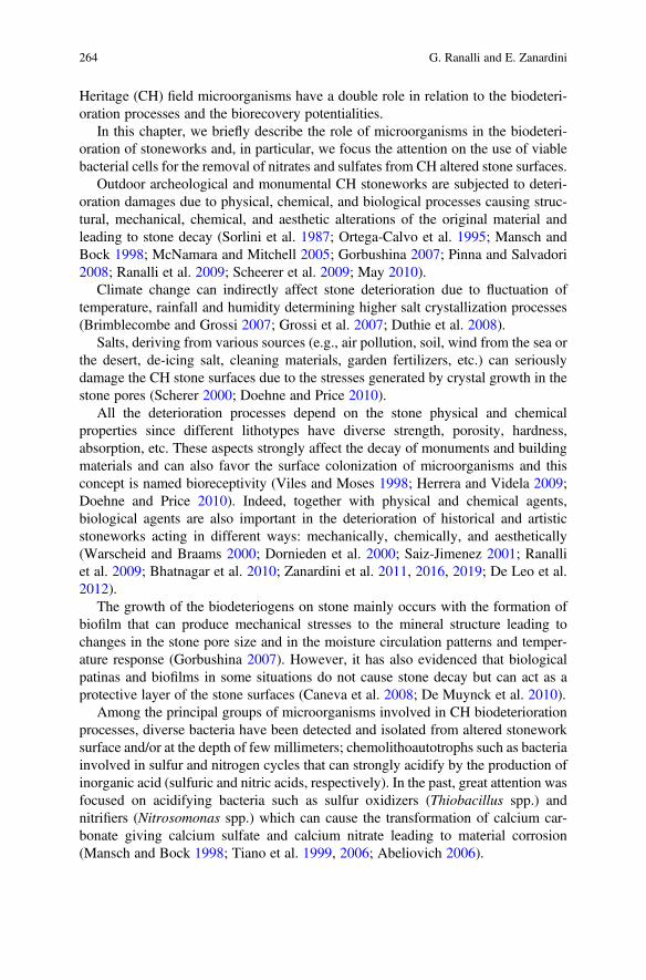
Heritage (CH) field microorganisms have a double role in relation to the biodeteri-oration processes and the biorecovery potentialities.
In this chapter, we briefly describe the role of microorganisms in the biodeteri-oration of stoneworks and, in particular, we focus the attention on the use of viablebacterial cells for the removal of nitrates and sulfates from CH altered stone surfaces.
Outdoor archeological and monumental CH stoneworks are subjected to deteri-oration damages due to physical, chemical, and biological processes causing struc-tural, mechanical, chemical, and aesthetic alterations of the original material andleading to stone decay (Sorlini et al. 1987; Ortega-Calvo et al. 1995; Mansch andBock 1998; McNamara and Mitchell 2005; Gorbushina 2007; Pinna and Salvadori2008; Ranalli et al. 2009; Scheerer et al. 2009; May 2010).
Climate change can indirectly affect stone deterioration due to fluctuation oftemperature, rainfall and humidity determining higher salt crystallization processes(Brimblecombe and Grossi 2007; Grossi et al. 2007; Duthie et al. 2008).
Salts, deriving from various sources (e.g., air pollution, soil, wind from the sea orthe desert, de-icing salt, cleaning materials, garden fertilizers, etc.) can seriouslydamage the CH stone surfaces due to the stresses generated by crystal growth in thestone pores (Scherer 2000; Doehne and Price 2010).
All the deterioration processes depend on the stone physical and chemicalproperties since different lithotypes have diverse strength, porosity, hardness,absorption, etc. These aspects strongly affect the decay of monuments and buildingmaterials and can also favor the surface colonization of microorganisms and thisconcept is named bioreceptivity (Viles and Moses 1998; Herrera and Videla 2009;Doehne and Price 2010). Indeed, together with physical and chemical agents,biological agents are also important in the deterioration of historical and artisticstoneworks acting in different ways: mechanically, chemically, and aesthetically(Warscheid and Braams 2000; Dornieden et al. 2000; Saiz-Jimenez 2001; Ranalliet al. 2009; Bhatnagar et al. 2010; Zanardini et al. 2011, 2016, 2019; De Leo et al.2012).
The growth of the biodeteriogens on stone mainly occurs with the formation ofbiofilm that can produce mechanical stresses to the mineral structure leading tochanges in the stone pore size and in the moisture circulation patterns and temper-ature response (Gorbushina 2007). However, it has also evidenced that biologicalpatinas and biofilms in some situations do not cause stone decay but can act as aprotective layer of the stone surfaces (Caneva et al. 2008; De Muynck et al. 2010).
Among the principal groups of microorganisms involved in CH biodeteriorationprocesses, diverse bacteria have been detected and isolated from altered stoneworksurface and/or at the depth of few millimeters; chemolithoautotrophs such as bacteriainvolved in sulfur and nitrogen cycles that can strongly acidify by the production ofinorganic acid (sulfuric and nitric acids, respectively). In the past, great attention wasfocused on acidifying bacteria such as sulfur oxidizers (Thiobacillus spp.) andnitrifiers (Nitrosomonas spp.) which can cause the transformation of calcium car-bonate giving calcium sulfate and calcium nitrate leading to material corrosion(Mansch and Bock 1998; Tiano et al. 1999, 2006; Abeliovich 2006).
264 G. Ranalli and E. Zanardini
1 Stone Sulfation and Nitration Mechanisms
Air pollution has been recognized in numerous studies as a strong factor involved inthe outdoor stoneworks deterioration for causing material transformation and surfacedeposits (Camuffo 1998; Warscheid and Braams 2000; Zanardini et al. 2000; Saiz-Jimenez 2003; Doehne and Price 2010). Inorganic atmospheric pollutants such as airsulfur oxides (SO2 and SO3), nitrogen oxides (NOx), and carbon oxides, in thepresence of water, produce acidic solutions, which react with the calcareous mate-rials and transform calcium carbonate into calcium sulfate dihydrate and calciumnitrate giving consequently decay phenomena of “sulfation” and “nitration” and theformation of damaging crusts namely “black crust” (Gauri et al. 1989; Rivadeneyraet al. 1991, 1994; Orial et al. 1992; Salvadori and Realini 1996; Saiz-Jimenez 2003,2004; Doehne and Price 2010).
In the case of the black crust formation, the main mechanism is the sulfationreaction involving calcium carbonate as reported below:
2SO2 + O2 ! 2SO3
SO3 + H2O ! H2SO4
H2SO4 + CaCO3 ! CaSO4. (2H2O) + CO2
The corrosion processes operated by acidic pollutants on stone can differentlyhappen based on the environment where monuments are located: exposed andsheltered areas can show in fact different levels of stone surfaces deterioration.When the products from chemical reactions can accumulate, black crusts on stonesurfaces are often formed and the blackness mainly depends on the air pollutantsfrom fossil fuel combustion, while in exposed areas the rainfall normally inhibits theformation of these kinds of alterations as shown in Fig. 12.1a and b. (Doehne andPrice 2010).
a b
Fig. 12.1 (a–b) Typical black crusts on ancient altered marble by exposition to outdoor conditions,at Pisa Cathedral (Credits: Photos by OpaPisa)
12 The Role of Microorganisms in the Removal of Nitrates and Sulfates on. . . 265
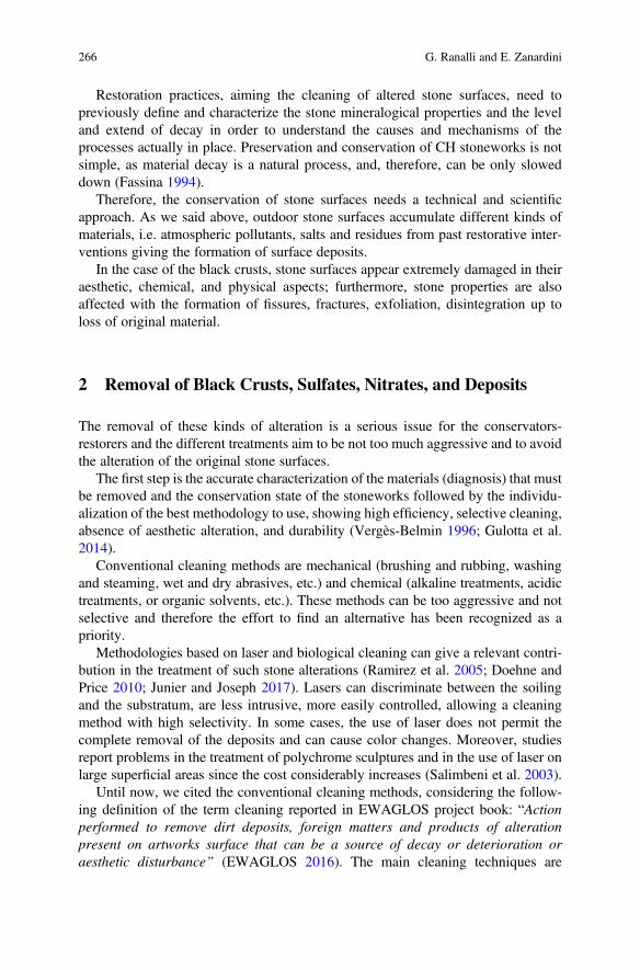
Restoration practices, aiming the cleaning of altered stone surfaces, need topreviously define and characterize the stone mineralogical properties and the leveland extend of decay in order to understand the causes and mechanisms of theprocesses actually in place. Preservation and conservation of CH stoneworks is notsimple, as material decay is a natural process, and, therefore, can be only sloweddown (Fassina 1994).
Therefore, the conservation of stone surfaces needs a technical and scientificapproach. As we said above, outdoor stone surfaces accumulate different kinds ofmaterials, i.e. atmospheric pollutants, salts and residues from past restorative inter-ventions giving the formation of surface deposits.
In the case of the black crusts, stone surfaces appear extremely damaged in theiraesthetic, chemical, and physical aspects; furthermore, stone properties are alsoaffected with the formation of fissures, fractures, exfoliation, disintegration up toloss of original material.
2 Removal of Black Crusts, Sulfates, Nitrates, and Deposits
The removal of these kinds of alteration is a serious issue for the conservators-restorers and the different treatments aim to be not too much aggressive and to avoidthe alteration of the original stone surfaces.
The first step is the accurate characterization of the materials (diagnosis) that mustbe removed and the conservation state of the stoneworks followed by the individu-alization of the best methodology to use, showing high efficiency, selective cleaning,absence of aesthetic alteration, and durability (Vergès-Belmin 1996; Gulotta et al.2014).
Conventional cleaning methods are mechanical (brushing and rubbing, washingand steaming, wet and dry abrasives, etc.) and chemical (alkaline treatments, acidictreatments, or organic solvents, etc.). These methods can be too aggressive and notselective and therefore the effort to find an alternative has been recognized as apriority.
Methodologies based on laser and biological cleaning can give a relevant contri-bution in the treatment of such stone alterations (Ramirez et al. 2005; Doehne andPrice 2010; Junier and Joseph 2017). Lasers can discriminate between the soilingand the substratum, are less intrusive, more easily controlled, allowing a cleaningmethod with high selectivity. In some cases, the use of laser does not permit thecomplete removal of the deposits and can cause color changes. Moreover, studiesreport problems in the treatment of polychrome sculptures and in the use of laser onlarge superficial areas since the cost considerably increases (Salimbeni et al. 2003).
Until now, we cited the conventional cleaning methods, considering the follow-ing definition of the term cleaning reported in EWAGLOS project book: “Actionperformed to remove dirt deposits, foreign matters and products of alterationpresent on artworks surface that can be a source of decay or deterioration oraesthetic disturbance” (EWAGLOS 2016). The main cleaning techniques are
266 G. Ranalli and E. Zanardini
mechanical, physico-chemical and also biological; the last (the biocleaning) includesthe use of living organisms and/or the enzymes as cleaning agents by controlledreproduction of their metabolic processes.
The biological methods based on the use of microorganisms and enzymes ascleaning agents in the “biorestoration” of artworks represented in the last decades anattractive alternative to the mechanical and chemical methods (Bellucci et al. 1999;Cremonesi 1999). The key idea of using living cells in the conservation andpreservation of works of art is supported by the fact that microorganisms (mainlybacteria) are the most versatile and ubiquitous organisms found on earth, and theyare able of colonizing almost any environment (Maier et al. 2000; Sorlini andCappitelli 2008).
Even if we know that some microorganisms have “negative effects,” many ofthem are responsible for “positive effects” such as the degradation and transforma-tion processes of pollutants (Sorlini et al. 1987).
The microorganisms involved in the biorestoration can be isolated from naturalenvironments (soil, water, etc.) and/or selected among the autochthonous microbialcommunities inhabiting historical artworks. Once verified their ability in the removalof the undesired substances, after an adequate increased cellular biomass, they can beused in the recovery applications (bioaugmentation) showing in this case not anegative factor, but a new and positive conservation perspective.
Therefore, the methods based on the use of specific microorganisms can posi-tively help in the cleaning of stone surfaces exploiting the microbial versatility, theirdifferent metabolic activities and role in the biogeochemical cycles in their naturalhabitat. (Atlas et al. 1988; Ranalli et al. 2003). Safe living microbial cells underoptimal controlled conditions reproduce the same processes that occur in nature,softly remove certain substances and represent an eco-friendly solution withouthealth risks for the conservator-restorers and the environment (Boquet et al. 1973;Atlas et al. 1988; Ferrer et al. 1988; Heselmeyer et al. 1991; Saiz-Jimenez 1997;Castanier et al. 2000; Maier et al. 2000; Rodriguez-Navarro et al. 2000; Ranalli et al.2003, 2005; Tiano et al. 2006; Biavati and Sorlini 2008; Valentini et al. 2010; Sassoet al. 2015).
In the last decades biocleaning technologies have been applied in the removal oforganic and inorganic unwanted substances on CH stone surfaces (marble, tuff,sandstone, limestone, etc.), on ceramic material (brick-work), on paper materials,and on concrete using specific bioformulations containing Desulfovibrio sp. andPseudomonas sp. cells (Gauri et al. 1989, 1992; Heselmeyer et al. 1991; DelgadoRodrigues and Valero 2003; Ranalli et al. 2005; De Graef et al. 2005; De Belie et al.2005; Cappitelli et al. 2006, 2007; Alfano et al. 2011; Gioventù et al. 2011;Barbabietola et al. 2012; Troiano et al. 2013).
Table 12.1 reports the biotreatment studies carried out specifically for the removalof salts from CH stoneworks, together with the bacterial strains and the deliverysystems adopted.
Sulfate and nitrate reducers (see Prokaryotes organisms involved in sulfur andnitrogen cycles and reported in Table 12.2 a and b) have been used for the removal of
12 The Role of Microorganisms in the Removal of Nitrates and Sulfates on. . . 267
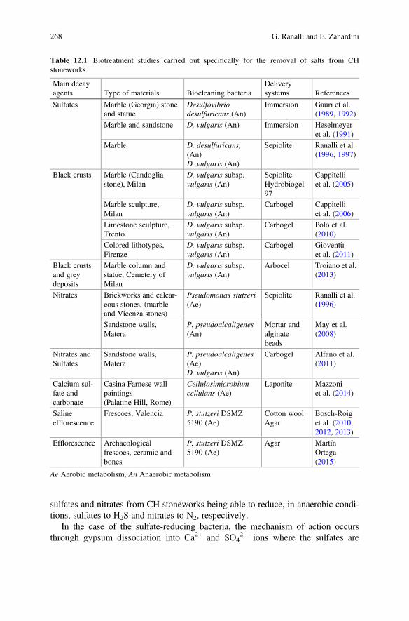
sulfates and nitrates from CH stoneworks being able to reduce, in anaerobic condi-tions, sulfates to H2S and nitrates to N2, respectively.
In the case of the sulfate-reducing bacteria, the mechanism of action occursthrough gypsum dissociation into Ca2+ and SO4
2� ions where the sulfates are
Table 12.1 Biotreatment studies carried out specifically for the removal of salts from CHstoneworks
Main decayagents Type of materials Biocleaning bacteria
Deliverysystems References
Sulfates Marble (Georgia) stoneand statue
Desulfovibriodesulfuricans (An)
Immersion Gauri et al.(1989, 1992)
Marble and sandstone D. vulgaris (An) Immersion Heselmeyeret al. (1991)
Marble D. desulfuricans,(An)D. vulgaris (An)
Sepiolite Ranalli et al.(1996, 1997)
Black crusts Marble (Candogliastone), Milan
D. vulgaris subsp.vulgaris (An)
SepioliteHydrobiogel97
Cappitelliet al. (2005)
Marble sculpture,Milan
D. vulgaris subsp.vulgaris (An)
Carbogel Cappitelliet al. (2006)
Limestone sculpture,Trento
D. vulgaris subsp.vulgaris (An)
Carbogel Polo et al.(2010)
Colored lithotypes,Firenze
D. vulgaris subsp.vulgaris (An)
Carbogel Gioventùet al. (2011)
Black crustsand greydeposits
Marble column andstatue, Cemetery ofMilan
D. vulgaris subsp.vulgaris (An)
Arbocel Troiano et al.(2013)
Nitrates Brickworks and calcar-eous stones, (marbleand Vicenza stones)
Pseudomonas stutzeri(Ae)
Sepiolite Ranalli et al.(1996)
Sandstone walls,Matera
P. pseudoalcaligenes(An)
Mortar andalginatebeads
May et al.(2008)
Nitrates andSulfates
Sandstone walls,Matera
P. pseudoalcaligenes(Ae)D. vulgaris (An)
Carbogel Alfano et al.(2011)
Calcium sul-fate andcarbonate
Casina Farnese wallpaintings(Palatine Hill, Rome)
Cellulosimicrobiumcellulans (Ae)
Laponite Mazzoniet al. (2014)
Salineefflorescence
Frescoes, Valencia P. stutzeri DSMZ5190 (Ae)
Cotton woolAgar
Bosch-Roiget al. (2010,2012, 2013)
Efflorescence Archaeologicalfrescoes, ceramic andbones
P. stutzeri DSMZ5190 (Ae)
Agar MartínOrtega(2015)
Ae Aerobic metabolism, An Anaerobic metabolism
268 G. Ranalli and E. Zanardini
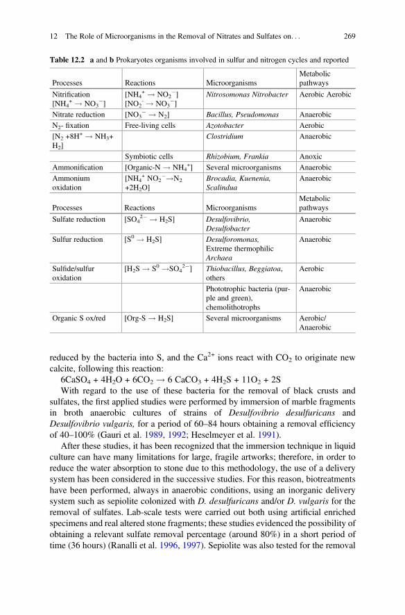
reduced by the bacteria into S, and the Ca2+ ions react with CO2 to originate newcalcite, following this reaction:
6CaSO4 + 4H2O + 6CO2 ! 6 CaCO3 + 4H2S + 11O2 + 2SWith regard to the use of these bacteria for the removal of black crusts and
sulfates, the first applied studies were performed by immersion of marble fragmentsin broth anaerobic cultures of strains of Desulfovibrio desulfuricans andDesulfovibrio vulgaris, for a period of 60–84 hours obtaining a removal efficiencyof 40–100% (Gauri et al. 1989, 1992; Heselmeyer et al. 1991).
After these studies, it has been recognized that the immersion technique in liquidculture can have many limitations for large, fragile artworks; therefore, in order toreduce the water absorption to stone due to this methodology, the use of a deliverysystem has been considered in the successive studies. For this reason, biotreatmentshave been performed, always in anaerobic conditions, using an inorganic deliverysystem such as sepiolite colonized with D. desulfuricans and/or D. vulgaris for theremoval of sulfates. Lab-scale tests were carried out both using artificial enrichedspecimens and real altered stone fragments; these studies evidenced the possibility ofobtaining a relevant sulfate removal percentage (around 80%) in a short period oftime (36 hours) (Ranalli et al. 1996, 1997). Sepiolite was also tested for the removal
Table 12.2 a and b Prokaryotes organisms involved in sulfur and nitrogen cycles and reported
Processes Reactions MicroorganismsMetabolicpathways
Nitrification[NH4
+ ! NO3�]
[NH4+ ! NO2
�][NO2
- ! NO3�]
Nitrosomonas Nitrobacter Aerobic Aerobic
Nitrate reduction [NO3� ! N2] Bacillus, Pseudomonas Anaerobic
N2- fixation Free-living cells Azotobacter Aerobic
[N2 +8H+ ! NH3+
H2]Clostridium Anaerobic
Symbiotic cells Rhizobium, Frankia Anoxic
Ammonification [Organic-N ! NH4+] Several microorganisms Anaerobic
Ammoniumoxidation
[NH4+ NO2
�!N2
+2H2O]Brocadia, Kuenenia,Scalindua
Anaerobic
Processes Reactions MicroorganismsMetabolicpathways
Sulfate reduction [SO42� ! H2S] Desulfovibrio,
DesulfobacterAnaerobic
Sulfur reduction [S0 ! H2S] Desulforomonas,Extreme thermophilicArchaea
Anaerobic
Sulfide/sulfuroxidation
[H2S ! S0 !SO42�] Thiobacillus, Beggiatoa,
othersAerobic
Phototrophic bacteria (pur-ple and green),chemolithotrophs
Anaerobic
Organic S ox/red [Org-S ! H2S] Several microorganisms Aerobic/Anaerobic
12 The Role of Microorganisms in the Removal of Nitrates and Sulfates on. . . 269
of nitrates on brick-works and calcareous stones (marble and Vicenza-stones) usinga Pseudomonas stutzeri strain in anaerobic conditions (Ranalli et al. 1996). How-ever, the use of sepiolite gave in some cases problems of dark precipitates (for thepresence of iron in trace) requiring a pre-treatment of the sepiolite to remove the iron.In addition, sepiolite as delivery system did not guarantee the right amount of waterfor the metabolic activity of bacterial cells during the application on stone surface.
The inorganic delivery system was therefore substituted by an organic carriernamed hydrobiogel-97 for the immobilization of D. vulgaris subsp. vulgaris ATCC29579 bacterial cells. The biotreatments were carried out both under lab-scale trialson artificially specimens (different lithotypes) and real altered stoneworks, and insitu trials to remove black crusts present on Candoglia stone at Milan Cathedral,Italy. The in situ bioapplication has been carried out using carbogel (CTS, Vicenza,Italy), covered by a thin film to create microaerophilic condition, and repeated3 times (15 hours each) giving a removal of 98% of sulfates. A comparison of thebiological methods with a chemical one (using ammonium carbonate and EDTA)was carried out in order to define the results in terms of removal, but also todetermine the effects on the stone surface; in this case the biotreatment was verypromising (Cappitelli et al. 2006, 2007).
Another case study where the sulfate crust was efficiently biocleaned byD. vulgaris subsp. vulgaris ATCC 29579 and carbogel was the Pietà Rondaninimarble base sculpture by Michelangelo Buonarroti (Milan, Italy). In order to elim-inate secondary iron black deposits, bacteria were grown in a modified brothmedium free of iron. Treatment times were of 24–30 hours in anaerobiosis(Cappitelli et al. 2005).
In another applied studies on altered stone sculptures carried out by Polo et al.(2010) the presence of black crusts and the microbial growth causing discolorationwere evidenced on the Demetra and Cronos sculptures, two of 12 stone statuesdecorating the courtyard of the Buonconsiglio Castle in Trento (Italy). Desulfovibriovulgaris subsp. vulgaris, grown in DSMZ 63 medium without iron source andcarbogel as delivery system, was applied to remove the black crusts but preservedthe original stone and the patina noble. The altered surfaces were treated with three12-hours applications for a total duration of 36 hours. This case study, combiningtraditional and biomolecular methods, showed that conservators can benefit from anintegrated biotechnological approach aimed at the biocleaning of the alterationstogether with the abatement of biodeteriogens (Polo et al. 2010).
Other positive results involved the colored lithotypes of Florence Cathedralexternal walls. Green serpentine, red marlstone, and Carrara white marble blackcrust were biocleaned. This work compared three different cleaning treatments:chemical poultice, laser, and biocleaning. Results shows that the better cleaningmethod was biocleaning using D. vulgaris subsp. vulgaris ATCC 29579 strain andcarbogel as a carrier, but long-time treatments and numerous applications wereneeded (Gioventù and Lorenzi 2013).
In another recent study, the combination of chemical and biological (arbocelnonionic detergent treatment colonized with D. vulgaris subsp. vulgaris) methodsallowed efficient cleaning of grey deposits and black crusts on marble columns and
270 G. Ranalli and E. Zanardini
statues at the Cemetery in Milan, Italy, with a notable reduction in the treatmentduration (Troiano et al. 2013).
Carbogel alone together with mortar and alginate beads have been also used ontuff stone walls at Matera Cathedral, Italy, for the removal of both sulfates andnitrates (the latter present at higher concentration) using different microorganismssuch as Pseudomonas pseudoalcaligenes and D. vulgaris (May et al. 2008; Alfanoet al. 2011).
At Matera Cathedral the main problem arises from the rising of salts from thegroundwater rich in nitrates and sulfates causing reduction in cohesion of the tuffstone, loss of materials, and the darkening of the lower external walls. The nitratesoriginate from the oxidation of various N organic compounds (including proteins,amino acids, and urea) from bodies buried in the ground when the area was used as acemetery (until eighteenth century). At the same way, sulfates derived mainly byamino acids such as methionine and cysteine rather than environmental depositions.In addition, chemical analyses detected egg residues as previous consolidant treat-ments of the tuff surface. The biotreatment was efficient, showing a removal ofsurface deposits and salt alterations after 24 hours (55% of the nitrate and 85% of thesulfate deposits) (Alfano et al. 2011; Rampazzi et al. 2018).
Many of the reported studies were performed during a 3 years EU project namedBIOBRUSH project (BIOremediation for Building Restoration of the Urban StoneHeritage). The main aims of the project were the selection of microorganisms anddelivery system for the removal of black crusts, sulfates, nitrates and also thepossibility of using bio-calcifying bacteria for the stone bio-consolidation (Websterand May 2006; May et al. 2008).
The EU BIOBRUSH project gave the opportunity to perform studies underdifferent scale (lab and in situ experiments) giving promising results and showingthe aspects to improve and optimize. For example, in some cases, the deliverysystems adopted resulted not appropriate to treat large surface areas or decorativeelements.
In another study, the combination of chemical and biological (arbocel nonionicdetergent treatment colonized with D. vulgaris subsp. vulgaris) methods allowedefficient cleaning of grey deposits and black crusts on marble columns and statues atthe Cemetery in Milan, Italy, with a notable reduction in the treatment duration(Troiano et al. 2013).
More recent studies showed the use of improved delivery systems as agar-gelcolonized by Pseudomonas stutzeri DSMZ 5190 strain for the removal of saltefflorescence on frescoes on the Santos Juanes Church in Valencia, Spain, asreported in Fig. 12.2 (Bosch-Roig et al. 2010, 2012, 2013) and on archeologicalfrescoes, ceramic, and bones (Martín Ortega 2015).
In the first case, Bosch-Roig et al. (2013) demonstrated that short term (90 min)application of P. stutzeri DSMZ 5190 and the use of agar as delivery systemprovided an efficient cleaning of insoluble nitrate efflorescence deposited on wallpainting surfaces confirmed by Ion Chromatography (92% in nitrate efflorescencereduction). Bosch-Roig et al. (2013) underlined the positive properties of agarsupporting the microorganisms as follows: (i) it can provided an adequate water
12 The Role of Microorganisms in the Removal of Nitrates and Sulfates on. . . 271
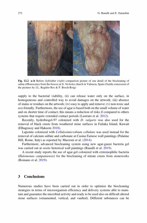
supply to the bacterial viability, (ii) can release water only on the surface, inhomogeneous and controlled way to avoid damages on the artwork; (iii) absenceof stains or residues on the artwork; (iv) easy to apply and remove; (v) non-toxic andeco-friendly. Furthermore, the use of agar is based both on the small volume of waterand on shorter time of contact; this means a reduction of risks if compared to otherssystems that require extended contact periods (Lustrato et al. 2012).
Recently, hydrobiogel-97 colonized with D. vulgaris was also used for theremoval of black crusts from weathered stone surfaces in Failaka Island, Kuwait(Elhagrassy and Hakeem 2018).
Laponite colonized with Cellulosimicrobium cellulans was used instead for theremoval of calcium sulfate and carbonate at Casina Farnese wall paintings (PalatineHill, Rome, Italy) as reported by Mazzoni et al. (2014).
Furthermore, advanced biocleaning system using new agar-gauze bacteria gelwas carried out at onsite historical wall paintings (Ranalli et al. 2019).
A recent study reports the use of agar-gel colonized with extremophilic bacteria(Halomonas campaniensis) for the biocleaning of nitrate crusts from stoneworks(Romano et al. 2019).
3 Conclusions
Numerous studies have been carried out in order to optimize the biocleaningstrategies in terms of microorganism efficiency and delivery systems able to main-tain and guarantee the microbial activity and ready to be used also on difficult alteredstone surfaces (ornamented, vertical, and vaulted). Different substances can be
Fig. 12.2 a–b Before (left)/after (right) comparison picture of one detail of the biocleaning ofsaline efflorescence from the frescos at St. Nicholas church in Valencia, Spain (Gentle concession ofthe pictures by J.L. Regidor Ros & P. Bosch-Roig)
272 G. Ranalli and E. Zanardini
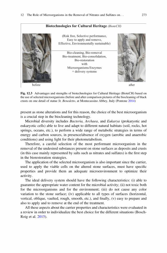
present as stone alterations and for this reason, the choice of the best microorganismis a crucial step in the biocleaning technology.
Microbial diversity includes Bacteria, Archaea, and Eukarya (prokaryotic andeukaryotic cells) able to live and adapt to different natural habitats (soil, rocks, hotsprings, oceans, etc.), to perform a wide range of metabolic strategies in terms ofenergy and carbon sources, in presence/absence of oxygen (aerobic and anaerobicconditions) and using light for their photometabolism.
Therefore, a careful selection of the most performant microorganism in theremoval of the undesired substances present on stone surfaces as deposits and crusts(in this case mainly represented by salts such as nitrates and sulfates) is the first stepin the biorestoration strategies.
The application of the selected microorganism is also important since the carrier,used to apply the viable cells on the altered stone surfaces, must have specificproperties and provide them an adequate microenvironment to optimize theiractivity.
The ideal delivery system should have the following characteristics: (i) able toguarantee the appropriate water content for the microbial activity; (ii) not toxic bothfor the microorganisms and for the environment; (iii) do not cause any colorvariation to the stone surface; (iv) applicable to all types of surfaces (horizontal,vertical, oblique, vaulted, rough, smooth, etc.), and finally, (v) easy to prepare andalso to apply and to remove at the end of the treatment.
All these aspects about the carrier properties and characteristics were evaluated ina review in order to individualize the best choice for the different situations (Bosch-Roig et al. 2015).
Biotechnologies for Cultural Heritage (BioteCH)
(Risk free, Selective performance,
Easy to apply and remove,
Effective, Environmentally sustainable)
Bio-cleaning, Bio-removal
Bio-treatment, Bio-consolidation,
Bio-restoration
with
Microorganisms/Enzymes
+ delivery systems
before after
Fig. 12.3 Advantages and strengths of biotechnologies for Cultural Heritage (BioteCH) based onthe use of selected microorganisms (before and after comparison pictures of the biocleaning of blackcrusts on one detail of statue St. Benedetto, at Montecassino Abbey, Italy (Pontone 2014)
12 The Role of Microorganisms in the Removal of Nitrates and Sulfates on. . . 273
The biocleaning process also requires a deep diagnostic study of the work of art tobe cleaned, an assessment of any associated risks, an evaluation of the effectivenessand efficiency of the process, and last but not least, an evaluation in terms ofeconomic and environmental sustainability (Bosch-Roig and Ranalli 2014)(Fig. 12.3).
Acknowledgements The authors wish to thank to the restorers involved in the before mentionedcase studies because without their confidence, help, and dedication none of this works could havebeen successfully applied on real artworks. Special thanks to Claudia Sorlini, Maria PerlaColombini, Alessia Andreotti, Laura Rampazzi and Cristina Corti for the continuous collaborationsand support in the chemical aspects in our studies on CH biotechnologies. Special thanks toArch. B. Lafratta, Superintendence for Restoration, The Technical and Scientific Committee(Matera, Italy); Mons. D. Lionetti, ArciDiocesi di Matera-Irsina; Restorer G. D’Alessandro,Impresa D’Alessandro Restauri, Matera, Italy. Moreover, we are very grateful to the VenerandaFabbrica del Duomo for assistance and support of the diagnostic work on the Milan Cathedral(Milan, Italy).
References
Abeliovich A (2006) The nitrite-oxidising bacteria. In: Dworkin M, Falkow S, Rosenberg E,Schleifer K-H, Stackebrandt E (eds) The prokaryotes. Springer, New York, pp 861–871
Alfano G, Lustrato G, Belli C, Zanardini E, Cappitelli F, Mello E, Sorlini C, Ranalli G (2011) Thebioremoval of nitrate and sulfate alterations on artistic stonework: the case-study of Materacathedral after six years from the treatment. Int Biodeterior Biodegradation 65:1004–1011
Atlas RM, Chowdhury AN, Gauri KL (1988) Microbial calcification of gypsum-rock and sulfatedmarble. J Stud Conserv 33:149–153
Barbabietola N, Tasso F, Grimaldi M, Alisi C, Chiavarini S, Marconi P, Perito B, Sprocati AR(2012) Microbe-based technology for a novel approach to conservation and restoration. EAISpeciale II. Knowledge, diagnostics and preservation of cultural heritage, pp. 69–76
Bellucci R, Cremonesi P, Pignagnoli G (1999) A preliminary note on the use of enzymes inconservation: the removal of aged acrylic resin coatings with lipase. Stud Conserv 44:278–281
Bhatnagar P, Khan AA, Jain SK, Rai MK (2010) Biodeterioration of archaeological monumentsand approach for restoration. In: Jain SK, Khan AA, Rai MK (eds) Geomicrobiology. CRCPress, Enfield, pp 255–302
Biavati B, Sorlini C (2008) Microbiologia agroambientale. Ambrosiana, Milano, p 684Boquet E, Boronat A, Ramos-Cormenzana A (1973) Production of calcite (calcium carbonate)
crystals by soil bacteria is a general phenomenon. Nature 246:527–528Bosch-Roig P, Lustrato G, Zanardini E, Ranalli G (2015) Biocleaning of cultural heritage stone
surfaces and frescoes: which delivery system can be the most appropriate. Ann Microbiol65:1227–1241
Bosch-Roig P, Montes-Estellés RM, Regidor-Ros JL, Roig Picazo P, Ranalli G (2012) Newfrontiers in the microbial bio-cleaning of artworks. The Picture Restorer 41:37–41
Bosch-Roig P, Ranalli G (2014) The safety of biocleaning technologies for cultural heritage. FrontMicrobiol 5:155
Bosch-Roig P, Regidor-Ros JL, Montes-Estellés R (2013) Biocleaning of nitrate alterations on wallpaintings by Pseudomonas stutzeri. Int Biodeterior Biodegradation 84:266–274
Bosch-Roig P, Regidor-Ros JL, Soriano-Sancho P, Domenech-Carbo MT, Montes-Estellés RM(2010) Ensayos de biolimpieza con bacterias en pinturas murales. Arché 4–5:115–122
274 G. Ranalli and E. Zanardini
Brimblecombe P, Grossi CM (2007) Damage to buildings from future climate and pollution. APTBull 38:13–18
Camuffo D (1998) Microclimate for cultural heritage. Elsevier, Amsterdam, p 415Caneva G, Nugari MP, Salvadori O (2008) Plant biology for cultural heritage: biodeterioration and
conservation. The Getty Conservation Institute, Los Angeles, CACappitelli F, Toniolo L, Sansonetti A, Gulotta D, Ranalli G, Zanardini E, Sorlini C (2007)
Advantages of using microbial technology over traditional chemical technology in removal ofblack crusts from stone surfaces of historical monuments. Appl Environ Microbiol73:5671–5675
Cappitelli F, Zanardini E, Ranalli G, Mello E, Daffonchio D, Sorlini C (2006) Improved method-ology for bioremoval of black crusts on historical stone artworks by use of sulfate-reducingbacteria. Appl Environ Microbiol 72:3733–3737
Cappitelli F, Zanardini E, Toniolo L, Abbruscato P, Ranalli G, Sorlini C (2005) Bioconservation ofthe marble base of the Pietà Rondanini by Michelangelo Buonarroti. Geophys Res Abstr7:06675–00676
Castanier S, Le Metayer-Levrel G, Orial G, Loubiere JF, Perthuisot JP (2000) Carbonatogenesisand applications to preservation and restoration of historic property. In: Ciferri O, Tiano P,Mastromei G (eds) Of microbes and art. Kluwer Academic/Plenum Publishers, Amsterdam, pp203–218
Cremonesi P (1999) L’uso degli enzimi nella pulitura di opere policrome. In: Cremonesi P (ed) ITalenti. Il Prato, Padova, pp 5–11
De Belie N, De Graef B, De Muynck W, Dick J, De Windt W, Verstraete W (2005) Biocatalyticprocesses on concrete: bacterial cleaning and repair. In: 10th DBMC international conference ondurability of building materials and components, Lyon, 17–20 April
De Graef B, De Windt W, Dick J, Verstraete W, De Belie N (2005) Cleaning of concrete fouled bylichens with the aid of Thiobacilli. Mater Struct 38:875–882
De Leo F, Iero A, Zammit G, Urzì CE (2012) Chemoorganotrophic bacteria isolated frombiodeteriorated surfaces in cave and catacomb. Int J Speleol 41:125–136
De Muynck W, De Belie N, Verstraete W (2010) Microbial carbonate precipitation in constructionmaterials: a review. Ecol Eng 36:118–136
Delgado Rodrigues J, Valero J (2003) A brief note on the elimination of black stains of biologicalorigin. Stud Conserv 48:17–22
Doehne EF, Price CA (2010) Stone conservation: an overview of current research. The GettyConservation Institute, Los Angeles
Dornieden T, Gorbushina AA, Krumbein WE (2000) Biodecay of cultural heritage as a space/time-related ecological situation an evaluation of a series of studies. Int Biodeterior Biodegradation46:261–270
Duthie L, Hyslop E, Kennedy C, Phoenix V, Lee MR (2008) Quantitative assessment of decaymechanisms in Scottish building sandstones. In: Lukaszewicz JW, Niemcewicz P (eds) Pro-ceedings of the 11th int. congress on deterioration and conservation of stone, Torun, pp. 73–80
Elhagrassy AF, Hakeem A (2018) Comparative study of biological cleaning and laser techniquesfor conservation of weathered stone in Failaka Island, Kuwait. Sci Cult 4:43–50
EWAGLOS (2016) Weyer A, Roig Picazo P, Pop D, Carras J, Ozkose A, Vallet JM, Srsa I (Eds.).In: EwaGlos-European illustrated glossary of conservation terms for wall paintings and archi-tectural surfaces. English definitions with translations into Bulgarian, Croatian, French, Ger-man, Hungarian, Italian, Polish, Romanian, Spanish and Turkish, vol. 17. Michael ImhofVerlag, Petersberg, Germany, pp. 304–307
Fassina V (1994) General criteria for the cleaning of stone: theoretical aspects and methodology ofapplication. In: Zezza F (ed) Stone material in monuments: diagnosis and conservation. ScuolaUniversitaria C.U.M. Conservazione dei Monumenti, Heraklion, pp 131–138
Ferrer MR, Quevedo-Sarmiento J, Rivadeneira MA, Bejar V, Delgado R, Ramos-Cormenzana A(1988) Calcium carbonate precipitation by two groups of moderately halophilic microorganismsat different temperatures and salt concentrations. Curr Microbiol 17:221–227
12 The Role of Microorganisms in the Removal of Nitrates and Sulfates on. . . 275
Gauri KL, Chowdhury AN, Kulshreshtha NP, Punuru AR (1989) The sulfation of marble and thetreatment of gypsum crusts. Stud Conserv 34:201–206
Gauri KL, Parks L, Jaynes J, Atlas R (1992) Removal of sulfated-crust from marble using sulfate-reducing bacteria. In: Proceedings of the int. conference on stone cleaning and the nature, soilingand decay mechanisms of stone, Edinburgh, 14–16 April, pp. 160–165
Gioventù E, Lorenzi P (2013) Bio-removal of black crust from marble surface: comparison withtraditional methodologies and application on a sculpture from the Florence’s English cemetery.Procedia Chem 8:123–129
Gioventù E, Lorenzi PF, Villa F, Sorlini C, Rizzi M, Cagnini A, Griffo A, Cappitelli F (2011)Comparing the bioremoval of black crusts on colored artistic lithotypes of the Cathedral ofFlorence with chemical and laser treatment. Int Biodeterior Biodegradation 65:832–839
Gorbushina AA (2007) Life on the rocks. Environ Microbiol 9:1613–1631Grossi CM, Brimblecombe B, Esbert RM, Alsono FJ (2007) Color changes in architectural
limestone from pollution and cleaning. Color Res Appl 32:320–331Gulotta D, Saviello D, Gherardi F, Toniolo L, Anzani M, Rabbolini A, Goidanich S (2014) Setup of
a sustainable indoor cleaning methodology for the sculpted stone surfaces of the Duomo ofMilan. Heritage Science 2:6–19
Herrera LK, Videla HA (2009) Surface analysis and materials characterization for the study ofbiodeterioration and weathering effects on cultural property. Int Biodeterior Biodegradation63:813–822
Heselmeyer K, Fischer U, Krumbein KE, Warscheid T (1991) Application of Desulfovibriovulgaris for the bioconversion of rock gypsum crusts into calcite. BIOforum 1(/2):89
Junier P, Joseph E (2017) Microbial biotechnology approaches to mitigating the deterioration ofconstruction and heritage materials. Microb Biotechnol 10:1145–1148
Lustrato G, Alfano G, Andreotti A, Colombini MP, Ranalli G (2012) Fast biocleaning of mediaevalfrescoes using viable bacterial cells. Int Biodeterior Biodegrad 69:51–61
Maier RM, Pepper I, Gerba CP (2000) Environmental microbiology. Elsevier, San Diego, p 585Mansch R, Bock E (1998) Biodeterioration of natural stone with special reference to nitrifying
bacteria. Biodegradation 9:47–64Martín Ortega S (2015) Neteges raonades: Experimentació amb nou tractaments de neteja aplicats
sobre 6 peces de material arqueologic divers. Treball final de grau, ESCRBCC, BarcelonaMay E (2010) Stone biodeterioration. In: Mitchell R, McNamara CJ (eds) Cultural heritage
microbiology: fundamental studies in conservation science. ASM Press, Washington, DC, pp1–39
May E, Webster AM, Inkpen R, Zamarreno D, Kuever J, Rudolph C, Warcheid T, Sorlini C,Cappitelli F, Zanardini E, Ranalli G, Kgrage L, Vgenopoulos A, Katsinis D, Mello E, MalagodiM (2008) The BIOBRUSH project for bioremediation of heritage stone. In: May E, Jones M,Mitchell J (eds) Heritage microbiology and science. Microbes, monuments and maritimematerials. RCS Publishing, Cambridge, pp 76–93
Mazzoni M, Alisi C, Tasso F, Cecchini A, Marconi P, Sprocati AR (2014) Laponite micro-packs forthe selective cleaning of multiple coherent deposits on wall paintings: the case study of CasinaFarnese on the Palatine Hill (Rome-Italy). Int Biodeterior Biodegradation 94:1–11
McNamara CJ, Mitchell R (2005) Microbial deterioration of historic stone. Front Ecol Environ3:445–451
Orial G, Castanier S, Le Métayer G, Loubiere JF (1992) The biomineralisation: a new process toprotect calcareous stone applied to historic monuments. In: Toishi K, Arai H, Kenjo T, YamanoK (eds) Proceedings of 2nd int. conference on biodeterioration of cultural property, Yokohama,pp. 98–116
Ortega-Calvo JJ, Arino X, Hernandez-Marine M, Saiz-Jimenez C (1995) Factors affecting theweathering and colonisation of monuments by phototrophic microorganisms. Sci Total Environ167:329–341
Pinna D, Salvadori O (2008) Biodeterioration processes in relation to cultural heritage materials.Stone and related materials. In: Caneva G, Nugari MP, Salvatori O (eds) Plant biology forcultural heritage. Biodeterioration and conservation. Getty Publications, Los Angeles, pp128–149
276 G. Ranalli and E. Zanardini
Polo A, Cappitelli F, Brusetti L, Principi P, Villa F, Giacomucci L, Ranalli G, Sorlini C (2010)Feasibility of removing surface deposits on stone using biological and chemical remediationmethods. Microb Ecol 60:1–14
Pontone M (2014) Biorestauro: applicazione di batteri alle opere d’arte - Il caso dell’abbazia diMontecassino. Thesis biology degree, University of Molise, Italy
Ramirez JL, Santana MA, Galindo-Castro I, Gonzalez A (2005) The role of biotechnology in artpreservation. Trends Biotechnol 23:584–588
Rampazzi L, Andreotti A, Bressan M, Colombini MP, Corti C, Cuzman O, D’Alessandro N,Liberatore L, Palombi L, Raimondi V, Sacchi B, Tiano P, Tonucci L, Vettori S, Zanardini E,Ranalli G (2018) An interdisciplinary approach to a knowledge-based restoration: the darkalteration on the Matera cathedral (Italy). Appl Surf Sci 458:529–539
Ranalli G, Alfano G, Belli C, Lustrato G, Colombini MP, Bonaduce I, Zanardini E, Abbruscato P,Cappitelli F, Sorlini C (2005) Biotechnology applied to cultural heritage: biorestoration offrescoes using viable bacterial cells and enzymes. J Appl Microbiol 98:73–83
Ranalli G, Belli C, Baracchini C, Caponi G, Pacini P, Zanardini E, Sorlini C (2003) Deteriorationand bioremediation of frescoes: a case-study. In: Saiz-Jimenez C (ed) Molecular biology andcultural heritage. Balkema Publishers, Lisse, pp 243–246
Ranalli G, Chiavarini M, Guidetti V, Marsala F, Matteini M, Zanardini E, Sorlini C (1996) The useof microorganisms for the removal of nitrates and organic substances on artistic stoneworks. In:Riederer J (ed) Proceedings of the 8th int. congress on deterioration and conservation of stone,Berlin, 30 September–4 October 1996
Ranalli G, Chiavarini M, Guidetti V, Marsala F, Matteini M, Zanardini E, Sorlini C (1997) The useof microorganisms for the removal of sulphates on artistic stoneworks. Int Biodeterior Biodeg-radation 40:255–261
Ranalli G, Zanardini E, Rampazzi L, Corti C, Andreotti A, Colombini MP, Bosch-Roig P,Lustrato G, Giantomassi C, Zari D (2019) Onsite advanced biocleaning system on historicalwall paintings using new agar-gauze bacteria gel. J Appl Microbiol 126:1785–1796
Ranalli G, Zanardini E, Sorlini C (2009) Biodeterioration—including cultural heritage. In:Schaechter M (ed) Encyclopedia of microbiology. Elsevier, Oxford, pp 191–205
Rivadeneyra MA, Delgado R, del Moral A, Ferrer MR, Ramos-Cormenzana A (1994) Precipitationof calcium carbonate by Vibrio spp. from an inland saltern. FEMS Microbiol Ecol 13:197–204
Rivadeneyra MA, Delgado R, Quesada E, Ramos-Cormenzana A (1991) Precipitation of calciumcarbonate by Deleya halophila in media containing NaCl as sole salt. Curr Microbiol22:185–190
Rodriguez-Navarro C, Rodriguez-Gallego M, Ben Chekroun K, Gonzales-Munoz MT (2000)Carbonate production by Myxococcus xanthus: a possible application to protect/consolidatecalcareous stones. In: Proceeding of the int. congress quarry, laboratory, Monument, Pavia,pp. 493–498
Romano I, Abbate M, Poli A, D’Orazio L (2019) Bio-cleaning of nitrate salt efflorescence on stonesamples using extremophilic bacteria. Sci Rep 9:1668
Saiz-Jimenez C (1997) Biodeterioration vs. biodegradation: the role of microorganisms in theremoval of pollutants deposited on historic buildings. Int Biodeterior Biodegradation24:225–232
Saiz-Jimenez C (2001) The biodeterioration of building materials. In: Stoecker J (ed) A practicalmanual on microbiologically influenced corrosion, vol 2. NACE, Houston, pp 4.1–4.20
Saiz-Jimenez C (2003) Organic pollutants in the built environment and their effect on themicrooorganisms. In: Brimblecombe P (ed) The effects of air pollution on the built environment.Air pollution reviews, vol 2. Imperial College Press, London, pp 183–225
Saiz-Jimenez C (2004) Air pollution and cultural heritage. In: Saiz-Jimenez C (ed) Proceedings ofthe int. workshop on air pollution and cultural heritage, Balkema, Seville
Salimbeni R, Pini R, Siano S (2003) A variable pulse width Nd: YAG laser for conservation. J CultHerit 4:72–76
12 The Role of Microorganisms in the Removal of Nitrates and Sulfates on. . . 277
Salvadori O, Realini M (1996) Characterization of biogenic oxalate films. In: Realini M, Toniolo L(eds) Proceedings of the 2nd int. symposium: the oxalate films in the conservation of works ofart, Editream, Milan, pp. 335–351
Sasso S, Miller AZ, Rogerio-Candelera MA, Cubero B, Scrano L, Bufo SA, Saiz-Jimenez C (2015)Non-destructive testing of stone biodeterioration and biocleaning effectiveness. In:Non-destructive and microanalytical techniques in art and cultural heritage. TECHNART2015. Catania
Scheerer S, Ortega-Morales O, Gaylarde C (2009) Microbial deterioration of stone monuments-anupdated overview. Adv Appl Microbiol 66:97–139
Scherer GW (2000) Stress from crystallization of salt pores. In: Fassina V (ed) Proceedings of the9th int. congress on deterioration and conservation of stone, Elsevier, Amsterdam, pp. 187–194
Sorlini C, Cappitelli F (2008) The application of viable bacteria for the biocleaning of culturalheritage surfaces. Coalition N 15:18–20
Sorlini C, Sacchi M, Ferrari A (1987) Microbiological deterioration of Gambaras’ frescos exposedin open at Brescia, Italy. Int Biodeterior 23:167–179
Tiano P, Biagiotti L, Mastromei G (1999) Bacterial bio-mediated calcite precipitation for monu-mental stone conservation: methods of evaluation. J Microbiol Methods 36:139–145
Tiano P, Cantisani E, Sutherland I, Paget JM (2006) Bioremediated reinforcement of weatheredcalcareous stones. J Cult Herit 7:49–55
Troiano F, Gulotta D, Balloi A, Polo A, Toniolo L, Lombardi E, Daffonchio D, Sorlini C, CappitelliF (2013) Successful combination of chemical and biological treatments for the cleaning of stoneartworks. Int Biodeterior Biodegradation 85:294–304
Valentini F, Diamanti A, Palleschi G (2010) New bio-cleaning strategies on porous buildingmaterials affected by biodeterioration event. Appl Surf Sci 256:6550–6563
Vergès-Belmin V (1996) Towards a definition of common evaluation criteria for the cleaning ofporous building materials: a review. Sci Tech Cult Heritage 5:69–83
Viles HA, Moses CA (1998) Experimental production of weathering nanomorphologies on car-bonate stone. Q J Eng Geol 31:347–357
Warscheid T, Braams J (2000) Biodeterioration of stone: a review. Int Biodeterior Biodegradation46:343–368
Webster A, May E (2006) Bioremediation of weathered-building stone surface. Trends Biotechnol24:255–260
Zanardini E, Abbruscato P, Ghedini N, Realini M, Sorlini C (2000) Influence of atmosphericpollutants on the biodeterioration of stone. Int Biodeterior Biodegradation 45:35–42
Zanardini E, May E, Inkpen R, Cappitelli F, Murrell JC, Purdy KJ (2016) Diversity of archaeal andbacterial communities on exfoliated sandstone from Portchester Castle (UK). Int BiodeteriorBiodegradation 109:78–87
Zanardini E, May E, Purdy KJ, Murrell JC (2019) Nutrient cycling potential within microbialcommunities on culturally important stoneworks. Environ Microbiol Rep 11:147–154
Zanardini E, Purdy K, May E, Moussard H, Williams S, Oakley B, Murrell C (2011) Investigationof archaeal and bacterial community structure and functional gene diversity in exfoliatedsandstone at Portchester Castle (UK). In: Sterflinger K, Piñar G (eds) Int. Symposium onbiodeterioration and biodegradation (IBBS-15), Vienna, September 19–24, p. 90
278 G. Ranalli and E. Zanardini

Open Access This chapter is licensed under the terms of the Creative Commons Attribution 4.0International License (http://creativecommons.org/licenses/by/4.0/), which permits use, sharing,adaptation, distribution and reproduction in any medium or format, as long as you give appropriatecredit to the original author(s) and the source, provide a link to the Creative Commons license andindicate if changes were made.
The images or other third party material in this chapter are included in the chapter’s CreativeCommons license, unless indicated otherwise in a credit line to the material. If material is notincluded in the chapter’s Creative Commons license and your intended use is not permitted bystatutory regulation or exceeds the permitted use, you will need to obtain permission directly fromthe copyright holder.
12 The Role of Microorganisms in the Removal of Nitrates and Sulfates on. . . 279
Chapter 13Protection and Consolidation of StoneHeritage by Bacterial Carbonatogenesis
Fadwa Jroundi, Maria Teresa Gonzalez-Muñoz, andCarlos Rodriguez-Navarro
Abstract For millennia, artists and architects around the world used natural stonefor the carving of sculptures and the construction of monuments, such as Roman,Greek, and Maya temples, the European cathedrals, and the Taj Mahal, just to namea few. Currently, the survival of these irreplaceable cultural and historical assets isunder threat due to their continued degradation caused by various biotic and abioticweathering processes that affect not only the aesthetic appearance of these structures,but also their durability and survival. The natural precipitation of calcium carbonateminerals by bacteria has been proposed for conservative interventions in monumentrestoration. This chapter reviews the application of biomineralization by (indige-nous) bacterial carbonatogenesis as a novel technology for the protection andconsolidation of altered ornamental materials. Carbonatogenesis is based on theability of some bacteria to induce calcium carbonate precipitation. Laboratory andin situ results support the efficacy of bacterial carbonatogenesis, since remarkableprotection and consolidation are achieved on the surface and in depth, withoutalterations in color or porosity, and without fostering the development of microbiotathat could be harmful to the stone material. A discussion on the advantages of thisnovel biotechnology is provided. Challenges and future work on bioconsolidation ofstone artifacts are also outlined.
Keywords Consolidation · Stone · Biomineralization · Self-inoculation strategy ·Indigenous bacteria · Cultural heritage · Conservation
F. Jroundi · M. T. Gonzalez-MuñozDepartment of Microbiology, Faculty of Science, University of Granada, Granada, Spaine-mail: [email protected]; [email protected]
C. Rodriguez-Navarro (*)Department of Mineralogy and Petrology, Faculty of Science, University of Granada, Granada,Spaine-mail: [email protected]
© The Author(s) 2021E. Joseph (ed.), Microorganisms in the Deterioration and Preservation of CulturalHeritage, https://doi.org/10.1007/978-3-030-69411-1_13
281
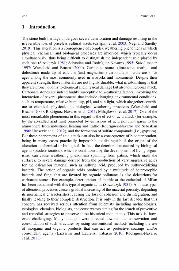
1 Introduction
The stone built heritage undergoes severe deterioration and damage resulting in theirreversible loss of priceless cultural assets (Crispim et al. 2003; Negi and Sarethy2019). This alteration is a consequence of complex weathering phenomena in whichphysical, chemical, and biological processes are involved, which typically occursimultaneously, thus being difficult to distinguish the independent role played byeach one (Strzelczyk 1981; Sebastián and Rodríguez-Navarro 1995; Saiz-Jimenez1997; Warscheid and Braams 2000). Carbonate stones (limestone, marble, anddolostone) made up of calcium (and magnesium) carbonate minerals are sinceages among the most commonly used in artworks and monuments. Despite theirapparent strength, these materials are not highly durable; what is astonishing is thatthey are prone not only to chemical and physical damage but also to microbial attack.Carbonate stones are indeed highly susceptible to weathering factors, involving theinteraction of several phenomena that include changing environmental conditions,such as temperature, relative humidity, pH, and sun light, which altogether contrib-ute to chemical, physical, and biological weathering processes (Warscheid andBraams 2000; Rodriguez-Navarro et al. 2011; Mihajlovski et al. 2017). One of themost remarkable phenomena in this regard is the effect of acid attack (for example,by the so-called acid rain) promoted by emissions of acid pollutant gases to theatmosphere from industries, heating and traffic (Rodriguez-Navarro and Sebastian1996; Urosevic et al. 2012), and the formation of sulfate compounds (i.e., gypsum).But these phenomena of acid attack can also be a consequence of biodeterioration,being in many cases practically impossible to distinguish if the origin of thealteration is chemical or biological. In fact, the deterioration caused by biologicalagents (biodeterioration), which is conditioned by the development of living organ-isms, can cause weathering phenomena spanning from patina, which mask thesurfaces, to severe damage derived from the production of very aggressive acidsfor the calcareous material such as sulfuric acid, produced by sulfur-oxidizingbacteria. The action of organic acids produced by a multitude of heterotrophicbacteria and fungi that are favored by organic pollutants is also deleterious forcarbonate stones. For example, deterioration of marble at the cathedral of Milanhas been associated with this type of organic acids (Strzelczyk 1981). All these typesof alteration processes cause a gradual increasing of the material porosity, degradingits mechanical characteristics, causing the loss of cohesion and disintegration, andfinally leading to their complete destruction. It is only in the last decades that thisconcern has received serious attention from scientists including archaeologists,geologists, chemists, biologists, and conservators aiming for the search of preventiveand remedial strategies to preserve these historical monuments. This task is, how-ever, challenging. Many attempts were directed towards the conservation andconsolidation of such structures by using conventional methods including the useof inorganic and organic products that can act as protective coatings and/orconsolidant agents (Lazzarini and Laurenzi Tabasso 2010; Rodriguez-Navarroet al. 2011).
282 F. Jroundi et al.
2 Conventional Methodologies for Stone Conservation
Organic products have been frequently used for their versatility, easy synthesis, andapplication methodology. This is the case of acrylic and epoxy resins or copolymersof a mix of diverse type of polymers. Among the non-polymeric products, there areorganic ones such as oxalates which aim at mimicking the formation of naturalprotective patinas or inorganic ones such as calcium hydroxide solutions (limewater), which is a well-known treatment used since Roman times for in situconsolidation of objects made of calcium carbonate. Other family of products arealkoxysilanes such as ethyl silicate that generate a silica gel, frequently used forfriable limestone and wall paintings consolidation, or alkyl alkoxysilanes whichimpart a dual protective and consolidation effect (Doehne and Price 2010). Morerecently, phosphates have emerged as protective coatings for carbonate stones(Sassoni et al. 2011). Some of these treatments have protective and sacrificialqualities, while others have more specific consolidating effects for limestone andlime plaster (Hansen et al. 2003). Although these traditional organic and inorganicconsolidants resulted to be efficient in some cases, providing the stone with somehydrophobicity and/or consolidation effect, most of them, however, prove to havevarious disadvantages. They were prone to form superficial films that block theporous system or they caused alteration of the color and brightness of the treatedstone, in addition to their limited resistance to alteration and their physico-chemicalincompatibility with the original substrate. In some cases, very limited consolidationwas achieved, as it is the case of the lime water technique which revealed ineffective,since precipitation of CaCO3 often led to the formation of a superficial, micrometer-thick, friable aggregate of submicron-size calcite crystals, as a powder withoutcohesion that had an insufficient protection and/or consolidation effect (Price et al.1988; Hansen et al. 2003; Doehne and Price 2010). These shortcomings have in partbeen overcome by the use of nanolimes, which are alcohol dispersions of Ca(OH)2nanoparticles (Rodriguez-Navarro and Ruiz-Agudo 2018). In general, however, theuse of conventional protection and consolidation treatments, sometimes indiscrim-inate, has led to the production of more drawbacks than those intended to becorrected (Rodriguez-Navarro and Ruiz-Agudo 2018). Additionally, large quantitiesof volatile organic solvents are commonly used during their application, whichcontribute to pollution (Rodriguez-Navarro et al. 2003; Doehne and Price 2010).Thus, most of the conventional treatments used so far in the conservation of decayedstone have revealed to be only partially successful for the preservation and consol-idation of deteriorated monuments (Price et al. 1988; Hansen et al. 2003; Wheeler2005; Rodriguez-Navarro et al. 2013). Moreover, some of them have been evenharmful because they accelerated the deterioration of the treated stone (Rodriguez-Navarro et al. 2003; Doehne and Price 2010). To address the shortcomings of theseconventional strategies, numerous efforts have been dedicated to the search anddevelopment of new more effective and compatible conservation treatments for theconsolidation and protection of the stone cultural heritage.
13 Protection and Consolidation of Stone Heritage by Bacterial Carbonatogenesis 283
3 New Methodologies for Stone Conservation
In the last decades, bacterial biomineralization has been proposed as a compatibleand an environmentally friendly technology for the protection of decayed ornamen-tal stones, particularly those having a carbonate composition (such as limestone andmarble) (Adolphe et al. 1990; Orial et al. 1993; Tiano et al. 1999; Castanier et al.2000). This strategy takes advantage on the capacity of bacteria to induce theformation of calcium carbonate, which cements carbonate rocks and is a biogeo-chemical phenomenon that commonly occurs in a wide range of natural environ-ments (Boquet et al. 1973). Although often bacteria have been related to harmfuleffects on stone structures, affecting the integrity of minerals or exacerbatingpowerful physico-chemical deterioration processes, there is increasing evidencethat carbonate-producing bacteria (i.e., carbonatogenic bacteria) could be used toreverse or ameliorate the effects of weathering processes affecting stone objects ofhistorical and artistic interest (Le Métayer-Levrel et al. 1999; Rodriguez-Navarroet al. 2003; Jimenez-Lopez et al. 2007, 2008; Dhami et al. 2014; Seifan andBerenjian 2019). The bacterial conservation method is based on the bacteriallyinduced precipitation of a compatible calcium carbonate mineral cement on thestone substrate, and unlike most conventional treatments, the precipitate seems tobe highly coherent (Le Métayer-Levrel et al. 1999; Rodriguez-Navarro et al. 2003).
Below, an attempt is made to provide an outline of the current methods based onbacterially induced carbonate mineralization for the conservation of stone works as apromising approach for the bioremediation of historical and important buildingmaterials.
4 Bacterial Biomineralization of Calcium Carbonates
Biomineralization refers to the process by which organisms synthesize minerals. Itcan be either biologically induced mineralization (BIM) or biologically controlledmineralization (BCM) (Lowenstan and Weiner 1989; Mann 1995; González-Muñozet al. 2010). BCM commonly results in the formation of complex and specializedstructures like skeletons, shells, and teeth, and occurs mainly in tissue-formingmulticellular eukaryotes. These biogenic minerals are synthesized either intracellu-larly or extracellularly in a way that is unique to each species, independently ofenvironmental conditions. The process is thought to be completely regulated underspecific metabolic and genetic control (Bazylinski and Moskowitz 1997; Bäuerlein2003; Baeuerlein 2004). Although rare, BCM also occurs in the case of microor-ganisms: well-known examples are magnetite mineralization by magnetotactic bac-teria, as well as calcite precipitation and silica deposition by algae and diatoms,respectively (Bäuerlein 2003; Baeuerlein 2004; Bazylinski and Schübbe 2007). Incontrast, in BIM, as it is the case of bacterial carbonatogenesis, the organisms do notcontrol the biomineralization process directly. Due to their metabolism, they alter the
284 F. Jroundi et al.
geochemistry around their cells favoring mineralization. As a result, the type ofmineral formed by BIM processes is strongly dependent on environmental condi-tions (Rivadeneyra et al. 1994; Ben Omar et al. 1997; Brennan et al. 2004). Differentmechanisms/processes responsible for bacterial carbonatogenesis have been pro-posed, which result in extracellular mineral growth and contribute to the formationof calcium carbonate sediments and rocks (Chafetz et al. 1991; Folk 1993;Vasconcelos et al. 1995; Paerl et al. 2001; Zavarzin 2002; Sharma 2013). In additionto the bacterial metabolic pathways that shift the environmental pH to alkalinity and,in the presence of calcium ions, foster calcium carbonate precipitation, bacterialsurfaces also play an important role in such a mineral formation by serving asheterogeneous nucleation sites (Fortin 1997; Rodriguez-Navarro et al. 2003). As aresult of successive stratification of the carbonate precipitates on the bacterialexternal surface, bacterial cells can be embedded or entombed within the growingcalcium carbonate crystals (Fig. 13.1).
BIM is the most commonly applied process in the field of stone protection andconsolidation, an emerging interdisciplinary biotechnology that consists in the use ofbacteria and their biological and metabolic pathways for the precipitation in the stoneof different biominerals, depending on bacteria and substrate types (Rodriguez-Navarro et al. 2012). Nearly 64 varieties of minerals of phosphates, carbonates,silicates, iron and manganese oxides, sulfide minerals, and amorphous silica can besynthetized by different microorganisms using specific metabolic routes (Knoll2003). Microbially induced carbonate mineral precipitation occurs via variousheterotrophic pathways such as the nitrogen cycle, the sulfur cycle and iron reduc-tion. In the nitrogen metabolism, calcium carbonate precipitation is thought to
Fig. 13.1 Representation of the process of bacterially induced calcium carbonate precipitation.Calcium ions in the solution are attracted to the bacterial cell wall due to the negative charge of thelatter. Oxidative deamination of amino acids results in the production of ammonia and CO2, whichis converted into HCO3
� or CO32� at higher pH in the microenvironment surrounding the bacteria
(A). In the presence of calcium ions, this can result in the heterogeneous precipitation of calciumcarbonate on the bacterial cell wall (B). The whole cell is finally entombed with time (C). Scanningelectron Microscopy image (D) showing the imprint of a bacterial cell involved in calciumcarbonate precipitation
13 Protection and Consolidation of Stone Heritage by Bacterial Carbonatogenesis 285
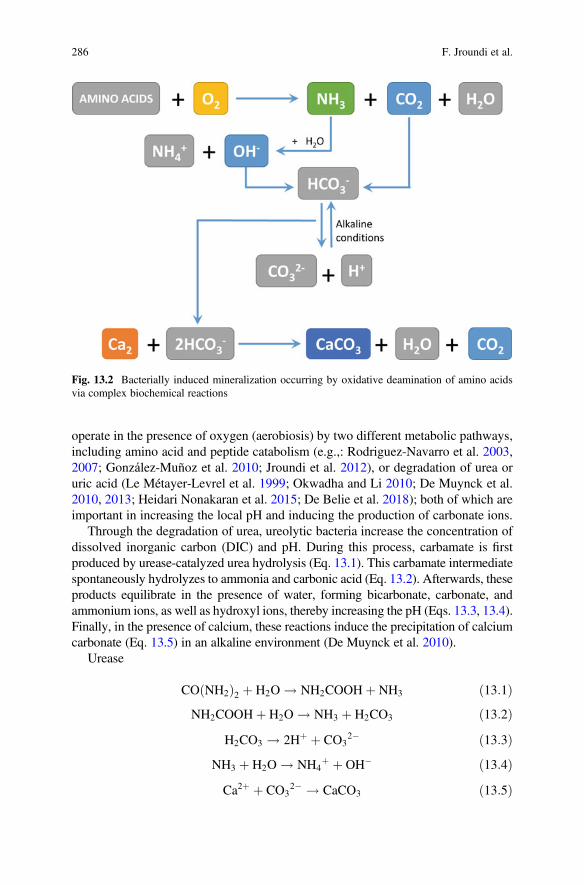
operate in the presence of oxygen (aerobiosis) by two different metabolic pathways,including amino acid and peptide catabolism (e.g.,: Rodriguez-Navarro et al. 2003,2007; González-Muñoz et al. 2010; Jroundi et al. 2012), or degradation of urea oruric acid (Le Métayer-Levrel et al. 1999; Okwadha and Li 2010; De Muynck et al.2010, 2013; Heidari Nonakaran et al. 2015; De Belie et al. 2018); both of which areimportant in increasing the local pH and inducing the production of carbonate ions.
Through the degradation of urea, ureolytic bacteria increase the concentration ofdissolved inorganic carbon (DIC) and pH. During this process, carbamate is firstproduced by urease-catalyzed urea hydrolysis (Eq. 13.1). This carbamate intermediatespontaneously hydrolyzes to ammonia and carbonic acid (Eq. 13.2). Afterwards, theseproducts equilibrate in the presence of water, forming bicarbonate, carbonate, andammonium ions, as well as hydroxyl ions, thereby increasing the pH (Eqs. 13.3, 13.4).Finally, in the presence of calcium, these reactions induce the precipitation of calciumcarbonate (Eq. 13.5) in an alkaline environment (De Muynck et al. 2010).
Urease
CO NH2ð Þ2 þ H2O ! NH2COOHþ NH3 ð13:1ÞNH2COOHþ H2O ! NH3 þ H2CO3 ð13:2Þ
H2CO3 ! 2Hþ þ CO32� ð13:3Þ
NH3 þ H2O ! NH4þ þ OH� ð13:4Þ
Ca2þ þ CO32� ! CaCO3 ð13:5Þ
Fig. 13.2 Bacterially induced mineralization occurring by oxidative deamination of amino acidsvia complex biochemical reactions
286 F. Jroundi et al.
Some Gram-negative aerobic microbial strains are able to use amino acids as theirsole source of energy to initiate the biomineralization of calcium carbonate throughamino acid and peptide catabolism in aerobiosis (Fig. 13.2). As shown in Fig. 13.2,carbon dioxide and ammonia are produced by oxidative deamination of amino acids.Ammonia (Eq. 13.4) increases the alkalinity in the microenvironment around thecell, whereas CO2 dissolves and transforms into either HCO3
� or CO32� at higher
pH. Here also, the presence of calcium and an alkaline environment favors theprecipitation of calcium carbonate by heterogeneous nucleation on the bacterialcell walls and cell debris (Rodriguez-Navarro et al. 2003), and on the extracellularpolymeric substances (EPS) forming a biofilm (Jroundi et al. 2017).
Incorporation of organics (bacterial cells or EPS) into the bacterial calciumcarbonate crystals (vaterite and calcite) has been shown (Rodriguez-Navarro et al.2007; Jroundi et al. 2017). EPS produced by bacteria have been demonstrated to beinvolved in biocalcification by entrapping calcium and carbonate ions and serving asa nucleation site (Braissant et al. 2003; Zamarreño et al. 2009). In Pseudomonasfluorescens, morphology and mineralogy of the carbonate crystals formed wasdemonstrated to be influenced by the composition and concentration of EPS(Braissant et al. 2003). Also, EPS appears to be involved in the aggregation ofsmaller crystals resulting in the growth of the final larger crystals (Buczynski andChafetz 1991). This process is commonly extracellular, occurring in the area sur-rounding the cells, and often leading to the join mineralization of EPS and bacterialcells (Fig. 13.1D) that become the nucleus of the biominerals (Ehrlich 1998;Rodriguez-Navarro et al. 2007).
The pathways of calcium carbonate formation described above are ubiquitous innature, accounting for the common occurrence of microbial carbonate precipitation(MCP) and confirms what Boquet et al. (1973) earlier reported, i.e., that most soilbacteria are able to induce the precipitation of carbonates under suitable conditions.This ability that bacteria have to promote carbonate mineral formation allowed thesearch and proposition, back in the 1990s, of a new kind of stone conservationtreatment based on bacterial biomineralization. In the last few decades, the potentialof bacterial biomineralization processes and their application to the conservation andconsolidation of cultural heritage have drawn a widespread interest and since a whilethis is a matter of considerable attention and investment (e.g.,: Adolphe et al. 1990;Orial et al. 1993; Castanier et al. 2000; Rodriguez-Navarro et al. 2003; DaSilva2004).
5 International Research Groups Involved in BacterialBiomineralization
The biomineralization strategy for stone protection have had several propositions.Among the firsts to consider the use of bacterially induced calcium carbonateprecipitation to stone conservation and protection were Adolphe et al. (1990)
13 Protection and Consolidation of Stone Heritage by Bacterial Carbonatogenesis 287
through the CALCITE Bioconcept, a patented technique developed at the UniversitéPierre et Marie Curie in Paris, which involved the application to the stone ofcarbonatogenic bacteria. These authors demonstrated the ability of carbonatogenicbacteria, typically those inhabiting soils (Boquet et al. 1973), to precipitate calcite ondamaged limestone surfaces, thus inducing their protection and consolidation. Fur-ther research was performed by Orial et al. (1993) from the University of Paris VIwho explored the application of living cultures of selected carbonatogenic bacterialstrains for calcite formation on stone and the replacement of the natural reinforcedlayer (CALCIN).
The technique was further optimized and industrialized by a collaborationbetween the French Company CALCITE, the University of Nantes, and the Histor-ical Monuments Research Laboratory (LRMH). They carried out in situ tests and theapplication to historic monuments observing the reinforcement and consolidation ofweathered stone. The first in situ application was carried out in 1993 in Thouars onthe tower of the Saint Médard Church (Le Métayer-Levrel et al. 1999). Theresearchers also described the “successful” application of the CALCITE methodon a sixteenth century limestone castle. Afterwards, a number of buildings in Francewere treated using this technique, including a castle at Châteaudun (Eure-et-Loir)and the Bordeaux cathedral (Castanier et al. 2000). There, it was reported thesuccessful precipitation of the biocalcin layer, which resulted in no side effects onthe esthetic appearance and highly reduced water absorption rate. Nevertheless,Orial (2000) concluded that every 10 years a new treatment would be needed torestore the protective effect of the biocalcin. In addition, upon investigating differentbacteria, the authors selected Bacillus cereus cultures for in situ applications, whichmay pose different health risks (Perito et al. 1999). Furthermore, since only a fewmicrometers thick consolidated layer was observed, the ineffectiveness for in-depthconsolidation was another main drawback of this technique (LeMétayer-Levrel et al.1999; Rodriguez-Navarro et al. 2011).
Tiano (1995) proposed another approach for the conservation of calcareous stonebased on the use of the “organic matrix” macromolecules extracted from shells ofMytilus californianus (a mollusc), which were applied on a sample of limestone toinduce the mineralization process. With this technique, a good efficiency of thecalcium carbonate precipitation process was showed leading to an increase in thestrength of the surface of treated stone and a decrease in the water absorption rate.However, in situ tests were not successful and the application of organic matterposed a risk as it could promote the development of harmful heterotrophic microor-ganisms. This was one of the two research lines attempted by the European projectBIOREINFORCE (BIOmediated calcite precipitation for monumental stones REIN-FORCEment, 2001–2004), which aimed at utilizing molecular biology and bacterialgenetic engineering to elucidate the genetic background of crystal formation as aninnovative alternative to the production of biomineralization-inducing macromole-cules for the improvement of the biomediated calcite precipitation. At first, theconsortium looked for molecules or structures from bacterial origin able to inducecalcium carbonate precipitation. They demonstrated the ability of dead cells and cellfragments (BCF) from active carbonatogenic strains (Bacillus cereus and Bacillus
288 F. Jroundi et al.
subtilis) to induce higher and/or faster production of calcium carbonate crystals witha more complex shape than those produced by dead cells from less active strains(Escherichia coli). BCF application to stone resulted in a slight decrease in waterabsorption, with better effects on highly porous stones. Here again, this methodrevealed to be more useful for delicate small calcareous stone pieces than for entirebuildings (Mastromei et al. 2008). The second research line consisted in identifyinggenes involved in the bacterial carbonatogenesis and the production at large-scale ofmacromolecules in bacterial cultures (Barabesi et al. 2007). The authors showed thepresence in Bacillus subtilis of lcfA and etfA as operon and gene, respectively, whichwere involved in calcite precipitation and suggested a possible link between thisprecipitation and the fatty-acid metabolism (Barabesi et al. 2007; Perito andMastromei 2011).
Tiano et al. (1999) evaluated the ability of Micrococcus sp. and B. subtilis in theconsolidation and protection of a highly porous limestone (Pietra de Lecce). Theyreported the physical obstruction of the stone pores by the presence of a consistentlayer of biological material (biofilm formation) and pointed out other potentialdrawbacks such as the reaction of stone and bacterial activity by-products or themodification of stone color.
Dick et al. (2006) proposed the application of several Bacillus species in atreatment that proceeded sequentially and in alternative cycles to biofilm formationand CaCO3 precipitation on Euville limestone (from quarries of the “Département dela Meuse” in France). The aim of their study was to determine the microbial keyfactors contributing to the performance of such treatment. Overall, several parame-ters were examined such as deposition of dense crystal layers, urea degradingcapacity, pH increase, calcite deposition on limestone, EPS-production, biofilmformation, the ζ-potential. According to the authors, optimal deposition of aCaCO3 layer in the treated limestone was based on a decrease in the water absorptionrate. They finally concluded that the dense calcium carbonate crystals formed byLysinibacillus sphaericus (¼Bacillus sphaericus) were most suitable for coherentcalcite production on degraded limestone and should be regarded as coating systems(Dick et al. 2006). Subsequently, the same bacterium was used for the bacterialdeposition of a layer of calcite on the surface of concrete and mortars, which resultedin an improvement of the mortar resistance and the concrete durability with a moreremarkable decrease in water uptake and a limited change of the chromatic aspect ascompared to a treatment based on mixed ureolytic cultures (De Muynck et al.2008a, b).
The BIOBRUSH (BIOremediation for Building Restoration of the Urban StoneHeritage) EU project was aimed at sequentially integrating salt removal and stoneconsolidation (May 2005). Within this project, Cappitelli et al. (2007) proposed aCarbogel as a delivery system for highly carbonatogenic bacteria such asDesulfovibrio vulgaris, which in addition to the elimination of black crusts alsoresulted in the conversion of gypsum to calcite. According to these authors, thisdelivery system could be employed for the control of potential harmful side effectsof bacteria (Cappitelli et al. 2007). However, the practical applicability of suchmethodology was challenged by the complexity of the procedure and the necessity
13 Protection and Consolidation of Stone Heritage by Bacterial Carbonatogenesis 289
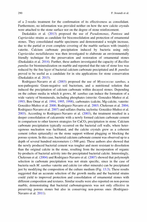
of a 2-weeks treatment for the confirmation of its effectiveness as consolidant.Furthermore, no information was provided neither on how the new calcite crystalswere attached to the stone surface nor on the degree of stone reinforcement.
Daskalakis et al. (2013) proposed the use of Pseudomonas, Pantoea andCupriavidus strains as candidate for bioconsolidation and protection of ornamentalstones. They consolidated marble specimens and demonstrated a weight increase,due to the partial or even complete covering of the marble surfaces with (mainly)vaterite. Calcium carbonate precipitation induced by bacteria using onlyCupriavidus metallidurans was then investigated to elaborate an environmentallyfriendly technique for the preservation and restoration of ornamental stones(Daskalakis et al. 2014). Further, these authors investigated the capacity of Bacilluspumilus for biomineralization on marble and reported that the rate of stone loss wasreduced by the fine layer of bacterial calcium carbonate precipitated and B. pumilusproved to be useful as a candidate for in situ applications for stone conservation(Daskalakis et al. 2015).
Rodriguez-Navarro et al. (2003) proposed the use of Myxococcus xanthus, anon-pathogenic Gram-negative soil bacterium, and a nutritional solution thatinduced the precipitation of calcium carbonate within decayed stones. Dependingon the culture media in which it grows, M. xanthus can induce the formation of awide variety of biominerals, including phosphates (struvite: Gonzalez-Muñoz et al.1993; Ben Omar et al. 1994, 1995, 1998), carbonates (calcite, Mg-calcite, vaterite:González-Muñoz et al. 2000; Rodriguez-Navarro et al. 2003; Chekroun et al. 2004;Rodriguez-Navarro et al. 2007) and sulfates (barita, taylorita: González-Muñoz et al.2003). According to Rodriguez-Navarro et al. (2003), the treatment resulted in adeeper consolidation of calcarenite with a newly formed calcium carbonate cementin comparison to other known strategies for CaCO3 precipitation in stone. Calciumcarbonate precipitation typically occurred on the bacterial cell walls, where heter-ogenous nucleation was facilitated, and the calcite crystals grew as a coherentcement (often epitaxially) on the stone support without plugging or blocking theporous system. In this case, bacterial calcium carbonate cement was observed up to adepth of several hundred micrometers (>500 μm). These authors also showed thatthe newly produced bacterial cement was tougher and more resistant to dissolutionthan the original calcite in the stone, resulting from the incorporation of organicby-products of bacterial activity into the precipitated bacterial calcite. Interestingly,Chekroun et al. (2004) and Rodriguez-Navarro et al. (2007) showed that polymorphselection in carbonate precipitation was not strain specific, since in the case ofbacteria such M. xanthus vaterite and calcite (or other minerals) can be precipitatedjust by modifying the composition of the culture medium (Fig. 13.3). These resultssuggested that an accurate selection of the growth media and the bacterial strainscould yield to improved protection and consolidation of ornamental stones withdifferent composition and textures. Similar results were also reported on non-porousmarble, demonstrating that bacterial carbonatogenesis was not only effective inpreserving porous stones but also in conserving non-porous ones (Rodriguez-Navarro et al. 2011).
290 F. Jroundi et al.
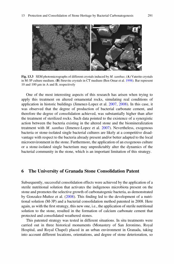
One of the most interesting aspects of this research has arisen when trying toapply this treatment on altered ornamental rocks, simulating real conditions ofapplication in historic buildings (Jimenez-Lopez et al. 2007, 2008). In this case, itwas observed that the degree of production of bacterial carbonate cement, andtherefore the degree of consolidation achieved, was substantially higher than afterthe treatment of sterilized rocks. Such data pointed to the existence of a synergisticaction between the bacteria existing in the altered stone and the biomineralizationtreatment with M. xanthus (Jimenez-Lopez et al. 2007). Nevertheless, exogenousbacteria or stone-isolated single bacterial cultures are likely at a competitive disad-vantage with respect to the bacteria already present and/or better adapted to the localmicroenvironment in the stone. Furthermore, the application of an exogenous cultureor a stone-isolated single bacterium may unpredictably alter the dynamics of thebacterial community in the stone, which is an important limitation of this strategy.
6 The University of Granada Stone Consolidation Patent
Subsequently, successful consolidation effects were achieved by the application of asterile nutritional solution that activates the indigenous microbiota present on thestone and promotes the selective growth of carbonatogenic bacteria, as demonstratedby Gonzalez-Muñoz et al. (2008). This finding led to the development of a nutri-tional solution (M-3P) and a bacterial consolidation method patented in 2008. Hereagain, as with the first strategy, this new one, i.e., the application of sterile nutritionalsolution to the stone, resulted in the formation of calcium carbonate cement thatprotected and consolidated weathered stones.
This patented strategy was tested in different situations. In situ treatments werecarried out in three historical monuments (Monastery of San Jeronimo, RoyalHospital, and Royal Chapel) placed in an urban environment in Granada, takinginto account different locations, orientations, and degree of stone deterioration, so
Fig. 13.3 SEM photomicrographs of different crystals induced byM. xanthus. (A) Vaterite crystalsin M-3P culture medium. (B) Struvite crystals in CT medium (Ben Omar et al. 1998). Bar represent10 and 100 μm in A and B, respectively
13 Protection and Consolidation of Stone Heritage by Bacterial Carbonatogenesis 291
that more extensive information could be provided on the suitability of the patentedmethodology (Jroundi et al. 2010; Ettenauer et al. 2011; Rodriguez-Navarro et al.2015). The results showed that in the first two historical buildings, a very similardegree of consolidation was achieved regardless of the type of treatment performed(with or without the additional foreign bacteria M. xanthus). Besides, the strength-ening associated with the production of the newly formed calcium carbonate cementat both buildings allowed reinforcing the porous system and binding the loose grainsof the stone with such a bacterial cement, which lasted (at least) for up to 4 years afterapplication (Jroundi et al. 2010; Rodriguez-Navarro et al. 2015). Regarding theRoyal Chapel, the in situ treatment was simultaneously performed during theapplication of ethyl silicate as a conventional treatment, to compare the degree ofconsolidation obtained by both types of treatments, in combination with a biocideapplication as a cleansing pre-treatment performed four-month prior to the consol-idation treatments. The stone strengthening in this case was due to the new formationof bacterial calcite crystals without plugging the pores, thereby proving that thebacterial consolidation using a sterile nutrititional solution was as efficient as thetraditional ethyl silicate consolidant (Jroundi et al. 2013). In addition, the evolutionof the microbiota in all three historical buildings was assessed by culture-dependentand independent techniques and the results revealed that the majority ofcarbonatogenic bacteria were activated by the application of the patented M-3Pnutritional solution and no negative effects associated with the possible activationof deleterious microbiota were observed (Ettenauer et al. 2011; Jroundi et al. 2010,2015; Rodriguez-Navarro et al. 2015). Overall, the results of these studies showedthat the in situ application of the bacterial consolidation method demonstrated nodetrimental side effects on the stones with a highly proved medium- and long-termeffectiveness.
7 Self-Inoculation with Indigenous CarbonatogenicBacterial Community
When applied to heavily salt weathered stones, both previously described strategies(with or without additional bacterium) demonstrated to have a limited efficacy. Theireffectiveness depended also on the stone characteristics (e.g., porosity, mineralogy,level of deterioration), the type of exogenous microorganism and bacterial loadapplied, and its interaction with indigenous microbiota. Salt weathering is knownto be highly deleterious (Rodriguez-Navarro and Doehne 1999; Schiro et al. 2012;Flatt et al. 2014) and usually results in the partial filling of stone pores with solublesalts (Webster and May 2006), which may further inhibit the proliferation ofindigenous or exogenous carbonatogenic bacteria. Therefore, a novel environmen-tally friendly bacterial self-inoculation approach was recently proposed to overcomethe limitations associated to the existing two bacterial biomineralization strategies(i.e., (i) inoculation with a single carbonatogenic bacterial strain, and (ii) activation
292 F. Jroundi et al.
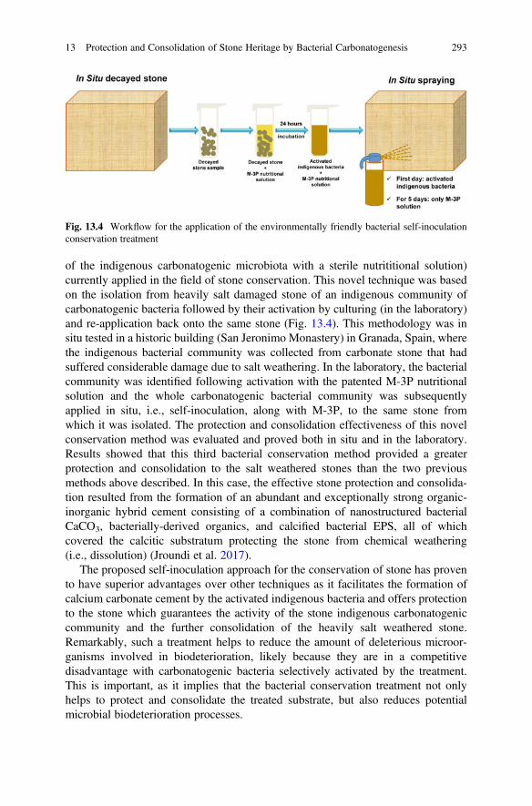
of the indigenous carbonatogenic microbiota with a sterile nutrititional solution)currently applied in the field of stone conservation. This novel technique was basedon the isolation from heavily salt damaged stone of an indigenous community ofcarbonatogenic bacteria followed by their activation by culturing (in the laboratory)and re-application back onto the same stone (Fig. 13.4). This methodology was insitu tested in a historic building (San Jeronimo Monastery) in Granada, Spain, wherethe indigenous bacterial community was collected from carbonate stone that hadsuffered considerable damage due to salt weathering. In the laboratory, the bacterialcommunity was identified following activation with the patented M-3P nutritionalsolution and the whole carbonatogenic bacterial community was subsequentlyapplied in situ, i.e., self-inoculation, along with M-3P, to the same stone fromwhich it was isolated. The protection and consolidation effectiveness of this novelconservation method was evaluated and proved both in situ and in the laboratory.Results showed that this third bacterial conservation method provided a greaterprotection and consolidation to the salt weathered stones than the two previousmethods above described. In this case, the effective stone protection and consolida-tion resulted from the formation of an abundant and exceptionally strong organic-inorganic hybrid cement consisting of a combination of nanostructured bacterialCaCO3, bacterially-derived organics, and calcified bacterial EPS, all of whichcovered the calcitic substratum protecting the stone from chemical weathering(i.e., dissolution) (Jroundi et al. 2017).
The proposed self-inoculation approach for the conservation of stone has provento have superior advantages over other techniques as it facilitates the formation ofcalcium carbonate cement by the activated indigenous bacteria and offers protectionto the stone which guarantees the activity of the stone indigenous carbonatogeniccommunity and the further consolidation of the heavily salt weathered stone.Remarkably, such a treatment helps to reduce the amount of deleterious microor-ganisms involved in biodeterioration, likely because they are in a competitivedisadvantage with carbonatogenic bacteria selectively activated by the treatment.This is important, as it implies that the bacterial conservation treatment not onlyhelps to protect and consolidate the treated substrate, but also reduces potentialmicrobial biodeterioration processes.
Fig. 13.4 Workflow for the application of the environmentally friendly bacterial self-inoculationconservation treatment
13 Protection and Consolidation of Stone Heritage by Bacterial Carbonatogenesis 293
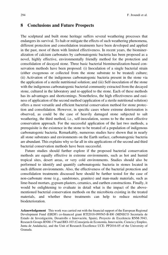
8 Conclusions and Future Prospects
The sculptural and built stone heritage suffers several weathering processes thatendangers its survival. To halt or mitigate the effects of such weathering phenomena,different protection and consolidation treatments have been developed and appliedin the past, most of them with limited effectiveness. In recent years, the biominer-alization of calcium carbonates by carbonatogenic bacteria has been proposed as anovel, highly effective, environmentally friendly method for the protection andconsolidation of decayed stone. Three basic bacterial biomineralization-based con-servation methods have been proposed: (i) Inoculation of a single bacterial strain(either exogenous or collected from the stone substrate to be treated) culture;(ii) Activation of the indigenous carbonatogenic bacteria present in the stone viathe application of a sterile nutritional solution; and (iii) Self-inoculation of the stonewith the indigenous carbonatogenic bacterial community extracted from the decayedstone, cultured in the laboratory and re-applied to the stone. Each of these methodshas its advantages and shortcomings. Nonetheless, the high effectiveness and easi-ness of application of the second method (application of a sterile nutritional solution)offers a most versatile and efficient bacterial conservation method for stone protec-tion and consolidation. However, in specific cases where extreme deterioration isobserved, as could be the case of heavily damaged stone subjected to saltweathering, the third method, i.e., self-inoculation, seems to be the most effectiveconservation approach. For the successful application of the last two methods, aprerequisite is the existence in the stone to be treated of a population of indigenouscarbonatogenic bacteria. Remarkably, numerous studies have shown that in nearlyall stone substrates and environments on the Earth surface, carbonatogenic bacteriaare abundant. This explains why so far all in situ applications of the second and thirdbacterial conservation methods have been successful.
Future studies should further explore if the proposed bacterial conservationmethods are equally effective in extreme environments, such as hot and humidtropical sites, desert areas, or very cold environments. Studies should also beperformed to identify and quantify carbonatogenic bacteria in stones located insuch different environments. Also, the effectiveness of the bacterial protection andconsolidation treatments discussed here should be further tested for the case ofnon-carbonate stone (e.g., sandstones, granites) and man-made materials, such aslime-based mortars, gypsum plasters, ceramics, and earthen constructions. Finally, itwould be enlightening to evaluate in detail what is the impact of the above-mentioned bacterial conservation methods on the microbiota existing in the treatedmaterials, and whether these treatments can help to reduce microbialbiodeterioration.
Acknowledgement This work was carried out with the financial support of the European RegionalDevelopment Fund (ERDF) co-financed grant RTI2018-099565-B-I00 (MINECO Secretaría deEstado de Investigación, Desarrollo e Innovación, Spain), Proyecto de Excelencia RNM-3943,Research Groups RNM-179 and BIO-103 (Consejería de Economía, Innovación, Ciencia y Empleo,Junta de Andalucía), and the Unit of Research Excellence UCE- PP2016-05 of the University ofGranada.
294 F. Jroundi et al.
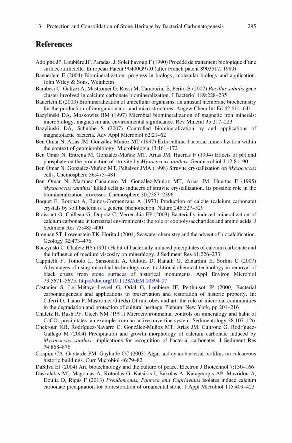
References
Adolphe JP, Loubière JF, Paradas, J, Soleilhavoup F (1990) Procédé de traitement biologique d’unesurface artificielle. European Patent 90400G97.0 (after French patent 8903517, 1989)
Baeuerlein E (2004) Biomineralization: progress in biology, molecular biology and application.John Wiley & Sons, Weinheim
Barabesi C, Galizzi A, Mastromei G, Rossi M, Tamburini E, Perito B (2007) Bacillus subtilis genecluster involved in calcium carbonate biomineralization. J Bacteriol 189:228–235
Bäuerlein E (2003) Biomineralization of unicellular organisms: an unusual membrane biochemistryfor the production of inorganic nano- and microstructures. Angew Chem Int Ed 42:614–641
Bazylinski DA, Moskowitz BM (1997) Microbial biomineralization of magnetic iron minerals:microbiology, magnetism and environmental significance. Rev Mineral 35:217–223
Bazylinski DA, Schübbe S (2007) Controlled biomineralization by and applications ofmagnetotactic bacteria. Adv Appl Microbiol 62:21–62
Ben Omar N, Arias JM, González-Muñoz MT (1997) Extracellular bacterial mineralization withinthe context of geomicrobiology. Microbiologia 13:161–172
Ben Omar N, Entrena M, González-Muñoz MT, Arias JM, Huertas F (1994) Effects of pH andphosphate on the production of struvite by Myxococcus xanthus. Geomicrobiol J 12:81–90
Ben Omar N, Gonzalez-Muñoz MT, Peñalver JMA (1998) Struvite crystallization on Myxococcuscells. Chemosphere 36:475–481
Ben Omar N, Martínez-Cañamero M, González-Muñoz MT, Arias JM, Huertas F (1995)Myxococcus xanthus’ killed cells as inducers of struvite crystallization. Its possible role in thebiomineralization processes. Chemosphere 30:2387–2396
Boquet E, Boronat A, Ramos-Cormenzana A (1973) Production of calcite (calcium carbonate)crystals by soil bacteria is a general phenomenon. Nature 246:527–529
Braissant O, Cailleau G, Dupraz C, Verrecchia EP (2003) Bacterially induced mineralization ofcalcium carbonate in terrestrial environments: the role of exopolysaccharides and amino acids. JSediment Res 73:485–490
Brennan ST, Lowenstein TK, Horita J (2004) Seawater chemistry and the advent of biocalcification.Geology 32:473–476
Buczynski C, Chafetz HS (1991) Habit of bacterially induced precipitates of calcium carbonate andthe influence of medium viscosity on mineralogy. J Sediment Res 61:226–233
Cappitelli F, Toniolo L, Sansonetti A, Gulotta D, Ranalli G, Zanardini E, Sorlini C (2007)Advantages of using microbial technology over traditional chemical technology in removal ofblack crusts from stone surfaces of historical monuments. Appl Environ Microbiol73:5671–5675. https://doi.org/10.1128/AEM.00394-07
Castanier S, Le Métayer-Levrel G, Orial G, Loubiere JF, Perthuisot JP (2000) Bacterialcarbonatogenesis and applications to preservation and restoration of historic property. In:Ciferri O, Tiano P, Mastromei G (eds) Of microbes and art: the role of microbial communitiesin the degradation and protection of cultural heritage. Plenum, New York, pp 201–216
Chafetz H, Rush PF, Utech NM (1991) Microenvironmental controls on mineralogy and habit ofCaCO3 precipitates: an example from an active travertine system. Sedimentology 38:107–126
Chekroun KB, Rodríguez-Navarro C, González-Muñoz MT, Arias JM, Cultrone G, Rodríguez-Gallego M (2004) Precipitation and growth morphology of calcium carbonate induced byMyxococcus xanthus: implications for recognition of bacterial carbonates. J Sediment Res74:868–876
Crispim CA, Gaylarde PM, Gaylarde CC (2003) Algal and cyanobacterial biofilms on calcareoushistoric buildings. Curr Microbiol 46:79–82
DaSilva EJ (2004) Art, biotechnology and the culture of peace. Electron J Biotechnol 7:130–166Daskalakis MI, Magoulas A, Kotoulas G, Katsikis I, Bakolas A, Karageorgis AP, Mavridou A,
Doulia D, Rigas F (2013) Pseudomonas, Pantoea and Cupriavidus isolates induce calciumcarbonate precipitation for biorestoration of ornamental stone. J Appl Microbiol 115:409–423
13 Protection and Consolidation of Stone Heritage by Bacterial Carbonatogenesis 295
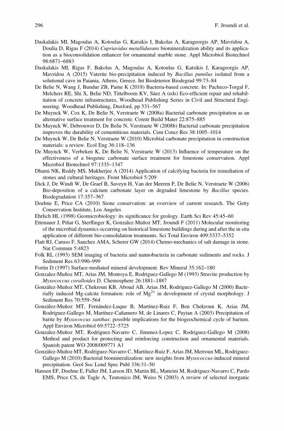
Daskalakis MI, Magoulas A, Kotoulas G, Katsikis I, Bakolas A, Karageorgis AP, Mavridou A,Doulia D, Rigas F (2014) Cupriavidus metallidurans biomineralization ability and its applica-tion as a bioconsolidation enhancer for ornamental marble stone. Appl Microbiol Biotechnol98:6871–6883
Daskalakis MI, Rigas F, Bakolas A, Magoulas A, Kotoulas G, Katsikis I, Karageorgis AP,Mavridou A (2015) Vaterite bio-precipitation induced by Bacillus pumilus isolated from asolutional cave in Paiania, Athens, Greece. Int Biodeterior Biodegrad 99:73–84
De Belie N, Wang J, Bundur ZB, Paine K (2018) Bacteria-based concrete. In: Pacheco-Torgal F,Melchers RE, Shi X, Belie ND, Tittelboom KV, Sáez A (eds) Eco-efficient repair and rehabil-itation of concrete infrastructures, Woodhead Publishing Series in Civil and Structural Engi-neering. Woodhead Publishing, Duxford, pp 531–567
De Muynck W, Cox K, De Belie N, Verstraete W (2008a) Bacterial carbonate precipitation as analternative surface treatment for concrete. Constr Build Mater 22:875–885
De Muynck W, Debrouwer D, De Belie N, Verstraete W (2008b) Bacterial carbonate precipitationimproves the durability of cementitious materials. Cem Concr Res 38:1005–1014
De Muynck W, De Belie N, Verstraete W (2010) Microbial carbonate precipitation in constructionmaterials: a review. Ecol Eng 36:118–136
De Muynck W, Verbeken K, De Belie N, Verstraete W (2013) Influence of temperature on theeffectiveness of a biogenic carbonate surface treatment for limestone conservation. ApplMicrobiol Biotechnol 97:1335–1347
Dhami NK, Reddy MS, Mukherjee A (2014) Application of calcifying bacteria for remediation ofstones and cultural heritages. Front Microbiol 5:209
Dick J, De Windt W, De Graef B, Saveyn H, Van der Meeren P, De Belie N, Verstraete W (2006)Bio-deposition of a calcium carbonate layer on degraded limestone by Bacillus species.Biodegradation 17:357–367
Doehne E, Price CA (2010) Stone conservation: an overview of current research. The GettyConservation Institute, Los Angeles
Ehrlich HL (1998) Geomicrobiology: its significance for geology. Earth Sci Rev 45:45–60Ettenauer J, Piñar G, Sterflinger K, Gonzalez-Muñoz MT, Jroundi F (2011) Molecular monitoring
of the microbial dynamics occurring on historical limestone buildings during and after the in situapplication of different bio-consolidation treatments. Sci Total Environ 409:5337–5352
Flatt RJ, Caruso F, Sanchez AMA, Scherer GW (2014) Chemo-mechanics of salt damage in stone.Nat Commun 5:4823
Folk RL (1993) SEM imaging of bacteria and nannobacteria in carbonate sediments and rocks. JSediment Res 63:990–999
Fortin D (1997) Surface-mediated mineral development. Rev Mineral 35:162–180Gonzalez-Muñoz MT, Arias JM, Montoya E, Rodriguez-Gallego M (1993) Struvite production by
Myxococcus coralloides D. Chemosphere 26:1881–1887González-Muñoz MT, Chekroun KB, Aboud AB, Arias JM, Rodriguez-Gallego M (2000) Bacte-
rially induced Mg-calcite formation: role of Mg2+ in development of crystal morphology. JSediment Res 70:559–564
González-Muñoz MT, Fernández-Luque B, Martínez-Ruiz F, Ben Chekroun K, Arias JM,Rodríguez-Gallego M, Martínez-Cañamero M, de Linares C, Paytan A (2003) Precipitation ofbarite by Myxococcus xanthus: possible implications for the biogeochemical cycle of barium.Appl Environ Microbiol 69:5722–5725
Gonzalez-Muñoz MT, Rodriguez-Navarro C, Jimenez-Lopez C, Rodriguez-Gallego M (2008)Method and product for protecting and reinforcing construction and ornamental materials.Spanish patent WO 2008/009771 A1
González-Muñoz MT, Rodriguez-Navarro C, Martínez-Ruiz F, Arias JM, Merroun ML, Rodriguez-Gallego M (2010) Bacterial biomineralization: new insights from Myxococcus-induced mineralprecipitation. Geol Soc Lond Spec Publ 336:31–50
Hansen EF, Doehne E, Fidler JM, Larson JD, Martin BL, Matteini M, Rodriguez-Navarro C, PardoEMS, Price CS, de Tagle A, Teutonico JM, Weiss N (2003) A review of selected inorganic
296 F. Jroundi et al.
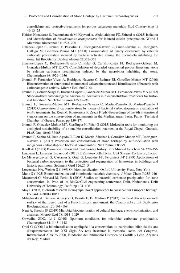
consolidants and protective treatments for porous calcareous materials. Stud Conserv (sup 1)49:13–25
Heidari Nonakaran S, Pazhouhandeh M, Keyvani A, Abdollahipour FZ, Shirzad A (2015) Isolationand identification of Pseudomonas azotoformans for induced calcite precipitation. World JMicrobiol Biotechnol 31:1993–2001
Jimenez-Lopez C, Jroundi F, Pascolini C, Rodriguez-Navarro C, Piñar-Larrubia G, Rodriguez-Gallego M, González-Muñoz MT (2008) Consolidation of quarry calcarenite by calciumcarbonate precipitation induced by bacteria activated among the microbiota inhabiting thestone. Int Biodeterior Biodegradation 62:352–363
Jimenez-Lopez C, Rodriguez-Navarro C, Piñar G, Carrillo-Rosúa FJ, Rodriguez-Gallego M,Gonzalez-Muñoz MT (2007) Consolidation of degraded ornamental porous limestone stoneby calcium carbonate precipitation induced by the microbiota inhabiting the stone.Chemosphere 68:1929–1936
Jroundi F, Fernández-Vivas A, Rodriguez-Navarro C, Bedmar EJ, González-Muñoz MT (2010)Bioconservation of deteriorated monumental calcarenite stone and identification of bacteria withcarbonatogenic activity. Microb Ecol 60:39–54
Jroundi F, Gómez-Suaga P, Jimenez-Lopez C, González-Muñoz MT, Fernandez-Vivas MA (2012)Stone-isolated carbonatogenic bacteria as inoculants in bioconsolidation treatments for histor-ical limestone. Sci Total Environ 425:89–98
Jroundi F, Gonzalez-Muñoz MT, Rodriguez-Navarro C, Martin-Peinado B, Martin-Peinado J(2013) Conservation of carbonate stone by means of bacterial carbonatogenesis: evaluation ofin situ treatments. In: Koui M, Koutsoukos P, Zezza F (eds) Proceedings of the 8th internationalsymposium on the conservation of monuments in the Mediterranean basin. Patras. TechnicalChamber of Greece, Patras, pp 159–171
Jroundi F, Gonzalez-Muñoz MT, Sterflinger K, Piñar G (2015) Molecular tools for monitoring theecological sustainability of a stone bio-consolidation treatment at the Royal Chapel, Granada.PLoS One 10:e0132465
Jroundi F, Schiro M, Ruiz-Agudo E, Elert K, Martín-Sánchez I, González-Muñoz MT, Rodriguez-Navarro C (2017) Protection and consolidation of stone heritage by self-inoculation withindigenous carbonatogenic bacterial communities. Nat Commun 8:279
Knoll AH (2003) Biomineralization and evolutionary history. Rev Mineral Geochem 54:329–356Lazzarini L, Laurenzi Tabasso M (2010) Il Restauro della Pietra. Utet Scienze Techniche, TorinoLe Métayer-Levrel G, Castanier S, Orial G, Loubière J-F, Perthuisot J-P (1999) Applications of
bacterial carbonatogenesis to the protection and regeneration of limestones in buildings andhistoric patrimony. Sediment Geol 126:25–34
Lowenstan HA, Weiner S (1989) On biomineralization. Oxford University Press, New YorkMann S (1995) Biomineralization and biomimetic materials chemistry. J Mater Chem 5:935–946Mastromei G, Marvasi M, Perito B (2008) Studies on bacterial carbonate precipitation for stone
conservation. In: Proc. of 1st BioGeoCivil engineering conference, Delft, Netherlands. DelftUniversity of Technology, Delft, pp 104–106
May E (2005) Biobrush research monograph: novel approaches to conserve our European heritage.EVK4-CT-2001-00055
Mihajlovski A, Gabarre A, Seyer D, Bousta F, Di Martino P (2017) Bacterial diversity on rocksurface of the ruined part of a French historic monument: the Chaalis abbey. Int BiodeteriorBiodegradation 120:161–169
Negi A, Sarethy IP (2019) Microbial biodeterioration of cultural heritage: events, colonization, andanalyses. Microb Ecol 78:1014–1029
Okwadha GDO, Li J (2010) Optimum conditions for microbial carbonate precipitation.Chemosphere 81:1143–1148
Orial G (2000) La biomineralisation appliquée à la conservation du patrimoine: bilan de dix ansd’experimentation. In: XXI Siglo SA (ed) Restaurar la memoria, Actas del Congreso,Internacional AR&PA 2000, Fundación del Patrimonio Histórico de Castilla y León. Argandadel Rey, Madrid
13 Protection and Consolidation of Stone Heritage by Bacterial Carbonatogenesis 297
Orial G, Castanier S, Metayer GL, Loubière JF (1993) The biomineralization: a new process toprotect calcareous stone; applied to historic monuments. In: Ktoishi H, Arai T, Yamano K (eds)Proceedings of the 2nd international conference on biodeterioration of cultural property.International Communications Specialists, Tokyo, Japan, pp 98–116
Paerl HW, Steppe TF, Reid RP (2001) Bacterially mediated precipitation in marine stromatolites.Environ Microbiol 3:123–130
Perito B, Biagiotti L, Daly S, Galizzi A, Tiano R, Mastromei G (1999) Bacterial genes involved incalcite crystal precipitation. In: Ciferri O, Tiano P, Mastromei G (eds) Of microbes and art: therole of microbial communities in the degradation and protection of cultural heritage, interna-tional conference on microbiology and conservation. Kluwer, New York, pp 219–230
Perito B, Mastromei G (2011) Molecular basis of bacterial calcium carbonate precipitation. ProgMol Subcell Biol 52:113–139
Price C, Ross K, White G (1988) A further appraisal of the “lime technique” for limestoneconsolidation, using a radioactive tracer. Stud Conserv 33:178–186
Rivadeneyra MA, Delgado R, del Moral A, Ferrer MR, Ramos-Cormenzana A (1994) Precipatationof calcium carbonate by Vibrio spp. from an inland saltern. FEMS Microbiol Ecol 13:197–204
Rodriguez-Navarro C, Doehne E (1999) Salt weathering: influence of evaporation rate, supersatu-ration and crystallization pattern. Earth Surf Process Landf 24:191–209
Rodriguez-Navarro C, González-Muñoz MT, Jimenez-Lopez C, Rodriguez-Gallego M (2011)Bioprotection. In: Reitner J, Thiel V (eds) Encyclopedia of geobiology. Springer, Netherlands,Dordrecht, pp 185–189
Rodriguez-Navarro C, Jimenez-Lopez C, Rodriguez-Navarro A, Gonzalez-Muñoz MT, Rodriguez-Gallego M (2007) Bacterially mediated mineralization of vaterite. Geochim Cosmochim Acta71:1197–1213
Rodriguez-Navarro C, Jroundi F, Gonzalez-Muñoz MT (2015) Stone consolidation by bacterialcarbonatogenesis: evaluation of in situ applications. Restor Build Monum 21:9–20
Rodriguez-Navarro C, Jroundi F, Schiro M, Ruiz-Agudo E, González-Muñoz MT (2012) Influenceof substrate mineralogy on bacterial mineralization of calcium carbonate: implications for stoneconservation. Appl Environ Microbiol 78:4017–4029
Rodriguez-Navarro C, Rodriguez-Gallego M, Chekroun KB, Gonzalez-Muñoz MT (2003) Con-servation of ornamental stone by Myxococcus xanthus-induced carbonate biomineralization.Appl Environ Microbiol 69:2182–2193
Rodriguez-Navarro C, Ruiz-Agudo E (2018) Nanolimes: from synthesis to application. Pure ApplChem 90:523–550
Rodriguez-Navarro C, Sebastian E (1996) Role of particulate matter from vehicle exhaust on porousbuilding stones (limestone) sulfation. Sci Total Environ 187:79–91
Rodriguez-Navarro C, Suzuki A, Ruiz-Agudo E (2013) Alcohol dispersions of calcium hydroxidenanoparticles for stone conservation. Langmuir 29:11457–11470
Saiz-Jimenez C (1997) Biodeterioration vs biodegradation: the role of microorganisms in theremoval of pollutants deposited on historic buildings. Int Biodeterior Biodegradation40:225–232
Sassoni E, Naidu S, Scherer GW (2011) The use of hydroxyapatite as a new inorganic consolidantfor damaged carbonate stones. J Cult Herit 12:346–355
Schiro M, Ruiz-Agudo E, Rodriguez-Navarro C (2012) Damage mechanisms of porous materialsdue to in-pore salt crystallization. Phys Rev Lett 109:265503
Sebastián E, Rodríguez-Navarro C (1995) Alteración y conservación de materiales pétreosornamentales: antecedentes y estado actual de conocimientos. Ingeniería Civil 96:167–178
Seifan M, Berenjian A (2019) Microbially induced calcium carbonate precipitation: a widespreadphenomenon in the biological world. Appl Microbiol Biotechnol 103:4693–4708
Sharma O (2013) Characterization of novel carbonic anhydrase from halophilic bacterial isolatesand it’s role in microbially induced calcium carbonate precipitation (Ms Thesis). ThaparUniversity, India
298 F. Jroundi et al.
Strzelczyk AB (1981) Stone. In: Rose AH (ed) Microbial biodeterioration. Academic Press,London, pp 61–80
Tiano P (1995) Stone reinforcement by calcite crystal precipitation induced by organic matrixmacromolecules. Stud Conserv 40:171–176
Tiano P, Biagiotti L, Mastromei G (1999) Bacterial bio-mediated calcite precipitation for monu-mental stones conservation: methods of evaluation. J Microbiol Methods 36:139–145
Urosevic M, Yebra-Rodríguez A, Sebastián-Pardo E, Cardell C (2012) Black soiling of anarchitectural limestone during two-year term exposure to urban air in the city of Granada(S Spain). Sci Total Environ 414:564–575
Vasconcelos C, McKenzie JA, Bernasconi S, Grujic D, Tiens AJ (1995) Microbial mediation as apossible mechanism for natural dolomite formation at low temperatures. Nature 377:220–222
Warscheid T, Braams J (2000) Biodeterioration of stone: a review. Int Biodeterior Biodegrad46:343–368
Webster A, May E (2006) Bioremediation of weathered-building stone surfaces. Trends Biotechnol24:255–260
Wheeler G (2005) Alkoxysilanes and the consolidation of stone. The Getty Conservation Institute,Los Angeles
Zamarreño DV, Inkpen R, May E (2009) Carbonate crystals precipitated by freshwater bacteria andtheir use as a limestone consolidant. Appl Environ Microbiol 75:5981–5990
Zavarzin GA (2002) Microbial geochemical calcium cycle. Microbiology 71:1–17
Open Access This chapter is licensed under the terms of the Creative Commons Attribution 4.0International License (http://creativecommons.org/licenses/by/4.0/), which permits use, sharing,adaptation, distribution and reproduction in any medium or format, as long as you give appropriatecredit to the original author(s) and the source, provide a link to the Creative Commons license andindicate if changes were made.
The images or other third party material in this chapter are included in the chapter's CreativeCommons license, unless indicated otherwise in a credit line to the material. If material is notincluded in the chapter's Creative Commons license and your intended use is not permitted bystatutory regulation or exceeds the permitted use, you will need to obtain permission directly fromthe copyright holder.
13 Protection and Consolidation of Stone Heritage by Bacterial Carbonatogenesis 299
Chapter 14Siderophores and their Applicationsin Wood, Textile, and Paper Conservation
Stavroula Rapti, Stamatis C. Boyatzis, Shayne Rivers, andAnastasia Pournou
Abstract Since the 1950s, siderophores have been acknowledged as nature’s che-lating powerhouse and have been given considerable attention concerning theircrucial roles in microorganisms and plants for capturing non-bioavailable ironfrom aquatic and terrestrial environments, as well as for their applications inagriculture, health, and materials science and environmental research. In recentyears, the exceptional affinity and complexing efficacy, as well as the high selectiv-ity of these potent chelators towards iron(III), have led to investigations byresearchers aiming at understanding their capacity for removing potentially harmfuland aesthetically unacceptable iron stains from organic substrates in cultural heritageobjects. In the context of the conservation of cultural heritage objects, potentchelators have been proposed to remove iron from surfaces by transferring it to themore soluble complexed phase. In this review, the origins and the types ofbio-environments of siderophores as well as their structure and chemistry areinvestigated and related to the requirements of conservation. It is evident that,given the enormous potential that these chelators have, the research for theirapplication in cultural heritage is at a preliminary level, and has to date been withinthe rather narrow context of cellulosic materials such as paper and wood. The resultsof research conducted to date are presented in this review and questions regardingthe optimal use of siderophores as iron-removing agents are posed.
Keywords Siderophores · Green chelators · EDTA (ethylene-diamino tetra-aceticacid) · DTPA (diethylene-triaminopenta-acetic or pentetic acid) · Cultural heritage ·Conservation
S. Rapti · S. C. Boyatzis (*) · A. PournouDepartment of Conservation of Antiquities and Works of Art, University of West Attica,Athens, Greecee-mail: [email protected]; [email protected]; [email protected]
S. RiversWest Dean College of Arts and Conservation, Chichester, West Sussex, UK
© The Author(s) 2021E. Joseph (ed.), Microorganisms in the Deterioration and Preservation of CulturalHeritage, https://doi.org/10.1007/978-3-030-69411-1_14
301
1 Introduction
Siderophores are among nature’s successful solutions to overcome the challenges ofbioavailable iron in aqueous systems (Barton and Hemming 1993; Neilands 1995;Boukhalfa and Crumbliss 2002; Crichton 2019).
The origin of the word siderophore is Greek and means an “iron carrier”(Neilands 1995; Saha et al. 2013); siderophores are natural metal-chelating agents,employed by a wide range of microorganisms and plants to mobilize iron from itsleast available sources (Höfte 1993; Neilands 1995; Kraemer 2004; Ali and Vidhale2013).
They are secondary metabolites of low molecular masses (200–2000 Da) andhigh affinity for iron (Kf> 1030) (Hider and Kong 2010; Saha et al. 2013; Wang et al.2014; Ahmed and Holmström 2014; Khan et al. 2018).
Siderophores have been classified depending on the oxygen ligands for Fe(III)coordination, as catecholate (sensu stricto, catecholates, and phenolates; bettertermed as aryl caps), hydroxamate, carboxylate, and mixed types (Miethke andMarahiel 2007; Ahmed and Holmström 2014; Saha et al. 2016; Khan et al. 2018).More than 500 different types of siderophores are known, of which 270 have beenstructurally characterized (Hider and Kong 2010; Ali and Vidhale 2013; Wang et al.2014; Ahmed and Holmström 2014).
They have a high biotechnological potential and a wide range of applications notonly in ecology, agriculture, and medicine but also in the field of conservation ofcultural heritage, mostly for removing iron corrosion products from artifacts(Albelda-Berenguer et al. 2019).
The question of removing iron from organic substrates of cultural heritage hasbeen previously considered through the use of chelators such as citric acid, EDTA(ethylene-diaminetetraacetic acid), DTPA (diethylene-triaminepenta-acetic, orpentetic acid), etc. (Almkvist et al. 2005; Burgess 1991; Margariti 2003; Riversand Ummey 2003; Richards et al. 2012). Water-soluble iron(III) ions are highlyreactive, as they participate in Fenton-type reactions (see Sect. 14.4.1) (Koppenol1993; Kolar and Strlic 2001; Bulska and Wagner 2002; Dunford 2002; Burkitt 2003;Jablonský et al. 2010; Corregidor et al. 2019) causing severe oxidative stress toorganic substrates.
The removal of iron stains from wood, textiles, and paper substrates emerges asan important issue, which although it has been typically dealt by the above-mentioned chelators, these were shown to not fully encompass iron(III) in theircomplexes, and significantly, not to prevent the harmful oxidative stress on cellu-losic materials (Kolar 1997; Kolar and Strlič 2004). In addition, these complexingmedia do not offer the best possible solution due to a lack of specificity. EDTA, forinstance, complexes similarly iron, copper, manganese, cobalt, and other culturalheritage-significant ions and unwanted side-reactions (see below) which in the caseof siderophores are avoided. Therefore the need for increased specificity fromchelators has been recognized in the field of cultural heritage and efforts employingsiderophores have been reported among some research groups (Wagner and Bulska
302 S. Rapti et al.
2003; Rapti et al. 2017; Albelda-Berenguer et al. 2019). In this review, these effortsand their results will be presented along with the questions that still need to beanswered.
The enormous biodiversity among bacteria, fungi, and particular types of plantshas led to optimized strategies for sequestering abundant, but minimally bioavailableiron from the environment by employing a vast palette of chelating agents, com-monly termed as siderophores. This highly successful strategy can only be seen as atriumph of nature which produces custom-made iron chelators according to the typesof organisms, the character of the environment itself, and the severity of the ironnutrient demand. Learning from nature can teach us the right approach under theright condition for our applications, including conservation. Within this review, thisleads to the necessity for presenting siderophores, their biological origins, thestructures, and their chemistry as a necessary background (Sects. 14.1–14.3) foreffectively considering their applications in conservation regarding the removal ofunwanted iron from certain types of materials (Sect. 14.4).
2 Biosynthesis, Roles, and Applications of Siderophores
Almost all microorganisms and living members of the animal and plant kingdom aredependent on iron (Renshaw et al. 2002; Hider and Kong 2010; Saha et al. 2013;Ahmed and Holmström 2014; Khan et al. 2018).
Iron’s unique chemical properties, such as its ability to coordinate and activateoxygen and its ideal redox chemistry (Fe2+, Fe3+, Fe5+), turn this element into anessential and vital nutrient of the growth and developmental processes of everyliving organism (Saha et al. 2013, 2016; Ahmed and Holmström 2014; Khan et al.2018), because it is acting as a catalyst in enzymatic processes, oxygen metabolism,electron transfer, and DNA and RNA syntheses (Hider and Kong 2010; Ahmed andHolmström 2014; Saha et al. 2016).
Although iron is abundant in nature, it is not easily bioavailable because at neutralpH and in the presence of atmospheric oxygen it undergoes rapid oxidation fromferrous (Fe2+) to ferric (Fe3+) iron and finally forms insoluble ferric hydroxides andoxy-hydroxide, which is almost unavailable for acquisition by organisms (Renshawet al. 2002; Saha et al. 2013; Wang et al. 2014). More specifically, the solubility andconsequently the bioavailability of iron as ferric oxy-hydroxide are less than10�18 M, while microbes require 10�5
–10�8 M for carrying out vital physiologicaland metabolic processes and have optimal growth (Saha et al. 2013; Wang et al.2014; Khan et al. 2018).
To solve this problem, a vast number of bacteria, fungi, plants, and even highereukaryotes under iron-deficient conditions produce siderophores, which is an essen-tial metabolic feature that allows them to survive (Renshaw et al. 2002; Saha et al.2013, 2016; Ahmed and Holmström 2014; Khan et al. 2018).
Hydroxamate types of siderophores comprise the most common group ofsiderophores found in nature and are produced by both bacteria and fungi (Ali and
14 Siderophores and their Applications in Wood, Textile, and Paper Conservation 303

Vidhale 2013; Saha et al. 2016; Khan et al. 2018). Catecholate siderophores arefound only in certain bacteria (Ali and Vidhale 2013; Saha et al. 2016; Khan et al.2018), while the carboxylate type is produced by few bacteria such as Rhizobium andStaphylococcus and fungi such as Mucorales belonging to the phylum Zygomycota(Saha et al. 2016; Khan et al. 2018).
2.1 Biosynthesis
Siderophore biosynthesis is typically regulated by iron levels of the environmentwhere the organism lives (Hider and Kong 2010). Although iron deficiency is thekey factor, other external factors such as pH, temperature, carbon source, and thepresence of other metals play an important role (Saha et al. 2013). Thus somehydroxamate siderophores are prevalent in lower or acidic pH, whereas othercatecholate siderophores are produced in neutral to alkaline pH (Saha et al. 2013).The distinction of the various siderophore types and their significance regardingapplications in conservation can be found in Sect. 14.3.2.
There are two biosynthesis pathways involved in siderophores’ synthesis (a) thedependent on non-ribosomal peptide synthetases (NRPSs) and (b) the independentof NRPS (Miethke and Marahiel 2007; Saha et al. 2013; Khan et al. 2018). Thenon-ribosomal peptide synthetases are large multi-enzyme complexes responsiblefor the synthesis of several biologically important peptidic products without an RNAtemplate (Miethke and Marahiel 2007; Saha et al. 2013). In general, NRPSs areessentially assembly lines of specialized domains that link amino acids via thioesterintermediates. They function in a similar manner to that of fatty acid synthetase(Hider and Kong 2010). They consist of three domains required for peptide bondformation (a) adenylation domain, (b) peptidyl carrier protein domain (PCP orthiolation), and (c) condensation domain, responsible for the assembly of a widearray of amino, carboxy, and hydroxy acids in various combinations to producemacrocyclic peptidic with high structural variability (Miethke and Marahiel 2007;Hider and Kong 2010; Saha et al. 2013).
NRPSs are responsible mainly for the synthesis of aryl-capped siderophores(Miethke and Marahiel 2007). Hydroxamate and carboxylate siderophores areassembled by NRPS-independent mechanisms in the majority of cases, even thoughin some cases, NRPSs are partially involved in the synthesis of hydroxamate andcarboxylate siderophores to build a peptidic backbone to which the iron-coordinating residues are attached (Miethke and Marahiel 2007).
Nonetheless, organisms such as bacteria, fungi, and plants have different ways tosynthesize their siderophores (Khan et al. 2018; Albelda-Berenguer et al. 2019) andtherefore the siderophores can be categorized based on the organism that producesthem: fungal, bacterial, plant (phytosiderophores), cyanobacterial, or mammaliansiderophores (Khan et al. 2018).
304 S. Rapti et al.
2.2 Roles
Fungal siderophores are mainly of hydroxamate type, belonging to the ferrichromefamily (i.e., ferrichrome A), coprogen and triacetylfusarinine families, and carbox-ylate type of siderophores (Renshaw et al. 2002; Hider and Kong 2010; Ahmed andHolmström 2014; Khan et al. 2018). Aspergillus fumigatus and Aspergillus nidulanshave been thoroughly studied for their siderophore production (Khan et al. 2018).
Bacterial siderophores comprise extracellular forms of siderophore (Khan et al.2018). Most of the bacterial siderophores are catecholates (i.e., enterobactin), whilethere are also some carboxylates (i.e., rhizobactin), hydroxamates (i.e., ferrioxamineB), and mixed types (i.e., pyoverdine) (Ahmed and Holmström 2014). The Gram-negative, facultative anaerobe Escherichia coli, found normally in the intestine, isthe most widely studied bacterium for siderophore production. It principally pro-duces enterobactin, with the highest affinity towards iron(III) ion, of any knownsiderophore (Khan et al. 2018). Members of Actinobacteria such as Streptomyces arealso well-recognized for their ability to produce multiple siderophores such as thecharacteristic desferrioxamines G, B, and E (Wang et al. 2014).
Mammalian siderophores may have structural and functional similarity to bacte-rial siderophores (Khan et al. 2018). Studies have confirmed the existence of amammalian siderophore and provided insight into its structure, biosynthesis, andfunction (Devireddy et al. 2010). Siderocalin is the only mammalian siderophore-binding protein currently known however, compounds that serve endogenously assiderophore equivalents have been identified and characterized through associationswith siderocalin (Correnti and Strong 2012).
Marine organisms such as phytoplankton and cyanobacteria can also producesiderophores (Ahmed and Holmström 2014). Cyanobacterial siderophores aremainly of dihydroxamate-type (such as schizokinen and anachelin H) (Khan et al.2018). Moreover, the coastal marine cyanobacterium Synechococcus sp. producesthree amphiphilic siderophores, synechobactins A, B, and C (Ito and Butler 2005).
Finally, plant siderophores (phytosiderophores) are hexadentate ligands thatcoordinate Fe(III) with their amino and carboxyl groups (Ahmed and Holmström2014). These are prevalent in members of Poaceae and belong to the mugineic acidfamily that form a hexadentate Fe-phytosiderophore complex (Kumar et al. 2016;Khan et al. 2018). Mugineic acid has been found to be a much better complexingagent for the ferrous ion than ligands which contain hydroxamate or catecholate(Sugiura and Nomoto 1984).
Under low iron availability, which is a critical growth-limiting factor for virtuallyall aerobic microorganisms, siderophores become important in several ecologicalniches. These are included in four major habitats: soil and surface water, marinewater, plant tissue (pathogens), and animal tissue (pathogens) (Hider and Kong2010). Therefore, mixtures of siderophores control the supply of iron in criticalenvironments such as rivers, marine surface water, forest soils and agricultural landand pathogenic bacteria and fungi employ siderophores in a chemical competitionwith their plant and animal hosts (Hider and Kong 2010).
14 Siderophores and their Applications in Wood, Textile, and Paper Conservation 305
The primary role of siderophores is to chelate the ferric iron from differentterrestrial and aquatic habitats and make it available for microbial and plant cells(Ahmed and Holmström 2014). Thus, they exhibit growth factor activity for nearlyall known fungi and plants (Renshaw et al. 2002; Ahmed and Holmström 2014).Additionally, many siderophores have several significant roles, such as virulence inpathogens, oxidative stress tolerance, and antimicrobial properties (Renshaw et al.2002; Khan et al. 2018).
Several studies have demonstrated the role of siderophores in mediating pathogenmultiplication and development of virulence, because iron is critical for manypathogenic species (Neilands 1995; Saha et al. 2013). Moreover, siderophoreshave been extensively reported to reduce oxidative stress in microorganisms pro-ducing them (Khan et al. 2018). Another important role of siderophores is theircontribution in biofilm formation: studies have shown that intracellular iron concen-tration plays an important role in the development of the complex community ofmicroorganisms growing in various substrates in an aqueous environment (Sahaet al. 2013).
Nonetheless, apart from their primary role in iron chelation and mobilization,siderophores have the ability to bind a variety of other heavy metals such as Al3+,Zn2+, Cu2+, Pb2+, and Cd2+ (Saha et al. 2013; Ahmed and Holmström 2014; Kumaret al. 2016; Złoch et al. 2016; Khan et al. 2018). Thus, siderophores are also seenserving in bioremediation for cadmium and lead toxicity (Khan et al. 2018) and canaffect homeostasis and heavy metal tolerance of microorganisms (Złoch et al. 2016).This is the reason why siderophore-producing bacteria have been used to assist inphytoremediation of heavy metals from contaminated environments or in the scar-city of nutrients (Saha et al. 2013; Thiem et al. 2018).
It becomes thus apparent why siderophores have wide applications in variousfields such as agriculture, medicine, ecology, and environmental applications (Aliand Vidhale 2013; Saha et al. 2016).
2.3 Applications
In agriculture, siderophores can be considered to be an eco-friendly alternative tohazardous chemical pesticides as they can play a significant role in the biologicalcontrol mechanism against certain phytopathogens. Moreover, they can promoteplant growth as iron is required for chlorophyll biosynthesis, redox reactions, andsome important physiological activities in plants (Saha et al. 2016).
Medical siderophores have been exploited to deliver drugs inside the cells, aconcept adopted from the naturally occurring sideromycins (daunomycins,albomycins, microsins) (Saha et al. 2013). Sideromycins are conjugates ofsiderophores and antibiotics, in which the antibiotic utilizes siderophores as medi-ators to enter the cells via the iron-uptake machinery. This process is known as theTrojan horse strategy (Ali and Vidhale 2013; Saha et al. 2013, 2016; Khan et al.2018). They can be also used to treat diseases caused by pathogenic microorganisms
306 S. Rapti et al.
by exploiting their antimicrobial property (Khan et al. 2018). Moreover, medicalsiderophores have been used to treat iron overload conditions such as ß-thalassemiaand aluminum overload (Renshaw et al. 2002; Ali and Vidhale 2013; Saha et al.2016; Khan et al. 2018). Desferrioxamine and other hydroxamate siderophores havealso been studied for use in the treatment of other medical conditions, includingcancer, infectious diseases such as malaria, due to their antimicrobial properties andfor decontamination by actinides such as neptunium and plutonium (Renshaw et al.2002; Saha et al. 2016; Khan et al. 2018).
The ability of siderophores to complex actinides, which result mainly by theproduction and testing of weapons and by nuclear power stations and reprocessingplants, indicates their use for reprocessing of nuclear fuel, bioremediation ofmetal-contaminated sites, and the treatment of industrial waste, including radioactivewaste (Renshaw et al. 2002; Saha et al. 2016; Khan et al. 2018).
Another application of siderophores is in the field of microbial ecology as it hasbeen demonstrated that a siderophore-based approach has markedly facilitated thegrowth and cultivation in the pure culture of many unculturable organisms (Sahaet al. 2016).
Finally, some siderophores have been characterized to function as sensitive,robust, and specific Fe3+ biosensors. Nonetheless, most of them have not yet beencharacterized and thus it could be just hypothesized that some of them may also turnout to be novel and potential biosensors (Saha et al. 2016).
Besides significant ecological agricultural, environmental, medical, and biotech-nological applications, siderophores have recently found a new field for applicationsin the conservation of cultural heritage, and in particular, paper and wood (Albelda-Berenguer et al. 2019), which will be thoroughly discussed in a following section.
3 The Structure and Chemistry of Siderophores
3.1 Chelation of Iron
Exceptionally stable complexes can be formed between metal ions such as iron(III)and multidentate organic Lewis bases, called chelators (from the Greek “χηλή,”meaning hoof or claw, suggesting two-sided, or bidentate gripping ability). Chela-tors are capable for two-, four-, or six-sided coordination, accordingly termed as bi-,tetra-, or hexadentate ligands, respectively (Cotton et al. 1995; Crichton 2019). Thisway, highly stable multidentate coordination complexes are formed.
Two forms of iron are the most common in the environment: (a) ferric or iron(III)(in its ionic form it is depicted as Fe3+) which is the most abundant and (b) ferrous oriron(II) which is the least stable, often oxidizable to ferric compounds. The former ismostly the case in the discussion within this paper as it is the main species in ironcorrosion products and therefore a usual target for conservators. The two forms ofiron are in a dynamic relationship between each other depending on factors such asthe presence of oxygen, the pH, complexing agents, the solubility of their
14 Siderophores and their Applications in Wood, Textile, and Paper Conservation 307
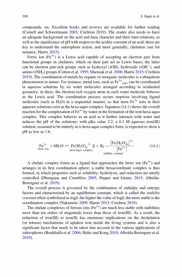
compounds, etc. Excellent books and reviews are available for further reading(Cornell and Schwertmann 2003; Crichton 2019). The reader also needs to havean adequate background on the acid and base character and their inter-relations, aswell as the significance of pH with respect to the acidity constant of an acid; these arekey to understand the siderophore action, and more generally, chelation (see forinstance, Harris 2015).
Ferric ion (Fe3+) is a Lewis acid capable of accepting an electron pair fromfunctional groups in chelators, which on their part act as Lewis bases; the lattercan be electron pair-rich groups such as hydroxyl (:OH), hydroxide (:OH�), andamino (:NH2) groups (Cotton et al. 1995; Marusak et al. 2006; Harris 2015; Crichton2019). The coordination of metals by organic or inorganic molecules is a ubiquitousphenomenon in nature. For instance, metal ions, such as Fe3+(aq), can be coordinatedin aqueous solutions by six water molecules arranged according to octahedralgeometry; in these, the electron-rich oxygen atom in each water molecule behavesas the Lewis acid. The coordination process occurs stepwise involving ligandmolecules (such as H2O) in a sequential manner, so that most Fe3+ ions in theiraqueous solutions exist as the hexa-aquo complex. Equation (14.1) shows the overallreaction for the complexation of Fe3+ by water in the formation of the iron hexa-aquocomplex. This complex behaves as an acid as it further interacts with water andreduces the pH of the solutions; with pKa value 2.2, a 0.1 M aqueous iron(III)solution, assumed to be entirely in is hexa-aquo complex form, is expected to show apH as low as 1.6.
Fe3þferric ion
þ 6H2O Ð Fe H2Oð Þ63þhexa‐aquo complex
β ¼ Kf ¼Fe H2Oð Þ6� �3þ
Fe3þ� �
stability constant
ð14:1Þ
A chelate complex forms as a ligand that approaches the ferric ion (Fe3+) andarranges in its first coordination sphere; a stable hexacoordinate complex is thusformed, in which properties such as solubility, hydrolysis, and reduction are utterlycontrolled (Dhungana and Crumbliss 2005; Pepper and Gentry 2015; Albelda-Berenguer et al. 2019).
The overall process is governed by the combination of enthalpy and entropyfactors and characterized by an equilibrium constant, which is called the stabilityconstant often symbolized as logβ; the higher the value of logβ, the more stable is thecoordination complex (Nakamoto 2009; Harris 2015; Crichton 2019).
The chelate complexes of ferrous ions (Fe2+) are much less stable with stabilitiesmore than ten orders of magnitude lower than those of iron(III). As a result, thereduction of iron(III) to iron(II) has enormous implications on the dechelation(or release) mechanisms of uptaken iron inside the living systems and is also asignificant factor that needs to be taken into account in the various applications ofsiderophores (Boukhalfa et al. 2006; Hider and Kong 2010; Albelda-Berenguer et al.2019).
308 S. Rapti et al.
The hard-soft acid base (HSAB) concept proposed by Pearson further helps toevaluate and understand the high tendency for reaction between Lewis acids andbases of similar hardness (Cotton et al. 1995; Marusak et al. 2006; Harris 2015;Crichton 2019). As a result, chelators having a hard base character can complex iron(III) (a hard acid) so efficiently that the latter can be removed from its insoluble andminimally bioavailable iron oxides, hydroxy-oxides, and hydroxide. Siderophorespossess such ability, which has been developed as one of nature’s successfulstrategies to sequester iron (Bauer and Exner 1974; Agrawal 1979; Boukhalfa andCrumbliss 2002; Kraemer 2004; Tseng et al. 2006; Reichard et al. 2007; Hider andKong 2010; Sharpe et al. 2011; Ahmed and Holmström 2014; Albelda-Berengueret al. 2019). In conservation, a similar issue needs to be addressed: potent chelatorscan be employed to remove iron from surfaces of cultural heritage objects bytransferring it to the more soluble complexed phase; this way, a cleaning effectcan be achieved.
An addition, most iron(III) chelate complexes are colored due to specific electrontransitions facilitated by charge transfer between the iron(III) and the ligand. Thisoffers an extra asset for visual observation, and additionally, a widely followedmethodology for titrating of such complexes (Boukhalfa et al. 2006; Harris 2015).
3.2 The Tool: Siderophores
Siderophores have been developed by living systems based on organic functionalgroups (phenolic hydroxyls, carboxyl groups, hydroxamic and sulfhydryl groups)capable of efficient complexation of metals. Their development is also based on thenumber and the stereochemical arrangement that these groups possess for moreefficient complexation in order to overcome the limited bioavailability of iron as amicronutrient (Kraemer 2004; Kalinowski and Richardson 2005; Bertrand et al.2009; Hider and Kong 2010).
Siderophores, as most chelators, form thermodynamically stable hexacoordinatecomplexes with iron(III) accommodated in the most favorable molecular geometry(see, for instance, Fig. 14.1b) (Crumbliss 1990; Kraemer 2004); this along with thehigh affinity of siderophores for iron(III) produce strong driving forces towardscomplexation. The stability constants for iron(III) are generally higher than iron(II) which means that ferric complexes are generally much easier to form ascompared to ferrous. This preference is more significant in the case of siderophores,as compared to other chelators such as EDTA. This accordingly plays a major role instability of ferric complexes (see Sect. 14.3.3.3); in the context of conservationapplications, it has the significance that in the presence of the wrong chelator, oreven more, in the absence of any chelator, ferric ions can be reduced to ferrous withdetrimental effects on many organic substrates (see Sect. 14.4.1).
The action of chelation by siderophores implies multiple acidic functional groups(such as OH, COOH) capable of chelating metals which are involved in successivedeprotonation schemes (Edwards et al. 2005; Boukhalfa et al. 2006; Reichard et al.
14 Siderophores and their Applications in Wood, Textile, and Paper Conservation 309
2007). As acids, they tend to dissociate in their aqueous solutions releasing freehydronium ions (H3O
+), thus decreasing the pH of the solution. Their tendency todissociate is characterized by their pKa values; the lower the pKa value, the strongerthe acid. As the deprotonated (mostly anionic) forms are generally capable of metalcomplexation, their pKa values are important as they control the range for their mostefficient action.
Siderophores can be categorized according to their functional groups as carbox-ylates, catecholates, hydroxamates, and mixed type (Neilands 1995; Boukhalfa andCrumbliss 2002; Kraemer 2004; Crichton 2019). A siderophore database can befound on the web (Bertrand 2010); chemical structures of selected siderophores areshown in Fig. 14.2. A brief presentation of the main types of siderophores follows.
3.2.1 Carboxylate Siderophores
The carboxylate siderophores contain α-hydroxycarboxylic acid moieties, whichrenders them as acids with a broad range of acidities as seen through their pKavalues. Citric acid has been considered as a model compound (Fig. 14.2a) with pKavalues 3.13, 4.76, and 6.40 (Harris 2015) the lowest value assigned to the a-ΟΗcarboxyl group (Silva et al. 2009; Heller et al. 2012). The pKa values of carboxylgroups in the siderophore molecules range between 3.5 and 5 (Miethke and Marahiel2007) and are therefore active for complexation in relatively low pH values.
These siderophores can coordinate with the metal ion through their electron-richα-OH carboxylate oxygens; as a result, they can form chelates at low pH values,which offer an asset for applications that require an acidic environment. Nature hastaken advantage of this fact by utilizing these agents in acidic microbial
Hydroxamic acid trihydroxamate complex
(a)
log β = 28.29
ΔH0 = -5.9 kcal/mole,
ΔS0 = 112 eu
Desferrioxamine B (DFO-B) Ferrioxamine B (FO-B) complex
(b)
log β =30.6
ΔH0 = -19.8 kcal/mole
ΔS0= 71 eu
Fe3+ N
CO
OH
C
NO–
OC
NO–
O
Fe3+
CH3
CH3
CH3
H
H
H
CH3
NH OH
O+ 3H+3
N
O
CH3
O–
N
NH
O
N
O–
NH2
O
O–
NH
O
O
Fe3+
N
O
CH3
OH
NNH
O
N
OH
NH2
O
OH
NH
O
O
Fe3+
+ 3H+
Fig. 14.1 Chelation schemes: (a) model chelate complex: iron(III) trihydroxamate; (b)ferrioxamine (or desferrioxamine B complex with iron); and (c) ferrioxamine E(or desferrioxamine E complex with iron); modified from Crumbliss (1990)
310 S. Rapti et al.
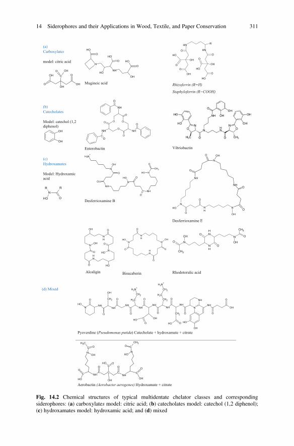
(a)
Carboxylates
model: citric acid
O OH
OH OC
OH
O OH
OHO
N
NH
OHOH
OHO OH
O
Mugineic acid
NH
OH
O
OH
O
OH
O
RNH
OH
O
OH
OH
OO
Rhizoferrin (R=H)
Staphyloferrin (R=COOH)
(b)
Catecholates
Model: catechol (1,2
diphenol)
OH
OH
NH
O
NH
O
OO
O
O
O
O
NH
O
Enterobactin Vibriobactin
(c)
Hydroxamates
Model: Hydroxamic
acid
N
OH O
R R
N
O
CH3
OH
NNH
O
N
OH
NH3+
O
OH
NH
O
O
Desferrioxamine B
N
OH
NH
O
O
N
NHO
OOH NH
N
OHO
O
Desferrioxamine E
NOH
O
NH
O
OH
NOH
O
NH
O
OH
Alcaligin
NH
N
O
OH
O
NOH
ONH
O
Bisucaberin
NH
NH
O
O
N O
CH3
OHN
CH3
O
OH
Rhodotorulic acid
(d) Mixed
NH
N+NHNH
NHNH
NHNH
NHN
O
OH
O
CH2
OH
O
O
CH2
CH2
NH3+
O
OH
OOH
O
CH2
CH2
NH3+
CH2 O
CH2
OOH
O
OH
OH
NHOH
O
O
Pyoverdine (Pseudomonas putida) Catecholate + hydroxamate + citrate
N
CCH3
O
OH
NHC C C
NHC
O
OH
COH O
O
OH
O
CO
OH
N
CCH3
O
OH
Aerobactin (Aerobacter aerogenes) Hydroxamate + citrate
Fig. 14.2 Chemical structures of typical multidentate chelator classes and correspondingsiderophores: (a) carboxylates model: citric acid; (b) catecholates model: catechol (1,2 diphenol);(c) hydroxamates model: hydroxamic acid; and (d) mixed
14 Siderophores and their Applications in Wood, Textile, and Paper Conservation 311
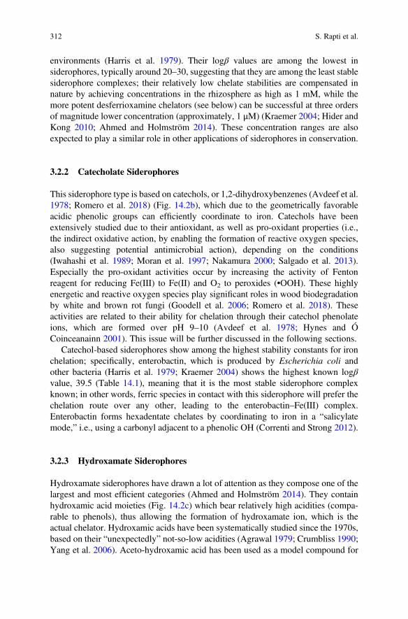
environments (Harris et al. 1979). Their logβ values are among the lowest insiderophores, typically around 20–30, suggesting that they are among the least stablesiderophore complexes; their relatively low chelate stabilities are compensated innature by achieving concentrations in the rhizosphere as high as 1 mM, while themore potent desferrioxamine chelators (see below) can be successful at three ordersof magnitude lower concentration (approximately, 1 μM) (Kraemer 2004; Hider andKong 2010; Ahmed and Holmström 2014). These concentration ranges are alsoexpected to play a similar role in other applications of siderophores in conservation.
3.2.2 Catecholate Siderophores
This siderophore type is based on catechols, or 1,2-dihydroxybenzenes (Avdeef et al.1978; Romero et al. 2018) (Fig. 14.2b), which due to the geometrically favorableacidic phenolic groups can efficiently coordinate to iron. Catechols have beenextensively studied due to their antioxidant, as well as pro-oxidant properties (i.e.,the indirect oxidative action, by enabling the formation of reactive oxygen species,also suggesting potential antimicrobial action), depending on the conditions(Iwahashi et al. 1989; Moran et al. 1997; Nakamura 2000; Salgado et al. 2013).Especially the pro-oxidant activities occur by increasing the activity of Fentonreagent for reducing Fe(III) to Fe(II) and O2 to peroxides (•OOH). These highlyenergetic and reactive oxygen species play significant roles in wood biodegradationby white and brown rot fungi (Goodell et al. 2006; Romero et al. 2018). Theseactivities are related to their ability for chelation through their catechol phenolateions, which are formed over pH 9–10 (Avdeef et al. 1978; Hynes and ÓCoinceanainn 2001). This issue will be further discussed in the following sections.
Catechol-based siderophores show among the highest stability constants for ironchelation; specifically, enterobactin, which is produced by Escherichia coli andother bacteria (Harris et al. 1979; Kraemer 2004) shows the highest known logβvalue, 39.5 (Table 14.1), meaning that it is the most stable siderophore complexknown; in other words, ferric species in contact with this siderophore will prefer thechelation route over any other, leading to the enterobactin–Fe(III) complex.Enterobactin forms hexadentate chelates by coordinating to iron in a “salicylatemode,” i.e., using a carbonyl adjacent to a phenolic OH (Correnti and Strong 2012).
3.2.3 Hydroxamate Siderophores
Hydroxamate siderophores have drawn a lot of attention as they compose one of thelargest and most efficient categories (Ahmed and Holmström 2014). They containhydroxamic acid moieties (Fig. 14.2c) which bear relatively high acidities (compa-rable to phenols), thus allowing the formation of hydroxamate ion, which is theactual chelator. Hydroxamic acids have been systematically studied since the 1970s,based on their “unexpectedly” not-so-low acidities (Agrawal 1979; Crumbliss 1990;Yang et al. 2006). Aceto-hydroxamic acid has been used as a model compound for
312 S. Rapti et al.
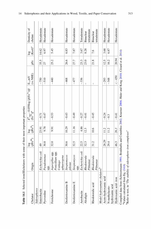
Tab
le14
.1Selectediron
(III)chelatorswith
someof
theirmostim
portantprop
erties
Chelator
Origin
log
(βFeIII)
log
(βFeII )
E0(aq)�
0.05
9log
[β(Fe3
+)/β
(Fe2
+)]a
E½,m
V(vsNHE)
pFe
log
(Ksol)a
Denticity
ofchelate
Sideroph
ores
Enterob
actin
Escherichia
coli
4923
.91
�0.71
�750
35.5
14.62
Hexadentate
Pyo
verdine
Pseud
omon
asspp.
30.8
9.78
�0.47
�510
276.97
Hexadentate
Ferrichrome
Aspergillu
sspp.
Penicillium
spp.
Ustila
gospha
erog
ena
32.0
9.91
�0.53
�440
25.2
5.45
Hexadentate
DesferrioxamineB
Streptom
yces
pilosus
30.6
10.29
�0.43
�468
26.6
6.83
Hexadentate
DesferrioxamineE
Streptom
yces
spp.
32.5
11.16
�0.49
�477
27.7
7.87
Hexadentate
Aerob
actin
Escherichia
coli
22.5
4.86
�0.27
�336
23.3
3.67
Tetradentate
Alcaligin
Rho
dotorula
Pilm
anae
32.3
12.3
�0.41
–23
.08.28
Binuclear
tetradentate
Rho
dotorulic
acid
Rho
dotorula
Pilm
anae
31.2
10.6
�0.45
–21
.87.6
Binuclear
tetradentate
Mod
elhydroxam
atechelatorsb
Aceto-hyd
roxamicacid
28.29
11.2
�0.24
�293
12.5
3.68
Hexadentate
N-m
ethy
laceto-
hydrox
amicacid
29.4
11.2
�0.3
�348
16.2
6.87
Hexadentate
Hyd
roxamicacid-iron
46.3
26.3
�0.41
<�4
5020
.94
Hexadentate
Com
pileddatafrom
(Schwertm
ann19
91;B
oukh
alfa
andCrumbliss20
02;Kraem
er20
04;Hider
andKon
g20
10;Cézardetal.2
014)
a Valuescalculated
from
Eq.
(14.6)
bRefer
totext,in“The
stab
ilityof
sideroph
ore–iron
complexes”
14 Siderophores and their Applications in Wood, Textile, and Paper Conservation 313
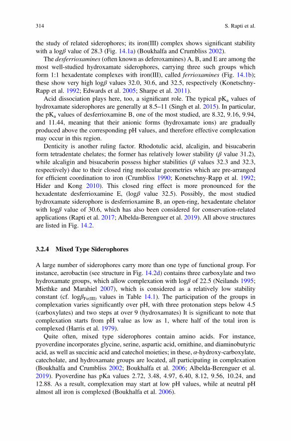
the study of related siderophores; its iron(III) complex shows significant stabilitywith a logβ value of 28.3 (Fig. 14.1a) (Boukhalfa and Crumbliss 2002).
The desferrioxamines (often known as deferoxamines) A, B, and E are among themost well-studied hydroxamate siderophores, carrying three such groups whichform 1:1 hexadentate complexes with iron(III), called ferrioxamines (Fig. 14.1b);these show very high logβ values 32.0, 30.6, and 32.5, respectively (Konetschny-Rapp et al. 1992; Edwards et al. 2005; Sharpe et al. 2011).
Acid dissociation plays here, too, a significant role. The typical pKa values ofhydroxamate siderophores are generally at 8.5–11 (Singh et al. 2015). In particular,the pKa values of desferrioxamine B, one of the most studied, are 8.32, 9.16, 9.94,and 11.44, meaning that their anionic forms (hydroxamate ions) are graduallyproduced above the corresponding pH values, and therefore effective complexationmay occur in this region.
Denticity is another ruling factor. Rhodotulic acid, alcaligin, and bisucaberinform tetradentate chelates; the former has relatively lower stability (β value 31.2),while alcaligin and bisucaberin possess higher stabilities (β values 32.3 and 32.3,respectively) due to their closed ring molecular geometries which are pre-arrangedfor efficient coordination to iron (Crumbliss 1990; Konetschny-Rapp et al. 1992;Hider and Kong 2010). This closed ring effect is more pronounced for thehexadentate desferrioxamine E, (logβ value 32.5). Possibly, the most studiedhydroxamate siderophore is desferrioxamine B, an open-ring, hexadentate chelatorwith logβ value of 30.6, which has also been considered for conservation-relatedapplications (Rapti et al. 2017; Albelda-Berenguer et al. 2019). All above structuresare listed in Fig. 14.2.
3.2.4 Mixed Type Siderophores
A large number of siderophores carry more than one type of functional group. Forinstance, aerobactin (see structure in Fig. 14.2d) contains three carboxylate and twohydroxamate groups, which allow complexation with logβ of 22.5 (Neilands 1995;Miethke and Marahiel 2007), which is considered as a relatively low stabilityconstant (cf. logβFe(III) values in Table 14.1). The participation of the groups incomplexation varies significantly over pH, with three protonation steps below 4.5(carboxylates) and two steps at over 9 (hydroxamates) It is significant to note thatcomplexation starts from pH value as low as 1, where half of the total iron iscomplexed (Harris et al. 1979).
Quite often, mixed type siderophores contain amino acids. For instance,pyoverdine incorporates glycine, serine, aspartic acid, ornithine, and diaminobutyricacid, as well as succinic acid and catechol moieties; in these, α-hydroxy-carboxylate,catecholate, and hydroxamate groups are located, all participating in complexation(Boukhalfa and Crumbliss 2002; Boukhalfa et al. 2006; Albelda-Berenguer et al.2019). Pyoverdine has pKa values 2.72, 3.48, 4.97, 6.40, 8.12, 9.56, 10.24, and12.88. As a result, complexation may start at low pH values, while at neutral pHalmost all iron is complexed (Boukhalfa et al. 2006).
314 S. Rapti et al.
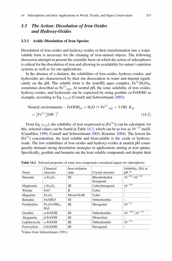
3.3 The Action: Dissolution of Iron Oxidesand Hydroxy-Oxides
3.3.1 Acidic Dissolution of Iron Species
Dissolution of iron oxides and hydroxy-oxides or their transformation into a water-soluble form is necessary for the cleaning of iron-stained objects. The followingdiscussion attempts to present the scientific basis on which the action of siderophoresis critical for the dissolution of iron and allowing its availability for nature’s nutritionsystems as well as for our applications.
In the absence of a chelator, the solubilities of iron oxides, hydroxy-oxides, andhydroxides are characterized by their ion dissociation in water and depend signifi-cantly on the pH. The soluble form is the iron(III) aquo complex, Fe3+(H2O)6,sometimes described as Fe3+(aq). At neutral pH, the ionic solubility of iron oxides,hydroxy-oxides, and hydroxide can be expressed by using goethite (α-FeOOH) asexample, according to Eq. (14.2) (Cornell and Schwertmann 2003).
Neutral environmentα� FeOOH sð Þ þ H2O Ð Fe3þ aqð Þ þ 3 OH�Ksp
¼ Fe3þ� �
OH�½ �3 ð14:2Þ
From Eq. (14.2), the solubility of iron (expressed as [Fe3+]) can be calculated; forthis, selected values can be found in Table 14.2, which can be as low as 10�13 mol/L(Crumbliss 1990; Cornell and Schwertmann 2003; Kraemer 2004). The lowest the[Fe3+] concentration, the least soluble and bioavailable is the oxide or hydroxy-oxide. The low solubilities of iron oxides and hydroxy-oxides at neutral pH conse-quently demand strong dissolution strategies in applications aiming at iron uptake.Specifically, goethite and hematite are the least soluble compounds and despite their
Table 14.2 Selected properties of some iron compounds considered targets for siderophores
NameChemicalstructure
Iron oxidationstate Crystal structure
Solubility, [Fe] atpH 7a
Hematite α-Fe2O3 III Rhombohedralhexagonal
10�8.8–10�5.8
Maghemite γ-Fe2O3 III Cubic/tetragonal 10�5.0
Wüstite FeO II Cubic –
Magnetite Fe3O4 Mixed II+III Cubic –
Bernalite Fe(OH)3 III Orthorhombic –
Ferrihydrite Fe5O7(OH)4.H20
III Hexagonal 10�3.1
Goethite α-FeOOH III Orthorhombic 10�10.9–10�5.9
Akaganeite β-FeOOH III Monoclinic –
Lepidocrocite γ-FeOOH III Orthorhombic 10�5.2
Ferroxyhyte δ-FeOOH III Hexagonal –
aValues from Schwertmann (1991)
14 Siderophores and their Applications in Wood, Textile, and Paper Conservation 315
high abundance in the environment, their bioavailability is extremely low because oftheir low solubilities; as a consequence, their mobilization (or bioavailability throughdissolution) becomes a real problem. Also, the same compounds, either as corrosionproducts or as unwanted iron stain factors, are among the most common iron species,and drastic tools for their removal through dissolution, such as citrate and cystinehave been proposed (Kolar et al. 2006; Henniges et al. 2008; Sharpe et al. 2011; Liuet al. 2017).
At acidic pH, the solubility for goethite can be expressed according to Eq. (14.3).The acidic dissolution constant (KH
s) values are relatively high as compared toneutral dissolution ones (expressed by Ksp values); this means that in order toeffectively solubilize such iron species and formation of the water-soluble Fe3+
ion, the pH needs to be significantly lowered (pH < 5). However, this can be quiteprohibitive for substrates in many cultural heritage objects for which pH values of6–8 are generally acceptable; on the other hand, this may be a workable region in thecase of materials, such as East Asian lacquer, which are stable in acidic pH. The aquocomplex of iron [Fe(HO2)6
3+] is also very acidic (Siddall and Vosburgh 1951;Milburn and Vosburgh 1955; Sapieszko et al. 1977; Wilkins 1991; Cotton et al.1995), and therefore, its presence may lower the solution pH. Moreover, iron(III) caninitiate Fenton reactions with detrimental effects on organic substrates (see below).
To complete the picture, the kinetics of dissolution (studying how fast thephenomenon occurs) is equally important as the thermodynamics (studying howstable the chemical species are). Kinetics is directly linked to the mechanism of irondissolution, which has been studied and has been shown that it is heavily protondependent. This happens because, as H+ ions are adsorbed on the mineral surface,the surface Fe3+-OH bonds weaken and the detachment of iron ions is facilitated(Stumm and Furrer 1987; Cornell and Schwertmann 2003). In particular, the initialdissolution of iron(III) is very fast and is subsequently followed by a slower step(Samson and Eggleston 1998; Reichard et al. 2007). This affects the waysiderophore molecules work which is able to capture a “burst” of liberated Fe3+
ions during this first step. This proton-mediated process has been found to control alltypes of dissolution, including the action of siderophores (see below).
3.3.2 Dissolution of Iron Species in the Presence of Siderophores
The stability of siderophore–iron complexes: In the presence of a siderophore, thecomplexation of iron occurs, which is expressed in Eq. (14.4); this involves an acidicenvironment as protons (H+) are produced during complexation; however, summingthese two equations lead to the combined dissolution reaction, shown in Eq. (14.5)(Kraemer 2004).
316 S. Rapti et al.
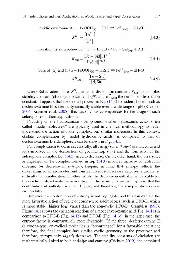
Acidic environmentα� FeOOH sð Þ þ 3Hþ Ð Fe3þ aqð Þ þ 2H2O
KHs ¼
Fe3þ� �
Hþ½ �3ð14:3Þ
Chelation by siderophoreFe3þ aqð Þ þ H3Sid Ð Fe� Sid aqð Þ þ 3Hþ
KSid ¼ Fe� Sid½ � Hþ½ �3H3Sid½ � Fe3þ� � ð14:4Þ
Sum of 2ð Þ and 3ð Þα� FeOOH sð Þ þ H3Sid Ð Fe3þ aqð Þ þ 2H2O
KHs,Sid ¼ Fe� Sid½ �
H3Sid½ � ð14:5Þ
where Sid is siderophore, KHs the acidic dissolution constant, KSid the complex
stability constant (often symbolized as logβ), and KHs,Sid the combined dissolution
constant. It appears that the overall process in Eq. (14.5) for siderophores, such asdesferrioxamine B is thermodynamically stable over a wide range of pH (Kraemer2004; Kraemer et al. 2005); this has obvious consequences for the usage of suchsiderophores in their applications.
Focusing on the hydroxamate siderophores, smaller hydroxamic acids, oftencalled “model molecules,” are typically used in chemical methodology to betterunderstand the action of more complex, but similar molecules. In this context,chelate complexation by model hydroxamic acids, as compared to that ofdesferrioxamine B siderophores, can be shown in Fig. 14.1.
For complexation to occur successfully, all energy (or enthalpy) of molecules andions involved in the destruction of goethite Eq. (14.2) and the formation of thesiderophore complex Eq. (14.3) need to decrease. On the other hand, the very strictarrangement of the complex formed in Eq. (14.3) involves increase of molecularordering (or decrease in entropy); keeping in mind that entropy reflects thedisordering of all molecules and ions involved, its decrease imposes a geometricdifficulty to complexation. In other words, the decrease in enthalpy is favorable forthe reaction, while the decrease in entropy is disfavoring; however, it appears that thecontribution of enthalpy is much bigger, and therefore, the complexation occurssuccessfully.
However, the contribution of entropy is not negligible, and this can explain themore favorable action of cyclic or corona-type siderophores, such as DFO-E, whichis more stable (higher logβ value) than the non-cyclic DFO-B (Crumbliss 1990).Figure 14.1 shows the chelation reactions of a model hydroxamic acid (Fig. 14.1a) incomparison to DFO-B (Fig. 14.1b) and DFO-E (Fig. 14.1c); in the latter case, theentropy factor is comparatively more favorable. Of the three, desferrioxamine E(a corona-type, or cyclical molecule) is “pre-arranged” for a favorable chelation;therefore, the final complex has similar cyclic geometry to the precursor andtherefore, entropy only slightly decreases. The stability constants of chelation aremathematically linked to both enthalpy and entropy (Crichton 2019), the combined
14 Siderophores and their Applications in Wood, Textile, and Paper Conservation 317
consideration of enthalpy and entropy finally explains why DFO-E (logβ ¼ 32.5) isbetter complexing agent toward iron(III) than DFO-B (logβ ¼ 30.6), by almost twoorders of magnitude, or 100 times.
The role of pH: As has already been mentioned above, carboxylate siderophores(Sect. 14.3.2.1) can form complexes in relatively low pH values, whilehydroxamates (Sect. 14.3.2.3) at generally are higher than 8–9. Finally, mixedsiderophores, such as pyoverdine (Sect. 14.3.2.4) may form their anionic forms ata much larger span, ranging from 2.7 to 12.9, meaning that complexes can beproduced in significantly lower pH. Therefore, optimal complexation is clearlydepending on the acidities in each case. As a result, the simultaneous considerationof the logβ values and the deprotonation constants may not allow a straightforwardevaluation of the chelation effectiveness of siderophores towards iron(III).
As a more practical approach, the concentration of dissolved iron (expressed aspFe) has been proposed to reflect more objectively the effectiveness of complexation(Harris et al. 1979; Albelda-Berenguer et al. 2019). The pFe, defined as log[Fefree],has been introduced for iron (and accordingly, pM for metals in general), at certainpH, metal and ligand concentrations. High pFe values mean low free iron concen-tration and therefore effective complexation. For instance, the high logβ value ofdesferrioxamine B (30.6) corresponds to reasonably high pFe value (26.6); on theother hand, aerobactin, although with significantly lower logβ (22.5), has relativelyhigh pFe (23.3) suggesting that it can still adequately complex iron(III). On the otherhand, aceto-hydroxamic acid with logβ ¼ 28.3 has low pFe (12.5). Values of pFe forvarious siderophores are shown in Table 14.1.
Selectivity of siderophores towards iron (III): The selectivity of complexationregarding metal ions is an obvious demand for several fields, including that ofconservation. Cultural heritage objects often contain metal ions as pigments, corro-sion products, depositions, etc., where the selective removal of iron(III) calls fordrastic chelating strategies; these generally involve considerable differences forstability constants of chelators towards ferric compounds as compared to othermetal ions. In other words, selective cleaning action is needed where the conservatoraims to remove a specific metal ion/stain/corrosion product without affecting others.
The drive for choosing iron(III) chelators better than EDTA and DTPA emanatesfrom the fact that these two widely used reagents complex effectively a wide range ofmetals, such as aluminum, cobalt, nickel, copper, and manganese, which all arepresent in cultural heritage objects. On the other hand, siderophores show a com-parative preference towards iron, which makes these chelators a suitable choice forselective complexation of iron(III) towards other metals. Ferric ion (Fe3+) is a hardLewis acid (see Sect. 14.3.2 for a brief introduction of the hard-soft acid baseconcept) and therefore shows a high affinity for hard Lewis bases such as mostsiderophores (Pearson 1973; Crumbliss 1990; Jensen 1991).
In addition, it has been shown that siderophores are particularly sensitive to theelectronegativity (χM) of metal ions as well as their charge (Ζ); the value of χM •Z forFe3+ is 5.9, while for Al3+ and Cu2+ is 4.9 and 4.0, respectively. As a result of theabove, the stability constants of DFO-B towards iron(III) is by far higher than anyother metal ion (logβ ¼ 30.7), followed by aluminum (logβ ¼ 23.1), Cu2
318 S. Rapti et al.
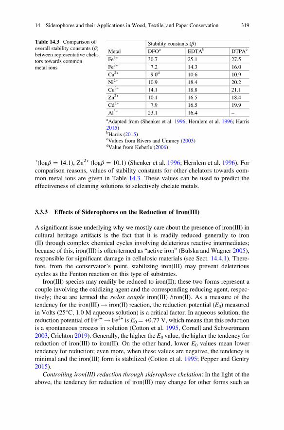
+(logβ ¼ 14.1), Zn2+ (logβ ¼ 10.1) (Shenker et al. 1996; Hernlem et al. 1996). Forcomparison reasons, values of stability constants for other chelators towards com-mon metal ions are given in Table 14.3. These values can be used to predict theeffectiveness of cleaning solutions to selectively chelate metals.
3.3.3 Effects of Siderophores on the Reduction of Iron(III)
A significant issue underlying why we mostly care about the presence of iron(III) incultural heritage artifacts is the fact that it is readily reduced generally to iron(II) through complex chemical cycles involving deleterious reactive intermediates;because of this, iron(III) is often termed as “active iron” (Bulska and Wagner 2005),responsible for significant damage in cellulosic materials (see Sect. 14.4.1). There-fore, from the conservator’s point, stabilizing iron(III) may prevent deleteriouscycles as the Fenton reaction on this type of substrates.
Iron(III) species may readily be reduced to iron(II); these two forms represent acouple involving the oxidizing agent and the corresponding reducing agent, respec-tively; these are termed the redox couple iron(III) /iron(II). As a measure of thetendency for the iron(III) ! iron(II) reaction, the reduction potential (E0) measuredin Volts (25�C, 1.0 M aqueous solution) is a critical factor. In aqueous solution, thereduction potential of Fe3+ ! Fe2+ is E0 ¼ +0.77 V, which means that this reductionis a spontaneous process in solution (Cotton et al. 1995, Cornell and Schwertmann2003, Crichton 2019). Generally, the higher the E0 value, the higher the tendency forreduction of iron(III) to iron(II). On the other hand, lower E0 values mean lowertendency for reduction; even more, when these values are negative, the tendency isminimal and the iron(III) form is stabilized (Cotton et al. 1995; Pepper and Gentry2015).
Controlling iron(III) reduction through siderophore chelation: In the light of theabove, the tendency for reduction of iron(III) may change for other forms such as
Table 14.3 Comparison ofoverall stability constants (β)between representative chela-tors towards commonmetal ions
Metal
Stability constants (β)
DFOa EDTAb DTPAc
Fe3+ 30.7 25.1 27.5
Fe2+ 7.2 14.3 16.0
Ca2+ 9.0d 10.6 10.9
Ni2+ 10.9 18.4 20.2
Cu2+ 14.1 18.8 21.1
Zn2+ 10.1 16.5 18.4
Cd2+ 7.9 16.5 19.9
Al3+ 23.1 16.4 –
aAdapted from (Shenker et al. 1996; Hernlem et al. 1996; Harris2015)bHarris (2015)cValues from Rivers and Ummey (2003)dValue from Keberle (2006)
14 Siderophores and their Applications in Wood, Textile, and Paper Conservation 319
iron oxides and chelate complexes. In particular, siderophore chelates of iron(III)show negative reduction potentials, meaning that iron(III) is practically not reducedto iron(II); for instance, values for DFO-B and enterobactin are�478 and�750 mV,respectively, meaning that enterobactin is a superior stabilizing agent and thatreduction of iron(III) is no longer spontaneous, unless a very strong reducingagent is available (Dhungana and Crumbliss 2005).
Microorganisms and plants after complexing non-bioavailable iron(III) in itsoxides and hydroxy-oxides, they need to utilize it as nutrient, often by reducing itto a more available form (see Sect. 14.3.3.4). For this, exceptionally strong reducingagents are needed, with negative reduction potentials (for instance, NADH, orhydrogen-nicotinamide dinucleotide, with E0 ¼ �320 mV) to override the complexstability and finally rendering it in the more bioavailable iron(II). This strategysucceeds in many cases, but fails for the more stable complexes, such as theenterobactin–iron(III) complex (Boukhalfa and Crumbliss 2002). This is a desiredsituation for applications such as conservation, because mild reducing agents (suchas polyphenols) are typically present in cellulosic materials, which consequently arenot strong enough to reduce iron from its siderophore complexes.
Very negative E0 values, such as those mentioned, do not allow unwanted redoxreactions, such as the Haber–Weiss cycle and more particular the Fenton reaction(see Sect. 14.4.1) which produces hydroxyl radical (•OH) and the superoxide ion(•O2�), which are highly reactive oxygen species, harmful to organic substrates(Hider and Kong 2010). For the benefit of conservation practice, a conservator needsto be aware of processes such as the Fenton reaction, which is responsible for most ofdamage in cellulosic materials; because of these, strategies for the decrease forunwanted redox reactions are direly needed (Dhungana and Crumbliss 2005;Albelda-Berenguer et al. 2019). The importance of this process in conservationwill be discussed in the following section.
Careful choice of complexing agents towards iron involves the selection ofchelators with suitable β values for their iron(III) and (II) chelates. It has beenproposed that the higher the ratio of the β value of siderophore complex of iron(III) (β Fe(III)-Sid) with respect to that of iron(II) (β Fe(II)-Sid), the better the suppressionof the unwanted redox cycles (Boukhalfa et al. 2006; Albelda-Berenguer et al.2019).
EFe IIIð Þ�Sid ¼ E0aq � 59 log
βFe IIIð Þ�Sid
βFe IIð Þ�Sidð14:6Þ
Selected logβ values towards iron(III) and iron(II) are shown in Table 14.3. Otherstrong iron chelators, such as EDTA notoriously fail to prevent Fenton reactions, asthe β values between the two iron species are not very different. This is a strongmotif towards a better practice concerning the removal of iron from organicsubstrates.
320 S. Rapti et al.
3.3.4 The Kinetics and Mechanisms of Siderophore Action
The thermodynamic stability of complexes (often called thermodynamic stability)and the rates of reactions such as dissolution and complexation have been alreadyencountered in Sect. 14.3.3.1 The thermodynamics of siderophore-mediated iron(III)and the corresponding chemical equilibria of complexation do not offer a completepicture of the process and the conditions that favor it unless the kinetics are equallyconsidered (Römheld 1991). As already mentioned in Sect. 14.3.3.1, understandinggoethite removal from substrates is critical to comprehend the necessity for potentchelators. This has been recognized in conservation, as it has been recognized inliving systems.
The rates of goethite dissolution in the absence of chelator are depending onacidity. It has been shown that the detachment of iron from its mineral surface istriggered by an H+-depending mechanism (Samson et al. 2000). It appears that pHplays a significant role in dissolution rates, which are related to the mechanisms ofiron removal from its crystal lattice sites on goethite and other minerals (Monzyk andCrumbliss 1979; Brink and Crumbliss 1984).
A mechanism has been proposed, generally involving three steps. It has beenfound that during the first step, which is the faster, siderophore molecules of thesolution are consumed, thus decreasing their local concentration around iron spots;this may negatively influence the overall effectiveness of complexation in anapplication, unless high concentrations of siderophores are employed (Hider andKong 2010). In the subsequent steps, gradual complexation occurs, involvingbidentate and hexadentate complexes of iron(II) in a progressive mode (Furrer andStumm 1986; Kraemer 2004; Reichard et al. 2007).
Specifically in the first step, the complexation process involves competitionbetween the chelator and protons provided by the solution towards iron(III). Asimilar competition has been observed when a second chelator (or a co-chelator) isadded. This is an interesting outcome at the application level: as the rates ofcomplexation by siderophores are favored by acidity, as well as, synergistically toother agents as co-chelators, such as oxalic acid (Brink and Crumbliss 1984;Crumbliss 1990; Cheah et al. 2003; Kraemer 2004).
Regarding applications in cultural heritage, the above mechanistic considerations,in combination with what has been presented in the context of thermodynamicstability of siderophore–iron complexes may impose extra difficulties regardingthe effect of acidity, and they consequently demand for careful design ofsiderophore-based strategies.
Dechelation of captured iron(III): The stability of siderophore complexes doesnot allow for their easy destruction (often called dechelation). The iron-uptakeprocess by microorganisms and plant roots from their immediate geo-environmenteventually involves a later stage for releasing chelated iron. This involves specificmechanisms, as the exceptionally high stabilities of the chelate complexes do notallow for easy complex dissociation, often called dechelation (Boukhalfa andCrumbliss 2002; Dhungana and Crumbliss 2005).
14 Siderophores and their Applications in Wood, Textile, and Paper Conservation 321
Although this is a desired property regarding conservation—related applications,living systems need (and finally succeed) to achieve dechelation, because complexediron, although soluble, is still non-bioavailable. The reduction strategy by bacteriainvolving potent reducing agents (such as NADH) has been presented above; inconservation, although reducing agents in substrates such as wood are generallymilder, this needs to be taken into account for targeted cleaning strategies.
This is achieved by proton-assisted mechanisms and/or by reducing iron(III) toiron(II), where the corresponding complex of the latter is less stable by many ordersof magnitude (Table 14.3). The proton-assisted dechelation mechanism has beenstudied extensively and showed that dechelation rates significantly increase at lowerpH values. Although the siderophore complexes are extremely stable, in low pH canbe labile, this factor has been found to favorably affect dechelation pathways for thebenefit of living systems. In the case of iron stain cleaning, pH needs to be seriouslyconsidered regarding the balance between iron capture by chelation and release bydechelation (Boukhalfa and Crumbliss 2002).
3.3.5 Side Reactions, Synergistic Effects, and Light Sensitivity
The Fenton-type reactions that were mentioned in the Sect. 14.1 are of significantconcern for the various applications as they act detrimentally to most substrates.While chelators such as EDTA do not prevent Fenton reactivity of iron(III) (Albelda-Berenguer et al. 2019), it has been reported that siderophores, such asdesferrioxamine B and enterobactin do not favor this type of unwanted side-reactions.
A synergistic effect between dicarboxylic acids and siderophores has beenobserved; increase of iron(III) dissolution rates through complexation bydesferrioxamine B in the presence of oxalate was observed (Reichard et al. 2007;Loring et al. 2008; Albelda-Berenguer et al. 2019). The similar accelerating effectwas observed with other dicarboxylic acids, such as fumaric, malonic, and succinic;on the other hand, citric acid was found to inhibit the rates, possibly due toadsorption competition effects (Reichard et al. 2007).
Light also seems to play a significant role in the final dechelation stages and thebioavailability of iron in marine systems. In particular, some iron(III) complexeswith marine siderophores have been found to dechelate the metal through a photol-ysis mechanism which leads to the final release of ferrous ions (Hider and Kong2010). Studies using aquachelin (a mixed type siderophore contains hydroxamateand α-hydroxy-carboxylate groups) have been shown to react photochemicallytowards releasing an iron(III) photoproduct complex, which undergoes a ligand-to-metal charge-transfer process, resulting in the reduction of iron(III) to iron(II) (Barbeau et al. 2001, 2003). The latter is easily released and therefore, availableto the benefit of iron-starving organisms. On the reasonable basis that photolyticprocesses may affect the effectiveness of applications employing iron(III) chelationby siderophores, this issue merits further investigation for other siderophore typesas well.
322 S. Rapti et al.
4 Application of Siderophores in Conservation
4.1 The Detrimental Effect of Iron
Museum artifacts such as furniture, toys, musical instruments, tools, householdutensils, manuscripts, and religious items are often consisted of organic materials,such as wood, textile, paper, leather, and elements made of iron. When thesecomposite objects are become wet or are exposed to humid environments, theiriron elements corrode, and the formed corrosion products may accumulate on thesurface of the organic substrate or they may impregnate its matrix.
The most common iron corrosion products formed are iron(III) (ferric) and iron(II) (ferrous) oxides, such as Fe2O3, Fe3O4, and FeO, hydroxy-oxides such asFeOOH, occurring in various polymorphs, often also called oxy-hydroxides andiron hydroxide Fe(OH)3. Iron corrosion products generally exhibit very low solu-bilities, which depend on their grain size, since the smaller the size the higher thesolubility. Therefore, for some compounds such as hematite and goethite, solubilityincreases, as their grain size can be extremely small ranging between 10 and 150 nm.In addition, other hydroxy-oxides of iron(III), such as ferrihydrite(Fe5O7(OH).4H2O), may also exist as corrosion products and are considered signif-icant participants in the overall iron corrosion process (Kraemer 2004). Hematite(red), magnetite (black), and goethite (pale brown-yellow, beige) have also beenhistorically used as pigments. In Table 14.2, a list of the most abundant forms of ironcompounds found on cultural heritage objects is presented (Cornell andSchwertmann 2003; Scott and Eggert 2009; Richards et al. 2012).
The destructive effect of iron on organic substrates, which can be either cellulosicor proteinaceous, has been the subject of several studies. Metal ions, when found inproximity to organic substrates, take part in redox reactions and act as catalysts tooxidation processes of these substrates. These processes are a wide variety ofchemical reactions leading to the formation of various deterioration products andto depolymerization of main chemical components of organic substrates and conse-quently to the reduction of their mechanical strength (Baker 1980; Timár-Balázsyand Eastop 1998; Strlič et al. 2001; Scott and Eggert 2009; Badillo-Sanchez et al.2019).
The oxidation of organic materials is probably caused by the free hydroxyl radicalformation and hydrogen peroxide, which allow the Fenton-type reactions to occur.Thus, during the oxidation of ferrous iron to its ferric state, the free radical formationmight be enhanced if organic substrates are present and this in turn leads to theformation of organic radicals, as seen in Eqs. (14.7a)–(14.7d), or to the formation ofhydrogen peroxide, as seen in Eqs. (14.7e)–(14.7f) (Neevel 1995). The hydrogenperoxide produced during oxidation of organic substrates is decomposed by reactingfurther with ferrous ions and forms additional hydroxyl radicals, according toEqs. (14.7e) and (14.7f).
14 Siderophores and their Applications in Wood, Textile, and Paper Conservation 323
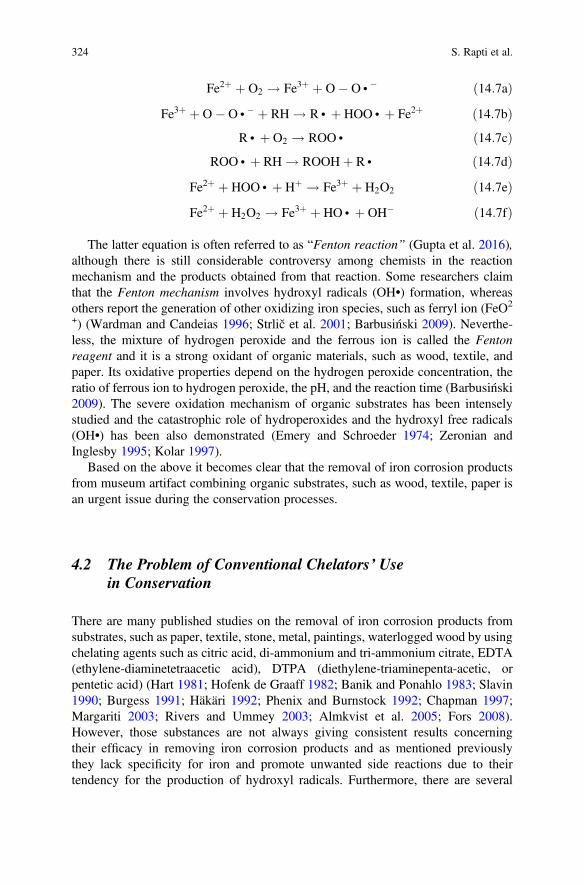
Fe2þ þ O2 ! Fe3þ þ O� O • � ð14:7aÞFe3þ þ O� O • � þ RH ! R • þ HOO • þ Fe2þ ð14:7bÞ
R • þ O2 ! ROO • ð14:7cÞROO • þ RH ! ROOHþ R • ð14:7dÞ
Fe2þ þ HOO • þ Hþ ! Fe3þ þ H2O2 ð14:7eÞFe2þ þ H2O2 ! Fe3þ þ HO • þ OH� ð14:7fÞ
The latter equation is often referred to as “Fenton reaction” (Gupta et al. 2016),although there is still considerable controversy among chemists in the reactionmechanism and the products obtained from that reaction. Some researchers claimthat the Fenton mechanism involves hydroxyl radicals (OH•) formation, whereasothers report the generation of other oxidizing iron species, such as ferryl ion (FeO2
+) (Wardman and Candeias 1996; Strlič et al. 2001; Barbusiński 2009). Neverthe-less, the mixture of hydrogen peroxide and the ferrous ion is called the Fentonreagent and it is a strong oxidant of organic materials, such as wood, textile, andpaper. Its oxidative properties depend on the hydrogen peroxide concentration, theratio of ferrous ion to hydrogen peroxide, the pH, and the reaction time (Barbusiński2009). The severe oxidation mechanism of organic substrates has been intenselystudied and the catastrophic role of hydroperoxides and the hydroxyl free radicals(OH•) has been also demonstrated (Emery and Schroeder 1974; Zeronian andInglesby 1995; Kolar 1997).
Based on the above it becomes clear that the removal of iron corrosion productsfrom museum artifact combining organic substrates, such as wood, textile, paper isan urgent issue during the conservation processes.
4.2 The Problem of Conventional Chelators’ Usein Conservation
There are many published studies on the removal of iron corrosion products fromsubstrates, such as paper, textile, stone, metal, paintings, waterlogged wood by usingchelating agents such as citric acid, di-ammonium and tri-ammonium citrate, EDTA(ethylene-diaminetetraacetic acid), DTPA (diethylene-triaminepenta-acetic, orpentetic acid) (Hart 1981; Hofenk de Graaff 1982; Banik and Ponahlo 1983; Slavin1990; Burgess 1991; Häkäri 1992; Phenix and Burnstock 1992; Chapman 1997;Margariti 2003; Rivers and Ummey 2003; Almkvist et al. 2005; Fors 2008).However, those substances are not always giving consistent results concerningtheir efficacy in removing iron corrosion products and as mentioned previouslythey lack specificity for iron and promote unwanted side reactions due to theirtendency for the production of hydroxyl radicals. Furthermore, there are several
324 S. Rapti et al.
studies giving considerable attention to the environmental impact and the biode-gradability of conventional chelators, such as EDTA and DTPA, which are exten-sively used, in the conservation field, especially on waterlogged objects (Sillanpää1997; Nörtemann 1999; Grčman et al. 2001; Nörtemann 2005; Kołodyńska 2011).EDTA, however, has been found to be more persistent than DTPA in degradation byconventional biological and physicochemical methods (Nörtemann 1999; Grčmanet al. 2001; Nörtemann 2005). Although DTPA is more biodegradable, it has beenreported that EDTA can be one of its decomposition products (Sillanpää 1997).Nonetheless, both EDTA and DTPA may result in several risks when released to theaquatic environment, as they may contribute to toxicity at significantly low concen-trations (Sillanpää 1997; Grčman et al. 2001). Additionally, their degradation prod-ucts can be used as a nitrogen source for algae growth and thus EDTA and DTPAhave the potential to contribute to eutrophication (Sillanpää 1997; Kołodyńska2011).
4.3 Applications of Siderophores in Conservation of CulturalHeritage Objects
The tendency nowadays is to find safer alternative materials and methods forconservation purposes. This tendency though, becomes a necessity, consideringthe established national legislations and international environmental restrictions(Balliana et al. 2016) and therefore the replacement of conventional chelators withnew ones with improved biodegradability, is critical.
There are a few studies regarding the removal of iron corrosion products fromcultural heritage objects, with “green” compounds such as siderophores (Wagnerand Bulska 2003; Rapti et al. 2015, 2017; Albelda-Berenguer et al. 2019). Thesestudies have investigated the application of siderophores, mainly desferrioxamine B(DFO-B), on different organic substrates and preservation states. The applicationmethodologies followed by both siderophores and conventional chelators were notsimilar and therefore comparative analysis of the obtained results cannot be done.
4.3.1 Extraction of Iron from Historical Manuscripts With IronGall Ink
Bulska and Wagner (2005) dealt with historic manuscripts written with iron gallinks1 that contained a substantial amount of “active” iron(II) ions, which can act as
1Historical recipes for gall ink production involve the use of aqueous solutions of iron(II) sulfatewith extracts of gall nuts to form complexes of oxidized iron(III) with gallic acid. However, most ofhistorical manuscripts contain an excess of iron(II) ions that were not complexed and thus are activeiron(II) ions that can act as catalysts to oxidative degradation processes of paper.
14 Siderophores and their Applications in Wood, Textile, and Paper Conservation 325
catalysts and are responsible for promoting the degradation processes of papercellulose via Fenton reactions. In order to prevent this paper degradation process,which is often accompanied by acid hydrolysis among other deteriorative mecha-nisms, Bulska and Wagner (2005) tried to find a cleaning treatment to remove theexcess of active iron ions. The treatment was expected, without altering the iron gallink color, to impede both Fenton reaction and acid hydrolysis leaving the paper ofthe manuscripts unaffected by the cleaning procedure. Therefore, they comparativelyevaluated conventional chelators versus siderophores for their efficiency of ironextraction on mock-ups mimicking historical manuscripts with iron gall inks andon mock-ups simulating only the amount of active iron ions. Chelators were requiredto demonstrate low cleaning efficacy on the first mock-up type and concurrently highon the second type of mock-ups. Moreover, they attempted to simultaneouslyneutralize the acidity of papers (deacidification) during the cleaning treatment.
Several physicochemical analyses were implemented in order to design thecleaning methodology and to evaluate the efficacy of the cleaning treatments,including Scanning Electron Microscopy (SEM), X-ray Fluorescence Spectrometry(XRF), Graphite Furnace Atomic Absorption Spectrometry (GFAAS), InductivelyCoupled Plasma Spectrometry (ICM-MS), Molecular spectroscopy (UV/VIS), X-rayAbsorption Near-Edge Structure (XANES) (Bulska et al. 2001; Wagner et al. 2001a,2001b; Wagner and Bulska 2003; Bulska and Wagner 2004; Wagner and Bulska2004).
The chelators used for iron extraction and iron complex formation includedbidentate ligand acetylacetone, dibenzoylmethane, diethylene-triaminepenta-aceticacid (DTPA), the potassium-magnesium salt of phytic acid and the mesylate salt ofDFO-B. All compounds were selected due to their inhibition effect of furtherformation of hydroxyl radicals and to their high stability constants of complexeswith iron as it was expected to be more effective and phytate, a known compound forits beneficial iron deactivation effect, as it reduces iron ability to undergo furtherreactions and occupy all coordination sites of iron (Neevel 1995). The solutions ofthe examined chelators were prepared at various concentrations (0.001, 0.005, and0.01 M) and pH values in the range of 7–9, in addition to that obtained after thechelators’ dissolution in water (low pH < 3). Ethanol/water solutions (10, 25, and50%) of DTPA, phytic acid, and DFO-B were also examined at 0.005 M. The mock-ups were immersed in the various chelators’ solutions for 20 min (Bulska et al. 2001;Wagner et al. 2001b; Wagner and Bulska 2003).
Results obtained by physicochemical analyses showed that iron–DFO-B complexformation at both types of mock-ups was above 80%. Moreover, on mock-upsmimicking the excess of active iron ions, DFO-B revealed an increase of ironextraction, which depended mainly on the pH of solution. The concentration of allprepared solutions of chelators was pronouncedly higher than the amount of ironions need to be extracted. In particular, at pH 8, a value typically used in paperconservation to enhance deacidification, and solution concentration of 0.005 M, ironextraction reached ca. 91%. Among all the tested pH, the lowest extraction of67–72% was observed at the highest concentration 0.01 M. Concerning applicationson mock-ups simulating manuscripts with iron gall ink, DFO-B with a concentration
326 S. Rapti et al.
of 0.005 M also showed high extraction capacity which reached 83 and 85%, at pH 8and 9, respectively (Wagner and Bulska 2003). However, this was considered adrawback because iron extraction was not selective, as the color of iron gall ink wasseverely affected. Consequently, the most effective solution for removing excessiron was proved unsuitable for iron gall ink mock-ups.
The influence of pH on siderophores’ efficacy has been also the subject of severalenvironmental studies unrelated to conservation. Borer et al. (2009) among others(Cheah et al. 2003; Borer et al. 2005; Reichard et al. 2005; Loring et al. 2008)investigated DFO-B and aerobactin in order to improve the understanding of thesesiderophores biogeochemistry and to gain information about siderophores reactivitytowards the dissolution and mobilization of iron hydroxy-oxides, such as goethiteand lepidocrocite, which are often found in terrestrial and aquatic environments.Their study revealed that pH influences the iron adsorption in ferric compounds, forinstance, in lepidocrocite via DFO-B and that was also associated with siderophores’functional group and their deprotonation stage. More specifically, they showed thatthe maximum absorption of lepidocrocite by DFO-B occurred at pH 8.6, which isclose to its first pKa (8.3), whereas at lower or higher pH values, the adsorption wasdecreased.
4.3.2 Extraction of Iron Compounds from Composite Museum Objects
Rapti et al. (2015) investigated in a preliminary study the removal of iron ions fromdry wooden substrates, which cannot tolerate waterborne treatments, due to thehygroscopic and anisotropic nature of wood. Desferrioxamine, in the form of acommercial injectable drug, Desferal® manufactured by Novartis, was comparedwith the conventional chelators EDTA and DTPA to treat wooden mock-upsartificially stained with iron corrosion products. To avoid immersion in aqueoussolutions and minimize the amount of water that would penetrate into the wood andat the same time maintain the cleaning potency of the solution applied, the chelatorswere applied by cotton swab and carboxymethyl cellulose gel (CMC) 4.5% w/v. Theeffectiveness of the two conventional chelators was also examined with the additionof a reducing agent, sodium dithionite (SDT) (Selwyn and Tse 2009). The solutionsof EDTA and DTPA were prepared at 2.5% w/v with or without the addition of SDT(5% w/v) at pH 6.2, and 6.8, respectively, whereas DFO-B concentration was5 � 10�5M, at pH 8.5. Colorimetry and elemental analysis by Energy DispersiveX-ray (EDX) spectroscopy were implemented before and after cleaning for evalu-ating the efficacy of the chelators. Results showed that DFO-B was an effectivechelator, as efficient as EDTA when SDT was added to it. However, residues of Naand S ions were detected, owed to SDT, which have the potential to promote futuredeterioration of substrates and therefore, this suggest the need to avoid SDT as partof the chelators solutions on this type of substrate; thus, siderophore was consideredmore appropriate. Conventional chelators when applied without the reducing agentwere shown clearly less efficient.
14 Siderophores and their Applications in Wood, Textile, and Paper Conservation 327
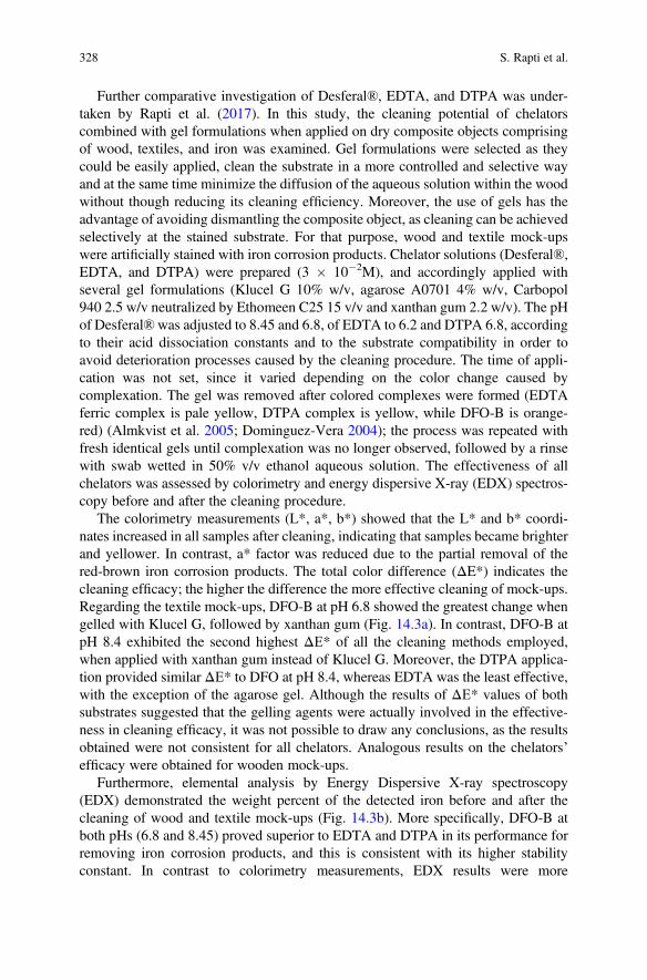
Further comparative investigation of Desferal®, EDTA, and DTPA was under-taken by Rapti et al. (2017). In this study, the cleaning potential of chelatorscombined with gel formulations when applied on dry composite objects comprisingof wood, textiles, and iron was examined. Gel formulations were selected as theycould be easily applied, clean the substrate in a more controlled and selective wayand at the same time minimize the diffusion of the aqueous solution within the woodwithout though reducing its cleaning efficiency. Moreover, the use of gels has theadvantage of avoiding dismantling the composite object, as cleaning can be achievedselectively at the stained substrate. For that purpose, wood and textile mock-upswere artificially stained with iron corrosion products. Chelator solutions (Desferal®,EDTA, and DTPA) were prepared (3 � 10�2M), and accordingly applied withseveral gel formulations (Klucel G 10% w/v, agarose A0701 4% w/v, Carbopol940 2.5 w/v neutralized by Ethomeen C25 15 v/v and xanthan gum 2.2 w/v). The pHof Desferal®was adjusted to 8.45 and 6.8, of EDTA to 6.2 and DTPA 6.8, accordingto their acid dissociation constants and to the substrate compatibility in order toavoid deterioration processes caused by the cleaning procedure. The time of appli-cation was not set, since it varied depending on the color change caused bycomplexation. The gel was removed after colored complexes were formed (EDTAferric complex is pale yellow, DTPA complex is yellow, while DFO-B is orange-red) (Almkvist et al. 2005; Dominguez-Vera 2004); the process was repeated withfresh identical gels until complexation was no longer observed, followed by a rinsewith swab wetted in 50% v/v ethanol aqueous solution. The effectiveness of allchelators was assessed by colorimetry and energy dispersive X-ray (EDX) spectros-copy before and after the cleaning procedure.
The colorimetry measurements (L*, a*, b*) showed that the L* and b* coordi-nates increased in all samples after cleaning, indicating that samples became brighterand yellower. In contrast, a* factor was reduced due to the partial removal of thered-brown iron corrosion products. The total color difference (ΔE*) indicates thecleaning efficacy; the higher the difference the more effective cleaning of mock-ups.Regarding the textile mock-ups, DFO-B at pH 6.8 showed the greatest change whengelled with Klucel G, followed by xanthan gum (Fig. 14.3a). In contrast, DFO-B atpH 8.4 exhibited the second highest ΔΕ* of all the cleaning methods employed,when applied with xanthan gum instead of Klucel G. Moreover, the DTPA applica-tion provided similar ΔΕ* to DFO at pH 8.4, whereas EDTA was the least effective,with the exception of the agarose gel. Although the results of ΔΕ* values of bothsubstrates suggested that the gelling agents were actually involved in the effective-ness in cleaning efficacy, it was not possible to draw any conclusions, as the resultsobtained were not consistent for all chelators. Analogous results on the chelators’efficacy were obtained for wooden mock-ups.
Furthermore, elemental analysis by Energy Dispersive X-ray spectroscopy(EDX) demonstrated the weight percent of the detected iron before and after thecleaning of wood and textile mock-ups (Fig. 14.3b). More specifically, DFO-B atboth pHs (6.8 and 8.45) proved superior to EDTA and DTPA in its performance forremoving iron corrosion products, and this is consistent with its higher stabilityconstant. In contrast to colorimetry measurements, EDX results were more
328 S. Rapti et al.
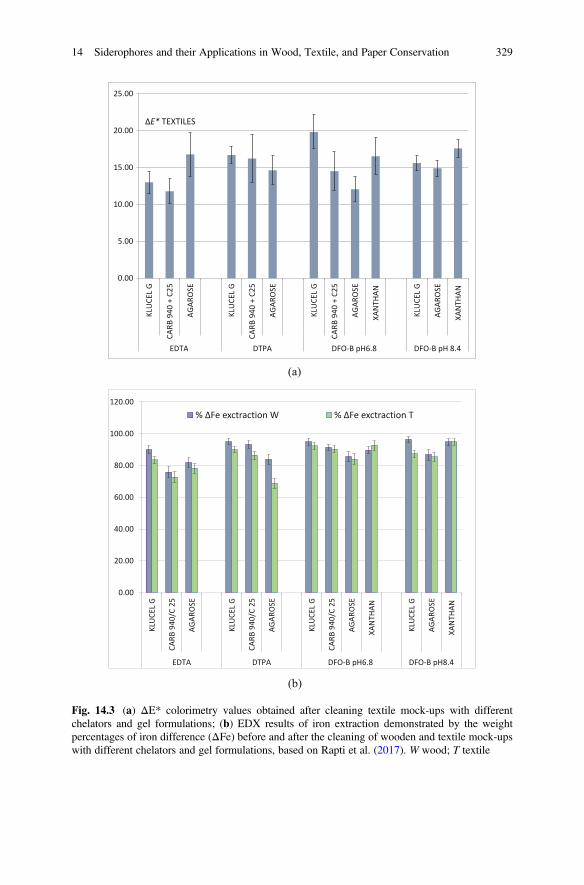
0.00
5.00
10.00
15.00
20.00
25.00
KLU
CEL
G
CARB
940
+ C
25
AGAR
OSE
KLU
CEL
G
CARB
940
+ C
25
AGAR
OSE
KLU
CEL
G
CARB
940
+ C
25
AGAR
OSE
XAN
THAN
KLU
CEL
G
AGAR
OSE
XAN
THAN
EDTA DTPA DFO-B pH6.8 DFO-B pH 8.4
ΔE* TEXTILES
0.00
20.00
40.00
60.00
80.00
100.00
120.00
KLU
CEL
G
CARB
940
/C 2
5
AGAR
OSE
KLU
CEL
G
CARB
940
/C 2
5
AGAR
OSE
KLU
CEL
G
CARB
940
/C 2
5
AGAR
OSE
XAN
THAN
KLU
CEL
G
AGAR
OSE
XAN
THAN
EDTA DTPA DFO-B pH6.8 DFO-B pH8.4
% ΔFe exctraction W % ΔFe exctraction T
(a)
(b)
Fig. 14.3 (a) ΔE* colorimetry values obtained after cleaning textile mock-ups with differentchelators and gel formulations; (b) EDX results of iron extraction demonstrated by the weightpercentages of iron difference (ΔFe) before and after the cleaning of wooden and textile mock-upswith different chelators and gel formulations, based on Rapti et al. (2017). W wood; T textile
14 Siderophores and their Applications in Wood, Textile, and Paper Conservation 329
consistent and showed that the efficacy of the chelators in iron extraction wasinfluenced by the gelling agents. In the case of DFO-Β at both pH values, EDTAand DTPA, gels prepared with Klucel G seemed to be more effective than othergelling agents.
Finally, as observed by EDX and confirmed by colorimetry results, wood wascleaned more effectively than the textile and this is possibly due to the fact that ironoxides have been impregnated deeper within the textile fibers (Glenn et al. 2015).Although iron corrosion products were not completely removed from either sub-strate, the overall appearance of both mock-ups was improved.
4.3.3 Extraction of Iron Oxides and Iron Sulfides FromWaterlogged Wood
Albelda-Berenguer et al. (2019) under the framework of the MICMAC project(microbes for archeological wood conservation) investigated the production andsubsequent application of siderophores in extraction and stabilization of sulfur andiron compounds found in waterlogged archeological wood. Pyoverdine (producedfrom the fluorescent Pseudomonas putida) was employed as the selectedsiderophore, due to the fact that its chromophore (see Fig. 14.2d) offers an additionalasset for its spectrophotometric detection. This was studied along with Desferal®and EDTA on mock-ups made of balsa wood that were artificially impregnatedeither with iron oxides or iron sulfides through immersion for 24 h in each chelatorsolution (20 mM, pH 7). The formation of iron complexes was visually evaluated,based on the fact that each iron–chelator complex acquires a unique color uponformation (Dominguez-Vera 2004; Almkvist et al. 2005). More specifically,pyoverdine changes from yellow-green to red-brown upon iron(III) complexation(Hohnadel and Meyer 1986). Repetition of the process with new solutions is neededto be applied to promote further iron extraction. Colorimetry measurements wereconducted on the wooden mock-ups after the cleaning process. Results showed thatmock-ups impregnated with iron sulfides were more efficiently cleaned than thoseprepared with iron oxides, which remained almost unchanged. Pyoverdine wasshowed to be the most effective for the iron sulfides removal during the cleaningapplication.
Albelda-Berenguer et al. (2019) ongoing study is focusing on the development ofan application protocol employing siderophores for extracting iron oxides or ironsulfides found in waterlogged archeological wood of shipwreck timbers. Moreover,the extraction of iron from known iron minerals by the use of siderophores is also atarget of this ongoing work in order to demonstrate their ability in complexing iron.In this regard, iron sulfides present in three mineral phases, mackinawite (FeS),pyrite (FeS2), and mineral sulfur α-S8 along with iron oxides (hematite) andoxy-hydroxides (goethite) are being tested with pyoverdine, Desferal®, and EDTAat different pH. Preliminary results revealed that pyoverdine could extract iron frommackinawite, pyrite, and goethite, but is not capable to extract it from hematite(Monachon et al. 2019).
330 S. Rapti et al.
4.3.4 Other Studies with Siderophores
Besides the above-mentioned studies, other disciplines employing siderophores aspossible preventive antioxidants of cellulose deterioration can be considered asindirect approaches to conservation issues (Strlič et al. 2001). The use ofsiderophores as antioxidants has been investigated in the food industry (Todokoroet al. 2016; Albelda-Berenguer et al. 2019). Strlič et al. (2001) studied whetherDFO-B, phytate, EDTA and DTPA chelators were possible preventive antioxidantsof cellulose degradation, as it typically constitutes several museum objects. Theirantioxidant role was investigated concerning the formation of stable complexes andthe occupation of all the available coordination sites of iron. The inhibition ofhydroxyl radical production during degradation of cellulose was comparativelyinvestigated in aqueous systems containing the above-mentioned chelators by cal-culating their rate constants. Their efficiency concerning their antioxidant behaviorat 20�C (range tested: 20–80�C) and in the pH range from 6.5 to 8 was found to behigher for DFO-B followed by phytate and DTPA.
5 Conclusions
The research investigations on siderophores were initially conducted mostly due totheir significant roles in making iron available for living systems by chelating ferriccompounds from terrestrial and aquatic habitats. These roles have provided animpressive body of biological, chemical, and biochemical data which have greatlyhelped in introducing those agents for several applications in fields such as medicine,agriculture, environmental research, and materials science. However, despite theirhigh potential, these materials have only recently been introduced in fields such asthe conservation of cultural heritage. Nonetheless, some promising research resultsin this field show that the siderophores are highly useful materials due to theirchelating abilities which allow for efficient and safe practices in conservation.
Siderophores, as naturally occurring chelators, have been considered as “green”chemical substances with low environmental risk. Their high affinity to iron and thepotential for minimum harm to museum objects may rank them among the mostpromising novel materials with potentially significant uses in the conservation field.However, factors such as their complex stability constants, the working conditions(such as the pH range, interfering metal ions and light), along with kinetic consid-erations regarding chelation and dechelation need to be examined and balancedbefore these compounds are recommended for a specific use. So far, onlydesferrioxamine B and pyoverdine have been investigated for cleaning culturalheritage objects. Nonetheless their application although limited to few organic sub-strates seems to provide promising results for the effective removal of iron corrosionproducts. In order to develop widely accepted strategies employing these powerfulagents for conservation treatments further investigation efforts are needed, mainly
14 Siderophores and their Applications in Wood, Textile, and Paper Conservation 331
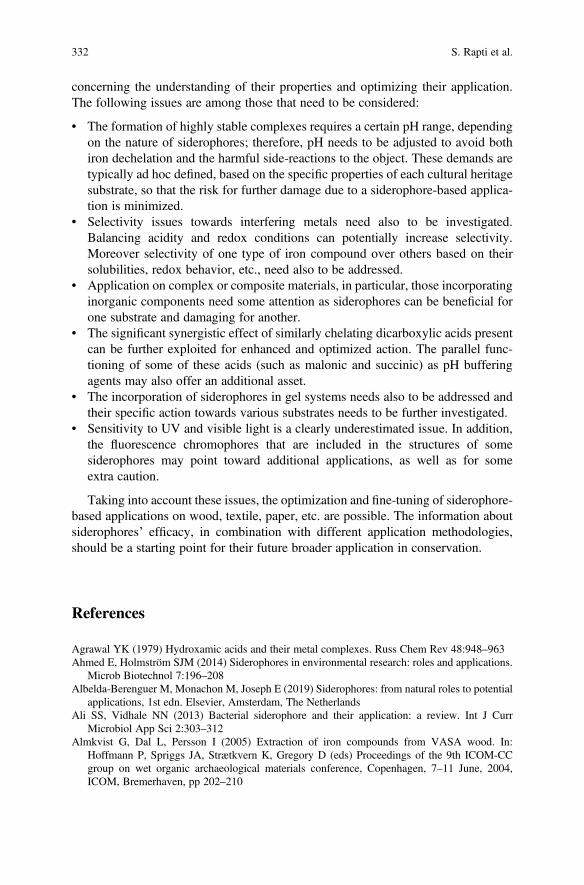
concerning the understanding of their properties and optimizing their application.Τhe following issues are among those that need to be considered:
• The formation of highly stable complexes requires a certain pH range, dependingon the nature of siderophores; therefore, pH needs to be adjusted to avoid bothiron dechelation and the harmful side-reactions to the object. These demands aretypically ad hoc defined, based on the specific properties of each cultural heritagesubstrate, so that the risk for further damage due to a siderophore-based applica-tion is minimized.
• Selectivity issues towards interfering metals need also to be investigated.Balancing acidity and redox conditions can potentially increase selectivity.Moreover selectivity of one type of iron compound over others based on theirsolubilities, redox behavior, etc., need also to be addressed.
• Application on complex or composite materials, in particular, those incorporatinginorganic components need some attention as siderophores can be beneficial forone substrate and damaging for another.
• The significant synergistic effect of similarly chelating dicarboxylic acids presentcan be further exploited for enhanced and optimized action. The parallel func-tioning of some of these acids (such as malonic and succinic) as pH bufferingagents may also offer an additional asset.
• The incorporation of siderophores in gel systems needs also to be addressed andtheir specific action towards various substrates needs to be further investigated.
• Sensitivity to UV and visible light is a clearly underestimated issue. In addition,the fluorescence chromophores that are included in the structures of somesiderophores may point toward additional applications, as well as for someextra caution.
Taking into account these issues, the optimization and fine-tuning of siderophore-based applications on wood, textile, paper, etc. are possible. The information aboutsiderophores’ efficacy, in combination with different application methodologies,should be a starting point for their future broader application in conservation.
References
Agrawal YK (1979) Hydroxamic acids and their metal complexes. Russ Chem Rev 48:948–963Ahmed E, Holmström SJM (2014) Siderophores in environmental research: roles and applications.
Microb Biotechnol 7:196–208Albelda-Berenguer M, Monachon M, Joseph E (2019) Siderophores: from natural roles to potential
applications, 1st edn. Elsevier, Amsterdam, The NetherlandsAli SS, Vidhale NN (2013) Bacterial siderophore and their application: a review. Int J Curr
Microbiol App Sci 2:303–312Almkvist G, Dal L, Persson I (2005) Extraction of iron compounds from VASA wood. In:
Hoffmann P, Spriggs JA, Strætkvern K, Gregory D (eds) Proceedings of the 9th ICOM-CCgroup on wet organic archaeological materials conference, Copenhagen, 7–11 June, 2004,ICOM, Bremerhaven, pp 202–210
332 S. Rapti et al.
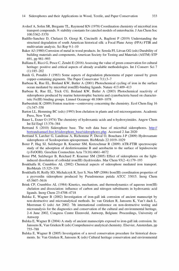
Avdeef A, Sofen SR, Bregante TL, Raymond KN (1978) Coordination chemistry of microbial irontransport compounds. 9: stability constants for catechol models of enterobactin. J Am Chem Soc100:5362–5370
Badillo-Sanchez D, Chelazzi D, Giorgi R, Cincinelli A, Baglioni P (2019) Understanding thestructural degradation of south American historical silk: a Focal Plane Array (FPA) FTIR andmultivariate analysis. Sci Rep 9:1–10
Baker AJ (1980) Corrosion of metal in wood products. In: Sereda PJ, Litvan GG (eds) Durability ofbuilding materials and components, American Society for Testing and Materials (ASTM) STP691, pp 981–993
Balliana E, Ricci G, Pesce C, Zendri E (2016) Assessing the value of green conservation for culturalheritage: positive and critical aspects of already available methodologies. Int J Conserv Sci 7(1):185–202
Banik G, Ponahlo J (1983) Some aspects of degradation phenomena of paper caused by greencopper-containing pigments. The Paper Conservator 7(1):3–7
Barbeau K, Rue EL, Bruland KW, Butler A (2001) Photochemical cycling of iron in the surfaceocean mediated by microbial iron(III)-binding ligands. Nature 413:409–413
Barbeau K, Rue EL, Trick CG, Bruland KW, Butler A (2003) Photochemical reactivity ofsiderophores produced by marine heterotrophic bacteria and cyanobacteria based on character-istic Fe(III) binding groups. Limnol Oceanogr 48:1069–1078
Barbusiński K (2009) Fenton reaction—controversy concerning the chemistry. Ecol Chem Eng 16(3):347–358
Barton LL, Hemming BC (eds) (1993) Iron chelation in plants and soil microorganisms. AcademicPress, New York
Bauer L, Exner O (1974) The chemistry of hydroxamic acids and n-hydroxyimides. Angew ChemInt Ed Engl 13:376–384
Bertrand S (2010) Siderophore base. The web data base of microbial siderophores. http://bertrandsamuel.free.fr/siderophore_base/siderophores.php. Accessed 2 Jan 2020
Bertrand S, Larcher G, Landreau A, Richomme P, Duval O, Bouchara J-P (2009) Hydroxamatesiderophores of Scedosporium apiospermum. BioMetals 22:1019–1029
Borer P, Hug SJ, Sulzberger B, Kraemer SM, Kretzschmar R (2009) ATR-FTIR spectroscopicstudy of the adsorption of desferrioxamine B and aerobactin to the surface of lepidocrocite(γ-FeOOH). Geochim Cosmochim Acta 73(16):4661–4672
Borer PM, Sulzberger B, Reichard P, Kraemer SM (2005) Effect of siderophores on the light-induced dissolution of colloidal iron(III) (hydr)oxides. Mar Chem 93(2–4):179–193
Boukhalfa H, Crumbliss AL (2002) Chemical aspects of siderophore mediated iron transport.BioMetals 15:325–339
Boukhalfa H, Reilly SD, Michalczyk R, Iyer S, Neu MP (2006) Iron(III) coordination properties ofa pyoverdin siderophore produced by Pseudomonas putida ATCC 33015. Inorg Chem45:5607–5616
Brink CP, Crumbliss AL (1984) Kinetics, mechanism, and thermodynamics of aqueous iron(III)chelation and dissociation: influence of carbon and nitrogen substituents in hydroxamic acidligands. Inorg Chem 23:4708–4718
Bulska E, Wagner B (2002) Investigation of iron-gall ink corrosion of ancient manuscript bynon-destructive and microanalytical methods. In: van Grieken R, Janssens K, Van’t dack L,Meersman G (eds) Art 2002: 7th international conference on non-destructive testing andmicroanalysis for the diagnostics and conservation of the cultural and environmental heritage,2–6 June 2002, Congress Centre Elzenveld, Antwerp, Belgium: Proceedings, University ofAntwerp
Bulska E, Wagner B (2004) A study of ancient manuscripts exposed to iron-gall ink corrosion. In:Janssens K, Van Grieken R (eds) Comprehensive analytical chemistry. Elsevier, Amsterdam, pp755–788
Bulska E, Wagner B (2005) Investigation of a novel conservation procedure for historical docu-ments. In: Van Grieken R, Janssens K (eds) Cultural heritage conservation and environmental
14 Siderophores and their Applications in Wood, Textile, and Paper Conservation 333
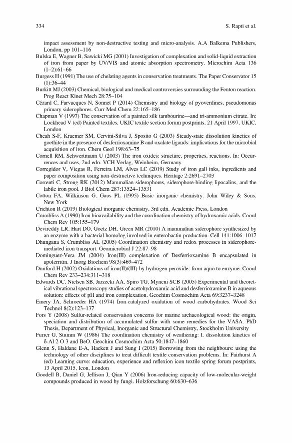
impact assessment by non-destructive testing and micro-analysis. A.A Balkema Publishers,London, pp 101–116
Bulska E, Wagner B, Sawicki MG (2001) Investigation of complexation and solid-liquid extractionof iron from paper by UV/VIS and atomic absorption spectrometry. Microchim Acta 136(1–2):61–66
Burgess H (1991) The use of chelating agents in conservation treatments. The Paper Conservator 15(1):36–44
Burkitt MJ (2003) Chemical, biological and medical controversies surrounding the Fenton reaction.Prog React Kinet Mech 28:75–104
Cézard C, Farvacques N, Sonnet P (2014) Chemistry and biology of pyoverdines, pseudomonasprimary siderophores. Curr Med Chem 22:165–186
Chapman V (1997) The conservation of a painted silk tambourine—and tri-ammonium citrate. In:Lockhead V (ed) Painted textiles, UKIC textile section forum postprints, 21 April 1997, UKIC,London
Cheah S-F, Kraemer SM, Cervini-Silva J, Sposito G (2003) Steady-state dissolution kinetics ofgoethite in the presence of desferrioxamine B and oxalate ligands: implications for the microbialacquisition of iron. Chem Geol 198:63–75
Cornell RM, Schwertmann U (2003) The iron oxides: structure, properties, reactions. In: Occur-rences and uses, 2nd edn. VCH Verlag, Weinheim, Germany
Corregidor V, Viegas R, Ferreira LM, Alves LC (2019) Study of iron gall inks, ingredients andpaper composition using non-destructive techniques. Heritage 2:2691–2703
Correnti C, Strong RK (2012) Mammalian siderophores, siderophore-binding lipocalins, and thelabile iron pool. J Biol Chem 287:13524–13531
Cotton FA, Wilkinson G, Gaus PL (1995) Basic inorganic chemistry. John Wiley & Sons,New York
Crichton R (2019) Biological inorganic chemistry, 3rd edn. Academic Press, LondonCrumbliss A (1990) Iron bioavailability and the coordination chemistry of hydroxamic acids. Coord
Chem Rev 105:155–179Devireddy LR, Hart DO, Goetz DH, Green MR (2010) A mammalian siderophore synthesized by
an enzyme with a bacterial homolog involved in enterobactin production. Cell 141:1006–1017Dhungana S, Crumbliss AL (2005) Coordination chemistry and redox processes in siderophore-
mediated iron transport. Geomicrobiol J 22:87–98Dominguez-Vera JM (2004) Iron(III) complexation of Desferrioxamine B encapsulated in
apoferritin. J Inorg Biochem 98(3):469–472Dunford H (2002) Oxidations of iron(II)/(III) by hydrogen peroxide: from aquo to enzyme. Coord
Chem Rev 233–234:311–318Edwards DC, Nielsen SB, Jarzecki AA, Spiro TG, Myneni SCB (2005) Experimental and theoret-
ical vibrational spectroscopy studies of acetohydroxamic acid and desferrioxamine B in aqueoussolution: effects of pH and iron complexation. Geochim Cosmochim Acta 69:3237–3248
Emery JA, Schroeder HA (1974) Iron-catalyzed oxidation of wood carbohydrates. Wood SciTechnol 8(2):123–137
Fors Y (2008) Sulfur-related conservation concerns for marine archaeological wood: the origin,speciation and distribution of accumulated sulfur with some remedies for the VASA. PhDThesis, Department of Physical, Inorganic and Structural Chemistry, Stockholm University
Furrer G, Stumm W (1986) The coordination chemistry of weathering: I. dissolution kinetics ofδ-Al 2 O 3 and BeO. Geochim Cosmochim Acta 50:1847–1860
Glenn S, Haldane E-A, Hackett J and Sung I (2015) Borrowing from the neighbours: using thetechnology of other disciplines to treat difficult textile conservation problems. In: Fairhurst A(ed) Learning curve: education, experience and reflexion icon textile spring forum postprints,13 April 2015, Icon, London
Goodell B, Daniel G, Jellison J, Qian Y (2006) Iron-reducing capacity of low-molecular-weightcompounds produced in wood by fungi. Holzforschung 60:630–636
334 S. Rapti et al.
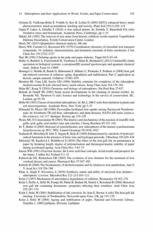
Grčman H, Velikonja-Bolta Š, Vodnik D, Kos B, Leštan D (2001) EDTA enhanced heavy metalphytoextraction: metal accumulation, leaching and toxicity. Plant Soil 235(1):105–114
Gupta P, Lakes A, Dziubla T (2016) A free radical primer. In: Dziubla T, Butterfield DA (eds)Oxidative stress and biomaterials. Academic Press, Cambridge, pp 1–33
Häkäri AE (1992) The removal of rust stains from historic cellulosic textile material. UnpublishedDiploma Dissertation, Textile Conservation Centre, London
Harris DC (2015) Quantitative chemical analysis. 8th ednHarris WR, Carrano CJ, Raymond KN (1979) Coordination chemistry of microbial iron transport
compounds. 16: isolation, characterization, and formation constants of ferric aerobactin. J AmChem Soc 101:2722–2727
Hart JR (1981) Chelating agents in the pulp and paper industry. Tappi 64(3):43–44Heller A, Barkleit A, Foerstendorf H, Tsushima S, Heim K, Bernhard G (2012) Curium(III) citrate
speciation in biological systems: a europium(III) assisted spectroscopic and quantum chemicalstudy. Dalton Trans 41:13969
Henniges U, Reibke R, Banik G, Huhsmann E, Hähner U, Prohaska T, Potthast A (2008) Iron gallink-induced corrosion of cellulose: aging, degradation and stabilization. Part 2: application onhistoric sample material. Cellulose 15:861–870
Hernlem BJ, Vane LM, Sayles GD (1996) Stability constants for complexes of the siderophoredesferrioxamine B with selected heavy metal cations. Inorg Chim Acta 244:179–184
Hider RC, Kong X (2010) Chemistry and biology of siderophores. Nat Prod Rep 27:637Hofenk de Graaff JH (1982) Some recent developments in the cleaning of ancient textiles. In:
Bromelle NS, Thomson G (eds) Science and technology in the service of conservation. IIC,London, pp 93–95
Höfte M (1993) Classes of microbial siderophores. In: BL L, HB C (eds) Iron chelation in plants andsoil microorganisms. Academic Press, New York, pp 3–27
Hohnadel D, Meyer JM (1986) Pyoverdine-facilitated iron uptake among fluorescent Pseudomo-nads. In: Swinburne TR (ed) Iron, siderophores, and plant diseases. NATO ASI series (series a:life sciences), vol 117. Springer, Boston, pp 119–129
Hynes MJ, Ó Coinceanainn M (2001) The kinetics and mechanisms of the reaction of iron(III) withgallic acid, gallic acid methyl ester and catechin. J Inorg Biochem 85:131–142
Ito Y, Butler A (2005) Structure of synechobactins, new siderophores of the marine cyanobacteriumSynechococcus sp. PCC 7002. Limnol Oceanogr 50:1918–1923
Iwahashi H, Morishita H, Ishii T, Sugata R, Kido R (1989) Enhancement by catechols of hydroxyl-radical formation in the presence of ferric ions and hydrogen peroxide. J Biochem 105:429–434
Jablonský M, Kazíková J, Holúbková S (2010) The effect of the iron-gall ink on permanence inpaper by breaking length, degree of polymerisation and thermogravimetric stability of paperduring accelerated ageing. Acta Chim Slov 3:63–73
Jensen WB (1991) Overview lecture: the Lewis acid-base concepts: recent results and prospects forthe future. J Adhes Sci Technol 5:1–21
Kalinowski DS, Richardson DR (2005) The evolution of iron chelators for the treatment of ironoverload disease and cancer. Pharmacol Rev 57:547–583
Keberle H (2006) The biochemistry of desferrioxamine and its relation to iron metabolism. Ann NY Acad Sci 119:758–768
Khan A, Singh P, Srivastava A (2018) Synthesis, nature and utility of universal iron chelator—siderophore: a review. Microbiol Res 212–213:103–111
Kolar J (1997) Mechanism of autoxidative degradation of cellulose. Restaurator 18:163–176Kolar J, Štolfa A, StrličM, Pompe M, Pihlar B, Budnar M, Simčič J, Reissland B (2006) Historical
iron gall ink containing documents—properties affecting their condition. Anal Chim Acta555:167–174
Kolar J, Strlic M (2001) Stabilisation of ink corrosion. In: Jean E, Brown A (eds) The Iron gall inkmeeting. University of Northumbria, Newcastle-upon-Tyne, UK, pp 135–139
Kolar J, Strlič M (2004) Ageing and stabilisation of paper. National and University Library,Turjaška 1, 1000 Ljubljana, Slovenia, Ljubljana
14 Siderophores and their Applications in Wood, Textile, and Paper Conservation 335
Kołodyńska D (2011) Chelating agents of a new generation as an alternative to conventionalchelators for heavy metal ions removal from different waste waters. In: Ning RY(ed) Expanding issues in desalination. InTech, Rijeka, pp 339–370
Konetschny-Rapp S, Jung G, Raymond KN, Meiwes J, Zähner H (1992) Solution thermodynamicsof the ferric complexes of new desferrioxamine siderophores obtained by directed fermentation.J Am Chem Soc 114:2224–2230
Koppenol WH (1993) The centennial of the Fenton reaction. Free Radic Biol Med 15:645–651Kraemer SM (2004) Iron oxide dissolution and solubility in the presence of siderophores. Aquat
Sci—Res Across Boundaries 66:3–18Kraemer SM, Butler A, Borer P, Cervini-Silva J (2005) Siderophores and the dissolution of iron-
bearing minerals in marine systems. In: Banfield JF, Cervini-Silva J, Nealson K (eds) Moleculargeomicrobiology. De Gruyter, Berlin, Boston, pp 53–84
Kumar L, Meena NL, Singh U (2016) Role of phytosiderophores in acquisition of iron and othermicronutrients in food legumes. In: Singh U, Praharaj C, Singh S, Singh N (eds) Biofortificationof food crops. Springer, New Delh, pp 291–302
Liu Y, Kralj-cigić I, Strlič M (2017) Kinetics of accelerated degradation of historic iron gallink-containing paper. Polym Degrad Stab. https://doi.org/10.1016/j.polymdegradstab.2017.07.010
Loring JS, Simanova AA, Persson P (2008) Highly mobile iron pool from a dissolution—readsorption process. Langmuir 24:7054–7057
Margariti C (2003) The use of chelating agents in textile conservation: an investigation of theefficiency and effects on the use of three chelating agents for the removal of copper and ironstaining from cotton textiles. In: Dawson L, Berkouwer M (eds) Dust, sweat and tears: recentadvances in cleaning techniques, UKIC Textile Section, AGM Spring Forum, 7 April 2003,UKIC, London, pp 28–38
Marusak RA, Doan K, Cummings SD (2006) Integrated approach to coordination chemistry: aninorganic laboratory guide, John Wiley & Sons, New Jersey
Miethke M, Marahiel MA (2007) Siderophore-based iron acquisition and pathogen control.Microbiol Mol Biol Rev 71:413–451
Milburn RM, Vosburgh WC (1955) A spectrophotometric study of the hydrolysis of iron(III) ion.II: polynuclear species. J Am Chem Soc 77:1352–1355
Monachon M, Albelda-Berenguer M, Joseph E (2019) Bio-based treatment for the extraction ofproblematic iron sulphides from waterlogged archaeological wood. In: Book of abstracts of the14th ICOM-CC group on wet organic archaeological materials conference, Portsmouth, UK,20–24 May 2019, ICOM, Portsmouth, pp 46–47
Monzyk B, Crumbliss AL (1979) Mechanism of ligand substitution on high-spin iron(III) byhydroxamic acid chelators: thermodynamic and kinetic studies on the formation and dissocia-tion of a series of monohydroxamatoiron(III) complexes. J Am Chem Soc 101:6203–6213
Moran JF, Klucas RV, Grayer RJ, Abian J, Becana M (1997) Complexes of iron with phenoliccompounds from soybean nodules and other legume tissues: prooxidant and antioxidant prop-erties. Free Radic Biol Med 22:861–870
Nakamoto K (2009) Infrared and Raman spectra of inorganic and coordination compounds. In:Theory and applications to inorganic chemistry, 6th edn. John Wiley & Sons, New York
Nakamura Y (2000) A simple phenolic antioxidant protocatechuic acid enhances tumor promotionand oxidative stress in female ICR mouse skin: dose- and timing-dependent enhancement andinvolvement of bioactivation by tyrosinase. Carcinogenesis 21:1899–1907
Neevel JG (1995) Phytate: a potential conservation agent for the treatment of ink corrosion causedby iron gall inks. Restaurator 16(3):143–160
Neilands JB (1995) Siderophores: structure and function of microbial iron transport compounds. JBiol Chem 270:26723–26726
Nörtemann B (1999) Biodegradation of EDTA. Appl Microbiol Biotechnol 51(6):751–759Nörtemann B (2005) Biodegradation of chelating agents: EDTA, DTPA, PDTA, NTA, and EDDS.
Biogeochemistry of chelating agents. ACS symposium series, American Society, Washington,DC, pp 150–170
336 S. Rapti et al.
Pearson R (1973) Hard and soft acids and bases. Dowden, Hutchinson and Ross, StroudsburgPepper IL, Gentry TJ (2015) Earth environments. In: Pepper IL, Gerba CP, Gentry TJ (eds)
Environmental microbiology. Elsevier, Amsterdam, pp 59–88Phenix A, Burnstock A (1992) The removal of surface dirt on paintings with chelating agents. The
Conservator 16(1):28–38Rapti S, Boyatzis S, Rivers S, Velios A, Pournou A (2017) Removing iron stains from wood and
textile objects: assessing gelled siderophores as novel green chelators. In: Angelova LV,Ormsby B, Townsend JH, Wolbers R (eds) Gels in the conservation of art. Archetype Books,London, pp 343–348
Rapti S, Rivers S, Pournou A (2015) Removing iron corrosion products from museum artefacts:Investigating the effectiveness of innovative green chelators. In: International conferenceScience in Technology, SCinTE, Athens
Reichard PU, Kraemer SM, Frazier SW, Kretzschmar R (2005) Goethite dissolution in the presenceof phytosiderophores: rates, mechanisms, and the synergistic effect of oxalate. Plant Soil 276(1–2):115–132
Reichard PU, Kretzschmar R, Kraemer SM (2007) Dissolution mechanisms of goethite in thepresence of siderophores and organic acids. Geochim Cosmochim Acta 71:5635–5650
Renshaw JC, Robson GD, Trinci APJ, Wiebe MG, Livens FR, Collison D, Taylor RJ (2002) Fungalsiderophores: structures, functions and applications. Mycol Res 106:1123–1142
Richards V, Kasi K, Godfrey I (2012) Iron removal from waterlogged wood and the effects on woodchemistry. In: Straetkvern K, Williams E, (eds) Proceedings of the 11th ICOM-CC group on wetorganic archaeological materials conference, ICOM, Greenville, 2010. Lulu, Greenville, pp383–400
Rivers S, Ummey N (2003) Conservation of furniture. Butterworth-Heinemann, OxfordRomero R, Salgado PR, Soto C, Contreras D, Melin V (2018) An experimental validated
computational–method for pKa determination of substituted 1,2-dihydroxybenzenes. FrontChem 6:1–11
Römheld V (1991) The role of phytosiderophores in acquisition of iron and other micronutrients ingraminaceous species: an ecological approach. Plant Soil 130:127–134
Saha M, Sarkar S, Sarkar B, Sharma BK, Bhattacharjee S, Tribedi P (2016) Microbial siderophoresand their potential applications: a review. Environ Sci Pollut Res 23:3984–3999
Saha R, Saha N, Donofrio RS, Bestervelt LL (2013) Microbial siderophores: a mini review. J BasicMicrobiol 53:303–317
Salgado P, Melin V, Contreras D, Moreno Y, Mansilla HD (2013) Fenton reaction driven by ironligands. J Chil Chem Soc 58:2096–2101
Samson SD, Eggleston CM (1998) Active sites and the non-steady-state dissolution of hematite.Environ Sci Technol 32:2871–2875
Samson SD, Stillings LL, Eggleston CM (2000) The depletion and regeneration of dissolution-active sites at the mineral-water interface: I. Fe, Al, and in sesquioxides. Geochim CosmochimActa 64:3471–3484
Sapieszko RS, Patel RC, Matijevic E (1977) Ferric hydrous oxide sols. 2: thermodynamics ofaqueous hydroxo and sulfato ferric complexes. J Phys Chem 81:1061–1068
Schwertmann U (1991) Solubility and dissolution of iron oxides. In: Plant and soil; selected papersfrom the fifth international symposium on iron nutrition and interactions in plants, pp 1–25
Scott DA, Eggert G (2009) Iron and steel in art: corrosion, colorants, conservation. ArchetypePublications, London
Selwyn L, Tse S (2009) The chemistry of sodium dithionite and its use in conservation. StudConserv 54:61–73
Sharpe PC, Richardson DR, Kalinowski DS, Bernhardt PV (2011) Synthetic and natural products asiron chelators. Curr Top Med Chem 11:591–607
Shenker M, Chen Y, Hadar Y (1996) Stability constants of the fungal siderophore rhizoferrin withvarious microelements and calcium. Soil Sci Soc Am J 60:1140
14 Siderophores and their Applications in Wood, Textile, and Paper Conservation 337
Siddall TH, Vosburgh WC (1951) A spectrophotometric study of the hydrolysis of iron(III) ion. JAm Chem Soc 73:4270–4272
Sillanpää M (1997) Environmental fate of EDTA and DTPA. Reviews of environmental contam-ination and toxicology, vol 152. Springer, New York, pp 85–111
Silva AMN, Kong X, Hider RC (2009) Determination of the pKa value of the hydroxyl group in theα-hydroxycarboxylates citrate, malate and lactate by 13C NMR: implications for metal coordi-nation in biological systems. BioMetals 22:771–778
Singh N, Karpichev Y, Sharma R, Gupta B, Sahu AK, Satnami ML, Ghosh KK (2015) Fromα-nucleophiles to functionalized aggregates: exploring the reactivity of hydroxamate iontowards esterolytic reactions in micelles. Org Biomol Chem 13:1–8
Slavin J (1990) The removal of salt deposits from decorative paintings on paper. In: Hackney S,Townsend JH, Eastaugh N (eds) Dirt and pictures separated, UKIC and tate gallery conference,UKIC, London, pp 49–50
Strlič M, Kolar J, Pihlar B (2001) Some preventive cellulose antioxidants studied by an aromatichydroxylation assay. Polym Degrad Stab 73(3):535–539
Stumm W, Furrer G (1987) The dissolution of oxides and aluminum silicates: examples of surface-coordination-controlled-kinetics. In: Stumm W (ed) Aquatic surface chemistry. John Wiley andSons, Somerset, NJ, pp 97–219
Sugiura Y, Nomoto K (1984) Phytosiderophores structures and properties of mugineic acids andtheir metal complexes. In: Siderophores from microorganisms and plants: structure and bond-ing, vol 58. Springer, Heidelberg; Berlin, pp 107–135
Thiem D, Złoch M, Gadzała-Kopciuch R, Szymańska S, Baum C, Hrynkiewicz K (2018)Cadmium-induced changes in the production of siderophores by a plant growth promotingstrain of Pseudomonas fulva. J Basic Microbiol 58:623–632
Timár-Balázsy AT, Eastop D (1998) Chemical principles in textile conservation. Butterworths-Heinemann, London
Todokoro T, Fukuda K, Matsumura K, Irie M, Hata Y (2016) Production of the natural iron chelatordeferriferrichrysin from Aspergillus oryzae and evaluation as a novel food-grade antioxidant. JSci Food Agric 96(9):2998–3006
Tseng CF, Burger A, Mislin GLA, Schalk IJ, Yu SSF, Chan SI, Abdallah MA (2006) Bacterialsiderophores: the solution stoichiometry and coordination of the Fe(III) complexes of pyochelinand related compounds. J Biol Inorg Chem 11:419–432
Wagner B, Bulska E (2003) Towards a new conservation method for ancient manuscripts byinactivation of iron via complexation and extraction. Anal Bioanal Chem 375:1148–1153
Wagner B, Bulska E (2004) On the use of laser ablation inductively coupled plasma massspectrometry for the investigation of the written heritage. J Anal At Spectrom 19(10):1325–1329
Wagner B, Bulska E, Hulanicki A, Heck M, Ortner HM (2001a) Topochemical investigation ofancient manuscripts. Fresenius J Anal Chem 369(7-8):674–679
Wagner B, Bulska E, Meisel T, Wegscheider W (2001b) Use of atomic spectrometry for theinvestigation of ancient manuscripts. J Anal At Spectrom 16(4):417–420
Wang W, Qiu Z, Tan H, Cao L (2014) Siderophore production by actinobacteria. BioMetals27:623–631
Wardman P, Candeias LP (1996) Fenton chemistry: an introduction. Radiat Res 145(5):523–531Wilkins RG (1991) Kinetics and mechanism of reactions of transition metal complexes, 2nd edn.
VCH, WeinheimYang J, Bremer PJ, Lamont IL, McQuillan AJ (2006) Infrared spectroscopic studies of siderophore-
related hydroxamic acid ligands adsorbed on titanium dioxide. Langmuir 22:10109–10117Zeronian SH, Inglesby MK (1995) Bleaching of cellulose by hydrogen peroxide. Cellulose 2
(4):265–272Złoch M, Thiem D, Gadzała-Kopciuch R, Hrynkiewicz K (2016) Synthesis of siderophores by
plant-associated metallotolerant bacteria under exposure to Cd2+. Chemosphere 156:312–325
338 S. Rapti et al.

Open Access This chapter is licensed under the terms of the Creative Commons Attribution 4.0International License (http://creativecommons.org/licenses/by/4.0/), which permits use, sharing,adaptation, distribution and reproduction in any medium or format, as long as you give appropriatecredit to the original author(s) and the source, provide a link to the Creative Commons license andindicate if changes were made.
The images or other third party material in this chapter are included in the chapter’s CreativeCommons license, unless indicated otherwise in a credit line to the material. If material is notincluded in the chapter’s Creative Commons license and your intended use is not permitted bystatutory regulation or exceeds the permitted use, you will need to obtain permission directly fromthe copyright holder.
14 Siderophores and their Applications in Wood, Textile, and Paper Conservation 339
Chapter 15Organic Green Corrosion InhibitorsDerived from Natural and/or BiologicalSources for Conservation of Metals CulturalHeritage
Vasilike Argyropoulos, Stamatis C. Boyatzis, Maria Giannoulaki,Elodie Guilminot, and Aggeliki Zacharopoulou
Abstract In the last decade, there has been an increase in research related to greencorrosion inhibitors for conservation of metals cultural heritage to help promotesustainable practices in the field that are safe, environmentally friendly, and ecolog-ically acceptable. The most common are organic substances derived either fromnatural and/or biological sources: plant extracts and oils, amino acids, microorgan-isms, and biopolymers. The chapter will provide a review of these substances ascorrosion inhibitors for metals conservation, by discussing the state-of-the-artresearch to date, with a special focus on cysteine. Most of the research has focusedon the examination of such inhibitors on metal coupons with or without corrosionproducts using electrochemical techniques or weight-loss measurements to deter-mine their effectiveness. Some of these studies have also considered the conserva-tion principles for practice, i.e., reversibility of the treatment and the visual aspect ofthe modification of the treated metal surface. However, before such green inhibitorscan be routinely applied by conservators, more research is required on their appli-cation to real artefacts/monuments using in situ corrosion measurements. Further-more, given that the composition of a green inhibitor is highly dependent on itsextraction process, research must also involve identifying the specific adsorptionmodels and involved mechanisms to ensure reproducibility of results.
V. Argyropoulos (*) · S. C. Boyatzis · M. GiannoulakiDepartment of Conservation of Antiquities and Works of Art, University of West Attica,Athens, Greecee-mail: [email protected]; [email protected]; [email protected]
E. GuilminotGPLA Arc’Antique, Nantes, Francee-mail: [email protected]
A. ZacharopoulouSchool of Chemical Engineering Materials Science and Engineering Department, NationalTechnical University of Athens, Athens, Greecee-mail: [email protected]
© The Author(s) 2021E. Joseph (ed.), Microorganisms in the Deterioration and Preservation of CulturalHeritage, https://doi.org/10.1007/978-3-030-69411-1_15
341
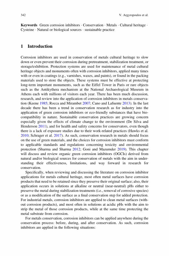
Keywords Green corrosion inhibitors · Conservation · Metals · Cultural heritage ·Cysteine · Natural or biological sources · sustainable practice
1 Introduction
Corrosion inhibitors are used in conservation of metals cultural heritage to slowdown or even prevent their corrosion during pretreatment, stabilization treatment, orstorage/exhibition. Protection systems are used for maintenance of metal culturalheritage objects and monuments often with corrosion inhibitors, applied many timeswith or even in coatings (e.g., varnishes, waxes, and paints), or found in the packingmaterials used to store the objects. These systems must be effective at protectinglong-term important monuments, such as the Eiffel Tower in Paris or rare objectssuch as the Antikythera mechanism at the National Archaeological Museum inAthens each with millions of visitors each year. There has been much discussion,research, and review into the application of corrosion inhibitors in metals conserva-tion (Keene 1985; Rocca and Mirambet 2007; Cano and Lafuente 2013). In the lastdecade there has been a trend in conservation research as for industry into theapplication of green corrosion inhibitors or eco-friendly substances that have bio-compatibility in nature. Sustainable conservation practices are growing concernespecially given the effects of climate change to the environment (De Silva andHenderson 2011), and for health and safety concerns for conservators, even thoughthere is a lack of exposure studies due to their work-related practices (Hawks et al.2010; Schrager et al. 2017). As such, conservation research in metals should focuson the use of green materials, and the choices for corrosion inhibitors must conformto applicable standards and regulations concerning toxicity and environmentalprotection (Sharma and Sharma 2012; Goni and Mazumder 2019). This chapterwill discuss and review organic green corrosion inhibitors (OGCIs) derived fromnatural and/or biological sources for conservation of metals with the aim in under-standing their effectiveness, limitations, and way forward in research forconservation.
Specifically, when reviewing and discussing the literature on corrosion inhibitorapplications for metals cultural heritage, most often metal surfaces have corrosionproducts that need to be retained since they preserve their original surface; also, theirapplication occurs in solutions at alkaline or neutral (near-neutral) pHs either topreserve the metal during stabilization treatments (i.e., removal of corrosive species)or as a modification of the surface as a final conservation step for added protection.For industrial metals, corrosion inhibitors are applied to clean metal surfaces (with-out corrosion products), and most often in solutions at acidic pHs with the aim tostrip the metal of those corrosion products, while at the same time protecting themetal substrate from corrosion.
For metals conservation, corrosion inhibitors can be applied anywhere during theconservation process: before, during, and after conservation. As such, corrosioninhibitors are applied in the following situations:
342 V. Argyropoulos et al.
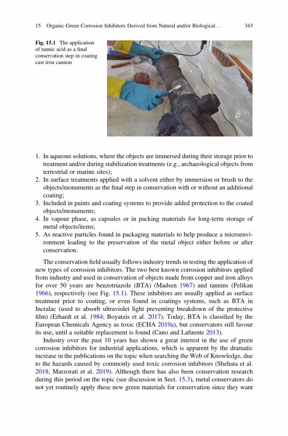
1. In aqueous solutions, where the objects are immersed during their storage prior totreatment and/or during stabilization treatments (e.g., archaeological objects fromterrestrial or marine sites);
2. In surface treatments applied with a solvent either by immersion or brush to theobjects/monuments as the final step in conservation with or without an additionalcoating;
3. Included in paints and coating systems to provide added protection to the coatedobjects/monuments;
4. In vapour phase, as capsules or in packing materials for long-term storage ofmetal objects/items;
5. As reactive particles found in packaging materials to help produce a microenvi-ronment leading to the preservation of the metal object either before or afterconservation.
The conservation field usually follows industry trends in testing the application ofnew types of corrosion inhibitors. The two best known corrosion inhibitors appliedfrom industry and used in conservation of objects made from copper and iron alloysfor over 50 years are benzotriazole (BTA) (Madsen 1967) and tannins (Pelikan1966), respectively (see Fig. 15.1). These inhibitors are usually applied as surfacetreatment prior to coating, or even found in coatings systems, such as BTA inIncralac (used to absorb ultraviolet light preventing breakdown of the protectivefilm) (Erhardt et al. 1984; Boyatzis et al. 2017). Today, BTA is classified by theEuropean Chemicals Agency as toxic (ECHA 2019a), but conservators still favourits use, until a suitable replacement is found (Cano and Lafuente 2013).
Industry over the past 10 years has shown a great interest in the use of greencorrosion inhibitors for industrial applications, which is apparent by the dramaticincrease in the publications on the topic when searching the Web of Knowledge, dueto the hazards caused by commonly used toxic corrosion inhibitors (Shehata et al.2018; Marzorati et al. 2019). Although there has also been conservation researchduring this period on the topic (see discussion in Sect. 15.3), metal conservators donot yet routinely apply these new green materials for conservation since they want
Fig. 15.1 The applicationof tannic acid as a finalconservation step in coatingcast iron cannon
15 Organic Green Corrosion Inhibitors Derived from Natural and/or Biological. . . 343
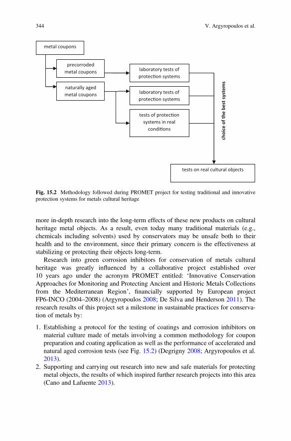
more in-depth research into the long-term effects of these new products on culturalheritage metal objects. As a result, even today many traditional materials (e.g.,chemicals including solvents) used by conservators may be unsafe both to theirhealth and to the environment, since their primary concern is the effectiveness atstabilizing or protecting their objects long-term.
Research into green corrosion inhibitors for conservation of metals culturalheritage was greatly influenced by a collaborative project established over10 years ago under the acronym PROMET entitled: ‘Innovative ConservationApproaches for Monitoring and Protecting Ancient and Historic Metals Collectionsfrom the Mediterranean Region’, financially supported by European projectFP6-INCO (2004–2008) (Argyropoulos 2008; De Silva and Henderson 2011). Theresearch results of this project set a milestone in sustainable practices for conserva-tion of metals by:
1. Establishing a protocol for the testing of coatings and corrosion inhibitors onmaterial culture made of metals involving a common methodology for couponpreparation and coating application as well as the performance of accelerated andnatural aged corrosion tests (see Fig. 15.2) (Degrigny 2008; Argyropoulos et al.2013).
2. Supporting and carrying out research into new and safe materials for protectingmetal objects, the results of which inspired further research projects into this area(Cano and Lafuente 2013).
metal coupons
precorroded metal coupons
naturally aged metal coupons
laboratory tests of protec�on systems
laboratory tests of protec�on systems
tests of protec�on systems in real
condi�ons
tests on real cultural objects
choi
ce o
f the
bes
t sys
tem
s
Fig. 15.2 Methodology followed during PROMET project for testing traditional and innovativeprotection systems for metals cultural heritage
344 V. Argyropoulos et al.
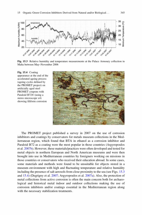
The PROMET project published a survey in 2007 on the use of corrosioninhibitors and coatings by conservators for metals museum collections in the Med-iterranean region, which found that BTA in ethanol as a corrosion inhibitor andParaloid B72 as a coating were the most popular in those countries (Argyropouloset al. 2007b). However, these materials/practices were often developed and tested formetal objects in northern European and North American museums and were thenbrought into use in Mediterranean countries by foreigners working on missions inthose countries or conservators who received their education abroad. In some cases,some materials and methods were found to be unsuitable for objects stored in amuseum environment with high and fluctuating temperature and relative humidityincluding the presence of salt aerosols from close proximity to the sea (see Figs. 15.3and 15.4) (Degrigny et al. 2007; Argyropoulos et al. 2007a). Also, the protection ofmetal collections from active corrosion is often the main concern both for archaeo-logical and historical metal indoor and outdoor collections making the use ofcorrosion inhibitors and/or coatings essential in the Mediterranean region alongwith the necessary stabilization treatments.
Fig. 15.3 Relative humidity and temperature measurements at the Palace Armoury collection inMalta between May–November 2006
Fig. 15.4 Coatingappearance at the end of theaccelerated ageing process(ageing cycles defined bythe PROMET project) onartificially aged steelPROMET coupons withParaloid B72® (using astereo microscope x4)showing filiform corrosion
15 Organic Green Corrosion Inhibitors Derived from Natural and/or Biological. . . 345
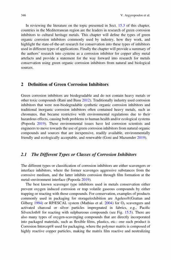
In reviewing the literature on the topic presented in Sect. 15.3 of this chapter,countries in the Mediterranean region are the leaders in research of green corrosioninhibitors to cultural heritage metals. This chapter will define the types of greenorganic corrosion inhibitors commonly used by industry, how they work, andhighlight the state-of-the-art research for conservation into these types of inhibitorsused in different types of applications. Finally the chapter will provide a summary ofthe authors’ research into cysteine as a corrosion inhibitor for copper alloy metalartefacts and provide a statement for the way forward into research for metalsconservation using green organic corrosion inhibitors from natural and biologicalsources.
2 Definition of Green Corrosion Inhibitors
Green corrosion inhibitors are biodegradable and do not contain heavy metals orother toxic compounds (Rani and Basu 2012). Traditionally industry used corrosioninhibitors that were non-biodegradable synthetic organic corrosion inhibitors andtraditional inorganic corrosion inhibitors often contained heavy metals, such aschromates, that became restrictive with environmental regulations due to theirhazardous effects, causing both problems to human health and/or ecological systems(Popoola 2019). These environmental issues have led corrosion scientists andengineers to move towards the use of green corrosion inhibitors from natural organiccompounds and sources that are inexpensive, readily available, environmentallyfriendly and ecologically acceptable, and renewable (Goni and Mazumder 2019).
2.1 The Different Types or Classes of Corrosion Inhibitors
The different types or classification of corrosion inhibitors are either scavengers orinterface inhibitors, where the former scavenges aggressive substances from thecorrosive medium, and the latter inhibits corrosion through film formation at themetal–environment interface (Popoola 2019).
The best known scavenger type inhibitors used in metals conservation eitherprevent oxygen induced corrosion or trap volatile gaseous compounds by eithertrapping or reacting with those compounds. For conservation, examples of productscommonly used in packaging for storage/exhibition are Ageless®(Grattan andGilberg 1994) or RP/ESCAL system (Mathias et al. 2004) for O2 scavengers andactivated charcoal or silver particles impregnated in fabrics, e.g., PacificSilvercloth® for reacting with sulphureous compounds (see Fig. 15.5). There arealso many types of oxygen-scavenging compounds that are directly incorporatedinto packaged materials, such as flexible films, plastics, etc.: one such product isCorrosion Intercept® used for packaging, where the polymer matrix is composed ofhighly reactive copper particles, making the matrix film reactive and neutralizing
346 V. Argyropoulos et al.
corrosive gases they come in contact with. There are also examples of such films,plastics that are bio-based containing, for example, gallic acid as an oxygen scav-enger (Pant et al. 2017), but to date there is no application in conservation to theauthors’ knowledge.
Interface inhibitors can be classified as either vapour phase or liquid phase, withmany known applications in conservation of metals. The vapour-phase inhibitors orvapour corrosion inhibitors (VCI) provide temporary atmospheric corrosion protec-tion especially in closed environments by loosely impregnating wrapping paperinside a closed container. Some popular type paper rolls used for wrapping differenttypes metals from museum collections contain VCI, such as Zerust® and CortecVpCI films.
However, the most prominent corrosion inhibitors used both by industry and forconservation of metals are the liquid-phase inhibitors, which can be furthersubdivided into cathodic, anodic, or mixed based on the reaction-type inhibition,which can be any of the cathodic, anodic, or both electrochemical reactions. Canoand Lafuente (2013) provide a good review for these types of corrosion inhibitorsused in metals conservation according to the different types of metal alloys. Theyconfirm through their review that the most commonly applied inhibitors to thesurface of a copper and iron alloyed objects are BTA and tannins, respectively.They also state that as opposed to industrial applications, for the metal conservationfield, interface inhibitors are used to produce surface modifications or films throughadsorption onto the metal surface using most often immersion followed by dryingand coating. They briefly touch upon research in conservation using green inhibitors,but state that their application for metals conservation is limited.
Like regular corrosion inhibitors, green inhibitors have the same classificationsbut are derived from organic natural and/or biological sources as opposed tosynthetic and for inorganic do not contain heavy metals. For the latter, some rareearth compounds (e.g., CeCl3 and LaCl3) are considered as inorganic green corro-sion inhibitors and for the former natural and/or biological sources for organic greencorrosion inhibitors are plants, drugs, amino acids, surfactants, biopolymers, andionic liquids (El Ibrahimi et al. 2020). Others classify organic green inhibitors
Fig. 15.5 An iron artefactstored in a sealedmicroenvironmentcontaining RP/ESCAL tocontrol the relative humidityand the O2 levels withscavenger inhibitor
15 Organic Green Corrosion Inhibitors Derived from Natural and/or Biological. . . 347
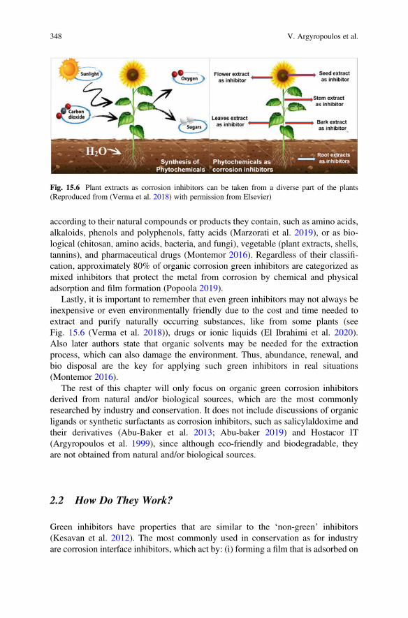
according to their natural compounds or products they contain, such as amino acids,alkaloids, phenols and polyphenols, fatty acids (Marzorati et al. 2019), or as bio-logical (chitosan, amino acids, bacteria, and fungi), vegetable (plant extracts, shells,tannins), and pharmaceutical drugs (Montemor 2016). Regardless of their classifi-cation, approximately 80% of organic corrosion green inhibitors are categorized asmixed inhibitors that protect the metal from corrosion by chemical and physicaladsorption and film formation (Popoola 2019).
Lastly, it is important to remember that even green inhibitors may not always beinexpensive or even environmentally friendly due to the cost and time needed toextract and purify naturally occurring substances, like from some plants (seeFig. 15.6 (Verma et al. 2018)), drugs or ionic liquids (El Ibrahimi et al. 2020).Also later authors state that organic solvents may be needed for the extractionprocess, which can also damage the environment. Thus, abundance, renewal, andbio disposal are the key for applying such green inhibitors in real situations(Montemor 2016).
The rest of this chapter will only focus on organic green corrosion inhibitorsderived from natural and/or biological sources, which are the most commonlyresearched by industry and conservation. It does not include discussions of organicligands or synthetic surfactants as corrosion inhibitors, such as salicylaldoxime andtheir derivatives (Abu-Baker et al. 2013; Abu-baker 2019) and Hostacor IT(Argyropoulos et al. 1999), since although eco-friendly and biodegradable, theyare not obtained from natural and/or biological sources.
2.2 How Do They Work?
Green inhibitors have properties that are similar to the ‘non-green’ inhibitors(Kesavan et al. 2012). The most commonly used in conservation as for industryare corrosion interface inhibitors, which act by: (i) forming a film that is adsorbed on
Fig. 15.6 Plant extracts as corrosion inhibitors can be taken from a diverse part of the plants(Reproduced from (Verma et al. 2018) with permission from Elsevier)
348 V. Argyropoulos et al.
the metal surface; (ii) producing corrosion products that acts as a passivator; and (iii)yielding precipitates that can eliminate or inactivate aggressive constituents (Goniand Mazumder 2019). Their action on the metal surface takes place via physical orchemical adsorption which removes water or corrosive species from the surface byproviding a barrier film formation (Goni and Mazumder 2019). Organic greencorrosion inhibitors are effective due to the availability of organic compoundswith polar functional groups with N, O, and S heteroatoms, which have a shieldingeffect and by repelling aqueous corrosive species away from the metal surface(Popoola 2019).
The most common organic green inhibitors to contain these heteroatoms, whichact to inhibit metals from corrosion are: derived from plant extracts adsorbing on themetal surface and forming a compact barrier film (Sharma and Sharma 2012); aminoacids which contain molecules with functional groups like carboxyl (-COOH) group,amino (-NH2), and S thiol (Goni and Mazumder 2019); and drugs because they aresynthesized from natural products that contain the presence of heteroatoms, benzenering, and heterocycles, such as thiophenes, pyridine, isoxazoles (Goni andMazumder 2019). Also, natural biopolymers such as chitosan are known to havegood complexing qualities with their -NH2 groups with metallic ions that can lead tocorrosion inhibition in some cases (Jmiai et al. 2017).
Another type of organic green corrosion inhibitor are surfactants which are madeup of two parts, a polar hydrophilic group and a nonpolar hydrophobic group, wherethe former adsorbs onto the metal surface. The charge on the polar head groupclassifies whether the surfactants are classified as anionic, cationic, nonionic, orzwitterionic (Malik et al. 2011). As well, carboxylates on copper alloy artefacts,where the reaction between the copper ions and the carboxylates leads to theprecipitation of metal carboxylates, such as Cu(C10)2 (metal soaps) on the surfaceof the corrosion products layer resulting inhibition; the soaps act as surfactant-typecorrosion inhibitor due to the carboxylate part of the molecule providing a hydro-philic character, but have also hydrophobic properties, in relation with the aliphaticchain (L’Héronde et al. 2019). Linseed oil in paints is another type of surfactantcorrosion inhibitor used as a traditional method to inhibit corrosion of the coppersubstrate for oil paintings (Pavlopoulou and Watkinson 2006).
In a completely different action are those of bio-based treatments that convertunstable corrosion products or corrosive species on metals to stable corrosionproducts and inhibit any further corrosion. One type of bio-based treatment whichleads to inhibition of copper alloy coupons with corrosion products involves the useof fungal strain known for their ability to produce oxalic acid; when used it canconvert the atacamite layer completely into copper oxalates, which results inenhancement of the surface inhibition against corrosion (Albini et al. 2018). Foriron alloys, microorganisms in the form of bacteria have been used to transformferric iron corrosion products (goethite and lepidocrocite) into stable ferrous iron-bearing minerals (vivianite and siderite) (Kooli et al. 2019).
15 Organic Green Corrosion Inhibitors Derived from Natural and/or Biological. . . 349
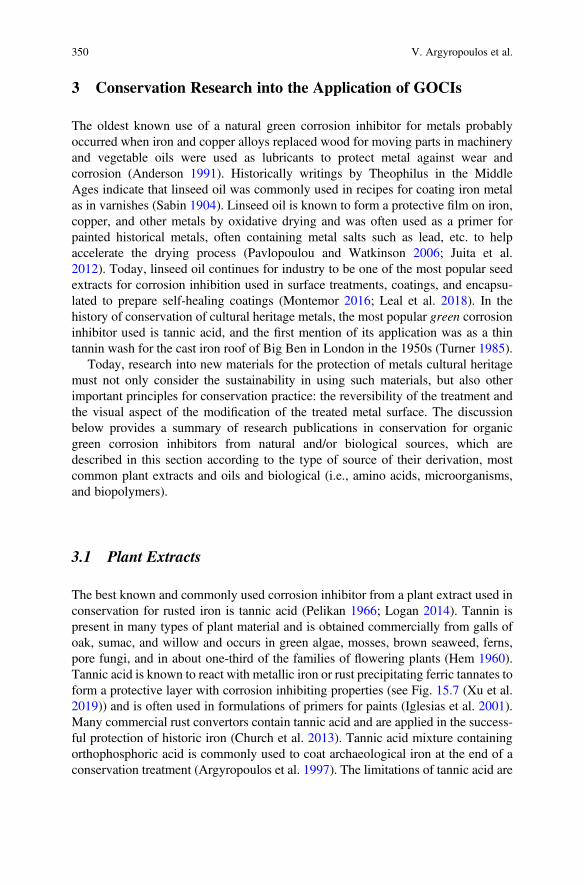
3 Conservation Research into the Application of GOCIs
The oldest known use of a natural green corrosion inhibitor for metals probablyoccurred when iron and copper alloys replaced wood for moving parts in machineryand vegetable oils were used as lubricants to protect metal against wear andcorrosion (Anderson 1991). Historically writings by Theophilus in the MiddleAges indicate that linseed oil was commonly used in recipes for coating iron metalas in varnishes (Sabin 1904). Linseed oil is known to form a protective film on iron,copper, and other metals by oxidative drying and was often used as a primer forpainted historical metals, often containing metal salts such as lead, etc. to helpaccelerate the drying process (Pavlopoulou and Watkinson 2006; Juita et al.2012). Today, linseed oil continues for industry to be one of the most popular seedextracts for corrosion inhibition used in surface treatments, coatings, and encapsu-lated to prepare self-healing coatings (Montemor 2016; Leal et al. 2018). In thehistory of conservation of cultural heritage metals, the most popular green corrosioninhibitor used is tannic acid, and the first mention of its application was as a thintannin wash for the cast iron roof of Big Ben in London in the 1950s (Turner 1985).
Today, research into new materials for the protection of metals cultural heritagemust not only consider the sustainability in using such materials, but also otherimportant principles for conservation practice: the reversibility of the treatment andthe visual aspect of the modification of the treated metal surface. The discussionbelow provides a summary of research publications in conservation for organicgreen corrosion inhibitors from natural and/or biological sources, which aredescribed in this section according to the type of source of their derivation, mostcommon plant extracts and oils and biological (i.e., amino acids, microorganisms,and biopolymers).
3.1 Plant Extracts
The best known and commonly used corrosion inhibitor from a plant extract used inconservation for rusted iron is tannic acid (Pelikan 1966; Logan 2014). Tannin ispresent in many types of plant material and is obtained commercially from galls ofoak, sumac, and willow and occurs in green algae, mosses, brown seaweed, ferns,pore fungi, and in about one-third of the families of flowering plants (Hem 1960).Tannic acid is known to react with metallic iron or rust precipitating ferric tannates toform a protective layer with corrosion inhibiting properties (see Fig. 15.7 (Xu et al.2019)) and is often used in formulations of primers for paints (Iglesias et al. 2001).Many commercial rust convertors contain tannic acid and are applied in the success-ful protection of historic iron (Church et al. 2013). Tannic acid mixture containingorthophosphoric acid is commonly used to coat archaeological iron at the end of aconservation treatment (Argyropoulos et al. 1997). The limitations of tannic acid are
350 V. Argyropoulos et al.
that for rusted iron it converts the surface colour from reddish to black, and in somecases the protection for iron was found not to be good (Cano and Lafuente 2013).
Other studies have successfully used plant extracts as corrosion inhibitors inconservation, but to date were only tested on metal coupons with and withoutcorrosion products:
For copper alloys:
1. An electrochemical study used a tannins extract of Aloe vera (TAV) (Benzidiaet al. 2018) as a corrosion inhibitor for Bronze B66 in 3% w/v NaCl solution and150 ppm of the inhibitor after 24 hours of immersion proved inhibition of themetal (Benzidia et al. 2019). This research was confirmed by another conserva-tion laboratory, where the use of Aloe vera extracts in 3.5% w/v NaCl solutionwas tested using electrochemical methods on Cu-Sn alloy coupons, and alsodocumented colour changes to the metal surface using different concentrationsof the inhibitor (Abdelwahab et al. 2018).
2. Bronze metal immersed in different concentration of extract of Salvia hispanicaseeds in a simulated acid rain solution was found to protect it from uniform typeof corrosion, but not completely from a localized type of corrosion (Larios-Galvez et al. 2017).
For iron alloys:
1. Cast iron coupons, the use of caffeine or nicotine applied in Paraloid B72 wasused to increase the coating’s resistance at high humidity, but the study did notmention the metal surface colour appearance (Barrera et al. 2019).
2. Cor-Ten steel coupons (often used for modern and contemporary artworks), theuse of natural extract of Brassica campestris was applied to the surface and testedin 3% w/v NaCl solution (Casaletto et al. 2018). The electrochemical study foundthat Brassica campestris, a widespread plant in Southern Europe, was effective at
Fig. 15.7 Schematic illustration of corrosion process of carbon steel in corrosive solution withtannic acid (Reproduced from (Xu et al. 2019) with permission from Elsevier)
15 Organic Green Corrosion Inhibitors Derived from Natural and/or Biological. . . 351
inhibiting corrosion of Cor-Ten steel coupons in conditions similar to coastalareas and harsh-weather environments.
3. For chloride contaminated rust samples (akaganeite), its transformation to stableform of oxides with the fruit extract of Emblica officinalis (Sanskrit name: Amla),naturally derived from a tree abundantly found in tropical climates of the world(Pandya et al. 2016). The study tested the corrosion inhibitor solution on carbonsteel samples exposed in a salt spray chamber to produce akaganeite and foundthe fruit extract containing gallic acid dissolved the unstable rust by transformingit into magnetite and by forming a protective black layer of iron gallate on thesurface of these transformed oxide particles.
3.2 Plant Oils
The best known corrosion inhibitor from vegetable oils (colza, sunflower, and palm)tested for conservation for metals is salts of carboxylic acids on iron, copper, andlead alloys (Rocca and Mirambet 2007; Hollner et al. 2010) and can also beclassified as surfactant-type corrosion inhibitor. As early as 2004, dicarboxylateswere found to be good inhibitors for conservation to replace toxic chromates duringrinsing of iron objects after acid stripping (Thurrowgood et al. 2004). For lead,NaC10 was found to inhibit corrosion in acetic acid-enriched solutions by theformation of a protective layer composed of the metallic soap, Pb(C10)2 (Roccaand Mirambet 2007). However, recent unpublished research has found that Pb(C10)2is not as effective as Pb oxides for conservation purposes (Guilminot Personalcommunication, 2019). For copper alloys, while the carboxylic acid solutionsapplied with ethanol were found to inhibit the corrosion of copper, the resultingblue colour of the copper carboxylate formed was not suitable result for culturalheritage metals (Elia et al. 2010). For iron alloys, during the PROMET project,0.1 M NaC10 and HC10 (carboxylatation solution) were tested and compared forinhibiting corrosion on PROMET iron coupons, the latter was found to have the bestperformance (Hollner et al. 2010). However, the study found that the immersion ofthe iron coupons in carboxylatation solutions caused a very soft red-orangecolouration of the surface, characteristic of the precipitation of a thick iron carbox-ylate protective layer that may not be suitable for conservation purposes. When thesame inhibitor was applied using a brush on a real object, a torpedo belonging to theNational Maritime Museum of Paris, stored in a boathouse in an uncontrolledenvironment, the surface appearance could be controlled using a brush applicationand any colour changes to the surface could easily be removed with ethanol. Thus,the treatment was found to be effective and reversible. Recent research into NaC10
treatment by the same research group found that corrosion inhibition mechanism wasbased on a chemical conversion of the reactive iron oxyhydroxide, as lepidocrocite,into iron carboxylates (or iron soaps), which inhibits the electrochemical activity ofFeOOH-type phases and blocks the dissolution of FeOOH (see Fig. 15.8) (Roccaet al. 2019).
352 V. Argyropoulos et al.

Another research group that has been very active in developing green corrosioninhibitors for conservation is from the Department of Chemistry at the University IbnTofail, in Kenitra, Morocco. They have participated in many national and ECprojects and studied plant oils from seeds, as well as plant extracts that are com-monly grown in the Mediterranean region as green corrosion inhibitors for metalscultural heritage. Their research has led to patent a green corrosion inhibitor knownas Opuntia ficus indica (OTH) from a cactus that originates from Mexico and is verywidespread in Morocco (Hammouch et al. 2013). The formulation preparation calledOTH contains Opuntia fatty acid, triethanolamine, and KOH and is easy to apply onmetal surfaces and totally reversible in ethanol (Hammouch et al. 2007a). For thePROMET project, it was tested both on the iron and copper alloy coupons producedfor the project. For the iron alloy coupons, application of pure OTH applied as brushas opposed to 1 hour immersion in 0.2%w/v OTH in ethanol (48 hours drying) wasmore effective at protecting them during mild corrosive conditions (Hammouch et al.2007b). However, during the PROMET project, when OTH was compared to HC10
(carboxylatation solution) (discussed above) on iron coupons in Rabat museum, thelatter was found to be more effective during natural ageing (Degrigny 2008).
The same research group in Morocco has continued to study other types of plantoils for iron alloys tested in acidic solution to simulate the atmospheric acid rain ofan industrial urban area, such as Nigella sativa (Chellouli et al. 2014; Chellouli et al.2016), Jatropha curcas seeds oil (Zouarhi et al. 2018), and Ceratonia siliqua seedsoil (Abbout et al. 2018). Their research used electrochemical and weight-lossmeasurements on iron alloy coupons to determine if various compositions of theplants’ oils in acidic solutions are effective. Again the research shows promisingresults on the coupons, but would require more research on real artefacts with naturalageing tests.
Fig. 15.8 (a) Schematic structural model of the ‘metal/CL’ system; (b) HC10-treated structure of‘metal/CL’ system (Reproduced from (Rocca et al. 2019) with permission from Elsevier)
15 Organic Green Corrosion Inhibitors Derived from Natural and/or Biological. . . 353
3.3 Biological
Other types of green inhibitors derived from biological and/or natural sources wherethere has been research in conservation metals are as follows: amino acids and theirderivatives, microorganisms, biopolymers, and even bee products. The discussionon amino acids is given in Sect. 15.4 where many researchers have considered theuse of cysteine as a replacement corrosion inhibitor to BTA for copper alloys.However, notable mention is given here to the study of the natural amino acidderivative, L-methionine methyl ester, found to be an effective replacement corro-sion inhibitor to toxic thiourea in citric acid pickling solutions for mild steel artefacts(Otieno-Alego et al. 2004).
Recently, microorganisms are being investigated for use in the stabilization ofmetals in cultural heritage, by converting ‘active’ corrosion products or corrosivespecies to stable corrosion products, ‘biopatina’ (Joseph et al. 2012). For copperalloys, a specific strain of Beauveria bassiana isolated from vineyard soils hasshown the best performance when applied to copper alloy coupons with corrosionproducts to convert copper hydroxysulfates and hydroxychlorides into copper oxa-lates (Albini et al. 2018). Albini et al. (2018) also compared their bio-based treatmentto BTA treatment in terms of conversion of corrosion stabilization of a patinacomposed of copper chlorides, and the bio-based treatment was found to be moreeffective than BTA.
For iron alloys, two Aeromonas strains (CA23 and CU5) were used by Kooli et al.(2019) to transform iron corrosion products (goethite and lepidocrocite) into stableferrous iron-bearing minerals (vivianite and siderite). These authors tried to establisha prototype treatment for archaeological iron with partial success using a commercialgel to apply bacteria to nails as test pieces in an attempt to convert the reactivecorrosion layer into chemically stable iron minerals; however, their prototypetreatment was not tested for chloride-containing iron corrosion products, and theproduction of undesirable products and abiotic reduction was also observed.
Recent research is now focusing on biopolymers such as chitosan to act as abarrier layer (coating) and reservoir for corrosion inhibitors for protecting Cu alloyedartefacts, where the polymers can easily be removed by water avoiding the use oftoxic solvents (Giuliani et al. 2018). Chitosan is derived from the polysaccharidechitin, and sources of chitin are structural components of the shells of crustaceans,such as shrimps, lobsters, and crabs. Montemor (2016) in his review states thatchitosan is used to form thin coatings to protect metals given that under certainconditions it acts as a corrosion inhibitor, but also, the chitosan layer can be used asreservoir for other corrosion inhibitors, for enhancing protection of metals. Forconservation research, the chitosan coating was loaded with two corrosion inhibitorsthat were separately tested and compared, the classic BTA and less toxicmercaptobenzothiazole (MBT) (Giuliani et al. 2018). The study highlights thesynergic effect between chitosan and corrosion inhibitor on the protection of copperalloys, since it showed the chitosan coating consisting of a first layer of pure inhibitorand a second layer of pure chitosan has a significantly lower stability and
354 V. Argyropoulos et al.
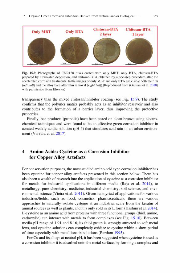
transparency than the mixed chitosan/inhibitor coating (see Fig. 15.9). The studyconfirms that the polymer matrix probably acts as an inhibitor reservoir and alsocontributes to the formation of a barrier layer, thus improving the protectiveproperties.
Finally, bee products (propolis) have been tested on clean bronze using electro-chemical techniques and were found to be an effective green corrosion inhibitor inaerated weakly acidic solution (pH 5) that simulates acid rain in an urban environ-ment (Varvara et al. 2017).
4 Amino Acids: Cysteine as a Corrosion Inhibitorfor Copper Alloy Artefacts
For conservation purposes, the most studied amino acid type corrosion inhibitor hasbeen cysteine for copper alloy artefacts presented in this section below. There hasalso been a wealth of research into the application of cysteine as a corrosion inhibitorfor metals for industrial applications in different media (Raja et al. 2014), tometallurgy, pure chemistry, medicine, industrial chemistry, soil science, and envi-ronmental science (Vieira et al. 2011). Given its myriad of applications for variousindustries/fields, such as food, cosmetics, pharmaceuticals, there are variousapproaches to naturally isolate cysteine at an industrial scale from the keratin ofanimal sources as well as plants, and it is only sold in its L form (Hashim et al. 2014).L-cysteine as an amino acid from proteins with three functional groups (thiol, amine,carboxylic) can interact with metals to form complexes (see Fig. 15.10). Betweenmedia pH range of 1.91 and 8.16, its thiol group is strongly attracted to soft metalions, and cysteine solutions can completely oxidize to cystine within a short periodof time especially with metal ions in solutions (Berthon 1995).
For Cu and its alloys at neutral pH, it has been suggested when cysteine is used asa corrosion inhibitor it is adsorbed onto the metal surface, by forming a complex and
Fig. 15.9 Photographs of CNR128 disks coated with only MBT, only BTA, chitosan-BTAprepared by a two-step deposition, and chitosan-BTA obtained by a one-step procedure after theaccelerated corrosion treatments. In the images of only MBT and only BTA are visible both the film(left half) and the alloy bare after film removal (right half) (Reproduced from (Giuliani et al. 2018)with permission from Elsevier)
15 Organic Green Corrosion Inhibitors Derived from Natural and/or Biological. . . 355
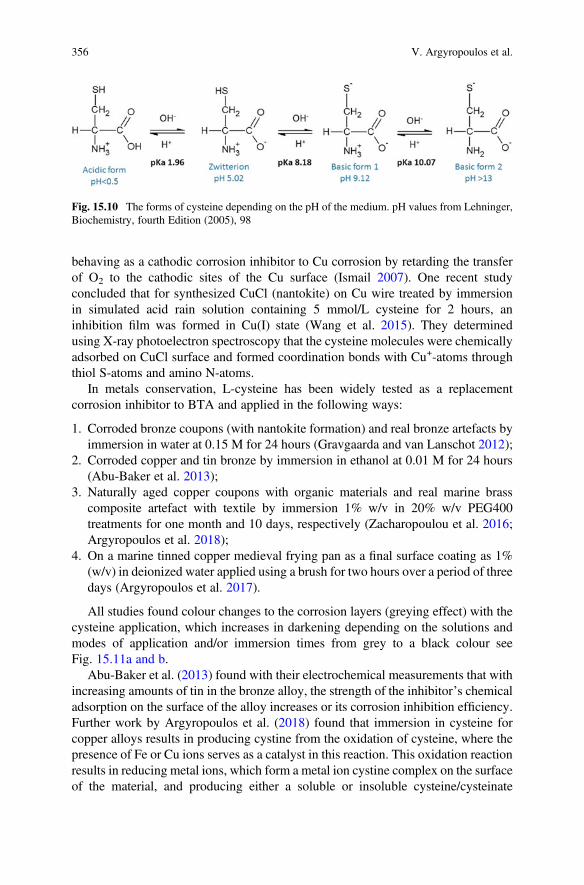
behaving as a cathodic corrosion inhibitor to Cu corrosion by retarding the transferof O2 to the cathodic sites of the Cu surface (Ismail 2007). One recent studyconcluded that for synthesized CuCl (nantokite) on Cu wire treated by immersionin simulated acid rain solution containing 5 mmol/L cysteine for 2 hours, aninhibition film was formed in Cu(I) state (Wang et al. 2015). They determinedusing X-ray photoelectron spectroscopy that the cysteine molecules were chemicallyadsorbed on CuCl surface and formed coordination bonds with Cu+-atoms throughthiol S-atoms and amino N-atoms.
In metals conservation, L-cysteine has been widely tested as a replacementcorrosion inhibitor to BTA and applied in the following ways:
1. Corroded bronze coupons (with nantokite formation) and real bronze artefacts byimmersion in water at 0.15 M for 24 hours (Gravgaarda and van Lanschot 2012);
2. Corroded copper and tin bronze by immersion in ethanol at 0.01 M for 24 hours(Abu-Baker et al. 2013);
3. Naturally aged copper coupons with organic materials and real marine brasscomposite artefact with textile by immersion 1% w/v in 20% w/v PEG400treatments for one month and 10 days, respectively (Zacharopoulou et al. 2016;Argyropoulos et al. 2018);
4. On a marine tinned copper medieval frying pan as a final surface coating as 1%(w/v) in deionized water applied using a brush for two hours over a period of threedays (Argyropoulos et al. 2017).
All studies found colour changes to the corrosion layers (greying effect) with thecysteine application, which increases in darkening depending on the solutions andmodes of application and/or immersion times from grey to a black colour seeFig. 15.11a and b.
Abu-Baker et al. (2013) found with their electrochemical measurements that withincreasing amounts of tin in the bronze alloy, the strength of the inhibitor’s chemicaladsorption on the surface of the alloy increases or its corrosion inhibition efficiency.Further work by Argyropoulos et al. (2018) found that immersion in cysteine forcopper alloys results in producing cystine from the oxidation of cysteine, where thepresence of Fe or Cu ions serves as a catalyst in this reaction. This oxidation reactionresults in reducing metal ions, which form a metal ion cystine complex on the surfaceof the material, and producing either a soluble or insoluble cysteine/cysteinate
Fig. 15.10 The forms of cysteine depending on the pH of the medium. pH values from Lehninger,Biochemistry, fourth Edition (2005), 98
356 V. Argyropoulos et al.
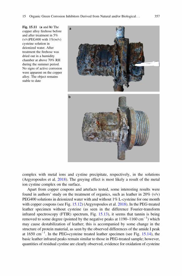
complex with metal ions and cystine precipitate, respectively, in the solutions(Argyropoulos et al. 2018). The greying effect is most likely a result of the metalion cystine complex on the surface.
Apart from copper coupons and artefacts tested, some interesting results werefound in authors’ study on the treatment of organics, such as leather in 20% (v/v)PEG400 solutions in deionized water with and without 1% L-cysteine for one monthwith copper coupons (see Fig. 15.12) (Argyropoulos et al. 2018). In the PEG-treatedleather specimen without cysteine (as seen in the difference Fourier-transforminfrared spectroscopy (FTIR) spectrum, Fig. 15.13), it seems that tannin is beingremoved to some degree (pointed by the negative peaks at 1190–1160 cm�1) whichmay cause destabilization of leather; this is accompanied by some change in thestructure of protein material, as seen by the observed differences of the amide I peakat 1650 cm�1. In the PEG+cysteine treated leather specimen (see Fig. 15.14), thebasic leather infrared peaks remain similar to those in PEG-treated sample; however,quantities of residual cystine are clearly observed, evidence for oxidation of cysteine
a
b
Fig. 15.11 (a and b) Thecopper alloy firehose beforeand after treatment in 5%(v/v)PEG400 with 1%(w/v)cysteine solution indeionized water. Aftertreatment the firehose wasdried out in a humiditychamber at above 70% RHduring the summer period.No signs of active corrosionwere apparent on the copperalloy. The object remainsstable to date
15 Organic Green Corrosion Inhibitors Derived from Natural and/or Biological. . . 357
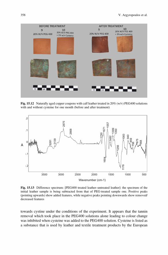
towards cystine under the conditions of the experiment. It appears that the tanninremoval which took place in the PEG400 solutions alone leading to colour changewas inhibited when cysteine was added to the PEG400 solution. Cysteine is listed asa substance that is used by leather and textile treatment products by the European
Fig. 15.12 Naturally aged copper coupons with calf leather treated in 20% (w/v) PEG400 solutionswith and without cysteine for one month (before and after treatment)
-.2
-.1
0
.1
.2
3500 3000 2500 2000 1500 1000 500
3281 3139
2875
2668
1750
1671
1604
1557
1482
1399
1350
1256
1178
1083
940
859
798 67
556
8
-.2
-.1
0
.1
.2
3500 3000 2500 2000 1500 1000 500
Wavenumber (cm-1)
A
Fig. 15.13 Difference spectrum: [PEG400 treated leather–untreated leather]: the spectrum of theinitial leather sample is being subtracted from that of PEG-treated sample one. Positive peaks(pointing upwards) show added features, while negative peaks pointing downwards show removed/decreased features
358 V. Argyropoulos et al.
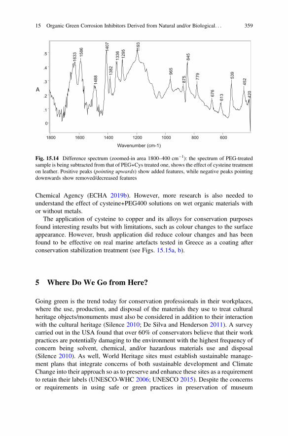
Chemical Agency (ECHA 2019b). However, more research is also needed tounderstand the effect of cysteine+PEG400 solutions on wet organic materials withor without metals.
The application of cysteine to copper and its alloys for conservation purposesfound interesting results but with limitations, such as colour changes to the surfaceappearance. However, brush application did reduce colour changes and has beenfound to be effective on real marine artefacts tested in Greece as a coating afterconservation stabilization treatment (see Figs. 15.15a, b).
5 Where Do We Go from Here?
Going green is the trend today for conservation professionals in their workplaces,where the use, production, and disposal of the materials they use to treat culturalheritage objects/monuments must also be considered in addition to their interactionwith the cultural heritage (Silence 2010; De Silva and Henderson 2011). A surveycarried out in the USA found that over 60% of conservators believe that their workpractices are potentially damaging to the environment with the highest frequency ofconcern being solvent, chemical, and/or hazardous materials use and disposal(Silence 2010). As well, World Heritage sites must establish sustainable manage-ment plans that integrate concerns of both sustainable development and ClimateChange into their approach so as to preserve and enhance these sites as a requirementto retain their labels (UNESCO-WHC 2006; UNESCO 2015). Despite the concernsor requirements in using safe or green practices in preservation of museum
0
.1
.2
.3
.4
.5
1800 1600 1400 1200 1000 800 600
1633 15
86
1488
1407
1382
1336
1295 11
93
965
875
845
779
676
613
539
452
420
0
.1
.2
.3
.4
.5
1800 1600 1400 1200 1000 800 600
Wavenumber (cm-1)
A
Fig. 15.14 Difference spectrum (zoomed-in area 1800–400 cm�1): the spectrum of PEG-treatedsample is being subtracted from that of PEG+Cys treated one, shows the effect of cysteine treatmenton leather. Positive peaks (pointing upwards) show added features, while negative peaks pointingdownwards show removed/decreased features
15 Organic Green Corrosion Inhibitors Derived from Natural and/or Biological. . . 359
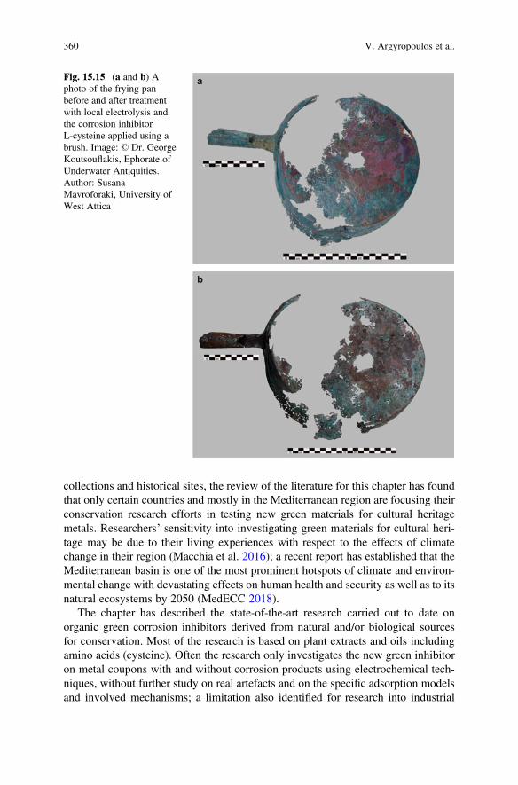
collections and historical sites, the review of the literature for this chapter has foundthat only certain countries and mostly in the Mediterranean region are focusing theirconservation research efforts in testing new green materials for cultural heritagemetals. Researchers’ sensitivity into investigating green materials for cultural heri-tage may be due to their living experiences with respect to the effects of climatechange in their region (Macchia et al. 2016); a recent report has established that theMediterranean basin is one of the most prominent hotspots of climate and environ-mental change with devastating effects on human health and security as well as to itsnatural ecosystems by 2050 (MedECC 2018).
The chapter has described the state-of-the-art research carried out to date onorganic green corrosion inhibitors derived from natural and/or biological sourcesfor conservation. Most of the research is based on plant extracts and oils includingamino acids (cysteine). Often the research only investigates the new green inhibitoron metal coupons with and without corrosion products using electrochemical tech-niques, without further study on real artefacts and on the specific adsorption modelsand involved mechanisms; a limitation also identified for research into industrial
a
b
Fig. 15.15 (a and b) Aphoto of the frying panbefore and after treatmentwith local electrolysis andthe corrosion inhibitorL-cysteine applied using abrush. Image: © Dr. GeorgeKoutsouflakis, Ephorate ofUnderwater Antiquities.Author: SusanaMavroforaki, University ofWest Attica
360 V. Argyropoulos et al.
applications (Marzorati et al. 2019). According to Marzorati et al. (2019) research forgreen corrosion inhibitors should aim to characterize the active compound in thegreen extract so as to be able to reproduce the results, which otherwise can bedependent on the harvesting location, weather conditions, season, harvesting tech-nique, and many other parameters. Finally, for conservation research, all the greeninhibitors tested seem to alter the surface colour of metal coupons when appliedusing immersion methods, which can be reduced using brush application, but arereversible either with water or ethanol.
Some interesting and promising research results were also identified in thisreview concerning the application of inhibitors from biological sources, such asbacteria and biopolymers. The stabilization of copper alloy artefacts using thebiopatina is promising. Also, interestingly, chitosan where the coating formulationincludes a corrosion inhibitor was found to be more effective than when the coatingprocesses were applied separately on the metal surface (i.e., corrosion inhibitorfollowed by coating). This research was in contradiction to research carried outwhen corrosion inhibitor additives were tested in synthetic coatings, such as ParaloidB72 or Poligen, and none was found to show any clear improvement of theprotection properties of the coatings, including one of them impaired the barriereffect of the coating (Argyropoulos et al. 2007a; Cano et al. 2010).
Today, conservators’ main concern should be to support sustainable practices intheir field in addition to using the most effective protection system on the metalscultural heritage. The conservation principles for appropriate protection systemsshould include sustainability or green practice in addition to reversibility andminimal surface alterations to the appearance. The authors believe that futureresearch should continue investigation into microorganisms as well as bio-basedcoatings with green corrosion inhibitors and their ability to work together synergis-tically to act as long-term protection systems for cultural heritage metals; theevaluation should also include how ‘green’ is the inhibitor with respect to theirproduction and use (i.e., Life Cycle Assessment). Such research requires studies onreal artefacts/monuments using in situ corrosion measurements, such as electro-chemical impedance spectroscopy (Barat et al. 2019) or oxygen consumption(Matthiesen 2013). Given the challenges involved in terms of vigorous testing andscientific examination required both on metal coupons and real artefacts and includ-ing identifying the specific adsorption models and involved mechanisms, the wayforward should be through funded collaborative projects.
Acknowledgements The research for testing new green inhibitors was co-financed by theEuropean Union (European Social Fund, ESF) and Greek national funds through the OperationalProgram ‘Education and Lifelong Learning’ of the National Strategic Reference Framework(NSRF)—Research Funding Program: ARCHIMEDES III. Investing in knowledge society throughthe European Social Fund. Vasilike Argyropoulos would also like to acknowledge the support inwriting this publication during her visiting professorship at the University of Toronto, Faculty ofInformation.
15 Organic Green Corrosion Inhibitors Derived from Natural and/or Biological. . . 361
References
Abbout S, Meryem Z, Benzidia B, Hammouch H (2018) New formulation based on CeratoniaSiliqua L seed oil, as a green corrosion inhibitor of iron in acidic medium. Anal BioanalElectrochem 10:789–804
Abdelwahab A, Rifai M, Abdelhamid Z (2018) Corrosion inhibition of bronze alloy by aloe veraextract in neutral media for application on bronze artifacts. In: EuroCorr. EuroCorr, Krakow, p83
Abu-baker AN (2019) From mono-oxime to tri-oxime: the development of a new group ofcorrosion inhibitors for copper alloys. In: Chemello C, Brambilla L, Joseph E (eds) ICOM-CC METAL 2019, ICOM, Neuchatel, pp 204–211
Abu-Baker AN, Macleod ID, Sloggett R, Taylor R (2013) A comparative study of salicylaldoxime,cysteine, and benzotriazole as inhibitors for the active chloride-based corrosion of copper andbronze artifacts. Eur Sci J 9:1857–7881
Albini M, Letardi P, Mathys L, Brambilla L, Schröter J, Junier P, Joseph E (2018) Comparison of abio-based corrosion inhibitor versus benzotriazole on corroded copper surfaces. Corros Sci143:84–92
Anderson KJ (1991) A history of lubricants. MRS Bull 16:69–69Argyropoulos V (ed) (2008) Metals and museums in the mediterranean. TEI of Athens, AthensArgyropoulos V, Boyatzis S, Giannoulaki M, Polikreti K (2013) The role of standards in conser-
vation methods for metals in cultural heritage. In: Dillmann P, Watkinson D, Angelini E,Adriaens A (eds) Corrosion and conservation of cultural heritage metallic artefacts. Elsevier,Oxford, pp 478–517
Argyropoulos V, Boyatzis SC, Giannoulaki M, Malea A, Pournou A, Rapti S, Zacharopoulou A,Guilminot E (2018) Preliminary investigation of L-cysteine as a corrosion inhibitor for marinecomposite artefacts containing copper or iron alloys. In: Williams E, Hocker E (eds) Pro-ceedings of the 13th ICOM-CC group on wet organic archaeological materials conference,Florence 2016, ICOM, pp 282–291
Argyropoulos V, Charalambous D, Kaminari A, Karabotsos A, Polikreti K, Siatou A, Cano E,Bastidas DM, Cayuela I, Bastidas J-M, Degrigny C, Vella D, Crawford J, Golfomitsou S(2007a) Testing of a new wax coating Poligen ES 91009 ® and corrosion inhibitor additivesused for improving coatings for historic iron alloys. In: Degrigny C, Langh R van, Joosten I,Ankersmit B (eds) METAL 07: proceedings of the ICOM-CC metal WG Interim meeting,Amsterdam, ICOM-CC, Amsterdam, pp 10–15
Argyropoulos V, Giannoulaki M, Michalakakos GP, Siatou A (2007b) A survey of the types ofcorrosion inhibitors and protective coatings used for the conservation of metal objects frommuseum collections in the Mediterranean basin. In: Argyropoulos V, Hein A, Harith MA (eds)Strategies for saving our cultural heritage: papers presented at the international conference onconservation strategies for saving indoor metallic collections with a satellite meeting on legalissues in the conservation of cultural heritage Cairo 25 Febr, TEI of Athens, Athens, pp 166–170
Argyropoulos V, Mavroforaki S, Giannoulaki M, Boyatzis SC, Karabotsos T, Zacharopoulou A,Guilminot E (2017) New approaches in stabilizing chloride-contaminated ancient bronzes usingcorrosion inhibitors and/or electrochemical methods to preserve information in the patinas. In:JD Lapatin, K, Spinelli A (eds) Artistry in Bronze the greeks and their legacy: acta of the XIXinternational congress on ancient bronzes. J. Paul Getty Trust, Los Angeles, pp 313–318
Argyropoulos V, Rameau J-J, Dalard F, Degrigny C (1999) Testing Hostacor it as a corrosioninhibitor for iron in polyethylene glycol solutions. Stud Conserv 44:49–57
Argyropoulos V, Selwyn LS, Logan JA (1997) Developing a conservation treatment usingethylenediamine as a corrosion inhibitor for wrought iron objects found at terrestrial archaeo-logical sites. In: MacLeod ID, Pennec SL, Robbiola L (eds) METAL 1995, James & James(Science Publishers) Ltd., London, pp 153–158
362 V. Argyropoulos et al.
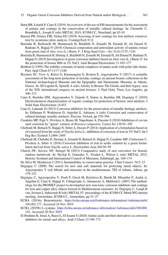
Barat BR, Letardi P, Cano E (2019) An overview of the use of EIS measurements for the assessmentof patinas and coatings in the conservation of metallic cultural heritage. In: Chemello C,Brambilla L, Joseph E (eds) METAL 2019, ICOM-CC, Neuchatel, pp 83–91
Barrera PR, Gómez FJR, Ochoa EG (2019) Assessing of new coatings for iron artifacts conserva-tion by recurrence plots analysis. CoatingsTech 9:12
Benzidia B, Barbouchi M, Hammouch H, Belahbib N, Zouarhi M, Erramli H, Ait Daoud N,Badrane N, Hajjaji N (2018) Chemical composition and antioxidant activity of tannins extractfrom green rind of Aloe vera (L.) Burm. F. J King Saud Univ—Sci 31(4):1175–1181
Benzidia B, Hammouch H, Dermaj A, Belahbib N, Zouarhi M, Erramli H, Ait Daoud N, Badrane N,Hajjaji N (2019) Investigation of green corrosion inhibitor based on Aloe vera (L.) Burm. F. forthe protection of bronze B66 in 3% NaCl. Anal Bioanal Electrochem 11:165–177
Berthon G (1995) The stability constants of metal complexes of amino acids with polar side chains.Pure Appl Chem 67:1117–1240
Boyatzis SC, Veve A, Kriezi G, Karamargiou G, Kontou E, Argyropoulos V (2017) A scientificassessment of the long-term protection of incralac coatings on ancient bronze collections in theNational Archaeological Museum and the Epigraphic and Numismatic Museum, in Athens,Greece. In: JD Lapatin K, Spinelli A (eds) Artistry in Bronze The Greeks and their legacy: actaof the XIX international congress on ancient bronzes. J. Paul Getty Trust, Los Angeles, pp300–312
Cano E, Bastidas DM, Argyropoulos V, Fajardo S, Siatou A, Bastidas JM, Degrigny C (2010)Electrochemical characterization of organic coatings for protection of historic steel artefacts. JSolid State Electrochem 14:453
Cano E, Lafuente D (2013) Corrosion inhibitors for the preservation of metallic heritage artefacts.In: Dillmann P, Watkinson D, Angelini E, Adriaens A (eds) Corrosion and conservation ofcultural heritage metallic artefacts. Elsevier, Oxford, pp 570–594
Casaletto MP, Figà V, Privitera A, Bruno M, Napolitano A, Piacente S (2018) Inhibition of cor-tensteel corrosion by “green” extracts of Brassica campestris. Corros Sci 136:91–105
Chellouli M, Bettach N, Hajjaji N, Srhiri A, Decaro P (2014) Application of a formulation based onoil extracted from the seeds of Nigella Sativa L, inhibition of corrosion of iron in 3% NaCl. Int JEng Res Technol 3:2489–2495
Chellouli M, Chebabe D, Dermaj A, Erramli H, Bettach N, Hajjaji N, Casaletto MP, Cirrincione C,Privitera A, Srhiri A (2016) Corrosion inhibition of iron in acidic solution by a green formu-lation derived from Nigella sativa L. Electrochim Acta 204:50–59
Church JW, Service NP, Striegel M (2013) Comparative study of rust converters for historicoutdoor metalwork. In: Hyslop E, Gonzalez V, Troalen L, Wilson L (eds) METAL 2013,Historic Scotland and International Council of Museums, Edinburgh, pp. 169–174
De Silva M, Henderson J (2011) Sustainability in conservation practice. J Inst Conserv 34:5–15Degrigny C (2008) The search for new and safe materials for protecting metal objects. In:
Argyropoulos V (ed) Metals and museums in the mediterranean. TEI of Athens, Athens, pp179–231
Degrigny C, Agryropoulos V, Pouli P, Grech M, Kreislova K, Harith M, Mirambet F, Arafat A,Angelini E, Cano E, Hajjaji N, Cilingiroglu A, Almansour A, Mahfoud L (2007) The method-ology for the PROMET project to develop/test new non-toxic corrosion inhibitors and coatingsfor iron and copper alloy objects housed in Mediterranean museums. In: Degrigny C, Langh Rvan, Joosten I, Ankersmit B (eds) METAL 07: proceedings of the ICOM-CCMetal WG interimmeeting, Amsterdam, ICOM-CC, Amsterdam, pp 31–37
ECHA (2019a) Benzotriazole. https://echa.europa.eu/el/substance-information/-/substanceinfo/100.002.177. Accessed 24 Nov 2019
ECHA (2019b) L-cysteine. https://echa.europa.eu/substance-information/-/substanceinfo/100.000.145. Accessed 26 Nov 2019
El Ibrahimi B, Jmiai A, Bazzi L, El Issami S (2020) Amino acids and their derivatives as corrosioninhibitors for metals and alloys. Arab J Chem 13:740–771
15 Organic Green Corrosion Inhibitors Derived from Natural and/or Biological. . . 363
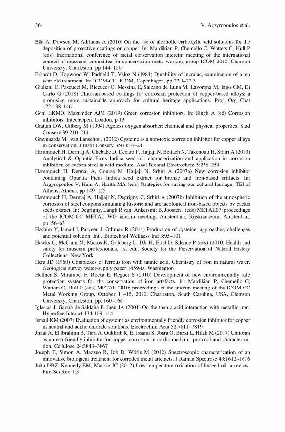
Elia A, Dowsett M, Adriaens A (2010) On the use of alcoholic carboxylic acid solutions for thedeposition of protective coatings on copper. In: Mardikian P, Chemello C, Watters C, Hull P(eds) International conference of metal conservation intereim meeting of the internationalcouncil of museums committee for conservation metal working group ICOM 2010. ClemsonUniversity, Charleston, pp 144–150
Erhardt D, Hopwood W, Padfield T, Veloz N (1984) Durability of incralac, examination of a tenyear old treatment. In: ICOM-CC. ICOM, Copenhagen, pp 22.1–22.3
Giuliani C, Pascucci M, Riccucci C, Messina E, Salzano de Luna M, Lavorgna M, Ingo GM, DiCarlo G (2018) Chitosan-based coatings for corrosion protection of copper-based alloys: apromising more sustainable approach for cultural heritage applications. Prog Org Coat122:138–146
Goni LKMO, Mazumder AJM (2019) Green corrosion inhibitors. In: Singh A (ed) Corrosioninhibitors. IntechOpen, London, p 13
Grattan DW, Gilberg M (1994) Ageless oxygen absorber: chemical and physical properties. StudConserv 39:210–214
Gravgaarda M, van Lanschot J (2012) Cysteine as a non-toxic corrosion inhibitor for copper alloysin conservation, J Instit Conserv 35(1):14–24
Hammouch H, Dermaj A, Chebabe D, Decaro P, Hajjaji N, Bettach N, Takenouti H, Srhiri A (2013)Analytical & Opuntia Ficus Indica seed oil: characterization and application in corrosioninhibition of carbon steel in acid medium. Anal Bioanal Electrochem 5:236–254
Hammouch H, Dermaj A, Goursa M, Hajjaji N, Srhiri A (2007a) New corrosion inhibitorcontaining Opuntia Ficus Indica seed extract for bronze and iron-based artifacts. In:Argyropoulos V, Hein A, Harith MA (eds) Strategies for saving our cultural heritage. TEI ofAthens, Athens, pp 149–155
Hammouch H, Dermaj A, Hajjaji N, Degrigny C, Srhiri A (2007b) Inhibition of the atmosphericcorrosion of steel coupons simulating historic and archaeological iron-based objects by cactusseeds extract. In: Degrigny, Langh R van, Ankersmit B, Joosten I (eds) METAL07: proceedingsof the ICOM-CC METAL WG interim meeting, Amsterdam, Rijskmuseum, Amsterdam,pp. 56–63
Hashim Y, Ismail I, Parveen J, Othman R (2014) Production of cysteine: approaches, challengesand potential solution. Int J Biotechnol Wellness Ind 3:95–101
Hawks C, McCann M, Makos K, Goldberg L, DJr H, Ertel D, Silence P (eds) (2010) Health andsafety for museum professionals, 1st edn. Society for the Preservation of Natural HistoryCollections, New York
Hem JD (1960) Complexes of ferrous iron with tannic acid. Chemistry of iron in natural water.Geological survey water-supply paper 1459-D, Washington
Hollner S, Mirambet F, Rocca E, Reguer S (2010) Development of new environmentally safeprotection systems for the conservation of iron artefacts. In: Mardikian P, Chemello C,Watters C, Hull P (eds) METAL 2010: proceedings of the interim meeting of the ICOM-CCMetal Working Group, October 11–15, 2010, Charleston, South Carolina, USA, ClemsonUniversity, Charleston, pp. 160–166
Iglesias J, García de Saldaña E, Jaén JA (2001) On the tannic acid interaction with metallic iron.Hyperfine Interact 134:109–114
Ismail KM (2007) Evaluation of cysteine as environmentally friendly corrosion inhibitor for copperin neutral and acidic chloride solutions. Electrochim Acta 52:7811–7819
Jmiai A, El Ibrahimi B, Tara A, Oukhrib R, El Issami S, Jbara O, Bazzi L, Hilali M (2017) Chitosanas an eco-friendly inhibitor for copper corrosion in acidic medium: protocol and characteriza-tion. Cellulose 24:3843–3867
Joseph E, Simon A, Mazzeo R, Job D, Wörle M (2012) Spectroscopic characterization of aninnovative biological treatment for corroded metal artefacts. J Raman Spectrosc 43:1612–1616
Juita DBZ, Kennedy EM, Mackie JC (2012) Low temperature oxidation of linseed oil: a review.Fire Sci Rev 1:3
364 V. Argyropoulos et al.
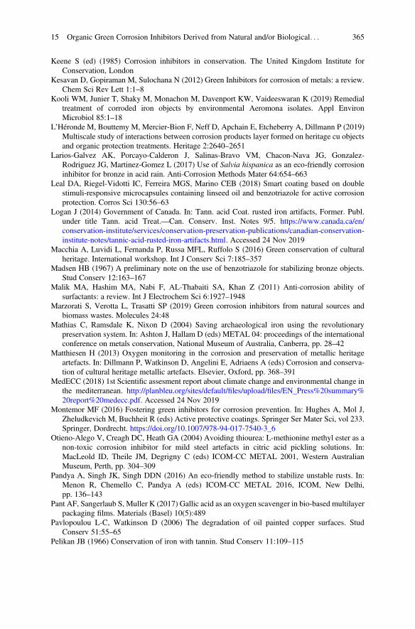
Keene S (ed) (1985) Corrosion inhibitors in conservation. The United Kingdom Institute forConservation, London
Kesavan D, Gopiraman M, Sulochana N (2012) Green Inhibitors for corrosion of metals: a review.Chem Sci Rev Lett 1:1–8
Kooli WM, Junier T, Shaky M, Monachon M, Davenport KW, Vaideeswaran K (2019) Remedialtreatment of corroded iron objects by environmental Aeromona isolates. Appl EnvironMicrobiol 85:1–18
L’Héronde M, Bouttemy M, Mercier-Bion F, Neff D, Apchain E, Etcheberry A, Dillmann P (2019)Multiscale study of interactions between corrosion products layer formed on heritage cu objectsand organic protection treatments. Heritage 2:2640–2651
Larios-Galvez AK, Porcayo-Calderon J, Salinas-Bravo VM, Chacon-Nava JG, Gonzalez-Rodriguez JG, Martinez-Gomez L (2017) Use of Salvia hispanica as an eco-friendly corrosioninhibitor for bronze in acid rain. Anti-Corrosion Methods Mater 64:654–663
Leal DA, Riegel-Vidotti IC, Ferreira MGS, Marino CEB (2018) Smart coating based on doublestimuli-responsive microcapsules containing linseed oil and benzotriazole for active corrosionprotection. Corros Sci 130:56–63
Logan J (2014) Government of Canada. In: Tann. acid Coat. rusted iron artifacts, Former. Publ.under title Tann. acid Treat.—Can. Conserv. Inst. Notes 9/5. https://www.canada.ca/en/conservation-institute/services/conservation-preservation-publications/canadian-conservation-institute-notes/tannic-acid-rusted-iron-artifacts.html. Accessed 24 Nov 2019
Macchia A, Luvidi L, Fernanda P, Russa MFL, Ruffolo S (2016) Green conservation of culturalheritage. International workshop. Int J Conserv Sci 7:185–357
Madsen HB (1967) A preliminary note on the use of benzotriazole for stabilizing bronze objects.Stud Conserv 12:163–167
Malik MA, Hashim MA, Nabi F, AL-Thabaiti SA, Khan Z (2011) Anti-corrosion ability ofsurfactants: a review. Int J Electrochem Sci 6:1927–1948
Marzorati S, Verotta L, Trasatti SP (2019) Green corrosion inhibitors from natural sources andbiomass wastes. Molecules 24:48
Mathias C, Ramsdale K, Nixon D (2004) Saving archaeological iron using the revolutionarypreservation system. In: Ashton J, Hallam D (eds) METAL 04: proceedings of the internationalconference on metals conservation, National Museum of Australia, Canberra, pp. 28–42
Matthiesen H (2013) Oxygen monitoring in the corrosion and preservation of metallic heritageartefacts. In: Dillmann P, Watkinson D, Angelini E, Adriaens A (eds) Corrosion and conserva-tion of cultural heritage metallic artefacts. Elsevier, Oxford, pp. 368–391
MedECC (2018) 1st Scientific assesment report about climate change and environmental change inthe mediterranean. http://planbleu.org/sites/default/files/upload/files/EN_Press%20summary%20report%20medecc.pdf. Accessed 24 Nov 2019
Montemor MF (2016) Fostering green inhibitors for corrosion prevention. In: Hughes A, Mol J,Zheludkevich M, Buchheit R (eds) Active protective coatings. Springer Ser Mater Sci, vol 233.Springer, Dordrecht. https://doi.org/10.1007/978-94-017-7540-3_6
Otieno-Alego V, Creagh DC, Heath GA (2004) Avoiding thiourea: L-methionine methyl ester as anon-toxic corrosion inhibitor for mild steel artefacts in citric acid pickling solutions. In:MacLeold ID, Theile JM, Degrigny C (eds) ICOM-CC METAL 2001, Western AustralianMuseum, Perth, pp. 304–309
Pandya A, Singh JK, Singh DDN (2016) An eco-friendly method to stabilize unstable rusts. In:Menon R, Chemello C, Pandya A (eds) ICOM-CC METAL 2016, ICOM, New Delhi,pp. 136–143
Pant AF, Sangerlaub S, Muller K (2017) Gallic acid as an oxygen scavenger in bio-based multilayerpackaging films. Materials (Basel) 10(5):489
Pavlopoulou L-C, Watkinson D (2006) The degradation of oil painted copper surfaces. StudConserv 51:55–65
Pelikan JB (1966) Conservation of iron with tannin. Stud Conserv 11:109–115
15 Organic Green Corrosion Inhibitors Derived from Natural and/or Biological. . . 365
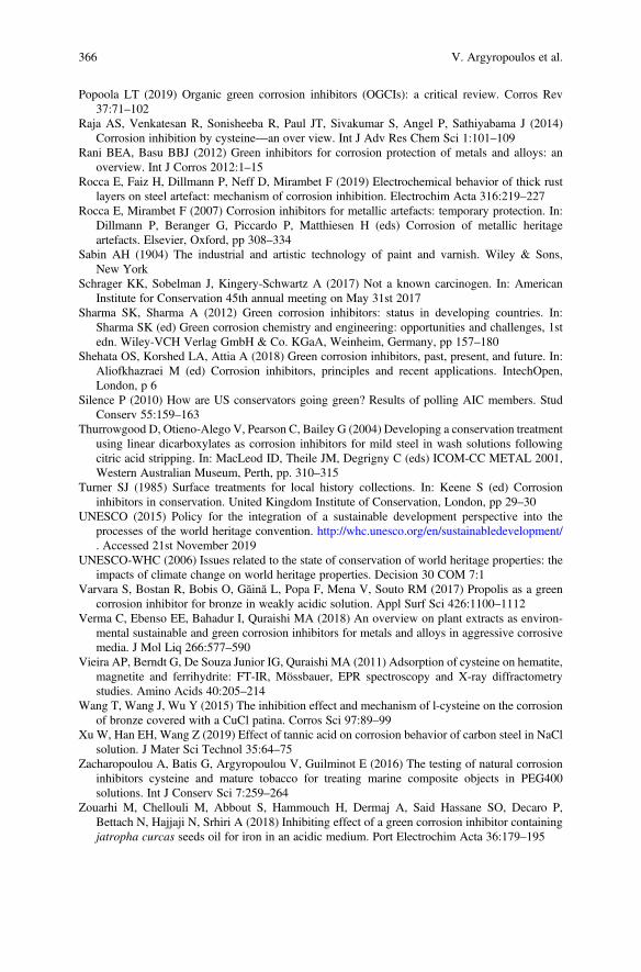
Popoola LT (2019) Organic green corrosion inhibitors (OGCIs): a critical review. Corros Rev37:71–102
Raja AS, Venkatesan R, Sonisheeba R, Paul JT, Sivakumar S, Angel P, Sathiyabama J (2014)Corrosion inhibition by cysteine—an over view. Int J Adv Res Chem Sci 1:101–109
Rani BEA, Basu BBJ (2012) Green inhibitors for corrosion protection of metals and alloys: anoverview. Int J Corros 2012:1–15
Rocca E, Faiz H, Dillmann P, Neff D, Mirambet F (2019) Electrochemical behavior of thick rustlayers on steel artefact: mechanism of corrosion inhibition. Electrochim Acta 316:219–227
Rocca E, Mirambet F (2007) Corrosion inhibitors for metallic artefacts: temporary protection. In:Dillmann P, Beranger G, Piccardo P, Matthiesen H (eds) Corrosion of metallic heritageartefacts. Elsevier, Oxford, pp 308–334
Sabin AH (1904) The industrial and artistic technology of paint and varnish. Wiley & Sons,New York
Schrager KK, Sobelman J, Kingery-Schwartz A (2017) Not a known carcinogen. In: AmericanInstitute for Conservation 45th annual meeting on May 31st 2017
Sharma SK, Sharma A (2012) Green corrosion inhibitors: status in developing countries. In:Sharma SK (ed) Green corrosion chemistry and engineering: opportunities and challenges, 1stedn. Wiley-VCH Verlag GmbH & Co. KGaA, Weinheim, Germany, pp 157–180
Shehata OS, Korshed LA, Attia A (2018) Green corrosion inhibitors, past, present, and future. In:Aliofkhazraei M (ed) Corrosion inhibitors, principles and recent applications. IntechOpen,London, p 6
Silence P (2010) How are US conservators going green? Results of polling AIC members. StudConserv 55:159–163
Thurrowgood D, Otieno-Alego V, Pearson C, Bailey G (2004) Developing a conservation treatmentusing linear dicarboxylates as corrosion inhibitors for mild steel in wash solutions followingcitric acid stripping. In: MacLeod ID, Theile JM, Degrigny C (eds) ICOM-CC METAL 2001,Western Australian Museum, Perth, pp. 310–315
Turner SJ (1985) Surface treatments for local history collections. In: Keene S (ed) Corrosioninhibitors in conservation. United Kingdom Institute of Conservation, London, pp 29–30
UNESCO (2015) Policy for the integration of a sustainable development perspective into theprocesses of the world heritage convention. http://whc.unesco.org/en/sustainabledevelopment/. Accessed 21st November 2019
UNESCO-WHC (2006) Issues related to the state of conservation of world heritage properties: theimpacts of climate change on world heritage properties. Decision 30 COM 7:1
Varvara S, Bostan R, Bobis O, Găină L, Popa F, Mena V, Souto RM (2017) Propolis as a greencorrosion inhibitor for bronze in weakly acidic solution. Appl Surf Sci 426:1100–1112
Verma C, Ebenso EE, Bahadur I, Quraishi MA (2018) An overview on plant extracts as environ-mental sustainable and green corrosion inhibitors for metals and alloys in aggressive corrosivemedia. J Mol Liq 266:577–590
Vieira AP, Berndt G, De Souza Junior IG, Quraishi MA (2011) Adsorption of cysteine on hematite,magnetite and ferrihydrite: FT-IR, Mössbauer, EPR spectroscopy and X-ray diffractometrystudies. Amino Acids 40:205–214
Wang T, Wang J, Wu Y (2015) The inhibition effect and mechanism of l-cysteine on the corrosionof bronze covered with a CuCl patina. Corros Sci 97:89–99
Xu W, Han EH, Wang Z (2019) Effect of tannic acid on corrosion behavior of carbon steel in NaClsolution. J Mater Sci Technol 35:64–75
Zacharopoulou A, Batis G, Argyropoulou V, Guilminot E (2016) The testing of natural corrosioninhibitors cysteine and mature tobacco for treating marine composite objects in PEG400solutions. Int J Conserv Sci 7:259–264
Zouarhi M, Chellouli M, Abbout S, Hammouch H, Dermaj A, Said Hassane SO, Decaro P,Bettach N, Hajjaji N, Srhiri A (2018) Inhibiting effect of a green corrosion inhibitor containingjatropha curcas seeds oil for iron in an acidic medium. Port Electrochim Acta 36:179–195
366 V. Argyropoulos et al.

Open Access This chapter is licensed under the terms of the Creative Commons Attribution 4.0International License (http://creativecommons.org/licenses/by/4.0/), which permits use, sharing,adaptation, distribution and reproduction in any medium or format, as long as you give appropriatecredit to the original author(s) and the source, provide a link to the Creative Commons license andindicate if changes were made.
The images or other third party material in this chapter are included in the chapter's CreativeCommons license, unless indicated otherwise in a credit line to the material. If material is notincluded in the chapter's Creative Commons license and your intended use is not permitted bystatutory regulation or exceeds the permitted use, you will need to obtain permission directly fromthe copyright holder.
15 Organic Green Corrosion Inhibitors Derived from Natural and/or Biological. . . 367