structure of endoglucanase cel9a from the thermoacidophilic alicyclobacillus acidocaldarius
TRANSCRIPT

research papers
744 doi:10.1107/S0907444909012773 Acta Cryst. (2009). D65, 744–750
Acta Crystallographica Section D
BiologicalCrystallography
ISSN 0907-4449
Structure of endoglucanase Cel9A from thethermoacidophilic Alicyclobacillus acidocaldarius
Jose Henrique Pereira,a,b Rajat
Sapra,a,c Joanne V. Volponi,c
Carol L. Kozina,c Blake
Simmonsa,c and Paul D.
Adamsa,b,d*
aJoint BioEnergy Institute, Emeryville, CA 94608,
USA, bPhysical Biosciences Division, Lawrence
Berkeley National Laboratory, Berkeley,
CA 94720, USA, cSandia National Laboratories,
7011 East Avenue, Livermore, CA 94551, USA,
and dDepartment of Bioengineering, University
of California Berkeley, CA 94720, USA
Correspondence e-mail: [email protected]
# 2009 International Union of Crystallography
Printed in Singapore – all rights reserved
The production of biofuels using biomass is an alternative
route to support the growing global demand for energy and to
also reduce the environmental problems caused by the
burning of fossil fuels. Cellulases are likely to play an
important role in the degradation of biomass and the
production of sugars for subsequent fermentation to fuel.
Here, the crystal structure of an endoglucanase, Cel9A, from
Alicyclobacillus acidocaldarius (Aa_Cel9A) is reported which
displays a modular architecture composed of an N-terminal
Ig-like domain connected to the catalytic domain. This paper
describes the overall structure and the detailed contacts
between the two modules. Analysis suggests that the inter-
action involving the residues Gln13 (from the Ig-like module)
and Phe439 (from the catalytic module) is important in
maintaining the correct conformation of the catalytic module
required for protein activity. Moreover, the Aa_Cel9A
structure shows three metal-binding sites that are associated
with the thermostability and/or substrate affinity of the
enzyme.
Received 20 January 2009
Accepted 3 April 2009
PDB Reference: Aa_Cel9A,
3ez8, 3ez8sf.
1. Introduction
The next generation of biofuels will use cellulose derived from
tailored crops as a source of fermentable sugars to produce
fuels such as ethanol. The sugars present in this kind of
biomass are located in the cell walls of plants, which are
composed of lignin, hemicelluloses and cellulose (Parsiegla et
al., 2008). Plants produce about 180 billion tons of cellulose
per year globally, making this polysaccharide the largest
organic carbon reservoir on earth (Festucci-Buselli et al.,
2007). An efficient breakdown of cellulosic biomass is a
prerequisite for the production of biofuels and cellulases are
key enzymes in this process. The �-1,4-glucanase (EC 3.2.1.4)
from Alicyclobacillus acidocaldarius (Aa_Cel9A), a thermo-
acidophilic Gram-positive bacterium, displays a temperature
optimum of 343 K and a pH optimum of 5.5 (Eckert et al.,
2002). Enzymes that can resist higher temperatures and a
range of pHs are required since heat and/or chemical pre-
treatment processes are currently used to remove lignin to
expose cellulose to cellulases (Sticklen, 2008).
Cellulases belong to a group of enzymes termed glycoside
hydrolases (GHs). Several members of the GH family
demonstrate a modular architecture composed of one or two
catalytic modules connected to several kinds of accessory
modules (Schubot et al., 2004). The accessory modules can be
involved in numerous functions. For example, some cellulases
contain carbohydrate-binding modules (CBMs), which
enhance the association of the catalytic modules with in-
soluble carbohydrates. Currently, glycoside hydrolases have
been grouped into 113 families (http://www.cazy.org).
Cellulases have been characterized as endo or exo and
processive or nonprocessive cellulases according to their mode
of action on the substrate (Parsiegla et al., 2002). The endo-
cellulases cleave the cellulose chain at arbitrary points, while
the exocellulases cleave at the terminus of a chain to start
the degradation process. Nonprocessive enzymes become
detached from their substrate after one step of substrate
hydrolysis, while processive cellulases remain bound to the
cellulose substrate and continue breaking down the poly-
saccharide. The nonprocessive endoglucanase Aa_Cel9A
belongs to subfamily E1 of family 9 of glycoside hydrolases.
Members of this group show an N-terminal immunoglobulin-
like (Ig-like) domain followed by the catalytic domain. The
function of the Ig-like module is still unclear; however, its
deletion promotes complete loss of enzymatic activity in a
related cellobiohydrolase, CbhA from Clostridium thermo-
cellum (Kataeva et al., 2004).
The endoglucanase Aa_Cel9A is most active against sub-
strates containing �-1,4-linked glucans (including carboxy-
methylcellulose and lichenan), but also exhibits activity
against �-1,4-xylans. The enzyme has been shown not to
hydrolyze substrates such as starch (�-1,4) or laminarin (�-1,6)
(Eckert et al., 2002). The crystallization and preliminary X-ray
analysis of Aa_Cel9A have previously been reported (Eckert
et al., 2003); however, the Aa_Cel9A structure was not sub-
sequently described, most likely because anisotropic disorder
in the crystal and weak diffraction thwarted structure solution.
In the present paper, we describe the crystal structure of the
endoglucanase Aa_Cel9A at 2.3 A resolution. The Aa_Cel9A
structure contains one zinc and two calcium ion-binding sites
and the presence of these metals is associated with the
temperature stability of the enzyme and/or substrate affinity.
Moreover, the structure of Aa_Cel9A reveals the detailed
contacts between the Ig-like and catalytic domains, providing
new information about the interactions between them.
2. Materials and methods
2.1. Cloning, expression, purification and crystallization ofAa_Cel9A
Cloning, expression and purification have been reported
elsewhere (Eckert et al., 2002). Aa_Cel9A was concentrated
and dialyzed against 15 mM Tris–HCl buffer pH 7.5 containing
50 mM NaCl. The final concentration of Aa_Cel9A used for
crystallization trials was 5 mg ml�1. The protein solution was
brought to 5.0 mM CaCl2 and centrifuged prior to crystal-
lization. Aa_Cel9A protein was screened using the sparse-
matrix method (Jancarik & Kim, 1991) with a Phoenix Robot
(Art Robbins Instruments, Sunnyvale, California, USA) using
the following crystallization screens: Crystal Screen I and II,
PEG/Ion, SaltRx and Index (Hampton Research, Aliso Viejo,
California, USA). The optimum conditions for crystallization
of Aa_Cel9A were found to be 0.1 M HEPES pH 7.3 and 55%
2-methyl-2,4-pentanediol (MPD). Crystals were obtained
after 2 d by the sitting-drop vapor-diffusion method with the
drops consisting of a mixture of 1.0 ml protein solution and
0.5 ml reservoir solution.
2.2. X-ray data collection and structure determination
Crystals were placed in a reservoir solution containing
55%(v/v) MPD and then flash-frozen in liquid nitrogen. A
native data set for the endoglucanase Aa_Cel9A was collected
on the Berkeley Center for Structural Biology beamline 5.0.1
of the Advanced Light Source at Lawrence Berkeley National
Laboratory (LBNL). The diffraction data were recorded using
an ADSC-Q210 detector. The data set was collected using
140� oscillation with �’ = 1� and a wavelength of 0.977 A. The
data were processed using the program HKL-2000 (Otwi-
nowski & Minor, 1997).
The crystal structure of Aa_Cel9A was determined by the
molecular-replacement method with the program Phaser
(McCoy et al., 2007), using as a search model the structure of
Cel9A (formerly CelD) from C. thermocellum (Ct_Cel9A;
PDB code 1clc), which shows only 27% sequence identity with
the target. The best solution was obtained with Euler angles
� = 153.2, � = 122.4, � = 103.1� and fractional coordinates
Tx = 1.918, Ty = �0.237, Tz = 0.452. The atomic positions
obtained from molecular replacement were used to initiate
crystallographic refinement and model rebuilding. Structure
research papers
Acta Cryst. (2009). D65, 744–750 Pereira et al. � Cel9A 745
Table 1Statistics for data collection and structure refinement of endoglucanaseCel9A.
Data collectionWavelength (A) 0.979Resolution range (A) 50–2.3 (2.34–2.30)Crystal-to-detector distance (mm) 300� collected/�� (�) 140/1.0Exposure time (s) 3Temperature of data collection (K) 100
Data statisticsSpace group P21212Unit-cell parameters (A) a = 49.06, b = 84.97, c = 129.48Total reflections 248498Unique reflections 23484Multiplicity 5.6 (4.2)Data completeness (%) 94.8 (93.7)I/�(I) 9.0 (1.70)Rmerge† (%) 0.140 (0.550)
Structure refinementResolution range 50–2.3R factor‡ (%) 19.6Rfree§ (%) 23.3R.m.s.d. from ideal geometry
Bond lengths (A) 0.008Bond angles (�) 1.061
Protein residues 528Ca2+ 2Zn2+ 1Water molecules 380Average isotropic B factors (A2)
Protein atoms 27.8Solvent atoms 35.6
Ramachandran plotFavored region (%) 97.3Outliers region (%) 0.0
† Rmerge =P
hkl
Pi jIiðhklÞ � hIðhklÞij=
Phkl
Pi IiðhklÞ, where
Phkl denotes the sum
over all reflections andP
i is the sum over all equivalent and symmetry-relatedreflections. ‡ R factor =
PjFobs � Fcalcj=
PFobs. § Rfree = R factor for 5% of the data
that were not included during crystallographic refinement.
refinement was performed using PHENIX (Adams et al.,
2002). TLS refinement with both domains as a single TLS
group was used in the process. Manual rebuilding using Coot
(Emsley & Cowtan, 2004) and the addition of water molecules
allowed construction of the final model. 5% of the data were
randomly selected for cross-validation. The final model has an
R factor of 19.6% and an Rfree of 23.3%.
Root-mean-square deviation differences from ideal geo-
metries for bond lengths, angles and dihedrals were calculated
with PHENIX (Adams et al., 2002). The overall stereo-
chemical quality of the final model for Aa_Cel9A was assessed
by the program MOLPROBITY (Davis et al., 2007). Atomic
models were superposed using the program LSQKAB from
CCP4 (Collaborative Computational Project, Number 4,
1994).
3. Results and discussion
The crystal of Aa_Cel9A diffracted to 2.3 A resolution and
belonged to the orthorhombic space group P21212, with unit-
cell parameters a = 49.06, b = 84.97, c = 129.48 A. The crys-
tallographic asymmetric unit contained one copy of the
Aa_Cel9A protein. The statistics for the crystallographic data
and refinement are summarized in Table 1. The electron-
density map showed clear positions for the residues present in
both the Ig-like and catalytic modules. The crystallization
conditions and unit-cell parameters of Aa_Cel9A are very
similar to those previously reported at 3.0 A resolution
(Eckert et al., 2003). We believe that the addition of 5 mM
divalent ion (Ca2+) prior to crystallization experiments was
essential in improving the quality of the crystal diffraction.
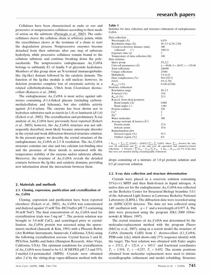
3.1. Overall structure of endoglucanase Aa_Cel9A
The 59 kDa endoglucanase Aa_Cel9A consists of two
modules. The N-terminal Ig-like module is composed of 85
amino-acid residues and is linked to the catalytic module
(residues 86–537). The overall fold of Aa_Cel9A is similar to
the previously reported structures of endoglucanase Ct_Cel9A
(Chauvaux et al., 1995) and the exoglucanase Ct_CbhA
(Schubot et al., 2004), both from C. thermocellum. The r.m.s.d.
between C� positions for overlapping residues between
Aa_Cel9A and Ct_Cel9A and Ct_CbhA are 1.622 and 1.923 A,
respectively.
The Ig-like module of Aa_Cel9A consists of six antiparallel
strands forming two �-sheets. The first �-sheet (�-strands 1
and 4) packs in opposition to the second �-sheet (�-strands 2,
3, 5 and 6), forming a �-barrel structure (Fig. 1). The Ig-like
module of Aa_Cel9A is more similar to the Ig-like module of
Ct_Cel9A than that of Ct_CbhA, even though the Ig-like
module of Ct_Cel9A contains seven antiparallel strands. The
disulfide bridge between �-strands 2 and 6 conserved in
immunoglobulin-domain structures is not observed in the Ig-
like module of cellulases. Moreover, the structural similarity
between the Ig-like module of cellulases and the immuno-
globulin domains is significant despite low sequence homology
(Juy et al., 1992).
The catalytic module of members of GH family 9 shows an
(�/�)6-barrel structure (Fig. 1). The 12 �-helices (�1–�12)
display an alternating connection pattern between outer and
research papers
746 Pereira et al. � Cel9A Acta Cryst. (2009). D65, 744–750
Figure 1Modular architecture of Aa_Cel9A. The N-terminal Ig-like moduleconsists of six �-strands, creating a �-barrel structure. The catalyticmodule has an (�/�)6-barrel motif formed by the inner �-helices. Themetal ions bound to Aa_Cel9A are shown as large spheres. The zinc ionand the two calcium ions are shown in green and yellow, respectively. N,amino-terminus; C, carboxyl-terminus.
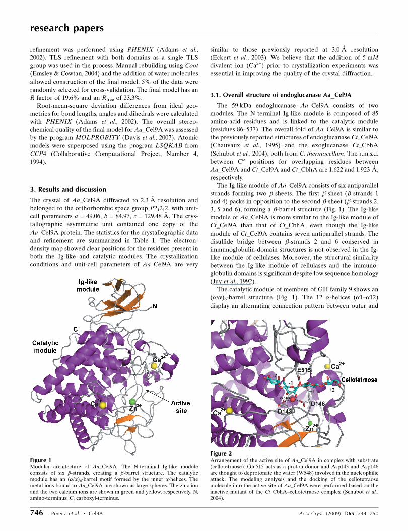
Figure 2Arrangement of the active site of Aa_Cel9A in complex with substrate(cellotetraose). Glu515 acts as a proton donor and Asp143 and Asp146are thought to deprotonate the water (W548) involved in the nucleophilicattack. The modeling analyses and the docking of the cellotetraosemolecule into the active site of Aa_Cel9A were performed based on theinactive mutant of the Ct_CbhA–cellotetraose complex (Schubot et al.,2004).
research papers
Acta Cryst. (2009). D65, 744–750 Pereira et al. � Cel9A 747
inner helices, as is common in (�/�)6-barrel structures (Par-
siegla et al., 1998). The barrel is formed by the parallel inner
helices �2, �4, �6, �8, �10 and �12. Besides the 12 �-helices,
the catalytic module of Aa_Cel9A shows two antiparallel
�-strands and three short �-helices which are structurally
conserved throughout the family 9 cellulases.
The active site of Aa_Cel9A is positioned at the N-terminal
region of the inner helices of the barrel structure. The simi-
larities between the active sites of Aa_Cel9A and of Ct_CbhA
solved in complex with substrate (cellotetraose) permits
inference of the residues involved in the catalytic mechanism.
In Aa_Cel9A, residue Glu515 acts as a proton donor in the
reaction. Asp143 and Asp146 are thought to deprotonate the
water involved in nucleophilic attack. This catalytic water
molecule is conserved in the Aa_Cel9A structure and is
hydrogen bonded to Asp143 and Asp146 with distances of 2.71
and 2.96 A, respectively (Fig. 2). A water chain connected to
the catalytic Asp146 was observed in Aa_Cel9A for an effi-
cient water supply. His461 and Arg463 are hydrogen bonded
to a glucose unit at substrate subsite +1 (nomenclature
according to Davies et al., 1997). Finally, the active site has
residues Phe221, Tyr300, Trp343, Trp401, Tyr511, Tyr519 and
Trp520 forming the substrate-binding cleft.
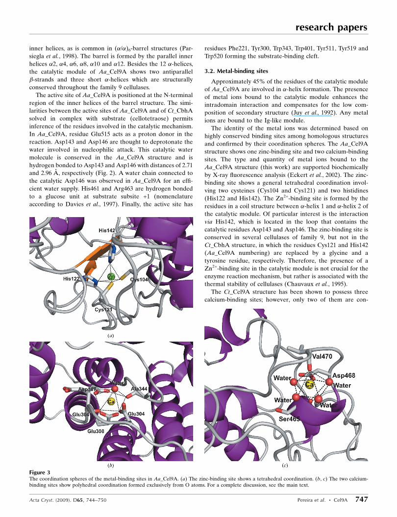
3.2. Metal-binding sites
Approximately 45% of the residues of the catalytic module
of Aa_Cel9A are involved in �-helix formation. The presence
of metal ions bound to the catalytic module enhances the
intradomain interaction and compensates for the low com-
position of secondary structure (Juy et al., 1992). Any metal
ions are bound to the Ig-like module.
The identity of the metal ions was determined based on
highly conserved binding sites among homologous structures
and confirmed by their coordination spheres. The Aa_Cel9A
structure shows one zinc-binding site and two calcium-binding
sites. The type and quantity of metal ions bound to the
Aa_Cel9A structure (this work) are supported biochemically
by X-ray fluorescence analysis (Eckert et al., 2002). The zinc-
binding site shows a general tetrahedral coordination invol-
ving two cysteines (Cys104 and Cys121) and two histidines
(His122 and His142). The Zn2+-binding site is formed by the
residues in a coil structure between �-helix 1 and �-helix 2 of
the catalytic module. Of particular interest is the interaction
via His142, which is located in the loop that contains the
catalytic residues Asp143 and Asp146. The zinc-binding site is
conserved in several cellulases of family 9, but not in the
Ct_CbhA structure, in which the residues Cys121 and His142
(Aa_Cel9A numbering) are replaced by a glycine and a
tyrosine residue, respectively. Therefore, the presence of a
Zn2+-binding site in the catalytic module is not crucial for the
enzyme reaction mechanism, but rather is associated with the
thermal stability of cellulases (Chauvaux et al., 1995).
The Ct_Cel9A structure has been shown to possess three
calcium-binding sites; however, only two of them are con-
Figure 3The coordination spheres of the metal-binding sites in Aa_Cel9A. (a) The zinc-binding site shows a tetrahedral coordination. (b, c) The two calcium-binding sites show polyhedral coordination formed exclusively from O atoms. For a complete discussion, see the main text.
served in Aa_Cel9A. The Aa_Cel9A calcium-binding sites are
characterized by seven or eight O atoms. In contrast to the
zinc-binding site, where the coordination is formed exclusively
by the side chains of protein residues, the calcium polyhedral
coordination is from side chains of aspartic or glutamic acid
residues, carbonyl groups of the main chain and water mole-
cules. The first Ca2+ is located close to the active site of
Aa_Cel9A (the nonreducing end), with a coordination formed
by the residues Asp302, Glu304, Asp307, Glu308, Ala344 and
one water molecule. This calcium-binding site is highly con-
served in members of GH family 9.
The second Ca2+ is also positioned close to the active site,
but at its opposite end (the reducing end). The coordination is
formed by residues Ile465, Asp468 and Val470 and four water
molecules. Functional analysis of Ct_Cel9A has shown that
Ca2+ bound to this site is able to increase the substrate-binding
affinity (Chauvaux et al., 1995). This is because the Ca2+ co-
ordination residues Ile465, Asp468 and Val470 are located in
same loop region as the substrate-binding residues His461 and
Arg463. The coordination sphere of all metal ions bound to
Aa_Cel9A is shown in Fig. 3.
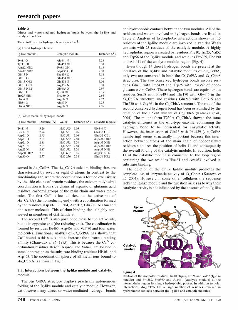
3.3. Interactions between the Ig-like module and catalyticmodule
The Aa_Cel9A structure displays practically autonomous
folding of the Ig-like module and catalytic module. However,
we observe many direct or water-mediated hydrogen bonds
and hydrophobic contacts between the two modules. All of the
residues and waters involved in hydrogen bonds are listed in
Table 2. Analysis of hydrophobic interactions shows that 15
residues of the Ig-like module are involved in van der Waals
contacts with 23 residues of the catalytic module. A highly
hydrophobic region is created by residues Phe10, Trp25, Val52
and Trp56 of the Ig-like module and residues Pro389, Phe390
and Ala441 of the catalytic module region (Fig. 4).
Even though 14 direct hydrogen bonds are present at the
interface of the Ig-like and catalytic modules of Aa_Cel9A,
only two are conserved in both the Ct_Cel9A and Ct_CbhA
structures. The two conserved hydrogen bonds involve resi-
dues Gln13 with Phe439 and Trp25 with Pro389 of endo-
glucanase Aa_Cel9A. These hydrogen bonds are equivalent to
residues Ser58 with Phe494 and Thr170 with Gly446 in the
Ct_Cel9A structure and residues Gln218 with Leu715 and
Thr230 with Gly661 in the Ct_CbhA structure. The role of the
second conserved hydrogen bond has been established by the
creation of the T230A mutant of Ct_CbhA (Kataeva et al.,
2004). The mutant form T230A Ct_CbhA showed the same
catalytic efficiency as the wild-type enzyme, confirming the
hydrogen bond to be inessential for enzymatic activity.
However, the interaction of Gln13 with Phe439 (Aa_Cel9A
numbering) seems structurally important because this inter-
action between atoms of the main chain of nonconserved
residues stabilizes the position of helix 11 and consequently
the overall folding of the catalytic module. In addition, helix
11 of the catalytic module is connected to the loop region
containing the two residues His461 and Arg463 involved in
substrate binding.
The deletion of the entire Ig-like module promotes the
complete loss of enzymatic activity of Ct_CbhA (Kataeva et
al., 2004). However, in some other cellulases the sequence
lacks the Ig-like module and the question arises as to why their
catalytic activity is not influenced by the absence of the Ig-like
research papers
748 Pereira et al. � Cel9A Acta Cryst. (2009). D65, 744–750
Table 2Direct and water-mediated hydrogen bonds between the Ig-like andcatalytic modules.
The cutoff used for hydrogen bonds was <3.4 A.
(a) Direct hydrogen bonds.
Ig-like module Catalytic module Distance (A)
Tyr11 O Ala441 N 3.33Tyr11 OH Gln453 OE1 3.38Asn12 ND2 Tyr88 OH 3.13Asn12 ND2 Asp436 OD1 2.78Gln13 N Phe439 O 3.14Gln13 O Gln454 OE1 3.25Gln13 OE1 Gln454 N 3.04Gln13 OE1 Arg455 N 3.18Gln13 NE2 Gly445 O 2.97Gly15 O Tyr88 OH 2.75Trp25 NE1 Pro389 O 2.86His84 O Arg86 N 2.92His84 O Ala87 N 3.25His84 ND1 Arg86 N 2.96
(b) Water-mediated hydrogen bonds.
Ig-like module Distance (A) Water Distance (A) Catalytic module
Tyr11 N 3.26 H2O 563 3.03 Gly440 OLeu17 N 2.94 H2O 551 3.06 Gln432 OE1Asp21 O 2.95 H2O 551 3.06 Gln432 OE1Asp21 O 2.81 H2O 552 2.89 Asp436 OD2Asp21 O 2.81 H2O 552 3.20 Arg433 NH1Arg23 N 2.87 H2O 552 2.89 Asp436 OD2Arg23 N 2.87 H2O 552 3.20 Arg433 NH1Asp60 OD2 3.03 H2O 687 3.16 Arg433 NH2Arg80 O 2.77 H2O 276 2.34 Gln454 NE2
Figure 4Position of the nonpolar residues Phe10, Trp25, Trp26 and Val52 (Ig-likemodule) and Pro389, Phe390 and Ala441 (catalytic module) at theintermodular region forming a hydrophobic pocket. In addition to polarinteractions, Aa_Cel9A has a large number of residues involved inhydrophobic contacts between the Ig-like and catalytic modules.
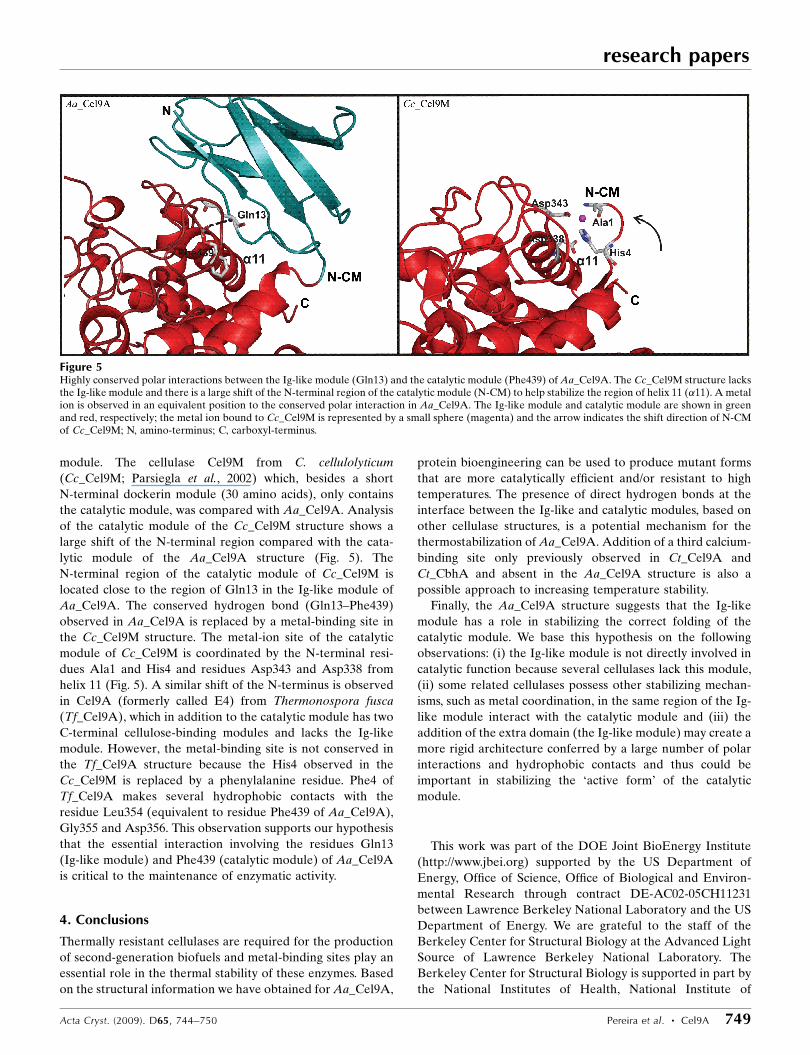
module. The cellulase Cel9M from C. cellulolyticum
(Cc_Cel9M; Parsiegla et al., 2002) which, besides a short
N-terminal dockerin module (30 amino acids), only contains
the catalytic module, was compared with Aa_Cel9A. Analysis
of the catalytic module of the Cc_Cel9M structure shows a
large shift of the N-terminal region compared with the cata-
lytic module of the Aa_Cel9A structure (Fig. 5). The
N-terminal region of the catalytic module of Cc_Cel9M is
located close to the region of Gln13 in the Ig-like module of
Aa_Cel9A. The conserved hydrogen bond (Gln13–Phe439)
observed in Aa_Cel9A is replaced by a metal-binding site in
the Cc_Cel9M structure. The metal-ion site of the catalytic
module of Cc_Cel9M is coordinated by the N-terminal resi-
dues Ala1 and His4 and residues Asp343 and Asp338 from
helix 11 (Fig. 5). A similar shift of the N-terminus is observed
in Cel9A (formerly called E4) from Thermonospora fusca
(Tf_Cel9A), which in addition to the catalytic module has two
C-terminal cellulose-binding modules and lacks the Ig-like
module. However, the metal-binding site is not conserved in
the Tf_Cel9A structure because the His4 observed in the
Cc_Cel9M is replaced by a phenylalanine residue. Phe4 of
Tf_Cel9A makes several hydrophobic contacts with the
residue Leu354 (equivalent to residue Phe439 of Aa_Cel9A),
Gly355 and Asp356. This observation supports our hypothesis
that the essential interaction involving the residues Gln13
(Ig-like module) and Phe439 (catalytic module) of Aa_Cel9A
is critical to the maintenance of enzymatic activity.
4. Conclusions
Thermally resistant cellulases are required for the production
of second-generation biofuels and metal-binding sites play an
essential role in the thermal stability of these enzymes. Based
on the structural information we have obtained for Aa_Cel9A,
protein bioengineering can be used to produce mutant forms
that are more catalytically efficient and/or resistant to high
temperatures. The presence of direct hydrogen bonds at the
interface between the Ig-like and catalytic modules, based on
other cellulase structures, is a potential mechanism for the
thermostabilization of Aa_Cel9A. Addition of a third calcium-
binding site only previously observed in Ct_Cel9A and
Ct_CbhA and absent in the Aa_Cel9A structure is also a
possible approach to increasing temperature stability.
Finally, the Aa_Cel9A structure suggests that the Ig-like
module has a role in stabilizing the correct folding of the
catalytic module. We base this hypothesis on the following
observations: (i) the Ig-like module is not directly involved in
catalytic function because several cellulases lack this module,
(ii) some related cellulases possess other stabilizing mechan-
isms, such as metal coordination, in the same region of the Ig-
like module interact with the catalytic module and (iii) the
addition of the extra domain (the Ig-like module) may create a
more rigid architecture conferred by a large number of polar
interactions and hydrophobic contacts and thus could be
important in stabilizing the ‘active form’ of the catalytic
module.
This work was part of the DOE Joint BioEnergy Institute
(http://www.jbei.org) supported by the US Department of
Energy, Office of Science, Office of Biological and Environ-
mental Research through contract DE-AC02-05CH11231
between Lawrence Berkeley National Laboratory and the US
Department of Energy. We are grateful to the staff of the
Berkeley Center for Structural Biology at the Advanced Light
Source of Lawrence Berkeley National Laboratory. The
Berkeley Center for Structural Biology is supported in part by
the National Institutes of Health, National Institute of
research papers
Acta Cryst. (2009). D65, 744–750 Pereira et al. � Cel9A 749
Figure 5Highly conserved polar interactions between the Ig-like module (Gln13) and the catalytic module (Phe439) of Aa_Cel9A. The Cc_Cel9M structure lacksthe Ig-like module and there is a large shift of the N-terminal region of the catalytic module (N-CM) to help stabilize the region of helix 11 (�11). A metalion is observed in an equivalent position to the conserved polar interaction in Aa_Cel9A. The Ig-like module and catalytic module are shown in greenand red, respectively; the metal ion bound to Cc_Cel9M is represented by a small sphere (magenta) and the arrow indicates the shift direction of N-CMof Cc_Cel9M; N, amino-terminus; C, carboxyl-terminus.
General Medical Sciences. The Advanced Light Source is
supported by the Director, Office of Science, Office of Basic
Energy Sciences of the US Department of Energy under
Contract No. DE-AC02-05CH11231. We also would like to
thank Professor E. Schneider at Humboldt-Universitat zu
Berlin for the gift of the original gene construct of Cel9A that
was used to subclone the gene for expression of the protein.
References
Adams, P. D., Grosse-Kunstleve, R. W., Hung, L.-W., Ioerger, T. R.,McCoy, A. J., Moriarty, N. W., Read, R. J., Sacchettini, J. C., Sauter,N. K. & Terwilliger, T. C. (2002). Acta Cryst. D58, 1948–1954.
Chauvaux, S., Souchon, H., Alzari, P. M., Chariot, P. & Beguin, P.(1995). J. Biol. Chem. 270, 9757–9762.
Collaborative Computational Project, Number 4 (1994). Acta Cryst.D50, 760–763.
Davies, G. J., Wilson, K. S. & Henrissat, B. (1997). Biochem. J. 321,557–559.
Davis, I. W., Leaver-Fay, A., Chen, V. B., Block, J. N., Kapral, G. J.,Wang, X., Murray, L. W., Arendall, W. B. III, Soeyink, J.,Richardson, J. C. & Richardson, D. C. (2007). Nucleic Acids Res.35, 375–383.
Eckert, K., Ernst, H. A., Schneider, E., Larsen, S. & Lo Leggio, L.(2003). Acta Cryst. D59, 139–141.
Eckert, K., Zielinski, F., Lo Leggio, L. & Schneider, E. (2002). Appl.Microbiol. Biotechnol. 60, 428–436.
Emsley, P. & Cowtan, K. (2004). Acta Cryst. D60, 2126–2132.Festucci-Buselli, R. A., Otoni, W. C. & Joshi, C. P. (2007). Braz. J.
Plant Physiol. 19, 1–13.Jancarik, J. & Kim, S.-H. (1991). J. Appl. Cryst. 24, 409–411.Juy, M., Amit, A. G., Alzari, P. M., Poljak, R. J., Claeyssens, M.,
Beguin, P. & Aubert, J.-P. (1992). Nature (London), 357, 89–91.Kataeva, I. A., Uversky, V. N., Brewer, J. M., Schubot, F., Rose, J. P.,
Wang, B.-C. & Ljungdahl, G. L. (2004). Protein Eng. Des. Sel. 17,759–769.
McCoy, A. J., Grosse-Kunstleve, R. W., Adams, P. D., Winn, M. D.,Storoni, L. C. & Read, R. J. (2007). J. Appl. Cryst. 40, 658–674.
Otwinowski, Z. & Minor, W. (1997). Methods Enzymol. 276, 307–326.Parsiegla, G., Belaich, A., Belaich, J. P. & Haser, R. (2002).
Biochemistry, 41, 11134–11142.Parsiegla, G., Juy, M., Reverbel-Leroy, C., Tardif, C., Belaich, J.-P.,
Driguez, H. & Haser, R. (1998). EMBO J. 17, 5551–5562.Parsiegla, G., Reverbel, C., Tardif, C., Driguez, H. & Haser, R. (2008).
J. Mol. Biol. 375, 499–510.Schubot, F. D., Kataeva, I. A., Chang, J., Shah, A. K., Ljungdahl, L. G.,
Rose, J. P. & Wang, B.-C. (2004). Biochemistry, 43, 1163–1170.Sticklen, M. B. (2008). Nature (London), 9, 433–443.
research papers
750 Pereira et al. � Cel9A Acta Cryst. (2009). D65, 744–750