modulation of myocardial contraction by peroxynitrite
TRANSCRIPT
REVIEW ARTICLEpublished: 12 December 2012
doi: 10.3389/fphys.2012.00468
Modulation of myocardial contraction by peroxynitriteMark J. Kohr1,2, Steve R. Roof1, Jay L. Zweier 3 and Mark T. Ziolo1*
1 Department of Physiology and Cell Biology, Davis Heart and Lung Research Institute, The Ohio State University, Columbus, OH, USA2 Division of Cardiovascular Pathology, Department of Pathology, Johns Hopkins University, Baltimore, MD, USA3 Department of Internal Medicine: Division of Cardiovascular Medicine, Davis Heart and Lung Research Institute, The Ohio State University, Columbus, OH, USA
Edited by:
Lars S. Maier,Georg-August-Universität, Germany
Reviewed by:
Jens Kockskämper,Philipps-University Marburg,GermanyChristoph Maack,Universitätsklinikum desSaarlandes, Germany
*Correspondence:
Mark T. Ziolo, Department ofPhysiology and Cell Biology, DavisHeart and Lung Research Institute,The Ohio State University, 304Hamilton Hall, 1645 Neil Avenue,Columbus, OH 43210, USA.e-mail: [email protected]
Peroxynitrite is a potent oxidant that is quickly emerging as a crucial modulator ofmyocardial function. This review will focus on the regulation of myocardial contractionby peroxynitrite during health and disease, with a specific emphasis on cardiomyocyteCa2+ handling, proposed signaling pathways, and protein end-targets.
Keywords: myocyte, peroxynitrite, nitric oxide synthase, Ca2+, cAMP-dependent protein kinase, protein
phosphatase 2a
The heart plays a vital role in the cardiovascular system by provid-ing the specialized tissues and organs of the body with a continualsupply of oxygen and other essential nutrients. Modulation ofmyocardial contraction allows the heart to meet the demands ofthe body despite continual changes in metabolism. This modu-lation occurs at several different levels, including the level of theventricular cardiomyocyte.
EXCITATION–CONTRACTION COUPLINGCardiomyocyte contraction occurs via a process termedexcitation–contraction coupling, in which the electrical activityof the heart is translated into cardiomyocyte contraction (Bers,2001, 2002). This process commences with the cardiac actionpotential and the depolarization of the myocyte membrane,which leads to the opening of L-type Ca2+ channels (Bers, 2001,2002). This causes an influx of Ca2+ into the myocyte via theL-type Ca2+ current (ICa), and subsequently triggers the openingof the sarcoplasmic reticulum (SR) Ca2+-release channels orryanodine receptors (RyRs). This results in an efflux of Ca2+from the SR, which is the major Ca2+ store in the cardiomyocyte,in a process termed Ca2+-induced Ca2+-release. This processinitiates the upstroke of the myocyte [Ca2+]i transient and thisCa2+ is now available for myocyte contraction. SR Ca2+ load,which represents the Ca2+ available for release from the SR, isa critical determinant of [Ca2+]i transient amplitude and thusmyocyte contraction (Bassani et al., 1995; Trafford et al., 1995).
The [Ca2+]i transient is responsible for the activation of car-diomyocyte contraction or shortening. When [Ca2+]i increaseswithin the cardiomyocyte, this Ca2+ diffuses to the myofilamentsand binds to troponin C (TnC) (Davis and Tikunova, 2008). Thisinduces a strong interaction between TnC and troponin I (TnI),
effectively destabilizing the interaction between TnI and actin.This shifts the troponin–tropomyosin complex in such a way asto allow the myosin head to bind directly to actin, thus leading tothe formation of a myosin crossbridge. Provided that ATP is read-ily available to the myosin head, myocyte force production and/orshortening ensues until Ca2+ is no longer bound to TnC.
Relaxation is initiated by the decline of the [Ca2+]i
transient, which is primarily mediated by the SR Ca2+-ATPase/phospholamban complex (SERCA/PLB) and theNa+/Ca2+ exchanger (NCX) (Bers, 2001, 2002). The SERCA/PLBcomplex serves to re-sequester Ca2+ into the SR, while NCXdirectly extrudes Ca2+ into the extracellular space. PLB is aphosphoprotein that plays a critical role in the decline of the[Ca2+]i transient by regulating the uptake of Ca2+ by SERCA.Under basal conditions, PLB remains hypophosphorylated andserves to inhibit the uptake of Ca2+ by SERCA. This inhibitioncan be relieved upon PLB Serine16 phosphorylation by proteinkinase A (PKA), thus allowing greater uptake of Ca2+ into theSR (Chu et al., 2000; Hagemann and Xiao, 2002; Mattiazzi et al.,2006). There are two main serine/threonine protein phosphatasesthat dephosphorylate PLB, protein phosphatase 1 (PP1) andprotein phosphatase 2a (PP2a) (Macdougall et al., 1991).
MODULATION OF EXCITATION–CONTRACTION COUPLINGSince every cardiomyocyte is recruited to contract with eachheartbeat, numerous endogenous systems have evolved to effec-tively regulate the ventricular myocyte contractile state by modu-lating excitation–contraction coupling. For example, stimulationof the β-adrenergic receptor (β-AR) pathway results in positiveinotropic and lusitropic effects via modulation of key excitation–contraction coupling proteins (Bers and Ziolo, 2001). Activation
www.frontiersin.org December 2012 | Volume 3 | Article 468 | 1

Kohr et al. ONOO− and myocardial contraction
of the β-AR signaling pathway transpires with binding of anagonist (e.g., epinephrine, isoproterenol) to the receptor, whichresults in the dissociation of the Gsα subunit. Gsα induces cAMPproduction via adenylate cyclase (AC). Increased cAMP thenactivates the cAMP-dependent protein kinase or PKA, whichphosphorylates many different targets in the cardiomyocyte,including ICa, RyR, TnI, and PLB. The end result is increasedCa2+ influx, increased SR Ca2+ uptake, load, and release, anddecreased myofilament Ca2+ sensitivity. PLB phosphorylation atSerine16 by PKA is one of the primary mechanisms responsiblefor the observed positive inotropic and lusitropic effects of β-ARstimulation (Chu et al., 2000; Li et al., 2000; Roof et al., 2011,2012). Therefore, altering PLB phosphorylation levels will lead todrastic changes in myocardial contraction and relaxation. Sincethe β-AR pathway is perhaps the most important regulator ofmyocardial contractility, this pathway itself is modulated by otherendogenous systems such as reactive nitrogen species (RNS).
REACTIVE NITROGEN SPECIESRNS are quickly emerging as crucial modulators of myocar-dial contraction during health and disease (Pacher et al., 2007).Alterations in the production of these reactive species may beresponsible for part of the dysfunction that is observed duringmany pathophysiological conditions of the myocardium and mayrepresent novel therapeutic targets. However, the modulation ofmyocardial function by RNS is a relatively new field and manyquestions remain unanswered. For example, studies have yet toconclusively determine whether this dysfunction results from theloss of endogenous RNS production [i.e., nitric oxide (NO)]or from the production of supraphysiological levels of reactivespecies. In all likelihood, this dysfunction stems from the par-tial loss of localized RNS signaling coupled with an increase inthe production of more reactive species, including peroxynitrite.Indeed, this appears to be the case (Feldman et al., 2008; Zioloet al., 2008).
NO is a well-known modulator of many physiological pro-cesses, including neural transmission and the regulation of bloodpressure. However, NO is also surfacing as a key modulatorof myocardial function by serving as a regulator of excitation–contraction coupling (Ziolo et al., 2001b, 2004; Wang et al.,2008a,b, 2009, 2010) and β-AR signaling (Balligand et al., 1993;Ziolo et al., 2001c). NO is produced endogenously within themyocardium by three distinct isoforms of NO synthase (NOS)(Ziolo and Bers, 2003). Neuronal NOS (NOS1) and endothelialNOS (NOS3) are constitutively expressed within cardiomyocytes.NOS1 and NOS3 produce low amounts of NO in phase with themyocyte [Ca2+]i transient. Inducible NOS (NOS2), on the otherhand, is only expressed within cardiomyocytes during inflam-matory responses which occur during many pathophysiologicalconditions of the myocardium. NOS2 produces much higheramounts of NO compared to constitutive isoforms and is [Ca2+]i
independent (Schulz et al., 1992; Ziolo et al., 1998, 2001a).NO is known to signal through at least two distinct signal-
ing pathways: cGMP-dependent and cGMP-independent (Ziolo,2008). cGMP-dependent signaling occurs through the activa-tion of guanylate cyclase and protein kinase G (PKG), whilecGMP-independent signaling primarily occurs via direct protein
modification (e.g., S-nitrosylation) (Martinez-Ruiz and Lamas,2004; Hess et al., 2005; Handy and Loscalzo, 2006; Kohr et al.,2011b). NO has also been shown to directly activate AC indepen-dent of β-AR activation, thus resulting in positive inotropic effectsvia increased cAMP production and PKA activation (Vila-Petroffet al., 1999). NO also signals by coupling to other reactive sig-naling species (e.g., superoxide anion) to form related congeners,such as peroxynitrite.
PEROXYNITRITEPeroxynitrite (ONOO−) is a potent oxidant that is formed uponthe reaction of NO and superoxide. This reaction occurs withan extremely high rate constant, making the formation of per-oxynitrite quite favorable (Huie and Padmaja, 1993; Beckmanand Koppenol, 1996). Although peroxynitrite is a highly reac-tive species, it is still capable of diffusing through biologi-cal membranes and interacting with intracellular constituents(Denicola et al., 1998). Under physiological conditions, perox-ynitrite production remains low and potential oxidative damageis reduced via endogenous cellular antioxidant defenses (Radiet al., 2002a,b). Low levels of peroxynitrite may also serve tomodulate various intracellular signaling pathways. In fact, per-oxynitrite appears to play a role in the regulation of manyimportant physiological processes. For example, peroxynitrite hasbeen demonstrated to modulate T cell proliferation (Brito et al.,1999; Vig et al., 2004), platelet aggregation (Schildknecht et al.,2008), and neutrophil adhesion (Zouki et al., 2001). Peroxynitriteis thought to mediate these physiological processes by targetingvarious proteins and/or signaling pathways, including receptortyrosine kinases, mitogen-activated protein kinases, phospho-inositide 3-kinase/protein kinase B (Akt), protein kinase C, andnuclear factor κB (Pacher et al., 2007).
The production of peroxynitrite increases greatly during thepathogenesis of numerous disorders, such as cerebral ischemia-reperfusion (Thiyagarajan et al., 2004; Dhar et al., 2006), myocar-dial ischemia-reperfusion (Wang and Zweier, 1996; Cheung et al.,2000), heart failure (Ferdinandy et al., 2000; Mihm et al., 2001;Zhang et al., 2007), atherosclerosis (Buttery et al., 1996; Luomaet al., 1998), diabetes (Suarez-Pinzon et al., 1997, 2001), andsepticemia (Bhattacharyya et al., 2004). Although peroxynitriteproduction plays a critical role for neutrophils and macrophagesin microbial defense (Macmicking et al., 1997; Nathan and Shiloh,2000), supraphysiological levels of peroxynitrite are almost alwaysdetrimental to cellular function. Peroxynitrite exerts damagingeffects by altering protein structure and function via irreversiblenitration to tyrosine residues and cysteine oxidation (Kamat,2006; Pacher et al., 2007). Peroxynitrite can also exert effects byreacting with transition metal centers, including heme prostheticgroups (Pietraforte et al., 2001; Lin et al., 2007) and zinc-thiolatemotifs (Crow et al., 1995; Zou et al., 2004), thus resulting in theinactivation of many enzymes. NOS3, an enzyme which exertsmany cardioprotective effects (Janssens et al., 2004; Buys et al.,2007; Wang et al., 2008b, 2012), can be inhibited upon exposureto high levels of peroxynitrite (Zou et al., 2004; Chen et al., 2010).Additionally, hydroxylation of ring systems (Szabo et al., 1997;Tuo et al., 1998), DNA strand breakage (Szabo et al., 1996, 1997),and lipid peroxidation (Radi et al., 1991a,b) can be induced by
Frontiers in Physiology | Striated Muscle Physiology December 2012 | Volume 3 | Article 468 | 2
Kohr et al. ONOO− and myocardial contraction
peroxynitrite. Apoptosis and necrosis can also be triggered uponperoxynitrite exposure (Gilad et al., 1997; Arstall et al., 1999;Pacher et al., 2002; Levrand et al., 2006). Moreover, peroxynitritewill react with other molecules, such as carbon dioxide (CO2),to form additional reactive species (i.e., nitrosoperoxycarbonate)(Uppu and Pryor, 1996). Increased peroxynitrite production isfurther compounded by the depletion of endogenous antioxidantpools, including glutathione (GSH), which is a major scavenger ofreactive oxygen and nitrogen species (Valko et al., 2007). The pro-duction of peroxynitrite occurs endogenously in several differentways.
Peroxynitrite can be formed endogenously within the ventric-ular cardiomyocyte through various routes under physiologicaland pathophysiological conditions. A major source for the pro-duction of peroxynitrite includes the NOS isoforms (namelyNOS1 and NOS2) and via non-classical pathways (namelynitroxyl).
NOS1 has been shown to co-immunoprecipitate with xan-thine oxidoreductase (Khan et al., 2004), a superoxide producingenzyme. This interaction between NOS1 and xanthine oxidore-ductase will lead to the physiological production of low levelsof peroxynitrite. Additionally, NOS1 can potentially produceboth NO and superoxide (Pou et al., 1992; Xia et al., 1996),although this is more likely to occur with NOS uncoupling dur-ing various disease states (Sun et al., 2008). NOS uncouplingwill increase superoxide production by NOS1, while decreasingNO production. NOS1 is considered to be a physiological regu-lator of myocardial function by increasing basal contractility, theforce-frequency response, and the response to β-AR stimulation(Barouch et al., 2002; Khan et al., 2003; Vandsburger et al., 2007;Wang et al., 2008a, 2010).
NOS2 is a high output NOS isoform and can readily becomeuncoupled upon depletion of the cofactors necessary for NO pro-duction, thus leading to the production of both NO and super-oxide (Xia and Zweier, 1997; Mungrue et al., 2002). Additionally,NADPH oxidase and xanthine oxidoreductase can increase super-oxide production under the same pathophysiological conditionsin which NOS2 is expressed (Heymes et al., 2003; Minhas et al.,2006). This will lead to the production of supraphysiological lev-els of peroxynitrite. NOS2 is considered to be a pathophysiologi-cal regulator of myocardial function by decreasing the response toβ-AR stimulation (Drexler et al., 1998; Ziolo et al., 2001b, 2004).
In addition to the NO produced via NOS1 and NOS2, addi-tional sources of peroxynitrite include nitroxyl, the one elec-tron reduction production of NO (Kirsch and De Groot, 2002).Endogenous production of nitroxyl within cardiomyocytes mayoccur through various routes, including via hydrogen atomextraction by NO (Akhtar et al., 1985). NOS1 also remains apotential source for the production of nitroxyl (Schmidt et al.,1996; Ishimura et al., 2005). Therefore, the reaction of nitroxylwith O2 will lead to the production of low levels of peroxynitrite.
PEROXYNITRITE AND CARDIOMYOCYTE CONTRACTIONRecent work has now demonstrated that peroxynitrite is ableto modulate excitation–contraction coupling, and hence con-tractility. In the myocardium, peroxynitrite has been demon-strated to exert biphasic effects on cardiomyocyte contraction.
More specifically, low levels of peroxynitrite (which occurs underphysiological conditions) have been shown to produce positiveinotropic effects on cardiomyocyte function. Conversely, highlevels of peroxynitrite (which occurs under pathophysiologicalconditions) are extremely detrimental to myocardial function bydecreasing cardiomyocyte contraction.
PHYSIOLOGICAL REGULATION OF CARDIOMYOCYTECONTRACTION BY PEROXYNITRITEMany studies have shown that low levels of peroxynitrite inducepositive inotropic effects on basal myocardial function. Chesnaiset al. demonstrated peroxynitrite to increase force productionin frog atrial and ventricular fibers using authentic peroxyni-trite, as well as the peroxynitrite donor SIN-1 (Chesnais et al.,1999a,b). Paolocci et al. confirmed this positive effect on basalfunction in mammalian ex vivo myocardial preparations usingSIN-1 (Paolocci et al., 2000). Interestingly, this effect was reversedwith GSH and occurred independently from global changes incAMP and cGMP levels. Furthermore, we demonstrated thata low concentration of peroxynitrite increased basal and sub-maximal β-AR-stimulated contraction in isolated cardiomyocytes(Kohr et al., 2008a). We next investigated the molecular mecha-nisms of this peroxynitrite-induced increase in basal contractionand found that it was dependent upon PLB. That is, perox-ynitrite had no effect on basal contraction in PLB knockoutmyocytes. Further explorations demonstrated that peroxynitriteresulted in the enhancement of PLB Serine16 (the PKA site)phosphorylation (Kohr et al., 2010) and was abolished uponinhibition of PKA with KT5720 (Kohr et al., 2010). Low per-oxynitrite also increased PKA activity in cardiac homogenatesand in purified preparations of PKA containing both the reg-ulatory and catalytic subunits of PKA, indicating that perox-ynitrite induces a direct, cAMP-independent activation of PKA.This direct effect may occur via S-nitrosylation, S-glutathiolationand/or cysteine oxidation (Ferdinandy, 2006; Pacher et al., 2007),as PKA has several cysteine residues which are susceptible tothese types of modifications (Brennan et al., 2006; Burgoyne andEaton, 2009; Kohr et al., 2011a). Thus, low levels of peroxyni-trite shift the kinase/phosphatase balance leading to increasedPLB phosphorylation. Interestingly, the effect of low perox-ynitrite on submaximal β-AR-stimulated contraction was stillobserved in PLB knockout myocytes, suggesting another proteintarget. A recent study demonstrated that PKA phosphorylationof RyR contributes to the inotropic effect of β-AR stimulationusing a low dose of isoproterenol (Shan et al., 2010). Thus,peroxynitrite-mediated activation of PKA may also lead to RyRphosphorylation contributing to the contractile effects of β-ARstimulation. Non PKA-mediated effects of peroxynitrite havealso been reported such as S-nitrosylation of SERCA2a, whichincreases Ca2+ uptake and relaxation (Bencsik et al., 2008).
We previously demonstrated in healthy ventricular cardiomy-ocytes that NOS1 signals partly through the production of per-oxynitrite (Wang et al., 2008a). Additionally, we and others haveshown that NOS1 signaling increases myocardial contraction(basal, force-frequency response and β-AR-stimulated contrac-tion) (Barouch et al., 2002; Khan et al., 2004; Wang et al., 2008a),parameters that can be considered physiologic. These effects are
www.frontiersin.org December 2012 | Volume 3 | Article 468 | 3
Kohr et al. ONOO− and myocardial contraction
consistent with the positive inotropic effects of peroxynitrite (i.e.,peroxynitrite increased basal and submaximal β-AR-stimulatedcontraction). Interestingly, NOS1−/− myocytes exhibit reducedbasal contraction and a diminished response to β-AR stimula-tion compared to wild type (Barouch et al., 2002; Khan et al.,2004; Wang et al., 2008a), which we and others demonstrated tooccur in part through a reduction in RyR activity (Wang et al.,2010) and PLB Serine16 phosphorylation (Wang et al., 2008a;Zhang et al., 2008). This is also consistent with the PLB andRyR dependent effects of low peroxynitrite (Kohr et al., 2008a).In addition, NOS1 has been shown to potentially modulate PKAactivity, as PKA inhibition decreased PLB Serine16 phosphoryla-tion and the rate of relaxation in wild-type myocytes, but had noeffect in NOS1−/− myocytes (Zhang et al., 2008). Therefore, thephysiological production of low levels of peroxynitrite may serveto maintain and/or increase basal and β-AR-stimulated myocytecontraction, in part, through a direct, cAMP-independent acti-vation of PKA. Further, the loss of localized NOS1-mediatedperoxynitrite production under certain disease states where NOS1translocates to the sarcolemma (Damy et al., 2003, 2004) mayresult in decreased myocyte contraction similar to that observedwith the knockout of NOS1.
PATHOPHYSIOLOGICAL REGULATION OF CARDIOMYOCYTECONTRACTION BY PEROXYNITRITEAt high concentrations, peroxynitrite is detrimental to myocar-dial function. Studies examining high levels of peroxynitritehave demonstrated dysfunctional effects via direct exposure orusing peroxynitrite donors (Lopez et al., 1997; Ma et al., 1997;Ferdinandy et al., 1999, 2000; Katori et al., 2006). Additionally,we have demonstrated a NOS2-induced reduction in β-AR-stimulated RyR activity through a cGMP-independent mech-anism, likely via peroxynitrite (Ziolo et al., 2001b). Further,we and others have shown a reduction in β-AR-stimulatedcontraction upon exposure to a high concentration of SIN-1in isolated cardiomyocytes (Stojanovic et al., 2001; Yin et al.,2002). We demonstrated that this SIN-1-induced decrease inβ-AR-stimulated contraction occurred via peroxynitrite forma-tion and was also dependent upon PLB. However, unlike lowperoxynitrite, high peroxynitrite results in a reduction in PKA-dependent PLB Serine16 phosphorylation (Kohr et al., 2008b).Upon further examination of this signaling pathway, we deter-mined that high peroxynitrite increased phosphatase activityin cardiac homogenates (low peroxynitrite had no effect onphosphatase activity). Interestingly, it appears that peroxynitriteselectively targets PP2a as we observed an increased interactionbetween PLB and PP2a (but not PP1) with peroxynitrite (Kohret al., 2009). This reversible effect may occur through a pro-tein intermediate or from the direct modification of PP2a byperoxynitrite via S-nitrosylation, S-glutathiolation, and/or cys-teine oxidation (Ferdinandy, 2006; Pacher et al., 2007). PP2ahas several cysteine residues which are susceptible to these typesof modifications (Sommer et al., 2002). Consistent with ourresults, peroxynitrite has been shown to directly activate PP2avia tyrosine nitration in endothelial cells (Wu and Wilson, 2009).Thus, high levels of peroxynitrite shift the kinase/phosphatasebalance leading to decreased PLB phosphorylation. While PP2Aalso modulates RyR phosphorylation (Terentyev et al., 2003),
we did not observe any effects of high peroxynitrite onthe β-AR response in PLB knockout myocytes, which sug-gests that this may be a compartmentalized effect targetedto PLB.
The production of high levels of peroxynitrite in themyocardium is often associated with the expression of NOS2 (Xiaet al., 1998; Mungrue et al., 2002) and increased ROS produc-tion via NADPH oxidase (Byrne et al., 2003; Heymes et al., 2003)and xanthine oxidase (Tziomalos and Hare, 2009). Specificallyin heart failure, studies have shown an increase in peroxynitriteproduction (Ferdinandy et al., 2000; Mihm et al., 2001; Zhanget al., 2007), as well as a diminished response to β-AR stimula-tion (Houser et al., 2000). We have previously demonstrated thatmyocytes isolated from failing human hearts expressing NOS2,displayed a blunted response to β-AR stimulation (Ziolo et al.,2004). This dysfunction could be reversed upon acute inhibi-tion of NOS2, such that peak [Ca2+]i and cell shortening weresignificantly increased. This same functional phenomenon wasobserved with high peroxynitrite (i.e., peroxynitrite decreasedcontraction during β-AR stimulation). Interestingly, PP2a activ-ity has been shown to be increased in heart failure (Boknik et al.,2000), while PLB Serine16 phosphorylation has been shown to bedecreased (Bartel et al., 1996; Schwinger et al., 1999; Sande et al.,2002). Therefore, the increased formation of peroxynitrite thatoccurs in heart failure may be an important component of theβ-AR dysfunction observed in heart failure and other cardiomy-opathies. A recent study demonstrated that peroxynitrite was anindependent risk predictor of post-operative complications (i.e.,atrial fibrillation, need for inotropic support, length of hospitalstay) among patients undergoing cardiac surgery for various rea-sons (Antoniades et al., 2012), further validating the significanceof increased peroxynitrite in cardiac dysfunction.
PEROXYNITRITE AND MITOCHONDRIAL FUNCTIONThe processes of cardiomyocyte contraction and Ca2+ reup-take/extrusion are dependent upon ATP. Through oxidative phos-phorylation, mitochondria produce the majority of the ATP thatis necessary for these processes. Ca2+ is also taken up by the mito-chondria during each Ca2+ transient, and this serves to coupleexcitation–contraction coupling with energetic demand (Maackand O’Rourke, 2007). Recent work has demonstrated that mito-chondrial function is modulated by RNS (i.e., NO, peroxynitrite).
NO is a well-established physiological mediator of mitochon-drial respiration, and these effects primarily result from theinhibition of Complex I and IV of the electron transport chain(Brookes, 2004). Although Complex I and IV are similarly inhib-ited by NO, this inhibition occurs through two distinct mecha-nisms. In the case of Complex I, NO acts via S-nitrosylation ofthe 75 kDa subunit of Complex I (Burwell et al., 2006). ComplexIV, on the other hand, is inhibited through the reaction of NOwith the catalytic metals located in the active site of cytochromec oxidase (Cleeter et al., 1994; Palacios-Callender et al., 2004).NO can also target additional mitochondrial proteins by reactingwith superoxide to yield peroxynitrite. The effects of peroxynitriteon mitochondrial function are much less defined when com-pared to those of NO, but peroxynitrite has the potential tosignal physiologically in the mitochondria or to contribute tomitochondrial dysfunction during pathological states.
Frontiers in Physiology | Striated Muscle Physiology December 2012 | Volume 3 | Article 468 | 4
Kohr et al. ONOO− and myocardial contraction
Peroxynitrite has been shown to target many different pro-teins in the mitochondria and this can occur via S-nitrosylation,as reported to occur with Complex I of the electron transportchain (Borutaite et al., 2000), or through irreversible nitrationand/or cysteine oxidation. The reversibility of S-nitrosylation mayrepresent a potential physiological signaling pathway for per-oxynitrite in the mitochondria, while the effects of irreversiblenitration and cysteine oxidation are likely to be highly detrimen-tal to mitochondrial function. For example, peroxynitrite inducedthe irreversible inhibition of mitochondrial creatine kinase, andthis effect could not be reversed with GSH treatment (Konorevet al., 1998). Peroxynitrite was also shown to inactivate the Kreb’scycle enzyme aconitase (Castro et al., 1994). Additional mito-chondrial targets of peroxynitrite include the adenine nucleotidetranslocase (Vieira et al., 2001), nicotinamide nucleotide tran-shydrogenase (Forsmark-Andree et al., 1996), and Complex I, II,and V of the electron transport chain (Radi et al., 1994; Cassinaand Radi, 1996; Riobo et al., 2001). Manganese superoxide dis-mutase, which is a major enzyme for the decomposition ofsuperoxide in the mitochondria, is another target of peroxyni-trite and is inactivated via nitration (Quijano et al., 2001). Thisperoxynitrite-induced inactivation is further compounded byincreased levels of superoxide, which could in turn lead to theformation of additional peroxynitrite. Hence, a decrease in energysupply by inhibiting mitochondrial function could contribute tothe depressed contractile function associated with high levels ofperoxynitrite.
LOW vs. HIGH LEVELS OF PEROXYNITRITEPeroxynitrite has a biphasic effect on cardiomyocyte contrac-tion, which is mainly concentration dependent (low vs. high).
However, since peroxynitrite is highly reactive and rapidly decom-posed, accurate measurements of concentration are difficult. Inaddition, determining intracellular concentrations of peroxyni-trite with exogenously applied authentic peroxynitrite or theperoxynitrite donor SIN-1 is challenging, making direct com-parisons to endogenously produced peroxynitrite formidable.In general, concentrations of 30 μM or less of authentic per-oxynitrite or 100 μM or less of SIN-1 will result in positiveinotropic effects as described. We have previously reported that10 μM SIN-1 produces 3 nM/min peroxynitrite in our physio-logical saline solution (Kohr et al., 2008a). Higher concentra-tions (100 μM authentic peroxynitrite or 200 μM SIN-1) willresult in negative inotropic effects as described. We have pre-viously reported that 200 μM SIN-1 results in a 6X greaterproduction of peroxynitrite compared to 10 μM SIN-1 (Kohret al., 2008b). We consider these levels to be physiologicallyand pathophysiologically relevant as we and others observedsimilar effects on function in studies investigating NOS1 andNOS2 signaling. Thus, these concentrations of peroxynitriteresult in the activation of various signaling pathways (PKA, PP2A,S-nitrosylation, etc.) resulting in the enhancement or reductionof myocardial contractility. There have been exceptions reportedin which low concentrations of authentic peroxynitrite/SIN-1 resulted in negative inotropic effects or high concentra-tions of SIN-1 resulted in positive inotropic effects (Yin et al.,2002; Katori et al., 2006; Kohr et al., 2008a). Thus, other fac-tors also contribute to the contractile effects of peroxynitritesuch as cardiomyocyte contractile state, signaling pathway acti-vation (e.g., S-nitrosylation, nitration, cGMP or cAMP) aswell as the length of exposure (Schulz et al., 1997; Ziolo,2008).
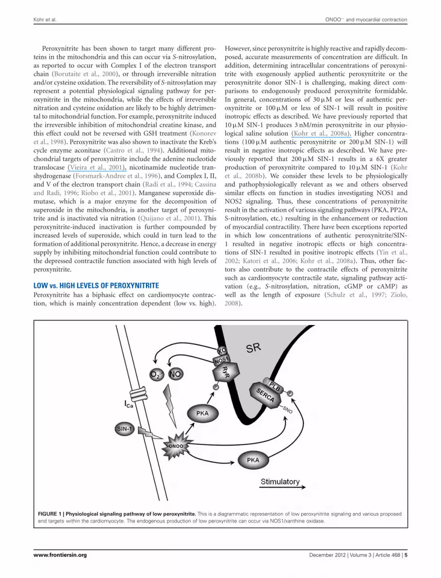
FIGURE 1 | Physiological signaling pathway of low peroxynitrite. This is a diagrammatic representation of low peroxynitrite signaling and various proposedend targets within the cardiomyocyte. The endogenous production of low peroxynitrite can occur via NOS1/xanthine oxidase.
www.frontiersin.org December 2012 | Volume 3 | Article 468 | 5

Kohr et al. ONOO− and myocardial contraction
FIGURE 2 | Pathophysiological signaling pathway of high peroxynitrite. This is a diagrammatic representation of high peroxynitrite signaling and variousproposed end targets within the cardiomyocyte. The endogenous production of high levels of peroxynitrite can occur via NOS2/NADPH oxidase/xanthineoxidase.
Studies have also been performed using very high concentra-tions of authentic peroxynitrite (mM) or long exposure times.These studies revealed irreversible effects of peroxynitrite thatdepressed myocyte contraction and inactivated SERCA (Ishidaet al., 1996; Knyushko et al., 2005; Lokuta et al., 2005). A con-sequence of this very high concentration of peroxynitrite is likelya toxic effect that results in cell damage. For example, peroxyni-trite is able to activate matrix metalloproteinases that will resultin the cleavage of α-actinin and TnI (Wang et al., 2002; Rorket al., 2006; Sung et al., 2007; Leon et al., 2008). We believe thatvery high concentrations of peroxynitrite can be produced in cer-tain disease states such as heart failure or ischemia/reperfusioninjury which have reported SERCA inactivation via nitration orTnI cleavage.
In conclusion, peroxynitrite is emerging as a crucial modulatorof myocardial function during health and disease. Under phys-iological conditions, low levels of peroxynitrite serve to main-tain and/or increase basal and β-AR-stimulated contraction in
the myocardium. This occurs via RyR and PLB phosphoryla-tion through direct PKA activation and SERCA activation viaS-nitrosylation (Figure 1). Conversely, supraphysiological levelsof peroxynitrite (due to NOS2 expression and increased ROSlevels via NADPH oxidase and xanthine oxidase) are detri-mental to myocardial function and decrease β-AR-stimulatedcardiomyocyte contraction. This occurs via PLB dephosphoryla-tion through PP2a activation, decreased RyR activity, mitochon-drial dysfunction, and myofilament protein cleavage (Figure 2).This dual effect lends critical insight into the physiologicaland pathophysiological regulation of myocardial contraction byperoxynitrite.
ACKNOWLEDGMENTSSupported by the American Heart Association (Pre-doctoralFellowship 0715159B, Mark J. Kohr) and the National Institutesof Health (K02HL094692, R01HL079283, Mark T. Ziolo;R01HL38324, HL63744, HL65608, Jay L. Zweier).
REFERENCESAkhtar, M. J., Bonner, F. T., and
Hughes, M. N. (1985). Reaction ofnitric oxide with hyponitrous acid:a hyrdogen atom abstraction reac-tion. Inorg. Chem. 24, 1934–1935.
Antoniades, C., Demosthenous, M.,Reilly, S., Margaritis, M., Zhang,M. H., Antonopoulos, A., et al.(2012). Myocardial redox state pre-dicts in-hospital clinical outcomeafter cardiac surgery effects of short-term pre-operative statin treatment.J. Am. Coll. Cardiol. 59, 60–70.
Arstall, M. A., Sawyer, D. B., Fukazawa,R., and Kelly, R. A. (1999).Cytokine-mediated apoptosisin cardiac myocytes: the roleof inducible nitric oxide syn-thase induction and peroxynitritegeneration. Circ. Res. 85, 829–840.
Balligand, J. L., Kelly, R. A., Marsden,P. A., Smith, T. W., and Michel,T. (1993). Control of cardiac mus-cle cell function by an endoge-nous nitric oxide signaling system.Proc. Natl. Acad. Sci. U.S.A. 90,347–351.
Barouch, L. A., Harrison, R. W., Skaf,M. W., Rosas, G. O., Cappola, T.P., Kobeissi, Z. A., et al. (2002).Nitric oxide regulates the heart byspatial confinement of nitric oxidesynthase isoforms. Nature 416,337–339.
Bartel, S., Stein, B., Eschenhagen, T.,Mende, U., Neumann, J., Schmitz,W., et al. (1996). Protein phos-phorylation in isolated trabeculaefrom nonfailing and failing humanhearts. Mol. Cell. Biochem. 157,171–179.
Bassani, J. W., Yuan, W., and Bers, D.M. (1995). Fractional SR Ca releaseis regulated by trigger Ca and SRCa content in cardiac myocytes. Am.J. Physiol. 268, C1313–C1319.
Beckman, J. S., and Koppenol, W.H. (1996). Nitric oxide, superox-ide, and peroxynitrite: the good, thebad, and ugly. Am. J. Physiol. 271,C1424–C1437.
Bencsik, P., Kupai, K., Giricz, Z., Gorbe,A., Huliak, I., Furst, S., et al.(2008). Cardiac capsaicin-sensitivesensory nerves regulate myocardial
Frontiers in Physiology | Striated Muscle Physiology December 2012 | Volume 3 | Article 468 | 6
Kohr et al. ONOO− and myocardial contraction
relaxation via S-nitrosylation ofSERCA: role of peroxynitrite. Br.J. Pharmacol. 153, 488–496.
Bers, D. M. (2001). Excitation-Contraction Coupling and CardiacContractile Force. Dordrecht,Netherlands: Kluwer AcademicPublishers.
Bers, D. M. (2002). Cardiac excitation-contraction coupling. Nature 415,198–205.
Bers, D. M., and Ziolo, M. T. (2001).When is cAMP not cAMP? Effects ofcompartmentalization. Circ. Res. 89,373–375.
Bhattacharyya, J., Biswas, S., and Datta,A. G. (2004). Mode of action ofendotoxin: role of free radicals andantioxidants. Curr. Med. Chem. 11,359–368.
Boknik, P., Fockenbrock, M., Herzig,S., Knapp, J., Linck, B., Luss, H.,et al. (2000). Protein phosphataseactivity is increased in a rat modelof long-term beta-adrenergic stimu-lation. Naunyn Schmiedebergs Arch.Pharmacol. 362, 222–231.
Borutaite, V., Budriunaite, A., andBrown, G. C. (2000). Reversal ofnitric oxide-, peroxynitrite- and S-nitrosothiol-induced inhibition ofmitochondrial respiration or com-plex I activity by light and thi-ols. Biochim. Biophys. Acta 1459,405–412.
Brennan, J. P., Bardswell, S. C.,Burgoyne, J. R., Fuller, W., Schroder,E., Wait, R., et al. (2006). Oxidant-induced activation of type I proteinkinase A is mediated by RI sub-unit interprotein disulfide bondformation. J. Biol. Chem. 281,21827–21836.
Brito, C., Naviliat, M., Tiscornia, A. C.,Vuillier, F., Gualco, G., Dighiero,G., et al. (1999). Peroxynitriteinhibits T lymphocyte activationand proliferation by promotingimpairment of tyrosine phospho-rylation and peroxynitrite-drivenapoptotic death. J. Immunol. 162,3356–3366.
Brookes, P. S. (2004). Mitochondrialnitric oxide synthase. Mito-chondrion 3, 187–204.
Burgoyne, J. R., and Eaton, P. (2009).Transnitrosylating nitric oxidespecies directly activate type Iprotein kinase A, providing a noveladenylate cyclase-independentcross-talk to beta-adrenergic-likesignaling. J. Biol. Chem. 284,29260–29268.
Burwell, L. S., Nadtochiy, S. M.,Tompkins, A. J., Young, S., andBrookes, P. S. (2006). Directevidence for S-nitrosation of mito-chondrial complex I. Biochem. J.394, 627–634.
Buttery, L. D., Springall, D. R., Chester,A. H., Evans, T. J., Standfield, E.N., Parums, D. V., et al. (1996).Inducible nitric oxide synthase ispresent within human atheroscle-rotic lesions and promotes the for-mation and activity of peroxynitrite.Lab. Invest. 75, 77–85.
Buys, E. S., Raher, M. J., Blake, S.L., Neilan, T. G., Graveline, A.R., Passeri, J. J., et al. (2007).Cardiomyocyte-restricted restora-tion of nitric oxide synthase 3attenuates left ventricular remodel-ing after chronic pressure overload.Am. J. Physiol. Heart Circ. Physiol.293, H620–H627.
Byrne, J. A., Grieve, D. J., Cave, A. C.,and Shah, A. M. (2003). Oxidativestress and heart failure. Arch. Mal.Coeur Vaiss. 96, 214–221.
Cassina, A., and Radi, R. (1996).Differential inhibitory action ofnitric oxide and peroxynitrite onmitochondrial electron trans-port. Arch. Biochem. Biophys. 328,309–316.
Castro, L., Rodriguez, M., and Radi, R.(1994). Aconitase is readily inacti-vated by peroxynitrite, but not byits precursor, nitric oxide. J. Biol.Chem. 269, 29409–29415.
Chen, W., Druhan, L. J., Chen, C.A., Hemann, C., Chen, Y. R.,Berka, V., et al. (2010). Peroxynitriteinduces destruction of the tetrahy-drobiopterin and heme in endothe-lial nitric oxide synthase: transi-tion from reversible to irreversibleenzyme inhibition. Biochemistry 49,3129–3137.
Chesnais, J. M., Fischmeister, R., andMery, P. F. (1999a). Peroxynitrite is apositive inotropic agent in atrial andventricular fibres of the frog heart.J. Physiol. 521, 375–388.
Chesnais, J. M., Fischmeister, R., andMery, P. F. (1999b). Positive andnegative inotropic effects of NOdonors in atrial and ventricularfibres of the frog heart. J. Physiol.518, 449–461.
Cheung, P. Y., Wang, W., and Schulz, R.(2000). Glutathione protects againstmyocardial ischemia-reperfusioninjury by detoxifying peroxynitrite.J. Mol. Cell. Cardiol. 32, 1669–1678.
Chu, G., Lester, J. W., Young, K. B.,Luo, W., Zhai, J., and Kranias, E. G.(2000). A single site (Ser16) phos-phorylation in phospholamban issufficient in mediating its maximalcardiac responses to beta -agonists.J. Biol. Chem. 275, 38938–38943.
Cleeter, M. W., Cooper, J. M., Darley-Usmar, V. M., Moncada, S., andSchapira, A. H. (1994). Reversibleinhibition of cytochrome c oxi-dase, the terminal enzyme of the
mitochondrial respiratory chain, bynitric oxide. Implications for neu-rodegenerative diseases. FEBS Lett.345, 50–54.
Crow, J. P., Beckman, J. S., and McCord,J. M. (1995). Sensitivity of the essen-tial zinc-thiolate moiety of yeastalcohol dehydrogenase to hypochlo-rite and peroxynitrite. Biochemistry34, 3544–3552.
Damy, T., Ratajczak, P., Robidel,E., Bendall, J. K., Oliviero, P.,Boczkowski, J., et al. (2003). Up-regulation of cardiac nitric oxidesynthase 1-derived nitric oxide aftermyocardial infarction in senescentrats. FASEB J. 17, 1934–1936.
Damy, T., Ratajczak, P., Shah, A. M.,Camors, E., Marty, I., Hasenfuss,G., et al. (2004). Increased neu-ronal nitric oxide synthase-derivedNO production in the failing humanheart. Lancet 363, 1365–1367.
Davis, J. P., and Tikunova, S. B.(2008). Ca(2+) exchange with tro-ponin C and cardiac muscle dynam-ics. Cardiovasc. Res. 77, 619–626.
Denicola, A., Souza, J. M., and Radi,R. (1998). Diffusion of peroxyni-trite across erythrocyte membranes.Proc. Natl. Acad. Sci. U.S.A. 95,3566–3571.
Dhar, A., Kaundal, R. K., and Sharma,S. S. (2006). Neuroprotectiveeffects of FeTMPyP: a peroxynitritedecomposition catalyst in globalcerebral ischemia model in gerbils.Pharmacol. Res. 54, 311–316.
Drexler, H., Kastner, S., Strobel, A.,Studer, R., Brodde, O. E., andHasenfuss, G. (1998). Expression,activity and functional signifi-cance of inducible nitric oxidesynthase in the failing humanheart. J. Am. Coll. Cardiol. 32,955–963.
Feldman, D. S., Elton, T. S., Sun,B., Martin, M. M., and Ziolo, M.T. (2008). Mechanisms of disease:detrimental adrenergic signaling inacute decompensated heart failure.Nat. Clin. Pract. Cardiovasc. Med. 5,208–218.
Ferdinandy, P. (2006). Peroxynitrite:just an oxidative/nitrosative stressoror a physiological regulator as well?Br. J. Pharmacol. 148, 1–3.
Ferdinandy, P., Danial, H., Ambrus,I., Rothery, R. A., and Schulz, R.(2000). Peroxynitrite is a majorcontributor to cytokine-inducedmyocardial contractile failure. Circ.Res. 87, 241–247.
Ferdinandy, P., Panas, D., and Schulz,R. (1999). Peroxynitrite contributesto spontaneous loss of cardiacefficiency in isolated workingrat hearts. Am. J. Physiol. 276,H1861–H1867.
Forsmark-Andree, P., Persson, B.,Radi, R., Dallner, G., and Ernster,L. (1996). Oxidative modificationof nicotinamide nucleotide tran-shydrogenase in submitochondrialparticles: effect of endogenousubiquinol. Arch. Biochem. Biophys.336, 113–120.
Gilad, E., Zingarelli, B., Salzman, A.L., and Szabo, C. (1997). Protectionby inhibition of poly (ADP-ribose)synthetase against oxidant injury incardiac myoblasts in vitro. J. Mol.Cell. Cardiol. 29, 2585–2597.
Hagemann, D., and Xiao, R. P. (2002).Dual site phospholamban phos-phorylation and its physiologicalrelevance in the heart. TrendsCardiovasc. Med. 12, 51–56.
Handy, D. E., and Loscalzo, J. (2006).Nitric oxide and posttranslationalmodification of the vascular pro-teome: S-nitrosation of reactive thi-ols. Arterioscler. Thromb. Vasc. Biol.26, 1207–1214.
Hess, D. T., Matsumoto, A., Kim, S.O., Marshall, H. E., and Stamler,J. S. (2005). Protein S-nitrosylation:purview and parameters. Nat. Rev.Mol. Cell Biol. 6, 150–166.
Heymes, C., Bendall, J. K., Ratajczak, P.,Cave, A. C., Samuel, J. L., Hasenfuss,G., et al. (2003). Increased myocar-dial NADPH oxidase activity inhuman heart failure. J. Am. Coll.Cardiol. 41, 2164–2171.
Houser, S. R., Piacentino, V. 3rd., andWeisser, J. (2000). Abnormalities ofcalcium cycling in the hypertro-phied and failing heart. J. Mol. Cell.Cardiol. 32, 1595–1607.
Huie, R. E., and Padmaja, S. (1993).The reaction of no with superox-ide. Free Radic. Res. Commun. 18,195–199.
Ishida, H., Ichimori, K., Hirota, Y.,Fukahori, M., and Nakazawa, H.(1996). Peroxynitrite-induced car-diac myocyte injury. Free Radic.Biol. Med. 20, 343–350.
Ishimura, Y., Gao, Y. T., Panda, S. P.,Roman, L. J., Masters, B. S., andWeintraub, S. T. (2005). Detectionof nitrous oxide in the neuronalnitric oxide synthase reaction bygas chromatography-mass spec-trometry. Biochem. Biophys. Res.Commun. 338, 543–549.
Janssens, S., Pokreisz, P., Schoonjans,L., Pellens, M., Vermeersch, P., Tjwa,M., et al. (2004). Cardiomyocyte-specific overexpression of nitricoxide synthase 3 improves left ven-tricular performance and reducescompensatory hypertrophy aftermyocardial infarction. Circ. Res. 94,1256–1262.
Kamat, J. P. (2006). Peroxynitrite:a potent oxidizing and nitrating
www.frontiersin.org December 2012 | Volume 3 | Article 468 | 7
Kohr et al. ONOO− and myocardial contraction
agent. Indian J. Exp. Biol. 44,436–447.
Katori, T., Donzelli, S., Tocchetti,C. G., Miranda, K. M., Cormaci,G., Thomas, D. D., et al. (2006).Peroxynitrite and myocardial con-tractility: in vivo versus in vitroeffects. Free Radic. Biol. Med. 41,1606–1618.
Khan, S. A., Lee, K., Minhas, K.M., Gonzalez, D. R., Raju, S.V., Tejani, A. D., et al. (2004).Neuronal nitric oxide synthasenegatively regulates xanthine oxi-doreductase inhibition of cardiacexcitation-contraction coupling.Proc. Natl. Acad. Sci. U.S.A. 101,15944–15948.
Khan, S. A., Skaf, M. W., Harrison, R.W., Lee, K., Minhas, K. M., Kumar,A., et al. (2003). Nitric oxide reg-ulation of myocardial contractilityand calcium cycling: independentimpact of neuronal and endothelialnitric oxide synthases. Circ. Res. 92,1322–1329.
Kirsch, M., and De Groot, H. (2002).Formation of peroxynitrite fromreaction of nitroxyl anion withmolecular oxygen. J. Biol. Chem.277, 13379–13388.
Knyushko, T. V., Sharov, V. S., Williams,T. D., Schoneich, C., and Bigelow, D.J. (2005). 3-Nitrotyrosine modifica-tion of SERCA2a in the aging heart:a distinct signature of the cellu-lar redox environment. Biochemistry44, 13071–13081.
Kohr, M. J., Aponte, A. M., Sun, J.,Wang, G., Murphy, E., Gucek, M.,et al. (2011a). Characterization ofpotential S-nitrosylation sites in themyocardium. Am. J. Physiol. HeartCirc. Physiol. 300, H1327–H1335.
Kohr, M. J., Sun, J., Aponte, A.,Wang, G., Gucek, M., Murphy,E., et al. (2011b). Simultaneousmeasurement of protein oxidationand S-nitrosylation during precon-ditioning and ischemia/reperfusioninjury with resin-assisted capture.Circ. Res. 108, 418–426.
Kohr, M. J., Davis, J. D., and Ziolo, M.T. (2009). Peroxynitrite increasesprotein phosphatase activityand promotes the interactionof phospholamban with proteinphosphatase 2a in the myocardium.Nitric Oxide 20, 217–221.
Kohr, M. J., Traynham, C. J., Roof,S. R., Davis, J. P., and Ziolo, M.T. (2010). cAMP-independent acti-vation of protein kinase A by theperoxynitrite generator SIN-1 elic-its positive inotropic effects in car-diomyocytes. J. Mol. Cell. Cardiol.48, 645–648.
Kohr, M. J., Wang, H., Wheeler, D. G.,Velayutham, M., Zweier, J. L., and
Ziolo, M. T. (2008a). Biphasic effectof SIN-1 is reliant upon cardiomy-ocyte contractile state. Free Radic.Biol. Med. 45, 73–80.
Kohr, M. J., Wang, H., Wheeler, D.G., Velayutham, M., Zweier, J. L.,and Ziolo, M. T. (2008b). Targetingof phospholamban by peroxyni-trite decreases {beta}-adrenergicstimulation in cardiomyocytes.Cardiovasc. Res. 77, 353–361.
Konorev, E. A., Hogg, N., andKalyanaraman, B. (1998). Rapidand irreversible inhibition of crea-tine kinase by peroxynitrite. FEBSLett. 427, 171–174.
Leon, H., Baczko, I., Sawicki, G.,Light, P. E., and Schulz, R.(2008). Inhibition of matrixmetalloproteinases preventsperoxynitrite-induced contrac-tile dysfunction in the isolatedcardiac myocyte. Br. J. Pharmacol.153, 676–683.
Levrand, S., Vannay-Bouchiche,C., Pesse, B., Pacher, P., Feihl,F., Waeber, B., et al. (2006).Peroxynitrite is a major trigger ofcardiomyocyte apoptosis in vitroand in vivo. Free Radic. Biol. Med.41, 886–895.
Li, L., Desantiago, J., Chu, G.,Kranias, E. G., and Bers, D. M.(2000). Phosphorylation of phos-pholamban and troponin I inbeta-adrenergic-induced accelera-tion of cardiac relaxation. Am. J.Physiol. Heart Circ. Physiol. 278,H769–H779.
Lin, H. L., Myshkin, E., Waskell,L., and Hollenberg, P. F. (2007).Peroxynitrite inactivation of humancytochrome P450s 2B6 and 2E1:heme modification and site-specificnitrotyrosine formation. Chem. Res.Toxicol. 20, 1612–1622.
Lokuta, A. J., Maertz, N. A., Meethal,S. V., Potter, K. T., Kamp, T. J.,Valdivia, H. H., et al. (2005).Increased nitration of sarcoplasmicreticulum Ca2+-ATPase in humanheart failure. Circulation 111,988–995.
Lopez, B. L., Liu, G. L., Christopher,T. A., and Ma, X. L. (1997).Peroxynitrite, the product of nitricoxide and superoxide, causesmyocardial injury in the isolatedperfused rat heart. Coron. ArteryDis. 8, 149–153.
Luoma, J. S., Stralin, P., Marklund,S. L., Hiltunen, T. P., Sarkioja,T., and Yla-Herttuala, S. (1998).Expression of extracellular SODand iNOS in macrophages andsmooth muscle cells in humanand rabbit atherosclerotic lesions:colocalization with epitopes char-acteristic of oxidized LDL and
peroxynitrite-modified proteins.Arterioscler. Thromb. Vasc. Biol. 18,157–167.
Ma, X. L., Lopez, B. L., Liu, G.L., Christopher, T. A., andIschiropoulos, H. (1997).Peroxynitrite aggravates myocardialreperfusion injury in the isolatedperfused rat heart. Cardiovasc. Res.36, 195–204.
Maack, C., and O’Rourke, B. (2007).Excitation-contraction couplingand mitochondrial energetics. BasicRes. Cardiol. 102, 369–392.
Macdougall, L. K., Jones, L. R., andCohen, P. (1991). Identification ofthe major protein phosphatases inmammalian cardiac muscle whichdephosphorylate phospholamban.Eur. J. Biochem. 196, 725–734.
Macmicking, J., Xie, Q. W., andNathan, C. (1997). Nitric oxide andmacrophage function. Annu. Rev.Immunol. 15, 323–350.
Martinez-Ruiz, A., and Lamas, S.(2004). S-nitrosylation: a potentialnew paradigm in signal trans-duction. Cardiovasc. Res. 62,43–52.
Mattiazzi, A., Mundina-Weilenmann,C., Vittone, L., Said, M., andKranias, E. G. (2006). The impor-tance of the Thr17 residue of phos-pholamban as a phosphorylationsite under physiological and patho-logical conditions. Braz. J. Med. Biol.Res. 39, 563–572.
Mihm, M. J., Coyle, C. M.,Schanbacher, B. L., Weinstein,D. M., and Bauer, J. A. (2001).Peroxynitrite induced nitrationand inactivation of myofibrillarcreatine kinase in experimentalheart failure. Cardiovasc. Res. 49,798–807.
Minhas, K. M., Saraiva, R. M.,Schuleri, K. H., Lehrke, S., Zheng,M., Saliaris, A. P., et al. (2006).Xanthine oxidoreductase inhibitioncauses reverse remodeling in ratswith dilated cardiomyopathy. Circ.Res. 98, 271–279.
Mungrue, I. N., Gros, R., You, X.,Pirani, A., Azad, A., Csont, T., et al.(2002). Cardiomyocyte overexpres-sion of iNOS in mice results in per-oxynitrite generation, heart block,and sudden death. J. Clin. Invest.109, 735–743.
Nathan, C., and Shiloh, M. U. (2000).Reactive oxygen and nitrogenintermediates in the relationshipbetween mammalian hosts andmicrobial pathogens. Proc. Natl.Acad. Sci. U.S.A. 97, 8841–8848.
Pacher, P., Beckman, J. S., and Liaudet,L. (2007). Nitric oxide and per-oxynitrite in health and disease.Physiol. Rev. 87, 315–424.
Pacher, P., Liaudet, L., Mabley, J.,Komjati, K., and Szabo, C. (2002).Pharmacologic inhibition ofpoly(adenosine diphosphate-ribose) polymerase may representa novel therapeutic approach inchronic heart failure. J. Am. Coll.Cardiol. 40, 1006–1016.
Palacios-Callender, M., Quintero, M.,Hollis, V. S., Springett, R. J., andMoncada, S. (2004). EndogenousNO regulates superoxide produc-tion at low oxygen concentrationsby modifying the redox state ofcytochrome c oxidase. Proc. Natl.Acad. Sci. U.S.A. 101, 7630–7635.
Paolocci, N., Ekelund, U. E., Isoda,T., Ozaki, M., Vandegaer, K.,Georgakopoulos, D., et al. (2000).cGMP-independent inotropiceffects of nitric oxide and perox-ynitrite donors: potential role fornitrosylation. Am. J. Physiol. HeartCirc. Physiol. 279, H1982–H1988.
Pietraforte, D., Salzano, A. M., Scorza,G., Marino, G., and Minetti, M.(2001). Mechanism of peroxynitriteinteraction with ferric hemoglobinand identification of nitratedtyrosine residues. CO(2) inhibitsheme-catalyzed scavenging andisomerization. Biochemistry 40,15300–15309.
Pou, S., Pou, W. S., Bredt, D. S.,Snyder, S. H., and Rosen, G. M.(1992). Generation of superox-ide by purified brain nitric oxidesynthase. J. Biol. Chem. 267,24173–24176.
Quijano, C., Hernandez-Saavedra,D., Castro, L., McCord, J. M.,Freeman, B. A., and Radi, R. (2001).Reaction of peroxynitrite withMn-superoxide dismutase. Role ofthe metal center in decompositionkinetics and nitration. J. Biol. Chem.276, 11631–11638.
Radi, R., Beckman, J. S., Bush, K.M., and Freeman, B. A. (1991a).Peroxynitrite-induced membranelipid peroxidation: the cytotoxicpotential of superoxide and nitricoxide. Arch. Biochem. Biophys. 288,481–487.
Radi, R., Beckman, J. S., Bush,K. M., and Freeman, B. A.(1991b). Peroxynitrite oxidation ofsulfhydryls. The cytotoxic potentialof superoxide and nitric oxide.J. Biol. Chem. 266, 4244–4250.
Radi, R., Cassina, A., and Hodara, R.(2002a). Nitric oxide and perox-ynitrite interactions with mitochon-dria. Biol. Chem. 383, 401–409.
Radi, R., Cassina, A., Hodara, R.,Quijano, C., and Castro, L. (2002b).Peroxynitrite reactions and forma-tion in mitochondria. Free Radic.Biol. Med. 33, 1451–1464.
Frontiers in Physiology | Striated Muscle Physiology December 2012 | Volume 3 | Article 468 | 8
Kohr et al. ONOO− and myocardial contraction
Radi, R., Rodriguez, M., Castro, L.,and Telleri, R. (1994). Inhibitionof mitochondrial electron transportby peroxynitrite. Arch. Biochem.Biophys. 308, 89–95.
Riobo, N. A., Clementi, E., Melani, M.,Boveris, A., Cadenas, E., Moncada,S., et al. (2001). Nitric oxide inhibitsmitochondrial NADH:ubiquinonereductase activity through perox-ynitrite formation. Biochem. J. 359,139–145.
Roof, S. R., Biesiadecki, B. J., Davis,J. P., Janssen, P. M., and Ziolo, M.T. (2012). Effects of increased sys-tolic Ca(2+) and beta-adrenergicstimulation on Ca(2+) transientdecline in NOS1 knockout cardiacmyocytes. Nitric Oxide 27, 242–247.
Roof, S. R., Shannon, T. R., Janssen, P.M., and Ziolo, M. T. (2011). Effectsof increased systolic Ca2+ and phos-pholamban phosphorylation dur-ing beta-adrenergic stimulation onCa2+ transient kinetics in cardiacmyocytes. Am. J. Physiol. Heart Circ.Physiol. 301, H1570–H1578.
Rork, T. H., Hadzimichalis, N. M.,Kappil, M. A., and Merrill, G.F. (2006). Acetaminophen attenu-ates peroxynitrite-activated matrixmetalloproteinase-2-mediated tro-ponin I cleavage in the isolatedguinea pig myocardium. J. Mol. Cell.Cardiol. 40, 553–561.
Sande, J. B., Sjaastad, I., Hoen, I.B., Bokenes, J., Tonnessen, T.,Holt, E., et al. (2002). Reducedlevel of serine(16) phosphorylatedphospholamban in the failing ratmyocardium: a major contribu-tor to reduced SERCA2 activity.Cardiovasc. Res. 53, 382–391.
Schildknecht, S., Van Der Loo, B.,Weber, K., Tiefenthaler, K., Daiber,A., and Bachschmid, M. M.(2008). Endogenous peroxynitritemodulates PGHS-1-dependentthromboxane A2 formation andaggregation in human platelets. FreeRadic. Biol. Med. 45, 512–520.
Schmidt, H. H., Hofmann, H.,Schindler, U., Shutenko, Z. S.,Cunningham, D. D., and Feelisch,M. (1996). No.NO from NO syn-thase. Proc. Natl. Acad. Sci. U.S.A.93, 14492–14497.
Schulz, R., Dodge, K. L., Lopaschuk,G. D., and Clanachan, A. S. (1997).Peroxynitrite impairs cardiac con-tractile function by decreasing car-diac efficiency. Am. J. Physiol. 272,H1212–H1219.
Schulz, R., Nava, E., and Moncada,S. (1992). Induction and potentialbiological relevance of a Ca(2+)-independent nitric oxide synthase inthe myocardium. Br. J. Pharmacol.105, 575–580.
Schwinger, R. H., Munch, G., Bolck,B., Karczewski, P., Krause, E. G.,and Erdmann, E. (1999). ReducedCa(2+)-sensitivity of SERCA 2a infailing human myocardium dueto reduced serin-16 phospholam-ban phosphorylation. J. Mol. Cell.Cardiol. 31, 479–491.
Shan, J., Kushnir, A., Betzenhauser, M.J., Reiken, S., Li, J., Lehnart, S.E., et al. (2010). Phosphorylationof the ryanodine receptor medi-ates the cardiac fight or flightresponse in mice. J. Clin. Invest. 120,4388–4398.
Sommer, D., Coleman, S., Swanson,S. A., and Stemmer, P. M. (2002).Differential susceptibilities ofserine/threonine phosphatases tooxidative and nitrosative stress.Arch. Biochem. Biophys. 404,271–278.
Stojanovic, M. O., Ziolo, M. T., Wahler,G. M., and Wolska, B. M. (2001).Anti-adrenergic effects of nitricoxide donor SIN-1 in rat cardiacmyocytes. Am. J. Physiol. CellPhysiol. 281, C342–C349.
Suarez-Pinzon, W. L., Mabley, J. G.,Strynadka, K., Power, R. F., Szabo,C., and Rabinovitch, A. (2001). Aninhibitor of inducible nitric oxidesynthase and scavenger of peroxyni-trite prevents diabetes developmentin NOD mice. J. Autoimmun. 16,449–455.
Suarez-Pinzon, W. L., Szabo, C.,and Rabinovitch, A. (1997).Development of autoimmunediabetes in NOD mice is associatedwith the formation of peroxyni-trite in pancreatic islet beta-cells.Diabetes 46, 907–911.
Sun, J., Druhan, L. J., and Zweier, J.L. (2008). Dose dependent effectsof reactive oxygen and nitrogenspecies on the function of neuronalnitric oxide synthase. Arch. Biochem.Biophys. 471, 126–133.
Sung, M. M., Schulz, C. G., Wang,W., Sawicki, G., Bautista-Lopez, N.L., and Schulz, R. (2007). Matrixmetalloproteinase-2 degrades thecytoskeletal protein alpha-actinin inperoxynitrite mediated myocardialinjury. J. Mol. Cell. Cardiol. 43,429–436.
Szabo, C., Ferrer-Sueta, G., Zingarelli,B., Southan, G. J., Salzman,A. L., and Radi, R. (1997).Mercaptoethylguanidine andguanidine inhibitors of nitric-oxidesynthase react with peroxynitriteand protect against peroxynitrite-induced oxidative damage. J. Biol.Chem. 272, 9030–9036.
Szabo, C., Zingarelli, B., O’Connor, M.,and Salzman, A. L. (1996). DNAstrand breakage, activation of poly
(ADP-ribose) synthetase, and cel-lular energy depletion are involvedin the cytotoxicity of macrophagesand smooth muscle cells exposed toperoxynitrite. Proc. Natl. Acad. Sci.U.S.A. 93, 1753–1758.
Terentyev, D., Viatchenko-Karpinski,S., Gyorke, I., Terentyeva, R.,and Gyorke, S. (2003). Proteinphosphatases decrease sarcoplas-mic reticulum calcium contentby stimulating calcium release incardiac myocytes. J. Physiol. 552,109–118.
Thiyagarajan, M., Kaul, C. L.,and Sharma, S. S. (2004).Neuroprotective efficacy andtherapeutic time window of perox-ynitrite decomposition catalysts infocal cerebral ischemia in rats. Br.J. Pharmacol. 142, 899–911.
Trafford, A. W., Lipp, P., O’Neill, S. C.,Niggli, E., and Eisner, D. A. (1995).Propagating calcium waves initiatedby local caffeine application in ratventricular myocytes. J. Physiol. 489,319–326.
Tuo, J., Wolff, S. P., Loft, S., andPoulsen, H. E. (1998). Formation ofnitrated and hydroxylated aromaticcompounds from benzene and per-oxynitrite, a possible mechanism ofbenzene genotoxicity. Free Radic.Res. 28, 369–375.
Tziomalos, K., and Hare, J. M. (2009).Role of xanthine oxidoreductasein cardiac nitroso-redox imbalance.Front. Biosci. 14, 237–262.
Uppu, R. M., and Pryor, W. A. (1996).Carbon dioxide catalysis of thereaction of peroxynitrite withethyl acetoacetate: an example ofaliphatic nitration by peroxynitrite.Biochem. Biophys. Res. Commun.229, 764–769.
Valko, M., Leibfritz, D., Moncol, J.,Cronin, M. T., Mazur, M., andTelser, J. (2007). Free radicals andantioxidants in normal physiolog-ical functions and human disease.Int. J. Biochem. Cell Biol. 39, 44–84.
Vandsburger, M. H., French, B. A.,Helm, P. A., Roy, R. J., Kramer,C. M., Young, A. A., et al. (2007).Multi-parameter in vivo cardiacmagnetic resonance imagingdemonstrates normal perfusionreserve despite severely attenuatedbeta-adrenergic functional responsein neuronal nitric oxide synthaseknockout mice. Eur. Heart J. 28,2792–2798.
Vieira, H. L., Belzacq, A. S., Haouzi, D.,Bernassola, F., Cohen, I., Jacotot,E., et al. (2001). The adeninenucleotide translocator: a targetof nitric oxide, peroxynitrite, and4-hydroxynonenal. Oncogene 20,4305–4316.
Vig, M., Srivastava, S., Kandpal, U.,Sade, H., Lewis, V., Sarin, A., et al.(2004). Inducible nitric oxide syn-thase in T cells regulates T celldeath and immune memory. J. Clin.Invest. 113, 1734–1742.
Vila-Petroff, M. G., Younes, A., Egan,J., Lakatta, E. G., and Sollott,S. J. (1999). Activation of dis-tinct cAMP-dependent and cGMP-dependent pathways by nitric oxidein cardiac myocytes. Circ. Res. 84,1020–1031.
Wang, H., Bonilla, I. M., Huang, X., He,Q., Kohr, M. J., Carnes, C. A., et al.(2012). Prolonged action potentialand after depolarizations are notdue to changes in potassium cur-rents in NOS3 knockout ventricularmyocytes. J. Signal Transduct. 2012,645721.
Wang, H., Kohr, M. J., Traynham,C. J., Wheeler, D. G., Janssen,P. M., and Ziolo, M. T. (2008a).Neuronal nitric oxide synthase sig-naling within cardiac myocytes tar-gets phospholamban. Am. J. Physiol.Cell Physiol. 294, C1566–C1575.
Wang, H., Kohr, M. J., Wheeler, D.G., and Ziolo, M. T. (2008b).Endothelial nitric oxide syn-thase decreases beta-adrenergicresponsiveness via inhibition ofthe L-type Ca2+ current. Am. J.Physiol. Heart Circ. Physiol. 294,H1473–H1480.
Wang, H., Kohr, M. J., Traynham,C. J., and Ziolo, M. T. (2009).Phosphodiesterase 5 restrictsNOS3/Soluble guanylate cyclasesignaling to L-type Ca2+ currentin cardiac myocytes. J. Mol. Cell.Cardiol. 47, 304–314.
Wang, H., Viatchenko-Karpinski, S.,Sun, J., Gyorke, I., Benkusky, N. A.,Kohr, M. J., et al. (2010). Regulationof myocyte contraction via neu-ronal nitric oxide synthase: role ofryanodine receptor S-nitrosylation.J. Physiol. 588, 2905–2917.
Wang, P., and Zweier, J. L. (1996).Measurement of nitric oxide andperoxynitrite generation in thepostischemic heart. Evidence forperoxynitrite-mediated reperfu-sion injury. J. Biol. Chem. 271,29223–29230.
Wang, W., Sawicki, G., and Schulz,R. (2002). Peroxynitrite-induced myocardial injuryis mediated through matrixmetalloproteinase-2. Cardiovasc.Res. 53, 165–174.
Wu, F., and Wilson, J. X. (2009).Peroxynitrite-dependent activationof protein phosphatase type 2Amediates microvascular endothelialbarrier dysfunction. Cardiovasc. Res.81, 38–45.
www.frontiersin.org December 2012 | Volume 3 | Article 468 | 9
Kohr et al. ONOO− and myocardial contraction
Xia, Y., Dawson, V. L., Dawson, T.M., Snyder, S. H., and Zweier, J. L.(1996). Nitric oxide synthase gen-erates superoxide and nitric oxidein arginine-depleted cells leadingto peroxynitrite-mediated cellularinjury. Proc. Natl. Acad. Sci. U.S.A.93, 6770–6774.
Xia, Y., Roman, L. J., Masters, B. S., andZweier, J. L. (1998). Inducible nitric-oxide synthase generates superoxidefrom the reductase domain. J. Biol.Chem. 273, 22635–22639.
Xia, Y., and Zweier, J. L. (1997).Superoxide and peroxynitritegeneration from inducible nitricoxide synthase in macrophages.Proc. Natl. Acad. Sci. U.S.A. 94,6954–6958.
Yin, X., Shan, Q., Deng, C., andBourreau, J. P. (2002). Effect ofSIN-1 in rat ventricular myocytes:interference with beta-adrenergicstimulation. Life Sci. 71, 287–297.
Zhang, P., Xu, X., Hu, X., Van Deel,E. D., Zhu, G., and Chen, Y.(2007). Inducible nitric oxide syn-thase deficiency protects the heartfrom systolic overload-induced ven-tricular hypertrophy and conges-tive heart failure. Circ. Res. 100,1089–1098.
Zhang, Y. H., Zhang, M. H., Sears,C. E., Emanuel, K., Redwood, C.,
El-Armouche, A., et al. (2008).Reduced phospholamban phos-phorylation is associated withimpaired relaxation in left ventric-ular myocytes from neuronal NOsynthase-deficient mice. Circ. Res.102, 242–249.
Ziolo, M. T. (2008). The fork inthe nitric oxide road: cyclic GMPor nitrosylation? Nitric Oxide 18,153–156.
Ziolo, M. T., and Bers, D. M. (2003).The real estate of NOS signaling:location, location, location. Circ.Res. 92, 1279–1281.
Ziolo, M. T., Dollinger, S. J., andWahler, G. M. (1998). Myocytesisolated from rejecting transplantedrat hearts exhibit reduced basalshortening which is reversibleby aminoguanidine. J. Mol. Cell.Cardiol. 30, 1009–1017.
Ziolo, M. T., Harshbarger, C. H.,Roycroft, K. E., Smith, J. M.,Romano, F. D., Sondgeroth, K.L., et al. (2001a). Myocytes iso-lated from rejecting transplantedrat hearts exhibit a nitric oxide-mediated reduction in the calciumcurrent. J. Mol. Cell. Cardiol. 33,1691–1699.
Ziolo, M. T., Katoh, H., and Bers, D.M. (2001b). Expression of induciblenitric oxide synthase depresses
beta-adrenergic-stimulated calciumrelease from the sarcoplasmicreticulum in intact ventricu-lar myocytes. Circulation 104,2961–2966.
Ziolo, M. T., Katoh, H., and Bers,D. M. (2001c). Positive and neg-ative effects of nitric oxide onCa(2+) sparks: influence of beta-adrenergic stimulation. Am. J.Physiol. Heart Circ. Physiol. 281,H2295–H2303.
Ziolo, M. T., Kohr, M. J., and Wang,H. (2008). Nitric oxide signalingand the regulation of myocardialfunction. J. Mol. Cell. Cardiol. 45,625–632.
Ziolo, M. T., Maier, L. S., Piacentino, V.3rd., Bossuyt, J., Houser, S. R., andBers, D. M. (2004). Myocyte nitricoxide synthase 2 contributes toblunted beta-adrenergic response infailing human hearts by decreasingCa2+ transients. Circulation 109,1886–1891.
Zou, M. H., Cohen, R., and Ullrich,V. (2004). Peroxynitrite and vas-cular endothelial dysfunction indiabetes mellitus. Endothelium 11,89–97.
Zouki, C., Zhang, S. L., Chan, J. S., andFilep, J. G. (2001). Peroxynitriteinduces integrin-dependent adhe-sion of human neutrophils to
endothelial cells via activationof the Raf-1/MEK/Erk pathway.FASEB J. 15, 25–27.
Conflict of Interest Statement: Theauthors declare that the researchwas conducted in the absence of anycommercial or financial relationshipsthat could be construed as a potentialconflict of interest.
Received: 08 October 2012; paper pend-ing published: 31 October 2012; accepted:26 November 2012; published online: 12December 2012.Citation: Kohr MJ, Roof SR, Zweier JLand Ziolo MT (2012) Modulation ofmyocardial contraction by peroxynitrite.Front. Physio. 3:468. doi: 10.3389/fphys.2012.00468This article was submitted to Frontiers inStriated Muscle Physiology, a specialty ofFrontiers in Physiology.Copyright © 2012 Kohr, Roof, Zweierand Ziolo. This is an open-access articledistributed under the terms of theCreative Commons Attribution License,which permits use, distribution andreproduction in other forums, providedthe original authors and source arecredited and subject to any copyrightnotices concerning any third-partygraphics etc.
Frontiers in Physiology | Striated Muscle Physiology December 2012 | Volume 3 | Article 468 | 10