mir29b and mir125a regulate podoplanin and suppress invasion in glioblastoma
TRANSCRIPT

GENES, CHROMOSOMES & CANCER 49:981–990 (2010)
miR-29b and miR-125a Regulate Podoplanin andSuppress Invasion in Glioblastoma
Maria Angelica Cortez,1,2 Milena Sabrina Nicoloso,1 Masayoshi Shimizu,1 Simona Rossi,1† Gopal Gopisetty,3
Jennifer R. Molina,3 Carlos Carlotti, Jr.,4 Daniela Tirapelli,4 Luciano Neder,5 Maria Sol Brassesco,6
Carlos Alberto Scrideli,6 Luiz Gonzaga Tone,6 Maria-Magdalena Georgescu,3 Wei Zhang,7
Vinay Puduvalli,3 and George Adrian Calin1*
1Departmentof Experimental Therapeutics and Center for RNAInterference and Non-coding RNAs,The Universityof Texas M.D.Anderson Cancer Center,Houston,TX2Departmentof Genetics,Facultyof Medicine of Ribeirao Preto,Universityof Sao Paulo,Brazil3Departmentof Neuro-Oncology,The Universityof Texas M.D.Anderson Cancer Center,Houston,TX4Departmentof Anatomy,Facultyof Medicine of Ribeirao Preto,Universityof Sao Paulo,Brazil5Departmentof Pathology,Facultyof Medicine of Ribeirao Preto,Universityof Sao Paulo,Brazil6Departmentof Pediatrics,Facultyof Medicine of Ribeirao Preto,Universityof Sao Paulo,Brazil7Departmentof Pathlogy,The Universityof Texas M.D.Anderson Cancer Center,Houston,TX
Glioblastoma is the most frequent and malignant brain tumor, characterized by an elevated capacity for cellular prolifera-
tion and invasion. Recently, it was demonstrated that podoplanin membrane sialo-glycoprotein encoded by PDPN gene is
over-expressed and related to cellular invasion in astrocytic tumors; however the mechanisms of regulation are still
unknown. MicroRNAs are noncoding RNAs that regulate gene expression and several biological processes and diseases,
including cancer. Nevertheless, their roles in invasion, proliferation, and apoptosis of glioblastoma are not completely
understood. In this study, we focused on miR-29b and miR-125a, which were predicted to regulate PDPN, and demon-
strated that these microRNAs directly target the 30 untranslated region of PDPN and inhibit invasion, apoptosis, and prolif-
eration of glioblastomas. Furthermore, we report that miR-29b and miR-125a are downregulated in glioblastomas and also
in CD133-positive cells. Taken together, these results suggest that miR-29b and miR-125a represent potential therapeutic
targets in glioblastoma. VVC 2010 Wiley-Liss, Inc.
INTRODUCTION
Glioblastoma is an aggressive and fatal tumor
that represents the majority of primary brain
tumors in adults (Ohgaki and Kleihues, 2009). The
main causes for treatment failure include resistance
to apoptosis and ability to invade adjacent normal
tissues. Several proteins have been related to the
invasion process in glioblastoma. We and others
previously reported that PDPN, which encodes the
mucin-type transmembrane sialoglycoprotein podo-
planin, is upregulated in glioblastoma and is corre-
lated with invasion and malignancy in astrocytic
tumors (Scrideli et al., 2008). Additionally, podopla-
nin was suggested as a putative marker for neural
stem cells and progenitor cells (Nakamura et al.,
2006). Podoplanin is overexpressed in a variety of
cancers, including squamous cell carcinomas (Kato
et al., 2005), malignant mesotheliomas (Ordonez,
2006), colorectal adenocarcinomas (Kato et al.,
2003), and central nervous system tumors (Mishima
et al., 2006; Shibahara et al., 2006). However, the
role of podoplanin in the pathogenesis of glioblas-
toma remains elusive.
Despite only modest improvement in patient
outcomes, new insights in our understanding of
glioblastoma molecular pathogenesis have opened
the possibility of new therapeutic approaches.
One of them is the connection between micro-
RNAs and cancer. MicroRNAs are small non-
coding RNAs that regulate gene expression at the
posttranscriptional level by degrading or repres-
sing translation of mRNA targets (Bartel, 2004).
Mature microRNAs regulate gene expression
Additional Supporting Information may be found in the onlineversion of this article.
Supported by: The University of Texas M. D. AndersonResearch Trust; The University of Texas System RegentsResearch Scholar Program; Ladjevardian Regents ResearchScholar Fund; National Institutes of Health; Department ofDefense; American Association for Cancer Research.
*Correspondence to: George Adrian Calin, Department ofExperimental Therapeutics, The University of Texas M. D.Anderson Cancer Center, 1515 Holcombe Blvd, Houston, Texas77030. E-mail: [email protected]
yPresent address: Bioinformatics Core Facility, Swiss Institute ofBioinformatics, Batiment Genopode, Lausanne, Switzerland.
Received 10 June 2010; Accepted 24 June 2010
DOI 10.1002/gcc.20808
Published online 27 July 2010 inWiley Online Library (wileyonlinelibrary.com).
VVC 2010 Wiley-Liss, Inc.
through imperfect complementary base-pairing
within the targeted transcript’s 30-untranslatedregion (UTR) (Ambros, 2004). Recent studies
suggest that microRNA function is not limited to
repressing individual key targets, but may also
include fine-tuning target activity, and modulat-
ing multiple genes simultaneously (Flynt and
Lai, 2008; Spizzo et al., 2009).
Although the biological functions of most
microRNAs have not been completely revealed,
several reports have demonstrated that aberrant
expression levels of microRNAs are involved in
glioblastoma initiation and progression. Notably,
miR-21 is highly overexpressed in glioblastoma
and has important roles in cellular proliferation,
invasion, and apoptosis (Chan et al., 2005; Ciafre
et al., 2005; Zhou et al., 2010). Alternately, im-
portant downregulated microRNAs have also
been identified in glioblastoma, such as miR-124,miR-128 (Godlewski et al., 2008) and miR-137(Silber et al., 2008). Nonetheless, little is yet
known about how altered microRNA expression
influences glioblastoma on a molecular basis.
Thus, we investigated the potential roles of dif-
ferentially expressed microRNAs in PDPN regu-
lation and invasion, apoptosis, and proliferation in
glioblastoma.
MATERIALS AND METHODS
PDPN microRNATarget Prediction
Potential microRNAs regulators of PDPN were
identified by comparing information from the
known target prediction databases: RNA22 (http://
cbcsrv.watson.ibm.com/rna22.html) (Miranda et al.,
2006), a pattern-based approach for the discovery
of microRNA binding sites and their corresponding
microRNA/mRNA complexes; miRanda (http://
www.microrna.org/microrna/home.do) which target
predictions incorporate current biological knowl-
edge on target rules and on the use of an up-to-
date compendium of mammalian microRNAs, and
TargetScan (http://www.targetscan.org/) which
predicts biological targets of microRNAs by search-
ing for the presence of conserved sites that match
the seed region of each microRNA.
Tumor and Normal Brain Samples
We analyzed tumors from two independent
sets of patients. Samples from nine macrodis-
sected glioblastomas [World Health Organization
(WHO) grade IV] obtained from the Department
of Pathology, The University of Texas M. D.
Anderson Cancer Center, were analyzed by
microRNA microarrays from patients ranged in
age from 17 to 73 years (average age 54 years,
median, and standard deviation [SD] 54 � 16
years). The samples were compared with three
normal adult brain samples purchased from
Ambion (Austin, Texas). The microarray data
were validated by quantitative real-time polymer-
ase chain reaction (qRT-PCR) in another set of
patient samples, microdissected WHO grade IV
glioblastomas from 15 patients ranged in age
from 29 to 73 years (average age 57 years,
median, and SD 60 � 13 years) obtained from
the Department of Anatomy, Faculty of Medicine
of Ribeirao Preto, University of Sao Paulo. Diagno-
ses were based on 2007 WHO criteria (Louis et al.,
2007). The study was approved by the Research
Ethics Committee of the University Hospital of
the Faculty of Medicine of University of Sao
Paulo (process number 7645/99). As controls, we
used three samples [FirstChoiceVR
Human Brain
Reference RNA (Applied Biosystems, Foster
City, CA), Human Brain MVPTM Total RNA
(Stratagene Agilent Technologies, Santa Clara,
CA), and Human Brain Total RNA [BioChain
Institute, Hayward, CA)].
RNA Extraction, Retrotranscription, and
Analysis of Mature microRNAs by RT-qPCR
Total RNA was isolated from tissues and cell
lines using Trizol reagent (Invitrogen) for both
PDPN mRNA and microRNA analyses according
to the manufacturer’s protocol. For mature micro-
RNA expression analysis, total RNA was retro-
transcribed with microRNA-specific primers using
TaqMan microRNA reverse transcription kit
(Applied Biosystems, Foster City, CA), and then
qRT-PCR was performed using TaqmanVRmicro-
RNA assays according to the manufacturer’s
protocol. For PDPN expression study, mRNA was
retrotranscribed by using high capacity cDNA
reverse transcription kit (Applied Biosystems) and
analyzed by qRT-PCR using TaqmanVR
specific
probe for PDPN (Applied Biosystems) according
to the manufacturer’s protocol. The comparative
cycle time (Ct) method was used to calculate the
relative abundance of PDPN and microRNAs
compared with RNAU6B small nuclear RNA
expression (Schmittgen and Livak, 2008).
Isolation of CD133-Positive Cells
from U87 Cell Line
U87 glioblastoma cells (20 � 106) were resus-
pended in 1 mL of PBS solution and dissociated
to a single cell suspension in 35-lm BD
982 CORTEZ ETAL.
Genes, Chromosomes & Cancer DOI 10.1002/gcc
Falcon round-bottom tubes (BD-Becton, Dickin-
son, Franklin Lakes, NJ). Cells were subjected to a
centrifugation and resuspended in separation
autoMACSTM running buffer (Miltenyi Biotec,
Bergisch Gladbach, Germany). FcR blocking
reagent (50 lL) was added to each sample, after
and before addition of specific antibodies. The
CD133/2 (293C3) antibody (50 lL) conjugated
with R-phycoerythrin was used for CD133-posi-
tive cells and 20 lL of mouse IgG2b was used as
control. Cells were incubated for 30 min at 4�C,resuspended in separation buffer, and sorted by
FAC-Scalibur flow cytometer (San Jose, CA).
CD133-positive cells were grown in Dulbecco’s
modified Eagle’s medium (DMEM/F12) (Medi-
atech, Manassas, Virginia) supplemented with B-27
(Gibco Invitrogen), 20 ng/mL epidermal growth
factor, and 20 ng/mL basic fibroblast growth factor
(Peprotech, Rocky Hill, NJ) at 37�C in a humidi-
fied 5% CO2 incubator.
Cell Lines
Established glioblastoma cell lines LN229,
U87, and U251 were acquired from the American
Type Culture Collection, whereas cell line
LN319 was kindly provided by Dr. Frank Furnari
(Ludwig Institute for Cancer Research, La Jolla,
CA). Cells were cultured in DMEM/F12 supple-
mented with 10% fetal bovine serum at 37�C in a
humidified 5% CO2 incubator. Cell count and
viability were determined by using a Vi-Cell XR
cell viability analyzer (Beckman Coulter, Missis-
sauga, ON, Canada).
Transfection of microRNAs and PDPN-siRNA
Pre-microRNAs miR-29b, miR-125a, miR-101,and negative control (scrambled oligos) (Ambion)
and two siRNAs specific for PDPN and negative
control (Ambion) were reverse-transfected into glio-
blastoma cell lines using Lipofectamine 2000 (Invi-
trogen) at a final concentration of 100 nM. The
microRNAs transfection efficiency was monitored
by qRT-PCR at 24 hr. The efficiency of siRNAs
transfection was verified by qRT-PCR and Western
blot for PDPN at 24, 48, 72, and 96 hr.
Protein Extraction and Western Blot Analysis
Total protein was extracted by using NP40 lysis
buffer (0.5% NP40, 250-mM NaCl, 50-mM
HEPES, 5-mM ethylenediaminetetraacetic acid,
0.5-mM egtazic acid) supplemented with pro-
tease inhibitor cocktails (Sigma-Aldrich, St. Louis,
Missouri). Lysates were subjected to centrifuga-
tion at 12,000 rpm for 10 min, and the supernatant
was collected for experiments. Protein lysates (40
lg) were resolved on denaturing sodium dodecyl
sulfate-polyacrylamide gels ranging from 4 to 20%
and transferred to nitrocellulose membranes (Bio-
Rad Laboratories, Hercules, CA). Membranes
were probed with the following antibodies: pri-
mary antibodies antipodoplanin and antivinculin
(Santa Cruz Biotechnology, Santa Cruz, CA), and
secondary antibody labeled by horseradish peroxi-
dase (Amersham GE Healthcare, Chalfont St
Giles, United Kingdom). The secondary antibody
was visualized by using the ECL chemilumines-
cent reagent kit (Amersham GE Healthcare).
Luciferase Assay and Vector Construction
For the luciferase assay, three regions predicted
to harbor interactor sites (700–1400 base pairs (bp),
1400–2200 bp, and 2200–2400 bp) of PDPN 30-UTR (NM_001006624.1) were amplified by PCR
from human genomic DNA and inserted into the
pGL3 control vector (wild-type) (Promega, Madi-
son, Wisconsin). The primers used are shown in
Supplementary Table 1. Three mutant vectors
were generated with deletions by using the Quik-
Change site-directed mutagenesis kit (Stratagene
Agilent Technologies) using the primers shown in
Supplementary Table 2. LN229 cells were plated
in 96-well dishes at 104 cells/well. Cells were
cotransfected with miR-29b, miR-125a, or scrambled
(100 nM) with pGL3 PDPN 30-UTR constructs
(wild-type or mutant) and with pRLTK vector.
Thirty-six hours after transfection, cells were incu-
bated for 10 min with 20 lL/well 1 � Passive Lysis
Buffer (Promega). Firefly and renilla luciferase
activities were measured sequentially using dual-
luciferase assays (Promega) by a Veritas microplate
luminometer (Turner BioSystems, Sunnyvale, CA).
The experiments were performed in quadruplicate
in three independent sets. Values are shown as
mean � SD.
Invasion Assay
Glioblastoma cells were transfected with micro-
RNAs or the two siRNAs specific for PDPN or
scrambled oligos and cultured for 48 hr, and were
then transferred to the top of Matrigel-coated
invasion chambers (24-well insert, 8-lm pore
size; BD-Becton, Dickinson) according to the
manufacturer’s protocol. The invasive cells
attached to the lower surface of the membrane
insert were fixed in 100% methanol at room tem-
perature for 2 min and stained with toluidine
blue 1% (Sigma-Aldrich) before being counted
REGULATION OF PDPN BY miR-29b AND -125a IN GLIOBLASTOMA 983
Genes, Chromosomes & Cancer DOI 10.1002/gcc

under an inverted microscope. The experiments
were performed in triplicate in three independent
sets. Values are shown as mean � SD.
Proliferation Assay
Glioblastoma cells were seeded in 96-well
plates at 5,000 per well and transfected with
microRNAs or control. At different time points
(24, 48, 72, and 96 hr), the culture medium was
removed and replaced with culture medium con-
taining 60 lL of XTT dye (3 mg/mL) (XTT II;
Roche Molecular Biochemicals, Indianapolis, IN)
in each well. The plates were incubated for 2 hr
at 37�C and the formazan product was measured
at 450 nm by using an iMark microplate reader
(Bio-Rad Laboratories). The experiments were
performed in triplicate in four independent sets.
Values are shown as mean � SD.
Apoptosis Assay
After transfection with microRNAs or control,
glioblastoma cells (1 � 106) were incubated with
5 lL of NucViewTM 488 caspase-3 substrate and
5 lL of sulforhodamine 101-annexin V (Biotium,
Hayward, CA) at room temperature in the dark
for 30 min. Cells were also labeled with DAPI
and examined by fluorescence microscopy. Levels
of apoptosis were expressed as percentages of
NucView-positive cells over the total cell counts.
The values of four independent experiments are
shown as mean � SD.
Statistical Analysis
The results were analyzed by Student’s two-
tailed t test (P < 0.05 was considered significant).
All statistical analyses and graphing were
performed by the SPSS 12.0 Windows version
software (SPSS, Chicago, IL).
RESULTS
miR-29b, miR-125a, and miR-149 are Predicted to
Target PDPN and are Downregulated in
Glioblastomas and in CD133-Positive Cells
We analyzed by in silico target prediction
programs (see Methods) putative interactor micro-
RNAs with 30-UTR of the PDPN mRNA (Fig. 1A).
On the basis of target prediction by at least two
Figure 1. MicroRNAs miR-29b, miR-125a, and miR-149 are pre-dicted to target the overexpressed PDPN and are downregulated inglioblastomas and in U87 CD133-positive cells. miR-101 bindingsites were also predicted for PDPN 30-UTR. The predictions wereperformed as described in methods. (A) The miR-29b (900–906 pb;1968–1974 bp), miR-101 (868–889 bp; 2164–2185 bp), miR-125a(766–789 bp; 1169–1192 bp), and miR-149 (1354–1376 bp; 1477–1499 bp) predicted target sites in PDPN 30-UTR are shown as tri-
angles. Levels of each microRNAs were measured by qRT-PCR inglioblastomas and adult normal brain samples and CD133-positiveand -negative cells. The comparative Ct method was used to calcu-late the fold changes of microRNA miR-29b (B), miR-125a (C), andmiR-149 (D) compared with RNAU6B small nuclear RNA expres-sion. Results are expressed as mean þ SD. The asterisk (*) repre-sents a significant difference (P < 0.05) by Student’s t test.
984 CORTEZ ETAL.
Genes, Chromosomes & Cancer DOI 10.1002/gcc
different programs, we selected three microRNAs,
miR-29b, miR-125a, and miR-149, as the most
likely potential regulators of PDPN that is known
to be overexpressed in glioblastomas (Scrideli
et al., 2008). These selected miRNAs were also
significantly downregulated in our microarray
screening (Supplementary Table 3 and data not
shown). QRT-PCR analysis confirmed the micro-
array results for miR-29b (P ¼ 0.03) and miR-125a(P ¼ 0.001) (Figs. 1B and 1C), whereas it showed
that miR-149 was downregulated in this set of
glioblastomas, but without statistically significant
P values (Fig. 1D). Because CD133-positive cells
isolated from glioblastomas express significantly
higher levels of neural stem cell genes among
which also PDPN stands out and promote tumor
initiation in in vivo models, we analyzed by qRT-
PCR whether the downregulated glioblastoma
microRNAs miR-29b, miR-125a, and miR-149,were also under-expressed in CD133-positive
cells. CD133-positive and CD133-negative cells
were isolated from the U87 glioblastoma cell line.
Consistently, all three microRNAs, miR-29b (P ¼
0.02), miR-125a (P ¼ 0.0005), and miR-149 (P ¼0.014), were also downregulated in CD133-posi-
tive cells compared with their negative counter-
parts (Figs. 1B–1D, respectively). On the basis of
these results, we focused on miR-29b and miR-125a for functional studies.
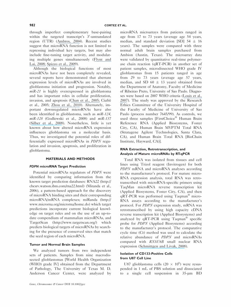
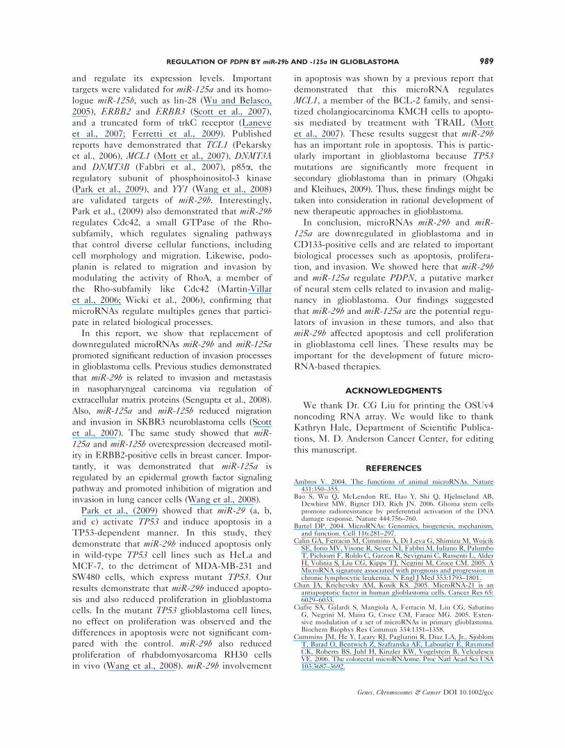
PDPN is a Direct Target of miR-29b and miR-125a
To correlate the expression of miR-29b and
miR-125a with that of PDPN, we analyzed by
qRT-PCR their expression in LN319 cells. The
results showed that LN319 overexpressed PDPN,as demonstrated by others (Kato et al., 2005; Scri-
deli et al., 2008), and also express very low levels
of miR-29b; interestingly, this cell line expresses
low levels of miR-125a and high levels of PDPN(Fig. 2A). To confirm the regulation of PDPN by
miR-29b and miR-125a, we analyzed the expres-
sion of the podoplanin protein by Western blot
48 hr after transfection of these microRNAs in
the LN319 cell line. Podoplanin expression
decreased significantly after miR-29b and miR-125a restoration, but not for miR-101 that is also
Figure 2. miR-29b and miR-125a downregulate PDPN levels andtarget the PDPN 30-UTR. (A) Inverse correlation of miR-29b and miR-125a levels with PDPN expression in cell line LN319. LN319expresses high PDPN and low miR-125a and miR-29b levels. (B)LN319 cells were transfected with miR-29b, miR-125a, miR-101 orscrambled and cell lysates were collected and run on a sodium do-decyl sulfate polyacrylamide gel for Western blotting. Podoplanin
expression decreases after miR-29b and miR-125a transfection. (C)LN229 cells were transfected with luciferase constructs which con-tain miR-29b and miR-125a-predicted targets. miR-29b and miR-125adownregulate target PDPN 30-UTR. Values represent averages andbars represent SD of three independent experiments. Results areexpressed as mean þ SD. The asterisk (*) represents a significant dif-ference (P < 0.05) by Student’s t test.
REGULATION OF PDPN BY miR-29b AND -125a IN GLIOBLASTOMA 985
Genes, Chromosomes & Cancer DOI 10.1002/gcc

predicted to target PDPN (Fig. 2B). Regulation
of PDPN by miR-29b and miR-125a was con-
firmed by luciferase assay. The results showed a
significant reduction of luciferase activity in cells
transfected with miR-29b (P ¼ 0.002) or miR-125a(P ¼ 0.003), compared with scrambled control
(Fig. 2C). Mutation of miR-29b and miR-125ainteraction sites rescued the luciferase activity,
thus confirming the direct interaction with PDPN30-UTR by these microRNAs. These results sug-
gest that miR-29b and miR-125a directly regulate
podoplanin expression in glioblastoma cells.
Restoration of miR-29b and miR-125a and
Downregulation of PDPN Impaired Invasion in
Glioblastoma Cells
Since PDPN was involved in invasion (Scrideli
et al., 2008), we next analyzed the effect of the
restoration of microRNAs levels on invasion in
glioblastoma cells. We performed the Matrigel
assay and found significant reductions of invasion,
by 40% (P ¼ 0.0003) and 23% (P ¼ 0.005) com-
pared with controls, in LN319 cells transfected
with miR-29b or miR-125a, respectively (Fig. 3A).
Furthermore, reductions of invasion by 23.5%
(P ¼ 0.03) and 56% (P ¼ 0.0008) were observed
also in U251 cells after transfection with miR-29bor miR-125a, respectively (Fig. 3B). The efficiency
of microRNA transfection was confirmed by qRT-
PCR (data not shown). The same significant
reduction of invasion in LN319 cells was found
after PDPN downregulation by specific siRNA
treatment for 96 hr (P ¼ 0.03) (Fig. 4). Therefore
the effects on invasion by restoration of miR-29band miR-125a are similar with the effects by down-
regulation of the proved direct PDPN target.
miR-29b Induces Apoptosis and Inhibits
Proliferation in Glioblastoma Cells
In a previous study, miR-29b was reported to
upregulate TP53 levels and induces apoptosis in a
TP53-dependent manner (Park et al., 2009). Thus,
Figure 3. Invasion assay using transwell chambers coated withMatrigel was performed 48 hr after transfection of LN319 (A) andU251 (B) cells with miR-29b, miR-125a, or scrambled. LN319 cellspresented a significant decrease in invasion rate after transfectionwith miR-29b or miR-125a, compared with scrambled. A decrease ininvasion rate was also observed for U251 cells transfected with miR-
29b or miR-125a, compared with scrambled. In (A) and (B), valuesrepresent averages and bars represent SD of three independentexperiments. Results are expressed as mean þ SD. The asterisk (*)represents a significant difference (P < 0.05) by Student’s t test.[Color figure can be viewed in the online issue, which is available atwileyonlinelibrary.com.]
986 CORTEZ ETAL.
Genes, Chromosomes & Cancer DOI 10.1002/gcc

we sought to determine the role of miR-29b in
apoptosis in glioblastoma cells by investigating its
effects in the mutant TP53 glioblastoma cell line
U251 (CGT to CAT/arginine to histidine) and the
wild-type TP53 cell line U87 (Van Meir et al.,
1994). miR-29b induced apoptosis in U87 wild-
type TP53 cells (P ¼ 0.01) (Fig. 5A). However, no
significant differences were observed in U251 mu-
tant TP53 cells transfected with miR-29b compared
with control (Fig. 5B). Moreover, we analyzed
whether TP53 status influences the effects of miR-29b in glioblastoma cells proliferation and found
no significant differences in the two mutant TP53cell lines U251 and LN319, transfected with miR-29b, compared with controls (Figs. 5C and 5D);
whereas, miR-29b was able to reduce proliferation
of wild-type TP53 cells (P ¼ 0.0002) (Fig. 5E).
LN319 cells contain a mutation of TP53 that
results in a change of arginine for histidine (CGC
to CAC/arginine to histidine) (Van Meir et al.,
1994). These results may suggest that miR-29b
induces apoptosis as well as reduces proliferation
in a TP53-dependent manner in glioblastoma.
DISCUSSION
In this study, we demonstrate that over-expressed
PDPN is regulated by microRNAs miR-29b and
miR-125a, which are deregulated in glioblastomas
and CD133-positive cells. We show, furthermore,
that the altered signaling pathways of invasion,
apoptosis, and proliferation that provide a selec-
tive advantage to tumors are affected by these
microRNAs.
On the basis of our microarray screening and
qRT-PCR results, we demonstrated that miR-29band miR-125a are significantly downregulated in
glioblastomas. miR-29b was also found downregu-
lated in several other types of tumor, including
invasive breast cancer (Iorio et al., 2005), lung
cancer (Yanaihara et al., 2006), chronic lympho-
cytic leukemia (Calin et al., 2005), prostate cancer
Figure 4. Silencing of podoplanin with specific siRNA impairedinvasion in LN319 cells. (A) Analysis of podoplanin silencing by spe-cific siRNA by qRT-PCR. After 48 hr, it was observed a reduction of82% of PDPN expression. (B) Confirmation of podoplanin silencingby specific siRNA 72 and 96 hr after transfection by Western blot-ting. Vinculin was used as normalizer. The same results wereobtained with two specific siRNAs (only one is shown here). (C)Invasion assay using transwell chambers coated with Matrigel was
performed 96 hr after transfection of LN319 with specific siRNA.LN319 cells presented a significant decrease in invasion rate com-pared with control (D). Values represent averages and bars repre-sent SD of three independent experiments. Results are expressed asmean þ SD. The asterisk (*) represents a significant difference (P <0.05) by Student’s t test. [Color figure can be viewed in the onlineissue, which is available at wileyonlinelibrary.com.]
REGULATION OF PDPN BY miR-29b AND -125a IN GLIOBLASTOMA 987
Genes, Chromosomes & Cancer DOI 10.1002/gcc

(Porkka et al., 2007), colon cancer (Cummins
et al., 2006). To date, this is the first report of
miR-125a downregulation in glioblastoma. Others
studies showed that miR-125a is also downregu-
lated in breast cancer (Iorio et al., 2005), neuro-
blastoma (Laneve et al., 2007), and medulloblas-
toma (Ferretti et al., 2009).
Recent reports demonstrated that some micro-
RNAs related to glioblastoma regulate such im-
portant molecular mechanisms in neural stem
cells as differentiation and self-renewal (for a
review, see Nicoloso et al., 2009). Conversely,
several studies revealed that genes related to self-
renewal are also implicated in important path-
ways in carcinogenesis (Godlewski et al., 2009).
Our findings show that miR-29b, miR-125a, and
miR-149 are under-expressed in CD133-positive
cells compared with their counterpart CD133-
negative cells. Interestingly, other reports have
indicated that these microRNAs are expressed
during brain development and are related to dif-
ferentiation of precursor cells in many types of
tumors. Krichevsky et al., (2006) and others
demonstrated that miR-125 is expressed during
mouse brain development and is strongly induced
during neural differentiation of embryonic stem
cells (Rybak et al., 2008). Moreover, miR-125induced development of P19 murine embryonal
carcinoma cells to neurons (Wu and Belasco,
2005). The reconstitution of miR-29 expression
promoted differentiation of rhabdomyosarcoma
RH30 cells in vivo (Wang et al., 2008). Taken
together, these findings suggest that miR-29b and
miR-125a have roles in differentiation of pre-
cursor cells in many tissues, including the brain.
CD133-positive cells present a significant tumori-
genic potential in vivo, capacity for angiogenesis
and multilineage differentiation, and high resist-
ance to radiation and chemotherapy (Bao et al.,
2006). These results and the fact that glioblasto-
mas are poorly differentiated tumors suggest a
potential role for these microRNAs in regulation
of signaling pathways related to maintenance of
stem cell properties such as differentiation and
self-renewal. Nonetheless, additional studies are
necessary to elucidate these results.
Our results indicate that miR-29b and miR-125aspecifically interact with the 30-UTR of PDPN
Figure 5. miR-29b induces apoptosis and represses proliferation inglioblastoma cells. Apoptosis assay was performed 48 hr after trans-fection of U87 (A) and U251 (B) cells with miR-29b or scrambled.Cellular proliferation was assessed 24, 48, 72, and 96 hr after trans-fection of LN319 (C), U251 (D), and U87 (E) cells with miR-29b or
scrambled control. Values represent averages and bars represent SDof three independent experiments. Results are expressed as mean þSD. The asterisk (*) represents a significant difference (P < 0.05) byStudent’s t test.
988 CORTEZ ETAL.
Genes, Chromosomes & Cancer DOI 10.1002/gcc
and regulate its expression levels. Important
targets were validated for miR-125a and its homo-
logue miR-125b, such as lin-28 (Wu and Belasco,
2005), ERBB2 and ERBB3 (Scott et al., 2007),
and a truncated form of trkC receptor (Laneve
et al., 2007; Ferretti et al., 2009). Published
reports have demonstrated that TCL1 (Pekarsky
et al., 2006), MCL1 (Mott et al., 2007), DNMT3Aand DNMT3B (Fabbri et al., 2007), p85a, the
regulatory subunit of phosphoinositol-3 kinase
(Park et al., 2009), and YY1 (Wang et al., 2008)
are validated targets of miR-29b. Interestingly,
Park et al., (2009) also demonstrated that miR-29bregulates Cdc42, a small GTPase of the Rho-
subfamily, which regulates signaling pathways
that control diverse cellular functions, including
cell morphology and migration. Likewise, podo-
planin is related to migration and invasion by
modulating the activity of RhoA, a member of
the Rho-subfamily like Cdc42 (Martin-Villar
et al., 2006; Wicki et al., 2006), confirming that
microRNAs regulate multiples genes that partici-
pate in related biological processes.
In this report, we show that replacement of
downregulated microRNAs miR-29b and miR-125apromoted significant reduction of invasion processes
in glioblastoma cells. Previous studies demonstrated
that miR-29b is related to invasion and metastasis
in nasopharyngeal carcinoma via regulation of
extracellular matrix proteins (Sengupta et al., 2008).
Also, miR-125a and miR-125b reduced migration
and invasion in SKBR3 neuroblastoma cells (Scott
et al., 2007). The same study showed that miR-125a and miR-125b overexpression decreased motil-
ity in ERBB2-positive cells in breast cancer. Impor-
tantly, it was demonstrated that miR-125a is
regulated by an epidermal growth factor signaling
pathway and promoted inhibition of migration and
invasion in lung cancer cells (Wang et al., 2008).
Park et al., (2009) showed that miR-29 (a, b,
and c) activate TP53 and induce apoptosis in a
TP53-dependent manner. In this study, they
demonstrate that miR-29b induced apoptosis only
in wild-type TP53 cell lines such as HeLa and
MCF-7, to the detriment of MDA-MB-231 and
SW480 cells, which express mutant TP53. Our
results demonstrate that miR-29b induced apopto-
sis and also reduced proliferation in glioblastoma
cells. In the mutant TP53 glioblastoma cell lines,
no effect on proliferation was observed and the
differences in apoptosis were not significant com-
pared with the control. miR-29b also reduced
proliferation of rhabdomyosarcoma RH30 cells
in vivo (Wang et al., 2008). miR-29b involvement
in apoptosis was shown by a previous report that
demonstrated that this microRNA regulates
MCL1, a member of the BCL-2 family, and sensi-
tized cholangiocarcinoma KMCH cells to apopto-
sis mediated by treatment with TRAIL (Mott
et al., 2007). These results suggest that miR-29bhas an important role in apoptosis. This is partic-
ularly important in glioblastoma because TP53mutations are significantly more frequent in
secondary glioblastoma than in primary (Ohgaki
and Kleihues, 2009). Thus, these findings might be
taken into consideration in rational development of
new therapeutic approaches in glioblastoma.
In conclusion, microRNAs miR-29b and miR-125a are downregulated in glioblastoma and in
CD133-positive cells and are related to important
biological processes such as apoptosis, prolifera-
tion, and invasion. We showed here that miR-29band miR-125a regulate PDPN, a putative marker
of neural stem cells related to invasion and malig-
nancy in glioblastoma. Our findings suggested
that miR-29b and miR-125a are the potential regu-
lators of invasion in these tumors, and also that
miR-29b affected apoptosis and cell proliferation
in glioblastoma cell lines. These results may be
important for the development of future micro-
RNA-based therapies.
ACKNOWLEDGMENTS
We thank Dr. CG Liu for printing the OSUv4
noncoding RNA array. We would like to thank
Kathryn Hale, Department of Scientific Publica-
tions, M. D. Anderson Cancer Center, for editing
this manuscript.
REFERENCES
Ambros V. 2004. The functions of animal microRNAs. Nature431:350–355.
Bao S, Wu Q, McLendon RE, Hao Y, Shi Q, Hjelmeland AB,Dewhirst MW, Bigner DD, Rich JN. 2006. Glioma stem cellspromote radioresistance by preferential activation of the DNAdamage response. Nature 444:756–760.
Bartel DP. 2004. MicroRNAs: Genomics, biogenesis, mechanism,and function. Cell 116:281–297.
Calin GA, Ferracin M, Cimmino A, Di Leva G, Shimizu M, WojcikSE, Iorio MV, Visone R, Sever NI, Fabbri M, Iuliano R, PalumboT, Pichiorri F, Roldo C, Garzon R, Sevignani C, Rassenti L, AlderH, Volinia S, Liu CG, Kipps TJ, Negrini M, Croce CM. 2005. AMicroRNA signature associated with prognosis and progression inchronic lymphocytic leukemia. N Engl J Med 353:1793–1801.
Chan JA, Krichevsky AM, Kosik KS. 2005. MicroRNA-21 is anantiapoptotic factor in human glioblastoma cells. Cancer Res 65:6029–6033.
Ciafre SA, Galardi S, Mangiola A, Ferracin M, Liu CG, SabatinoG, Negrini M, Maira G, Croce CM, Farace MG. 2005. Exten-sive modulation of a set of microRNAs in primary glioblastoma.Biochem Biophys Res Commun 334:1351–1358.
Cummins JM, He Y, Leary RJ, Pagliarini R, Diaz LA, Jr., SjoblomT, Barad O, Bentwich Z, Szafranska AE, Labourier E, RaymondCK, Roberts BS, Juhl H, Kinzler KW, Vogelstein B, VelculescuVE. 2006. The colorectal microRNAome. Proc Natl Acad Sci USA103:3687–3692.
REGULATION OF PDPN BY miR-29b AND -125a IN GLIOBLASTOMA 989
Genes, Chromosomes & Cancer DOI 10.1002/gcc

Fabbri M, Garzon R, Cimmino A, Liu Z, Zanesi N, Callegari E,Liu S, Alder H, Costinean S, Fernandez-Cymering C, VoliniaS, Guler G, Morrison CD, Chan KK, Marcucci G, Calin GA,Huebner K, Croce CM. 2007. MicroRNA-29 family revertsaberrant methylation in lung cancer by targeting DNA methyl-transferases 3A and 3B. Proc Natl Acad Sci USA 104:15805–15810.
Ferretti E, De Smaele E, Po A, Di Marcotullio L, Tosi E, Espi-nola MS, Di Rocco C, Riccardi R, Giangaspero F, FarcomeniA, Nofroni I, Laneve P, Gioia U, Caffarelli E, Bozzoni I, Scre-panti I, Gulino A. 2009. MicroRNA profiling in human medullo-blastoma. Int J Cancer 124:568–577.
Flynt AS, Lai EC. 2008. Biological principles of microRNA-medi-ated regulation: Shared themes amid diversity. Nat Rev Genet9:831–842.
Godlewski J, Nowicki MO, Bronisz A, Williams S, Otsuki A,Nuovo G, Raychaudhury A, Newton HB, Chiocca EA, LawlerS. 2008. Targeting of the Bmi-1 oncogene/stem cell renewalfactor by microRNA-128 inhibits glioma proliferation and self-renewal. Cancer Res 68:9125–9130.
Iorio MV, Ferracin M, Liu CG, Veronese A, Spizzo R, Sabbioni S,Magri E, Pedriali M, Fabbri M, Campiglio M, Menard S, PalazzoJP, Rosenberg A, Musiani P, Volinia S, Nenci I, Calin GA, QuerzoliP, Negrini M, Croce CM. 2005. MicroRNA gene expression dereg-ulation in human breast cancer. Cancer Res 65:7065–7070.
Kato Y, Fujita N, Kunita A, Sato S, Kaneko M, Osawa M, TsuruoT. 2003. Molecular identification of aggrus/T1alpha as a plateletaggregation-inducing factor expressed in colorectal tumors. J BiolChem 278:51599–51605.
Kato Y, Kaneko M, Sata M, Fujita N, Tsuruo T, Osawa M. 2005.Enhanced expression of aggrus (T1alpha/podoplanin), a platelet-aggregation-inducing factor in lung squamous cell carcinoma.Tumor Biol 26:195–200.
Krichevsky AM, Sonntag KC, Isacson O, Kosik KS. 2006. SpecificmicroRNAs modulate embryonic stem cell-derived neurogene-sis. Stem Cells 24:857–864.
Laneve P, Di Marcotullio L, Gioia U, Fiori ME, Ferretti E,Gulino A, Bozzoni I, Caffarelli E. 2007. The interplay betweenmicroRNAs and the neurotrophin receptor tropomyosin-relatedkinase C controls proliferation of human neuroblastoma cells.Proc Natl Acad Sci USA 104:7957–7962.
Louis DN, Ohgaki H, Wiestler OD, Cavenee WK, Burger PC,Jouvet A, Scheithauer BW, Kleihues P. 2007. The 2007 WHOclassification of tumours of the central nervous system. ActaNeuropathol 114:97–109.
Martin-Villar E, Megias D, Castel S, Yurrita MM, Vilaro S, Quin-tanilla M. 2006. Podoplanin binds ERM proteins to activateRhoA and promote epithelial-mesenchymal transition. J Cell Sci119 (Pt 21):4541–4553.
Miranda KC, Huynh T, Tay Y, Ang YS, Tam WL, Thomson AM,Lim B, Rigoutsos I. 2006. A pattern-based method for the iden-tification of MicroRNA binding sites and their correspondingheteroduplexes. Cell 126:1203–1217.
Mishima K, Kato Y, Kaneko MK, Nakazawa Y, Kunita A, FujitaN, Tsuruo T, Nishikawa R, Hirose T, Matsutani M. 2006.Podoplanin expression in primary central nervous system germcell tumors: A useful histological marker for the diagnosis ofgerminoma. Acta Neuropathol 111:563–568.
Mott JL, Kobayashi S, Bronk SF, Gores GJ. 2007. mir-29 regulatesMcl-1 protein expression and apoptosis. Oncogene 26:6133–6140.
Nakamura Y, Kanemura Y, Yamada T, Sugita Y, Higaki K, Yama-moto M, Takahashi M, Yamasaki M. 2006. D2-40 antibody im-munoreactivity in developing human brain, brain tumors andcultured neural cells. Mod Pathol 19:974–985.
Nicoloso MS, Spizzo R, Shimizu M, Rossi S, Calin GA. 2009.MicroRNAs–the micro steering wheel of tumour metastases.Nat Rev Cancer 9:293–302.
Ohgaki H, Kleihues P. 2009. Genetic alterations and signalingpathways in the evolution of gliomas. Cancer Sci 100:2235–2241.
Ordonez NG. 2006. Podoplanin: A novel diagnostic immunohisto-chemical marker. Adv Anat Pathol 13:83–88.
Park SY, Lee JH, Ha M, Nam JW, Kim VN. 2009. miR-29 micro-RNAs activate p53 by targeting p85 alpha and CDC42. NatStruct Mol Biol 16:23–29.
Pekarsky Y,Santanam U, Cimmino A, Palamarchuk A, Efanov A,Maximov V, Volinia S, Alder H, Liu CG, Rassenti L, Calin GA,Hagan JP, Kipps T, Croce CM. 2006. Tcl1 expression in chroniclymphocytic leukemia is regulated by miR-29 and miR-181.Cancer Res 66:11590–11593.
Porkka KP, Pfeiffer MJ, Waltering KK, Vessella RL, TammelaTL, Visakorpi T. 2007. MicroRNA expression profiling in pro-state cancer. Cancer Res 67:6130–6135.
Rybak A, Fuchs H, Smirnova L, Brandt C, Pohl EE, Nitsch R,Wulczyn FG. 2008. A feedback loop comprising lin-28 and let-7controls pre-let-7 maturation during neural stem-cell commit-ment. Nat Cell Biol 10:987–993.
Schmittgen TD, Livak KJ. 2008. Analyzing real-time PCR databy the comparative C(T) method. Nat Protoc 3:1101–1108.
Scott GK, Goga A, Bhaumik D, Berger CE, Sullivan CS, BenzCC. 2007. Coordinate suppression of ERBB2 and ERBB3 byenforced expression of micro-RNA miR-125a or miR-125b. J BiolChem 282:1479–1486.
Scrideli CA, Carlotti CG, Jr., Okamoto OK, Andrade VS, CortezMA, Motta FJ, Lucio-Eterovic AK, Neder L, Rosemberg S,Oba-Shinjo SM, Marie SK, Tone LG. 2008. Gene expressionprofile analysis of primary glioblastomas and non-neoplasticbrain tissue: Identification of potential target genes by oligo-nucleotide microarray and real-time quantitative PCR. J Neuro-oncol 88:281–291.
Sengupta S, den Boon JA, Chen IH, Newton MA, Stanhope SA,Cheng YJ, Chen CJ, Hildesheim A, Sugden B, Ahlquist P.2008. MicroRNA 29c is down-regulated in nasopharyngealcarcinomas, up-regulating mRNAs encoding extracellular matrixproteins. Proc Natl Acad Sci USA 105:5874–5878.
Shibahara J, Kashima T, Kikuchi Y, Kunita A, Fukayama M. 2006.Podoplanin is expressed in subsets of tumors of the central nerv-ous system. Virchows Arch 448:493–499.
Silber J, Lim DA, Petritsch C, Persson AI, Maunakea AK, Yu M,Vandenberg SR, Ginzinger DG, James CD, Costello JF, BergersG, Weiss WA, Alvarez-Buylla A, Hodgson JG. 2008. miR-124 andmiR-137 inhibit proliferation of glioblastoma multiforme cellsand induce differentiation of brain tumor stem cells. BMC Med6:14.
Spizzo R, Nicoloso MS, Croce CM, Calin GA. 2009. SnapShot:MicroRNAs in Cancer. Cell 137:586–586 e1.
Van Meir EG, Polverini PJ, Chazin VR, Su Huang HJ, de TriboletN, Cavenee WK. 1994. Release of an inhibitor of angiogenesisupon induction of wild type p53 expression in glioblastoma cells.Nat Genet 8:171–176.
Wang H, Garzon R, Sun H, Ladner KJ, Singh R, Dahlman J,Cheng A, Hall BM, Qualman SJ, Chandler DS, Croce CM,Guttridge DC. 2008. NF-kappaB-YY1-miR-29 regulatory cir-cuitry in skeletal myogenesis and rhabdomyosarcoma. CancerCell 14:369–381.
Wicki A, Lehembre F, Wick N, Hantusch B, Kerjaschki D, Chris-tofori G. 2006. Tumor invasion in the absence of epithelial-mesenchymal transition: Podoplanin-mediated remodeling of theactin cytoskeleton. Cancer Cell 9:261–272.
Wu L, Belasco JG. 2005. Micro-RNA regulation of the mamma-lian lin-28 gene during neuronal differentiation of embryonalcarcinoma cells. Mol Cell Biol 25:9198–9208.
Yanaihara N, Caplen N, Bowman E, Seike M, Kumamoto K, Yi M,Stephens RM, Okamoto A, Yokota J, Tanaka T, Calin GA, Liu CG,Croce CM, Harris CC. 2006. Unique microRNA molecular profilesin lung cancer diagnosis and prognosis. Cancer Cell 9:189–198.
Zhou X, Ren Y, Moore L, Mei M, You Y, Xu P, Wang B, WangG, Jia Z, Pu P, Zhang W, Kang C. 2010. Down-regulation ofmiR-21 inhibits EGFR pathway and suppresses the growth ofhuman glioblastoma cells independent of PTEN status. LabInvest 90:144–155.
990 CORTEZ ETAL.
Genes, Chromosomes & Cancer DOI 10.1002/gcc