egr1 modulated lncrna hnf1a-as1 drives glioblastoma
TRANSCRIPT
ARTICLE OPEN
EGR1 modulated LncRNA HNF1A-AS1 drives glioblastomaprogression via miR-22-3p/ENO1 axisChunchun Ma1,2,3, Hongliang Wang1,2,3, Gang Zong1,2, Jie He1,2, Yuyang Wang1,2, Fan Yang1,2, Zhihao Yang1,2, Erbao Bian1,2 andBing Zhao 1,2✉
© The Author(s) 2021
Accumulating evidences revealed that long noncoding RNAs (lncRNAs) have been participated in cancer malignant progression,including glioblastoma multiforme (GBM). Despite much studies have found the precise biological role in the regulatorymechanisms of GBM, however the molecular mechanisms, particularly upstream mechanisms still need further elucidated. RT-QPCR,cell transfection, western blotting and bioinformatic analysis were executed to detect the expression of EGR1, HNF1A-AS1, miR-22-3p and ENO1 in GBM. Cell proliferation assay, colony formation assay, wound healing, migration and invasion assays wereperformed to detect the malignant characters of GBM cells. The molecular regulation mechanism was confirmed by luciferasereporter assay, ChIP and RIP. Finally, orthotopic mouse models were established to examine the effect of HNF1A-AS1 in vivo. In thecurrent study, we analyzed clinical samples to show that the HNF1A-AS1 expression is upregulated and associated with poorpatient survival in GBM. Functional studies revealed that HNF1A-AS1 knockdown markedly inhibits malignant phenotypes of GBMcells, whereas overexpression of HNF1A-AS1 exerts opposite effect. Mechanistically, the transcription factor EGR1 forced the HNF1A-AS1 expression by directly binding the promoter region of HNF1A-AS1. Furthermore, combined bioinformatics analysis with ourmechanistic work, using luciferase reporter assays and RIP, we first demonstrated that HNF1A-AS1 functions as a competingendogenous RNA (ceRNA) with miR-22-3p to regulate ENO1 expression in GBM cells. HNF1A-AS1 directly binds to miR-22-3p andsignificantly inhibits miR-22-3p expression, while ENO1 expression was increased. miR-22-3p inhibitor offsets the HNF1A-AS1silencing induced suppression in malignant behaviors of GBM cells. ENO1 was verified as a direct target of miR-22-3p and itsexpression levels was negatively with the prognosis in GBM patients. Taken together, our study illuminated the definite mechanismof HNF1A-AS1 in promoting GBM malignancy, and provided a novel therapeutic target for further clinical application.
Cell Death Discovery (2021) 7:350 ; https://doi.org/10.1038/s41420-021-00734-3
INTRODUCTIONGBM, the most aggressive subtype of glioma in adults, highlymalignant and high risk of recurrence, accounting for 47.1% of allmalignant tumors of the nervous system [1]. In the light of theWorld Health Organization (WHO) classification of tumor in thecentral nervous system (CNS), GBM is classified as a grade IVglioma, with an unfavorable prognosis and a five-year overallsurvival rate less than 10% [2, 3]. Even after multimodal therapies,including maximal surgical resection, adjuvant radiotherapy,temozolomide (TMZ)-based chemotherapy or targeted therapywith rituximab, which have been commonly used in GBM patients,the patients’ overall survival rate is still unsatisfactory with amedian survival time of 12–15 months from first diagnosis [4].Therefore, it is extremely vital for us to establish new targetedtherapies and to gain a clear understanding of the definitemechanisms of GBM malignant progression for the identificationof new diagnostic and prognosis markers.LncRNAs comprise a class of transcripts that are over 200 nt in
length, which have many ways of regulating ability except fornone protein-coding potential [5, 6]. The functions and mechan-isms of lncRNAs exert their biological role through diverse modes,
including chromatin modification, alternative splicing, mRNAstability, encode functional micropeptides, ceRNA molecularsponge to miRNAs, and so on [7–14]. An increasing number ofreports have demonstrated that lncRNAs play a vital role in cancerprogression, and are dysregulated in various human cancers,including GBM [15–17]. For example, LncRNA miR155HG isoverexpressed in GBM, and promotes GBM progression by actingas a ceRNA for the tumor suppressor miR-185 to upregulateANXA2 [18]. LncRNA AC016405.3 endpin cell proliferation andmetastasis by regulating TET2, via sponging of miR-19a-5p in GBMcells [19]. LncRNA HOTAIRM1 is highly upregulated in GBM, whichis positively correlated with tumor grade in patients with glioma,and aggravates the progression of GMB by regulating HOXA1gene [20]. Despite several lncRNAs have been well studied, thefunctional mechanism of most lncRNAs in GBM remain largelyunknow.Hepatocyte nuclear factor 1 homeobox A antisense RNA 1
(HNF1A-AS1), was first identified as a lncRNA that upregulated inesophageal adenocarcinoma [21], and its overexpression wasdrastically associated with tumor advanced-stage and unfavorableoutcomes in various cancers, including oral squamous carcinoma,
Received: 4 June 2021 Revised: 30 September 2021 Accepted: 20 October 2021
1Department of Neurosurgery, The Second Affiliated Hospital of Anhui Medical University, Hefei 230601, China. 2Cerebral Vascular Disease Research Center, Anhui MedicalUniversity, Hefei 230601, China. 3These authors contributed equally: Chunchun Ma, Hongliang Wang. ✉email: [email protected]
www.nature.com/cddiscovery
Official journal of CDDpress
1234567890();,:

urothelial carcinoma of the bladder and lung carcinoma, indicat-ing an oncogenic function of HNF1A-AS1 in tumor progression[22–24]. However, the functional mechanism of HNF1A-AS1 inGBM has not been fully revealed yet. Herein, we demonstrate thatHNF1A-AS1 is highly overexpressed in GBM tissues and cells,associates with poor patient survival, by acting as ceRNA for miR-22 and facilitating ENO1, to promote GBM malignant phenotypes.Therefore, these results imply that HNF1A-AS1 may serve as adruggable target for GBM.
RESULTSHNF1A-AS1 is upregulated in GBM and negatively related withpatient prognosisTo identify the expression of HNF1A-AS1 in GBM, using RT–QPCRanalysis, we firstly measured HNF1A-AS1 expression in 15 normalbrain tissues, 41 low-grade glioma tissues, 72 GBM tissues. Wefound that HNF1A-AS1 was obviously upregulated in GBM, ascompared to low-grade glioma tissues and normal brain tissues,but there was no significant difference between low-grade gliomatissues and normal brain tissues (Fig. 1A). Furthermore, we alsotested HNF1A-AS1 expression in normal human astrocyte (HA) andfour GBM cell lines (U251, LN18, U87 and A172). The resultsindicated that HNF1A-AS1 was highly upregulated in four GBM celllines in comparison with HA (Fig. 1B).To further clarify the clinical significance of HNF1A-AS1 in GBM
patients. The Kaplan–Meier method and log-rank test wereperformed to assess the expression of HNF1A-AS1 in GBM TCGAdate cohort and our clinic study. TCGA date cohort demonstrated
that GBM patients with high expression of HNF1A-AS1 wasnegatively associated with overall survival time (Fig. 1C). In ourstudy, we also observed the similar results, indicating that highHNF1A-AS1 expression was significantly correlated with poorsurvival patients (Fig. 1D). In addition, subcellular fractionation andRT–QPCR analyses showed that HNF1A-AS1 is localized both in thecytoplasm and nucleus of GBM cells (Fig. 1E), which indicated itscomplicated functions.
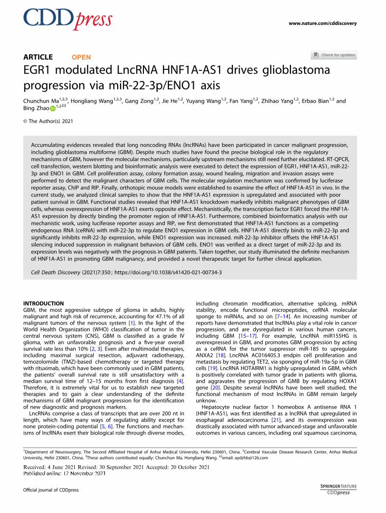
HNF1A-AS1 underpins GBM cells proliferation, migration,invasionTo evaluate the biofunctional role of HNF1A-AS1 in GBM cells, theGBM cells were transfected with si-HNF1A-AS1 or si-NC. RT–QPCRwas performed to examine the knockdown efficiency of HNF1A-AS1 after transfection 48 h (Fig. 2A). Compared with si-NC groups,CCK-8 assays revealed that HNF1A-AS1 knockdown significantlydecreased cell proliferation (Fig. 2B). Colony formation assaysindicated that the clone numbers and colony size was attenuatedin the HNF1A-AS1 knockdown group, suggesting that depletion ofHNF1A-AS1 slow down the growth of GBM cells (Fig. 2C). Woundhealing assays indicated that the wound-healing capacity wasworse and slower in si-HNF1A-AS1 groups than in si-NC groups(Fig. 2D). In addition, transwell assay showed that knockdown ofHNF1A-AS1 significantly reduced migratory and invasive capacitycompared with si-NC groups (Fig. 2E). On the contrary, over-expression of HNF1A-AS1 obviously promote the malignancy ofGBM cells (Fig. S1A–C). To sum up, these results proved thatHNF1A-AS1 plays an important role in promoting GBM cellsmalignant behaviors.
Fig. 1 HNF1A-AS1 was highly expressed in GBM. A RT-QPCR analysis of HNF1A-AS1 expression in normal brain tissues (NBT) (n= 15), low-grade glioma tissues (n= 41) and GBM tissues (n= 72). **P < 0.01 vs. NBT group. B RT-QPCR analysis the expression levels of HNF1A-AS1 innormal human astrocyte (HA) and four GBM cell lines (U251, LN18, U87 and A172). *P < 0.05, **P < 0.01 vs. HA group. C In TCGA date, theoverall survival rate of GBM patients with high (HNF1A-AS1-high, n= 55) and low (HNF1A-AS1-low, n= 112) expression of HNF1A-AS1 intumor. D Kaplan–Meier analyses of the associations between HNF1A-AS1 expression level and overall survival of patients with human gliomain our department (The log-rank test was used to calculate P-values). **P < 0.01 vs. low HNF1A-AS1 expression group. E Nuclear andcytoplasmic fractions of HNF1A-AS1 in GBM cell lysates were analyzed by RT-QPCR.
C. Ma et al.
2
Cell Death Discovery (2021) 7:350
EGR1 strengthen HNF1A-AS1 expression in GBM cellsWe next discover the potential regulatory mechanisms whichcause the upregulation of HNF1A-AS1 in GBM. By using UCSCGenome Browser, we found that Early Growth Response gene 1(EGR1) is a latent transcription factor of HNF1A-AS1. Furthermore,previous study has demonstrated that EGR1 transcriptionallyactivated HNF1A-AS1 in human gastric cancer [25]. According toJASPAR database, the predicted transcription factor-binding site ofEGR1 in the HNF1A-AS1 promoter is indicated in (Fig. 3A).Importantly, TCGA database confirmed that EGR1 is highlyexpressed in GBM tissues, and patients with higher HNF1A-AS1expression indicated a shorter survival time (Fig. S2A, B). To detectthe effect of EGR1 on HNF1A-AS1 expression, we successfullyestablish EGR1 overexpression or knockdown model and wastested by RT-QPCR and western blot (Fig. S2C–E). As a result,depletion of EGR1 significantly decrease the levels of HNF1A-AS1,while EGR1 overexpressed dramatically increase the levels ofendogenous HNF1A-AS1 (Fig. 3B, C). Luciferase reporter assayfurther demonstrated that the region of HNF1A-AS1 promoter isresponsible for HNF1A-AS1 transcription (Fig. 3D–F). Moreover,ChIP assay showed that the EGR1 specifically associated with thepromoter of HNF1A-AS1 (Fig. 3G). These results demonstrated thatEGR1 enhance HNF1A-AS1 expression at transcriptional level bydirectly binding to its promoter.
HNF1A-AS1 physically bound to miR-22 and induced itsdegradationTo further clarify the underlying molecular mechanism by whichHNF1A-AS1 regulates GBM cells proliferation, migration andinvasion. As we all know, lncRNAs could restrain miRNAsexpression and activity on their target mRNAs, by function as aceRNA [26]. Bioinformatics data such as RegRNA 2.0, Diana-lncBaseand miRANDA, were performed to predict the potential miRNAtargets of HNF1A-AS1. According to the mirSVR and PhastConsscores, we found that miR-22 contain the potential target sites onHNF1A-AS1. Our studies indicated that the expression of miR-22was significantly inverse in correlation with HNF1A-AS1 in GBMtissues (Fig. 3H). Moreover, HNF1A-AS1 knockdown dramaticallyincreased miR-22 expression, while GBM cells transfected withPCDNA3.1-HNF1A-AS1 significantly inhibited the expression ofmiR-22 (Fig. 4A, B). However, no expression changed on HNF1A-AS1 when GBM cells transfected with miR-22 mimics or inhibitors(Fig. 4C, D). These results indicated that miR-22 was negativelyregulated by HNF1A-AS1 in GBM.Bioinformatics analysis predicted the potential binding sites of
miR-22 on HNF1A-AS1 and the conservation of HNF1A-AS1 in thebinding site of miR-22 was snapshotted from human genome inUCSC Genome Browser (Fig. 4E, F). Then, luciferase reporter assayindicated that there was no significant difference in the relative
Fig. 2 Knockdown of HNF1A-AS1 inhibited the proliferation, migration, and invasion of glioma cells in vitro. A Relative expression levelsof HNF1A-AS1 after GBM cells transfected with si-HNF1A-AS1 and si-NC. **P < 0.01 vs. si-NC. B CCK-8 assay was performed to determine theproliferation effect of si-HNF1A-AS1 and si-NC transfected U251 and LN18 cells. *P < 0.05, **P < 0.01 vs. si-NC group. C Colony formation assaywas performed to detect the proliferation of U251 and LN18 cells after transfected with si-HNF1A-AS1 and si-NC. **P < 0.01 vs. si-NC group.D Wound healing assay to evaluate the effect of HNF1A-AS1 on cell migration in U251 and LN18 cells (scale bar: 200 μm). **P < 0.01 < 0.05 vs.si-NC group. E Transwell assay for testing cell migration and invasion capacity (scale bar: 200 μm). **P < 0.01 vs. si-NC group. Data arepresented as mean ± SD from three independent experiments.
C. Ma et al.
3
Cell Death Discovery (2021) 7:350

luciferase activity between HNF1A-AS1-Mut+ miR-22 mimics andHNF1A-AS1-Mut+ miR-22 NC groups, but co-transfection ofpmirGLO-HNF1A-AS1-WT and miR-22 mimics dramatically reducedthe luciferase activity compared with HNF1A-AS1-WT+miR-22 NCgroups (Fig. 4G, H). To confirm whether HNF1A-AS1 and miR-22are in the same RNA-induced silencing complex (RISC), weconducted anti-Ago2 RNA-binding protein immunoprecipitation(RIP) assay, and the results showed that Ago2 antibody enrichedHNF1A-AS1 (Fig. 4I, J). These findings suggested that HNF1A-AS1directly targeted miR-22 in GBM cells.
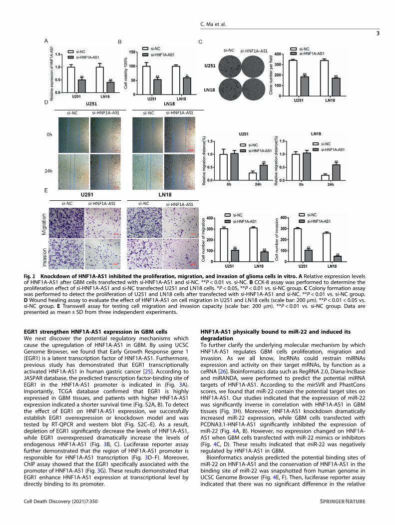
Downregulation of miR-22 promotes GBM cells malignantbehaviorsAccording to the TCGA data, the expression of miR-22 wasobviously decreased in GBM samples, and was negativelycorrelated the pathological grades of glioma (Fig. 5A), whichwas coincided with our study (Fig. 5B). RT–QPCR analysisconfirmed that miR-22 expression was significantly down-regulated in four GBM cell lines (Fig. 5C). Compared with themiR-22 NC group, the proliferation, migration and invasionability of GBM cells were distinctly reduced when cellstransfected with miR-22 mimics, and cells’ malignant behaviorability were enhanced when cells transfected with miR-22
inhibitors (Fig. 5D–E). These results indicated that miR-22 actedas an anti-oncogene in GBM cells.
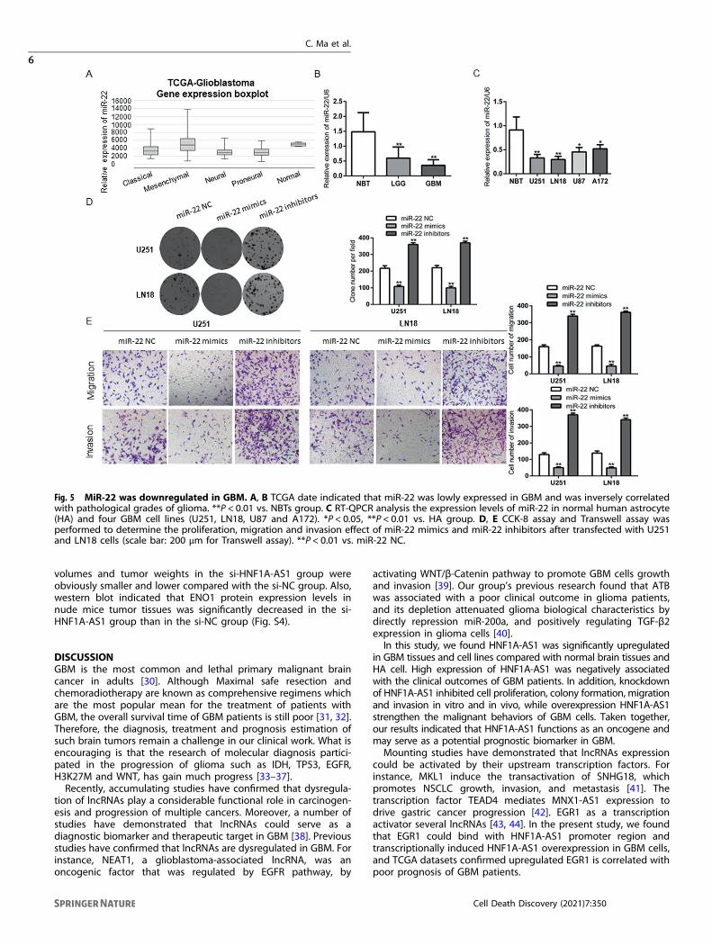
ENO1, a direct target of miR-22 in GBM cellsBioinformatic tools were adopted to predict potential targets ofmiR-22 in GBM cells. We found that 3′UTR regions of theoverlapped potential candidates both have the predicted bindingsites of miR-22. Ultimately, a key glycolytic enzyme, ENO1, wasidentified as target gene, in view of its upregulation is associatedwith glioma progression and prognosis [27, 28]. A previous studyshowed that miR-22 suppresses the proliferation of retinoblas-toma cells by inhibiting ENO1, and ENO1 was a target of miR-22[29]. As showed in Fig. 6A, the presumptive binding sites of miR-22within the 3’UTR of ENO1 were predicted by TargetScan andMirDB. To confirm whether miR-22 regulated ENO1, GBM cells weretransfected with miR-22 mimic or miR-22 inhibitors. The resultsindicated that increasing miR-22 markedly suppressed ENO1mRNA and protein levels compared to miR-22 NC and converselyENO1 expression significantly increased after inhibited miR-22(Fig. 6B, C). Then, luciferase reporter assay demonstrated ed thatco-transfection of ENO1 WT and miR-22 mimics drastically reducedluciferase activity compared with the ENO1 WT+miR-22 NCgroup, whereas miR-22 Mut binding site within ENO1 abrogated
Fig. 3 The transcription factor EGR1, specially activate HNF1A-AS1 expression at transcriptional level. A JASPAR database was adopted topredict the putative bind site of EGR1 on the promoter region of HNF1A-AS1. B, C The expression level of HNF1A-AS1 was tested in GBM cellstransfected with si-EGR1 or pcDNA3.1-EGR1 using RT-QPCR assay. **P < 0.01 vs. si-NC or **P < 0.01 vs. Vector. D Serial truncations of HNF1A-AS1 promoter fragments spanning from −2000/ −1500/ −1000/ −500/ to 0. E, F This promoter fragments were cloned into pGL3-basicvectors, and dual luciferase reporter assays were performed to assess the exact EGR1 binding site on the HNF1A-AS1 promoter region. **P <0.01 vs. si-NC or **P < 0.01 vs. Vector, n.s. indicates no significance relative to si-NC or Vector. G ChIP assay showed that the binding affinitybetween EGR1 and HNF1A-AS1 promoter region. **P < 0.01 vs. IgG. H Pearson’s correlation was performed to analyze the relationship betweenHNF1A-AS1 expression and miR-22 expression in 72 GBM patients.
C. Ma et al.
4
Cell Death Discovery (2021) 7:350

the inhibitory effect of miR-22 mimics on the reporter geneexpression (Fig. 6D, E). In addition, we found that ENO1 wassignificantly overexpressed in GBM tissues and its high expressionwas inverse correlation with overall survival time in GBM TCGAdata, which was consistent with CGGA data (Fig. S3A–D). Thesefindings indicated that miR-22 inhibited ENO1 expression in GBMcells by targeting the 3′ UTR of oncogene ENO1.
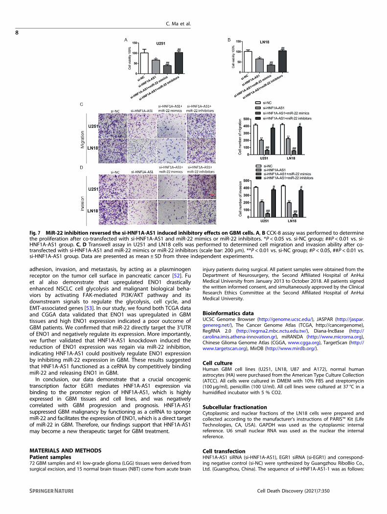
Knockdown of HNF1A-AS1 induced suppression of malignantphenotype was smothered by knockdown of miR-22 in GBMcellsPrevious study has confirmed that ENO1 attenuated led tounderpin cancer progression in glioma cells [27]. However,whether HNF1A-AS1 promote the malignant behaviors of GBMcells by inhibiting miR-22 remained largely unknown. miR-22mimics and miR-22 inhibitors were transfected into si-HNF1A-AS1GBM cells. The results indicated that cell proliferation, migrationand invasion ability were attenuated in si-HNF1A-AS1 and miR-22mimics groups. si-HNF1A-AS1 combined with miR-22 mimicsgroup was strongly reduced the malignant phenotype of GBMcells, while miR-22 inhibitors reversed the suppression of HNF1A-AS1 attenuated in GBM cells (Fig. 7A–D). Therefore, these results
suggested that HNF1A-AS1 exerts its biofunctional roles thoughmiR-22 in GBM cells.
HNF1A-AS1 functions as a ceRNA for ENO1 via modulatingmiR-22 and advances tumorigenesis in vivo in GBMTo explore whether HNF1A-AS1 regulates the expression of ENO1by inhibiting miR-22 in GBM. ENO1 mRNA and protein levels weredetected by RT-QPCR and Western blot assays after U251 andLN18 cells co-transfected with si-HNF1A-AS1 and miR-22 mimicsor miR-22 inhibitors. The results showed that HNF1A-AS1 knock-down decreased the mRNA and protein levels of ENO1 comparewith si-NC, and si-HNF1A-AS1 combined with miR-22 mimicsdrastically decreased the expression levels of ENO1 but werereversed by co-transfection with si-HNF1A-AS1 and miR-22inhibitors (Fig. 8A–C). Furthermore, our study first found thatENO1 expression was negatively correlated to miR-22 andpositively correlated to HNF1A-AS1 in 72 GBM patients (Fig. 8D,E). Hence, these results indicated that HNF1A-AS1 regulates ENO1expression by sponging miR-22 in GBM.Tumor xenograft models were performed to evaluate the
functional roles of HNF1A-AS1 in vivo. We inoculated subcuta-neously the treated U251 cell, as showed in (Fig. 8A–C), Tumor
Fig. 4 HNF1A-AS1 expression was negatively associated with miR-22 expression in GBM. A, B Relative expression of miR-22 in U251 andLN18 cells transfected with si-HNF1A-AS1, si-NC, pcDNA3.1-HNF1A-AS1 and Vector. **P < 0.01 vs. si-NC group. C The efficiency of miR-22expression levels after GBM cells transfected with miR-22 NC, miR-22 mimics and miR-22 inhibitors. **P < 0.01 vs. miR-22 NC group. D Relativeexpression of HNF1A-AS1 in U251 and LN18 cells transfected with miR-22 mimics and miR-22 NC. E, F The conservation of HNF1A-AS1 in thebinding site of miR-22 was snapshotted from human genome in UCSC Genome Browser and bioinformatics date predicted the putativebinding sites of miR-22 on HNF1A-AS1. G, H Luciferase activity in U251 and LN18 glioma cells co-transfected with miR-22 mimics andluciferase reporters containing HNF1A-AS1-WT or HNF1A-AS1-MUT transcript. **P < 0.01 vs. miR-22 NC group. I, J RNA-IP with anti-antibodywas performed in U251 and LN18 cells transfected with miR-22 NC and miR-22 mimics. HNF1A-AS1 expression level was detected usingRT-QPCR. **P < 0.01 vs. miR-22 NC group. Data were presented as mean ± SD from three independent experiments.
C. Ma et al.
5
Cell Death Discovery (2021) 7:350
volumes and tumor weights in the si-HNF1A-AS1 group wereobviously smaller and lower compared with the si-NC group. Also,western blot indicated that ENO1 protein expression levels innude mice tumor tissues was significantly decreased in the si-HNF1A-AS1 group than in the si-NC group (Fig. S4).
DISCUSSIONGBM is the most common and lethal primary malignant braincancer in adults [30]. Although Maximal safe resection andchemoradiotherapy are known as comprehensive regimens whichare the most popular mean for the treatment of patients withGBM, the overall survival time of GBM patients is still poor [31, 32].Therefore, the diagnosis, treatment and prognosis estimation ofsuch brain tumors remain a challenge in our clinical work. What isencouraging is that the research of molecular diagnosis partici-pated in the progression of glioma such as IDH, TP53, EGFR,H3K27M and WNT, has gain much progress [33–37].Recently, accumulating studies have confirmed that dysregula-
tion of lncRNAs play a considerable functional role in carcinogen-esis and progression of multiple cancers. Moreover, a number ofstudies have demonstrated that lncRNAs could serve as adiagnostic biomarker and therapeutic target in GBM [38]. Previousstudies have confirmed that lncRNAs are dysregulated in GBM. Forinstance, NEAT1, a glioblastoma-associated lncRNA, was anoncogenic factor that was regulated by EGFR pathway, by
activating WNT/β-Catenin pathway to promote GBM cells growthand invasion [39]. Our group’s previous research found that ATBwas associated with a poor clinical outcome in glioma patients,and its depletion attenuated glioma biological characteristics bydirectly repression miR-200a, and positively regulating TGF-β2expression in glioma cells [40].In this study, we found HNF1A-AS1 was significantly upregulated
in GBM tissues and cell lines compared with normal brain tissues andHA cell. High expression of HNF1A-AS1 was negatively associatedwith the clinical outcomes of GBM patients. In addition, knockdownof HNF1A-AS1 inhibited cell proliferation, colony formation, migrationand invasion in vitro and in vivo, while overexpression HNF1A-AS1strengthen the malignant behaviors of GBM cells. Taken together,our results indicated that HNF1A-AS1 functions as an oncogene andmay serve as a potential prognostic biomarker in GBM.Mounting studies have demonstrated that lncRNAs expression
could be activated by their upstream transcription factors. Forinstance, MKL1 induce the transactivation of SNHG18, whichpromotes NSCLC growth, invasion, and metastasis [41]. Thetranscription factor TEAD4 mediates MNX1-AS1 expression todrive gastric cancer progression [42]. EGR1 as a transcriptionactivator several lncRNAs [43, 44]. In the present study, we foundthat EGR1 could bind with HNF1A-AS1 promoter region andtranscriptionally induced HNF1A-AS1 overexpression in GBM cells,and TCGA datasets confirmed upregulated EGR1 is correlated withpoor prognosis of GBM patients.
Fig. 5 MiR-22 was downregulated in GBM. A, B TCGA date indicated that miR-22 was lowly expressed in GBM and was inversely correlatedwith pathological grades of glioma. **P < 0.01 vs. NBTs group. C RT-QPCR analysis the expression levels of miR-22 in normal human astrocyte(HA) and four GBM cell lines (U251, LN18, U87 and A172). *P < 0.05, **P < 0.01 vs. HA group. D, E CCK-8 assay and Transwell assay wasperformed to determine the proliferation, migration and invasion effect of miR-22 mimics and miR-22 inhibitors after transfected with U251and LN18 cells (scale bar: 200 μm for Transwell assay). **P < 0.01 vs. miR-22 NC.
C. Ma et al.
6
Cell Death Discovery (2021) 7:350
Recently, increasing evidences demonstrated that lncRNAs cancompetitively binding to miRNAs and then regulate the expres-sion of miRNA downstream target genes [8]. Liu et al. find thathigh HNF1A-AS1 expression function as a ceRNA that spongingmiR-661, thereby increasing CDC34 and in turn acceleratingHNF1A-AS1 expression in in gastric cancer [25]. Cai et ai. show thatHNF1A-AS1 is overexpressed in colon tissues and cell lines, andserved as a ceRNA to modulate miRNA-34a expression, subse-quently with repression of miR-34a/SIRT1/p53 feedback loop andactivation of canonical Wnt signaling pathway in metastasis ofcolon cancer [45]. Here, we found that HNF1A-AS1 located in boththe nucleus and cytoplasm, indicating its complicated functions.To clarify the underlying mechanism by which HNF1A-AS1functions as an oncogene in GBM. According to our bioinformaticsanalysis, we found that miR-22 might has putative binding siteswith HNF1A-AS1 in GBM. Moreover, a negative associationbetween HNF1A-AS1 and miR-22 expression in GBM tissues wasconfirmed from our study. HNF1A-AS1 knockdown or over-expression significantly increased or decreased the expression ofmiR-22 in GBM cells. In addition, dual-luciferase reporter and RIPassay demonstrated that HNF1A-AS1 acted as miRNA sponge andnegatively regulates miR-22 expression in GBM cells.MiR-22, which was an exon of the C17orf91 gene, was located at
chromosome 17p13.3. Previous studies have confirmed that miR-22 is markedly downregulated and functions as a tumorsuppressor miRNA in various cancers. Sun et al. find that miR-22is downregulated in colon cancer, overexpression of miR-22significantly inhibits cell proliferation, migration, metastasis, and
epithelial-mesenchymal (EMT) transition by directly targetingBCL9L [46]. Jiang et al. show that miR-22 is significantly down-regulated in AML and forced expression of miR-22 significantlysuppresses leukemic cell viability and growth, and restoration ofmiR-22 expression holds great therapeutic potential to treat AML[47]. Chen et al. find that miR-22 mimics suppresses cellproliferation, migration, and invasion via targeting the 3′-UTR ofSIRT1 in the progression of GBM [48]. However, many otherimportant downstream target genes of miR-22 in GBM are notclear. In our study, we found miR-22 expression was down-regulated in GBM tissues and cells in comparison with normaltumor tissues and HA cell. Furthermore, overexpression of miR-22remarkably suppressed cell proliferation, migration, and invasion,and miR-22 inhibition exhibited the opposite effects. In addition,miR-22 suppression reversed the inhibitory effects caused byHNF1A-AS1 knockdown. In a word, these findings suggested thatHNF1A-AS1 aggravated the biological characteristic of GBM cellsby directly targets miR-22.Alpha-enolase (ENO1), a famous glycolytic enzyme functioning
during aerobic glycolysis, was found in nearly all parts of adulthuman, and contributed to the Warburg effect in cancer cells [49].Previous studies showed that ENO1 was up-regulated andfunctioned as an oncogene in various cancer types [50, 51]. Songet al. find that elevated ENO1 expression was an independentprognostic factor, and boosted cell proliferation, migration, andinvasion ability by activating the PI3K/AKT pathway in glioma cells[27]. Principe et al. show that ENO1 silencing which increasedintegrins and uPAR (an ECM receptor), could impeded cell
Fig. 6 MiR-22 targets the 3’UTR of ENO1 and inhibits its expression in GBM. A Schematic diagram showing the predicted miR-22 bindingsites within the 3’UTR of oncogene ENO1. B, C Relative expression of ENO1 mRNA and protein levels in U251 and LN18 cells after transfectedwith miR-22 mimics, miR-22 inhibitors, and miR-22 NC. *P < 0.05, **P < 0.01 vs. miR-22 NC group. D, E Luciferase activity in U251 and LN18 cellsco-transfected with miR-22 mimics and luciferase reporters containing ENO1 wild type (WT) or mutant type (MUT) 3′-UTR. **P < 0.01 vs. miR-22 NC group. Data are presented as mean ± SD from three independent experiments.
C. Ma et al.
7
Cell Death Discovery (2021) 7:350
adhesion, invasion, and metastasis, by acting as a plasminogenreceptor on the tumor cell surface in pancreatic cancer [52]. Fuet al also demonstrate that upregulated ENO1 drasticallyenhanced NSCLC cell glycolysis and malignant biological beha-viors by activating FAK-mediated PI3K/AKT pathway and itsdownstream signals to regulate the glycolysis, cell cycle, andEMT-associated genes [53]. In our study, we found both TCGA dataand CGGA data validated that ENO1 was upregulated in GBMtissues and high ENO1 expression indicated a poor outcome ofGBM patients. We confirmed that miR-22 directly target the 3′UTRof ENO1 and negatively regulate its expression. More importantly,we further validated that HNF1A-AS1 knockdown induced thereduction of ENO1 expression was regain via miR-22 inhibition,indicating HNF1A-AS1 could positively regulate ENO1 expressionby inhibiting miR-22 expression in GBM. These results suggestedthat HNF1A-AS1 functioned as a ceRNA by competitively bindingmiR-22 and releasing ENO1 in GBM.In conclusion, our data demonstrate that a crucial oncogenic
transcription factor EGR1 mediates HNF1A-AS1 expression viabinding to the promoter region of HNF1A-AS1, which is highlyexpressed in GBM tissues and cell lines, and was negativelycorrelated with GBM progression and prognosis. HNF1A-AS1suppressed GBM malignancy by functioning as a ceRNA to spongemiR-22 and facilitates the expression of ENO1, which is a direct targetof miR-22 in GBM. Therefore, our findings support that HNF1A-AS1may become a new therapeutic target for GBM treatment.
MATERIALS AND METHODSPatient samples72 GBM samples and 41 low-grade glioma (LGG) tissues were derived fromsurgical excision, and 15 normal brain tissues (NBT) come from acute brain
injury patients during surgical. All patient samples were obtained from theDepartment of Neurosurgery, the Second Affiliated Hospital of AnHuiMedical University from January 2013 to October 2018. All patients signedthe written informed consent, and simultaneously approved by the ClinicalResearch Ethics Committee at the Second Affiliated Hospital of AnHuiMedical University.
Bioinformatics dataUCSC Genome Browser (http://genome.ucsc.edu/), JASPAR (http://jaspar.genereg.net/), The Cancer Genome Atlas (TCGA, http://cancergenome),RegRNA 2.0 (http://regrna2.mbc.nctu.edu.tw/), Diana-lncBase (http://carolina.imis.athena-innovation.gr), miRANDA (http://www.microrna.org),Chinese Glioma Genome Atlas (CGGA, www.cgga.org), TargetScan (http://www.targetscan.org), MirDB (http://www.mirdb.org/).
Cell cultureHuman GBM cell lines (U251, LN18, U87 and A172), normal humanastrocytes (HA) were purchased from the American Type Culture Collection(ATCC). All cells were cultured in DMEM with 10% FBS and streptomycin(100 μg/ml), penicillin (100 U/ml). All cell lines were cultured at 37 °C in ahumidified incubator with 5 % CO2.
Subcellular fractionationCytoplasmic and nuclear fractions of the LN18 cells were prepared andcollected according to the manufacturer’s instructions of PARIS™ Kit (LifeTechnologies, CA, USA). GAPDH was used as the cytoplasmic internalreference. U6 small nuclear RNA was used as the nuclear the internalreference.
Cell transfectionHNF1A-AS1 siRNA (si-HNF1A-AS1), EGR1 siRNA (si-EGR1) and correspond-ing negative control (si-NC) were synthesized by Guangzhou RiboBio Co.,Ltd. (Guangzhou, China). The sequence of si-HNF1A-AS1-1 was as follows:
Fig. 7 MiR-22 inhibition reversed the si-HNF1A-AS1 induced inhibitory effects on GBM cells. A, B CCK-8 assay was performed to determinethe proliferation after co-transfected with si-HNF1A-AS1 and miR-22 mimics or miR-22 inhibitors. *P < 0.05 vs. si-NC group; ##P < 0.01 vs. si-HNF1A-AS1 group. C, D Transwell assay in U251 and LN18 cells was performed to determined cell migration and invasion ability after co-transfected with si-HNF1A-AS1 and miR-22 mimics or miR-22 inhibitors (scale bar: 200 μm). **P < 0.01 vs. si-NC group; #P < 0.05, ##P < 0.01 vs.si-HNF1A-AS1 group. Data are presented as mean ± SD from three independent experiments.
C. Ma et al.
8
Cell Death Discovery (2021) 7:350

CCCTCCATCTAACATTCAA, si-EGR1, CAACGAGAAGGTGCTGGTG. U251 andLN18 cells transfected with these fragments respectively by usingLipofectamine3000 (Invitrogen, USA). Full‐length of HNF1A‐AS1 and EGR1was amplified by PCR and sub-cloned into pcDNA3.1-Vector (pcDNA3.1-HNF1A-AS1 and Vector), pcDNA3.1-Vector (pcDNA3.1-EGR1 and Vector)(Sangon Biotech, Shanghai, China). MiR-22-3p negative control (NC), miR-22-3p mimics and miR-22-3p inhibitors were purchased from GuangzhouRiboBio (Guangzhou, China).
RT–QPCRCells and tissues were lysed in TRIzol (Invitrogen, USA), and Total RNA isolationwas reverse transcribed to complementary DNA (cDNA) using the PrimeScriptRT (Takara, Nanjing, China). The primers for genes were determined as follows:HNF1A-AS1 forward 5′- CAAGAAATGGTGGCTATGA-3′, reverse 5′- TGGACTGAAGGACAAGGGT-3′; GAPDH forward 5′-AGCAAGAGCACAAGAGGAAG-3′, reverse5′-GGTTGAGCACAGGGTACTTT-3′.EGR1, forward: 5’-CAGCACCTTCAACCCTCAG-3′, reverse: 5’-CACAAGGTGTT
GCCACTGTT-3′; ENO1 forward 5′- GCCTCCTGCTCAAAGTCAAC-3′, reverse 5′-AACGATGAGACACCATGACG-3′; GAPDH and U6 were used as loading controlfor HNF1A-AS1, ENO1 and miR-22. All RT–QPCR reactions were performed intriplicate. The data were determined using the 2−△△Ct method.
Cell proliferation assayU251and LN18 cells after transfection were placed into 96-well plate (1000cells/well), and cultured them at 37 °C with five percent CO2. Approxi-mately 10 μl of CCK (Dojindo, Shanghai, China). solution was added into
per well. Finally, the absorbance at 450 nm was measured using a ST-360micro-plate reader (KHB, Shanghai, China) after incubated at 37 °C for 2 h.
Colony formation assayFor the clone formation assay, 48 h after transfection, GBM cells (200 viablecells per well) were seeded in a 6-well plate and cultured with completemedium for 12 days. cells were fixed with 4 % polyoxymethylene andstained with 1.5 % methylene blue for 30min at room temperature.
Wound healingU251and LN18 cells after transfection 48 h were seeded in a 6-well plateand cultured with complete medium (200 viable cells per well). a 10-μlpipette tip was used to create wound gaps, gently washed, and culturedwith serum-free medium for 24 h. The wound gaps were observed at 0 and24 h after wounding and photographed with a light microscope (Olympus,Japan).
Transwell assayThe 24-well chambers with 8 μm polycarbonate membrane inserts(Corning, New York, USA) was used to detect the migration and invasionability. A total of 2×10⁴ cells were resuspended in 150 μl serum‑freemedium and 500 μl of 10 % FBS medium, respectively place in the upperchamber with or without pre-coated with 400 ng/ml Matrigel solution (BDBiosciences, New jersey, USA), and placed in the lower chamber ofTranswell plates. After 48 h, the migrated and invaded cells on the lower
Fig. 8 HNF1A-AS1 strengthen ENO1 expression by inhibiting miR-22 in GBM, and promotes tumor growth in vivo. A–C RT-QPCR andWestern blot assays were performed to detect the mRNA and protein levels of ENO1 after cells transfected with si-HNF1A-AS1 and miR-22mimics or miR-22 inhibitors. *P < 0.05, **P < 0.01 vs. si-NC group; #P < 0.05, ##P < 0.01 vs. si-HNF1A-AS1 group. Data are presented as mean ±SD from three independent experiments. D Pearson’s correlation analysis of the relationship between ENO1 expression and HNF1A-AS1expression. E Pearson’s correlation analysis of the relationship between ENO1 expression and miR-22 expression. F U251 cells were stablytransfected with si-NC or si-HNF1A-AS1, which were injected subcutaneously into nude mice, respectively. G Tumor volumes were calculatedevery 5 days after injection. **P < 0.01 vs. si-NC group. H Tumor weight were significantly decreased in the si-HNF1A-AS1 group. **P < 0.01 vs.si-NC group.
C. Ma et al.
9
Cell Death Discovery (2021) 7:350
chamber membrane were fixed with 4% polyoxymethylene and stainedwith crystal violet (Sigma). Five predetermined fields were counted under amicroscope (Olympus, Japan). All assays were performed in triplicate.
Luciferase reporter assaysThe fragments of HNF1A-AS1 and 3′ UTR of ENO1, both containing thepredicted miR-22 binding site, then the predicted wild-type (WT) binding sitesof miR-22 and mutant binding sites (Mut) were cloned into a pmiRGLO Dual-luciferase miRNA Target Expression Vector (Promega, Madison, WI, USA),termed as pmiRGLO-HNF1A-AS1-wild-type (HNF1A-AS1-WT), pmiRGLO-HNF1A-AS1-mutated-type (HNF1A-AS1-Mut), pmiRGLO-ENO1-wild-type(ENO1-WT) and pmiRGLO-ENO1-mutated-type (ENO1-Mut). Then HNF1A-AS1-WT or HNF1A-AS1-Mut was co-transfected with the miR-22 negativecontrol or mimics into GBM cells by using Lipofectamie3000 (Invitrogen, USA).Dual-Luciferase Reporter Assay System (Promega, Madison, WI, USA) was usedto detect the relative luciferase activity. ENO1-WT and ENO1-Mut werehandled similarly as described above.To confirm the bind relation between EGR1 and HNF1A-AS1 promoter,
pGL3-HNF1A-AS1 promoter was co-transfected into cells along with si-EGR1 or si-NC using Lipofectamine3000. The luciferase activity wasmeasured by a Dual-Luciferase reporter assay system (Promega, USA). Allassays were independently performed in triplicate.
Chromatin immunoprecipitation assayThe EZ-Magna ChIP™ Chromatin Immunoprecipitation Kit (Millipore, USA)was used for Chromatin immunoprecipitation (ChIP) assay. U251 and LN18cells were fixed with 1% formaldehyde for 10min at room temperatureand lysed in ChIP lysis buffer, and then the DNA was sonicated for shearingDNA into 500-bp fragments. Subsequently, DNA samples were precipitatedwith anti-IgG or anti-EGR1 antibody and Protein A/G magnetic beads forovernight. Finally, the co-precipitated chromatin DNA was collected, andwas tested by RT-QPCR.
RNA immunoprecipitationRNA immunoprecipitation (RIP) experiments were performed by theMagna RIP™ RNA-Binding Protein Immunoprecipitation Kit (Millipore, USA),and was conducted as previously described [40].
Western blottingProtein extract from U251, LN18 cells and tissue samples by using RIPAprotein extraction reagent (Beyotime, Shanghai, China). The concentration ofprotein was tested by the BCA Protein Assay Kit (Beyotime, Shanghai, China).Then on ice, subjected to SDS-PAGE and transferred to PVDF membranes(Millipore, USA), which were blocked in buffer (5% nonfat milk in TBST) about1.5 h before being incubated with primary antibodies (anti-rabbit-ENO1,1:1000, Abcam, EUGENE, USA), (anti-rabbit-EGR1, 1:1000, CST, USA) and anti-rabbit-β-actin (1:1000, Abcam, EUGENE, USA) at 4 °C overnight. Horseradishperoxidase-conjugated goat anti-rabbit (1:5000, Beyotime, Shanghai, China)was applied as a secondary antibody and incubated at 4 °C for 1 h, and thenimmune complexes were visualized by ECL reagent.
In vivo xenograft modelFemale nude mice at 4–6 weeks of age were used in this study, and weredivided into two groups (five/group). U251 cells stably transfected with si-NC or si-HNF1A-AS1 were collected, and injected into the subcutaneoustissues of the axillary skin. The growth of tumor was measured every fivedays. 40 days after injection, the mice were sacrificed, and the tumornodules were harvested for further study. All experiments was wasapproved by the Animal Care and Use Committee of AnHui MedicalUniversity.
Statistical analysisUnless stated otherwise, all experiments were performed in triplicate andall data were presented as the mean ± standard deviation (SD). GraphPadPrism V6.01 (GraphPad Software, Inc., La Jolla, CA, USA) software was usedfor statistical analysis and generate figures. Differences were analyzed bySPSS statistical software (version 19.0, Armonk, NY, USA) with the Student’st-test or one-way ANOVA. Pearson’s correlation was performed to analyzethe relationship between the expression of HNF1A-AS1, miR-22 and ENO1in tissues. Survival analysis was performed using the Kaplan-Meier methodand log-rank tests in GraphPad Prism 6.01. Differences were consideredsignificant if P < 0.05.
DATA AVAILABILITYData supporting present findings are available from the corresponding author uponreasonable request.
REFERENCES1. Ostrom QT, Gittleman H, Liao P, Vecchione-Koval T, Wolinsky Y, Kruchko C, et al.
CBTRUS Statistical Report: Primary brain and other central nervous system tumorsdiagnosed in the United States in 2010-2014. Neuro Oncol. 2017;19:v1–v88.
2. Lieberman F. Glioblastoma update: molecular biology, diagnosis, treatment,response assessment, and translational clinical trials. F1000Res. 2017;6:1892.
3. Reifenberger G, Wirsching HG, Knobbe-Thomsen CB, Weller M. Advances in themolecular genetics of gliomas - implications for classification and therapy. NatRev Clin Oncol. 2017;14:434–52.
4. Delgado-Martin B, Medina MA. Advances in the knowledge of the molecularbiology of glioblastoma and its impact in patient diagnosis, stratification, andtreatment. Adv Sci. 2020;7:1902971.
5. Gandhi M, Gross M, Holler JM, Coggins SA, Patil N, Leupold JH, et al. The lncRNAlincNMR regulates nucleotide metabolism via a YBX1 - RRM2 axis in cancer. NatCommun. 2020;11:3214.
6. Dong Z, Gao M, Li C, Xu M, Liu S. LncRNA UCA1 antagonizes arsenic-induced cellcycle arrest through destabilizing EZH2 and facilitating NFATc2 expression. AdvSci. 2020;7:1903630.
7. Jin X, Ge LP, Li DQ, Shao ZM, Di GH, Xu XE, et al. LncRNA TROJAN promotesproliferation and resistance to CDK4/6 inhibitor via CDK2 transcriptional activa-tion in ER+ breast cancer. Mol Cancer. 2020;19:87.
8. Liang Y, Song X, Li Y, Chen B, Zhao W, Wang L, et al. LncRNA BCRT1 promotes breastcancer progression by targeting miR-1303/PTBP3 axis. Mol Cancer. 2020;19:85.
9. Liu J, Liu ZX, Wu QN, Lu YX, Wong CW, Miao L, et al. Long noncoding RNA AGPGregulates PFKFB3-mediated tumor glycolytic reprogramming. Nat Commun.2020;11:1507.
10. Zheng S, Yang L, Zou Y, Liang JY, Liu P, Gao G, et al. Long non-coding RNA HUMThypomethylation promotes lymphangiogenesis and metastasis via activatingFOXK1 transcription in triple-negative breast cancer. J Hematol Oncol.2020;13:17.
11. Li Z, Zhang J, Liu X, Li S, Wang Q, Di C, et al. The LINC01138 drives malignanciesvia activating arginine methyltransferase 5 in hepatocellular carcinoma. NatCommun 2018;9:1572.
12. Yari H, Jin L, Teng L, Wang Y, Wu Y, Liu GZ, et al. LncRNA REG1CP promotestumorigenesis through an enhancer complex to recruit FANCJ helicase for REG3Atranscription. Nat Commun. 2019;10:5334.
13. Li D, Liu X, Zhou J, Hu J, Zhang D, Liu J, et al. Long noncoding RNA HULCmodulates the phosphorylation of YB-1 through serving as a scaffold of extra-cellular signal-regulated kinase and YB-1 to enhance hepatocarcinogenesis.Hepatology 2017;65:1612–27.
14. Ji J, Xu R, Ding K, Bao G, Zhang X, Huang B, et al. Long noncoding RNA SChLAP1forms a growth-promoting complex with HNRNPL in human glioblastomathrough stabilization of ACTN4 and activation of NF-kappaB signaling. Clin.Cancer Res. 2019;25:6868–81.
15. Tang F, Wang H, Chen E, Bian E, Xu Y, Ji X, et al. LncRNA-ATB promotes TGF-beta-induced glioma cells invasion through NF-kappaB and P38/MAPK pathway. J CellPhysiol. 2019;234:23302–14.
16. Xu J, Yang B, Wang L, Zhu Y, Zhu X, Xia Z, et al. LncRNA BBOX1-AS1 upregulatesHOXC6 expression through miR-361-3p and HuR to drive cervical cancer pro-gression. Cell Prolif. e12823 (2020).
17. Liang Y, Chen X, Wu Y, Li J, Zhang S, Wang K, et al. LncRNA CASC9 promotesesophageal squamous cell carcinoma metastasis through upregulating LAMC2expression by interacting with the CREB-binding protein. Cell Death Differ.2018;25:1980–95.
18. Wu W, Yu T, Wu Y, Tian W, Zhang J, Wang Y. The miR155HG/miR-185/ANXA2 loopcontributes to glioblastoma growth and progression. J Exp Clin Cancer Res.2019;38:133.
19. Ren S, Xu Y. AC016405.3, a novel long noncoding RNA, acts as a tumor suppressorthrough modulation of TET2 by microRNA-19a-5p sponging in glioblastoma.Cancer Sci. 2019;110:1621–32.
20. Li Q, Dong C, Cui J, Wang Y, Hong X. Over-expressed lncRNA HOTAIRM1 pro-motes tumor growth and invasion through up-regulating HOXA1 and seques-tering G9a/EZH2/Dnmts away from the HOXA1 gene in glioblastoma multiforme.J Exp Clin Cancer Res. 2018;37:265.
21. Yang X, Song JH, Cheng Y, Wu W, Bhagat T, Yu Y, et al. Long non-coding RNAHNF1A-AS1 regulates proliferation and migration in oesophageal adenocarci-noma cells. Gut 2014;63:881–90.
22. Liu Z, Li H, Fan S, Lin H, Lian W. STAT3-induced upregulation of long noncodingRNA HNF1A-AS1 promotes the progression of oral squamous cell carcinoma viaactivating Notch signaling pathway. Cancer Biol Ther. 2019;20:444–53.
C. Ma et al.
10
Cell Death Discovery (2021) 7:350
23. Wang YH, Liu YH, Ji YJ, Wei Q, Gao TB. Upregulation of long non-coding RNAHNF1A-AS1 is associated with poor prognosis in urothelial carcinoma of thebladder. Eur Rev Med Pharm Sci. 2018;22:2261–5.
24. Zhang G, An X, Zhao H, Zhang Q, Zhao H. Long non-coding RNA HNF1A-AS1promotes cell proliferation and invasion via regulating miR-17-5p in non-smallcell lung cancer. Biomed Pharmacother. 2018;98:594–9.
25. Liu HT, Liu S, Liu L, Ma RR, Gao P. EGR1-mediated transcription of lncRNA-HNF1A-AS1promotes cell-cycle progression in gastric cancer. Cancer Res. 2018;78:5877–90.
26. Feng J, Zhou Q, Yi H, Ma S, Li D, Xu Y, et al. A novel lncRNA n384546 promotesthyroid papillary cancer progression and metastasis by acting as a competingendogenous RNA of miR-145-5p to regulate AKT3. Cell Death Dis. 2019;10:433.
27. Song Y, Luo Q, Long H, Hu Z, Que T, Zhang X, et al. Alpha-enolase as a potentialcancer prognostic marker promotes cell growth, migration, and invasion inglioma. Mol Cancer. 2014;13:65.
28. Chen S, Zhang Y, Wang H, Zeng YY, Li Z, Li ML, et al. WW domain-binding protein2 acts as an oncogene by modulating the activity of the glycolytic enzyme ENO1in glioma. Cell Death Dis. 2018;9:347.
29. Liu Y, Li H, Liu Y, Zhu Z. MiR-22-3p targeting alpha-enolase 1 regulates theproliferation of retinoblastoma cells. Biomed Pharmacother. 2018;105:805–12.
30. Cheng X, Geng F, Pan M, Wu X, Zhong Y, Wang C, et al. Targeting DGAT1ameliorates glioblastoma by increasing fat catabolism and oxidative stress. CellMetab. (2020).
31. Pottoo FH, Javed MN, Rahman JU, Abu-Izneid T, Khan FA. Targeted delivery ofmiRNA based therapeuticals in the clinical management of glioblastoma multi-forme. Semin Cancer Biol. (2020).
32. Turaga SM, Silver DJ, Bayik D, Paouri E, Peng S, Lauko A, et al. JAM-A functions as afemale microglial tumor suppressor in glioblastoma. Neuro Oncol. 2020;22:1591–601.
33. Burgenske DM, Yang J, Decker PA, Kollmeyer TM, Kosel ML, Mladek AC, et al.Molecular profiling of long-term IDH-wildtype glioblastoma survivors. NeuroOncol. 2019;21:1458–69.
34. Ham SW, Jeon HY, Jin X, Kim EJ, Kim JK, Shin YJ, et al. TP53 gain-of-function mutationpromotes inflammation in glioblastoma. Cell Death Differ. 2019;26:409–25.
35. Guo G, Gong K, Puliyappadamba VT, Panchani N, Pan E, Mukherjee B, et al.Efficacy of EGFR plus TNF inhibition in a preclinical model of temozolomide-resistant glioblastoma. Neuro Oncol. 2019;21:1529–39.
36. Harutyunyan AS, Krug B, Chen H, Papillon-Cavanagh S, Zeinieh M, De Jay N, et al.H3K27M induces defective chromatin spread of PRC2-mediated repressiveH3K27me2/me3 and is essential for glioma tumorigenesis. Nat Commun.2019;10:1262.
37. Louis DN, Perry A, Reifenberger G, von Deimling A, Figarella-Branger D, CaveneeWK, et al. The 2016 World Health Organization Classification of Tumors of theCentral Nervous System: a summary. Acta Neuropathol. 2016;131:803–20.
38. Zong Z, Song Y, Xue Y, Ruan X, Liu X, Yang C, et al. Knockdown of LncRNASCAMP1 suppressed malignant biological behaviours of glioma cells via mod-ulating miR-499a-5p/LMX1A/NLRC5 pathway. J Cell Mol Med. 2019;23:5048–62.
39. Chen Q, Cai J, Wang Q, Wang Y, Liu M, Yang J, et al. Long noncoding RNA NEAT1,regulated by the EGFR pathway, Contributes to glioblastoma progressionthrough the WNT/beta-catenin pathway by scaffolding EZH2. Clin Cancer Res.2018;24:684–95.
40. Ma CC, Xiong Z, Zhu GN, Wang C, Zong G, Wang HL, et al. Long non-coding RNAATB promotes glioma malignancy by negatively regulating miR-200a. J Exp ClinCancer Res. 2016;35:90.
41. Fan H, Yuan J, Li Y, Jia Y, Li J, Wang X, et al. MKL1-induced lncRNA SNHG18 drivesthe growth and metastasis of non-small cell lung cancer via the miR-211-5p/BRD4 axis. Cell Death Dis. 2021;12:128.
42. Shuai Y, Ma Z, Liu W, Yu T, Yan C, Jiang H, et al. TEAD4 modulated LncRNA MNX1-AS1 contributes to gastric cancer progression partly through suppressing BTG2and activating BCL2. Mol Cancer. 2020;19:6.
43. Lei T, Zhu X, Zhu K, Jia F, Li S. EGR1-induced upregulation of lncRNA FOXD2-AS1promotes the progression of hepatocellular carcinoma via epigenetically silen-cing DKK1 and activating Wnt/beta-catenin signaling pathway. Cancer Biol Ther.2019;20:1007–16.
44. Ma Z, Gao X, Shuai Y, Wu X, Yan Y, Xing X, et al. EGR1-mediated linc01503promotes cell cycle progression and tumorigenesis in gastric cancer. Cell Prolif.2021;54:e12922.
45. Fang C, Qiu S, Sun F, Li W, Wang Z, Yue B, et al. Long non-coding RNA HNF1A-AS1mediated repression of miR-34a/SIRT1/p53 feedback loop promotes the meta-static progression of colon cancer by functioning as a competing endogenousRNA. Cancer Lett. 2017;410:50–62.
46. Sun R, Liu Z, Han L, Yang Y, Wu F, Jiang Q, et al. miR-22 and miR-214 targetingBCL9L inhibit proliferation, metastasis, and epithelial-mesenchymal transition bydown-regulating Wnt signaling in colon cancer. FASEB J. 2019;33:5411–24.
47. Jiang X, Hu C, Arnovitz S, Bugno J, Yu M, Zuo Z, et al. miR-22 has a potent anti-tumour role with therapeutic potential in acute myeloid leukaemia. Nat Com-mun. 2016;7:11452.
48. Chen H, Lu Q, Fei X, Shen L, Jiang D, Dai D. miR-22 inhibits the proliferation,motility, and invasion of human glioblastoma cells by directly targeting SIRT1.Tumour Biol. 2016;37:6761–8.
49. Cairns RA, Harris IS, Mak TW. Regulation of cancer cell metabolism. Nat RevCancer. 2011;11:85–95.
50. Zhu X, Miao X, Wu Y, Li C, Guo Y, Liu Y, et al. ENO1 promotes tumor proliferationand cell adhesion mediated drug resistance (CAM-DR) in Non-Hodgkin’s Lym-phomas. Exp Cell Res. 2015;335:216–23.
51. Hsiao KC, Shih NY, Fang HL, Huang TS, Kuo CC, Chu PY, et al. Surface alpha-enolase promotes extracellular matrix degradation and tumor metastasis andrepresents a new therapeutic target. PLoS ONE. 2013;8:e69354.
52. Principe M, Borgoni S, Cascione M, Chattaragada MS, Ferri-Borgogno S, CapelloM, et al. Alpha-enolase (ENO1) controls alpha v/beta 3 integrin expression andregulates pancreatic cancer adhesion, invasion, and metastasis. J Hematol Oncol.2017;10:16.
53. Fu QF, Liu Y, Fan Y, Hua SN, Qu HY, Dong SW, et al. Alpha-enolase promotes cellglycolysis, growth, migration, and invasion in non-small cell lung cancer throughFAK-mediated PI3K/AKT pathway. J Hematol Oncol. 2015;8:22.
ACKNOWLEDGEMENTSThis research was supported by grants from the Anhui Medical University Foundation(Grant No. 2018xkj037), National Natural Science Foundation Youth IncubationProject of the Second Affiliated Hospital of Anhui Medical University (Grant No.2019GQFY01).
AUTHOR CONTRIBUTIONSCCM and HLW conceived and designed the experiments. GZ performed theexperiments. FY played an important role in interpreting the results. EBB, and JHanalyzed the data. YYW drafted the manuscript. ZHY performed the bioinformaticsanalysis. BZ supervised the whole work and revised the manuscript. All authors readand approved the final manuscript.
COMPETING INTERESTSThe authors declare no competing interests.
ADDITIONAL INFORMATIONSupplementary information The online version contains supplementary materialavailable at https://doi.org/10.1038/s41420-021-00734-3.
Correspondence and requests for materials should be addressed to Bing Zhao.
Reprints and permission information is available at http://www.nature.com/reprints
Publisher’s note Springer Nature remains neutral with regard to jurisdictional claimsin published maps and institutional affiliations.
Open Access This article is licensed under a Creative CommonsAttribution 4.0 International License, which permits use, sharing,
adaptation, distribution and reproduction in anymedium or format, as long as you giveappropriate credit to the original author(s) and the source, provide a link to the CreativeCommons license, and indicate if changes were made. The images or other third partymaterial in this article are included in the article’s Creative Commons license, unlessindicated otherwise in a credit line to the material. If material is not included in thearticle’s Creative Commons license and your intended use is not permitted by statutoryregulation or exceeds the permitted use, you will need to obtain permission directlyfrom the copyright holder. To view a copy of this license, visit http://creativecommons.org/licenses/by/4.0/.
© The Author(s) 2021
C. Ma et al.
11
Cell Death Discovery (2021) 7:350