primary cilia can both mediate and suppress hedgehog pathway–dependent tumorigenesis
TRANSCRIPT
Primary cilia can both mediate and suppress Hedgehogpathway–dependent tumorigenesis
Sunny Y Wong1, Allen D Seol1, Po-Lin So2, Alexandre N Ermilov3, Christopher KBichakjian3, Ervin H Epstein Jr2, Andrzej A Dlugosz3, and Jeremy F Reiter11Department of Biochemistry and Biophysics, Cardiovascular Research Institute, University ofCalifornia, San Francisco (UCSF), San Francisco, California, USA2Children's Hospital of Oakland Research Institute, Oakland, California, USA3Department of Dermatology, University of Michigan, Ann Arbor, Michigan, USA
AbstractPrimary cilia are present on most mammalian cells and are implicated in transducing Hedgehog(Hh) signals during development; however, the prevalence of cilia on human tumors remainsunclear, and the role of cilia in cancer has not been examined. Here we show that human basal cellcarcinomas (BCCs) are frequently ciliated, and we test the role of cilia in BCC by conditionallydeleting Kif3a (encoding kinesin family member 3A) or Ift88 (encoding intraflagellar transportprotein 88), genes required for ciliogenesis, in two Hh pathway–dependent mouse tumor models.Ciliary ablation strongly inhibited BCC-like tumors induced by an activated form of Smoothened.In contrast, removal of cilia accelerated tumors induced by activated Gli2, a transcriptionaleffector of Hh signaling. These seemingly paradoxical effects are consistent with a dual role forcilia in mediating both the activation and the repression of the Hh signaling pathway. Our findingsdemonstrate that cilia function as unique signaling organelles that can either mediate or suppresstumorigenesis depending on the nature of the oncogenic initiating event.
Elevated Hh pathway activity is associated with diverse tumors, including basal cellcarcinoma, the most commonly diagnosed cancer in North America1,2. Hh signaling isnormally restrained by the tumor suppressor Patched (Ptch1), which inhibits the function ofthe protooncogene Smoothened (Smo), a central activator of the pathway3. Binding of Hhligand to Ptch1 relieves its inhibition of Smo, allowing Smo to induce downstream Gliactivators and inhibit the formation of Gli repressors. In BCCs, loss of function mutations inPTCH1, gain of function mutations in SMO, and upregulation of GLI1 and GLI2 arefrequently observed, suggesting that dysregulated Hh signaling is the underlying cause ofthis disease4–8.
Recent studies have shown that the primary cilium has a prominent role in modulatingmammalian Hh signaling9. Ptch1 suppresses the Hh pathway, at least in part, by preventingthe trafficking of Smo into the primary cilium10. Upon binding of Hh ligand to Ptch1, Smomoves to the cilium11. This event seems to be crucial for activating the Hh pathway, as amutant form of Smo that fails to localize to the cilium cannot fully transduce Hh signals,
Correspondence should be addressed to J.F.R. ([email protected]).Author Contributions: S.Y.W. and J.F.R. designed experiments, performed research, analyzed data and wrote the manuscript. A.D.S.assisted with mouse experiments. P.-L.S., A.N.E., C.K.B., E.H.E. and A.A.D. provided reagents and technical advice.Note: Supplementary information is available on the Nature Medicine website.
NIH Public AccessAuthor ManuscriptNat Med. Author manuscript; available in PMC 2010 September 1.
Published in final edited form as:Nat Med. 2009 September ; 15(9): 1055–1061. doi:10.1038/nm.2011.
NIH
-PA Author Manuscript
NIH
-PA Author Manuscript
NIH
-PA Author Manuscript

whereas an activated mutant form of Smo isolated from individuals with BCC localizesconstitutively to the cilium11.
In addition to its essential role in transducing Hh signals, the cilium also negatively regulatesthe pathway. Genetic analyses have shown that the cilium is required for the proteolyticprocessing of Gli3 into a form that represses the Hh transcriptional program (Gli3-R)12–15.Cilium-dependent formation of Gli3-R occurs in the absence of Hh and is suppressed uponSmo movement to the cilium. Thus, the cilium exerts both positive and negative control overthe Hh pathway.
Although cilia are present on many vertebrate cells, their involvement in cancer has not beenexplored. Early ultrastructural studies have indicated that individual cells from human BCCscan be ciliated16; however, the prevalence of these ciliated cells has remained unclear. Todetermine whether cancer cells frequently possess cilia, we examined clinical biopsies fromeight human BCCs. We observed that five BCCs contained numerous ciliated cells (Fig. 1aand Supplementary Fig. 1). In addition, ciliated cells were present in BCC-like lesions thatarose in mouse skin expressing the constitutively active M2 allele of Smo (SmoM2cond)4,17
(Fig. 1b). We also observed ciliated cells in normal mouse skin, including the follicularouter root sheath, the mesenchymal dermal papilla and the interfollicular epidermis, as wellas in cultured keratinocytes (Fig. 1c,d, data not shown and as recently reported18).
To test the role of cilia in skin tumorigenesis, we generated mice that harbor both aconditionally-activatable allele of SmoM2 and a conditional loss of function allele of Kif3a(Kif3aflox), which encodes a subunit of kinesin-II, a heterotrimeric motor required forassembly of the primary cilium19. Deletion of Kif3a and induction of SmoM2 was mediatedby a Ker14 gene (encoding keratin-14) promoter–driven Cre recombinase (Ker14-CreERT),which is active only in the presence of tamoxifen and induces recombination throughout theskin20 (Supplementary Fig. 2). Thus, adult skin cells harboring these conditional alleles bothactivate SmoM2 expression and delete the floxed allele of Kif3a in response to tamoxifen,allowing spatial and temporal control of tumor initiation and ciliation (Fig. 2a).
We treated Ker14-CreERT;SmoM2cond mice that were either Kif3aflox/+ or Kif3aflox/− withtamoxifen at approximately 30 d of age and collected skin biopsies 5, 10 and 20 weeks afterinduction. We observed that Ker14-CreERT;SmoM2cond;Kif3aflox/+ mice developedepidermal hyperplasia beginning 5 weeks after induction (data not shown). By 10–20 weeks,the neoplastic epithelium extended numerous downgrowths into the underlying dermis (Fig.2b). Neoplastic cells showed high nuclear to cytoplasmic ratios and manifestedhistopathological features of BCC, such as palisading (Supplementary Fig. 3). Consistentwith our previously reported results showing that an oncogenic form of Smo constitutivelylocalizes to cilia in vitro11, SmoM2 localized to tumor cilia in vivo (Fig. 2c). As expected,ablation of Kif3a in Ker14-CreERT;Kif3aflox/− mice reduced the number of ciliated cells inthe follicular epithelium (Supplementary Fig. 4).
Strikingly, loss of cilia protected against SmoM2-induced tumorigenesis (Fig. 2b,d). Theaverage thickness of the interfollicular epidermis from Ker14-CreERT;SmoM2cond;Kif3aflox/− mice was nearly indistinguishable from that of controlnontumorigenic skin of Ker14-CreERT;Kif3aflox/− or Ker14-CreERT;Kif3aflox/+ mice 10weeks after induction (Fig. 2d). Within 10 weeks after SmoM2 induction, Kif3aflox/+ micealso developed hyperplastic ear and tail skin, which was markedly alleviated in mice thathad deleted Kif3a (Fig. 2e–f and Supplementary Figs. 5 and 6).
Genetic analyses in the embryonic neural tube have previously shown that cilia are requiredfor Smo-mediated induction of Hh target genes14,15,21. Consistent with this, in situ analysesin skin revealed that SmoM2-induced upregulation of Gli1 and Ptch1, direct targets of Hh
Wong et al. Page 2
Nat Med. Author manuscript; available in PMC 2010 September 1.
NIH
-PA Author Manuscript
NIH
-PA Author Manuscript
NIH
-PA Author Manuscript

signaling, was blocked by ciliary ablation (Fig. 2g). Furthermore, loss of cilia preventedSmoM2-induced upregulation of Gli2 (Fig. 2h). We confirmed this observation in vitro byexpressing the mouse homolog of SmoM2 (SmoA1) in transformed mouse embryonicfibroblasts (MEFs). Whereas SmoA1 increased Gli2 protein levels in ciliated transformedMEFs, we did not observe such elevation in unciliated MEFs deficient in either Kif3a orIft172, another gene required for ciliogenesis21 (Fig. 2i).
As previous studies have shown that Hh signaling promotes proliferation of the follicularepithelium22–24, we examined whether cilia are required for SmoM2-induced proliferationin the skin. Indeed, ciliated Kif3aflox/+ tumors showed significantly higher proliferationrelative to unciliated Kif3aflox/− lesions (Fig. 2j). Thus, these results indicate that cilia areessential for Smo-mediated skin tumorigenesis.
In a variety of organs including skin, Hh pathway activity downstream of Smo is mediatedprimarily by Gli2 (ref. 24). To further define how cilia function in the Hh pathway, we testedthe requirement for Kif3a in a second skin tumorigenesis model that uses conditionalexpression of a Myc-tagged, constitutively active human GLI2 lacking its amino-terminalrepressor domain Tg(CAG-loxP-eGFP-loxP-GLI2ΔN-Myc), referred to here as CLEG2cond
(ref. 25). We predicted that if cilia function specifically at the level of Smo for Hh pathwayactivation, loss of Kif3a or cilia should not affect GLI2ΔN-induced skin tumorigenesis.
As in the SmoM2 experiments, we injected Ker14-CreERT;CLEG2cond mice that were eitherKif3aflox/+ or Kif3aflox/− with tamoxifen. By 4–5 weeks after induction, GLI2ΔN-drivenKif3aflox/+ epidermal lesions were mildly hyperplastic and rarely extended into the dermis(Fig. 3a,b). However, in contrast to both our predictions and the effects that we observed inSmoM2-dependent tumorigenesis, loss of Kif3a markedly accelerated GLI2ΔN-induced skinneoplasia (Fig. 3a–c). Ker14-CreERT;CLEG2cond;Kif3aflox/− mice showed gross changes inthe skin, including hair loss, disorderly pelage hair and scaling, after induction (Fig. 3a).Histological analysis revealed that the majority of these Ker14-CreERT;CLEG2cond;Kif3aflox/− mice developed BCC-like lesions that extended strands andnests of cells into the dermis (Fig. 3b). As expected, lesions that arose in Kif3a flox/− micedid not contain ciliated cells (Supplementary Fig. 7).
Further molecular characterization revealed that the number of lesions with high endogenousGli2 protein expression was significantly expanded in Kif3aflox/− mice compared toKif3aflox/+ mice (Fig. 3c). In addition, we observed that Gli1 and Ptch1 were bothupregulated in Kif3a flox/− lesions compared to normal skin, although some Kif3aflox/+
lesions also showed increased Hh signaling (Fig. 3d). Together, these results indicate thatcilia suppress GLI2ΔN-induced tumorigenesis in the skin.
As noted above, in addition to being required for full Hh pathway activation, the cilium alsonegatively regulates the Hh transcriptional program by mediating Gli3 processing into Gli3-R12–15. Transformed MEFs lacking cilia formed less Gli3-R than did ciliated transformedMEFs, confirming the role of cilia in Gli3-R formation (Fig. 3e). To determine whether ciliaalso mediate Gli3 processing in skin cells, we deleted Kif3a and induced expression ofGLI2ΔN by treating keratinocytes isolated from Ker14-CreERT;CLEG2cond mice that wereeither Kif3aflox/+ or Kif3aflox/− with the tamoxifen derivative 4-hydroxytamoxifen (4-OHT).4-OHT induced recombination in ∼60% of cells (Supplementary Fig. 8), and, consistentwith results from MEFs, loss of Kif3a decreased Gli3-R formation (Fig. 3f). These findingssuggest that loss of cilia may promote GLI2ΔN-mediated tumorigenesis by inhibiting Gli3-R formation.
To elucidate how the cilium restrains GLI2ΔN activity, we examined GLI2ΔN induction ofHh target genes in 4-OHT–treated keratinocytes. 4-OHT increased expression of GLI2ΔN to
Wong et al. Page 3
Nat Med. Author manuscript; available in PMC 2010 September 1.
NIH
-PA Author Manuscript
NIH
-PA Author Manuscript
NIH
-PA Author Manuscript
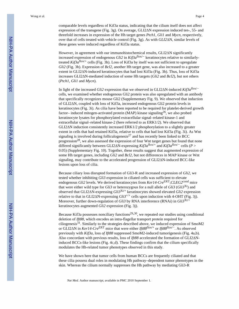
comparable levels regardless of Kif3a status, indicating that the cilium itself does not affectexpression of the transgene (Fig. 3g). On average, GLI2ΔN expression induced ten-, 55- andthreefold increases in expression of the Hh target genes Ptch1, Gli1 and Mycn, respectively,over that of cells treated with vehicle control (Fig. 3g). As with GLI2ΔN, similar levels ofthese genes were induced regardless of Kif3a status.
However, in agreement with our immunohistochemical results, GLI2ΔN significantlyincreased expression of endogenous Gli2 in Kif3aflox/− keratinocytes relative to similarly-treated Kif3aflox/+ cells (Fig. 3h). Loss of Kif3a by itself was not sufficient to upregulateGli2 (Fig. 3h). Expression of Bcl2, another Hh target gene, was also increased to a greaterextent in GLI2ΔN-induced keratinocytes that had lost Kif3a (Fig. 3h). Thus, loss of Kif3aincreases GLI2ΔN-mediated induction of some Hh targets (Gli2 and Bcl2), but not others(Ptch1, Gli1 and Mycn).
In light of the increased Gli2 expression that we observed in GLI2ΔN-induced Kif3aflox/−
cells, we examined whether endogenous Gli2 protein was also upregulated with an antibodythat specifically recognizes mouse Gli2 (Supplementary Fig. 9). We observed that inductionof GLI2ΔN, coupled with loss of Kif3a, increased endogenous Gli2 protein levels inkeratinocytes (Fig. 3i). As cilia have been reported to be required for platelet-derived growthfactor– induced mitogen-activated protein (MAP) kinase signaling26, we also probedkeratinocyte lysates for phosphorylated extracellular signal–related kinase-1 andextracellular signal–related kinase-2 (here referred to as ERK1/2). We observed thatGLI2ΔN induction consistently increased ERK1/2 phosphorylation to a slightly greaterextent in cells that had retained Kif3a, relative to cells that had lost Kif3a (Fig. 3i). As Wntsignaling is involved during folliculogenesis27 and has recently been linked to BCCprogression28, we also assessed the expression of four Wnt target genes but found that nonediffered significantly between GLI2ΔN-expressing Kif3aflox/+ and Kif3aflox/− cells (P >0.05) (Supplementary Fig. 10). Together, these results suggest that augmented expression ofsome Hh target genes, including Gli2 and Bcl2, but not differences in MAP kinase or Wntsignaling, may contribute to the accelerated progression of GLI2ΔN-induced BCC-likelesions upon loss of cilia.
Because ciliary loss disrupted formation of Gli3-R and increased expression of Gli2, wetested whether inhibiting Gli3 expression in ciliated cells was sufficient to elevateendogenous Gli2 levels. We derived keratinocytes from Ker14-CreERT;CLEG2cond micethat were either wild type for Gli3 or heterozygous for a null allele of Gli3 (Gli3Xt) andobserved that GLI2ΔN-expressing Gli3Xt/+ keratinocytes showed elevated Gli2 expressionrelative to that in GLI2ΔN-expressing Gli3+/+ cells upon induction with 4-OHT (Fig. 3j).Moreover, further down-regulation of Gli3 by RNA interference (RNAi) in Gli3Xt/+
keratinocytes augmented Gli2 expression (Fig. 3j).
Because Kif3a possesses nonciliary functions29,30, we repeated our studies using conditionaldeletion of Ift88, which encodes an intra-flagellar transport protein required forciliogenesis31. Similarly to the strategies described above, we induced expression of SmoM2or GLI2ΔN in Ker14-CreERT mice that were either Ift88flox/+ or Ift88flox/−. As observedpreviously with Kif3a, loss of Ift88 suppressed SmoM2-induced tumorigenesis (Fig. 4a,b).Also concordant with previous results, loss of Ift88 accelerated the formation of GLI2ΔN-induced BCCs-like lesions (Fig. 4c,d). These findings confirm that the cilium specificallymodulates the Hh-related tumor phenotypes observed in this study.
We have shown here that tumor cells from human BCCs are frequently ciliated and thatthese cilia possess dual roles in modulating Hh pathway–dependent tumor phenotypes in theskin. Whereas the cilium normally suppresses the Hh pathway by mediating Gli3-R
Wong et al. Page 4
Nat Med. Author manuscript; available in PMC 2010 September 1.
NIH
-PA Author Manuscript
NIH
-PA Author Manuscript
NIH
-PA Author Manuscript

formation in the absence of Hh, the cilium is also required for Smo-mediated activation ofthe pathway in the presence of Hh (Fig. 4e). Our data suggest that tumors initiated by Smodepend upon the cilium both for transducing positive signals through Gli activators and forsuppressing negative ones such as Gli3-R. For tumors initiated by constitutively activeGLI2ΔN, loss of the cilium prevents Gli3-R formation, permitting unopposed activation ofdownstream signaling (Fig. 4e).
We have also shown here that in Smo-mediated tumorigenesis, upregulation of Gli2 requirescilia, whereas GLI2ΔN-induced elevation of endogenous Gli2 is augmented by loss of cilia,consistent with our hypothesis that ciliary control of Gli3 processing likely modulates geneexpression in tumors. Indeed, we also observed that disruption of Gli3 increases Gli2expression, concordant with previous findings that Gli3 binds the Gli2 promoter32. Inaddition to Gli2, Gli3-R likely represses other genes that promote tumorigenesis, such asBcl2.
In addition to disrupting Gli3-R formation, loss of cilia might potentially inhibit formationof a Gli2 repressor, although evidence for a Gli2 repressor function is currently lacking. In avariety of cells, loss of cilia has also been associated with increased response to Wnt,although basal levels of Wnt signaling are unaffected30,33. As Wnt signaling regulatesfolliculogenesis27 and has recently been implicated in the pathogenesis of BCC28, weexamined the expression of Wnt target genes in keratinocytes. Although we found noevidence that Wnt signaling is affected either by GLI2ΔN induction or by ciliary status, itremains possible that increased Wnt responsiveness may contribute to tumorigenesis afterloss of cilia in vivo.
Because the majority of human BCCs possess mutations in either SMO or PTCH1, and lossof a single copy of PTCH1 underlies basal cell nevus (Gorlin's) syndrome, it is noteworthythat cells from three of eight human BCCs examined appeared poorly ciliated. Oneexplanation for this may be that Hh signaling in a small population of ciliated cells issufficient for tumor growth. Alternatively, unciliated cells may be supported by signalingpathways functioning independently of cilia and, as our GLI2ΔN studies might suggest, mayeven show growth advantages upon ciliary loss. Indeed, several reports have describedsignaling pathways that potentiate Gli transcriptional activity, including KRAS-mediatedMAP kinase signaling34, phosphoinositide 3-kinase–Akt signaling35 and transforminggrowth factor-β signaling34,36. Although it remains unclear whether these pathways arefrequently perturbed in BCC, the GLI2ΔN-induced skin tumors provide illustrative supportfor a tumor-suppressive capacity for cilia during Smo- and ciliary-independenttumorigenesis. As recent studies have reported a lack of correlation between Smo activityand downstream Hh pathway activity in a variety of human cancer cells, it is possible thatSmo-independent modulation of the Hh pathway occurs in many different tumor types37.
Our findings advance several general predictions. First, human tumors that rely on upstreamactivation of Hh signaling, such as many BCCs and medulloblastomas, are predicted toretain cilia. Indeed, we have shown here that both human and mouse skin tumors areciliated, and others have found that SmoM2-induced medulloblastomas are similarly ciliatedand dependent upon ciliary function (Y.-G. Han, H. Kim, A.A.D., D. Ellison, R. Gilbertsonand A. Alvarez-Buylla, personal communication). Second, inhibition of ciliogenesis in Smo-dependent tumors is likely to prevent growth. This has major implications for therapy, as thepresence of cilia may be a useful biomarker for identifying Hh-dependent cancersresponsive to Smo antagonists currently being tested in clinical trials2. Third, given thatrecent studies have suggested a paracrine role for Hh signaling during tumorigenesis34,37,disruption of cilia in the surrounding tumor mesenchyme may also indirectly affect tumorgrowth. A final prediction from this study is that loss of cilia may favor progression of
Wong et al. Page 5
Nat Med. Author manuscript; available in PMC 2010 September 1.
NIH
-PA Author Manuscript
NIH
-PA Author Manuscript
NIH
-PA Author Manuscript
tumors initiated by oncogenic events that activate the Hh pathway independently of Smo.Thus, the dual functions of cilia as both a mediator of Smo-dependent tumorigenesis and asa suppressor of Smo-independent tumorigenesis suggest that inhibiting ciliogenesis may betherapeutic in a subset of cancers but may also exacerbate cancer growth in others.
MethodsMethods and any associated references are available in the online version of the paper athttp://www.nature.com/naturemedicine/.
Supplementary MaterialRefer to Web version on PubMed Central for supplementary material.
AcknowledgmentsWe thank D. Hanahan, O. Nolan-Stevaux, G. Evan and the members of the Reiter lab for critical reading of thismanuscript; K. Thorn and the UCSF Nikon Imaging Center for assistance with confocal microscopy; C. Miller andJ.D. Fish for assistance with histology; and R. T Bronson for help with pathology We thank T Li, Harvard MedicalSchool, for rabbit antibody to rootletin; K.V. Anderson, Sloan-Kettering Institute, for Ift172−/−- MEFs; S. Scales,Genentech, for mouse antibody to Gli3; J.T Eggenschwiler, Princeton University, for guinea pig antibody to Gli2;B. Yoder, University of Alabama at Birmingham, for Ift88flox mice; L. Goldstein, University of California, SanDiego, for Kif3aflox -knockout mice; and L.V. Goodrich, Harvard Medical School, for Ptch1 riboprobe plasmid.This work was funded by grants from the US National Institutes of Health (RO1AR054396), the BurroughsWellcome Fund, the Packard Foundation and the Sandler Family Supporting Foundation to J.F.R. A.A.D.acknowledges the support of the US National Institutes of Health (RO1CA087837). S.Y.W. acknowledges thesupport of the A.P. Giannini Foundation, the Herbert W. Boyer Fund and the American Cancer Society.
References1. Dahmane N, Lee J, Robins P, Heller P, Ruiz i Altaba A. Activation of the transcription factor Gli1
and the Sonic hedgehog signalling pathway in skin tumours. Nature 1997;389:876–881. [PubMed:9349822]
2. Rubin LL, de Sauvage FJ. Targeting the Hedgehog pathway in cancer. Nat Rev Drug Discov2006;5:1026–1033. [PubMed: 17139287]
3. Hooper JE, Scott MP. Communicating with Hedgehogs. Nat Rev Mol Cell Biol 2005;6:306–317.[PubMed: 15803137]
4. Xie J, et al. Activating Smoothened mutations in sporadic basal-cell carcinoma. Nature1998;391:90–92. [PubMed: 9422511]
5. Reifenberger J, et al. Missense mutations in SMOH in sporadic basal cell carcinomas of the skin andprimitive neuroectodermal tumors of the central nervous system. Cancer Res 1998;58:1798–1803.[PubMed: 9581815]
6. Wolter M, Reifenberger J, Sommer C, Ruzicka T, Reifenberger G. Mutations in the humanhomologue of the Drosophila segment polarity gene patched (PTCH) in sporadic basal cellcarcinomas of the skin and primitive neuroectodermal tumors of the central nervous system. CancerRes 1997;57:2581–2585. [PubMed: 9205058]
7. Tojo M, Kiyosawa H, Iwatsuki K, Nakamura K, Kaneko F. Expression of the GLI2 oncogene andits isoforms in human basal cell carcinoma. Br J Dermatol 2003;148:892–897. [PubMed: 12786818]
8. Oro AE, et al. Basal cell carcinomas in mice overexpressing sonic hedgehog. Science1997;276:817–821. [PubMed: 9115210]
9. Eggenschwiler JT, Anderson KV. Cilia and developmental signaling. Annu Rev Cell Dev Biol2007;23:345–373. [PubMed: 17506691]
10. Rohatgi R, Milenkovic L, Scott MP. Patched1 regulates Hedgehog signaling at the primary cilium.Science 2007;317:372–376. [PubMed: 17641202]
11. Corbit KC, et al. Vertebrate Smoothened functions at the primary cilium. Nature 2005;437:1018–1021. [PubMed: 16136078]
Wong et al. Page 6
Nat Med. Author manuscript; available in PMC 2010 September 1.
NIH
-PA Author Manuscript
NIH
-PA Author Manuscript
NIH
-PA Author Manuscript

12. Liu A, Wang B, Niswander LA. Mouse intraflagellar transport proteins regulate both the activatorand repressor functions of Gli transcription factors. Development 2005;132:3103–3111. [PubMed:15930098]
13. Haycraft CJ, et al. Gli2 and Gli3 localize to cilia and require the intraflagellar transport proteinpolaris for processing and function. PLoS Genet 2005;1:e53. [PubMed: 16254602]
14. Huangfu D, Anderson KV. Cilia and hedgehog responsiveness in the mouse. Proc Natl Acad SciUSA 2005;102:11325–11330. [PubMed: 16061793]
15. May SR, et al. Loss of the retrograde motor for IFT disrupts localization of Smo to cilia andprevents the expression of both activator and repressor functions of Gli. Dev Biol 2005;287:378–389. [PubMed: 16229832]
16. Wilson RB, McWhorter CA. Isolated flagella in human skin Election microscopic observations.Lab Invest 1963;12:242–249. [PubMed: 14001080]
17. Mao J, et al. A novel somatic mouse model to survey tumorigenic potential applied to theHedgehog pathway. Cancer Res 2006;66:10171–10178. [PubMed: 17047082]
18. Lehman JM, Laag E, Michaud EJ, Yoder BK. An essential role for dermal primary cilia in hairfollicle morphogenesis. J Invest Dermatol 2009;129:438–448. [PubMed: 18987668]
19. Marszalek JR, et al. Genetic evidence for selective transport of opsin and arrestin by kinesin-II inmammalian photoreceptors. Cell 2000;102:175–187. [PubMed: 10943838]
20. Vasioukhin V, Degenstein L, Wise B, Fuchs E. The magical touch: genome targeting in epidermalstem cells induced by tamoxifen application to mouse skin. Proc Natl Acad Sci USA1999;96:8551–8556. [PubMed: 10411913]
21. Huangfu D, et al. Hedgehog signalling in the mouse requires intraflagellar transport proteins.Nature 2003;426:83–87. [PubMed: 14603322]
22. St Jacques B, et al. Sonic hedgehog signaling is essential for hair development. Curr Biol1998;8:1058–1068. [PubMed: 9768360]
23. Chiang C, et al. Essential role for Sonic hedgehog during hair follicle morphogenesis. Dev Biol1999;205:1–9. [PubMed: 9882493]
24. Mill P, et al. Sonic hedgehog–dependent activation of Gli2 is essential for embryonic hair follicedevelopment. Genes Dev 2003;17:282–294. [PubMed: 12533516]
25. Pasca di Magliano M, et al. Hedgehog-Ras interactions regulate early stages of pancreatic cancer.Genes Dev 2006;20:3161–3173. [PubMed: 17114586]
26. Schneider L, et al. PDGFRαα signaling is regulated through the primary cilium in fibroblasts. CurrBiol 2005;15:1861–1866. [PubMed: 16243034]
27. Fuchs E. Skin stem cells: rising to the surface. J Cell Biol 2008;180:273–284. [PubMed:18209104]
28. Yang SH, et al. Pathological responses to oncogenic Hedgehog signaling in skin are dependent oncanonical Wnt/β-catenin signaling. Nat Genet 2008;40:1130–1135. [PubMed: 19165927]
29. Nishimura T, et al. Role of the PAR-3–KIF3 complex in the establishment of neuronal polarity.Nat Cell Biol 2004;6:328–334. [PubMed: 15048131]
30. Corbit KC, et al. Kif3a constrains β-catenin–dependent Wnt signalling through dual ciliary andnon-ciliary mechanisms. Nat Cell Biol 2008;10:70–76. [PubMed: 18084282]
31. Haycraft CJ, et al. Intraflagellar transport is essential for endochondral bone formation.Development 2007;134:307–316. [PubMed: 17166921]
32. Hu MC, et al. GLI3-dependent transcriptional repression of Gli1, Gli2 and kidney patterning genesdisrupts renal morphogenesis. Development 2006;133:569–578. [PubMed: 16396903]
33. Gerdes JM, et al. Disruption of the basal body compromises proteasomal function and perturbsintracellular Wnt response. Nat Genet 2007;39:1350–1360. [PubMed: 17906624]
34. Nolan-Stevaux O, et al. GLI1 is regulated through Smoothened-independent mechanisms inneoplastic pancreatic ducts and mediates PDAC cell survival and transformation. Genes Dev2009;23:24–36. [PubMed: 19136624]
35. Riobó NA, Lu K, Ai X, Haines GM, Emerson CP Jr. Phosphoinositide 3-kinase and Akt areessential for Sonic Hedgehog signaling. Proc Natl Acad Sci USA 2006;103:4505–4510. [PubMed:16537363]
Wong et al. Page 7
Nat Med. Author manuscript; available in PMC 2010 September 1.
NIH
-PA Author Manuscript
NIH
-PA Author Manuscript
NIH
-PA Author Manuscript

36. Dennler S, et al. Induction of sonic hedgehog mediators by transforming growth factor-β: Smad3-dependent activation of Gli2 and Gli1 expression in vitro and in vivo. Cancer Res 2007;67:6981–6986. [PubMed: 17638910]
37. Yauch RL, et al. A paracrine requirement for hedgehog signalling in cancer. Nature 2008;455:406–410. [PubMed: 18754008]
Wong et al. Page 8
Nat Med. Author manuscript; available in PMC 2010 September 1.
NIH
-PA Author Manuscript
NIH
-PA Author Manuscript
NIH
-PA Author Manuscript

Figure 1.BCC and normal skin cells possess primary cilia. (a) Immunofluorescence imaging of theciliary marker acetylated tubulin (green) and rootletin (red), a ciliary rootlet component.Five of eight independent human clinical BCCs, including nodular and superficial subtypes,contain numerous ciliated cells (arrows). Insets, H&E staining of adjacent biopsies. Rightimages are views of the left and middle panels. (b) Immunofluorescence imaging of theciliary marker detyrosinated tubulin (green) and the centrosomal marker γ -tubulin (red)(dotted line, epidermal basal layer;inset, enlarged view). Arrows indicate ciliated cellspresent in SmoM2-induced regions of mouse epidermal hyperplasia. (c) Cilia extendingfrom normal skin cells (arrows). Left images, low magnification;right images, highmagnification of boxed areas. (d) Cilia extending from cultured keratinocytes (arrows). Allscale bars are 50 μm except that in d (10 μm).
Wong et al. Page 9
Nat Med. Author manuscript; available in PMC 2010 September 1.
NIH
-PA Author Manuscript
NIH
-PA Author Manuscript
NIH
-PA Author Manuscript

Figure 2.Cilia are essential for SmoM2-induced neoplasia. (a) Schematic showing that skin epithelialcells exposed to tamoxifen (TAM) both delete Kif3aflox and activate SmoM2cond (red cells),whereas unexposed cells retain Kif3aflox and do not activate SmoM2cond (gray cells). Ciliaare lost in cells from Kif3aflox/− but not Kif3aflox/+ mice exposed to tamoxifen. (b) H&Estaining of dorsal skin biopsies, showing that removal of Kif3a (Kif3aflox/−) blocks SmoM2-initiated tumorigenesis (arrows). (c) Immunofluorescence imaging showing that SmoM2localizes to cilia (arrows) in neoplastic epidermal downgrowths. (d) Quantification ofinterfollicular epidermis thickness. Seven or eight independent Ker 14-CreERT;SmoM2cond;Kif3aflox/+ and Ker14-CreERT;SmoM2cond;Kif3aflox/− mice per genotype
Wong et al. Page 10
Nat Med. Author manuscript; available in PMC 2010 September 1.
NIH
-PA Author Manuscript
NIH
-PA Author Manuscript
NIH
-PA Author Manuscript

were analyzed. Four or five mice were analyzed for all other genotypes. (e) Protectionagainst SmoM2-induced ear skin hyperplasia upon loss of Kif3a (arrow). (f) H&E stainingof representative ear sections. (g) In situ staining showing that SmoM2-induced skin lesionsupregulate Hh target genes Gli1 and Ptch1 (arrows). (h) Immunofluorescence imagingshowing that Gli2 is upregulated in SmoM2-induced tumors that express Kif3a (stainingsare representative of the mice analyzed in d and j). (i) Immunofluorescence imagingshowing that Gli2 is upregulated upon expression of Myc-tagged SmoA1 in ciliated (wildtype), but not unciliated (Kif3a−/− and Ift172−/−), transformed MEFs. (j) Cellularproliferation in skin, as assessed by Ki67 staining. Eight Ker14-CreERT;SmoM2cond;Kif3aflox/+ and Ker14-CreERT;SmoM2cond;Kif3aflox/− mice per genotypewere analyzed, as well as four or five mice for the other genotypes. All scale bars are 50 μmexcept that in e (5 mm). All error bars shown, ± s.e.m.
Wong et al. Page 11
Nat Med. Author manuscript; available in PMC 2010 September 1.
NIH
-PA Author Manuscript
NIH
-PA Author Manuscript
NIH
-PA Author Manuscript

Figure 3.Loss of cilia accelerates GLI2ΔN-induced neoplasia. (a) Gross morphology of mice uponinduction of CLEG2cond. (b) H&E staining showing that loss of Kif3a increases the severityof GLI2ΔN-induced skin lesions (arrows). Images are representative of five or six miceanalyzed per genotype. (c) Immunofluorescence imaging showing that loss of Kif3aincreases the number of Gli2+ downgrowths in the skin. (d) In situ staining revealing thatPtch1 is upregulated in small regions of CLEG2-expressing Kif3aflox/+ skin (arrowhead),whereas Gli1 and Ptch1 are more frequently upregulated in CLEG2-induced lesions lackingKif3a (arrows). (e) Formation of Gli3-R in ciliated (wild type) and non-ciliated (Kif3a−/−,Ift172−/−) transformed MEFs (Gli3-FL, full length Gli3). (f) Western blot indicating that
Wong et al. Page 12
Nat Med. Author manuscript; available in PMC 2010 September 1.
NIH
-PA Author Manuscript
NIH
-PA Author Manuscript
NIH
-PA Author Manuscript
Kif3a protein expression is lower and Gli3-R formation is impaired in 4-OHT–treatedKer14-CreERT;CLEG2cond;Kif3aflox/−keratinocytes, relative to control cells. (g) Expressionof constitutively active human GLI2 (GLI2ΔN), Ptch1, Gli1 and Mycn in Kif3aflox/+ andKif3aflox/−keratinocytes treated with 4-OHT. (All expression values expressed relative tothose of vehicle-treated control cells, set to a baseline of ‘1’, dotted line.) (h) QuantiativeRT-PCR assessment of GLI2ΔN-mediated induction of endogenous Gli2 and Bcl2 inciliated and unciliated Kif3aflox/− keratinocytes. (i) Endogenous Gli2 and phosphorylatedERK1/2 protein levels in 4-OHT–treated Ker14-CreERT;CLEG2cond keratinocytes that areeither Kif3aflox/+ or Kif3aflox/−. (j) Expression of endogenous Gli2 in GLI2ΔN-inducedGli3Xt/+ or Gli3+/+ keratinocytes, treated with siRNA against Gli3 or control. All expressionvalues are normalized to those of vehicle-treated cells transfected with control siRNAs(baseline set to ‘1’, dotted line). All scale bars are 50 μm except that in a (5 mm). All errorbars show means ± s.e.m.
Wong et al. Page 13
Nat Med. Author manuscript; available in PMC 2010 September 1.
NIH
-PA Author Manuscript
NIH
-PA Author Manuscript
NIH
-PA Author Manuscript

Figure 4.Loss of Ift88 restrains SmoM2-mediated tumorigenesis and promotes GLI2ΔN-inducedBCC-like lesions in tamoxifen-treated mice. (a) H&E staining of dorsal skin biopsies,revealing that loss of Ift88 (Ift88flox/−) blocks SmoM2-induced epidermal hyperplasia andtumorigenesis. (b) Quantification of epidermal thickness for a. Three to five mice pergenotype were analyzed 10 weeks after induction by tamoxifen, and five mice per genotypewere assessed 20 weeks after tamoxifen treatment. (c) H&E staining revealing that loss ofIft88 promotes skin lesions induced by activated Gli2. All scale bars, 50 μm. (d)Quantification of the number of Gli2+ downgrowths in the skin. Four mice were analyzedper genotype. (e) Schematic showing that in nonpathological conditions and in the absenceof Hh, the cilium mediates the formation of Gli3-R, a repressor of downstream Hh targetgenes. In the presence of Hh ligand, Ptch1-mediated inhibition of Smo is suppressed,allowing Smo to act through the cilium to inhibit Gli3-R formation and induce Gli activators(Gli-Act), which turn on the Hh transcriptional program. In cilium-dependent oncogenesis(for example, SmoM2-induced tumors), the cilium is essential for Gli-Act formation and,thus, tumorigenesis. In cilium-independent oncogenesis (for example, tumors induced byconstitutively active GLI2 (Gli-Act*)), the cilium restrains tumorigenesis throughproduction of counter-balancing Gli3-R. Loss of the cilium inhibits Gli3-R formation,allowing unopposed Gli-Act* to aggressively drive tumorigenesis. All error bars showmeans ± s.e.m.
Wong et al. Page 14
Nat Med. Author manuscript; available in PMC 2010 September 1.
NIH
-PA Author Manuscript
NIH
-PA Author Manuscript
NIH
-PA Author Manuscript