minimally invasive intraoral approach to submandibular lodge
TRANSCRIPT
Journal of
Clinical Medicine
Article
Minimally Invasive Intraoral Approach toSubmandibular Lodge
Massimo Galli 1 , Massimo Fusconi 2 , Francesca Romana Federici 1 , Francesca Candelori 2,Marco De Vincentiis 2, Antonella Polimeni 1 , Luca Testarelli 1 , Benedetta Cassese 1,Gabriele Miccoli 1,* and Antonio Greco 2
1 Department of Oral and Maxillofacial Science, Sapienza University of Rome, Via Caserta 6, 00161 Rome,Italy; [email protected] (M.G.); [email protected] (F.R.F.);[email protected] (A.P.); [email protected] (L.T.); [email protected] (B.C.)
2 Department of Sensory Organs, Division of Otorhinolaryngology, Sapienza University of Rome, Viale delPoliclinico 161, 00185 Rome, Italy; [email protected] (M.F.);[email protected] (F.C.); [email protected] (M.D.V.);[email protected] (A.G.)
* Correspondence: [email protected] or [email protected]; Tel.: +39-388-045-9246
Received: 5 August 2020; Accepted: 11 September 2020; Published: 14 September 2020�����������������
Abstract: The purpose of this study is to describe the Minimally Invasive Intraoral Approach (MIIA)performed on selected cases of abscesses and neck phlegmons of odontogenic origin when theinfection has not spread beyond the inferior mandibular margin. This technique allows us to avoidcervicotomy by a direct approach to the abscess, draining it through the oral cavity. If the limits havealready been crossed, then cervicotomy is necessary. The aim of the study is to show the surgicaloutcomes that we have achieved during a time span of two years, and to show the effectiveness ofthe MIIA and its results. We selected 66 patients with abscesses and neck phlegmons, from January2018 to June 2020. Among these cases, five patients were excluded as it was not possible to recovermedical records from database. The MIIA technique has been performed on 16 patients (26.2%)when a successful dental extraction and drainage of the submandibular lodge were accomplished.The patients who underwent the MIIA surgery have all perfectly healed and did not suffer fromrelapses during the follow-up. The results show the achievement of excellent healing, underlining thelower impact required by MIIA when compared to a more traditional approach through cervicotomy.
Keywords: odontogenic abscess; cervicotomy; surgery; oral surgery; neck surgery; infection;phlegmon; oral cavity
1. Introduction
Recently, an increasing number of patients with dental symptoms admitted to the EmergencyRoom (ER) has been recorded. These patients were diagnosed with abscesses and neck phlegmonsof odontogenic origin, which required surgical treatment [1–3]. Unfortunately, in some cases theabscess had already spread to the point where it was not possible to solve the disease through simplertreatments, such as root canal therapy or periodontal approach, both aiming to resolve the infectionand at the same time to save the involved teeth [4]. Most of the time, this kind of infection forces thepatient to undergo complex surgery. Such an invasive surgical treatment is based on the extraction ofan untreatable tooth and involves cervicotomy in order to clean the infected tissues and the placementof a Redon drainage. All of this inevitably results in a longer hospitalization. The procedure usuallyinvolves a submandibular incision that may lead to severities, such as the impairment of neuro-vascularstructures, not to mention aesthetic issues relating to a healing neck wound [5,6]. The aim of this
J. Clin. Med. 2020, 9, 2971; doi:10.3390/jcm9092971 www.mdpi.com/journal/jcm
J. Clin. Med. 2020, 9, 2971 2 of 9
paper is to show an alternative surgical technique performed on selected cases. This technique avoidscervicotomy by means of a direct approach to the abscess, draining it directly through the oral cavity.Our hereby proposed Minimally Invasive Intraoral Approach (MIIA) can only be performed whenthe infection spreads up to the inferior mandibular margin and no further. As a matter of fact, theanatomical localization of the abscess and its spreading have to be pre-emptively evaluated withcomputerized tomography (CT) and the results then lead us to the most appropriate surgical technique.The goal of this study is to demonstrate the achievement of excellent healing after a MIIA, underliningthe lower impact of MIIA when compared to the classic approach through cervicotomy. Moreover,we obtained our best results among all the patients selected for MIIA when positioning a drainagedevice to improve the anti-gravity discharge of liquid from the residual cavity by contraction of thesuprahyoid muscles. This drainage consists of a latex glove finger, e.g. Penrose, placed between thealveolar bone and the mucoperiosteal flap. Specific antibiotic therapy remains absolutely essential forthe complete cure of the infection. We proceed to show all of the surgical outcomes achieved during atime span of two years, alongside the criteria and algorithms used for surgical decision-making.
2. Experimental Section
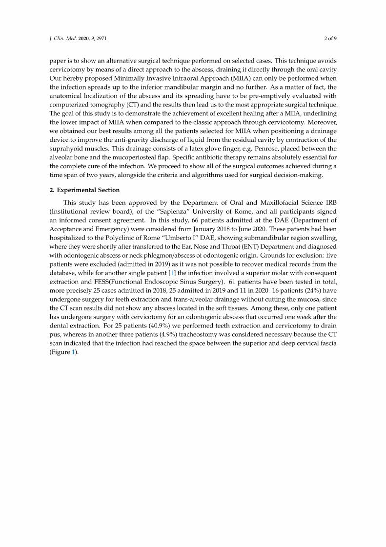
This study has been approved by the Department of Oral and Maxillofacial Science IRB(Institutional review board), of the “Sapienza” University of Rome, and all participants signedan informed consent agreement. In this study, 66 patients admitted at the DAE (Department ofAcceptance and Emergency) were considered from January 2018 to June 2020. These patients had beenhospitalized to the Polyclinic of Rome “Umberto I” DAE, showing submandibular region swelling,where they were shortly after transferred to the Ear, Nose and Throat (ENT) Department and diagnosedwith odontogenic abscess or neck phlegmon/abscess of odontogenic origin. Grounds for exclusion: fivepatients were excluded (admitted in 2019) as it was not possible to recover medical records from thedatabase, while for another single patient [1] the infection involved a superior molar with consequentextraction and FESS(Functional Endoscopic Sinus Surgery). 61 patients have been tested in total,more precisely 25 cases admitted in 2018, 25 admitted in 2019 and 11 in 2020. 16 patients (24%) haveundergone surgery for teeth extraction and trans-alveolar drainage without cutting the mucosa, sincethe CT scan results did not show any abscess located in the soft tissues. Among these, only one patienthas undergone surgery with cervicotomy for an odontogenic abscess that occurred one week after thedental extraction. For 25 patients (40.9%) we performed teeth extraction and cervicotomy to drainpus, whereas in another three patients (4.9%) tracheostomy was considered necessary because the CTscan indicated that the infection had reached the space between the superior and deep cervical fascia(Figure 1).
J. Clin. Med. 2020, 9, 2971 3 of 9
Figure 1. CT scan of coronal sections (A,B). The green line delimits the abscess’s area, showing thecrossing of the inferior-medial side of the mandibula.
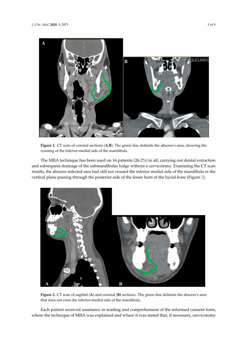
The MIIA technique has been used on 16 patients (26.2%) in all, carrying out dental extractionand subsequent drainage of the submandibular lodge without a cervicotomy. Examining the CT scanresults, the abscess–infected area had still not crossed the inferior medial side of the mandibula or thevertical plane passing through the posterior side of the lesser horn of the hyoid bone (Figure 2).
Figure 2. CT scan of sagittal (A) and coronal (B) sections. The green line delimits the abscess’s areathat does not cross the inferior-medial side of the mandibula.
Each patient received assistance in reading and comprehension of the informed consent form,where the technique of MIIA was explained and where it was stated that, if necessary, cervicotomy
J. Clin. Med. 2020, 9, 2971 4 of 9
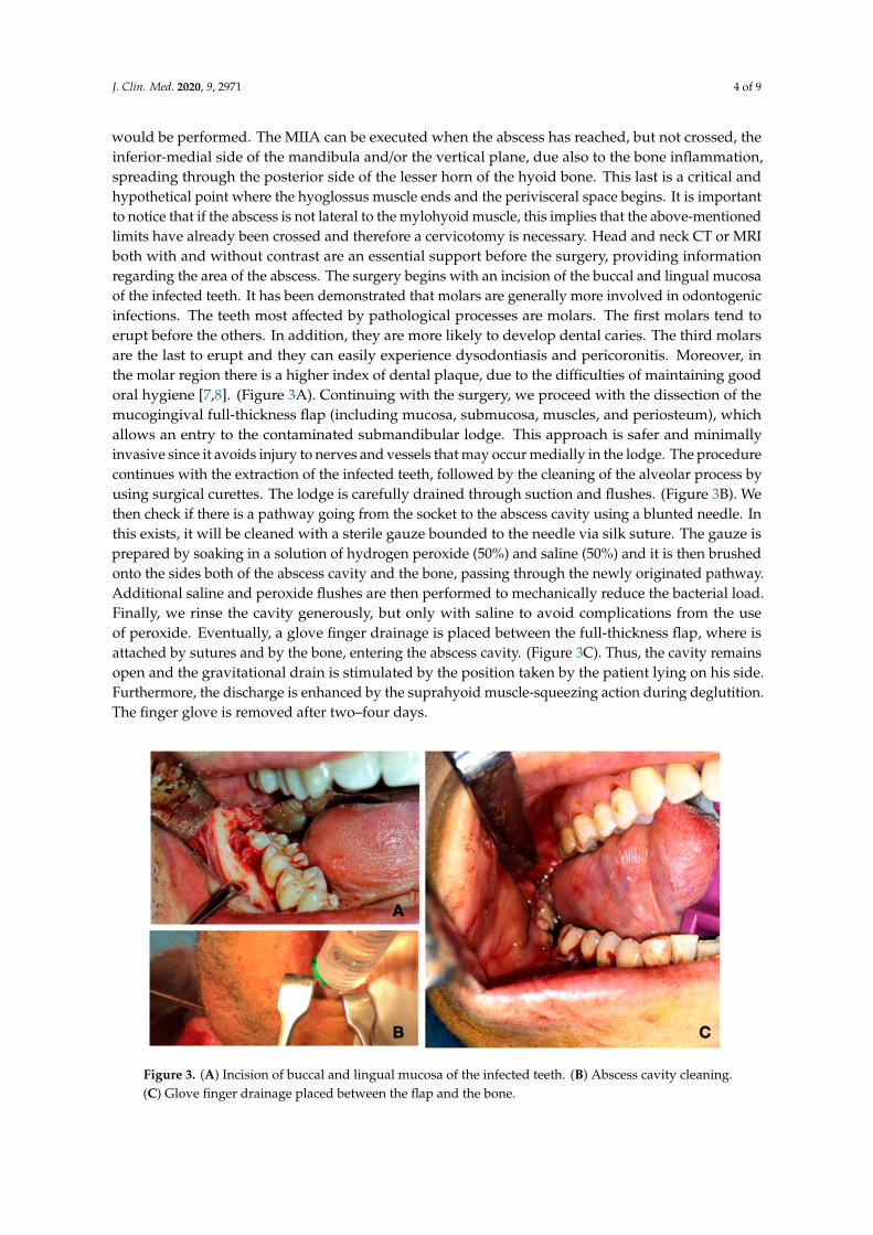
would be performed. The MIIA can be executed when the abscess has reached, but not crossed, theinferior-medial side of the mandibula and/or the vertical plane, due also to the bone inflammation,spreading through the posterior side of the lesser horn of the hyoid bone. This last is a critical andhypothetical point where the hyoglossus muscle ends and the perivisceral space begins. It is importantto notice that if the abscess is not lateral to the mylohyoid muscle, this implies that the above-mentionedlimits have already been crossed and therefore a cervicotomy is necessary. Head and neck CT or MRIboth with and without contrast are an essential support before the surgery, providing informationregarding the area of the abscess. The surgery begins with an incision of the buccal and lingual mucosaof the infected teeth. It has been demonstrated that molars are generally more involved in odontogenicinfections. The teeth most affected by pathological processes are molars. The first molars tend toerupt before the others. In addition, they are more likely to develop dental caries. The third molarsare the last to erupt and they can easily experience dysodontiasis and pericoronitis. Moreover, inthe molar region there is a higher index of dental plaque, due to the difficulties of maintaining goodoral hygiene [7,8]. (Figure 3A). Continuing with the surgery, we proceed with the dissection of themucogingival full-thickness flap (including mucosa, submucosa, muscles, and periosteum), whichallows an entry to the contaminated submandibular lodge. This approach is safer and minimallyinvasive since it avoids injury to nerves and vessels that may occur medially in the lodge. The procedurecontinues with the extraction of the infected teeth, followed by the cleaning of the alveolar process byusing surgical curettes. The lodge is carefully drained through suction and flushes. (Figure 3B). Wethen check if there is a pathway going from the socket to the abscess cavity using a blunted needle. Inthis exists, it will be cleaned with a sterile gauze bounded to the needle via silk suture. The gauze isprepared by soaking in a solution of hydrogen peroxide (50%) and saline (50%) and it is then brushedonto the sides both of the abscess cavity and the bone, passing through the newly originated pathway.Additional saline and peroxide flushes are then performed to mechanically reduce the bacterial load.Finally, we rinse the cavity generously, but only with saline to avoid complications from the useof peroxide. Eventually, a glove finger drainage is placed between the full-thickness flap, where isattached by sutures and by the bone, entering the abscess cavity. (Figure 3C). Thus, the cavity remainsopen and the gravitational drain is stimulated by the position taken by the patient lying on his side.Furthermore, the discharge is enhanced by the suprahyoid muscle-squeezing action during deglutition.The finger glove is removed after two–four days.
Figure 3. (A) Incision of buccal and lingual mucosa of the infected teeth. (B) Abscess cavity cleaning.(C) Glove finger drainage placed between the flap and the bone.
J. Clin. Med. 2020, 9, 2971 5 of 9
3. Results
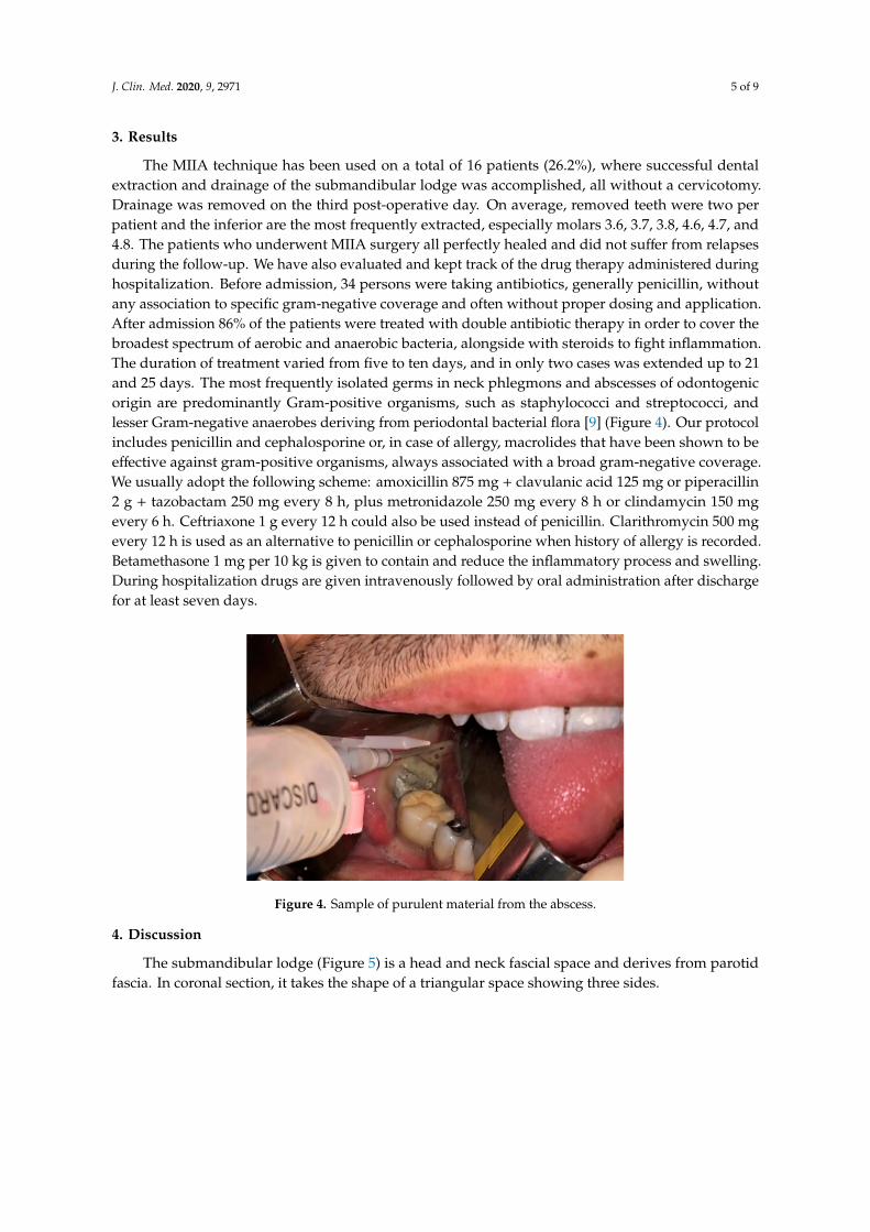
The MIIA technique has been used on a total of 16 patients (26.2%), where successful dentalextraction and drainage of the submandibular lodge was accomplished, all without a cervicotomy.Drainage was removed on the third post-operative day. On average, removed teeth were two perpatient and the inferior are the most frequently extracted, especially molars 3.6, 3.7, 3.8, 4.6, 4.7, and4.8. The patients who underwent MIIA surgery all perfectly healed and did not suffer from relapsesduring the follow-up. We have also evaluated and kept track of the drug therapy administered duringhospitalization. Before admission, 34 persons were taking antibiotics, generally penicillin, withoutany association to specific gram-negative coverage and often without proper dosing and application.After admission 86% of the patients were treated with double antibiotic therapy in order to cover thebroadest spectrum of aerobic and anaerobic bacteria, alongside with steroids to fight inflammation.The duration of treatment varied from five to ten days, and in only two cases was extended up to 21and 25 days. The most frequently isolated germs in neck phlegmons and abscesses of odontogenicorigin are predominantly Gram-positive organisms, such as staphylococci and streptococci, andlesser Gram-negative anaerobes deriving from periodontal bacterial flora [9] (Figure 4). Our protocolincludes penicillin and cephalosporine or, in case of allergy, macrolides that have been shown to beeffective against gram-positive organisms, always associated with a broad gram-negative coverage.We usually adopt the following scheme: amoxicillin 875 mg + clavulanic acid 125 mg or piperacillin2 g + tazobactam 250 mg every 8 h, plus metronidazole 250 mg every 8 h or clindamycin 150 mgevery 6 h. Ceftriaxone 1 g every 12 h could also be used instead of penicillin. Clarithromycin 500 mgevery 12 h is used as an alternative to penicillin or cephalosporine when history of allergy is recorded.Betamethasone 1 mg per 10 kg is given to contain and reduce the inflammatory process and swelling.During hospitalization drugs are given intravenously followed by oral administration after dischargefor at least seven days.
Figure 4. Sample of purulent material from the abscess.
4. Discussion
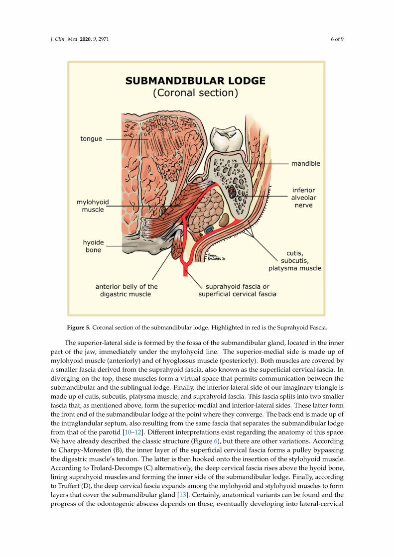
The submandibular lodge (Figure 5) is a head and neck fascial space and derives from parotidfascia. In coronal section, it takes the shape of a triangular space showing three sides.
J. Clin. Med. 2020, 9, 2971 6 of 9
Figure 5. Coronal section of the submandibular lodge. Highlighted in red is the Suprahyoid Fascia.
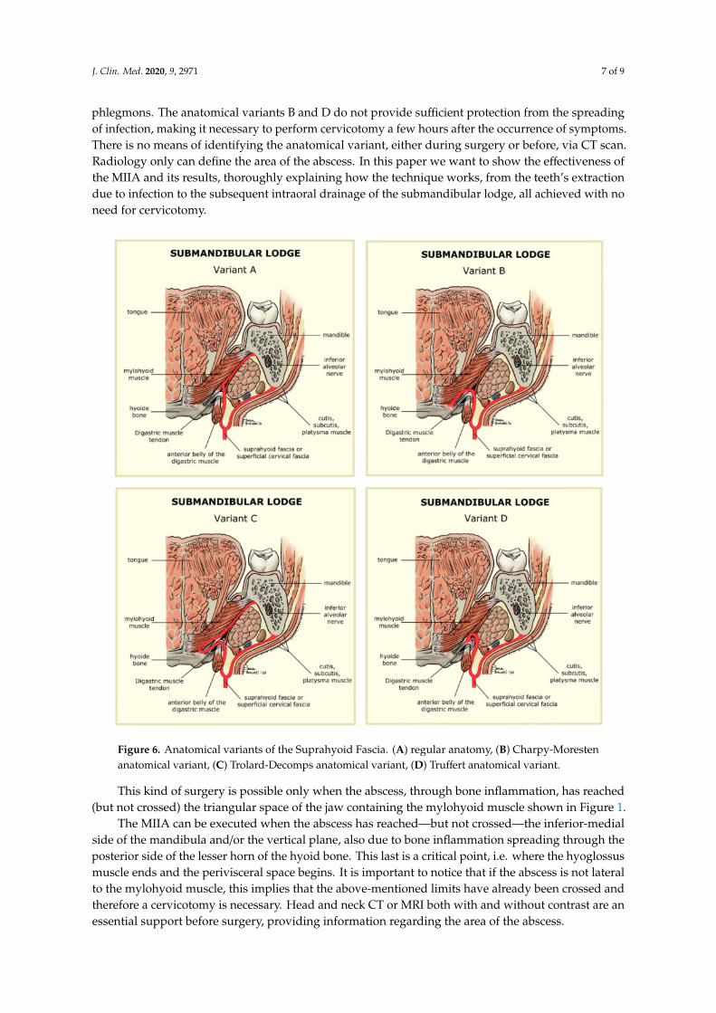
The superior-lateral side is formed by the fossa of the submandibular gland, located in the innerpart of the jaw, immediately under the mylohyoid line. The superior-medial side is made up ofmylohyoid muscle (anteriorly) and of hyoglossus muscle (posteriorly). Both muscles are covered bya smaller fascia derived from the suprahyoid fascia, also known as the superficial cervical fascia. Indiverging on the top, these muscles form a virtual space that permits communication between thesubmandibular and the sublingual lodge. Finally, the inferior lateral side of our imaginary triangle ismade up of cutis, subcutis, platysma muscle, and suprahyoid fascia. This fascia splits into two smallerfascia that, as mentioned above, form the superior-medial and inferior-lateral sides. These latter formthe front end of the submandibular lodge at the point where they converge. The back end is made up ofthe intraglandular septum, also resulting from the same fascia that separates the submandibular lodgefrom that of the parotid [10–12]. Different interpretations exist regarding the anatomy of this space.We have already described the classic structure (Figure 6), but there are other variations. Accordingto Charpy-Moresten (B), the inner layer of the superficial cervical fascia forms a pulley bypassingthe digastric muscle’s tendon. The latter is then hooked onto the insertion of the stylohyoid muscle.According to Trolard-Decomps (C) alternatively, the deep cervical fascia rises above the hyoid bone,lining suprahyoid muscles and forming the inner side of the submandibular lodge. Finally, accordingto Truffert (D), the deep cervical fascia expands among the mylohyoid and stylohyoid muscles to formlayers that cover the submandibular gland [13]. Certainly, anatomical variants can be found and theprogress of the odontogenic abscess depends on these, eventually developing into lateral-cervical
J. Clin. Med. 2020, 9, 2971 7 of 9
phlegmons. The anatomical variants B and D do not provide sufficient protection from the spreadingof infection, making it necessary to perform cervicotomy a few hours after the occurrence of symptoms.There is no means of identifying the anatomical variant, either during surgery or before, via CT scan.Radiology only can define the area of the abscess. In this paper we want to show the effectiveness ofthe MIIA and its results, thoroughly explaining how the technique works, from the teeth’s extractiondue to infection to the subsequent intraoral drainage of the submandibular lodge, all achieved with noneed for cervicotomy.
Figure 6. Anatomical variants of the Suprahyoid Fascia. (A) regular anatomy, (B) Charpy-Morestenanatomical variant, (C) Trolard-Decomps anatomical variant, (D) Truffert anatomical variant.
This kind of surgery is possible only when the abscess, through bone inflammation, has reached(but not crossed) the triangular space of the jaw containing the mylohyoid muscle shown in Figure 1.
The MIIA can be executed when the abscess has reached—but not crossed—the inferior-medialside of the mandibula and/or the vertical plane, also due to bone inflammation spreading through theposterior side of the lesser horn of the hyoid bone. This last is a critical point, i.e. where the hyoglossusmuscle ends and the perivisceral space begins. It is important to notice that if the abscess is not lateralto the mylohyoid muscle, this implies that the above-mentioned limits have already been crossed andtherefore a cervicotomy is necessary. Head and neck CT or MRI both with and without contrast are anessential support before surgery, providing information regarding the area of the abscess.
J. Clin. Med. 2020, 9, 2971 8 of 9
This virtual space ensures that the infection does not overstep the anatomical limits towards theadjacent visceral tissues, which would then lead to the need for cervicotomy. Such limits are enclosedby the mylohyoid muscle—defining the external side of the sublingual region and the tongue base—bythe inferior side of the jaw, and by the lesser horn of the hyoid bone. The latter corresponds to theposterior side of the submandibular lodge which is open at its rear to allow the passage of sublingualvessels (artery and vein), the lingual nerve, and the hypoglossal nerve [14,15].
Precisely, this rear opening is the most critical point—among the other anatomical structuresmentioned earlier—since it is often too weak to contain the infection that will therefore easily spreadunder the effect of gravity. Obviously if the CT scan shows an infection that has crossed these limits,the MIIA would be an inadequate measure to prevent a further spreading of the infection and it wouldbe necessary to proceed with a cervicotomy.
We have already described the anatomical variants of the submandibular lodge. When theinfection fills the virtual spaces of the superficial and deep cervical fascia (B,D) it could potentiallyspread into the visceral region. On the contrary, when the case of an empty submandibular lodgeoccurs, if we operate in time we are able to drain the deep sides of the lodge, eluding the risk of theinfection slipping through an intraoral pathway, which could potentially turn from an odontogenicabscess into a neck’s phlegmon, for which a cervicotomy would be required [10].
5. Conclusions
The MIIA, in selected cases, can lower the impact of the surgery, consequently reducing thelength of hospitalization and cutting health costs. When abscess occurs and does not cross thepreviously described anatomical limits, we suggest the use of this technique to obtain a shorterpost-operative recovery.
Author Contributions: We count a total of ten persons in our medical team, which is made up of two separatesurgical, dental and otolaryngologist teams who work jointly on cases of various diseases, such as abscesses, neckphlegmons derived from odontogenic infection, and so on. The team focusing on the ENT trait consists of M.D.V.,M.F., A.G. and F.C., while the team that operates on the dental side consists of A.P., M.G., L.T., G.M., F.R.F. and B.C.On a more detailed level: conceptualization, M.G. and M.F.; methodology, M.G.; validation, M.D.V., A.G. and A.P.;formal analysis, L.T.; investigation, F.C.; resources, M.G.; data curation, B.C.; writing—original draft preparation,F.R.F.; writing—review and editing, G.M.; visualization, F.R.F.; supervision, M.F.; project administration, A.P. Allauthors have read and agreed to the published version of the manuscript.
Funding: This research received no external funding.
Acknowledgments: Sara Briotti, DDS, designer of the anatomical images.
Conflicts of Interest: The authors declare no conflict of interest.
References
1. Moghimi, M.; Baart, J.A.; Karagozoglu, K.H.; Forouzanfar, T. Spread of odontogenic infections: A retrospectiveanalysis and review of the literature. Quintessence Int. 2013, 44, 351–361. [PubMed]
2. Bali, R.K.; Sharma, P.; Gaba, S.; Kaur, A.; Ghanghas, P. A review of complications of odontogenic infections.Natl. J. Maxillofac. Surg. 2015, 6, 136–143. [CrossRef] [PubMed]
3. Bakathir, A.A.; Moos, K.F.; Ayoub, A.F.; Bagg, J. Factors Contributing to the Spread of Odontogenic infections:A prospective pilot study. Sultan Qaboos Univ. Med. J. 2009, 9, 296–304. [PubMed]
4. Green, A.W.; Flower, E.A.; New, N.E. Mortality associated with odontogenic infection! Br. Dent. J. 2001, 190,529–530. [CrossRef] [PubMed]
5. D’Erme, G.; Galli, M.; Candelori, F.; Federici, F.R.; Greco, A.; Ralli, M. A rare case of odontogenic bilateralsubmandibular abscess. Otorinolaringologia 2020, 70.
6. Pesis, M.; Bar-Droma, E.; Ilgiyaev, A.; Givol, N. Deep Neck Infections Are Life Threatening Infections ofDental Origin: A Presentation and Management of Selected Cases. ISR. Med. Assoc. J. IMAJ 2019, 12, 806–811.[PubMed]
J. Clin. Med. 2020, 9, 2971 9 of 9
7. Fusconi, M.; Greco, A.; Galli, M.; Polimeni, A.; Yusef, M.; Di Cianni, S.; De Soccio, G.; FR, F.S.; Lombardi, R.;de Vincentiis, M. Odontogenic phlegmons and abscesses in relation to the financial situation of Italianfamilies. Minerva Stomatol. 2019, 68, 236–241. [CrossRef] [PubMed]
8. D’Erme, G.; Galli, M.; Federici, F.R.; Colizza, A.; Ralli, M.; de Vincentiis, M. Report of a rare case of Lemierre’ssyndrome associated with a multidrug resistant dental abscess. Otorinolaringologia 2019, 69.
9. Heim, N.; Faron, A.; Wiedemeyer, V.; Reich, R.; Martini, M. Microbiology and antibiotic sensitivity of headand neck space infections of odontogenic origin. Differences in inpatient and outpatient management. J.Cranio-Maxillofac. Surg. 2017, 45, 1731–1735. [CrossRef] [PubMed]
10. Kitamura, S. Anatomy of the fasciae and fascial spaces of the maxillofacial and the anterior neck regions.Anat. Sci. Int. 2018, 93, 1–13. [CrossRef] [PubMed]
11. Kohan, E.J.; Wirth, G.A. Anatomy of the neck. Okajimas Folia Anat. Jpn. 2014, 41, 1–6. [CrossRef] [PubMed]12. Carlson, G.W. Surgical anatomy of the neck. Surg. Clin. North Am. 1993, 73, 837–852. [CrossRef]13. Testut, L.; Jacob, O. Anatomia Umana; UTET: Torino, Italy, 1972; Volume 5.14. Calearo, C.V.; Teatini, G. Functional neck dissection. Anatomical grounds, surgical technique, clinical
observations. Ann. Otol. Rhinol. Laryngol. 1983, 92, 215–222. [CrossRef] [PubMed]15. Osborn, T.M.; Assael, L.A.; Bell, R.B. Deep space neck infection: Principles of surgical management. Oral
Maxillofac. Surg. Clin. North Am. 2008, 20, 353–365. [CrossRef] [PubMed]
© 2020 by the authors. Licensee MDPI, Basel, Switzerland. This article is an open accessarticle distributed under the terms and conditions of the Creative Commons Attribution(CC BY) license (http://creativecommons.org/licenses/by/4.0/).