microstructure and magnetic studies of zinc ferrite nano
TRANSCRIPT
Int. J. Electrochem. Sci., 7 (2012) 6501 - 6511
International Journal of
ELECTROCHEMICAL SCIENCE
www.electrochemsci.org
Microstructure and Magnetic Studies of Zinc Ferrite Nano-
Particles
N. M. Deraz*, A. Alarifi
Catalytic Chemistry Chair, Chemistry Department , College of Science, King Saud University, P.O.
Box 2455, Riyadh 11451, Saudi Arabia
*E-mail: [email protected]
Received: 14 May 2012 / Accepted: 30 May 2012 / Published: 1 July 2012
Zinc ferrite nano-particles are synthesised by advanced combustion route. The nano-sized Zn ferrite
characterized by X-ray diffraction (XRD), Scanning electron micrographs (SEM) and Energy
dispersive X-ray (EDX) techniques. The magnetic properties were determined by using vibrating
sample magnetometer (VSM). The preparation method investigated brought about formation of
moderate crystalline ZnFe2O4 as a single phase with irregular shape. Both the saturation magnetization
(60 emu/g) and the remnant magnetization (20 emu/g) were found to be highly depending upon the
size and crystallinity of the investigated ferrite. Our results indicate that this method might provide a
promising option for synthesizing high-quality nano-sized ZnFe2O4.
Keywords: XRD; SEM, EDX; Ms, ZnFe2O4
1. INTRODUCTION
Ferrite material has been widely used in various technical applications including in magnetic
refrigeration, detoxification of biological fluids, magnetically controlled transport of anti-cancer drugs,
magnetic resonance imaging contrast enhancement, magnetic cell separation, magnetic devices,
switching devices, recording tapes, permanent magnets, hard disc recording media, flexible recording
media, read-write heads, active components of ferrofluids, color imaging, gas-sensitive materials and
catalytic materials [1-7]. Ferrite based nano-materials show novel properties that are often significantly
different from the bulk due to fundamental changes in structural and concomitant electronic
rearrangements (induced by the reduced dimensionality) and to significant dominance of the surface
atoms. [8–10]. Among the ferrite materials, zinc ferrite that has been many applications in various
fields of industry including magnetic materials, gas sensor and absorbent material for hot-gas
Int. J. Electrochem. Sci., Vol. 7, 2012
6502
desulphurization [11-14]. Recently, it was found that Zn ferrite is a promising semiconductor photo-
catalyst for various processes due to its ability to absorb visible light, high efficiency, low cost and
excellent photochemical stability. In addition, zinc ferrite shows potentially wide applications in photo
induced electron transfer, photo-electrochemical cells and photo-chemical hydrogen production [15-
22]. Zinc ferrite is fabricated by numerous methods, such as ceramic method, sol–gel, co-precipitation,
ball-milling technique, hydrothermal synthesis and thermal decomposition [23-27].
The traditional bulk ZnFe2O4 is a normal spinel with Zn2+
ions only on the tetrahedral (A) sites
and Fe3+
ions only on the octahedral (B) sites. It has antiferromagnetic properties below the Néel
temperature of about 10 K and behaves paramagnetic at room temperature [28]. Recent investigations
of nano-crystalline ZnFe2O4 have suggested that the cation distribution in this material is partly
inverted and exhibits anomaly in its magnetization [28, 29]. Néel suggested that small
antiferromagnetic particles can exhibit super-paramagnetism and weak ferromagnetism due to
uncompensated spins in the two sublattices [30]. One of the most challenging open questions in the
study of spinel ferrite nanoparticles is the cation distribution between the two interstitial sites of the
structure and its influence on the different properties of the ferrite materials. Indeed, the cation
distribution over the tetrahedral and octahedral sites in the spinel-type lattice is strongly dependent on
the ionic radii, concentration of the substituted divalent metal ions and the synthesis pathway [31-34].
Large cation redistributions/inversion parameters can be obtained only by placing ZnFe2O4 into a non-
equilibrium state [35].
In recent years, combustion synthesis of zinc ferrite has attracted the interest of many
researchers as an energy and time-saving process [31–34, 36, 37]. In addition, this method resulted in
ceramic products have high purity, chemical homogeneity on an atomic scale, small uniform particle
sizes and controlled particle shapes. In previous our investigations, the combustion route with different
fuels have been used to synthesize undoped and Li, Mg and Al doped zinc ferrites [31-34]. These
studies showed that the molar ratio of fuel and doping affect the cation distribution between the two
interstitial sites of the spinel structure with subsequent modification in different properties of the as
prepared ferrites.
The present work aims to investigate the structural, morphologically and magnetic properties of
Zn ferrite sample which prepared by using the advanced combustion method. Detailed analyses of the
structural, morphologically and magnetic properties of as prepared ferrite are discussed. The
techniques employed were XRD, SEM, EDX and VSM.
2. EXPERIMENTAL
2.1. Materials
Zn/Fe mixed oxide sample was prepared by mixing calculated proportions of zinc and iron
nitrates with a mixture of glycine and ammonium nitrate. The mixed precursors were concentrated in a
porcelain crucible on a hot plate at 350 o
C for 10 minutes. The crystal water was gradually vaporized
during heating and when a crucible temperature was reached, a great deal of foams produced and spark
Int. J. Electrochem. Sci., Vol. 7, 2012
6503
appeared at one corner which spread through the mass, yielding a brown voluminous and fluffy
product in the container. In our experiment, the ratio of the H4NNO3: H2NCH2COOH:
Zn(NO3)2.6H2O : Fe(NO3)3.9H2O were 1: 4 : 1 : 2, respectively. The chemicals employed in the
present work were of analytical grade supplied by Prolabo Company.
2.2. Techniques
An X-ray measurement of various mixed solids was carried out using a BRUKER D8 advance
diffractometer (Germany). The patterns were run with Cu K radiation at 40 kV and 40 mA with
scanning speed in 2 of 2 ° min-1
.
The crystallite size of Zn-ferrite present in the investigated solids was based on X-ray
diffraction line broadening and calculated by using Scherrer equation [38].
(1)
where d is the average crystallite size of the phase under investigation, B is the Scherrer
constant (0.89), is the wave length of X-ray beam used, is the full-with half maximum (FWHM) of
diffraction and is the Bragg's angle.
Scanning electron micrographs (SEM) were recorded on SEM-JEOL JAX-840A electron
microanalyzer (Japan). The samples were dispersed in ethanol and then treated ultrasonically in order
disperse individual particles over a gold grids.
Energy dispersive X-ray (EDX) analysis was carried out on Hitachi S-800 electron microscope
with an attached kevex Delta system. The parameters were as follows: accelerating voltage 10, 15 and
20 kV, accumulation time 100s, window width 8 μm. The surface molar composition was determined
by the Asa method, Zaf-correction, Gaussian approximation.
The magnetic properties of the investigated solids were measured at room temperature using a
vibrating sample magnetometer (VSM; 9600-1 LDJ, USA) in a maximum applied field of 15 kOe.
From the obtained hysteresis loops, the saturation magnetization (Ms), remanence magnetization (Mr)
and coercivity (Hc) were determined.
3. RESULTS
3.1. XRD investigation
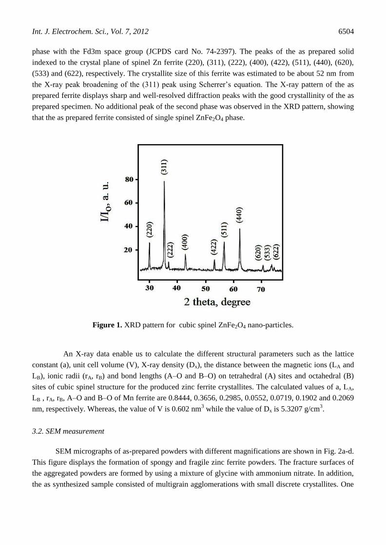
The XRD pattern of the as synthesized solid is shown in Fig.1. This figure showed that the as
prepared sample consisted entirely of nano-crystalline ZnFe2O4 particles. Indeed, the XRD pattern
contains ten sharp lines coincide with the standard data of the cubic spinel Zn ferrite (Franklinite)
cos
B d
Int. J. Electrochem. Sci., Vol. 7, 2012
6504
phase with the Fd3m space group (JCPDS card No. 74-2397). The peaks of the as prepared solid
indexed to the crystal plane of spinel Zn ferrite (220), (311), (222), (400), (422), (511), (440), (620),
(533) and (622), respectively. The crystallite size of this ferrite was estimated to be about 52 nm from
the X-ray peak broadening of the (311) peak using Scherrer’s equation. The X-ray pattern of the as
prepared ferrite displays sharp and well-resolved diffraction peaks with the good crystallinity of the as
prepared specimen. No additional peak of the second phase was observed in the XRD pattern, showing
that the as prepared ferrite consisted of single spinel ZnFe2O4 phase.
Figure 1. XRD pattern for cubic spinel ZnFe2O4 nano-particles.
An X-ray data enable us to calculate the different structural parameters such as the lattice
constant (a), unit cell volume (V), X-ray density (Dx), the distance between the magnetic ions (LA and
LB), ionic radii (rA, rB) and bond lengths (A–O and B–O) on tetrahedral (A) sites and octahedral (B)
sites of cubic spinel structure for the produced zinc ferrite crystallites. The calculated values of a, LA,
LB , rA, rB, A–O and B–O of Mn ferrite are 0.8444, 0.3656, 0.2985, 0.0552, 0.0719, 0.1902 and 0.2069
nm, respectively. Whereas, the value of V is 0.602 nm3 while the value of Dx is 5.3207 g/cm
3.
3.2. SEM measurement
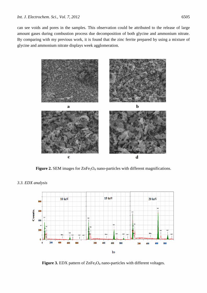
SEM micrographs of as-prepared powders with different magnifications are shown in Fig. 2a-d.
This figure displays the formation of spongy and fragile zinc ferrite powders. The fracture surfaces of
the aggregated powders are formed by using a mixture of glycine with ammonium nitrate. In addition,
the as synthesized sample consisted of multigrain agglomerations with small discrete crystallites. One
Int. J. Electrochem. Sci., Vol. 7, 2012
6505
can see voids and pores in the samples. This observation could be attributed to the release of large
amount gases during combustion process due decomposition of both glycine and ammonium nitrate.
By comparing with my previous work, it is found that the zinc ferrite prepared by using a mixture of
glycine and ammonium nitrate displays week agglomeration.
Figure 2. SEM images for ZnFe2O4 nano-particles with different magnifications.
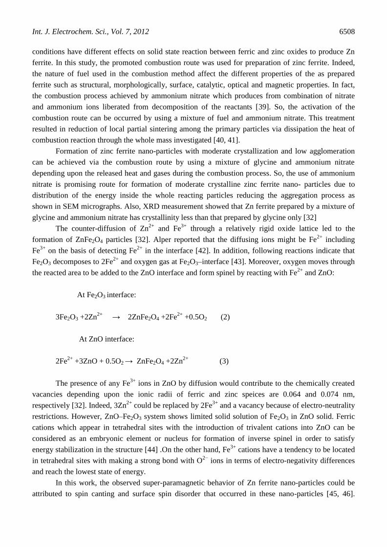
3.3. EDX analysis
Figure 3. EDX pattern of ZnFe2O4 nano-particles with different voltages.

Int. J. Electrochem. Sci., Vol. 7, 2012
6506
Figure 4. EDX pattern of ZnFe2O4 nano-particles with different areas.
Energy dispersive X-ray (EDX) analysis of the as prepared specimen was carried out at
different voltages and various areas on the surface of solid. Figs. 3 and 4 display EDX analysis with
different voltages and various areas, respectively. This finding shows almost homogeneous and
uniform distribution of Zn and Fe particles in the powder sample.
The effective atomic concentration of Zn, Fe and oxygen species on top surface layers of the
solid investigated are determined by EDX technique. The relative atomic abundance of Zn, Fe and O
species present in the uppermost surface and bulk layers of Zn ferrite are given in Tables 1 and 2.
Inspection of Table 1 revealed that the surface concentrations of Zn and Fe species at 10, 15 and 20
keV are very close to those in the bulk of the as synthesized solid. This observation may be reported to
the increase in the mobility of Zn and Fe species with subsequent an increase in the formation of Zn
ferrite. In addition, Table 2 showed that the surface concentrations of Zn, Fe and oxygen species at 20

Int. J. Electrochem. Sci., Vol. 7, 2012
6507
keV on different areas over the surface of specimen studied are much closed to each other. This
indicates the homogeneous distribution of Zn, Fe and O species in the investigated sample.
Table 1. The atomic abundance (surface and bulk) of elements measured at different voltages over the
same area for the as prepared solids.
Elements Atomic abundance (%)
Calculated
(Bulk)
Found (Surface)
10
keV
15
keV
20
keV
O
Fe
Zn
26.55
46.33
27.12
22.37
77.73
00.00
22.50
54.90
23.60
22.50
54.89
23.61
Table 2. The atomic abundance (surface) of elements measured at 20 keV and different areas over the
as prepared solids.
Elements Area
1
Area
2
Area
3
Area
4
O
Fe
Zn
22.36
25.75
22.67
22.36
24.81
22.67
22.36
25.34
24.10
22.36
25.05
23.10
3.4. Magnetic properties
The saturation magnetization (MS), remanent magnetization (Mr) and the coercivity of the as-
prepared powders were determined by measuring the magnetic hysteresis loop (not given) at room
temperature. The MS value was found to be 60 emu/g and the value Mr was 20 emu/g for the ZnFe2O4
sample. The corresponding squareness ratio (Mr/Ms) was found to be 0.333. In addition, the coercivity
of the investigated sample was found to 50 Oe. . It was found that the as-prepared Zn ferrite particles
in this work exhibited a saturation magnetization greater than that of ZnFe2O4, prepared by glycine as
fuel [32].
4. DISCUSSION
Spinel zinc ferrite, ZnFe2O4, based materials can be prepared via solid state reaction between
ZnO and Fe2O3 [28]. The enhancement of this reaction is controlled by thermal diffusion of Zn and Fe
cations through the zinc ferrite film which covers the surfaces of grains of reacting oxides (ZnO and
Fe2O3) and acts as energy barrier. The precursor compounds, preparation method and preparation
Int. J. Electrochem. Sci., Vol. 7, 2012
6508
conditions have different effects on solid state reaction between ferric and zinc oxides to produce Zn
ferrite. In this study, the promoted combustion route was used for preparation of zinc ferrite. Indeed,
the nature of fuel used in the combustion method affect the different properties of the as prepared
ferrite such as structural, morphologically, surface, catalytic, optical and magnetic properties. In fact,
the combustion process achieved by ammonium nitrate which produces from combination of nitrate
and ammonium ions liberated from decomposition of the reactants [39]. So, the activation of the
combustion route can be occurred by using a mixture of fuel and ammonium nitrate. This treatment
resulted in reduction of local partial sintering among the primary particles via dissipation the heat of
combustion reaction through the whole mass investigated [40, 41].
Formation of zinc ferrite nano-particles with moderate crystallization and low agglomeration
can be achieved via the combustion route by using a mixture of glycine and ammonium nitrate
depending upon the released heat and gases during the combustion process. So, the use of ammonium
nitrate is promising route for formation of moderate crystalline zinc ferrite nano- particles due to
distribution of the energy inside the whole reacting particles reducing the aggregation process as
shown in SEM micrographs. Also, XRD measurement showed that Zn ferrite prepared by a mixture of
glycine and ammonium nitrate has crystallinity less than that prepared by glycine only [32]
The counter-diffusion of Zn2+
and Fe3+
through a relatively rigid oxide lattice led to the
formation of ZnFe2O4 particles [32]. Alper reported that the diffusing ions might be Fe2+
including
Fe3+
on the basis of detecting Fe2+
in the interface [42]. In addition, following reactions indicate that
Fe2O3 decomposes to 2Fe2+
and oxygen gas at Fe2O3–interface [43]. Moreover, oxygen moves through
the reacted area to be added to the ZnO interface and form spinel by reacting with Fe2+
and ZnO:
At Fe2O3 interface:
3Fe2O3 +2Zn2+
→ 2ZnFe2O4 +2Fe2+
+0.5O2 (2)
At ZnO interface:
2Fe2+
+3ZnO + 0.5O2 → ZnFe2O4 +2Zn2+
(3)
The presence of any Fe3+
ions in ZnO by diffusion would contribute to the chemically created
vacancies depending upon the ionic radii of ferric and zinc speices are 0.064 and 0.074 nm,
respectively [32]. Indeed, 3Zn2+
could be replaced by 2Fe3+
and a vacancy because of electro-neutrality
restrictions. However, ZnO–Fe2O3 system shows limited solid solution of Fe2O3 in ZnO solid. Ferric
cations which appear in tetrahedral sites with the introduction of trivalent cations into ZnO can be
considered as an embryonic element or nucleus for formation of inverse spinel in order to satisfy
energy stabilization in the structure [44] .On the other hand, Fe3+
cations have a tendency to be located
in tetrahedral sites with making a strong bond with O2−
ions in terms of electro-negativity differences
and reach the lowest state of energy.
In this work, the observed super-paramagnetic behavior of Zn ferrite nano-particles could be
attributed to spin canting and surface spin disorder that occurred in these nano-particles [45, 46].
Int. J. Electrochem. Sci., Vol. 7, 2012
6509
Indeed, the zinc ferrite prepared by a mixture of glycine and ammonium nitrate has saturation
magnetization (60 emu/g) greater than that for Zn ferrite synthesized by glycine only (52 emu/g) due to
the redistribution of the reacting cations on A and B sites involved in the spinel Zn ferrite [32]. In other
words, the higher saturation magnetization of Zn ferrite prepared in this investigation could be
attributed migration of some Fe3+
ions from B site to A site via conversion of some Fe2+
ions to Fe3+
ions with subsequent increase in the FeA3+
–FeB3+
super-exchange interactions [47]. This conversion
brought about an increase in the saturation magnetization and a decrease in the crystallinity of zinc
ferrite prepared by using a mixture of glycine and ammonium nitrate comparing with that prepared by
using glycine only. This decrease in the crystallinity could be attributed to the contraction in the lattice.
This contraction may be due to the difference in the ionic radii of both ferric (0.076 nm) and ferrous
ions (0.064) [32]. The saturation magnetization of the as prepared ZnFe2O4 nano-particles is clearly
higher compared with the reported value of ∼5 emu/g for the bulk ZnFe2O4 [23].
The large value of magnetization observed in the present study shows that the cation
distribution changed from normal to mixed spinel type. Hence, the percentage of Fe3+
ions occupies
the tetrahedral sites which switches on the A–B super-exchange interaction and gives rise in the
magnetization. EXAFS studies conducted by Jeyadevan et al. support the presence of Zn2+
ion on the
B-sites [48]. Liganza found that 4% of the A-sites was occupied by Fe3+
ions [49]. The neutron
diffraction study of nanocrystalline ZnFe2O4 reports that the occupancy of Fe3+
ions at the A sites is
0.018 and 0.142 for the fine particles of diameters 96 and 29 nm, respectively [50]. However, it has
been reported that the spin disorder may occur on the surface of the nano-particles and the cores of the
nano-particles could be attributed to the vacant sub-lattice disorder sites (FeA3+
) and poor crystal
structure [51].
5. CONCLUSIONS
Using a mixture of glycine and ammonium nitrate as fuel resulted in formation of Zn ferrite
with moderate crystalline cubic spinel structure, homogeneously distributed nano- particles and nano-
scale size. Higher saturation magnetization (60 emu/g) and coercivity values (50 Oe) of Zn ferrite are
obtained by using a mixture of glycine and ammonium nitrate as fuel. These values are greater than
those for nano-magnetic Zn ferrite materials prepared by using glycine only.
ACKNOWLEDGEMENT
This project was supported by King Saud University, Deanship of Scientific Research, College of
Science Research Centre.
References
1. N. M. Deraz, S. Shaban, J. Analyt. Appl. Pyrolysis, 86 (2009) 173.
2. N. M. Deraz, M. K. El- Aiashy, Suzan. A. Ali, Adsorp. Sci. Technol. 27(2009)803.
3. N.M. Deraz, S.A. Shaban, A. Alarifi, J. Saudi Chemical Society 14(2010)357.
Int. J. Electrochem. Sci., Vol. 7, 2012
6510
4. Y. KÖseoglu, A. Baykal, F. Gzüak, H. Kavas, Polyhedron 28 (2009) 2887.
5. Shao-Wen Cao, Ying-Jie Zhu, Guo-Feng Cheng, Yue-Hong Huang, J. Hazard. Mater. 171 (2009)
431.
6. Z.H. Zhou, J.M. Xue, J. Wang, H.S.O. Chan, T. Yu, Z.X. Shen, J. Appl. Phys. 91(2002) 6015.
7. Y. KÖseoglu, F. Yıldız, B. Aktas, G.S. Alvarez, M. Toprak, M. Muhammed, Phys. Status Solidi B
242 (2005) 1712.
8. M. Tsuji, Y. Wada, T. Yamamoto, T. Sano, Y. Tamaura, J. Mater. Sci. Lett. 15 (1996)156.
9. J.W. Choung, Z. Xu, J.A. Finch, Ind. Eng. Chem. Res. 38 (1999) 4689.
10. A.J. Rondinone, A.C.S. Samia, Z.J. Zhang, J. Phys. Chem. B (2000) 7919.
11. L.D. Tung, V. Kolesnichenko, G. Caruntu, D. Caruntu, Y. Remond, V.O. Golub, C.J. O’Connor, L.
Spinu, Physica B 319 (2002) 116.
12. X. Chu, X. Liu, G. Meng, Sens. Actuators B 55 (1999) 12.
13. U. Steinike, K. Tkacova, J. Mater. Synth. Process. 8 (2000) 199.
14. S. Zhuykov, T. Ona, N. Yamazoe, N. Miura, Solid State Ionics 152–153 (2002) 801.
15. S.B. Li, G.X. Lu, New J. Chem. 16 (1992) 517.
16. G.X. Lu, S.B. Li, Int. Hydrogen Energy 17 (1992) 767.
17. Rahmatollah Rahimi, Hamed Kerdari, Mahboubeh Rabbani, Majid Shafiee, Desalination 280
(2011) 412.
18. S. Zhuiykov, M. Muta, T. Ono, M. Hasei, N. Yamazoe, N. Miura, Electrochem. Solid-State Lett. 4
(2001) H19.
19. S. Zhuiykov, T. Ono, N. Yamazoe, N. Miura, Solid State Ionics 152–153 (2002)801.
20. G. Zhang, L. Chunsheng, C. Fangyi, J. Chen, Sensors & Actuators B 120 (2007)403.
21. K. Arshak, I. Gaidan, Mater. Sci. Eng. B 118 (2005) 44.
22. J.Z. Zhang, D.H. Chen, L. Chen, Sensors Mater. 5 (2006) 227.
23. T.M. Clark, B.J. Evans, IEEE Trans. Mag. 33 (1997) 3745.
24. J.L. Mart´ın de Vidales, A. L´opez-Delgado, E. Vila, F.A. L´opez, J. Alloys Compd. 287 (1999)
276.
25. S.H. Yu, T. Fujino, M. Yoshimura, J. Magn. Magn. Mater. 256 (2003)420.
26. J.A. Toledo, M.A. Valenzuela, P. Bosch, H. Armendáriz, A. Montoya, N. Nava, A. V´azquez,
Appl. Catal. A 198 (2000) 94.
27. N.S. Gajbhiye, U. Bhattacharya, V.S. Darshane, Thermochim. Acta 264 (1995) 219.
28. C. N. Chinnasamy, A. Narayanasamy, N. Ponpandian, K. Chattopadhyay, H. Guerault, J. M.
Greneche, J. Phys.Condens. Matter. 12 (2000) 7795.
29. C. N. Chinnasamy, A. Narayanasamy, N. Ponpandian, K. Chattopadhyay, H. Guerault, J. M.
Greneche, Scripta Mater. 44 (2001) 1407.
30. L. Néel, Comput. Rend. 252 (1961) 4075.
31. N. M. Deraz, A. Alarifi, Int. J. Electrochem. Sci., 7 (2012) 3798.
32. N. M. Deraz, A. Alarifi, Polyhedron, 28(2009) 4122.
33. N. M. Deraz, A. Alarifi, Int. J. Electrochem. Sci., 7 (2012) 3809.
34. N. M. Deraz, J. Analyt. Appl. Pyrolysis, 91(2011) 48.
35. T. Sato, K. Haneda, M. Seki, T. Iijima, Appl. Phys. A 50 (1990) 13.
36. P.B. Avakyan, E.L. Nersisyan, M.D. Nersesyan, Int. J. SHS. 4(1995) 79.
37. Y. Li, J.P. Zhao, J.C. Han, Mater. Res. Bull. 37(2002) 583.
38. B.D. Cullity, Elements of X-ray Diffraction, Addison-Wesly Publishing Co. Inc. 1976 (Chapter
14).
39. P. Priyadharsini, A. Pradeep, G. Chandrasekaran, J. Magn Magn. Mater. 321(2009)1898.
40. N. M. Deraz, Current Applied Physics 12 (2012) 928.
41. N. M. Deraz, Int. J. Electrochem. Sci., 7 (2012) 4608.
42. Alper, High Temperature Oxides, Academic Press, New York, 1970.
43. A. Azhari, M. Sharif Sh., F. Golestanifard, A. Saberi, Mater. Chem. Physics 124 (2010) 658.

Int. J. Electrochem. Sci., Vol. 7, 2012
6511
44. S.L. Blank, J.A. Pask, J. Am. Ceram. Soc. 52 (1969) 669.
45. Z. Gu, X. Xiang, G. Fan, F. Li, J. Phys. Chem. C 112 (2008) 18459.
46. L. Ai, J. Jiang, Curr. Appl. Phys. 10 (2010) 284.
47. L. Jianjun, Y. Hongming, L. Guodong, L. Yanju, L. Jinsong, J. Magn. Magn. Mater.
322(2010)3396.
48. B. Jeyadevan, K. Tohj, K. Nakatsuka, J. Appl. Phys. 76 (1994) 6325.
49. S. Liganza, Phys. Stat. Sol. 75 (1976) 315.
50. F.K. Lotzering, J. Phys. Chem. Solids 27 (1996) 139.
51. M. P. Morales, S. Veintemillas-Verdaguer, M. I. Montero, C. J. Serna, A. Roig, L. Casas, B.
Martȋnez, F. Sandiumenge, Chem. Mater. 11(1999)3058.
© 2012 by ESG (www.electrochemsci.org)