microrna-143 regulates human osteosarcoma metastasis by regulating matrix metalloprotease-13...
TRANSCRIPT

original article© The American Society of Gene & Cell Therapy
Molecular Therapy vol. 19 no. 6, 1123–1130 june 2011 1123
Pulmonary metastases are the main cause of death in patients with osteosarcoma, however, the molecular mechanisms of metastasis are not well understood. To detect lung metastasis-related microRNA (miRNA) in human osteosarcoma, we compared parental (HOS) and its subclone (143B) human osteosarcoma cell lines show-ing lung metastasis in a mouse model. miR-143 was the most downregulated miRNA (P < 0.01), and transfection of miR-143 into 143B significantly decreased its inva-siveness, but not cell proliferation. Noninvasive optical imaging technologies revealed that intravenous injec-tion of miR-143, but not negative control miRNA, sig-nificantly suppressed lung metastasis of 143B (P < 0.01). To search for miR-143 target mRNA in 143B, microar-ray analyses were performed using an independent RNA pool extracted by two different comprehensive miR-143-target mRNA collecting systems. Western blot analyses revealed that MMP-13 was mostly protein downregu-lated by miR-143. Immunohistochemistry using clinical samples clearly revealed MMP-13-positive cells in lung metastasis-positive cases, but not in at least three cases showing higher miR-143 expression in the no meta-stasis group. Taken together, these data indicated that the downregulation of miR-143 correlates with the lung metastasis of human osteosarcoma cells by promoting cellular invasion, probably via MMP-13 upregulation, suggesting that miRNA could be used to develop new molecular targets for osteosarcoma metastasis.
Received 28 December 2010; accepted 22 February 2011; published online 22 March 2011. doi:10.1038/mt.2011.53
IntroductIonOsteosarcoma is the most common primary bone malignancy and accounts for 60% of all malignant childhood bone tumors.1 The age distribution is bimodal: the first major peak occurring dur-ing the second decade of life, and the second much smaller peak
being observed in patients over 50 years of age. The distal femo-ral and proximal tibial metaphyses are the most common sites for osteosarcoma. Approximately 50% of cases are localized in the knee region.2 With combined treatment (neoadjuvant chemo-therapy, surgery, and adjuvant chemotherapy), the 5-year survival of patients with no metastatic disease at diagnosis is 60–70%;3–5 however, for patients who present with metastatic disease, the out-come is far worse at <30% survival.6 Pulmonary metastasis is the predominant site of osteosarcoma recurrence and the most com-mon cause of death. Unfortunately, survival has not improved for 20 years despite multiple clinical trials with increased intensity, and further gains with refinements of cytotoxic chemotherapy regimens alone are unlikely; therefore, for better prognosis, new therapeutic targets and approaches must be sought to suppress pulmonary metastasis of osteosarcoma.
MicroRNA (miRNA) belongs to a class of endogenously expressed, non-coding small RNA and contains about 22 nucleo-tides. Based on miRBase release 16.0, >1,000 human miRNA have been registered with a large number being evolutionarily con-served.7 It has been shown that miRNA can regulate the expres-sion of protein-coding genes at the post-transcriptional level through imperfect base pairing with the 3′-untranslated region (3′-UTR) of target mRNA.8 miRNA is predicted to regulate the expression of at least 30% of all genes.9 Growing evidence sug-gests that deregulation of miRNA may contribute to many types of human diseases, including cancer. Errors in the expression of miRNA have been observed in various types of cancers10,11 and are also associated with the clinical outcome of cancer patients.12,13 Consistently, miRNA has been implicated in the regulation of various cellular processes that are often deregulated during tumor development and progression,8,14–17 suggesting that miRNA might be a target for cancer therapy.
The most direct way for molecules to correct altered miRNA expression is by treatment with RNA oligonucleotides. Therapeutic potentials using RNA oligonucleotides have been proposed, although our understanding of the role of miRNA in cancer is still very limited. There are two possible approaches: blocking oncogenic
Correspondence: Mitsuhiko Osaki, Division of Molecular Genetics and Biofunction, Tottori University Graduate School of Medical Science, 86 Nishi-cho, Yonago, Tottori 683–8503, Japan. E-mail: [email protected]
MicroRNA-143 Regulates Human Osteosarcoma Metastasis by Regulating Matrix Metalloprotease-13 ExpressionMitsuhiko Osaki1–3, Fumitaka Takeshita1, Yui Sugimoto2, Nobuyoshi Kosaka1, Yusuke Yamamoto1, Yusuke Yoshioka1, Eisuke Kobayashi4, Tesshi Yamada4, Akira Kawai5, Toshiaki Inoue3, Hisao Ito6, Mitsuo Oshimura2,3 and Takahiro Ochiya1
1Division of Molecular and Cellular Medicine, National Cancer Center Research Institute, Tokyo, Japan; 2Division of Molecular Genetics and Biofunction, Tottori University Graduate School of Medical Science, Tottori, Japan; 3Chromosome Engineering Research Center, Tottori University, Tottori, Japan; 4Division of Chemotherapy and Clinical Reasearch, National Cancer Center Research Institute, Tokyo, Japan; 5Orthopedics Division, National Cancer Center Hospital, Tokyo, Japan; 6Division of Organ Pathology, Tottori University, Tottori, Japan
1124 www.moleculartherapy.org vol. 19 no. 6 june 2011
© The American Society of Gene & Cell TherapymiR-143 Suppresses Lung Metastasis of Osteosarcoma
miRNA by anti-miRNA oligonucleotides or replacement of miRNA with tumor suppressor activity by miRNA mimetics. In fact, in vitro studies have revealed that anti-miR-17-5p treatment halts the growth of a human neuroblastoma cell line, LAN-5, overexpress-ing miR-17-5p.18 Si et al. also reported that anti-miR-21 inhibited cell growth via increased apoptosis and decreased cell proliferation, which could partly be due to the downregulation of antiapoptotic Bcl-2 in a human breast cancer cell line, MCF-7.19 Recently, Ma et al. reported that systemic administration of miR-10b antagomir inhibited lung metastasis of mouse breast cancer cells in a mouse model.20 On the other hand, it has been reported that cell prolifera-tion or invasion was suppressed by miRNA mimetics transfection into human cancer cells. For example, the introduction of synthe-sized miR-143 or miR-145 into a human B-cell lymphoma cell line, Raji, resulted in significant growth inhibition that occurred in a dose-dependent manner.21 Crawford et al. reported that treatment with miR-126 decreased adhesion, migration, and the invasion of a human nonsmall-cell lung carcinoma cell line, H1703.22 Valastyan et al. found that overexpression of miR-31 independently inhibited the invasive capacity of MDA-MB-231 breast cancer cells, extrava-sation into or survival in the lung parenchyma, and metastatic colo-nization.23 Moreover, Tazawa et al. demonstrated in a mouse model that direct intratumoral injection of a miR-34a/atelocollagen com-plex successfully suppressed the growth of tumors derived from human colon cancer cells.24 Furthermore, significant reduction of the tumor volume was observed until day 6 after miR-34a admin-istration. Interestingly, the authors showed that the expression of miR-34a was downregulated in more than one-third of human colon cancers compared with counterpart normal colon mucosa. Therefore, these data suggested that restoring decreased miRNA in cancer cells was able to suppress the progression of cancer in vivo.
Our goal is to understand the mechanisms of metastases and, based on this knowledge, identify new targets that can be used for the development of new molecular markers and therapeu-tic approaches to inhibit metastasis from osteosarcoma. In this study, we explored miRNA and its target mRNA associated with cell invasion of osteosarcoma cells in vitro using two human oste-osarcoma cell lines, HOS and 143B, and aimed to clarify whether spontaneous lung metastasis from osteosarcoma could be sup-pressed by restoring or blocking miRNA in vivo using a mouse model.
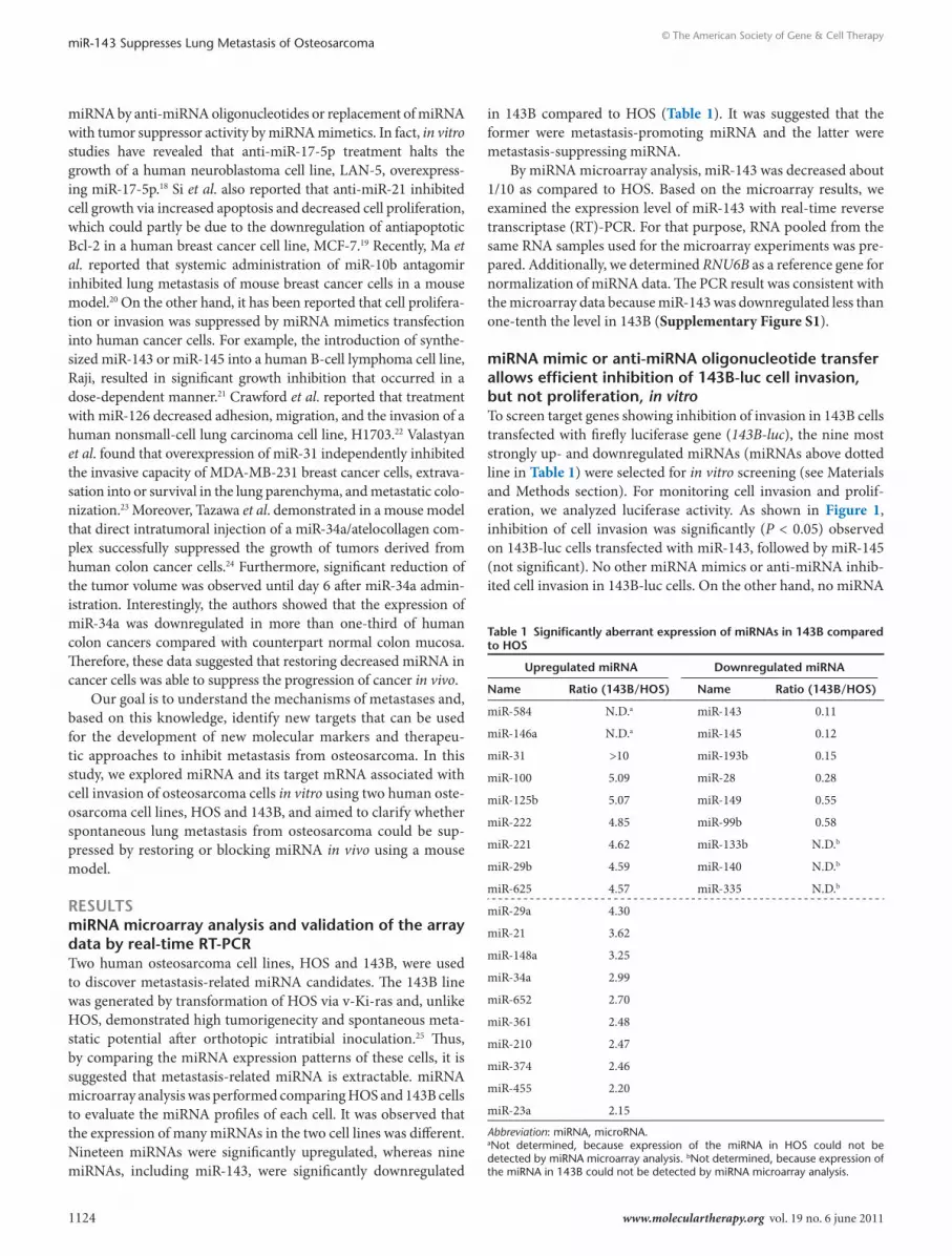
resultsmirnA microarray analysis and validation of the array data by real-time rt-PcrTwo human osteosarcoma cell lines, HOS and 143B, were used to discover metastasis-related miRNA candidates. The 143B line was generated by transformation of HOS via v-Ki-ras and, unlike HOS, demonstrated high tumorigenecity and spontaneous meta-static potential after orthotopic intratibial inoculation.25 Thus, by comparing the miRNA expression patterns of these cells, it is suggested that metastasis-related miRNA is extractable. miRNA microarray analysis was performed comparing HOS and 143B cells to evaluate the miRNA profiles of each cell. It was observed that the expression of many miRNAs in the two cell lines was different. Nineteen miRNAs were significantly upregulated, whereas nine miRNAs, including miR-143, were significantly downregulated
in 143B compared to HOS (Table 1). It was suggested that the former were metastasis-promoting miRNA and the latter were metastasis-suppressing miRNA.
By miRNA microarray analysis, miR-143 was decreased about 1/10 as compared to HOS. Based on the microarray results, we examined the expression level of miR-143 with real-time reverse transcriptase (RT)-PCR. For that purpose, RNA pooled from the same RNA samples used for the microarray experiments was pre-pared. Additionally, we determined RNU6B as a reference gene for normalization of miRNA data. The PCR result was consistent with the microarray data because miR-143 was downregulated less than one-tenth the level in 143B (Supplementary Figure S1).
mirnA mimic or anti-mirnA oligonucleotide transfer allows efficient inhibition of 143B-luc cell invasion, but not proliferation, in vitroTo screen target genes showing inhibition of invasion in 143B cells transfected with firefly luciferase gene (143B-luc), the nine most strongly up- and downregulated miRNAs (miRNAs above dotted line in Table 1) were selected for in vitro screening (see Materials and Methods section). For monitoring cell invasion and prolif-eration, we analyzed luciferase activity. As shown in Figure 1, inhibition of cell invasion was significantly (P < 0.05) observed on 143B-luc cells transfected with miR-143, followed by miR-145 (not significant). No other miRNA mimics or anti-miRNA inhib-ited cell invasion in 143B-luc cells. On the other hand, no miRNA
table 1 significantly aberrant expression of mirnAs in 143B compared to Hos
upregulated mirnA downregulated mirnA
name ratio (143B/Hos) name ratio (143B/Hos)
miR-584 N.D.a miR-143 0.11
miR-146a N.D.a miR-145 0.12
miR-31 >10 miR-193b 0.15
miR-100 5.09 miR-28 0.28
miR-125b 5.07 miR-149 0.55
miR-222 4.85 miR-99b 0.58
miR-221 4.62 miR-133b N.D.b
miR-29b 4.59 miR-140 N.D.b
miR-625 4.57 miR-335 N.D.b
miR-29a 4.30
miR-21 3.62
miR-148a 3.25
miR-34a 2.99
miR-652 2.70
miR-361 2.48
miR-210 2.47
miR-374 2.46
miR-455 2.20
miR-23a 2.15
Abbreviation: miRNA, microRNA.aNot determined, because expression of the miRNA in HOS could not be detected by miRNA microarray analysis. bNot determined, because expression of the miRNA in 143B could not be detected by miRNA microarray analysis.
Molecular Therapy vol. 19 no. 6 june 2011 1125
© The American Society of Gene & Cell TherapymiR-143 Suppresses Lung Metastasis of Osteosarcoma
mimics or anti-miRNA used in this assay significantly affected cell proliferation (Supplementary Figure S2). These results revealed that miR-143 might be the miRNA with the most potential to sup-press the metastasis of 143B-luc cells.
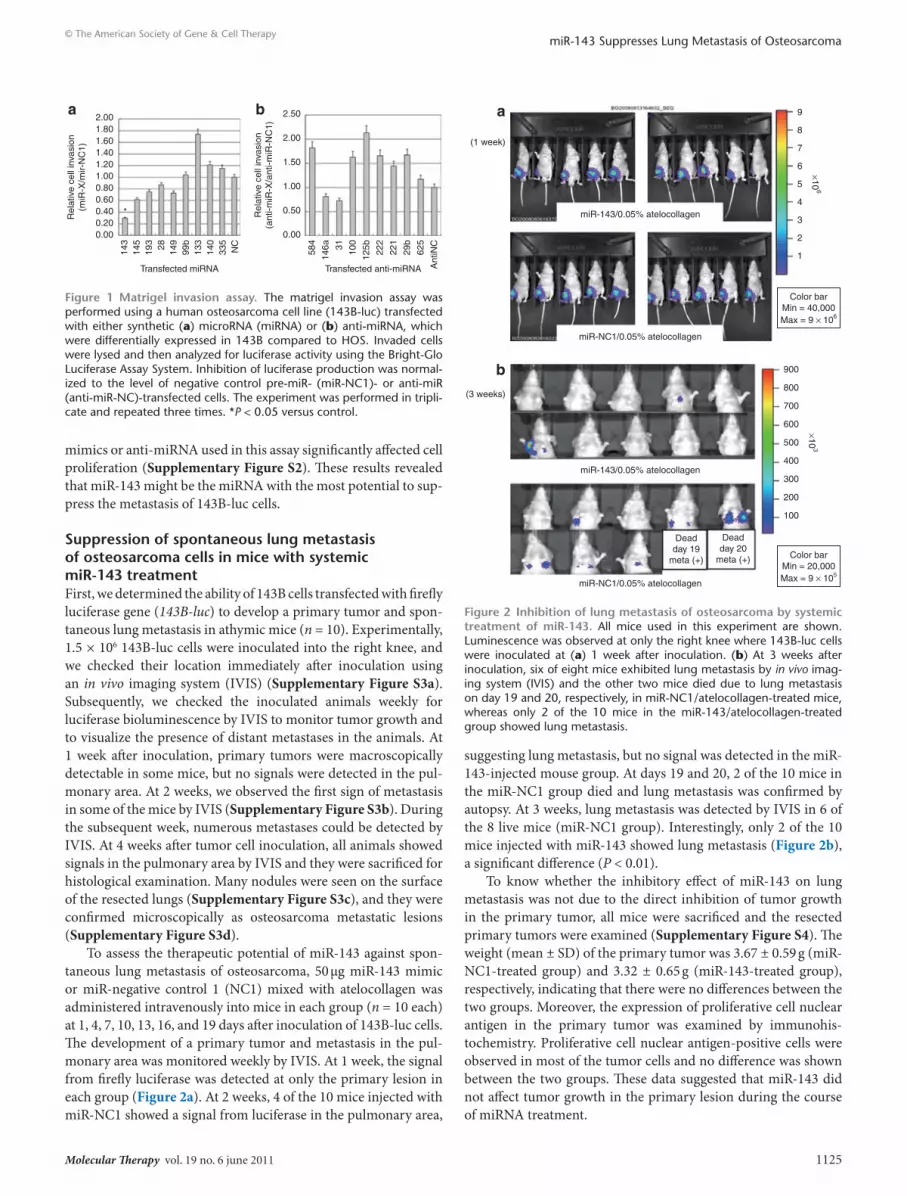
suppression of spontaneous lung metastasis of osteosarcoma cells in mice with systemic mir-143 treatmentFirst, we determined the ability of 143B cells transfected with firefly luciferase gene (143B-luc) to develop a primary tumor and spon-taneous lung metastasis in athymic mice (n = 10). Experimentally, 1.5 × 10⁶ 143B-luc cells were inoculated into the right knee, and we checked their location immediately after inoculation using an in vivo imaging system (IVIS) (Supplementary Figure S3a). Subsequently, we checked the inoculated animals weekly for luciferase bioluminescence by IVIS to monitor tumor growth and to visualize the presence of distant metastases in the animals. At 1 week after inoculation, primary tumors were macroscopically detectable in some mice, but no signals were detected in the pul-monary area. At 2 weeks, we observed the first sign of metastasis in some of the mice by IVIS (Supplementary Figure S3b). During the subsequent week, numerous metastases could be detected by IVIS. At 4 weeks after tumor cell inoculation, all animals showed signals in the pulmonary area by IVIS and they were sacrificed for histological examination. Many nodules were seen on the surface of the resected lungs (Supplementary Figure S3c), and they were confirmed microscopically as osteosarcoma metastatic lesions (Supplementary Figure S3d).
To assess the therapeutic potential of miR-143 against spon-taneous lung metastasis of osteosarcoma, 50 μg miR-143 mimic or miR-negative control 1 (NC1) mixed with atelocollagen was administered intravenously into mice in each group (n = 10 each) at 1, 4, 7, 10, 13, 16, and 19 days after inoculation of 143B-luc cells. The development of a primary tumor and metastasis in the pul-monary area was monitored weekly by IVIS. At 1 week, the signal from firefly luciferase was detected at only the primary lesion in each group (Figure 2a). At 2 weeks, 4 of the 10 mice injected with miR-NC1 showed a signal from luciferase in the pulmonary area,
suggesting lung metastasis, but no signal was detected in the miR-143-injected mouse group. At days 19 and 20, 2 of the 10 mice in the miR-NC1 group died and lung metastasis was confirmed by autopsy. At 3 weeks, lung metastasis was detected by IVIS in 6 of the 8 live mice (miR-NC1 group). Interestingly, only 2 of the 10 mice injected with miR-143 showed lung metastasis (Figure 2b), a significant difference (P < 0.01).
To know whether the inhibitory effect of miR-143 on lung metastasis was not due to the direct inhibition of tumor growth in the primary tumor, all mice were sacrificed and the resected primary tumors were examined (Supplementary Figure S4). The weight (mean ± SD) of the primary tumor was 3.67 ± 0.59 g (miR-NC1-treated group) and 3.32 ± 0.65 g (miR-143-treated group), respectively, indicating that there were no differences between the two groups. Moreover, the expression of proliferative cell nuclear antigen in the primary tumor was examined by immunohis-tochemistry. Proliferative cell nuclear antigen-positive cells were observed in most of the tumor cells and no difference was shown between the two groups. These data suggested that miR-143 did not affect tumor growth in the primary lesion during the course of miRNA treatment.
2.001.801.601.401.201.000.800.600.400.200.00
Rel
ativ
e ce
ll in
vasi
on(m
iR-X
/mir-
NC
1)
143
145
193 28 149
99b
133
140
335
NC
Transfected miRNA
a
Rel
ativ
e ce
ll in
vasi
on(a
nti-m
iR-X
/ant
i-miR
-NC
1)
584
146a 31 100
125b 222
221
29b
625
Ant
iNC
Transfected anti-miRNA
2.50
2.00
1.50
1.00
0.50
0.00
b
*
Figure 1 Matrigel invasion assay. The matrigel invasion assay was performed using a human osteosarcoma cell line (143B-luc) transfected with either synthetic (a) microRNA (miRNA) or (b) anti-miRNA, which were differentially expressed in 143B compared to HOS. Invaded cells were lysed and then analyzed for luciferase activity using the Bright-Glo Luciferase Assay System. Inhibition of luciferase production was normal-ized to the level of negative control pre-miR- (miR-NC1)- or anti-miR (anti-miR-NC)-transfected cells. The experiment was performed in tripli-cate and repeated three times. *P < 0.05 versus control.
miR-143/0.05% atelocollagen
miR-NC1/0.05% atelocollagen
miR-143/0.05% atelocollagen
miR-NC1/0.05% atelocollagen
Color barMin = 40,000Max = 9 × 10⁶
Deadday 19
meta (+)
Deadday 20
meta (+)
900
800
700
600
500
400
300
200
100
×103
×106
9
8
7
6
5
4
3
2
1
(1 week)
(3 weeks)
a
b
Color barMin = 20,000Max = 9 × 10⁵
Figure 2 Inhibition of lung metastasis of osteosarcoma by systemic treatment of mir-143. All mice used in this experiment are shown. Luminescence was observed at only the right knee where 143B-luc cells were inoculated at (a) 1 week after inoculation. (b) At 3 weeks after inoculation, six of eight mice exhibited lung metastasis by in vivo imag-ing system (IVIS) and the other two mice died due to lung metastasis on day 19 and 20, respectively, in miR-NC1/atelocollagen-treated mice, whereas only 2 of the 10 mice in the miR-143/atelocollagen-treated group showed lung metastasis.
1126 www.moleculartherapy.org vol. 19 no. 6 june 2011
© The American Society of Gene & Cell TherapymiR-143 Suppresses Lung Metastasis of Osteosarcoma
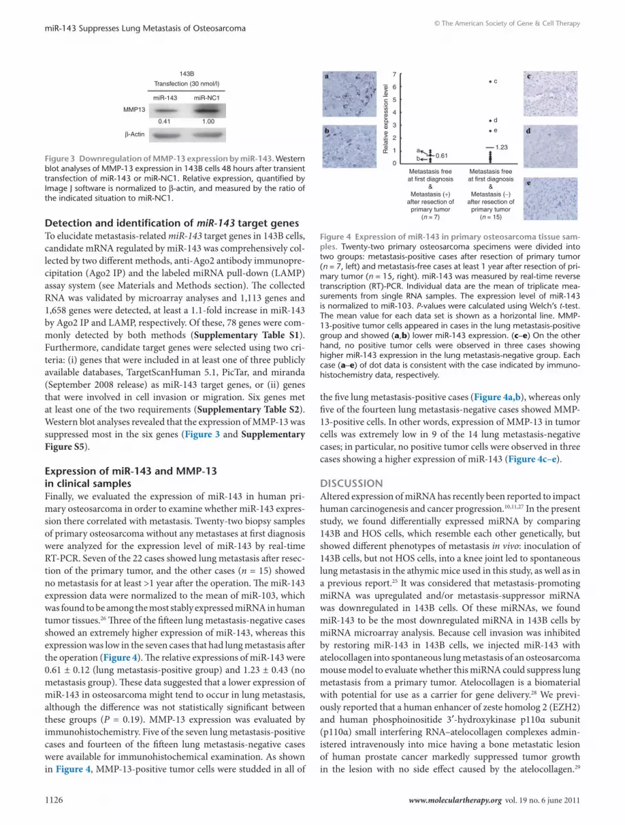
detection and identification of miR-143 target genesTo elucidate metastasis-related miR-143 target genes in 143B cells, candidate mRNA regulated by miR-143 was comprehensively col-lected by two different methods, anti-Ago2 antibody immunopre-cipitation (Ago2 IP) and the labeled miRNA pull-down (LAMP) assay system (see Materials and Methods section). The collected RNA was validated by microarray analyses and 1,113 genes and 1,658 genes were detected, at least a 1.1-fold increase in miR-143 by Ago2 IP and LAMP, respectively. Of these, 78 genes were com-monly detected by both methods (Supplementary Table S1). Furthermore, candidate target genes were selected using two cri-teria: (i) genes that were included in at least one of three publicly available databases, TargetScanHuman 5.1, PicTar, and miranda (September 2008 release) as miR-143 target genes, or (ii) genes that were involved in cell invasion or migration. Six genes met at least one of the two requirements (Supplementary Table S2). Western blot analyses revealed that the expression of MMP-13 was suppressed most in the six genes (Figure 3 and Supplementary Figure S5).
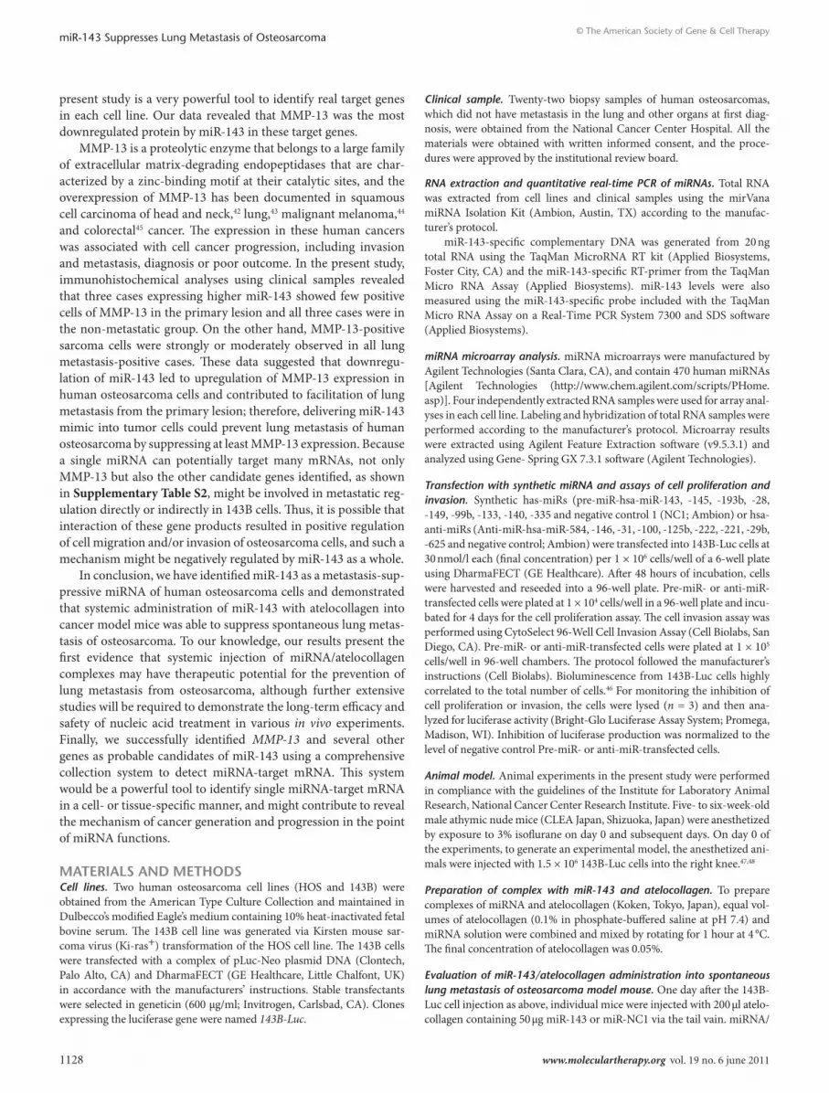
expression of mir-143 and MMP-13 in clinical samplesFinally, we evaluated the expression of miR-143 in human pri-mary osteosarcoma in order to examine whether miR-143 expres-sion there correlated with metastasis. Twenty-two biopsy samples of primary osteosarcoma without any metastases at first diagnosis were analyzed for the expression level of miR-143 by real-time RT-PCR. Seven of the 22 cases showed lung metastasis after resec-tion of the primary tumor, and the other cases (n = 15) showed no metastasis for at least >1 year after the operation. The miR-143 expression data were normalized to the mean of miR-103, which was found to be among the most stably expressed miRNA in human tumor tissues.26 Three of the fifteen lung metastasis-negative cases showed an extremely higher expression of miR-143, whereas this expression was low in the seven cases that had lung metastasis after the operation (Figure 4). The relative expressions of miR-143 were 0.61 ± 0.12 (lung metastasis-positive group) and 1.23 ± 0.43 (no metastasis group). These data suggested that a lower expression of miR-143 in osteosarcoma might tend to occur in lung metastasis, although the difference was not statistically significant between these groups (P = 0.19). MMP-13 expression was evaluated by immunohistochemistry. Five of the seven lung metastasis-positive cases and fourteen of the fifteen lung metastasis-negative cases were available for immunohistochemical examination. As shown in Figure 4, MMP-13-positive tumor cells were studded in all of
the five lung metastasis-positive cases (Figure 4a,b), whereas only five of the fourteen lung metastasis-negative cases showed MMP-13-positive cells. In other words, expression of MMP-13 in tumor cells was extremely low in 9 of the 14 lung metastasis-negative cases; in particular, no positive tumor cells were observed in three cases showing a higher expression of miR-143 (Figure 4c–e).
dIscussIonAltered expression of miRNA has recently been reported to impact human carcinogenesis and cancer progression.10,11,27 In the present study, we found differentially expressed miRNA by comparing 143B and HOS cells, which resemble each other genetically, but showed different phenotypes of metastasis in vivo: inoculation of 143B cells, but not HOS cells, into a knee joint led to spontaneous lung metastasis in the athymic mice used in this study, as well as in a previous report.25 It was considered that metastasis-promoting miRNA was upregulated and/or metastasis-suppressor miRNA was downregulated in 143B cells. Of these miRNAs, we found miR-143 to be the most downregulated miRNA in 143B cells by miRNA microarray analysis. Because cell invasion was inhibited by restoring miR-143 in 143B cells, we injected miR-143 with atelocollagen into spontaneous lung metastasis of an osteosarcoma mouse model to evaluate whether this miRNA could suppress lung metastasis from a primary tumor. Atelocollagen is a biomaterial with potential for use as a carrier for gene delivery.28 We previ-ously reported that a human enhancer of zeste homolog 2 (EZH2) and human phosphoinositide 3′-hydroxykinase p110α subunit (p110α) small interfering RNA–atelocollagen complexes admin-istered intravenously into mice having a bone metastatic lesion of human prostate cancer markedly suppressed tumor growth in the lesion with no side effect caused by the atelocollagen.29
Transfection (30 nmol/l)
143B
miR-143 miR-NC1
MMP13
β-Actin
0.41 1.00
Figure 3 downregulation of MMP-13 expression by mir-143. Western blot analyses of MMP-13 expression in 143B cells 48 hours after transient transfection of miR-143 or miR-NC1. Relative expression, quantified by Image J software is normalized to β-actin, and measured by the ratio of the indicated situation to miR-NC1.
7
6
5
4
3
2
1
0Metastasis freeat first diagnosis
&Metastasis (+)
after resection ofprimary tumor
(n = 7)
Metastasis freeat first diagnosis
&Metastasis (−)
after resection ofprimary tumor
(n = 15)
ab 0.61
d
e
c
1.23
Rel
ativ
e ex
pres
sion
leve
l
Figure 4 expression of mir-143 in primary osteosarcoma tissue sam-ples. Twenty-two primary osteosarcoma specimens were divided into two groups: metastasis-positive cases after resection of primary tumor (n = 7, left) and metastasis-free cases at least 1 year after resection of pri-mary tumor (n = 15, right). miR-143 was measured by real-time reverse transcription (RT)-PCR. Individual data are the mean of triplicate mea-surements from single RNA samples. The expression level of miR-143 is normalized to miR-103. P-values were calculated using Welch’s t-test. The mean value for each data set is shown as a horizontal line. MMP-13-positive tumor cells appeared in cases in the lung metastasis-positive group and showed (a,b) lower miR-143 expression. (c–e) On the other hand, no positive tumor cells were observed in three cases showing higher miR-143 expression in the lung metastasis-negative group. Each case (a–e) of dot data is consistent with the case indicated by immuno-histochemistry data, respectively.

Molecular Therapy vol. 19 no. 6 june 2011 1127
© The American Society of Gene & Cell TherapymiR-143 Suppresses Lung Metastasis of Osteosarcoma
Recently, we also reported that systemic administration of miR-16 with atelocollagen successfully regressed prostate cancer in a bone metastatic lesion in a mouse model.30 Thus, for prevention of lung metastasis from osteosarcoma, a new atelocollagen-mediated systemic delivery method could be a reliable and safe approach to achieve maximal function of miRNA in vivo, as well as small interfering RNA. In the present study, systemic administration of miR-143 with atelocollagen surprisingly suppressed lung metasta-sis in a spontaneous lung metastatic mouse model. On the other hand, treatment with miR-143 did not affect tumor growth in a primary lesion in an in vivo model. These data are consistent with in vitro data demonstrating that miR-143 transfection into 143B cells could suppress cell invasion but not cell proliferation, sug-gesting that miR-143 might specifically regulate the invasion and/or migration signal pathway(s) of osteosarcoma cells.
New approaches that can complement and improve on cur-rent strategies for the prediction of prognosis are urgently needed. Many independent studies on different tissues have demonstrated that miRNA expression patterns correlated with the prognosis of cancer patients,17,31 which generally depends upon the occur-rence of metastasis, because ~90% of deaths from solid tumors are caused by metastasis. Therefore, the expression of miRNA that regulate cell adhesion, migration and invasion could be a good diagnostic marker to predict cancer prognosis. Ma et al. reported that miR-10b is highly expressed in metastatic breast cancer cells and positively regulates cell migration and invasion,32 and they also demonstrated that systemic administration of miR-10b antagomir inhibited lung metastasis of mouse breast cancer cells in a mouse model.20 Overexpression of miR-10b in otherwise nonmetastatic breast tumors initiates robust invasion and metas-tasis. Expression of miR-10b is induced by the transcription factor Twist, which is an epithelial-mesenchymal transition factor and is known to bind directly to the putative promoter of miR-10b. The miR-10b induced by twist inhibits translation of the mRNA encoding homeobox D10, resulting in increased expression of a well-characterized prometastatic gene, RhoC. Significantly, the level of miR-10b expression in primary breast carcinomas corre-lates with clinical progression. On the other hand, Tavazoie et al. showed that restoring the expression of miR-335 in a human breast cancer cell line MDA-MB-231 inhibited metastatic cell invasion.33 miR-335 suppresses metastasis and migration through targeting of the progenitor cell transcription factor SOX4 and extracellular matrix component tenascin C. Moreover, the expression of miR-335 is downregulated in the majority of primary breast tumors from patients who relapse, and hence loss of the expression of miR-335 is associated with poor distal metastasis-free survival. These reports suggested that altered expression of metastasis-associated miRNA could be used for the prediction of prognosis. In the present study, expression analysis of miR-143 using clini-cal osteosarcoma samples showed that the mean of the expres-sion level was two times higher in metastasis-free cases than in metastasis-positive cases. However, no statistical significance was shown, which might be because only three cases that showed a higher expression of miR-143 raised the mean in metastasis-free cases. In other words, however, it could be considered that osteo-sarcoma in which a relatively higher level of miR-143 is expressed might be considered a low risk for metastasis. It is suggested that
the expression level of miR-143 at a primary osteosarcoma lesion might be a prognostic marker for lung metastasis.
It has been reported that reduced expression of tumor- suppressor miRNA was caused by chromosome deletions, epi-genetical changes, abberant transcription, and disturbances in miRNA processing. Suzuki et al. reported that P53 enhances the post-transcriptional maturation of several miRNAs, including miR-143. P53 interacts with Drosha processing complex through association with DEAD-box RNA helicase p68. Thus, wild-type P53 could facilitate the processing of primary miR-143 to precur-sor miR-143, but mutated P53 interferes with functional assem-bly between Dorosha complex and P68, leading to attenuation of miR-143 processing activity in HCT116, a human colon cancer cell line.34 Another study showed that upregulation of KRAS leads to downregulation of miR-143 in human pancreatic cancer cell lines;35 however, the mechanism of this downregulation has not been investigated in osteosarcoma cells in detail. Thus, further studies are needed to reveal the precise mechanism of miR-143 downregulation in 143B cells. The downregulation of miR-143 expression was also reported in several human cancers, e.g., colorectal cancer,36 prostate cancer,37 cervical cancer,38 ovarian cancer,39 B-cell lymphoma;21 thus, it is considered that miR-143 is a tumor-suppressor miRNA. In these cancers, downregulation of miR-143 resulted in the promotion of cell proliferation or inhibi-tion of apoptosis, indicating that miR-143 acts as a suppressor on cell proliferation and viability. Akao et al. reported that the target gene of miR-143 was determined to be ERK5/MAPK7, the upregu-lation of which leads to cell growth via activation of c-Myc, in Raji cells, a human B-cell lymphoma cell line.21 Recently, another paper showed that miR-143 suppressed cell proliferation by inhib-iting KRAS translation in human colorectal cancer;40 however, our data showed that restoring miR-143 in human osteosarcoma cell 143B could suppress cell invasion, but not cell proliferation in in vitro and in vivo studies. Also, western blotting showed that the expression levels of KRAS and ERK5 did not change by transfect-ing miR-143 into 143B cells (Supplementary Figure S6). These data suggested that miR-143 might have different targets in a cell-type-dependent manner. Additionally, Tome et al. reported that in vivo transfer of the KRAS gene from high-metastatic cancer cells to coimplanted low-metastatic cancer cells enhanced lung metastasis of human osteosarcoma cells,41 indicating that KRAS is a key factor in the metastasis of osteosarcoma cells; however, our data showed that miR-143 could suppress lung metastasis of 143B cells without KRAS downregulation. Therefore, this might also indicate that miR-143-target genes play roles downstream of the KRAS-related metastasis-promoting pathway(s).
To find which targets are regulated by miR-143 in 143B cells, microarray analyses were performed after collecting target RNA by two independent comprehensive methods (Ago2 IP and LAMP), respectively. By extracting the common genes after the two different collection methods, 78 common genes were iden-tified in >1,000 genes. Of those, six genes matched the require-ments of (i) predicted genes by database (Target Scan, PicTar, or miRanda) or (ii) genes related to migration, invasion, or metas-tasis. Interestingly, protein expressions of at least four of the six genes were downregulated by miR-143 transfection in 143B cells, indicating that the miRNA-target gene detection system in the
1128 www.moleculartherapy.org vol. 19 no. 6 june 2011
© The American Society of Gene & Cell TherapymiR-143 Suppresses Lung Metastasis of Osteosarcoma
present study is a very powerful tool to identify real target genes in each cell line. Our data revealed that MMP-13 was the most downregulated protein by miR-143 in these target genes.
MMP-13 is a proteolytic enzyme that belongs to a large family of extracellular matrix-degrading endopeptidases that are char-acterized by a zinc-binding motif at their catalytic sites, and the overexpression of MMP-13 has been documented in squamous cell carcinoma of head and neck,42 lung,43 malignant melanoma,44 and colorectal45 cancer. The expression in these human cancers was associated with cell cancer progression, including invasion and metastasis, diagnosis or poor outcome. In the present study, immunohistochemical analyses using clinical samples revealed that three cases expressing higher miR-143 showed few positive cells of MMP-13 in the primary lesion and all three cases were in the non-metastatic group. On the other hand, MMP-13-positive sarcoma cells were strongly or moderately observed in all lung metastasis-positive cases. These data suggested that downregu-lation of miR-143 led to upregulation of MMP-13 expression in human osteosarcoma cells and contributed to facilitation of lung metastasis from the primary lesion; therefore, delivering miR-143 mimic into tumor cells could prevent lung metastasis of human osteosarcoma by suppressing at least MMP-13 expression. Because a single miRNA can potentially target many mRNAs, not only MMP-13 but also the other candidate genes identified, as shown in Supplementary Table S2, might be involved in metastatic reg-ulation directly or indirectly in 143B cells. Thus, it is possible that interaction of these gene products resulted in positive regulation of cell migration and/or invasion of osteosarcoma cells, and such a mechanism might be negatively regulated by miR-143 as a whole.
In conclusion, we have identified miR-143 as a metastasis-sup-pressive miRNA of human osteosarcoma cells and demonstrated that systemic administration of miR-143 with atelocollagen into cancer model mice was able to suppress spontaneous lung metas-tasis of osteosarcoma. To our knowledge, our results present the first evidence that systemic injection of miRNA/atelocollagen complexes may have therapeutic potential for the prevention of lung metastasis from osteosarcoma, although further extensive studies will be required to demonstrate the long-term efficacy and safety of nucleic acid treatment in various in vivo experiments. Finally, we successfully identified MMP-13 and several other genes as probable candidates of miR-143 using a comprehensive collection system to detect miRNA-target mRNA. This system would be a powerful tool to identify single miRNA-target mRNA in a cell- or tissue-specific manner, and might contribute to reveal the mechanism of cancer generation and progression in the point of miRNA functions.
MAterIAls And MetHodsCell lines. Two human osteosarcoma cell lines (HOS and 143B) were obtained from the American Type Culture Collection and maintained in Dulbecco’s modified Eagle’s medium containing 10% heat-inactivated fetal bovine serum. The 143B cell line was generated via Kirsten mouse sar-coma virus (Ki-ras+) transformation of the HOS cell line. The 143B cells were transfected with a complex of pLuc-Neo plasmid DNA (Clontech, Palo Alto, CA) and DharmaFECT (GE Healthcare, Little Chalfont, UK) in accordance with the manufacturers’ instructions. Stable transfectants were selected in geneticin (600 µg/ml; Invitrogen, Carlsbad, CA). Clones expressing the luciferase gene were named 143B-Luc.
Clinical sample. Twenty-two biopsy samples of human osteosarcomas, which did not have metastasis in the lung and other organs at first diag-nosis, were obtained from the National Cancer Center Hospital. All the materials were obtained with written informed consent, and the proce-dures were approved by the institutional review board.
RNA extraction and quantitative real-time PCR of miRNAs. Total RNA was extracted from cell lines and clinical samples using the mirVana miRNA Isolation Kit (Ambion, Austin, TX) according to the manufac-turer’s protocol.
miR-143-specific complementary DNA was generated from 20 ng total RNA using the TaqMan MicroRNA RT kit (Applied Biosystems, Foster City, CA) and the miR-143-specific RT-primer from the TaqMan Micro RNA Assay (Applied Biosystems). miR-143 levels were also measured using the miR-143-specific probe included with the TaqMan Micro RNA Assay on a Real-Time PCR System 7300 and SDS software (Applied Biosystems).
miRNA microarray analysis. miRNA microarrays were manufactured by Agilent Technologies (Santa Clara, CA), and contain 470 human miRNAs [Agilent Technologies (http://www.chem.agilent.com/scripts/PHome.asp)]. Four independently extracted RNA samples were used for array anal-yses in each cell line. Labeling and hybridization of total RNA samples were performed according to the manufacturer’s protocol. Microarray results were extracted using Agilent Feature Extraction software (v9.5.3.1) and analyzed using Gene- Spring GX 7.3.1 software (Agilent Technologies).
Transfection with synthetic miRNA and assays of cell proliferation and invasion. Synthetic has-miRs (pre-miR-hsa-miR-143, -145, -193b, -28, -149, -99b, -133, -140, -335 and negative control 1 (NC1; Ambion) or hsa-anti-miRs (Anti-miR-hsa-miR-584, -146, -31, -100, -125b, -222, -221, -29b, -625 and negative control; Ambion) were transfected into 143B-Luc cells at 30 nmol/l each (final concentration) per 1 × 10⁶ cells/well of a 6-well plate using DharmaFECT (GE Healthcare). After 48 hours of incubation, cells were harvested and reseeded into a 96-well plate. Pre-miR- or anti-miR-transfected cells were plated at 1 × 10⁴ cells/well in a 96-well plate and incu-bated for 4 days for the cell proliferation assay. The cell invasion assay was performed using CytoSelect 96-Well Cell Invasion Assay (Cell Biolabs, San Diego, CA). Pre-miR- or anti-miR-transfected cells were plated at 1 × 10⁵ cells/well in 96-well chambers. The protocol followed the manufacturer’s instructions (Cell Biolabs). Bioluminescence from 143B-Luc cells highly correlated to the total number of cells.46 For monitoring the inhibition of cell proliferation or invasion, the cells were lysed (n = 3) and then ana-lyzed for luciferase activity (Bright-Glo Luciferase Assay System; Promega, Madison, WI). Inhibition of luciferase production was normalized to the level of negative control Pre-miR- or anti-miR-transfected cells.
Animal model. Animal experiments in the present study were performed in compliance with the guidelines of the Institute for Laboratory Animal Research, National Cancer Center Research Institute. Five- to six-week-old male athymic nude mice (CLEA Japan, Shizuoka, Japan) were anesthetized by exposure to 3% isoflurane on day 0 and subsequent days. On day 0 of the experiments, to generate an experimental model, the anesthetized ani-mals were injected with 1.5 × 10⁶ 143B-Luc cells into the right knee.47,48
Preparation of complex with miR-143 and atelocollagen. To prepare complexes of miRNA and atelocollagen (Koken, Tokyo, Japan), equal vol-umes of atelocollagen (0.1% in phosphate-buffered saline at pH 7.4) and miRNA solution were combined and mixed by rotating for 1 hour at 4 °C. The final concentration of atelocollagen was 0.05%.
Evaluation of miR-143/atelocollagen administration into spontaneous lung metastasis of osteosarcoma model mouse. One day after the 143B-Luc cell injection as above, individual mice were injected with 200 μl atelo-collagen containing 50 μg miR-143 or miR-NC1 via the tail vain. miRNA/

Molecular Therapy vol. 19 no. 6 june 2011 1129
© The American Society of Gene & Cell TherapymiR-143 Suppresses Lung Metastasis of Osteosarcoma
atelocollagen complexes were injected on days 1, 4, 7, 10, 13, 16, 19, 22, and 25 postinoculation of 143B-Luc cells. Each experimental condition included 10 animals/group. For in vivo imaging, the mice were injected with d-luciferin (150 mg/kg; Promega) by intraperitoneal injection. Ten minutes later, photons from firefly luciferase were counted using the IVIS imaging system (Xenogen, Alameda, CA) according to the manufacturer’s instructions. Data were analyzed using LivingImage software (version 2.50; Xenogen). The development of subsequent lung metastasis was monitored once a week in vivo by bioluminescent imaging for 4 weeks. At the end of the experiment on day 28, the primary tumor and lung of each animal were resected at necropsy for histological analysis.
Comprehensive collection and identification of miR-143 target mRNAs in 143B cells. We used two experimental approaches, immunoprecipi-tation of RNA-induced silencing complex by anti-Ago2 antibody (Ago2 IP) and the labeled miRNA pull-down (LAMP) assay system, to collect comprehensive target genes of miR-143. In the former method, after transfection of miR-143 or miR-NC1 into 143B, RNA-induced silencing complex was immunoprecipitated by the miRNA isolation kit, human Ago2 (Wako, Osaka, Japan) and RNA was isolated by mirVana (Ambion). The latter method, the LAMP assay system, was performed according to a previous report.49 Briefly, cell lysate was prepared by an ultrasonic proces-sor (Sonifier 250; Sonifier, Branson, CT) at duty cycle: 20% and output control: 1 with an interval of 30 seconds for 10 times on ice from 5 to 10 × 106 143B cells. After sonication, cell lysate was centrifuged at 15,600g for 30 minutes at 4 °C, and the clear cell extract was collected. Next, the pre-miR-143 sequence was amplified by PCR and then subcloned into the pSPT19 (Roche Diagnostics K.K., Tokyo, Japan). Digoxigenin-labeled pre-miR-143 or pre-miR-NC1 was transcribed by the digoxigenin RNA labeling kit (Roche), and then mixed with cell extracts. Next, this digoxi-genin-labeled miRNA processed by Dicer in vitro was attached to its target genes by endogenous RNA-induced silencing complex. The mixtures of miRNA-target mRNA were then pulled down by anti- digoxigenin mono-clonal antibody (1.71.256; Roche) and RNA was isolated by mirVana (Ambion). Finally, the isolated mRNA was reverse-transcribed to com-plementary DNA and 3D-Gene Human Oligo chip 25k (Toray Industries, Tokyo, Japan) was used to analyze and identify the target genes of miR-143. Genes with miR-143/miR-NC1 normalized ratios >1.1 were defined as candidates for miR-143 target genes.
Immunohistochemistry. All tumors resected from mouse primary tumors at the right knee joint were fixed with 10% buffered formalin and embed-ded in paraffin. Sections 3-μm thick were examined using immunohis-tochemistry. The sections were deparaffinized, and antigens were retrieved by autoclave in 10 mmol/l citrate buffer (pH 6.0) at 121 °C for 10 minutes. Endogenous peroxidase activity was blocked by immersing the slides in 0.6% hydrogen peroxide in methanol for 30 min. The sections were immu-nostained using a Histofine mouse stain kit (Nichirei, Tokyo, Japan). The primary antibodies used in this study were a mouse monoclonal antibody against human proliferative cell nuclear antigen (1:200; DAKO, Glostrup, Denmark) and a rabbit polyclonal antibody against human MMP-13 antigen (1:2,000; Abcam, Cambridge, UK). Immunoreactions were visu-alized with diaminobenzidine and the sections were counterstained with hematoxylin.
Western blotting. Western blotting was performed as described previ-ously.50 The membranes were blotted with a rabbit polyclonal antibody against human MMP-13 antigen (1:2,000; Abcam), or with a monoclonal antibody against β-actin (1:2,000; AC-15; Sigma, St Louis, MO). Signals were visualized with an enhanced chemiluminescence system (ECL Detection System; Amersham Pharmacia Biotech).
Statistical analyses. Statistical analyses were conducted using Student’s t-test for in vitro screening of cell invasion and proliferation, and also to evaluate lung metastasis in an in vivo assay, and Welch’s t-test was used for
miR-143 expression analysis using clinical samples. P < 0.05 was consid-ered significant.
suPPleMentArY MAterIAlFigure S1. Expression level of miR-143 in human osteosarcoma cell lines, 143B and HOS.Figure S2. Effect of miRNA transfection for cell proliferation.Figure S3. Spontaneous lung metastasis of osteosarcoma mouse model.Figure S4. miR-143 does not effect to tumor growth in in vivo.Figure S5. Six miR-143 candidate protein expression were down-regulated by miR-143 transfection in 143B.Figure S6. No effect to expression of KRAS and ERK5 protein by miR-143 in 143B.Table S1. Seventy-eight genes commonly detected by Ago2 IP and LAMP.Table S2. Six candidates of miR-143 target genes.
AcKnoWledGMentsWe thank Ms Ayako Inoue and Ms Ayano Matsumoto for their excellent technical assistance. The authors thank KOKEN for providing atelocol-lagen. This work was supported in-part by a Grant-in-Aid for the Third-Term Comprehensive 10-Year Strategy for Cancer Control, a Grant-in-Aid for Scientific Research on Priority Areas Cancer and Grant-in-Aid for Young Scientists (B) (21791395) in the Ministry of Education, Culture, Sports, Science and Technology, and the Program for Promotion of Fundamental Studies in Health Sciences of the National Institute of Biomedical Innovation (NiBio), and a Takeda Science Foundation.
reFerences1. Link, MP (1993). Osteosarcoma in adolescents and young adults: new developments
and controversies. Commentary on the use of presurgical chemotherapy. Cancer Treat Res 62: 383–385.
2. Dorfman, HD and Czerniak, B (1995). Bone cancers. Cancer 75(1 Suppl): 203–210.3. Provisor, AJ, Ettinger, LJ, Nachman, JB, Krailo, MD, Makley, JT, Yunis, EJ et al. (1997).
Treatment of nonmetastatic osteosarcoma of the extremity with preoperative and postoperative chemotherapy: a report from the Children’s Cancer Group. J Clin Oncol 15: 76–84.
4. Bacci, G, Ferrari, S, Bertoni, F, Ruggieri, P, Picci, P, Longhi, A et al. (2000). Long-term outcome for patients with nonmetastatic osteosarcoma of the extremity treated at the istituto ortopedico rizzoli according to the istituto ortopedico rizzoli/osteosarcoma-2 protocol: an updated report. J Clin Oncol 18: 4016–4027.
5. Rytting, M, Pearson, P, Raymond, AK, Ayala, A, Murray, J, Yasko, AW et al. (2000). Osteosarcoma in preadolescent patients. Clin Orthop Relat Res : 39–50.
6. Ferguson, WS and Goorin, AM (2001). Current treatment of osteosarcoma. Cancer Invest 19: 292–315.
7. Pasquinelli, AE, Reinhart, BJ, Slack, F, Martindale, MQ, Kuroda, MI, Maller, B et al. (2000). Conservation of the sequence and temporal expression of let-7 heterochronic regulatory RNA. Nature 408: 86–89.
8. Bartel, DP (2004). MicroRNAs: genomics, biogenesis, mechanism, and function. Cell 116: 281–297.
9. Lewis, BP, Burge, CB and Bartel, DP (2005). Conserved seed pairing, often flanked by adenosines, indicates that thousands of human genes are microRNA targets. Cell 120: 15–20.
10. Lu, J, Getz, G, Miska, EA, Alvarez-Saavedra, E, Lamb, J, Peck, D et al. (2005). MicroRNA expression profiles classify human cancers. Nature 435: 834–838.
11. Volinia, S, Calin, GA, Liu, CG, Ambs, S, Cimmino, A, Petrocca, F et al. (2006). A microRNA expression signature of human solid tumors defines cancer gene targets. Proc Natl Acad Sci USA 103: 2257–2261.
12. Calin, GA, Ferracin, M, Cimmino, A, Di Leva, G, Shimizu, M, Wojcik, SE et al. (2005). A MicroRNA signature associated with prognosis and progression in chronic lymphocytic leukemia. N Engl J Med 353: 1793–1801.
13. Jiang, J, Gusev, Y, Aderca, I, Mettler, TA, Nagorney, DM, Brackett, DJ et al. (2008). Association of MicroRNA expression in hepatocellular carcinomas with hepatitis infection, cirrhosis, and patient survival. Clin Cancer Res 14: 419–427.
14. Cimmino, A, Calin, GA, Fabbri, M, Iorio, MV, Ferracin, M, Shimizu, M et al. (2005). miR-15 and miR-16 induce apoptosis by targeting BCL2. Proc Natl Acad Sci USA 102: 13944–13949.
15. Meng, F, Henson, R, Wehbe-Janek, H, Ghoshal, K, Jacob, ST and Patel, T (2007). MicroRNA-21 regulates expression of the PTEN tumor suppressor gene in human hepatocellular cancer. Gastroenterology 133: 647–658.
16. Asangani, IA, Rasheed, SA, Nikolova, DA, Leupold, JH, Colburn, NH, Post, S et al. (2008). MicroRNA-21 (miR-21) post-transcriptionally downregulates tumor suppressor Pdcd4 and stimulates invasion, intravasation and metastasis in colorectal cancer. Oncogene 27: 2128–2136.
17. Osaki, M, Takeshita, F and Ochiya, T (2008). MicroRNAs as biomarkers and therapeutic drugs in human cancer. Biomarkers 13: 658–670.

1130 www.moleculartherapy.org vol. 19 no. 6 june 2011
© The American Society of Gene & Cell TherapymiR-143 Suppresses Lung Metastasis of Osteosarcoma
18. Fontana, L, Fiori, ME, Albini, S, Cifaldi, L, Giovinazzi, S, Forloni, M et al. (2008). Antagomir-17-5p abolishes the growth of therapy-resistant neuroblastoma through p21 and BIM. PLoS ONE 3: e2236.
19. Si, ML, Zhu, S, Wu, H, Lu, Z, Wu, F and Mo, YY (2007). miR-21-mediated tumor growth. Oncogene 26: 2799–2803.
20. Ma, L, Reinhardt, F, Pan, E, Soutschek, J, Bhat, B, Marcusson, EG et al. (2010). Therapeutic silencing of miR-10b inhibits metastasis in a mouse mammary tumor model. Nat Biotechnol 28: 341–347.
21. Akao, Y, Nakagawa, Y, Kitade, Y, Kinoshita, T and Naoe, T (2007). Downregulation of microRNAs-143 and -145 in B-cell malignancies. Cancer Sci 98: 1914–1920.
22. Crawford, M, Brawner, E, Batte, K, Yu, L, Hunter, MG, Otterson, GA et al. (2008). MicroRNA-126 inhibits invasion in non-small cell lung carcinoma cell lines. Biochem Biophys Res Commun 373: 607–612.
23. Valastyan, S, Reinhardt, F, Benaich, N, Calogrias, D, Szász, AM, Wang, ZC et al. (2009). A pleiotropically acting microRNA, miR-31, inhibits breast cancer metastasis. Cell 137: 1032–1046.
24. Tazawa, H, Tsuchiya, N, Izumiya, M and Nakagama, H (2007). Tumor- suppressive miR-34a induces senescence-like growth arrest through modulation of the E2F pathway in human colon cancer cells. Proc Natl Acad Sci USA 104: 15472–15477.
25. Luu, HH, Kang, Q, Park, JK, Si, W, Luo, Q, Jiang, W et al. (2005). An orthotopic model of human osteosarcoma growth and spontaneous pulmonary metastasis. Clin Exp Metastasis 22: 319–329.
26. Peltier, HJ and Latham, GJ (2008). Normalization of microRNA expression levels in quantitative RT-PCR assays: identification of suitable reference RNA targets in normal and cancerous human solid tissues. RNA 14: 844–852.
27. Calin, GA and Croce, CM (2006). MicroRNA signatures in human cancers. Nat Rev Cancer 6: 857–866.
28. Ochiya, T, Takahama, Y, Nagahara, S, Sumita, Y, Hisada, A, Itoh, H et al. (1999). New delivery system for plasmid DNA in vivo using atelocollagen as a carrier material: the Minipellet. Nat Med 5: 707–710.
29. Takeshita, F, Minakuchi, Y, Nagahara, S, Honma, K, Sasaki, H, Hirai, K et al. (2005). Efficient delivery of small interfering RNA to bone-metastatic tumors by using atelocollagen in vivo. Proc Natl Acad Sci USA 102: 12177–12182.
30. Takeshita, F, Patrawala, L, Osaki, M, Takahashi, RU, Yamamoto, Y, Kosaka, N et al. (2010). Systemic delivery of synthetic microRNA-16 inhibits the growth of metastatic prostate tumors via downregulation of multiple cell-cycle genes. Mol Ther 18: 181–187.
31. Cho, WC (2007). OncomiRs: the discovery and progress of microRNAs in cancers. Mol Cancer 6: 60.
32. Ma, L, Teruya-Feldstein, J and Weinberg, RA (2007). Tumour invasion and metastasis initiated by microRNA-10b in breast cancer. Nature 449: 682–688.
33. Tavazoie, SF, Alarcón, C, Oskarsson, T, Padua, D, Wang, Q, Bos, PD et al. (2008). Endogenous human microRNAs that suppress breast cancer metastasis. Nature 451: 147–152.
34. Suzuki, HI, Yamagata, K, Sugimoto, K, Iwamoto, T, Kato, S and Miyazono, K (2009). Modulation of microRNA processing by p53. Nature 460: 529–533.
35. Kent, OA, Chivukula, RR, Mullendore, M, Wentzel, EA, Feldmann, G, Lee, KH et al. (2010). Repression of the miR-143/145 cluster by oncogenic Ras initiates a tumor-promoting feed-forward pathway. Genes Dev 24: 2754–2759.
36. Michael, MZ, O’ Connor, SM, van Holst Pellekaan, NG, Young, GP and James, RJ (2003). Reduced accumulation of specific microRNAs in colorectal neoplasia. Mol Cancer Res 1: 882–891.
37. Porkka, KP, Pfeiffer, MJ, Waltering, KK, Vessella, RL, Tammela, TL and Visakorpi, T (2007). MicroRNA expression profiling in prostate cancer. Cancer Res 67: 6130–6135.
38. Lui, WO, Pourmand, N, Patterson, BK and Fire, A (2007). Patterns of known and novel small RNAs in human cervical cancer. Cancer Res 67: 6031–6043.
39. Iorio, MV, Visone, R, Di Leva, G, Donati, V, Petrocca, F, Casalini, P et al. (2007). MicroRNA signatures in human ovarian cancer. Cancer Res 67: 8699–8707.
40. Chen, X, Guo, X, Zhang, H, Xiang, Y, Chen, J, Yin, Y et al. (2009). Role of miR-143 targeting KRAS in colorectal tumorigenesis. Oncogene 28: 1385–1392.
41. Tome, Y, Tsuchiya, H, Hayashi, K, Yamauchi, K, Sugimoto, N, Kanaya, F et al. (2009). In vivo gene transfer between interacting human osteosarcoma cell lines is associated with acquisition of enhanced metastatic potential. J Cell Biochem 108: 362–367.
42. Stokes, A, Joutsa, J, Ala-Aho, R, Pitchers, M, Pennington, CJ, Martin, C et al. (2010). Expression profiles and clinical correlations of degradome components in the tumor microenvironment of head and neck squamous cell carcinoma. Clin Cancer Res 16: 2022–2035.
43. Hsu, CP, Shen, GH and Ko, JL (2006). Matrix metalloproteinase-13 expression is associated with bone marrow microinvolvement and prognosis in non-small cell lung cancer. Lung Cancer 52: 349–357.
44. Corte, MD, Gonzalez, LO, Corte, MG, Quintela, I, Pidal, I, Bongera, M et al. (2005). Collagenase-3 (MMP-13) expression in cutaneous malignant melanoma. Int J Biol Markers 20: 242–248.
45. Huang, MY, Chang, HJ, Chung, FY, Yang, MJ, Yang, YH, Wang, JY et al. (2010). MMP13 is a potential prognostic marker for colorectal cancer. Oncol Rep 24: 1241–1247.
46. Jenkins, DE, Oei, Y, Hornig, YS, Yu, SF, Dusich, J, Purchio, T et al. (2003). Bioluminescent imaging (BLI) to improve and refine traditional murine models of tumor growth and metastasis. Clin Exp Metastasis 20: 733–744.
47. Berlin, O, Samid, D, Donthineni-Rao, R, Akeson, W, Amiel, D and Woods, VL Jr (1993). Development of a novel spontaneous metastasis model of human osteosarcoma transplanted orthotopically into bone of athymic mice. Cancer Res 53: 4890–4895.
48. Miretti, S, Roato, I, Taulli, R, Ponzetto, C, Cilli, M, Olivero, M et al. (2008). A mouse model of pulmonary metastasis from spontaneous osteosarcoma monitored in vivo by Luciferase imaging. PLoS ONE 3: e1828.
49. Hsu, RJ, Yang, HJ and Tsai, HJ (2009). Labeled microRNA pull-down assay system: an experimental approach for high-throughput identification of microRNA-target mRNAs. Nucleic Acids Res 37: e77.
50. Hayashi, H, Tatebe, S, Osaki, M, Goto, A, Sato, K and Ito, H (1998). Anti-Fas antibody-induced apoptosis in human colorectal carcinoma cell lines: role of the p53 gene. Apoptosis 3: 431–437.