smac mimetics lcl161 and gdc-0152 inhibit osteosarcoma
TRANSCRIPT
RESEARCH ARTICLE Open Access
Smac mimetics LCL161 and GDC-0152inhibit osteosarcoma growth andmetastasis in miceTanmay M. Shekhar1, Ingrid J. G. Burvenich2,3, Michael A. Harris1, Angela Rigopoulos2,3, Damien Zanker1,Alex Spurling1, Belinda S. Parker1, Carl R. Walkley4,5,6, Andrew M. Scott2,3,7,8 and Christine J. Hawkins1*
Abstract
Background: Current therapies fail to cure over a third of osteosarcoma patients and around three quarters ofthose with metastatic disease. “Smac mimetics” (also known as “IAP antagonists”) are a new class of anti-canceragents. Previous work revealed that cells from murine osteosarcomas were efficiently sensitized by physiologicallyachievable concentrations of some Smac mimetics (including GDC-0152 and LCL161) to killing by the inflammatorycytokine TNFα in vitro, but survived exposure to Smac mimetics as sole agents.
Methods: Nude mice were subcutaneously or intramuscularly implanted with luciferase-expressing murine 1029Hor human KRIB osteosarcoma cells. The impacts of treatment with GDC-0152, LCL161 and/or doxorubicin wereassessed by caliper measurements, bioluminescence, 18FDG-PET and MRI imaging, and by weighing resectedtumors at the experimental endpoint. Metastatic burden was examined by quantitative PCR, through amplificationof a region of the luciferase gene from lung DNA. ATP levels in treated and untreated osteosarcoma cells werecompared to assess in vitro sensitivity. Immunophenotyping of cells within treated and untreated tumors wasperformed by flow cytometry, and TNFα levels in blood and tumors were measured using cytokine bead arrays.
Results: Treatment with GDC-0152 or LCL161 suppressed the growth of subcutaneously or intramuscularlyimplanted osteosarcomas. In both models, co-treatment with doxorubicin and Smac mimetics impeded averageosteosarcoma growth to a greater extent than either drug alone, although these differences were not statisticallysignificant. Co-treatments were also more toxic. Co-treatment with LCL161 and doxorubicin was particularlyeffective in the KRIB intramuscular model, impeding primary tumor growth and delaying or preventing metastasis.Although the Smac mimetics were effective in vivo, in vitro they only efficiently killed osteosarcoma cells whenTNFα was supplied. Implanted tumors contained high levels of TNFα, produced by infiltrating immune cells.Spontaneous osteosarcomas that arose in genetically-engineered immunocompetent mice also containedabundant TNFα.Conclusions: These data imply that Smac mimetics can cooperate with TNFα secreted by tumor-associatedimmune cells to kill osteosarcoma cells in vivo. Smac mimetics may therefore benefit osteosarcoma patients whosetumors contain Smac mimetic-responsive cancer cells and TNFα-producing infiltrating cells.
Keywords: Osteosarcoma, Bone cancer, Sarcoma, Smac mimetic, IAP antagonist, Metastasis, Anthracycline, Mousecancer model, Targeted therapy
© The Author(s). 2019 Open Access This article is distributed under the terms of the Creative Commons Attribution 4.0International License (http://creativecommons.org/licenses/by/4.0/), which permits unrestricted use, distribution, andreproduction in any medium, provided you give appropriate credit to the original author(s) and the source, provide a link tothe Creative Commons license, and indicate if changes were made. The Creative Commons Public Domain Dedication waiver(http://creativecommons.org/publicdomain/zero/1.0/) applies to the data made available in this article, unless otherwise stated.
* Correspondence: [email protected] M. Shekhar, Ingrid J. G. Burvenich, Andrew M. Scott and Christine J.Hawkins contributed equally to this work. Tanmay and Ingrid contributedequally in terms of performing experiments. Andrew and Christinecontributed equally in terms of project design.1Department of Biochemistry and Genetics, La Trobe Institute for MolecularScience, La Trobe University, Bundoora, Victoria 3086, AustraliaFull list of author information is available at the end of the article
Shekhar et al. BMC Cancer (2019) 19:924 https://doi.org/10.1186/s12885-019-6103-5
BackgroundOsteosarcoma is the most common primary bone malig-nancy. These genomically unstable cancers develop due tooncogenic transformation, usually involving inactivationof p53 [1], of osteoblast lineage cells or their mesenchymalprogenitors [2, 3]. Osteosarcomas typically arise in the ex-tremities of teenagers. Osteosarcoma is rarer in older pop-ulations, and approximately half of elderly osteosarcomapatients acquire these cancers secondarily to Paget’s dis-ease or bone irradiation [4]. Osteosarcoma preferentiallymetastasises to the lungs, and around a fifth of patientshave detectable metastases at diagnosis [5, 6].Interventions for osteosarcoma patients typically in-
volve chemotherapy (usually methotrexate, doxorubicinand cisplatin) before and after amputation or limb-spar-ing surgery [7]. The introduction of chemotherapeuticsto osteosarcoma treatment regimens in the 1970s and1980s improved 5-year osteosarcoma survival rate from~ 20% in the 1960s to ~ 60% by the 1980s [8], howeverthere has been no significant improvement since [9], andcurrent treatments are only effective for 20–30% of pa-tients with metastatic disease [6, 9]. Better therapies areneeded for non-responsive tumors. Various targetedtherapeutic agents such as inhibitors of VEGFR, IGF1-R,mTOR, and immune checkpoint molecules, are pres-ently being evaluated clinically for osteosarcoma [10].“Smac mimetics” (also known as “IAP antagonists”)
are small molecules developed to mimic the activity ofthe cellular protein Smac [11, 12]. They induce celldeath by inhibiting the activity of pro-survival IAP pro-teins such as XIAP, cIAP1 and cIAP2 [13]. XIAP exertsits pro-survival activity through inhibition of pro-apop-totic caspase-3, − 7 and − 9 [14], and some IAP antago-nists can relieve this inhibition by binding to XIAP. Onthe other hand, cIAP1/2 polyubiquitinate RIPK1, ultim-ately promoting NF-κB-mediated induction of genes thatinduce cell proliferation, migration and invasion in cellsexposed to TNFα [15]. Smac and its mimetics promotecIAP1/2 auto-ubiquitination and degradation, whichleads to de-ubiquitination of RIPK1, resulting in forma-tion of the “ripoptosome” complex [16]. The pro-apop-totic protein caspase-8 is activated in this complex toinduce cell death through activation of executioner cas-pases, if their inhibition by XIAP is relieved [16]. RIPK1can also activate RIPK3 and MLKL to induce necropto-sis, a form of caspase-independent cell death [17] thatcan be activated by TNFα in cells lacking caspase-8 andIAP activity [18].Monovalent Smac mimetics, such as GDC-0152 [19]
and LCL161 [20, 21], resemble the amino terminus ofSmac, and can interact at one site of an IAP protein,whereas bivalent compounds like Birinapant [22] targettwo such sites conferring higher potency and affinity.Smac mimetics also differ in their affinities towards
particular IAP proteins. Birinapant preferentially bindsto cIAP1 and cIAP2 [22], however LCL161 and GDC-0152 bind with similar affinities to XIAP, cIAP1 andcIAP2 [19, 20]. Smac mimetics can induce cell death insome cell types as sole agents, through stimulation ofthe non-canonical NF-κB pathway to produce TNFα,which then stimulates TNFR1-mediated cell death path-ways [23–25]. Other cells types, including osteosarcomacells [26], fail to produce autocrine TNFα and thereforeare only efficiently killed by Smac mimetics when ex-posed to exogenous TNFα.Smac mimetics have been shown to be well-tolerated
in patients, however high doses of LCL161 triggeredcytokine release syndrome due to autocrine TNFαproduction [20], and occasional patients administeredBirinapant experienced Bell’s Palsy [27, 28]. As singleagents, Smac mimetics induced complete or partial re-missions in a minority of patients and stabilized diseasein others [29]. Over a third of acute myeloid leukemiapatients administered DEBIO1143 with chemotherapyexperienced complete remissions, although half subse-quently relapsed [30]. Pre-clinical studies revealed thatSmac mimetics could also augment the cytotoxicity ofother targeted therapies [22, 31–43]. The utilities ofsome of these co-treatments are presently beingassessed in clinical trials. As mentioned above, expos-ure to Smac mimetics only provokes autocrine TNFαproduction to facilitate sole agent killing in cells from asubset of tumors. This does not necessarily preclude ef-fective Smac mimetic-based treatment of tumors com-posed of such cells though, as Smac mimetics can boostsystemic TNFα levels, conceivably providing sufficientTNFα at the tumor site to enable Smac mimetics to ac-tivate cell death pathways. Oncolytic viruses that stimu-lated intratumoral inflammatory cytokine productionsynergized strongly with Smac mimetics in mousemodels of glioblastoma, rhabdomyosarcoma, mammarycarcinoma and colon cancer [44–47]. Cooperation byinflammatory cytokines and Smac mimetics has beendocumented to stimulate anti-tumor immunity via bothinnate and adaptive mechanisms [48, 49]. Indeed, Smacmimetics enhanced the efficacy of immune checkpointinhibitors in mice [47], even in a context in which thetumor cells lacked cIAP1 and 2 [50].There have been very limited investigations into the
possible utility of Smac mimetics for treating osteosar-coma, with no clinical trials registered or conducted todate, however several lines of evidence suggest that theseagents may be efficacious for this malignancy. The majormolecular targets of these drugs, cIAP1 and 2, have beendocumented to be upregulated in osteosarcoma, and theirsilencing impaired osteosarcoma growth in mice [51]. Asubset of Smac mimetics (SM-164, LCL161 and GDC-0152) potently cooperated with TNFα to kill cells from
Shekhar et al. BMC Cancer (2019) 19:924 Page 2 of 18

many murine osteosarcomas in vitro, and this toxicity waspotentiated by co-treatment with doxorubicin [26]. Otherstudies have also reported the sensitivity of osteosarcomacells to SM-164 [52], GDC-0152 [53] and DEBIO1143/AT-406 [54] in vitro. So far, only two articles have re-ported effects of Smac mimetics on osteosarcomas in vivo.DEBIO1143, a Smac mimetic that exhibited poor anti-osteosarcoma in vitro [26], did not significantly affect thegrowth of KHOS/NP cells implanted into nude mice as asole agent [54]. Co-treatment with doxorubicin yielded aslight but statistically significant reduction in tumorgrowth a week after treatment began, although that ef-fect’s duration was not reported [54]. The other in vivostudy examined the anti-osteosarcoma efficacy of LCL161,which was one of the most active Smac mimetics in vitro[26]. Disappointingly, those authors observed that LCL161treatment only slightly reduced the growth of humanosteosarcoma xenografts in SCID mice [21]. However,SCID mice have lower levels of TNFα than wild type mice[55], and since osteosarcoma cells were only sensitive toSmac mimetics in vitro when co-treated with TNFα [26],the SCID xenograft model may underestimate the efficacyof LCL161. Levels of TNFα within osteosarcomas havenot been previously reported, but published data suggestthat they may be high. Serum TNFα levels were docu-mented to be elevated in osteosarcoma patients, with con-centrations reflecting disease progression and primarytumor size [56, 57]. Osteosarcomas harbor a large popula-tion of macrophages [58–60] that could secrete TNFα,and implantation of transformed mesenchymal cells intomice produced osteosarcomas that were infiltrated byTNFα-expressing macrophages [61]. The observation thatosteosarcoma cells were sensitive in vitro to TNFα com-bined with physiologically achievable concentrations ofSmac mimetics, coupled with these suggestions that osteo-sarcomas may contain high levels of TNFα, prompted usto examine the anti-osteosarcoma activity of selectedSmac mimetics in vivo, as sole agents or in combinationwith doxorubicin, using nude mice implanted subcutane-ously or intramuscularly with murine or human osteosar-coma cells.
MethodsAnimal and cellsMurine 1029H osteosarcoma cells [26] and humanosteosarcoma cell lines OS9, OS17 [62] (generatedfrom in vivo-passaged tumors provided by PeterHoughton), SaOS2, U2OS and SJSA1 (provided byDamian Myers) were cultured in αMEM (Lonza,Australia) supplemented with 100 units/ml Penicillin/Streptomycin (Sigma-Aldrich, USA), 2.92 mg/ml L-glu-tamine (Sigma-Aldrich) and 10% fetal bovine serum(FBS) (Scientifix, Australia). Human OS cells KHOS,KRIB and 143B (provided by Nicholas Saunders) were
cultured in DMEM media (Invitrogen, USA) supple-mented by 10% FBS. 1029H, KRIB and 143B cellswere engineered to express luciferase and mCherrygenes through retroviral transduction with a pMSCV-Luciferase-IRES-mCherry plasmid [63]. Phoenix-Eco(ATCC) and PT67 (ATCC) packaging cells were cul-tured in DMEM media supplemented with 10% FBS.For ex vivo treatments, cells were isolated from tu-mors as previously described [64] and cultured in themedia specified above for 1029H cells. All cells werecultured at 37 °C in air supplemented with 5% CO2.Five to 6 week old BALB/c-Foxn1nu/Arc (“nude”) mice
were purchased from ARC (Australia). These animals, andOsx-Cre p53fl/fl pRbfl/fl mice [65] and p53fl/fl pRbfl/fl mice[65] were housed at La Trobe Animal Research Facility inindividual ventilated cages, with 12-h light/dark cycling,and unrestricted access to food and water. Mice weremonitored and weighed each day. Euthanasia was per-formed by CO2 asphyxiation or cervical dislocation, withor without prior cardiac puncture.
Tumor implantation and in vivo imagingFor sub-cutaneous implantation, 500,000 luciferase-ex-pressing 1029H cells (1029H-Luc) were resuspended in200 μl of media and Cultrex Reduced Growth Factor Base-ment Membrane Matrix (Cultrex) (Trevigen; USA) mix-ture (1:1) and injected sub-cutaneously into the hind flankof a mouse using a 26-gauge needle. Luciferase-expressingKRIB-Luc cells were implanted intramuscularly in the an-terior tibial muscle of mice: under isoflurane-inducedanesthesia, 20 μl of a cell suspension containing 50,000cells in phosphate-buffered saline (PBS) and cultrex (1:1)was injected into the anterior tibial (cranial tibialis) muscleusing a 29-gauge insulin syringe. Mice were subjected tobioluminescence imaging using an IVIS Lumina XR III(Perkin Elmer; USA) to monitor tumor growth. Eachmouse was injected intraperitoneally with 150mg/kg ofD-Luciferin, Potassium salt (Pure Science, New Zealand),anesthetized using isoflurane and placed on the imagingplatform of the IVIS machine. Eight mins after injection,bioluminescence was acquired in 12 segments with 1minintervals between each segment. A circular region of inter-est was constructed encompassing the tumor, and lumi-nesce intensity was determined for this region bymeasuring photons/sec. The highest luminescence meas-urement recorded within those segments was used as ameasure of tumor size for that time point.
PET/MRIIn vivo PET imaging was performed on three GDC-0152-treated and three control (vehicle-treated)1029H-Luc tumor-bearing nude mice 9 days after finaltherapy administration. Mice were fasted for threehours before receiving a dose of 14.8 MBq 18F-FDG
Shekhar et al. BMC Cancer (2019) 19:924 Page 3 of 18

(Austin Health, Heidelberg, Australia). After injection,mice were anesthetized immediately by inhalation ofisofluorane for the duration of the imaging study.Mice were imaged with a nanoScan PET/MR camera(Mediso, Budapest, Hungary). For each animal,Magnetic Resonance Imaging (MRI) acquisition wasperformed first using a T1-FSE sequence. PositronEmission Tomography (PET) acquisition was per-formed 1 h after injection, for 15 min. For visualizationof 18F-FDG uptake in different organs, PET imageswere decay-corrected using the half-life of 18F (109.77mins) and normalized using the standardized uptake(SUV) factor defined as injected dose (kBq) per g bodyweight. To calculate 18F-FDG SUV uptake in thetumor, regions of interest were drawn in each sectionto define the volume of interest (VOI, mL) of thetumor in each section. SUV is defined as:
SUV ¼ Ct kBq=mLð ÞInjected Dose kBqð ÞBody Weight gð Þ
where Ct is the radioactivity concentration in a specificVOI at time t after injection.
In vivo treatmentsMice were ordered on the basis of their tumour bio-luminescence, then alternately distributed into thetreatment groups to ensure that each group containedmice with a similar range of tumor sizes prior to treat-ment. Doxorubicin (Sigma-Aldrich) was dissolved anddiluted in PBS to achieve concentrations of 0.4 to 0.6mg/ml. Doxorubicin was injected at 2–6 mg/kg once aweek for 4 weeks through tail intravenous injectionsusing 30-gauge needles. GDC-0152 (Genentech, USA)was prepared by dissolving the drug in DMSO at 80mg/ml, and then diluting to desired concentrationusing PBS (pH 6.0). LCL161 (Novartis, USA) formula-tions and working solutions were prepared as previ-ously described [21]. GDC-0152 and LCL161 wereadministered through oral gavage.
Cell viability assayIn vitro responses of cells to doxorubicin, GDC-0152,LCL161 and/or murine or human TNFα (Peprotech,USA) were determined by measuring the amount ofATP activity in cells using CellTiter-Glo 2.0 (Promega;USA), as previously described [26].
Cell and tumor lysis, electrophoresis and immunoblottingCells and tumor samples were lysed using RIPA lysis buf-fer (150mM sodium chloride, 1.0% Triton X-100, 0.5% so-dium deoxycholate, 0.1% SDS, 50mM Tris, pH 8.0)supplemented with protease inhibitor cocktail (Roche;
Switzerland). Tumor samples were homogenized in RIPAlysis buffer using an electrical tissue homogenizer. The ly-sates were cleared by centrifuging for 15min at 16,100 gat 4 °C. Total protein was determined using the bicincho-ninic acid (BCA) method (Micro BCA Protein assay kit,Thermo Fisher Scientific; USA). Immunoblotting was per-formed as previously described [26]. Antibodies used inthis study were anti-cIAP (MBL Life Science, Japan),mouse anti-actin (Sigma-Aldrich), donkey anti-rabbit-HRP (GE Healthcare Life Sciences; USA) and rabbit anti-mouse-HRP (Sigma-Aldrich).
Cytokine bead array assayThe concentrations of TNFα in sera and tumors were mea-sured using mouse enhanced sensitivity cytokine bead arraykit (BD Biosciences; USA) according to the manufacturer’sprotocol. Serum was isolated by incubating blood samplesat room temperature for 30min and then centrifuging at1500 g for 15min at room temperature to collect the super-natant. To measure TNFα levels in tumors, tumor lysatewas prepared as described above and was used at a 1:25 di-lution in parallel with standards spiked with an equivalentamount of RIPA lysis buffer. The beads samples were ana-lyzed on a FACS Canto (BD Biosciences), and the TNFαconcentrations were calculated using FCAP array software(BD Biosciences).
Tumor phenotyping and intracellular stainingCells were isolated from tumors as described previ-ously [26] and resuspended in media. A portion ofcells was treated with 10 μg/ml of brefeldin-A (BFA)for 16 h in media alone or in media containing ei-ther 100 nM of GDC-0152 or 100 μg/ml of LPS. Theremaining portion of untreated cells was used forcellular phenotyping. Cells were mixed with sortingbuffer (PBS, 4% FBS, 5 mM EDTA) containing acocktail of surface staining antibodies: CD49b(DX5)-PE, CD3-APC, Siglec-F-APC, F4/80-PE-Cy7, CD11c-V450, Ly6c-APC-Cy7, CD103-BV510 and Ly6G-BV711 (BD Biosciences) for 30 min at 4 °C, washedonce with PBS and analyzed on a FACS ARIA III(BD Biosciences). mCherry fluorescence was used toidentify tumor cells. For intracellular staining, sam-ples treated with BFA were stained using the sameantibody cocktail and then fixed with 1% paraformal-dehyde for 15 min at room temperature in the dark.Samples were washed once with PBS and incubatedwith a TNFα-FITC antibody (BD Biosciences) in0.4% saponin/PBS for 1 h at RT, washed and ana-lyzed on a FACS ARIA III to detect TNFα positivecells co-stained with phenotyping markers. Flow cy-tometric data were analyzed using FCS Express (Denovo Software; USA).
Shekhar et al. BMC Cancer (2019) 19:924 Page 4 of 18
Quantitative PCRDNA was extracted from luciferase clones using theDNeasy Blood and Tissue Kit (Qiagen, Hilden, Germany)as per the manufacturer’s instructions. Left and right mouselungs were separated and ground with a scalpel blade be-fore being transferred to a tube containing digestion buffer(10mM Tris-HCl, 1mM EDTA, 1mg/mL Proteinase K,0.5% SDS). Samples were incubated for 24 to 36 h at 56 °Cwith shaking at 800 rpm until all tissue appeared visually tobe dissolved. Digested lungs were vortexed for 10 s thenwashed twice in an equal volume of Phenol: Chloroform:Isoamyl Alcohol (25:24:1) and centrifuged at 13,000 g for 5min at 4 °C. DNA was precipitated in an equal volume ofisopropanol and 0.3M sodium acetate and centrifuged at13,000 g for 15min at 4 °C. The DNA pellet was washedwith 70% cold ethanol. DNA was resuspended in TE buffer(10mM Tris-Cl, pH 8.0, 1mM EDTA). DNA was quanti-tated using a NanoDrop 1000 and diluted prior to qPCRanalysis with Milli-Q water. All qPCR assays were per-formed on a Bio-Rad C1000 thermocycler using PowerSYBR green PCR master Mix (Thermo Fisher Scientific) intear-away 96 well PCR plates. Primers designed to amplifyluciferase DNA were GCAACCAGATCATCCCCGACand GCTGCGCAAGAATAGCTCCT. Primers used toamplify part of the murine vimentin gene were AGCTGCTAACTACCAGGACACTATTG and CGAAGGTGACGAGCCATCTC [63]. All reactions contained 500 nM ofeach primer and 100 ng of template DNA, and used theseconditions: 50 °C for 2min, 95 °C for 2min, then forty cy-cles of 95 °C for 15 s, 56 °C for 15 s, 72 °C for 1min. Cyclethreshold (Ct) values were set to 10 standard deviationsfrom the mean fluorescence during cycles 5 to 15. Relativetumor burden (RTB) was calculated using the equationRTB = 10,000/2ΔCt, where ΔCt was the difference betweenthe Ct values for luciferase and vimentin reactions [63].GraphPad Prism software was used to calculate the amountof DNA present in unknown samples from standard curvesthat were generated using DNA extracted from KRIB-Luccells serially-diluted into DNA isolated from lungs oftumor-free mice.
StatisticsGraphPad Prism 8.0 was used to perform the statisticaltests specified in the figure legends.
ResultsWe previously profiled the in vitro sensitivity of cellsfrom a number of spontaneous primary and metastaticmurine osteosarcomas to a panel of Smac mimetics.SM-164, GDC-0152 and LCL161 potently sensitizedcells from most tumors to killing by TNFα, althoughwe observed some inter-tumor variability in the mag-nitude of this effect [26]. We generated luciferase- andmCherry-expressing derivatives of a subset of those
murine osteosarcoma cell lines, to monitor tumorgrowth and drug responses in vivo. A reporter gene-expressing derivative of the murine osteosarcoma cellline 1029H, which displayed intermediate in vitrosensitivity [26], was reproducibly tumorigenic uponsubcutaneous implantation into nude mice, so was se-lected for initial evaluation of the in vivo efficacy ofSmac mimetics. Of the three Smac mimetics thatcooperated most potently with TNFα to kill osteosar-coma cells in vitro, LCL161 and GDC-0152 have pro-gressed furthest towards clinical use [19, 20, 66], sothey were selected for pre-clinical in vivo anti-osteo-sarcoma testing. Bioluminescence readings during thefirst 5 weeks after implantation demonstrated thatGDC-0152 strongly suppressed tumor growth (Fig. 1a).Bioluminescence readings were unreliable after thistime, presumably reflecting poor uptake of luciferininto large tumors. Tumors were resected and weighedpost-mortem to assess and compare the ultimate out-come of the treatments. Tumors regrew after GDC-0152 treatment ceased, as reflected by the weights ofthe tumors and the bioluminescence reading taken aweek after the last drug administration. Caliper mea-surements, 18FDG-PET and MRI were also used toevaluate tumor responses to GDC-0152 treatment(Fig. 1b-e). Confirming the anti-osteosarcoma activityof GDC-0152 detected using bioluminescence and viatumor weights at endpoint (Fig. 1a), tumors in GDC-0152-treated mice were less metabolically active andsignificantly smaller than the untreated tumors (Fig.1b-e). Mice given the highest dose of GDC-0152, 50mg/kg, lost around 5% of their body weight the dayafter each drug delivery, but gradually recovered to at-tain similar weights to their untreated peers within aweek of each treatment (Fig. 1a, right panel). This wasa more pronounced adverse effect than that reportedby Flygare et al., who only noted a reduction in bodyweight when tumor-bearing nude mice were given100 mg/kg of GDC-0152 [19]. The likelihood that fur-ther dose escalation would have been intolerably toxicprecluded us from testing whether a higher dose ofGDC-0152 may have produced a more durable anti-tumor response.LCL161 treatment also significantly impeded osteosar-
coma growth (Fig. 1f). A published regimen (100 mg/kgeach weekday) was very effective but, in contrast to aprevious report that failed to detect any toxicity associ-ated with this treatment [67], we observed substantialweight loss. After noting cumulative weight loss after theinitial five daily administrations, we reduced the admin-istration frequency to twice weekly, which prevented fur-ther net weight loss, but the animals failed to reachnormal weights (Fig. 1f, right panel). The intermediatedosing regimen, 50 mg/kg twice per week, was slightly
Shekhar et al. BMC Cancer (2019) 19:924 Page 5 of 18
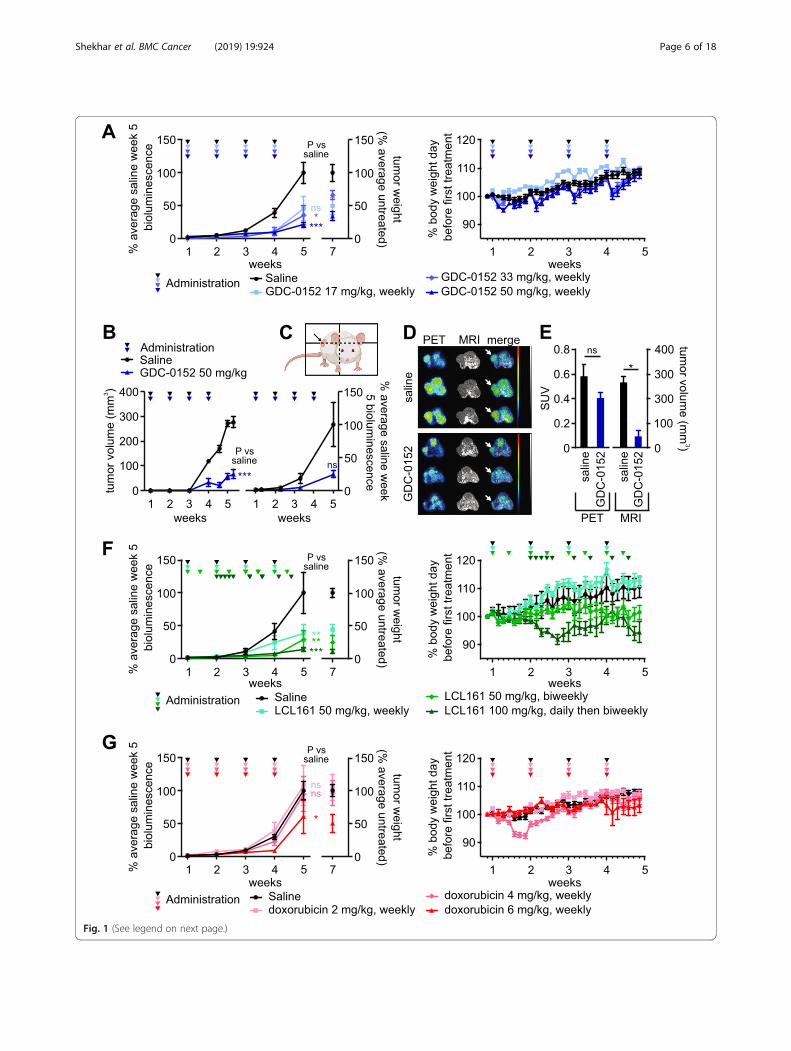
Fig. 1 (See legend on next page.)
Shekhar et al. BMC Cancer (2019) 19:924 Page 6 of 18

less effective but better tolerated, although this dosingprevented normal weight gain by these young animals.Doxorubicin had less impact on osteosarcoma
growth than the Smac mimetics in this model. Onlythe highest dose of 6 mg/kg/week significantly im-paired tumor growth (Fig. 1g). This was counter-in-tuitive, given the clinical efficacy of doxorubicin fortreating osteosarcoma patients [68], and the in vitrosensitivity of 1029H cells to this agent [26]. Doxo-rubicin has been documented to penetrate poorly intotumors [69] so it is possible the marginal efficacy ofdoxorubicin in this context reflects a low bioavailabil-ity of this poorly penetrant drug within subcutaneoustumors that may not be extensively vascularized [70].On average, tumor growth was more substantially ham-
pered by co-treatment with medium to high doses ofSmac mimetics and doxorubicin than by the drugs as soleagents (Fig. 2a-d, left panels), although tumors regrewafter treatment cessation. Although this trend of cooper-ation was observed in multiple experiments, statisticalanalyses failed to rule out the possibility that these differ-ences were due to chance. The suggestion of an improve-ment in efficacy associated with the co-treatment washowever accompanied by enhanced toxicity (right panels).One mouse that received twice-weekly treatment with 50mg/kg LCL161 plus weekly administration of 6mg/kgdoxorubicin lost more than 15% of its weight within a day,necessitating euthanasia. In subsequent experiments in-volving co-treatment with these drugs, we therefore re-duced the frequency of LCL161 administration fromtwice-weekly to weekly.Although GDC-0152 and LCL161 could theoretically
kill cells via relieving XIAP-mediated caspase inhibition,their major mechanism of lethality involves stimulationof cIAP1/2 degradation, facilitating RIPK1 de-
ubiquitination, which redirects TNFα-mediated TNFR1signaling towards apoptotic or necroptotic pathways[71]. Unlike some other cell types that can produceautocrine TNFα in response to Smac mimetic treatment[23–25], this class of drugs only killed osteosarcomacells upon provision of exogenous TNFα [26]. The invivo efficacy of GDC-0152 and LCL161 we observed inthis study therefore implied either that the in vivo tumormicroenvironment somehow imbued osteosarcoma cellswith the ability to produce autocrine TNFα, or that hostcells in or around the tumors secreted TNFα that coop-erated with the administered Smac mimetics to kill theosteosarcoma cells in vivo. Our data support the lattermodel. Flow cytometry revealed that only 37% of thecells comprising a subcutaneous tumor expressed detect-able mCherry fluorescence. Around half of the cellswithin this tumor were infiltrating host cells, mostlymacrophages (Fig. 3a). The phenotypes of 12 % of thecells could not be determined with the antibody panelwe used; some were probably osteosarcoma cells whosemCherry fluorescence was too weak to detect and otherswere probably other types of infiltrating host cells. Weperformed intracellular cytokine staining of fixed tumorcells from three untreated mice and three animals thatreceived a single dose of GDC-0152 six hours prior toculling. Unfortunately, the fixation abolished mCherryfluorescence, so 1029H-Luc cells could not be distin-guished from other cells that lacked markers detected byour antibodies. Approximately 2–4% of the cells withintumors, mostly immune cells, produced TNFα, and thisproportion was very slightly higher in samples from micethat received GDC-0152 treatment (Fig. 3b). Hardly anycells that lacked immune cell markers, which presum-ably were mostly 1029H-Luc osteosarcoma cells (Fig.3a), contained TNFα (Fig. 3b). Ex vivo incubation of the
(See figure on previous page.)Fig. 1 GDC-0152, LCL161 and doxorubicin impede the growth of subcutaneously implanted osteosarcomas in mice. Luciferase-expressing murine1029H osteosarcoma cells were implanted subcutaneously into nude mice. 1 week after implantation the mice commenced the specifiedregimens of GDC-0152 (a-e), LCL161 (f) or doxorubicin (g). a, f, g Left panels: Primary tumor growth was monitored via bioluminescence, andtumor weights were measured post-mortem. One way ANOVAs with Sidak post-tests were used to estimate the probability that the drugtreatments significantly affected tumor growth, as measured by bioluminescence at week 5, relative to saline treatment (*** P < 0.001; ** P < 0.01;* P < 0.05; ns P > 0.05 (colors of asterisks and “ns” labels; reflect the treatments, as indicated in the in-figure legends). Right panels: Mice wereweighed each day to assess drug toxicity (n = 5–25, +/− SEM). b-e Tumor-bearing mice were treated with saline or GDC-0152 50 mg/kg/week.b Tumor responses were monitored by caliper measurements (left) or bioluminescence (right) at the indicated times (n = 3, +/− SEM). Thedifferences between responses in saline and drug-treated mice were analyzed by a one way ANOVA with Sidak post-tests (*** P < 0.001). (C, D)18F-FDG PET/MRI imaging was performed 30 days after the first treatment. c A cartoon, created using BioRender, illustrates the plane of transversePET/MR images taken through 1029H osteosarcoma tumors (denoted by the arrow). d PET/MR imaging was performed on each mouse pertreatment group (n = 3), oriented with the spine at the top and the femurs in the lower left and right parts of the images: left, positron emissiontomography (PET); middle, magnetic resonance imaging (MRI); right column, PET/MRI overlay with white arrows indicating tumors . The colorscale, which ranged from 0 to 1.5 SUV, indicates highest uptake of 18F-FDG in red and lowest uptake in black. The gray scale used for MRimaging, which ranged from 40.95 to 4095, indicates brightest signals from fat-containing soft tissues versus darker signals from water-containingsoft tissues. (e) Mean standardized uptake values (SUV) of 18F-FDG-PET were determined by volume of interest (VOI) analysis, and tumor volumeswere determined by VOI analysis of MRI images (n = 3, +/− SEM). Mann-Whitney non-parametric U-tests were used to calculate the significanceof differences between treated and untreated mice * P < 0.05; ns P > 0.05
Shekhar et al. BMC Cancer (2019) 19:924 Page 7 of 18

Fig. 2 (See legend on next page.)
Shekhar et al. BMC Cancer (2019) 19:924 Page 8 of 18
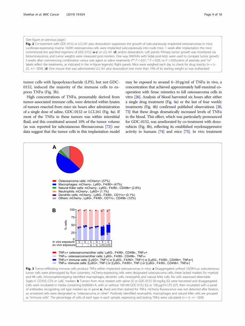
tumor cells with lipopolysaccharide (LPS), but not GDC-0152, induced the majority of the immune cells to ex-press TNFα (Fig. 3b).High concentrations of TNFα, presumably derived from
tumor-associated immune cells, were detected within lysatesof tumors resected from mice six hours after administrationof a single dose of saline, GDC-0152 or LCL161 (Fig. 4a). Ifmost of the TNFα in these tumors was within interstitialfluid, and this constituted around 10% of the tumor volume(as was reported for subcutaneous fibrosarcomas [72]) ourdata suggest that the tumor cells in this implantation model
may be exposed to around 6–10 pg/ml of TNFα in vivo, aconcentration that achieved approximately half-maximal co-operation with Smac mimetics to kill osteosarcoma cells invitro [26]. Analysis of blood harvested six hours after eithera single drug treatment (Fig. 4a) or the last of four weeklytreatments (Fig. 4b) confirmed published observations [20,73] that these drugs dramatically increased levels of TNFαin the blood. This effect, which was particularly pronouncedfor GDC-0152, was ameliorated by co-treatment with doxo-rubicin (Fig. 4b), reflecting its established myelosuppressiveactivity in humans [74] and mice [75]. In vivo treatment
(See figure on previous page.)Fig. 2 Co-treatment with GDC-0152 or LCL161 plus doxorubicin suppresses the growth of subcutaneously implanted osteosarcomas in mice.Luciferase-expressing murine 1029H osteosarcoma cells were implanted subcutaneously into nude mice. 1 week after implantation the micecommenced the specified regimens of GDC-0152 (a-c) or LCL161 (d) and/or doxorubicin. Left panels: Primary tumor growth was monitored viabioluminescence, and tumor weights were measured post-mortem. One way ANOVAs with Sidak post-tests were used to compare tumor growth5 weeks after commencing combination versus sole agent or saline treatments (** P < 0.01; * P < 0.05; ns P > 0.05;colors of asterisks and “ns”labels reflect the treatments, as indicated in the in-figure legends). Right panels: Mice were weighed each day to check for drug toxicity (n = 5–25, +/− SEM). (d) One mouse that was administered LCL161 plus doxorubicin lost more than 15% of its starting weight so was euthanized
Fig. 3 Tumor-infiltrating immune cells produce TNFα within implanted osteosarcomas in mice. a Disaggregated unfixed 1029H-Luc subcutaneoustumor cells were phenotyped by flow cytometry. mCherry-expressing cells were designated osteosarcoma cells; these lacked markers for myeloidand NK cells. Immunophenotyping identified macrophages, dendritic cells, neutrophils and natural killer cells. No cells expressed detectableSiglec-F, CD103, CD3 or Ly6C markers. b Tumors from mice treated with saline (S) or GDC-0152 50mg/kg (G) were harvested and disaggregated.Cells were incubated in media containing brefeldin-A, with or without 100 nM GDC-0152 (G) or 100 μg/ml LPS (LP), then incubated with a panelof antibodies recognizing cell type markers (as in panel a), fixed and then stained for TNFα. mCherry fluorescence was not detected after fixation,so unstained cells were designated as “osteosarcoma or other”. Positively identified neutrophils, macrophages and natural killer cells are groupedas “immune cells”. The percentage of cells of each type in each sample, expressing and lacking TNFα were calculated (n = 3, +/− SEM)
Shekhar et al. BMC Cancer (2019) 19:924 Page 9 of 18
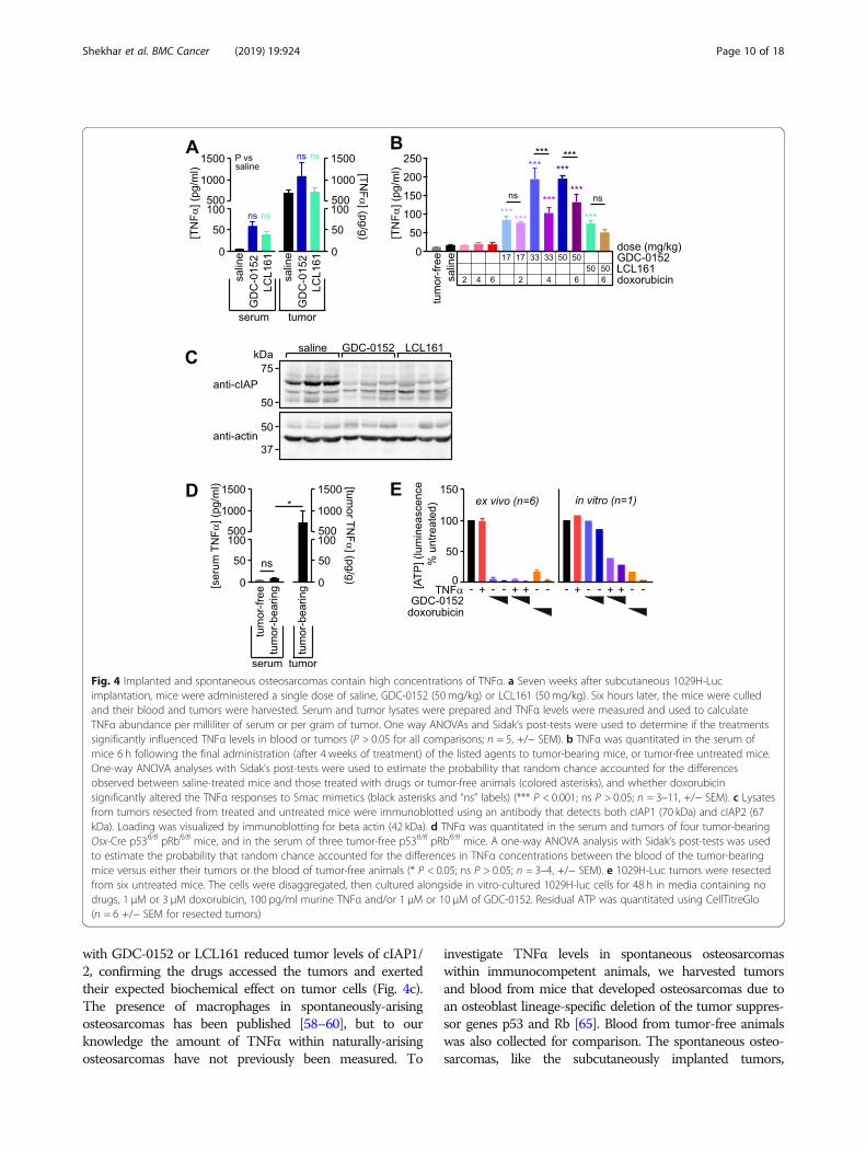
with GDC-0152 or LCL161 reduced tumor levels of cIAP1/2, confirming the drugs accessed the tumors and exertedtheir expected biochemical effect on tumor cells (Fig. 4c).The presence of macrophages in spontaneously-arisingosteosarcomas has been published [58–60], but to ourknowledge the amount of TNFα within naturally-arisingosteosarcomas have not previously been measured. To
investigate TNFα levels in spontaneous osteosarcomaswithin immunocompetent animals, we harvested tumorsand blood from mice that developed osteosarcomas due toan osteoblast lineage-specific deletion of the tumor suppres-sor genes p53 and Rb [65]. Blood from tumor-free animalswas also collected for comparison. The spontaneous osteo-sarcomas, like the subcutaneously implanted tumors,
Fig. 4 Implanted and spontaneous osteosarcomas contain high concentrations of TNFα. a Seven weeks after subcutaneous 1029H-Lucimplantation, mice were administered a single dose of saline, GDC-0152 (50 mg/kg) or LCL161 (50 mg/kg). Six hours later, the mice were culledand their blood and tumors were harvested. Serum and tumor lysates were prepared and TNFα levels were measured and used to calculateTNFα abundance per milliliter of serum or per gram of tumor. One way ANOVAs and Sidak’s post-tests were used to determine if the treatmentssignificantly influenced TNFα levels in blood or tumors (P > 0.05 for all comparisons; n = 5, +/− SEM). b TNFα was quantitated in the serum ofmice 6 h following the final administration (after 4 weeks of treatment) of the listed agents to tumor-bearing mice, or tumor-free untreated mice.One-way ANOVA analyses with Sidak’s post-tests were used to estimate the probability that random chance accounted for the differencesobserved between saline-treated mice and those treated with drugs or tumor-free animals (colored asterisks), and whether doxorubicinsignificantly altered the TNFα responses to Smac mimetics (black asterisks and “ns” labels) (*** P < 0.001; ns P > 0.05; n = 3–11, +/− SEM). c Lysatesfrom tumors resected from treated and untreated mice were immunoblotted using an antibody that detects both cIAP1 (70 kDa) and cIAP2 (67kDa). Loading was visualized by immunoblotting for beta actin (42 kDa). d TNFα was quantitated in the serum and tumors of four tumor-bearingOsx-Cre p53fl/fl pRbfl/fl mice, and in the serum of three tumor-free p53fl/fl pRbfl/fl mice. A one-way ANOVA analysis with Sidak’s post-tests was usedto estimate the probability that random chance accounted for the differences in TNFα concentrations between the blood of the tumor-bearingmice versus either their tumors or the blood of tumor-free animals (* P < 0.05; ns P > 0.05; n = 3–4, +/− SEM). e 1029H-Luc tumors were resectedfrom six untreated mice. The cells were disaggregated, then cultured alongside in vitro-cultured 1029H-luc cells for 48 h in media containing nodrugs, 1 μM or 3 μM doxorubicin, 100 pg/ml murine TNFα and/or 1 μM or 10 μM of GDC-0152. Residual ATP was quantitated using CellTitreGlo(n = 6 +/− SEM for resected tumors)
Shekhar et al. BMC Cancer (2019) 19:924 Page 10 of 18
contained abundant TNFα (Fig. 4d). In keeping with our ob-servation that the anti-osteosarcoma potential of Smac mi-metics hinges on the presence of TNFα produced bymyeloid cells within the tumors, disaggregated cells fromfreshly-resected implanted tumors (consisting of both osteo-sarcoma and infiltrating non-cancerous cells) were efficientlykilled in vitro by Smac mimetics as sole agents, whereas thecorresponding in vitro-cultured osteosarcoma cells wereonly sensitive to Smac mimetics when co-treated with ex-ogenous TNFα (Fig. 4e).We were interested in whether cells from human
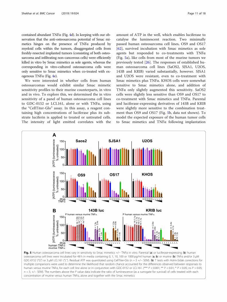
osteosarcomas would exhibit similar Smac mimeticsensitivity profiles to their murine counterparts, in vitroand in vivo. To explore this, we determined the in vitrosensitivity of a panel of human osteosarcoma cell linesto GDC-0152 or LCL161, alone or with TNFα, usingthe “CellTiter-Glo” assay. In this assay, a reagent con-taining high concentrations of luciferase plus its sub-strate luciferin is applied to treated or untreated cells.The intensity of light emitted correlates with the
amount of ATP in the well, which enables luciferase tocatalyse the luminescent reaction. Two minimallypassed human osteosarcoma cell lines, OS9 and OS17[62], survived incubation with Smac mimetics as soleagents but responded to co-treatments with TNFα(Fig. 5a), like cells from most of the murine tumors wepreviously tested [26]. The responses of established hu-man osteosarcoma cell lines (SaOS2, SJSA1, U2OS,143B and KRIB) varied substantially, however. SJSA1and U2OS were resistant, even to co-treatment withSmac mimetics plus TNFα. KHOS cells were somewhatsensitive to Smac mimetics alone, and addition ofTNFα only slightly augmented this sensitivity. SaOS2cells were slightly less sensitive than OS9 and OS17 toco-treatment with Smac mimetics and TNFα. Parentaland luciferase-expressing derivatives of 143B and KRIBwere slightly more sensitive to the combination treat-ment than OS9 and OS17 (Fig. 5b, data not shown). Tomodel the expected exposure of the human tumor cellsto Smac mimetics and TNFα following implantation
Fig. 5 Human osteosarcoma cell lines vary in sensitivity to Smac mimetics +/− TNFα in vitro. Parental (a) or luciferase-expressing (b) humanosteosarcoma cell lines were incubated for 48 h in media containing 0, 1, 10, 100 or 1000 pg/ml human (a, b) or murine (b) TNFα and/or 3 μMGDC-0152 (“G”) or 3 μM LCL161 (“L”). Residual ATP was quantitated using CellTiter-Glo (n = 3 +/− SEM). (b) T tests with Holm-Sidak corrections formultiple comparisons were used to determine the likelihood that random chance accounted for the differences observed between responses tohuman versus murine TNFα, for each cell line alone or in conjunction with GDC-0152 or LCL161. (*** P < 0.001; ** P < 0.01; * P < 0.05; ns P > 0.05;n = 3, +/− SEM). The numbers above the P value data indicate the ratio of luminescence (as a surrogate for survival) of cells treated with eachconcentration of murine versus human TNFα, alone and together with the Smac mimetics
Shekhar et al. BMC Cancer (2019) 19:924 Page 11 of 18
into nude mice, we compared the extents to whichSmac mimetics sensitized the luciferase-tagged KRIBand 143B human osteosarcoma cells to murine versushuman TNFα. The CellTiter-Glo assay was used forthese experiments. The CellTiter-Glo reagent was de-signed to contain sufficient luciferase to ensure that re-action rates are proportional to ATP concentrationsacross a large range of cell densities, so we suspect thatthe additional presence of some transgene-encodedluciferase in these cells would be unlikely to affect thereaction rate and hence the light emitted. However wecannot conclusively exclude the possibility that lowerluminescent readings following drug treatment may re-flect a reduction in cellular luciferase, as well as ATPlevels, as cells died. Although published data suggesthuman TNF receptors bind murine TNFα with onlyslightly lower affinity than human TNFα [76–78], thehuman osteosarcoma cells were significantly more sen-sitive to Smac mimetics coupled with human than mur-ine TNFα (Fig. 5b).Our observation that doxorubicin only slightly impaired
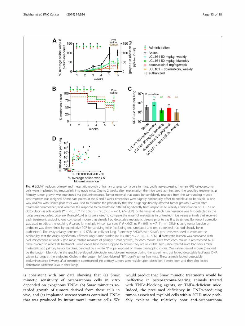
the growth of subcutaneously implanted murine osteosar-comas raised the possibility that vascularization of these tu-mors may be poor, despite obviously being sufficient tomediate intratumoral access of immune cells and Smac mi-metics. We therefore decided to use a different implantationroute for testing in vivo drug efficacy against human osteo-sarcomas. We first considered orthotopic routes. Intrafe-moral and intratibial osteosarcoma implantation modelshave been developed, but the technical challenges associatedwith these procedures can lead to highly variable tumorigen-icity rates, and intraosseous tumors are not well tolerated bymice [79–81]. These factors would have necessitated usinglarge numbers of animals to discern significant drug effects,and the need to provide analgesia could have introducedpotentially confounding drug-drug interactions. Given therequirement of the inflammatory cytokine TNFα for theanti-osteosarcoma activity of Smac mimetics, we were par-ticularly keen to avoid analgesics with anti-inflammatoryactivities. We therefore decided to establish an intramuscu-lar implantation model for testing the impact of Smac mi-metics on human osteosarcoma xenografts. Intramuscularimplantations of osteosarcoma cells, either into the upperhind paw [82] or gastrocnemius muscle [83, 84], were re-ported to be highly tumorigenic. To minimise the tumors’impact on leg function, we chose to inject luciferase-ex-pressing KRIB human tumor cells into the cranial tibialmuscle of mice. This yielded reproducible primary tumorgrowth that was well tolerated by the mice (obviating theneed for analgesia), and metastases to the lungs of all un-treated mice within 7 weeks of implantation.As mentioned above, KRIB cells were only sensitive to
Smac mimetics in vitro in the presence of exogenousTNFα, and murine TNFα cooperated with these drugs
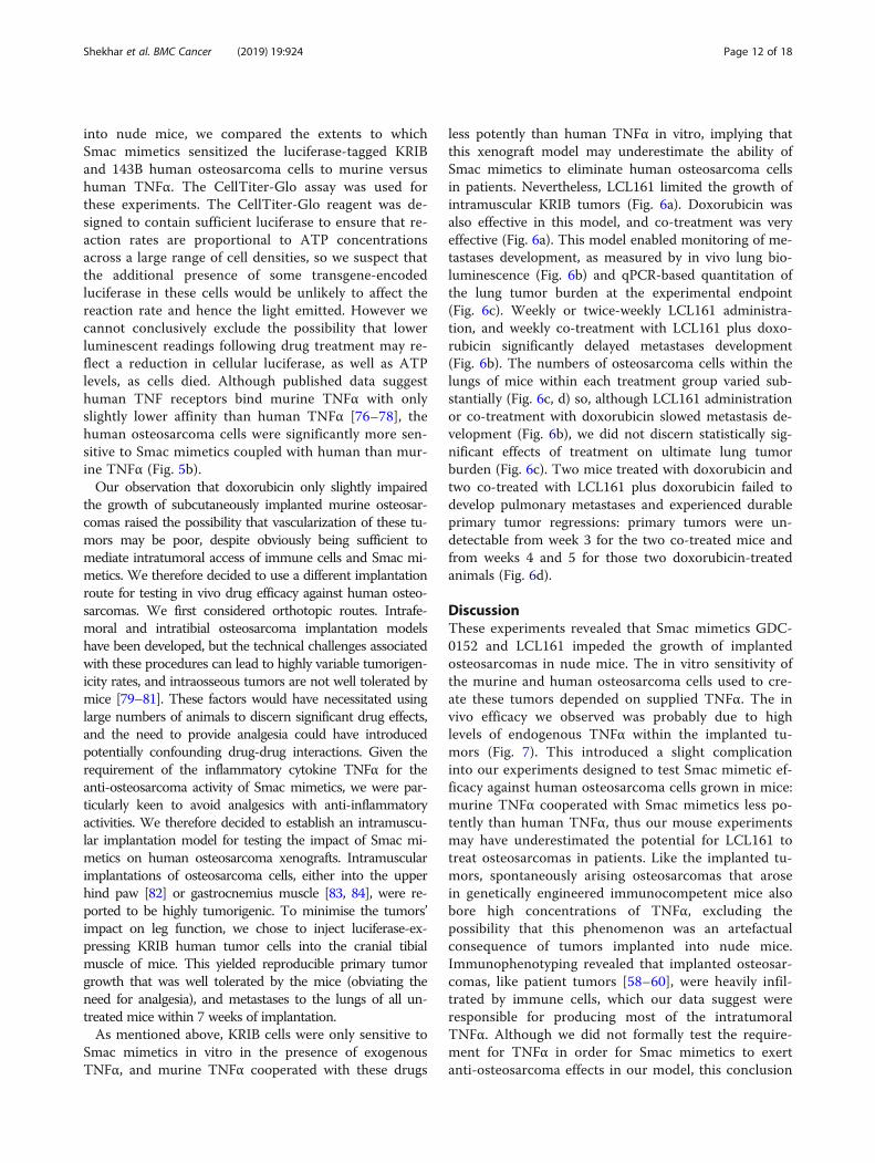
less potently than human TNFα in vitro, implying thatthis xenograft model may underestimate the ability ofSmac mimetics to eliminate human osteosarcoma cellsin patients. Nevertheless, LCL161 limited the growth ofintramuscular KRIB tumors (Fig. 6a). Doxorubicin wasalso effective in this model, and co-treatment was veryeffective (Fig. 6a). This model enabled monitoring of me-tastases development, as measured by in vivo lung bio-luminescence (Fig. 6b) and qPCR-based quantitation ofthe lung tumor burden at the experimental endpoint(Fig. 6c). Weekly or twice-weekly LCL161 administra-tion, and weekly co-treatment with LCL161 plus doxo-rubicin significantly delayed metastases development(Fig. 6b). The numbers of osteosarcoma cells within thelungs of mice within each treatment group varied sub-stantially (Fig. 6c, d) so, although LCL161 administrationor co-treatment with doxorubicin slowed metastasis de-velopment (Fig. 6b), we did not discern statistically sig-nificant effects of treatment on ultimate lung tumorburden (Fig. 6c). Two mice treated with doxorubicin andtwo co-treated with LCL161 plus doxorubicin failed todevelop pulmonary metastases and experienced durableprimary tumor regressions: primary tumors were un-detectable from week 3 for the two co-treated mice andfrom weeks 4 and 5 for those two doxorubicin-treatedanimals (Fig. 6d).
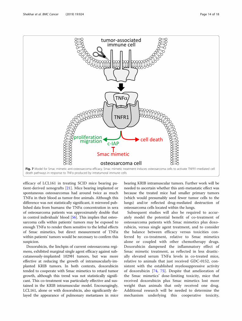
DiscussionThese experiments revealed that Smac mimetics GDC-0152 and LCL161 impeded the growth of implantedosteosarcomas in nude mice. The in vitro sensitivity ofthe murine and human osteosarcoma cells used to cre-ate these tumors depended on supplied TNFα. The invivo efficacy we observed was probably due to highlevels of endogenous TNFα within the implanted tu-mors (Fig. 7). This introduced a slight complicationinto our experiments designed to test Smac mimetic ef-ficacy against human osteosarcoma cells grown in mice:murine TNFα cooperated with Smac mimetics less po-tently than human TNFα, thus our mouse experimentsmay have underestimated the potential for LCL161 totreat osteosarcomas in patients. Like the implanted tu-mors, spontaneously arising osteosarcomas that arosein genetically engineered immunocompetent mice alsobore high concentrations of TNFα, excluding thepossibility that this phenomenon was an artefactualconsequence of tumors implanted into nude mice.Immunophenotyping revealed that implanted osteosar-comas, like patient tumors [58–60], were heavily infil-trated by immune cells, which our data suggest wereresponsible for producing most of the intratumoralTNFα. Although we did not formally test the require-ment for TNFα in order for Smac mimetics to exertanti-osteosarcoma effects in our model, this conclusion
Shekhar et al. BMC Cancer (2019) 19:924 Page 12 of 18
is consistent with our data showing that (a) Smacmimetic sensitivity of osteosarcoma cells in vitrodepended on exogenous TNFα, (b) Smac mimetics re-tarded growth of tumors derived from these cells invivo, and (c) implanted osteosarcomas contained TNFαthat was produced by intratumoral immune cells. We
would predict that Smac mimetic treatments would beineffective in osteosarcoma-bearing animals treatedwith TNFα-blocking agents, or TNFα-deficient mice.Indeed, the presumed deficiency in TNFα-producingtumor-associated myeloid cells within SCID mice prob-ably explains the relatively poor anti-osteosarcoma
Fig. 6 LCL161 reduces primary and metastatic growth of human osteosarcoma cells in mice. Luciferase-expressing human KRIB osteosarcomacells were implanted intramuscularly into nude mice. One to 2 weeks after implantation the mice were administered the specified treatments. aPrimary tumor growth was monitored via bioluminescence. Tumor material that could be confidently resected from the surrounding musclepost-mortem was weighed. Some data points at the 5 and 6 week timepoints were slightly horizontally offset to enable all to be visible. A oneway ANOVA with Sidak’s post-tests was used to estimate the probability that the drugs significantly affected tumor growth 5 weeks aftertreatment commenced, and whether the response to co-treatment differed significantly from responses to weekly administration of LCL161 ordoxorubicin as sole agents (** P < 0.01; * P < 0.05; ns P > 0.05; n = 7–11, +/− SEM). b The times at which luminescence was first detected in thelungs were recorded. Log-rank (Mantel-Cox) tests were used to compare the onset of metastases in untreated mice versus animals that receivedeach treatment, excluding one co-treated mouse that already had detectable metastatic disease prior to the first treatment. Bonferroni correctionwas used to adjust the resulting P values for multiple (4) comparisons (* P < 0.05; ns P > 0.05; n = 7–11, +/− SEM). c Lung tumor burden atendpoint was determined by quantitative PCR for surviving mice (excluding one untreated and one-co-treated that had already beeneuthanized). The assay reliably detected > 10 KRIB-Luc cells per lung. A one way ANOVA with Sidak’s post-tests was used to estimate theprobability that the drugs significantly affected lung tumor burden (ns P > 0.05; n = 7–10, +/− SEM). d Metastatic burden was compared withbioluminescence at week 5 (the most reliable measure of primary tumor growth), for each mouse. Data from each mouse is represented by acircle colored to reflect its treatment. Some circles have been cropped to ensure they are all visible. Two saline-treated mice had very similarmetastatic and primary tumor burdens; denoted by a white “2” superimposed on those overlapping circles. One saline-treated mouse (denotedby the bottom black dot in the graph) developed detectable lung bioluminescence during the experiment but lacked detectable luciferase DNAwithin its lungs at the endpoint. Circles in the bottom left box (labeled “TF”) signify tumor free mice. These animals lacked detectablebioluminescence 5 weeks after treatment commenced, no primary tumors were visible upon dissection 1 week later, and they also lackeddetectable luciferase DNA in their lungs
Shekhar et al. BMC Cancer (2019) 19:924 Page 13 of 18
efficacy of LCL161 in treating SCID mice bearing pa-tient-derived xenografts [21]. Mice bearing implanted orspontaneous osteosarcomas had around twice as muchTNFα in their blood as tumor-free animals. Although thisdifference was not statistically significant, it mirrored pub-lished data from humans: the TNFα concentration in seraof osteosarcoma patients was approximately double thatin control individuals’ blood [56]. This implies that osteo-sarcoma cells within patients’ tumors may be exposed toenough TNFα to render them sensitive to the lethal effectsof Smac mimetics, but direct measurement of TNFαwithin patients’ tumors would be necessary to confirm thissuspicion.Doxorubicin, the linchpin of current osteosarcoma regi-
mens, exhibited marginal single agent efficacy against sub-cutaneously-implanted 1029H tumors, but was moreeffective at reducing the growth of intramuscularly-im-planted KRIB tumors. In both contexts, doxorubicintended to cooperate with Smac mimetics to retard tumorgrowth, although this trend was not statistically signifi-cant. This co-treatment was particularly effective and sus-tained in the KRIB intramuscular model. Encouragingly,LCL161, alone or with doxorubicin, also significantly de-layed the appearance of pulmonary metastases in mice
bearing KRIB intramuscular tumors. Further work will beneeded to ascertain whether this anti-metastatic effect wasbecause the treated mice had smaller primary tumors(which would presumably seed fewer tumor cells to thelungs) and/or reflected drug-mediated destruction ofosteosarcoma cells located within the lungs.Subsequent studies will also be required to accur-
ately model the potential benefit of co-treatment ofosteosarcoma patients with Smac mimetics plus doxo-rubicin, versus single agent treatment, and to considerthe balance between efficacy versus toxicities con-ferred by co-treatment, relative to Smac mimeticsalone or coupled with other chemotherapy drugs.Doxorubicin dampened the inflammatory effect ofSmac mimetic treatment, as reflected in less drastic-ally elevated serum TNFα levels in co-treated mice,relative to animals that just received GDC-0152, con-sistent with the established myelosuppressive activityof doxorubicin [74, 75]. Despite that amelioration ofthe Smac mimetics’ dose-limiting toxicity, mice thatreceived doxorubicin plus Smac mimetics lost moreweight than animals that only received one drug.Additional research will be needed to determine themechanism underlying this cooperative toxicity,
Fig. 7 Model for Smac mimetic anti-osteosarcoma efficacy. Smac mimetic treatment induces osteosarcoma cells to activate TNFR1-mediated celldeath pathways in response to TNFα produced by intratumoral immune cells
Shekhar et al. BMC Cancer (2019) 19:924 Page 14 of 18

including exploration of the possibility that Smac mi-metics may exacerbate doxorubicin’s cardiotoxicity[85]. It will be important to determine whether co-operative toxicities would be avoided by sequentialexposure, in which case subsequent Smac mimetictreatment may be considered for patients whose tu-mors persist or recur after administration of the max-imal cumulative dose of doxorubicin recommended toavoid dose-limiting cardiotoxicity. The outcome ofthose experiments may help define clinical contexts inwhich the anti-osteosarcoma efficacy of Smac mi-metics could be maximized while their toxicities aremanaged.The clinical responsiveness of individual osteosarcomas
to Smac mimetics will presumably be strongly dictated bythe ability of the drugs to cooperate with TNFα to triggerapoptotic or necroptotic death of the individual’s cancercells. Further work will be needed to gain a comprehensiveunderstanding of intertumoral variability in the in vitro sen-sitivity of human osteosarcomas to Smac mimetics. Cellsfrom two minimally-passaged human osteosarcomas werequite sensitive to Smac mimetic/TNFα co-treatment, how-ever established human osteosarcoma cell lines varied sub-stantially in their sensitivity to Smac mimetics as soleagents and together with TNFα. This heterogeneity may re-flect biological variability between different tumors, and/orit may be a consequence of genomic instability-mediatedphenotypic drift during extended in vitro culturing [86]. Ifthe latter is a major factor, the sensitive phenotypes of theminimally passaged lines (OS9 and OS17) may reflect thetypical responsiveness of osteosarcoma cells within patients’tumors better than the established cell lines, some of whichwere more resistant.It is important to note that our experiments were con-
ducted in nude mice. Although these mice possess innateimmune cells, which can produce the TNFα required forSmac mimetic-mediated osteosarcoma cell destruction,they have almost no T cells [87]. If Smac mimetics canstimulate immune-targeting of osteosarcoma cells throughboosting lymphocyte survival and activation, as has beendemonstrated in other cancers [48], this may augment thedirect osteosarcoma cell killing we observed in nude mice,to yield a more pronounced anti-osteosarcoma effect inimmunocompetent animals or humans.
ConclusionsThe Smac mimetics LCL161 and GDC-0152 cooperatedwith TNFα produced by infiltrating immune cells tolimit osteosarcoma growth and metastasis in nude mice.These data illustrate the potential for Smac mimetics totarget malignancies like osteosarcoma in which the can-cer cells fail to produce autocrine TNFα in response tothese agents. Results from this study suggest that saferegimens involving Smac mimetics like LCL161 or
GDC-0152 may improve treatment outcomes for osteo-sarcoma patients.
AbbreviationsBCA: Bicinchoninic acid; BFA: Brefeldin-A; FBS: Fetal bovine serum;LPS: Lipopolysaccharide; MRI: Magnetic Resonance Imaging; PET: PositronEmission Tomography
AcknowledgementsNovartis and Genentech generously provided LCL161 and GDC-0152 respect-ively. We thank Damien Myers and Nicholas Saunders for cell lines, AlyceMayfosh for assistance with qPCR assay development, and Carmelo Cerraand staff from the La Trobe Animal Research and Training Facility for assist-ance with animal experiments.
Authors’ contributionsCJH conceived the study; CRW and BSP provided materials; CJH, TMS, IJGB,AMS, CRW and BSP developed the methods and planned the experiments;TMS, IJGB, MAH, AR, DZ and AS performed the experiments; CJH, TMS, IJGB,AR, DZ, AMS and MAH interpreted the data; CJH, TMS, IJGB and AMSprepared the figures and wrote the manuscript with help from the otherauthors. All authors have read and approved the manuscript.
FundingThis study was funded by a grant from The Kids’ Cancer Project, a SarcomaResearch Grant from the GPA Andrew Ursini Charitable Fund and theAustralasian Sarcoma Study Group, and a Grant-in-Aid from the CancerCouncil Victoria. These organizations financed the work but played no role inthe design of the study and collection, analysis, and interpretation of data orin writing the manuscript.
Availability of data and materialsThe datasets used and/or analyzed during the current study available fromthe corresponding author on reasonable request.
Ethics approval and consent to participateAnimal experiments were conducted in accordance with Australian Code ofPractice for the Care and Use of Animals for Scientific Purposes, as approvedby the La Trobe Animal Ethics Committee (approvals AEC16–25 and AEC17–76). The study did not involve human participants, human data or humantissue.
Consent for publicationNot applicable
Competing interestsThe authors declare that they have no competing interests.
Author details1Department of Biochemistry and Genetics, La Trobe Institute for MolecularScience, La Trobe University, Bundoora, Victoria 3086, Australia. 2TumourTargeting Laboratory, Ludwig Institute for Cancer Research and OliviaNewton-John Cancer Research Institute, Melbourne, Australia. 3School ofCancer Medicine, La Trobe University, Melbourne, Australia. 4St. Vincent’sInstitute, Fitzroy, Victoria 3065, Australia. 5Department of Medicine, St.Vincent’s Hospital, University of Melbourne, Fitzroy, Victoria 3065, Australia.6Mary MacKillop Institute for Health Research, Australian Catholic University,Melbourne, Victoria 3000, Australia. 7Departments of Medical Oncology andMolecular Imaging & Therapy, Austin Health, Heidelberg, Melbourne,Australia. 8Department of Medicine, University of Melbourne, Melbourne,Australia.
Received: 22 March 2019 Accepted: 28 August 2019
References1. Gianferante DM, Mirabello L, Savage SA. Germline and somatic genetics of
osteosarcoma - connecting aetiology, biology and therapy. Nat RevEndocrinol. 2017;13:480–91.
Shekhar et al. BMC Cancer (2019) 19:924 Page 15 of 18
2. Walia MK, Castillo-Tandazo W, Mutsaers AJ, Martin TJ, Walkley CR. Murinemodels of osteosarcoma: A piece of the translational puzzle. J Cell Biochem.2018;119:4241–50.
3. Abarrategi A, Tornin J, Martinez-Cruzado L, Hamilton A, Martinez-Campos E,Rodrigo JP, Gonzalez MV, Baldini N, Garcia-Castro J, Rodriguez R.Osteosarcoma: Cells-of-Origin, Cancer Stem Cells, and Targeted Therapies.Stem Cells Int. 2016;2016:3631764.
4. Kumar R, Kumar M, Malhotra K, Patel S. Primary Osteosarcoma in theElderly Revisited: Current Concepts in Diagnosis and Treatment. CurrOncol Rep. 2018;20:13.
5. Marko TA, Diessner BJ, Spector LG. Prevalence of Metastasis atDiagnosis of Osteosarcoma: An International Comparison. Pediatr BloodCancer. 2016;63:1006–11.
6. Meazza C, Scanagatta P. Metastatic osteosarcoma: a challengingmultidisciplinary treatment. Expert Rev Anticancer Ther. 2016;16:543–56.
7. McGuire J, Utset-Ward TJ, Reed DR, Lynch CC. Re-calculating! Navigating throughthe osteosarcoma treatment roadblock. Pharmacol Res. 2017;117:54–64.
8. Anderson ME. Update on Survival in Osteosarcoma. Orthop Clin North Am.2016;47:283–92.
9. Smeland S, Bielack SS, Whelan J, Bernstein M, Hogendoorn P, KrailoMD, Gorlick R, Janeway KA, Ingleby FC, Anninga J, et al. Survival andprognosis with osteosarcoma: outcomes in more than 2000 patients inthe EURAMOS-1 (European and American Osteosarcoma Study) cohort.European journal of cancer (Oxford, England : 1990). 2019;109:36–50.
10. Otoukesh B, Boddouhi B, Moghtadaei M, Kaghazian P, Kaghazian M. Novelmolecular insights and new therapeutic strategies in osteosarcoma. CancerCell Int. 2018;18:158.
11. Verhagen AM, Ekert PG, Pakusch M, Silke J, Connolly LM, Reid GE, Moritz RL,Simpson RJ, Vaux DL. Identification of DIABLO, a mammalian protein thatpromotes apoptosis by bnding to and antagonizing IAP proteins. Cell. 2000;102:43–53.
12. Du C, Fang M, Li Y, Li L, Wang X. Smac, a mitochondrial protein thatpromotes cytochrome c-dependent caspase activation by eliminating IAPinhibition. Cell. 2000;102:33–42.
13. Fulda S. Smac Mimetics to Therapeutically Target IAP Proteins in Cancer. IntRev Cell Mol Biol. 2017;330:157–69.
14. Eckelman BP, Salvesen GS. The human anti-apoptotic proteins cIAP1 andcIAP2 bind but do not inhibit caspases. J Biol Chem. 2006;281:3254–60.
15. Silke J, Vince J. IAPs and Cell Death. Curr Top Microbiol Immunol. 2017;403:95–117.
16. Schilling R, Geserick P, Leverkus M. Characterization of the ripoptosome andits components: implications for anti-inflammatory and cancer therapy.Methods Enzymol. 2014;545:83–102.
17. Petrie EJ, Czabotar PE, Murphy JM. The Structural Basis of Necroptotic CellDeath Signaling. Trends Biochem Sci. 2018;44:53–63.
18. Pasparakis M, Vandenabeele P. Necroptosis and its role in inflammation.Nature. 2015;517:311–20.
19. Flygare JA, Beresini M, Budha N, Chan H, Chan IT, Cheeti S, Cohen F, DeshayesK, Doerner K, Eckhardt SG, et al. Discovery of a potent small-moleculeantagonist of inhibitor of apoptosis (IAP) proteins and clinical candidate for thetreatment of cancer (GDC-0152). J Med Chem. 2012;55:4101–13.
20. Infante JR, Dees EC, Olszanski AJ, Dhuria SV, Sen S, Cameron S, Cohen RB.Phase I Dose-Escalation Study of LCL161, an Oral Inhibitor of ApoptosisProteins Inhibitor, in Patients With Advanced Solid Tumors. J Clin Oncol.2014;32:3103–10.
21. Houghton PJ, Kang MH, Reynolds CP, Morton CL, Kolb EA, Gorlick R, Keir ST,Carol H, Lock R, Maris JM, et al. Initial testing (Stage 1) of LCL161, a SMACmimetic, by the pediatric preclinical testing program. Pediatr Blood Cancer.2011;58:636–9.
22. Allensworth JL, Sauer SJ, Lyerly HK, Morse MA, Devi GR. Smac mimeticBirinapant induces apoptosis and enhances TRAIL potency in inflammatorybreast cancer cells in an IAP-dependent and TNF-alpha-independentmechanism. Breast Cancer Res Treat. 2013;137:359–71.
23. Vince JE, Wong WW, Khan N, Feltham R, Chau D, Ahmed AU, Benetatos CA,Chunduru SK, Condon SM, McKinlay M, et al. IAP antagonists target cIAP1 toinduce TNFalpha-dependent apoptosis. Cell. 2007;131:682–93.
24. Petersen SL, Wang L, Yalcin-Chin A, Li L, Peyton M, Minna J, Harran P, WangX. Autocrine TNFalpha signaling renders human cancer cells susceptible toSmac-mimetic-induced apoptosis. Cancer Cell. 2007;12:445–56.
25. Varfolomeev E, Blankenship JW, Wayson SM, Fedorova AV, Kayagaki N, GargP, Zobel K, Dynek JN, Elliott LO, Wallweber HJ, et al. IAP antagonists induce
autoubiquitination of c-IAPs, NF-kappaB activation, and TNFalpha-dependent apoptosis. Cell. 2007;131:669–81.
26. Shekhar TM, Miles MA, Gupte A, Taylor S, Tascone B, Walkley CR, HawkinsCJ. IAP antagonists sensitize murine osteosarcoma cells to killing by TNFα.Oncotarget. 2016;7:33866–86.
27. Amaravadi RK, Schilder RJ, Martin LP, Levin M, Graham MA, Weng DE, AdjeiAA. A Phase I Study of the SMAC-Mimetic Birinapant in Adults withRefractory Solid Tumors or Lymphoma. Mol Cancer Ther. 2015;14:2569–75.
28. Noonan AM, Bunch KP, Chen JQ, Herrmann MA, Lee JM, Kohn EC, O'SullivanCC, Jordan E, Houston N, Takebe N, et al. Pharmacodynamic markers andclinical results from the phase 2 study of the SMAC mimetic birinapant inwomen with relapsed platinum-resistant or -refractory epithelial ovariancancer. Cancer. 2015;13:29783.
29. Fulda S. Promises and Challenges of Smac Mimetics as Cancer Therapeutics.Clin Cancer Res. 2015;21:5030–6.
30. DiPersio JF, Erba HP, Larson RA, Luger SM, Tallman MS, Brill JM, VuagniauxG, Rouits E, Sorensen JM, Zanna C. Oral Debio1143 (AT406), an antagonist ofinhibitor of apoptosis proteins, combined with daunorubicin and cytarabinein patients with poor-risk acute myeloid leukemia--results of a phase I dose-escalation study. Clin Lymphoma Myeloma Leuk. 2015;15:443–9.
31. Carter BZ, Mak PY, Mak DH, Shi Y, Qiu Y, Bogenberger JM, Mu H, Tibes R,Yao H, Coombes KR, et al. Synergistic targeting of AML stem/progenitorcells with IAP antagonist birinapant and demethylating agents. J NatlCancer Inst. 2014;106:djt440.
32. Steinwascher S, Nugues AL, Schoeneberger H, Fulda S. Identification of anovel synergistic induction of cell death by Smac mimetic and HDACinhibitors in acute myeloid leukemia cells. Cancer Lett. 2015;366:32–43.
33. Steinhart L, Belz K, Fulda S. Smac mimetic and demethylating agentssynergistically trigger cell death in acute myeloid leukemia cells andovercome apoptosis resistance by inducing necroptosis. Cell Death Dis.2013;4:e802.
34. Wu MS, Wang GF, Zhao ZQ, Liang Y, Wang HB, Wu MY, Min P, Chen LZ,Feng QS, Bei JX, et al. Smac mimetics in combination with TRAIL selectivelytarget cancer stem cells in nasopharyngeal carcinoma. Mol Cancer Ther.2013;12:1728–37.
35. Zhang S, Li G, Zhao Y, Liu G, Wang Y, Ma X, Li D, Wu Y, Lu J. Smac mimeticSM-164 potentiates APO2L/TRAIL- and doxorubicin-mediated anticanceractivity in human hepatocellular carcinoma cells. PLoS One. 2012;7:e51461.
36. Lu J, McEachern D, Sun H, Bai L, Peng Y, Qiu S, Miller R, Liao J, Yi H, Liu M,et al. Therapeutic Potential and Molecular Mechanism of a Novel, Potent,Nonpeptide, Smac Mimetic SM-164 in Combination with TRAIL for CancerTreatment. Mol Cancer Ther. 2011;10:902–14.
37. Metwalli AR, Khanbolooki S, Jinesh G, Sundi D, Shah JB, Shrader M,Choi W, Lashinger LM, Chunduru S, McConkey DJ, et al. Smacmimetic reverses resistance to TRAIL and chemotherapy in humanurothelial cancer cells. Cancer Biol Ther. 2010;10:885–92.
38. Lecis D, Drago C, Manzoni L, Seneci P, Scolastico C, Mastrangelo E,Bolognesi M, Anichini A, Kashkar H, Walczak H, et al. Novel SMAC-mimetics synergistically stimulate melanoma cell death in combinationwith TRAIL and Bortezomib. Br J Cancer. 2010;102:1707–16.
39. Fingas CD, Blechacz BR, Smoot RL, Guicciardi ME, Mott J, Bronk SF,Werneburg NW, Sirica AE, Gores GJ. A smac mimetic reduces TNFrelated apoptosis inducing ligand (TRAIL)-induced invasion andmetastasis of cholangiocarcinoma cells. Hepatology. 2010;52:550–61.
40. Vogler M, Walczak H, Stadel D, Haas TL, Genze F, Jovanovic M, BhanotU, Hasel C, Moller P, Gschwend JE, et al. Small molecule XIAP inhibitorsenhance TRAIL-induced apoptosis and antitumor activity in preclinicalmodels of pancreatic carcinoma. Cancer Res. 2009;69:2425–34.
41. Dai Y, Liu M, Tang W, Li Y, Lian J, Lawrence TS, Xu L. A Smac-mimeticsensitizes prostate cancer cells to TRAIL-induced apoptosis viamodulating both IAPs and NF-kappaB. BMC cancer. 2009;9:392.
42. Vogler M, Walczak H, Stadel D, Haas TL, Genze F, Jovanovic M,Gschwend JE, Simmet T, Debatin KM, Fulda S. Targeting XIAP bypassesBcl-2-mediated resistance to TRAIL and cooperates with TRAIL tosuppress pancreatic cancer growth in vitro and in vivo. Cancer Res.2008;68:7956–65.
43. Li L, Thomas RM, Suzuki H, De Brabander JK, Wang X, Harran PG. A smallmolecule Smac mimic potentiates TRAIL- and TNFalpha-mediated celldeath. Science. 2004;305:1471–4.
44. Kim DS, Dastidar H, Zhang C, Zemp FJ, Lau K, Ernst M, Rakic A, Sikdar S,Rajwani J, Naumenko V, et al. Smac mimetics and oncolytic viruses
Shekhar et al. BMC Cancer (2019) 19:924 Page 16 of 18

synergize in driving anticancer T-cell responses through complementarymechanisms. Nat Commun. 2017;8:344.
45. Dobson CC, Naing T, Beug ST, Faye MD, Chabot J, St-Jean M, Walker DE,LaCasse EC, Stojdl DF, Korneluk RG, et al. Oncolytic virus synergizes withSmac mimetic compounds to induce rhabdomyosarcoma cell death in asyngeneic murine model. Oncotarget. 2017;8:3495–508.
46. Beug ST, Pichette SJ, St-Jean M, Holbrook J, Walker DE, LaCasse EC, KornelukRG. Combination of IAP Antagonists and TNF-alpha-Armed Oncolytic VirusesInduce Tumor Vascular Shutdown and Tumor Regression. Mol TherOncolytics. 2018;10:28–39.
47. Beug ST, Beauregard CE, Healy C, Sanda T, St-Jean M, Chabot J, Walker DE,Mohan A, Earl N, Lun X, et al. Smac mimetics synergize with immunecheckpoint inhibitors to promote tumour immunity against glioblastoma.Nat Commun. 2017;8:14278.
48. Dougan SK, Dougan M. Regulation of innate and adaptive antitumorimmunity by IAP antagonists. Immunotherapy. 2018;10:787–96.
49. Knights AJ, Fucikova J, Pasam A, Koernig S, Cebon J. Inhibitor ofapoptosis protein (IAP) antagonists demonstrate divergentimmunomodulatory properties in human immune subsets withimplications for combination therapy. Cancer Immunol Immunother.2013;62:321–35.
50. Chesi M, Mirza NN, Garbitt VM, Sharik ME, Dueck AC, Asmann YW,Akhmetzyanova I, Kosiorek HE, Calcinotto A, Riggs DL, et al. IAPantagonists induce anti-tumor immunity in multiple myeloma. Nat Med.2016;22:1411–20.
51. Ma O, Cai WW, Zender L, Dayaram T, Shen J, Herron AJ, Lowe SW, Man TK,Lau CC, Donehower LA. MMP13, Birc2 (cIAP1), and Birc3 (cIAP2), amplifiedon chromosome 9, collaborate with p53 deficiency in mouse osteosarcomaprogression. Cancer Res. 2009;69:2559–67.
52. Jiang J, Yang Z, Fan C, Sun H, Yang D. SMAC mimetic SM-164 enhancedadriamycin induced apoptosis and cell cycle arrest in osteosarcoma cell lineHOS. Int J Clin Exp Med. 2017;10:2818–25.
53. Yang L, Shu T, Liang Y, Gu W, Wang C, Song X, Fan C, Wang W. GDC-0152attenuates the malignant progression of osteosarcoma promoted byANGPTL2 via PI3K/AKT but not p38MAPK signaling pathway. Int J Oncol.2015;46:1651–8.
54. Kamata E, Kawamoto T, Ueha T, Hara H, Fukase N, Minoda M, Morishita M,Takemori T, Fujiwara S, Nishida K, et al. Synergistic Effects of a SmacMimetic with Doxorubicin Against Human Osteosarcoma. Anticancer Res.2017;37:6097–106.
55. Choudhury HR, Sheikh NA, Bancroft GJ, Katz DR, De Souza JB. Earlynonspecific immune responses and immunity to blood-stagenonlethal Plasmodium yoelii malaria. Infect Immun. 2000;68:6127–32.
56. Xiao H, Chen L, Luo G, Son H, Prectoni JH, Zheng W. Effect of the cytokinelevels in serum on osteosarcoma. Tumour Biol. 2014;35:1023–8.
57. Savitskaya YA, Rico-Martinez G, Linares-Gonzalez LM, Delgado-CedilloEA, Tellez-Gastelum R, Alfaro-Rodriguez AB, Redon-Tavera A, Ibarra-Ponce de Leon JC. Serum tumor markers in pediatric osteosarcoma: asummary review. Clin Sarcoma Res. 2012;2:9. https://doi.org/10.1186/2045-3329-2-9.
58. Koirala P, Roth ME, Gill J, Piperdi S, Chinai JM, Geller DS, Hoang BH,Park A, Fremed MA, Zang X, et al. Immune infiltration and PD-L1expression in the tumor microenvironment are prognostic inosteosarcoma. Sci Rep. 2016;6:30093.
59. Buddingh EP, Kuijjer ML, Duim RA, Burger H, Agelopoulos K, Myklebost O,Serra M, Mertens F, Hogendoorn PC, Lankester AC, et al. Tumor-infiltratingmacrophages are associated with metastasis suppression in high-gradeosteosarcoma: a rationale for treatment with macrophage activating agents.Clin Cancer Res. 2011;17:2110–9.
60. Withers SS, Skorupski KA, York D, Choi JW, Woolard KD, Laufer-Amorim R,Sparger EE, Rodriguez CO, McSorley SJ, Monjazeb AM, et al. Association ofmacrophage and lymphocyte infiltration with outcome in canineosteosarcoma. Vet Comp Oncol. 2018;17:49–60.
61. Mori T, Sato Y, Miyamoto K, Kobayashi T, Shimizu T, Kanagawa H, KatsuyamaE, Fujie A, Hao W, Tando T, et al. TNFalpha promotes osteosarcomaprogression by maintaining tumor cells in an undifferentiated state.Oncogene. 2014;33:4236–41.
62. Patatsos K, Shekhar TM, Hawkins CJ. Pre-clinical evaluation of proteasomeinhibitors for canine and human osteosarcoma. Vet Comp Oncol. 2018;16:544–53.
63. Rautela J, Baschuk N, Slaney CY, Jayatilleke KM, Xiao K, Bidwell BN,Lucas EC, Hawkins ED, Lock P, Wong CS, et al. Loss of Host Type-I
IFN Signaling Accelerates Metastasis and Impairs NK-cell AntitumorFunction in Multiple Models of Breast Cancer. Cancer Immunol Res.2015;3:1207–17.
64. Ho PW, Goradia A, Russell MR, Chalk AM, Milley KM, Baker EK, Danks JA,Slavin JL, Walia M, Crimeen-Irwin B, et al. Knockdown of PTHR1 inosteosarcoma cells decreases invasion and growth and increases tumordifferentiation in vivo. Oncogene. 2015;34:2922–33.
65. Walkley CR, Qudsi R, Sankaran VG, Perry JA, Gostissa M, Roth SI, Rodda SJ,Snay E, Dunning P, Fahey FH, et al. Conditional mouse osteosarcoma,dependent on p53 loss and potentiated by loss of Rb, mimics the humandisease. Genes Dev. 2008;22:1662–76.
66. Bardia A, Parton M, Kummel S, Estevez LG, Huang CS, Cortes J, Ruiz-BorregoM, Telli ML, Martin-Martorell P, Lopez R, et al. Paclitaxel With Inhibitor ofApoptosis Antagonist, LCL161, for Localized Triple-Negative Breast Cancer,Prospectively Stratified by Gene Signature in a Biomarker-DrivenNeoadjuvant Trial. J Clin Oncol. 2018;36(31):3126–33.
67. Yuan Z, Syrkin G, Adem A, Geha R, Pastoriza J, Vrikshajanani C, Smith T,Quinn TJ, Alemu G, Cho H, et al. Blockade of inhibitors of apoptosis(IAPs) in combination with tumor-targeted delivery of tumor necrosisfactor-alpha leads to synergistic antitumor activity. Cancer Gene Ther.2013;20:46–56.
68. Anninga JK, Gelderblom H, Fiocco M, Kroep JR, Taminiau AH,Hogendoorn PC, Egeler RM. Chemotherapeutic adjuvant treatment forosteosarcoma: where do we stand? Eur J Cancer (Oxford, England :1990). 2011;47:2431–45.
69. Primeau AJ, Rendon A, Hedley D, Lilge L, Tannock IF. The distribution of theanticancer drug Doxorubicin in relation to blood vessels in solid tumors.Clin Cancer Res. 2005;11:8782–8.
70. Singh RK, Bucana CD, Gutman M, Fan D, Wilson MR, Fidler IJ. Organ site-dependent expression of basic fibroblast growth factor in human renal cellcarcinoma cells. Am J Pathol. 1994;145:365–74.
71. Darding M, Meier P. IAPs: guardians of RIPK1. Cell Death Differ. 2012;19:58–66.
72. Krol A, Maresca J, Dewhirst MW, Yuan F. Available volume fraction ofmacromolecules in the extravascular space of a fibrosarcoma: implicationsfor drug delivery. Cancer Res. 1999;59:4136–41.
73. Erickson RI, Tarrant J, Cain G, Lewin-Koh SC, Dybdal N, Wong H, BlackwoodE, West K, Steigerwalt R, Mamounas M, et al. Toxicity profile of small-molecule IAP antagonist GDC-0152 is linked to TNF-alpha pharmacology.Toxicol Sci. 2013;131:247–58.
74. Rafiyath SM, Rasul M, Lee B, Wei G, Lamba G, Liu D. Comparison of safetyand toxicity of liposomal doxorubicin vs. conventional anthracyclines: ameta-analysis. Exp Hematol Oncol. 2012;1:10.
75. Gatti DM, Weber SN, Goodwin NC, Lammert F, Churchill GA. Geneticbackground influences susceptibility to chemotherapy-inducedhematotoxicity. Pharmacogenomics J. 2018;18:319–30.
76. Smith RA, Kirstein M, Fiers W, Baglioni C. Species specificity of human andmurine tumor necrosis factor. A comparative study of tumor necrosis factorreceptors. J Biol Chem. 1986;261:14871–4.
77. Ameloot P, Fiers W, De Bleser P, Ware CF, Vandenabeele P, Brouckaert P.Identification of tumor necrosis factor (TNF) amino acids crucial for bindingto the murine p75 TNF receptor and construction of receptor-selectivemutants. J Biol Chem. 2001;276:37426–30.
78. Kramer SM, Aggarwal BB, Eessalu TE, McCabe SM, Ferraiolo BL, Figari IS,Palladino MA Jr. Characterization of the in vitro and in vivo speciespreference of human and murine tumor necrosis factor-alpha. Cancer Res.1988;48:920–5.
79. Guijarro MV, Ghivizzani SC, Gibbs CP. Animal models in osteosarcoma. FrontOncol. 2014;4:189.
80. Husmann K, Arlt MJ, Jirkof P, Arras M, Born W, Fuchs B. Primary tumourgrowth in an orthotopic osteosarcoma mouse model is not influenced byanalgesic treatment with buprenorphine and meloxicam. Lab Anim. 2015;49:284–93.
81. Jacques C, Renema N, Lezot F, Ory B, Walkley CR, Grigoriadis AE,Heymann D. Small animal models for the study of bone sarcomapathogenesis:characteristics, therapeutic interests and limitations. J BoneOncol. 2018;12:7–13.
82. Mohseny AB, Machado I, Cai Y, Schaefer KL, Serra M, Hogendoorn PC,Llombart-Bosch A, Cleton-Jansen AM. Functional characterization ofosteosarcoma cell lines provides representative models to study the humandisease. Lab Invest. 2011;91:1195–205.
Shekhar et al. BMC Cancer (2019) 19:924 Page 17 of 18
83. Scharf VF, Farese JP, Coomer AR, Milner RJ, Taylor DP, Salute ME, Chang MN, NealD, Siemann DW. Effect of bevacizumab on angiogenesis and growth of canineosteosarcoma cells xenografted in athymic mice. Am J Vet Res. 2013;74:771–8.
84. Coomer AR, Farese JP, Milner R, Taylor D, Salute ME, Rajon DA, Bova FJ,Siemann DW. Development of an intramuscular xenograft model of canineosteosarcoma in mice for evaluation of the effects of radiation therapy. AmJ Vet Res. 2009;70:127–33.
85. Corremans R, Adao R, De Keulenaer GW, Leite-Moreira AF, Bras-Silva C.Update on pathophysiology and preventive strategies of anthracycline-induced cardiotoxicity. Clin Exp pharmacol Physiol. 2018;46(3):204–15.
86. Muff R, Rath P, Ram Kumar RM, Husmann K, Born W, Baudis M, Fuchs B.Genomic instability of osteosarcoma cell lines in culture: impact on theprediction of metastasis relevant genes. PLoS One. 2015;10:e0125611.
87. Budzynski W, Radzikowski C. Cytotoxic cells in immunodeficient athymicmice. Immunopharmacol Immunotoxicol. 1994;16:319–46.
Publisher’s NoteSpringer Nature remains neutral with regard to jurisdictional claims inpublished maps and institutional affiliations.
Shekhar et al. BMC Cancer (2019) 19:924 Page 18 of 18