methods for studying nodule development and function
TRANSCRIPT
2.1. Methods for studying nodule development and function
Makoto Hayashi1, Myra L. Tansengco1, Norio Suganuma2, Krzysztof Szczyglowski3, Lene
Krusell4, Thomas Ott4, Michael Udvardi4.
1Osaka University, Graduate School of Engineering, Department of Biotechnology,
Yamadaoka 2-1, Suita, Osaka 565-0871, Japan. E-mail [email protected];
Fax +81-6-6879-7418.2Department of Life Science, Aichi University of Education, Kariya Aichi, 448-8542,
Japan. E-mail [email protected]; Fax +81 566 262310.3Agriculture and Agri-Food Canada, Southern Crop Protection and Food Research Centre,
1391 Sandford Street, London, Ontario N5V 4T3, Canada. E-mail
[email protected]; Fax +1 519-457-3997.4Max planck Institute of Molecular Plant Physiology, Am Mühlenberg 1, 14476 Golm,
Germany. E-mail [email protected]; Fax +49 331 5678250; Tel +49 331
5678149.
Summary
Interaction between Lotus japonicus and Mesorhizobium loti results in the development of
a specialised organ: the root nodule. Lotus root nodules develop from de-differentiated
root cells, which form a meristem that undergoes a limited number of cell divisions. The
result is a determinate, roughly spherical organ. Invading rhizobia colonise cells in the
nodule cortex, each of which ultimately accommodates many thousands of nitrogen-fixing
bacteria called bacteroids. Differentiation of both plant and bacterial cells is crucial for
symbiotic nitrogen fixation (SNF), and genetic defects in either partner can compromise
SNF. Identification of Lotus and M. loti genes that are necessary for nodule development
and function is a major focus of current research. This chapter briefly describes the major
steps in Lotus nodule development and differentiation, before presenting methodologies
that are used routinely to characterise these processes in wild type and mutant interactions.
2.1.1. Overview of Lotus nodule development and differentiation
Lotus japonicus is a diploid, perennial, legume with a natural habitat in the Far East
(Handberg et al., 1992). As most leguminous species, Lotus interacts symbiotically with
the beneficial soil bacteria to form root derived organs, the nitrogen fixing nodules. Fast
growing strains of Mezorhizobium loti (e.g. NZP2235, JRL501), the broad host-range
Rhizobium strain NGR234, as well as some slow growing Bradyrhizobium spp. have been
reported to nodulate L. japonicus. However, M. loti is the most commonly used species
since it is an effective microsymbiont, and the complete nucleotide sequence of the M. loti
strain JRL501 (MAFf303099) genome has recently become available (Kaneko et al.,
2000).
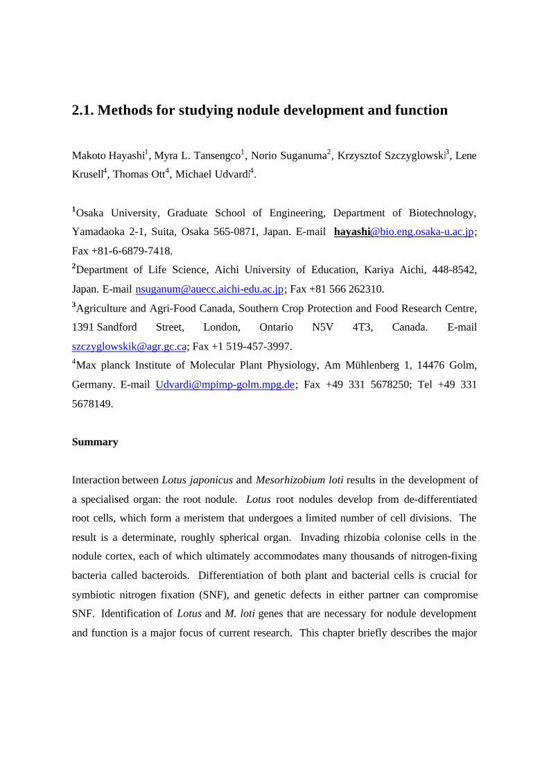
Like soybean, L. japonicus forms spherical root nodule structures of the determinate type
(Fig.1). These nodules are characterized by lack of
persistent meristematic activity, a developmental
feature which distinguishes them from the elongated,
indeterminate root nodules of temperate legumes
such as pea and alfalfa. However, the organogenesis
of L. japonicus nodules has been shown to combine
some features of indeterminate and determinate
nodule development (van Sprousen et al., 2001).
Lotus japonicus responds to M. loti by initiating a
complex developmental program for nodule organogenesis within the susceptible zone of
the root system. The morphogenic signals of bacterial origin, called nodulation (Nod)
factors, have been characterized from several different M. loti strains (Lopez-Lara et al.,
1995; Olsthoorm et al., 1998; Niwa et al., 2001). They were shown to consist of a mixture
Fig. 1

of lipochitin oligosaccharide (LCO) molecules that are synthesized and secreted from the
bacteria in response to an as yet uncharacterized inducer molecule(s), likely to represent
isoflavonoids, derived from Lotus roots. The major component of the LCO mixture was
found to be N-acetylglucosamine pentasaccharide in which the non-reducing residue is N-
acylated with a C18:1 acyl moiety, N-methylated, and carries a carbamoyl group, while the
reducing end is substituted with 4-0-acetylfucose (Niwa et al., 2001). Ectopic application
of the LCO mixture on L. japonicus roots incites various cellular and molecular responses
that are reminiscent of the early responses to Nod factors and/or rhizobial infection in other
legume species (Lopez-Lara et al., 1995; Niwa et al., 2001).
The earliest responses to Nod factors described in L. japonicus include membrane
depolarization, extra-cellular alkalinization and calcium spiking in root hair cells (Harris et
al., 2003; Radutoiu et al., 2003). These physiological reactions occur within a few minutes
upon application of purified Nod factors and/or living bacteria to the root system and were
shown to depend on the presence of a functional root perception apparatus that involves
NFR1/NFR5 LysM receptor-like kinases (Madsen et al., 2003; Radutoiu et al. 2003).
On the cytological level, cells of the epidermis, cortex and root pericycle respond to the
presence of M. loti and/or application of Nod
factors by initiating various growth patterns. In
the epidermis, root hair cells deform by tip
swelling, branching and curling, leading to the
formation of the typical “shepherd’s crook”
structures (Szczyglowski et al., 1998, Niwa et
al., 2001). These structures entrap the bacteria
and serve as a starting point for initiation and growth of the infection thread (IT). Detailed
microscopic examination of infected root hairs revealed the presence of bona fide ITs
originating at the curled tip and extending the full length of the root hair (Fig. 2;
Szczyglowski et al., 1998).
Fig. 2
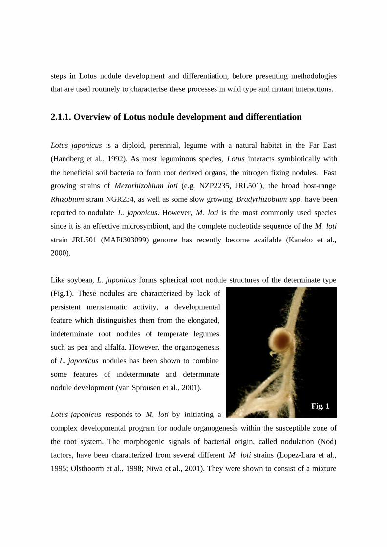
The L. japonicus ITs were shown to be tubular structures consisting of a multilayered
fibrillar cell wall, an overlying enclosure membrane continuous with the cytoplasmic
membrane of the infected host cell, and a lumen containing vegetative bacteria embedded
in an amorphous matrix (Fig. 3). Unlike many legumes, L. japonicus forms very broad
infection threads (Fig. 3A and 3B) that extend from infected root hairs through polarized
cortical cells of the outer cortex to the meristematic cells of the nodule primordium. Thick
and prominent infection threads, usually
observed in association with indeterminate-type
nodulation, spread between and inside Lotus
cortical root cells forming multiple, narrow
lateral branches that initiate host cell invasion
(Szczyglowski et al., 1998, van Spronsen et al.,
2001).
In the root cortex, the cells assume an
interesting position dependent pattern of
dedifferentiation. The outermost cortical cells
positioned immediately underneath the infected
root hairs either do not divide or make a single
anticlinal division. They undergo significant
swelling with a concomitant polarization of the
cytoplasm in a radial direction to gives rise to so called cytoplasmic bridges. (Niwa et al.,
2001, von Spronsen 2001). The formation of cytoplasmic bridges has been observed in the
first, second and only occasionally in the third cortical root cell layer (van Spronsen et al.,
2001). This type of cell polarization is assumed to guide the progression of the infection
thread towards the underlying nodule primordium (Szczyglowski and Amyot, 2003 and
references therein).
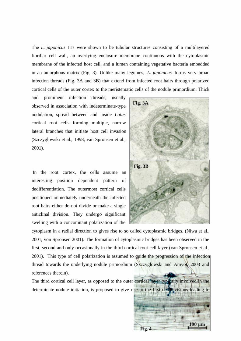
The third cortical cell layer, as opposed to the outer cortical layer typically involved in the
determinate nodule initiation, is proposed to give rise to the first cell divisions leading to
Fig. 3A
Fig. 3B
Fig. 4
the formation of nodule primordia in inoculated L. japonicus roots (van Spronsen et al.,
2001). The fully activated cortical cells of the inner cortex are characterized by enlarged,
centrally located, nuclei (Fig. 4), and contain trans-vacuolar cytoplasmic strands radiating
from the nucleus to the cell wall. These cells undergo successive divisions, which during
further development spread to surrounding cell layers, and eventually, give rise to a clearly
defined nodule meristem.
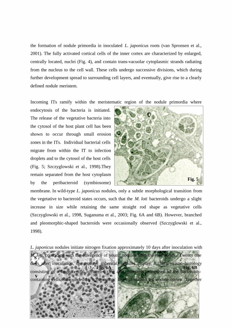
Incoming ITs ramify within the meristematic region of the nodule primordia where
endocytosis of the bacteria is initiated.
The release of the vegetative bacteria into
the cytosol of the host plant cell has been
shown to occur through small erosion
zones in the ITs. Individual bacterial cells
migrate from within the IT to infection
droplets and to the cytosol of the host cells
(Fig. 5; Szczyglowski et al., 1998).They
remain separated from the host cytoplasm
by the peribacteroid (symbiosome)
membrane. In wild-type L. japonicus nodules, only a subtle morphological transition from
the vegetative to bacteroid states occurs, such that the M. loti bacteroids undergo a slight
increase in size while retaining the same straight rod shape as vegetative cells
(Szczyglowski et al., 1998, Suganuma et al., 2003; Fig. 6A and 6B). However, branched
and pleomorphic-shaped bacteroids were occasionally observed (Szczyglowski et al.,
1998).
L. japonicus nodules initiate nitrogen fixation approximately 10 days after inoculation with
M. loti, coinciding with the emergence of young nodules from the root cortex. Twenty one
days after inoculation, the mature, spherical, nodules display a characteristic histology
consisting of a large central nitrogen fixing area, primarily composed of the bacteroids-
containing cells, surrounded by a concentric layer of uninfected nodule cortex. Together
Fig. 5
Fig. 6A Fig. 6B
with peripherally located vascular elements and the apparent lack of a discernible
meristem, these cytological features typify a determinate type of nodule (Szczyglowski et
al., 1998).
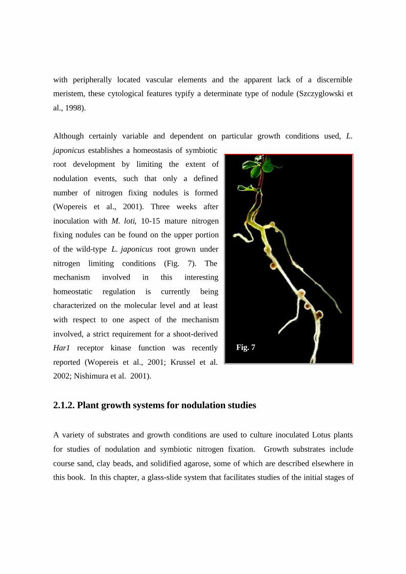
Although certainly variable and dependent on particular growth conditions used, L.
japonicus establishes a homeostasis of symbiotic
root development by limiting the extent of
nodulation events, such that only a defined
number of nitrogen fixing nodules is formed
(Wopereis et al., 2001). Three weeks after
inoculation with M. loti, 10-15 mature nitrogen
fixing nodules can be found on the upper portion
of the wild-type L. japonicus root grown under
nitrogen limiting conditions (Fig. 7). The
mechanism involved in this interesting
homeostatic regulation is currently being
characterized on the molecular level and at least
with respect to one aspect of the mechanism
involved, a strict requirement for a shoot-derived
Har1 receptor kinase function was recently
reported (Wopereis et al., 2001; Krussel et al.
2002; Nishimura et al. 2001).
2.1.2. Plant growth systems for nodulation studies
A variety of substrates and growth conditions are used to culture inoculated Lotus plants
for studies of nodulation and symbiotic nitrogen fixation. Growth substrates include
course sand, clay beads, and solidified agarose, some of which are described elsewhere in
this book. In this chapter, a glass-slide system that facilitates studies of the initial stages of
Fig. 7
infection, and a plastic ‘pillow’ system for the production of mature nodulated plants are
described.
2.1.2.1. Glass slide culture of seedlings for microscopy
Liquid culture of seedlings on a slide glass is useful for observation of roots under a
microscope without damaging root hairs. This technique can be adopted for root hair
deformation assay by application of LCOs (Niwa et al., 2001) or inoculation of M. loti, or
pharmaceutical assays. This system is useful for studying early infection and nodulation
events, but is not appropriate for later stages when plants become too large.
Equipment and reagents
• Glass slides and cover slips
• Silicone caulk
• Staining jars
• Forceps
• B & D medium
Method
A. Preparation of Fåhraeus slides
1. Put roughly 10 µl of silicone caulk at each corner of a cover slip.
2. Set the cover slip on a glass slide in a way keeping 1 mm space between them, and
1 cm above from the bottom of the glass slide (Fig. 8).
3. Let them still for a day in order to solidify silicone.
4. Sterilize by autoclave in a glass jar filled with distilled water.
B. Transfer of seedlings to the slides
1. Put 50 ml of B & D medium in a sterilized staining jar.
2. Using forceps, transfer seedlings to the slide one by one, not to damage on the root
hair (Fig. 9). Select seedlings only those have straight roots (Miyakojima; 2 days-
old, Gifu; 3 days-old as in the section of spot inoculation).
3. Insert slides with seedlings to the staining jar (Fig. 10), cover the bottom half of the
jar with black paper and aluminum foil to avoid light to roots (Fig. 11), culture
them at 26ºC-16h-light/23ºC-8h-dark regime.
Notes
1. We set seedlings 4 days before applying Nod factors or other chemicals in order to
avoid potential wounding response.
2.1.2.2. The ‘pillow’ growth system
The pillow system allows effective nodulation analysis of a large plant population with a
relatively small area needed for growing the samples. As several plants can be grown
simultaneously, the time-consuming cleaning of root tissue is reduced substantially.
To observe and screen plants for nodule phenotype, it is necessary to germinate and
establish the plants on agar media, then transplant them to soil after about a week. Root
growth and nodulation kinetics using this method resemble those observed in plants grown
in vermiculite/sand mixtures.
I. Sowing of seeds in agar medium
Equipment and reagents
• Plastic tray
• 50 ml plastic tube
• Germination medium: 0.8% (w/v) Bacto agar
• Bleach solution: 10% NaClO in sterile distilled water
• Sterile distilled water
• Sterile filter paper
• Sterile petri dishes or rectangular plastic plates
• Parafilm
Method
1. Prepare the germination medium by autoclaving at 121oC for 15 min and then pour into
pre-sterilized petri dishes.
2. Place the seeds in a 50 ml plastic tube and add 10-15 ml concentrated sulfuric acid.
Vortex vigorously for 10-15 min and then pour the solution into a container with tap
water. Rinse the seeds at least three times.
3. Put the seeds in a clean plastic tube and add the bleach solution. Shake the container
gently for 10 min.
4. Pour out the bleach solution and then wash 4-5 times with sterile water. Keep the seeds
in sterile distilled water and shake the container gently for 2-3 hr, then discard the
water.
5. Place the seeds in sterile filter paper to dry, and transfer the seeds in germination
medium (work in laminar flow hood and use sterile forceps). Seal the petri plates with
parafilm and incubate in growth chamber under 16-h-light/26oC and 8-h-dark/23oC
cycle with 60oC relative humidity.
6. Keep the plates under dark condition for 2 days and then allow the seeds to germinate
for another 7-10 days (i.e. once two to four true leaves have formed).
II. Growing of seedlings in the pillow system
Equipment and reagents

• Plastic tray: 30 x 10 x 10 cm
• Pillow bags: polypropylene tea packs (120 x 95 mm) or nylon bags
• Vermiculite:perlite mix (6:1, v/v)
• Broughton and Dilworth (B&D) medium (Table 1)
• 10 µM KNO3
• Bleach solution: 10% commercial bleach diluted in distilled water
• Saran wrap
• 2 to 3-day-old culture of Mesorhizobium loti strain
Method
1. Fill the pillow bags with vermiculite:perlite mix and autoclave at 121oC for 20 min.
2. Soak the pillows in sterile B&D nutrient solution containing minimal nitrate concen-
tration (10 µM KNO3) until the substrate has absorbed the liquid.
3. Sterilize the plastic tray with bleach solution and wash several times with distilled water.
Place 10-15 pillow bags side by side in the plastic tray (Fig. 12).
4. Sow the seedlings in-between individual pillows (five plants per row, Fig. 13).
5. Incubate the trays in growth chamber. Cover the trays with Saran wrap for a few days to
maintain high humidity.
6. After 2 days in the “pillow system”, inoculate roots with M. loti culture at a cell density
of 108 cells/ml.
7. Water the plants with B&D nutrient solution (without nitrate) for at least 2 weeks and
then with distilled water for the succeeding weeks. Avoid having the growth condition
overly wet, or overly dry.
8. For nodule examination, carefully remove the pillows individually taking care not to
damage or cut the nodulated root parts.
9. Place the plants in a clean dish with water and examine by stereomicroscopy.
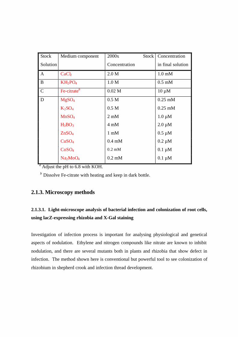
Table 1. Broughton and Dilworth (1971) nutrient solutiona
Stock
Solution
Medium component 2000x Stock
Concentration
Concentration
in final solution
A CaCl2 2.0 M 1.0 mM
B KH2PO4 1.0 M 0.5 mM
C Fe-citrateb 0.02 M 10 µM
D MgSO4 0.5 M 0.25 mM
K2SO4 0.5 M 0.25 mM
MnSO4 2 mM 1.0 µM
H3BO3 4 mM 2.0 µM
ZnSO4 1 mM 0.5 µM
CuSO4 0.4 mM 0.2 µM
CoSO4 0.2 mM 0.1 µM
Na2MoO4 0.2 mM 0.1 µM a Adjust the pH to 6.8 with KOH. b Dissolve Fe-citrate with heating and keep in dark bottle.
2.1.3. Microscopy methods
2.1.3.1. Light-microscope analysis of bacterial infection and colonization of root cells,
using lacZ-expressing rhizobia and X-Gal staining
Investigation of infection process is important for analysing physiological and genetical
aspects of nodulation. Ethylene and nitrogen compounds like nitrate are known to inhibit
nodulation, and there are several mutants both in plants and rhizobia that show defect in
infection. The method shown here is conventional but powerful tool to see colonization of
rhizobium in shepherd crook and infection thread development.
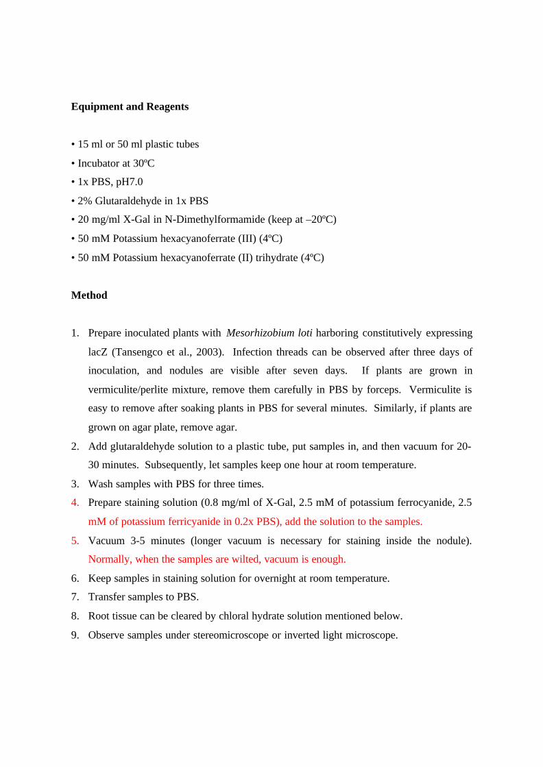
Equipment and Reagents
• 15 ml or 50 ml plastic tubes
• Incubator at 30ºC
• 1x PBS, pH7.0
• 2% Glutaraldehyde in 1x PBS
• 20 mg/ml X-Gal in N-Dimethylformamide (keep at –20ºC)
• 50 mM Potassium hexacyanoferrate (III) (4ºC)
• 50 mM Potassium hexacyanoferrate (II) trihydrate (4ºC)
Method
1. Prepare inoculated plants with Mesorhizobium loti harboring constitutively expressing
lacZ (Tansengco et al., 2003). Infection threads can be observed after three days of
inoculation, and nodules are visible after seven days. If plants are grown in
vermiculite/perlite mixture, remove them carefully in PBS by forceps. Vermiculite is
easy to remove after soaking plants in PBS for several minutes. Similarly, if plants are
grown on agar plate, remove agar.
2. Add glutaraldehyde solution to a plastic tube, put samples in, and then vacuum for 20-
30 minutes. Subsequently, let samples keep one hour at room temperature.
3. Wash samples with PBS for three times.
4. Prepare staining solution (0.8 mg/ml of X-Gal, 2.5 mM of potassium ferrocyanide, 2.5
mM of potassium ferricyanide in 0.2x PBS), add the solution to the samples.
5. Vacuum 3-5 minutes (longer vacuum is necessary for staining inside the nodule).
Normally, when the samples are wilted, vacuum is enough.
6. Keep samples in staining solution for overnight at room temperature.
7. Transfer samples to PBS.
8. Root tissue can be cleared by chloral hydrate solution mentioned below.
9. Observe samples under stereomicroscope or inverted light microscope.
Notes
For observation of infection threads, shorter period (90 min to 3 hrs) of staining is better,
because endogenous activity of lacZ in root stele interferes the observation of stained
infection threads.
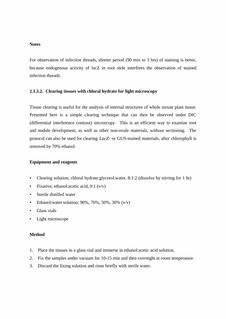
2.1.3.2. Clearing tissues with chloral hydrate for light microscopy
Tissue clearing is useful for the analysis of internal structures of whole mount plant tissue.
Presented here is a simple clearing technique that can then be observed under DIC
(differential interference contrast) microscopy. This is an efficient way to examine root
and nodule development, as well as other non-ovule materials, without sectioning. The
protocol can also be used for clearing LacZ- or GUS-stained materials, after chlorophyll is
removed by 70% ethanol.
Equipment and reagents
• Clearing solution; chloral hydrate:glycerol:water, 8:1:2 (dissolve by stirring for 1 hr)
• Fixative; ethanol:acetic acid, 9:1 (v/v)
• Sterile distilled water
• Ethanol/water solution; 90%, 70%, 50%, 30% (v/v)
• Glass vials
• Light microscope
Method
1. Place the tissues in a glass vial and immerse in ethanol:acetic acid solution.
2. Fix the samples under vacuum for 10-15 min and then overnight at room temperature.
3. Discard the fixing solution and rinse briefly with sterile water.
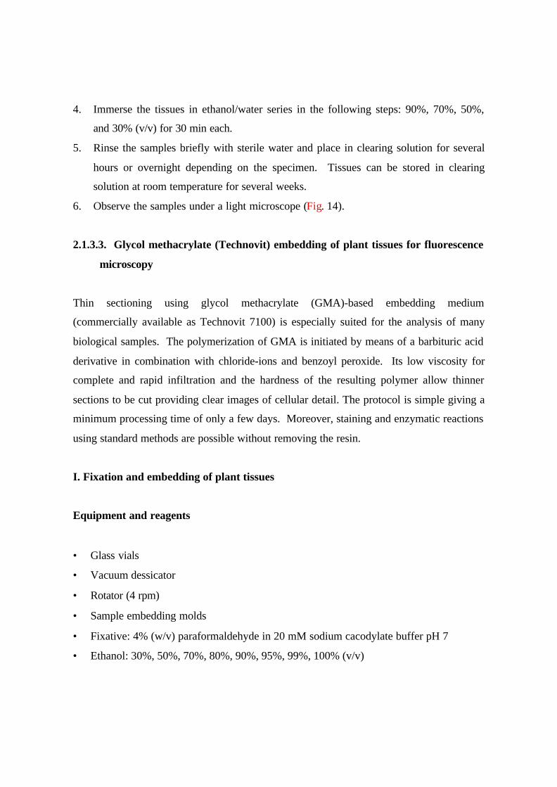
4. Immerse the tissues in ethanol/water series in the following steps: 90%, 70%, 50%,
and 30% (v/v) for 30 min each.
5. Rinse the samples briefly with sterile water and place in clearing solution for several
hours or overnight depending on the specimen. Tissues can be stored in clearing
solution at room temperature for several weeks.
6. Observe the samples under a light microscope (Fig. 14).
2.1.3.3. Glycol methacrylate (Technovit) embedding of plant tissues for fluorescence
microscopy
Thin sectioning using glycol methacrylate (GMA)-based embedding medium
(commercially available as Technovit 7100) is especially suited for the analysis of many
biological samples. The polymerization of GMA is initiated by means of a barbituric acid
derivative in combination with chloride-ions and benzoyl peroxide. Its low viscosity for
complete and rapid infiltration and the hardness of the resulting polymer allow thinner
sections to be cut providing clear images of cellular detail. The protocol is simple giving a
minimum processing time of only a few days. Moreover, staining and enzymatic reactions
using standard methods are possible without removing the resin.
I. Fixation and embedding of plant tissues
Equipment and reagents
• Glass vials
• Vacuum dessicator
• Rotator (4 rpm)
• Sample embedding molds
• Fixative: 4% (w/v) paraformaldehyde in 20 mM sodium cacodylate buffer pH 7
• Ethanol: 30%, 50%, 70%, 80%, 90%, 95%, 99%, 100% (v/v)
• Technovit 7100 embedding kit (Heraeaus Kulzer, Wehrheim, Germany)
• Technovit 3020 kit (Heraeaus Kulzer)
Method
A. Fixation
1. Cut small pieces of tissue (<5 mm). Try to keep the sample size small to facilitate
processing, i.e. fixation, infiltration, and sectioning.
2. Prepare fresh 4% (w/v) paraformaldehyde solution by dissolving the powder to the
buffer with heating in fume hood and add 1-2 drops of 1N NaOH solution. Adjust
pH after cooling down. Fix samples for 30 min under vacuum.
3. Change to fresh fixative and fix overnight at 4oC.
B. Dehydration
1. Dehydrate tissue in an ethanol/water series at RT in the following steps: twice in
30%, 50%, 70%, 80% (v/v) for 15 min each, twice in 90%, 95%, 99% for 5 min
each, then in 100% for 1 hr.
2. Use a rotator (4 rpm) or a rocking platform for agitation.
C. Infiltration
1. Prepare infiltration solution consisting of 100 ml Technovit resin (2-hydroxyethyl
methacrylate) and 1 g Hardener I (benzoyl peroxide, as accelerator).
2. Mix until the powder is completely dissolved and store solution in dark bottle. At
4oC, the solution remains stable for approximately 4 weeks (avoid air).
3. Immerse the tissue in fresh 1 ml absolute ethanol and add 250 µl infiltration
solution every 15 min.
4. Mix the sample after adding the infiltration solution and keep the tissue vials in
rotator.
5. Remove half volume of the solution every hour (for a total of 3 hr) making the
solution 50% infiltration solution after 1 hr, 75% after 2 hr, and 82.5% after 3 hr.
6. Leave the sample in 100% infiltration solution overnight in a rotator.
7. Change to fresh infiltration solution and leave for 2-3 hr with agitation.
D. Embedding
1. Prepare embedding solution consisting of 15 parts of infiltration solution and 1
part of Hardener II.
2. Mix the solution and immediately pour in a suitable mold (we use a plastic film
case) and add the samples. Arrange the specimen appropriately and allow
polymerizing for at least several hours.
E. Mounting
1. Mix Technovit 3040 in a volume ratio of 2 parts powder to 1 part liquid.
2. Mix vigorously and immediately pour on top of the embedded sample.
3. Remove the block from the mould after about 10 min. If the sample remains soft
due to high temperature and humidity, store the samples in auto-dry desiccators for
one to several days.
II. Sectioning and staining
Equipment and reagents
• Fret saw
• Epoxy glue
• Plastic or metal stubs
• Razorblade
• Ultramicrotome
• Sterile distilled water
• Glass slides and cover slips
• Forceps
• Hotplate
• 0.05% Toluidine Blue O in 1x PBS (137 mM NaCl, 8.10 mM Na2HPO4·12H2O, 2.68
mM KCl, 1.47 mM KH2PO4, pH 7.4)
• DAPI (4’,6-diamidino-2-phenylindole) staining solution: 10 µg/ml in vectashield
antifade mounting medium (Vector Laboratories, Inc.) with 5 µg/ml fluorescent
brightener 28 (Sigma-Aldrich)
Method
1. Separate embedded tissues in plastic blocks by sawing the block into small pieces
using a fret saw.
2. Mount the blocks onto metal or plastic stubs using epoxy glue.
3. Trim the edges of the block to a rectangular face using a razor blade.
4. Prepare triangular glass knives using a knife maker machine.
5. Set the block and glass knife in the ultramicrotome such that the cutting face of the
block is parallel with the knife-edge.
6. Cut sections at 1-2 µm thick and float individual sections one at a time on distilled
water on a cover slip by forceps.
7. Handle each section with a forceps and let go sections just before it touches the water.
8. Place cover slips at 50oC on a hot plate and let the water evaporate.
9. Stain with toluidine blue solution and observe under brightfield light microscopy (Fig.
15). Alternatively, for fluorescent microscopy, stain with DAPI solution containing
fluorescent brightener to stain the nuclei and cell wall, respectively (Fig. 16).
2.1.3.4. Spot inoculation
To investigate the initial events of nodule development, spot inoculation is a powerful
method in terms of temporal observation because it is easy to identify the focus of nodule
initiation. In contrast to the slide inoculation method described below, it is not easy to
evaluate the quantity of root hair deformation using this method, but the slide inoculation

hardly induces nodules and they are not spatially fixed. For inducing nodule primordia,
one can apply rhizobium (Imaizumi-Anraku et al., 2000) as well as LCOs, but the latter
fail to induce complete (empty) nodules (Niwa et al., 2001). In general, MG-20
‘Miyakojima’ forms bigger nodule primordia than B-129 ‘Gifu’ accession by LCOs spot.
For inoculation, we use agarose for matrix to spot rhizobium or LCOs, other application
method using quartz sands (van Spronsen et al., 2001) however would be also possible.
Equipment and reagents
• Square Petri dish (either 8 cm x 12 cm or 8 cm square)
• Low melting agarose (SeaPlaque GTG agarose, BMA)
• TY medium
• B & D medium
• LCOs mixture from M. loti JRL501 harboring pMP2112 (Niwa et al., 2001)
Method
A. Preparation of seedlings for spot inoculation
1. Treat seeds with sulfuric acid (conc) for 5 to 8 min (depending on the seed
condition) followed by extensive washing by tap water. Scarification by a piece of
sandpaper is the alternative.
2. Sterilize seeds with sodium hypochlorite solution (1% active chlorine) for less than
10 min, and rinse by sterilized water for three times.
3. Imbibe seeds in sterile water for a few hours at room temperature, with gentle
shaking.
4. Transfer seeds to Petri dish containing 0.8% Bacto Agar (BD). Cover the dish with
aluminum foil. Incubate at 26ºC-16h-light/23ºC-8h-dark regime. Remove foil after
one day (Miyakojima) or two days (Gifu), and incubate one extra day.
5. Transfer seedlings to the square Petri dish containing B & D medium solidified
with 1% Bacto Agar.
6. Cover the dish with black paper and aluminum foil (leave top 1 cm open), put the
dish vertically, and incubate for 2 days (Fig. 17). Addition of AVG is of choice,
use 10 µM in case.
B. Preparation of spot solution
Rhizobium:
1. Inoculate M. loti from plate to 3 ml of TY liquid medium, culture at 28ºC for 2
days.
2. Spin down culture by centrifuge at 8000 rpm, 2 min, 4ºC.
3. Wash pellet with saline (0.8% NaCl) for 2 times, adjust the concentration to
O.D.600=0.1.
4. Mix 25µl of the suspension with 50 µl of sterilized low melting agarose (1%).
LCOs:
5. Mix 25µl of LCOs (10-5 to 10-3M) with 50 µl of sterilized low melting agarose
(1%).
C. Spot inoculation
1. Pipette out 10µl of the solution above, and spot 1 µl of it to the root tip, using
micropippete.
2. Wrap the rid of the dish with Parafilm. Use surgical tape (Micropore: 3M) at the
top to avoid excess humidity.
3. Set black paper and aluminum foil again as above, and incubate.
4. After one week, developing nodule can be observed by naked eye (rhizobium
inoculation). In case of LCOs, 10 days are enough to recognize the nodule under
stereomicroscope.
Notes
1. Higher concentration of LCOs bring better nodulation, still it is not 100%.
Normally more than 50% of the spotted seedlings show nodulation.
2. Nodule can be observed 0 to 2 cm below the spot point.
3. In the original papers, the authors recommend to add black ink so that the spot
point can be easily recognized. In our experience, however, some of black ink
inhibits root elongation, so we do not add it. Holding the dish to the light can
identify the spot point.
2.1.4. Acetylene reduction assay for nitrogenase activity
Biological nitrogen fixation is assessed by several methods, such as Kjeldahl analysis, 15N-
incorporation assay, acetylene reduction assay and H2-evolution assay. Among these
methods, the closed acetylene reduction assay is simple, rapid and sensitive (Hardy et al.
1968; Vessey 1994), and has been widely used for estimating the nitrogen-fixing activity
of legumes. This assay is based on the fact that nitrogenase has a higher affinity for
acetylene and in the presence of acetylene total electron flow through nitrogenase is
virtually used to reduce acetylene to ethylene. The ethylene produced can be detected by
hydrogen flame ionization after gas chromatography (Fig. 18).
Reagents and apparatus
• Acetylene
• Ethylene (for standard)
• 35-mL vial (Any vials with different volumes are available if the vial can be sealed with a
cap, from which a gas sample can be withdrawn with a syringe.)
• Serum cap
• 1-mL disposable syringe with a needle (25G x 1’’)
• Glass column (2 mm i.d. x 2 m) packed with Porapak N (Waters, Milford, MA, USA)
• Gas chromatograph equipped with a hydrogen flame-ionization detector (FID)
• Integrator (for determination of the peak area separated by gas chromatography).
Procedures
• Detach a nodulated root from a freshly harvested plant.
• Place the nodulated root in a 35-mL vial and close with a serum cap. A single nodulated
root is enough for determination of activity using the 35-mL vial.
• Withdraw 3.5 mL of air from the closed vial and inject the same volume of acetylene gas
to be 0.1 atmosphere of acetylene in the vial.
• Immediately withdraw 0.5 mL of gas sample from the closed vial and measure the
concentration of ethylene contaminated in the 0.1 atmosphere of acetylene injected in the
vial with gas chromatograph.
• Incubate the closed vial containing the nodulated root in darkness at 25°C.
• After 30 min, withdraw 0.5 mL of gas sample and measure the concentration of ethylene.
• Based on standard ethylene, calculate the ethylene produced during the incubation after
subtracting the ethylene contaminated in acetylene gas.
Notes
• Acetylene cylinder contains a small amount of ethylene and the ethylene impurity should
be determined daily since it varies inversely with the pressure in the cylinder.
• Preparations of tissue seriously affect acetylene reduction activity (Vessey 1994). The
least disturbed and more intact tissue should be used in acetylene reduction assay.
• Rates of acetylene reduction are linear up to 60 minutes, but it is recommended that the
assay time should be kept to within 30 minutes (Hardy et al. 1968).
• Nodulated roots evolve endogenous ethylene (Suganuma et al. 1995a), though the
amounts of evolution are thought to be negligible compared with acetylene reduction
activity determined within 30 min.
• Replacement of air (N2) with Ar:O2 slightly increase acetylene reduction activity (Hardy
et al. 1968). If required, air in each vial should be replaced by introducing a mixture of
Ar:02 (0.8:0.2 atmosphere) gas before adding acetylene gas.
• Greater information concerning nitrogenase assay is referred to ‘Methods for Evaluating
Biological Nitrogen Fixation’ edited by F. J. Bergersen (1980) John Wiley & Sons, Inc,
New York, NY.
2.1.5. Molecular markers and measuring methods
Nodule development and differentiation are orchestrated by changes in gene expression
that occur in a precise temporal sequence. ‘Nodule-specific’ plant genes that are induced
within hours or days after contact with rhizobia are called early nodulin genes, while those
that are induced a week or so later, just prior to or following the start of nitrogen fixation
are called late nodulin genes. Early nodulins are believed to play important roles during
bacterial colonisation or nodule development, while late nodulins presumably fulfil
structural or functional roles during differentiation and/or nitrogen fixation. Sequencial
changes in rhizobial gene expression also accompany nodule development and
differentiation. Because of their well-defined temporal expression patterns, many plant
and bacterial nodule-induced genes, or the proteins that they encode, have been used as
molecular markers for different stages of nodule development. Such markers are
especially useful for studies with plant or bacterial mutants that are defective in nodulation
or nitrogen fixation, as they help to identify the stage at which nodule development or
differentiation is impaired. Expression of marker genes in wild type and ineffective
nodules has been examined by Northern analysis (Purdom and Trese 1995; Kato et al.
2002), in situ hybridization (Banba et al. 2001; Kawashima et al. 2001), immunoblotting
(Egli et al. 1991; Romanov et al. 1995; Suganuma et al. 2003) and use of bacterial reporter
gene fusions (Voroshilova et al. 2001). The following two sub-sections describe the
methods of real-time RT-PCR and immunoblotting for measuring gene transcript and
protein levels, respectively.
2.1.5.1. Transcript profiling using real-time RT-PCR
Real-time reverse transcription-polymerase chain reaction (RT-PCR) is an extremely
sensitive and quantitative method of measuring gene transcript levels that can be
multiplexed to handle transcripts of many different genes in parallel. However, care must
be taken to remove all genomic DNA from isolated RNA prior to RT-PCR measurements
(Czechowski et al., 2004).
Materials and Methods
Harvested plant material is typically frozen immediately in liquid nitrogen and stored at –
70 °C, prior to use. Total RNA is extracted from frozen organs of Lotus plants using an
RNeasy kit (Qiagen, Hilden, Germany). Prior to cDNA synthesis, 1-2 µg of total RNA is
treated with RNase-free DNaseI (Sigma, St Louis, MI, USA), to destroy genomic DNA.
DNAseI-treated RNA is subsequently phenol-chloroform extracted, precipitated, and
resuspended in 10 µL water. PCR, using gene-specific primers, is performed on the
resulting RNA to confirm the absence of DNA. Reverse transcription is then performed
with SuperScript III reverse transcriptase (Invitrogen, Carlsbad, CA, USA) usinng an
oligo(dT) primer. The efficiency of cDNA synthesis is assessed by real-time PCR
amplification of ubiquitin (see Table 2), and only those reactions that exhibit similar
efficiencies (similar CT values for ubiquitin) are analysed further.
PCR reactions can be performed in a 96-well plate with an Applied Biosystems ABI Prism
5700 Sequence Detection System (Foster City, CA, USA), using SYBR Green to monitor
dsDNA synthesis. A standard PCR reaction contains approximately 4 ng cDNA template,
10 µL of SYBR Green Mix (Applied Biosystems, Foster City CA, USA), 5 pmol forward
and reverse primers, and water to a final volume of 20 ìL. The following standard thermal
profile is used for all PCR reactions: 50°C for 2 min; 95 °C for 10 min; 40 cycles of 95 °C

for 15 sec and 60 °C for 1 min. Data is analysed using SDS 2.0 software (Applied
Biosystems, Foster city, CA, USA). For quality control, PCR products are run on a 3 %
agarose gel to confirm the presence of a single amplicon. Gene-specific primers can be
designed with PrimerExpress software (Applied Biosystems, Foster city, CA, USA), and
typically have melting temperatures of 60 +/- 2 and GC content of between 45-55%.
Primers are designed to produce short amplicons, typically between 60-150 bp to ensure
high PCR efficiencies.
Threshold cycle (CT) values for each gene transcript are normalized to those of
polyubiquitin (GenBank AW720576; see Table 2) before making comparisons between
samples (Colebatch et al. 2002). Gene expression ratios are calculated using the following
equations:
nCT = CT – CTLjUbi
∆CT = nCT x – nCTy
nRatio = 2- ∆ CT
Where CT is the PCR cycle number at which a set threshold value (usually 0.1) of SYBR
Green fluorescence is reached, nCT is the normalised CT value, ∆CT is the difference in
normalised CT values in sample x and y, and Ration is the ratio of gene transcript level in
sample x compared to y. The last equation assumes a PCR efficiency (E) of 100%.
Generally this is not the case, and several methods are available to estimate PCR
efficiency. The first method of measuring PCR efficiency uses template dilutions and the
equation (1+E)=10(-1/slope), as described previously (Pfaffl et al., 2001). The second method
uses data obtained from the exponential phase of each individual amplification plot and the
equation (1+E)=10slope (Ramakers et al., 2003). If E is known, the gene expression ratio
can be calculated using the equation:
nRatio = (1+E) - ∆ CT
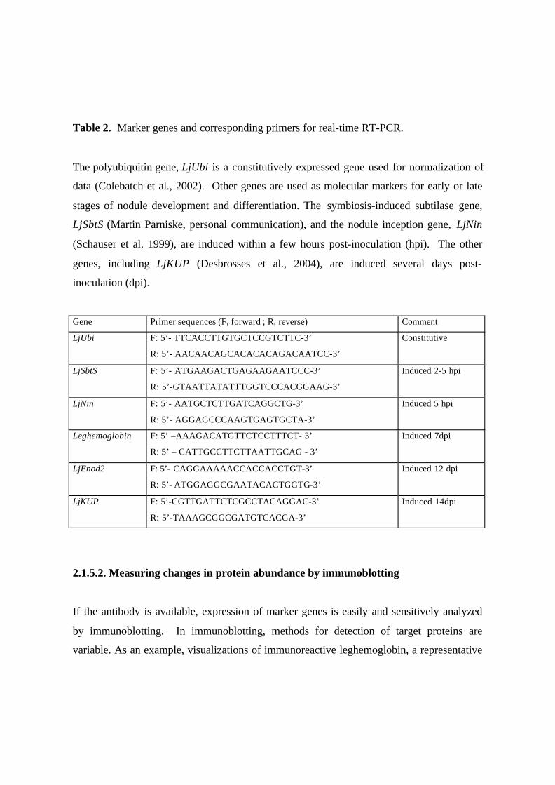
Table 2. Marker genes and corresponding primers for real-time RT-PCR.
The polyubiquitin gene, LjUbi is a constitutively expressed gene used for normalization of
data (Colebatch et al., 2002). Other genes are used as molecular markers for early or late
stages of nodule development and differentiation. The symbiosis-induced subtilase gene,
LjSbtS (Martin Parniske, personal communication), and the nodule inception gene, LjNin
(Schauser et al. 1999), are induced within a few hours post-inoculation (hpi). The other
genes, including LjKUP (Desbrosses et al., 2004), are induced several days post-
inoculation (dpi).
Gene Primer sequences (F, forward ; R, reverse) Comment
LjUbi F: 5’- TTCACCTTGTGCTCCGTCTTC-3’
R: 5’- AACAACAGCACACACAGACAATCC-3’
Constitutive
LjSbtS F: 5’- ATGAAGACTGAGAAGAATCCC-3’
R: 5’-GTAATTATATTTGGTCCCACGGAAG-3’
Induced 2-5 hpi
LjNin F: 5’- AATGCTCTTGATCAGGCTG-3’
R: 5’- AGGAGCCCAAGTGAGTGCTA-3’
Induced 5 hpi
Leghemoglobin F: 5’ –AAAGACATGTTCTCCTTTCT- 3’
R: 5’ – CATTGCCTTCTTAATTGCAG - 3’
Induced 7dpi
LjEnod2 F: 5’- CAGGAAAAACCACCACCTGT-3’
R: 5’- ATGGAGGCGAATACACTGGTG-3’
Induced 12 dpi
LjKUP F: 5’-CGTTGATTCTCGCCTACAGGAC-3’
R: 5’-TAAAGCGGCGATGTCACGA-3’
Induced 14dpi
2.1.5.2. Measuring changes in protein abundance by immunoblotting
If the antibody is available, expression of marker genes is easily and sensitively analyzed
by immunoblotting. In immunoblotting, methods for detection of target proteins are
variable. As an example, visualizations of immunoreactive leghemoglobin, a representative
late nodulin, and nitrogenase in bacteroids with a ProtoBlot immunoblotting system
(Promega, Madison, WI, USA) are described (Fig. 19).
Reagents and Apparatus
• Grinding medium (50mM Tris-HCl pH7.4, 0.3M sucrose, 1mM EDTA, 5mM DDT,
1mM PMSF)
• Polyclar AT
• PVDF membrane (Immobilon-P membrane; Millipore, Bedford, MA, USA)
• TBS (20mM Tris-HCl pH 7.5, 150mM NaCl)
• TBST (20mM Tris-HCl pH 7.5, 150mM NaCl, 0.05% Tween 20)
• Blocking solution (1% (w/v) BSA in TBST)
• Alkaline phosphate buffer (100mM Tris-HCl pH 9.5, 100mM NaCl, 5mM MgCl2)
• Color development solution (BCIP/NBT in alkaline phosphate buffer)
• Primary antibody
• Anti-IgG secondary antibody conjugated to alkaline phosphatase
• Sonicator
• Electrophoresis system
• Blotting apparatus
Procedures
1. Preparation of extracts for crude bacteroids and for plant cytosol
• Homogenize nodules with a mortar and pestle in grinding medium with Polyclar AT
powder in a ratio of 0.3 g per g fresh weight of nodules.
• Squeeze the homogenate through four layers of gauze and centrifuge the filtrate at 200 xg
for 10 min at 4°C.
• Centrifuge the supernatant at 5,000 xg for 10 min at 4°C and wash the pellet with
grinding medium three times.

• Sonicate the final suspension ten times for 10 sec each time at 100W and centrifuge at
16,000 xg for 30 min at 4°C. The resulting supernatant is used for immunoblotting as
bacteroid fraction.
• Centrifuge the supernatant, obtained by the centrifugation at 5,000 xg for 10 min, at
16,000 xg for 30 min at 4°C. The resulting supernatant is used for immunoblotting as plant
cytosol fraction.
2. Electrophoresis and transfer to membrane
• Separate the polypeptides by SDS-PAGE (polyacrylamide gel electrophoresis) on a 12%
(w/v) polyacrylamide gel. Bacteroid fraction is used for detection of nitrogenase and plant
cytosol fraction is for detection of leghemoglobin, respectively.
• Transfer the polypeptides separated by SDS-PAGE to PVDF membranes
electrophretically (Towbin et al. 1979).
3. Immunodetection
• Incubate the membrane in blocking solution for 60 min to block nonspecific protein
binding.
• Incubate the membrane in TBST containing the appropriate dilution of primary antibody
for 30 min with gentle agitation. Antibodies against nitrogenase components I and II
isolated from Rhizobium leguminosarum bv viciae bacteroids (Bisseling et al 1980) were
diluted to 1:10,000 and that against leghemoglobin isolated from soybean nodules
(Suganuma et al. 1995b) was diluted to 1:10,000.
• Wash the membrane three times in TBST for 10 min each.
• Incubate the membrane in TBST containing the appropriate dilution of anti-IgG alkaline
phosphatase conjugate for 30 min with gentle agitation.
• Wash the membrane three times in TBST for 10 min each.
• Rinse the membrane briefly in TBS two times.
• Incubate the membrane in color development solution.
• Stop the reaction by washing the membrane in deionized water for 15 min when the color
has developed to the desired intensity.
Notes
• PVDF membrane, a hydrophobic membrane, must be prewet first in 100% methanol or
ethanol. After prewetting, submerge the membrane to blotting solution until use.
• Do not dry out the membrane during any steps of the procedures after prewetting.
• You can store the membrane covered with a plastic sheet at 4°C after blotting. For the
dried PVDF membrane, first rewet in methanol or ethanol followed by floating in TBS.
• Detailed procedures for electrophoresis and electrophoretic transfer are usually included
with commercial devices.
References
Banba M, Siddique ABM, Kouchi H, Izui K, Hata S (2001) Lotus japonicus forms early
senescent root nodules with Rhizobium etli. Mol Plant-Microbe Interact 14:173-180.
Berleth T, Jurgens G (1993) The role of the monopteros gene in organizing the basal body
region of the Arabidopsis embryo. Development 118: 575-587.
Bisseling T, Moen AA, van den Bos RC, van Kammen A (1980) The sequence of
appearance of leghaemoglobin and nitrogenase components I and II in root nodules of
Pisum sativum L. J Gen Microbiol 118:377-381.
Broughton.Wj, and Dilworth, M.J. (1971). Control of Leghaemoglobin Synthesis in Snake
Beans. Biochemical Journal 125, 1075-1080.
Colebatch, G., Kloska, S., Trevaskis, B., Freund, S., Altmann, T., and Udvardi, M.K.
(2002b). Novel aspects of symbiotic nitrogen fixation uncovered by transcript profiling
with cDNA arrays. Molecular Plant-Microbe Interactions 15, 411-420.
Czechowski, T., Bari, R.P., Stitt, M., Scheible W-R., and Udvardi, M.K. (2004) Real-time
RT-PCR profiling of over 1,400 Arabidopsis transcription factors: unprecedented
sensitivity reveals novel root- and shoot-specific genes. Plant J 38, 366-79.
Desbrosses, G., Kopka, C., Ott, T., and Udvardi, M.K. (2004) Lotus japonicus LjKUP
encodes a potassium transporter of the plasma membrane and is induced significantly
during nodule organogenesis. Molecular Plant-Microbe Interactions (in press).
Egli MA, Larson RJ, Hruschka WR, Vance CP (1991) Synthesis of nodulins and nodule-
enhanced polypeptides by plant gene-controlled ineffective alfalfa nodules. J Exp Bot
42:969-977.
Gerrits PO, Smid L (1983) A new, less toxic polymerization system for the embedding of
soft tissues in glycol methacrylate and subsequent preparing of serial sections. J Microsc.
132 (Pt 1):81-85.
Handberg, K., and Stougaard, J. (1992) Lotus japonicus, an autogamous, diploid legume
species for classical and molecular genetics. Plant J. 2, 487-496.
Hardy RWF, Holsten RD, Jackson EK, Burns RC (1968) The acetylene – ethylene assay
for N2 fixation: Laboratory and field evaluation. Plant Physiol 43:1185-1207.
Harris, J.M., Wais, R., and Long,S.R. (2003) Rhizobium-induced calcium spiking in Lotus
japonicus. Mol. Plant-Microbe. Interact. 16, 335-341.
Imaizumi-Anraku H, Kouchi H, Syono K, Akao S, Kawaguchi M (2000) Analysis of
ENOD40 expression in alb1, a symbiotic mutant of Lotus japonicus that forms empty
nodules with incompletely developed nodule vascular bundles. Mol. Gen. Genet. 264:402-
410.
Kaneko, T., Nakamura, Y., Sato, S., Asamizu, E., et al. (2000) Complete genome structure
of the nitrogen-fixing symbiotic bacterium Mezorhizobium loti. DNA Res. 7, 331-338.
Kato T, Kawashima K, Miwa M, Mimura Y, Tamaoki M, Kouchi H, Suganuma N (2002)
Expression of genes encoding late nodulins characterized by a putative signal peptide and
conserved cysteine residues is reduced in ineffective pea nodules. Mol Plant-Microbe
Interact 15:129-137.
Kawashima K, Suganuma N, Tamaoki M, Kouchi H (2001) Two types of pea
leghemoglobin genes showing different O2-binding affinities and distinct patterns of
spatial expression in nodules. Plant Physiol 125:641-651.
Krussel, L., Madsen, L.H., Sato, S., Aubert, G., Genua, A., Szczyglowski, K., Duc, G.,
Tabata, S., de Bruijn, F., Pajuelo, E., Sandal, N., and Stougaard, J. (2002) Shoot control of
nodule organogenesis and root development is mediated by a serine/threonine receptor
kinase. Nature 420, 422-426.
Lopez-Lara, I., van den Berg, J.D.J., Thomas-Oates, J.E., Glushka, J., Lugtenberg, B.J.J.,
and Spaink, H.P. (1995) Structural identification of the lipo-chitin oligosaccharide
nodulation signals of Rhizobium loti. Mol. Microbiol. 15, 627-638.
Madsen, EB., Madsen, LH., Radutoiu, S., Olbryt, M., Rakwalska, M., Szczyglowski, K.,
Sato, S., Kaneko, T., Tabata, S., Sandal N., and Stougaard, J. (2003) A receptor kinase
gene of the LysM type is involved in legume perception of rhizobial signals. Nature 425,
637-640.
Nishimura, R., Hayashi, M., Wu, G-J., Kouchi, H., et al. (2002) HAR1 mediates systemic
regulation of symbiotic organ development. Nature 420, 426-429.
Nishimura R, Ohmori M, Kawaguchi M (2002) The novel symbiotic phenotype of
enhanced-nodulating mutant of Lotus japonicus: astray mutant is an early nodulating
mutant with wider nodulation zone. Plant Cell Physiol 43: 853-859
Niwa, S., Kawaguchi, M., Imaizumi-Anraku, H., Chechetka, S.A., Ishizaka, M., Ikuta, A.,
and Kouchi, H. (2001) Responses of a model legume Lotus japonicus to lipochitin
oligosaccharide nodulation factors purified from Mesorhizobium loti JRL501. Mol. Plant-
Microbe. Interact. 14, 848-856.
Olsthoorm, M.M.A., Lopez-Lara, I.M., Peterson, B.O., Bock, K., Haverkamp, J., Spaink,
H.P., and Thomas-Oates, J.E. (1998) Novel branched Nod factor structure results from
alpha-(1->3) fucosyl transfer activity: The major lipo-chitin oligosaccharides from

Mesorhizobium loti strain NZP2213 bear alpha-(1->3) fucosyl substituent on a
nonterminal backbone residue. Biochem. 37, 9024-9032.
Purdom D, Trese AT (1995) Morphological and molecular characteristics of host-
conditioned ineffective root nodules in cowpea. Plant Physiol 109:239-244.
Rodutoiu, S., Madsen, L.H., Madsen, E.B., Felle, H.H., Umehara, Y., Gronlund, M., Sato,
S., Nakamura, Y., Tabata, S., Sandal, N., and Stougaard, J. (2003) Plant recognition of
symbiotic bacteria requires two LysM receptor-like kinases. Nature, 425, 585-592.
Romanov VI, Gordon AJ, Minchin FR, Witty JF, Skot L, James CL, Borisov AY,
Tikhonovich IA (1995) Anatomy, physiology and biochemistry of root nodules of Sprint-2
Fix-, a symbiotically defective mutant of pea (Pisum sativum L.). J Exp Bot 46:1809-1816.
Ruzin SE (1999) Infiltration and embedding tissues. In Plant Microtechnique and
Microscopy. Oxford University Press, NY, pp. 61-72.
Schauser, L., Roussis, A., Stiller, J., and Stougaard, J. (1999). A plant regulator controlling
development of symbiotic root nodules. Nature 402, 191-195.
van Spronsen, P.C., Gronlund, M., Bras, C.P., Spaink, H., Kijne, J.W. (2001) Cell
biological changes of outer cortical root cells in early determinate nodulation. Mol. Plant-
Microbe. Interact. 14, 839-847.
Suganuma, N., Nakamura, Y., Yamamoto, M., Ohta, T., Koiwa, H., Akao, S., and
Kawaguchi, M. (2003) The Lotus japonicus Sen1 gene controls rhizobial differentiation
into nitrogen-fixing bacteroids in nodules. Mol. Gen. Genomics 269, 312-320.
Suganuma N, Yamauchi H, Yamamoto K (1995a) Enhanced production of ethylene by
soybean roots after inoculation with Bradyrhizobium japonicum. Plant Sci 111:163-168.
Suganuma N, Tamaoki M, Kouchi H (1995b) Expression of nodulin genes in plant-
determined ineffective nodules of pea. Plant Mol Biol 28:1027-1038.
Szczyglowski, K., and Amyot, L. Symbiosis, Inventiveness by Recruitment? (2003) Plant
Physiol. 131, 935-940.
Szczyglowski, K., Shaw, S.R., Wopereis, J., Hamburger, D., Copeland, S., Dazzo, F.B.,
and de Bruijn, F.J. (1998) Nodule organogenesis and symbiotic mutants of the model
legume Lotus japonicus Mol. Plant-Microbe Interact., 11, 684-697.
Tansengco ML, Hayashi M, Kawaguchi M, Imaizumi-Anraku H, Murooka Y (2003)
crinkle, a novel symbiotic mutant that affects the infection thread growth and alters the
root hair, trichome, and seed development in Lotus japonicus. Plant Physiol 131:1054-
1063.
Towbin H, Staehelin T, Gordon J (1979) Electophoretic transfer of proteins from
polyacrylamide gels to nitrocellulose sheets: procedure and some applications. Proc Natl
Acad Sci USA 76:4350-4353.
Vessey JK (1994) Measurement of nitrogenase activity in legume root nodules: In defense
of the acetylene reduction assay. Plant and Soil 158:151-162.
Voroshilova VA, Boesten B, Tsyganov VE, Borisov AY, Tikhonovich IA, Priefer UB
(2001) Effect of mutations in Pisum sativum L. genes blocking different stages of nodule
development on the expression of late symbiotic genes in Rhizobium leguminosarum bv.
viciae. Mol Plant-Microbe Interact 14:471-476.
Wopereis, J., Pajuelo, E., Gresshoff, P.M., Dazzo, F.B., de Bruijn, F.J., Stougaard, J., and
Szczyglowski, K. (2000) Short root mutant of Lotus japonicus with a dramatically altered
symbiotic phenotype. Plant J.23, 97-114.
Acknowledgements.
Takaki Maekawa and Naoya Takeda are thanked for technical support in developing some
of the methods described above.
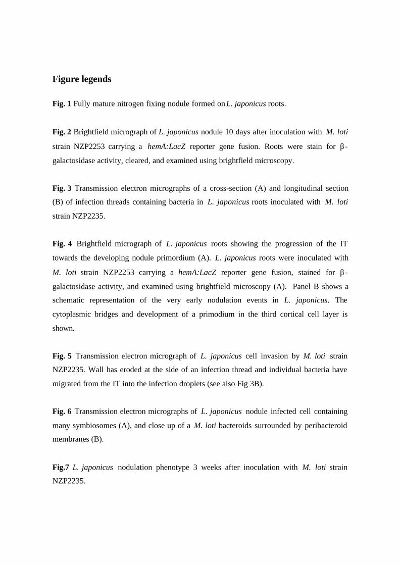
Figure legends
Fig. 1 Fully mature nitrogen fixing nodule formed on L. japonicus roots.
Fig. 2 Brightfield micrograph of L. japonicus nodule 10 days after inoculation with M. loti
strain NZP2253 carrying a hemA:LacZ reporter gene fusion. Roots were stain for β-
galactosidase activity, cleared, and examined using brightfield microscopy.
Fig. 3 Transmission electron micrographs of a cross-section (A) and longitudinal section
(B) of infection threads containing bacteria in L. japonicus roots inoculated with M. loti
strain NZP2235.
Fig. 4 Brightfield micrograph of L. japonicus roots showing the progression of the IT
towards the developing nodule primordium (A). L. japonicus roots were inoculated with
M. loti strain NZP2253 carrying a hemA:LacZ reporter gene fusion, stained for β-
galactosidase activity, and examined using brightfield microscopy (A). Panel B shows a
schematic representation of the very early nodulation events in L. japonicus. The
cytoplasmic bridges and development of a primodium in the third cortical cell layer is
shown.
Fig. 5 Transmission electron micrograph of L. japonicus cell invasion by M. loti strain
NZP2235. Wall has eroded at the side of an infection thread and individual bacteria have
migrated from the IT into the infection droplets (see also Fig 3B).
Fig. 6 Transmission electron micrographs of L. japonicus nodule infected cell containing
many symbiosomes (A), and close up of a M. loti bacteroids surrounded by peribacteroid
membranes (B).
Fig.7 L. japonicus nodulation phenotype 3 weeks after inoculation with M. loti strain
NZP2235.
Fig. 8 Fåhraeus glass slide with cover slip.
Fig. 9 Seedlings in the slide.
Fig. 10 Slides set in the staining jar.
Fig. 11 The jar covered by black paper and aluminium foil.
Fig. 12 The “pillow system” for growing L. japonicus plants. Pillow bags containing a
vermiculite-perlite mixture are placed side by side in the narrow tray.
Fig. 13 L. japonicus seedlings in-bedween pillows.
Fig. 14 Cleared tissues showing successive stages of nodule development. A: Cortical cell
division, B: Bump, C: Developing nodule.
Fig. 15 Longitudinal section of L. japonicus anther showing pollen mother cells. TBO
staining.
Fig. 16 Lotus japonicus nodule 2 weeks after infection with Mesorhizobium loti. DAPI
staining.
Fig. 17 Miyakojima seedlings transferred to the square dish set vertically.
Fig. 18 Nitrogenase acetylene reduction assay. Typical GC chromatograms with separate
peaks for acetylene and ethylene are shown. A small amount of ethylene is detected just
after injecting acetylene into a vial (0 time). After incubating the vial for 30 min, the peak
of ethylene increases by reduction of acetylene (30 min).
Fig. 19 Examples of immunoblotting analysis of nitrogenase component I (C-I) (A) and
component II (C-II) in a crude bacteroids, and of leghemoglobin (B) in a host plant fraction
isolated from nodules on wild-type Gifu (G) and Lotus japonicus sen1 (Ljsym75) mutant
(S) plants. For reference, free-living Mesorhizobium loti (M) and root extracts from Gifu
plants (R) were also tested, respectively.
Fig. 18+19 are reproduced from Mol. Gen. Genomics 269:312-320 (2003) with
permission of Springer-Verlag. (Check this!!!!!!!!!!!!!!!!!!!!!!!!!!!)
Figures 1-7 have also been published before. Krzysztof is seeking permission for
reproduction of these
Figures indicated by bold and undelined in the text are yet to be provided to me by
Makoto. We will have to add figure legends to these, or remove them completely
from the text, depending upon whether we receive them in time for publication.
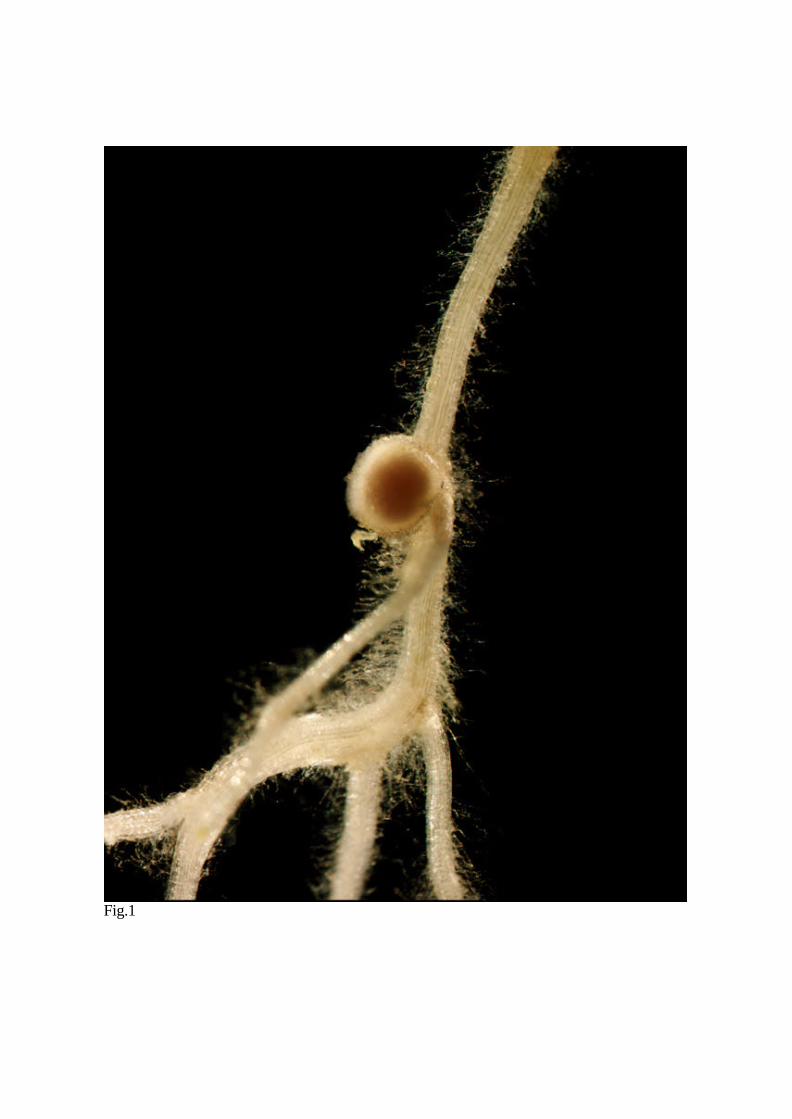
Fig.1
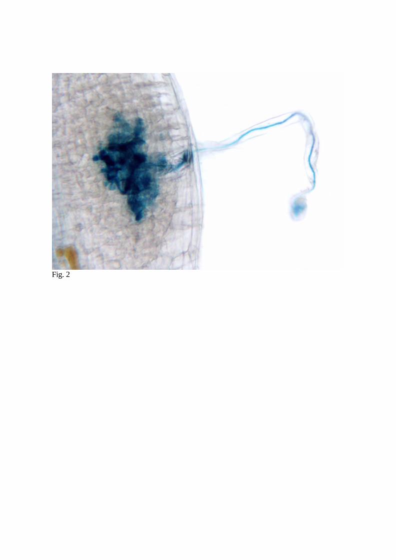
Fig. 2
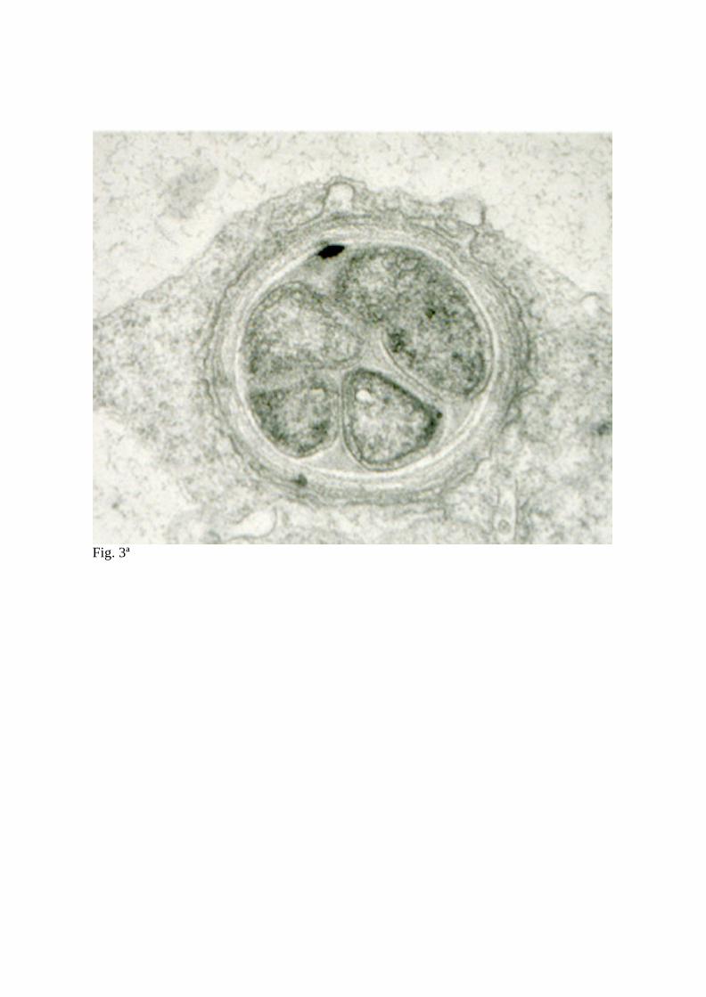
Fig. 3ª
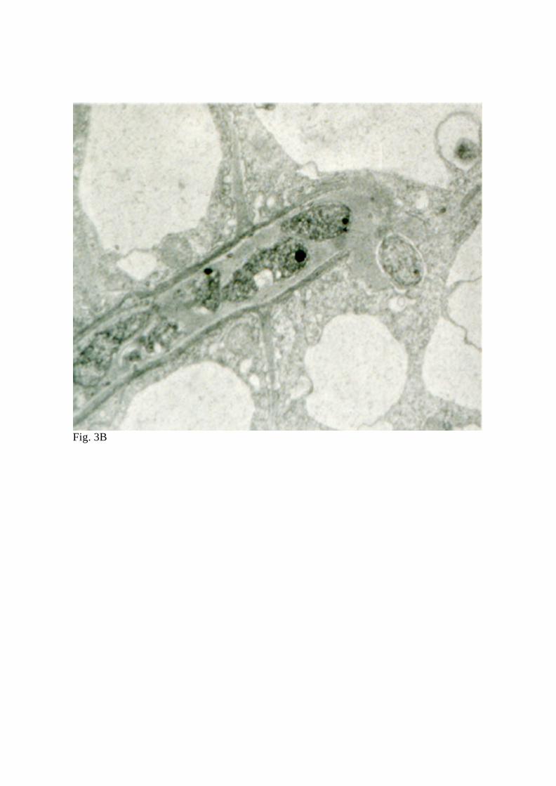
Fig. 3B
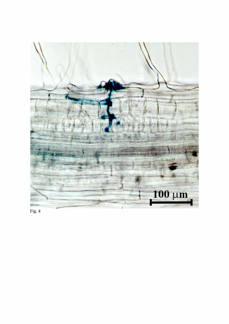
Fig. 4
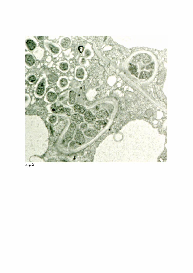
Fig. 5
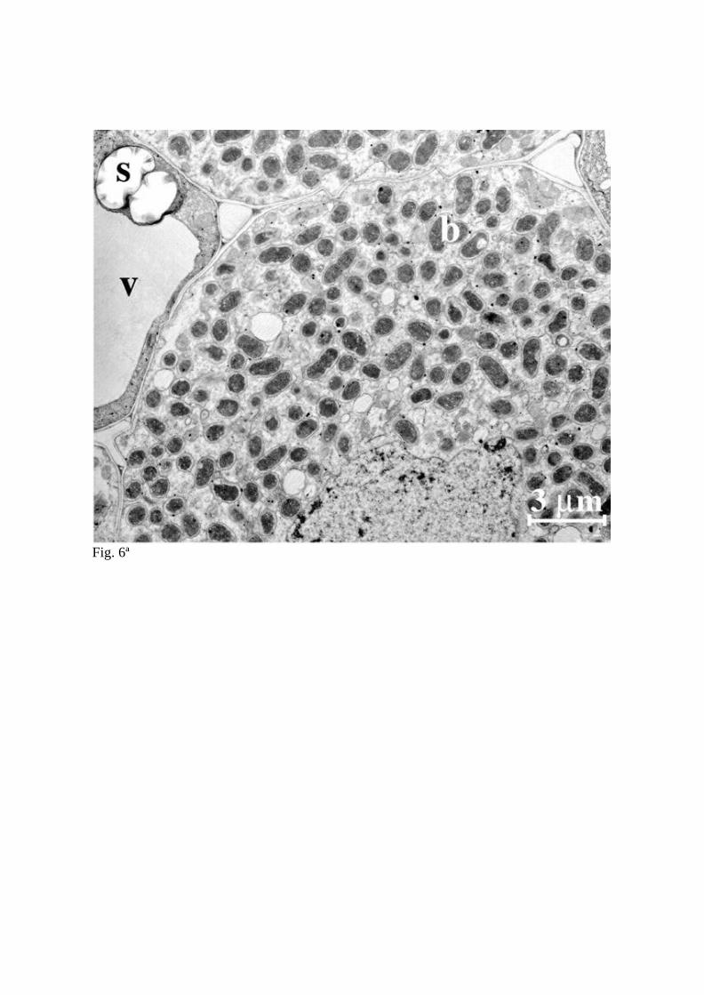
Fig. 6ª

Fig. 6B
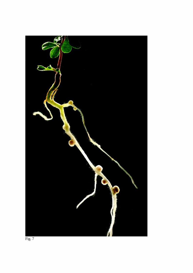
Fig. 7

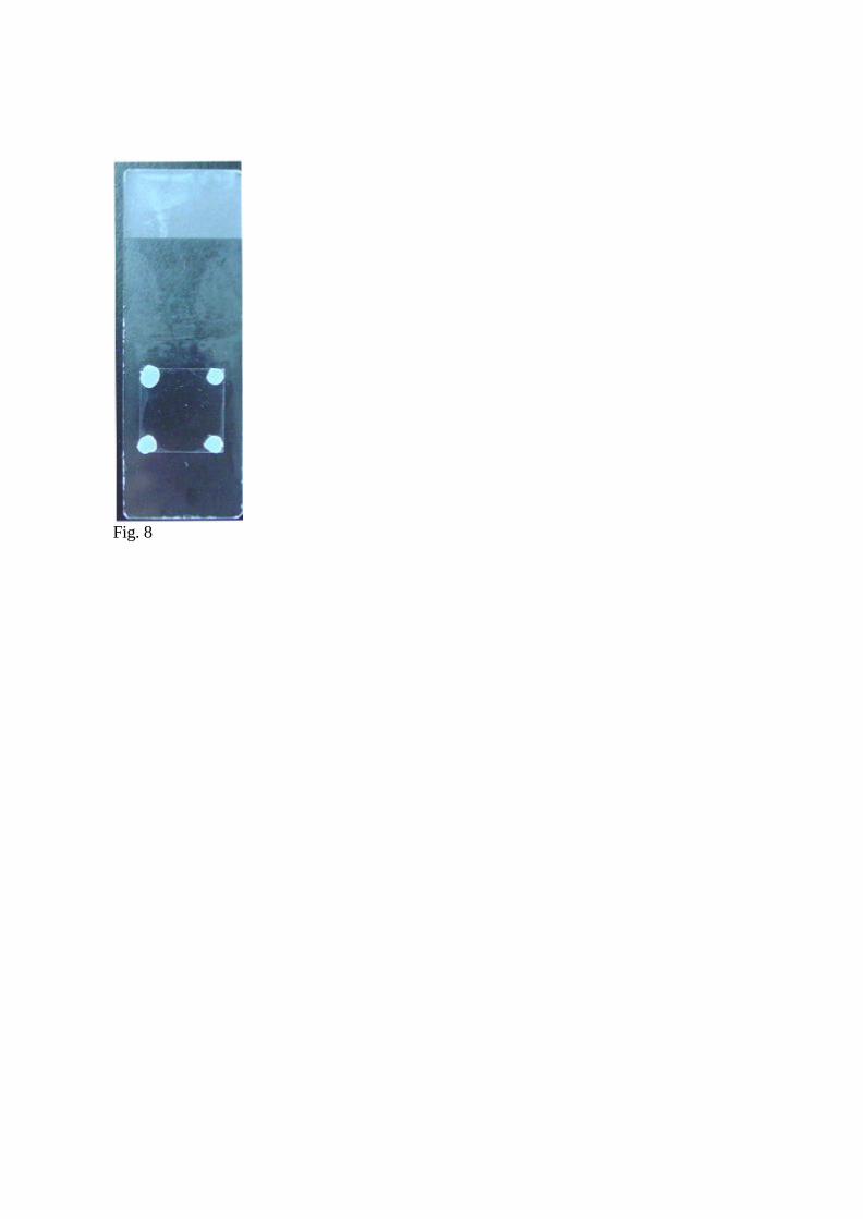
Fig. 8
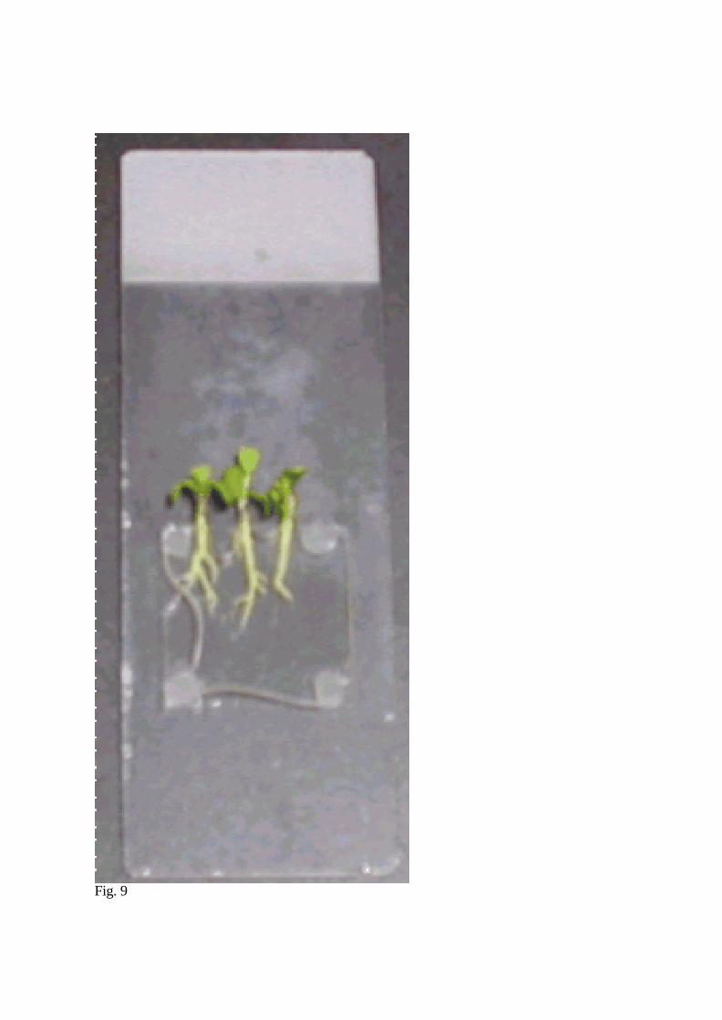
Fig. 9

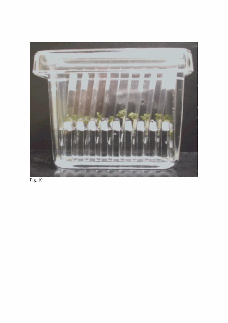
Fig. 10
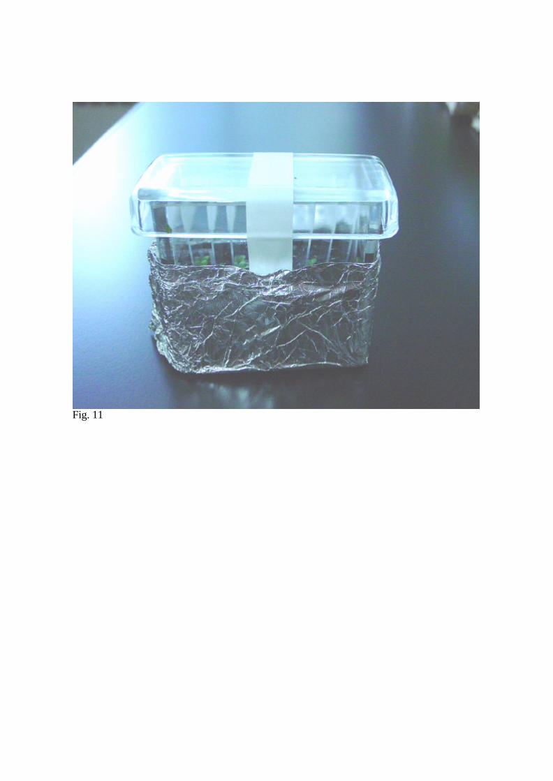
Fig. 11
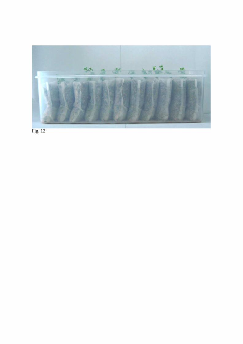
Fig. 12
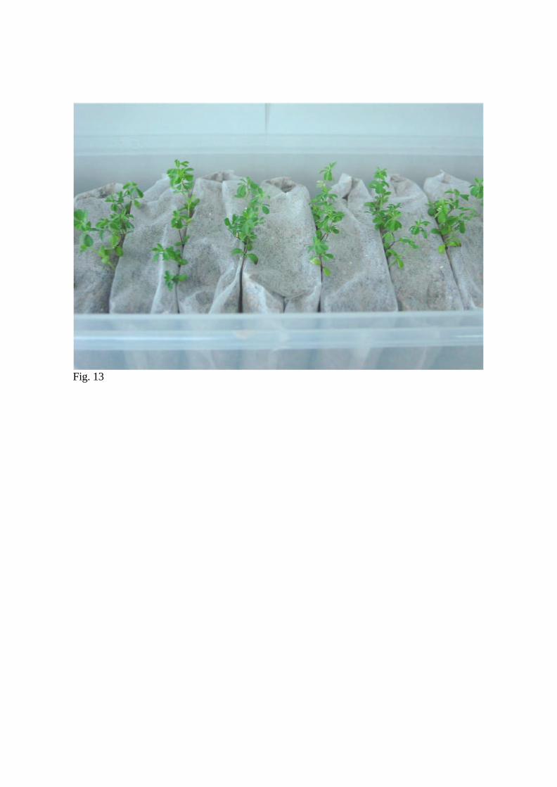
Fig. 13
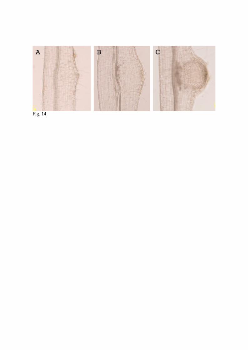
Fig. 14
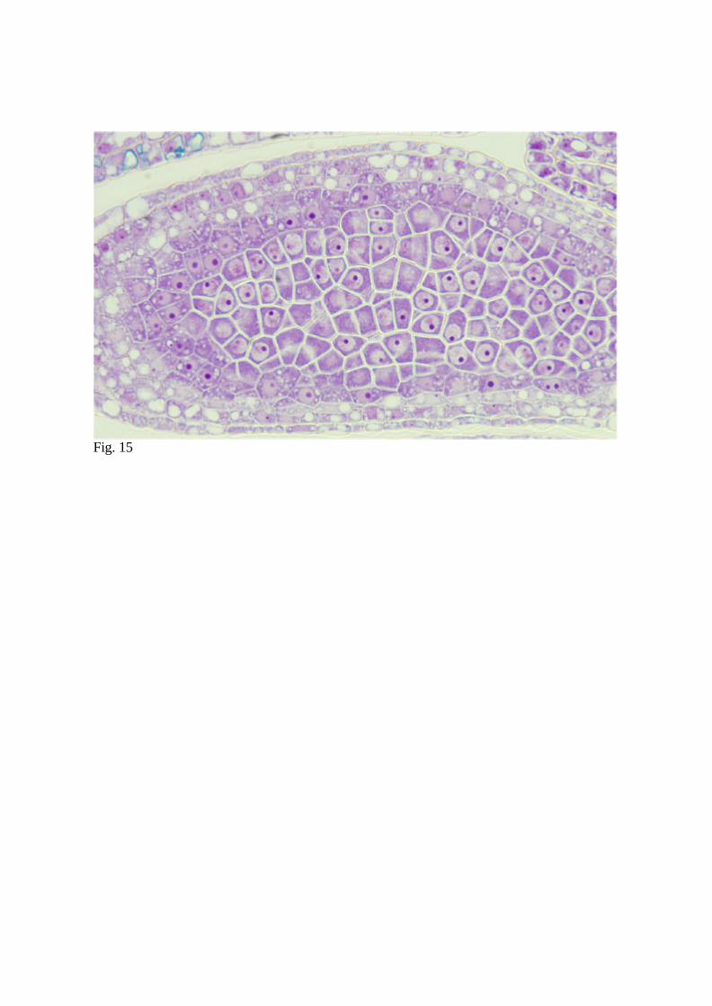
Fig. 15
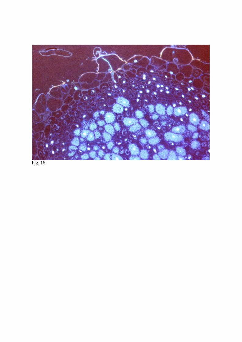
Fig. 16
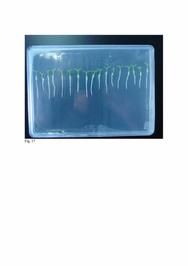
Fig. 17
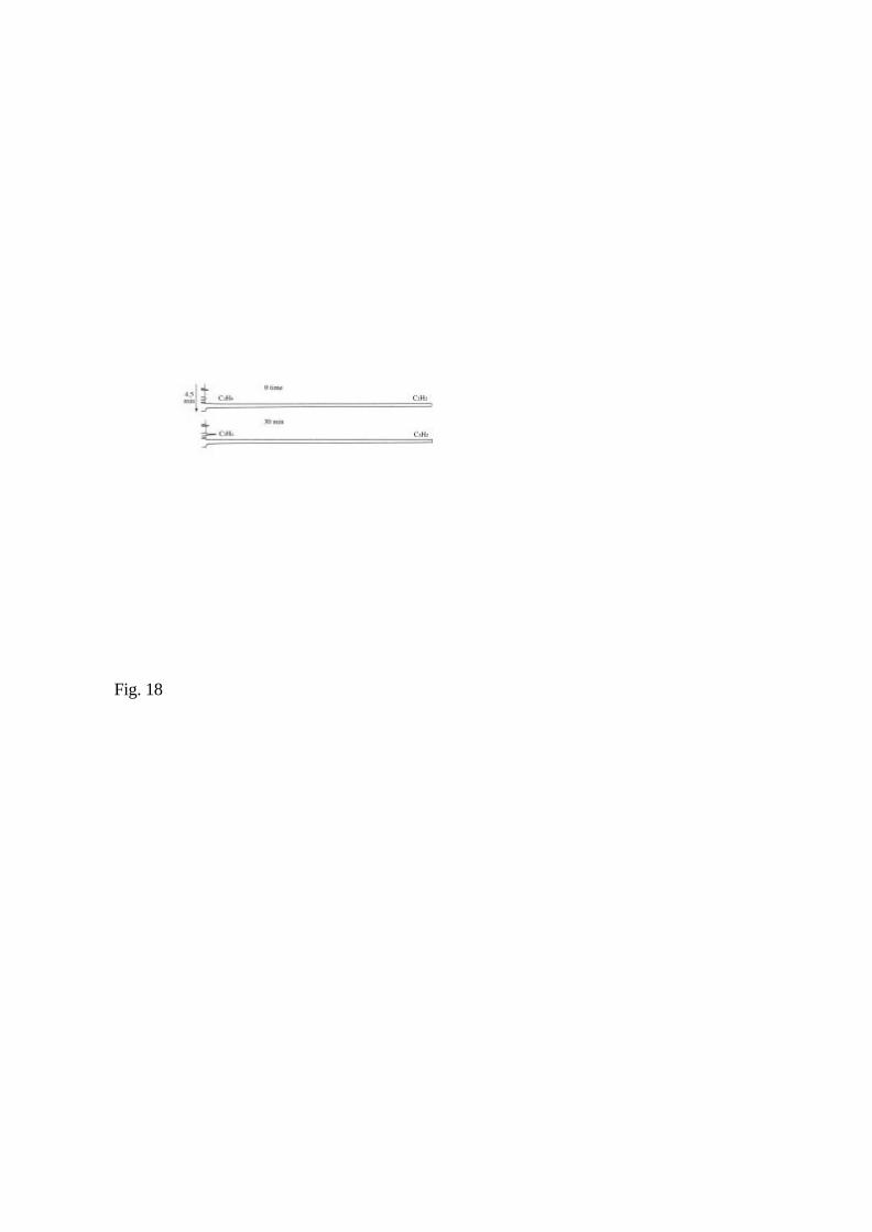
Fig. 18
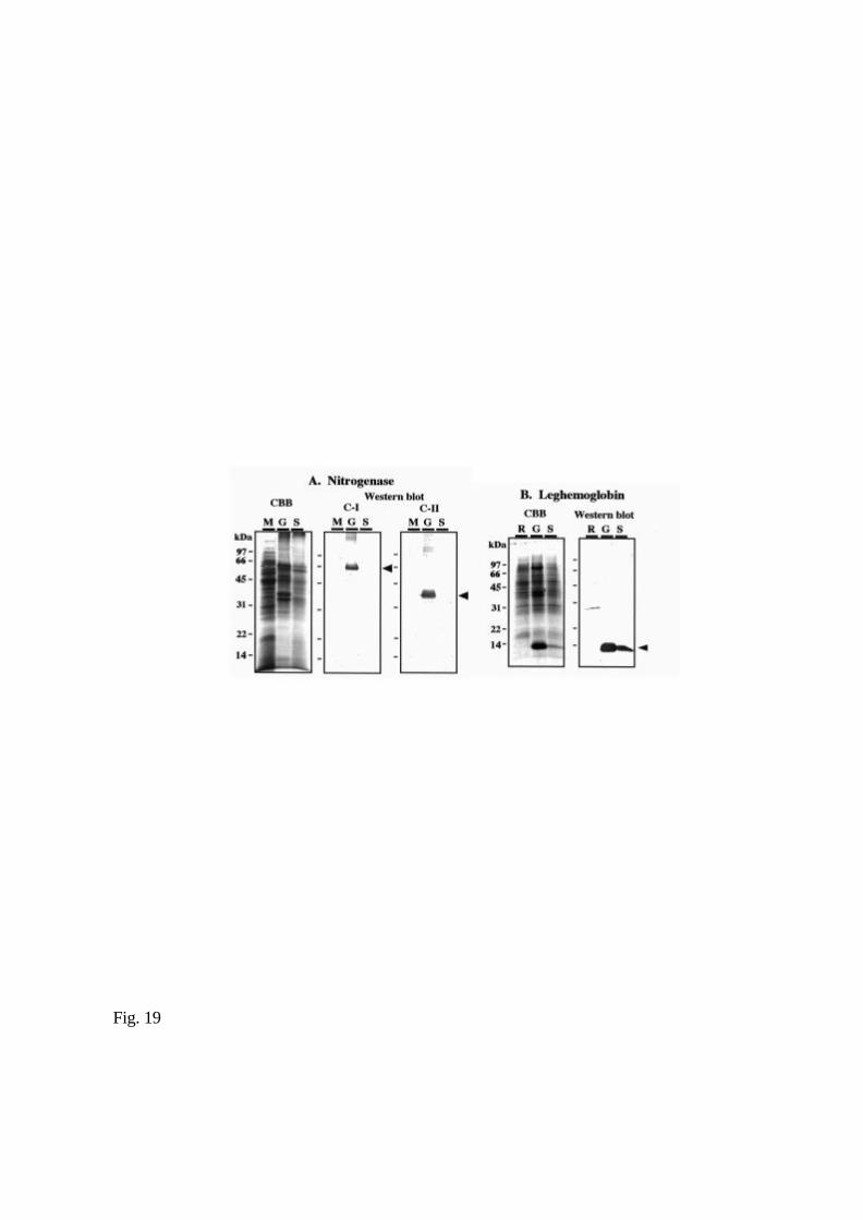
Fig. 19