metabotropic glutamate receptors in the lateral superior olive activate trp-like channels: age- and...
TRANSCRIPT

Metabotropic Glutamate Receptors in the Lateral Superior OliveActivate TRP-Like Channels: Age- and Experience-DependentRegulation
F. Aura Ene, Abigail Kalmbach, and Karl KandlerDepartment of Neurobiology and Center for the Neural Basis of Cognition, University of PittsburghSchool of Medicine, Pittsburgh, Pennsylvania
AbstractThe lateral superior olive (LSO) is the primary auditory nucleus for processing of interaural soundlevel differences, which is one of the major cues for sound localization. During development, survivaland maturation of LSO neurons critically depend on synaptic activity and intracellular calciumsignaling. Before hearing onset, glutamatergic synaptic inputs from the cochlear nucleus (CN) to theLSO activate group I metabotropic glutamate receptors (mGluRs), which leads to calcium releasefrom intracellular stores and large calcium influx from the extracellular milieu. Here, we investigatedthe nature of the mGluR-activated membrane channel that mediates the influx of ex-tracellularcalcium. Using Fura-2 calcium imaging in brain stem slices of neonatal and juvenile mice, we foundthat this calcium channel is blocked by Ni2+, La3+, and 2-aminoethoxydiphenylborane (2-APB),known antagonists of transient receptor potential (TRP) channels. During postnatal development,the contribution of extracellular calcium influx to mGluR-mediated Ca2+ responses graduallydecreased and was almost abolished by the end of the third postnatal week. Over this period, thecontribution of Ca2+ release from internal stores remained unchanged. The developmental decreaseof TRP-like channel-mediated calcium influx was significantly less in congenitally deaf waltzer mice,suggesting that early auditory experience is necessary for the normal age-dependent downregulationof functional TRP channels.
INTRODUCTIONThe lateral superior olive (LSO) is a binaural auditory brain stem nucleus involved in theprocessing of interaural sound level differences. LSO neurons receive excitatory, glutamatergicinputs from the ipsilateral cochlea by the anteroventral cochlear nucleus (AVCN) andinhibitory, glycinergic inputs from the contralateral cochlea by the medial nucleus of thetrapezoid body (MNTB) (Boudreau and Tsuchitani 1968; Oertel 1999; Tollin 2003). Bothinputs are tonotopically organized and aligned, which enables LSO neurons to process binauralinputs in a frequency-specific manner (Sanes and Rubel 1988; Tollin 2003).
During development, the LSO circuit undergoes a number of morphological and functionalchanges that include growth and refinement of dendritic arbors (Rietzel and Friauf 1998; Saneset al. 1992), functional and structural refinement of the MNTB–LSO pathway (Kandler andGillespie 2005; Kim and Kandler 2003; Sanes and Siverls 1991; Sanes and Takacs 1993), anda switch in neurotransmitter phenotype (Gillespie et al. 2005; Kotak et al. 1998; Nabekura etal. 2004). Many of these processes occur before hearing onset but nevertheless depend on
Address for reprint requests and other correspondence: K. Kandler, Department of Neurobiology, University of Pittsburgh School ofMedicine, Biomedical Science Tower W1457, 3500 Terrace Street, Pittsburgh, PA 15261 (E-mail: [email protected]).
NIH Public AccessAuthor ManuscriptJ Neurophysiol. Author manuscript; available in PMC 2008 April 17.
Published in final edited form as:J Neurophysiol. 2007 May ; 97(5): 3365–3375.
NIH
-PA Author Manuscript
NIH
-PA Author Manuscript
NIH
-PA Author Manuscript

neuronal activity and intracellular Ca2+ signaling (Friauf and Lohmann 1999; Kotak and Sanes2000; Lohmann et al. 1998; Sanes and Siverls 1991; Sanes and Takacs 1993).
Before hearing onset, spontaneous neuronal activity is present in the form of high-frequencybursts at various levels in the auditory pathway (Glowatzki and Fuchs 2000; Gummer and Mark1994; Jones et al. 2001; Kotak and Sanes 1995; Kros et al. 1998; Lippe 1994; Romand andEhret 1990). In the prehearing mouse, activation of glutamatergic cochlear nucleus (CN) inputsonto LSO neurons elicits postsynaptic Ca2+ responses that are mediated by ionotropic andmetabotropic glutamate receptors (mGluRs). The recruitment of specific types of glutamatereceptors depends on the spatial and temporal patterns of synaptic activation (Ene et al.2003). Burstlike synaptic activity activates group I and group II mGluRs, which in turn generatepostsynaptic Ca2+ responses with a typical biphasic profile. The initial phase of mGluR-elicitedCa2+ responses in immature LSO neurons is mediated by Ca2+ release from intracellular stores,whereas the later and prolonged phase is mediated by an influx of extracellular Ca2+ throughmembrane Ca2+ channels.
The identity of mGluR-activated membrane Ca2+ channels in LSO neurons is unclear, althoughtheir dependency on intracellular Ca2+ release suggests that they belong to the family oftransient receptor potential channels (TRPCs) (Clapham 2003; Montell 2005), which can beactivated by mGluRs (Bengtson et al. 2004; Gee et al. 2003; Kim et al. 2003; Tozzi et al.2003). In this study, we investigated the developmental changes of mGluR-mediated Ca2+
responses in mouse LSO neurons and characterized the pharmacological properties of themGluR-activated Ca2+ channel. We found that the pharmacological profile of mGluR-activatedCa2+ channels is consistent with the characteristics of a TRPC. During the first three postnatalweeks, group I mGluR-evoked Ca2+ responses decreased, with the most pronounced changesoccurring around the onset of hearing when mGluRs ceased to trigger extracellular Ca2+ influx.These developmental changes were delayed in congenitally deaf waltzer mice, suggesting thatdevelopmental downregulation of TRP-like Ca2+ responses is influenced by normal auditoryexperience.
METHODSAnimals
Experiments were performed in C57BL/6J and homozygote waltzer Cdh23 mice (C57BL/6J-Cdh23v−2J) of both genders (Jackson Laboratory, Bar Harbor, ME; Charles River, Wilmington,MA), ages between postnatal day 0 (P0: the day of birth) and P20. Waltzer homozygotes wereidentified by their rollover behavior deficits (Wada et al. 2001). Heterozygote mice wereobtained by breeding homozygote/wild-type pairs. All experimental procedures were inaccordance with National Institutes of Health guidelines and were approved by the InstitutionalAnimal Care and Use Committee at the University of Pittsburgh.
Slice preparationAnimals between P0 and P7 were anesthetized by hypothermia and animals older than P7 wereanesthetized using isoflurane. Animals were quickly decapitated and the brains were removedand placed into cold (4–8°C) artificial cerebrospinal fluid with kynurenic acid [ACSF withKA, composition (in mM): NaCl 124, NaHCO3 26, glucose 10, KCl 5, KH2PO4 1.25,MgSO4 1.3, CaCl2 2, and kynurenic acid 1; pH 7.4 when aerated with 95% O2-5% CO2].Transversal, 200- to 300- μm-thick slices of the brain stem were cut with a vibrotome(DTK-1500E, Ted Pella, Redding, CA) and slices containing the LSO were used for Fura-2labeling.
Ene et al. Page 2
J Neurophysiol. Author manuscript; available in PMC 2008 April 17.
NIH
-PA Author Manuscript
NIH
-PA Author Manuscript
NIH
-PA Author Manuscript

Fura-2 labelingSlices from P0 to P7 animals were labeled with an acetoxymethyl ester of Fura-2 (Fura-2 AM),using bulk-labeling, as described previously (Ene et al. 2003). In slices from animals older thanP7 LSO neurons labeled poorly or not at all, using the bulk-labeling method, most likelyresulted from poor penetration of Fura-2 AM through the extracellular matrix. Therefore slicesfrom P9 to P20 were labeled using a novel spin-labeling procedure. Slices were placed on filterpaper (12-μm pores; Corning Life Sciences, Acton, MA) in an interface-type chamber and after15–30 min were transferred into a microcentrifuge tube equipped with a 10-kDa cutoffmolecular filter (Amicon; Millipore, Bedford, MA). Slices were covered with 100 μM Fura-2AM in ACSF and aerated with 95% O2-5% CO2. The micro-centrifuge tube was centrifugedfor 15–20 min at about 430 g (IEC Clinical Centrifuge; International Equipment, NeedhamHeights, MA), forcing the Fura-2 AM solution to pass through the slice. Slices were thenremoved from the microcentrifuge tube, immediately washed with freshly aerated ACSF, andkept in an interface chamber in the dark at room temperature (22–25°C) until used for Ca2+
imaging.
Calcium imagingCa2+ imaging was performed using an inverted epifluorescence microscope (Nikon EclipseTE200) equipped with ×10 and ×20 air objectives [numerical aperture (NA): 0.5 and 0.75,respectively] as previously described (Ene et al. 2003). Slices were continuously superfusedwith oxygenated ACSF at 30–32°C (perfusion rate 2–3 ml/min) containing 100 μM Trolox tominimize photodamage (Scheenen et al. 1996). Fluorescence images were acquired every 5–10 s with a 12-bit, cooled, interline-transfer CCD camera (IMAGO; T.I.L.L. Photonics,Martinsried, Germany) using alternating excitation (duration 20–50 ms) at 340 and 380 nm(Polychrome II; T.I.L.L. Photonics).
In some experiments, Fura-2 fluorescence was converted to intra-cellular Ca2+ concentrations([Ca2+]i) as previously described (Ene et al. 2003) using the equation: [Ca2+]i = Kdβ(R −Rmin)/(Rmax − R), where Rmin is the fluorescence ratio of Ca2+ free Fura-2, Rmax is the ratioof Ca2+ bound Fura-2, β is the ratio of the fluorescence intensity of Ca2+ free Fura-2 at 380nm to the fluorescence intensity of Ca2+-bound Fura-2 at 380 nm (Grynkiewicz et al. 1985).The Ca2+ Kd value for Fura-2 was taken as 224. Rmin and Rmax were determined by incubatingthe slices first in Ca2+-free ACSF (Ca2+ was replaced with an equimolar concentration ofMg2+) with 2 mM EGTA and 4 β M Ca2+ ionophore Br-A23187 (Alomone Labs, Jerusalem,Israel). CaCl2 (10 mM) was then added to determine Rmax. The imaging system was calibratedrepeatedly over the course of this study and Rmin varied from 0.25 to 0.31, Rmax varied from1.22 to 1.8, and β varied from 2.3 to 3.7.
Drug applicationDrugs from concentrated stock solutions were dissolved in ACSF and delivered by bathapplication. Group I mGluRs were stimulated using the specific agonist (S)-3,5-dihydroxyphenylglycine (DHPG 20–50 μM). All experiments were performed in the presenceof antagonists of ionotropic GluRs, glycine receptors, and μ-aminobutyric acid type A(GABAA) receptors. Ionotropic GluRs were blocked by 6-cyano-7-nitroquinoxaline-2,3-dionedisodium (CNQX, 20 μM; Tocris, Ballwin, MO) and D-2-amino-5-phosphonopentanoic acid(D-APV, 100 μM, Tocris). Glycine receptors were blocked by strychnine (10 μ M; Sigma, St.Louis, MO) and GABAA receptors were blocked by bicuculline (10 μM; Tocris). Tetrodotoxin(TTX, 1 μM; Alomone Labs) was added to block glutamate release resulting from spontaneousaction potentials. For TRPC antagonists we used Ni2+, La3+ (Sigma, St. Louis, MO), and 2-APB (Calbiochem, San Diego, CA).
Ene et al. Page 3
J Neurophysiol. Author manuscript; available in PMC 2008 April 17.
NIH
-PA Author Manuscript
NIH
-PA Author Manuscript
NIH
-PA Author Manuscript

Data analysisSeries of 340- and 380-nm paired images were low-pass filtered (Gaussian 3 ×3 kernel) andthe background was subtracted using the program Tillvision (T.I.L.L. Photonics) as describedpreviously (Ene et al. 2003). Ca2+ responses were monitored and measured from the soma ofneurons. Cells with high resting [Ca2+]i (>250 nM, indicating unhealthy cells) (Zirpel andRubel 1996), cells in which the Fura-2 signal was saturated ([Ca2+]i >1.5 μM, because Fura-2does not faithfully report [Ca2+]i >1.5–2 μM), and cells in which [Ca2+]i did not return tobaseline after stimulation were excluded from analysis. Changes in [Ca2+]i that exceeded 2SDs of the baseline and changes in ΔR/R > 0.1 were considered as responses. Excel (Microsoft,Redmond, WA), Origin (OriginLab, Northampton, MA), and Matlab (The MathWorks, Natick,MA) were used for data analysis.
Responses elicited by the specific group I mGluR agonist DHPG (50 μM, 90 s) were quantifiedand categorized using the following parameters: peak amplitude, plateau amplitude, duration,and area under the curve. Traces were aligned to the onset of the response that was consideredtime 0. Baseline [Ca2+]i was determined as the average of five data points before responseonset (in the window −50 to 0 s). Peak amplitude was measured in the time window 0 to 40 s.Plateau amplitude was determined by averaging three to five data points in the window 80 to100 s. Response duration was measured from the onset of the response until the responsereturned to baseline. Based on these measurements, responses were classified into threeresponse types (Table 1, nFig. 2): peak and plateau (pp), peak small plateau (psp), and peakno plateau (pnp). In pp responses the peak response was followed by a clear plateau (peakamplitude 236 ± 50 nM; plateau amplitude 107 ± 22 nM, = 287 cells), with a duration thatalways exceeded the 90-s duration of drug application (182 ± 13 s, n = 287 cells). In pnpresponses the plateau phase was completely missing, the peak amplitude was small (60 ± 8nM, n = 177 cells), and the duration was always shorter than the duration of drug application(70 ± 3 s, n = 177 cells). The intermediate group of psp responses was characterized by agradually decreasing plateau phase, which always exceeded the duration of drug application(141 ± 15 s, n = 124 cells).
Statistical analysisStatistical significance was analyzed using paired t-test, ANOVA followed by Student–Newman–Keuls post hoc test, Fisher’s exact test, linear (Pearson) correlation test, Mann–Whitney nonparametric statistical test, Kolmogorov–Smirnov (KS) test, and chi-square test.Values of P < 0.05 were considered significant. Throughout the text, values are expressed asmeans > SE.
RESULTSGroup I mGluR-elicited Ca2+ responses were analyzed in 738 LSO neurons from 51 C57Bl6mice (ages between P0 and P19), in 60 LSO neurons from six heterozygote waltzer mice, andin 156 LSO neurons from 14 waltzer Cdh23 homozygote mice (ages between P11 and P20).All cells included in the analysis responded to KCl-evoked depolarizations (60 mM, 30 s) withpeak amplitudes ranging from about 100 nM to Fura-2–saturating responses (>1.5 μM).
Spin-labeling of LSO neurons in older slicesTo overcome the poor labeling with Fura-2 AM of slices from animals older than P7 wedeveloped a new method, spin-labeling, which uses centrifugation to force Fura-2 AM into theslice. To evaluate whether this new method affects Ca2+ responses of LSO neurons, the twolabeling methods were compared in slices from P0–P5 animals. At this age, both methodsproduced a similar number of cells loaded with Fura-2 (bulk-labeling: n = 7.39 ± 0.92 cells/100 μm2, n = 7 slices; spin-labeling: n = 7.98 ± 0.37 cells/100 μm2, n = 3 slices; t-test P > 0.05)
Ene et al. Page 4
J Neurophysiol. Author manuscript; available in PMC 2008 April 17.
NIH
-PA Author Manuscript
NIH
-PA Author Manuscript
NIH
-PA Author Manuscript

and a similar somatic fluorescence intensity at 360 nm, the isosbestic point of Fura-2, measuredwith identical settings (bulk-labeling: 338 ± 67 AU, n = 7 slices; spin-labeling: 272 ± 108 AU,n = 3 slices; t-test, P > 0.05; Fig.1A). Resting somatic Ca23 concentrations were not signifi-cantly different (bulk-labeling: 94.2 3 47.6 nM, n 3 96 cells, n 3 4 slices; spin-labeling: 109.23 57.6 nM, n 3 70 cells, n 3 3 slices; t-test, P 3 0.05). Finally, Ca23 responses elicited by bathapplication of the selective group I mGluR agonist (S)-3,5-dihydroxyphenylglycine (DHPG,50 μM, 90 s) and by KCl depolarization (60 mM, 30 s) were undistinguishable (Fig. 1B). Theseresults suggest that spin-labeling, compared with conventional bulk-labeling, does not changethe basic Ca23 response properties of LSO neurons.
Developmental changes of group I mGluR-mediated Ca2+ responsesIn LSO neurons from neonatal mice (P0–P5), Ca2+ responses elicited by DHPG (50 μM, 90 s)consisted of an initial peak followed by a large plateau phase that always exceeded the durationof the agonist application (Fig. 2). During post-natal development, these Ca2+ responsesgradually changed their profile, amplitude, and duration (Fig. 2 and Table 1). At P9–P12,around hearing onset (approximately P10–P12; Shnerson and Pujol 1981;Song et al. 2006),peak and plateau amplitudes decreased and response durations had become shorter. By the endof the third postnatal week, plateau phases were absent in almost all responses. Responseamplitudes, durations, and areas were significantly larger in the P0–P5 group compared withall older age groups (Mann–Whitney test, P < 0.05) and were significantly smaller in the P17–P19 group compared with all younger age groups (Mann–Whitney test, P < 0.05; Fig. 2B andTable 1; see also Supplemental Fig. S2).1 Plateau amplitudes and response durations alsosignifi-cantly decreased from P9–P12 to P13–P16 (Mann–Whitney test, P 3 0.05; Fig. 2B andTable 1).
In the first postnatal week, all Ca2+ responses elicited by DHPG showed a prominent plateauand were categorized as pp responses (n = 222/222 cells, Fig. 2C). With increasing age, thefrequency of pp responses decreased, from 100% at P0–P5 to 24% at P9–P12 (n = 46/178cells), to 11% at P13–P16 (n = 15/132 cells) and to 7% at P17–P19 (n = 4/54 cells) (Fig. 2C).Responses with a peak and small plateau (psp responses) were found in all older age groups.The relative occurrence of responses that completely lacked a plateau (pnp responses) was agedependent as well, steadily increasing from 32% at P9–P12 (n = 59/178 cells), to 57% at P13–P16 (n = 75/132) and to 80% at P17–P19 (n = 43/54 cells).
In summary, during the first 3 week of postnatal development, group I mGluR-elicited Ca2+
responses gradually lose the sustained plateau phase that, in neonatal mice, is caused by aninflux of extracellular Ca2+ through a channel not sensitive to voltage-gated calcium channel(VGCC) blockers (Ene et al. 2003). Next we used pharmacological tools to investigate thesources of [Ca2+]i triggered by group I mGluRs in developing LSO neurons in more detail.
Contribution of extracellular Ca2+ to group I mGluR-elicited responses decreases duringdevelopment
To determine the age dependency of mGluR-elicited influx of extracellular Ca2+ and itscontribution to the three types of responses (pp, psp, pnp) we compared mGluR-elicitedCa2+ responses in standard and nominally Ca2+-free ACSF (Fig. 3). Regardless of age,eliminating extracellular Ca2+ significantly reduced both the peak and the plateau phase ofresponses of the pp and psp type (P0–P5 pp response: peak reduced to 32 ± 2% and area to 13± 1%, n = 82; P9–P12 psp response: peak reduced to 23 ± 4% and area to 17 ± 4%, n = 14;P13–P16.psp response: Peak reduced to 44 ± 10% and area to 29 ± 6%, n 3 22). For pnpresponses, however, removal of extracellular Ca2+ had no significant effect on response
1The online version of this article contains supplemental data.
Ene et al. Page 5
J Neurophysiol. Author manuscript; available in PMC 2008 April 17.
NIH
-PA Author Manuscript
NIH
-PA Author Manuscript
NIH
-PA Author Manuscript
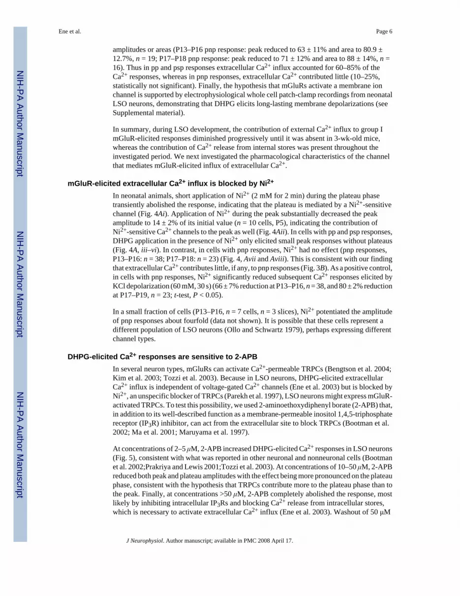
amplitudes or areas (P13–P16 pnp response: peak reduced to 63 ± 11% and area to 80.9 ±12.7%, n = 19; P17–P18 pnp response: peak reduced to 71 ± 12% and area to 88 ± 14%, n =16). Thus in pp and psp responses extracellular Ca2+ influx accounted for 60–85% of theCa2+ responses, whereas in pnp responses, extracellular Ca2+ contributed little (10–25%,statistically not significant). Finally, the hypothesis that mGluRs activate a membrane ionchannel is supported by electrophysiological whole cell patch-clamp recordings from neonatalLSO neurons, demonstrating that DHPG elicits long-lasting membrane depolarizations (seeSupplemental material).
In summary, during LSO development, the contribution of external Ca2+ influx to group ImGluR-elicited responses diminished progressively until it was absent in 3-wk-old mice,whereas the contribution of Ca2+ release from internal stores was present throughout theinvestigated period. We next investigated the pharmacological characteristics of the channelthat mediates mGluR-elicited influx of extracellular Ca2+.
mGluR-elicited extracellular Ca2+ influx is blocked by Ni2+
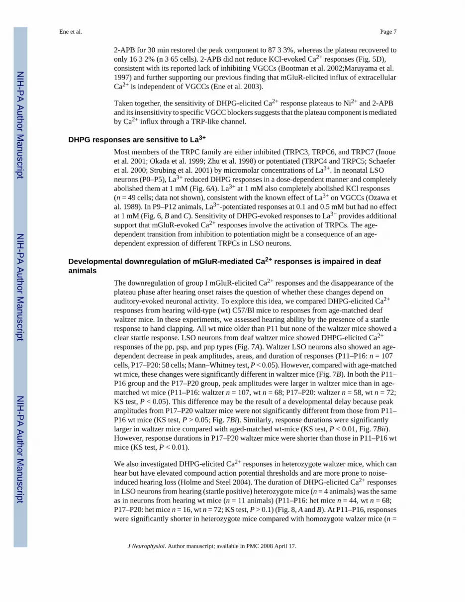
In neonatal animals, short application of Ni2+ (2 mM for 2 min) during the plateau phasetransiently abolished the response, indicating that the plateau is mediated by a Ni2+-sensitivechannel (Fig. 4Ai). Application of Ni2+ during the peak substantially decreased the peakamplitude to 14 ± 2% of its initial value (n = 10 cells, P5), indicating the contribution ofNi2+-sensitive Ca2+ channels to the peak as well (Fig. 4Aii). In cells with pp and psp responses,DHPG application in the presence of Ni2+ only elicited small peak responses without plateaus(Fig. 4A, iii–vi). In contrast, in cells with pnp responses, Ni2+ had no effect (pnp responses,P13–P16: n = 38; P17–P18: n = 23) (Fig. 4, Avii and Aviii). This is consistent with our findingthat extracellular Ca2+ contributes little, if any, to pnp responses (Fig. 3B). As a positive control,in cells with pnp responses, Ni2+ significantly reduced subsequent Ca2+ responses elicited byKCl depolarization (60 mM, 30 s) (66 ± 7% reduction at P13–P16, n = 38, and 80 ± 2% reductionat P17–P19, n = 23; t-test, P < 0.05).
In a small fraction of cells (P13–P16, n = 7 cells, n = 3 slices), Ni2+ potentiated the amplitudeof pnp responses about fourfold (data not shown). It is possible that these cells represent adifferent population of LSO neurons (Ollo and Schwartz 1979), perhaps expressing differentchannel types.
DHPG-elicited Ca2+ responses are sensitive to 2-APBIn several neuron types, mGluRs can activate Ca2+-permeable TRPCs (Bengtson et al. 2004;Kim et al. 2003; Tozzi et al. 2003). Because in LSO neurons, DHPG-elicited extracellularCa2+ influx is independent of voltage-gated Ca2+ channels (Ene et al. 2003) but is blocked byNi2+, an unspecific blocker of TRPCs (Parekh et al. 1997), LSO neurons might express mGluR-activated TRPCs. To test this possibility, we used 2-aminoethoxydiphenyl borate (2-APB) that,in addition to its well-described function as a membrane-permeable inositol 1,4,5-triphosphatereceptor (IP3R) inhibitor, can act from the extracellular site to block TRPCs (Bootman et al.2002; Ma et al. 2001; Maruyama et al. 1997).
At concentrations of 2–5 μM, 2-APB increased DHPG-elicited Ca2+ responses in LSO neurons(Fig. 5), consistent with what was reported in other neuronal and nonneuronal cells (Bootmanet al. 2002;Prakriya and Lewis 2001;Tozzi et al. 2003). At concentrations of 10–50 μM, 2-APBreduced both peak and plateau amplitudes with the effect being more pronounced on the plateauphase, consistent with the hypothesis that TRPCs contribute more to the plateau phase than tothe peak. Finally, at concentrations >50 μM, 2-APB completely abolished the response, mostlikely by inhibiting intracellular IP3Rs and blocking Ca2+ release from intracellular stores,which is necessary to activate extracellular Ca2+ influx (Ene et al. 2003). Washout of 50 μM
Ene et al. Page 6
J Neurophysiol. Author manuscript; available in PMC 2008 April 17.
NIH
-PA Author Manuscript
NIH
-PA Author Manuscript
NIH
-PA Author Manuscript
2-APB for 30 min restored the peak component to 87 3 3%, whereas the plateau recovered toonly 16 3 2% (n 3 65 cells). 2-APB did not reduce KCl-evoked Ca2+ responses (Fig. 5D),consistent with its reported lack of inhibiting VGCCs (Bootman et al. 2002;Maruyama et al.1997) and further supporting our previous finding that mGluR-elicited influx of extracellularCa2+ is independent of VGCCs (Ene et al. 2003).
Taken together, the sensitivity of DHPG-elicited Ca2+ response plateaus to Ni2+ and 2-APBand its insensitivity to specific VGCC blockers suggests that the plateau component is mediatedby Ca2+ influx through a TRP-like channel.
DHPG responses are sensitive to La3+
Most members of the TRPC family are either inhibited (TRPC3, TRPC6, and TRPC7 (Inoueet al. 2001; Okada et al. 1999; Zhu et al. 1998) or potentiated (TRPC4 and TRPC5; Schaeferet al. 2000; Strubing et al. 2001) by micromolar concentrations of La3+. In neonatal LSOneurons (P0–P5), La3+ reduced DHPG responses in a dose-dependent manner and completelyabolished them at 1 mM (Fig. 6A). La3+ at 1 mM also completely abolished KCl responses(n = 49 cells; data not shown), consistent with the known effect of La3+ on VGCCs (Ozawa etal. 1989). In P9–P12 animals, La3+-potentiated responses at 0.1 and 0.5 mM but had no effectat 1 mM (Fig. 6, B and C). Sensitivity of DHPG-evoked responses to La3+ provides additionalsupport that mGluR-evoked Ca2+ responses involve the activation of TRPCs. The age-dependent transition from inhibition to potentiation might be a consequence of an age-dependent expression of different TRPCs in LSO neurons.
Developmental downregulation of mGluR-mediated Ca2+ responses is impaired in deafanimals
The downregulation of group I mGluR-elicited Ca2+ responses and the disappearance of theplateau phase after hearing onset raises the question of whether these changes depend onauditory-evoked neuronal activity. To explore this idea, we compared DHPG-elicited Ca2+
responses from hearing wild-type (wt) C57/Bl mice to responses from age-matched deafwaltzer mice. In these experiments, we assessed hearing ability by the presence of a startleresponse to hand clapping. All wt mice older than P11 but none of the waltzer mice showed aclear startle response. LSO neurons from deaf waltzer mice showed DHPG-elicited Ca2+
responses of the pp, psp, and pnp types (Fig. 7A). Waltzer LSO neurons also showed an age-dependent decrease in peak amplitudes, areas, and duration of responses (P11–P16: n = 107cells, P17–P20: 58 cells; Mann–Whitney test, P < 0.05). However, compared with age-matchedwt mice, these changes were significantly different in waltzer mice (Fig. 7B). In both the P11–P16 group and the P17–P20 group, peak amplitudes were larger in waltzer mice than in age-matched wt mice (P11–P16: waltzer n = 107, wt n = 68; P17–P20: waltzer n = 58, wt n = 72;KS test, P < 0.05). This difference may be the result of a developmental delay because peakamplitudes from P17–P20 waltzer mice were not significantly different from those from P11–P16 wt mice (KS test, P > 0.05; Fig. 7Bi). Similarly, response durations were significantlylarger in waltzer mice compared with aged-matched wt-mice (KS test, P < 0.01, Fig. 7Bii).However, response durations in P17–P20 waltzer mice were shorter than those in P11–P16 wtmice (KS test, P < 0.01).
We also investigated DHPG-elicited Ca2+ responses in heterozygote waltzer mice, which canhear but have elevated compound action potential thresholds and are more prone to noise-induced hearing loss (Holme and Steel 2004). The duration of DHPG-elicited Ca2+ responsesin LSO neurons from hearing (startle positive) heterozygote mice (n = 4 animals) was the sameas in neurons from hearing wt mice (n = 11 animals) (P11–P16: het mice n = 44, wt n = 68;P17–P20: het mice n = 16, wt n = 72; KS test, P > 0.1) (Fig. 8, A and B). At P11–P16, responseswere significantly shorter in heterozygote mice compared with homozygote walzer mice (n =
Ene et al. Page 7
J Neurophysiol. Author manuscript; available in PMC 2008 April 17.
NIH
-PA Author Manuscript
NIH
-PA Author Manuscript
NIH
-PA Author Manuscript

6 animals; n = 107 cells, KS test, P < 0.005). In heterozygote mice, response durations in startle-negative P12–P13 mice were significantly longer compared with startle-positive P14–P16 mice(Fig. 8, C and D).
Similar to wt mice, in waltzer mice we also observed a developmental decrease in thepercentage of DHPG-responses of the pp and psp types and a developmental increase in thepercentage of pnp responses (Fig. 9; Fisher’s exact test, P < 0.05). However, in waltzer mice,more cells showed pp and psp responses at P11–P16 (Fig. 9A; waltzer n = 80 cells, wt n = 32cells; Fisher’s exact test, P < 0.05).
Taken together, in deaf waltzer mice the age-dependent changes of mGluR-mediated Ca2+
responses were significantly smaller, suggesting that auditory experience is necessary for thenormal time course of the functional downregulation of TRP-like channel(s). In support of thishypothesis, in P16 waltzer mice DHPG-elicited responses were strongly diminished by theTRPC antagonist Ni2+ (peak amplitude reduced by 80.5 ± 4.7%; area reduced by 82.4 ± 4.2%;n = 21 cells with responses in Ni2+).
DISCUSSIONThis study provides a developmental picture of the Ca2+ entry pathways underlying group ImGluR-mediated Ca2+ responses in developing LSO neurons. In neonatal mice, these Ca2+
responses were characterized by a large transient peak followed by a sustained plateau. Weidentified two Ca2+ sources that contributed to these responses: release of Ca2+ from internalstores and influx of Ca2+ from the extracellular milieu. The extracellular component wassensitive to the TRPC blockers Ni2+, 2-APB, and La3+, suggesting that it was mediated by aTRP-like Ca2+ channel. With increasing age, the contribution of extracellular Ca2+ influx togroup I mGluR-mediated Ca2+ responses diminished and was mostly absent after hearing onset,whereas the contribution of internal stores was unchanged. Finally, the developmentaldownregulation of mGluR-elicited extracellular Ca2+ influx was delayed in deaf waltzer mice,suggesting that an age-dependent decrease in the activation and/or expression of TRPCs isinfluenced by auditory experience.
Developing LSO neurons express functional TRP-like channelsOur results suggest that LSO neurons in prehearing mice express Ca2+-permeable, TRP-likechannels that are activated by group I mGluRs. This is supported by the following observations.First, the typical biphasic peak-plateau profile of mGluR-elicited Ca2+ responses in LSOneurons resembles the response profiles that were observed in a number of other neuronal andnonneuronal cell types that express store-operated Ca2+ channels or TRPCs (Boulay et al.1997; Hu et al. 1994; Moller et al. 1997; Ramsey et al. 2006; Sosa et al. 2002). Second, theplateau phase of mGluR-elicted Ca2+ responses was mediated entirely by influx of extracellularCa2+ (Ene et al. 2003). Third, extracellular Ca2+ influx was insensitive to specific blockers ofvoltage-gated Ca2+ channels (Ene et al. 2003) but was sensitive to blockers of TRPCs such asNi2+, 2-APB, and La3+ (Figs. 4–6). Fourth, activation of mGluRs by the specific agonist DHPGresulted in depolarizations (see Supplemental material) similar to the activation of TRPCs inother neuronal systems (Kim et al. 2003. Tempia et al. 2001).
TRPCs can be activated by a variety of intracellular messengers depending on the exact TRPCand cell type. In LSO neurons, Ca2+ release from intracellular Ca2+ stores is required forcoupling mGluR activation to the opening of TRP-like channels—based on the observationthat depletion of intracellular Ca2+ stores with thapsigargine abolished all mGluR-elicitedCa2+ responses (Ene et al. 2003). However, Ca2+ does not appear to be the only signalresponsible for activation of TRP-like channels because influx of Ca2+ through VGCC whileintracellular Ca2+ stores were depleted by thapsigargine did not trigger long-lasting Ca2+
Ene et al. Page 8
J Neurophysiol. Author manuscript; available in PMC 2008 April 17.
NIH
-PA Author Manuscript
NIH
-PA Author Manuscript
NIH
-PA Author Manuscript

plateaus. Further studies are needed to identify what additional components of the group ImGluR-activated intracellular cascade are the relevant stimuli that, together with Ca2+, activatethe opening of TRP-like channels in LSO neurons.
Which TRPCs are responsible for group I mGluR-mediated Ca2+ responses in LSO neurons?All members of the TRPC family (TRPC1–TRPC7) are expressed in the brain (Minke andCook 2002; Riccio et al. 2002), although their expression patterns have not been investigatedspecifically for the LSO. TRPCs are activated by an increase in [Ca2+]i and/or diacyl-glycerol(Boulay et al. 1997; Hofmann et al. 1999; Kim et al. 2003; Okada et al. 1998; Schaefer et al.2000; Zitt et al. 1997) and it was previously suggested that the TRPC3 channel mediates groupI mGluR-mediated Ca2+ responses in retinal amacrine cells (Sosa et al. 2002). TRPC3 alsoseems to be a good candidate for LSO neurons because TRPC3 is strongly expressed in theembryonic and early postnatal brain stem, including in auditory brain stem nuclei (Li et al.1999), and because TRPC3 expression levels decrease after P10, which corresponds to the timewhen group I mGluR-mediated Ca2+ responses in LSO neurons also decrease. Finally, DHPG-evoked responses in neonatal LSO neurons were inhibited by millimolar concentrations ofLa3+ (Fig. 6), concentrations that are in the range of TRPC3 sensitivity to La3+ (Zhu et al.1998).
Developmental decrease in the contribution of TRP-like channels to mGluR-mediated Ca2+
responses and the role of sensory-evoked activityDuring the first three postnatal weeks, group I mGluR-mediated Ca2+ responses becameprogressively smaller in amplitude, shorter in duration, and the plateau component disappeared(Fig. 2). These changes are consistent with the idea that the decrease in mGluR-elicited Ca2+
responses resulted, in large part, from a developmental downregulation of TRP-like channelsor their functional uncoupling from mGluR activation. In support of this, both Ni2+, whichreduced the peak amplitude and abolished the Ca2+ plateau by blocking the influx ofextracellular Ca2+ in neonatal LSO neurons, and removal of extracellular Ca2+ (Fig. 3) had noeffect on responses during the third postnatal week, when most responses were small and shortand were mediated primarily by Ca2+ release from internal stores (Fig. 4, A and B).
The contribution of extracellular Ca2+ influx to mGluR-mediated Ca2+ responses declined at,and shortly after, hearing onset (approximately P12; Song et al. 2006), raising the possibilitythat sensory activity plays a role in the functional downregulation of TRP-like channels. Wetested this hypothesis by investigating mGluR-mediated Ca2+ responses in developing waltzermice. Waltzer mice have spontaneous mutation in the gene for cadherin 23, which causes adisorganization of stereocilia bundles of cochlear hair cells resulting in deafness (Di Palma etal. 2001; Holme and Steel 2002). In waltzer mice, the changes in group I mGluR-mediatedCa2+ responses appeared to be significantly delayed compared with heterozygote and wild-type control mice (Figs. 7–9). Notably, more LSO neurons in waltzer mice than in age-matchedcontrol mice displayed responses with a long Ca2+ plateau and fewer neurons displayed peak-only responses. These results support the idea that auditory experience is important for thedevelopmental downregulation of TRP-like channel expression or their developmentaluncoupling from mGluR activation.
It should be noted that our results cannot exclude the possibility that spontaneous activity beforehearing onset also contributes to the downregulation of TRP-like responses. Further studiesthat investigate mGluR-mediated Ca2+ responses in prehearing waltzer mice and characterizespontaneous activity patterns in vivo are necessary to conclusively address this possibility.Another issue to consider is that cadherin 23 is expressed not only in hair cells but also in thebrain (Di Palma 2001; Rzadzinska et al. 2005), although it is currently unknown whethercadherin 23 is expressed in the LSO or in any other auditory nuclei. Anatomical studiesinvestigating cadherin expression in developing LSO neurons are required to address the
Ene et al. Page 9
J Neurophysiol. Author manuscript; available in PMC 2008 April 17.
NIH
-PA Author Manuscript
NIH
-PA Author Manuscript
NIH
-PA Author Manuscript

question whether impairment in the developmental decrease in TRP-like channels activationby mGluRs in waltzer mice is influenced by potential changes of cadherin 23 in the LSO.Nevertheless, our conclusion that auditory experience is an important contributing factor tothe downregulation of TRP activation by mGluRs is supported by our results from heterozygotewaltzer mice. Heterozygotes, after hearing onset, as indicated by the presence of an acousticstartle response, showed mGluR-mediated Ca2+ responses similar to age-matched wild-typemice and in P12–P16 heterozygotes response durations became significantly shorter right afterhearing onset (Fig. 8, C and D).
Functional implicationsSimilar to other neuronal types, in neonatal LSO neurons mGluRs are activated preferentiallyby high-frequency stimulation of glutamatergic inputs (Ene et al. 2003). Spontaneous high-frequency bursts of neuronal activity exist in a variety of auditory nuclei before hearing onset(Durham et al. 1989; Gummer and Mark 1994; Jones et al. 2001; Kotak and Sanes 1995; Kroset al. 1998; Leao et al. 2006; Lippe 1994), making it likely that TRP-like channels in LSOneurons are activated by spontaneous burstlike activity in vivo. In addition to LSO neurons,opening of TRPCs by mGluR activation also seems to occur in other auditory brain stemneurons. For example, in neonatal rat MNTB neurons, DHPG elicits inward currents(Kushmerick et al. 2004) and in preliminary studies we observed long-lasting Ca2+ responseswith typical peak-plateau profiles after DHPG application in the MNTB.
Spontaneous activity before hearing onset is important for numerous developmental processesincluding neuronal survival of auditory brain stem neurons (Sie and Rubel 1992; Zirpel andRubel 1996) and various other aspects of auditory brain stem circuits (Gabriele et al. 2000;Kitzes et al. 1995; Kotak and Sanes 1997; Russell and Moore 1995; Sanes and Takacs 1993).Intracellular Ca2+ plays a central role in linking neuronal activity to the survival of auditorybrain stem neurons (Lachica et al. 1995; Lohmann et al. 1998; Zirpel et al. 1995) and waspreviously implicated in activity-dependent refinement of LSO circuitry (Kotak and Sanes2000). TRP-like channels, by greatly amplifying the initial mGluR-elicited Ca2+ response(Figs. 3 and 4), may provide an important function in translating burstlike synaptic activityinto functional LSO circuitry, perhaps by influencing growth cone behavior (Li et al. 2005;Wang and Poo 2005), regulating neurite length (Greka et al. 2003), or by adjusting synapticstrength (Baba et al. 2003). In the LSO, TRP-like channels are activated preferentially beforehearing onset, which is the period of major developmental changes that include dendritic andsynaptic refinement (Kim and Kandler 2003; Rietzel and Friauf 1998; Sanes and Takacs1993; Sanes et al. 1992), activity-dependent plasticity (Kotak and Sanes 2000), and a switchof neurotransmitter phenotype (Gillespie et al. 2005; Kotak et al. 1998; Nabekura et al.2004).
It will be interesting to find out whether and to what degree mGluR activation and TRPCs areinvolved in mediating any or which of these events and thus contribute to the development ofnormal sound localization and hearing.
Although activation of TRPCs by mGluRs might play an important role in the cellular detectionand encoding of spontaneous bursts of activity, auditory experience-dependent downregulationof TRPC activation might protect LSO neurons from reaching toxic levels of [Ca2+]i afterhearing onset when glutamatergic inputs to LSO neurons fire at high rates and when LSOneurons express Ca2+-permeable μ-amino-3-hydroxy-5-methyl-4-isoxazolepropionic acid(AMPA) receptors (Caicedo et al. 1998).
Finally, TRPCs might plausibly be the mechanism responsible for long-lasting depolarizationsthat occur in LSO neurons of neonatal gerbils after tetanic stimulation of glutama-tergic cochleanucleus afferents (Kotak and Sanes 1995). These long-lasting depolarizations occur during the
Ene et al. Page 10
J Neurophysiol. Author manuscript; available in PMC 2008 April 17.
NIH
-PA Author Manuscript
NIH
-PA Author Manuscript
NIH
-PA Author Manuscript

first two postnatal weeks, require mGluR receptors activation, and are sensitive to Ni2+,properties that parallel mGluR-mediated TRPC activation.
Supplementary MaterialRefer to Web version on PubMed Central for supplementary material.
Acknowledgements
We thank K. Cihil for excellent technical support, D. Goldring for editorial help, NBK for encouragement, and Dr.Elias Aizenman and members of the Kandler laboratory for valuable discussion.
GRANTS
This work was supported by National Institute on Deafness and Other Communication Disorders Grants DC-04199and DC-008938 to K. Kandler and A. Kalmbach, respectively, and by the Center for the Neural Basis of Cognition atCarnegie Mellon University and the University of Pittsburgh.
ReferencesBaba A, Yasui T, Fujisawa S, Yamada RX, Yamada MK, Nishiyama N, Matsuki N, Ikegaya Y. Activity-
evoked capacitative Ca2+ entry: implications in synaptic plasticity. J Neurosci 2003;23:7737–7741.[PubMed: 12944501]
Bengtson CP, Tozzi A, Bernardi G, Mercuri NB. Transient receptor potential-like channels mediatemetabotropic glutamate receptor EPSCs in rat dopamine neurones. J Physiol 2004;555:323–330.[PubMed: 14724196]
Bootman MD, Collins TJ, Mackenzie L, Roderick HL, Berridge MJ, Peppiatt CM. 2-Aminoethoxydiphenyl borate (2-APB) is a reliable blocker of store-operated Ca2+ entry but aninconsistent inhibitor of InsP3-induced Ca2+ release. FASEB J 2002;16:1145–1150. [PubMed:12153982]
Boudreau JC, Tsuchitani C. Binaural interaction in the cat superior olive S segment. J Neurophysiol1968;31:442–454. [PubMed: 5687764]
Boulay G, Zhu X, Peyton M, Jiang M, Hurst R, Stefani E, Birnbaumer L. Cloning and expression of anovel mammalian homolog of Drosophila transient receptor potential (Trp) involved in calcium entrysecondary to activation of receptors coupled by the Gq class of G protein. J Biol Chem1997;272:29672–29680. [PubMed: 9368034]
Caicedo A, Kungel M, Pujol R, Friauf E. Glutamate-induced Co2+ uptake in rat auditory brainstemneurons reveals developmental changes in Ca2+ permeability of glutamate receptors. Eur J Neurosci1998;10:941–954. [PubMed: 9753161]
Clapham DE. TRP channels as cellular sensors. Nature 2003;426:517–524. [PubMed: 14654832]Di Palma F, Holme RH, Bryda EC, Belyantseva IA, Pellegrino R, Kachar B, Steel KP, Noben-Trauth K.
Mutations in Cdh23, encoding a new type of cadherin, cause stereocilia disorganization in waltzer,the mouse model for Usher syndrome type 1D. Nat Genet 2001;27:103–107. [PubMed: 11138008]
Durham D, Rubel EW, Steel KP. Cochlear ablation in deafness mutant mice: 2-eoxyglucose analysissuggests no spontaneous activity of cochlear origin. Hear Res 1989;43:39–46. [PubMed: 2613565]
Ene FA, Kullmann PH, Gillespie DC, Kandler K. Glutamatergic calcium responses in the developinglateral superior olive: receptor types and their specific activation by synaptic activity patterns. JNeurophysiol 2003;90:2581–2591. [PubMed: 12853437]
Friauf E, Lohmann C. Development of auditory brainstem circuitry. Activity-dependent and activity-independent processes. Cell Tissue Res 1999;297:187–195. [PubMed: 10470488]
Gabriele ML, Brunso-Bechtold JK, Henkel CK. Plasticity in the development of afferent patterns in theinferior colliculus of the rat after unilateral cochlear ablation. J Neurosci 2000;20:6939–6949.[PubMed: 10995838]
Gee CE, Benquet P, Gerber U. Group I metabotropic glutamate receptors activate a calcium-sensitivetransient receptor potential-like conductance in rat hippocampus. J Physiol 2003;546:655–664.[PubMed: 12562994]
Ene et al. Page 11
J Neurophysiol. Author manuscript; available in PMC 2008 April 17.
NIH
-PA Author Manuscript
NIH
-PA Author Manuscript
NIH
-PA Author Manuscript

Gillespie DC, Kim G, Kandler K. Inhibitory synapses in the developing auditory system areglutamatergic. Nat Neurosci 2005;8:332–338. [PubMed: 15746915]
Glowatzki E, Fuchs PA. Cholinergic synaptic inhibition of inner hair cells in the neonatal mammaliancochlea. Science 2000;288:2366–2368. [PubMed: 10875922]
Greka A, Navarro B, Oancea E, Duggan A, Clapham DE. TRPC5 is a regulator of hippocampal neuritelength and growth cone morphology. Nat Neurosci 2003;6:837–845. [PubMed: 12858178]
Grynkiewicz G, Poenie M, Tsien RY. A new generation of Ca2+ indicators with greatly improvedfluorescence properties. J Biol Chem 1985;260:3440–3450. [PubMed: 3838314]
Gummer AW, Mark RF. Patterned neural activity in brain stem auditory areas of a prehearing mammal,the tammar wallaby (Macropus eugenii). Neuroreport 1994;5:685–688. [PubMed: 8199338]
Hofmann T, Obukhov AG, Schaefer M, Harteneck C, Gudermann T, Schultz G. Direct activation ofhuman TRPC6 and TRPC3 channels by diacylglycerol. Nature 1999;397:259–263. [PubMed:9930701]
Holme RH, Steel KP. Stereocilia defects in waltzer (Cdh23), shaker1 (Myo7a) and double waltzer/shaker1 mutant mice. Hear Res 2002;169:13–23. [PubMed: 12121736]
Holme RH, Steel KP. Progressive hearing loss and increased susceptibility to noise-induced hearing lossin mice carrying a Cdh23 but not a Myo7a mutation. J Assoc Res Otolaryngol 2004;5:66–79.[PubMed: 14648237]
Hu Y, Vaca L, Zhu X, Birnbaumer L, Kunze DL, Schilling WP. Appearance of a novel Ca2+ influxpathway in Sf9 insect cells following expression of the transient receptor potential-like (trpl) proteinof Drosophila. Biochem Biophys Res Commun 1994;201:1050–1056. [PubMed: 7516156]
Inoue R, Okada T, Onoue H, Hara Y, Shimizu S, Naitoh S, Ito Y, Mori Y. The transient receptor potentialprotein homologue TRP6 is the essential component of vascular alpha(1)-adrenoceptor-activated Ca(2+)-permeable cation channel. Circ Res 2001;88:325–332. [PubMed: 11179201]
Jones TA, Jones SM, Paggett KC. Primordial rhythmic bursting in embryonic cochlear ganglion cells. JNeurosci 2001;21:8129–8135. [PubMed: 11588185]
Kandler K, Gillespie DC. Developmental refinement of inhibitory sound-localization circuits. TrendsNeurosci 2005;28:290–296. [PubMed: 15927684]
Kim G, Kandler K. Elimination and strengthening of glycinergic/GABAergic connections duringtonotopic map formation. Nat Neurosci 2003;6:282–290. [PubMed: 12577063]
Kim SJ, Kim YS, Yuan JP, Petralia RS, Worley PF, Linden DJ. Activation of the TRPC1 cation channelby metabotropic glutamate receptor mGluR1. Nature 2003;426:285–291. [PubMed: 14614461]
Kitzes LM, Kageyama GH, Semple MN, Kil J. Development of ectopic projections from the ventralcochlear nucleus to the superior olivary complex induced by neonatal ablation of the contralateralcochlea. J Comp Neurol 1995;353:341–363. [PubMed: 7751435]
Kotak VC, Korada S, Schwartz IR, Sanes DH. A developmental shift from GABAergic to glycinergictransmission in the central auditory system. J Neurosci 1998;18:4646–4655. [PubMed: 9614239]
Kotak VC, Sanes DH. Synaptically evoked prolonged depolarizations in the developing auditory system.J Neurophysiol 1995;74:1611–1620. [PubMed: 8989397]
Kotak VC, Sanes DH. Deafferentation weakens excitatory synapses in the developing central auditorysystem. Eur J Neurosci 1997;9:2340–2347. [PubMed: 9464928]
Kotak VC, Sanes DH. Long-lasting inhibitory synaptic depression is age- and calcium-dependent. JNeurosci 2000;20:5820–5826. [PubMed: 10908623]
Kros CJ, Ruppersberg JP, Rusch A. Expression of a potassium current in inner hair cells duringdevelopment of hearing in mice. Nature 1998;394:281–284. [PubMed: 9685158]
Kushmerick C, Price GD, Taschenberger H, Puente N, Renden R, Wadiche JI, Duvoisin RM, GrandesP, von Gersdorff H. Retroinhibition of presynaptic Ca2+ currents by endocannabinoids released viapostsynaptic mGluR activation at a calyx synapse. J Neurosci 2004;24:5955–5965. [PubMed:15229243]
Lachica EA, Rubsamen R, Zirpel L, Rubel EW. Glutamatergic inhibition of voltage-operated calciumchannels in the avian cochlear nucleus. J Neurosci 1995;15:1724–1734. [PubMed: 7891130]
Ene et al. Page 12
J Neurophysiol. Author manuscript; available in PMC 2008 April 17.
NIH
-PA Author Manuscript
NIH
-PA Author Manuscript
NIH
-PA Author Manuscript

Leao RN, Sun H, Svahn K, Berntson A, Youssoufian M, Paolini AG, Fyffe RE, Walmsley B. Topographicorganization in the auditory brainstem of juvenile mice is disrupted in congenital deafness. J Physiol2006;571:563–578. [PubMed: 16373385]
Li HS, Xu XZ, Montell C. Activation of a TRPC3-dependent cation current through the neurotrophinBDNF. Neuron 1999;24:261–273. [PubMed: 10677043]
Li Y, Jia YC, Cui K, Li N, Zheng ZY, Wang YZ, Yuan XB. Essential role of TRPC channels in theguidance of nerve growth cones by brain-derived neurotrophic factor. Nature 2005;434:894–898.[PubMed: 15758952]
Lippe WR. Rhythmic spontaneous activity in the developing avian auditory system. J Neurosci1994;14:1486–1495. [PubMed: 8126550]
Lohmann C, Ilic V, Friauf E. Development of a topographically organized auditory network in sliceculture is calcium dependent. J Neurobiol 1998;34:97–112. [PubMed: 9468382]
Ma HT, Venkatachalam K, Li HS, Montell C, Kurosaki T, Patterson RL, Gill DL. Assessment of the roleof the inositol 1,4,5-trisphosphate receptor in the activation of transient receptor potential channelsand store-operated Ca2+ entry channels. J Biol Chem 2001;276:18888–18896. [PubMed: 11259416]
Maruyama T, Kanaji T, Nakade S, Kanno T, Mikoshiba K. 2APB, 2-aminoethoxydiphenyl borate, amembrane-penetrable modulator of Ins(1,4,5)P3-induced Ca2+ release. J Biochem (Tokyo)1997;122:498–505. [PubMed: 9348075]
Minke B, Cook B. TRP channel proteins and signal transduction. Physiol Rev 2002;82:429–472.[PubMed: 11917094]
Moller T, Nolte C, Burger R, Verkhratsky A, Kettenmann H. Mechanisms of C5a and C3a complementfragment-induced [Ca2+]i signaling in mouse microglia. J Neurosci 1997;17:615–624. [PubMed:8987784]
Montell C. TRP channels in Drosophila photoreceptor cells. J Physiol 2005;567:45–51. [PubMed:15961416]
Nabekura J, Katsurabayashi S, Kakazu Y, Shibata S, Matsubara A, Jinno S, Mizoguchi Y, Sasaki A,Ishibashi H. Developmental switch from GABA to glycine release in single central synapticterminals. Nat Neurosci 2004;7:17–23. [PubMed: 14699415]
Oertel D. The role of timing in the brain stem auditory nuclei of vertebrates. Annu Rev Physiol1999;61:497–519. [PubMed: 10099699]
Okada T, Inoue R, Yamazaki K, Maeda A, Kurosaki T, Yamakuni T, Tanaka I, Shimizu S, Ikenaka K,Imoto K, Mori Y. Molecular and functional characterization of a novel mouse transient receptorpotential protein homologue TRP7. Ca(2+)-permeable cation channel that is constitutively activatedand enhanced by stimulation of G protein-coupled receptor. J Biol Chem 1999;274:27359–27370.[PubMed: 10488066]
Okada T, Shimizu S, Wakamori M, Maeda A, Kurosaki T, Takada N, Imoto K, Mori Y. Molecular cloningand functional characterization of a novel receptor-activated TRP Ca2+ channel from mouse brain.J Biol Chem 1998;273:10279–10287. [PubMed: 9553080]
Ollo C, Schwartz IR. The superior olivary complex in C57BL/6 mice. Am J Anat 1979;155:349–374.[PubMed: 474450]
Ozawa S, Tsuzuki K, Iino M, Ogura A, Kudo Y. Three types of voltage-dependent calcium current incultured rat hippocampal neurons. Brain Res 1989;495:329–336. [PubMed: 2548673]
Parekh AB, Fleig A, Penner R. The store-operated calcium current I(CRAC): nonlinear activation byInsP3 and dissociation from calcium release. Cell 1997;89:973–980. [PubMed: 9200615]
Prakriya M, Lewis RS. Potentiation and inhibition of Ca(2+) release-activated Ca(2+) channels by 2-aminoethyldiphenyl borate (2-APB) occurs independently of IP(3) receptors. J Physiol 2001;536:3–19. [PubMed: 11579153]
Ramsey IS, Delling M, Clapham DE. An introduction to trp channels. Annu Rev Physiol 2006;68:619–647. [PubMed: 16460286]
Riccio A, Mattei C, Kelsell RE, Medhurst AD, Calver AR, Randall AD, Davis JB, Benham CD, PangalosMN. Cloning and functional expression of human short TRP7, a candidate protein for store-operatedCa2+ influx. J Biol Chem 2002;277:12302–12309. [PubMed: 11805119]
Rietzel HJ, Friauf E. Neuron types in the rat lateral superior olive and developmental changes in thecomplexity of their dendritic arbors. J Comp Neurol 1998;390:20–40. [PubMed: 9456173]
Ene et al. Page 13
J Neurophysiol. Author manuscript; available in PMC 2008 April 17.
NIH
-PA Author Manuscript
NIH
-PA Author Manuscript
NIH
-PA Author Manuscript

Romand R, Ehret G. Development of tonotopy in the inferior colliculus. I. Electrophysiological mappingin house mice. Dev Brain Res 1990;54:221–234. [PubMed: 2397588]
Russell FA, Moore DR. Afferent reorganisation within the superior olivary complex of the gerbil:development and induction by neonatal, unilateral cochlear removal. J Comp Neurol 1995;352:607–625. [PubMed: 7722003]
Rzadzinska AK, Derr A, Kachar B, Noben-Trauth K. Sustained cadherin 23 expression in young andadult cochlea of normal and hearing-impaired mice. Hear Res 2005;208:114–121. [PubMed:16005171]
Sanes DH, Rubel EW. The ontogeny of inhibition and excitation in the gerbil lateral superior olive. JNeurosci 1988;8:682–700. [PubMed: 3339433]
Sanes DH, Siverls V. Development and specificity of inhibitory terminal arborizations in the centralnervous system. J Neurobiol 1991;8:837–854. [PubMed: 1663990]
Sanes DH, Song J, Tyson J. Refinement of dendritic arbors along the tonotopic axis of the gerbil lateralsuperior olive. Dev Brain Res 1992;67:47–55. [PubMed: 1638742]
Sanes DH, Takacs C. Activity-dependent refinement of inhibitory connections. Eur J Neurosci1993;5:570–574. [PubMed: 8261131]
Schaefer M, Plant TD, Obukhov AG, Hofmann T, Gudermann T, Schultz G. Receptor-mediatedregulation of the nonselective cation channels TRPC4 and TRPC5. J Biol Chem 2000;275:17517–17526. [PubMed: 10837492]
Scheenen WJ, Makings LR, Gross LR, Pozzan T, Tsien RY. Photodegra-dation of indo-1 and its effecton apparent Ca2+ concentrations. Chem Biol 1996;3:765–774. [PubMed: 8939693]
Shnerson A, Pujol R. Age-related changes in the C57BL/6J mouse cochlea. I. Physiological findings.Brain Res 1981;254:65–75. [PubMed: 7272773]
Sie KC, Rubel EW. Rapid changes in protein synthesis and cell size in the cochlear nucleus followingeighth nerve activity blockade or cochlea ablation. J Comp Neurol 1992;320:501–508. [PubMed:1629400]
Song L, Mcgee J, Walsh EJ. Frequency- and level-dependent changes in auditory brainstem responses(ABRS) in developing mice. J Acoust Soc Am 2006;119:2242–2257. [PubMed: 16642839]
Sosa R, Hoffpauir B, Rankin ML, Bruch RC, Gleason EL. Metabotropic glutamate receptor 5 and calciumsignaling in retinal amacrine cells. J Neu-rochem 2002;81:973–983.
Strubing C, Krapivinsky G, Krapivinsky L, Clapham DE. TRPC1 and TRPC5 form a novel cation channelin mammalian brain. Neuron 2001;29:645–655. [PubMed: 11301024]
Tempia F, Alojado ME, Strata P, Knopfel T. Characterization of the mGluR(1)-mediated electrical andcalcium signaling in Purkinje cells of mouse cerebellar slices. J Neurophysiol 2001;86:1389–1397.[PubMed: 11535685]
Tollin DJ. The lateral superior olive: a functional role in sound source localization. Neuroscientist2003;9:127–143. [PubMed: 12708617]
Tozzi A, Bengtson CP, Longone P, Carignani C, Fusco FR, Bernardi G, Mercuri NB. Involvement oftransient receptor potential-like channels in responses to mGluR-I activation in midbrain dopamineneurons. Eur J Neu-rosci 2003;18:2133–2145.
Wada T, Wakabayashi Y, Takahashi S, Ushiki T, Kikkawa Y, Yonekawa H, Kominami R. A pointmutation in a cadherin gene, Cdh23, causes deafness in a novel mutant, Waltzer mouse niigata.Biochem Biophys Res Commun 2001;283:113–117. [PubMed: 11322776]
Wang GX, Poo MM. Requirement of TRPC channels in netrin-1-induced chemotropic turning of nervegrowth cones. Nature 2005;434:898–904. [PubMed: 15758951]
Zhu X, Jiang M, Birnbaumer L. Receptor-activated Ca2+ influx via human Trp3 stably expressed inhuman embryonic kidney (HEK)293 cells. Evidence for a non-capacitative Ca2+ entry. J Biol Chem1998;273:133–142. [PubMed: 9417057]
Zirpel L, Lachica EA, Rubel EW. Activation of a metabotropic glutamate receptor increases intracellularcalcium concentrations in neurons of the avian cochlear nucleus. J Neurosci 1995;15:214–222.[PubMed: 7823131]
Zirpel L, Rubel EW. Eighth nerve activity regulates intracellular calcium concentration of avian cochlearnucleus neurons via a metabotropic glutamate receptor. J Neurophysiol 1996;76:4127–4139.[PubMed: 8985906]
Ene et al. Page 14
J Neurophysiol. Author manuscript; available in PMC 2008 April 17.
NIH
-PA Author Manuscript
NIH
-PA Author Manuscript
NIH
-PA Author Manuscript

Zitt C, Obukhov AG, Strubing C, Zobel A, Kalkbrenner F, Luckhoff A, Schultz G. Expression of TRPC3in Chinese hamster ovary cells results in calcium-activated cation currents not related to storedepletion. J Cell Biol 1997;138:1333–1341. [PubMed: 9298988]
Ene et al. Page 15
J Neurophysiol. Author manuscript; available in PMC 2008 April 17.
NIH
-PA Author Manuscript
NIH
-PA Author Manuscript
NIH
-PA Author Manuscript

FIG. 1.Comparisons between bulk-labeling and spin-labeling. A: examples of lateral superior olive(LSO) slices labeled using bulk (left) and spin-labeling (right) at postnatal day 0 (P0: the dayof birth). Images show fluorescence at 360 nm (exposure time 20 ms, ×20 objective). Dottedlines outline the boundaries of the LSO. D, dorsal; L, lateral. B: example of changes inintracellular Ca2+ elicited by bath application of (S)-3,5-dihydroxyphenylglycine (DHPG, 50μM, 90 s) and KCl (60 mM, 30 s). LSO neurons from a P3-old mouse.
Ene et al. Page 16
J Neurophysiol. Author manuscript; available in PMC 2008 April 17.
NIH
-PA Author Manuscript
NIH
-PA Author Manuscript
NIH
-PA Author Manuscript

FIG. 2.Developmental changes of group I metabo-tropic glutamate receptor (mGluR)–mediatedCa2+ responses in LSO neurons. A: example of DHPG-elicited (50 μM, 90 s) Ca2+ responsesin 3 LSO neurons, each from a P0, P10, and P14 mouse. Responses undergo a transition froma profile characterized by peak and plateau (pp) to a profile characterized by a smaller peakand a smaller plateau (psp), and finally to a profile characterized by a peak only [peak and noplateau (pnp)]. Dotted line indicates baseline Ca2+ levels. B: group data of peak amplitudes,plateau amplitudes, area, and duration of DHPG-evoked Ca2+ responses at different ages.Number of cells in each age group: P0–P5: 222; P9–P12: 165; P13–P16: 135; P17–P19: 54(asterisks: P < 0.05, ANOVA). C: developmental changes in the percentage of pp-type (blackbars), psp-type (gray bars), and pnp-type (white bars) responses (asterisks: P < 0.05, chi-squaretest). Number of cells in each age group: P0–P5: pp n = 222; P9–P12: pp n = 46; psp n = 75,pnp n = 59; P13–P16: pp n = 15, psp n = 42, pnp n = 75; P17–P19: pp n = 4, psp n = 7, pnpn = 43.
Ene et al. Page 17
J Neurophysiol. Author manuscript; available in PMC 2008 April 17.
NIH
-PA Author Manuscript
NIH
-PA Author Manuscript
NIH
-PA Author Manuscript
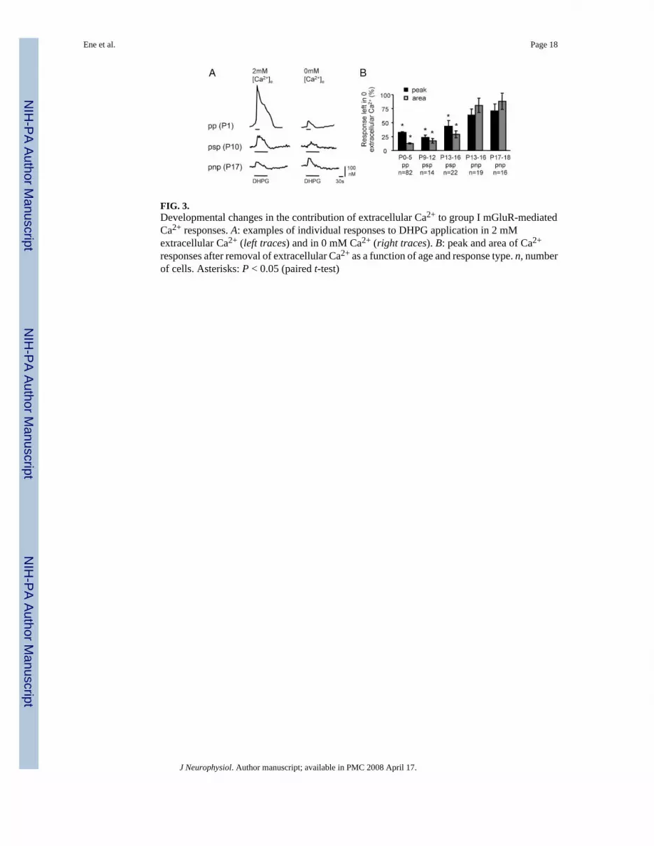
FIG. 3.Developmental changes in the contribution of extracellular Ca2+ to group I mGluR-mediatedCa2+ responses. A: examples of individual responses to DHPG application in 2 mMextracellular Ca2+ (left traces) and in 0 mM Ca2+ (right traces). B: peak and area of Ca2+
responses after removal of extracellular Ca2+ as a function of age and response type. n, numberof cells. Asterisks: P < 0.05 (paired t-test)
Ene et al. Page 18
J Neurophysiol. Author manuscript; available in PMC 2008 April 17.
NIH
-PA Author Manuscript
NIH
-PA Author Manuscript
NIH
-PA Author Manuscript
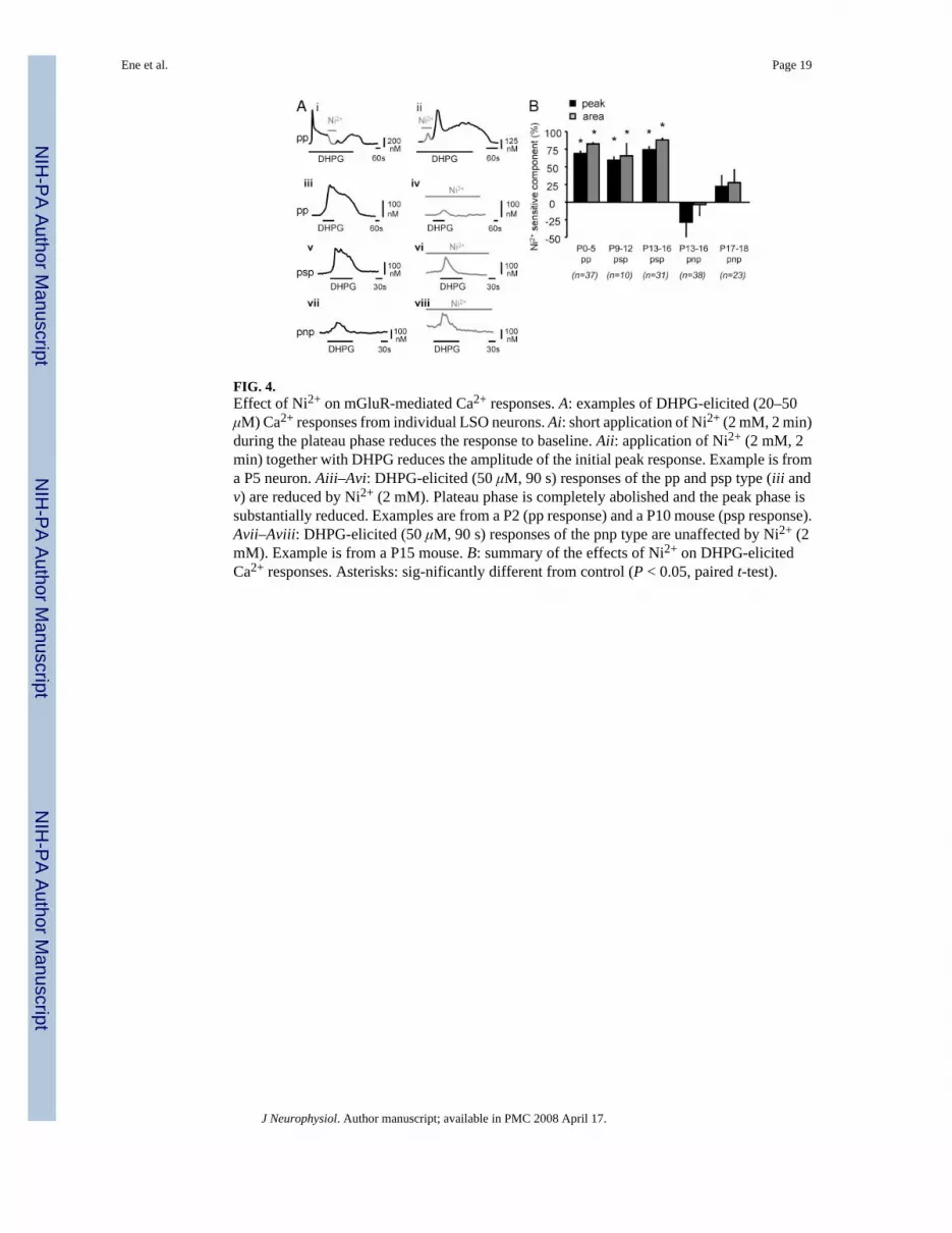
FIG. 4.Effect of Ni2+ on mGluR-mediated Ca2+ responses. A: examples of DHPG-elicited (20–50μM) Ca2+ responses from individual LSO neurons. Ai: short application of Ni2+ (2 mM, 2 min)during the plateau phase reduces the response to baseline. Aii: application of Ni2+ (2 mM, 2min) together with DHPG reduces the amplitude of the initial peak response. Example is froma P5 neuron. Aiii–Avi: DHPG-elicited (50 μM, 90 s) responses of the pp and psp type (iii andv) are reduced by Ni2+ (2 mM). Plateau phase is completely abolished and the peak phase issubstantially reduced. Examples are from a P2 (pp response) and a P10 mouse (psp response).Avii–Aviii: DHPG-elicited (50 μM, 90 s) responses of the pnp type are unaffected by Ni2+ (2mM). Example is from a P15 mouse. B: summary of the effects of Ni2+ on DHPG-elicitedCa2+ responses. Asterisks: sig-nificantly different from control (P < 0.05, paired t-test).
Ene et al. Page 19
J Neurophysiol. Author manuscript; available in PMC 2008 April 17.
NIH
-PA Author Manuscript
NIH
-PA Author Manuscript
NIH
-PA Author Manuscript

FIG. 5.Effect of 2-aminoethoxydiphenylborane (2-APB) on group I mGluR-mediated Ca2+ responses.A: effect of increasing concentrations of 2-APB on DHPG-elicited Ca2+ responses in a P10LSO neuron. B. summary data. C: effect of 2-APB on KCl-elicited (60 mM) Ca2+ responses.Data are from the same cells as in B.
Ene et al. Page 20
J Neurophysiol. Author manuscript; available in PMC 2008 April 17.
NIH
-PA Author Manuscript
NIH
-PA Author Manuscript
NIH
-PA Author Manuscript
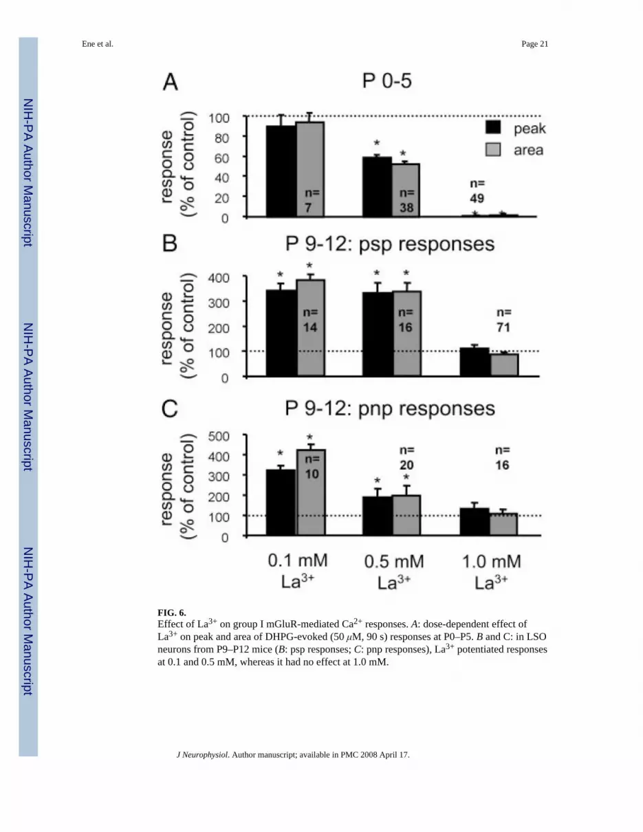
FIG. 6.Effect of La3+ on group I mGluR-mediated Ca2+ responses. A: dose-dependent effect ofLa3+ on peak and area of DHPG-evoked (50 μM, 90 s) responses at P0–P5. B and C: in LSOneurons from P9–P12 mice (B: psp responses; C: pnp responses), La3+ potentiated responsesat 0.1 and 0.5 mM, whereas it had no effect at 1.0 mM.
Ene et al. Page 21
J Neurophysiol. Author manuscript; available in PMC 2008 April 17.
NIH
-PA Author Manuscript
NIH
-PA Author Manuscript
NIH
-PA Author Manuscript
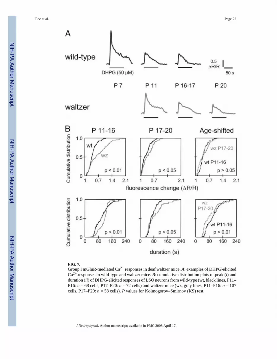
FIG. 7.Group I mGluR-mediated Ca2+ responses in deaf waltzer mice. A: examples of DHPG-elicitedCa2+ responses in wild-type and waltzer mice. B: cumulative distribution plots of peak (i) andduration (ii) of DHPG-elicited responses of LSO neurons from wild-type (wt, black lines, P11–P16: n = 68 cells, P17–P20: n = 72 cells) and waltzer mice (wz, gray lines, P11–P16: n = 107cells, P17–P20: n = 58 cells). P values for Kolmogorov–Smirnov (KS) test.
Ene et al. Page 22
J Neurophysiol. Author manuscript; available in PMC 2008 April 17.
NIH
-PA Author Manuscript
NIH
-PA Author Manuscript
NIH
-PA Author Manuscript

FIG. 8.Durations of group I mGluR-mediated Ca2+responses in waltzer, heterozygote, and wild-typemice. A: cumulative distribution of DHPG-elicited Ca2+ response durations in P11 to P16 LSOneurons from homozygote waltzer (wz, gray line, n = 107 cells), hearing heterozygote (het,dotted line, n = 44 cells), and hearing wild-type mice (wt, black line, n = 68 cells). Durationsin heterozygote mice were significantly shorter compared with homozygote mice (P < 0.005)and were not different from wild-type mice (P > 0.1). B: same as A but for postnatal days 17–20. Durations in heterozygote mice were not significantly different from wild-type orhomozygote mice (wz: n = 58 cells; het: n = 16 cells; wt: n = 72 cells). C: response durationsin heterozygote mice with a startle response (black line; P14–P16, n = 44 cells) and mice lackinga startle response (gray line; P12–P13, n = 29 cells). Durations in mice lacking a startle responsewere significantly longer than in mice with a startle response (P < 0.001). D: examples ofDHGP-elicited Ca2+-responses from heterozygote mice with and without startle response.Scale bars, 0.02 ΔR/R (top traces), 0.01 ΔR/R (bottom traces), 50 s. P values for KS test.
Ene et al. Page 23
J Neurophysiol. Author manuscript; available in PMC 2008 April 17.
NIH
-PA Author Manuscript
NIH
-PA Author Manuscript
NIH
-PA Author Manuscript

FIG. 9.Developmental changes in response types in wild-type and waltzer mice. A: percentage of ppand psp responses is greater in wz than in wt mice for both age groups (P11–P16: wt = 32 cells,wz = 80 cells; Fisher’s exact test, P < 0.05; P17–P20: wt = 17 cells, wz = 21 cells; Fisher’sexact test, P > 0.05). B: percentage of pnp responses is smaller in wz than in wt mice (P11–P16: wt = 36 cells, wz = 27 cells; Fisher’s exact test, P < 0.05; P17–P20: wt = 55 cells, wz =37 cells; Fisher’s exact test, P > 0.05)
Ene et al. Page 24
J Neurophysiol. Author manuscript; available in PMC 2008 April 17.
NIH
-PA Author Manuscript
NIH
-PA Author Manuscript
NIH
-PA Author Manuscript

NIH
-PA Author Manuscript
NIH
-PA Author Manuscript
NIH
-PA Author Manuscript
Ene et al. Page 25TA
BLE
1Pa
ram
eter
s des
crib
ing
mG
luR
-elic
ited
calc
ium
resp
onse
s in
deve
lopi
ng L
SO n
euro
ns
Peak
and
Pla
teau
(pp)
Gro
upPe
ak a
nd S
mal
l Pla
teau
(psp
) Gro
upPe
ak a
nd N
o Pl
atea
u (p
np) G
roup
Age
P0–P
5P9
–P12
P13–
P16
P17–
P19
P9–P
12P1
3–P1
6P1
7–P1
9P9
–P12
P13–
P16
P17–
P19
N22
217
813
254
178
132
5417
813
254
n22
246
154
7542
759
7543
n/N
, %10
024
117
4232
1232
5780
Peak
am
plitu
de, n
M30
4 ±
1524
7 ±
2730
4 ±
5190
± 6
106
± 13
138
± 10
84 ±
1.5
56 ±
976
± 6
51 ±
5Pl
atea
u am
plitu
de, n
M15
7 ±
797
± 1
112
1 ±
2051
± 3
51 ±
446
± 6
65 ±
13
——
—D
urat
ion,
s19
0 ±
315
7 ±
516
7 ±
621
5 ±
812
9 ±
312
2 ±
417
2 ±
2265
± 3
76 ±
265
± 3
Are
a, m
M ·
s25
.9 ±
1.0
16.0
± 1
.826
. ± 3
4.2
11.6
± 0
.38.
3 ±
0.7
9.2
± 0.
711
.9 ±
1.5
2.1
± 0.
33.
1 ±
0.3
2.5
± 0.
4
Val
ues a
re m
eans
± S
E. N
= to
tal n
umbe
r of c
ells
test
ed; n
= n
umbe
r of c
ells
in a
resp
onse
gro
up.
J Neurophysiol. Author manuscript; available in PMC 2008 April 17.