brainstem metabotropic glutamate receptors reduce food intake and activate dorsal pontine and...
TRANSCRIPT
lable at ScienceDirect
Neuropharmacology 107 (2016) 146e159
Contents lists avai
Neuropharmacology
journal homepage: www.elsevier .com/locate/neuropharm
Brainstem metabotropic glutamate receptors reduce food intake andactivate dorsal pontine and medullar structures after peripheralbacterial lipopolysaccharide administration
L�ea Chaskiel a, b, Flora Paul a, b, Rüdiger Gerstberger c, Thomas Hübschle c,Jan Pieter Konsman a, b, *
a CNRS, PsychoNeuroImmunologie, Nutrition et G�en�etique, UMR 5226, Bordeaux, Franceb Univ. Bordeaux, PsyNuGen, UMR 5226, Bordeaux, Francec Institut für Veterin€ar-Physiologie und -Biochemie, Justus-Liebig-Universit€at Giessen, 35392 Giessen, Germany
a r t i c l e i n f o
Article history:Received 6 January 2016Received in revised form18 March 2016Accepted 21 March 2016Available online 22 March 2016
Keywords:AnorexiaBrainstemFood intakeGlutamateLipopolysaccharideNucleus of the solitary tractParabrachial nucleiSickness behavior
* Corresponding author. Present address: CNRS UMtique des Syst�emes Biologiques, Universit�e de BordeauBordeaux, France.
E-mail address: jan-pieter.konsman@u-bordeaux2
http://dx.doi.org/10.1016/j.neuropharm.2016.03.0300028-3908/© 2016 Published by Elsevier Ltd.
a b s t r a c t
During infection-induced inflammation food intake is reduced. Vagal and brainstem pathways areimportant both in feeding regulation and immune-to-brain communication. Glutamate is released byvagal afferent terminals in the nucleus of the solitary tract and by its neurons projecting to the para-brachial nuclei. We therefore studied the role of brainstem glutamate receptors in spontaneous foodintake of healthy animals and during sickness-associated hypophagia after peripheral administration ofbacterial lipopolysaccharides or interleukin-1beta. Brainstem group I and II metabotropic, but not ion-otropic, glutamate receptor antagonism increased food intake both in saline- and lipopolysaccharide-treated rats. In these animals, expression of the cellular activation marker c-Fos in the lateral para-brachial nuclei and lipopolysaccharide-induced activation of the nucleus of the solitary tract rostral tothe area postrema were suppressed. Group I metabotropic glutamate receptors did not colocalize with c-Fos or neurons regulating gastric function in these structures. Group I metabotropic glutamate receptorswere, however, found on raph�e magnus neurons that were part of the brainstem circuit innervating thestomach and on trigeminal and hypoglossal motor neurons. In conclusion, our findings show thatbrainstem metabotropic glutamate receptors reduce food intake and activate the lateral parabrachialnuclei as well as the rostral nucleus of the solitary tract after peripheral bacterial lipopolysaccharideadministration. They also provide insight into potential group I metabotropic glutamate receptor-dependent brainstem circuits mediating these effects.
© 2016 Published by Elsevier Ltd.
1. Introduction
The regulation of food intake by the nervous system is thoughtto involve energy homeostasis and emergency feeding systems(Morton et al., 2014). The latter may temporarily override theformer either by stimulating feeding when hypoglycemia occurs orby inhibiting food intakewhen the organism is facedwith infection,trauma or threats from its environment (Morton et al., 2014).Temporary hypophagia may constitute an evolutionary-conservedadaptive response to acute infection (Konsman and Dantzer,
R 5536, R�esonance Magn�e-x, 146, rue L�eo Saignat, 33076
.fr (J.P. Konsman).
2001), even though long-term lowered food consumption ispotentially deleterious in chronic inflammatory diseases.
Recently, a brainstem projection from the nucleus of the solitarytract (NTS) to the lateral parabrachial (PB) nuclei has been shown toreduce food intake (Wu et al., 2012). The NTS, in turn, is innervatedby neuropeptidergic fibers from the hypothalamus, serotonergicneurons in the raph�e magnus (RM) and glutamate-containing af-ferents of the gustatory and vagus nerves (Grill, 2010; Wu et al.,2012). Administration of a-amino-3-hydroxy-5-methyl-4-isoxazolepropionic acid (AMPA) and N-methyl-D-aspartic acid(NMDA) glutamate receptor antagonists in the dorsocaudal brain-stem attenuates neuronal activation in the NTS after gustatory orgastric stimulation (Li and Smith, 1997; Zhang and Fogel, 2003) andmodulates ingestion of palatable food and deprivation-inducedintake (Treece et al., 1998; Zheng et al., 2002; Guard et al., 2009).
L. Chaskiel et al. / Neuropharmacology 107 (2016) 146e159 147
These observations indicate that glutamate action in the NTS isinvolved in the regulation of feeding when the motivation to eat ishigh.
The projections of the NTS, in particular from its rostral part, tothe PB nuclei also contain glutamate (Gill et al., 1999). These pro-jections seem to be activated by visceral malaise as intraperitonealinjection of LiCl increases extracellular glutamate in the PB nuclei(Bielavska et al., 2000). Interestingly, inhibition of calcitonin gene-related peptide (CGRP)-expressing neurons of the lateral PB nu-cleus attenuates the reduction in food intake after the peripheraladministration of LiCl or bacterial lipopolysaccharides (LPS) (Carteret al., 2013). However, the neuronal mechanisms and brainstemcircuits underlying LPS-induced hypophagia remain to be fullyelucidated.
The vagus nerve expresses the LPS-recognizing Toll-likereceptor-4 (TLR4) (Hosoi et al., 2005) and the signaling receptor IL-1R1 for the pro-inflammatory cytokine interleukin-1beta (IL-1beta)(Ek et al., 1998). Alternatively, circulating LPS and IL-1beta can act inthe area postrema (AP) that lacks a functional endothelial blood-brain barrier (Van Houten and Posner, 1983) but contains TLR4-and IL-1R1-expressing cells (Ericsson et al., 1995; Laflamme andRivest, 2001). Regardless of their exact sites of action, peripheralLPS or IL-1beta administration results in glutamate release in theNTS (Mascarucci et al., 1998). Interestingly, peripheral administra-tion of a group I and II metabotropic glutamate receptor (mGluR)antagonist attenuates the reduction in food intake and body weightobserved after bacterial LPS injection (Weiland et al., 2006), but theextent to which this antagonist penetrates the brain is unknown.
In the present work, we studied the role of brainstem glutamatereceptors in the regulation of spontaneous intake of regular chowin healthy animals and in sickness-associated hypophagia broughtabout by peripheral LPS or IL-1beta administration. For thoseglutamate receptor antagonists that had an effect on food intake,we analyzed the brainstem distribution of the targeted receptorsand assessed c-Fos expression in the NTS, PB nuclei and RM. Gastricmotility is important in the regulation of food intake and controlledby neurons of the dorsal motor nucleus of the vagus nerve (DMV),which, in turn, are contacted by neurons of the NTS, PB nuclei anddifferent raph�e nuclei (Rinaman et al., 1993). We therefore alsostudied the presence of glutamate receptors in the brainstemneuronal network controlling gastric motility.
2. Materials and methods
2.1. Animals
Experiments, including surgical procedures and treatments,were conducted according to local and European recommendationson animal research (ethics approval number GI 18/2 e No. 13/2002and European Council Directive of 24 November 1986 (86/609/EEC)).
2.1.1. Pharmacological studiesOne hundred and eighty-seven male Wistar Han rats (Charles
River, L'Arbresle, France), weighing between 250 and 275 g uponarrivals were used. After one week of acclimation in group cages,rats were housed individually two days prior to surgery with adlibitum access to food and water in a temperature (22 ± 1 �C)controlled environment where lights were on from 07:00 to19:00 h.
2.1.2. Neuroanatomical tracing studiesTen male Wistar rats with a mean body weight of 230 g were
housed individually with free access to water and standard labo-ratory chow. Room temperature was adjusted to 23 ± 1 �C at
relative humidity of 50e55%, and lights were on from 07:00 to19:00 h.
2.2. Surgery
2.2.1. Guide cannula placementRats were anesthetized with 61 mg/kg ketamine (Imalgene
1000, Rhone M�erieux, Lyon, France) and 9 mg/kg xylazine (BayerPharma, Puteaux, France) administered intraperitoneally, andsecured in a stereotactic apparatus (David Kopf Instruments,Tujunga, CA, USA). A midline incision was made into the skincovering the skull and skin flaps were pulled aside. A stainless-steel12 mm long guide cannula (23 gauge) was lowered on the midline,4.5 mm below the cortical surface through a hole drilled in the skull2.5 mm posterior to lambda corresponding to bregma �11.5 andpermanently fixed to the skull with dental cement and watch-makers' screws. These coordinates were chosen to position the tipof this guide cannula 2 mm above the roof of the 4th ventricle at alevel where this ventricle it relatively large and profound. The headwound was closed by suturing skin flaps and rats were allowed torecover for at least one week during which they were handled dailybefore injections.
2.2.2. Neurotropic virus injectionAttenuated Bartha pseudorabies virus (PRV-Ba) was used to
trace the efferent neural pathways regulating gastric motility andsecretion. PRV-Ba propagated as previously described (Yoshidaet al., 2003) and stocked at a concentration of 8.03 TCID50/mLat �70 �C until use. Rats were anesthetized with intraperitonealinjections of ketamine-xylazine solution (100 mg/kg ketaminehydrochloride (Pharmacia Upjohn, Erlangen, Germany) and 25 mg/kg Rompun (Bayer Vital, Leverkusen, Germany). A midline lapa-rotomy was made to expose the stomach and approximately 0.7 mlof 108 pfu/ml PRV-Ba was injected at five sites in the stomach wallalong its great curvature with a 5-ml syringe (Hamilton, Bonaduz,Switzerland) that was kept in place for 30 s after injection. Anyefflux from the injection site was immediately adsorbed withsterile cotton buds after which the wound was closed. After wakeup and recovery for several day, rats were deeply anesthetized andkilled for transcardial perfusion-fixation after 54 h (n ¼ 1), whencell bodies of neurons in the DMV that project to the stomach viathe vagus nerve can be expected to be infected, 72 h (n ¼ 3), cor-responding to the published infection of the first second orders inthe NTS and the area postrema, or 96e102 h (n ¼ 6) during whichthe density of infected medullary neurons is reported to reach itsmaximum (Rinaman et al., 1993).
2.3. Treatments
For the studies addressing the effects of AMPA/kainate andmetabotropic glutamate receptor antagonism, two sets of experi-ments we performed. In the series of experiments designated for c-Fos immunohistochemistry, the effects of intra-4th ventricle (i4v)glutamate receptor antagonism on inflammatory hypophagia andweight loss were assessed until 6 h after intraperitoneal (ip) in-jection of LPS or its saline vehicle. In another set of experiments, theeffects of i4v glutamate receptor antagonism on inflammatoryhypophagia and weight loss were compared between ip IL-1betaand LPS. These latter series were set up so that at the end of theexperiment all animals had received a glutamate receptor antago-nist and its vehicle centrally and a pro-inflammatory stimulus,consisting of either IL-1beta or LPS and their vehicles peripherally.Thus, if one week after i4v administration and subsequent ip in-jection, food and water intake as well as body weight had returnedto levels observed before treatment for at least three days, the
L. Chaskiel et al. / Neuropharmacology 107 (2016) 146e159148
experiment was repeated inversing i4v and ip treatments. The or-der of administration of the inflammatory stimulus (IL-1beta orLPS) between the two injections was determined by coin toss.
Given 1) that the NMDA glutamate receptor requires membranedepolarization to remove theMg2þ ion blocking this channel beforeit can become active (Nowak et al., 1984) and 2) that 97% of NTSneurons containing the calcium-permeable AMPA-receptor subunitalso express the NMDA receptor and vice versa (Lin and Talman,2002), we decided to only perform an experiment with an NMDAreceptor antagonist in case of a positive effect of AMPA/kainate/quisqualate receptor antagonism on food intake was observed.
2.3.1. Intra-4th ventricle (i4v) administration of glutamate receptorantagonists
A 30 gauge (14mm long) injection cannula filled with glutamatereceptor antagonists or their vehicle and connected to a 10 ml sy-ringe (Hamilton, Villebon sur Yvette, France) with plastic tubingwas lowered through the guide cannula into the 4th ventricle.
The AMPA/kainate/quisqualate water soluble receptor antago-nist 6-cyano-7-nitroquinoxaline-2-3-dione (CNQX; Alexis Bio-chemicals, Enzo Life Sciences, Vileurbanne, France) was dissolved in2 ml phosphate-buffered saline (PBS 0.1 M; pH 7.4) and adminis-tered at a dose of 23.2 nmol (6.4 mg) per animal into the 4thventricle based on the following findings: 1) 1.2 nmol CNQXinjected directly into the dorsal vagal complex containing the NTSand DMV attenuates gastrointestinal vago-vagal reflexes initiatedby gastrointestinal distention in the rat (Zhang and Fogel, 2003)and 2) i4v administration of 5 nmol (0.46 mg) of the AMPA/kainatereceptor antagonist 6-nitro-7-sulphamoylbenzo[f]quinoxaline-2,3-dione (NBQX) attenuates sucrose intake (Zheng et al., 2002) and 3)taking into consideration that CNQX is about 4.5 times less potentthan NBQX in displacing binding to AMPA receptors (Dev et al.,1996).
Since one of our aims was to determine if the effects of theperipheral administration of the metabotropic glutamate receptorantagonists on LPS-induced hypophagia observed by Weiland et al.(2006) were due to a brainstem site of action, we used the samegroup I and II mGluR antagonist L(þ)-2-amino-3-3-phosphonoproprionic acid (AP3; Sigma, St-Fallavier, France). Inthe present work, it was dissolved in PBS (0.1M; pH 7.4) and used ata dose of 237 nmol (40 mg) per animal given into the 4th ventriclebased on the finding that administration of 60 nmol AP3 in thesmaller intrathecal spinal CSF compartment reduces inflammatoryhyperalgesia (Young et al., 1997).
Since the PB and medial NTS are located adjacent to the 4thventricle, glutamate receptors in these structures were targeted byi4v administration of antagonists. This route allows for adminis-tration of relatively concentrated solutions compared to intra-parenchymal injection while keeping the risks of side effects low.Indeed, i4v administered molecules are diluted in the 10 ml cere-brospinal fluid (CSF) of the 4th ventricle (Levinger, 1971) withwhich it rapidly leaves the ventricular space to the subarachnoidspace via CSF convection and bulk (Pardridge, 2011). Instead, theentry of molecules from the CSF into the brain parenchyma occursthrough diffusion, which is a much slower process than CSF con-vection and bulk flow. Moreover, the concentration of a diffusingmolecule decreases exponentially with diffusion distance. Thus, theconcentration of a small molecule in the brain parenchyma 1 mmbeyond the ependymal surface of the ventricles is only 1% of its CSFconcentration (Pardridge, 2011). Keeping this in mind, it is impor-tant to point out that the dose of CNQX injected into the 4thventricle (5 nmol or 0.46 mg) was already lower than that givenlocally into the PB shown to decrease tachycardia and pressor re-sponses after stimulation of the hypothalamic defense area (Diaz-Casares et al., 2012). Considering its dilution in the CSF and the
rapid decrease of its concentration during diffusion into the pa-renchyma, CNQX was therefore not expected to yield any cardio-vascular side effects in the present work.
As for the dose of i4v AP3 (0.237 m mol or 40 mg) administeredinto the fourth ventricle it is not expected to have any non-specificeffects on behavior since injection of up to 200 mg into the lateralcerebral ventricle (40 ml volume (Levinger, 1971)) does not affectspontaneous locomotor activity (Winnicka et al., 1994). The dose ofAP3 used in the present work is estimated to have resulted in aconcentration gradient of 2.4 mMe0.024 mM in the NTS betweenthe surface and 1 mm below the surface of the fourth ventricle,respectively. These concentrations are still lower than the 30mMofAP3 that has been shown to result in a transient decrease of 10 mmHg in blood pressure from a baseline of 100 mm Hg when injectedinto the NTS or ventrolateral medulla in rats (Vitagliano et al., 1994).Similarly, the dose of i4v AP3 administered is expected to haveresulted in concentration gradient of 2.4 mMe0.024 mM in the PBbetween the part facing the ventricular surface and 1 mm beyondit. This concentration gradient in the PB covers that known to blockthe acquisition of conditioned taste aversion (0.1e5 nM (Vales et al.,2006)). Thus, the dose of AP3 administered into the fourth ventricleis expected to have minimal behavioral and cardiovascular sideeffects while likely antagonizing brainstem mechanisms that occurafter systemic LPS injection, such as the learning of the associationof a particular tastewith visceral malaise (Cross-Mellor et al., 2004).
The 2 ml containing glutamate receptor antagonists or theirvehicle were injected over two minutes after which the injectioncannula was left in place in the 4th ventricle for an additionalminute. Intra-4th ventricle injection was completed 45 min beforeIL-1 or PBS administration and just before LPS or saline injection,since IL-1beta can be detected in macrophages and dendritic cellsin proximity of the vagus nerve 45 min after intraperitoneal LPSinjection (Goehler et al., 1999).
2.3.2. Intraperitoneal (ip) injectionsEscherichia coli LPS (200 mg/kg; 0127:B8, Sigma, St Louis, MO,
USA) or its vehicle 0.9% NaCl was injected intraperitoneally 2 hbefore lights went off. Rat recombinant IL-1b (30 mg/kg, (Anforthet al., 1998)) or its vehicle PBS was injected into the peritonealcavity 1 h before lights went off. This difference in timing of theadministration of these two inflammatory stimuli was motivatedby the finding that IL-1beta can be detected in macrophages anddendritic cells in proximity of the vagus nerve 45min and in plasma60 min after intraperitoneal LPS injection (Goehler et al., 1999).
2.4. Food intake and body weight measurements
Since food restriction prior to administration of bacterial LPS hasbeen shown to affect subsequent hypophagia and weight loss(MacDonald et al., 2014), the effects of inflammatory stimuli onthese parameters were studied in spontaneously-feeding animalswith ad libitum access to regular chow for the entire light-darkcycle. As rats show a peak of spontaneous feeding during the firsthours of the dark, injections were timed to produce their maximumeffect during this period. Food intake and body weight changeswere obtained by measuring food ruffs and rats just before andevery 2 h until 6 h after ip injection on a high-precision balanceequipped with an integrator to correct for movement. Animalswere habituated to this procedure for at least three days prior to theday of experiment.
In spite of the fact that all rats show a peak of food intake duringthe first hours of the dark phase when provided ad libitum access tofood, each individual still has its ownmeal pattern, which is mostlyrepeated from day to day (van Dijk and Strubbe, 2003). Cumulativefood intake and changes in body weight over periods longer than

L. Chaskiel et al. / Neuropharmacology 107 (2016) 146e159 149
2 h (that is 0e4 h; 0e6 h and 2e6 h) were therefore favored for datapresentation and analysis, even though food intake and changes inbody weight were also analyzed over consecutive 2 h timewindows.
2.5. Immunohistochemistry
Six hours after ip injection of saline or LPS, when animals areexpected to have consumed food during the first four hours of theiractive phase and c-Fos expression in the caudal brainstem inresponse to comparable doses of LPS to still be high (Konsman et al.,1999), animals were deeply anesthetized with pentobarbital(60 mg/kg). Once the hind paw reflex upon plantar pinching haddisappeared, animals were fixed by intracardiac perfusion of 4%paraformaldehyde in 0.1 M PBS (pH 7.5 at 10 �C for 15 min) after ashort rinse with 0.1 M PBS. The brain was removed from the skull,post-fixed for 4 h in the same fixative and cryoprotected in 30%sucrose in 0.1 M PBS for 48 h. Brains were deep frozen and storedat �80 �C until they were cut in 25 mm-thick sections on a cryostat.Brain sections were collected in cold cryoprotectant (0.05 Mphosphate buffer, 20% glycerol and 30% ethylene glycol) and storedat �20 �C until further processing.
2.5.1. Single-labeling immunohistochemistryAfter washing off the cryoprotectant solution, immunohisto-
chemical processing was performed on a 1 in 6 series of freefloating sections using the streptavi-dinebiotineimmunoperoxidase technique (Konsman andBlomqvist, 2005). Briefly, non-specific binding sites were blockedby a 1 h incubation of sections in PBS, pH 7.4, containing 0.3% TritonX-100 and 1.0% bovine albumin. Commercially available rabbitantisera directed against a peptide at the N-terminus of c-Fos hu-man origin (Cat. n�: sc-52; Santa Cruz Biotechnology, Santa Cruz,CA; diluted 1:2000 in the same buffer) or against purified pseu-dorabies virus-Bartha (Cat n�: PA1-081; Affinity BioReagents,Thermo Electron, Courtaboeuf, France; diluted 1:500 in the samebuffer) or a mouse antibody raised against recombinant ratmGluR1alpha (Cat. n�: 556331, Clone G209-488, BD Pharmingen,BD France, Le Point de Claix, France; diluted 1:1000 in the samebuffer) were added to the sections overnight at room temperatureor for 60 h at 4 �C.
The c-Fos antiserum 1) has been characterized by the vendor torecognize a 62 kDa protein, corresponding to the molecular weightof c-Fos, on Western blots, and 2) has previously been used by us(Konsman and Blomqvist, 2005) and others (Engler et al., 2011;Farzi et al., 2015) at comparable dilutions in streptavi-dinebiotineimmunoperoxidase immunohistochemistry protocolson formaldehyde-fixed rat brain tissue.
The PRV-Ba antiserum has been shown by the vendor 1) todetect proteins on Western blots in the range of 80e155 kDa rep-resenting various PRV proteins, 2) to result in immunohistochem-ical staining of the suprachiasmatic nucleus of the hypothalamusafter PRV injection into vitreous body of the eye and 3) has alreadybeen used by us under conditions that are comparable to thoseused here (Yoshida et al., 2003).
The mGluR1alpha antibody has been characterized by thevendor 1) to recognize a 130 kDa protein, corresponding to themolecular weight of mGluR1alpha, onWestern blots, and 2) to yieldimmunohistochemical staining of Purkinje cells in formaldehyde-fixed rat cerebellum, where mGluR1alpha mRNA is stronglyexpressed (Berthele et al., 1998), which can be abolished by pre-incubation of the antibody with rat recombinant mGluR1alpha. In
addition, 3) cellular surface staining with his antibody has beenshown to decrease after mGluR1alpha agonist-induced receptorinternalization (Hong et al., 2009).
After four rinses in PBS, sections were treated for 30 min in 0.3%(v/v) hydrogen peroxide followed by rinses in PBS. Sections wereincubated for 2 h at room temperature with biotinylated donkeyanti-rabbit IgG (Amersham, Les Ulis, France) diluted 1:1000 in PBScontaining 0.3% Triton X-100 and 1.0% bovine albumin and stainedusing the ABC protocol (Vectastain Elite, Vector Laboratories,Eurobio/Abcys, Les Ullis, France) with nickel-enhanced dia-minobenzidine as a chromogene.
Stained sections were examined with a microscope (LeicaDM5500B, Leica Microsystems, Wetzlar, Germany) and imageswere captured by a high-resolution CCD video camera image andfed into a personal computer. Photomicrographs to illustrate c-Foslabeling were obtained using this system and saved as TIFF files.Image J software (http://rsbweb.nih.gov/ij/) was used to generate adigitized signal proportional to the intensity of illumination. Imageprocessing was performed on the grey image by defining a setthreshold for grey value and surface above which labeling had to betaken into account. C-Fos expression in a structure was firstdetermined with an inclusive threshold allowing for the estimationthe total number of labeled cells unilaterally per section. In case asignificant effect of glutamate receptor antagonism was found, asecond more restrictive threshold was applied to determine if thesame treatment also altered the number of intensely-stained c-Fos-positive cells as well. After application of the threshold, the imagewas converted to a binary image and measurements made.
Since the PB and RM have been proposed to be part of a brain-stem circuit involving the NTS that inhibits food intake (Mortonet al., 2014), the number of c-Fos-positive cells were counted inthese brainstem structures. To do so, for each animal, at least 3sections were analyzed for the parabrachial nuclei (betweenbregma �8.60 and �9.50 mm) and a minimum of four sections forthe raph�e magnus (between bregma �9.25 and �11.10 mm). TheNTS is a rostrocaudal long nucleus and receives sensory input in aloosely viscerotopically segregated manner along this axis and istherefore typically studied distinguishing 1) its part rostral to theAP, 2) the part at the level of the AP and 3) that caudal of the AP(Loewy, 1990; Jean, 1991). Thus, for each animal the number of c-Fos-immunoreactive cells in the “visceral” NTS were counted in atleast 4 sections caudal to the AP (between bregma �14.36and �14.86 mm), in a minimum of 2 sections at the level of the AP(between bregma �13.76 and �14.16 mm) known to receivevisceral afferents and to display c-Fos induction in response to in-flammatory signals (Rinaman, 2011), and in no less than 3 sectionsrostral to the AP (between bregma �13.15 and �13.60 mm) wherecatecholaminergic neurons have been shown to specifically expressc-Fos after peripheral LPS administration as compared to the con-sumption of a liquid palatable meal (Gaykema et al., 2009). Inaddition, the number of c-Fos-positive cells was quantified in atleast 2 sections of the AP for every animal. Finally, to assess theeffect of i4v administration of glutamate receptor antagonists onforebrain activation, c-Fos expression was quantified in at least 4sections of the paraventricular nucleus of the hypothalamus (be-tween bregma �1.33 and �1.78 mm), a structure well known toshow robust c-Fos induction after peripheral LPS administration(Sagar et al., 1995; Konsman et al., 1999).
2.5.2. Double-labeling immunohistochemistryTo determine if neurons that are part of the brainstem network
innervating the greater curvature of the stomach or those activated
L. Chaskiel et al. / Neuropharmacology 107 (2016) 146e159150
after ip LPS administration express glutamate receptor types foundto influence food intake, double-labeling immunohistochemistrywas performed to detect the presence of pseudorabies virus or c-Fos and glutamate receptor subtypes. For this purpose a mouseantibody recognizing rat mGluR1-alpha (Cat. n�: 556331, CloneG209-488, BD Pharmingen, BD France, Le Point de Claix, France) ormGluR2 (Cat. n�: ab15672, Abcam, Paris, France), the main mGluRspresent on cell bodies throughout the dorsal vagal complex thatcomprises the NTS, AP and DMV (Hay et al., 1999), was used incombination with the pseudorabies virus-specific rabbit antiserum(Cat. n�: PA1-081, Affinity BioReagents, Thermo Electron, Courta-boeuf, France) or the c-Fos-specific antiserum (Cat. n�: sc-52, SantaCruz Biotechnology, Santa Cruz, CA), all diluted 1:500 in PBS con-taining 0.3% Triton X-100 and 1.0% bovine albumin and added to thesections for 60 h at 4 �C after washing off the cryoprotectant so-lution. After four washes in PBS, sections were incubated for 2 h atroom temperature with biotinylated horse anti-mouse IgG (Vec-tashield, Vector Laboratories, Eurobio/Abcys, Les Ullis, France)diluted 1:250 in PBS containing 0.3% Triton X-100 and 1.0% bovinealbumin. After washes in PBS, Avidin-Alexa 488 and Donkey anti-rabbit IgG-Alexa 546 or Avidin-Alexa 594 and Goat anti-rabbitIgG-Alexa 488 (Invitrogen, Leiden, the Netherlands), all diluted in1:250 in PBS, were added to sections for 2 h. Sections were thenmounted in the dark and coverslipped with anti-fading mountingmedium containing 40,6-diamidino-2-phenylindole (DAPI) (Vecta-shield, Vector Laboratories, Eurobio/Abcys, Les Ullis, France).
Sections were examined on an epifluorescent microscopeequipped (Leica DM5500B, Leica Microsystems, Wetzlar, Germany)and on a confocal microscope (Leica SP5, Leica Microsystems,Wetzlar, Germany). Images from channels corresponding to DAPI,Alexa 488, 546 or 594 were obtained with a 1 mm z-step andreconstituted as stacks in MetaMorph (Molecular Devices, Sunny-vale, CA, USA). The occurrence of double-labeled neurons wasexamined on stacks obtained by confocal microscopy with help ofthe Overlay andMontage functions. For illustrative purposes, stacksof the individual channels underwent a best focus operation inImage J. Image editing software (Adobe Photoshop, Adobe Systems,San Jose, CA) was used to adjust contrast and brightness only and toprepare microphotographs.
2.6. Data presentation and analysis
The experiments designated for c-Fos immunohistochemistryafter i4v glutamate receptor antagonism and ip LPS administrationand those aimed at comparing the effects of i4v glutamate receptorantagonism between ip LPS and ip IL-1beta were expected to yieldsimilar effects on food intake and changes in body weight. Whentherewere no significant differences in terms of food intake or bodyweight between experiments addressing the effects of a glutamatereceptor antagonist, that is when the different experiments wereentered as an independent variable or factor, data obtained after ipinjection of LPS or its vehicle were pooled and analyzedaccordingly.
All data are reported as means ± SEM. Cumulative food intakeand changes in body weight (0e4 h; 0e6 h and 2e6 h) after ipinjection were analyzed by a two-way ANOVA using i4v and iptreatments as between factors and represented in figures. Foodintake and changes in body weight over consecutive 2 h timewindows were analyzed by a two-way ANOVA using i4v and iptreatments as between factors and time as a within factor andprovided as supplementary tables. The unilateral mean number ofc-Fos-positive nuclei per brainstem section was analyzed by a two-way ANOVA with i4v and ip treatments as between factors. Sig-nificant effects were further analyzed by the Newman-Keuls post-hoc test. In all cases, a level of p < 0.05 was considered as
statistically significant.
3. Results
3.1. I4v AMPA receptor antagonism exacerbated ip LPS-inducedweight loss
Food intake. A two-way ANOVA on cumulated food intakerevealed a significant anorectic effect of LPS (F1,42: 0e4 h: 22.0,p < 0.001; 0e6 h: 49.5, p < 0.001; 2e6 h: 59.3, p < 0.001) as well asa significant interaction between CNQX and LPS treatments (F1,42:2e6 h: 4.67, p < 0.05). Post-hoc tests, however, did not show asignificant effect of i4v CNQX on ip LPS-induced hypophagia(Fig. 1A). A similar ANOVA also indicated a significant hypophagiceffect of IL-1beta (F1,24: 0e4 h: 22.4, p < 0.001; 0e6 h: 38.8,p < 0.001; 2e6 h: 39.5, p < 0.001) as well as a significant foodintake-reducing effect of CNQX (F1,24: 2e6 h: 4.64, p < 0.05), but nointeraction between treatments (Fig. 1B).
Body weight. A two-way ANOVA on cumulative changes in bodyweight showed a significant reduction in body weight after LPS(F1,44: 0e4 h: 39.5, p < 0.001; 0e6 h: 54.4, p < 0.001; 2e6 h: 61.0,p < 0.001) and after CNQX (F1,44: 0e4 h: 5.07, p < 0.05) as well as atendency for an interaction between CNQX and LPS treatments(F1,44: 0e6 h: 3.29, p ¼ 0.077; 2e6 h: 2.97, p < 0.092). Post-hoc testsindicated that i4v CNQX exacerbated ip LPS-induced weight lossover the 6 h after the experiment (p < 0.05; Fig. 1C). A two-wayANOVA on cumulated changes in body weight showed a signifi-cant loss of body weight after IL-1beta injection (F1,24: 0e4 h: 31.9,p < 0.001; 0e6 h: 36.5, p < 0.001; 2e6 h: 32.7, p < 0.001), but nosignificant effect of CNQX or interaction between treatments(Fig. 1D).
Effects of i4v CNQX and ip LPS and ip IL-beta on food intake andbody weight changes during consecutive 2 h periods are providedin Supplementary Tables 1 and 2, respectively.
Since circulating corticosterone levels modulate weight lossafter LPS administration (Johnson et al., 1996), its plasma concen-trations were measured after i4v AMPA/kainate receptor antago-nism and ip LPS administration. A two-way ANOVA on plasmacorticosterone concentrations, determined by radioimmunoassay(Konsman et al., 2008), revealed a significant increase after LPSinjection (F1,24: 10.6, p < 0.01), but no effect of i4v CNQX admin-istration or interaction between CNQX and LPS treatments (Fig. 2A).As weight loss after LPS administration is likely to be mediated inpart by the peripheral actions of pro-inflammatory cytokines,circulating IL-1beta concentrations were also analyzed after i4vAMPA/kainate receptor antagonism and ip LPS administration. Atwo-way ANOVA on plasma IL-1beta, measured by enzyme-linkedimmunosorbent assay (Konsman et al., 2008), showed a signifi-cant rise after LPS injection (F1,22: 7.84, p < 0.05) as well as a sig-nificant interaction between CNQX and LPS treatments (F1,22: 31.9,p < 0.05). Post-hoc tests indicated that i4v CNQX significantlyattenuated the ip LPS-induced increase in circulating IL-1betaconcentrations (p < 0.05; Fig. 2B).
3.2. I4v metabotropic glutamate receptor antagonism increasedspontaneous food intake
Food intake. A two-way ANOVA on cumulated food intake indi-cated a significant reduction after LPS injection (F1,95: 0e4 h: 36.0,p < 0.001; 0e6 h: 54.3, p < 0.001; 2e6 h: 30.0, p < 0.001) and asignificant increase after AP3 administration (F1,95: 0e4 h: 11.6,p < 0.001; 0e6 h: 10.3, p < 0.01), but no significant interactionbetween treatments (Fig. 3A). The same analysis also showed a foodintake-reducing effect of IL-1beta (F1,57: 0e4 h: 56.2, p < 0.001;0e6 h: 102, p < 0.001; 2e6 h: 26.1, p < 0.001) and a tendency for an

Fig. 1. Cumulative food intake and changes in body weight after brainstem ionotropic glutamate receptor antagonism. Rats were injected intraperitoneally (ip) with saline (Sal) orbacterial lipopolysaccharides (LPS; A, C), phosphate-buffered saline (PBS) or interleukin-1beta (IL-1b; B, D) and intracerebroventricularly (i4v) with PBS or an AMPA/kainate/quisqualate receptor antagonist (CNQX). Food intake was estimated by the difference in weight of food pellets measured every two hours after ip injection. * in C indicates thestatistically significant exacerbating effect of i4v CNQX administration on LPS-induced weight loss between 0 and 6 h after its administration. Group sizes for CNQX-LPS experimentswere: PBS Sal: n ¼ 11; CNQX Sal: n ¼ 11; PBS LPS: n ¼ 11; CNQX LPS: n ¼ 12 and for the CNQX-IL-1beta experiment: PBS PBS: n ¼ 7; CNQX PBS: n ¼ 7; IL-1beta: n ¼ 7; CNQX IL-1beta: n ¼ 7.
Fig. 2. Circulating corticosterone and IL-1beta concentrations. Plasma corticosterone was determined by radioimmunoassay six hours after ip injection of rats with Sal or LPS andi4v administration of PBS or CNQX, an AMPA/kainate/quisqualate receptor antagonist. Plasma IL-1beta was determined by enzyme-linked immunosorbent assay six hours after ipinjection of rats with Sal or LPS and i4v administration of PBS or CNQX, an AMPA/kainate/quisqualate receptor antagonist. * indicates the statistically significant reducing effect ofi4v CNQX administration on LPS-induced IL-1beta concentrations 6 h later.
L. Chaskiel et al. / Neuropharmacology 107 (2016) 146e159 151
interaction between ip and i4v treatments (F1,57: 0e4 h: 3.91,p ¼ 0.053). Post-hoc tests indicated that i4v AP3 increased foodintake between 0 and 4 h in animals injected ip with saline(p < 0.05), but not in those administered IL-1beta (Fig. 3B).
Body weight. A two-way ANOVA on cumulated changes in bodyweight showed significant weight loss after LPS (F1,95: 0e4 h: 46.8,p < 0.001; 0e6 h: 51.7, p < 0.001; 2e6 h: 30.4, p < 0.001), but nosignificant effect of AP3 either globally or in interaction with LPS(Fig. 3C). The same analysis also revealed significant weight lossafter IL-1beta injection (F1,57: 0e4 h: 40.6, p < 0.001; 0e6 h: 28.2,p < 0.001), no significant main effect of AP3, but a tendency for aninteraction between ip and i4v treatments (F1,57: 0e4 h: 3.56,
p ¼ 0.064). Post-hoc tests did however not show a significant effectof i4v AP3 on changes in body weight after ip saline or IL-1betaadministration (Fig. 3D).
Effects of i4v AP3 and ip LPS and ip IL-beta on food intake andbody weight changes during consecutive 2 h periods are providedin Supplementary Tables 3 and 4, respectively.
3.3. Brainstem distribution of mGluR1-alpha-immunoreactivity
Given that i4v administration of the group I and II mGluRantagonist AP3 resulted in a robust increase in food intake, itspotential brainstem sites of action were studied next. Since
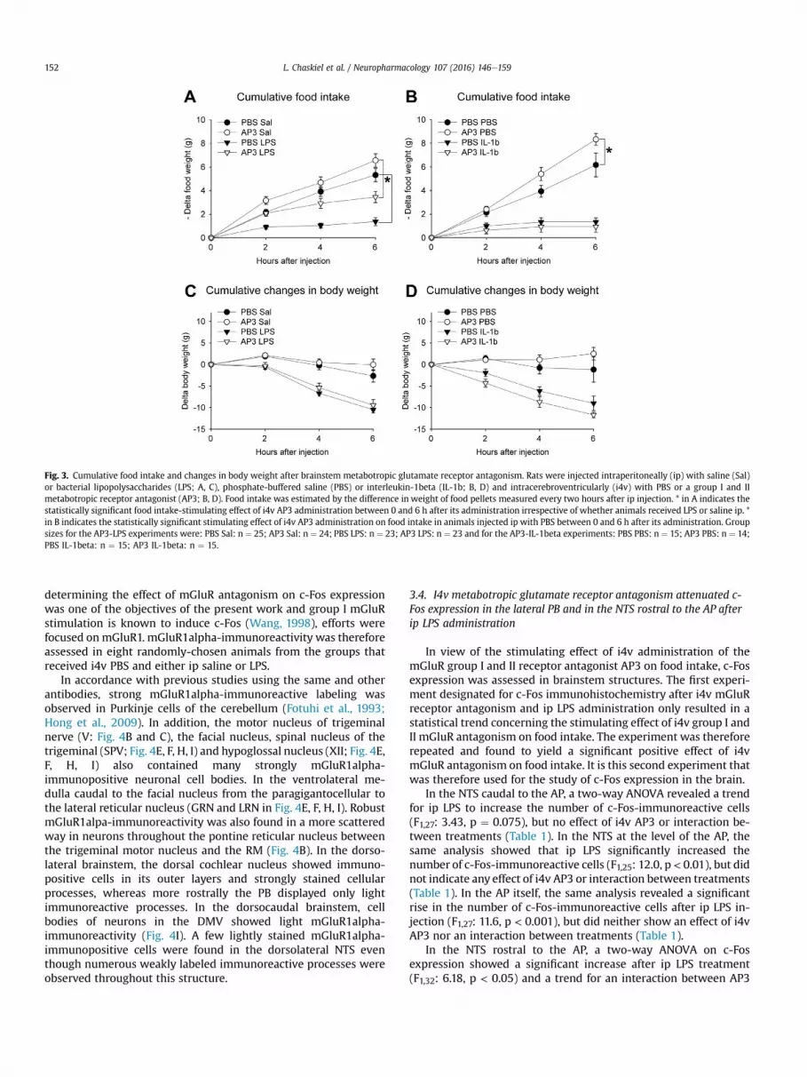
Fig. 3. Cumulative food intake and changes in body weight after brainstem metabotropic glutamate receptor antagonism. Rats were injected intraperitoneally (ip) with saline (Sal)or bacterial lipopolysaccharides (LPS; A, C), phosphate-buffered saline (PBS) or interleukin-1beta (IL-1b; B, D) and intracerebroventricularly (i4v) with PBS or a group I and IImetabotropic receptor antagonist (AP3; B, D). Food intake was estimated by the difference in weight of food pellets measured every two hours after ip injection. * in A indicates thestatistically significant food intake-stimulating effect of i4v AP3 administration between 0 and 6 h after its administration irrespective of whether animals received LPS or saline ip. *in B indicates the statistically significant stimulating effect of i4v AP3 administration on food intake in animals injected ip with PBS between 0 and 6 h after its administration. Groupsizes for the AP3-LPS experiments were: PBS Sal: n ¼ 25; AP3 Sal: n ¼ 24; PBS LPS: n ¼ 23; AP3 LPS: n ¼ 23 and for the AP3-IL-1beta experiments: PBS PBS: n ¼ 15; AP3 PBS: n ¼ 14;PBS IL-1beta: n ¼ 15; AP3 IL-1beta: n ¼ 15.
L. Chaskiel et al. / Neuropharmacology 107 (2016) 146e159152
determining the effect of mGluR antagonism on c-Fos expressionwas one of the objectives of the present work and group I mGluRstimulation is known to induce c-Fos (Wang, 1998), efforts werefocused onmGluR1. mGluR1alpha-immunoreactivity was thereforeassessed in eight randomly-chosen animals from the groups thatreceived i4v PBS and either ip saline or LPS.
In accordance with previous studies using the same and otherantibodies, strong mGluR1alpha-immunoreactive labeling wasobserved in Purkinje cells of the cerebellum (Fotuhi et al., 1993;Hong et al., 2009). In addition, the motor nucleus of trigeminalnerve (V: Fig. 4B and C), the facial nucleus, spinal nucleus of thetrigeminal (SPV; Fig. 4E, F, H, I) and hypoglossal nucleus (XII; Fig. 4E,F, H, I) also contained many strongly mGluR1alpha-immunopositive neuronal cell bodies. In the ventrolateral me-dulla caudal to the facial nucleus from the paragigantocellular tothe lateral reticular nucleus (GRN and LRN in Fig. 4E, F, H, I). RobustmGluR1alpa-immunoreactivity was also found in a more scatteredway in neurons throughout the pontine reticular nucleus betweenthe trigeminal motor nucleus and the RM (Fig. 4B). In the dorso-lateral brainstem, the dorsal cochlear nucleus showed immuno-positive cells in its outer layers and strongly stained cellularprocesses, whereas more rostrally the PB displayed only lightimmunoreactive processes. In the dorsocaudal brainstem, cellbodies of neurons in the DMV showed light mGluR1alpha-immunoreactivity (Fig. 4I). A few lightly stained mGluR1alpha-immunopositive cells were found in the dorsolateral NTS eventhough numerous weakly labeled immunoreactive processes wereobserved throughout this structure.
3.4. I4v metabotropic glutamate receptor antagonism attenuated c-Fos expression in the lateral PB and in the NTS rostral to the AP afterip LPS administration
In view of the stimulating effect of i4v administration of themGluR group I and II receptor antagonist AP3 on food intake, c-Fosexpression was assessed in brainstem structures. The first experi-ment designated for c-Fos immunohistochemistry after i4v mGluRreceptor antagonism and ip LPS administration only resulted in astatistical trend concerning the stimulating effect of i4v group I andII mGluR antagonism on food intake. The experiment was thereforerepeated and found to yield a significant positive effect of i4vmGluR antagonism on food intake. It is this second experiment thatwas therefore used for the study of c-Fos expression in the brain.
In the NTS caudal to the AP, a two-way ANOVA revealed a trendfor ip LPS to increase the number of c-Fos-immunoreactive cells(F1,27: 3.43, p ¼ 0.075), but no effect of i4v AP3 or interaction be-tween treatments (Table 1). In the NTS at the level of the AP, thesame analysis showed that ip LPS significantly increased thenumber of c-Fos-immunoreactive cells (F1,25: 12.0, p< 0.01), but didnot indicate any effect of i4v AP3 or interaction between treatments(Table 1). In the AP itself, the same analysis revealed a significantrise in the number of c-Fos-immunoreactive cells after ip LPS in-jection (F1,27: 11.6, p < 0.001), but did neither show an effect of i4vAP3 nor an interaction between treatments (Table 1).
In the NTS rostral to the AP, a two-way ANOVA on c-Fosexpression showed a significant increase after ip LPS treatment(F1,32: 6.18, p < 0.05) and a trend for an interaction between AP3
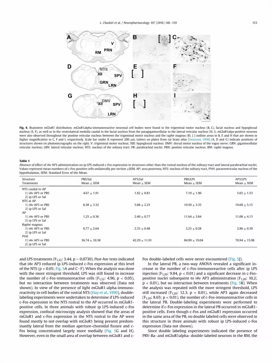
Fig. 4. Brainstem mGluR1 distribution. mGluR1alpha-immunoreactive neuronal cell bodies were found in the trigeminal motor nucleus (B, C), facial nucleus and hypoglossalnucleus (E, F), as well as in the ventrolateral medulla caudal to the facial nucleus from the paragigantocellular to the lateral reticular nucleus (H, I). mGluR1alpa-positive neuronswere also observed throughout the pontine reticular nucleus between the trigeminal motor nucleus and the raph�e magnus (B). [ ] outline areas in B, E and H that are shown inhigher magnification in C, F and I, respectively. Scale bar under K represent 200 mm. Letters on plates from rat brain atlas (Swanson, 1998) (A, D and G) indicate positions ofstructures shown on photomicrographs on the right. V: trigeminal motor nucleus; XIIl: hypoglossal nucleus; DMV: dorsal motor nucleus of the vagus nerve; GRN: gigantocellularreticular nucleus; LRN: lateral reticular nucleus; NTS: nucleus of the solitary tract; PB: parabrachial nuclei; PRN: pontine reticular nucleus; RM: raph�e magnus.
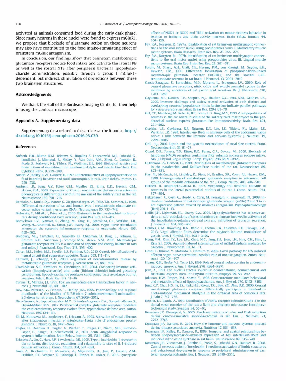
Table 1Absence of effect of i4v AP3 administration on ip LPS-induced c-Fos expression in structures other than the rostral nucleus of the solitary tract and lateral parabrachial nuclei.Values represent mean numbers of c-Fos-positive cells unilaterally per section ±SEM. AP: area postrema, NTS: nucleus of the solitary tract, PVH: paraventricular nucleus of thehypothalamus, SEM: Standard Error of the Mean.
StructureTreatments
PBS/SalMean ± SEM
AP3/SalMean ± SEM
PBS/LPSMean ± SEM
AP3/LPSMean ± SEM
NTS caudal to AP1) i4v AP3 or PBS2) ip LPS or Sal
4.67 ± 1.91 1.82 ± 0.81 7.19 ± 1.96 5.65 ± 1.53
NTS at AP1) i4v AP3 or PBS2) ip LPS or Sal
8.38 ± 3.32 5.68 ± 2.23 19.50 ± 3.35 19.68 ± 5.15
AP1) i4v AP3 or PBS2) ip LPS or Sal
1.25 ± 0.36 2.40 ± 0.77 11.64 ± 3.84 11.08 ± 4.13
Raph�e magnus1) i4v AP3 or PBS2) ip LPS or Sal
6.77 ± 2.64 2.35 ± 0.48 3.23 ± 0.58 2.86 ± 0.39
PVH1) i4v AP3 or PBS2) ip LPS or Sal
36.74 ± 10.38 42.29 ± 11.91 84.99 ± 19.04 70.94 ± 15.98
L. Chaskiel et al. / Neuropharmacology 107 (2016) 146e159 153
and LPS treatments (F1,32: 3.44, p¼ 0.0730). Post-hoc tests indicatedthat i4v AP3 reduced ip LPS-induced c-Fos expression at this levelof the NTS (p < 0.05; Fig. 5A and CeF). When the analysis was donewith the more stringent threshold, LPS was still found to increasethe number of c-Fos-immunoreactive cells (F1,32: 4.96, p < 0.05),but no interaction between treatments was observed (Data notshown). In view of the presence of light mGluR1-alpha-immuno-reactivity in cell bodies of the rostral NTS (Hay et al., 1999), double-labeling experiments were undertaken to determine if LPS-inducedc-Fos expression in the NTS rostral to the AP occurred in mGluR1-positive cells. In three animals with robust ip LPS-induced c-Fosexpression, confocal microscopy analysis showed that the areas ofmGluR1 and c-Fos expression in the NTS rostral to the AP werefound mostly to not overlap with mGluR1 being present predom-inantly lateral from the median aperture-choroidal fissure and c-Fos being concentrated largely more medially (Fig. 5G and H).However, even in the small area of overlap between mGluR1 and c-
Fos double-labeled cells were never encountered (Fig. 5J).In the lateral PB, a two-way ANOVA revealed a significant in-
crease in the number of c-Fos-immunoreactive cells after ip LPSinjection (F1,20: 9.84, p < 0.01) and a significant decrease in c-Fos-positive nuclei subsequent to i4v AP3 administration (F1,20: 10.2;p < 0.01), but no interaction between treatments (Fig. 5B). Whenthe analysis was repeated with the more stringent threshold, LPSstill increased (F1,20: 12.3, p < 0.01), while AP3 again decreased(F1,20: 8.65; p < 0.01), the number of c-Fos-immunoreactive cells inthe lateral PB. Double-labeling experiments were performed todetermine if c-Fos expression in the lateral PB occurred in mGluR1-positive cells. Even though c-Fos and mGluR1 expression occurredin the same area of the PB, no double labeled cells were observed inthis structure in three animals with robust ip LPS-induced c-Fosexpression (Data not shown).
Since double labeling experiments indicated the presence ofPRV-Ba- and mGluR1alpha- double-labeled neurons in the RM, the

Fig. 5. Metabotropic glutamate receptor-dependent c-Fos expression in the rostral nucleus of the solitary tract and lateral parabrachial nuclei. Mean numbers of c-Fos-positive cellsunilaterally per section in the nucleus of the solitary tract (NTS) rostral to the AP (A) and the lateral parabrachial nuclei (PB) (B) six hours after intraperitoneal (ip) injection of saline(Sal) or bacterial lipopolysaccharides (LPS) and intra-fourth brain ventricle (i4v) administration of phosphate-buffered saline (PBS) a group I and II metabotropic receptor antagonist(AP3). * indicates the statistically significant reduction of ip LPS-induced c-Fos expression by i4v AP3 in A and the overall significant decrease of the number of c-Fos-immuno-reactive nuclei by i4v AP3 in B. Black and white photomicrographs illustrate c-Fos expression in the NTS rostral to the AP of animals that received ip Sal and i4v PBS (C), ip Sal andi44v AP3 (D), ip LPS and i4v PBS (E) or ip LPS and i4v AP3 (F). Color photomicrographs illustrate combined immunohistochemistry for c-Fos (G and I) and mGluR1alpha (H and I) theNTS rostral to the AP. Pseudocolors: red, c-Fos; green, mGluR. No double-labeled neurons were observed. Scale bars in H and under K represent 100 mm. Rectangle on plate from ratbrain atlas (Swanson, 1998) indicates where photomicrographs were taken (J). Group sizes for c-Fos experiments were, depending on the structure analyzed, PBS Sal: n ¼ 6e9; AP3Sal: n ¼ 6e9; PBS LPS: n ¼ 6e8; AP3 LPS n ¼ 6e9. (For interpretation of the references to color in this figure legend, the reader is referred to the web version of this article.)
L. Chaskiel et al. / Neuropharmacology 107 (2016) 146e159154
number of c-Fos-immunoreactive cells was also analyzed in thisstructure. A two-way ANOVA did not show any effect of treatmentsor interaction between treatments on c-Fos expression in the RM(Table 1). Finally, to assess the effect of i4v administration of themGluR receptor antagonist on forebrain activation, c-Fos expres-sion was analyzed in the paraventricular nucleus of the hypothal-amus. In this structure, a two-way ANOVA indicated a significantincrease in the number of c-Fos-immunoreactive cells after ip LPS(F1,30: 5.81, p< 0.05), but no effect of i4v AP3 or interaction betweentreatments (Table 1).
Given that AMPA/kainate receptors are strongly expressed in theAP and in the NTS at the interface with and level of the AP and itself(Kessler and Baude, 1999), c-Fos-immunopositive cells were alsocounted in these structures after i4v administration of CNQX. In theNTS at the level of the AP, a two-way ANOVA showed that ip LPSsignificantly increased the number of c-Fos-immunoreactive cells(F1,23: 14.2, p < 0.01), but did not reveal any effect of CNQX orinteraction between treatments (Data not shown). In the AP itself,the same analysis revealed a significant rise in the number of c-Fos-immunoreactive cells after ip LPS injection (F1,23: 11.6, p < 0.01), butdid neither show an effect of i4v CNQX nor an interaction betweentreatments (Data not shown).
3.5. mGluR expression in the brainstem network innervating thestomach
Four out of the six animals that were allowed to survive
96e102 h after neurotropic virus injection showed robust labelingthroughout the NTS and in addition to the DMV (Fig. 6B, D, F, H, Jand L). In addition, brainstem PRV-Ba immunoreactive neuronswere found in the lateral PB, the pontine reticular nucleus and therostral RM (Fig. 7B, D and F). Animals that had been subject tointragastric injection of PRV-Ba showed the same brainstem dis-tribution of mGluR1alpha-immunoreactivity with strongly labeledneurons being concentrated in the trigeminal motor nucleus andthe hypoglossal nucleus (Fig. 6A, C, E and G), robust and morescattered cells being present in the pontine reticular nucleus andRM (Fig. 7A and C) and weaker staining occurring in the dorsalvagal complex.
Epifluorescent examination of combined immunohistochem-istry for PRV-Ba and mGluR1alpha resulted in less mGluR1alpha-immunoreactive-like signal in all regions examined compared todiaminobenzidine-peroxidase staining. Neurons of the hypoglossalnucleus (XII) could still be distinguished (Fig. 6I and K), but forexample cell bodies and processes in the RM were only weaklystained (Fig. 7E). In contrast, PRV-immunoreactivity was readilydetected using epifluorescence (Figs. 6J, L and 7F). Confocal analysisof brainstem structures resulted in a signal-to-noise ratio compa-rable to peroxidase-based staining of mGluR1alpha, but did notreveal any double labeled cells in the NTS and DMV, even thoughthe former two contained numerous single-labeled cells(Fig. 6MeO). In the parabrachial nuclei PRV-Ba- and mGluR1alpha-like immunoreactivity never occurred in the same neurons (notshown). However, double-labeled neurons (max 3 per section)

Fig. 6. Dorsal vagal complex metabotropic glutamate receptor 1 expression and neuronal network innervating the stomach. The dorsocaudal brainstem neuronal networkinnervating the greater curvature of the stomach, as revealed by the presence of pseudorabies-Bartha viral (PRV) particles, included neurons in the dorsal motor nucleus of thevagus (DMV) and NTS (B, D, F, and H). Metabotropic glutamate receptor 1 (mGluR1) expression was not altered by PRV injection (compare A, C, E, and G to Fig. 3H and K). E-Hrepresent higher magnifications of AeD. Scale bar in H represents 200 mm for EeH. Epifluorescent microscopic examination of combined immunohistochemistry for mGluR1alpha(green) and PRV (red) showed that mGluR1-immunoreactive cells in the hypoglossal nucleus, be that rostrally adjacent to the fourth ventricle (4v; I) or caudally adjacent to thecentral canal (cc; K), did not overlap with PRV-positive cells in the DMV and the NTS (J, L). In addition, some lightly stained mGluR1-positive cells were also observed in the DMXand to a lesser extent in the NTS. However, confocal microscopic analysis of combined immunohistochemistry for mGluR1alpha (green; M) and PRV (red; N) did not reveal anydouble labeled cells in the DMX at the interface with the NTS (O). Scale bar under O: 25 mm. 4v: fourth ventricle; XII: hypoglossal nucleus; cc: central canal; DMV: dorsal motornucleus of the vagus nerve; NTS: the nucleus of the solitary tract. (For interpretation of the references to color in this figure legend, the reader is referred to the web version of thisarticle.)
L. Chaskiel et al. / Neuropharmacology 107 (2016) 146e159 155
were found to be present in the rostroventral part of the RM(Fig. 7G, H, IeK).
Although mGluR2-immunoreactive cellular processes werefrequently observed in the dorsal and central area postrema, nodouble labeling with PRV-Ba and mGluR2 was observed (Data notshown).
4. Discussion
In the present work, we studied the role of brainstem glutamatereceptors in the regulation of food intake in healthy animals and insickness-associated hypophagia brought about by peripheral LPS orIL-1beta administration. Its main findings are that brainstem groupI and II mGluR antagonism increased food intake both in saline- andLPS-, but not IL-1beta-, treated rats and that it reduced the numberof c-Fos-positive cells in the lateral PB and LPS-induced c-Fosexpression n the NTS rostral to the AP. Group I mGluRs were foundto be absent from NTS and PB c-Fos-positive cells or neurons thatare part of the network innervating the stomach, but present on RM
neurons regulating gastric function as well as on neurons of thetrigeminal motor and hypoglossal nuclei.
In the different series of experiments addressing the effects ofi4v ionotropic or metabotropic glutamate receptor antagonism,control animals were found to eat between 5 and 10 g of food overthe total period of 6 h comprising the last hour(s) of the light phaseand the first hours of the dark phase when rats are most prone tospontaneously feed. This variability between series of experimentscan be explained by the fact that, when given ad libitum access tofood, each rat has its own meal pattern, which is mostly repeatedfrom day to day (van Dijk and Strubbe, 2003). The rationale to studythe effects of brainstem glutamate and systemic inflammatorystimuli in spontaneously-feeding animals with ad libitum access toregular chowwas based on the finding that food restriction prior toadministration of bacterial LPS has been shown to affect subse-quent hypophagia and weight loss (MacDonald et al., 2014).
In accordance with previous studies using comparable doses ofLPS or IL-1beta (Anforth et al., 1998; Konsman et al., 2008), animalsinjected with these molecules displayed signs of sickness behavior

Fig. 7. Raph�e magnus metabotropic glutamate receptor 1 expression and neuronalnetwork innervating the stomach. Metabotropic glutamate receptor 1 (mGluR1; A, C)expression and innervation of the stomach revealed by the presence of pseudorabies-Bartha viral (PRV) particles (B, D). CeD represent higher magnifications of AeB. Scalebar in D represents 100 mm for C and D. Epifluorescent microscopic examination ofcombined immunohistochemistry for mGluR1alpha (green) and PRV (red) resulted inlight labeling of the former (E) and robust staining of the latter (F). Confocal micro-scopic analysis of combined immunohistochemistry for mGluR1alpha (green; G) andPRV (red; H) did allow for detection of double-labeled neurons, shown at highermagnification in the bottom row (IeK with K showing overlap of green and red labels).ml: medial lemniscus; > indicates double-labeled neuron. Scale bar under K represents25 mm. (For interpretation of the references to color in this figure legend, the reader isreferred to the web version of this article.)
L. Chaskiel et al. / Neuropharmacology 107 (2016) 146e159156
with reduced food intake and locomotion. Interestingly, animalsinjected with a comparable dose of LPS continue to hoard food
pellets when environmental conditions favor this behavior, but donot consume those (Aubert et al., 1997). Thus, hypophagia is notmerely a consequence of reduced locomotion or muscle weakness,but rather due to a changedmotivation to consume and therefore ofinterest to study with functional neuroanatomical approaches.
Previous work has shown that administration of an AMPA re-ceptor into the fourth ventricle (i4v) decreases sucrose consump-tion and to a lesser extent deprivation-induced chow intake (Zhenget al., 2002). We observed that an AMPA/kainate receptor antago-nist given i4v one hour (as in the IL-1beta experiment), but not twohours (as in the LPS experiment), before the start of the dark phasereduced subsequent chow intake during the first hours of the darkphase in rats that with ad libitum access to food that were injectedip with vehicle. Thus, brainstem AMPA/kainate receptors seem toplay a role in stimulating food intake when the motivation toconsume food is high.
Given that central ionotropic and metabotropic glutamate re-ceptors play part in the inhibition of gastric acid secretion andorofacial allodynia after peripheral LPS or IL-1beta administration,respectively (Garcia-Zaragoza et al., 2000; Jung et al., 2006), we setout to study if brainstem glutamate receptors are involved in thereduction of food intake induced by these inflammatory stimuli.Brainstem AMPA/kainate receptor antagonism aggravated LPS-induced weight loss without significantly affecting food intake.Altered activation of the hypothalamo-pituitary-adrenal (HPA) axismay explain this finding. Indeed, HPA-axis activation resulting inincreased circulating corticosterone levels modulates weight lossafter LPS administration (Johnson et al., 1996). Moreover, underinflammatory conditions the NTS and AP are important for activa-tion of the paraventricular nucleus of the hypothalamus that con-trols HPA axis activity (Lee et al., 1998; Marvel et al., 2004).However, our findings that AMPA/kainate receptor antagonism didneither modify expression of the cellular activation marker c-Fos inthese brainstem structures nor affect circulating corticosteronelevels argue against the possibility that caudal brainstem-dependent HPA-activation would modulate LPS-associated bodyweight loss. Alternatively, inhibition of anti-inflammatory vago-vagal reflexes by NTS or DMV action of the AMPA/kainate receptorantagonist, resulting in higher peripheral concentration of pro-inflammatory cytokines, may explain why its i4v administrationexacerbated LPS-induced weight loss. Indeed, stimulation of vagalefferents has been shown to inhibit LPS-induced systemic pro-inflammatory cytokine production (Borovikova et al., 2000). How-ever, our finding that brainstem AMPA/kainate receptor antago-nism reduced rather than increased ip LPS-induced circulating IL-1beta concentrations does not corroborate the hypothesis thatbrainstem AMPA/kainate glutamate receptors in anti-inflammatoryvago-vagal circuits inhibit systemic inflammation.
To the best of our knowledge the present findings are the first toindicate a role for brainstem mGluRs in inhibiting food intake.Previous work has established that while peripheral administrationof a group I mGluR5 antagonist inhibits deprivation-induced foodintake (Bradbury et al., 2005), peripheral injection of a group I and IImGluR antagonist has no effect on food intake in healthy animalsmaking a fixed number of nose pokes to obtain food (Weiland et al.,2006). The mGluR antagonist used in the latter study did, however,increase food intake in hypophagic animals rendered sick by pe-ripheral LPS administration. As the extent to which this antagonistpenetrates the nervous system is unknown andmGluRs are presentboth on peripheral nerves and in the brain (Walker et al., 2001;Page et al., 2005; Slattery et al., 2006; Young et al., 2007;Lindstrom et al., 2008), we administered the same mGluR antago-nist into the 4v to study if brainstem mGluRs play a role in theregulation of spontaneous food intake and sickness-associatedhypophagia. We showed that brainstem mGluR antagonism

L. Chaskiel et al. / Neuropharmacology 107 (2016) 146e159 157
increased food intake both in animals that received an ip salineinjection and in animals rendered sick by ip LPS, but not by ip IL-1beta, administration, suggesting that brainstem glutamate actionon mGluRs inhibits spontaneous feeding.
Our finding that brainstem mGluR antagonism attenuated LPS-but not IL-1beta-induced hypophagia may be explained by IL-1betaaction on the hypothalamus where the arcuate nucleus expressesIL-1 receptors (Ericsson et al., 1995). The extent to which thearcuate hypothalamus mediates the rapid anorectic effects of IL-1beta remains however to be established. Indeed, neonatal exci-totoxic lesions of the majority of neuropeptide Y/Agouti-relatedprotein and proopiomelanocortin arcuate neurons and interrup-tion of arcuate projections to the paraventricular and lateral hy-pothalamus exacerbate, rather than attenuate, IL-1beta-inducedhypophagia during the first hours after food deprivation (Reyes andSawchenko, 2002). However, it was subsequently established thatonly part of the arcuate neuropeptide Y/Agouti-related protein-containing neurons and none of the proopiomelanocortin-expressing neurons express IL-1 receptors (Scarlett et al., 2008)(Chaskiel et al., unpublished observations). The latter finding ren-ders the interpretation of the effects of lesioning the majority ofarcuate neuronal cell bodies or their projections with respect to therole of arcuate IL-1 receptors in IL-1beta-induced anorexia lessstraightforward. Finally, and although proopiomelanocortinarcuate neurons do not express IL-1 receptors, central melanocortinantagonism does alleviate hypophagia from 8 h onwards after theperipheral administration of either IL-1beta or LPS (Huang et al.,1999; Whitaker and Reyes, 2008). The role of arcuate IL-1 re-ceptors in IL-1-induced anorexia remains therefore to bedetermined.
Since group I mGluR stimulation induces the cellular activationmarker c-Fos (Wang, 1998), we focused our neuroanatomical ana-lyses on mGluR1 and c-Fos. The mGluR1alpha-immunoreactivityobserved in the present work was in accordance with that of pre-vious studies addressing its expression either in the rostral orcaudal brainstem (Fotuhi et al., 1993; Petralia et al., 1997; Hay et al.,1999). Based on our observations throughout its rostrocaudallength, a more general pattern of mGluR1alpha expression in facialnerve motor neurons of the brainstem emerged. However, with thecontext of food intake in mind, we focused on strongly labeledneurons in the trigeminal motor nucleus and hypoglossal nucleusand to a lesser extent in the RM. In other brainstem nuclei struc-tures relevant for the regulation of food intake mGluR1 expressionwas weaker with the NTS containing scattered immunoreactiveneurons in addition to processes and the PB only displaying weaklabeling of fibers.
Many stimuli known to reduce food intake result in c-Fosexpression in the NTS and PB (Rinaman, 2011; Carter et al., 2013).We observed that mGluR brainstem antagonism reduced c-Fos inthe lateral PB both in saline- and LPS-treated rats and that thiseffect concerned both the total number of as well as the most-intensely stained c-Fos-immunoreactive cells. These findings sug-gest that activation of this structure is highly relevant to the overallanorexigenic effect of endogenous mGluR activation. CGRP neuronsin the lateral PB nucleus are known to express c-Fos after LPSexpression (Paues et al., 2001). Interestingly, inhibition of theseneurons alleviates LPS-induced hypophagia but also increases foodintake in the absence of sickness (Carter et al., 2013). Since group IImGluR antagonism alone leads to robust c-Fos expression in the PB(Hetzenauer et al., 2008), it seems highly unlikely that activation ofendogenous group II mGluRs played a role in activation of the PB inour freely-feeding animals regardless of whether or not they weresick. Our observation that c-Fos and mGluR1 did not colocalize inthe PB suggests that the anorexigenic effect of endogenous brain-stem mGluR stimulation does not involve metabotropic glutamate
action on c-Fos-expressing neuronal cell bodies of the lateral PB.However, mGluR1-immunoreactivity in the PB is mainly observedon presumed dendritic profiles, even though the external lateraland internal lateral PB are largely immunonegative (Guthmann andHerbert, 1999). But since some neurons in the lateral PB havedendrites that travel across subnuclei (Herbert and Bellintani-Guardia, 1995), it is possible that glumate action throughmGluR1s present on these dendrites would result in c-Fosexpression in the lateral PB.
We found that i4v administration of the group I and II mGluRantagonist specifically reduced ip LPS-induced c-Fos expression inthe rostral NTS. Even though LPS administration, but not the con-sumption of a liquid palatable meal, results in activation of cate-cholaminergic neurons in the rostral NTS (Gaykema et al., 2009), animportant part of these c-Fos-positive cells do not express cate-cholamines (Sagar et al., 1995; Hollis et al., 2005). Since catechol-amines and group I mGluRs do not overlap in the NTS (Hay et al.,1999; Austgen et al., 2009), we assessed if LPS-induced c-Fosexpression in the rostral NTS occurred in mGluR1-expressingneurons. Our observation that c-Fos and mGluR1 did not coloc-alize in the NTS indicates that the inhibiting effect of endogenousbrainstemmGluR activation on food intake after LPS administrationmay bemediated by rostral NTS c-Fos-expressing neurons, but doesnot involve mGluR1 action on the cell bodies of these neurons.
The mGluR antagonist we used also blocks group II mGluRs(Cartmell and Schoepp, 2000), which are expressed in the dorsalvagal complex (Hay et al., 1999). Interestingly, i4v application of agroup II mGluR antagonist prior to oxytocin microinjection into thedorsal vagal complex results in increased gastric tone, whereas inthe absence of mGluR blockade this neuropeptide invariably de-creases gastric tone (Holmes et al., 2013). As oxytocin neurons inthe paraventricular hypothalamus project to the dorsal vagalcomplex (Sawchenko and Swanson, 1982) and express c-Fos afterperipheral LPS injection (Sagar et al., 1995; Matsunaga et al., 2000),i4v administration of AP3 may have alleviated LPS-inducedoxytocin-mediated gastric stasis and, in turn, have increased foodintake.
Gastric motility is indeed important in the regulation of foodintake (Rinaman et al., 1993). As an alternative approach to identifypathways mediating the inhibiting effect of brainstem mGluRs onfood intake, we therefore determined if mGluRs are expressed bybrainstem neurons that are part of the neuronal network control-ling gastric motility. Double labeling for the neurotropic PRV-Bainjected into the stomach wall and mGluR1 resulted in severaldouble-labeled neurons in the ventral RM. Interestingly, seroto-nergic RM neurons project to the rostral NTS and inhibit food intake(Wu et al., 2012). Although situated at several mm distance fromthe ventricular system, the ventral RM is close to the subarachnoidCSF containing-space downstream of the 4v fromwhich moleculescan reach the extracellular fluid of the adjacent parenchymathrough paravascular spaces (Rennels et al., 1985). Thus, our find-ings can be interpreted to suggest that the food intake-promotingeffect of mGluR antagonism may have occurred through blockadeof glutamate action on mGluR1-expressing RM neurons that arepart of the brainstem network innervating the stomach.
However, when considering this hypothesis, one should bear inmind that the RM did not express c-Fos. This may be explained byconsidering that RM c-Fos expression occurs after food restrictionrather than after consumption (Takase and Nogueira, 2008) andthat our animals had free access to food. But one should also keep inmind that c-Fos is not a universal marker of neuronal activation(Curran and Morgan, 1995). Indeed, neurons in the trigeminalmotor and the hypoglossal nuclei that control jaw masticatory andtongue muscles, respectively (Fay and Norgren, 1997a, 1997b) didnot express c-Fos in the present study, but were most likely
L. Chaskiel et al. / Neuropharmacology 107 (2016) 146e159158
activated as animals consumed food during the early dark phase.Since many neurons in these nuclei were found to express mGluR1,we propose that blockade of glutamate action on these neuronsmay also have contributed to the food intake-stimulating effect ofbrainstem mGluR antagonism.
In conclusion, our findings show that brainstem metabotropicglutamate receptors reduce food intake and activate the lateral PBas well as the rostral NTS after peripheral bacterial lipopolysac-charide administration, possibly through a group I mGluR1-dependent, but indirect, stimulation of projections between thesetwo brainstem structures.
Acknowledgments
We thank the staff of the Bordeaux Imaging Center for their helpin using the confocal microscope.
Appendix A. Supplementary data
Supplementary data related to this article can be found at http://dx.doi.org/10.1016/j.neuropharm.2016.03.030.
References
Anforth, H.R., Bluthe, R.M., Bristow, A., Hopkins, S., Lenczowski, M.J., Luheshi, G.,Lundkvist, J., Michaud, B., Mistry, Y., Van Dam, A.M., Zhen, C., Dantzer, R.,Poole, S., Rothwell, N.J., Tilders, F.J., Wollman, E.E., 1998. Biological activity andbrain actions of recombinant rat interleukin-1alpha and interleukin-1beta. Eur.Cytokine Netw. 9, 279e288.
Aubert, A., Kelley, K.W., Dantzer, R., 1997. Differential effect of lipopolysaccharide onfood hoarding behavior and food consumption in rats. Brain Behav. Immun. 11,229e238.
Austgen, J.R., Fong, A.Y., Foley, C.M., Mueller, P.J., Kline, D.D., Heesch, C.M.,Hasser, E.M., 2009. Expression of Group I metabotropic glutamate receptors onphenotypically different cells within the nucleus of the solitary tract in the rat.Neuroscience 159, 701e716.
Berthele, A., Laurie, D.J., Platzer, S., Zieglgansberger, W., Tolle, T.R., Sommer, B., 1998.Differential expression of rat and human type I metabotropic glutamate re-ceptor splice variant messenger RNAs. Neuroscience 85, 733e749.
Bielavska, E., Miksik, I., Krivanek, J., 2000. Glutamate in the parabrachial nucleus ofrats during conditioned taste aversion. Brain Res. 887, 413e417.
Borovikova, L.V., Ivanova, S., Zhang, M., Yang, H., Botchkina, G.I., Watkins, L.R.,Wang, H., Abumrad, N., Eaton, J.W., Tracey, K.J., 2000. Vagus nerve stimulationattenuates the systemic inflammatory response to endotoxin. Nature 405,458e462.
Bradbury, M.J., Campbell, U., Giracello, D., Chapman, D., King, C., Tehrani, L.,Cosford, N.D., Anderson, J., Varney, M.A., Strack, A.M., 2005. Metabotropicglutamate receptor mGlu5 is a mediator of appetite and energy balance in ratsand mice. J. Pharmacol. Exp. Ther. 313, 395e402.
Carter, M.E., Soden, M.E., Zweifel, L.S., Palmiter, R.D., 2013. Genetic identification of aneural circuit that suppresses appetite. Nature 503, 111e114.
Cartmell, J., Schoepp, D.D., 2000. Regulation of neurotransmitter release bymetabotropic glutamate receptors. J. Neurochem. 75, 889e907.
Cross-Mellor, S.K., Kavaliers, M., Ossenkopp, K.P., 2004. Comparing immune acti-vation (lipopolysaccharide) and toxin (lithium chloride)-induced gustatoryconditioning: lipopolysaccharide produces conditioned taste avoidance but notaversion. Behav. Brain Res. 148, 11e19.
Curran, T., Morgan, J.I., 1995. Fos: an immediate-early transcription factor in neu-rons. J. Neurobiol. 26, 403e412.
Dev, K.K., Petersen, V., Honore, T., Henley, J.M., 1996. Pharmacology and regionaldistribution of the binding of 6-[3H]nitro-7-sulphamoylbenzo[f]-quinoxaline-2,3-dione to rat brain. J. Neurochem. 67, 2609e2612.
Diaz-Casares, A., Lopez-Gonzalez, M.V., Peinado-Aragones, C.A., Gonzalez-Baron, S.,Dawid-Milner, M.S., 2012. Parabrachial complex glutamate receptors modulatethe cardiorespiratory response evoked from hypothalamic defense area. Auton.Neurosci. 169, 124e134.
Ek, M., Kurosawa, M., Lundeberg, T., Ericsson, A., 1998. Activation of vagal afferentsafter intravenous injection of interleukin-1beta: role of endogenous prosta-glandins. J. Neurosci. 18, 9471e9479.
Engler, H., Doenlen, R., Engler, A., Riether, C., Prager, G., Niemi, M.B., Pacheco-Lopez, G., Krugel, U., Schedlowski, M., 2011. Acute amygdaloid response tosystemic inflammation. Brain Behav. Immun. 25, 1384e1392.
Ericsson, A., Liu, C., Hart, R.P., Sawchenko, P.E., 1995. Type 1 interleukin-1 receptor inthe rat brain: distribution, regulation, and relationship to sites of IL-1-inducedcellular activation. J. Comp. Neurol. 361, 681e698.
Farzi, A., Reichmann, F., Meinitzer, A., Mayerhofer, R., Jain, P., Hassan, A.M.,Frohlich, E.E., Wagner, K., Painsipp, E., Rinner, B., Holzer, P., 2015. Synergistic
effects of NOD1 or NOD2 and TLR4 activation on mouse sickness behavior inrelation to immune and brain activity markers. Brain Behav. Immun. 44,106e120.
Fay, R.A., Norgren, R., 1997a. Identification of rat brainstem multisynaptic connec-tions to the oral motor nuclei using pseudorabies virus. I. Masticatory musclemotor systems. Brain Research. Brain Res. Rev. 25, 255e275.
Fay, R.A., Norgren, R., 1997b. Identification of rat brainstem multisynaptic connec-tions to the oral motor nuclei using pseudorabies virus. III. Lingual musclemotor systems. Brain Res. Brain Res. Rev. 25, 291e311.
Fotuhi, M., Sharp, A.H., Glatt, C.E., Hwang, P.M., von Krosigk, M., Snyder, S.H.,Dawson, T.M., 1993. Differential localization of phosphoinositide-linkedmetabotropic glutamate receptor (mGluR1) and the inositol 1,4,5-trisphosphate receptor in rat brain. J. Neurosci. 13, 2001e2012.
Garcia-Zaragoza, E., Barrachina, M.D., Moreno, L., Esplugues, J.V., 2000. Role ofcentral glutamate receptors, nitric oxide and soluble guanylyl cyclase in theinhibition by endotoxin of rat gastric acid secretion. Br. J. Pharmacol. 130,1283e1288.
Gaykema, R.P., Daniels, T.E., Shapiro, N.J., Thacker, G.C., Park, S.M., Goehler, L.E.,2009. Immune challenge and satiety-related activation of both distinct andoverlapping neuronal populations in the brainstem indicate parallel pathwaysfor viscerosensory signaling. Brain Res. 1294, 61e79.
Gill, C.F., Madden, J.M., Roberts, B.P., Evans, L.D., King, M.S., 1999. A subpopulation ofneurons in the rat rostral nucleus of the solitary tract that project to the par-abrachial nucleus express glutamate-like immunoreactivity. Brain Res. 821,251e262.
Goehler, L.E., Gaykema, R.P., Nguyen, K.T., Lee, J.E., Tilders, F.J., Maier, S.F.,Watkins, L.R., 1999. Interleukin-1beta in immune cells of the abdominal vagusnerve: a link between the immune and nervous systems? J. Neurosci. 19,2799e2806.
Grill, H.J., 2010. Leptin and the systems neuroscience of meal size control. Front.Neuroendocrinol. 31, 61e78.
Guard, D.B., Swartz, T.D., Ritter, R.C., Burns, G.A., Covasa, M., 2009. Blockade ofhindbrain NMDA receptors containing NR2 subunits increases sucrose intake.Am. J. Physiol. Regul. Integr. Comp. Physiol. 296, R921eR928.
Guthmann, A., Herbert, H., 1999. Distribution of metabotropic glutamate receptorsin the parabrachial and Kolliker-Fuse nuclei of the rat. Neuroscience 89,873e881.
Hay, M., McKenzie, H., Lindsley, K., Dietz, N., Bradley, S.R., Conn, P.J., Hasser, E.M.,1999. Heterogeneity of metabotropic glutamate receptors in autonomic cellgroups of the medulla oblongata of the rat. J. Comp. Neurol. 403, 486e501.
Herbert, H., Bellintani-Guardia, B., 1995. Morphology and dendritic domains ofneurons in the lateral parabrachial nucleus of the rat. J. Comp. Neurol. 354,377e394.
Hetzenauer, A., Corti, C., Herdy, S., Corsi, M., Ferraguti, F., Singewald, N., 2008. In-dividual contribution of metabotropic glutamate receptor (mGlu) 2 and 3 to c-Fos expression pattern evoked by mGlu2/3 antagonism. Psychopharmacology(Berl) 201, 1e13.
Hollis, J.H., Lightman, S.L., Lowry, C.A., 2005. Lipopolysaccharide has selective ac-tions on sub-populations of catecholaminergic neurons involved in activation ofthe hypothalamic-pituitary-adrenal axis and inhibition of prolactin secretion.J. Endocrinol. 184, 393e406.
Holmes, G.M., Browning, K.N., Babic, T., Fortna, S.R., Coleman, F.H., Travagli, R.A.,2013. Vagal afferent fibres determine the oxytocin-induced modulation ofgastric tone. J. Physiol. 591, 3081e3100.
Hong, Y.H., Kim, J.Y., Lee, J.H., Chae, H.G., Jang, S.S., Jeon, J.H., Kim, C.H., Kim, J.,Kim, S.J., 2009. Agonist-induced internalization of mGluR1alpha is mediated bycaveolin. J. Neurochem. 111, 61e71.
Hosoi, T., Okuma, Y., Matsuda, T., Nomura, Y., 2005. Novel pathway for LPS-inducedafferent vagus nerve activation: possible role of nodose ganglion. Auton. Neu-rosci. 120, 104e107.
Huang, Q.H., Hruby, V.J., Tatro, J.B., 1999. Role of central melanocortins in endotoxin-induced anorexia. Am. J. Physiol. 276, R864eR871.
Jean, A., 1991. The nucleus tractus solitarius: neuroanatomic, neurochemical andfunctional aspects. Arch. Int. Physiol. Biochim. Biophys. 99, A3eA52.
Johnson, R.W., Propes, M.J., Shavit, Y., 1996. Corticosterone modulates behavioraland metabolic effects of lipopolysaccharide. Am. J. Physiol. 270, R192eR198.
Jung, C.Y., Choi, H.S., Ju, J.S., Park, H.S., Kwon, T.G., Bae, Y.C., Ahn, D.K., 2006. Centralmetabotropic glutamate receptors differentially participate in interleukin-1beta-induced mechanical allodynia in the orofacial area of conscious rats.J. Pain 7, 747e756.
Kessler, J.P., Baude, A., 1999. Distribution of AMPA receptor subunits GluR1-4 in thedorsal vagal complex of the rat: a light and electron microscope immunocy-tochemical study. Synapse 34, 55e67.
Konsman, J.P., Blomqvist, A., 2005. Forebrain patterns of c-Fos and FosB inductionduring cancer-associated anorexia-cachexia in rat. Eur. J. Neurosci. 21,2752e2766.
Konsman, J.P., Dantzer, R., 2001. How the immune and nervous systems interactduring disease-associated anorexia. Nutrition 17, 664e668.
Konsman, J.P., Kelley, K., Dantzer, R., 1999. Temporal and spatial relationships be-tween lipopolysaccharide-induced expression of Fos, interleukin-1beta andinducible nitric oxide synthase in rat brain. Neuroscience 89, 535e548.
Konsman, J.P., Veeneman, J., Combe, C., Poole, S., Luheshi, G.N., Dantzer, R., 2008.Central nervous action of interleukin-1 mediates activation of limbic structuresand behavioural depression in response to peripheral administration of bac-terial lipopolysaccharide. Eur. J. Neurosci. 28, 2499e2510.
L. Chaskiel et al. / Neuropharmacology 107 (2016) 146e159 159
Laflamme, N., Rivest, S., 2001. Toll-like receptor 4: the missing link of the cerebralinnate immune response triggered by circulating gram-negative bacterial cellwall components. FASEB J. 15, 155e163.
Lee, H.Y., Whiteside, M.B., Herkenham, M., 1998. Area postrema removal abolishesstimulatory effects of intravenous interleukin-1beta on hypothalamic-pituitary-adrenal axis activity and c-fos mRNA in the hypothalamic paraventricular nu-cleus. Brain Res. Bull. 46, 495e503.
Levinger, I.M., 1971. The cerebral ventricles of the rat. J. Anat. 108, 447e451.Li, C.S., Smith, D.V., 1997. Glutamate receptor antagonists block gustatory afferent
input to the nucleus of the solitary tract. J. Neurophysiol. 77, 1514e1525.Lin, L.H., Talman, W.T., 2002. Coexistence of NMDA and AMPA receptor subunits
with nNOS in the nucleus tractus solitarii of rat. J. Chem. Neuroanat. 24,287e296.
Lindstrom, E., Brusberg, M., Hughes, P.A., Martin, C.M., Brierley, S.M., Phillis, B.D.,Martinsson, R., Abrahamsson, C., Larsson, H., Martinez, V., Blackshaw, L.A., 2008.Involvement of metabotropic glutamate 5 receptor in visceral pain. Pain 137,295e305.
Loewy, A.D., 1990. Central autonomic pathways. In: Loewy, A.D., Spyer, K.M. (Eds.),Central regulation of Autonomic Functions. Oxford University Press, New York,pp. 88e103.
MacDonald, L., Hazi, A., Paolini, A.G., Kent, S., 2014. Calorie restriction dose-dependently abates lipopolysaccharide-induced fever, sickness behavior, andcirculating interleukin-6 while increasing corticosterone. Brain Behav. Immun.40, 18e26.
Marvel, F.A., Chen, C.C., Badr, N., Gaykema, R.P., Goehler, L.E., 2004. Reversibleinactivation of the dorsal vagal complex blocks lipopolysaccharide-inducedsocial withdrawal and c-Fos expression in central autonomic nuclei. BrainBehav. Immun. 18, 123e134.
Mascarucci, P., Perego, C., Terrazzino, S., De Simoni, M.G., 1998. Glutamate release inthe nucleus tractus solitarius induced by peripheral lipopolysaccharide andinterleukin-1 beta. Neuroscience 86, 1285e1290.
Matsunaga, W., Miyata, S., Takamata, A., Bun, H., Nakashima, T., Kiyohara, T., 2000.LPS-induced Fos expression in oxytocin and vasopressin neurons of the rathypothalamus. Brain Res. 858, 9e18.
Morton, G.J., Meek, T.H., Schwartz, M.W., 2014. Neurobiology of food intake inhealth and disease. Nat. Rev. Neurosci. 15, 367e378.
Nowak, L., Bregestovski, P., Ascher, P., Herbet, A., Prochiantz, A., 1984. Magnesiumgates glutamate-activated channels in mouse central neurones. Nature 307,462e465.
Page, A.J., Young, R.L., Martin, C.M., Umaerus, M., O'Donnell, T.A., Cooper, N.J.,Coldwell, J.R., Hulander, M., Mattsson, J.P., Lehmann, A., Blackshaw, L.A., 2005.Metabotropic glutamate receptors inhibit mechanosensitivity in vagal sensoryneurons. Gastroenterology 128, 402e410.
Pardridge, W.M., 2011. Drug transport in brain via the cerebrospinal fluid. FluidsBarriers CNS 8, 7.
Paues, J., Engblom, D., Mackerlova, L., Ericsson-Dahlstrand, A., Blomqvist, A., 2001.Feeding-related immune responsive brain stem neurons: association withCGRP. Neuroreport 12, 2399e2403.
Petralia, R.S., Wang, Y.X., Singh, S., Wu, C., Shi, L., Wei, J., Wenthold, R.J., 1997.A monoclonal antibody shows discrete cellular and subcellular localizations ofmGluR1 alpha metabotropic glutamate receptors. J. Chem. Neuroanat. 13,77e93.
Rennels, M.L., Gregory, T.F., Blaumanis, O.R., Fujimoto, K., Grady, P.A., 1985. Evidencefor a ‘paravascular’ fluid circulation in the mammalian central nervous system,provided by the rapid distribution of tracer protein throughout the brain fromthe subarachnoid space. Brain Res. 326, 47e63.
Reyes, T.M., Sawchenko, P.E., 2002. Involvement of the arcuate nucleus of the hy-pothalamus in interleukin-1-induced anorexia. J. Neurosci. 22, 5091e5099.
Rinaman, L., 2011. Hindbrain noradrenergic A2 neurons: diverse roles in autonomic,endocrine, cognitive, and behavioral functions. Am. J. Physiol. Regul. Integr.Comp. Physiol. 300, R222eR235.
Rinaman, L., Card, J.P., Enquist, L.W., 1993. Spatiotemporal responses of astrocytes,ramified microglia, and brain macrophages to central neuronal infection withpseudorabies virus. J. Neurosci. 13, 685e702.
Sagar, S.M., Price, K.J., Kasting, N.W., Sharp, F.R., 1995. Anatomic patterns of Fosimmunostaining in rat brain following systemic endotoxin administration.Brain Res. Bull. 36, 381e392.
Sawchenko, P.E., Swanson, L.W., 1982. Immunohistochemical identification ofneurons in the paraventricular nucleus of the hypothalamus that project to themedulla or to the spinal cord in the rat. J. Comp. Neurol. 205, 260e272.
Scarlett, J.M., Zhu, X., Enriori, P.J., Bowe, D.D., Batra, A.K., Levasseur, P.R., Grant, W.F.,Meguid, M.M., Cowley, M.A., Marks, D.L., 2008. Regulation of AgRP mRNAtranscription and peptide secretion by acute and chronic inflammation. Endo-crinology 149, 4837e4845.
Slattery, J.A., Page, A.J., Dorian, C.L., Brierley, S.M., Blackshaw, L.A., 2006. Potentiationof mouse vagal afferent mechanosensitivity by ionotropic and metabotropicglutamate receptors. J. Physiol. 577, 295e306.
Swanson, L.W., 1998. Brain Maps: Structure of the Rat Brain, second ed.nd. Elsevier,Amsterdam.
Takase, L.F., Nogueira, M.I., 2008. Patterns of fos activation in rat raphe nuclei duringfeeding behavior. Brain Res. 1200, 10e18.
Treece, B.R., Covasa, M., Ritter, R.C., Burns, G.A., 1998. Delay in meal terminationfollows blockade of N-methyl-D-aspartate receptors in the dorsal hindbrain.Brain Res. 810, 34e40.
Vales, K., Zach, P., Bielavska, E., 2006. Metabotropic glutamate receptor antagonistsbut not NMDA antagonists affect conditioned taste aversion acquisition in theparabrachial nucleus of rats. Exp. Brain Res. 169, 50e57.
van Dijk, G., Strubbe, J.H., 2003. Time-dependent effects of neuropeptide Y infusionin the paraventricular hypothalamus on ingestive and associated behaviors inrats. Physiol. Behav. 79, 575e580.
Van Houten, M., Posner, B.I., 1983. Circumventricular organs: receptors and medi-ators of direct peptide hormone action on brain. Adv. Metab. Disord. 10,269e289.
Vitagliano, S., Berrino, L., Pizzirusso, A., D'Amico, M., Calderaro, V., Maione, S.,Rossi, F., 1994. Metabotropic glutamate receptors are involved in the control ofbreathing at the medulla oblongata level of anaesthetized rats. Neuropharma-cology 33, 859e864.
Walker, K., Reeve, A., Bowes, M., Winter, J., Wotherspoon, G., Davis, A., Schmid, P.,Gasparini, F., Kuhn, R., Urban, L., 2001. mGlu5 receptors and nociceptive func-tion II. mGlu5 receptors functionally expressed on peripheral sensory neuronesmediate inflammatory hyperalgesia. Neuropharmacology 40, 10e19.
Wang, J.Q., 1998. Regulation of immediate early gene c-fos and zif/268 mRNAexpression in rat striatum by metabotropic glutamate receptor. Brain Res. Mol.Brain Res. 57, 46e53.
Weiland, T.J., Anthony-Harvey-Beavis, D., Voudouris, N.J., Kent, S., 2006. Metabo-tropic glutamate receptors mediate lipopolysaccharide-induced fever andsickness behavior. Brain Behav. Immun. 20, 233e245.
Whitaker, K.W., Reyes, T.M., 2008. Central blockade of melanocortin receptors at-tenuates the metabolic and locomotor responses to peripheral interleukin-1beta administration. Neuropharmacology 54, 509e520.
Winnicka, M.M., Wisniewski, K., Kupczyk-Subotkowska, L., Subotkowski, W.,Mastalerz, P., 1994. Central activity of 2-amino-3-phosphonopropionic acid(AP3) and its N-methyl analog. Pol. J. Pharmacol. 46, 395e401.
Wu, Q., Clark, M.S., Palmiter, R.D., 2012. Deciphering a neuronal circuit that medi-ates appetite. Nature 483, 594e597.
Yoshida, K., Nakamura, K., Matsumura, K., Kanosue, K., Konig, M., Thiel, H.J.,Boldogkoi, Z., Toth, I., Roth, J., Gerstberger, R., Hubschle, T., 2003. Neurons of therat preoptic area and the raphe pallidus nucleus innervating the brown adiposetissue express the prostaglandin E receptor subtype EP3. Eur. J. Neurosci. 18,1848e1860.
Young, M.R., Fleetwood-Walker, S.M., Dickinson, T., Blackburn-Munro, G.,Sparrow, H., Birch, P.J., Bountra, C., 1997. Behavioural and electrophysiologicalevidence supporting a role for group I metabotropic glutamate receptors in themediation of nociceptive inputs to the rat spinal cord. Brain Res. 777, 161e169.
Young, R.L., Page, A.J., O'Donnell, T.A., Cooper, N.J., Blackshaw, L.A., 2007. Peripheralversus central modulation of gastric vagal pathways by metabotropic glutamatereceptor 5. Am. J. Physiol. Gastrointest. Liver Physiol. 292, G501eG511.
Zhang, X., Fogel, R., 2003. Involvement of glutamate in gastrointestinal vago-vagalreflexes initiated by gastrointestinal distention in the rat. Auton. Neurosci.103, 19e37.
Zheng, H., Patterson, C., Berthoud, H.R., 2002. Behavioral analysis of anorexia pro-duced by hindbrain injections of AMPA receptor antagonist NBQX in rats. Am. J.Physiol. Regul. Integr. Comp. Physiol. 282, R147eR155.