medical images simulation, storage, and processing on the european datagrid testbed
TRANSCRIPT
Medical images simulation storage and processing on the
European DataGrid testbed
Johan Montagnat, F. Bellet, H. Benoit-Cattin, Vincent Breton, L. Brunie, H.
Duque, Yannick Legre, I.E. Magnin, L. Maigne, S. Miguet, et al.
To cite this version:
Johan Montagnat, F. Bellet, H. Benoit-Cattin, Vincent Breton, L. Brunie, et al.. Medicalimages simulation storage and processing on the European DataGrid testbed. Journal of GridComputing, Springer Verlag, 2004, 2, pp.387-400. <10.1007/s10723-004-5744-y>. <in2p3-00023294>
HAL Id: in2p3-00023294
http://hal.in2p3.fr/in2p3-00023294
Submitted on 23 Nov 2004
HAL is a multi-disciplinary open accessarchive for the deposit and dissemination of sci-entific research documents, whether they are pub-lished or not. The documents may come fromteaching and research institutions in France orabroad, or from public or private research centers.
L’archive ouverte pluridisciplinaire HAL, estdestinee au depot et a la diffusion de documentsscientifiques de niveau recherche, publies ou non,emanant des etablissements d’enseignement et derecherche francais ou etrangers, des laboratoirespublics ou prives.
Medical images simulation, storage, and processing on the
European DataGrid testbed
J. Montagnat1, F. Bellet1, H. Benoit-Cattin1, V. Breton4, L. Brunie2, H.Duque1,2, Y. Legre4, I.E. Magnin1, L. Maigne4, S. Miguet3, J.-M. Pierson2,L. Seitz2, T. Tweed3
1 CREATIS, CNRS UMR5515-INSERM U630, INSA, 20 av. A. Einstein, Villeurbanne,France2 LIRIS, CNRS FRE 2672, INSA, 20 av. A. Einstein, Villeurbanne, France3 LIRIS, CNRS FRE 2672, Universite Lyon 2, Bron, France4 LPC, CNRS/IN2P3, 24 avenue des Landais, 63177 Aubiere Cedex, France
Abstract. The European IST DataGrid project was a pioneer in identifying the medicalimaging field as an application domain that can benefit from grid technologies. This paperdescribes how and for which purposes medical imaging applications can be grid-enabled.Applications that have been deployed on the DataGrid testbed and middleware are de-scribed. They relate to medical image manipulation, including image production, securedimage storage, and image processing. Results show that grid technologies are still in theiryouth to address all issues related to complex medical imaging applications. If the benefitof grid enabling for some medical applications is clear, there remain opened research andtechnical issues to develop and integrate all necessary services.
Keywords: Medical imaging, grid computing and storage, European DataGrid project,simulation
1. Context
Medical images play a key role in medicine for diagnosis, therapy planningand treatment follow-ups. All major medical imaging modalities today pro-duce digital images (Acharya et al., 1995). Digital medical images representan enormous amount of distributed data for which automated processing isincreasingly needed. Most recent medical imaging devices produce 3D images.A standard 3D Computed Tomography scan (CTscan) of Magnetic ResonanceImage (MRI) represents tens to hundreds of MB of data. A single radiologydepartment in a medium size hospital is estimated to produce tens of TBof digital images each year. Medical images are distributed over the medicalacquisition centers throughout the territory. Although national regulation con-cerning medical images are heterogeneous in Europe, the current trend is: (i)a free access of patients to their medical data, and (ii) the long term archiving(from 20 to 70 years) of all medical data for pathology and epidemiologystudies.
Automated medical image analysis and processing tools have been devel-oped in computer science and signal processing laboratories for more than 15
2 J. Montagnat et al
years. Beyond the low level processing for signal filtering or 3D reconstructioninternal to medical imagers, medical image processing algorithms proved tobe useful for image enhancing, visualization, comparison, quantitative evalu-ation, and various simulation processes. Medical image processing algorithmsprovide diagnosis assistance, therapy planning tools, and a way of performingtedious image analysis tasks which are not human tractable for large datasets.In addition, some medical image analysis tools require very large computingpower.
Grid technologies, that have recently emerged as a data intensive manipula-tion tool, are promising for medical image management. They offer large scaleand distributed storage associated to better use of computing power. Theypermit to share data and resources which is important for clinical practice sincehospitals and clinics usually do not own much computing power. Beyond theobvious interest of grids for clinical practice, these technologies favor researchby allowing scientists to share datasets and image processing algorithms moreeasily than ever. All these facts made the awareness about grid technologybenefits raise in the medical community these very last years.
The European DataGrid IST project main objective was to develop amiddleware layer capable of addressing application requirements coming fromthree different communities: High Energy Physics, Earth Observation, andBiomedical applications (EDG, 2001). It was a pioneer in identifying thebiomedical applications as a candidate for grid enabling. The requirementsidentified by the Biomedical applications working group early revealed to bethe most complex and the most challenging for the middleware developers. Asa result, all of them could not be addressed within the project lifetime. Earlyin the project, two communities were identified inside the biomedical applica-tions working group: the bioinformatics and the medical imaging communities.This paper exclusively focuses on the later and does not address all workdone on genomics, proteomics, and phylogenetics among the bioinformaticiansparticipating to this working group.
This paper summarizes medical image processing application requirementsidentified during the project in section 2. It further details the need for complexand distributed medical datasets management on which specific effort has beenallocated in section 3. Section 4 describes and reports on several medical imageprocessing related applications that illustrate the interest of grid technologiesin this field.
2. New trends in medical imaging and grid promises
Grids make the promise of large computing power and data storage space,but more benefits are expected in the medical imaging domain beyond thesecapabilities. Indeed, grids are a vector for permitting the creation of large scale
wp10.tex; 27/07/2004; 15:42; p.2
Medical images processing on the DataGrid testbed 3
distributed datasets, enforcing the use of common standards, and permittingthe medical communities to share computing resources and algorithms. Gridsare likely to have a deep impact on health related applications by playing akey federative role (Breton et al., 2003). They provide a logical extension toregional health networks (Huang, 1996) by allowing distant sites to collaborateand exchange their data for specific research purposes.
Medical imaging applications that can benefit from grid technologies ofteninvolve large and/or distributed datasets. However, their successful deploy-ment requires to tackle specific needs related to medical data manipulationand computations that we detail thereafter. The level of maturity of the EDGmiddleware regarding all these requirements is indicated.
2.1. Data-related requirements
Medical data security. The primary concern when distributing medicaldata over a grid is privacy. Medical applications often deal with patient datathat are confidential and should only be accessible to the patient himself, themedical team involved in his health care, and, under some restrictions, forresearch purposes. Therefore, a medical grid, opened to a wide communityof users, should enforce strict access right control. The lack of data securityintegration is today a major weakness of the EDG middleware to addressmedical requirements. Section 3.2 further comments on the needed securityinfrastructure.
Medical data semantics. Another particularity of medical data is theirstrong semantic content. As illustrated in section 3, a medical image itself isoften of low interest if it is not related to a context (patient medical record,other similar cases...). Tools to manipulate metadata attached to the data area first step in this direction. Metadata and application metadata facilities havebeen integrated lately within the EDG middleware.
Traceability. Another related requirement for a medical data managementsystem is traceability. It should always be possible to know, for a given imagewhere it originates from (which algorithm and which input image(s) were usedto produce it). Indeed, physicians often need to come back to the unaltereddata when studying a processed image. Conversely, for each input data it is ofinterest for optimizing computations to record which output has already beenprocessed using various algorithms (computation results cache). Only low levellogging is performed by the EDG middleware and medical traceability has tobe implemented at the application level today.
2.2. Computation-related requirements
Pipelining computations. Medical application usually require more than amiddleware offering batch job submission services and data access. A medicalexperiment often involves not a single algorithm but a set of processings that
wp10.tex; 27/07/2004; 15:42; p.3
4 J. Montagnat et al
can sometimes be executed concurrently. Processing pipelines are compoundjobs composed of several elementary stages Stages are chained but not nec-essarily linearly. The EDG project has developed a Directed Acyclic Graph(DAG) job submission service allowing the user to describe compound jobs asDAGs of elementary processes. The DAG job manager is a computation flowcontroller. However it does not implement a data flow manager yet. Pipelinesare of real interest when processing a large number of input data rather thana single input. Through pipelines, the user can describe once for all the chainof transformations that each element of the input dataset should undergo.
Parallel computations. Some image processing, simulation, and model-ing algorithms are very compute intensive and need a parallel implementationin order to get executed in a reasonable amount of time compatible withclinical practice constraints. Local area parallelism is widely available todaythrough message passing interfaces. The EDG project has lately developed aparallel job interface on top of the MPICH-G2 (MPICH for Globus Toolkit 2,(Karonis et al., 2003)) implementation.
Interactive applications. Interaction with the user may be needed forcontrolling an algorithm, to solve legal issues when dealing with medical data,or for the application itself (e.g. therapy simulator). Data compression andhigh-bandwidth networks should ensure a limited response time which is manda-tory for interactive usage. Interactive feedback often involves 3D visualizationof medical scenes. This is challenging due to the large size of 3D medical imagesand the complexity of meshes used for realistic 3D modeling (Montagnat et al.,2002). The EDG middleware allows the user to specify outbound connectivityas a requirement for job execution to ensure possible communication betweenrunning jobs and the user interface.
2.3. Future trends and opened doors
Sharing data sources will facilitate research on pathologies and epidemiology.Connecting distributed data sources will allow researchers to assemble virtualdata sets suited for statistics extraction or study of rare diseases. With aproper grid infrastructure, experiments can be led at a scale never reachedbefore. Sharing resources will facilitate the access of health centers to imageprocessing services even though they might involve computation. Finally, shar-ing algorithms will ease the access to such image processing tools for the enduser and foster collaboration, comparison, and algorithms assessment on thesoftware developer side. Grid technologies are not only providing additionalcomputing and storage power but they are also an opportunity to address newmedicine challenges.
wp10.tex; 27/07/2004; 15:42; p.4
Medical images processing on the DataGrid testbed 5
3. Managing medical data in a grid environment
The Digital Image and COmmunication in Medicine (DICOM) specificationhas recently emerged as the standard for image storage (DICOM, 1996).DICOM describes an image format, a communication protocol between animage server and its clients, and other image related capabilities. On top ofsuch a standard, Picture Archiving and Communication Systems (PACS) aredeployed to manage data storage and data flow inside hospitals. However,medical images by themselves are not sufficient for most medical applications.A physician is not analyzing images but he needs to interpret an image ora set of images in a medical context. The image content is only relevantwhen considering the patient age and sex, the medical record for this patient,sociological and environmental considerations, etc. Beyond simple diagnosis,many other medical applications are concerned with the data semantics and re-quire rich metadata content. Therefore, medical metadata carrying additionalinformation on the images are mandatory.
In addition to PACS, hospitals have a need for Radiological InformationSystems (RIS). The PACS archives the images and performs image transfers.The RIS contains full medical records: image-related metadata and additionalinformation on the patient history, pathology follow-up, etc. Although somevendors propose integrated PACS and RIS, there exists no open standardsfor the data structure and the communication between the services in thisarchitecture. Moreover, they are usually designed to handle information insidean hospital but there is no system taking into account larger data sets northe integration with an external component such as a computation/storagegrid. Inside the EDG, we have been working on interfacing DICOM serverswith the grid Storage Element specification in order to build a high level med-ical information system benefiting from the grid data storage and metadatamanagement services.
3.1. Medical images distributed storage and retrieval
The DataGrid data manager identifies files through a Grid Unique IDentifier(GUID). To each GUID is associated one or several physical instances of thefile named replicas. The data manager manipulates files that are stored indifferent Mass Storage Systems (MSS) through a unified storage interface.To ensure fault tolerance and to provide an efficient access to data, files areregistered into the data manager and may be replicated transparently by themiddleware in several identical instances, on different MSS. When a file isneeded, the grid middleware will automatically choose its best available copy.To solve consistency problems, replicas are accessible in read only mode.
To easily manipulate medical images from the EDG testbed, we have de-signed a storage interface to DICOM medical servers. This proved to be
wp10.tex; 27/07/2004; 15:42; p.5
6 J. Montagnat et al
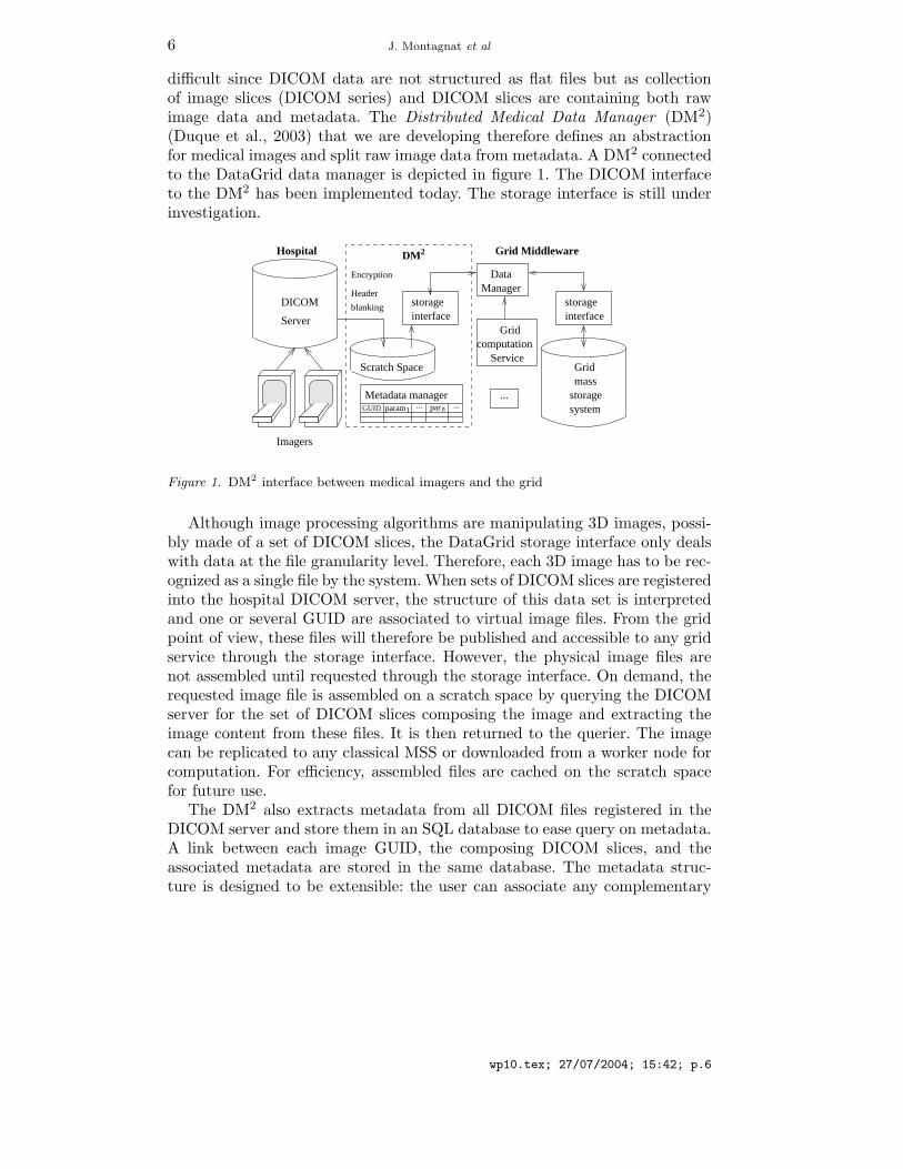
difficult since DICOM data are not structured as flat files but as collectionof image slices (DICOM series) and DICOM slices are containing both rawimage data and metadata. The Distributed Medical Data Manager (DM2)(Duque et al., 2003) that we are developing therefore defines an abstractionfor medical images and split raw image data from metadata. A DM2 connectedto the DataGrid data manager is depicted in figure 1. The DICOM interfaceto the DM2 has been implemented today. The storage interface is still underinvestigation.
DICOM
Server
Scratch Space
...
2DM
interfacestorage
interfacestorage
storagesystem
massGrid
parnparam1
Hospital Grid Middleware
Imagers
Header
blanking
Encryption
Service
Gridcomputation
ManagerData
Metadata managerGUID ......
Figure 1. DM2 interface between medical imagers and the grid
Although image processing algorithms are manipulating 3D images, possi-bly made of a set of DICOM slices, the DataGrid storage interface only dealswith data at the file granularity level. Therefore, each 3D image has to be rec-ognized as a single file by the system. When sets of DICOM slices are registeredinto the hospital DICOM server, the structure of this data set is interpretedand one or several GUID are associated to virtual image files. From the gridpoint of view, these files will therefore be published and accessible to any gridservice through the storage interface. However, the physical image files arenot assembled until requested through the storage interface. On demand, therequested image file is assembled on a scratch space by querying the DICOMserver for the set of DICOM slices composing the image and extracting theimage content from these files. It is then returned to the querier. The imagecan be replicated to any classical MSS or downloaded from a worker node forcomputation. For efficiency, assembled files are cached on the scratch spacefor future use.
The DM2 also extracts metadata from all DICOM files registered in theDICOM server and store them in an SQL database to ease query on metadata.A link between each image GUID, the composing DICOM slices, and theassociated metadata are stored in the same database. The metadata struc-ture is designed to be extensible: the user can associate any complementary
wp10.tex; 27/07/2004; 15:42; p.6
Medical images processing on the DataGrid testbed 7
metadata needed for a medical application to the image. Later versions of theDataGrid data manager also permit registration of metadata associated todata files. However, the granularity is not necessarily sufficient in this case,and integration with the metadata facility of the data manager is not possibletoday for security reasons. Indeed, medical metadata is the most critical partof the data as it may contain patient private and identifying information. Themetadata database stored inside the DM2 also contains additional securityelements detailed in the next section.
The DM2 is able to register and provide a grid interface to data comingfrom several distributed DICOM servers. It enables the DICOM server witha storage interface that makes it visible as any MSS. However, the DM2 is aread-only MSS as it does not allow external grid data to be stored on the sitesit controls: new medical images are registered internally when produced onthe medical imagers and DICOM servers are not intended to store any otherkind of data.
3.2. Security and privacy
Preserving patients privacy is a major concern for medical data processingsystems. The distribution of data over a grid makes data control much moredifficult than on closed systems. Data on grids may be replicated but all storagesites are not accredited to receive medical data. Therefore, their administratorsshould not have read access to the data content. Some identifying metadataare not accessible to non accredited users as well. Achieving a high securitylevel is mandatory but security is always a trade off between inconveniencefor the users and the desired level of protection. In order to convince users(physicians and patients) to use grids for their data storage and processingneeds, many functionalities need to be provided such as:
− Reliable authentication of users.− Secure transfer of data from one grid element to another.− Secure storage of data on a grid element.− Access control for resources such as data, storage space or computing
power.− Anonymization of medical records to make them available for research.− Tamper-proof logging of operations performed on medical files.− Robustness against denial-of-service attacks
Note that we have not included secure processing of data in this discussion.Performing computations on encrypted data without the without explicit de-cryption is a burning research area today. These techniques can accommodateto simple arithmetic operations but they are not mature enough to handle thecomplexity of image processing, not to mention the efficiency problems. Toremain realistic, the features that should protect data while it is being pro-cessed on a grid are based on best effort technologies, i.e. on-disk encryption,
wp10.tex; 27/07/2004; 15:42; p.7
8 J. Montagnat et al
access control, and anonymization. Users need to trust the servers on whichtheir data is to be processed, to our knowledge no systems for data processingon untrusted resources exist.
Our proposal for addressing all these requirements are detailed below.Authentication is not a grid-specific problem. It is well researched and
standard solutions exist. The use of a public key infrastructure (PKI) withcertification authorities (CA) and X.509 certificates is a reasonable way tohandle authentication in grid environments. The EDG middleware relies onGlobus (Foster and Kesselman, 1997) and its public key-based infrastructure(Foster et al., 1998).
Secure transfer is also a well researched area independently of grid tech-nologies. It is addressed in various standardized protocols such as SSL/TLS,IpSec or SSH. Data transfers are handled by GridFTP (Allcock et al., 2002)in the EDG middleware and can be encrypted although this functionality isnot used in the EDG testbed.
For secure storage of data, encryption and signing is an obvious solution.The problem in grid environments is that mechanisms are required to sharedecryption keys between users authorized to access data. Common encryptedstorage systems lack the flexibility to deal with the dynamic nature of gridaccess permissions. We have therefore proposed an architecture with a genericinterface to grid access control mechanisms, that provides access to decryptionkeys based on access permissions. For further details on this system see (Seitzet al., 2003a)
Authorization and access control raises the most problematic issues formedical data processing in grid environments. Classic access control techniquesare not designed to deal with the problems arising from the decentralized,cross-organizational nature of grid access permissions. The medical field ofapplications adds another inherent problem. Grid applications such as nuclearphysics deal with data that has relatively low confidentiality and that is acces-sible for large groups of users. Classical grid access control mechanisms such asCAS (Pearlman et al., 2002) are satisfactory. Nevertheless these systems fail toprovide sufficient permission granularity and flexibility for ad hoc permissiongranting that is required in medical applications. Furthermore such systemsuse centralized permission databases. We want to avoid this since they are asingle point of failure.
An alternative approach is to manage grid access control using decentralizedpermission checking through attribute certificates. Such certificates permitresource administrators to issue permissions in a simple way without havingto resort to third party services. Local servers can easily verify the permis-sions granted in such certificates, using a local database that specifies thesources of authority (SOA) of the resources on their systems. The attributecertificates enable the local servers to trace a permission from SOA of theconcerned resource to the user requesting it. The database that specifies the
wp10.tex; 27/07/2004; 15:42; p.8
Medical images processing on the DataGrid testbed 9
SOAs is managed by the access control system itself and is updated, whennew resources (e.g. files) are added to the system.
The EDG security model is based on Virtual Organizations (VO, (Fosteret al., 2001)). Resource providers assign permissions to those VOs, and theVOs have policies to dispatch the resources they have been assigned betweentheir members (Alfieri et al., 2003). Our access control system supports thiscooperation model by providing role based access control (RBAC) (Ferraioloand Kuhn, 1992). Using RBAC, administrators can manage user groups (VOs)that are assigned sets of permissions and the membership of users within thosegroups. Our access control system also provides a generic program executioninterface, that permits users to run their own specific programs in a sandboxenvironment prior to giving access to a resource. For details on our proposedaccess control system see (Seitz et al., 2003b).
Anonymization is required to provide large sets of data for medical re-search. Legislation imposes severe regulations as to what can be consideredan anonymized information. The main problem is that even if obvious sec-tions such as name and address of the patient have been removed a medicaldocument could be re-identified with secondary information. We have notyet addressed that problem within the project, however our access controlsystem is designed to provide an interface where a data filtering software canbe plugged in before a medical file is delivered in order to ensure privacyprotection. A promising approach to deal with anonymization is described in(Claerhout and De Moor, 2004).
Traceability is clearly another important factor in medical grids. The pre-access program execution interface, integrated in our access control system,can be used to plug in a log-keeping system. Through this interface the systemwill be able to get the necessary information about access requests to keep thelog-file. However since the logs and the programs that create them are locatedon the distant storage element, users have to trust the administrators of thisstorage element not to interfere with the log-file creation.
The availability of services may be a critical factor in medical environ-ments. Most measures to prevent denial-of-service attacks are not specific togrid architectures. However it is important to realize that centralized servicesare very vulnerable to such attacks. Therefore none of our proposed securityservices relies on a single centralized service.
4. Processing medical images
wp10.tex; 27/07/2004; 15:42; p.9
10 J. Montagnat et al
4.1. Magnetic Resonance Images simulation
4.1.1. MRI physics simulationThe simulation of Magnetic Resonance Images (MRI) is an important counter-part to MRI acquisitions. Simulation is naturally suited to acquire theoreticalunderstanding of the complex MR technology. It is used as an educational toolin medical and technical environments (Torheim et al., 1994). By offering ananalysis independent of the multiple parameters involved in the MR technol-ogy, MRI simulation permits the investigation of artifact causes and effects(Olsson et al., 1995; Brenner et al., 1997). Likewise simulation may help inthe development and optimization of MR sequences (Brenner et al., 1997).Simulated MR images also provide an interesting assessment tool (Kwan et al.,1996) since it generates 3D realistic images from medical virtual objects whichstructure is perfectly known while this ground truth is usually not availablewhen dealing with clinical data.
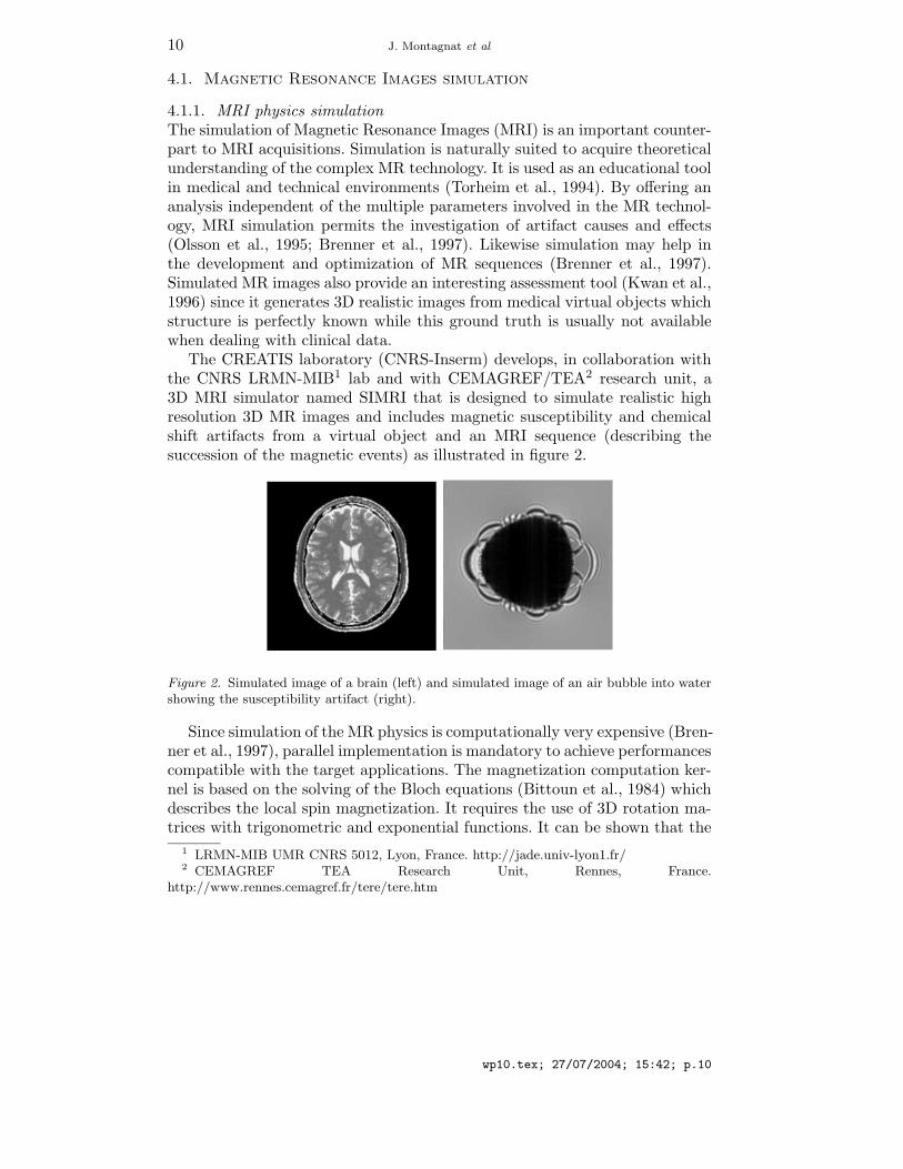
The CREATIS laboratory (CNRS-Inserm) develops, in collaboration withthe CNRS LRMN-MIB1 lab and with CEMAGREF/TEA2 research unit, a3D MRI simulator named SIMRI that is designed to simulate realistic highresolution 3D MR images and includes magnetic susceptibility and chemicalshift artifacts from a virtual object and an MRI sequence (describing thesuccession of the magnetic events) as illustrated in figure 2.
Figure 2. Simulated image of a brain (left) and simulated image of an air bubble into watershowing the susceptibility artifact (right).
Since simulation of the MR physics is computationally very expensive (Bren-ner et al., 1997), parallel implementation is mandatory to achieve performancescompatible with the target applications. The magnetization computation ker-nel is based on the solving of the Bloch equations (Bittoun et al., 1984) whichdescribes the local spin magnetization. It requires the use of 3D rotation ma-trices with trigonometric and exponential functions. It can be shown that the
1 LRMN-MIB UMR CNRS 5012, Lyon, France. http://jade.univ-lyon1.fr/2 CEMAGREF TEA Research Unit, Rennes, France.
http://www.rennes.cemagref.fr/tere/tere.htm
wp10.tex; 27/07/2004; 15:42; p.10
Medical images processing on the DataGrid testbed 11
overall volume simulation time is proportional to the object size (X × Y ×Z)multiplied by the image size (M ×N ×P ). As an example, the simulation of a1282 image takes only 3 minutes on a P4-2.6GHz PC, but multiplying by twoall the dimensions of the virtual object and the MR image leads to a simulationtime multiplication by 16 in two dimensions and by 64 in three dimensions(Benoit-Cattin et al., 2003). Therefore, we turn toward Grid technologies thatpromise a virtually unlimited computing power and we propose a gridificationof our MRI simulator.
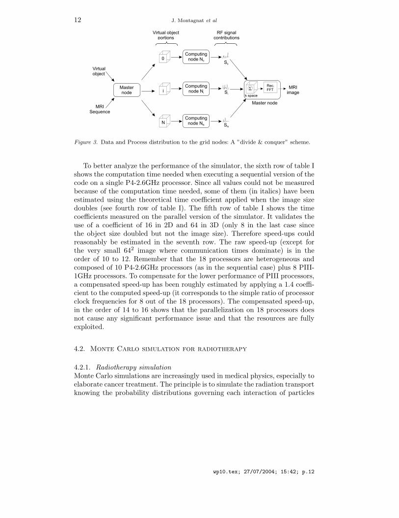
4.1.2. Gridification strategy and resultsThe parallelization of the magnetization kernel has been done using the MPIversion for Globus (MPICH-G2). Because all the spin magnetization vec-tors are independents and because the signal acquisition process is linear,a parallelization scheme of type ”divide & conquer” (see figure 3) has beenimplemented. It consists in distributing the magnetization computation of asubset of spin vectors. This subset can be fixed to a given size or adapted tothe number of active nodes.
All the computation nodes have the MRI sequence knowledge and theyreceive from the master node a part of the virtual object. They computethe magnetization evolution of the corresponding spin vector subset. At theend of each acquisition step, the master node collects and adds all the Ra-dio Frequency (RF) signal contributions. At the end of the MRI sequence,the master node applies the reconstruction algorithm to generate the MRIsimulated image.
When using an homogeneous grid, the virtual object portion distributed tothe nodes has a maximal size equal to the object size divided by the numberof nodes. Only one distribution is done at the process begin which limits thecommunication between the master node and the computation nodes. Whenusing an heterogeneous grid, to avoid to be penalized by the slowest node, thedistributed object portion is reduced. In this case, the lowest node will receiveone portion of the object to process while the fastest nodes will receive severalportions.
The third line of table I gives computation time values for different objectand image sizes obtained using a small grid based on an 18 PC cluster (8 PIII1GHz, 10 P4 2.6GHz). These simulation results show that with a small cluster,MRI simulation of high resolution (10242) 2D images is possible within oneday. Concerning 3D images, it is not realistic to simulate on such a small setof processors over 643 MRI. Nevertheless, it is possible to simulate within aweek 3D multi-slice images (32 slices of 5122 pixels). The simulation of highresolution 3D images should be tractable on full scale grids. However, largescale experiments were not possible on the DataGrid testbed due to the limiteddeployment of MPI-enabled nodes at the end of the project. This applicationremained at the testing phase and could not scale up to production.
wp10.tex; 27/07/2004; 15:42; p.11
12 J. Montagnat et al
Virtualobject
MRISequence
Virtual objectportions
RF signalcontributions
0S0
Si
SN
k space
MRIimage
Master node
Computingnode N0
Computingnode Ni
Computingnode NN
Masternode i
N
SRec.FFT
Figure 3. Data and Process distribution to the grid nodes: A ”divide & conquer” scheme.
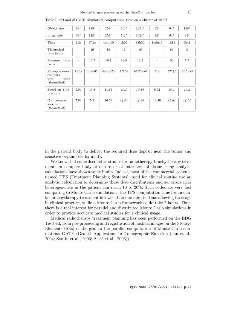
To better analyze the performance of the simulator, the sixth row of table Ishows the computation time needed when executing a sequential version of thecode on a single P4-2.6GHz processor. Since all values could not be measuredbecause of the computation time needed, some of them (in italics) have beenestimated using the theoretical time coefficient applied when the image sizedoubles (see fourth row of table I). The fifth row of table I shows the timecoefficients measured on the parallel version of the simulator. It validates theuse of a coefficient of 16 in 2D and 64 in 3D (only 8 in the last case sincethe object size doubled but not the image size). Therefore speed-ups couldreasonably be estimated in the seventh row. The raw speed-up (except forthe very small 642 image where communication times dominate) is in theorder of 10 to 12. Remember that the 18 processors are heterogeneous andcomposed of 10 P4-2.6GHz processors (as in the sequential case) plus 8 PIII-1GHz processors. To compensate for the lower performance of PIII processors,a compensated speed-up has been roughly estimated by applying a 1.4 coeffi-cient to the computed speed-up (it corresponds to the simple ratio of processorclock frequencies for 8 out of the 18 processors). The compensated speed-up,in the order of 14 to 16 shows that the parallelization on 18 processors doesnot cause any significant performance issue and that the resources are fullyexploited.
4.2. Monte Carlo simulation for radiotherapy
4.2.1. Radiotherapy simulationMonte Carlo simulations are increasingly used in medical physics, especially toelaborate cancer treatment. The principle is to simulate the radiation transportknowing the probability distributions governing each interaction of particles
wp10.tex; 27/07/2004; 15:42; p.12
Medical images processing on the DataGrid testbed 13
Table I. 2D and 3D MRI simulation computation time on a cluster of 18 PC.
Object size 642 1282 2562 5122 10242 323 643 1283
Image size 642 1282 2562 5122 10242 323 643 643
Time 2.2s 17.2s 4min10 1h09 18h50 1min15 1h13 9h41
Theoreticaltime factor
- 16 16 16 16 - 64 8
Measure timefactor
- 12.7 16.7 16.8 16.3 - 60 7.7
Monoprocessorcomputa-tion time(theoretical)
11.1s 3min05 49min23 11h58 7d 23h30 714 12h41 4d 5h33
Speed-up (the-oretical)
5.04 10.8 11.85 10.4 10.16 9.52 10.4 10.4
Compensatedspeed-up(theoretical)
7.09 15.21 16.69 14.64 14.30 13.40 14.64 14.64
in the patient body to deliver the required dose deposit near the tumor andsensitive organs (see figure 4).
We know that some dosimetric studies for radiotherapy-brachytherapy treat-ments in complex body structure or at interfaces of tissue using analyticcalculations have shown some limits. Indeed, most of the commercial systems,named TPS (Treatment Planning Systems), used for clinical routine use ananalytic calculation to determine these dose distributions and so, errors nearheterogeneities in the patient can reach 10 to 20%. Such codes are very fastcomparing to Monte Carlo simulations: the TPS computation time for an ocu-lar brachytherapy treatment is lower than one minute, thus allowing its usagein clinical practice, while a Monte Carlo framework could take 2 hours. Thus,there is a real interest for parallel and distributed Monte Carlo simulations inorder to provide accurate medical studies for a clinical usage.
Medical radiotherapy treatment planning has been performed on the EDGTestbed, from pre-processing and registration of medical images on the StorageElements (SEs) of the grid to the parallel computation of Monte Carlo sim-ulations GATE (Geant4 Application for Tomographic Emission (Jan et al.,2004; Santin et al., 2003; Assie et al., 2003)).
wp10.tex; 27/07/2004; 15:42; p.13

14 J. Montagnat et al
Figure 4. GATE Monte Carlo simulations: a) PET simulation; b) Radiotherapy simulation;c) Ocular brachytherapy simulation
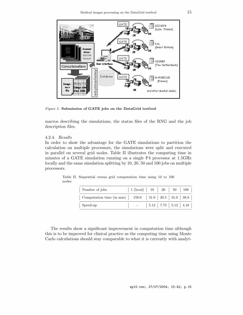
4.2.2. Medical images treatmentThe application framework is depicted in figure 5. Sets of 40 DICOM slices orso, 5122 pixels each, acquired by CT scanners are concatenated and stored ina 3D image file format(see section 3). Such image files can reach up to 20 MBin size for our application. To solve privacy issues, DICOM headers are wipedout in this process.
The 3D image files are then registered and replicated on the sites of theEDG testbed where GATE is installed in order to compute simulations (5sites to date). During the computation of the GATE simulation, the imagesare read by GATE and interpreted in order to produce a 3D array of voxelswhose value is describing a body tissue. A relational database is used to linkthe GUID of image files with metadata extracted from the DICOM slices onthe patient and additional medical information. The EDG Spitfire software(EDG WP2, 2001) is used to provide access to the relational databases.
4.2.3. The parallelization of GATE simulations on the DataGrid testbedEvery Monte Carlo simulation is based on the generation of pseudo-randomnumbers using a Random Numbers Generator (RNG). An obvious way toparallelize the calculations on multiple processors is to partition a sequenceof random numbers generated by the RNG into suitable independent sub-sequences. To perform this step, the choice has been done to use the SequenceSplitting Method (Traore and Hill, 2001; Coddington, 1996; Maigne et al.,2004). For each sub-sequence, we save in a file (some KBs) the current statusof the random engine. Each simulation is then launched on the grid with thestatus file.
All the other files necessary to run Gate on the grid are automaticallycreated: the script describing the environment of computation, the GATE
wp10.tex; 27/07/2004; 15:42; p.14
Medical images processing on the DataGrid testbed 15
Figure 5. Submission of GATE jobs on the DataGrid testbed
macros describing the simulations, the status files of the RNG and the jobdescription files.
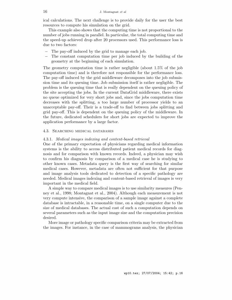
4.2.4. ResultsIn order to show the advantage for the GATE simulations to partition thecalculation on multiple processors, the simulations were split and executedin parallel on several grid nodes. Table II illustrates the computing time inminutes of a GATE simulation running on a single P4 processor at 1.5GHzlocally and the same simulation splitting by 10, 20, 50 and 100 jobs on multipleprocessors.
Table II. Sequential versus grid computation time using 10 to 100nodes
Number of jobs 1 (local) 10 20 50 100
Computation time (in min) 159.0 31.0 20.5 31.0 38.0
Speed-up - 5.12 7.75 5.12 4.18
The results show a significant improvement in computation time althoughthis is to be improved for clinical practice as the computing time using MonteCarlo calculations should stay comparable to what it is currently with analyt-
wp10.tex; 27/07/2004; 15:42; p.15
16 J. Montagnat et al
ical calculations. The next challenge is to provide daily for the user the bestresources to compute his simulation on the grid.
This example also shows that the computing time is not proportional to thenumber of jobs running in parallel. In particular, the total computing time andthe speed-up achieved drop after 20 processors used. This performance loss isdue to two factors:
− The pay-off induced by the grid to manage each job.− The constant computation time per job induced by the building of the
geometry at the beginning of each simulation.
The geometry computation time is rather negligible (about 1.5% of the jobcomputation time) and is therefore not responsible for the performance loss.The pay-off induced by the grid middleware decomposes into the job submis-sion time and its queuing time. Job submission itself is rather negligible. Theproblem is the queuing time that is really dependent on the queuing policy ofthe site accepting the jobs. In the current DataGrid middleware, there existsno queue optimized for very short jobs and, since the jobs computation timedecreases with the splitting, a too large number of processor yields to anunacceptable pay-off. Their is a trade-off to find between jobs splitting andgrid pay-off. This is dependent on the queuing policy of the middleware. Inthe future, dedicated schedulers for short jobs are expected to improve theapplication performance by a large factor.
4.3. Searching medical databases
4.3.1. Medical images indexing and content-based retrievalOne of the primary expectation of physicians regarding medical informationsystems is the ability to access distributed patient medical records for diag-nosis and for comparison with known records. Indeed, a physician may wishto confirm his diagnosis by comparison of a medical case he is studying toother known cases. Metadata query is the first way of searching for similarmedical cases. However, metadata are often not sufficient for that purposeand image analysis tools dedicated to detection of a specific pathology areneeded. Medical images indexing and content-based retrieval of images is veryimportant in the medical field.
A simple way to compare medical images is to use similarity measures (Pen-ney et al., 1998; Montagnat et al., 2004). Although each measurement is notvery compute intensive, the comparison of a sample image against a completedatabase is intractable, in a reasonable time, on a single computer due to thesize of medical databases. The actual cost of such a computation depends onseveral parameters such as the input image size and the computation precisiondesired.
More image or pathology specific comparison criteria may be extracted fromthe images. For instance, in the case of mammograms analysis, the physician
wp10.tex; 27/07/2004; 15:42; p.16
Medical images processing on the DataGrid testbed 17
is mainly interested in the detection of tumors, their classification (malignantor benign), and their location.
4.3.2. An application to computer aided diagnostic in mammogramsBreast cancer is one of the most common cause of women mortality. In France,a systematic screening for breast cancer is generalized for women between 50and 74 years old, in order to detect the early signs of change that could pointout the presence of a malignant tumor. The number of mammograms to beanalyzed is in constant increasing; the data corresponding to mammogramsand medical diagnosis reports are distributed among several medical sites.Thus, an early detection using mammographic screening is essential. In orderto be specific, a computer-aided diagnosis system (CAD) is an ideal tool inassisting a radiologist, and can be used as a second opinion or a second reading.Those tools, based on a segmentation or a detection, then a feature extraction,and finally a classification or decision making (Bick and Doi, 2000), need tobe trained among different databases.
Our aim in this application is to evaluate the grid possibilities in order tobuild a distributed system of stored mammographic data and metadata, thatwill work as a CAD tool. This system must allow the different users, specificallyphysicians or researchers, to analyze and index the images distributed amongthe different geographic medical sites, to do some content-based requests onthe image databases, and then offer an assistance to the diagnosis, based onthe research of a set of images that are similar to a request image according tosome extracted features. Two types of scenarii of content-based request wereconsidered:
− A physician has doubts about a particular region in the mammogram he isanalyzing. He can search for images of the database that contain regionshaving similar properties, based on similarity measures. Two types ofrequests can be submitted to the system: find the set of images containingregions that are similar to the query zone, or find the set of images thatcontain regions previously detected as cancerous and that are similar tothe query zone.
− Without the help of a specialist, a new image is compared with a set ofimages in the database, for example in the case of a second reading. Theidea is to highlight in the image all the zones that are close to the regionsthat have previously been noted as cancerous. This can be helpful forattracting the attention of a specialist to a precise region that he couldhave missed during his first reading.
For our tests, we are working on a digital database containing 2620 patients,divided into three groups: benign, malignant and normal. This database iscomposed of 230 GB of mammographic data and comes from the Universityof South Florida (Heath et al., 1998). Each case/patient includes two views of
wp10.tex; 27/07/2004; 15:42; p.17
18 J. Montagnat et al
each breast and information about the patient, the study date, or the scannerused for digitalization. In the case of benign and malignant cases, a descriptionof the malign regions, delimited by a specialist and confirmed by later exam-inations, is given using the ACR BI-RADS lexicon (BI-RADS Committee,1998). The whole image database is stored on a mass storage system calledHPSS (High Performance Storage System) in the IN2P3 Computing Center,which is one of the major resource provider in the EDG project.
The heart of our algorithms in this application is the comparison of ele-mentary regions in images. For computation optimizations, this comparisonis not done on the image data themselves, but on feature vectors extractedfrom the regions. We have developed an indexing algorithm that describeselementary regions of the images by the way of feature vectors based on graylevel distribution as well as texture analysis. We have reported in our previousworks (Tweed and Miguet, 2002) the indexing process we use to describe imageregions. This indexing is compute intensive: from 8 to 30 minutes per case (4images) on 2.4GHz P4 to 750MHz PIII based machines.
We have then experimented several proximity criteria on these featurevectors: simple thresholds on the histograms, and Euclidean distances on thetexture attributes, in order to make requests as described above. We have beenworking on the optimization of the database indexing on the EDG testbed. Wehave developed jobs that transfer the image data from the storage elementsto the workers and that perform image indexing.
0
1000
2000
3000
4000
5000
6000
7000
0 5 10 15 20 25 30 35 40 45 50 55Worker node index
Abs
olut
e tim
e (s
econ
ds)
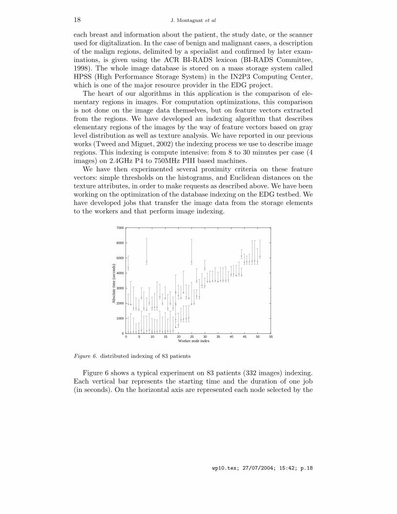
Figure 6. distributed indexing of 83 patients
Figure 6 shows a typical experiment on 83 patients (332 images) indexing.Each vertical bar represents the starting time and the duration of one job(in seconds). On the horizontal axis are represented each node selected by the
wp10.tex; 27/07/2004; 15:42; p.18
Medical images processing on the DataGrid testbed 19
scheduler for the computations. The results are very conclusive: the computingtime execution is of less than two hours using the EDG testbed. The sequentialcomputing time for the same experiment is 23 hours. The speed-up in thiscase is 13.14 using up to 51 processors. Although this is far from linear, oneshould notice that this experiment has been ran on a production computinginfrastructure and other jobs were scheduled on the same processors. Indeed,the starting time of some jobs is delayed by the late availability of processors.We observe that several jobs start at the same time, but not all according tothe availability of the resources at the time the experiment was led. More than800 processors could theoretically have been used but the racing conditionswith other users lead to a consumption of 51 processors and the re-use ofsome worker nodes for jobs once the first indexing/task was finished. Giventhe parallel nature of this application, almost linear speed-up can be obtainedby dedicating resources to this task.
5. Discussion
The EDG middleware and testbed provide a basic grid infrastructure fortesting grid-enabled medical applications. As reported in section 4, differentkind of applications could be experimented to some level and real benefits interms of computation time and size of datasets processed have been demon-strated. This platform is still in its youth though, and most advanced devel-opments, only recently made available, could hardly be tested. More servicesare expected in order to cover all medical image application requirements.
Privacy and security remain primary concerns for deploying large scaleapplications that involve real patient data. Medical data security requirementsare complex: several category of users with different access rights, encryptionto avoid accessibility to data for non accredited system administrators onstorage sites, security controls to prevent intrusion on data storage sites, etc.However, these constraints have to be enforced in order to be able to inter-connect medical information systems with patient data to the grid resources.Another important related aspect is the confidence the medical users will putin the system. As long as the system is not trusted by the community, progresson grid-enabling medical applications will remain slow.
Data and metadata management is another domain that requires furtherinvestigation. Medical data are widely distributed due to their acquisition indifferent centers spread out the territory. The management of medical datarequires an information system capable of dealing with data sets rather thanflat files. Moreover, processing often concerns full data sets rather than singledata. The semantics of data and metadata should be taken into account bythe data manager to ease meaningful retrieval of medical data.
wp10.tex; 27/07/2004; 15:42; p.19
20 J. Montagnat et al
Other computational aspects can also improve medical data processingin the future. A pipelines computation system such as the EDG DAG jobsmanager does not cover all application requirements for instance since it doesnot take into account the processing of full datasets. Parallelism is anotherpoint that has been well studied on cluster architectures but for which grid-wide implementation on a large scale and heterogeneous infrastructure is stillto be investigated.
A key factor in the success of grid technologies in the medical domain will beits accessibility to non computer scientists. End users are often non specialistswho need well designed interfaces and algorithms applying to precise medicalanalysis needs. Grid technologies will only be adopted once it has proved tobe more useful and as easily accessible as existing PACS and RIS.
Considering the medical application themselves, all development and de-ployment of medical applications made during the EDG project have beenperformed in parallel to the middleware development. This has made thingsdifficult as applications were supposed to adapt to a continuously movingtarget. As a consequence, mostly simple applications with a rather straightforward capability for parallel execution could be ported in the project lifetime.The real impact of grid technologies in porting large scale applications is stillto be investigated. We are just beginning this exploration now that the basictools are available for development and testing.
6. Conclusions
The EDG project was a pioneer in identifying the biomedical domain as arelevant area of application for grid technologies. Within the project 3 yearslifetime, the awareness of these technologies has raised in the medical imageprocessing community and, to some extend, in the medical community. Medicalinformatics, and more generally biomedical informatics and life sciences arenow well established candidates with a clear interest for grid enabling. Severalclues testify of the growth of this emerging community such as conferencesand workshops organized in this domain and the creation of internationalbodies such as the HealthGrid association (HealthGrid, 2003) or the GGFLife Science research group (LSG-RG, 2003) aiming at federating researchprojects in this field. The European Community is eager to develop gridsas a high level infrastructure for e-Health and funds many research projectsand networks of excellence in the domains of biomedical informatics and gridinfrastructures. Among these, the EGEE (EGEE, 2004) project will deploy aproduction testbed for which biomedical applications are identified candidates.
wp10.tex; 27/07/2004; 15:42; p.20
Medical images processing on the DataGrid testbed 21
Acknowledgements
The authors are grateful to the European IST DataGrid project (EDG, 2001)for financial and operational support in the various experiments related inthis paper. This paper synthesizes the work performed in several laboratoriesdeveloping grid technologies for health applications that are supported by sev-eral national and international research programs. The authors express theirgratitude to the ACI GRID research program funded by the French ministryfor research (ACI MEDIGRID, ACI GLOP), the Rhone-Alpes regional support(RAGTIME project), the French-Algerian CMEP agreement (Avicenne Gridproject), and the French-Colombian ECOS Nord Committee (action C03S02).The LPC Clermont also acknowledges fruitful discussions with David Hill andthe GATE collaboration.
References
Acharya, R., Wasserman, R., Sevens, J., and Hinojosa, C. (1995). Biomedical ImagingModalities: a Tutorial. Computerized Medical Imaging and Graphics, 19(1):3–25.
Alfieri, R., Cecchini, R., Ciaschini, V., dell’Agnello, L., Frohner, A., Gianoli, A., Lorentey,K., and Spataro, F. (2003). VOMS, an authorization system for virtual organizations.In Proceedings of the 1st European Across Grids Conference.
Allcock, B., Bester, J., Bresnahan, J., Chervenak, A., Foster, I., Kesselman, C., Meder,S., Nefedova, V., Quesnal, D., and Tuecke, S. (2002). Data Management and Transferin High Performance Computational Grid Environments. Parallel Computing Journal,28(5):749–771.
Assie, K., Breton, V., Buvat, I., Comtat, C., Jan, S., Krieguer, M., Lazaro, D., Morel, C.,Rey, M., Santin, G., Simon, L., Staelens, S., Strul, D., Vieira, J., and Van de Walle, R.(2003). Monte Carlo simulation in PET and SPECT instrumentation using GATE. Nucl.Instr. and Methods.
Benoit-Cattin, H., Bellet, F., Montagnat, J., and Odet, C. (2003). Magnetic ResonanceImaging (MRI) simulation on a grid computing architecture. In IEEE CGIGRID’03 -BIOGRID’03, pages 582–587, Tokyo.
BI-RADS Committee (1998). Illustrated Breast Imaging Reporting And Data System,American College of Radiology edition.
Bick, U. and Doi, K. (2000). Computer Aided Diagnosis Tutorial. CARS 2000 Tutorial onComputer Aided-Diagnosis, Hyatt Regency, San Francisco, USA.
Bittoun, J., Taquin, J., and Sauzade, M. (1984). A computer algorithm for the simulation ofany nuclear magnetic resonance (NMR) imaging method. Magnetic Resonance Imaging,3:363–376.
Brenner, A., Kursch, J., and Noll, T. (1997). Distributed large-scale simulation of magneticresonance imaging. Magnetic Resonance Materials in Biology, Physics, and Medicine,5:129–138.
Breton, V., Medina, R., and Montagnat, J. (2003). DataGrid, Prototype of a BiomedicalGrid. Methods MIMST, 42(2).
Claerhout, B. and De Moor, G. (2004). Privacy protection for healthgrid applications. toappear in the Methods of Information in Medcine journal.
wp10.tex; 27/07/2004; 15:42; p.21
22 J. Montagnat et al
Coddington, P., editor (1996). Random Number Generators For Parallel Computers, SecondIssue. NHSE Review.
DICOM (1996). Digital Imaging and COmmunications in Medicine,http://medical.nema.org/.
Duque, H., Montagnat, J., Pierson, J., Brunie, L., and Magnin, I. (2003). DM2: A DistributedMedical Data Manager for Grids. In Biogrid’03, proceedings of the IEEE CCGrid03,Tokyo, Japan.
EDG (2001). European DataGrid IST project, FP5, jan. 2001-feb. 2004,http://www.edg.org/.
EDG WP2 (2001). Spitfire. http://edg-wp2.web.cern.ch/edg-wp2/spitfire/.EGEE (2004). European IST project of the FP6, Enabling Grids for E-science and industry
in Europe, apr. 2004-mar. 2006, http://www.eu-egee.org/.Ferraiolo, D. and Kuhn, D. (1992). Role based access control. In 15th NIST-NCSC National
Computer Security Conference, pages 554–563.Foster, I. and Kesselman, C. (1997). Globus: A Metacomputing Infrastructure Toolkit.
International Journal of Supercomputer Applications, 11(2):115–128.Foster, I., Kesselman, C., Tsudik, G., and Tuecke, S. (1998). A Security Architecture for
Computational Grids. In Proc. 5th ACM Conference on Computer and CommunicationsSecurity Conference, pages 83–92, San Francisco, CA, USA.
Foster, I., Kesselman, C., and Tuecke, S. (2001). The Anatomy of the Grid: Enabling ScalableVirtual Organizations. International Journal of Supercomputer Applications, 15(3).
HealthGrid (2003). HealthGrid Association, http://www.healthgrid.org/.Heath, M., Bowyer, K. W., and Kopans, D. (1998). Current Status of the Digital Database
for Screening Mammography. In Digital Mammography, pages 457–460. Kluwer AcademicPublishers. http://marathon.csee.usf.edu/Mammography/Database.html.
Huang, H. K. (1996). PACS: Picture Archiving and Communication Systems in BiomedicalImaging. Hardcover.
Jan, S., Santin, G., Strul, D., and et al. (2004). GATE (Geant4 Application for TomographicEmission): a simulation toolkit for PET and SPECT. to appear in Phys. Med. Biol.
Karonis, N., Toonen, B., and Foster, I. (2003). MPICH-G2: A Grid-Enabled Implementa-tion of the Message Passing Interface. Journal of Parallel and Distributed Computing,63(5):551–563.
Kwan, R.-S., Evans, A. C., and Pike, G. B. (1996). An extensible MRI simulator forpost-processing evaluation. In International Conference on Visualization in BiomedicalComputing, VBC’96, pages 135–140.
LSG-RG (2003). Global Grid Forum Life Sciences Grid Research Group,http://forge.gridforum.org/projects/lsg-rg.
Maigne, L., Hill, D., Breton, V., and et al. (2004). Parallelization of Monte Carlo simulationsand submission to a Grid environment. to appear in Parallel Processing Letters.
Montagnat, J., Breton, V., and I.E., M. (2004). Partitionning medical image databases forcontent-based queries on a grid. In Healthgrid’04, Clermont-Ferrand, France.
Montagnat, J., Davila, E., and Magnin, I. (2002). 3D objects visualization for remoteinteractive medical applications. In 3D Data Processing, Visualization, Transmission,Padova, Italy.
Olsson, M. B. E., Wirestam, R., and Persson, B. R. R. (1995). A Computer-SimulationProgram For Mr-Imaging - Application to Rf and Static Magnetic-Field Imperfections.Magnetic Resonance in Medicine, 34(4):612–617.
Pearlman, L., Welch, V., Foster, I., Kesselman, C., and Tuecke, S. (2002). A communityauthorization service for group collaboration. In Proceedings of the 2002 IEEE Workshopon Policies for Distributed Systems and Networks.
wp10.tex; 27/07/2004; 15:42; p.22
Medical images processing on the DataGrid testbed 23
Penney, G., Weese, J., Little, J., Desmedt, P., Hill, D., and Hawkes, D. (1998). A Comparisonof Similarity Measures for Use in 2D-3D Medical Image Registration. In Medical ImageComputing and Computer-Assisted Intervention (MICCAI), volume 1496 of LNCS, pages1153–1161, Cambridge, USA. Springer.
Santin, G., Strul, D., Lazaro, D., Simon, L., Krieguer, M., Vieira Martins, M., Breton, V.,and C., M. (2003). GATE, a Geant4-based simulation platform for PET and SPECTintegrating movement and time managment. IEEE Trans. Nucl. Sci., 50:1516–1521.
Seitz, L., Pierson, J., and Brunie, L. (2003a). Key management for encrypted data storage indistributed systems. In Proceedings of the second Security In Storage Workshop (SISW).
Seitz, L., Pierson, J., and Brunie, L. (2003b). Semantic access control for medical applicationsin grid environments. In Euro-Par 2003 Parallel Processing, volume LNCS 2790, pages374–383. Springer.
Torheim, G., Rinck, P., Jones, R., and Kvaerness, J. (1994). A simulator for teaching MRimage contrast behavior. MAGMA, 2:515–522.
Traore, M. and Hill, D. (2001). The use of random number generation for stochastic dis-tributed simulation: application to ecological modeling. In 13th European SimulationSymposium, Marseille, pages 555–559, Marseille, France.
Tweed, T. and Miguet, S. (2002). Automatic Detection of Regions of Interest in Mammo-graphies Based on a Combined Analysis of Texture and Histogram. In InternationalConference on Pattern Recognition, pages 448–552, Qubec City, Canada.
wp10.tex; 27/07/2004; 15:42; p.23