manipulating il2 availability amid presentation of donor mhc antigens suppresses murine alloimmune...
TRANSCRIPT
Manipulating IL-2 Availability Amid Presentation ofDonor MHC Antigens Suppresses Murine AlloimmuneResponses by Inducing Regulatory T CellsShuzi Zhang, Hehua Dai, Ni Wan, Yolonda Moore, Zhenhua Dai*
Center for Biomedical Research, University of Texas Health Science Center, Tyler, Texas, United States of America
Abstract
Background: Major histocompatibility complex (MHC) antigens are important for alloimmune responses as well as immunetolerance. Previous studies have shown that presentation of donor MHC antigens by donor-specific transfusion prior to orupon transplantation promotes transplant tolerance induced by other agents. However, it is unclear whether presentationof donor MHC antigens by DNA vaccination induces long-term allograft survival.
Methodology/Principal Findings: We investigated whether presentation of MHC class-II and/or class-I donor antigensby DNA vaccination suppresses alloimmune responses and promotes long-term allograft acceptance. We initially foundthat presentation of both MHC donor antigens by DNA vaccination itself prior to transplantation fails to significantlyprolong islet allograft survival in otherwise untreated mice. However, islet allograft survival was significantly prolongedwhen MHC class-II DNA vaccination was accompanied with IL-2 administration (MHCII + IL-2) while MHC class-I DNAvaccination was followed by IL-2 and subsequent neutralizing anti-IL-2 treatments (MHCI + IL-2/anti-IL-2). Especially,this protocol promoted long-term allograft survival in the majority of recipients (57%) when combined with low dosesof rapamycin post-transplantation. Importantly, MHCII + IL-2 induced FoxP3+ Treg cells in both spleens and grafts andsuppressed graft-infiltrating CD4+ cell proliferation, whereas MHCI + IL-2/anti-IL-2 mainly inhibited graft-infiltratingCD8+ cell proliferation and donor-specific CTL activity. The combined protocol plus rapamycin treatment furtherreduced both CD4+ and CD8+ T cell proliferation as well as donor-specific CTL activity but spared FoxP3+ Treg cells.Depleting CD25+ Treg cells or adoptive transfer of pre-sensitized CD8+ T cells abolished this long-term allograftsurvival.
Conclusions/Significance: Manipulating IL-2 availability during presentation of MHC class-II and class-I donor antigens byDNA vaccination pre-transplantation induces Treg cells, suppresses alloimmune responses and promotes long-termallograft survival.
Citation: Zhang S, Dai H, Wan N, Moore Y, Dai Z (2010) Manipulating IL-2 Availability Amid Presentation of Donor MHC Antigens Suppresses Murine AlloimmuneResponses by Inducing Regulatory T Cells. PLoS ONE 5(1): e8756. doi:10.1371/journal.pone.0008756
Editor: Derya Unutmaz, New York University, United States of America
Received November 9, 2009; Accepted December 18, 2009; Published January 18, 2010
Copyright: � 2010 Zhang et al. This is an open-access article distributed under the terms of the Creative Commons Attribution License, which permitsunrestricted use, distribution, and reproduction in any medium, provided the original author and source are credited.
Funding: This study was supported by research grants from American Diabetes Association (ADA 1-06-RA-36) and Juvenile Diabetes Research FoundationInternational (JDRF 1-2008-583). The funders had no role in study design, data collection and analysis, decision to publish, or preparation of the manuscript.
Competing Interests: The authors have declared that no competing interests exist.
* E-mail: [email protected]
Introduction
A transplanted organ or islet is always rejected without
immunosuppressive treatments. However, treatments with immu-
nosuppressive drugs usually cause severe side-effects including viral
infection and tumors. An approach to inducing long-term allograft
survival or tolerance without long-term immunosuppression after
transplantation is highly desired in the field. Current main
strategies to promote long-term graft survival or tolerance include
donor-specific transfusion, T cell costimulatory blockade and
induction of Treg cells. In particular, the presentation of donor
MHC antigens to recipients prior to or upon transplantation,
though does not generally prolong allograft survival by itself,
promotes long-term allograft survival or tolerance induced by
additional treatments including CD40/CD154 or CD28/B7
costimulatory blockade. This presentation of donor MHC antigens
has been largely carried out by donor-specific transfusion (DST)
[1–4], MHC allopeptides recognized in the thymus [5], and
transgenic expression of donor MHC antigens in bone marrow
cells, resulting in mixed chimerism [6,7]. However, these measures
require a live donor for the source of donor-derived blood cells
and complex models such as bone-marrow chimerism, and are not
practical for clinical applications. Moreover, T cell costimulatory
blockade fails to consistently induce transplant tolerance in many
animal models.
IL-2, primarily produced by activated T cells, is a major effector
cytokine that mediates immunity and inflammatory responses
including allograft rejection. However, it also promotes activation-
induced T cell death [8,9] and is essential for CD4+CD25+ Treg
cell development and homeostasis [10–13]. Therefore, IL-2 is
indispensable for the induction of long-term allograft survival or
tolerance [14–18].
In this study, we sought to study whether presentation of both
MHC class II and class I donor antigens by DNA vaccination
PLoS ONE | www.plosone.org 1 January 2010 | Volume 5 | Issue 1 | e8756
promotes long-term allograft acceptance, since cadaverous donor
MHC genes can be still constructed. We initially found that the
presentation of MHC class II and/or class I donor antigens by
DNA vaccination alone fails to significantly prolong islet allograft
survival in immune competent wild-type mice. We then sought to
take advantage of the redundant features of IL-2 and promote
long-term allograft survival by inducing Treg cells. In these
experiments, we investigated whether manipulating IL-2 avail-
ability during presentation of MHC class II and/or class I donor
antigens by DNA vaccination prior to transplantation suppresses
alloimmune responses. We found that administration of IL-2
following MHC class-II DNA vaccination prior to transplantation
induces FoxP3+ Treg cells in both spleens and allografts and
suppresses graft-infiltrating CD4+ cell proliferation, and that
MHC class-I DNA vaccination, followed by IL-2 administration
and subsequent neutralizing anti-IL-2 treatment pre-transplanta-
tion, inhibits graft-infiltrating CD8+ T cell proliferation and
donor-specific CTL activity. The combined treatment protocol
with both MHC class-II and class-I DNA vaccination and
manipulating IL-2 availability prior to transplantation significantly
prolonged islet allograft survival in the absence of any subsequent
immunosuppressive treatment post-transplantation. More impor-
tantly, this protocol induced long-term islet allograft survival
in the majority of recipient mice when further combined with
an additional treatment with low doses of rapamycin post-
transplantation.
Results
Presentation of Both MHC Class-II and Class-I DonorAntigens by DNA Vaccination Prior to TransplantationPromotes the Induction of Long-Term Islet AllograftAcceptance
To study whether presentation of both MHC class-II and class-I
donor antigens by DNA vaccination promotes long-term allograft
survival, B6 mice were immunized with MHC class-II and/or
class-I donor antigens by DNA vaccines and treated with
recombinant IL-2 or neutralizing anti-IL-2 Ab, as described in
the Methods section and Figure 1, before they received islet
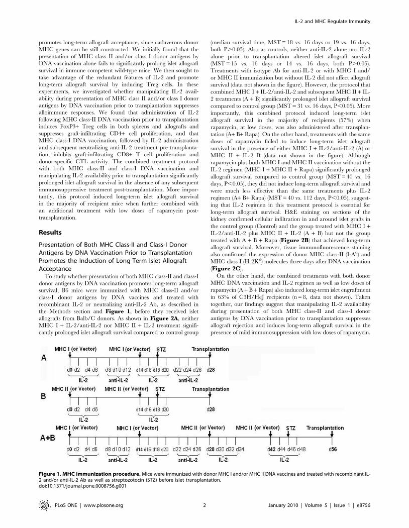
allografts from Balb/C donors. As shown in Figure 2A, neither
MHC I + IL-2/anti-IL-2 nor MHC II + IL-2 treatment signifi-
cantly prolonged islet allograft survival compared to control group
(median survival time, MST = 18 vs. 16 days or 19 vs. 16 days,
both P.0.05). Also as controls, neither anti-IL-2 alone nor IL-2
alone prior to transplantation altered islet allograft survival
(MST = 15 vs. 16 days or 14 vs. 16 days, both P.0.05).
Treatments with isotype Ab for anti-IL-2 or with MHC I and/
or MHC II immunization but without IL-2 did not affect allograft
survival (data not shown in the figure). However, the protocol that
combined MHC I + IL-2/anti-IL-2 and subsequent MHC II + IL-
2 treatments (A + B) significantly prolonged islet allograft survival
compared to control group (MST = 31 vs. 16 days, P,0.05). More
importantly, this combined protocol induced long-term islet
allograft survival in the majority of recipients (57%) when
rapamycin, at low doses, was also administered after transplan-
tation (A+ B+ Rapa). On the other hand, treatments with the same
doses of rapamycin failed to induce long-term islet allograft
survival in the presence of either MHC I + IL-2/anti-IL-2 (A) or
MHC II + IL-2 B (data not shown in the figure). Although
rapamycin plus both MHC I and MHC II vaccination without the
IL-2 regimen (MHC I + MHC II + Rapa) significantly prolonged
allograft survival compared to control group (MST = 40 vs. 16
days, P,0.05), they did not induce long-term allograft survival and
were much less effective than the same treatments plus IL-2
regimen (A+ B+ Rapa) (MST = 40 vs. 112 days, P,0.05), suggest-
ing that IL-2 regimen in this treatment protocol is essential for
long-term allograft survival. H&E staining on sections of the
kidney confirmed cellular infiltration in and around islet grafts in
the control group (Control) and the group treated with MHC I +IL-2/anti-IL-2 plus MHC II + IL-2 (A + B) but not the group
treated with A + B + Rapa (Figure 2B) that achieved long-term
allograft survival. Moreover, tissue immunofluorescence staining
also confirmed the expression of donor MHC class-II (I-Ad) and
MHC class-I (H-2Kd) molecules three days after DNA vaccination
(Figure 2C).
On the other hand, the combined treatments with both donor
MHC DNA vaccination and IL-2 regimen as well as low doses of
rapamycin (A + B + Rapa) also induced long-term islet engraftment
in 63% of C3H/HeJ recipients (n = 8, data not shown). Taken
together, our findings suggest that manipulating IL-2 availability
during presentation of both MHC class-II and class-I donor
antigens by DNA vaccination prior to transplantation suppresses
allograft rejection and induces long-term allograft survival in the
presence of mild immunosuppression with low doses of rapamycin.
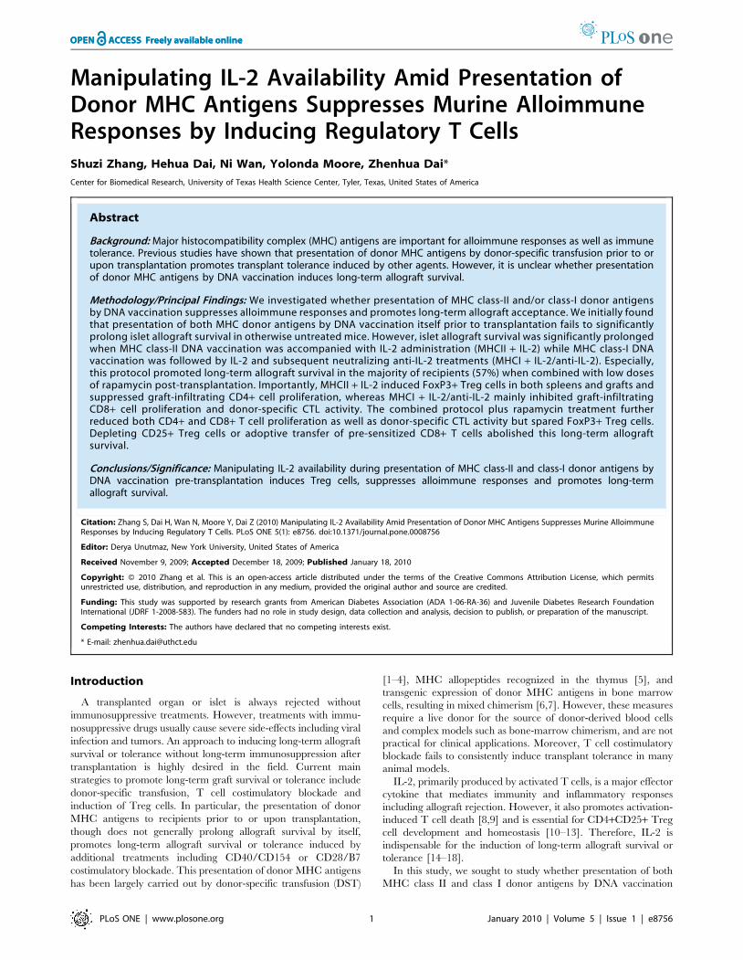
Figure 1. MHC immunization procedure. Mice were immunized with donor MHC I and/or MHC II DNA vaccines and treated with recombinant IL-2 and/or anti-IL-2 Ab as well as streptozotocin (STZ) before islet transplantation.doi:10.1371/journal.pone.0008756.g001
IL-2 and MHC Regulate Immunity
PLoS ONE | www.plosone.org 2 January 2010 | Volume 5 | Issue 1 | e8756
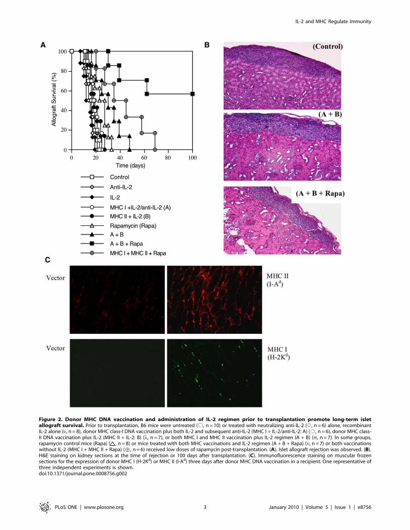
Figure 2. Donor MHC DNA vaccination and administration of IL-2 regimen prior to transplantation promote long-term isletallograft survival. Prior to transplantation, B6 mice were untreated (%, n = 10) or treated with neutralizing anti-IL-2 (e, n = 6) alone, recombinantIL-2 alone (u, n = 8), donor MHC class-I DNA vaccination plus both IL-2 and subsequent anti-IL-2 (MHC I + IL-2/anti-IL-2: A) (#, n = 6), donor MHC class-II DNA vaccination plus IL-2 (MHC II + IL-2: B) (l, n = 7), or both MHC I and MHC II vaccination plus IL-2 regimen (A + B) (s, n = 7). In some groups,rapamycin control mice (Rapa) ( , n = 8) or mice treated with both MHC vaccinations and IL-2 regimen (A + B + Rapa) (n, n = 7) or both vaccinationswithout IL-2 (MHC I + MHC II + Rapa) (›, n = 6) received low doses of rapamycin post-transplantation. (A). Islet allograft rejection was observed. (B).H&E staining on kidney sections at the time of rejection or 100 days after transplantation. (C). Immunofluorescence staining on muscular frozensections for the expression of donor MHC I (H-2Kd) or MHC II (I-Ad) three days after donor MHC DNA vaccination in a recipient. One representative ofthree independent experiments is shown.doi:10.1371/journal.pone.0008756.g002
IL-2 and MHC Regulate Immunity
PLoS ONE | www.plosone.org 3 January 2010 | Volume 5 | Issue 1 | e8756
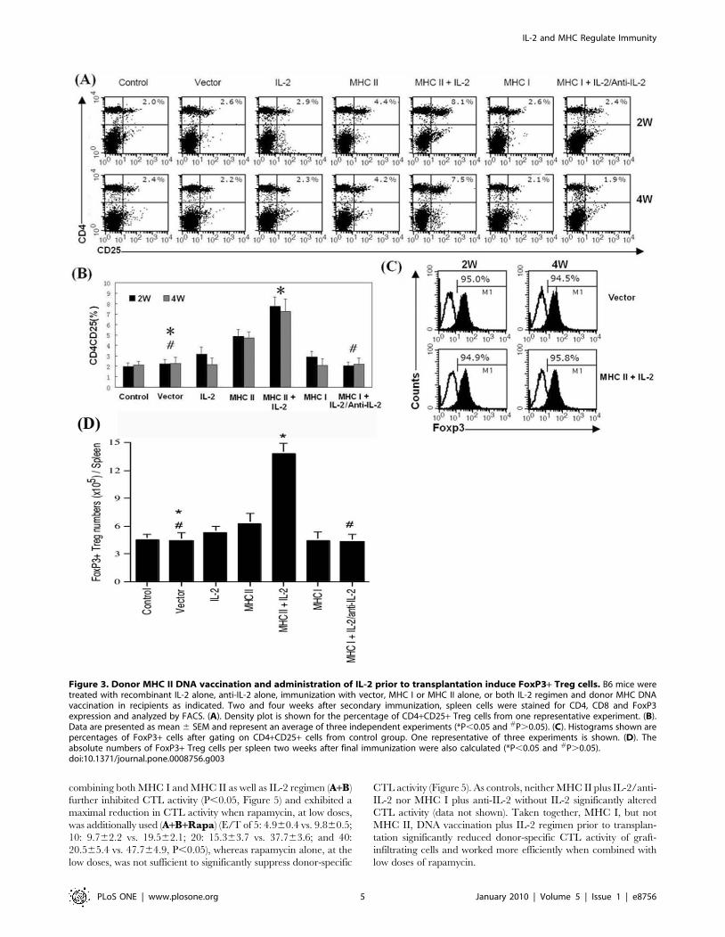
Donor MHC DNA Vaccination and Administration of IL-2Prior to Transplantation Induce FoxP3+ Treg Cells
IL-2 is essential for CD4+CD25+ Treg cell development
and homeostasis [10–12]. To study whether IL-2 manipulation
and presentation of both MHC class-II and class-I donor antigens
by DNA vaccination prior to transplantation suppress allograft
rejection by inducing Tregs, we first measured CD4+CD25+FoxP3+ Treg cells from spleens of B6 mice, which were
immunized and/or treated with IL-2/antiIL-2 regimen without
transplantation, two and four weeks after second immunization as
described in the section of Methods. Based on one representative
FACS data, as shown in Figure 3A, immunization with MHC II-
expressing DNA alone increased the percentages of Treg cells (4.4
vs. 2.5% at 2 weeks and 4.2 vs. 2.2% at 4 weeks) that were further
increased when IL-2 was also administered (8.1 vs. 2.5% at 2
weeks and 7.5 vs. 2.2% at 4 weeks) while immunization with MHC
I-expressing DNA alone or in combination with IL-2/anti-IL-2
did not increase Treg cells. Based on three separate experiments in
which data are presented as mean 6 SEM as shown in Figure 3B,
we confirmed that MHC II DNA immunization alone signifi-
cantly increased Treg cells compared to vector control (2 weeks:
4.960.6% vs. 2.260.4%, and 4 weeks: 4.760.5% vs. 2.360.6%,
both P,0.05) while addition of IL-2 further increased Treg cells
compared to MHC II DNA immunization alone (2 weeks:
7.860.8% vs. 4.960.6%, and 4 weeks: 7.261.1 vs. 4.760.5%,
both P,0.05). IL-2 alone slightly increased Treg cells at the time
point of 2 weeks (3.160.6% vs. 2.060.4%, P = 0.054) but not 4
weeks (2.260.6% vs. 2.160.3%, P.0.05). Moreover, MHC I
immunization alone or in combination with IL-2 (data not shown)
or with IL-2 plus subsequent anti-IL-2 did not significantly alter
Treg numbers (Figure 3B). Around 95% of CD4+CD25+ cells
were confirmed to be FoxP3+ Treg cells (Figure 3C). Finally, as
shown in Figure 3D, MHC II + IL-2 also significantly increased
the absolute number of FoxP3+ cells in the spleen two weeks after
last immunization (13.861.1 vs. 4.460.9, 6105/spleen, P,0.05)
while MHC I + IL-2/anti-IL-2 did not (4.360.8 vs. 4.460.9,
6105/spleen, P.0.05).
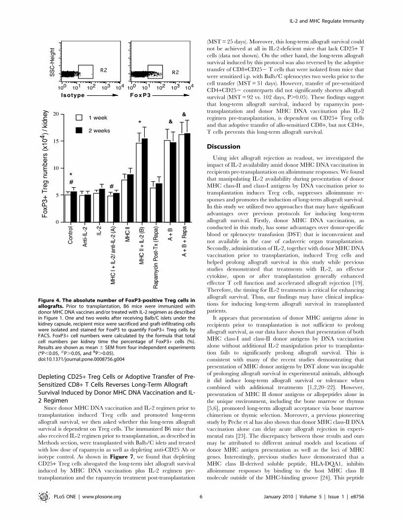
To further quantify Treg cells in grafts after transplantation,
mice that were immunized and/or treated with IL-2/anti-IL-2
were sacrificed one and two weeks after islet transplantation, and
graft-infiltrating cells were isolated and analyzed to determine the
absolute numbers of FoxP3+ Treg cells per kidney. As shown in
Figure 4, treatments with IL-2, anti-IL-2, or together with MHC
I DNA vaccination prior to transplantation did not alter the
numbers of Treg cells in grafts one week after islet transplantation.
Neither did the vector control (data not shown in the figure).
However, MHC II DNA vaccination significantly increased Treg
cell numbers compared to control group (7.861.1 vs. 5.060.5,
P,0.05) while Treg numbers were further increased in MHC II +IL-2 group compared to MHC II alone (14.661.9 vs. 7.861.1,
P,0.05) one week after transplantation. Administration of
rapamycin alone post-transplantation slightly but not significantly
increased the numbers of Tregs in the grafts compared to the
control (6.361.2 vs. 5.060.5, P.0.05). Rapamycin also did not
significantly alter Treg numbers when combined with MHC II +MHC I plus IL-2 regimen (A+ B+ Rapa vs. A+ B: 16.262.3 vs.
15.162.1, P.0.05). The similar results were obtained two weeks
after transplantation (Figure 4). Taken together, MHC II DNA
vaccination alone or together with administration of IL-2 prior to
transplantation dramatically increased the number of Treg cells in
both spleens and grafts of recipients while addition of rapamycin
after transplantation did not significantly alter the numbers of
Treg cells in grafts. These findings suggest that donor MHC class-
II, but not MHC class-I, DNA vaccination induces Treg cells and
is more efficient when additional IL-2 is available.
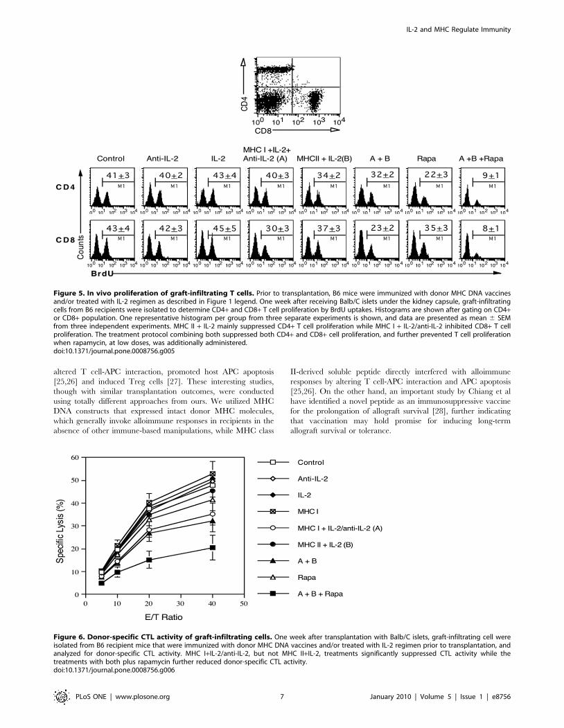
Donor MHC DNA Vaccination and Manipulating IL-2Availability prior to Transplantation Suppress Graft-Infiltrating T Cell Proliferation
To study how donor MHC DNA vaccination and manipulating
IL-2 suppress alloimmune responses, mice that were immunized
and/or treated with IL-2 regimen prior to transplantation were
sacrificed one week after islet transplantation, and graft-infiltrating
cells from the kidney harboring islet allografts were isolated to
measure T cell proliferation by BrdU uptakes. As shown in
Figure 5, treatment with IL-2 or anti-IL-2 alone prior to
transplantation did not alter the percentages of BrdU-positive cells
in both CD4+ and CD8+ components one week after transplan-
tation (BrdU+ in anti-IL-2 group: CD4: 4062% vs. 4163%,
CD8: 4263% vs. 4364%; and BrdU+ in IL-2 group: CD4:
4364% vs. 4163%, CD8: 4565% vs. 4364%, all P.0.05).
Neither did the vector control, MHC II alone, MHC I alone or
MHC I + anti-IL-2 (data not shown in the figure). However, MHC
I + IL-2/anti-IL-2 (A) pre-transplantation significantly suppressed
CD8+, but not CD4+, T cell proliferation (CD4: 4063% vs.
4163%, P.0.05 and CD8: 3063% vs. 4364%, P,0.05),
whereas MHC II + IL-2 (B) mainly inhibited CD4+ cell prolifer-
ation and only slightly inhibited CD8+ cell proliferation (CD4:
3462% vs. 4163%, P,0.05 and CD8: 3763% vs. 4364%,
P.0.05). Moreover, treatments with both MHC II and MHC I
plus IL-2 regimen (A+B) suppressed both CD4+ and CD8+ cell
proliferation (CD4: 3262% vs. 4163%, P,0.05 and CD8:
2362% vs. 4364%, P,0.05). On the other hand, low dose of
rapamycin alone post-transplantation without immunization pre-
transplantation significantly suppressed CD4+ cell proliferation
but only slightly inhibited CD8+ cell proliferation (CD4: 2263%
vs. 4163%, P,0.05 and CD8: 3563% vs. 4364%, P = 0.056).
Importantly, the combined treatments with MHC I/II vaccina-
tion, IL-2 regimen and rapamycin (A+ B+ Rapa) largely pre-
vented both CD4+ and CD8+ cell proliferation (CD4: 961% vs.
4163%, P,0.05 and CD8: 861% vs. 4364%, P,0.05,
Figure 5). Taken together, MHC I + IL-2/anti-IL-2 pre-
transplantation mainly suppressed CD8+ T cell proliferation in
the grafts, whereas MHC II + IL-2 pre-transplantation mainly
inhibited CD4+ T cell proliferation. The measures combining
both suppressed both CD4+ and CD8+ T cell turnover and largely
prevented T cell proliferation when rapamycin, at low doses, was
also administered post-transplantation.
Donor MHC Class-I DNA Vaccination and Treatment withIL-2 Regimen prior to Transplantation Reduce Donor-Specific Cytotoxic T Lymphocyte (CTL) Activity
Since donor MHC I DNA vaccination plus IL-2 regimen prior
to transplantation suppressed the proliferation of graft-infiltrating
CD8+ T cells (Figure 4), we asked whether they also affect donor-
specific CTL activity ex vivo of graft-infiltrating cells in B6 recipient
mice one week after islet transplantation. As shown in Figure 6,
treatment with anti-IL-2 alone, IL-2 alone or MHC II + IL-2 (B)
prior to transplantation did not alter the CTL activity of graft-
infiltrating cells. MHC I vaccination alone slightly, but not
significantly, increased CTL activity (E/T of 5: 10.360.7 vs.
9.860.5; 10: 21.462.3 vs. 19.562.1; 20: 40.263.9 vs. 37.763.6;
and 40: 52.865.6 vs. 47.764.9, P.0.05). However, MHC I + IL-
2/anti-IL-2 (A) significantly suppressed CTL activity (E/T of 5:
8.060.4 vs. 9.860.5; 10: 14.561.6 vs. 19.562.1; 20: 28.463.2 vs.
37.763.6; and 40: 35.264.8 vs. 47.764.9, P,0.05). The treatment
IL-2 and MHC Regulate Immunity
PLoS ONE | www.plosone.org 4 January 2010 | Volume 5 | Issue 1 | e8756
combining both MHC I and MHC II as well as IL-2 regimen (A+B)
further inhibited CTL activity (P,0.05, Figure 5) and exhibited a
maximal reduction in CTL activity when rapamycin, at low doses,
was additionally used (A+B+Rapa) (E/T of 5: 4.960.4 vs. 9.860.5;
10: 9.762.2 vs. 19.562.1; 20: 15.363.7 vs. 37.763.6; and 40:
20.565.4 vs. 47.764.9, P,0.05), whereas rapamycin alone, at the
low doses, was not sufficient to significantly suppress donor-specific
CTL activity (Figure 5). As controls, neither MHC II plus IL-2/anti-
IL-2 nor MHC I plus anti-IL-2 without IL-2 significantly altered
CTL activity (data not shown). Taken together, MHC I, but not
MHC II, DNA vaccination plus IL-2 regimen prior to transplan-
tation significantly reduced donor-specific CTL activity of graft-
infiltrating cells and worked more efficiently when combined with
low doses of rapamycin.
Figure 3. Donor MHC II DNA vaccination and administration of IL-2 prior to transplantation induce FoxP3+ Treg cells. B6 mice weretreated with recombinant IL-2 alone, anti-IL-2 alone, immunization with vector, MHC I or MHC II alone, or both IL-2 regimen and donor MHC DNAvaccination in recipients as indicated. Two and four weeks after secondary immunization, spleen cells were stained for CD4, CD8 and FoxP3expression and analyzed by FACS. (A). Density plot is shown for the percentage of CD4+CD25+ Treg cells from one representative experiment. (B).Data are presented as mean 6 SEM and represent an average of three independent experiments (*P,0.05 and #P.0.05). (C). Histograms shown arepercentages of FoxP3+ cells after gating on CD4+CD25+ cells from control group. One representative of three experiments is shown. (D). Theabsolute numbers of FoxP3+ Treg cells per spleen two weeks after final immunization were also calculated (*P,0.05 and #P.0.05).doi:10.1371/journal.pone.0008756.g003
IL-2 and MHC Regulate Immunity
PLoS ONE | www.plosone.org 5 January 2010 | Volume 5 | Issue 1 | e8756
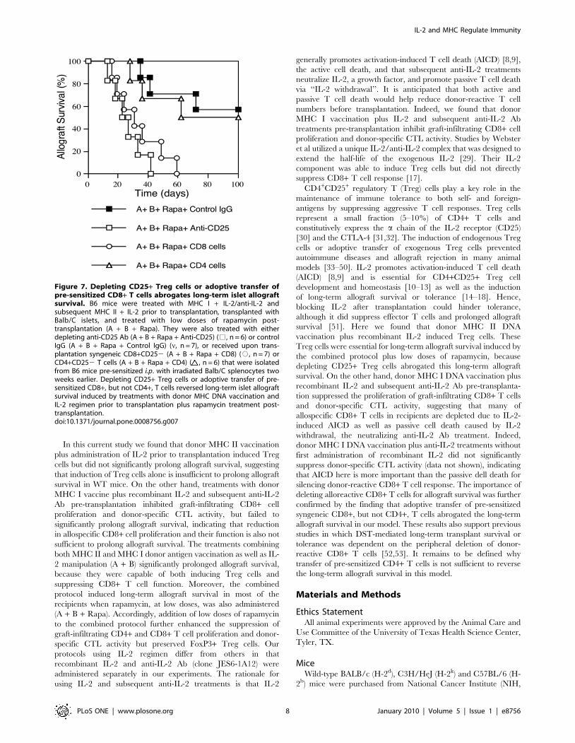
Depleting CD25+ Treg Cells or Adoptive Transfer of Pre-Sensitized CD8+ T Cells Reverses Long-Term AllograftSurvival Induced by Donor MHC DNA Vaccination and IL-2 Regimen
Since donor MHC DNA vaccination and IL-2 regimen prior to
transplantation induced Treg cells and promoted long-term
allograft survival, we then asked whether this long-term allograft
survival is dependent on Treg cells. The immunized B6 mice that
also received IL-2 regimen prior to transplantation, as described in
Methods section, were transplanted with Balb/C islets and treated
with low dose of rapamycin as well as depleting anti-CD25 Ab or
isotype control. As shown in Figure 7, we found that depleting
CD25+ Treg cells abrogated the long-term islet allograft survival
induced by MHC DNA vaccination plus IL-2 regimen pre-
transplantation and the rapamycin treatment post-transplantation
(MST = 25 days). Moreover, this long-term allograft survival could
not be achieved at all in IL-2-deficient mice that lack CD25+ T
cells (data not shown). On the other hand, the long-term allograft
survival induced by this protocol was also reversed by the adoptive
transfer of CD8+CD252 T cells that were isolated from mice that
were sensitized i.p. with Balb/C splenocytes two weeks prior to the
cell transfer (MST = 31 days). However, transfer of pre-sensitized
CD4+CD252 counterparts did not significantly shorten allograft
survival (MST = 92 vs. 102 days, P.0.05). These findings suggest
that long-term allograft survival, induced by rapamycin post-
transplantation and donor MHC DNA vaccination plus IL-2
regimen pre-transplantation, is dependent on CD25+ Treg cells
and that adoptive transfer of allo-sensitized CD8+, but not CD4+,
T cells prevents this long-term allograft survival.
Discussion
Using islet allograft rejection as readout, we investigated the
impact of IL-2 availability amid donor MHC DNA vaccination in
recipients pre-transplantation on alloimmune responses. We found
that manipulating IL-2 availability during presentation of donor
MHC class-II and class-I antigens by DNA vaccination prior to
transplantation induces Treg cells, suppresses alloimmune re-
sponses and promotes the induction of long-term allograft survival.
In this study we utilized two approaches that may have significant
advantages over previous protocols for inducing long-term
allograft survival. Firstly, donor MHC DNA vaccination, as
conducted in this study, has some advantages over donor-specific
blood or splenocyte transfusion (DST) that is inconvenient and
not available in the case of cadaveric organ transplantation.
Secondly, administration of IL-2, together with donor MHC DNA
vaccination prior to transplantation, induced Treg cells and
helped prolong allograft survival in this study while previous
studies demonstrated that treatments with IL-2, an effector
cytokine, upon or after transplantation generally enhanced
effector T cell function and accelerated allograft rejection [19].
Therefore, the timing for IL-2 treatments is critical for enhancing
allograft survival. Thus, our findings may have clinical implica-
tions for inducing long-term allograft survival in transplanted
patients.
It appears that presentation of donor MHC antigens alone in
recipients prior to transplantation is not sufficient to prolong
allograft survival, as our data have shown that presentation of both
MHC class-I and class-II donor antigens by DNA vaccination
alone without additional IL-2 manipulation prior to transplanta-
tion fails to significantly prolong allograft survival. This is
consistent with many of the recent studies demonstrating that
presentation of MHC donor antigens by DST alone was incapable
of prolonging allograft survival in experimental animals, although
it did induce long-term allograft survival or tolerance when
combined with additional treatments [1,2,20–22]. However,
presentation of MHC II donor antigens or allopeptides alone in
the unique environment, including the bone marrow or thymus
[5,6], promoted long-term allograft acceptance via bone marrow
chimerism or thymic selection. Moreover, a previous pioneering
study by Peche et al has also shown that donor MHC class-II DNA
vaccination alone can delay acute allograft rejection in experi-
mental rats [23]. The discrepancy between those results and ours
may be attributed to different animal models and locations of
donor MHC antigen presentation as well as the loci of MHC
genes. Interestingly, previous studies have demonstrated that a
MHC class II-derived soluble peptide, HLA-DQA1, inhibits
alloimmune responses by binding to the host MHC class II
molecule outside of the MHC-binding groove [24]. This peptide
Figure 4. The absolute number of FoxP3-positive Treg cells inallografts. Prior to transplantation, B6 mice were immunized withdonor MHC DNA vaccines and/or treated with IL-2 regimen as describedin Figure 1. One and two weeks after receiving Balb/C islets under thekidney capsule, recipient mice were sacrificed and graft-infiltrating cellswere isolated and stained for FoxP3 to quantify FoxP3+ Treg cells byFACS. FoxP3+ cell numbers were calculated by the formula that totalcell numbers per kidney time the percentage of FoxP3+ cells (%).Results are shown as mean 6 SEM from four independent experiments(*P,0.05, #P.0.05, and &P.0.05).doi:10.1371/journal.pone.0008756.g004
IL-2 and MHC Regulate Immunity
PLoS ONE | www.plosone.org 6 January 2010 | Volume 5 | Issue 1 | e8756
altered T cell-APC interaction, promoted host APC apoptosis
[25,26] and induced Treg cells [27]. These interesting studies,
though with similar transplantation outcomes, were conducted
using totally different approaches from ours. We utilized MHC
DNA constructs that expressed intact donor MHC molecules,
which generally invoke alloimmune responses in recipients in the
absence of other immune-based manipulations, while MHC class
II-derived soluble peptide directly interfered with alloimmune
responses by altering T cell-APC interaction and APC apoptosis
[25,26]. On the other hand, an important study by Chiang et al
have identified a novel peptide as an immunosuppressive vaccine
for the prolongation of allograft survival [28], further indicating
that vaccination may hold promise for inducing long-term
allograft survival or tolerance.
Figure 5. In vivo proliferation of graft-infiltrating T cells. Prior to transplantation, B6 mice were immunized with donor MHC DNA vaccinesand/or treated with IL-2 regimen as described in Figure 1 legend. One week after receiving Balb/C islets under the kidney capsule, graft-infiltratingcells from B6 recipients were isolated to determine CD4+ and CD8+ T cell proliferation by BrdU uptakes. Histograms are shown after gating on CD4+or CD8+ population. One representative histogram per group from three separate experiments is shown, and data are presented as mean 6 SEMfrom three independent experiments. MHC II + IL-2 mainly suppressed CD4+ T cell proliferation while MHC I + IL-2/anti-IL-2 inhibited CD8+ T cellproliferation. The treatment protocol combining both suppressed both CD4+ and CD8+ cell proliferation, and further prevented T cell proliferationwhen rapamycin, at low doses, was additionally administered.doi:10.1371/journal.pone.0008756.g005
Figure 6. Donor-specific CTL activity of graft-infiltrating cells. One week after transplantation with Balb/C islets, graft-infiltrating cell wereisolated from B6 recipient mice that were immunized with donor MHC DNA vaccines and/or treated with IL-2 regimen prior to transplantation, andanalyzed for donor-specific CTL activity. MHC I+IL-2/anti-IL-2, but not MHC II+IL-2, treatments significantly suppressed CTL activity while thetreatments with both plus rapamycin further reduced donor-specific CTL activity.doi:10.1371/journal.pone.0008756.g006
IL-2 and MHC Regulate Immunity
PLoS ONE | www.plosone.org 7 January 2010 | Volume 5 | Issue 1 | e8756
In this current study we found that donor MHC II vaccination
plus administration of IL-2 prior to transplantation induced Treg
cells but did not significantly prolong allograft survival, suggesting
that induction of Treg cells alone is insufficient to prolong allograft
survival in WT mice. On the other hand, treatments with donor
MHC I vaccine plus recombinant IL-2 and subsequent anti-IL-2
Ab pre-transplantation inhibited graft-infiltrating CD8+ cell
proliferation and donor-specific CTL activity, but failed to
significantly prolong allograft survival, indicating that reduction
in allospecific CD8+ cell proliferation and their function is also not
sufficient to prolong allograft survival. The treatments combining
both MHC II and MHC I donor antigen vaccination as well as IL-
2 manipulation (A + B) significantly prolonged allograft survival,
because they were capable of both inducing Treg cells and
suppressing CD8+ T cell function. Moreover, the combined
protocol induced long-term allograft survival in most of the
recipients when rapamycin, at low doses, was also administered
(A + B + Rapa). Accordingly, addition of low doses of rapamycin
to the combined protocol further enhanced the suppression of
graft-infiltrating CD4+ and CD8+ T cell proliferation and donor-
specific CTL activity but preserved FoxP3+ Treg cells. Our
protocols using IL-2 regimen differ from others in that
recombinant IL-2 and anti-IL-2 Ab (clone JES6-1A12) were
administered separately in our experiments. The rationale for
using IL-2 and subsequent anti-IL-2 treatments is that IL-2
generally promotes activation-induced T cell death (AICD) [8,9],
the active cell death, and that subsequent anti-IL-2 treatments
neutralize IL-2, a growth factor, and promote passive T cell death
via ‘‘IL-2 withdrawal’’. It is anticipated that both active and
passive T cell death would help reduce donor-reactive T cell
numbers before transplantation. Indeed, we found that donor
MHC I vaccination plus IL-2 and subsequent anti-IL-2 Ab
treatments pre-transplantation inhibit graft-infiltrating CD8+ cell
proliferation and donor-specific CTL activity. Studies by Webster
et al utilized a unique IL-2/anti-IL-2 complex that was designed to
extend the half-life of the exogenous IL-2 [29]. Their IL-2
component was able to induce Treg cells but did not directly
suppress CD8+ T cell response [17].
CD4+CD25+ regulatory T (Treg) cells play a key role in the
maintenance of immune tolerance to both self- and foreign-
antigens by suppressing aggressive T cell responses. Treg cells
represent a small fraction (5–10%) of CD4+ T cells and
constitutively express the a chain of the IL-2 receptor (CD25)
[30] and the CTLA-4 [31,32]. The induction of endogenous Treg
cells or adoptive transfer of exogenous Treg cells prevented
autoimmune diseases and allograft rejection in many animal
models [33–50]. IL-2 promotes activation-induced T cell death
(AICD) [8,9] and is essential for CD4+CD25+ Treg cell
development and homeostasis [10–13] as well as the induction
of long-term allograft survival or tolerance [14–18]. Hence,
blocking IL-2 after transplantation could hinder tolerance,
although it did suppress effector T cells and prolonged allograft
survival [51]. Here we found that donor MHC II DNA
vaccination plus recombinant IL-2 induced Treg cells. These
Treg cells were essential for long-term allograft survival induced by
the combined protocol plus low doses of rapamycin, because
depleting CD25+ Treg cells abrogated this long-term allograft
survival. On the other hand, donor MHC I DNA vaccination plus
recombinant IL-2 and subsequent anti-IL-2 Ab pre-transplanta-
tion suppressed the proliferation of graft-infiltrating CD8+ T cells
and donor-specific CTL activity, suggesting that many of
allospecific CD8+ T cells in recipients are depleted due to IL-2-
induced AICD as well as passive cell death caused by IL-2
withdrawal, the neutralizing anti-IL-2 Ab treatment. Indeed,
donor MHC I DNA vaccination plus anti-IL-2 treatments without
first administration of recombinant IL-2 did not significantly
suppress donor-specific CTL activity (data not shown), indicating
that AICD here is more important than the passive dell death for
silencing donor-reactive CD8+ T cell response. The importance of
deleting alloreactive CD8+ T cells for allograft survival was further
confirmed by the finding that adoptive transfer of pre-sensitized
syngeneic CD8+, but not CD4+, T cells abrogated the long-term
allograft survival in our model. These results also support previous
studies in which DST-mediated long-term transplant survival or
tolerance was dependent on the peripheral deletion of donor-
reactive CD8+ T cells [52,53]. It remains to be defined why
transfer of pre-sensitized CD4+ T cells is not sufficient to reverse
the long-term allograft survival in this model.
Materials and Methods
Ethics StatementAll animal experiments were approved by the Animal Care and
Use Committee of the University of Texas Health Science Center,
Tyler, TX.
MiceWild-type BALB/c (H-2d), C3H/HeJ (H-2k) and C57BL/6 (H-
2b) mice were purchased from National Cancer Institute (NIH,
Figure 7. Depleting CD25+ Treg cells or adoptive transfer ofpre-sensitized CD8+ T cells abrogates long-term islet allograftsurvival. B6 mice were treated with MHC I + IL-2/anti-IL-2 andsubsequent MHC II + IL-2 prior to transplantation, transplanted withBalb/C islets, and treated with low doses of rapamycin post-transplantation (A + B + Rapa). They were also treated with eitherdepleting anti-CD25 Ab (A + B + Rapa + Anti-CD25) (%, n = 6) or controlIgG (A + B + Rapa + Control IgG) (n, n = 7), or received upon trans-plantation syngeneic CD8+CD252 (A + B + Rapa + CD8) (#, n = 7) orCD4+CD252 T cells (A + B + Rapa + CD4) ( , n = 6) that were isolatedfrom B6 mice pre-sensitized i.p. with irradiated Balb/C splenocytes twoweeks earlier. Depleting CD25+ Treg cells or adoptive transfer of pre-sensitized CD8+, but not CD4+, T cells reversed long-term islet allograftsurvival induced by treatments with donor MHC DNA vaccination andIL-2 regimen prior to transplantation plus rapamycin treatment post-transplantation.doi:10.1371/journal.pone.0008756.g007
IL-2 and MHC Regulate Immunity
PLoS ONE | www.plosone.org 8 January 2010 | Volume 5 | Issue 1 | e8756
Bethesda, MD, USA). IL-2-deficient mice were purchased from
the Jackson Laboratory. All mice were housed in a specific
pathogen-free environment.
Plasmid Constructs and DNA PreparationMurine major histocompatibility complex (MHC) class I (H-
2Kd) or class II (I-Ad) cDNA was obtained by RT-PCR using total
RNA isolated from the spleens of BALB/c mice, and subcloned
into the eukaryotic expression vector pcDNA3.1+ (Invitrogen) at
BamH I/Xho I sites, which carries the cytomegalovirus (CMV)
and T7 promoter upstream of the cloned cDNA. The plasmid
DNA vectors were prepared using a Qiagen Giga-prep kit
(Qiagen, Valencia, CA) and are referred to as MHC I and
MHC II respectively. The following PCR primers were used: H-
2Kd forward primer was 59-GGATCCATGGCACCCTGCACG-
39 and the reverse primer was 59-CTCGAG TCACGCTAGA-
GAATGAGGG-39; I-Ad forward primer was 59-GGATCCAT-
GCCGTGCAGTAGAGC-39 and the reverse primer was 59-
CTCGAGTCATAAAGGCCCTGGG-39.
In Vivo Expression of Donor MHC Molecules and TissueImmunofluorescence Staining
B6 mice received a single intramuscular (i.m.) injection (day 0)
of 100 mg of plasmid DNA or empty vector in 100 ml of sterile
saline. The injection sites were sampled at day 3 from each
experimental group. The muscle samples were embedded in OTC
and stored at 280uC until sectioning. Cryostat sections were fixed
with acetone, washed, and blocked with 1:20-diluted goat serum.
Then slides were incubated with the primary Abs mouse anti-
mouse H-2Kd and I-Ad (Biolegend) for 1 hour at 37uC, washed
and incubated with 1:200-diluted AlexaFluor 488- or 568-
conjugated secondary Ab goat anti-mouse IgG (Invitrogen) at
4uC overnight. Slides were washed and air-dried. A coverslip was
placed on the slide that was finally visualized under a fluorescence
microscope.
Immunization ProtocolsB6 mice received (i.m.) 100 mg of plasmids encoding donor
MHC I or MHC II gene or control vector in each quadricep
muscle (day 0) alone or in combination with 16104 unit (1 mg) of
recombinant mouse IL-2 (eBioscience) i.p. for four times (days 0,
2, 4 and 6), as described in Figure 1. Some mice that received
MHC I vaccination plus recombinant IL-2 were treated with
50 mg of anti-mouse IL-2 mAb (Clone JES6-1A12, eBioscience)
on days 8, 10 and 12, referred to as the group ‘‘MHC I + IL-2/
anti-IL-2’’ (A). The immunization was boosted by repeating the
same procedures once again in all groups two weeks after first
immunization (day 14). In some groups, mice were treated with
both ‘‘MHC I + IL-2/anti-IL-2’’ and subsequent ‘‘MHC II + IL-
2’’, once at a time, referred to as ‘‘A + B’’ (Figure 1). As controls,
some mice also were treated with IL-2 alone or anti-IL-2 Ab
alone at the same time points without immunization with the
plasmid.
Pancreatic Islet TransplantationIslet donors were 8–10 week-old female Balb/C mice. Islet
recipients were 8–10 week-old female C57BL/6 mice. Islets were
isolated and transplanted into the subcapsular space of the right
kidney of recipient mice (400 islets per recipient), as described
previously [54–56]. Recipient mice were rendered diabetic by a
single injection of streptozotocin (180 mg/kg) (Sigma) 10 days
before transplantation. Primary graft function was defined as a
blood glucose level under 200 mg/dl for 48 hours after transplan-
tation. Islet graft rejection was defined as a rise in blood glucose to
.300 mg/dl for three consecutive days after primary function.
HistologyKidney samples containing islet grafts were fixed in 10%
formalin, embedded in paraffin, sectioned with a microtome,
stained using haematoxylin/eosin (H&E), and observed for cell
infiltration under a light microscope.
Flow CytometrySpleen cells from immunized mice were stained with anti-CD4-
PE, anti-CD25-FITC (BD Biosciences) and anti-FoxP3-APC Abs
(eBioscience) and were analyzed by a FACSCalibur (BD Bioscienc-
es). In some experiments, graft-infiltrating cells were also stained
for intracellular FoxP3 and BrdU (refer to BrdU labeling). To
purify CD4+ and CD8+ T cells for adoptive transfer experi-
ments, splenocytes from pre-sensitized B6 mice were stained with
anti-CD4-PE, anti-CD8-FITC and anti-CD25-PerCP, and CD4+CD252 or CD8+CD252 cells were sorted out by FACSAria (BD
Biosciences). The purity after sorting was typically .97%.
Isolation of Tissue-Infiltrating CellsTissue-infiltrating cells were isolated as described in our
previous publications [54,57]. Briefly, the kidneys harboring islets
were perfused in situ with heparinized 0.9% saline. They were then
minced and digested at 37uC for 30 min in 20 ml RPMI-1640
medium containing 5% FCS and 350 u/ml collagenase (Sigma,
St. Louis, MO). To clear the debris, cell suspensions were rapidly
passed down 70 mm cell strainer, then mixed with Percoll solution
(Sigma) to a concentration of 30%, and centrifuged at 2000 rpm
for 15 minutes at room temperature. The pellet was re-suspended
and analyzed for CTL activity or stained with Abs before FACS
analysis.
Analysis of T Cell Proliferation In Vivo by 5-Bromo-29-Deoxyuridine (BrdU) Labeling
Recipient mice were pulsed i.p. with 0.8 mg of BrdU (Sigma) six
days after transplantation. 24 hours later, renal graft-infiltrating
cells were isolated and first stained with anti-CD4-PE or anti-
CD8-PE. Cells were then fixed in 70% ethanol followed by 1%
paraformaldehyde and incubated with 50 Units/ml of DNase I
(Sigma). Cells were finally stained with anti-BrdU-FITC (BD
Biosciences) and analyzed by a four-color FACSCalibur [54,57].
Cytotoxic T Lymphocyte (CTL) ActivityGraft-infiltrating cells were isolated and immediately assayed for
ex vivo CTL activity against BALB/c targets, splenocytes. Allospe-
cific CTL activity was measured by incubating the cells with either
Con A-activated (H-2d) BALB/c target spleen cells or third-party
cells, LK35.2 (H-2k) (American Type Culture Collection)
for 3 h. Target cells were labeled with calcein-AM (Molecular
Probes), and calcein release was measured in a LS50B lumines-
cence spectrometer (Perkin–Elmer) [58]. Experiments in which
spontaneous calcein release was more than 25% of maximal
release were excluded. Antigen-specific CTL activity was calcu-
lated according to the following formula: % specific lysis =
100 6[(sample release - spontaneous release)/(maximum release -
spontaneous release)].
Treatment of Mice with Depleting Anti-CD25 Ab andRapamycin
To deplete CD4+CD25+ Treg cells, mice were treated with
depleting anti-CD25 Ab (clone PC61, BioExpress, West Lebanon,
IL-2 and MHC Regulate Immunity
PLoS ONE | www.plosone.org 9 January 2010 | Volume 5 | Issue 1 | e8756
NH)) at 0.25 mg every other day for four doses upon
transplantation as described previously [54]. Over 80% of
CD25+ T cells were depleted according FACS analysis. To
provide mild immunosuppression, recipient mice were treated i.p.
with low doses of rapamycin (0.2 mg/kg/day, Sigma) for seven
consecutive days after islet transplantation.
Statistical AnalysisThe analysis of allograft survival data was performed using the
Kaplan-Meier (Log-rank test). Comparison of means between
groups was conducted using ANOVA. A value of p,0.05 was
considered statistically significant.
Author Contributions
Conceived and designed the experiments: SZ HD NW ZD. Performed the
experiments: SZ HD NW YM. Analyzed the data: SZ HD NW ZD.
Contributed reagents/materials/analysis tools: YM. Wrote the paper: SZ
ZD.
References
1. Lin H, Bolling SF, Linsley PS, Wei RQ, Gordon D, et al. (1993) Long-term
acceptance of major histocompatibility complex mismatched cardiac allografts
induced by CTLA4Ig plus donor-specific transfusion. J Exp Med 178:
1801–1806.
2. Markees TG, Phillips NE, Gordon EJ, Noelle RJ, Shultz LD, et al. (1998) Long-
term survival of skin allografts induced by donor splenocytes and anti-CD154
antibody in thymectomized mice requires CD4+ T cells, interferon-c, and
CTLA4. J Clin Invest 101: 2446–2455.
3. Medawar PB (1963) The use of antigenic tissue extracts to weaken the
immunological reaction against skin homografts in mice. Transplantation 1:
21–38.
4. Soulillou JP, Blandin F, Gunther E, Lemoine V (1984) Genetics of the blood
transfusion effect on heart allografts in rats. Transplantation 38: 63–67.
5. Sayegh MH, Perico N, Imberti O, Hancock WW, Carpenter CB, et al. (1993)
Thymic recognition of class II major histocompatibility complex allopeptides
induces donor-specific unresponsiveness to renal allografts. Transplantation 56:
461–465.
6. Sonntag KC, Emery DW, Yasumoto A, Haller G, Germana S, et al. (2001)
Tolerance to solid organ transplants through transfer of MHC class II genes.
J Clin Invest 107: 65–71.
7. Umemura A, Monaco AP, Maki T (2000) Donor MHC class II antigen is
essential for induction of transplantation tolerance by bone marrow cells.
J Immunol 164: 4452–4457.
8. Lenardo MJ (1991) Interleukin-2 programs mouse alpha/beta T lymphocytes for
apoptosis. Nature 353: 858–861.
9. Kneitz B, Herman T, Yonehara S, Schimpl A (1995) Normal clonal expansion
but impaired Fas- mediated cell death and anergy in IL-2 deficient mice.
Eur J Immunol 25: 2572–2577.
10. Walsh PT, Buckler JL, Zhang J, Gelman AE, Dalton NM, et al. (2006) PTEN
inhibits IL-2 receptor-mediated expansion of CD4+CD25+ Tregs. J Clin Invest
116: 2521–2531.
11. Fontenot JD, Rasmussen JP, Gavin MA, Rudensky AY (2005) A function for
interleukin 2 in Foxp3-expressing regulatory T cells. Nat Immunol 6:
1142–1151.
12. Yu A, Zhu L, Altman NH, Malek TR (2009) A low interleukin-2 receptor
signaling threshold supports the development and homeostasis of T regulatory
cells. Immunity 30: 204–217.
13. Tang Q, Adams JY, Penaranda C, Melli K, Piaggio E, et al. (2008) Central role
of defective interleukin-2 production in the triggering of islet autoimmune
destruction. Immunity 28: 687–697.
14. Dai Z, Konieczny BT, Baddoura FK, Lakkis FG (1998) Impaired alloantigen-
mediated T cell apoptosis and failure to induce long term allograft survival in
interleukin-2-deficient mice. J Immunol 161: 1659–1663.
15. Kang HG, Zhang D, Degauque N, Mariat C, Alexopoulos S, et al. (2007) Effects
of cyclosporine on transplant tolerance: the role of IL-2. Am J Transplant 7:
1907–1916.
16. Li Y, Li XC, Zheng XX, Wells AD, Turka LA, et al. (1999) Blocking both signal
1 and signal 2 of T-cell activation prevents apoptosis of alloreactive T cells and
induction of peripheral tolerance. Nature Medicine 5: 1298–1302.
17. Webster KE, Walters S, Kohler RE, Mrkvan T, Boyman O, et al. (2009) In vivo
expansion of T reg cells with IL-2-mAb complexes: induction of resistance to
EAE and long-term acceptance of islet allografts without immunosuppression.
J Exp Med 206: 751–760.
18. Wells AD, Li XC, Li Y, Walsh MC, Zheng XX, et al. (1999) Requirement for T-
cell apoptosis in the induction of peripheral transplantation tolerance. Nat Med
5: 1303–1307.
19. Paineau J, Priestley C, Fabre J, Chevalier S, van der Meide P, et al. (1991) Effect
of recombinant interferon gamma and interleukin-2 and of a monoclonal
antibody against interferon gamma on the rat immune response against heart
allografts. J Heart Lung Transplant 10: 424–430.
20. Ringers J, Haanstra KG, Kroczek RA, Kliem K, Kuhn EM, et al. (2002)
Blockade of CD40-CD154 at the time of donor-specific blood transfusion does
not lead to prolonged kidney allograft survival in nonhuman primates.
Transplantation 73: 862–866.
21. Markees TG, Pearson T, Cuthbert A, Pearson AL, Shultz LD, et al. (2004)
Evaluation of donor-specific transfusion sources: unique failure of bone marrow
cells to induce prolonged skin allograft survival with anti-CD154 monoclonal
antibody. Transplantation 78: 1601–1608.
22. Wu K, Xiang F, Yuan J, Zeng Z, Zhou H, et al. (2008) A combination of donor
specific transfusion and rapamycin prolongs cardiac allograft survival in mice.
Transplant Proc 40: 3699–3701.
23. Peche H, van Denderen B, Roussel JC, Trinite B, Soulillou JP, et al. (2004)
Presentation of donor major histocompatibility complex class II antigens by
DNA vaccination prolongs heart allograft survival. Transplantation 77:
733–740.
24. Zang W, Kalache S, Lin M, Schroppel B, Murphy B (2005) MHC Class II-
mediated apoptosis by a nonpolymorphic MHC Class II peptide proceeds by
activation of protein kinase C. J Am Soc Nephrol 16: 3661–3668.
25. Murphy B, Magee CC, Alexander SI, Waaga AM, Snoeck HW, et al. (1999)
Inhibition of allorecognition by a human class II MHC-derived peptide through
the induction of apoptosis. J Clin Invest 103: 859–867.
26. Murphy B, Yu J, Jiao Q, Lin M, Chitnis T, et al. (2003) A novel mechanism for
the immunomodulatory functions of class II MHC-derived peptides. J Am Soc
Nephrol 14: 1053–1065.
27. Zang W, Lin M, Kalache S, Zhang N, Kruger B, et al. (2008) Inhibition of the
alloimmune response through the generation of regulatory T cells by a MHC
class II-derived peptide. J Immunol 181: 7499–7506.
28. Chiang KC, Shimada Y, Nakano T, Lai CY, Hsu LW, et al. (2009) A novel
peptide mimotope identified as a potential immunosuppressive vaccine for organ
transplantation. J Immunol 182: 4282–4288.
29. Boyman O, Kovar M, Rubinstein MP, Surh CD, Sprent J (2006) Selective
stimulation of T cell subsets with antibody-cytokine immune complexes. Science
311: 1924–1927.
30. Sakaguchi S, Sakaguchi N, Asano M, Itoh M, Toda M (1995) Immunological
self-tolerance maintained by activated T cells expressing IL-2 receptor alpha-
chains (CD25)-breakdown of a single mechanism of self-tolerance causes various
autoimmune diseases. J Immunol 155: 1151–1164.
31. Read S, Malmstrom V, Powrie F (2000) Cytotoxic T lymphocyte-associated
antigen 4 plays an essential role in the function of CD25+CD4+ regulatory cells
that control intestinal inflammation. J Exp Med 192: 295–302.
32. Takahashi T, Tagami T, Yamazaki S, Uede T, Shimizu J, et al. (2000)
Immunologic self-tolerance maintained by CD25+CD4+ regulatory T cells
constitutively expressing cytotoxic T lymphocyte-associated antigen 4. J Exp
Med 192: 303–309.
33. Asano M, Toda M, Sakaguchi N, Sakaguchi S (1996) Autoimmune disease as a
consequence of developmental abnormality of a T cell subpopulation. J Exp
Med 184: 387–396.
34. Suri-Payer E, Amar A, Thornton A, Shevach E (1998) CD4+CD25+ T cells
inhibit both induction and effector function of autoreactive T cells and represent
a unique lineage of immunoregulatory cells. J Immunol 160: 1212–1218.
35. Itoh M, Takahashi T, Sakaguchi N, Kuniyasu Y, Shimizu J, et al. (1999)
Thymus and Autoimmunity: Production of CD25+CD4+ Naturally Anergic and
Suppressive T Cells as a Key Function of the Thymus in Maintaining
Immunologic Self-Tolerance. J Immunol 162: 5317–5326.
36. Zhai Y, Kupiec-Weglinski JW (1999) What is the role of regulatory T cells in
transplantation tolerance? Curr Opin Immunol 11: 497–503.
37. Shevach EM (2000) Regulatory T cells in autoimmunity. Annu Rev Immunol
18: 423–449.
38. Qin S, Cobbold S, Pope H, Elliott J, Kioussis D, et al. (1993) ‘‘Infectious’’
transplantation tolerance. Science 259: 974–977.
39. Hoffmann P, Ermann J, Edinger M, Fathman CG, Strober S (2002) Donor-
type CD4+CD25+ regulatory T cells suppress lethal acute graft-versus-host
disease after allogeneic bone marrow transplantation. J Exp Med 196:
389–399.
40. Fu S, Yopp AC, Mao X, Chen D, Zhang N, et al. (2004) CD4+ CD25+ CD62+T-regulatory cell subset has optimal suppressive and proliferative potential.
Am J Transplant 4: 65–78.
41. Graca L, Cobbold SP, Waldmann H (2002) Identification of regulatory T cells in
tolerated allografts. J Exp Med 195: 1641–1646.
42. Graca L, Thompson S, Lin CY, Adams E, Cobbold SP, et al. (2002) Both
CD4+CD25+ and CD4+CD252 regulatory cells mediate dominant transplan-
tation tolerance. J Immunol 168: 5558–5565.
IL-2 and MHC Regulate Immunity
PLoS ONE | www.plosone.org 10 January 2010 | Volume 5 | Issue 1 | e8756
43. Kingsley CI, Karim M, Bushell AR, Wood KJ (2002) CD25+CD4+ regulatory T
cells prevent graft rejection: CTLA-4- and IL-10-dependent immunoregulationof alloresponses. J Immunol 168: 1080–1086.
44. Zhai Y, Meng L, Gao F, Wang Y, Busuttil RW, et al. (2006) CD4+ T regulatory
cell induction and function in transplant recipients after CD154 blockade isTLR4 independent. J Immunol 176: 5988–5994.
45. Walker WE, Nasr IW, Camirand G, Tesar BM, Booth CJ, et al. (2006) Absenceof innate MyD88 signaling promotes inducible allograft acceptance. J Immunol
177: 5307–5316.
46. Nakamura T, Sonoda KH, Faunce DE, Gumperz J, Yamamura T, et al. (2003)CD4+ NKT cells, but not conventional CD4+ T cells, are required to generate
efferent CD8+ T regulatory cells following antigen inoculation in an immune-privileged site. J Immunol 171: 1266–1271.
47. Sonoda KH, Taniguchi M, Stein-Streilein J (2002) Long-term survival of cornealallografts is dependent on intact CD1d-reactive NKT cells. J Immunol 168:
2028–2034.
48. Schenk S, Kish DD, He C, El-Sawy T, Chiffoleau E, et al. (2005) Alloreactive Tcell responses and acute rejection of single class II MHC-disparate heart
allografts are under strict regulation by CD4+CD25+ T cells. J Immunol 174:3741–3748.
49. Lee I, Wang L, Wells AD, Dorf ME, Ozkaynak E, et al. (2005) Recruitment of
Foxp3+ T regulatory cells mediating allograft tolerance depends on the CCR4chemokine receptor. J Exp Med 201: 1037–1044.
50. Chauhan SK, Saban DR, Lee HK, Dana R (2009) Levels of Foxp3 in regulatoryT cells reflect their functional status in transplantation. J Immunol 182:
148–153.
51. Jones TR, Ha J, Williams MA, Adams AB, Durham MM, et al. (2002) The role
of the IL-2 pathway in costimulation blockade-resistant rejection of allografts.
J Immunol 168: 1123–1130.
52. Iwakoshi NN, Mordes JP, Markees TG, Phillips NE, Rossini AA, et al. (2000)
Treatment of allograft recipients with donor-specific transfusion and anti-CD154
antibody leads to deletion of alloreactive CD8+ T cells and prolonged graft
survival in a CTLA4-dependent manner. J Immunol 164: 512–521.
53. Margenthaler JA, Kataoka M, Flye MW (2003) Donor-specific antigen
transfusion-mediated skin graft tolerance results from the peripheral deletion
of donor-reactive CD8+ T cells. Transplantation 75: 2119–2127.
54. Nasr IW, Wang Y, Gao G, Deng S, Diggs L, et al. (2005) Testicular immune
privilege promotes transplantation tolerance by altering the balance between
memory and regulatory T cells. J Immunol 174: 6161–6168.
55. Liu ZW, Dai H, Wan N, Wang T, Bertera S, et al. (2007) Suppression of
memory CD8 T cell generation and function by tryptophan catabolism.
J Immunol 178: 4260–4266.
56. Wan N, Dai H, Wang T, Moore Y, Zheng XX, et al. (2008) Bystander central
memory but not effector memory CD8+ T cells suppress allograft rejection.
J Immunol 180: 113–121.
57. Dai Z, Li Q, Wang Y, Gao G, Diggs LS, et al. (2004) CD4+CD25+ regulatory T
cells suppress allograft rejection mediated by memory CD8+ T cells via a CD30-
dependent mechanism. J Clin Invest 113: 310–317.
58. Dai Z, Konieczny BT, Lakkis FG (2000) The dual role of interleukin-2 in the
generation and maintenance of CD8+ memory T cells. J Immunol 165:
3031–3036.
IL-2 and MHC Regulate Immunity
PLoS ONE | www.plosone.org 11 January 2010 | Volume 5 | Issue 1 | e8756