cd4+ t cells stimulate memory cd8+ t cell expansion via acquired pmhc i complexes and costimulatory...
TRANSCRIPT
CD4� T cells stimulate memory CD8� T cell expansion viaacquired pMHC I complexes and costimulatory molecules,and IL-2 secretion
Meiqing Shi, Siguo Hao, Tim Chan, and Jim Xiang1
Research Unit, Saskatchewan Cancer Agency, Department of Microbiology and Immunology, College of Medicine,University of Saskatchewan, Saskatoon, Canada
Abstract: The rapid and efficient expansion ofCD8� memory T cells after the second encounterwith a pathogen constitutes a hallmark trait ofadaptive immunity. Yet, the contribution of CD4�
T cells to the expansion of memory CD8� T cellsremains the subject of controversy. Here, we showthat, antigen-specific CD4� T cells, once activatedby dendritic cells (DC) in vitro, have the capacity tostimulate expansion of memory CD8� T cells invivo. The memory CD8� T cell expansion trig-gered by active CD4� T cells are mediated throughDC-derived MHC I/peptide complexes and CD80molecules displayed on the active CD4� T cells,with the involvement of IL-2 secreted by the activeCD4� T cells. These results highlight a previouslyundescribed role of active CD4� T cells in trigger-ing expansion of memory CD8� T cells. J. Leukoc.Biol. 80: 1354–1363; 2006.
Key words: dendritic cells � CD8 memory � intracellular staining� tetramer staining � flow cytometry
INTRODUCTION
To generate functional CD8� memory T (Tm) cells is the goalof vaccinations against infection of virus and intracellularbacteria. After priming, 5% to 10% of the effector CD8� Tcells give rise to long-lived CD8� Tm cells [1]. These CD8�
Tm cells expand rapidly after the second encounter with apathogen. CD4� T cells are essential in the development ofCD8 memory as evidenced by recent data showing that CD8�
Tm cells generated in the absence of CD4� T cells proliferatepoorly [2–7] and that the frequency of CD8� Tm cells arediminished in the CD4� T cell-absent environment [8]. It hasbeen also proposed that expansion of CD8� Tm cells is CD4�
T cell dependent, as well [9, 10]. However, in other modelsystems, it was demonstrated that CD8� Tm cells proliferateequally in the absence or presence of CD4� T cells [5, 7]. Themechanism(s) behind the involvement of CD4� T cells for themounting of functional CD8 memory still remains mostly un-known. One of the critical questions is whether the “dictation”of CD8 memory development by CD4� T cells is mediatedthrough direct interaction between CD4� and CD8� T cells orvia antigen-presenting cells (APC) indirectly. Indeed, recent
evidence suggested that a direct interaction between CD8� Tcells and CD4� T cells through CD40/CD40L is necessary forCD8� Tm cell development [2], although it was also shown thatfailure of CD40 expression by CD8� T cells does not affect thedevelopment of functional CD8 memory in other model systems[11–13]. Further, it has been recently reported that CD4� Tcells can directly stimulate differentiation of naı̈ve CD8� Tcells into memory cells through their endogenous MHC classI/peptide (pMHC I) complexes generated by processing exog-enously acquired antigen [14]. Hypothetically, direct interac-tions between CD4� and CD8� T cells would be most efficient,if CD4� T cells possess cell surface molecules characteristic ofprofessional APC and the interaction occurred in an antigen-specific manner.
Recent data have demonstrated that, during the activation ofT cells by APCs, an immunological synapse is rapidly formedat the contact sites, which consists of a central cluster ofTCR-MHC-peptide complexes and CD28/CD80 surrounded bya ring of engaged accessory molecules, including CD40L/CD40and complexed lymphocyte function-associated antigen (LFA)-1/CD54 [15, 16]. It has been further demonstrated that CD8�
T cell-APC interactions through the immune synapses can leadto rapid internalization of synapse-composed pMHC I com-plexes through T cell receptor (TCR)-mediated endocytosis andexpression of these APC molecules on the surface of CD8� Tcells by recycling [17, 18]. Similarly, it has been also reportedthat active CD4� T cells display APC-derived MHC classII/peptide and CD80 [19–23]. Alternatively, T cells are alsoable to acquire APC-derived molecules through exosomes shedfrom APCs [24]. Recently, we have demonstrated, in additionto previously reported immune synapse-composed MHC classII/peptide, CD54 and CD80 [19–23], that CD4� T cells canalso acquire the bystander pMHC I complexes [25].
CD4� T cells acquire pMHC I complexes and costimulatorymolecules during their activation, raising the possibility thatactive CD4� T cells have the capacity to directly stimulate therecall responses of CD8� Tm cells through the interaction
1 Correspondence: Research Unit, Saskatchewan Cancer Agency, Depart-ment of Microbiology and Immunology, College of Medicine, University ofSaskatchewan, 20 Campus Dr., Saskatoon S7N 4H4, Canada. E-mail address:[email protected]
Received May 10 2006; revised July 4, 2006; accepted July 31, 2006;doi: 10.1189/jlb.0506321.
1354 Journal of Leukocyte Biology Volume 80, December 2006 0741-5400/06/0080-1354 © Society for Leukocyte Biology
between active CD4� T cells and CD8� Tm cells. To test this,we established a model system in which ovalbumin (OVA)-specific CD8� T cells were activated in vitro by dendritic cells(DC) pulsed with OVA and then adoptively transferred intonaı̈ve mice. The development of CD8 memory was then deter-mined by MHC tetramer staining and intracellular IFN-� stain-ing. Consecutively, we examined the capacity of active CD4�
T cells to stimulate CD8� Tm cells and the underlying mech-anisms.
MATERIALS AND METHODS
Reagents, antibodies, cell lines, and animals
Ovalbumin (OVA) was obtained from Sigma (St. Louis, MO). The OVA I(SIINFEKL) peptide was synthesized by Multiple Peptide Systems (Universityof Calgary, Calgary, AB, Canada). Golgistop were purchased from BD-Bio-sciences (San Diego, CA). The following monoclonal antibodies (mAb) werepurchased from BD-Biosciences: FITC-labeled rat anti-mouse CD4 (Clone:GK1.5); FITC-labeled anti-mouse V�5.1, 5.2 TCR (Clone: MR9-4); FITC-labeled rat anti-mouse CD11c (Clone: HL3); FITC-labeled rat anti-mouseCD25 (Clone: 7D4); FITC-labeled rat anti-mouse CD40 (Clone: 3/23); FITC-labeled rat anti-mouse CD62L (Clone: MEL-14); FITC-labeled anti-mouseCD69 (Clone: H1.2F3); FITC-labeled rat anti-mouse CD44 (Clone: IM7);FITC-labeled rat anti-mouse CD54 (Clone: 3E2); FITC-labeled anti-mouseCD80 (Clone: 16-10A1); FITC-labeled anti-mouse H-2Kb (Clone: AF6-88.5);FITC-labeled anti-mouse Iab (Clone: AF6-120.1); FITC-labeled rat IgG2a(Clone: B39-4); FITC-labeled rat IgG2b (Clone: A95-1); FITC-labeled rat IgM(Clone: R4-22); FITC-labeled mouse IgG1 (Clone: A112-2); FITC-labeledmouse IgG2a (Clone: G155-178); FITC-labeled Hamster IgG (A19-4); biotin-conjugated anti-mouse CD45.1 (Clone: A20); biotin-conjugated mouse IgG2a(G155-178); PE-labeled rat anti-mouse IFN-� (Clone: XMG1.2); PE-labeledrat IgG1 (R3-34); FITC-labeled rat IgG2a (R35-95). PE-labeled H-2Kb/OVA257-264 tetramer, FITC-labeled rat anti-mouse CD8 (Clone: KT15), andECD (phycoerythrin-Texas Red-X)-conjugated streptavidin were obtained fromBeckman Coulter (San Diego, CA). FITC-labeled mAb specific for Kb/OVA257-264 complexes [26] was gifted by Dr. Germain (NIH, Bethesda, MD).Recombinant mouse granulocyte macrophage colony stimulating factor (GM-CSF), IL-2, and IL4, and CTLA-4/Ig were obtained from R&D systems (Min-neapolis, MN). The OVA-transfected tumor cell line EG7 expressing H-2Kb
was obtained from American Type Culture Collection (ATCC, Rockville, MD).Female C57BL/6 mice (CD45.2�, B6) were obtained from Charles RiverLaboratories (St. Laurent, Quebec, Canada). The OVA-specific T cell receptor(TCR) transgenic OT I or OT II mice and CD54�/�, CD80�/� as well asH-2Kb�/� mice on C57BL/6 background, and B6.SJL-Ptpcra mice (CD45.1�,B6.1) were purchased from the Jackson Laboratory (Bar Harbor, ME). Ho-mozygous OT I/B6.SJL-Ptpcra mice were generated by backcrossing theB6.SJL-Ptpcra mice onto the OT I mice on C57BL/6 background for threegenerations; OT II/H-2Kb�/�, OT II/CD54�/�, OT II/CD80�/�, OT II/IL-2�/�, as well as OT II/IFN-��/� C57BL/B6 mice were generated by back-crossing the designated gene-deficient mice onto OT II background for threegenerations; homozygosity was confirmed by polymerase chain reaction(PCR), according to Jackson ImmunoResearch Laboratory’s protocols. Allmice were housed in the animal facility at the Saskatoon Cancer Center andtreated according to the Animal Care Committee guidelines of University ofSaskatchewan. The mice were used at 8–10 wk of age.
Dendritic cells (DC)
Bone marrow (BM)-derived DC were generated as described previously [25].Briefly, BM cells were collected from the femorae and tibiae of normal ordesignated gene-deleted C57BL/6 mice. The BM cells were depleted of redblood cells with 0.84% ammonium chloride and plated in DC culture medium(Dulbecco's modified Eagle’s medium [DMEM] plus 10% FCS, GM-CSF [20ng/ml], and IL-4 [20 ng/ml]). On day 3, the nonadherent granulocytes and Tand B cells were gently removed, and fresh media were added. Two days later,the loosely adherent proliferating DC aggregates were dislodged and replanted.
On day 6, the nonadherent DC cells were dendritic cells. These DC were thenpulsed with 0.4 mg/ml OVA overnight at 37°C in the presence of LPS (1�g/ml), then washed extensively [25] and referred to as DCOVA.
OT I CD8� and OT II CD4� T cells
Spleens were removed from OT I or OT II C57BL/6 mice and mechanicallydisrupted to obtain a single-cell suspension. The erythrocytes were lysedusing 0.84% ammonium chloride. Naı̈ve T cells were enriched by passagethrough nylon wool columns (C&A Scientific, Manassas, VA). Naı̈ve OVA-specific CD8� T cells (OT I mice) or CD4� T cells (OT II mice) were thenpurified by negative selection using anti-mouse CD4 (L3T4) or CD8 (Ly2)paramagnetic beads (DYNAL Inc., Lake Success, NY). To generate OVA-specific active CD8� or CD4� T cells, naı̈ve CD8� T cells (2�105 cells/ml)from OT I mice or naı̈ve CD4� T cells from OT II mice were stimulated for 72 hwith irradiated (4,000 rads) DCOVA (1�105 cells/ml) in the presence of IL-2(10 U/ml). These in vitro activated CD8� or CD4� T cells were separated byFicoll-Paque (Sigma) density gradient centrifugation and further purified usingCD8 or CD4 microbeads (Miltenyi Biotech, Auburn, CA) [25]. The viability ofpurified T cells was examined by trypan blue exclusion methods. More than95% purified T cells were alive. In vitro DCOVA-activated CD4� T cellsderived from wild-type OT II mice were referred to Th-APC. In vitro DCOVA-activated CD4� T cells derived from OT II/IL-2�/� and OT II/IFN-��/� micewere referred to Th(IL-2�/�)-APC and Th(IFN-��/�)-APC, respectively. Invitro (CD54�/�)DCOVA- and (CD80�/�)DCOVA-activated CD4� T cells derivedfrom OT II/CD54�/� and OT II/CD80�/� mice were referred to Th(CD54�/�)-APC(CD54�/�) and Th(CD80�/�)-APC(CD80�/�), respectively. In vitro(Kb�/�)DCOVA- and (CD80�/�)DCOVA-activated CD4� T cells derivedfrom wild-type OT II mice were referred to Th-APC(Kb�/�) and Th-APC(CD80�/�).
Phenotypic characterization of DCOVA andDCOVA-activated T cells
For the phenotypic analyses, DCOVA were stained with FITC-labeled mAbsspecific for Iab, CD11c, CD40, CD54, CD80, and Kb/OVA257-264 complexes,respectively. DCOVA-activated CD4� or CD8� T cells were also stained withFITC-labeled mAbs specific for CD4, CD8, V�5.1, 5.2 TCR, CD25, CD62L,and CD69, respectively, and analyzed by flow cytometry. For analysis of thecytokine profile, T cells were incubated in the presence or absence of 4000rad-irradiated EG7 tumor cells for 24 h. Culture supernatants were analyzedfor secretion of IL-2, IL-4, and IFN-� using ELISA kits, according to themanufacturer’s protocols.
Membrane molecule transfer assays
CD4� T cells derived from designated gene-deleted OT II mice were stainedwith mAbs specific for the corresponding molecules and Kb/OVA257-264 com-plexes before or after incubation with DC or DCOVA for 72 h and then analyzedby flow cytometry. In addition, active CD4� T cells generated by incubation ofnaı̈ve CD4� T cells from OT II mice with DCOVA derived from wild-type orH-2Kb�/� C57BL/6 mice were analyzed for the display of pMHC I followingstaining with FITC-labeled mAb specific for Kb/OVA257-264 complexes. Inanother set of experiments, CD4� T cells from OT II mice were incubated withDCOVA derived from wild-type or CD80�/� C57BL/6 mice for 72 h and thenstained with mAb specific for Kb/OVA257-264 complexes.
Adoptive transfer and challenge
C57BL/6 mice were injected in tail veins with 5�106 in vitro DCOVA-activatedOVA-specific CD8� T cells diluted in PBS. In some cases, mice were chal-lenged with DCOVA or active CD4� T cells. OVA-specific CD8� T cells wereenumerated using tetramer staining or intracellular cytokine staining.
Tetramer staining
Blood was taken from the tail of mice. Spleens were removed from mice, andspleen cells were separated and depleted of erythrocytes. The blood samples orspleen cells were incubated with 10 �l PE-conjugated H-2Kb/OVA257-264
tetramer and 1 �l FITC-conjugated anti-CD8 mAb or FITC-conjugated anti-CD44 mAb for 30 min at room temperature. In some cases, the cells wereadditionally stained with biotin-conjugated anti-CD45.1 mAb, followed by
Shi et al. Expansion of memory CD8� T cells stimulated by CD4� T cells 1355
washes and staining with ECD-conjugated streptavidin. The erythrocytes werethen lysed using lysis/fixed buffer (Beckman Coulter). The cells were washedand analyzed by flow cytometry.
Intracellular cytokine staining
Cytokine expression was examined ex vivo in freshly isolated spleen cells.Spleens were removed from mice, and spleen cells were harvested and de-pleted of erythrocytes. The spleen cells were cultured for 4 h with 2 �Mmonensin (GolgiStop) in the presence of 2 �M OVA I peptide. After culture,cells were stained with FITC-anti-CD8 mAb. The cells were then fixed, andcell membranes were permeabilized in Cytofix/Cytoperm solution (BD Bio-sciences) and stained with PE-labeled anti-IFN-� mAb. Cells were thenwashed and analyzed using a FACSCalibur.
Statistical analysis
Data are presented as the means � SE. The significance of differences wasdetermined by Student’s t-test using StatView SE Software (Abacus Concepts,Berkeley, CA).
RESULTS
Phenotypic characterization of in vitro-activatedOVA-specific T cell subsets
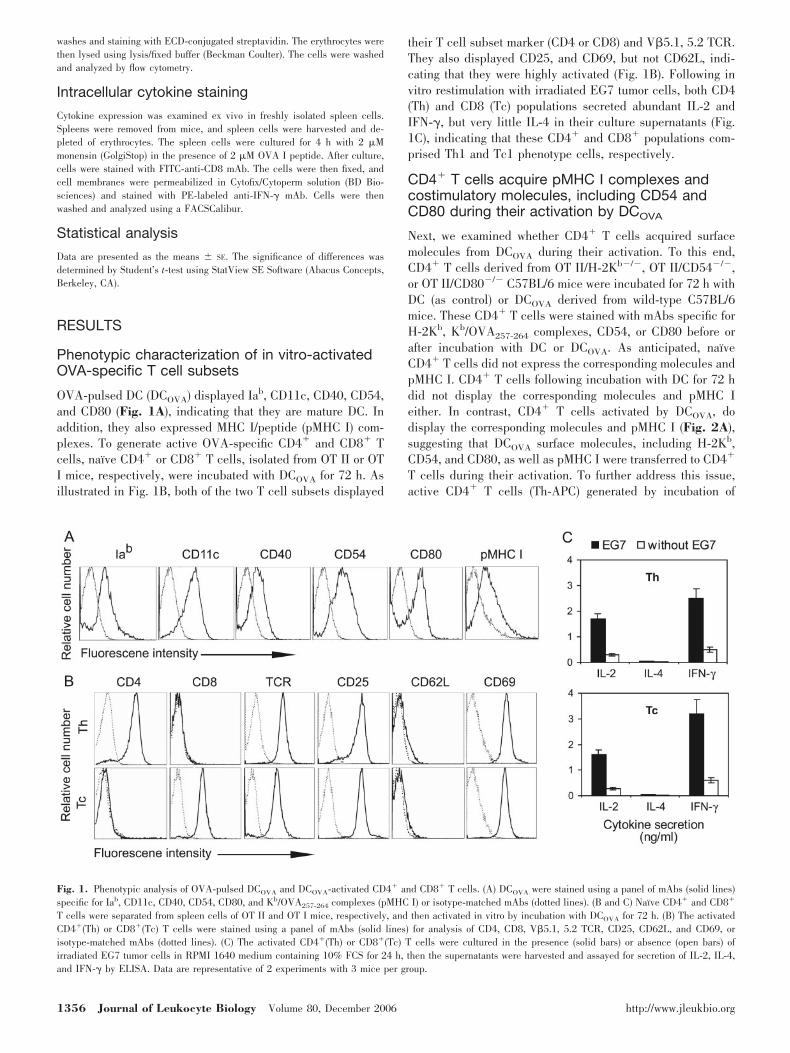
OVA-pulsed DC (DCOVA) displayed Iab, CD11c, CD40, CD54,and CD80 (Fig. 1A), indicating that they are mature DC. Inaddition, they also expressed MHC I/peptide (pMHC I) com-plexes. To generate active OVA-specific CD4� and CD8� Tcells, naı̈ve CD4� or CD8� T cells, isolated from OT II or OTI mice, respectively, were incubated with DCOVA for 72 h. Asillustrated in Fig. 1B, both of the two T cell subsets displayed
their T cell subset marker (CD4 or CD8) and V�5.1, 5.2 TCR.They also displayed CD25, and CD69, but not CD62L, indi-cating that they were highly activated (Fig. 1B). Following invitro restimulation with irradiated EG7 tumor cells, both CD4(Th) and CD8 (Tc) populations secreted abundant IL-2 andIFN-�, but very little IL-4 in their culture supernatants (Fig.1C), indicating that these CD4� and CD8� populations com-prised Th1 and Tc1 phenotype cells, respectively.
CD4� T cells acquire pMHC I complexes andcostimulatory molecules, including CD54 andCD80 during their activation by DCOVA
Next, we examined whether CD4� T cells acquired surfacemolecules from DCOVA during their activation. To this end,CD4� T cells derived from OT II/H-2Kb�/�, OT II/CD54�/�,or OT II/CD80�/� C57BL/6 mice were incubated for 72 h withDC (as control) or DCOVA derived from wild-type C57BL/6mice. These CD4� T cells were stained with mAbs specific forH-2Kb, Kb/OVA257-264 complexes, CD54, or CD80 before orafter incubation with DC or DCOVA. As anticipated, naı̈veCD4� T cells did not express the corresponding molecules andpMHC I. CD4� T cells following incubation with DC for 72 hdid not display the corresponding molecules and pMHC Ieither. In contrast, CD4� T cells activated by DCOVA, dodisplay the corresponding molecules and pMHC I (Fig. 2A),suggesting that DCOVA surface molecules, including H-2Kb,CD54, and CD80, as well as pMHC I were transferred to CD4�
T cells during their activation. To further address this issue,active CD4� T cells (Th-APC) generated by incubation of
Fig. 1. Phenotypic analysis of OVA-pulsed DCOVA and DCOVA-activated CD4� and CD8� T cells. (A) DCOVA were stained using a panel of mAbs (solid lines)specific for Iab, CD11c, CD40, CD54, CD80, and Kb/OVA257-264 complexes (pMHC I) or isotype-matched mAbs (dotted lines). (B and C) Naı̈ve CD4� and CD8�
T cells were separated from spleen cells of OT II and OT I mice, respectively, and then activated in vitro by incubation with DCOVA for 72 h. (B) The activatedCD4�(Th) or CD8�(Tc) T cells were stained using a panel of mAbs (solid lines) for analysis of CD4, CD8, V�5.1, 5.2 TCR, CD25, CD62L, and CD69, orisotype-matched mAbs (dotted lines). (C) The activated CD4�(Th) or CD8�(Tc) T cells were cultured in the presence (solid bars) or absence (open bars) ofirradiated EG7 tumor cells in RPMI 1640 medium containing 10% FCS for 24 h, then the supernatants were harvested and assayed for secretion of IL-2, IL-4,and IFN-� by ELISA. Data are representative of 2 experiments with 3 mice per group.
1356 Journal of Leukocyte Biology Volume 80, December 2006 http://www.jleukbio.org
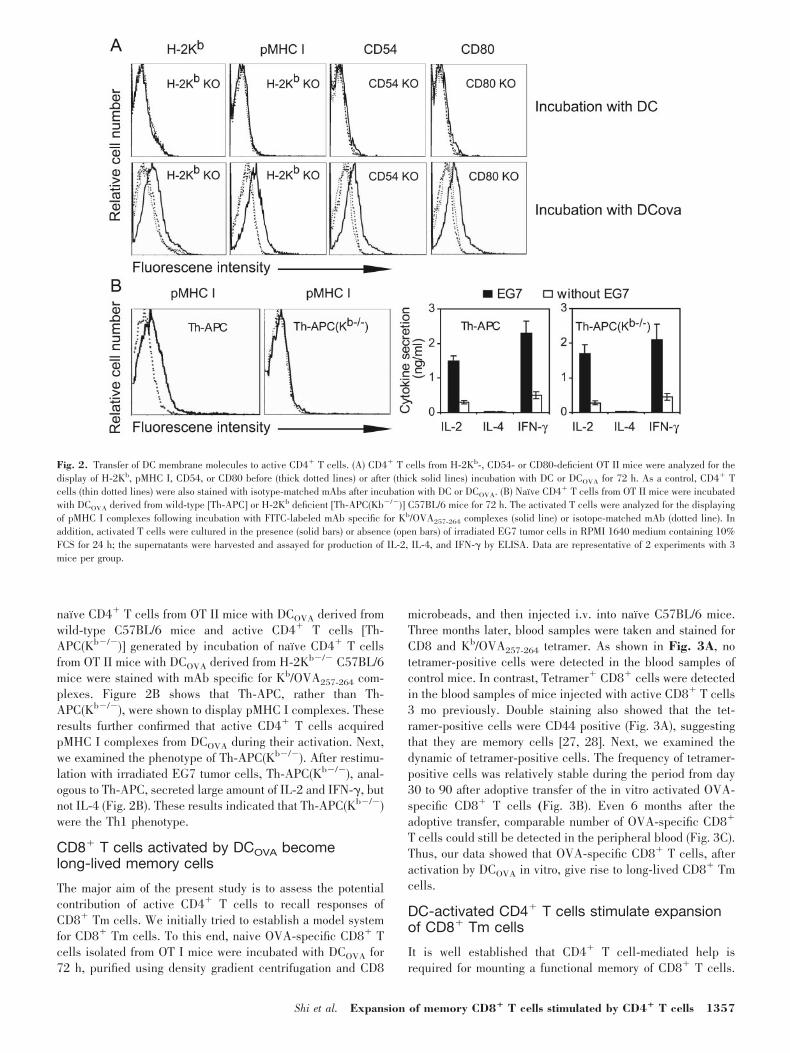
naı̈ve CD4� T cells from OT II mice with DCOVA derived fromwild-type C57BL/6 mice and active CD4� T cells [Th-APC(Kb�/�)] generated by incubation of naı̈ve CD4� T cellsfrom OT II mice with DCOVA derived from H-2Kb�/� C57BL/6mice were stained with mAb specific for Kb/OVA257-264 com-plexes. Figure 2B shows that Th-APC, rather than Th-APC(Kb�/�), were shown to display pMHC I complexes. Theseresults further confirmed that active CD4� T cells acquiredpMHC I complexes from DCOVA during their activation. Next,we examined the phenotype of Th-APC(Kb�/�). After restimu-lation with irradiated EG7 tumor cells, Th-APC(Kb�/�), anal-ogous to Th-APC, secreted large amount of IL-2 and IFN-�, butnot IL-4 (Fig. 2B). These results indicated that Th-APC(Kb�/�)were the Th1 phenotype.
CD8� T cells activated by DCOVA becomelong-lived memory cells
The major aim of the present study is to assess the potentialcontribution of active CD4� T cells to recall responses ofCD8� Tm cells. We initially tried to establish a model systemfor CD8� Tm cells. To this end, naive OVA-specific CD8� Tcells isolated from OT I mice were incubated with DCOVA for72 h, purified using density gradient centrifugation and CD8
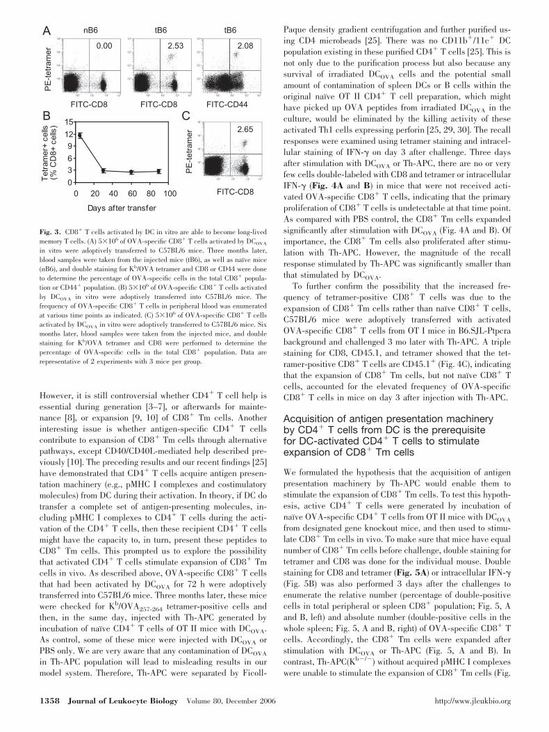
microbeads, and then injected i.v. into naı̈ve C57BL/6 mice.Three months later, blood samples were taken and stained forCD8 and Kb/OVA257-264 tetramer. As shown in Fig. 3A, notetramer-positive cells were detected in the blood samples ofcontrol mice. In contrast, Tetramer� CD8� cells were detectedin the blood samples of mice injected with active CD8� T cells3 mo previously. Double staining also showed that the tet-ramer-positive cells were CD44 positive (Fig. 3A), suggestingthat they are memory cells [27, 28]. Next, we examined thedynamic of tetramer-positive cells. The frequency of tetramer-positive cells was relatively stable during the period from day30 to 90 after adoptive transfer of the in vitro activated OVA-specific CD8� T cells (Fig. 3B). Even 6 months after theadoptive transfer, comparable number of OVA-specific CD8�
T cells could still be detected in the peripheral blood (Fig. 3C).Thus, our data showed that OVA-specific CD8� T cells, afteractivation by DCOVA in vitro, give rise to long-lived CD8� Tmcells.
DC-activated CD4� T cells stimulate expansionof CD8� Tm cells
It is well established that CD4� T cell-mediated help isrequired for mounting a functional memory of CD8� T cells.
Fig. 2. Transfer of DC membrane molecules to active CD4� T cells. (A) CD4� T cells from H-2Kb-, CD54- or CD80-deficient OT II mice were analyzed for thedisplay of H-2Kb, pMHC I, CD54, or CD80 before (thick dotted lines) or after (thick solid lines) incubation with DC or DCOVA for 72 h. As a control, CD4� Tcells (thin dotted lines) were also stained with isotype-matched mAbs after incubation with DC or DCOVA. (B) Naı̈ve CD4� T cells from OT II mice were incubatedwith DCOVA derived from wild-type [Th-APC] or H-2Kb deficient [Th-APC(Kb�/�)] C57BL/6 mice for 72 h. The activated T cells were analyzed for the displayingof pMHC I complexes following incubation with FITC-labeled mAb specific for Kb/OVA257-264 complexes (solid line) or isotope-matched mAb (dotted line). Inaddition, activated T cells were cultured in the presence (solid bars) or absence (open bars) of irradiated EG7 tumor cells in RPMI 1640 medium containing 10%FCS for 24 h; the supernatants were harvested and assayed for production of IL-2, IL-4, and IFN-� by ELISA. Data are representative of 2 experiments with 3mice per group.
Shi et al. Expansion of memory CD8� T cells stimulated by CD4� T cells 1357
However, it is still controversial whether CD4� T cell help isessential during generation [3–7], or afterwards for mainte-nance [8], or expansion [9, 10] of CD8� Tm cells. Anotherinteresting issue is whether antigen-specific CD4� T cellscontribute to expansion of CD8� Tm cells through alternativepathways, except CD40/CD40L-mediated help described pre-viously [10]. The preceding results and our recent findings [25]have demonstrated that CD4� T cells acquire antigen presen-tation machinery (e.g., pMHC I complexes and costimulatorymolecules) from DC during their activation. In theory, if DC dotransfer a complete set of antigen-presenting molecules, in-cluding pMHC I complexes to CD4� T cells during the acti-vation of the CD4� T cells, then these recipient CD4� T cellsmight have the capacity to, in turn, present these peptides toCD8� Tm cells. This prompted us to explore the possibilitythat activated CD4� T cells stimulate expansion of CD8� Tmcells in vivo. As described above, OVA-specific CD8� T cellsthat had been activated by DCOVA for 72 h were adoptivelytransferred into C57BL/6 mice. Three months later, these micewere checked for Kb/OVA257-264 tetramer-positive cells andthen, in the same day, injected with Th-APC generated byincubation of naı̈ve CD4� T cells of OT II mice with DCOVA.As control, some of these mice were injected with DCOVA orPBS only. We are very aware that any contamination of DCOVA
in Th-APC population will lead to misleading results in ourmodel system. Therefore, Th-APC were separated by Ficoll-
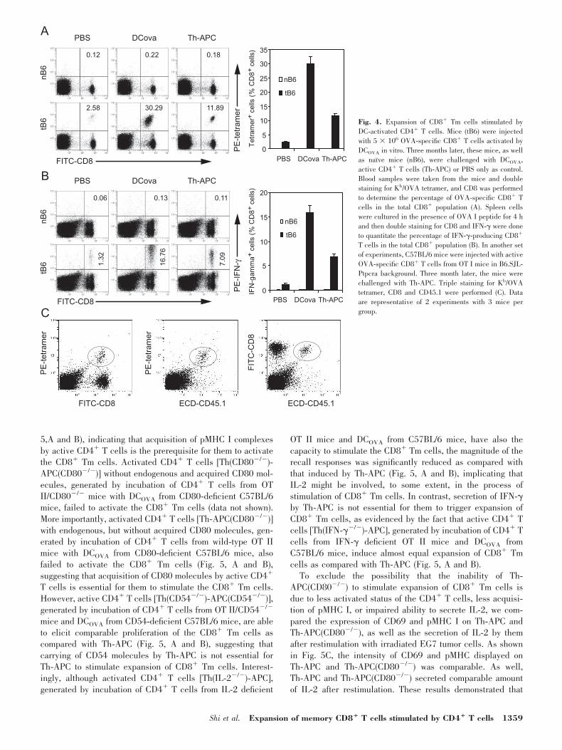
Paque density gradient centrifugation and further purified us-ing CD4 microbeads [25]. There was no CD11b�/11c� DCpopulation existing in these purified CD4� T cells [25]. This isnot only due to the purification process but also because anysurvival of irradiated DCOVA cells and the potential smallamount of contamination of spleen DCs or B cells within theoriginal naı̈ve OT II CD4� T cell preparation, which mighthave picked up OVA peptides from irradiated DCOVA in theculture, would be eliminated by the killing activity of theseactivated Th1 cells expressing perforin [25, 29, 30]. The recallresponses were examined using tetramer staining and intracel-lular staining of IFN-� on day 3 after challenge. Three daysafter stimulation with DCOVA or Th-APC, there are no or veryfew cells double-labeled with CD8 and tetramer or intracellularIFN-� (Fig. 4A and B) in mice that were not received acti-vated OVA-specific CD8� T cells, indicating that the primaryproliferation of CD8� T cells is undetectable at that time point.As compared with PBS control, the CD8� Tm cells expandedsignificantly after stimulation with DCOVA (Fig. 4A and B). Ofimportance, the CD8� Tm cells also proliferated after stimu-lation with Th-APC. However, the magnitude of the recallresponse stimulated by Th-APC was significantly smaller thanthat stimulated by DCOVA.
To further confirm the possibility that the increased fre-quency of tetramer-positive CD8� T cells was due to theexpansion of CD8� Tm cells rather than naı̈ve CD8� T cells,C57BL/6 mice were adoptively transferred with activatedOVA-specific CD8� T cells from OT I mice in B6.SJL-Ptpcrabackground and challenged 3 mo later with Th-APC. A triplestaining for CD8, CD45.1, and tetramer showed that the tet-ramer-positive CD8� T cells are CD45.1� (Fig. 4C), indicatingthat the expansion of CD8� Tm cells, but not naı̈ve CD8� Tcells, accounted for the elevated frequency of OVA-specificCD8� T cells in mice on day 3 after injection with Th-APC.
Acquisition of antigen presentation machineryby CD4� T cells from DC is the prerequisitefor DC-activated CD4� T cells to stimulateexpansion of CD8� Tm cells
We formulated the hypothesis that the acquisition of antigenpresentation machinery by Th-APC would enable them tostimulate the expansion of CD8� Tm cells. To test this hypoth-esis, active CD4� T cells were generated by incubation ofnaı̈ve OVA-specific CD4� T cells from OT II mice with DCOVA
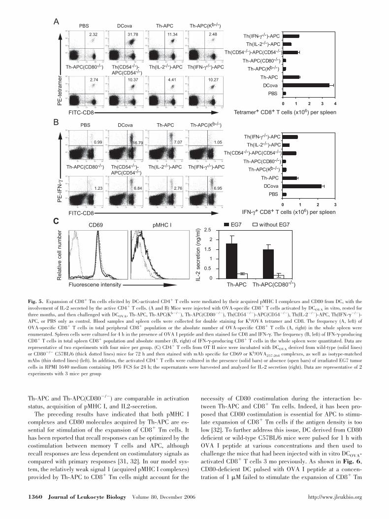
from designated gene knockout mice, and then used to stimu-late CD8� Tm cells in vivo. To make sure that mice have equalnumber of CD8� Tm cells before challenge, double staining fortetramer and CD8 was done for the individual mouse. Doublestaining for CD8 and tetramer (Fig. 5A) or intracellular IFN-�(Fig. 5B) was also performed 3 days after the challenges toenumerate the relative number (percentage of double-positivecells in total peripheral or spleen CD8� population; Fig. 5, Aand B, left) and absolute number (double-positive cells in thewhole spleen; Fig. 5, A and B, right) of OVA-specific CD8� Tcells. Accordingly, the CD8� Tm cells were expanded afterstimulation with DCOVA or Th-APC (Fig. 5, A and B). Incontrast, Th-APC(Kb�/�) without acquired pMHC I complexeswere unable to stimulate the expansion of CD8� Tm cells (Fig.
Fig. 3. CD8� T cells activated by DC in vitro are able to become long-livedmemory T cells. (A) 5�106 of OVA-specific CD8� T cells activated by DCOVA
in vitro were adoptively transferred to C57BL/6 mice. Three months later,blood samples were taken from the injected mice (tB6), as well as naı̈ve mice(nB6), and double staining for Kb/OVA tetramer and CD8 or CD44 were doneto determine the percentage of OVA-specific cells in the total CD8� popula-tion or CD44� population. (B) 5�106 of OVA-specific CD8� T cells activatedby DCOVA in vitro were adoptively transferred into C57BL/6 mice. Thefrequency of OVA-specific CD8� T cells in peripheral blood was enumeratedat various time points as indicated. (C) 5�106 of OVA-specific CD8� T cellsactivated by DCOVA in vitro were adoptively transferred to C57BL/6 mice. Sixmonths later, blood samples were taken from the injected mice, and doublestaining for Kb/OVA tetramer and CD8 were performed to determine thepercentage of OVA-specific cells in the total CD8� population. Data arerepresentative of 2 experiments with 3 mice per group.
1358 Journal of Leukocyte Biology Volume 80, December 2006 http://www.jleukbio.org
5,A and B), indicating that acquisition of pMHC I complexesby active CD4� T cells is the prerequisite for them to activatethe CD8� Tm cells. Activated CD4� T cells [Th(CD80�/�)-APC(CD80�/�)] without endogenous and acquired CD80 mol-ecules, generated by incubation of CD4� T cells from OTII/CD80�/� mice with DCOVA from CD80-deficient C57BL/6mice, failed to activate the CD8� Tm cells (data not shown).More importantly, activated CD4� T cells [Th-APC(CD80�/�)]with endogenous, but without acquired CD80 molecules, gen-erated by incubation of CD4� T cells from wild-type OT IImice with DCOVA from CD80-deficient C57BL/6 mice, alsofailed to activate the CD8� Tm cells (Fig. 5, A and B),suggesting that acquisition of CD80 molecules by active CD4�
T cells is essential for them to stimulate the CD8� Tm cells.However, active CD4� T cells [Th(CD54�/�)-APC(CD54�/�)],generated by incubation of CD4� T cells from OT II/CD54�/�
mice and DCOVA from CD54-deficient C57BL/6 mice, are ableto elicit comparable proliferation of the CD8� Tm cells ascompared with Th-APC (Fig. 5, A and B), suggesting thatcarrying of CD54 molecules by Th-APC is not essential forTh-APC to stimulate expansion of CD8� Tm cells. Interest-ingly, although activated CD4� T cells [Th(IL-2�/�)-APC],generated by incubation of CD4� T cells from IL-2 deficient
OT II mice and DCOVA from C57BL/6 mice, have also thecapacity to stimulate the CD8� Tm cells, the magnitude of therecall responses was significantly reduced as compared withthat induced by Th-APC (Fig. 5, A and B), implicating thatIL-2 might be involved, to some extent, in the process ofstimulation of CD8� Tm cells. In contrast, secretion of IFN-�by Th-APC is not essential for them to trigger expansion ofCD8� Tm cells, as evidenced by the fact that active CD4� Tcells [Th(IFN-��/�)-APC], generated by incubation of CD4� Tcells from IFN-� deficient OT II mice and DCOVA fromC57BL/6 mice, induce almost equal expansion of CD8� Tmcells as compared with Th-APC (Fig. 5, A and B).
To exclude the possibility that the inability of Th-APC(CD80�/�) to stimulate expansion of CD8� Tm cells isdue to less activated status of the CD4� T cells, less acquisi-tion of pMHC I, or impaired ability to secrete IL-2, we com-pared the expression of CD69 and pMHC I on Th-APC andTh-APC(CD80�/�), as well as the secretion of IL-2 by themafter restimulation with irradiated EG7 tumor cells. As shownin Fig. 5C, the intensity of CD69 and pMHC displayed onTh-APC and Th-APC(CD80�/�) was comparable. As well,Th-APC and Th-APC(CD80�/�) secreted comparable amountof IL-2 after restimulation. These results demonstrated that
Fig. 4. Expansion of CD8� Tm cells stimulated byDC-activated CD4� T cells. Mice (tB6) were injectedwith 5 � 106 OVA-specific CD8� T cells activated byDCOVA in vitro. Three months later, these mice, as wellas naı̈ve mice (nB6), were challenged with DCOVA,active CD4� T cells (Th-APC) or PBS only as control.Blood samples were taken from the mice and doublestaining for Kb/OVA tetramer, and CD8 was performedto determine the percentage of OVA-specific CD8� Tcells in the total CD8� population (A). Spleen cellswere cultured in the presence of OVA I peptide for 4 hand then double staining for CD8 and IFN-� were doneto quantitate the percentage of IFN-�-producing CD8�
T cells in the total CD8� population (B). In another setof experiments, C57BL/6 mice were injected with activeOVA-specific CD8� T cells from OT I mice in B6.SJL-Ptpcra background. Three month later, the mice werechallenged with Th-APC. Triple staining for Kb/OVAtetramer, CD8 and CD45.1 were performed (C). Dataare representative of 2 experiments with 3 mice pergroup.
Shi et al. Expansion of memory CD8� T cells stimulated by CD4� T cells 1359
Th-APC and Th-APC(CD80�/�) are comparable in activationstatus, acquisition of pMHC I, and IL2-secretion.
The preceding results have indicated that both pMHC Icomplexes and CD80 molecules acquired by Th-APC are es-sential for stimulation of the expansion of CD8� Tm cells. Ithas been reported that recall responses can be optimized by thecostimulation between memory T cells and APC, althoughrecall responses are less dependent on costimulatory signals ascompared with primary responses [31, 32]. In our model sys-tem, the relatively weak signal 1 (acquired pMHC I complexes)provided by Th-APC to CD8� Tm cells might account for the
necessity of CD80 costimulation during the interaction be-tween Th-APC and CD8� Tm cells. Indeed, it has been pro-posed that CD80 costimulation is essential for APC to stimu-late expansion of CD8� Tm cells if the antigen density is toolow [32]. To further address this issue, DC derived from CD80deficient or wild-type C57BL/6 mice were pulsed for 1 h withOVA I peptide at various concentrations and then used tochallenge the mice that had been injected with in vitro DCOVA-activated CD8� T cells 3 mo previously. As shown in Fig. 6,CD80-deficient DC pulsed with OVA I peptide at a concen-tration of 1 �M failed to stimulate the expansion of CD8� Tm
Fig. 5. Expansion of CD8� Tm cells elicited by DC-activated CD4� T cells were mediated by their acquired pMHC I complexes and CD80 from DC, with theinvolvement of IL-2 secreted by the active CD4� T cells. (A and B) Mice were injected with OVA-specific CD8� T cells activated by DCOVA in vitro, rested forthree months, and then challenged with DCOVA, Th-APC, Th-APC(Kb�/�), Th-APC(CD80�/�), Th(CD54�/�)-APC(CD54�/�), Th(IL-2�/�)-APC, Th(IFN-��/�)-APC, or PBS only as control. Blood samples and spleen cells were collected for double staining for Kb/OVA tetramer and CD8. The frequency (A, left) ofOVA-specific CD8� T cells in total peripheral CD8� population or the absolute number of OVA-specific CD8� T cells (A, right) in the whole spleen wereenumerated. Spleen cells were cultured for 4 h in the presence of OVA I peptide and then stained for CD8 and IFN-�. The frequency (B, left) of IFN-�-producingCD8� T cells in total spleen CD8� population and absolute number (B, right) of IFN-�-producing CD8� T cells in the whole spleen were quantitated. Data arerepresentative of two experiments with four mice per group. (C) CD4� T cells from OT II mice were incubated with DCOVA derived from wild-type (solid lines)or CD80�/� C57BL/6 (thick dotted lines) mice for 72 h and then stained with mAb specific for CD69 or Kb/OVA257-264 complexes, as well as isotype-matchedmAbs (thin dotted lines) (left). In addition, the activated CD4� T cells were cultured in the presence (solid bars) or absence (open bars) of irradiated EG7 tumorcells in RPMI 1640 medium containing 10% FCS for 24 h; the supernatants were harvested and analyzed for IL-2 secretion (right). Data are representative of 2experiments with 3 mice per group
1360 Journal of Leukocyte Biology Volume 80, December 2006 http://www.jleukbio.org

cells. In contrast, pulsed with OVA I at this concentration, DCderived from wild-type mice do have the capacity, to someextent, to elicit the expansion of CD8� Tm cells. Althoughpulsing with higher concentrations enable CD80-deficient DCto activate CD8� Tm cells, the magnitude of recall responses[both relative number (Fig. 6A) and absolute number (Fig. 6B)of OVA-specific CD8� T cells] stimulated by CD80-deficientDC is significantly smaller as compared with that triggered bywild-type DC (Fig. 6). These results are consistent with previ-ous reports [32] and support our hypothesis.
DISCUSSION
It has been previously shown that Th-APC acquire antigen-presenting machinery (i.e., MHC II-peptides complexes andcostimulatory molecules) from APCs during their activation[17, 20–23, 33]. In our previous study [25] and our presentstudy, we demonstrated that pMHC I complexes are transferredfrom APCs to Th-APC as well. In addition, CD4� T cells,analogous to professional APC, are able to generate endoge-nous pMHC I complexes by processing exogenously acquiredantigen [14]. The Th-APC that carries antigen-presenting ma-chinery may potentially activate other naı̈ve or memory T cellsin an antigen-specific manner. Indeed, it has been demon-strated that Th-APC or active CD8� T cells are able to serveas APC to stimulate other naı̈ve T cells [14, 17, 21–23]. In thepresent study, we focused on addressing whether expansion ofCD8� Tm cells can be elicited by Th-APC-carrying antigenpresenting machinery. Our data indicated that Th-APC, afteracquisition of antigen-presenting machinery, do have the abil-ity to stimulate recall responses of CD8� Tm cells.
The underlying mechanisms responsible for stimulation ofCD8� Tm cells by Th-APC have been elucidated in the currentstudy. We found that Th-APC(Kb�/�) failed to activate theCD8� Tm cells. As Th-APC(Kb�/�) do not hold pMHC Icomplexes, which are considered as the first signal for T cellactivation [34, 35], they cannot serve as APC to stimulateCD8� Tm cells. Thus, the acquisition of pMHC I complexes byCD4� T cells is essential for their function as APC to stimulateCD8� Tm cells. The inability to stimulate recall responses ofCD8� Tm cells by Th-APC(CD80�/�) suggested that acquisi-tion of CD80 molecules from DC is essential for Th-APC toactivate CD8� Tm cells. This was further confirmed by theobservation that blocking of CD80 on Th-APC using CTLA-4/Ig led to the inability of Th-APC to stimulate the recallresponses of CD8� Tm cells (data not shown). It has beenreported that costimulation provided by CD80 is required forrecall responses of memory cells in some circumstances [36],particularly in the case of lower antigen concentrations [32],although it is well known that recall responses are less depen-dent on costimulation as compared with primary responses [31,32]. In the current study, we showed that CD80 costimulationis crucial for stimulating CD8� Tm cells in the case of lowerconcentrations of antigens, supporting previous reports [31, 32,36]. One of the explanations for the necessity of CD80 duringthe activation of CD8� Tm cells stimulated by Th-APC is thatthe first signal delivered by acquired pMHC I complexes isrelatively weak, compared with DC. Thus, it is conceivable thatTh-APC need both signal 1 and signal 2 (e.g., CD80 costimu-lation) to activate CD8� Tm cells.
Our data presented in the current study, which shows thatTh-APC secrete a great amount of IL-2 and IFN-�, raise thequestion regarding whether IL-2 and IFN-� secreted by Th-APC are involved in the stimulation of expansion of CD8� Tmcells, as a direct delivery of immunity-potentiating cytokines toCD8� T cells by Th-APC via an immunological synapse wouldcertainly be an efficient way for facilitating CD8� T cell expan-sion. To this end, we assessed the capacity of Th(IL-2�/�)-APCand Th(IFN-��/�)-APC to activate CD8� Tm cells in the currentstudy. It is of interest to note that IL-2, but not IFN-�, secreted byTh-APC is also involved, to some extent, in the expansion ofCD8� Tm cells. It was reported that, during T cell activation, thesignal 2 mediated by costimulation can lead to IL-2 production[34, 37, 38]. In particular, the development of primary CD8responses has been proven to be supported by CD4� T cells viaproducing cytokines, notably IL-2 [39, 40]. In our model system,although not an absolute necessity, IL-2 secretion by Th-APCappeared to optimize the expansion of CD8� Tm cells stimulatedby Th-APC.
The interplay among APC, CD4� T cells, and CD8� T cellsleading to the activation and proliferation of CD8� naı̈ve orCD8� Tm cells has been extensively studied, and the under-standing of how these cells interact will assist the developmentof future vaccination strategies. Mounting evidences have dem-onstrated that CD4� T cells are critically involved in thedevelopment of CD8� Tm cells [2–10, 14]. Once CD8� Tmcells are generated, CD4� T cell are not essential for expan-sion of the CD8� Tm cells [5, 7]. Nevertheless, it was alsoreported that antigen-specific CD4� T cells are required toactivate CD8� Tm cells via a yet still unknown mechanism(s)
Fig. 6. CD80 costimulation is required for DC to stimulate expansion ofCD8� Tm cells when antigen concentration is low. OVA-specific CD8� T cells(5�106) activated in vitro by DCOVA were adoptively transferred to C57BL/6mice. Three months later, the mice were challenged with DC derived fromwild-type or CD80�/� C57BL/6 mice. The DC used for challenge were pulsedfor 1 h with OVA I peptide at various concentrations, as indicated. Thefrequency (A) and absolute number (B) of OVA-specific CD8� T cells inperipheral blood or spleen were enumerated using double staining for Kb/OVAtetramer and CD8. Data are presented as the means � SE (Student t-test, *P� 0.05). The results presented are representative of 2 separate experimentswith 3 mice per group.
Shi et al. Expansion of memory CD8� T cells stimulated by CD4� T cells 1361
[9]. A recent report also showed that the magnitude of recallresponses of CD8� Tm cells was diminished in the absence ofCD4� T cells [10], indicating that CD4� T cells might, to someextent, contribute to the expansion of CD8� Tm cells in somemodel systems. Of interest, CD40L is also required for stimu-lation of expansion of CD8� Tm cells [10], implying that CD4�
T cells might be involved in recall responses of CD8� Tm cellsthrough CD40/CD40L-mediated “licensing” of APC or evenCD40-CD40L interaction between CD4� and CD8� Tm cells[2]. Thus, it is highly likely that one of the mechanismsunderlying the importance of CD4� T cells for recall responsesof CD8� Tm cells is that CD4� T cells provide help to APC oreven directly to CD8� T cells through CD40L signaling. Ourfindings that active CD4� T cells have the ability to stimulatethe expansion of CD8� Tm cells through acquired pMHC Icomplexes and costimulatory molecules might implicate an-other potential mechanism behind the importance of CD4� Tcells for recall responses of CD8� Tm cells. However, it is ofimportance to realize that the frequency of recall CD8� Tmcells stimulated by active CD4� T cells was much smaller thanthat stimulated by DC pulsed with protein, raising the possi-bility that the recall responses of CD8� Tm cells induced byAPCs overwrite those induced by active CD4� T cells inphysiological situation, which might explain the fact that CD4deficiency does not significantly affect expansion of CD8� Tmcells described previously [5, 7, 41]
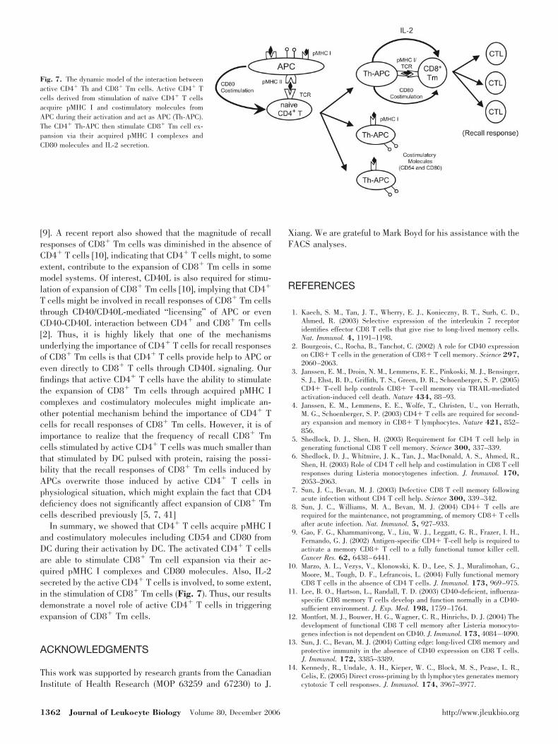
In summary, we showed that CD4� T cells acquire pMHC Iand costimulatory molecules including CD54 and CD80 fromDC during their activation by DC. The activated CD4� T cellsare able to stimulate CD8� Tm cell expansion via their ac-quired pMHC I complexes and CD80 molecules. Also, IL-2secreted by the active CD4� T cells is involved, to some extent,in the stimulation of CD8� Tm cells (Fig. 7). Thus, our resultsdemonstrate a novel role of active CD4� T cells in triggeringexpansion of CD8� Tm cells.
ACKNOWLEDGMENTS
This work was supported by research grants from the CanadianInstitute of Health Research (MOP 63259 and 67230) to J.
Xiang. We are grateful to Mark Boyd for his assistance with theFACS analyses.
REFERENCES
1. Kaech, S. M., Tan, J. T., Wherry, E. J., Konieczny, B. T., Surh, C. D.,Ahmed, R. (2003) Selective expression of the interleukin 7 receptoridentifies effector CD8 T cells that give rise to long-lived memory cells.Nat. Immunol. 4, 1191–1198.
2. Bourgeois, C., Rocha, B., Tanchot, C. (2002) A role for CD40 expressionon CD8� T cells in the generation of CD8� T cell memory. Science 297,2060–2063.
3. Janssen, E. M., Droin, N. M., Lemmens, E. E., Pinkoski, M. J., Bensinger,S. J., Ehst, B. D., Griffith, T. S., Green, D. R., Schoenberger, S. P. (2005)CD4� T-cell help controls CD8� T-cell memory via TRAIL-mediatedactivation-induced cell death. Nature 434, 88–93.
4. Janssen, E. M., Lemmens, E. E., Wolfe, T., Christen, U., von Herrath,M. G., Schoenberger, S. P. (2003) CD4� T cells are required for second-ary expansion and memory in CD8� T lymphocytes. Nature 421, 852–856.
5. Shedlock, D. J., Shen, H. (2003) Requirement for CD4 T cell help ingenerating functional CD8 T cell memory. Science 300, 337–339.
6. Shedlock, D. J., Whitmire, J. K., Tan, J., MacDonald, A. S., Ahmed, R.,Shen, H. (2003) Role of CD4 T cell help and costimulation in CD8 T cellresponses during Listeria monocytogenes infection. J. Immunol. 170,2053–2063.
7. Sun, J. C., Bevan, M. J. (2003) Defective CD8 T cell memory followingacute infection without CD4 T cell help. Science 300, 339–342.
8. Sun, J. C., Williams, M. A., Bevan, M. J. (2004) CD4� T cells arerequired for the maintenance, not programming, of memory CD8� T cellsafter acute infection. Nat. Immunol. 5, 927–933.
9. Gao, F. G., Khammanivong, V., Liu, W. J., Leggatt, G. R., Frazer, I. H.,Fernando, G. J. (2002) Antigen-specific CD4� T-cell help is required toactivate a memory CD8� T cell to a fully functional tumor killer cell.Cancer Res. 62, 6438–6441.
10. Marzo, A. L., Vezys, V., Klonowski, K. D., Lee, S. J., Muralimohan, G.,Moore, M., Tough, D. F., Lefrancois, L. (2004) Fully functional memoryCD8 T cells in the absence of CD4 T cells. J. Immunol. 173, 969–975.
11. Lee, B. O., Hartson, L., Randall, T. D. (2003) CD40-deficient, influenza-specific CD8 memory T cells develop and function normally in a CD40-sufficient environment. J. Exp. Med. 198, 1759–1764.
12. Montfort, M. J., Bouwer, H. G., Wagner, C. R., Hinrichs, D. J. (2004) Thedevelopment of functional CD8 T cell memory after Listeria monocyto-genes infection is not dependent on CD40. J. Immunol. 173, 4084–4090.
13. Sun, J. C., Bevan, M. J. (2004) Cutting edge: long-lived CD8 memory andprotective immunity in the absence of CD40 expression on CD8 T cells.J. Immunol. 172, 3385–3389.
14. Kennedy, R., Undale, A. H., Kieper, W. C., Block, M. S., Pease, L. R.,Celis, E. (2005) Direct cross-priming by th lymphocytes generates memorycytotoxic T cell responses. J. Immunol. 174, 3967–3977.
Fig. 7. The dynamic model of the interaction betweenactive CD4� Th and CD8� Tm cells. Active CD4� Tcells derived from stimulation of naı̈ve CD4� T cellsacquire pMHC I and costimulatory molecules fromAPC during their activation and act as APC (Th-APC).The CD4� Th-APC then stimulate CD8� Tm cell ex-pansion via their acquired pMHC I complexes andCD80 molecules and IL-2 secretion.
1362 Journal of Leukocyte Biology Volume 80, December 2006 http://www.jleukbio.org
15. Grakoui, A., Bromley, S. K., Sumen, C., Davis, M. M., Shaw, A. S., Allen,P. M., Dustin, M. L. (1999) The immunological synapse: a molecularmachine controlling T cell activation. Science 285, 221–227.
16. Viola, A., Schroeder, S., Sakakibara, Y., Lanzavecchia, A. (1999) Tlymphocyte costimulation mediated by reorganization of membrane mi-crodomains. Science 283, 680–682.
17. Huang, J. F., Yang, Y., Sepulveda, H., Shi, W., Hwang, I., Peterson, P. A.,Jackson, M. R., Sprent, J., Cai, Z. (1999) TCR-Mediated internalization ofpeptide-MHC complexes acquired by T cells. Science 286, 952–954.
18. Hwang, I., Huang, J. F., Kishimoto, H., Brunmark, A., Peterson, P. A.,Jackson, M. R., Surh, C. D., Cai, Z., Sprent, J. (2000) T cells can use eitherT cell receptor or CD28 receptors to absorb and internalize cell surfacemolecules derived from antigen-presenting cells. J. Exp. Med. 191,1137–1148.
19. Baba, E., Takahashi, Y., Lichtenfeld, J., Tanaka, R., Yoshida, A., Sug-amura, K., Yamamoto, N., Tanaka, Y. (2001) Functional CD4 T cells afterintercellular molecular transfer of 0X40 ligand. J. Immunol. 167, 875–883.
20. Patel, D. M., Arnold, P. Y., White, G. A., Nardella, J. P., Mannie, M. D.(1999) Class II MHC/peptide complexes are released from APC and areacquired by T cell responders during specific antigen recognition. J. Im-munol. 163, 5201–5210.
21. Sabzevari, H., Kantor, J., Jaigirdar, A., Tagaya, Y., Naramura, M., Hodge,J., Bernon, J., Schlom, J. (2001) Acquisition of CD80 (B7–1) by T cells.J. Immunol. 166, 2505–2513.
22. Tatari-Calderone, Z., Semnani, R. T., Nutman, T. B., Schlom, J., Sabze-vari, H. (2002) Acquisition of CD80 by human T cells at early stages ofactivation: functional involvement of CD80 acquisition in T cell to T cellinteraction. J. Immunol. 169, 6162–6169.
23. Tsang, J. Y., Chai, J. G., Lechler, R. (2003) Antigen presentation bymouse CD4� T cells involving acquired MHC class II:peptide complexes:another mechanism to limit clonal expansion? Blood 101, 2704–2710.
24. Hwang, I., Shen, X., Sprent, J. (2003) Direct stimulation of naive T cellsby membrane vesicles from antigen-presenting cells: distinct roles forCD54 and B7 molecules. Proc. Natl. Acad. Sci. USA 100, 6670–6675.
25. Xiang, J., Huang, H., Liu, Y. (2005) A new dynamic model of CD8� Teffector cell responses via CD4� T helper-antigen-presenting cells. J. Im-munol. 174, 7497–7505.
26. Porgador, A., Yewdell, J. W., Deng, Y., Bennink, J. R., Germain, R. N.(1997) Localization, quantitation, and in situ detection of specific peptide-MHC class I complexes using a monoclonal antibody. Immunity 6, 715–726.
27. Budd, R. C., Cerottini, J. C., Horvath, C., Bron, C., Pedrazzini, T., Howe,R. C., MacDonald, H. R. (1987) Distinction of virgin and memory T
lymphocytes. Stable acquisition of the Pgp-1 glycoprotein concomitantwith antigenic stimulation. J. Immunol. 138, 3120–3129.
28. MacDonald, H. R., Budd, R. C., Cerottini, J. C. (1990) Pgp-1 (Ly 24) asa marker of murine memory T lymphocytes. Curr. Top. Microbiol. Immu-nol. 159, 97–109.
29. Huang, H., Li, F., Gordon, J. R., Xiang, J. (2002) Synergistic enhancementof antitumor immunity with adoptively transferred tumor-specific CD4�and CD8� T cells and intratumoral lymphotactin transgene expression.Cancer Res. 62, 2043–2051.
30. Li, Y., Wang, M. N., Li, H., King, K. D., Bassi, R., Sun, H., Santiago, A.,Hooper, A. T., Bohlen, P., Hicklin, D. J. (2002) Active immunizationagainst the vascular endothelial growth factor receptor flk1 inhibits tumorangiogenesis and metastasis. J. Exp. Med. 195, 1575–1584.
31. Croft, M., Bradley, L. M., Swain, S. L. (1994) Naive versus memory CD4T cell response to antigen. Memory cells are less dependent on accessorycell costimulation and can respond to many antigen-presenting cell typesincluding resting B cells. J. Immunol. 152, 2675–2685.
32. London, C. A., Lodge, M. P., Abbas, A. K. (2000) Functional responsesand costimulator dependence of memory CD4� T cells. J. Immunol. 164,265–272.
33. Wetzel, S. A., McKeithan, T. W., Parker, D. C. (2005) Peptide-specificintercellular transfer of MHC class II to CD4� T cells directly from theimmunological synapse upon cellular dissociation. J. Immunol. 174,80–89.
34. Lenschow, D. J., Walunas, T. L., Bluestone, J. A. (1996) CD28/B7 systemof T cell costimulation. Annu. Rev. Immunol. 14, 233–258.
35. Bretscher, P., Cohn, M. (1970) A theory of self-nonself discrimination.Science 169, 1042–1049.
36. Liu, Y., Wenger, R. H., Zhao, M., Nielsen, P. J. (1997) Distinct costimu-latory molecules are required for the induction of effector and memorycytotoxic T lymphocytes. J. Exp. Med. 185, 251–262.
37. Jenkins, M. K., Johnson, J. G. (1993) Molecules involved in T-cellcostimulation. Curr. Opin. Immunol. 5, 361–367.
38. Janeway, C. A., Jr., Bottomly, K. (1994) Signals and signs for lymphocyteresponses. Cell 76, 275–285.
39. Tham, E. L., Shrikant, P., Mescher, M. F. (2002) Activation-inducednonresponsiveness: a Th-dependent regulatory checkpoint in the CTLresponse. J. Immunol. 168, 1190–1197.
40. Lu, Z., Yuan, L., Zhou, X., Sotomayor, E., Levitsky, H. I., Pardoll, D. M.(2000) CD40-independent pathways of T cell help for priming of CD8(�)cytotoxic T lymphocytes. J. Exp. Med. 191, 541–550.
41. Shi, M., Xiang, J. (2006) CD4� T cell-independent maintenance andexpansion of memory CD8� T cells derived from in vitro dendritic cellactivation. Int. Immunol. 18, 887–895.
Shi et al. Expansion of memory CD8� T cells stimulated by CD4� T cells 1363