t cell stimulator cells, an efficient and versatile cellular system to assess the role of...
TRANSCRIPT
T cell stimulator cells, an efficient and versatile cellular systemto assess the role of costimulatory ligands in the activation ofhuman T cells
Judith Leitnera, Werner Kuscheia, Katharina Grabmeier-Pfistershammerb, RamonaWoitekc, Ernst Kriehuberb, Otto Majdica, Gerhard Zlabingera, Winfried F. Pickla, and PeterSteinbergera,⁎aInstitute of Immunology, Center for Pathophysiology, Infectiology and Immunology, MedicalUniversity of Vienna, Vienna, AustriabDepartment of Dermatology, Division of Immunology, Allergy and Infectious Diseases, MedicalUniversity of Vienna, Vienna, AustriacDepartment of Radiology at the General Hospital of the Medical University of Vienna, Vienna,Austria
AbstractIt is well established that full activation of T cells requires the interaction of the TCR complexwith the peptide–MHC complex (Signal 1) and additional signals (Signal 2). These second signalsare generated by the interaction of costimulatory ligands expressed on antigen presenting cellswith activating receptors on T cells. In addition, T cell responses are negatively regulated byinhibitory costimulatory pathways. Since professional antigen presenting cells (APC) harbour aplethora of stimulating and inhibitory surface molecules, the contribution of individualcostimulatory molecules is difficult to assess on these cells. We have developed a system ofstimulator cells that can give signal 1 to human T cells via a membrane bound anti-CD3 antibodyfragment. By expressing human costimulatory ligands on these cells, their role in T cell activationprocesses can readily be analyzed. We demonstrate that T cell stimulator cells are excellent toolsto study various aspects of human T cell costimulation, including the effects ofimmunomodulatory drugs or how costimulatory signals contribute to the in vitro expansion of Tcells. T cell stimulator cells are especially suited for the functional evaluation of ligands that areimplicated in costimulatory processes. In this study we have evaluated the role of the CD2 familymember CD150 (SLAM) and the TNF family member TL1A (TNFSF15) in the activation ofhuman T cells. Whereas our results do not point to a significant role of CD150 in T cell activationwe found TL1A to potently costimulate human T cells. Taken together our results demonstratethat T cell stimulator cells are excellent tools to study various aspects of costimulatory processes.
© 2010 Elsevier B.V.⁎Corresponding author. Institute of Immunology, Center for Pathophysiology, Infectiology and Immunology, Medical University ofVienna, Borschkegasse 8a, 1090 Vienna, Austria. Tel.: +43 1 4277 64941; fax: +43 1 4277 [email protected] document was posted here by permission of the publisher. At the time of deposit, it included all changes made during peerreview, copyediting, and publishing. The U.S. National Library of Medicine is responsible for all links within the document and forincorporating any publisher-supplied amendments or retractions issued subsequently. The published journal article, guaranteed to besuch by Elsevier, is available for free, on ScienceDirect.
Sponsored document fromJournal of ImmunologicalMethods
Published as: J Immunol Methods. 2010 October 31; 362(1-2): 131–141.
Sponsored Docum
ent Sponsored D
ocument
Sponsored Docum
ent
KeywordsT cell activation; Costimulation; TL1A; CD150
1 IntroductionThe two signal hypothesis of lymphocyte activation proposes that T cells that receive Signal1 via their T cell receptor (TCR) complex depend on concomitant triggering ofcostimulatory receptors to achieve full activation (Greenwald et al., 2005; Watts, 2005). Tcell activation is also modulated by inhibitory costimulatory receptors that are able toattenuate TCR-signals. By acting as potent regulators of host-protective as well aspathological processes, T cell costimulatory pathways play a pivotal role in immunity(Saunders et al., 2005; Keir et al., 2008; Nurieva et al., 2009). Consequently, such pathwaysare prime therapeutic targets in diseases that are associated with aberrant T cell responses(Ford and Larsen, 2009; Li et al., 2009). Likewise, tumor patients or individuals sufferingfrom chronic viral infection might benefit from therapies that enhance costimulatorypathways or block inhibitory receptors (Blank and Mackensen, 2007). In this context it isevident that a more complete understanding regarding the function of human T cellcostimulatory molecules is a prerequisite for the development of efficient therapeuticstrategies.
Studies on costimulatory pathways on human cells are hampered by several circumstances.Antigen presenting cells (APC) harbour a plethora of activating and inhibitory ligands withoverlapping and redundant functions, which complicate the assessment of the contributionof single molecules to T cell activation processes. Studies on individual costimulatorypathways often rely on the use of immobilized antibodies. Such antibodies might differ fromthe natural ligands regarding their binding site and affinity. Furthermore, the crosslinking ofreceptors by immobilized antibodies generates signals that might not accurately reflect theeffects of interaction of costimulatory ligands with their receptors. However, there arenumerous molecules that have been categorized as costimulatory based solely on theirability to generate a second signal when ligated with antibodies (Leitner et al., 2010).Recombinant proteins representing the extracellular domains of costimulatory ligands arevaluable and widely used tools to study T cell activation processes. However, theirgeneration is time consuming and costly and they might differ from their membrane residentnatural counterparts regarding their capability to modulate T cell responses.
We have developed a simple cellular system to assess the role of costimulatory ligands inthe activation of human T cells. This system, which we have designated T cell stimulatorcells, is based on the murine thymoma cell line Bw5417 that expresses membrane-boundanti-human CD3 single chain antibody fragments at high or low densities. Upon retroviralexpression of human costimulatory ligands on these cells their contribution to the activationof human T cells can readily be determined. In this study we describe this system in detailand demonstrate that T cell stimulator cells are an efficient and versatile tool to studyvarious aspects of human T cell costimulatory processes.
2 Material and methods2.1 Antibodies, cell culture and FACS staining
293T cells and the mouse thymoma cell line Bw5147 (short designation within this workBw) were cultured as described (Pfistershammer et al., 2006, 2008). The ethical reviewboard of the General Hospital and the Medical University of Vienna approved the humanstudies performed within this work and informed consent was obtained from the donors.
Leitner et al. Page 2
Published as: J Immunol Methods. 2010 October 31; 362(1-2): 131–141.
Sponsored Docum
ent Sponsored D
ocument
Sponsored Docum
ent
PBMC were isolated from heparinised whole blood of healthy volunteer donors by standarddensity centrifugation with Ficoll-Paque (Amersham Bioscience, Roosendaal, Netherlands).Human T cells were obtained through depletion of CD11b, CD14, CD16, CD19, CD33 andMHC-class II bearing cells with the respective mAbs by MACS (Miltenyi Biotech, BergischGladbach, Germany). The mAbs to CD11b (VIM12), CD14 (VIM13), CD33 (4D3), MHC-class II (1/47), CD80 (7-480), CD58 (1-456) and the non-binding control antibody VIAP(calf intestine alkaline phosphatase specific) were produced at our institute. The mAbs toCD14 (MEM-18) was purchased from An der Grub (Kaumberg, Austria), CD19 mAb(BU12) from Ancell (Bayport, MN), and 41BB-L and CD150/SLAM (A12) from Biolegend(San Diego, CA). Goat anti-human TL1A/TNFSF15 antibodies were obtained from R&D(Minneapolis, MN). FACS analysis was performed as described previously (Pfistershammeret al., 2006). Briefly, binding of primary antibodies was detected with PE-conjugated goatanti-mouse IgG-Fcγ specific Abs or donkey anti-goat IgG (H + L) (both JacksonImmunoResearch, West Grove, PA). Expression of membrane-bound anti-CD3 antibodyfragment was detected via APC-conjugated goat anti-mouse IgG (H + L) Abs, which reactswith the variable regions of murine antibodies (Jackson ImmunoResearch). Fluorescenceintensity is shown on a standard logarithmic scale.
2.2 Double immunoflourescence of T cell and stimulator cell co-culturesHuman T cells were CFSE-labeled as described in detail (Kober et al., 2008). Irradiated Tcell stimulator cells (2 × 10⁶/ml) were incubated with 0.5 μM working solution ofCellTracker™ Orange CMTMR (5-and 6 (4-chloromethyl-benzoyl-amino-tetramethylrhodamine) mixed isomers for 30 min at 37 °C in a CO2 incubator. The reactionwas stopped by washing once with pre-warmed medium. For double-immunoflourescenceCMTMR-labeled stimulator cells (8 × 10⁴/well) and CFSE-labeled T cells (4 × 10⁵/well)were co-cultured in a 24-well cell culture plate in phenolred-free cell culture medium for24 h or 48 h. To visualize the stimulator cell–T cell interaction at a higher magnification,cells were co-cultured for 24 h, fixed in 4% paraformaldehyde and washed once withmedium. Subsequently, cells were analyzed by laser scanning microscopy (LSM 410,ZEISS) (Kriehuber et al., 2001). CellTrace™ CFSE and CellTracker™ Orange CMTMRwere both purchased from Molecular Probes (Eugene, OR).
2.3 Generation of expression constructs encoding membrane-bound anti-CD3 singlechain fragments
cDNA derived from hybridoma cells producing the anti-human CD3 antibody OKT3(ATCC, Manassas, VA) was subjected to PCR amplification using primer pairs specific forthe variable regions of the heavy chain (VH-for 5′GGAATTCGCTAGCCCAGGTCCAGCTGCAGCAGTCT 3′, VH-rev 5′GGGGGATCCGGTGACCGTGGTGCCTTGGCCCCAGTA 3′) and light chain (VL-for 5GGAATTCGAGCTCCCAAATTGTTCTCACCCAGTCTCCA 3′ and VL-rev 5GGGATCCCCACCGCCCCGGTTTATTTCCAACTTTGT 3′). The resulting PCR productswere digested with Nhe I plus BstE II (VH) and Sac I plus BamH I (VL) and joined via a SacI to BstE II fragment encoding a (G4S)3-linker by ligation. Two distinct DNA-fragmentswere generated by employing additional PCR and ligation steps: CD5L-OKT3scFv-CD28encoded the OKT3-single chain antibody fragment flanked by the CD5 leader sequence anda BamH I to Not I fragment encoding the transmembrane and intracellular domains ofhuman CD28, which was amplified using the primer pair (5′CGCGGGGGATCCCCCAAGTCCCCTATTTCCCGG 3′ and 5′GCGCCCGCGGCCGCTTTAGGAGCGATAGGCTGCGAAGT 3′), whereas CD5L-OKT3-CD14 encoded the OKT3-single chain antibody fragment flanked by the CD5 leaderpeptide and the leaderless human CD14 molecule generated by fusing a CD14 BamH I toNhe I fragment, which was amplified using the primer pair (5′
Leitner et al. Page 3
Published as: J Immunol Methods. 2010 October 31; 362(1-2): 131–141.
Sponsored Docum
ent Sponsored D
ocument
Sponsored Docum
ent
CGCGGGGGATCCCACCACGCCAGAACCTTGTGA 3′ and 5′CCTTGAGGCGGGAGTACGCT 3′) to the Nhe I to Not I fragment of CD14 cDNA. Bothconstructs were cloned into the retroviral expression vector pMMP and the integrity of thesynthetic expression constructs was confirmed by DNA-sequence analysis.
The nucleotide sequences encoding the surface expressed anti-CD3 antibody fragments havebeen submitted to GenBank: accession ns. HM208751 – CD5L-OKT3-scFv-CD28(protein_id ADN42858); and HM208750 – CD5L-OKT3-scFv-CD14 (protein_idADN42857).
2.4 Generation of T cell stimulatorsBw5147 cells were retrovirally transduced to express the CD5L-OKT3-scFv-CD28 or theCD5L-OKT3-scFv-CD14 constructs. Transduction with the OKT3::CD28 yielded Bw5147cells expressing anti-CD3 antibodies at low density; the Bw-anti-CD3low stimulator cells.Transduction with the OKT3::CD14 construct resulted in Bw5147 cells expressing highlevels of membrane-bound anti-CD3 antibody fragment on their surface and were thustermed Bw-anti-CD3high stimulator cells. Single cell clones were obtained from both Bwlines and cell clones expressing homogenous amounts of membrane-bound anti-CD3antibodies were selected for further use. cDNAs encoding human CD80, CD58, CD54,CD150, TL1A, 41BB-L and ICOS-L were PCR amplified from a human dendritic celllibrary and cloned into the retroviral expression vector pCJK2 generated in our laboratory.Integrity of these expression plasmids was confirmed by DNA sequencing. Using retroviraltransduction these molecules were expressed on the T cell stimulator cells as described(Steinberger et al., 2004). Control stimulator cell lines expressing no human molecule weregenerated by treating T cell stimulator cells with supernatants derived from retroviralproducer cell lines transfected with empty vector DNA or a vector encoding GFP.
2.5 T cell proliferation assaysAll T cell proliferation assays were done in triplicates, means and SD are shown. For T cellproliferation assays human T cells (1 × 10⁵/well) were co-cultured with irradiated (6000 rad)T cell stimulator cells (2 × 10⁴/well) for 72 h. In some experiments Adalimumab (Humira,Abbott Laboratories, Chicago, IL) or Beriglobin P as control (CSL Behring GmbH,Marburg, Germany), was added at a final concentration of 10 μg/ml at the onset of culture.To assess T cell proliferation methyl-3[H]-thymidine (final concentration: 0.025 mCi; PerkinElmer/New England Nuclear Corporation, Wellesley, MA) was added for the last 18 h priorharvesting of the cells. Methyl-3[H]-thymidine uptake was measured as described(Pfistershammer et al., 2004).
2.6 In vitro expansion of human T cellsPurified human T cells (5 × 10⁵/well) were co-cultured in 1 ml medium with 1.2 × 10⁵
irradiated anti-CD3high T cell stimulator cells expressing human costimulatory molecules asindicated. Following 7 days of culture, T cells were harvested, counted and analyzed forCD8+ expression. 5 × 10⁵ T cells were re-cultured with 1.2 × 10⁵ irradiated stimulator cellsas described above. Five rounds of stimulation were performed. For each round ofstimulation the T cell expansion factor was calculated by dividing the starting cell numberby the cell number obtained after 7 days of stimulation.
2.7 Cellular cytotoxicity assayCytotoxic activity of expanded T cells was measured using a europium release assay kit(Delfia, Perkin Elmer) following the manufacturer's protocol. Briefly, expanded T cells(1 × 10⁵/well) were incubated with the labeled target cells (5 × 10³/well; Bw-anti-CD3high
Leitner et al. Page 4
Published as: J Immunol Methods. 2010 October 31; 362(1-2): 131–141.
Sponsored Docum
ent Sponsored D
ocument
Sponsored Docum
ent
cells or Bw cells not expressing anti-CD3 as control) for 2 h at 37 °C. For detection of celllysis-associated europium release 20 μl of supernatant was transferred to a 96-well flatbottom plate and 200 μl enhancement solution was added. Fluorescence was measured usinga time-resolved fluorometer (Victor; Perkin Elmer). The percentage of specific cytotoxicitywas calculated as described using the formula: (experimental release-spontaneous release)/(maximum release-spontaneous release) × 100 (Pfistershammer et al., 2009).
2.8 Cytokine measurementFor cytokine measurement supernatants of T cell proliferation assays were collected after48 h and pooled from triplicate wells. IFN-γ, IL-10 and IL-13 were measured in thesupernatants using the Luminex System 100 (Luminex, Texas, USA).
2.9 StatisticsTwo-tailed Student t test was used to assess significance. IMB® SPSS statistics softwarewas used for Box plot and for analysis of variance (ANOVA) in Fig. 2.
3 Results3.1 Generation of T cell stimulator cells
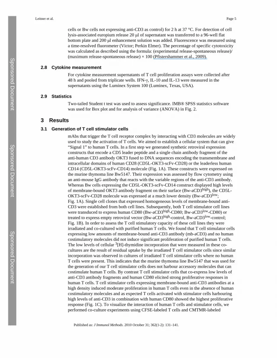
mAbs that trigger the T cell receptor complex by interacting with CD3 molecules are widelyused to study the activation of T cells. We aimed to establish a cellular system that can give“Signal 1” to human T cells. In a first step we generated synthetic retroviral expressionconstructs that encode a CD5 leader peptide and a single chain antibody fragment of theanti-human CD3 antibody OKT3 fused to DNA sequences encoding the transmembrane andintracellular domains of human CD28 (CD5L-OKT3-scFv-CD28) or the leaderless humanCD14 (CD5L-OKT3-scFv-CD14) molecule (Fig. 1A). These constructs were expressed onthe murine thymoma line Bw5147. Their expression was assessed by flow cytometry usingan anti-mouse IgG antibody that reacts with the variable regions of the anti-CD3 antibody.Whereas Bw cells expressing the CD5L-OKT3-scFv-CD14 construct displayed high levelsof membrane-bound OKT3 antibody fragment on their surface (Bw-aCD3high), the CD5L-OKT3-scFv-CD28 molecule was expressed at a much lower density (Bw-aCD3low;Fig. 1A). Single cell clones that expressed homogeneous levels of membrane-bound anti-CD3 were established from both cell lines. Subsequently, both T cell stimulator cell lineswere transduced to express human CD80 (Bw-aCD3high-CD80; Bw-aCD3low-CD80) ortreated to express empty retroviral vector (Bw-aCD3high-control, Bw-aCD3low-control;Fig. 1B). In order to assess the T cell stimulatory capacity of these cell lines they wereirradiated and co-cultured with purified human T cells. We found that T cell stimulator cellsexpressing low amounts of membrane-bound anti-CD3 antibody (mb-aCD3) and no humancostimulatory molecules did not induce significant proliferation of purified human T cells.The low levels of cellular 3[H]-thymidine incorporation that were measured in these co-cultures are the result of residual uptake by the irradiated T cell stimulator cells since similarincorporation was observed in cultures of irradiated T cell stimulator cells where no humanT cells were present. This indicates that the murine thymoma line Bw5147 that was used forthe generation of our T cell stimulator cells does not harbour accessory molecules that cancostimulate human T cells. By contrast T cell stimulator cells that co-express low levels ofanti-CD3 antibody fragments and human CD80 elicited strong proliferative responses inhuman T cells. T cell stimulator cells expressing membrane-bound anti-CD3 antibodies at ahigh density induced moderate proliferation in human T cells even in the absence of humancostimulatory molecules and as expected T cells activated with stimulator cells harbouringhigh levels of anti-CD3 in combination with human CD80 showed the highest proliferativeresponse (Fig. 1C). To visualize the interaction of human T cells and stimulator cells, weperformed co-culture experiments using CFSE-labeled T cells and CMTMR-labeled
Leitner et al. Page 5
Published as: J Immunol Methods. 2010 October 31; 362(1-2): 131–141.
Sponsored Docum
ent Sponsored D
ocument
Sponsored Docum
ent
stimulator cells. Large clusters of T cells and stimulator cells expressing CD80 can beobserved whereas much smaller clusters are formed when T cells were activated bystimulator cells expressing anti-CD3 but no human costimulatory molecule (Fig. 1D).
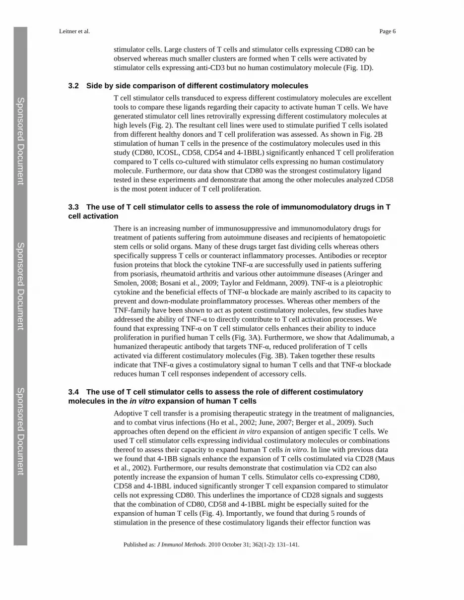
3.2 Side by side comparison of different costimulatory moleculesT cell stimulator cells transduced to express different costimulatory molecules are excellenttools to compare these ligands regarding their capacity to activate human T cells. We havegenerated stimulator cell lines retrovirally expressing different costimulatory molecules athigh levels (Fig. 2). The resultant cell lines were used to stimulate purified T cells isolatedfrom different healthy donors and T cell proliferation was assessed. As shown in Fig. 2Bstimulation of human T cells in the presence of the costimulatory molecules used in thisstudy (CD80, ICOSL, CD58, CD54 and 4-1BBL) significantly enhanced T cell proliferationcompared to T cells co-cultured with stimulator cells expressing no human costimulatorymolecule. Furthermore, our data show that CD80 was the strongest costimulatory ligandtested in these experiments and demonstrate that among the other molecules analyzed CD58is the most potent inducer of T cell proliferation.
3.3 The use of T cell stimulator cells to assess the role of immunomodulatory drugs in Tcell activation
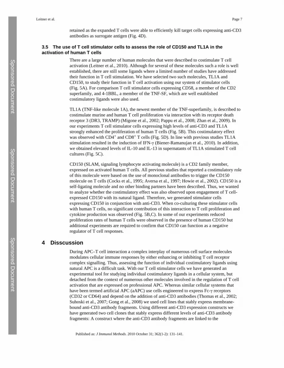
There is an increasing number of immunosuppressive and immunomodulatory drugs fortreatment of patients suffering from autoimmune diseases and recipients of hematopoieticstem cells or solid organs. Many of these drugs target fast dividing cells whereas othersspecifically suppress T cells or counteract inflammatory processes. Antibodies or receptorfusion proteins that block the cytokine TNF-α are successfully used in patients sufferingfrom psoriasis, rheumatoid arthritis and various other autoimmune diseases (Aringer andSmolen, 2008; Bosani et al., 2009; Taylor and Feldmann, 2009). TNF-α is a pleiotrophiccytokine and the beneficial effects of TNF-α blockade are mainly ascribed to its capacity toprevent and down-modulate proinflammatory processes. Whereas other members of theTNF-family have been shown to act as potent costimulatory molecules, few studies haveaddressed the ability of TNF-α to directly contribute to T cell activation processes. Wefound that expressing TNF-α on T cell stimulator cells enhances their ability to induceproliferation in purified human T cells (Fig. 3A). Furthermore, we show that Adalimumab, ahumanized therapeutic antibody that targets TNF-α, reduced proliferation of T cellsactivated via different costimulatory molecules (Fig. 3B). Taken together these resultsindicate that TNF-α gives a costimulatory signal to human T cells and that TNF-α blockadereduces human T cell responses independent of accessory cells.
3.4 The use of T cell stimulator cells to assess the role of different costimulatorymolecules in the in vitro expansion of human T cells
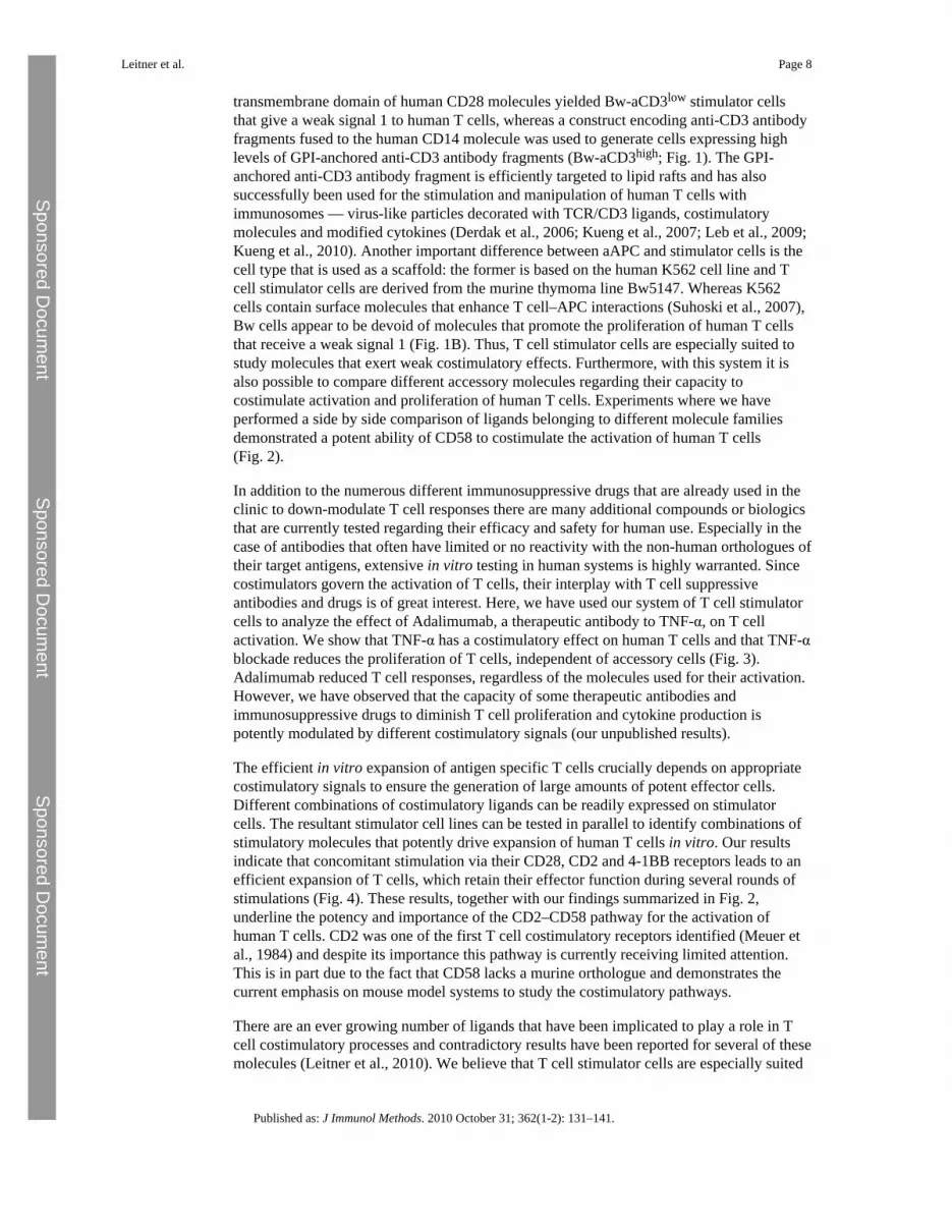
Adoptive T cell transfer is a promising therapeutic strategy in the treatment of malignancies,and to combat virus infections (Ho et al., 2002; June, 2007; Berger et al., 2009). Suchapproaches often depend on the efficient in vitro expansion of antigen specific T cells. Weused T cell stimulator cells expressing individual costimulatory molecules or combinationsthereof to assess their capacity to expand human T cells in vitro. In line with previous datawe found that 4-1BB signals enhance the expansion of T cells costimulated via CD28 (Mauset al., 2002). Furthermore, our results demonstrate that costimulation via CD2 can alsopotently increase the expansion of human T cells. Stimulator cells co-expressing CD80,CD58 and 4-1BBL induced significantly stronger T cell expansion compared to stimulatorcells not expressing CD80. This underlines the importance of CD28 signals and suggeststhat the combination of CD80, CD58 and 4-1BBL might be especially suited for theexpansion of human T cells (Fig. 4). Importantly, we found that during 5 rounds ofstimulation in the presence of these costimulatory ligands their effector function was
Leitner et al. Page 6
Published as: J Immunol Methods. 2010 October 31; 362(1-2): 131–141.
Sponsored Docum
ent Sponsored D
ocument
Sponsored Docum
ent
retained as the expanded T cells were able to efficiently kill target cells expressing anti-CD3antibodies as surrogate antigen (Fig. 4D).
3.5 The use of T cell stimulator cells to assess the role of CD150 and TL1A in theactivation of human T cells
There are a large number of human molecules that were described to costimulate T cellactivation (Leitner et al., 2010). Although for several of these molecules such a role is wellestablished, there are still some ligands where a limited number of studies have addressedtheir function in T cell stimulation. We have selected two such molecules, TL1A andCD150, to study their function in T cell activation using our system of stimulator cells(Fig. 5A). For comparison T cell stimulator cells expressing CD58, a member of the CD2superfamily, and 4-1BBL, a member of the TNF-SF, which are well establishedcostimulatory ligands were also used.
TL1A (TNF-like molecule 1A), the newest member of the TNF-superfamily, is described tocostimulate murine and human T cell proliferation via interaction with its receptor deathreceptor 3 (DR3, TRAMP) (Migone et al., 2002; Pappu et al., 2008; Zhan et al., 2009). Inour experiments T cell stimulator cells expressing high levels of anti-CD3 and TL1Astrongly enhanced the proliferation of human T cells (Fig. 5B). This costimulatory effectwas observed with CD4+ and CD8+ T cells (Fig. 5D). In line with previous studies TL1Astimulation resulted in the induction of IFN-γ (Biener-Ramanujan et al., 2010). In addition,we obtained elevated levels of IL-10 and IL-13 in supernatants of TL1A stimulated T cellcultures (Fig. 5C).
CD150 (SLAM, signaling lymphocyte activating molecule) is a CD2 family member,expressed on activated human T cells. All previous studies that reported a costimulatory roleof this molecule were based on the use of monoclonal antibodies to trigger the CD150molecule on T cells (Cocks et al., 1995; Aversa et al., 1997; Howie et al., 2002). CD150 is aself-ligating molecule and no other binding partners have been described. Thus, we wantedto analyze whether the costimulatory effect was also observed upon engagement of T cell-expressed CD150 with its natural ligand. Therefore, we generated stimulator cellsexpressing CD150 in conjunction with anti-CD3. When co-culturing these stimulator cellswith human T cells, no significant contribution of this interaction to T cell proliferation andcytokine production was observed (Fig. 5B,C). In some of our experiments reducedproliferation rates of human T cells were observed in the presence of human CD150 butadditional experiments are required to confirm that CD150 can function as a negativeregulator of T cell responses.
4 DisscussionDuring APC–T cell interaction a complex interplay of numerous cell surface moleculesmodulates cellular immune responses by either enhancing or inhibiting T cell receptorcomplex signalling. Thus, assessing the function of individual costimulatory ligands usingnatural APC is a difficult task. With our T cell stimulator cells we have generated anexperimental tool for studying individual costimulatory ligands in a cellular system, butdetached from the context of numerous other molecules involved in the regulation of T cellactivation that are expressed on professional APC. Whereas similar cellular systems thathave been termed artificial APC (aAPC) use cells engineered to express Fc-γ receptors(CD32 or CD64) and depend on the addition of anti-CD3 antibodies (Thomas et al., 2002;Suhoski et al., 2007; Gong et al., 2008) we used cell lines that stably express membrane-bound anti-CD3 antibody fragments. Using different anti-CD3 expression constructs wehave generated two cell clones that stably express different levels of anti-CD3 antibodyfragments: A construct where the anti-CD3 antibody fragments are linked to the
Leitner et al. Page 7
Published as: J Immunol Methods. 2010 October 31; 362(1-2): 131–141.
Sponsored Docum
ent Sponsored D
ocument
Sponsored Docum
ent
transmembrane domain of human CD28 molecules yielded Bw-aCD3low stimulator cellsthat give a weak signal 1 to human T cells, whereas a construct encoding anti-CD3 antibodyfragments fused to the human CD14 molecule was used to generate cells expressing highlevels of GPI-anchored anti-CD3 antibody fragments (Bw-aCD3high; Fig. 1). The GPI-anchored anti-CD3 antibody fragment is efficiently targeted to lipid rafts and has alsosuccessfully been used for the stimulation and manipulation of human T cells withimmunosomes — virus-like particles decorated with TCR/CD3 ligands, costimulatorymolecules and modified cytokines (Derdak et al., 2006; Kueng et al., 2007; Leb et al., 2009;Kueng et al., 2010). Another important difference between aAPC and stimulator cells is thecell type that is used as a scaffold: the former is based on the human K562 cell line and Tcell stimulator cells are derived from the murine thymoma line Bw5147. Whereas K562cells contain surface molecules that enhance T cell–APC interactions (Suhoski et al., 2007),Bw cells appear to be devoid of molecules that promote the proliferation of human T cellsthat receive a weak signal 1 (Fig. 1B). Thus, T cell stimulator cells are especially suited tostudy molecules that exert weak costimulatory effects. Furthermore, with this system it isalso possible to compare different accessory molecules regarding their capacity tocostimulate activation and proliferation of human T cells. Experiments where we haveperformed a side by side comparison of ligands belonging to different molecule familiesdemonstrated a potent ability of CD58 to costimulate the activation of human T cells(Fig. 2).
In addition to the numerous different immunosuppressive drugs that are already used in theclinic to down-modulate T cell responses there are many additional compounds or biologicsthat are currently tested regarding their efficacy and safety for human use. Especially in thecase of antibodies that often have limited or no reactivity with the non-human orthologues oftheir target antigens, extensive in vitro testing in human systems is highly warranted. Sincecostimulators govern the activation of T cells, their interplay with T cell suppressiveantibodies and drugs is of great interest. Here, we have used our system of T cell stimulatorcells to analyze the effect of Adalimumab, a therapeutic antibody to TNF-α, on T cellactivation. We show that TNF-α has a costimulatory effect on human T cells and that TNF-αblockade reduces the proliferation of T cells, independent of accessory cells (Fig. 3).Adalimumab reduced T cell responses, regardless of the molecules used for their activation.However, we have observed that the capacity of some therapeutic antibodies andimmunosuppressive drugs to diminish T cell proliferation and cytokine production ispotently modulated by different costimulatory signals (our unpublished results).
The efficient in vitro expansion of antigen specific T cells crucially depends on appropriatecostimulatory signals to ensure the generation of large amounts of potent effector cells.Different combinations of costimulatory ligands can be readily expressed on stimulatorcells. The resultant stimulator cell lines can be tested in parallel to identify combinations ofstimulatory molecules that potently drive expansion of human T cells in vitro. Our resultsindicate that concomitant stimulation via their CD28, CD2 and 4-1BB receptors leads to anefficient expansion of T cells, which retain their effector function during several rounds ofstimulations (Fig. 4). These results, together with our findings summarized in Fig. 2,underline the potency and importance of the CD2–CD58 pathway for the activation ofhuman T cells. CD2 was one of the first T cell costimulatory receptors identified (Meuer etal., 1984) and despite its importance this pathway is currently receiving limited attention.This is in part due to the fact that CD58 lacks a murine orthologue and demonstrates thecurrent emphasis on mouse model systems to study the costimulatory pathways.
There are an ever growing number of ligands that have been implicated to play a role in Tcell costimulatory processes and contradictory results have been reported for several of thesemolecules (Leitner et al., 2010). We believe that T cell stimulator cells are especially suited
Leitner et al. Page 8
Published as: J Immunol Methods. 2010 October 31; 362(1-2): 131–141.
Sponsored Docum
ent Sponsored D
ocument
Sponsored Docum
ent
to assess the function of accessory molecules during T cell activation since they allowanalyzing human T cell responses under conditions that only differ regarding the presence ofthe molecules of interest. We have recently used stimulator cells expressing PD-L2 and B7-H3, two members of the extended B7 family, to address their function during the activationof human T cells (Pfistershammer et al., 2006; Leitner et al., 2009). In these studies wecould show that these molecules consistently inhibited T cell responses and our experimentsdid give any evidence for positive costimulatory functions for human PD-L2 and B7-H3.The CD2 superfamily member CD150 and the TNF-SF member TL1A have both beendescribed to costimulate T cell activation. CD150 is a self-ligating receptor, whereas TL1Abinds to DR3 a member of the TNFR-SF. However, few studies on these molecules havedirectly analyzed the consequences of the interaction of CD150 or TL1A with human Tcells. In the present study we have generated T cell stimulator cell lines expressing CD150and TL1A and used them to stimulate purified human T cells. Our results demonstrate thatthe presence of TL1A during T cell activation significantly costimulates their proliferationand production of cytokines, whereas T cells stimulated in the presence of stimulator cellsexpressing CD150 did not show enhanced proliferation and cytokine production. Previousstudies that have described a positive costimulatory function for CD150 have usedantibodies to crosslink the CD150 molecules on T cells (Cocks et al., 1995; Aversa et al.,1997). In contrast, we have used T cell stimulator cells expressing its natural ligand CD150,to assess the role of CD150–CD150 interaction in the activation of T cells. Our results,which suggest that CD150 does not function as a classical T cell costimulatory molecule,underline the importance of using natural ligands to study the functional consequences ofreceptor–ligand pairs implicated in T cell activation processes. The homophilic interactionof CD150 is of particular low affinity (Kd 200 mM; (Chattopadhyay et al., 2009)), whichmight explain the different outcome of our experiments compared to studies that usedantibodies. It has previously been suggested that antibody-induced SLAM/CD150 activationmay not fully mimic the physiological role of CD150, because mice deficient in thismolecule exhibit essentially normal levels of IFN-γ, whereas the ligation of T cell-expressedCD150 by antibodies promotes strong IFN-γ secretion (Ostrakhovitch and Li, 2006). Furtherstudies are required to address the physiological role of CD150 during human T cellactivation. Since T cells that express costimulatory ligands can receive potent costimulatorysignals (“autocostimulation”) it is also possible that homotypic interaction of CD150 in cisplays a role during human T cell activation (Stephan et al., 2007).
Taken together our results demonstrate that the system of T cell stimulator cells is a usefultool to assess the function of costimulatory ligands. In particular they are suited to comparethe function of individual costimulatory molecules and analyze their effect on different Tcell subsets and in context of a strong or weak signal 1. Since professional APC like DCharbour stimulatory as well as inhibitory ligands, the interplay of positive and negativesignals determines the outcome of T cell responses. We have previously shown thatcombinations of costimulatory molecules can be expressed and analyzed on T cell stimulatorcells (Kober et al., 2008). We are currently using our system of stimulator cells to analyzethe interplay of defined costimulatory and coinhibitory molecules during the activation ofhuman T cells. Studies on individual costimulatory pathways can complement investigationsusing experimental systems employing natural human APC or animal studies to get a betterinsight into the complex interplay of the numerous accessory surface molecules that governhuman T cell responses.
ReferencesAringer M. Smolen J.S. Efficacy and safety of TNF-blocker therapy in systemic lupus erythematosus.
Expert Opin. Drug Saf.. 2008; 7:411. [PubMed: 18613805]
Leitner et al. Page 9
Published as: J Immunol Methods. 2010 October 31; 362(1-2): 131–141.
Sponsored Docum
ent Sponsored D
ocument
Sponsored Docum
ent
Aversa G. Chang C.C. Carballido J.M. Cocks B.G. de Vries J.E. Engagement of the signalinglymphocytic activation molecule (SLAM) on activated T cells results in IL-2-independent,cyclosporin A-sensitive T cell proliferation and IFN-gamma production. J. Immunol.. 1997;158:4036. [PubMed: 9126961]
Berger C. Turtle C.J. Jensen M.C. Riddell S.R. Adoptive transfer of virus-specific and tumor-specificT cell immunity. Curr. Opin. Immunol.. 2009; 21:224. [PubMed: 19304470]
Biener-Ramanujan E. Gonsky R. Ko B. Targan S.R. Functional signaling of membrane-bound TL1Ainduces IFN-gamma expression. FEBS Lett.. 2010; 584:2376. [PubMed: 20403353]
Blank C. Mackensen A. Contribution of the PD-L1/PD-1 pathway to T-cell exhaustion: an update onimplications for chronic infections and tumor evasion. Cancer Immunol. Immunother.. 2007;56:739. [PubMed: 17195077]
Bosani M. Ardizzone S. Porro G.B. Biologic targeting in the treatment of inflammatory boweldiseases. Biologics. 2009; 3:77. [PubMed: 19707398]
Chattopadhyay K. Lazar-Molnar E. Yan Q. Rubinstein R. Zhan C. Vigdorovich V. Ramagopal U.A.Bonanno J. Nathenson S.G. Almo S.C. Sequence, structure, function, immunity: structuralgenomics of costimulation. Immunol. Rev.. 2009; 229:356. [PubMed: 19426233]
Cocks B.G. Chang C.C. Carballido J.M. Yssel H. de Vries J.E. Aversa G. A novel receptor involved inT-cell activation. Nature. 1995; 376:260. [PubMed: 7617038]
Derdak S.V. Kueng H.J. Leb V.M. Neunkirchner A. Schmetterer K.G. Bielek E. Majdic O. Knapp W.Seed B. Pickl W.F. Direct stimulation of T lymphocytes by immunosomes: virus-like particlesdecorated with T cell receptor/CD3 ligands plus costimulatory molecules. Proc. Natl. Acad. Sci.USA. 2006; 103:13144. [PubMed: 16924110]
Ford M.L. Larsen C.P. Translating costimulation blockade to the clinic: lessons learned from threepathways. Immunol. Rev.. 2009; 229:294. [PubMed: 19426229]
Gong W. Ji M. Cao Z. Wang L. Qian Y. Hu M. Qian L. Pan X. Establishment and characterization of acell based artificial antigen-presenting cell for expansion and activation of CD8+ T cells ex vivo.Cell. Mol. Immunol.. 2008; 5:47. [PubMed: 18318994]
Greenwald R.J. Freeman G.J. Sharpe A.H. The B7 family revisited. Annu. Rev. Immunol.. 2005;23:515. [PubMed: 15771580]
Ho W.Y. Yee C. Greenberg P.D. Adoptive therapy with CD8(+) T cells: it may get by with a little helpfrom its friends. J. Clin. Invest.. 2002; 110:1415. [PubMed: 12438439]
Howie D. Simarro M. Sayos J. Guirado M. Sancho J. Terhorst C. Molecular dissection of the signalingand costimulatory functions of CD150 (SLAM): CD150/SAP binding and CD150-mediatedcostimulation. Blood. 2002; 99:957. [PubMed: 11806999]
June C.H. Adoptive T cell therapy for cancer in the clinic. J. Clin. Invest.. 2007; 117:1466. [PubMed:17549249]
Keir M.E. Butte M.J. Freeman G.J. Sharpe A.H. PD-1 and its ligands in tolerance and immunity.Annu. Rev. Immunol.. 2008; 26:677. [PubMed: 18173375]
Kober J. Leitner J. Klauser C. Woitek R. Majdic O. Stockl J. Herndler-Brandstetter D. Grubeck-Loebenstein B. Reipert B.M. Pickl W.F. Pfistershammer K. Steinberger P. The capacity of theTNF family members 4-1BBL, OX40L, CD70, GITRL, CD30L and LIGHT to costimulate humanT cells. Eur. J. Immunol.. 2008; 38:2678. [PubMed: 18825741]
Kriehuber E. Breiteneder-Geleff S. Groeger M. Soleiman A. Schoppmann S.F. Stingl G. Kerjaschki D.Maurer D. Isolation and characterization of dermal lymphatic and blood endothelial cells revealstable and functionally specialized cell lineages. J. Exp. Med.. 2001; 194:797. [PubMed:11560995]
Kueng H.J. Leb V.M. Haiderer D. Raposo G. Thery C. Derdak S.V. Schmetterer K.G. NeunkirchnerA. Sillaber C. Seed B. Pickl W.F. General strategy for decoration of enveloped viruses withfunctionally active lipid-modified cytokines. J. Virol.. 2007; 81:8666. [PubMed: 17537846]
Kueng H.J. Manta C. Haiderer D. Leb V.M. Schmetterer K.G. Neunkirchner A. Byrne R.A.Scheinecker C. Steinberger P. Seed B. Pickl W.F. Fluorosomes: a convenient new reagent todetect and block multivalent and complex receptor–ligand interactions. FASEB J.. 2010; 24:1572.[PubMed: 20056716]
Leitner et al. Page 10
Published as: J Immunol Methods. 2010 October 31; 362(1-2): 131–141.
Sponsored Docum
ent Sponsored D
ocument
Sponsored Docum
ent

Leb V.M. Jahn-Schmid B. Kueng H.J. Schmetterer K.G. Haiderer D. Neunkirchner A. Fischer G.F.Hartl A. Thalhamer J. Steinberger P. Bohle B. Seed B. Pickl W.F. Modulation of allergen-specificT-lymphocyte function by virus-like particles decorated with HLA class II molecules. J. AllergyClin. Immunol.. 2009; 124:121. [PubMed: 19500826]
Leitner J. Klauser C. Pickl W.F. Stockl J. Majdic O. Bardet A.F. Kreil D.P. Dong C. Yamazaki T.Zlabinger G. Pfistershammer K. Steinberger P. B7-H3 is a potent inhibitor of human T-cellactivation: no evidence for B7-H3 and TREML2 interaction. Eur. J. Immunol.. 2009; 39:1754.[PubMed: 19544488]
Leitner J. Grabmeier-Pfistershammer K. Steinberger P. Receptors and ligands implicated in human Tcell costimulatory processes. Immunol. Lett.. 2010; 128:89. [PubMed: 19941899]
Li X.C. Rothstein D.M. Sayegh M.H. Costimulatory pathways in transplantation: challenges and newdevelopments. Immunol. Rev.. 2009; 229:271. [PubMed: 19426228]
Maus M.V. Thomas A.K. Leonard D.G. Allman D. Addya K. Schlienger K. Riley J.L. June C.H. Exvivo expansion of polyclonal and antigen-specific cytotoxic T lymphocytes by artificial APCsexpressing ligands for the T-cell receptor, CD28 and 4-1BB. Nat. Biotechnol.. 2002; 20:143.[PubMed: 11821859]
Meuer S.C. Hussey R.E. Fabbi M. Fox D. Acuto O. Fitzgerald K.A. Hodgdon J.C. Protentis J.P.Schlossman S.F. Reinherz E.L. An alternative pathway of T-cell activation: a functional role forthe 50 kd T11 sheep erythrocyte receptor protein. Cell. 1984; 36:897. [PubMed: 6231105]
Migone T.S. Zhang J. Luo X. Zhuang L. Chen C. Hu B. Hong J.S. Perry J.W. Chen S.F. Zhou J.X.Cho Y.H. Ullrich S. Kanakaraj P. Carrell J. Boyd E. Olsen H.S. Hu G. Pukac L. Liu D. Ni J. KimS. Gentz R. Feng P. Moore P.A. Ruben S.M. Wei P. TL1A is a TNF-like ligand for DR3 and TR6/DcR3 and functions as a T cell costimulator. Immunity. 2002; 16:479. [PubMed: 11911831]
Nurieva R.I. Liu X. Dong C. Yin–Yang of costimulation: crucial controls of immune tolerance andfunction. Immunol. Rev.. 2009; 229:88. [PubMed: 19426216]
Ostrakhovitch E.A. Li S.S. The role of SLAM family receptors in immune cell signaling. Biochem.Cell Biol.. 2006; 84:832. [PubMed: 17215871]
Pappu B.P. Borodovsky A. Zheng T.S. Yang X. Wu P. Dong X. Weng S. Browning B. Scott M.L. MaL. Su L. Tian Q. Schneider P. Flavell R.A. Dong C. Burkly L.C. TL1A-DR3 interaction regulatesTh17 cell function and Th17-mediated autoimmune disease. J. Exp. Med.. 2008; 205:1049.[PubMed: 18411337]
Pfistershammer K. Majdic O. Stockl J. Zlabinger G. Kirchberger S. Steinberger P. Knapp W. CD63 asan activation-linked T cell costimulatory element. J. Immunol.. 2004; 173:6000. [PubMed:15528334]
Pfistershammer K. Klauser C. Pickl W.F. Stockl J. Leitner J. Zlabinger G. Majdic O. Steinberger P.No evidence for dualism in function and receptors: PD-L2/B7-DC is an inhibitory regulator ofhuman T cell activation. Eur. J. Immunol.. 2006; 36:1104. [PubMed: 16598819]
Pfistershammer K. Klauser C. Leitner J. Stockl J. Majdic O. Weichhart T. Sobanov Y. Bochkov V.Saemann M. Zlabinger G. Steinberger P. Identification of the scavenger receptors SREC-I, Cla-1(SR-BI), and SR-AI as cellular receptors for Tamm–Horsfall protein. J. Leukoc. Biol.. 2008;83:131. [PubMed: 17928461]
Pfistershammer K. Lawitschka A. Klauser C. Leitner J. Weigl R. Heemskerk M.H. Pickl W.F. MajdicO. Bohmig G.A. Fischer G.F. Greinix H.T. Steinberger P. Allogeneic disparities inimmunoglobulin-like transcript 5 induce potent antibody responses in hematopoietic stem celltransplant recipients. Blood. 2009; 114:2323. [PubMed: 19617579]
Saunders P.A. Hendrycks V.R. Lidinsky W.A. Woods M.L. PD-L2:PD-1 involvement in T cellproliferation, cytokine production, and integrin-mediated adhesion. Eur. J. Immunol.. 2005;35:3561. [PubMed: 16278812]
Steinberger P. Majdic O. Derdak S.V. Pfistershammer K. Kirchberger S. Klauser C. Zlabinger G. PicklW.F. Stockl J. Knapp W. Molecular characterization of human 4Ig-B7-H3, a member of the B7family with four Ig-like domains. J. Immunol.. 2004; 172:2352. [PubMed: 14764704]
Stephan M.T. Ponomarev V. Brentjens R.J. Chang A.H. Dobrenkov K.V. Heller G. Sadelain M. T cell-encoded CD80 and 4-1BBL induce auto- and transcostimulation, resulting in potent tumorrejection. Nat. Med.. 2007; 13:1440. [PubMed: 18026115]
Leitner et al. Page 11
Published as: J Immunol Methods. 2010 October 31; 362(1-2): 131–141.
Sponsored Docum
ent Sponsored D
ocument
Sponsored Docum
ent
Suhoski M.M. Golovina T.N. Aqui N.A. Tai V.C. Varela-Rohena A. Milone M.C. Carroll R.G. RileyJ.L. June C.H. Engineering artificial antigen-presenting cells to express a diverse array of co-stimulatory molecules. Mol. Ther.. 2007; 15:981. [PubMed: 17375070]
Taylor P.C. Feldmann M. Anti-TNF biologic agents: still the therapy of choice for rheumatoidarthritis. Nat. Rev. Rheumatol.. 2009; 5:578. [PubMed: 19798034]
Thomas A.K. Maus M.V. Shalaby W.S. June C.H. Riley J.L. A cell-based artificial antigen-presentingcell coated with anti-CD3 and CD28 antibodies enables rapid expansion and long-term growth ofCD4 T lymphocytes. Clin. Immunol.. 2002; 105:259. [PubMed: 12498807]
Watts T.H. TNF/TNFR family members in costimulation of T cell responses. Annu. Rev. Immunol..2005; 23:23. [PubMed: 15771565]
Zhan C. Yan Q. Patskovsky Y. Li Z. Toro R. Meyer A. Cheng H. Brenowitz M. Nathenson S.G. AlmoS.C. Biochemical and structural characterization of the human TL1A ectodomain. Biochemistry.2009; 48:7636. [PubMed: 19522538]
AcknowledgmentsWe appreciate the excellent technical assistance of Christoph Klauser, Margarete Merio, Petra Cejka and ClausWenhart. We thank Vera Kaiser, Graz University of Technology, for help with the statistical analyses. This studywas supported by a grant from the Austrian Science Fund (FWF p21964-B20), a grant from the Austrian NationalBank 12731 and in part by a grant from Abbott Austria. Judith Leitner is supported by a Doc fForte fellowship fromthe Austrian Academy of Science. WFP is supported by SFB grant 1816 from the Austrian Science Fund and by theChristian Doppler Society. The authors declare no conflict of interest.
Leitner et al. Page 12
Published as: J Immunol Methods. 2010 October 31; 362(1-2): 131–141.
Sponsored Docum
ent Sponsored D
ocument
Sponsored Docum
ent
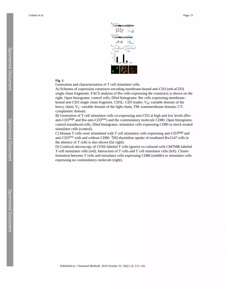
Fig. 1.Generation and characterisation of T cell stimulator cells.A) Schemes of expression constructs encoding membrane-bound anti-CD3 (mb-aCD3)single chain fragments. FACS analysis of Bw cells expressing the constructs is shown on theright. Open histograms: control cells; filled histograms: Bw cells expressing membrane-bound anti-CD3 single chain fragment. CD5L: CD5 leader; VH: variable domain of theheavy chain; VL: variable domain of the light chain; TM: transmembrane domain; CT:cytoplasmic domain.B) Generation of T cell stimulator cells co-expressing anti-CD3 at high and low levels (Bw-anti-CD3high and Bw-anti-CD3low) and the costimulatory molecule CD80. Open histograms:control transduced cells; filled histograms: stimulator cells expressing CD80 or mock treatedstimulator cells (control).C) Human T cells were stimulated with T cell stimulator cells expressing anti-CD3high andanti-CD3low with and without CD80. 3[H]-thymidine uptake of irradiated Bw5147 cells inthe absence of T cells is also shown (far right).D) Confocal microscopy of CFSE-labeled T cells (green) co-cultured with CMTMR-labeledT-cell stimulator cells (red). Interaction of T cells and T cell stimulator cells (left). Clusterformation between T cells and stimulator cells expressing CD80 (middle) or stimulator cellsexpressing no costimulatory molecule (right).
Leitner et al. Page 13
Published as: J Immunol Methods. 2010 October 31; 362(1-2): 131–141.
Sponsored Docum
ent Sponsored D
ocument
Sponsored Docum
ent
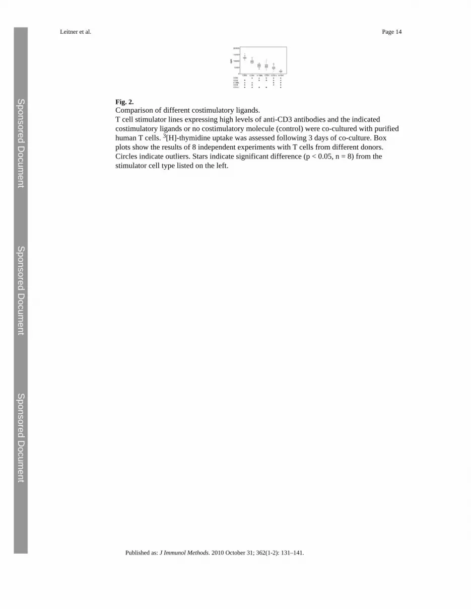
Fig. 2.Comparison of different costimulatory ligands.T cell stimulator lines expressing high levels of anti-CD3 antibodies and the indicatedcostimulatory ligands or no costimulatory molecule (control) were co-cultured with purifiedhuman T cells. 3[H]-thymidine uptake was assessed following 3 days of co-culture. Boxplots show the results of 8 independent experiments with T cells from different donors.Circles indicate outliers. Stars indicate significant difference (p < 0.05, n = 8) from thestimulator cell type listed on the left.
Leitner et al. Page 14
Published as: J Immunol Methods. 2010 October 31; 362(1-2): 131–141.
Sponsored Docum
ent Sponsored D
ocument
Sponsored Docum
ent
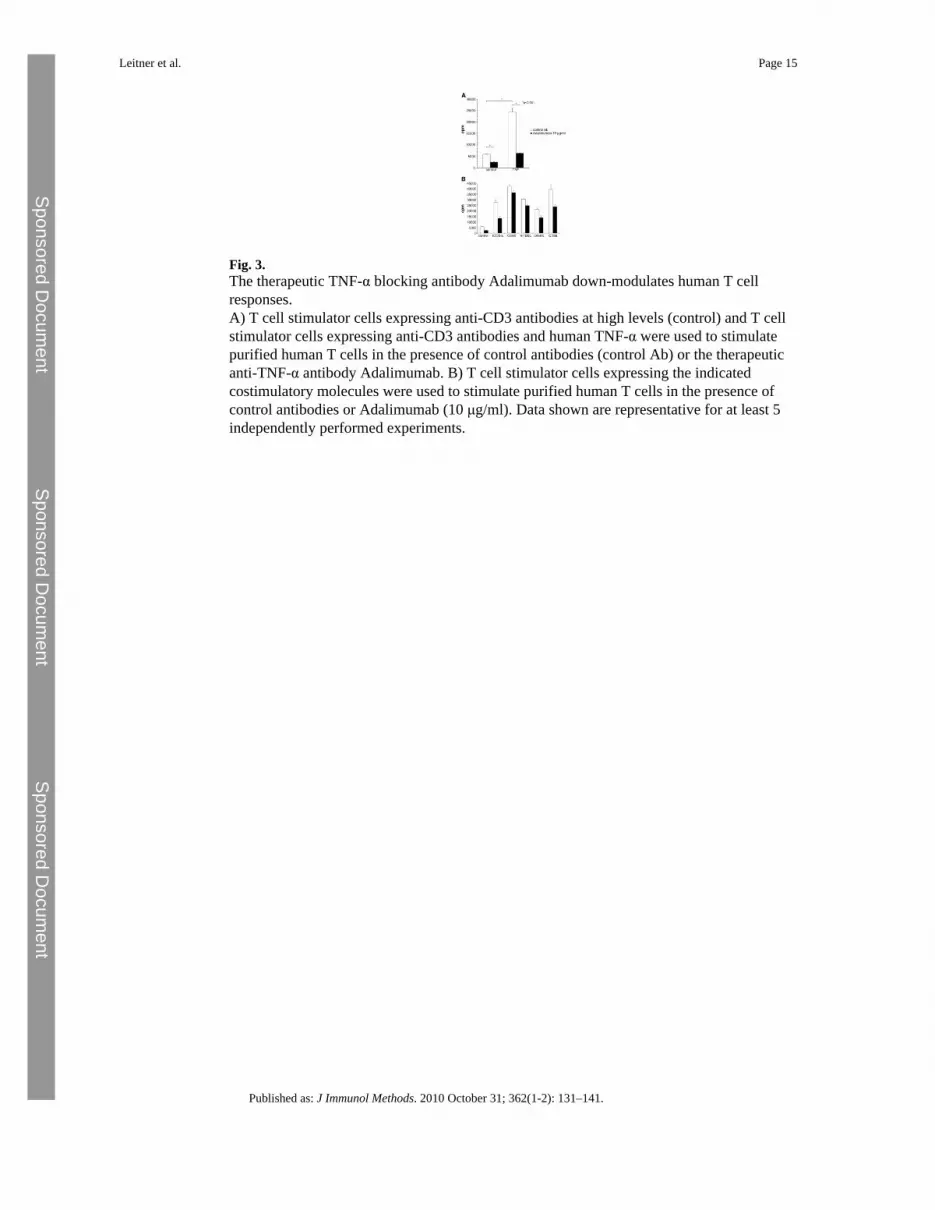
Fig. 3.The therapeutic TNF-α blocking antibody Adalimumab down-modulates human T cellresponses.A) T cell stimulator cells expressing anti-CD3 antibodies at high levels (control) and T cellstimulator cells expressing anti-CD3 antibodies and human TNF-α were used to stimulatepurified human T cells in the presence of control antibodies (control Ab) or the therapeuticanti-TNF-α antibody Adalimumab. B) T cell stimulator cells expressing the indicatedcostimulatory molecules were used to stimulate purified human T cells in the presence ofcontrol antibodies or Adalimumab (10 μg/ml). Data shown are representative for at least 5independently performed experiments.
Leitner et al. Page 15
Published as: J Immunol Methods. 2010 October 31; 362(1-2): 131–141.
Sponsored Docum
ent Sponsored D
ocument
Sponsored Docum
ent
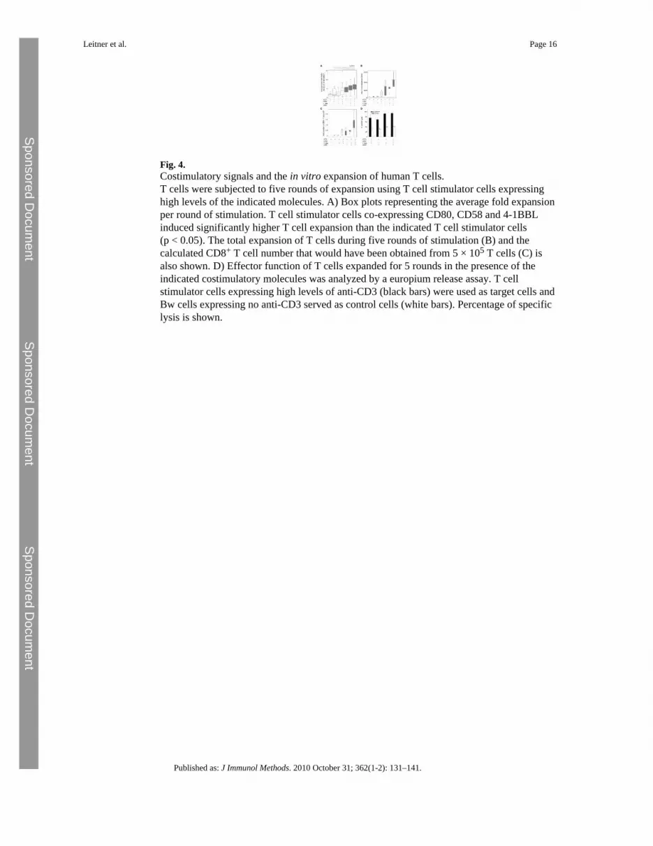
Fig. 4.Costimulatory signals and the in vitro expansion of human T cells.T cells were subjected to five rounds of expansion using T cell stimulator cells expressinghigh levels of the indicated molecules. A) Box plots representing the average fold expansionper round of stimulation. T cell stimulator cells co-expressing CD80, CD58 and 4-1BBLinduced significantly higher T cell expansion than the indicated T cell stimulator cells(p < 0.05). The total expansion of T cells during five rounds of stimulation (B) and thecalculated CD8+ T cell number that would have been obtained from 5 × 10⁵ T cells (C) isalso shown. D) Effector function of T cells expanded for 5 rounds in the presence of theindicated costimulatory molecules was analyzed by a europium release assay. T cellstimulator cells expressing high levels of anti-CD3 (black bars) were used as target cells andBw cells expressing no anti-CD3 served as control cells (white bars). Percentage of specificlysis is shown.
Leitner et al. Page 16
Published as: J Immunol Methods. 2010 October 31; 362(1-2): 131–141.
Sponsored Docum
ent Sponsored D
ocument
Sponsored Docum
ent
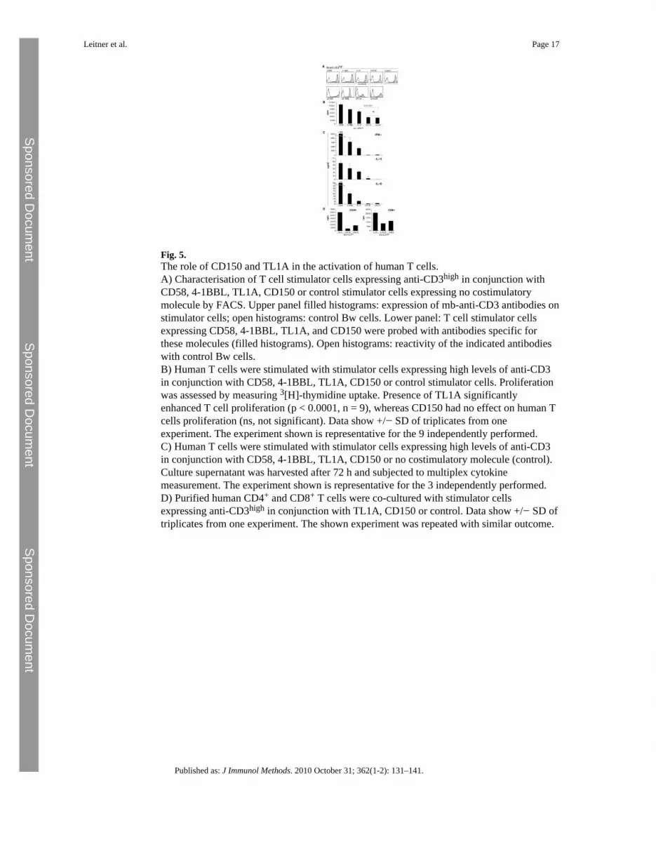
Fig. 5.The role of CD150 and TL1A in the activation of human T cells.A) Characterisation of T cell stimulator cells expressing anti-CD3high in conjunction withCD58, 4-1BBL, TL1A, CD150 or control stimulator cells expressing no costimulatorymolecule by FACS. Upper panel filled histograms: expression of mb-anti-CD3 antibodies onstimulator cells; open histograms: control Bw cells. Lower panel: T cell stimulator cellsexpressing CD58, 4-1BBL, TL1A, and CD150 were probed with antibodies specific forthese molecules (filled histograms). Open histograms: reactivity of the indicated antibodieswith control Bw cells.B) Human T cells were stimulated with stimulator cells expressing high levels of anti-CD3in conjunction with CD58, 4-1BBL, TL1A, CD150 or control stimulator cells. Proliferationwas assessed by measuring 3[H]-thymidine uptake. Presence of TL1A significantlyenhanced T cell proliferation (p < 0.0001, n = 9), whereas CD150 had no effect on human Tcells proliferation (ns, not significant). Data show +/− SD of triplicates from oneexperiment. The experiment shown is representative for the 9 independently performed.C) Human T cells were stimulated with stimulator cells expressing high levels of anti-CD3in conjunction with CD58, 4-1BBL, TL1A, CD150 or no costimulatory molecule (control).Culture supernatant was harvested after 72 h and subjected to multiplex cytokinemeasurement. The experiment shown is representative for the 3 independently performed.D) Purified human CD4+ and CD8+ T cells were co-cultured with stimulator cellsexpressing anti-CD3high in conjunction with TL1A, CD150 or control. Data show +/− SD oftriplicates from one experiment. The shown experiment was repeated with similar outcome.
Leitner et al. Page 17
Published as: J Immunol Methods. 2010 October 31; 362(1-2): 131–141.
Sponsored Docum
ent Sponsored D
ocument
Sponsored Docum
ent