evaluation of different methods of leukoreduction of donor platelets to prevent alloimmune platelet...
TRANSCRIPT
doi:10.1182/blood-2003-08-2942Prepublished online July 1, 2004;
Sherrill J Slichter, Douglas Fish, V Kraig Abrams, Lakshmi K Gaur, Karen Nelson and Doug Bolgiano transfusion model
canineprevent alloimmune platelet refractoriness and induce tolerance in a Evaluation of different methods of leukoreduction of donor platelets to
(238 articles)Transfusion Medicine � (5012 articles)Immunobiology �
(2497 articles)Hemostasis, Thrombosis, and Vascular Biology �Articles on similar topics can be found in the following Blood collections
http://bloodjournal.hematologylibrary.org/site/misc/rights.xhtml#repub_requestsInformation about reproducing this article in parts or in its entirety may be found online at:
http://bloodjournal.hematologylibrary.org/site/misc/rights.xhtml#reprintsInformation about ordering reprints may be found online at:
http://bloodjournal.hematologylibrary.org/site/subscriptions/index.xhtmlInformation about subscriptions and ASH membership may be found online at:
digital object identifier (DOIs) and date of initial publication. theindexed by PubMed from initial publication. Citations to Advance online articles must include
final publication). Advance online articles are citable and establish publication priority; they areappeared in the paper journal (edited, typeset versions may be posted when available prior to Advance online articles have been peer reviewed and accepted for publication but have not yet
Copyright 2011 by The American Society of Hematology; all rights reserved.20036.the American Society of Hematology, 2021 L St, NW, Suite 900, Washington DC Blood (print ISSN 0006-4971, online ISSN 1528-0020), is published weekly by
For personal use only. by guest on May 30, 2013. bloodjournal.hematologylibrary.orgFrom

S.J. SLICHTER, et al. PREVENTION OF ALLOIMMUNE PLATELET REFRACTORINESSPage 1 of 31
1
Evaluation of different methods of leukoreduction of donor platelets to
prevent alloimmune platelet refractoriness and induce tolerance in a canine
transfusion model
Submitted:
Authors: Sherrill J. Slichter, Douglas Fish, V. Kraig Abrams, Lakshmi Gaur, Karen Nelson,
and Doug Bolgiano
Institutions: Puget Sound Blood Center and University of Washington School of
Medicine, Seattle, WA
Supported By: Grants from the National Heart, Lung and Blood Institute,
National Institutes of Health (RO1 HL47227), the
Haemonetics Corporation, Braintree, MA; the Boeing
Corporation, Seattle, WA; Pat’s Fund, Seattle,WA; and the
Leukemia and Lymphoma Society (6147-02).
Dr. Fish was supported by a grant from the Fonds de la Recherche en
Santé du Québec; Montréal, Québec, Canada.
Short Title: Prevention Of Alloimmune Platelet Refractoriness
Scientific Heading: Transfusion Medicine
Key Words: Platelet Transfusion; Platelet Refractoriness, Alloimmunization; Leukoreduction; Tolerance
Correspondence/Reprints: Sherrill J. Slichter, M.D., Puget Sound Blood Center, 921 Terry Avenue, Seattle, WA 98104-1256; (206) 292-6540 / (206) 343-5043-FAX; e-mail [email protected]
Blood First Edition Paper, prepublished online July 1, 2004; DOI 10.1182/blood-2003-08-2942
Copyright (c) 2004 American Society of Hematology
For personal use only. by guest on May 30, 2013. bloodjournal.hematologylibrary.orgFrom

S.J. SLICHTER, et al. PREVENTION OF ALLOIMMUNE PLATELET REFRACTORINESSPage 2 of 31
2
Word Count – Abstract 199
Word Count – Text 5,482
For personal use only. by guest on May 30, 2013. bloodjournal.hematologylibrary.orgFrom
S.J. SLICHTER, et al. PREVENTION OF ALLOIMMUNE PLATELET REFRACTORINESSPage 3 of 31
3
ABSTRACT
The effectiveness of different methods of leukoreduction in preventing alloimmune
platelet refractoriness was evaluated in a canine model. Platelets from a random donor dog were
administered for up to 8 weeks or until platelet refractoriness. Standard (STD) (unmodified)
platelets were accepted by 14% of recipients (n=7), compared to 14% for centifuge leukoreduced
(CLR) platelets (n=21), and 31% for filter leukoreduced (F-LR) platelets (n=13), no significant
differences.
Surprisingly, using both F-LR and C-LR was highly effective (87% acceptance, n=15).
Transfusing F-LR/C-LR red blood cells (n=4) or F-LR/C-LR plasma (n=4), along with F-LR/C-
LR platelets did not affect platelet acceptance (100% acceptance). . Overall acceptance of F-
LR/C-LR platelets was 91% (n=23) (p<0.05 versus STD, C-LR, or F-LR platelets).
F-LR/C-LR transfusions also induced tolerance to subsequent STD platelet transfusions
from the same donor (82% acceptance, n=19) as well as to donor skin grafts without recipient
immunosuppression (57% acceptance, n=7). To evaluate mechanisms of tolerance induction, F-
LR/C-LR platelets were γ-irradiated. Although the γ-irradiated F-LR/C-LR platelets were
uniformly accepted (n=6), tolerance to STD platelets was lost.
These data suggest that some allostimulatory white cells are filter adherent while others
escape filtration but can be removed by centrifugation and tolerance requires a residual
functioning white cell.
For personal use only. by guest on May 30, 2013. bloodjournal.hematologylibrary.orgFrom
S.J. SLICHTER, et al. PREVENTION OF ALLOIMMUNE PLATELET REFRACTORINESSPage 4 of 31
4
INTRODUCTION
Prolonged periods of thrombocytopenia requiring repeated platelet transfusions often
result in alloimmunization. In one recent study, platelet alloimmunization to standard (non-
leukoreduced) platelet transfusions developed in 45% of AML patients undergoing induction
chemotherapy.(1) Development of alloantibodies is often associated with clinical platelet
refractoriness, and an increased bleeding risk. Due to the difficulties of providing platelet
support when alloimmunization occurs,(2) several different strategies to prevent alloimmunization
have been evaluated in patients and animal models. Most of these strategies rely on removing
the donor’s antigen presenting cells (APC) by filtration to prevent immune recognition.(1,3-13)
However, filtration leukoreduction is not uniformly successful; e.g. in a large trial, 18% of
patients given filter leukoreduced platelets still became alloimmunized.(1)
The dog is a valuable pre-clinical model for predicting human responses to platelet
transfusions.(14,15) We compared rates of alloimmune platelet refractoriness following standard
(unmodified) platelet transfusions (STD) versus centrifuge leukoreduced (C-LR) and filter
leukoreduced (F-LR) or combined (F-LR/C-LR) platelet transfusions. In addition, we evaluated
whether these treated platelet transfusions would induce tolerance to subsequent STD platelet
transfusions or even skin grafts.
MATERIALS AND METHODS
Experimental Animals.
Dogs were housed at the Fred Hutchinson Cancer Research Center (FHCRC) animal
facilities, and the protocols used were approved by the FHCRC Institutional Animal Care and
Use Committee. A total of 92 dogs were used; 22 as donors and 70 as recipients.
For personal use only. by guest on May 30, 2013. bloodjournal.hematologylibrary.orgFrom
S.J. SLICHTER, et al. PREVENTION OF ALLOIMMUNE PLATELET REFRACTORINESSPage 5 of 31
5
Reagents And Filters.
Acid Citrate Dextrose (ACD) and Ringer’s Citrate Dextrose (RCD) were obtained from
the Fred Hutchinson Cancer Research Center (Seattle, Washington). PFL-1, PXL, PL1-B, and
ASTPL pediatric platelet leukoreduction filters were used based on their availability from the
manufacturer. BPF 4S filters were used for red cell leukoreduction. All filters were generously
donated by the Pall Corporation (East Hills, NY).
Platelet Component Preparations.
All platelet preparations started with ACD anticoagulated whole blood. For a STD
platelet transfusion, platelet-rich plasma (PRP) was prepared by centrifuging 30-60 ml of whole
blood diluted 1:1 with RCD for 10 minutes at 250g in a 150 ml transfer pack (Baxter/Fenwal,
Deerfield, IL). The PRP was expressed into another transfer pack, centrifuged at 900g for 10
minutes, the supernatant was expressed and the pellet was re-suspended in 6 ml of added RCD.
C-LR platelets were made by centrifuging the 6 ml of STD platelets for 5 minutes at 180g
in a tube. The supernatant from this preparation constituted the C-LR platelets. The F-LR
platelets were prepared by filtering the PRP using either one or two of the same type of filter
sequentially prior to pelleting. Two filters were used to determine if a further reduction in the
number of contaminating leukocytes would improve the acceptance of donor platelets. F-LR/C-
LR transfusions were made by conducting the C-LR step on F-LR platelets.
As F-LR/C-LR were accepted by most donors, we evaluated whether F-LR/C-LR plasma
or F-LR/C-LR washed RBC added back to the same donor’s F-LR/C-LR platelets would reduce
acceptance rates. Plasma addbacks were done by centrifuging 20 ml of ACD blood at 3800g for
15 minutes. The supernatant plasma was filtered with a platelet filter and injected along with the
For personal use only. by guest on May 30, 2013. bloodjournal.hematologylibrary.orgFrom
S.J. SLICHTER, et al. PREVENTION OF ALLOIMMUNE PLATELET REFRACTORINESSPage 6 of 31
6
donor’s F-LR/C-LR platelets. The RBC addbacks were prepared from the residual packed RBC
obtained after removing the PRP. The RBC were diluted with an equal volume of RCD, filtered
sequentially through two whole blood filters, and the filtered RBC were transferred to a
vacutainer tube. The tube was inverted, spun at 180g for 5 minutes, and 2 ml of RBC were
removed through the inverted rubber stopper. The RBC’s were washed twice with saline, and
0.5 ml was added to the donor’s F-LR/C-LR platelets.
We further investigated the potential role of any residual WBC in the F-LR/C-LR
platelets in the induction of tolerance by γ-irradiating whole blood from the initial donor at a dose
of 25 Gy (Gamma Cell 1000 Irradiator, Atomic Energy of Canada) prior to preparing F-LR/C-
LR platelets (γ–F-LR/C-LR platelets).
Radiolabeling Of Donor Platelets.
To determine the recovery and survival of the donor dog's platelets in the transfused
recipient, the donor’s platelets were radiolabeled with 51Cr.(16),re-suspended in 6 ml of RCD, and
5 ml was injected
within 4 to 8 hours of collection. Two 0.1 ml aliquots of the radiolabeled platelets were counted
for radioactivity to allow a determination of the total amount of radioactivity injected. Two ml
samples of EDTA blood were drawn from the transfused recipients at 1 and 20 hours post-
injection, then daily thereafter for up to 4 days. Radioactivity in the samples was determined
using a gamma counter (Packard Minaxi Auto Gamma 5000 series, Downers Grove, IL). Platelet
survival was estimated by computerized fitting of the radioactive counts in each blood sample.
Initial platelet recovery was obtained by extrapolating the survival curve back to time zero. In 73
normal dogs, autologous platelet recoveries averaged 53% ± 12%, and platelet survivals averaged
For personal use only. by guest on May 30, 2013. bloodjournal.hematologylibrary.orgFrom
S.J. SLICHTER, et al. PREVENTION OF ALLOIMMUNE PLATELET REFRACTORINESSPage 7 of 31
7
4.1 ± 0.8 days (+ 1 S.D.). Rejection of donor platelets was defined as the recovery of ≤5% of
the donor’s injected platelet radioactivity at 20 hours post-transfusion following 2 sequential
weekly transfusions. This criterion was selected as a survival of <1 day would not be a useful
transfusion.
Platelet And White Blood Cell Counts Of The Platelet Preparations.
Platelet counts were determined by a Coulter T890 Automatic Hematology Analyzer
(Coulter Corporation, Hialeah, FL), and WBC counts by propidium iodide staining of
leukocytes.(17) The lower limit of sensitivity for the WBC counts is 0.6 WBC/µl or 3 x 10³ WBC
per transfusion.
Antibody Detection.
Antibody detection techniques evolved over time. For the initial experiments, no
antibody detection tests were performed, and any platelet refractoriness that developed in these
normal dogs was assumed to be antibody mediated. A platelet Staph A ELISA assay was
adapted for the dog,(18) and later, a modified flow cytometry assay(19) was used to detect
antibodies against both the donor’s platelets and lymphocytes.
For the ELISA, PRP from EDTA anticoagulated blood was plated onto a 96-well round
bottom plate (Nunc, Roskilde, Denmark), reacted with test sera, washed and incubated with
Staph Protein-A conjugated to alkaline phosphatase (Roche Molecular Biochemicals,
Indianapolis, IN), then washed again and allowed to react with phophatase substrate (Sigma
104, Sigma Diagnostics, Inc., St. Louis, MO). A positive reaction, similar to our human
antibody studies (18), was defined as an optical density read with a Microplate Autoreader (Biotek
For personal use only. by guest on May 30, 2013. bloodjournal.hematologylibrary.orgFrom
S.J. SLICHTER, et al. PREVENTION OF ALLOIMMUNE PLATELET REFRACTORINESSPage 8 of 31
8
Instruments, EL-310, Winooski, VT) greater than 1.2 times that of a negative sera pool run
against the same platelet preparation.
The flow assay used EDTA whole blood that was RBC lysed with ammonium chloride,
reacted with test sera, washed and platelet or lymphocyte bound IgG was detected with anti-dog
IgG-FITC (Jackson Immuno Research Laboratories, West Grove, PA). A total of 10,000 events
were acquired, then platelets and lymphocytes were gated according to characteristic size and
complexity and subsequently analyzed for FL-1 intensities (FACScan, Lysis II, Becton-
Dickinson, San Jose, CA). A serum sample that resulted in a shift in FL-1 intensity by the mean
population of gated cells of 100 channels above that of the negative control pooled sera on a
1024 channel scale was considered a positive test result. This represented a 10% change in
reactivity and was considered to be specific enough to define a relevant immune response.
Baseline and weekly recipient serum samples for antibody testing were frozen at -80°C,
and these samples were batch tested at study conclusion against fresh autologous and donor cells.
For both antibody assays, two sequential samples had to test positive by the assay criteria before
the result was considered positive.
Skin Grafting.
Some platelet recipients had skin grafts performed from one or two of their platelet
donors and an autologous graft. Recipients were selected for skin grafting only if they had
accepted F-LR/C-LR platelets from their initial donor and then a mix of those who had accepted
or rejected platelets from third party donors was used. This allowed us to determine whether
platelet acceptance predicted acceptance of skin grafts. Skin grafting was performed under
anesthesia, an autologous skin patch was removed, the hair growth direction was reoriented, and
For personal use only. by guest on May 30, 2013. bloodjournal.hematologylibrary.orgFrom
S.J. SLICHTER, et al. PREVENTION OF ALLOIMMUNE PLATELET REFRACTORINESSPage 9 of 31
9
the autologous skin graft was reapplied with sutures. An allogeneic skin patch was removed
from a platelet donor, an autologous patch of skin was removed from the recipient, and the
allogeneic skin graft was sutured in place. Graft sites were inspected daily for up to 21 days
post-grafting for evidence of rejection as determined by ≥ two-thirds of the graft site having
undergone necrosis. There was no pre- or post-grafting immunosuppression given.
Trial Design.
Selection Of Donor/Recipient Pairs.
Recipient and donor dogs were kennel bred and of either sex. None of the female
dogs had been pregnant. Bothrecipient and donor dogs had two radiolabeled autologous platelet
recovery and survival measurements prior to study entryto ensure they had no underlying
condition that would compromise their response to transfused donor platelets, and to demonstrate
that their platelets should survive normally after transfusion, respectively. All of the dogs had
normal autologous platelet recoveries and survivals. Using an ELISA, baseline recipient serum
samples were screened for antibodies against platelet samples from random donor dogs; only
crossmatch-negative donor-recipient pairs were used. Of the 46 recipient dogs who had antibody
tests performed with potential donor dogs prior to transfusion, the antibody tests were negative
with 138 donors and they were positive with 85 donors(38% of the donors). As dogs are usually
born in litters, there may have been inadvertent cross-contamination of body fluids, there may be
naturally occurring antibodies to platelets, or possibly there were immune resposes to
immunizations given to the dogs.
For personal use only. by guest on May 30, 2013. bloodjournal.hematologylibrary.orgFrom
S.J. SLICHTER, et al. PREVENTION OF ALLOIMMUNE PLATELET REFRACTORINESSPage 10 of 31
10
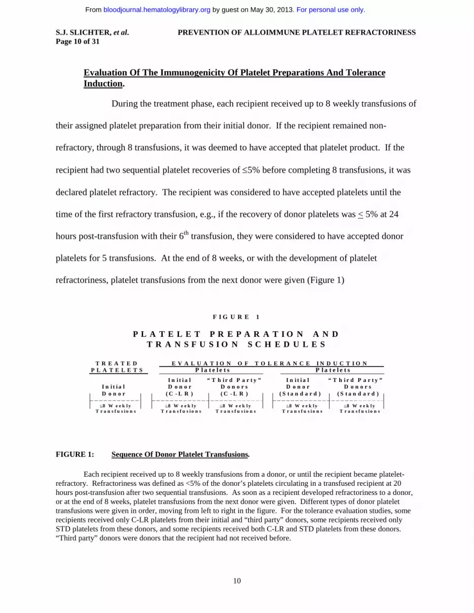
Evaluation Of The Immunogenicity Of Platelet Preparations And Tolerance Induction.
During the treatment phase, each recipient received up to 8 weekly transfusions of
their assigned platelet preparation from their initial donor. If the recipient remained non-
refractory, through 8 transfusions, it was deemed to have accepted that platelet product. If the
recipient had two sequential platelet recoveries of ≤5% before completing 8 transfusions, it was
declared platelet refractory. The recipient was considered to have accepted platelets until the
time of the first refractory transfusion, e.g., if the recovery of donor platelets was < 5% at 24
hours post-transfusion with their 6th transfusion, they were considered to have accepted donor
platelets for 5 transfusions. At the end of 8 weeks, or with the development of platelet
refractoriness, platelet transfusions from the next donor were given (Figure 1)
P L A T E L E T P R E P A R A T I O N A N D T R A N S F U S I O N S C H E D U L E S
< 8 W e e k l y < 8 W e e k l y < 8 W e e k l y < 8 W e e k l y < 8 W e e k l yT r a n s f u s i o n s T r a n s f u s i o n s T r a n s f u s i o n s T r a n s f u s i o n s T r a n s f u s i o n s
I n i t i a l “ T h i r d P a r t y ” I n i t i a l “ T h i r d P a r t y ”I n i t i a l D o n o r D o n o r s D o n o r D o n o r sD o n o r ( C - L R ) ( C - L R ) ( S t a n d a r d ) ( S t a n d a r d )
T R E A T E D E V A L U A T I O N O F T O L E R A N C E I N D U C T I O NP L A T E L E T S P l a t e l e t s P l a t e l e t s
F I G U R E 1
FIGURE 1: Sequence Of Donor Platelet Transfusions.
Each recipient received up to 8 weekly transfusions from a donor, or until the recipient became platelet-refractory. Refractoriness was defined as <5% of the donor’s platelets circulating in a transfused recipient at 20 hours post-transfusion after two sequential transfusions. As soon as a recipient developed refractoriness to a donor, or at the end of 8 weeks, platelet transfusions from the next donor were given. Different types of donor platelet transfusions were given in order, moving from left to right in the figure. For the tolerance evaluation studies, some recipients received only C-LR platelets from their initial and “third party” donors, some recipients received only STD platelets from these donors, and some recipients received both C-LR and STD platelets from these donors. “Third party” donors were donors that the recipient had not received before.
For personal use only. by guest on May 30, 2013. bloodjournal.hematologylibrary.orgFrom
S.J. SLICHTER, et al. PREVENTION OF ALLOIMMUNE PLATELET REFRACTORINESSPage 11 of 31
11
Groups of animals were assigned to specific transfusion programs, and at the conclusion
of one treatment program, another program was initiated. The C-LR initial donor transfusions
were originally performed and reported in 1987 (14). The F-LR transfusion programs were
initiated next, followed by the F-LR/C-LR transfusion program, and finally, standard rather than
treated platelet transfusions were given as the initial transfusion to the last group of recipients.
The number of recipient animals in each group was selected to provide enough data to
demonstrate that the treatment given to the donor’s platelets did or did not prevent platelet
refractoriness, and secondarily, whether tolerance was induced.
Most of the recipients that accepted the treated platelet preparation from their initial
donor were then transfused with C-LR platelets from their initial donor for up to an additional 8
weeks or until they became refractory. C-LR transfusions were then given from third party
donor(s), i.e. dogs to whom the recipient had not been previously exposed to test for non-specific
tolerance. Even if the recipient did not accept the initial donor’s platelets during the treatment
phase C-LR platelet transfusions from up to two “third party” donors were still given. Dogs that
accepted C-LR platelets from at least one “third party” donor were considered non-specifically
tolerant to C-LR platelets. Finally, a single C-LR transfusion was given from the initial donor to
determine if the recipient had developed refractoriness to this product following the third party
transfusions.
If the recipient was not refractory to the initial donor’s C-LR platelets and had completed
any planned third party C-LR transfusions, the recipient was given STD transfusions from the
initial donor, and any third party donors to whom they had not developed refractoriness, to
further evaluate donor specific and non-specific tolerance, respectively. Some recipients were
For personal use only. by guest on May 30, 2013. bloodjournal.hematologylibrary.orgFrom
S.J. SLICHTER, et al. PREVENTION OF ALLOIMMUNE PLATELET REFRACTORINESSPage 12 of 31
12
never given C-LR donor transfusions and were tested for tolerance only by giving STD platelet
transfusions. If a recipient had not developed refractoriness to their initial donor’s treated
platelets, they were given standard platelets from this donor, and all recipients - whether or not
they had accepted their initial donors treated platelets - received STD platelets from third party
donors. After these third party STD transfusions, a final STD transfusion from the initial donor
was done to determine whether the recipient remained tolerant to this donor. Finally, autologous
transfusions were done to determine whether the recipient dogs still had normal platelet
recoveries and survivals.
If both C-LR and STD platelets from the same donor were transfused into the same
recipient, it could be determined whether their was a differential effect on loss of tolerance
depending on the type of WBCs transfused. The WBCs removed by filtration would be present
in both types of platelets (C-LR and STD), while the WBCs removed by centrifugation would be
present only in the STD platelets.
Statistical Methods.
Estimates of rates of acceptance and tolerance are based on the binomial distribution.
The associated confidence intervals were also derived from the binomial distribution. Time to
refractoriness to treated platelets from the initial donor was analyzed using the Cox proportional
hazards model. Comparisons between treatments were based on Wald chi-square statistics from
the Cox model. P values associated with these statistics were adjusted for multiple comparisons
using the Bonferroni method. Descriptive statistics are presented for cell counts from each of the
platelet preparations.
For personal use only. by guest on May 30, 2013. bloodjournal.hematologylibrary.orgFrom
S.J. SLICHTER, et al. PREVENTION OF ALLOIMMUNE PLATELET REFRACTORINESSPage 13 of 31
13
RESULTS
Cell Counts And Viability Of Transfused Platelets.
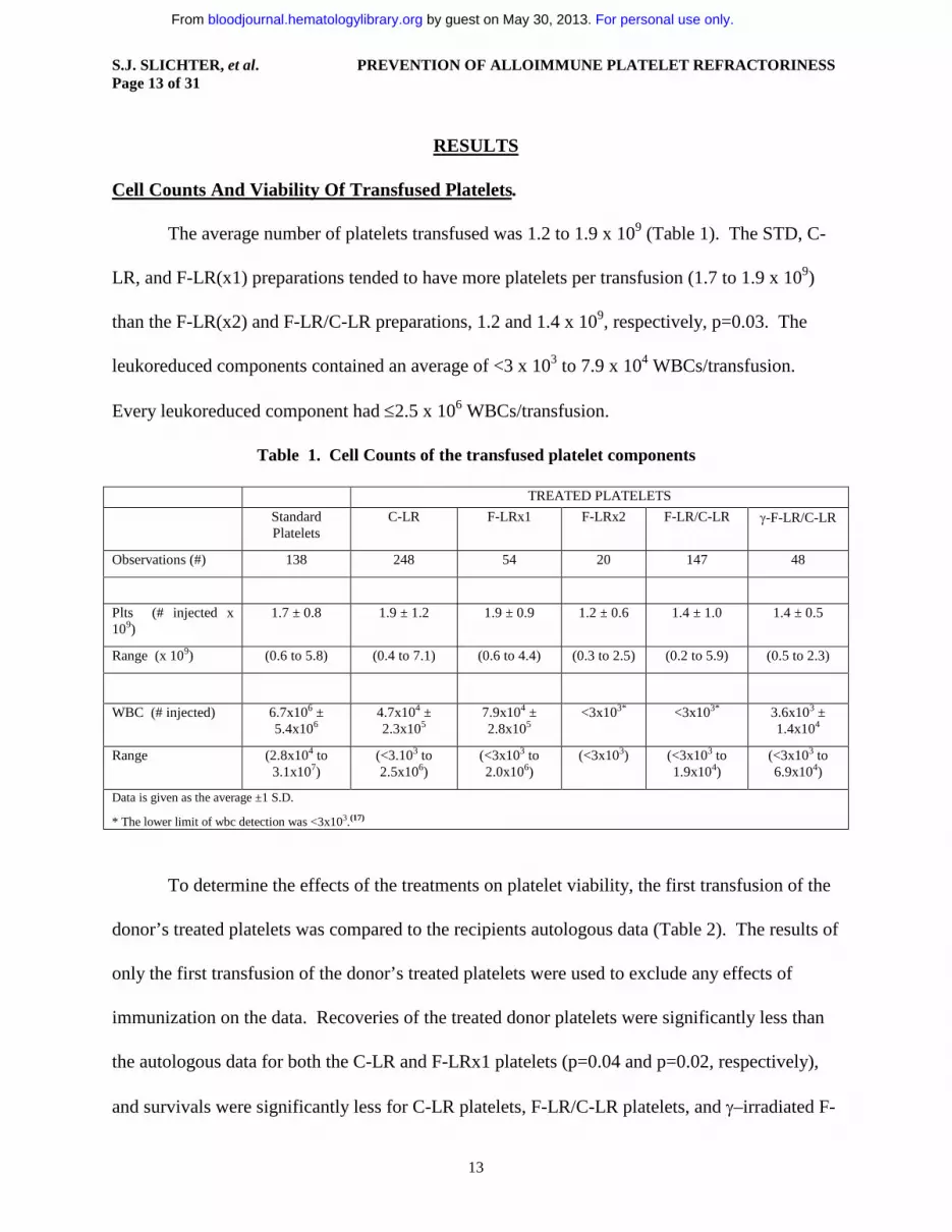
The average number of platelets transfused was 1.2 to 1.9 x 10⁹ (Table 1). The STD, C-
LR, and F-LR(x1) preparations tended to have more platelets per transfusion (1.7 to 1.9 x 10⁹)
than the F-LR(x2) and F-LR/C-LR preparations, 1.2 and 1.4 x 10⁹, respectively, p=0.03. The
leukoreduced components contained an average of <3 x 10³ to 7.9 x 10⁴ WBCs/transfusion.
Every leukoreduced component had ≤2.5 x 10⁶ WBCs/transfusion.
Table 1. Cell Counts of the transfused platelet components
TREATED PLATELETS
Standard Platelets
C-LR F-LRx1 F-LRx2 F-LR/C-LR γ-F-LR/C-LR
Observations (#) 138 248 54 20 147 48
Plts (# injected x 109)
1.7 ± 0.8 1.9 ± 1.2 1.9 ± 0.9 1.2 ± 0.6 1.4 ± 1.0 1.4 ± 0.5
Range (x 109) (0.6 to 5.8) (0.4 to 7.1) (0.6 to 4.4) (0.3 to 2.5) (0.2 to 5.9) (0.5 to 2.3)
WBC (# injected) 6.7x10⁶ ± 5.4x10⁶
4.7x10⁴ ± 2.3x10⁵
7.9x10⁴ ± 2.8x10⁵
<3x10³* <3x103* 3.6x103 ± 1.4x10⁴
Range (2.8x10⁴ to 3.1x10⁷)
(<3.103 to 2.5x10⁶)
(<3x10³ to 2.0x10⁶)
(<3x10³) (<3x10³ to 1.9x10⁴)
(<3x10³ to 6.9x10⁴)
Data is given as the average ±1 S.D.
* The lower limit of wbc detection was <3x10³.(17)
To determine the effects of the treatments on platelet viability, the first transfusion of the
donor’s treated platelets was compared to the recipients autologous data (Table 2). The results of
only the first transfusion of the donor’s treated platelets were used to exclude any effects of
immunization on the data. Recoveries of the treated donor platelets were significantly less than
the autologous data for both the C-LR and F-LRx1 platelets (p=0.04 and p=0.02, respectively),
and survivals were significantly less for C-LR platelets, F-LR/C-LR platelets, and γ–irradiated F-
For personal use only. by guest on May 30, 2013. bloodjournal.hematologylibrary.orgFrom
S.J. SLICHTER, et al. PREVENTION OF ALLOIMMUNE PLATELET REFRACTORINESSPage 14 of 31
14
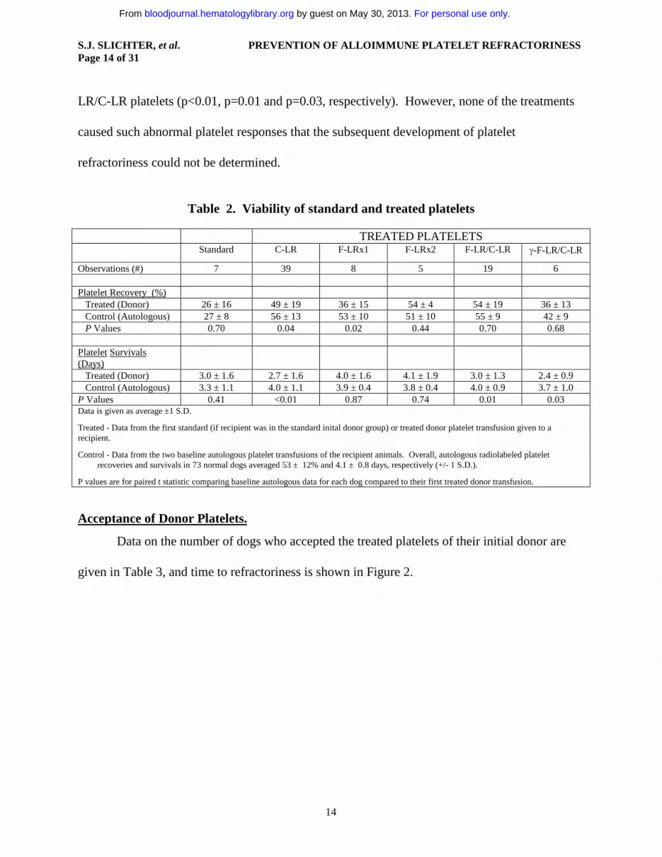
LR/C-LR platelets (p<0.01, p=0.01 and p=0.03, respectively). However, none of the treatments
caused such abnormal platelet responses that the subsequent development of platelet
refractoriness could not be determined.
Table 2. Viability of standard and treated platelets
TREATED PLATELETSStandard C-LR F-LRx1 F-LRx2 F-LR/C-LR γ-F-LR/C-LR
Observations (#) 7 39 8 5 19 6
Platelet Recovery (%) Treated (Donor) 26 ± 16 49 ± 19 36 ± 15 54 ± 4 54 ± 19 36 ± 13 Control (Autologous) 27 ± 8 56 ± 13 53 ± 10 51 ± 10 55 ± 9 42 ± 9
P Values 0.70 0.04 0.02 0.44 0.70 0.68
Platelet Survivals (Days) Treated (Donor) 3.0 ± 1.6 2.7 ± 1.6 4.0 ± 1.6 4.1 ± 1.9 3.0 ± 1.3 2.4 ± 0.9 Control (Autologous) 3.3 ± 1.1 4.0 ± 1.1 3.9 ± 0.4 3.8 ± 0.4 4.0 ± 0.9 3.7 ± 1.0P Values 0.41 <0.01 0.87 0.74 0.01 0.03Data is given as average ±1 S.D.
Treated - Data from the first standard (if recipient was in the standard inital donor group) or treated donor platelet transfusion given to a recipient.
Control - Data from the two baseline autologous platelet transfusions of the recipient animals. Overall, autologous radiolabeled platelet recoveries and survivals in 73 normal dogs averaged 53 ± 12% and 4.1 ± 0.8 days, respectively (+/- 1 S.D.).
P values are for paired t statistic comparing baseline autologous data for each dog compared to their first treated donor transfusion.
Acceptance of Donor Platelets.
Data on the number of dogs who accepted the treated platelets of their initial donor are
given in Table 3, and time to refractoriness is shown in Figure 2.
For personal use only. by guest on May 30, 2013. bloodjournal.hematologylibrary.orgFrom
S.J. SLICHTER, et al. PREVENTION OF ALLOIMMUNE PLATELET REFRACTORINESSPage 15 of 31
15
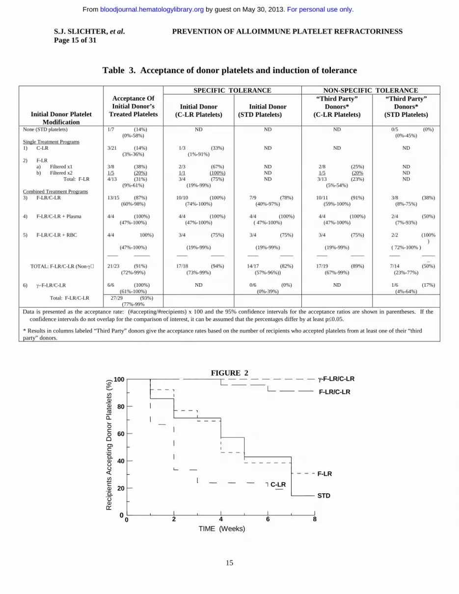
Table 3. Acceptance of donor platelets and induction of tolerance
SPECIFIC TOLERANCE NON-SPECIFIC TOLERANCE
Initial Donor Platelet Modification
Acceptance OfInitial Donor’s
Treated PlateletsInitial Donor
(C-LR Platelets)Initial Donor
(STD Platelets)
“Third Party”Donors*
(C-LR Platelets)
“Third Party”Donors*
(STD Platelets)
None (STD platelets) 1/7 (14%) ND ND ND 0/5 (0%)(0%-58%) (0%-45%)
Single Treatment Programs1) C-LR 3/21 (14%) 1/3 (33%) ND ND ND
(3%-36%) (1%-91%)2) F-LR
a) Filtered x1 3/8 (38%) 2/3 (67%) ND 2/8 (25%) NDb) Filtered x2 1/5 (20%) 1/1 (100%) ND 1/5 (20% ND
Total: F-LR 4/13 (31%) 3/4 (75%) ND 3/13 (23%) ND(9%-61%) (19%-99%) (5%-54%)
Combined Treatment Programs3) F-LR/C-LR 13/15 (87%) 10/10 (100%) 7/9 (78%) 10/11 (91%) 3/8 (38%)
(60%-98%) (74%-100%) (40%-97%) (59%-100%) (8%-75%)
4) F-LR/C-LR + Plasma 4/4 (100%) 4/4 (100%) 4/4 (100%) 4/4 (100%) 2/4 (50%)(47%-100%) (47%-100%) ( 47%-100%) (47%-100%) (7%-93%)
5) F-LR/C-LR + RBC 4/4 100%) 3/4 (75%) 3/4 (75%) 3/4 (75%) 2/2 (100%)
(47%-100%) (19%-99%) (19%-99%) (19%-99%) ( 72%-100% )____ ______ ____ _____ ____ _____ ____ _____ ____ _____
_TOTAL: F-LR/C-LR (Non-γ) 21/23 (91%) 17/18 (94%) 14/17 (82%) 17/19 (89%) 7/14 (50%)
(72%-99%) (73%-99%) (57%-96%)) (67%-99%) (23%-77%)
6) γ−F-LR/C-LR 6/6 (100%) ND 0/6 (0%) ND 1/6 (17%)(61%-100%) (0%-39%) (4%-64%)
Total: F-LR/C-LR 27/29 (93%)(77%-99%
Data is presented as the acceptance rate: (#accepting/#recipients) x 100 and the 95% confidence intervals for the acceptance ratios are shown in parentheses. If the confidence intervals do not overlap for the comparison of interest, it can be assumed that the percentages differ by at least p≤0.05.
* Results in columns labeled “Third Party” donors give the acceptance rates based on the number of recipients who accepted platelets from at least one of their “third party” donors.
FIGURE 2
0 2 4 6 8
TIME (Weeks)
0
20
40
60
80
100
Rec
ipie
nts
Acc
eptin
g D
onor
Pla
tele
ts (
%) γ-F-LR/C-LR
F-LR/C-LR
F-LR
STD
C-LR
For personal use only. by guest on May 30, 2013. bloodjournal.hematologylibrary.orgFrom
S.J. SLICHTER, et al. PREVENTION OF ALLOIMMUNE PLATELET REFRACTORINESSPage 16 of 31
16
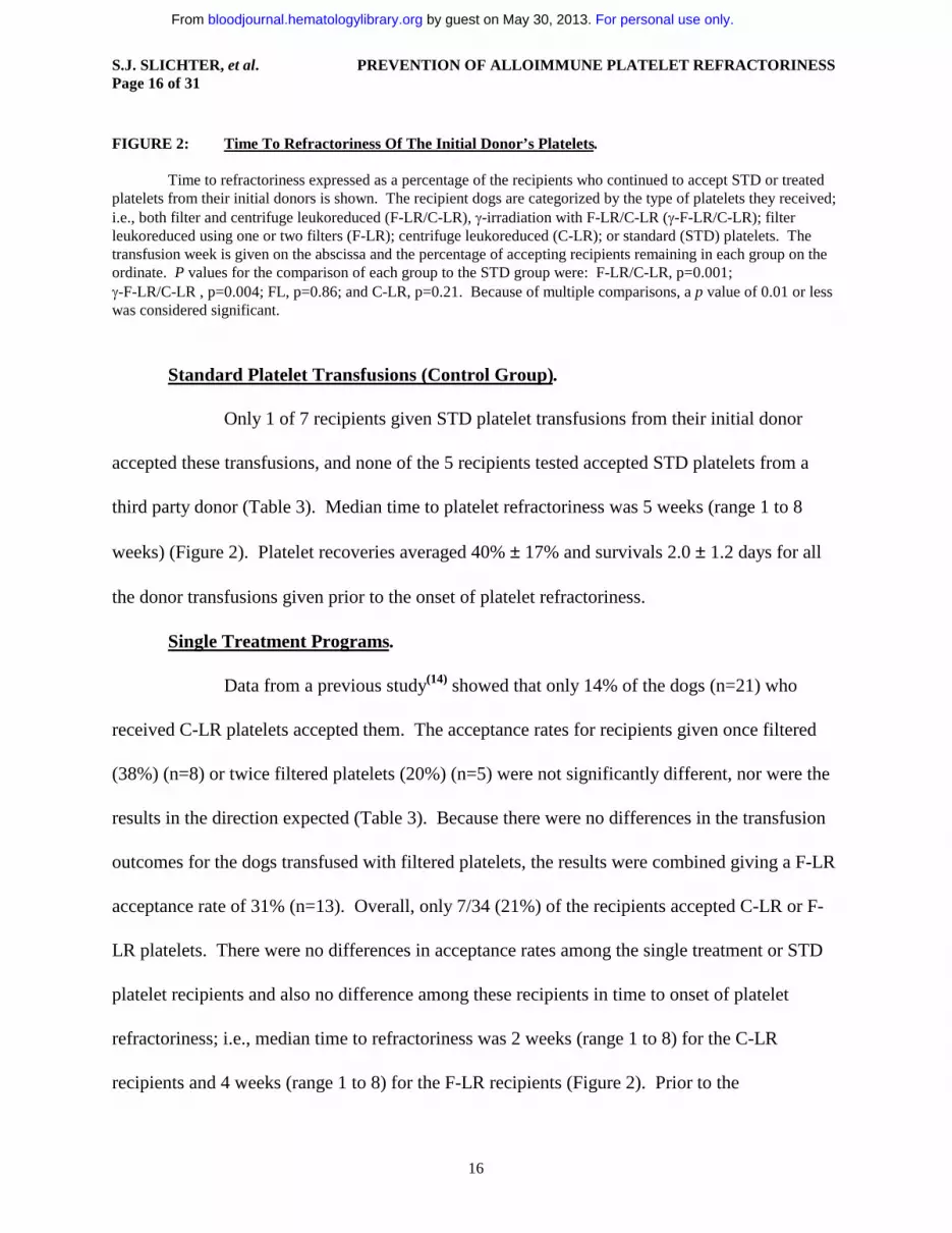
FIGURE 2: Time To Refractoriness Of The Initial Donor’s Platelets.
Time to refractoriness expressed as a percentage of the recipients who continued to accept STD or treated platelets from their initial donors is shown. The recipient dogs are categorized by the type of platelets they received; i.e., both filter and centrifuge leukoreduced (F-LR/C-LR), γ-irradiation with F-LR/C-LR (γ-F-LR/C-LR); filter leukoreduced using one or two filters (F-LR); centrifuge leukoreduced (C-LR); or standard (STD) platelets. The transfusion week is given on the abscissa and the percentage of accepting recipients remaining in each group on the ordinate. P values for the comparison of each group to the STD group were: F-LR/C-LR, p=0.001; γ-F-LR/C-LR , p=0.004; FL, p=0.86; and C-LR, p=0.21. Because of multiple comparisons, a p value of 0.01 or less was considered significant.
Standard Platelet Transfusions (Control Group).
Only 1 of 7 recipients given STD platelet transfusions from their initial donor
accepted these transfusions, and none of the 5 recipients tested accepted STD platelets from a
third party donor (Table 3). Median time to platelet refractoriness was 5 weeks (range 1 to 8
weeks) (Figure 2). Platelet recoveries averaged 40% ± 17% and survivals 2.0 ± 1.2 days for all
the donor transfusions given prior to the onset of platelet refractoriness.
Single Treatment Programs.
Data from a previous study(14) showed that only 14% of the dogs (n=21) who
received C-LR platelets accepted them. The acceptance rates for recipients given once filtered
(38%) (n=8) or twice filtered platelets (20%) (n=5) were not significantly different, nor were the
results in the direction expected (Table 3). Because there were no differences in the transfusion
outcomes for the dogs transfused with filtered platelets, the results were combined giving a F-LR
acceptance rate of 31% (n=13). Overall, only 7/34 (21%) of the recipients accepted C-LR or F-
LR platelets. There were no differences in acceptance rates among the single treatment or STD
platelet recipients and also no difference among these recipients in time to onset of platelet
refractoriness; i.e., median time to refractoriness was 2 weeks (range 1 to 8) for the C-LR
recipients and 4 weeks (range 1 to 8) for the F-LR recipients (Figure 2). Prior to the
For personal use only. by guest on May 30, 2013. bloodjournal.hematologylibrary.orgFrom
S.J. SLICHTER, et al. PREVENTION OF ALLOIMMUNE PLATELET REFRACTORINESSPage 17 of 31
17
development of platelet refractoriness, average platelet recoveries were 56 ± 11% and 39 ± 16%;
and survivals were 2.8 ± 1.1 and 3.1 ± 2.2 days for all C-LR and F-LR donor transfusions,
respectively (no significant difference among STD, C-LR or F-LR transfusions) .
Combined Treatment Programs.
Among the combined treatment programs, 87% of the F-LR/C-LR recipients
(n=15) and 100% of the γ-F-LR/C-LR (n=6) accepted their initial donor’s treated platelet
transfusions (Table 3). To ensure that these results were specifically related to WBC removal
and not to the removal of plasma or RBC that also occurred during the processing steps, F-LR/C-
LR plasma and F-LR/C-LR washed RBC were added back to F-LR/C-LR platelets from the same
donor. Neither the addition of F-LR/C-LR plasma (n=4) nor F-LR/C-LR washed RBC (n=4) to
F-LR/C-LR platelets changed the acceptance rates of the F-LR/C-LR platelets (100%
acceptance). Grouping the data from all these F-LR/C-LR recipients, the acceptance rate was
93% (n=29,) (p<0.05 compared to STD, C-LR or F-LR platelets). Time to platelet refractoriness
for recipients given F-LR/C-LR or γ–F-LR/C-LR platelets versus STD platelets were statistically
different, p<0.001 and p=0.004, respectively (Figure 2).
For all the donor transfusions given prior to the onset of platelet refractoriness.
platelet recoveries averaged 47% ± 16% and 55% ± 13%, and platelet survivals averaged 2.6 ±
1.0 days and 2.6 ± 0.9 days for F-LR/C-LR and γ-F-LR/C-LR transfusions, respectively (no
significant difference compared to STD, C-LR or F-LR transfusions).
For personal use only. by guest on May 30, 2013. bloodjournal.hematologylibrary.orgFrom
S.J. SLICHTER, et al. PREVENTION OF ALLOIMMUNE PLATELET REFRACTORINESSPage 18 of 31
18
Tolerance Induction To Platelets.
Specific Tolerance.
For recipients who had not become refractory to treated platelet transfusions from
their initial donor, most received C-LR platelet transfusions from these same donors. And, for
the recipients of F-LR or F-LR/C-LR, these C-LR platelets were well tolerated; i.e., 75% and
94% of these recipients, respectively, remained platelet responsive. Overall, among the 22
initially non-refractory recipients tested in the F-LR and F-LR/C-LR groups, 20 (91%) remained
responsive to their initial donor’s C-LR platelets. This is in contrast to the dogs in the C-LR arm,
all of whom were exposed to C-LR platelets from their initial donor without ‘prior conditioning’.
Only 3 out of 21 (14%) remained platelet responsive to their initial donors treated transfusions
during the first 8 weeks of C-LR transfusions and when the 3 remaining recipients were given an
additional 8 weeks of C-LR platelets from their initial donor only 1 remained non-refractory
(p=0.001). This suggests that, under ordinary circumstances, C-LR transfusions are very
immunogenic.
STD platelet transfusions from their initial donors were only given to some of the
non-refractory recipients of the combined treatment programs. Of the animals that had
previously received F-LR/C-LR platelets, 82% of the recipients (n=17) remained responsive to
STD platelets from their initial donor.
However, if the initial donor’s F-LR/C-LR platelets were also γ-irradiated prior to
transfusion, tolerance was not induced to STD platelets from any of the initial donors even
though the donor’s γ-irradiated F-LR/C-LR platelets were accepted (p<0.01 compared to STD
transfusions given to recipients of non-γ-irradiated F-LR/C-LR platelets).
For personal use only. by guest on May 30, 2013. bloodjournal.hematologylibrary.orgFrom
S.J. SLICHTER, et al. PREVENTION OF ALLOIMMUNE PLATELET REFRACTORINESSPage 19 of 31
19
Non-Specific Tolerance.
Excluding the C-LR recipients who received transfusions only from their initial
donor, all of the other recipients received either C-LR, STD, or both C-LR and STD platelets
from other donors; i.e., so-called “third party” donors. Most of the recipients (30/31; 97%)
received C-LR platelets from two other donors. If they remained responsive to platelets from at
least one of these donors, they were considered as accepting that product. Acceptance of C-LR
platelets from “third party” donors varied widely from only 23% of the recipients (n=18) of F-LR
platelets to 89% of the recipients (n=19) of F-LR/C-LR platelets (p<0.05) (Table 3).
Among the 20 animals who were tolerant to both treated and C-LR platelet
transfusions from their initial donor, only 4 (20%) became refractory to C-LR platelets from at
least one “third party” donor [3 were refractory to one donor (75%) and 1 to two donors (25%)].
In contrast, of the 11 dogs who became refractory to their initial donor’s either treated or C-LR
platelets, all (100%) became refractory to C-LR platelets from at least one “third party” donor
(p<0.01) [1 was refractory to one of two donors (9%) and 10 were refractory to both donors
(91%)].
Of the recipients who received STD platelets from “third party” donors, 16/19
(84%) received platelets from two donors. The STD platelets from “third party” donors were not
tolerated nearly as well as the C-LR platelets from “third party” donors. Only 7/14 recipients
(50%) of F-LR/C-LR platelets accepted STD platelets from at least one of their “third party”
donors (Table 3). Three recipients accepted platelets from both donors, and 4 accepted platelets
from one donor. Again, γ-irradiation of the initial donor’s F-LR/C-LR platelets essentially
eliminated tolerance to STD platelets from “third party” donors; i.e., only 1/6 recipients (17%)
For personal use only. by guest on May 30, 2013. bloodjournal.hematologylibrary.orgFrom
S.J. SLICHTER, et al. PREVENTION OF ALLOIMMUNE PLATELET REFRACTORINESSPage 20 of 31
20
accepted platelets from 1 of the 11 “third party” donors tested (p=0.32 compared to recipients of
F-LR/C-LR non-γ-irradiated platelets).
Refractoriness to transfused platelets from an earlier donor predicted a recipient’s
response to platelets from a later donor, i.e., 27/31 (87%) recipients continued to “reject”
platelets from all later donors. At the end of the study period, previous recipients who had
accepted donor platelets were re-transfused to determine whether refractoriness to a later donor’s
platelets had induced refractoriness to an earlier donor’s tolerated platelets. Of 18 informative
pairs who had accepted some but not all donors, 7 recipients (39%) had become refractory to a
previously-tolerated donor. In contrast, of 7 recipients who tolerated platelets from all of their
donors, only 1 recipient (14%) showed refractoriness to platelets from a previously accepted
donor.
All end of study autologous platelet recovery and survival measurements gave
results comparable to each recipient dogs’ pre-transfusion (baseline) autologous platelet studies
(data not shown). This suggests that all refractoriness to donor platelets was immune mediated
regardless of whether antibodies were detected. If platelet refractoriness was related to some
intercurrent condition, it would have adversely effected the recipient’s response to both
autologous as well as allogeneic platelets.
Development Of Platelet Alloantibodies.
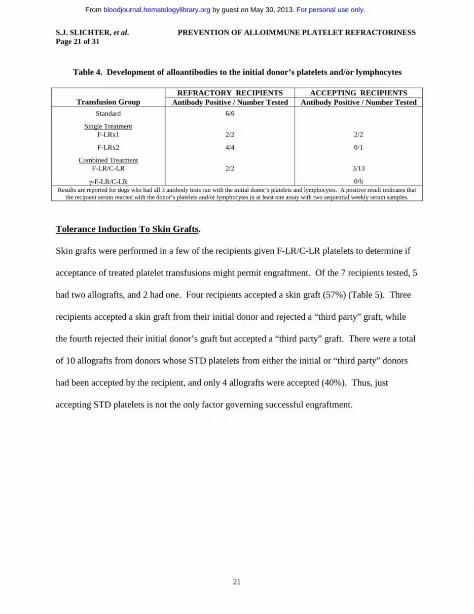
Weekly serum samples were available for antibody testing against donor platelets from a
variable number of recipients (Table 4). Antibody results were positive with platelets or
lymphocytes from all 14 donors whose platelets were not accepted (100%) and from only 5/22
(23%) of donors whose platelets were accepted (p=0.04).
For personal use only. by guest on May 30, 2013. bloodjournal.hematologylibrary.orgFrom
S.J. SLICHTER, et al. PREVENTION OF ALLOIMMUNE PLATELET REFRACTORINESSPage 21 of 31
21
Table 4. Development of alloantibodies to the initial donor’s platelets and/or lymphocytes
REFRACTORY RECIPIENTS ACCEPTING RECIPIENTSTransfusion Group Antibody Positive / Number Tested Antibody Positive / Number Tested
Standard 6/6
Single TreatmentF-LRx1 2/2 2/2
F-LRx2 4/4 0/1
Combined TreatmentF-LR/C-LR 2/2 3/13
γ-F-LR/C-LR 0/6Results are reported for dogs who had all 3 antibody tests run with the initial donor’s platelets and lymphocytes. A positive result indicates that
the recipient serum reacted with the donor’s platelets and/or lymphocytes in at least one assay with two sequential weekly serum samples.
Tolerance Induction To Skin Grafts.
Skin grafts were performed in a few of the recipients given F-LR/C-LR platelets to determine if
acceptance of treated platelet transfusions might permit engraftment. Of the 7 recipients tested, 5
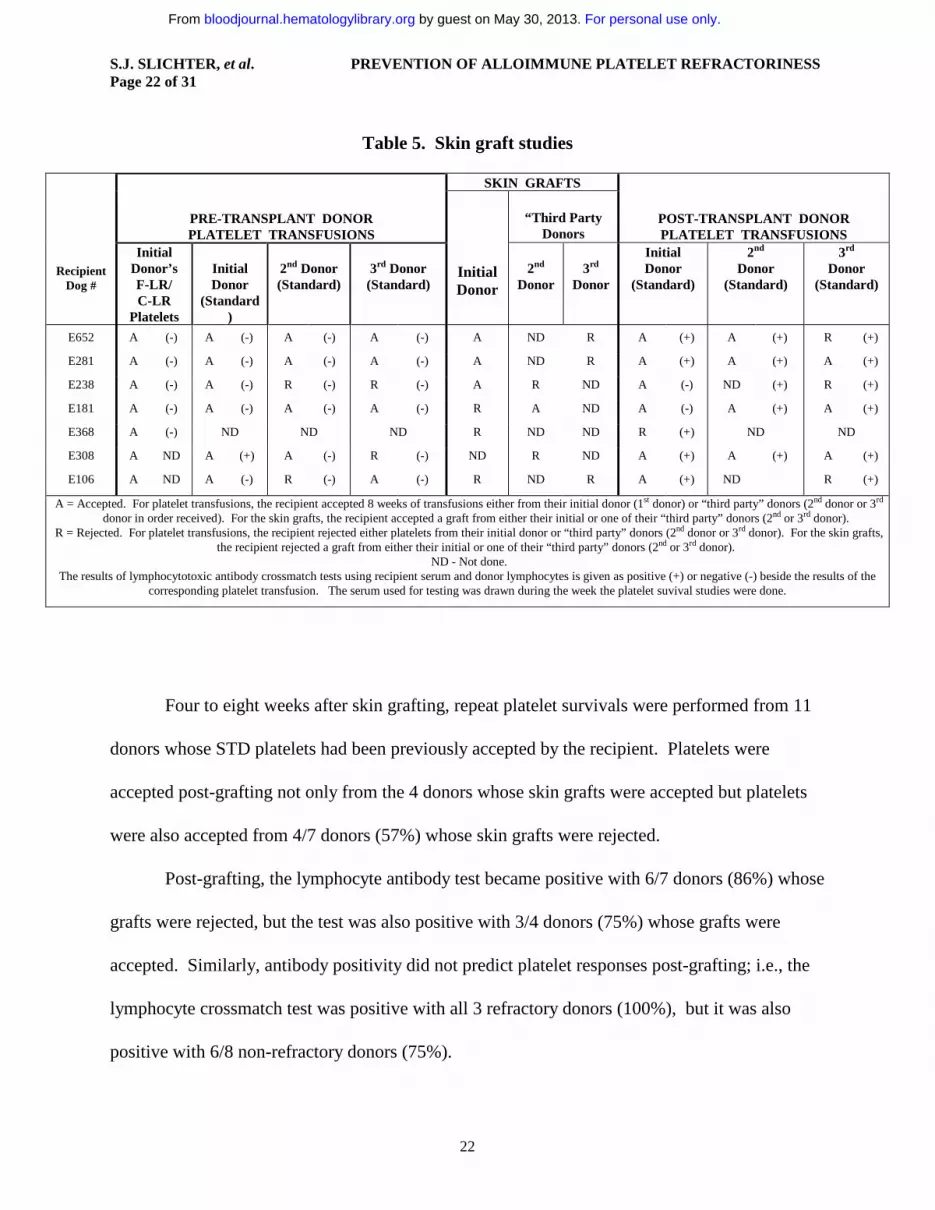
had two allografts, and 2 had one. Four recipients accepted a skin graft (57%) (Table 5). Three
recipients accepted a skin graft from their initial donor and rejected a “third party” graft, while
the fourth rejected their initial donor’s graft but accepted a “third party” graft. There were a total
of 10 allografts from donors whose STD platelets from either the initial or “third party” donors
had been accepted by the recipient, and only 4 allografts were accepted (40%). Thus, just
accepting STD platelets is not the only factor governing successful engraftment.
For personal use only. by guest on May 30, 2013. bloodjournal.hematologylibrary.orgFrom
S.J. SLICHTER, et al. PREVENTION OF ALLOIMMUNE PLATELET REFRACTORINESSPage 22 of 31
22
Table 5. Skin graft studies
SKIN GRAFTS
PRE-TRANSPLANT DONORPLATELET TRANSFUSIONS
“Third Party Donors
POST-TRANSPLANT DONORPLATELET TRANSFUSIONS
RecipientDog #
Initial Donor’s F-LR/C-LR
Platelets
Initial Donor
(Standard)
2nd Donor(Standard)
3rd Donor(Standard)
InitialDonor
2nd
Donor3rd
Donor
InitialDonor
(Standard)
2nd
Donor (Standard)
3rd
Donor(Standard)
E652 A (-) A (-) A (-) A (-) A ND R A (+) A (+) R (+)
E281 A (-) A (-) A (-) A (-) A ND R A (+) A (+) A (+)
E238 A (-) A (-) R (-) R (-) A R ND A (-) ND (+) R (+)
E181 A (-) A (-) A (-) A (-) R A ND A (-) A (+) A (+)
E368 A (-) ND ND ND R ND ND R (+) ND ND
E308 A ND A (+) A (-) R (-) ND R ND A (+) A (+) A (+)
E106 A ND A (-) R (-) A (-) R ND R A (+) ND R (+)
A = Accepted. For platelet transfusions, the recipient accepted 8 weeks of transfusions either from their initial donor (1st donor) or “third party” donors (2nd donor or 3rd
donor in order received). For the skin grafts, the recipient accepted a graft from either their initial or one of their “third party” donors (2nd or 3rd donor).R = Rejected. For platelet transfusions, the recipient rejected either platelets from their initial donor or “third party” donors (2nd donor or 3rd donor). For the skin grafts,
the recipient rejected a graft from either their initial or one of their “third party” donors (2nd or 3rd donor).ND - Not done.
The results of lymphocytotoxic antibody crossmatch tests using recipient serum and donor lymphocytes is given as positive (+) or negative (-) beside the results of the corresponding platelet transfusion. The serum used for testing was drawn during the week the platelet suvival studies were done.
Four to eight weeks after skin grafting, repeat platelet survivals were performed from 11
donors whose STD platelets had been previously accepted by the recipient. Platelets were
accepted post-grafting not only from the 4 donors whose skin grafts were accepted but platelets
were also accepted from 4/7 donors (57%) whose skin grafts were rejected.
Post-grafting, the lymphocyte antibody test became positive with 6/7 donors (86%) whose
grafts were rejected, but the test was also positive with 3/4 donors (75%) whose grafts were
accepted. Similarly, antibody positivity did not predict platelet responses post-grafting; i.e., the
lymphocyte crossmatch test was positive with all 3 refractory donors (100%), but it was also
positive with 6/8 non-refractory donors (75%).
For personal use only. by guest on May 30, 2013. bloodjournal.hematologylibrary.orgFrom
S.J. SLICHTER, et al. PREVENTION OF ALLOIMMUNE PLATELET REFRACTORINESSPage 23 of 31
23
DISCUSSION
The most important study findings were the effects of the different methods of
leukoreduction and γ-irradiation on acceptance of donor platelets. In a normal
immunocompetent recipient, centrifuge leukoreduction (C-LR) or filtration leukoreduction (F-
LR) - in spite of reducing the average level of contaminating leukocytes to 4.7 to 7.9 x
104/transfusion, respectively - prevented alloimmune platelet refractoriness in only 14% and 38%
of the transfused recipients.. These results do not differ from those observed with the transfusion
of standard (STD) unmodified donor platelets; i.e., 1/7 recipients (14%) accepted donor platelets.
The levels of leukocyte reduction achieved were well below the level of <5.0 x 10⁶
leukocytes/transfusion considered to prevent alloimmunization in humans. (20) The dogs given
leukoreduced platelets received 4.7 to 7.9 x 10³ WBC’s/kg, versus a 70kg man would receive 7.1
x 104 WBC’s /kg with a transfusion containing 5 x 10⁶ WBC’s; i.e. dogs received a log less
WBC’s/kg. In addition, the leukoreduction was done at the time of blood drawing which prior
rabbit platelet transfusion experiments had suggested was important to prevent immunization.(21)
Even when the platelets were filtered twice to further reduce the level of the contaminating WBC
to <3 x 10³/transfusion (at the lower limit of detection of the assay), the level of protection was
not improved; i.e., only 20% of the recipients (n=5) receiving platelets filtered twice accepted
donor platelets compared to an acceptance rate of 38% in recipients who received platelets
filtered only once (n=8). These data suggest that just making a quantitative reduction in the level
of the contaminating leukocytes does not prevent alloimmune platelet refractoriness. In fact, data
in a mouse transfusion model suggests that extremely leukoreduced platelet transfusions are
more immunogenic then when some residual wbc’s are transfused along with the platelets.(22)
For personal use only. by guest on May 30, 2013. bloodjournal.hematologylibrary.orgFrom
S.J. SLICHTER, et al. PREVENTION OF ALLOIMMUNE PLATELET REFRACTORINESSPage 24 of 31
24
Surprisingly, combining the two methods of leukoreduction (F-LR and C-LR) produced
the best results; i.e., 87% of the recipients (n=15) accepted F-LR/C-LR donor platelets (p<0.05
compared to C-LR, F-LR, or STD platelet transfusions). Furthermore, addback experiments
demonstrated that neither F-LR/C-LR plasma nor F-LR/C-LR washed red cells were
immunogenic, suggesting that the results achieved with F-LR/C-LR platelets were related to
leukocyte removal rather than the elimination of contaminating plasma or red cells during
platelet preparation. Acceptance of F-LR/C-LR platelets using all these results was 91% (n=23).
These studies are in conflict with prior rabbit platelet transfusion studies that suggested that
soluable leukocyte antigens present in plasma are immunogenic.(21)
These data suggest that there is a qualitative difference in the WBC populations removed
by the two leukocyte-reduction techniques and imply the presence of at least two populations of
allostimulatory cells in platelet transfusions. One of these allostimulatory cell populations is a
filter-adherent cell, perhaps in the lymphocyte and/or monocyte populations, both of which are
removed by filtration,(23) whereas the other population may consist of dense, non-adherent cells
that can be removed by centrifugation. A candidate for this second cell type would be dendritic
precursors,(24) known to be allostimulatory, and present in very low quantities in peripheral
blood. Only when both of these populations of WBC are removed is a non-immunogenic platelet
preparation achieved. We may have, by chance, arrived at an optimal type and concentration of
residual white cells that prevent alloimmunization as has been previously suggested by Semple,
et al.(22) Currently, studies are being performed to characterize the cells that are removed or
remain after each of the preparative steps and these results will be reported when completed.
For personal use only. by guest on May 30, 2013. bloodjournal.hematologylibrary.orgFrom
S.J. SLICHTER, et al. PREVENTION OF ALLOIMMUNE PLATELET REFRACTORINESSPage 25 of 31
25
Significantly, not only did the F-LR/C-LR platelets prevent alloimmune platelet
refractoriness, but they also induced tolerance to donor platelets and skin grafts. Following the
F-LR/C-LR platelet transfusions, two different types of donor platelets were transfused to test for
tolerance induction: C-LR platelets and STD platelets from both the initial and “third party”
platelet donors. The former platelets contained only the cells that would ordinarily be removed
by filtration, while the latter contained both types of WBC. Two observations are of note: 1) F-
LR/C-LR recipients accepted C-LR platelets from both the initial donor as well as”third party”
donors (94% and 89% acceptance, respectively) at higher rates than STD platelets from the same
donors (82% and 50% acceptance, respectively); and 2) not unexpectedly, acceptance rates were
higher for platelets from the initial donor than from”third party” donors; i.e., specific tolerance
rates were better than non-specific rates. These studies also suggest that, after the treated
transfusions, the cell that is removed by filtration is often accepted, while the cell that is removed
by centrifugation is more likely to “break” tolerance.
Of the seven F-LR/C-LR recipients tested, four (57%) accepted at least one skin graft
from an allogeneic donor (3 from the initial platelet donor and 1 from a “third party” donor). We
postulated that acceptance of STD platelets from a donor might predict the acceptance of that
donor’s skin graft. Although STD platelets from 10 donors were accepted, skin grafts from only
4 of these donors were accepted (40%) suggesting that there are other factors involved in
accepting skin grafts. This is reinforced by the observation that STD donor platelets were still
accepted following skin graft rejection by 4/7 (57%) of the recipients. Possibly fortuitous
histocompatibility matching between donors and recipients may have influenced not only the
acceptance of STD platelets but also skin grafts from some but not all donors. As skin grafts are
For personal use only. by guest on May 30, 2013. bloodjournal.hematologylibrary.orgFrom
S.J. SLICHTER, et al. PREVENTION OF ALLOIMMUNE PLATELET REFRACTORINESSPage 26 of 31
26
very immunogenic, these data may suggest that prior transfusions of F-LR/C-LR platelets to
recipients awaiting organ grafts may facilitate engraftment.
To further evaluate the mechanisms of tolerance induction by F-LR/C-LR platelets, six
recipients received F-LR/C-LR donor platelets that had also been γ-irradiated. Although all these
recipients accepted the F-LR/C-LR γ-irradiated platelets (100%) similar to the 91% acceptance
rate for F-LR/C-LR platelets, none of these recipients accepted STD platelets from their initial
donor and only one accepted STD platelets from a “third party” donor (p<0.01 and p=0.32,
respectively, compared to STD platelet transfusions given to recipients of F-LR/C-LR platelets).
These data suggest that, following F-LR/C-LR, there remains a residual white cell that induces
tolerance and that the function of this white cell is abrogated by γ-irradiation. Thus, in order for
tolerance to occur, a white cell capable of dividing must be transfused; i.e., microchimerism has
occurred. It may be that Semple and his colleagues inadvertently removed the tolerance inducing
cell when they produced an extremely leukoreduced product.(22)
Antibody assays were positive with either platelets, lymphocytes, or both from all 14
tested recipients who became refractory to their initial donors (100%) and with only 5/24 (21%)
of donors they accepted. Thus, there was a direct correlation between antibody positivity and
platelet refractoriness (p=0.04). However, regardless of the results of the antibody tests, platelet
refractoriness in this study was assumed to be alloimmune as these were normal dogs, on no
medications, they were clinically well, and their autologous platelet recoveries and survivals
were all within the normal range both pre- and post-study suggesting that non-immune
mechanisms of platelet refractoriness were not present in these recipient dogs.
For personal use only. by guest on May 30, 2013. bloodjournal.hematologylibrary.orgFrom
S.J. SLICHTER, et al. PREVENTION OF ALLOIMMUNE PLATELET REFRACTORINESSPage 27 of 31
27
Of interest, lymphocytoxic antibody tests did not predict the results of skin grafting or
donor platelet survival measurements post-grafting. All seven of the skin graft recipients were
lymphocytotoxic antibody negative pre-grafting with their 12 skin graft donors. As expected
post-grafting, lymphocytotoxic antibody tests became positive with 5/6 donors whose skin grafts
were rejected (83%). However, they were also positive with 3/4 (75%) of the recipients whose
skin grafts were accepted. Similarly, the lymphocytotoxic antibody test was positive with the 3
donors whose STD platelets were rejected post-grafting (100%), but the test was also positive
with 6/8 donors (75%) whose STD platelets were accepted post-grafting.
Although it can never be assumed that studies performed in an animal model can be
transferred to man, there is data to suggest that, for bone marrow transplantation studies(25,26) and
potentially also for platelet transfusion studies,(14,15) the dog may serve as a good pre-clinical
model. However, it could be argued that the dog studies do not predict outcomes in man because
filtration leukoreduction was not nearly as effective in the dog as it has been in patients.(1)
However, leukoreduction may have been more successful in patients because they were receiving
high doses of potentially immunosuppressive chemotherapy while they were being transfused as
opposed to the dogs who had a normal immune system. Certainly, in prior studies, patients
receiving chemotherapy had much lower rates of antibody formation compared to patients with
aplastic anemia (27). It is possible that F-LR/C-LR platelets may 0 prevent alloimmune platelet
refractoriness in all patients regardless of the status of their immune system. In addition, the
tolerance induction studies may lend insight into the so-called “immunomodulatory effects of
transfusion”(28) and may suggest that specific types of transfusions may be needed, depending on
the desired clinical outcome. For example, kidney transplant recipients might benefit from F-
For personal use only. by guest on May 30, 2013. bloodjournal.hematologylibrary.orgFrom
S.J. SLICHTER, et al. PREVENTION OF ALLOIMMUNE PLATELET REFRACTORINESSPage 28 of 31
28
LR/C-LR platelet transfusions that could lead to tolerance induction without inducing
immunization, while transfused surgery patients who may have higher rates of post-operation
infection or tumor recurrences might benefit from γ-irradiated transfusions to prevent
immunomodulation.
For personal use only. by guest on May 30, 2013. bloodjournal.hematologylibrary.orgFrom

S.J. SLICHTER, et al. PREVENTION OF ALLOIMMUNE PLATELET REFRACTORINESSPage 29 of 31
29
ACKNOWLEDGMENTS
The authors are indebted to Ginny Knight and Leta Stever for administrative and secretarial assistance.
For personal use only. by guest on May 30, 2013. bloodjournal.hematologylibrary.orgFrom
S.J. SLICHTER, et al. PREVENTION OF ALLOIMMUNE PLATELET REFRACTORINESS
30
REFERENCES
1. The Trial To Reduce Alloimmunization To Platelets Study Group. Leukocyte reduction and ultraviolet B irradiation of platelets to prevent alloimmunization and refractoriness to platelet transfusions. N Engl J Med. 1997;337:1861-1869.
2. Kickler TS. The challenge of platelet alloimmunization management and prevention. Transfus Med Rev. 1990;4(4 Suppl 1):8-18.
3. Eernisse JG, Brand A. Prevention of platelet refractoriness due to HLA antibodies by administration of leukocyte-poor blood components. Exp Hematol. 1981;9:77-83.
4. Schiffer CA, Dutcher JP, Aisner J, Hogge D, Wiernik PH, Reilly JP. A randomized trial of leukocyte-depleted platelet transfusion to modify alloimmunization in patients with leukemia. Blood. 1983;62:815-820.
5. Andreu G, Dewailly J, Leberre C, et al. Prevention of HLA immunization with leukocyte-poor packed red cells and platelet concentrates obtained by filtration. Blood. 1988;72:964-969, 1988.
6. Sniecinski I, O’Donnell MR, Nowicki B, Hill LR. Prevention of refractoriness and HLA-alloimmunization using filtered blood products. Blood. 1988;71:1402-1407.
7. Saarinen UM, Kekomaki R, Siimes MA, Myllyla G. Effective prophylaxis against platelet refractoriness in multitransfused patients by use of leukocyte-free blood components. Blood. 1990;75:512-517.
8. van Marwijk Kooy M, van Prooijen HC, Moes M, Bosma-Stants I, Akkerman JW. Use of leukocyte-depleted platelet concentrates for the prevention of refractoriness and primary HLA alloimmunization: A prospective, randomized trial. Blood. 1991;77:201-205.
9. Snyder EL. Prevention of HLA alloimmunization: Role of leukocyte depletion and UVB irradiation. Yale J Biol Med. 1990;63:419-427.
10. Oksanen K, Kekomäki R, Ruutu T, Koskimies S, Myllylä G. Prevention of alloimmunization in patients with acute leukemia by use of white cell-reduced blood components - a randomized trial. Transfusion. 1991;31:588-594.
11. Freedman JJ, Blajchman MA, McCombie N. Canadian Red Cross Society Symposium on leukodepletion: Report of proceedings. Transfus Med Rev. 1994;8:1-14.
12. Williamson LM, Wimperis JZ, Williamson P, et al. Bedside filtration of blood products in the prevention of HLA alloimmunization - A prospective randomized study. Blood. 1994;83:3028-3035.
13. Novotny VMJ, van Doorn R, Witvliet MD, Claas FH, Brand A. Occurrence of allogeneic HLA and non-HLA antibodies after transfusion of prestorage filtered platelets and red blood cells: A prospective study. Blood. 1995;85:1736-1741.
14. Slichter SJ, Deeg HJ, Kennedy MS. Prevention of platelet alloimmunization in dogs with systemic cyclosporine and by UV-irradiation or cyclosporine-loading of donor platelets. Blood. 1987;69:414-418.
15. Slichter SJ, O’Donnell MR, Weiden PL, Storb R, Schroeder ML. Canine platelet alloimmunization: The role of donor selection. Br J Haematol 63:713, 1986.
16. Weiden PL, Storb R, Slichter S, Warren RP, Sale GE. Effect of six weekly transfusions on canine marrow grafts: Tests for sensitization and abrogation of sensitization by procarbazine and antithymocyte serum. J Immunol. 1976;117:143-150.
For personal use only. by guest on May 30, 2013. bloodjournal.hematologylibrary.orgFrom
S.J. SLICHTER, et al. PREVENTION OF ALLOIMMUNE PLATELET REFRACTORINESS
31
17. Kao KJ, Scornik JC. Accurate quantitation of the low number of white cells in white cell-depleted blood components. Transfusion. 1989;29:774-777.
18. Schwartz, KA, et al. Immune-mediated platelet destruction and thrombocytopenia in patients with solid tumors. Br J Haematol 1982;51:17-24.
19. Lewis DC, McVey DS, Shuman WS, Muller WB. Development and characterization of a flow cytometric assay for detection of platelet-cound immunoglobulin G in dogs. Am J Vet Res 1995;56:1555-1558.
20. Fisher M, Chapman JR, Ting A, Morris PJ. Alloimmunization to HLA antigens following transfusion with leucocyte-poor and purified platelet suspensions. Vox Sang. 1985;49:331-335.
21. Bordin JO, Bardossy L, Blajchman MA. Experimental animal model of refractoriness todonor platelets: The effect of plasma removal and the extent of white cell reduction on allogeneic alloimmunization. Transfusion. 1993;33:798-801.
22. Semple JW, Speck ER, Cosgrave D, Lazarus AH, Blanchette VS, Freedman J. Extreme leukoreduction of major histocompatibility complex class II positive B cells enhances allogeneic platelet immunity. Blood 1999;93:713-720.
23. Sowemimo-Coker SO, Kim A, Tribble E, Brandwein HJ, Wenz BJ. White cell subsets in apheresis and filtered platelet concentrates. Transfusion. 1998;38:650-657.
24. Kalhs P, White JS, Gervassi A, Storb R, Bean MJ. In vitro recall of proliferative and cytolytic responses to minor histocompatibility antigens by dendritic cell enriched canine peripheral blood mononuclear cells. Transplantation 1995;59:112-118.
25. Weiden PL, Storb R, Thomas ED, et al. Preceding transfusions and marrow graft rejection in dogs and man. Transplant Proc. 1976;8:551-554.
26. Deeg HJ, Aprile J, Graham TC, Appelbaum FR, Storb R. Ultraviolet irradiation of blood prevents transfusion-induced sensitization and marrow graft rejection in dogs. Blood. 1986;67:537-539
27. Holohan W, Terasaki PI, Diesseroth AB. Suppression of transfusion-related alloimmunization in intensively treated cancer patients. Blood. 1981; 58(1), 122-128 .
28. Vamvakas EC. Transfusion-associated cancer recurrence and postoperative infection: meta-analysis of randomized, controlled clinical trials. Transfusion 1996;36:175-186
For personal use only. by guest on May 30, 2013. bloodjournal.hematologylibrary.orgFrom