ltp inhibits ltd in the hippocampus via regulation of gsk3β
TRANSCRIPT

Neuron
Article
LTP Inhibits LTD in the Hippocampusvia Regulation of GSK3b
Stephane Peineau,1,4 Changiz Taghibiglou,2,4 Clarrisa Bradley,2 Tak Pan Wong,2 Lidong Liu,2 Jie Lu,2
Edmond Lo,2 Dongchuan Wu,2 Emilia Saule,1 Tristan Bouschet,1 Paul Matthews,1 John T.R. Isaac,1,3
Zuner A. Bortolotto,1 Yu Tian Wang,2 and Graham L. Collingridge1,2,*1MRC Centre for Synaptic Plasticity, Department of Anatomy, School of Medical Sciences, University Walk, Bristol,BS8 1TD, United Kingdom2Brain Research Center and Department of Medicine, UBC Hospital, University of British Columbia, Vancouver,
V6T 1Z3, Canada3NINDS, NIH, 35 Convent Drive, Bethesda, MD 20892, USA4These authors contributed equally to this work.
*Correspondence: [email protected]
DOI 10.1016/j.neuron.2007.01.029
SUMMARY
Glycogen synthase kinase-3 (GSK3) has beenimplicated in major neurological disorders, butits role in normal neuronal function is largely un-known. Here we show that GSK3b mediates aninteraction between two major forms of synap-tic plasticity in the brain, N-methyl-D-aspartate(NMDA) receptor-dependent long-term poten-tiation (LTP) and NMDA receptor-dependentlong-term depression (LTD). In rat hippocampalslices, GSK3b inhibitors block the induction ofLTD. Furthermore, the activity of GSK3b is en-hanced during LTD via activation of PP1. Con-versely, following the induction of LTP, thereis inhibition of GSK3b activity. This regulationof GSK3b during LTP involves activation ofNMDA receptors and the PI3K-Akt pathwayand disrupts the ability of synapses to undergoLTD for up to 1 hr. We conclude that the regu-lation of GSK3b activity provides a powerfulmechanism to preserve information encodedduring LTP from erasure by subsequent LTD,perhaps thereby permitting the initial consoli-dation of learnt information.
INTRODUCTION
GSK3 is a multifunctional serine/threonine (ser/thr) kinase
that was originally identified as a regulator of glycogen
metabolism (Embi et al., 1980). Ubiquitously expressed
in eukaryotes (see Ali et al., 2001), GSK3 plays a funda-
mental role in a wide variety of functions, including the
division, proliferation, differentiation, and adhesion of
cells (Frame and Cohen, 2001; Grimes and Jope, 2001).
GSK3 dysfunction is implicated in major diseases, includ-
ing cancer and diabetes (Frame and Cohen, 2001). There
are two known isoforms of GSK3 in mammals that are en-
coded by different genes (GSK3a and GSK3b) (Woodgett,
1990). Only GSK3b, however, is highly enriched in the
brain (Leroy and Brion, 1999; Woodgett, 1990; Takahashi
et al., 1994), where it has been implicated in several cen-
tral nervous system (CNS) dysfunctions, such as Alz-
heimer’s disease (Alvarez et al., 2002; Anderton, 1999;
Bhat et al., 2004; Eldar-Finkelman, 2002; Grimes and
Jope, 2001), schizophrenia (Beasley et al., 2001; Eldar-
Finkelman, 2002; Kozlovsky et al., 2002), and bipolar dis-
orders (Eldar-Finkelman, 2002; Grimes and Jope, 2001;
Klein and Melton, 1996). Therefore, GSK3b is a prime
drug target for a variety of CNS therapies. Nonetheless,
its normal functions in the nervous system have remained
largely unknown.
The GSK3b isoform is an unusual enzyme in that it has
high basal activity, which is primarily determined by the
phosphorylation status of ser9. The dephosphorylation
of this residue by ser/thr protein phosphatases leads to
further activation of GSK3b. Conversely, phosphorylation
of ser9 by a variety of kinases results in inhibition of its ac-
tivity. For example, in glycogen metabolism, insulin stimu-
lates PI3K, which leads to activation of Akt (also known as
protein kinase B). This then results in phosphorylation of
GSK3b to inhibit its activity, allowing dephosphorylation
of glycogen synthase and the stimulation of glycogen syn-
thesis (Doble and Woodgett, 2003; Frame and Cohen,
2001). Other upstream signaling pathways can also regu-
late GSK3b and, in the case of Wnt signaling, this involves
direct protein:protein interactions with GBP/FRAT1 (Doble
and Woodgett, 2003). Numerous potential substrates for
GSK3b have been identified, including several different
transcription factors, metabolic enzymes, proteins that
bind to microtubules, and components of the machinery
involved in cell division and cell adhesion (Doble and
Woodgett, 2003; Frame and Cohen, 2001). Of particular
relevance to neurological disorders, GSK3b has been
shown to bind to and phosphorylate both presenilin-1
and tau, proteins implicated in the etiology of Alzheimer’s
disease (Avila et al., 2004; Hanger et al., 1992; Kirschen-
baum et al., 2001). There are a variety of inhibitors avail-
able to address the function of GSK3b in the brain. Lithium
Neuron 53, 703–717, March 1, 2007 ª2007 Elsevier Inc. 703

Neuron
A Role for GSK3b in Synaptic Plasticity
is of particular interest, because inhibition of GSK3b may
account for lithium’s ability to act as a mood stabilizing
drug (Klein and Melton, 1996).
A major function of the brain is to store information. It is
widely believed that most information is stored at synap-
ses in the form of alterations in synaptic efficiency. In par-
ticular, two forms of synaptic plasticity, LTP and LTD, have
been extensively investigated in the pursuit of understand-
ing the molecular and cellular basis of learning and mem-
ory (Bliss and Collingridge, 1993; Bear and Abraham,
1996). The dominant forms of both LTP and LTD are trig-
gered by the synaptic activation of one class of glutamate
receptor, the NMDA receptor, and are expressed as alter-
ations in synaptic transmission mediated by another class
of glutamate receptor, the a-amino-3-hydroxy-5-methyl-
4-isoxazole propionic acid (AMPA) receptor (Collingridge
et al., 1983; Dudek and Bear, 1992; Mulkey and Malenka,
1992). Most information has been derived from studies in
the hippocampus, a brain region that is critically involved
in learning and memory. It is known, for example, that dur-
ing LTD the transient activation of NMDA receptors leads
to internalization of AMPA receptors from the surface of
the neuron (Beattie et al., 2000; Collingridge et al., 2004).
Synapses are able to undergo bidirectional plasticity,
such that different patterns of synaptic activation result
in NMDA receptor signals that can induce either LTP or
LTD. This property is likely to be critically important in en-
abling the brain to store vast amounts of information (Bliss
and Collingridge, 1993; Malenka and Bear, 2004). How-
ever, it is not known whether mechanisms exist to prevent
interference between LTP and LTD at synapses.
To address the normal function of GSK3b in the CNS,
we have made recordings from hippocampal neurons
and have discovered that various inhibitors of GSK3b
completely prevent the induction of LTD. Furthermore,
we describe a physiological function for the inhibition of
GSK3b. Thus, we show that LTP inhibits the induction of
LTD for up to an hour via activation of the PI3K-Akt-
GSK3b pathway.
RESULTS
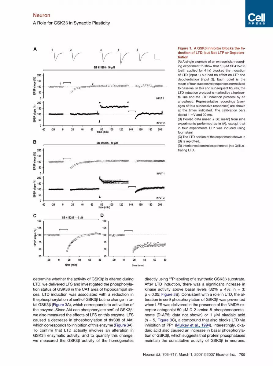
GSK3 Inhibitors Block the Induction of LTD
To determine whether GSK3b is involved in synaptic
plasticity in the brain, we tested the effects of the highly se-
lective GSK3 inhibitor 3-[(3-chloro-4-hydroxyphenyl)-
amino]-4-(2-nitrophenyl)-1H-pyrrole-2,5-dione (SB415286)
(Coghlan et al., 2000) in rat hippocampal slices. We applied
SB415286 (10 mM) to the perfusing medium and used ex-
tracellular recording to investigate its effects on LTD,
LTP, and the reversal of LTP by depotentiation (Figure 1).
SB415286 had no effect on baseline synaptic transmission
(data not shown), but it blocked the induction of LTD in-
duced by low-frequency stimulation (LFS; 900 stimuli de-
livered at 1 Hz; n = 9). Thus, 60 min following LFS, the slope
of the field excitatory postsynaptic potential (fEPSP) of
SB415286-treated slices was 103% ± 2% of baseline
(n = 9) compared with 65% ± 5% (n = 3) of baseline for
704 Neuron 53, 703–717, March 1, 2007 ª2007 Elsevier Inc.
untreated slices (p < 0.001). In contrast, in an independent
input of SB415286 treated slices, LTP was readily induced
by tetanic stimulation (either one tetanus [n = 5] or four
tetani delivered at 30 s intervals [n = 4]; each tetanus com-
prising 100 Hz stimulation for 1 s) and was reversed by de-
potentiation (900 stimuli delivered at 1 Hz) in each of the
seven slices tested. Therefore, of the three major forms
of synaptic plasticity studied, SB415286 specifically
blocks the induction of LTD.
Since LTD is induced and expressed postsynaptically
(Collingridge et al., 2004; Kemp and Bashir, 2001), we in-
fused individual CA1 pyramidal neurons in rat hippocam-
pal slices with SB415286 via a whole-cell patch electrode
to selectively block only postsynaptic GSK3b (Figure 2).
Consistent with no effect on basal synaptic transmission,
excitatory postsynaptic currents (EPSCs), evoked by
stimulating Schaffer collateral-commissural fibers, were
similar in the presence or absence of intracellular
SB415286 (10 mM). However, SB415286 blocked the in-
duction of LTD (Figures 2B and 2D), which was routinely
induced in interleaved control neurons by delivering 300
pulses at �40 mV (Luthi et al., 1999; Figures 2A and 2C).
Thus, 20 min following this stimulation, the EPSC ampli-
tude of the test pathway was 100% ± 3% (n = 7) and
63% ± 4% (n = 16) of baseline, for the SB415286-treated
and untreated neurons, respectively (p < 0.001).
We next sought to firmly establish a role for GSK3b in
LTD by further exploring the pharmacological block of
LTD. We first tested SB415286 at a lower concentration
and found that this inhibitor exhibited a concentration-
dependent inhibition of LTD, with 3 mM producing �50%
inhibition (Figure 2H). Although SB415286 has previously
been found to be a highly selective inhibitor of GSK3b
(Coghlan et al., 2000), we wanted to rule out the possibility
that it was acting through inhibition of other kinases over
this concentration range. We therefore tested the effects
of two other structurally unrelated GSK3b inhibitors, lith-
ium (Klein and Melton, 1996) and kenpaullone (Leost
et al., 2000). Lithium also produced a concentration-
dependent inhibition of LTD, with a partial inhibition at
2 mM and a complete block at 20 mM (Figures 2E and
2H), while kenpaullone fully blocked LTD at a concentra-
tion of 10 mM (Figures 2F and 2H). GSK3b is most closely
related to the cyclin-dependent protein kinases (CDKs).
We therefore tested the potent CDK inhibitor roscovitine
(Meijer et al., 1997), which does not affect GSK3b (Bain
et al., 2003). This compound had no effect on LTD when
applied at 10 mM (Figures 2G and 2H), a concentration
40 times the IC50 for inhibition of CDK2 (Bain et al.,
2003). The ability of all three GSK3b inhibitors to prevent
LTD over the concentration range at which they are inhib-
itors of GSK3b (Bain et al., 2003), considered together with
the insensitivity to the CDK2 inhibitor, strongly implicates
a role for this enzyme in LTD.
LTD Is Associated with an Increase in GSK3b Activity
These pharmacological experiments demonstrate the re-
quirement for GSK3b activity for the induction of LTD. To
Neuron
A Role for GSK3b in Synaptic Plasticity
determine whether the activity of GSK3b is altered during
LTD, we delivered LFS and investigated the phosphoryla-
tion status of GSK3b in the CA1 area of hippocampal sli-
ces. LTD induction was associated with a reduction in
the phosphorylation of ser9 of GSK3b but no change in to-
tal GSK3b (Figure 3A), which corresponds to activation of
the enzyme. Since Akt can phosphorylate ser9 of GSK3b,
we also measured the effects of LFS on this enzyme. LFS
caused a decrease in phosphorylation of thr308 of Akt,
which corresponds to inhibition of this enzyme (Figure 3A).
To confirm that LTD actually involves an alteration in
GSK3b enzymatic activity, and to quantify this change,
we measured the GSK3b activity of the homogenates
directly using 32P labeling of a synthetic GSK3b substrate.
After LTD induction, there was a significant increase in
kinase activity above basal levels (32% ± 4%; n = 3;
p < 0.05; Figure 3B). Consistent with a role in LTD, the al-
teration in ser9 phosphorylation of GSK3b was prevented
when LFS was delivered in the presence of the NMDA re-
ceptor antagonist 50 mM D-2-amino-5-phosphonopenta-
noate (D-AP5; data not shown) or 1 mM okadaic acid
(n = 5, Figure 3C), a compound that also blocks LTD via
inhibition of PP1 (Mulkey et al., 1994). Interestingly, oka-
daic acid also caused an increase in basal phosphoryla-
tion of GSK3b, which suggests that protein phosphatases
maintain the constitutive activity of GSK3b in neurons.
Figure 1. A GSK3 Inhibitor Blocks the In-
duction of LTD, but Not LTP or Depoten-
tiation
(A) A single example of an extracellular record-
ing experiment to show that 10 mM SB415286
(bath applied for 4 hr) blocked the induction
of LTD (input 1) but had no effect on LTP and
depotentiation (input 2). Each point is the
mean of four successive responses normalized
to baseline. In this and subsequent figures, the
LTD induction protocol is marked by a horizon-
tal line and the LTP induction protocol by an
arrowhead. Representative recordings (aver-
ages of four successive responses) are shown
at the times indicated. The calibration bars
depict 1 mV and 20 ms.
(B) Pooled data (mean ± SE mean) from nine
experiments performed as in (A), except that
in four experiments LTP was induced using
four tetani.
(C) The LTD portion of the experiment shown in
(B) is replotted.
(D) Interleaved control experiments (n = 3) illus-
trating LTD.
Neuron 53, 703–717, March 1, 2007 ª2007 Elsevier Inc. 705

Neuron
A Role for GSK3b in Synaptic Plasticity
Figure 2. Pharmacological Evidence that
GSK3 Activity Is Required for LTD
(A) A single experiment illustrating homosynap-
tic LTD. Each point plots the average amplitude
of six successive EPSCs normalized with re-
spect to the baseline. At t = 0, the neuron was
depolarized to �40 mV and stimuli delivered
at 0.75 Hz to the test input for the duration indi-
cated by the bar. EPSCs obtained before and
following the induction of LTD are illustrated
for the control and test inputs at the times
indicated (1, 2). Calibration bars for the traces
depict 40 pA and 50 ms.
(B) A single example from an experiment in
which 10 mM SB415286 was contained within
the patch pipette.
(C) Pooled data (mean ± SE) from 16 control
experiments.
(D) Pooled data from seven experiments per-
formed as in (B).
(E) Effects of 20 mM lithium (n = 5).
(F) Effects of 10 mM kenpaullone (n = 5).
(G) Effects of 10 mM roscovitine (n = 5).
(H) Summary graphs illustrating the effects of
various inhibitors on homosynaptic LTD quan-
tified 20 min following induction. *p < 0.05,
***p < 0.001.
Okadaic acid also prevented the LFS-induced decrease in
phosphorylation of Akt, but it had no effect on basal levels
(Figure 3D). Therefore, during LTD the activation of GSK3b
may be due to the phosphatase inhibiting Akt and dephos-
phorylating ser9 of GSK3b.
To determine whether the activation of GSK3b is tran-
sient, we measured its phosphorylation immediately and
20 min following the delivery of LFS. We also examined
the effects of a tetanus (100 Hz, 1 s). LFS and tetanic stim-
ulation caused opposite effects on the phosphorylation
status of ser9 of GSK3b that were similar in magnitude
at both time points (Figures 3E and 3F). The finding that
the change in GSK3b phosphorylation persisted beyond
the delivery of LFS suggests that its increase in activity
could be involved in the maintenance of LTD, though alter-
natively, it may be an epiphenomenon. To directly exam-
ine the possibility that GSK3b activity may be required
706 Neuron 53, 703–717, March 1, 2007 ª2007 Elsevier Inc.
beyond the delivery of LFS, we investigated the effects
of its inhibition after LTD had been induced. For these ex-
periments we used lithium, since it is rapidly membrane
permeant. Lithium (20 mM) had no effect on baseline
transmission but fully blocked the induction of LTD
(Figure 3G). Interestingly, lithium was also fully effective
when applied after the delivery of LFS (Figure 3H).
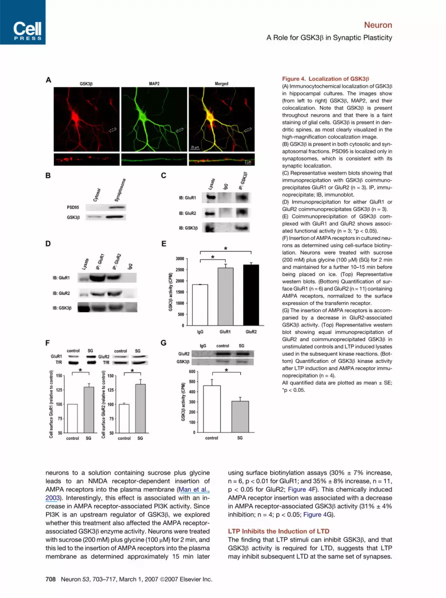
GSK3b Is Present in Dendritic Spines and Forms
a Complex with AMPA Receptors
GSK3b is mainly found in the cytosol. However, our exper-
iments predict that a fraction of GSK3b should be present
in the vicinity of synapses. As shown in Figure 4A, GSK3b
is distributed throughout hippocampal neurons and, to
a lesser extent, throughout glial cells too. Importantly,
GSK3b is present in dendritic spines. Consistent with the
immunocytochemistry, western blotting revealed the

Neuron
A Role for GSK3b in Synaptic Plasticity
Figure 3. GSK3b Activity Is Increased
during LTD
(A) Representative western blots from three
separate experiments showing a reduction in
phosphorylated GSK3b, but no change in ex-
pression of total GSK3b, following LFS. There
is also a reduction in phosphothreonine Akt,
but no change in Akt mass. b-tubulin repre-
sents equal loading of lysates for each condi-
tion.
(B) LFS-induced increase in GSK3b activity
relative to controls (n = 3).
(C) Pooled data to show LFS-induced changes
in phosphorylated GSK3b under control condi-
tions (n = 10) and following treatment with 1 mM
okadaic acid (OA; n = 5).
(D) Pooled data to show LFS-induced changes
in phosphorylated Akt under control conditions
(n = 4) and following treatment with okadaic
acid (n = 4).
(E) Representative western blots (upper) and
pooled data from four experiments (lower)
showing a decrease in phosphorylated
GSK3b immediately following and 20 min fol-
lowing LFS.
(F) Representative western blots (upper) and
pooled data from five experiments (lower)
showing an increase in phosphorylated
GSK3b immediately following and 20 min fol-
lowing tetanic stimulation. All quantified data
are plotted as mean ± SE; *p < 0.05, **p < 0.01.
(G) Pooled data from six experiments showing
that lithium blocks the induction of LTD.
(H) Pooled data from four experiments showing
that lithium reverses LTD when applied follow-
ing the induction stimulus.
presence of GSK3b in both cytosolic and synaptosomal
fractions prepared from both hippocampal (Figure 4B)
and neocortical (data not shown) slices.
NMDA receptor-dependent LTD is due to the internali-
zation of AMPA receptors and involves protein inter-
actions directly associated with the AMPA receptor
subunits, particularly GluR2 (Collingridge et al., 2004;
Malinow and Malenka, 2002). We reasoned that GSK3b
might form a complex with AMPA receptors, and thus
sought to investigate this by probing for an association
of native GSK3b with AMPA receptors in the CA1 area of
hippocampal slices. A specific antibody against GSK3b
was able to coimmunoprecipitate the GluR1 and GluR2
AMPA receptor subunits (Figure 4C), and conversely, im-
munoprecipitation of AMPA receptors produced coimmu-
noprecipitation of GSK3b (Figure 4D). To determine the
functional status of AMPA receptor-associated GSK3b,
we immunoprecipitated AMPA receptors, using anti-
bodies against either GluR1 or GluR2, and then assayed
for kinase activity. GSK3b activity was readily detected
in both GluR1 and GluR2 immunoprecipitates relative to
the background IgG control (Figure 4E), demonstrating
that endogenous GSK3b associates with native AMPA re-
ceptors in the brain, and that the bound GSK3b is func-
tionally active. This association of GSK3b with AMPA re-
ceptors suggests a compartmentalization of this enzyme
for the efficient regulation of AMPA receptors during LTD.
We wondered whether the GSK3b activity that is asso-
ciated with AMPA receptors could be regulated. Previous
work has shown that transient exposure of cultured
Neuron 53, 703–717, March 1, 2007 ª2007 Elsevier Inc. 707
Neuron
A Role for GSK3b in Synaptic Plasticity
Figure 4. Localization of GSK3b
(A) Immunocytochemical localization of GSK3b
in hippocampal cultures. The images show
(from left to right) GSK3b, MAP2, and their
colocalization. Note that GSK3b is present
throughout neurons and that there is a faint
staining of glial cells. GSK3b is present in den-
dritic spines, as most clearly visualized in the
high-magnification colocalization image.
(B) GSK3b is present in both cytosolic and syn-
aptosomal fractions. PSD95 is localized only in
synaptosomes, which is consistent with its
synaptic localization.
(C) Representative western blots showing that
immunoprecipitation with GSK3b coimmuno-
precipitates GluR1 or GluR2 (n = 3). IP, immu-
noprecipitate; IB, immunoblot.
(D) Immunoprecipitation for either GluR1 or
GluR2 coimmunoprecipitates GSK3b (n = 3).
(E) Coimmunoprecipitation of GSK3b com-
plexed with GluR1 and GluR2 shows associ-
ated functional activity (n = 3; *p < 0.05).
(F) Insertion of AMPA receptors in cultured neu-
rons as determined using cell-surface biotiny-
lation. Neurons were treated with sucrose
(200 mM) plus glycine (100 mM) (SG) for 2 min
and maintained for a further 10–15 min before
being placed on ice. (Top) Representative
western blots. (Bottom) Quantification of sur-
face GluR1 (n = 6) and GluR2 (n = 11) containing
AMPA receptors, normalized to the surface
expression of the transferrin receptor.
(G) The insertion of AMPA receptors is accom-
panied by a decrease in GluR2-associated
GSK3b activity. (Top) Representative western
blot showing equal immunoprecipitation of
GluR2 and coimmunoprecipitated GSK3b in
unstimulated controls and LTP induced lysates
used in the subsequent kinase reactions. (Bot-
tom) Quantification of GSK3b kinase activity
after LTP induction and AMPA receptor immu-
noprecipitation (n = 4).
All quantified data are plotted as mean ± SE;
*p < 0.05.
neurons to a solution containing sucrose plus glycine
leads to an NMDA receptor-dependent insertion of
AMPA receptors into the plasma membrane (Man et al.,
2003). Interestingly, this effect is associated with an in-
crease in AMPA receptor-associated PI3K activity. Since
PI3K is an upstream regulator of GSK3b, we explored
whether this treatment also affected the AMPA receptor-
associated GSK3b enzyme activity. Neurons were treated
with sucrose (200 mM) plus glycine (100 mM) for 2 min, and
this led to the insertion of AMPA receptors into the plasma
membrane as determined approximately 15 min later
708 Neuron 53, 703–717, March 1, 2007 ª2007 Elsevier Inc.
using surface biotinylation assays (30% ± 7% increase,
n = 6, p < 0.01 for GluR1; and 35% ± 8% increase, n = 11,
p < 0.05 for GluR2; Figure 4F). This chemically induced
AMPA receptor insertion was associated with a decrease
in AMPA receptor-associated GSK3b activity (31% ± 4%
inhibition; n = 4; p < 0.05; Figure 4G).
LTP Inhibits the Induction of LTD
The finding that LTP stimuli can inhibit GSK3b, and that
GSK3b activity is required for LTD, suggests that LTP
may inhibit subsequent LTD at the same set of synapses.

Neuron
A Role for GSK3b in Synaptic Plasticity
Figure 5. The Synaptic Activation of NMDA Receptors Inhibits Subsequent LTD
(A) A conditioning stimulus (60 pulses, 0.5 Hz, 0 mV; arrowhead) induces LTP when delivered within 5 min of whole-cell recording (filled symbols;
n = 8), but not when baseline recording was extended for at least 10 min (open symbols; n = 18).
(B) A conditioning stimulation completely blocks the induction of LTD (n = 8). (Note that interleaved control experiments invariably induced LTD; these
experiments are included in the grand pool illustrated in Figure 2C).
(C) Full recovery from the effects of conditioning when the LTD induction protocol was delivered 1 hr after the pairing (n = 5).
(D) LTD is largely blocked when the LTD induction protocol was delivered 20 min following pairing (n = 4).
(E) The conditioning effect of pairing is prevented by an NMDA receptor antagonist (D-2-amino-5-phosphonopentanoate [D-AP5], 50 mM; n = 4).
(F) Time course of conditioning. Each point plots the mean ± SE of at least four experiments (*p < 0.05, ***p < 0.001).
(G) The induction of LTP (four bursts of 100 pulses at 100 Hz, delivered at 30 s intervals) blocks the induction of long-term synaptic depression.
LY341495 was present to block mGlu receptor-dependent synaptic plasticity (n = 5).
(H) Long-term synaptic depression can be induced 1 hr after the induction of LTP (n = 4).
(I) The long-term synaptic depression induced 1 hr after LTP is dependent on activation of NMDA receptors (n = 4).
The calibration bars for traces (B–D) depict 40 pA and 50 ms.
To test this idea we developed a protocol whereby the ef-
fects of a prior LTP stimulus on the induction of LTD could
be examined without the complication of LTP-induced al-
terations in synaptic strength. For this we delivered an LTP
induction protocol of 60 pulses at 0.5 Hz while the neuron
was depolarized to 0 mV. This well-established ‘‘pairing’’
protocol produced a robust LTP when applied soon after
commencement of a whole-cell recording (Figure 5A).
However, when this pairing protocol was applied after
more than 10 min of whole-cell dialysis, LTP did not occur
(Figure 5A), due to the phenomenon of ‘‘washout’’ of LTP
(Malinow and Tsien, 1990). We reasoned that pairing
would still drive the PI3K-Akt-GSK3b pathway following
washout. This pathway is resistant to dialysis since both
PI3K inhibitors (Daw et al., 2002) and GSK3b inhibitors
(Figure 2) still affect LTD after an extended period of
whole-cell recording. All our subsequent whole-cell
experiments were, therefore, conducted after at least
10 min of recording to enable washout to occur. Consis-
tent with our prediction, when pairing was delivered imme-
diately prior to the LTD induction protocol, it completely
prevented the generation of LTD (Figure 5B). The condi-
tioning effect was fully reversible since the full level of
LTD could be obtained if the induction protocol was deliv-
ered 1 hr following pairing (Figure 5C). The effect of condi-
tioning was, however, fairly long-lasting. For example,
when pairing was delivered 20 min beforehand, it still
largely blocked the generation of LTD (Figures 5D and
5F). Conditioning involved the activation of NMDA recep-
tors since it was prevented by the NMDA receptor antag-
onist D-AP5 (Figures 5E and 5F).
Although it is unlikely that washout affected the signal-
ing mechanisms involved in the NMDA receptor-mediated
inhibition of LTD, we went on to investigate whether LTD
Neuron 53, 703–717, March 1, 2007 ª2007 Elsevier Inc. 709

Neuron
A Role for GSK3b in Synaptic Plasticity
was blocked following the production of LTP under more
physiological conditions. So as not to perturb the neurons,
we performed extracellular recordings and induced LTP
with tetanic stimuli. An inherent complication of these ex-
periments is that the stimulus used to induce LTD can also
induce an mGluR-dependent form of depotentiation (Bor-
tolotto et al., 1994). To remove this additional factor,
we performed these experiments in the presence of the
broad spectrum metabotropic glutamate (mGlu) receptor
antagonist LY341495 (100 mM), which blocks this type of
depotentiation (Fitzjohn et al., 1998). In the presence of
LY341495, LFS consistently induced LTD of baseline re-
sponses (control [input 1]: 64% ± 4% of baseline;
LY341495 [input 2]: 68% ± 2% of baseline; n = 3; data
not illustrated). In contrast, if the same stimulation was de-
livered shortly after inducing LTP, then long-term synaptic
depression was fully blocked (Figure 5G). Consistent with
the reversibility of the conditioning effect, the same stimu-
lation delivered 60 min following the induction of LTP was
able to induce long-term synaptic depression (Figure 5H)
that was dependent on the synaptic activation of NMDA
receptors (Figure 5I). Therefore, the synaptic activation
of NMDA receptors during LTP inhibits the induction of
LTD for approximately 1 hr.
A Role for PI3K and Akt in the Conditioned
Block of LTD
Using whole-cell recordings, we next investigated
whether this block of LTD by prior NMDA receptor activa-
tion was due to activation of the PI3K-Akt pathway. First
we tested the role of PI3K using the selective inhibitors
wortmannin (500 nM) or LY294002 (10 mM). In the pres-
ence of wortmannin, EPSCs were readily evoked, and
pairing alone, after LTP washout, had no effect on trans-
mission (Figure 6A). Treatment with wortmannin also had
no effect on the amount of LTD induced (Figure 6B). How-
ever, wortmannin completely prevented the block of LTD
by the LTP pairing stimulus (Figure 6C). LY294002 had
the same effect; it also completely prevented the effect
of the prior conditioning on LTD (Figure 6D). Thus, using
two structurally unrelated inhibitors, our data indicate
that PI3K is required for the NMDA receptor-dependent in-
hibition of LTD. An alternative mechanism for inhibition of
GSK3b may be via the mammalian target of rapamycin
(mTOR) pathway (Frame and Cohen, 2001). However, in-
hibition of mTOR using rapamycin did not affect condition-
ing (Figure 6E). A possible alternative explanation for this
result is that rapamycin directly blocks LTD. However,
LTD was readily induced in the presence of rapamycin
if the conditioning pairing stimulus was not delivered
(Figure 6F). Thus, NMDA receptor-dependent inhibition
of LTD involves the PI3K, but not the mTOR, pathway.
As discussed above, our data suggest that the PI3K-
Akt-GSK3b pathway operates normally in whole-cell re-
cording experiments regardless of washout of LTP. How-
ever, we further confirmed the involvement of PI3K in the
synaptic inhibition of long-term synaptic depression under
circumstances in which LTP was induced. To do this, we
710 Neuron 53, 703–717, March 1, 2007 ª2007 Elsevier Inc.
again performed extracellular recording experiments in
the presence of a broad spectrum mGluR antagonist to
block mGluR-dependent depotentiation. We performed
two-input experiments and (1) induced LTP and then
tested for long-term synaptic depression in the presence
of a reversible PI3K inhibitor, and (2) applied these same
protocols to the second input after washout of the PI3K in-
hibitor. The results after washout of LY294002 were the
same as those for the control experiments (illustrated in
Figure 5G) and show that LTP blocks long-term synaptic
depression (Figure 6G). In contrast, long-term synaptic de-
pression is readily induced in the presence of LY294002
(Figure 6G). This is due to block of conditioning rather
than an effect of LY294002 on LTP per se, since, in inter-
leaved experiments, LY294002 did not affect LTP induced
by four tetani (Figure 6H) (see also Horwood et al., 2006).
Using whole-cell recording, we next investigated
whether Akt is involved in this process using three com-
plementary strategies (Figure 7A). First, we used a peptide
(TCS 183) that mimics the Akt phosphorylation site on
GSK3b. Inclusion of this false substrate at a concentration
of 300 mM in the patch electrode solution prevented the ef-
fect of conditioning (Figure 7B). Next we used a construct
containing just the pleckstrin homology (PH) domain of
Akt, since this acts as a highly specific inhibitor of Akt
within this signaling pathway (Wang et al., 2003). In inter-
leaved blind experiments, GST-PH-Akt (Figure 7D), but
not GST alone (Figure 7C), also blocked conditioning. Fi-
nally, we examined the effects of an antibody that inhibits
the function of Akt (Hill et al., 1999). This antibody also in-
hibited the conditioning effect (Figure 7F). In contrast, the
same antibody when heat-inactivated proved ineffective
at blocking conditioning (Figure 7E), as did an antibody
against mTOR (99% ± 3% of control; n = 4; data not illus-
trated). These experiments demonstrate the essential role
of Akt in the conditioning blockade of LTD. Collectively,
these experiments demonstrate that the transient activa-
tion of NMDA receptors during LTP inhibits subsequent
LTD via the PI3K-Akt-GSK3b pathway.
DISCUSSION
In the present study we have identified a form of regulation
of synaptic plasticity in which the transient synaptic acti-
vation of NMDA receptors, as occurs during LTP, leads
to inhibition of LTD. This regulation is very powerful since
LTD is fully inhibited immediately following the condition-
ing stimulus and the effect lasts for approximately 1 hr.
We have also identified some of the signaling pathways re-
sponsible for this potent regulation of synaptic plasticity.
We have found that GSK3b activity is an absolute require-
ment for the induction of LTD and that the conditioning
stimulus inhibits its activity via activation of the PI3K-Akt
pathway. Finally, we show that there is a correlation be-
tween the phosphorylation state of GSK3b ser9 and
whether NMDA receptor activation leads to the induction
or inhibition of LTD. This new regulatory mechanism is
shown schematically in Figure 8.

Neuron
A Role for GSK3b in Synaptic Plasticity
Figure 6. Conditioning of LTD Requires Activation of PI3K(A) Wortmannin (500 nM) has no effect on the conditioning train (n = 3).
(B) Wortmannin has no effect on LTD (n = 5).
(C) Wortmannin completely prevents the conditioning of LTD (n = 9).
(D) LY294002 (10 mM) completely prevents the conditioning of LTD (n = 6).
(E) Rapamycin (0.1 mM) has no effect on conditioning (n = 4).
(F) Rapamycin has no effect on LTD (n = 5).
(G) Prior induction of LTP did not inhibit long-term synaptic depression when the experiment was performed in the presence of LY294002 (10 mM).
Following washout of LY294002, the block of long-term synaptic depression by LTP was invariably observed (tested on an independent input; n = 5).
(H) LY294002 (10 mM) did not affect the generation of LTP (n = 4).
A Function for GSK3b in the CNS
GSK3b is an unusual kinase that has been implicated in
many diseases. However, very little is known about its nor-
mal function in the nervous system. It is important during
early development and it has been shown to play a key
role in cell polarity (Etienne-Manneville and Hall, 2003)
and in the growth of neuromuscular junctions (Franco
et al., 2004). Recently, it has been shown that GSK3b is
important for determining neuronal polarity during the de-
velopment of hippocampal neurons (Jiang et al., 2005;
Yoshimura et al., 2005). However, though GSK3b is also
highly expressed in the mature brain (Woodgett, 1990),
its function in the nervous system has, hitherto, been
largely unexplored. In the nucleus of hippocampal neu-
rons, GSK3b is involved in the regulation of gene tran-
scription by promoting the nuclear export of the transcrip-
tion factor NF-ATc4 (Graef et al., 1999). In addition, it has
been shown that overexpression of GSK3b impairs spatial
learning (Hernandez et al., 2002), though the mechanism
underlying this effect is unknown. Here we show that in
2-week-old rats, an age at which the expression of
GSK3b is near its peak (Takahashi et al., 2000), GSK3b
activity is essential for NMDA receptor-dependent LTD
in the hippocampus. This form of LTD is widespread
throughout the brain and has been strongly implicated in
development and learning and memory (Bear and Abra-
ham, 1996; Kemp and Bashir, 2001). Therefore, this novel
GSK3b-dependent mechanism may be of general signifi-
cance in regulating the interaction between LTP and LTD
throughout the brain.
Neuron 53, 703–717, March 1, 2007 ª2007 Elsevier Inc. 711

Neuron
A Role for GSK3b in Synaptic Plasticity
Figure 7. Conditioning of LTD Requires Activation of Akt
(A) Strategies for inhibiting Akt.
(B) A peptide that mimics the Akt phosphorylation site on GSK3b prevents conditioning of LTD (n = 4).
(C) GST has no effect on conditioning (n = 4).
(D) GST-PH-Akt inhibits conditioning of LTD (n = 7).
(E) The heat-inactivated antibody has no effect on conditioning (n = 5).
(F) An Akt-neutralizing antibody prevents conditioning of LTD (n = 6).
Activation of GSK3b during LTD
GSK3b, unlike most enzymes, possesses high basal level
constitutive activity and can be bidirectionally regulated to
either further increase or decrease its activity. Our exper-
iments show that during LTD there is additional activation
712 Neuron 53, 703–717, March 1, 2007 ª2007 Elsevier Inc.
of GSK3b, probably via dephosphorylation of ser9. We
show further that this effect is prevented by an inhibitor
of PP1/PP2A. This suggests that the activation of PP1,
which is known to occur during LTD (Mulkey et al.,
1993), is responsible for the activation of GSK3b, via its
Figure 8. The Regulation of GSK3b in the
Control of NMDA Receptor-Dependent
Synaptic Plasticity
The activity of GSK3b determines whether
NMDA receptor activation induces LTD or in-
hibits LTD. During LTD, activation of PP1 leads
to dephosphorylation of GSK3b at ser9 to fur-
ther activate GSK3b and enable LTD to occur.
PP1 also inhibits Akt. During LTP, activation
of NMDA receptors leads to stimulation of the
PI3K-Akt pathway, which phosphorylates
GSK3b at ser9 to inhibit its activity and prevent
the induction of LTD. Thus, GSK3b, under the
control of Akt and PP1, is a critical determinant
of the direction of NMDA receptor-dependent
plasticity.

Neuron
A Role for GSK3b in Synaptic Plasticity
dephosphorylation of ser9 (Lee et al., 2005; Morfini et al.,
2004; Szatmari et al., 2005). We also show that LTD is as-
sociated with inhibition of Akt, probably also via the acti-
vation of PP1. These data suggest that GSK3b activity is
increased during LTD because the phosphatase concom-
itantly inhibits Akt and directly dephosphorylates ser9 of
GSK3b.
Interestingly, the alteration in the phosphorylation sta-
tus of GSK3b persisted beyond the delivery of LFS, and
lithium completely blocked LTD when applied after the de-
livery of LFS. These data suggest that GSK3b is required
for the LTD process beyond the initial induction phase.
Further studies are required to determine the full time
course of the involvement of GSK3b in LTD.
Signaling Pathways Involved in the Actions of GSK3b
GSK3b has several upstream regulators and numerous
downstream targets. In the present study, we have identi-
fied two of its upstream regulators. During LTD, GSK3b is
activated via an okadaic acid-sensitive protein phospha-
tase, which is probably PP1 (Mulkey et al., 1993; Szatmari
et al., 2005). During LTP, GSK3b is inhibited via the PI3K-
Akt pathway. Since GSK3b is such a ubiquitous kinase, it
needs mechanisms to localize its access to its substrates.
This is achieved in part via direct interactions with other
proteins to form complexes. For example, in the canonical
Wnt pathway, GSK3b binding proteins control access of
b-catenin (Jope and Johnson, 2004). It seems likely that
the association between GSK3b and AMPA receptors
serves to localize the kinase close to substrates that are
involved in the trafficking of these receptors during synap-
tic plasticity. Further studies are required to establish the
mechanism of this interaction as well as the downstream
pathways mediated by GSK3b in the regulation of LTD.
The finding that the synaptic activation of NMDA recep-
tors during LTP inhibits NMDA receptor-dependent LTD
raises an intriguing issue: what determines whether the
synaptic activation of NMDA receptors leads to the induc-
tion or inhibition of LTD? We present evidence that the
phosphorylation state of ser9 of GSK3b is a critical deter-
minant. Thus, during LTP, activation of the PI3K-Akt path-
way results in phosphorylation of GSK3b, and hence inhi-
bition of its activity. In contrast, during LTD, activation of
PP1 results in inhibition of Akt and the dephosphorylation
of GSK3b at ser9, and this leads to an increase in the en-
zyme’s activity. The activation of PI3K-Akt (Man et al.,
2003) and inhibition of PP1 (Atkins et al., 2005) during
LTP, but inhibition of Akt during LTD (the present study)
as well as the selective activation of PP1 during LTD (Mul-
key et al., 1993), can be explained by the differences in the
magnitude and spatiotemporal properties of the Ca2+ rise
associated with the synaptic activation of NMDA recep-
tors during these two forms of synaptic plasticity.
GSK3b-Mediated Metaplasticity
Previous work (e.g., Huang et al., 1992; Montgomery and
Madison, 2002) has described other ways in which synap-
tic plasticity can be powerfully influenced by the prior his-
tory of synaptic activity. However, the mechanisms in-
volved in these forms of metaplasticity are not known.
Why synapses need such regulatory mechanisms is a mat-
ter of conjecture. One intriguing possible role for the reg-
ulation described here is to stabilize a synaptic modifica-
tion over the short term by protecting synapses from the
effects of additional NMDA receptor-dependent plasticity
until the information can be either consolidated or erased
by NMDA receptor-independent mechanisms.
The regulation of synaptic plasticity is further compli-
cated by the involvement of mGluRs, which are involved
in depotentiation (Bortolotto et al., 1994), LTD of baseline
transmission (Bolshakov and Siegelbaum, 1994), hetero-
synaptic LTD (Daw et al., 2002), and metaplasticity (Abra-
ham and Bear, 1996). So that we could focus on interac-
tions between the NMDA receptor-dependent forms of
synaptic plasticity, we eliminated the additional complica-
tion of mGluR-dependent synaptic plasticity by using the
broad spectrum mGluR antagonist LY341495 and by em-
ploying stimulus protocols optimized for NMDA receptor-
dependent synaptic plasticity. However, given that PI3K
has been implicated in a chemically induced form of
mGluR-dependent LTD (Hou and Klann, 2004) and heter-
osynaptic LTD (Daw et al., 2002), it will be interesting to
determine whether GSK3b is also involved in these forms
of synaptic plasticity. One possibility is that the PI3K-Akt-
GSK3b pathway serves to inhibit NMDA receptor-depen-
dent LTD both homosynaptically following the induction of
LTP and heterosynaptically following the induction of LTD.
Clinical Implications
Our finding that in the normal brain activation of GSK3b is
essential for NMDA receptor-dependent LTD, and that its
activity can be regulated by LTP, may offer clues to the
pathological role of this enzyme in neurological disorders.
For example, the primary therapeutic action of lithium in
bipolar disorders may be via inhibition of GSK3b (Grimes
and Jope, 2001; Klein and Melton, 1996). Indeed, specific
inhibition of GSK3b has recently been shown to produce
antidepressive-like activity in vivo (Kaidanovich-Beilin
et al., 2004). Overactivity of GSK3b may, therefore, lead
to this mood disorder by affecting the balance and inter-
play between NMDA receptor-dependent LTP and LTD.
EXPERIMENTAL PROCEDURES
General Antibodies and Reagents
Rabbit anti GSK3b was obtained from Santa Cruz. Rabbit phospho-
GSK3b (ser9); mouse monoclonal antibody (mAb) Akt1 and rabbit
mAb phospho-Akt (thr308) were obtained from Cell Signaling Technol-
ogy (Beverly, MA). Mouse mAb MAP2 was obtained from BD Pharma-
gen (San Diego, CA). ProLong Gold mounting medium and secondary
fluorophore-bound IgGs (Alexa Fluor series) for immunocytochemistry
were obtained from Invitrogen-Molecular Probes (Portland, OR). Rab-
bit IgG, recombinant rat GSK3b, and phosphoglycogen synthase
peptide-2 were obtained from Upstate Biotechnology, Inc. (Lake
Placid, NY). [g-32P]ATP was purchased from Amersham Biosciences
(Quebec, Canada). Leupeptin protease inhibitor was obtained from
Peptides International (Louisville, Kentucky) and all other lysate buffer
components, including phosphatase and protease inhibitors, were
Neuron 53, 703–717, March 1, 2007 ª2007 Elsevier Inc. 713
Neuron
A Role for GSK3b in Synaptic Plasticity
obtained from Sigma Aldritch (St. Louis, MO). Kenpaullone was ob-
tained from Calbiochem, okadaic acid was obtained from LC laborato-
ries (Woburn, MA), and the other chemicals from Tocris Cookson
(Avonmouth, UK). TCS 183 (MSGRPRTTSFAES) was custom synthe-
sized by Tocris Cookson. The Akt antibody [Akt 1/2 (N-19), sc-1619]
and mTOR antibody [FRAP (C-19):sc-1550] were obtained from Santa
Cruz. GST-PH Akt was constructed as follows. The DNA fragment for
bovine Akt N terminus (amino acids 1–148) containing the PH domain
(1–106) (GST-PH-Akt) was generated by PCR using the following
primers: 50-CGCGGATTCATGAACGACGTGGCCATCGTG-30 and 50-
CCGGAATTCTCAAAACTCATTCATGGTCAC-30. The PCR-generated
DNA fragment was ligated into the BamHI and EcoRI sites of the
pGEX-4T-2 vector (Amersham Biosciences biotech), cloned, and
then verified by sequencing. The GST-PH-Akt (amino acids 1–148)
and GST alone proteins were prepared from bacterial lysates and pu-
rified according to the protocol provided by Amersham Biosciences.
Electrophysiology
Experiments were performed on 400 mm thick parasagittal hippocam-
pal slices obtained from juvenile (13- to 15-day-old) rats, which were
perfused with artificial cerebrospinal fluid (ACSF) which was com-
prised of the following: 124 mM NaCl, 3 mM KCl, 26 mM NaHCO3,
1.25 mM NaH2PO4, 2 mM CaCl2, 1 mM MgSO4, 15 mM glucose,
2 mM Ascorbate, and 0.01 mM (-)-bicuculline methochloride. In exper-
iments in which LiCl was added to the perfusuate, the equivalent
amount of NaCl was omitted from the ACSF. Visually guided, whole-
cell recordings were obtained at room temperature (RT) from the
soma of CA1 neurons using patch electrodes that contained the fol-
lowing: 130 mM CsMeSO4, 10 mM HEPES, 8 mM NaCl, 0.5 mM
EGTA, 4 mM Mg-ATP, 0.3 mM Na-GTP, and 5 mM QX-314. In exper-
iments in which LiCl was included in the whole-cell solution, the equiv-
alent concentration of CsMeSO4 was removed. Some control experi-
ments were performed in this way, but by substituting CsCl for LiCl.
When antibodies were added to the whole-cell solution, the NaCl con-
centration was adjusted appropriately. Two independent sets of
Schaffer collateral-commissural fibers were stimulated alternately,
each at a frequency of 0.1 Hz, and EPSC amplitude and access resis-
tance were recorded on-line at a holding potential of�70 mV. LTD was
induced by delivering 300 pulses (at 0.75 Hz) at �40 mV to one of the
two inputs. The induction of this form of LTD was completely inhibited
by the NMDA receptor antagonist D-AP5 (50 mM). Kinase inhibitors
were included in the patch solution. For experiments using kenpaul-
lone or roscovitine, these were also added to the perfusate (at
10 mM). Extracellular recordings were obtained as described previ-
ously (Fitzjohn et al., 1998). All inhibitors were added to the perfusing
medium. Data were stored and analyzed using the WinLTP Program
(Anderson and Collingridge, 2001).
Western Blotting for Alterations in GSK3b
Phosphorylation State
For western blotting, 40–50 mg of the hippocampal slice lysate from
each treatment condition was separated with SDS-PAGE, transferred
onto a PVDF membrane, and probed with the relevant antibodies. For
sequential reprobing of the same blots, the membranes were stripped
of the initial primary and secondary antibodies and subjected to immu-
noblotting with another target antibody. Blots were developed using
enhanced chemiluminescence detection (Amersham). Band intensi-
ties were quantified using NIH ImageJ software and normalized to
the quantity of b-tubulin in each sample lane. LFS was comprised of
delivering 900 stimuli at 1 or 3 Hz. A small region of the slice (approx-
imately 1.25 mm2) surrounding the stimulating electrode was then
dissected and homogenized. Tetanic stimulation was comprised of
delivering 100 stimuli at 100 Hz to three separate locations.
GSK3b Kinase Activity Assay after LTD
To measure GSK3b activity in hippocampal slices following electrode-
induced LTD (900 stimuli delivered at 3 Hz), microdissected areas of
714 Neuron 53, 703–717, March 1, 2007 ª2007 Elsevier Inc.
slices were solubilized and 30 mg of the lysate was used for each assay
as described (Li et al., 2003) with some modifications. Briefly, the ki-
nase reaction occurred in a 50 ml total volume containing 20 mM
MOPS (pH 7.4), 25 mM b-glycerol phosphate, 5 mM EGTA, 1 mM
NA3VO4, 1 mM DTT, 15 mM MgCl2, 100 mM cold ATP, 7.5 ml phospho-
glycogen synthase peptide-2 (1 mg/ml stock solution, Upstate, NY),
30 mg whole slice lysate (10 ml in volume), or 10 ml of water as negative
control, and 2.5 mCi [g-32P]ATP. In order to take into account the con-
tribution of the other kinases in the consumption of [g-32P]ATP, 20 mM
LiCl was added to a second set of the same samples. The reactions
were incubated for 30 min at 30�C, then stopped with the addition of
50 ml of 10% H3PO4, mixed, and spotted (30 ml) on filter paper for sub-
sequent analysis. Finally, CPM for GSK3b activity was calculated by
deducting total counts from the CPM of the duplicate samples that
contained LiCl.
Coimmunoprecipitation of GSKb and AMPA
Receptor Complexes
Coimmunoprecipitation and western blotting were carried out as pre-
viously reported (Taghibiglou et al., 2002). The CA1 areas of hippo-
campal slices from 20-day-old SD rats were microdissected and
then homogenized on ice in lysis buffer [150 mM NaCl, 10 mM tris(hy-
droxymethyl)aminomethane (pH 7.4), 1 mM EDTA, 1 mM EGTA, 1%
Triton X-100, and 1% Nonidet P 40] containing an inhibitor cocktail
(2 mM PMSF, 10 mg/ml aprotinin, 10 mg/ml leupeptin, 100 mM NaF,
10 mM Na pyrophosphate, and 2 mM NA3VO4) for blocking phospha-
tase and protease activity. Lysates were subjected to coimmunopreci-
pitation for 16–18 hr at 4�C with a rabbit polyclonal antibody against
GSK3b (2–10 mg of antibody/1–1.5 mg of total lysate), with rabbit poly-
clonal antibodies against the C terminus of either GluR1 or GluR2 (both
raised in-house), or with control IgG antibodies bound to 50 ml of a 50%
(w/v) protein A-sepharose bead slurry per sample. Precipitates were
washed three to four times on ice with wash buffer (phosphate-
buffered saline [PBS] containing 0.1% Nonidet P40, 0.1% Triton
X-100 with the inhibitor cocktail) and boiled in 60 ml sample buffer for
5–10 min. Precipitates were loaded for SDS-PAGE separation and
subjected to western blotting for the appropriate targets as described
below.
Assay for GSK3b Associated with AMPA Receptors
In order to assess any GSK3b kinase activity associated with GluR1
and GluR2 subunits of AMPA receptor, we subjected 1 mg of slice ly-
sate to coimmunoprecipitation with either rabbit IgG (background con-
trol) or antibodies against GluR1 and GluR2. These bead-bound coim-
munoprecipitates were subsequently used for GSK3b kinase assays
according to the supplier’s protocol (Upstate Biotechnology, Inc.,
Lake Placid, NY) with some modifications. Briefly, kinase activity
was assayed in a total volume of 50 ml of kinase buffer containing
8 mM MOPS (pH 7.4), 10 mM MgCl2, 1 mM DTT, 250 mM ATP, 2 mCi
of [g-32P]ATP, and 100 mM phosphoglycogen synthase peptide-2
(YRRAAVPPSPSLSRHSSPHQSEDEEE), and incubated at 30�C for
30–60 min. Recombinant rat GSK3b (1–3 mg active kinase per assay)
was used as a positive control. After pelleting the beads, 25 ml of reac-
tion supernatant was spotted onto filter paper (Millipore, Bedford, MA)
and the proteins were briefly allowed to bind the filter paper for 30 s.
The filter papers were washed three times in 60–100 ml 0.75% phos-
phoric acid for a total time of 15 min, once more in acetone for
5 min, dried, and then counted in a liquid scintillation counter. Back-
ground subtraction was done using counts from IgG controls for
each independent experiment.
GSK3b Kinase Assay Associated with AMPA Receptor Insertion
GSK3b activity assays were performed on GluR2-GSK3b coimmuno-
preciptated lysates from cortical neurons (12–14 DIV), cultured as pre-
viously described (Man et al., 2003). AMPA receptor insertion was
induced using 200 mM sucrose plus 100 mM glycine, applied for 2 min
at 37�C as previously described (Lu et al., 2001). Cells were lysed as

Neuron
A Role for GSK3b in Synaptic Plasticity
before and coimmunoprecipitated for 2–4 hr at 4�C with a mouse
GluR2 N-terminal monocolonal antibody (10 mg/mL; Chemicon) bound
to Protein G-sepharose beads. Kinase activity was assessed on coim-
munoprecipitates as described above and background subtracted
using counts from IgG controls.
Immunocytochemistry
Cultured hippocampal neurons (14–17 DIV) were fixed in 4% PFA and
4% sucrose/PBS solution for 10 min, permeabilized in 0.1% Triton
X-100 for 5 min, and blocked with 2% goat serum diluted in PBS for
30 min with extensive PBS washings between each step. Blocking-
buffer-diluted antibodies against GSK3b (1:250) and MAP2 (1:2000)
were incubated for either 1 hr at RT or overnight at 4�C. Goat-raised
secondary antibodies (1:1000), rabbit Alexa 555 (for GSK3b), and
mouse Alexa 488 (for MAP2) were incubated for 1 hr at RT and washed
extensively in 0.5 M NaCl supplemented PBS prior to mounting on
slides in ProLong Gold medium. Captured images were obtained
from a Leica DMIRE2 deconvolution microscope using OpenLab soft-
ware. Representative images have been adjusted to maximize the sig-
nal:noise ratio. Relative intensities from each fluorophore channel were
adjusted for a 50:50 contribution of signal intensities prior to merge
of the two channel images and subsequent analysis of the cellular
location of neuronal GSK3b.
Preparation of Synaptosomes
Slices of cerebral cortex or hippocampus were homogenized in 10 vol-
umes of solution containing 20 mM HEPES (pH 7.4), 0.32 M sucrose,
1 mM EDTA, 5 mM DTT, and protease/phosphatase inhibitor cocktail.
Homogenates were subjected to centrifugation at 1000 3 gmax, 4�C,
for 10 min. The supernatant was centrifuged at 17,000 gmax at 4�C
for 15 min using a SW55 Beckman rotor. The resultant pellet was re-
suspended in 4 ml of the abovementioned homogenization buffer
and layered top to bottom with 4 ml of 0.8 M sucrose overlaid with
4 ml of 1.2 M sucrose. The gradient was centrifuged at 54,000 gmax,
4�C, for 90 min, using a SWTi41 Beckman rotor. The synaptosome
fraction was removed from the 0.8 M and 1.2 M sucrose interface.
The collected fraction (�0.5 ml) was diluted with 10 volumes of ice-
cold 0.32 M sucrose and centrifuged at 20,000 gmax, 4�C, for 15 min,
using a SW55 Beckman rotor. The resulting synaptosomal pellet was
dissolved in a solubilizing buffer.
Biotin Labeling of Neuronal Surface Proteins
Cultured cortical neurons subjected to chemical LTP induction were
cooled to 4�C after treatment and labeled for 30 min with sulfo-NHS-
LC-Biotin (1 mg/ml). Cells were washed three times in cold phos-
phate-buffered saline and then harvested in lysis buffer. Lysates
were subjected to overnight avidin precipitation (280 mg of total pro-
tein/60 ml of avidin suspension), washed four to five times, loaded on
a gel for SDS-PAGE, and western blotted for surface AMPA receptor
expression.
ACKNOWLEDGMENTS
This work was supported by the MRC, Wellcome Trust, CIHR, and
NINDS. S.P. was supported by a fellowship from FRM (Fondation
pour la Recherche Medicale). C.T. was supported by fellowships
from CIHR and MSFHR. G.L.C. is a Royal Society-Wolfson Merit
Award Holder. Y.T.W. is an HHMI international scholar.
Received: September 5, 2006
Revised: December 26, 2006
Accepted: January 26, 2007
Published: February 28, 2007
REFERENCES
Abraham, W.C., and Bear, M.F. (1996). Metaplasticity: the plasticity of
synaptic plasticity. Trends Neurosci. 19, 126–130.
Ali, A., Hoeflich, K.P., and Woodgett, J.R. (2001). Glycogen synthase
kinase-3: properties, functions, and regulation. Chem. Rev. 101,
2527–2540.
Alvarez, G., Munoz-Montano, J.R., Satrustegui, J., Avila, J., Bogonez,
E., and Diaz-Nido, J. (2002). Regulation of tau phosphorylation and
protection against beta-amyloid-induced neurodegeneration by lith-
ium. Possible implications for Alzheimer’s disease. Bipolar Disord. 4,
153–165.
Anderson, W.W., and Collingridge, G.L. (2001). The LTP Program:
a data acquisition program for on-line analysis of long-term potentia-
tion and other synaptic events. J. Neurosci. Methods 108, 71–83.
Anderton, B.H. (1999). Alzheimer’s disease: clues from flies and
worms. Curr. Biol. 9, R106–R109.
Atkins, C.M., Davare, M.A., Oh, M.C., Derkach, V., and Soderling, T.R.
(2005). Bidirectional regulation of cytoplasmic polyadenylation ele-
ment-binding protein phosphorylation by Ca2+/calmodulin-depen-
dent protein kinase II and protein phosphatase 1 during hippocampal
long-term potentiation. J. Neurosci. 25, 5604–5610.
Avila, J., Lucas, J.J., Perez, M., and Hernandez, F. (2004). Role of tau
protein in both physiological and pathological conditions. Physiol. Rev.
84, 361–384.
Bain, J., McLauchlan, H., Elliott, M., and Cohen, P. (2003). The speci-
ficities of protein kinase inhibitors: an update. Biochem. J. 371, 199–
204.
Bear, M.F., and Abraham, W.C. (1996). Long-term depression in hip-
pocampus. Annu. Rev. Neurosci. 19, 437–462.
Beasley, C., Cotter, D., Khan, N., Pollard, C., Sheppard, P., Varndell, I.,
Lovestone, S., Anderton, B., and Everall, I. (2001). Glycogen synthase
kinase-3beta immunoreactivity is reduced in the prefrontal cortex in
schizophrenia. Neurosci. Lett. 302, 117–120.
Beattie, E.C., Carroll, R.C., Yu, X., Morishita, W., Yasuda, H., von
Zastrow, M., and Malenka, R.C. (2000). Regulation of AMPA receptor
endocytosis by a signaling mechanism shared with LTD. Nat. Neuro-
sci. 3, 1291–1300.
Bhat, R.V., Budd Haeberlein, S.L., and Avila, J. (2004). Glycogen syn-
thase kinase 3: a drug target for CNS therapies. J. Neurochem. 89,
1313–1317.
Bliss, T.V., and Collingridge, G.L. (1993). A synaptic model of memory:
long-term potentiation in the hippocampus. Nature 361, 31–39.
Bolshakov, V.Y., and Siegelbaum, S.A. (1994). Postsynaptic induction
and presynaptic expression of hippocampal long-term depression.
Science 264, 1148–1152.
Bortolotto, Z.A., Bashir, Z.I., Davies, C.H., and Collingridge, G.L.
(1994). A molecular switch activated by metabotropic glutamate re-
ceptors regulates induction of long-term potentiation. Nature 368,
740–743.
Coghlan, M.P., Culbert, A.A., Cross, D.A., Corcoran, S.L., Yates, J.W.,
Pearce, N.J., Rausch, O.L., Murphy, G.J., Carter, P.S., Roxbee Cox, L.,
et al. (2000). Selective small molecule inhibitors of glycogen synthase
kinase-3 modulate glycogen metabolism and gene transcription.
Chem. Biol. 7, 793–803.
Collingridge, G.L., Kehl, S.J., and McLennan, H. (1983). Excitatory
amino acids in synaptic transmission in the Schaffer collateral-com-
missural pathway of the rat hippocampus. J. Physiol. 334, 33–46.
Collingridge, G.L., Isaac, J.T., and Wang, Y.T. (2004). Receptor traf-
ficking and synaptic plasticity. Nat. Rev. Neurosci. 5, 952–962.
Daw, M.I., Bortolotto, Z.A., Saulle, E., Zaman, S., Collingridge, G.L.,
and Isaac, J.T. (2002). Phosphatidylinositol 3 kinase regulates synapse
Neuron 53, 703–717, March 1, 2007 ª2007 Elsevier Inc. 715

Neuron
A Role for GSK3b in Synaptic Plasticity
specificity of hippocampal long-term depression. Nat. Neurosci. 5,
835–836.
Doble, B.W., and Woodgett, J.R. (2003). GSK-3: tricks of the trade for
a multi-tasking kinase. J. Cell Sci. 116, 1175–1186.
Dudek, S.M., and Bear, M.F. (1992). Homosynaptic long-term depres-
sion in area CA1 of hippocampus and effects of N-methyl-D-aspartate
receptor blockade. Proc. Natl. Acad. Sci. USA 89, 4363–4367.
Eldar-Finkelman, H. (2002). Glycogen synthase kinase 3: an emerging
therapeutic target. Trends Mol. Med. 8, 126–132.
Embi, N., Rylatt, D.B., and Cohen, P. (1980). Glycogen synthase
kinase-3 from rabbit skeletal muscle. Separation from cyclic-AMP-
dependent protein kinase and phosphorylase kinase. Eur. J. Biochem.
107, 519–527.
Etienne-Manneville, S., and Hall, A. (2003). Cdc42 regulates GSK-
3beta and adenomatous polyposis coli to control cell polarity. Nature
421, 753–756.
Fitzjohn, S.M., Bortolotto, Z.A., Palmer, M.J., Doherty, A.J., Ornstein,
P.L., Schoepp, D.D., Kingston, A.E., Lodge, D., and Collingridge,
G.L. (1998). The potent mGlu receptor antagonist LY341495 identifies
roles for both cloned and novel mGlu receptors in hippocampal synap-
tic plasticity. Neuropharmacology 37, 1445–1458.
Frame, S., and Cohen, P. (2001). GSK3 takes centre stage more than
20 years after its discovery. Biochem. J. 359, 1–16.
Franco, B., Bogdanik, L., Bobinnec, Y., Debec, A., Bockaert, J., Par-
mentier, M.L., and Grau, Y. (2004). Shaggy, the homolog of glycogen
synthase kinase 3, controls neuromuscular junction growth in Dro-
sophila. J. Neurosci. 24, 6573–6577.
Graef, I.A., Mermelstein, P.G., Stankunas, K., Neilson, J.R., Deisser-
oth, K., Tsien, R.W., and Crabtree, G.R. (1999). L-type calcium chan-
nels and GSK-3 regulate the activity of NF-ATc4 in hippocampal
neurons. Nature 401, 703–708.
Grimes, C.A., and Jope, R.S. (2001). The multifaceted roles of glyco-
gen synthase kinase 3beta in cellular signaling. Prog. Neurobiol. 65,
391–426.
Hanger, D.P., Hughes, K., Woodgett, J.R., Brion, J.P., and Anderton,
B.H. (1992). Glycogen synthase kinase-3 induces Alzheimer’s dis-
ease-like phosphorylation of tau: generation of paired helical filament
epitopes and neuronal localisation of the kinase. Neurosci. Lett. 147,
58–62.
Hernandez, F., Borrell, J., Guaza, C., Avila, J., and Lucas, J.J. (2002).
Spatial learning deficit in transgenic mice that conditionally over-ex-
press GSK-3beta in the brain but do not form tau filaments. J. Neuro-
chem. 83, 1529–1533.
Hill, M.M., Clark, S.F., Tucker, D.F., Birnbaum, M.J., James, D.E., and
Macaulay, S.L. (1999). A role for protein kinase Bbeta/Akt2 in insulin-
stimulated GLUT4 translocation in adipocytes. Mol. Cell. Biol. 19,
7771–7781.
Horwood, J.M., Dufour, F., Laroche, S., and Davis, S. (2006). Signalling
mechanisms mediated by the phosphoinositide 3-kinase Akt cascade
in synaptic plasticity and memory in the rat. Eur. J. Neurosci. 23, 3375–
3384.
Hou, L., and Klann, E. (2004). Activation of the phosphoinositide 3-
kinase-Akt-mammalian target of rapamycin signaling pathway is
required for metabotropic glutamate receptor-dependent long-term
depression. J. Neurosci. 24, 6352–6361.
Huang, Y.Y., Colino, A., Selig, D.K., and Malenka, R.C. (1992). The in-
fluence of prior synaptic activity on the induction of long-term potenti-
ation. Science 255, 730–733.
Jiang, H., Guo, W., Liang, X., and Rao, Y. (2005). Both the establish-
ment and the maintenance of neuronal polarity require active mecha-
nisms: critical roles of GSK-3beta and its upstream regulators. Cell
120, 123–135.
716 Neuron 53, 703–717, March 1, 2007 ª2007 Elsevier Inc.
Jope, R.S., and Johnson, G.V. (2004). The glamour and gloom of
glycogen synthase kinase-3. Trends Biochem. Sci. 29, 95–102.
Kaidanovich-Beilin, O., Milman, A., Weizman, A., Pick, C.G., and Eldar-
Finkelman, H. (2004). Rapid antidepressive-like activity of specific gly-
cogen synthase kinase-3 inhibitor and its effect on beta-catenin in
mouse hippocampus. Biol. Psychiatry 55, 781–784.
Kemp, N., and Bashir, Z.I. (2001). Long-term depression: a cascade of
induction and expression mechanisms. Prog. Neurobiol. 65, 339–365.
Kirschenbaum, F., Hsu, S.C., Cordell, B., and McCarthy, J.V. (2001).
Glycogen synthase kinase-3beta regulates presenilin 1 C-terminal
fragment levels. J. Biol. Chem. 276, 30701–30707.
Klein, P.S., and Melton, D.A. (1996). A molecular mechanism for the
effect of lithium on development. Proc. Natl. Acad. Sci. USA 93,
8455–8459.
Kozlovsky, N., Belmaker, R.H., and Agam, G. (2002). GSK-3 and the
neurodevelopmental hypothesis of schizophrenia. Eur. Neuropsycho-
pharmacol. 12, 13–25.
Lee, Y.I., Seo, M., Kim, Y., Kim, S.Y., Kang, U.G., Kim, Y.S., and Juhnn,
Y.S. (2005). Membrane depolarization induces the undulating phos-
phorylation/dephosphorylation of glycogen synthase kinase 3beta,
and this dephosphorylation involves protein phosphatases 2A and
2B in SH-SY5Y human neuroblastoma cells. J. Biol. Chem. 280,
22044–22052.
Leost, M., Schultz, C., Link, A., Wu, Y.Z., Biernat, J., Mandelkow, E.M.,
Bibb, J.A., Snyder, G.L., Greengard, P., Zaharevitz, D.W., et al. (2000).
Paullones are potent inhibitors of glycogen synthase kinase-3beta and
cyclin-dependent kinase 5/p25. Eur. J. Biochem. 267, 5983–5994.
Leroy, K., and Brion, J.P. (1999). Developmental expression and local-
ization of glycogen synthase kinase-3beta in rat brain. J. Chem. Neuro-
anat. 16, 279–293.
Li, B., Su, Y., Ryder, J., Yan, L., Na, S., and Ni, B. (2003). RIFLE: a novel
ring zinc finger-leucine-rich repeat containing protein, regulates select
cell adhesion molecules in PC12 cells. J. Cell. Biochem. 90, 1224–
1241.
Lu, W., Man, H., Ju, W., Trimble, W.S., MacDonald, J.F., and Wang,
Y.T. (2001). Activation of synaptic NMDA receptors induces membrane
insertion of new AMPA receptors and LTP in cultured hippocampal
neurons. Neuron 29, 243–254.
Luthi, A., Chittajallu, R., Duprat, F., Palmer, M.J., Benke, T.A., Kidd,
F.L., Henley, J.M., Isaac, J.T., and Collingridge, G.L. (1999). Hippo-
campal LTD expression involves a pool of AMPARs regulated by the
NSF-GluR2 interaction. Neuron 24, 389–399.
Malenka, R.C., and Bear, M.F. (2004). LTP and LTD: an embarrass-
ment of riches. Neuron 44, 5–21.
Malinow, R., and Malenka, R.C. (2002). AMPA receptor trafficking and
synaptic plasticity. Annu. Rev. Neurosci. 25, 103–126.
Malinow, R., and Tsien, R.W. (1990). Presynaptic enhancement shown
by whole-cell recordings of long-term potentiation in hippocampal
slices. Nature 346, 177–180.
Man, H.Y., Wang, Q., Lu, W.Y., Ju, W., Ahmadian, G., Liu, L., D’Souza,
S., Wong, T.P., Taghibiglou, C., Lu, J., et al. (2003). Activation of PI3-
kinase is required for AMPA receptor insertion during LTP of mEPSCs
in cultured hippocampal neurons. Neuron 38, 611–624.
Meijer, L., Borgne, A., Mulner, O., Chong, J.P., Blow, J.J., Inagaki, N.,
Inagaki, M., Delcros, J.G., and Moulinoux, J.P. (1997). Biochemical
and cellular effects of roscovitine, a potent and selective inhibitor of
the cyclin-dependent kinases cdc2, cdk2 and cdk5. Eur. J. Biochem.
243, 527–536.
Montgomery, J.M., and Madison, D.V. (2002). State-dependent
heterogeneity in synaptic depression between pyramidal cell pairs.
Neuron 33, 765–777.
Morfini, G., Szebenyi, G., Brown, H., Pant, H.C., Pigino, G., DeBoer, S.,
Beffert, U., and Brady, S.T. (2004). A novel CDK5-dependent pathway

Neuron
A Role for GSK3b in Synaptic Plasticity
for regulating GSK3 activity and kinesin-driven motility in neurons.
EMBO J. 23, 2235–2245.
Mulkey, R.M., and Malenka, R.C. (1992). Mechanisms underlying
induction of homosynaptic long-term depression in area CA1 of the
hippocampus. Neuron 9, 967–975.
Mulkey, R.M., Herron, C.E., and Malenka, R.C. (1993). An essential role
for protein phosphatases in hippocampal long-term depression. Sci-
ence 261, 1051–1055.
Mulkey, R.M., Endo, S., Shenolikar, S., and Malenka, R.C. (1994). In-
volvement of a calcineurin/inhibitor-1 phosphatase cascade in hippo-
campal long-term depression. Nature 369, 486–488.
Szatmari, E., Habas, A., Yang, P., Zheng, J.J., Hagg, T., and Hetman,
M. (2005). A positive feedback loop between glycogen synthase kinase
3beta and protein phosphatase 1 after stimulation of NR2B NMDA re-
ceptors in forebrain neurons. J. Biol. Chem. 280, 37526–37535.
Taghibiglou, C., Rashid-Kolvear, F., Van Iderstine, S.C., Le-Tien, H.,
Fantus, I.G., Lewis, G.F., and Adeli, K. (2002). Hepatic very low density
lipoprotein-ApoB overproduction is associated with attenuated he-
patic insulin signaling and overexpression of protein-tyrosine phos-
phatase 1B in a fructose-fed hamster model of insulin resistance.
J. Biol. Chem. 277, 793–803.
Takahashi, M., Tomizawa, K., Kato, R., Sato, K., Uchida, T., Fujita,
S.C., and Imahori, K. (1994). Localization and developmental changes
of tau protein kinase I/glycogen synthase kinase-3 beta in rat brain.
J. Neurochem. 63, 245–255.
Takahashi, M., Tomizawa, K., and Ishiguro, K. (2000). Distribution of
tau protein kinase I/glycogen synthase kinase-3beta, phosphatases
2A and 2B, and phosphorylated tau in the developing rat brain. Brain
Res. 857, 193–206.
Wang, Q., Liu, L., Pei, L., Ju, W., Ahmadian, G., Lu, J., Wang, Y., Liu, F.,
and Wang, Y.T. (2003). Control of synaptic strength, a novel function of
Akt. Neuron 38, 915–928.
Woodgett, J.R. (1990). Molecular cloning and expression of glycogen
synthase kinase-3/factor A. EMBO J. 9, 2431–2438.
Yoshimura, T., Kawano, Y., Arimura, N., Kawabata, S., Kikuchi, A., and
Kaibuchi, K. (2005). GSK-3beta regulates phosphorylation of CRMP-2
and neuronal polarity. Cell 120, 137–149.
Neuron 53, 703–717, March 1, 2007 ª2007 Elsevier Inc. 717