stimulation of the hippocampus and the seizure focus
TRANSCRIPT
169 Stimulation of theHippocampus and the SeizureFocusA. L. Velasco . F. Velasco . M. Velasco . G. Castro . J. D. Carrillo-Ruiz . J. M. Nunez D. Trejo
Introduction
Neuromodulation has been used as an alternative
nonlesional surgical procedure for patients with
intractable epilepsy. Several targets for such
modulation have been proposed. Cerebellar
[1–3], centromedian thalamic [4–6], vagal nerve
[7–9], anterior nucleus of the thalamus [10] and
subthalamic nucleus [11] stimulation have all
shown variable degrees of efficacy in decreasing
either primary or secondary generalized seizures.
Regardless of the chosen target, neuromodu-
lation has proved to be very well tolerated by
patients. If adverse effects occur, there are options
in changing stimulation parameters such as
decreasing intensity, frequency, or changing sti-
mulated contacts. Another advantage is that there
is no deterioration of neurologic functions. On
the contrary, improvement in the performance of
ability scales of patients with Lennox-Gastaut
Syndrome who have undergone chronic stimula-
tion of the thalamic centromedian nuclei have
been reported [12].
The above mentioned targets are stimulated
with the purpose of influencing seizure propaga-
tion or the excitability of large cortical or subcor-
tical areas; they are not directed to the epileptic
focus itself. As a result, their effect on partial
seizures has been limited. Our experience, as
well as other Epilepsy surgery clinics [13,14] is
that 70% of referrals for epilepsy surgery are
patients with focal onset of epileptic discharges
in mesial temporal lobe. Even though ablative
surgery in these cases has demonstrated to be
# Springer-Verlag Berlin/Heidelberg 2009
very efficient in reducing or eliminating seizures
[15–19], there are a number of patients in whom
there is a high risk of neurologic sequelae, for
example patients with epileptic focus localized in
dominant hippocampus have high risk of mem-
ory deficit [20,21] and [22]; which is even higher
in resection of bilateral hippocampal foci can
produce amnesia [23].
In these patients, a reversible nonlesional
method such as neuromodulation is a very at-
tractive option.
Rationale for Using HippocampalStimulation
SusanWeiss and her group [24] observed that low
level direct current inhibits amygdala kindling in
rats. The rats were implantedwith depth electrodes
in the temporal lobe amygdala and were intended
to be used for electrical stimulation in a kindling
paradigm to produce seizures and afterdis-
charchges. She applied simultaneous continuous
1–5 mA DC current delivered through the same
depth electrode and observed that this stimulation
disrupted epileptogenesis. In fact, these rats could
not be kindled and instead, they showed an
increase in the threshold to induce afterdischarges.
This antiepileptic effect was named quenching. We
performed initial study based on Weiss’s obser-
vations an in ten patients with intractable mesial
temporal lobe epilepsy, candidates for tempo-
ral lobectomy, in whom diagnostic hippocampal
electrodes or basotemporal grids were implanted
2840 169 Stimulation of the hippocampus and the seizure focus
for epileptic foci localization. Once the epileptic
focus was detected, patients underwent a 3 week
trial of continuous 130 Hz, 300 mA, 450 ms pulsewidth, electrical stimulation. Thereafter, patients
underwent temporal lobectomy. In seven out
of ten patients, seizures stopped after 6 days of
stimulation and EEG showed a significant reduc-
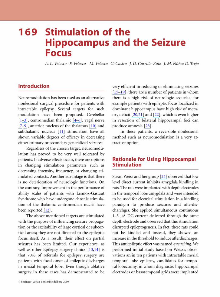
tion of interictal spikes (> Figure 169-1). Our
conclusion was that electrical stimulation of epi-
leptic focus arrested epileptic seizures within a
period of days [25]. Those patients had no MRI
evidence of mesial temporal sclerosis, whereas
. Figure 169-1The effect of subacute stimulation on electroencephalograparoxysmal EEG activities in records performed on day 1 asurface right and left fronto-temporal (FP2, F8, FP1, F7), ce(MED) and posterior (POST) parahippocampal (PHC), fusiforings were referred to ipsilateral ear lobe electrodes (A2 anspikes and slow waves in all subdural recordings, which wgyrus where the seizures initiated. After 9 days of subacutewaves disappeared. In addition a monomorphic delta act(immediately posterior to the stimulated region)
those areas that did not improve have. This was
not realized in the first report and failure in
improvement was attributed to other factors
such as failure to reach the precise stimulation
target.
To have been able to perform a temporal
lobectomy after the stimulation trial permitted
us to study the epileptic tissue under light micros-
copy. The surgical specimens of stimulated tem-
poral lobes were compared with specimens from
other ones obtained from epileptic patients who
underwent diagnostic electrode implantation,
m background activity. Two 10 s samples of maximalnd day 9 of stimulation. Recordings were made fromntral (C4, C3) and left subdural anterior (ANT), medialm (FUS), and inferior temporal gyri (IT). All EEG record-d A1). On day 1 there was a large number of interictalere more prominent in the anterior parahippocampalstimulation of the epileptic site, both spikes and slow
ivity appeared in the medial parahippocampal region
Stimulation of the hippocampus and the seizure focus 169 2841
but were not stimulated. The pathologist was
asked to report differences between the adjacent
brain tissue to electrode contacts used for stimu-
lation and those that were not stimulated in the
same patient [26]. The histopathology abnor-
malities consisted in diffuse, moderate gliosis
and cell loss of cortical layers I and II, increase in
mononuclear inflammatory cells in the subarach-
noid space and meningeal thickening of the cere-
bral tissue attached to the electrode grid. Similar
abnormalities were found in depth electrode tra-
jectories, most likely in relation to body reaction to
the presence of the silastic sheet of the electrodes.
The pathologist was unable to determine which
contacts of the grid or area of the trajectory of
the hippocampal electrodes had been stimulated.
Therefore, seizure reduction in the stimulated pat-
ients was related to electrical stimulation and
not to microlesions induced by the implanted
electrodes.
We performed neurophysiologic tests such
as producing epileptogenic afterdischarges, as
Weiss et al. used [26], recovery cycle tests, as
well as SPECT studies to try to explain the
mechanisms through which the stimulation pro-
duced its antiepileptic effect [25,27,28,29)]. We
compared basal conditions with 3 weeks stimu-
lation conditions (previous to lobectomy).
Producing hippocampal afterdischarges by
using acute local electrical stimulation is a tech-
nique used to evaluate the susceptibility of cerebral
tissue to present clinical and electroencephalo-
graphic epileptic responses. For this purpose, we
applied short (10 s) trains of high frequency (130/s)
1.0ms duration square pulses and increasing inten-
sities every 5 V to the site of the EEG epileptic foci.
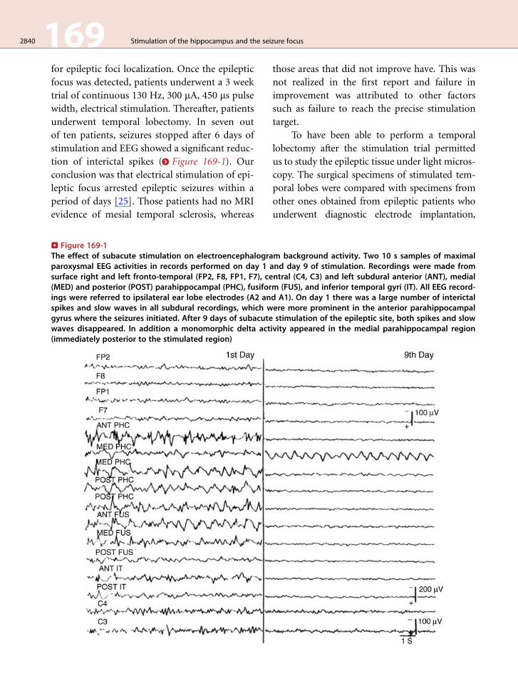
The threshold (mA) and duration of afterdischargeswere measured. > Figure 169-2 shows the after-
discharge obtained in basal condition (previous
to subacute hippocampal stimulation) in the
upper record. An 88 s afterdischarge consisting
of fast-frequency recruiting EEG spikes initiated
at the contiguous hippocampal amygdaloidal
region, which propagated to the other parasagittal
and lateral regions bilaterally, was elicited by
acute 8 s stimulation at 560 mA. This after-
discharge was accompanied by a clinical complex
partial seizure (epigastric sensation, behavioral
arrest, right adversion of the head, left hand
exploratory automatisms). The lower record
shows the response obtained in the same patient
after 560 h of subacute hippocampal stimulation.
We had to increase stimulation intensity to 5,300
mA to elicit only a few spikes in the area, with no
propagation and no accompanying symptoms.
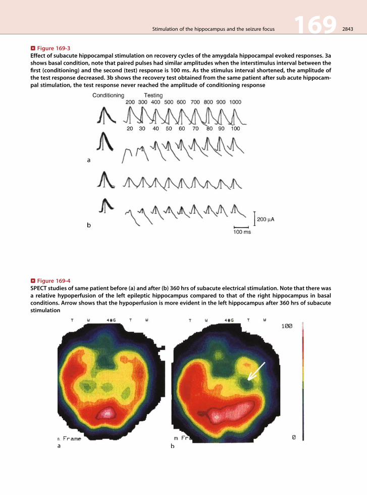
The recovery cycle test (> Figure 169-3) is an
electrophysiological technique that evaluates
changes in neuronal excitability. It consists of
the application of a pair of pulses with identical
physical characteristics to evoke a response,
applied at different interstimulus time intervals.
As shown in > Figure 169-3a, when we stimu-
lated the amygdala and recorded the hippocam-
pus in basal condition, the paired pulses had
similar amplitudes when the interstimulus inter-
val between the first (conditioning) and the
second (test) response was over 100 ms. As the
stimulus interval shortened, the amplitude of
the test response decreased (refractory period).
When compared to the recovery test obtained
from the same patient after sub acute hippocam-
pal stimulation, the test response never reached
the amplitude of conditioning response suggest-
ing an inhibitory effect (> Figure 169-3b).
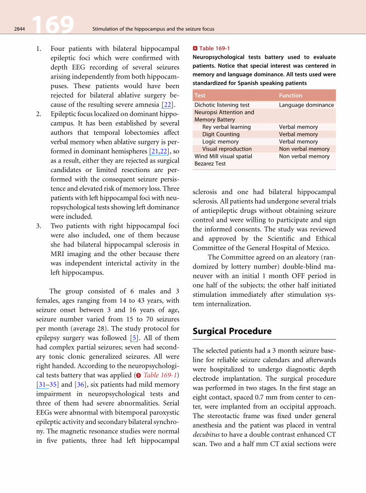
Single photon emission computed tomogra-
phy (SPECT) is used to evaluate the regional
cerebral blood flow (rCBF) perfusion as and
indirect evidence of hyper or hypo metabolic
neuronal dysfunction. > Figure 169-4a shows
the basal SPECT in a patient with left temporal
epilepsy demonstrating hypoperfusion in the
corresponding area. > Figure 169-4b in same
patient after sub acute hippocampal stimulation
shows a further decrease in (rCBF) in the stimu-
lated hippocampal area.
Compared to basal conditions, the results after
the hippocampal stimulation showed that after-
discharges were blocked, paired pulses diminished
. Figure 169-2Effect of subacute stimulation on afterdischarges in patient KG 111: 2A shows the threshold and duration of theafterdischarge produced by acute stimulation of the right anterior parahippocampal gyrus with 8 s trains ofrectangular pulses (130/s and 1.0 ms) before initiating subacute stimulation. Threshold stimulation of 600 mAproduced an 80 s afterdischarge initiated in the amygdala PHC region close to the stimulation site (indicated by thestar), which spread 10 s later to the right and left fronto-temporal scalp regions. The afterdischarge was accom-panied by symptoms of a spontaneous complex partial seizure. 2B shows that after 560 hrs of subacute stimulation,the threshold for the afterdischarge increased to 5,300mA and its duration decreased from 80 to 8 s, with no clinicalsymptoms. (Arrows indicate ON and OFF for the 8 s trains to obtain afterdischarges)
2842 169 Stimulation of the hippocampus and the seizure focus
their amplitude and SPECT showed decreased
rCBF in the stimulated area. Moreover, determina-
tion of benzodiazepine receptor binding measured
by autoradiography was used to evaluate the activ-
ity of GABA system receptors, indicative of neuro-
nal inhibition of the stimulated hippocampal
region. There was an increase of hippocampal
benzodiazepine receptors density in those patients
who had undergone hippocampal stimulation
compared to non stimulated tissue, obtained
from temporal lobectomies in epileptic patients
who had not undergone subacute stimulation [29].
Chronic Hippocampal Stimulation
Patient Selection
In our experience, 30% of intractable temporal
lobe epilepsy patients undergo bilateral hippo-
campal electrode implantation for diagnostic pur-
poses before performing a temporal lobectomy.
From this group of patients we selected nine for
chronic electrical stimulation of the hippocampal
foci [30]. We settled the criteria for selection
according to the following parameters:
. Figure 169-4SPECT studies of same patient before (a) and after (b) 360 hrs of subacute electrical stimulation. Note that there wasa relative hypoperfusion of the left epileptic hippocampus compared to that of the right hippocampus in basalconditions. Arrow shows that the hypoperfusion is more evident in the left hippocampus after 360 hrs of subacutestimulation
. Figure 169-3Effect of subacute hippocampal stimulation on recovery cycles of the amygdala hippocampal evoked responses. 3ashows basal condition, note that paired pulses had similar amplitudes when the interstimulus interval between thefirst (conditioning) and the second (test) response is 100 ms. As the stimulus interval shortened, the amplitude ofthe test response decreased. 3b shows the recovery test obtained from the same patient after sub acute hippocam-pal stimulation, the test response never reached the amplitude of conditioning response
Stimulation of the hippocampus and the seizure focus 169 2843
. Table 169-1
Neuropsychological tests battery used to evaluate
patients. Notice that special interest was centered in
memory and language dominance. All tests used were
standardized for Spanish speaking patients
Test Function
Dichotic listening test Language dominanceNeuropsi Attention andMemory Battery
Rey verbal learning Verbal memoryDigit Counting Verbal memoryLogic memory Verbal memoryVisual reproduction Non verbal memory
Wind Mill visual spatialBezarez Test
Non verbal memory
2844 169 Stimulation of the hippocampus and the seizure focus
1. Four patients with bilateral hippocampal
epileptic foci which were confirmed with
depth EEG recording of several seizures
arising independently from both hippocam-
puses. These patients would have been
rejected for bilateral ablative surgery be-
cause of the resulting severe amnesia [22].
2. Epileptic focus localized on dominant hippo-
campus. It has been established by several
authors that temporal lobectomies affect
verbal memory when ablative surgery is per-
formed in dominant hemispheres [21,22], so
as a result, either they are rejected as surgical
candidates or limited resections are per-
formed with the consequent seizure persis-
tence and elevated risk ofmemory loss. Three
patients with left hippocampal foci with neu-
ropsychological tests showing left dominance
were included.
3. Two patients with right hippocampal foci
were also included, one of them because
she had bilateral hippocampal sclerosis in
MRI imaging and the other because there
was independent interictal activity in the
left hippocampus.
The group consisted of 6 males and 3
females, ages ranging from 14 to 43 years, with
seizure onset between 3 and 16 years of age,
seizure number varied from 15 to 70 seizures
per month (average 28). The study protocol for
epilepsy surgery was followed [5]. All of them
had complex partial seizures; seven had second-
ary tonic clonic generalized seizures. All were
right handed. According to the neuropsychologi-
cal tests battery that was applied (> Table 169-1)
[31–35] and [36], six patients had mild memory
impairment in neuropsychological tests and
three of them had severe abnormalities. Serial
EEGs were abnormal with bitemporal paroxystic
epileptic activity and secondary bilateral synchro-
ny. The magnetic resonance studies were normal
in five patients, three had left hippocampal
sclerosis and one had bilateral hippocampal
sclerosis. All patients had undergone several trials
of antiepileptic drugs without obtaining seizure
control and were willing to participate and sign
the informed consents. The study was reviewed
and approved by the Scientific and Ethical
Committee of the General Hospital of Mexico.
The Committee agreed on an aleatory (ran-
domized by lottery number) double-blind ma-
neuver with an initial 1 month OFF period in
one half of the subjects; the other half initiated
stimulation immediately after stimulation sys-
tem internalization.
Surgical Procedure
The selected patients had a 3 month seizure base-
line for reliable seizure calendars and afterwards
were hospitalized to undergo diagnostic depth
electrode implantation. The surgical procedure
was performed in two stages. In the first stage an
eight contact, spaced 0.7 mm from center to cen-
ter, were implanted from an occipital approach.
The stereotactic frame was fixed under general
anesthesia and the patient was placed in ventral
decubitus to have a double contrast enhanced CT
scan. Two and a half mm CT axial sections were
Stimulation of the hippocampus and the seizure focus 169 2845
taken to be fused with preoperative MRI sections
taken the day before surgery. On the fused image,
virtual trajectories that traversed the entire hip-
pocampus, avoiding blood vessels and ending in
the basal part of the temporal lobe amygdala
were planned. Those trajectories had a laterome-
dial angle of 10–15�, starting 26.0–30.0 mm in
the skull entrance and ending 22.0–25.0 mm
lateral to the midline. It is important to mention
that the estimated center of the planned burr
holes averages 3.0 cm lateral to the midline.
Therefore, about 10.0 cm distance between the
pins that will fix the posterior part of the skull is
required. This has to be taken into consideration
when placing the stereotactic frame.
An occipital horseshoe shape incision is per-
formed, leaving a distance of 2 cm between the
estimated edge of the 14 mm diameter burr
holes, the electrodes fixation device and the
edge of the incision. This is to avoid skin ero-
sions over the implanted electrodes. Diagnostic
electrodes are left externalized for EEG localiza-
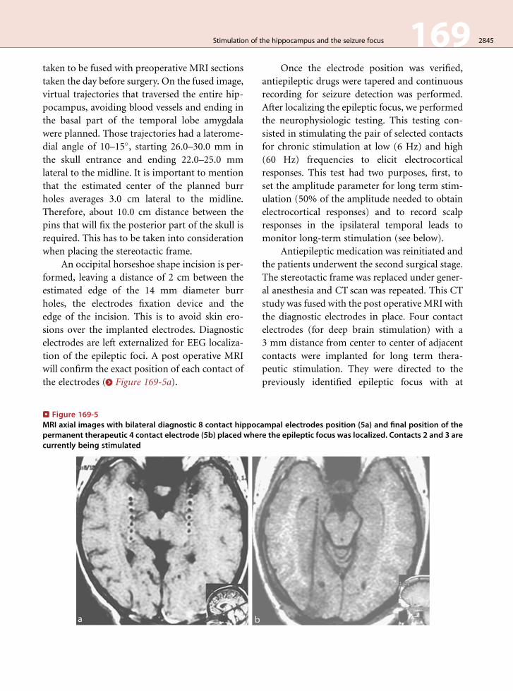
tion of the epileptic foci. A post operative MRI
will confirm the exact position of each contact of
the electrodes (> Figure 169-5a).
. Figure 169-5MRI axial images with bilateral diagnostic 8 contact hippocpermanent therapeutic 4 contact electrode (5b) placed whecurrently being stimulated
Once the electrode position was verified,
antiepileptic drugs were tapered and continuous
recording for seizure detection was performed.
After localizing the epileptic focus, we performed
the neurophysiologic testing. This testing con-
sisted in stimulating the pair of selected contacts
for chronic stimulation at low (6 Hz) and high
(60 Hz) frequencies to elicit electrocortical
responses. This test had two purposes, first, to
set the amplitude parameter for long term stim-
ulation (50% of the amplitude needed to obtain
electrocortical responses) and to record scalp
responses in the ipsilateral temporal leads to
monitor long-term stimulation (see below).
Antiepileptic medication was reinitiated and
the patients underwent the second surgical stage.
The stereotactic frame was replaced under gener-
al anesthesia and CT scan was repeated. This CT
study was fused with the post operative MRIwith
the diagnostic electrodes in place. Four contact
electrodes (for deep brain stimulation) with a
3 mm distance from center to center of adjacent
contacts were implanted for long term thera-
peutic stimulation. They were directed to the
previously identified epileptic focus with at
ampal electrodes position (5a) and final position of there the epileptic focus was localized. Contacts 2 and 3 are
2846 169 Stimulation of the hippocampus and the seizure focus
least two contacts within the epileptic site
(> Figure 169-5b). Afterwards the pulse genera-
tor was implanted and connected to the depth
stimulation electrode.
Double Blind Maneuver
Five patients had an initial 1 month OFF accord-
ing to the aleatory selection and four patients
initiated stimulation immediately. Patients and
medical personnel that collected seizure calen-
dars were unaware whether pulse generator was
ON or OFF as previously explained.
Bipolar stimulation was performed choosing
the pair of contiguous contacts which covered
the area where the epileptic focus was localized.
In case of bilateral foci, bilateral hippocampal
stimulation was used. The parameters for chro-
nic stimulation of the hippocampus were the
following:
Cyclic stimulation: 1 min trains of square
pulses with 4 min interstimulus interval
Charge density adjusted to 2–4 mC/sq cm/
phase
High frequency: 130 Hz
Pulse width: 450 msAmplitude of 300 mA which equals 50% of
the amplitude needed to obtain electrocortical
responses.
According to the formula referred by Velasco
et al. [3], the charge density for these parameters
is <3.0 mC/cm2/phase. In patients with bilateral
foci, parameters were the same but alternating
1 min stimulation on one side with a 4 min
interval between right and left sides. The stimu-
lation parameters used are within a safe nonle-
sional range [3,37].
Follow Up Protocol
Follow up in all patients was at least 18 months
(18–84 months) after initiating hippocampal
stimulation. Seizure calendars were collected
once a month; EEG and neuropsychological tests
were performed on months 6, 12 and 18 after
stimulation onset. Neurophysiologic tests to assess
the viability of the stimulated tissue to electrical
stimuli were carried out every 6 months. The
internalized pulse generators were programmed
with a transcutaneous computer to stimulate
through the selected contacts for therapeutic stim-
ulation using 8 Hz pulses, 6–8 V 450 ms to induce
electrocortical responses, EEG recording referred
to ipsilateral ear; responses are seen in the ipsilat-
eral temporal leads.
Results
The postsurgical control MRIs showed no evi-
dence of hemorrhage or edema, all patients tol-
erated the surgical procedure well. None of
the patients had side effects with the stimulation
parameters employed, as a matter of fact; they
were unaware of device activation which
permitted us to have a double blind ON OFF
protocol.
Impact on seizure number: > Figure 169-6
shows the seizure graphs of two patients as exam-
ples of the response to stimulation. Significance
of seizure reduction was determined by Student’s
T test. Although all patients improved, there
were two types of responses. One of them is
shown in > Figure 169-6a. The graph shows the
seizure number per month (individual bars), the
first three bars show the baseline seizure count
followed by the OFF bar which indicates the first
month of the double blind protocol during
which this particular patient had the stimulator
OFF. The following 18 bars show the seizure
counts during 18 months follow-up. Observe
the immediate decrease in seizures as soon as
stimulation was initiated (ON). It is also
outstanding how the patient was seizure free
after 2 months of stimulation and remained so
during the whole follow-up. A total of five
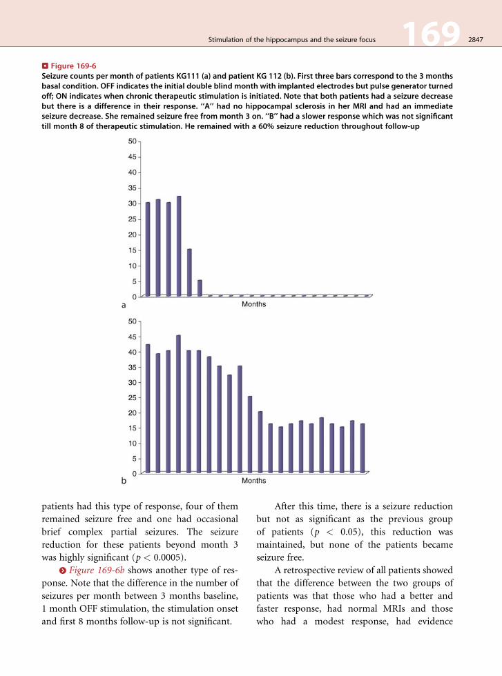
. Figure 169-6Seizure counts per month of patients KG111 (a) and patient KG 112 (b). First three bars correspond to the 3 monthsbasal condition. OFF indicates the initial double blind month with implanted electrodes but pulse generator turnedoff; ON indicates when chronic therapeutic stimulation is initiated. Note that both patients had a seizure decreasebut there is a difference in their response. ‘‘A’’ had no hippocampal sclerosis in her MRI and had an immediateseizure decrease. She remained seizure free from month 3 on. ‘‘B’’ had a slower response which was not significanttill month 8 of therapeutic stimulation. He remained with a 60% seizure reduction throughout follow-up
Stimulation of the hippocampus and the seizure focus 169 2847
patients had this type of response, four of them
remained seizure free and one had occasional
brief complex partial seizures. The seizure
reduction for these patients beyond month 3
was highly significant (p < 0.0005).
> Figure 169-6b shows another type of res-
ponse. Note that the difference in the number of
seizures per month between 3 months baseline,
1 month OFF stimulation, the stimulation onset
and first 8 months follow-up is not significant.
After this time, there is a seizure reduction
but not as significant as the previous group
of patients (p < 0.05), this reduction was
maintained, but none of the patients became
seizure free.
A retrospective review of all patients showed
that the difference between the two groups of
patients was that those who had a better and
faster response, had normal MRIs and those
who had a modest response, had evidence
2848 169 Stimulation of the hippocampus and the seizure focus
of hippocampal sclerosis shown in the MRI
studies. Stimulation parameters in both groups
were similar. Conceivably, sclerotic tissue may
have higher impedance and therefore, those
patients with mesial temporal sclerosis should
have been stimulated with higher density charge.
Or maybe, the smaller response is due to the
disruption of the normal histology of the tissue.
ON-OFF protocol: According to the aleatory
double-blind maneuver with an initial 1 month
OFF period that was authorized, five patients
underwent an OFF period after deep brain
stimulation system implantation. The other
four patients initiated stimulation immediately
after neurostimulator implantation. This study
design was based in the preliminary subacute
studies where we had observed a significant
seizure reduction after day 6 of therapeutic stim-
ulation. As already mentioned, patients who
responded to subacute stimulation in the study
had no evidence of hippocampal sclerosis by
MRI. In contrast, the double blind protocol
included both patients with normal MRI and
with evidence of mesial temporal sclerosis, so
we did not realize that there would be a retarded
response in patients with mesial temporal sclero-
sis. For this reason, the results of double blind
protocol are difficult to analyze. We can say
that all patients who underwent an initial OFF
period, showed either no changes in seizure
number when compared with baseline in four
patients and seizure increase in one patient.
From four patients who went ON electrical stim-
ulation immediately, three had a modest non
significant seizure decrease (two of them had
mesial temporal sclerosis); one patient had a
90% seizure decrease. Regardless of having or
not mesial temporal lobe sclerosis in MRI, all
patients had a significant lower seizure count by
the end of the 18 months follow-up compared
with baseline.
Neuropsychological impact: Our main con-
cern and reason for including these patients was
the high risk of memory deficit with temporal
lobectomy due to either bilateral hippocampal foci
or left side (dominant hemisphere) localization.
All patients had some degree of memory loss in
the baseline stage, probably due to the long seizure
history and poor medical treatment response.
When stimulation started, no patient had a mem-
ory decline and after 18 months stimulation, and
there was a trend to improve in both verbal and
non verbal memory evaluations. The small num-
ber of patients studied does not permit statistical
analysis.
After the initial publication in 2000 of our
first report on hippocampal stimulation, a series
of studies using hippocampal stimulation for
the control of mesial temporal lobe epilepsy
[38,39] and [40] have been published. Results
have varied for a number of reasons: consider-
ing evidence of hippocampal sclerosis in MRI an
inclusion criteria, when we have described above
that these patients do not have the same seizure
reduction than patients with no sclerosis. Skip-
ping the first surgery where diagnostic eight con-
tact electrodes are implanted and thus possibly
missing the exact location of the hippocampal
focus could also explain different results.
Regardless of the differences in seizure re-
duction, all studies agree that stimulation of the
hippocampus is a safe method with no evidence
of tissue damage, all studies use stimulation
parameters within a safe nonlesional range, and
more importantly, all authors agree that there is
no deterioration in memory function.
We can conclude that stimulation of the
hippocampus is a safe, nonlesional alternative
for patients with complex partial seizures, with
or without secondary generalization who are
not candidates for resective surgery. Other inclu-
sion criteria might be considered, for example,
patients with previous temporal lobectomy who
develop or have residual contralateral intractable
seizures. More studies have to be performed
to clarify a number of questions. What if we use
other stimulation parameters for the sclerotic
hippocampus? What happens if, instead of
Stimulation of the hippocampus and the seizure focus 169 2849
stimulating a hippocampal sclerotic tissue, we
stimulate the parahippocampus to avoid seizure
propagation? We should also conduct multicen-
ter studies to validate the neuropsychological
findings. Even though patients with mesial tem-
poral lobe epilepsy are the most frequently
referred ones for surgery, could other types of
partial seizures be treated with stimulation? We
will address the last question in the next part of
this chapter.
Stimulation of the Motor Cortexfor the Treatment ofSupplementary and PrimaryMotor Cortex Seizures
Ablative surgery of epileptic foci located in the
supplementary motor or the primary motor cor-
tices has been performed in several epilepsy sur-
gery centers [41–44,45]. Though results vary
within each center, the outcome in seizure reduc-
tion varies from 65 to 100%. Most of the cases
are patients who have lesions such as cortical
dysplasia, cavernomas and gliosis; very few non
lesional cases are included. The main problem
with these surgeries is that there are a number
of neurological sequelae, i.e., paralysis, paresis,
apraxia, aphasia and mutism. There were also
complications due to the surgical procedure
itself. No wonder the epileptologists have a
great concern when they have to operate these
patients. If the patient has no evidence of a lesion
in the MRI, this concern is even greater. With all
this considered, we decided to evaluate the
possible anticonvulsive effect of stimulating the
epileptic foci located in the motor area in two
patients with non lesional intractable epilepsy,
one of them in the supplementary motor area
and the other in the primary motor area.
Both had severe seizures despite multiple
antiepileptic drugs and were candidates for intra-
cranial grids for foci detection. The two patients
were studied following the Epilepsy Surgery
Protocol of the General Hospital of Mexico. The
study was reviewed and approved by the Scientific
and Ethical Committee of the General Hospital
of Mexico. Patients were willing to participate and
signed the informed consents. The selected
patients had a 3 month seizure baseline to collect
reliable seizure calendars. Neuropsychological
evaluation was performed and QOL scales were
applied. Afterwards patients were hospitalized to
undergo diagnostic depth electrode implantation.
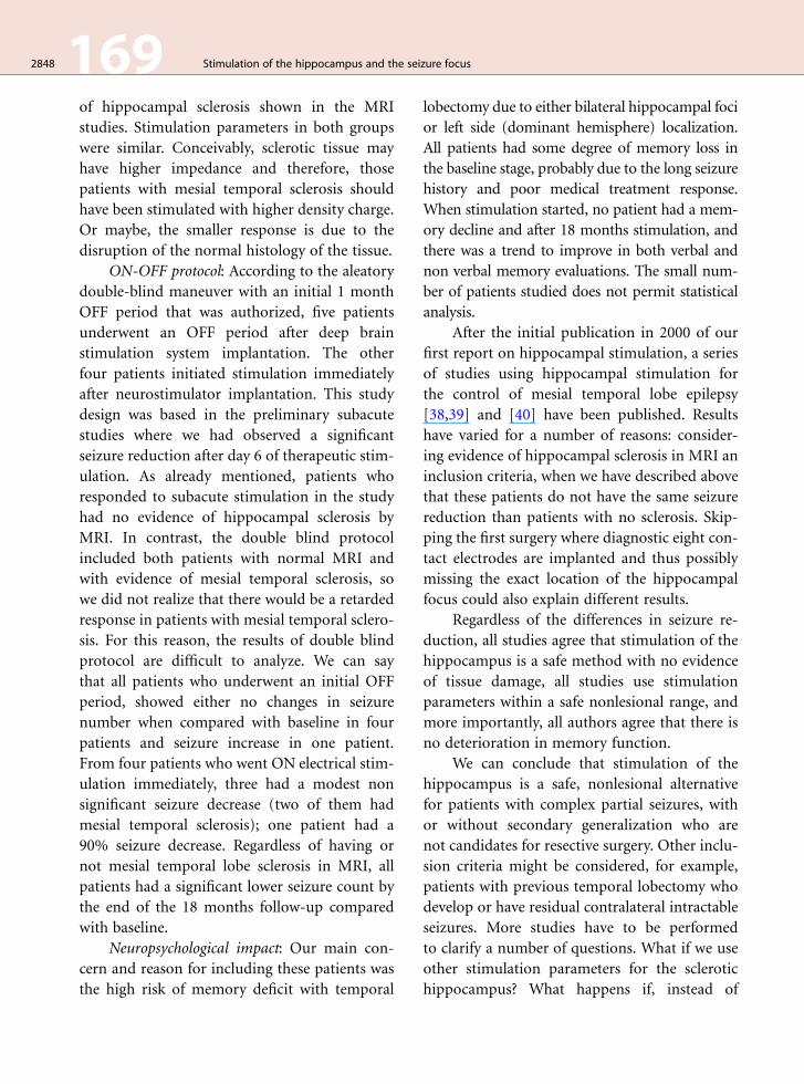
Two diagnostic 20 contact grids were implanted
through frontal craniotomies.
Patient I: 17 year old male with refractory
supplementary motor seizures. Perseverance and
verbal aggressiveness were present. Surface
EEG showed frontal parasagittal epileptic acti-
vity. MRI was normal. Bilateral 20 contact grids
were implanted in right and left SMA
(> Figure 169-7a). Ictal depth EEG showed spon-
taneous seizure onset located at contacts 3, 2 and
9 of right grid.
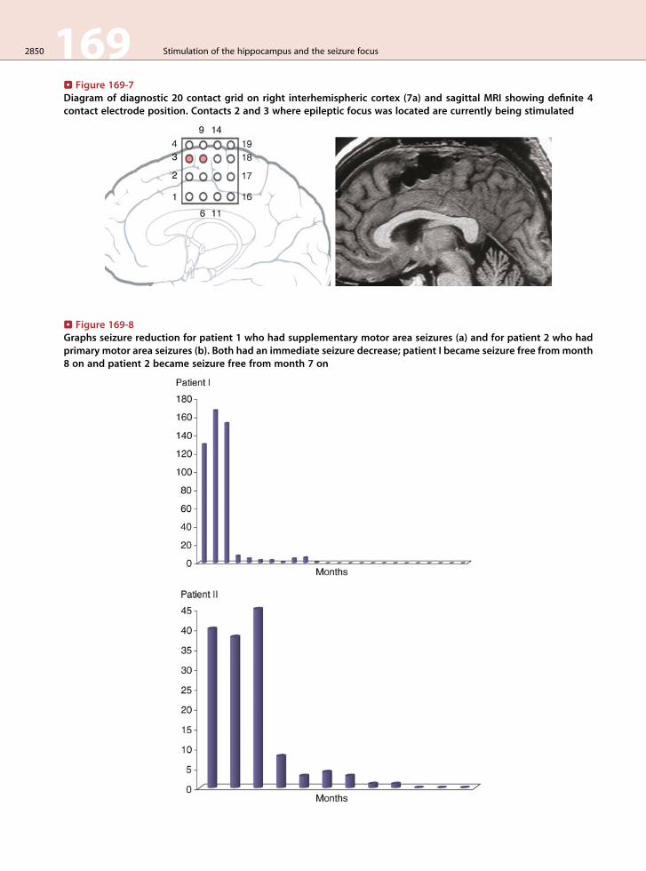
Patient II: 24 year old female with left pri-
mary motor seizures and progressive loss of
motility of the left side of the body and face.
Surface EEG showed slowing in right frontal
area, MRI was normal. Two 20 contact grids
were implanted in the upper and lower right
motor cortex. Ictal EEG showed seizure starting
in contact 10 of the upper grid (> Figure 169-8b).
Daily depth recording was performed with-
out AEDs and ictal EEG activity was obtained.
Once the epileptic focus was detected, patients
reinitiated AEDs. Grids were explanted and
replaced by a four contact, 1cm diameter, plate
electrode localized over the epileptic focus
(> Figure 169-7b) for chronic stimulation. The
position of the electrodes was fixed by suturing
them to the dura matter using nylon stitches in
each end of the electrode. In the case of primary
motor cortex, electrode was fixed to the dura
in the convexity; in the case of supplementary
motor cortex focus, the electrode was fixed to the
cerebral falx. Thereafter, electrode was connected
to a DBS system. Stimulation was started with
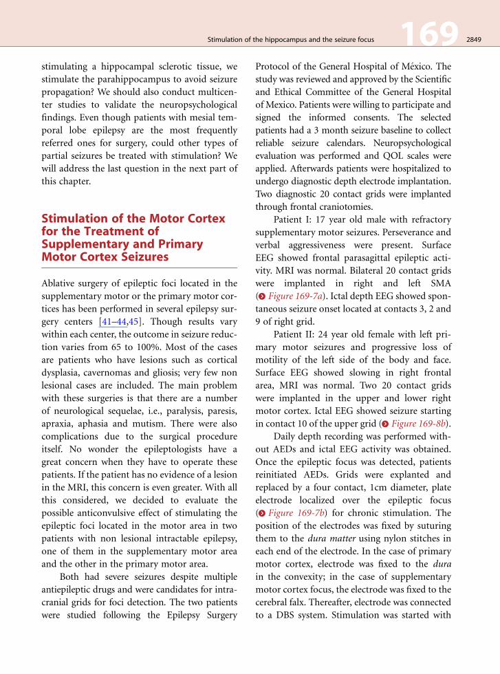
. Figure 169-8Graphs seizure reduction for patient 1 who had supplementary motor area seizures (a) and for patient 2 who hadprimary motor area seizures (b). Both had an immediate seizure decrease; patient I became seizure free frommonth8 on and patient 2 became seizure free from month 7 on
. Figure 169-7Diagram of diagnostic 20 contact grid on right interhemispheric cortex (7a) and sagittal MRI showing definite 4contact electrode position. Contacts 2 and 3 where epileptic focus was located are currently being stimulated
2850 169 Stimulation of the hippocampus and the seizure focus
Stimulation of the hippocampus and the seizure focus 169 2851
the following parameters: bipolar continuous
stimulation between contacts that covered the
epileptogenic zone, 130 Hz, 350 mA and 450 ms.Both patients had a seizure reduction which
was observed from the beginning of the stimula-
tion of the epileptic focus. Patient I became sei-
zure free from month 8 on and has remained so
for 14 months and patient II from month 7 with
no seizures for 3 months. Motor function was
preserved, no adverse effects were observed and
patients are unaware of stimulation. QOL scales
improved in both patients.
We present two cases where results are dra-
matic and encourage us to continue attempting to
stimulate themotor cortex too.We are sure wewill
face other challenges, as for example, having a huge
epileptic areawithmultiple foci for which there are
no grid type electrodes available for cortical stim-
ulation. Would we need several electrodes, could
they be connected to a single pulse generator,
would we need a specially designed electrode? All
these questions point to the need of a near collab-
oration between clinicians and biomedical engi-
neers to design better hardware for stimulation
that can be well tolerated by the patient and
avoid skin erosions or other problems such as
lead breakage [30,46] Another goal to achieve is
to decrease costs so more patients have the oppor-
tunity to try this alternative surgical method.
Many questions and challenges arise, and
to be able to answer them, a multidisciplinary
group is mandatory with collaboration of basic
scientists, neuropsychologists, epileptologists and
neurosurgeons.
References
1. Cooper IS, Amin I, Ricklan M. Chronic cerebellar stimu-
lation in epilepsy: clinical and anatomical studies. Arch
Neurol 1976;33:559-70.
2. Davis R, Emmans SE. Cerebellar stimulation for seizure
control 17 year study. Stereotact Funct Neurosurg
1992;58:200-8.
3. Velasco F, Carrillo-Ruiz JD, Brito F, Velasco M, Velasco
AL, Marquez I, Davis R. Double-blind randomized
controlled pilot study of bilateral cerebellar stimulation
for treatment of intractable motor seizures. Epilepsia
2005;46:1-11.
4. Velasco F, Velasco M, Velasco AL. Effect of chronic elec-
trical stimulation of the centromedian thalamic nuclei on
various intractable seizure patterns. I. Clinical seizures
and paroxysmal EEG activity. Epilepsia 1993;34:1052-64.
5. Velasco F, Velasco M, Jimenez F. Predictors in the treat-
ment of difficult to control seizures by electrical stimula-
tion of the centromedian thalamic nucleus. Neurosurgery
2000;47:295-305.
6. Velasco M, Velasco F, Velasco AL. Acute and chronic
electrical stimulation of the centromedian thalamic nu-
cleus: modulation of reticulo-cortical systems and predic-
tor factors for generalized seizure control. Arch Med Res
2000;31:304-15.
7. Morris G, Mueller W. Long-term treatment with vagus
nerve stimulation in patients with refractory epilepsy.
Neurology 1999;53:1731-5.
8. Frost M, Gates J, Helmers SL. Vagus nerve stimulation in
children with refractory seizures associated with Lennox–
Gastaut syndrome. Epilepsia 2001;42:1148-52.
9. Amar AP, Apuzzo MLJ, Liu CY. Vagus nerve stimula-
tion therapy alters failed cranial surgery for intract-
able epilepsy: results from the vagus nerve stimulation
therapy patient outcome registry. Neurosurgery 2004;55:
1086-93.
10. Kerrigan JF, Litt B, Fisher RS, Cranstoun S, French JA,
Blum DE, Dichter M, Shetter A, Baltuch G, Jaggi J,
Krone S, Brodie M, Rise M, Graves N. Electrical stimu-
lation of the anterior nucleus of the thalamus for the
treatment of intractable epilepsy. Epilepsia 2004;
45:346-54.
11. VesperJ, Steinhoff B, Rona S, Wille C, Bilic S, Nikkhah G,
Ostertag C. Chronic high frequency deep brain stimula-
tion of the STN/SNr for progressive myoclonic epilepsy.
Epilepsia 2007;48:1984-9.
12. Velasco AL, Velasco F, Jimenez F, Velasco M, Castro G,
Carrillo J-D, Fanghanel G, Boleaga B. Neuromodulation
of the centromedian thalamic nuclei in the treatment of
generalized seizures and the improvement of the quality of
life in patients with Lennox–Gastaut syndrome. Epilepsia
2006;47: 1203-12.
13. Wieser HG, Engel J Jr, Williamson PD, et al. Surgically
remediable temporal lobe syndromes. In: Engel J Jr,
editor. Surgical treatment of the epilepsies. New York:
Raven Press; 1993. p. 49-63.
14. Williamson PD, Wiesser HG, Delgado Escueta, AV.
Clinical characteristics of partial seizures. In: Engel J Jr,
editor. Surgical treatment of the epilepsies. New York:
Raven Press; 1993. p. 387-97.
15. Velasco AL, Boleaga B, Brito F, Jimenez F, Gordillo JL,
Velasco F, Velasco M. Absolute and relative predictor
values of some non-invasive and invasive studies for the
outcome of anterior temporal lobectomy. Arch Med Res
2000;31:62-74.
2852 169 Stimulation of the hippocampus and the seizure focus
16. Primrose DC, Ojeman GA. Outcome of resective surgery
for temporal lobe epilepsy. In: Luders H, editor. Epilepsy
surgery. New York: Raven Press; 1961. p. 601-18.
17. Cahan LD, Sutherling W, McCullough MA. Review of
the 20-year UCLA experience with surgery of epilepsy.
Cleve Clin J Med 1984;51:313-23.
18. Engel J Jr. Outcome with respect to epileptic seizures.
In: Engel J Jr, editor. Surgical treatment of the epilepsies.
New York: Raven Press; 1987. p. 553-69.
19. Radhakrishnan K, So EL, Silbert PL, Jack CR Jr, Cascino
G, Sharborough FW, O’Brien PC. Predictors of outcome
of anterior temporal lobectomy for intractable epilepsy.
Neurology 1998;51:465-71.
20. Helmstaedter C, Kurthen M, Lux S, Reuber M, Elger CE.
Chronic epilepsy and cognition: a longitudinal study in
temporal lobe epilepsy. Ann Neurol 2003;54:425-32.
21. Kapur N, Prevett M. Unexpected amnesia: are there
lessons to be learned from cases of amnesia following
unilateral temporal lobe surgery? Brain 2003;126
(Pt 12):2573-85.
22. Trenerry MR, Jack CR Jr., Ivnik RJ, Sharbrough FW,
Cascino GD, Hirschorn KA, Marsh WR, Kelly PJ,
Meyer FB.MRI hippocampal volumes and memory func-
tion before and after temporal lobectomy. Neurology
1993;43:1800-5.
23. Scoville WB and Milner B. Loss of recent memory after
bilateral hippocampal Lesions. J. Neurol 1957;20:11-21.
24. Weiss SR, Eidsath A, Li XL, Heynen T, Post RM.
Quenching revisited: low level direct current inhibits
amıgdala kindling. Exp Neurol 1898;154:185-92.
25. Velasco AL, Velasco M, Velasco F, Menes D, Gordon F,
Rocha L, Briones M, Marquez I. Subacute and chronic
electrical stimulation of the hippocampus on intractable
temporal lobe seizures. Arch Med Res 2000;31:316-28.
26. Velasco M, Velasco F, VELASCOAL, Boleaga B, Jimenez
F, Brito F, Marquez I. Subacute electrical stimulation of the
hippocampus blocks intractable temporal lobe seizures and
paroxysmal EEG activities. Epilepsia 2000; 41:158-69.
27. Velasco F, Velasco M, Velasco AL, Menes D, Rocha L.
Electrical stimulation for epilepsy 1. Stimulation of hippo-
campal foci. Stereotact Funct Neurosurg 2001;77:223-7.
28. Velasco M, Velasco F, Velasco AL. Centromedian thala-
mic and hippocampal electrical stimulation for the control
of intractable epileptic seizures. Clin Neurophys 2001;
18:1-15.
29. Cuellar-Herrera M, Velasco M, Velasco F, Velasco AL,
Jimenez F, Orozco S, Briones M, Rocha L. Evaluation of
GABA system and cell damage in parahippocampus
of patients with temporal lobe epilepsy showing antiepi-
leptic effects alter subacute electrical stimulation. Epilep-
sia 2004;45:459-66.
30. Velasco AL, Velasco F, Velasco M, Trejo D, Castro G,
Carrillo-Ruiz JD. Electrical stimulation of the hippocam-
pal epileptic foci for seizure control: a double blind,
long-term follow-up study. Epilepsia 2007;48:1895-903.
31. Kimura D. Cerebral dominance and the perception of
verbal stimuli. Can J Neuropsych 1961;15:166-71.
32. Voyer D. Reliability and magnitude of perceptual asym-
metries in a dichotic word recognition task. Neuropsy-
chology 2003;17:393-401.
33. Azanon-Gracia E, Sebastian-Gales N. Test de escucha
dicotica en espanol: pares de palabras bisilabicas. Rev
Neurol 2005;41:657-63.
34. Rey A. Solicitation de la memoire de fixation par des mots
et des objects presentes simultanement. Arch Psychol
1959;37:126-37.
35. Deutsch Lezac M.Memory I: tests in neurophsychological
assessment. 3rd ed. New York: Oxford University Press;
1995. p. 429-98.
36. Ostrosky-Solıs F, Ardila A, Roselli M. Neuropsi a brief
neuropsychological test battery in Spanish with norms by
age and educational level. J Int Neuropsychol Soc
1999;5:413-33.
37. McCreery DB, Agnew WF, Yuen TG, Bullara L. Charge
density and charge per phase as cofactors in neural injury
induced by electrical stimulation. IEEE Trans Biomed
Eng 1990;37:996-1001.
38. Vonck K, Boon P, Achten E, De Reuck J, Caemaert J.
Long term amygdalohippocampal stimulation for refrac-
tory temporal lobe epilepsy. Ann Neurol 2002; 52:556-65.
39. Tellez-Zenteno JF, McLachlan RS, Parrent A, Kubu CS,
Wiebe S. Hippocampal electrical stimulation in mesial
temporal lobe epilepsy. Neurology 2006;66:1-5.
40. BoonP, Vonck K, De Herdt V, Van Dycke A, Goethals M,
Goosens L, Van Zandijcke M, De Smedt T, Dewaele I,
Achten R, Wadman W, Dewaele F, Caemaert J,
Van Roost D. Deep brain stimulation in patients with
refractory temporal lobe epilepsy. Epilepsia 2007;48:
1551-60.
41. Mihara T, Tottori T, Inoue Y, Seino M. Surgical strategies
for patients with supplementary sensorimotor area
epilepsy. The Japanese experience. Adv Neurol 1996;
70:405-14.
42. Smith JR and King DW. Surgical strategies for patients
with supplementary sensorimotor area epilepsy. The
Medical College of Georgia experience. Adv Neurol
1996;70:415-427.
43. Engel J, Van Ness PC, Rasmussen TB, Ojeman LM.
Outcome with respect to epileptic seizures. In: Jerome
Engel J, editor. Surgical treatment of the epilepsies.
2nd ed. New York: Raven Press; 1993. p. 609-21.
44. Olivier A. Surgical strategies for patients with supplemen-
tary sensorimotor area epilepsy. The Montreal experience.
Adv Neurol 1996;70:429-43.
45. Spencer DD, Schumacher J. Surgical strategies for
patients with supplementary sensorimotor area epilepsy.
The Yale experience. Adv Neurol 1996;70:415-27.
46. Oh MY, Abosch A, Kim SH. Long-term hardware-related
complications of deep brain stimulation. Neurosurgery
2002;50:1268-74.