longitudinal functional magnetic resonance imaging of cognition in preclinical huntington's...
TRANSCRIPT
Chapter 14
Longitudinal Functional Magnetic Resonance Imagingin Animal Models
Afonso C. Silva, Junjie V. Liu, Yoshiyuki Hirano, Renata F. Leoni,Hellmut Merkle, Julie B. Mackel, Xian Feng Zhang, George C.Nascimento, and Bojana Stefanovic
Abstract
Functional magnetic resonance imaging (fMRI) has had an essential role in furthering our understandingof brain physiology and function. fMRI techniques are nowadays widely applied in neuroscience research,as well as in translational and clinical studies. The use of animal models in fMRI studies has been fun-damental in helping elucidate the mechanisms of cerebral blood-flow regulation, and in the explorationof basic neuroscience questions, such as the mechanisms of perception, behavior, and cognition. Becauseanimals are inherently non-compliant, most fMRI performed to date have required the use of anesthesia,which interferes with brain function and compromises interpretability and applicability of results to ourunderstanding of human brain function. An alternative approach that eliminates the need for anesthesiainvolves training the animal to tolerate physical restraint during the data acquisition. In the present chap-ter, we review these two different approaches to obtaining fMRI data from animal models, with a specificfocus on the acquisition of longitudinal data from the same subjects.
Key words: Anesthesia, awake, brain, BOLD, cerebral blood flow, cerebral blood volume,neurovascular coupling, non-human primates, rodents, songbirds.
1. Introduction
Functional magnetic resonance imaging (fMRI) has made aremarkable impact on brain research, establishing itself as themost prominent research tool in cognitive neuroscience (1, 2)and showing great promise in translational and clinical studies(3). fMRI relies on the neurovascular coupling, a tight relation-ship between changes in neural activity and local regulation of
M. Modo, J.W.M. Bulte (eds.), Magnetic Resonance Neuroimaging, Methods in Molecular Biology 711,DOI 10.1007/978-1-61737-992-5_14, © Springer Science+Business Media, LLC 2011
281

282 Silva et al.
cerebral blood flow (CBF), cerebral blood volume (CBV), andoxygen consumption (CMRO2) (4). Functional MRI providesexcellent contrast-to-noise ratio, sub-millimeter spatial resolution,coverage of the whole brain, and relative ease of implementation.On the other hand, the temporal resolution of fMRI is relativelylow (particularly with respect to the time scale of neuronal events)and the underlying fMRI signal mechanism (5) and its functionalspecificity (6) are still a subject of research.
The use of animal models has been essential to the develop-ment of fMRI techniques. Rodent models were the subjects in thefirst studies employing the blood oxygenation-level-dependent(BOLD) contrast (7, 8), as well as the first CBF measurementsusing exogenous MRI tracers, including deuterium (9, 10), flu-orine (11, 12), and gadolinium chelates (13, 14), which alsoallowed the estimation of CBV in addition to CBF. The advan-tages of using an endogenous tracer led to the development ofarterial spin labeling (ASL) techniques to quantify CBF non-invasively, with the first demonstrations once again occurring inrodents (15, 16). Animal models of functional brain activationhave been widely employed to address issues related to spatiallocalization of the functional signals, the magnitude of signalchanges as a function of stimulation parameters, as well as tem-poral aspects of the hemodynamic response, giving critical insightinto the brain’s physiology and function (17). Furthermore, theuse of animal models in fMRI has been particularly advantageousin preclinical and translational studies of various models of braindisease.
A major practical issue related to performing fMRI in ani-mals relates to compliance. The MRI environment poses strin-gent restrictions on subject positioning and movement, and typi-cal fMRI studies last from a few to several hours. Notwithstandingthe duration of the experiments, because animals are inherentlynon-compliant, most fMRI performed to date have required theuse of anesthesia, which offers the important advantages of ensur-ing compliance, minimizing movement, and alleviating stress, atthe expense of requiring the investigator to monitor and controlthe systemic physiological status of the animal. Another major dis-advantage of the use of anesthesia is that it interferes with bothneural activity and neurovascular coupling, thus compromisinginterpretability and applicability of results to the understandingof human brain function. An alternative choice to the use of anes-thesia is to acclimate, condition, and train the animal to toleratephysical restraint during the data acquisition. This approach offersthe advantage of minimizing the need for physiological monitor-ing and maintenance. On the other hand, it is difficult to dynam-ically monitor the level of stress in awake animals and virtuallyimpossible to establish its absence, even with extensive training.

Longitudinal fMRI in Animal Models 283
Stress can be a significant confound in the study of brain function,particularly when hemodynamic variables are used as surrogatemarkers of neural activity. Indeed, continued research on trainingprotocols and evaluation of stress indicators is a subject of intenseresearch (18). In the present chapter, we review these two differ-ent approaches to obtaining fMRI data from animal models, witha specific focus on the acquisition of longitudinal data from thesame subjects.
2. fMRIof AnesthetizedAnimal Models:From Terminalto LongitudinalPreparations
2.1. Anestheticsfor TerminalExperiments
Urethane (19–23) and α-chloralose (24–36) are the most widelyused anesthetics in fMRI studies of rodents. A major advantage ofboth anesthetics is that they preserve the neurovascular coupling(37). However, both substances are toxic and thus not adequatefor longitudinal use, being used only in terminal preparations(38, 39).
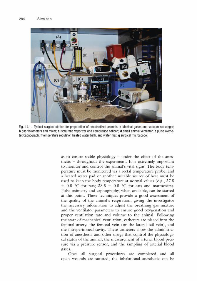
The use of anesthesia makes the animal lose the ability toregulate its own physiology and body temperature, forcing theinvestigator to monitor and maintain the vital signs of the animalto ensure stable physiology throughout the entire experiment.Therefore, surgical preparation is required. This consists of (a)induction of anesthesia, (b) oral intubation or tracheostomy, and(c) placement of intravenous, intra-arterial, and intraperitonealcatheters. Figure 14.1 shows a typical surgical preparation sta-tion and all associated equipment.
Fifteen to thirty minutes prior to induction of anesthesia, itis useful to treat the animal with atropine sulfate (Vedco Inc,Saint Joseph, MO), given subcutaneously or intramuscularly ata single dose of 0.5 mg/kg, to decrease bronchial secretions andsalivation during anesthesia, and as an anesthesia adjuvant. Anes-thesia induction in rodents usually uses halogenated anesthetics(halothane, sevoflurane, and isoflurane). In contrast, intramuscu-lar bolus of ketamine (either alone, or in combination with itspotentiators xylazine, acepromazine or medetomidine hydrochlo-ride) is frequently used for induction in both cats and non-humanprimates. Following the anesthesia induction, the animal is typi-cally orally intubated or tracheostomized and mechanically ven-tilated to allow dynamic adjustments of respiratory parameters
284 Silva et al.
Fig. 14.1. Typical surgical station for preparation of anesthetized animals. a Medical gases and vacuum scavenger;b gas flowmeters and mixer; c isoflurane vaporizer and compliance balloon; d small animal ventilator; e pulse oxime-ter/capnograph; f temperature regulator, heated water bath, and water mat; g surgical microscope.
as to ensure stable physiology – under the effect of the anes-thetic – throughout the experiment. It is extremely importantto monitor and control the animal’s vital signs. The body tem-perature must be monitored via a rectal temperature probe, anda heated water pad or another suitable source of heat must beused to keep the body temperature at normal values (e.g., 37.5± 0.5 ◦C for rats; 38.5 ± 0.5 ◦C for cats and marmosets).Pulse oximetry and capnography, when available, can be startedat this point. These techniques provide a good assessment ofthe quality of the animal’s respiration, giving the investigatorthe necessary information to adjust the breathing gas mixtureand the ventilator parameters to ensure good oxygenation andproper ventilation rate and volume to the animal. Followingthe start of mechanical ventilation, catheters are placed into thefemoral artery, the femoral vein (or the lateral tail vein), andthe intraperitoneal cavity. These catheters allow the administra-tion of anesthesia and other drugs that control the physiologi-cal status of the animal, the measurement of arterial blood pres-sure via a pressure sensor, and the sampling of arterial bloodgases.
Once all surgical procedures are completed and allopen wounds are sutured, the inhalational anesthetic can be

Longitudinal fMRI in Animal Models 285
discontinued and the anesthesia switched to either α-chloralose(α-chloralose, product #C8091, Sigma-Aldrich, St. Louis, MO)or urethane (urethane, product #U2500, Sigma-Aldrich). Typ-ical dosage for α-chloralose is 80 mg/kg initial IV bolus (37)followed by 27 mg/kg·h constant IV infusion (33). For ure-thane, the typical dosage is 1.25 g/kg single dose IP (23), asurethane is known to be a long-acting (8–10 h) anesthetic. Bothα-chloralose and urethane can be dissolved in slightly warmedphosphate-buffered saline (PBS) at a concentration of 10 mg/mlor 100 mg/ml, respectively. However, urethane is a known car-cinogen and should only be manipulated in the hood usinggloves. A muscular relaxant such as pancuronium bromide (Pavu-lon, 2 mg/ml, Teva Pharmaceuticals USA, North Wales, PA) maybe periodically administered at a dose of 2 mg/kg/hr IV or IP toaid with immobilization.
2.2. Anestheticsfor LongitudinalExperiments
Due to the extensive surgical preparation described above and alsoto the toxicity and adverse side effects of both α-chloralose andurethane, different anesthetics need to be used for longitudinalstudies. These studies require minimal interventions on the ani-mal so that its overall physiological state is stable throughout theduration of the study and especially each time the animal is tested.Because of this, inhalational compounds such as halothane (30)and isoflurane (40–48) are attractive in that they can be admin-istered via a face mask to the animal, thus obviating catheteriza-tions for vascular access. Furthermore, these agents provide stablephysiology and allow for easy control of the plane of anesthesiaand quick and smooth recovery of the animal upon withdrawal ofthe anesthetic. However, while both anesthetics are safe to use inrepetitive studies in the same animal, they suppress neuronal activ-ity (49) and cerebral metabolism (50) and affect both cerebrovas-cular tone as well as cerebrovascular reactivity (51), thus greatlyinfluencing the cerebrovascular coupling (52) and requiring opti-mization of the stimulus parameters to produce robust activation(46). In fact, the influence of halogenated inhalational anestheticson neurovascular coupling and cerebrovascular regulation can belong lasting. A recent study on the long-term effects of a shorthypoxic episode on CBF regulation in isoflurane-anesthetizedrats showed a dramatic reduction in the CBF response tohypoxia in animals that were exposed to isoflurane 5 daysearlier (53).
As an alternative to inhalational anesthetics, injectable agentssuch as propofol and medetomidine are attractive in providing sat-isfactory depth of anesthesia, quick onset of action, and smoothrecovery of the animals. Propofol is an injectable anesthetic withrapid mechanism of action that is increasingly used in fMRI exper-iments in animal models (40, 54–57). The depth of anesthe-sia under propofol can be readily adjusted by varying the rate

286 Silva et al.
of infusion, and the animals quickly recover at the end of theexperiment, thus facilitating longitudinal studies. Another agentthat has been recently proposed as suitable for repetitive stud-ies is the α2-adrenoreceptor agonist medetomidine hydrochloride(22, 58–61), which has been shown to allow robust fMRIresponses (58, 59) and the measurement of resting state sig-nal fluctuations in the brain (59–61). The sedative and analgesiceffects of medetomidine can be quickly reversed with the appli-cation of atipamezole hydrochloride. However, some limitationsof prolonged exposures to medetomidine include a gradual risein blood pressure and heart rate, lower pO2 values, and a moredifficult control of the plane of anesthesia (61).
When using injectable, intravenous anesthetics, special atten-tion must be given to the total infusion volume in relationto the total blood volume, especially in small animals such asrodents and small non-human primates, so that the infusiondoes not change blood chemistry. For example, the dose ofpropofol (10 mg/ml) required to maintain immobilization inrats and marmosets is 0.5–1.0 mg/kg/min (3–6 ml/kg/hr),which is about five times higher than that required in humans(0.05–0.2 mg/kg/min). Yet the total blood volume of a 400-grat is ∼25 ml (62), circa 1/200 of the total human blood vol-ume. Thus, intravenous anesthetics have a much greater poten-tial to disturb blood chemistry in small animals than in humans,and drug preparations should be chosen carefully. Propofol, asmade by certain manufacturers, has a pH value as low as 4, pos-ing a significant risk of inducing acidosis in small animals, espe-cially in prolonged and/or longitudinal experiments. Like in clin-ical medicine, concurrent intravenous infusion of lactated Ringersolution with the anesthetics may partially alleviate the detrimen-tal effects of blood chemical disturbance.
2.3. Placementof the Animal in anMRI-Compatible Bed
Magnetic resonance imaging poses stringent requirements onimmobilization. After surgery, the animals are placed in astereotaxic-like head holder and strapped to an MRI-compatiblebed. A number of beds are available from the major ven-dors of small animal MRI scanners, such as Bruker (Bruker-Biospin, Corp., Ettlingen, Germany) or Varian (Varian, Inc., PaloAlto, CA), or from third-party vendors (e.g., Rapid Biomed-ical, GmbH, Rimpar, Germany; Ekam Imaging, Inc., Shrews-bury, MA). Figure 14.2 shows a picture of the bed that we haveused for fMRI of small animals. The bed is an essential part ofthe experiment, as it integrates, in a single platform, a restingplace for the animal, the stereotaxic head holder, the physiologicalmaintenance and monitoring devices, a stage for receive RF coilsand preamplifiers, and another stage for the functional stimula-tion devices. Therefore, significant effort needs to be put on thedesign, fabrication, and adaptation of an animal bed to include

Longitudinal fMRI in Animal Models 287
Fig. 14.2. a MRI-compatible animal bed, containing a stereotaxic head holder with earpieces and a bite bar (b), to which the head of the animal is secured. Mechanical ven-tilation is provided by the gas lines. c Pulse oximetry sensor and rectal temperatureprobe. d Heated water mat (shown unfolded), which can be wrapped around the ani-mal’s body to maintain temperature. e Multi-channel receive RF coils. f Multi-channelRF preamplifiers.
and integrate all the features necessary to the successful executionof the experiment.
2.4. PhysiologicalMonitoring
As mentioned above, the use of anesthesia makes the animallose the ability to regulate its own physiology and body tem-perature, and it is the responsibility of the investigator to mon-itor and maintain the vital signs of the animal throughout theremainder of the experiment. As soon as the animal has beenmoved to the MRI-compatible bed, physiological monitoringmust start – or resume if started during animal preparation. Allessential physiological parameters, including, but not limited tothe list below, should be monitored and maintained at normalvalues.
1. Rectal temperature: via temperature probe inserted in rec-tum. This is one of the most critical parameters to be mon-itored and maintained, as anesthesia impedes the animal’sown temperature regulation. Normal rectal temperature val-ues for rats are 37.5 ± 0.5◦C, and 38.5 ± 0.5◦C for cats andmarmosets.
2. Mean arterial blood pressure (MABP): via pressure trans-ducer hooked up to femoral arterial line, if available, orvia pressure cuff wrapped around the tail or thigh. TypicalMABP values in rats are 110–120 mmHg (63, 64). TypicalMABP values in conscious marmosets are 100–110 mmHg(65).
3. Pulse oximetry (SPO2): via transducer placed on forelimb orhindlimb. Usually SPO2 values remain above 90%.

288 Silva et al.
4. Heart rate (HR): either derived from MABP trace orreported by pulse oximeter. For rats under α-chloralose, HRtypically rises above 300 BPM but stays below that valueunder other anesthetics. For marmosets under propofol, HRtypically decreases from above 350 BPM in the conscious,awake condition to 150–250 BPM in anesthetized animals.
5. End-tidal CO2 (ETCO2): via micro-capnometer hooked upto face mask or to the respiratory line. The distance betweenthe mechanical ventilator and the MRI magnet forces theuse of long respiration lines, resulting in substantial mixingbetween expired and recirculated gas. Therefore, the micro-capnometer tends to display significantly attenuated valuesof ETCO2, especially in small animals. Yet, these values arevalid relatives and a significant correlation exists betweenETCO2 and PaCO2, as shown in Fig. 14.3.
6. Respiratory pressure: monitored via pressure transducer inmechanical ventilator. It is important to check the respira-tory compliance of the intubated or tracheostomized ani-mal throughout the experiment. The end-inspiratory pres-sure can be set by adjusting the flow of the air mixture inthe ventilator. Typically, end-inspiratory pressures are in therange of 8–12 cm H2O.
7. Arterial blood gases, including pH, PaCO2, and PaO2: sam-pled from the femoral artery, when available. Even though
Fig. 14.3. Pooled data plot of ETCO2 as measured with a capnograph versus PaCO2 sampled from arterial blood (n=34 rats, average of six points per animal) in normocapnia and hypercapnia. The dashed line is the line of identity, andthe correlation coefficient between ETCO2 and PaCO2 is 0.77 (r 2 = 0.59). Even though the absolute value of ETCO2 isinfluenced by the size of the animal relative to the total flow and volume of air in the ventilator, the length of the gas lines,and the flow of expired air into the capnograph, the significant correlation between ETCO2 and PaCO2 allows ETCO2values to be used as a relative index of changes in PaCO2.

Longitudinal fMRI in Animal Models 289
arterial blood sampling in mice is restricted to much fewersamples than in rats, latest-generation blood gas analyzersare able to work with samples as small as 30 μl of blood(typically 60 μl) (e.g., ABL80 FLEX, Radiometer America,Westlake, OH). Periodic sampling of arterial blood is alsoperformed to yield information on arterial blood gases,hematocrit, and electrolytes. Data collected during unstablephysiological states are best discarded. Furthermore, largedeviations in the arterial blood gases often call for adminis-tration of corrective pharmacological agents.
All the physiological parameters listed above can be recorded dur-ing the experiment using a multi-channel data acquisition system,such as the system MP150 (Biopac Systems, Inc., Goleta, CA).Figure 14.4 shows the physiological monitoring graph of a typi-cal fMRI experiment to measure the BOLD and CBF response toelectrical stimulation of the forepaws in a rat anesthetized withα-chloralose. Traces of the arterial blood pressure, respiratorypressure, and heart rate were recorded along with the forepawstimulation epochs, the EPI acquisition times, and the gradienttemperature. Monitoring of the animal’s physiology during theexperiment ensures that the data are acquired under stable con-ditions. For example, no changes in arterial blood pressure arenoticed during the forepaw stimulation epochs. Another advan-tage of acquiring physiological parameters is the ability to do ret-rospective correction of the influence of either the respiratory orcardiac cycles on the fMRI signal (66).
Fig. 14.4. Physiological and fMRI data acquisition traces recorded during a typical fMRI experiment of electrical stim-ulation of both forepaws of a rat anesthetized with α-chloralose. The experiment lasted 4 min. From top to bottom, thetraces show the arterial blood pressure (red), respiratory pressure (green), EPI acquisition tics (blue), stimulation of theleft (green) and the right (blue) forepaws, the MRI gradient temperature (magenta), and the heart rate derived from theABP trace (blue).

290 Silva et al.
2.5. Recoveryfrom Anesthesia
In longitudinal experiments in which artificial ventilation is used,the animal’s autonomous control of the respiration may be sup-pressed even after anesthetics are withdrawn. Thus, recovery fromanesthesia needs to be closely monitored, and emergency pro-cedures should be planned in advance. Throughout the recov-ery, artificial ventilation and rectal temperature monitoring shouldcontinue, and the intravenous infusion site should remain patent,until the animal is clearly able to breathe on its own after beingdisconnected from the breathing circuit. Antidotes for someanesthetics are available, such as naloxone for opioids and ati-pamezole for medetomidine chloride, and should be adminis-tered. In the event of respiratory and/or cardiovascular suspen-sion after removal of intubation tube, a respiratory stimulant(e.g., doxapram) and sympathetic stimulant (e.g., epinephrine)can be administered through the intravenous infusion tube, whichshould not be removed from the animal until it is fully alert.
3. fMRIof Conscious,Awake Animals
The use of anesthesia for MRI and fMRI studies in animal modelshas the advantages of effectively ensuring compliance and of min-imizing stress via sedation. However, any anesthetic interferes, indifferent ways, with neural activity and cerebrovascular reactiv-ity, representing a complex confound to the interpretability andapplicability of the obtained data to the understanding of humanbrain function. The alternative to the use of anesthesia as a meansof ensuring compliance is to condition the conscious, awake ani-mal, to tolerate the rigid head restraint required to allow theacquisition of good quality data with acceptable levels of motionartifacts. The use of conscious, awake animals in the MRI set-ting is becoming increasingly popular, as exemplified by stud-ies performed in rodents (18, 29, 42, 55, 67–71) and monkeys(71–84). To provide effective, yet relaxed restraint to the animals,the animal bed needs to be designed taking into account comfort-able support for their head and body (77, 85). Equally importantis the development of acclimatization and training procedures tocondition the animal to tolerate long periods of restraint withminimal stress (18), as a stressed-out animal is as useful as anunresponsive one!
In this section, we describe our own experience with obtain-ing longitudinal fMRI data from conscious, awake marmosets in a7T horizontal MRI scanner. Traditionally, the use of awake, con-scious animals in the MRI requires the surgical implantation ofhead posts that can be rigidly secured by clamps to a specially
Longitudinal fMRI in Animal Models 291
designed frame (85). This approach allows maximum restraint ofthe animals but has many disadvantages. The head implants typi-cally generate susceptibility artifacts that degrade image quality byintroducing geometric distortions and/or signal dropouts in theMR images. In addition, they require constant aseptic cleaning toprevent infections. Moreover, if an infection appears, the animalneeds to be treated with antibiotics and anti-inflammatory drugsthat may interfere with neurovascular coupling and confoundinterpretation of the data. Further, the use of a head implantsignificantly detracts from a major advantage of MRI as a non-invasive technique. An alternative to the use of head implants is tosecure the animal to an MRI-compatible stereotaxic head holderby means of ear pieces and a bite bar. To do this, the animals needto be sedated with a short-acting anesthetic, such as medetomi-dine, so that they can be attached to the stereotaxic head holder,after which the head posts are allowed to wake up by reversal ofthe anesthesia (e.g., with atipamezole) (18, 42, 70, 80). While ithas been shown that animals can be successfully conditioned notto fight the head restraint upon regaining consciousness (18), thisapproach has the disadvantages of requiring the use of anesthesiaand of utilizing a head holder that may potentially hurt the ani-mal due to the presence of the ear bars. Our approach to restrainthe head of the animals is different than either of the two above.We opted to eliminate the need for implanting head posts or toadminister any anesthetics or sedatives altogether by acclimatiz-ing the animals to being restrained by a custom-fit helmet thatwas specifically designed to match the contour of each individualhead exactly, providing a comfortable, yet effective restraint.
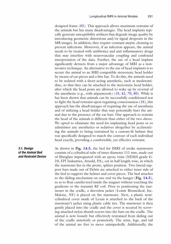
3.1. Designof the Animal Bedand Restraint Device
As shown in Fig. 14.5, the bed for fMRI of awake marmosetsconsists of a cylindrical tube of inner diameter 111 mm, made outof fiberglass impregnated with an epoxy resin (NEMA grade G-10, FPI Industries, Arnold, PA), cut in half length-wise, in whichthe marmoset lies in the prone, sphinx position. Two lateral sup-port bars made out of Delrin are attached to either inner side ofthe bed to support the helmet and cover pieces. The bed attachesto the sliding mechanism on one end via the hanger (Fig. 14.5),so as to float cantilevered inside the magnet without touching thegradients or the transmit RF coil. Prior to positioning the mar-moset in the cradle, a sleeveless jacket (Lomir Biomedical, Inc.Malone, NY) is placed on the marmoset. Next, a plastic semi-cylindrical cover made of Lexan is attached to the back of themarmoset’s jacket using plastic cable ties. The marmoset is thengently placed into the cradle and the cover is secured by screw-ing attached nylon thumb screws into the bars on the cradle. Theanimal is now loosely but effectively restrained from sliding outof the cradle anteriorly or posteriorly. The arms, legs, and tailof the animal are free to move unimpededly. Additionally, the

292 Silva et al.
Fig. 14.5. Illustration of a restrained marmoset in the MRI-compatible bed. The body of the animal is loosely attached toa back cover via zip ties secured to a sleeveless jacket worn by the animal. The back cover is screwed to the side barson the cradle, while the arms, legs, and tail of the animal are free to move. The head of the marmoset is secured to atwo-piece, custom-built helmet made specifically for that individual. The chin piece on the bottom supports the chin ofthe animal, and the head piece on the top prevents head motion. Note that the helmet pieces are lined up with foam onthe inside to provide a comfortable support to the entire head. The animal sits in the sphinx position looking out towardsthe back of the magnet. The bed is secured to the bed sliding mechanism on one end via the hanger.
semi-cylindrical cover is a few centimeters from the marmoset’sback, allowing the animal to stretch and to adjust its body posi-tion as needed for comfort, as shown in Fig. 14.5.
Because the shape and size of marmosets’ skulls vary, individ-ual custom-built helmets are designed for each animal. For this,a 3D spin-echo MRI of the entire head and neck is acquiredfrom each animal. Next, a 3D surface-rendering algorithm isapplied to obtain the contour of the head, which is then fed intoRhinoceros 3d (McNeel North America, Seattle, WA), a 3D mod-eling program, to design the top (head) and bottom (chin) hel-met pieces. After the head and chin helmet pieces are designed (seeFig. 14.6), they are sent to a 3D printer (ProJet HD3000, 3DSystems Corp., Rock Hill, SC), which builds the helmets, layer by

Longitudinal fMRI in Animal Models 293
Fig. 14.6. Detail of the construction of the custom-fit helmet. Based on a 3D MRI ofthe entire head of the marmoset, a 3D model of the helmet is created consisting of twopieces: the chin piece (blue) to support the chin of the animal and the head piece (red)that supports the head and prevents motion. Once the model is created, it is sent to a3D plastic printer that creates the helmet.
layer, from liquid ABS plastic that hardens during the manufac-turing process. After the helmet printing is complete, 3-mm thickfoam is glued onto the inside surface of both top and bottompieces to provide greater comfort for the animal.
Proper and perfect fitting of the pieces to each animal isguaranteed by design. Since the shape of each animal is used toproduce a helmet manufactured specifically for its head, we canensure that the animal is comfortable yet immobilized. After theanimal is lowered into the cradle and secured by the cover piece,the helmet top and bottom pieces are carefully placed around theanimal’s head and screwed into the bars.
3.2. Acclimatizationof the Animal to Bodyand Head Restraint
Once the individualized helmet pieces are built, the animal needsto be acclimatized to the bed and to physical restraint during theMRI sessions. This acclimatization procedure consists of threephases, as illustrated in Fig. 14.7:
1. Phase 1: Acclimatization of Awake Marmosets to Being Con-tained in the Bed. In this phase, the animals are dressed withthe jackets, attached to the back cover by zip ties, and low-ered into the bed. The cover is secured to the bed by nylonthumb screws. The bed is then inserted into a mock MRItube and the animal is observed from a distance via a web-cam. For phase 1, training begins with 15 min on day 1,and progresses to an hour by day 4 (see Table 14.1). As areward, 3–6 cc of infant milk formula (Pediasure) and 3–5mini-marshmallows are given at the beginning and end ofeach training session.
Prior to, during, and after each of the acclimatizationphases, a behavioral assessment takes place to provide a mea-sure of the tolerance of the animals to the acclimatizationprocedures. The scoring of each animal is performed using

294 Silva et al.
Fig. 14.7. Illustration of the three phases of training. In phase 1 (top), the body of the animal is loosely attached tothe MRI bed and the animal is conditioned to staying in the bed for increasing periods of time. In phase 2 (middle),reinforcement of the training in phase continues while the animal gets used to MRI sounds. In phase 3 (bottom), theindividualized helmet is introduced to restrain the head of the animal.
Table 14.1Three-week acclimatization schedule
Phase Monday Wednesday Friday Sunday Procedures
1 0:15 0:30 0:45 1:00 Jacket
2 1:00 1:20 1:40 2:00 Jacket + MRI sounds3 1:00 1:20 1:40 2:00 Jacket + MRI sounds
+ helmet
the Behavioral Assessment Scale shown in Table 14.2 (86).It is our experience that all animals successfully complete thetraining in phase 1, starting with an average score of 4 on theBehavioral Assessment Scale and acclimatizing to an averagescore of 2 by the end of phase 1.
2. Phase 2: Acclimatization of Awake Marmosets to Being Con-tained in the Bed in the Presence of MRI Sounds. In spiteof being entirely non-invasive, MRI is, unfortunately, loud.Therefore, it is necessary that the animals get properly accli-matized to the sounds generated by the MRI scanner dur-ing imaging. In phase 2, the animals are restrained as inphase 1 for increasing periods (Table 14.1). While in themock MRI tube, they were allowed to hear the sounds pro-duced by the MRI scanner, played out at a softer level thanin a real MRI session. This schedule reinforces the adapta-tion to the body restraint initiated in phase 1 and condi-tions the animals to ignore the MRI sounds produced by
Longitudinal fMRI in Animal Models 295
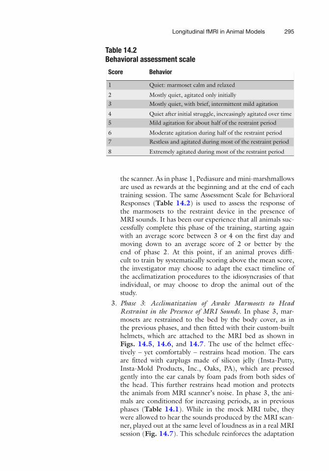
Table 14.2Behavioral assessment scale
Score Behavior
1 Quiet: marmoset calm and relaxed
2 Mostly quiet, agitated only initially3 Mostly quiet, with brief, intermittent mild agitation
4 Quiet after initial struggle, increasingly agitated over time5 Mild agitation for about half of the restraint period
6 Moderate agitation during half of the restraint period7 Restless and agitated during most of the restraint period
8 Extremely agitated during most of the restraint period
the scanner. As in phase 1, Pediasure and mini-marshmallowsare used as rewards at the beginning and at the end of eachtraining session. The same Assessment Scale for BehavioralResponses (Table 14.2) is used to assess the response ofthe marmosets to the restraint device in the presence ofMRI sounds. It has been our experience that all animals suc-cessfully complete this phase of the training, starting againwith an average score between 3 or 4 on the first day andmoving down to an average score of 2 or better by theend of phase 2. At this point, if an animal proves diffi-cult to train by systematically scoring above the mean score,the investigator may choose to adapt the exact timeline ofthe acclimatization procedures to the idiosyncrasies of thatindividual, or may choose to drop the animal out of thestudy.
3. Phase 3: Acclimatization of Awake Marmosets to HeadRestraint in the Presence of MRI Sounds. In phase 3, mar-mosets are restrained to the bed by the body cover, as inthe previous phases, and then fitted with their custom-builthelmets, which are attached to the MRI bed as shown inFigs. 14.5, 14.6, and 14.7. The use of the helmet effec-tively – yet comfortably – restrains head motion. The earsare fitted with earplugs made of silicon jelly (Insta-Putty,Insta-Mold Products, Inc., Oaks, PA), which are pressedgently into the ear canals by foam pads from both sides ofthe head. This further restrains head motion and protectsthe animals from MRI scanner’s noise. In phase 3, the ani-mals are conditioned for increasing periods, as in previousphases (Table 14.1). While in the mock MRI tube, theywere allowed to hear the sounds produced by the MRI scan-ner, played out at the same level of loudness as in a real MRIsession (Fig. 14.7). This schedule reinforces the adaptation
296 Silva et al.
to the body restraint of the previous phases and further con-ditions the animals to ignore the MRI sounds produced bythe scanner, while enforcing full head fixation. As in previousphases, the animals’ response to training will be evaluated bythe Behavioral Response Scale shown in Table 14.2. It hasbeen our experience that all animals are able to successfullycomplete this last phase of the training, starting again withan average score between 3 or 4 on the first day and mov-ing down to an average score of 2 or better by the end ofphase 3.
Out of 14 marmosets trained to date, we only dropped one animalout of the study due to increased agitation during phase 2 of theacclimatization. After the animals have successfully completed the3-week training program, they proceed to undergoing the actualMRI studies. Continued exposure to the MRI scanner in actualstudies fully consolidates the acclimatization, and all animals aretolerant of all procedures after a couple of MRI sessions. Overall,this new method of restraint is completely non-invasive, comfort-able for the animals, and of great scientific payoff in eliminatingthe need to either perform surgery for installation of head-postimplants or use sedatives to restrain the animals, allowing any mar-moset to be acclimatized to restraint and engaged in longitudinalexperiments.
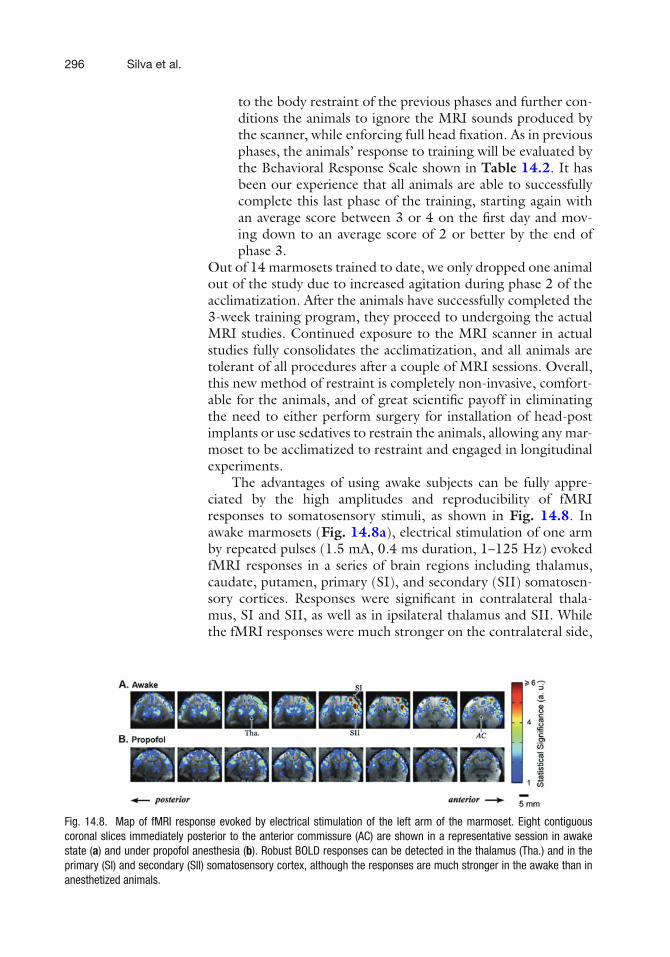
The advantages of using awake subjects can be fully appre-ciated by the high amplitudes and reproducibility of fMRIresponses to somatosensory stimuli, as shown in Fig. 14.8. Inawake marmosets (Fig. 14.8a), electrical stimulation of one armby repeated pulses (1.5 mA, 0.4 ms duration, 1–125 Hz) evokedfMRI responses in a series of brain regions including thalamus,caudate, putamen, primary (SI), and secondary (SII) somatosen-sory cortices. Responses were significant in contralateral thala-mus, SI and SII, as well as in ipsilateral thalamus and SII. Whilethe fMRI responses were much stronger on the contralateral side,
Fig. 14.8. Map of fMRI response evoked by electrical stimulation of the left arm of the marmoset. Eight contiguouscoronal slices immediately posterior to the anterior commissure (AC) are shown in a representative session in awakestate (a) and under propofol anesthesia (b). Robust BOLD responses can be detected in the thalamus (Tha.) and in theprimary (SI) and secondary (SII) somatosensory cortex, although the responses are much stronger in the awake than inanesthetized animals.

Longitudinal fMRI in Animal Models 297
occasionally robust ipsilateral responses were also detected in SI.In marmosets anesthetized with propofol (Fig. 14.8b), however,both the spatial extent as well as the fMRI response amplitudeswere significantly smaller compared to awake subjects. In par-ticular, responses in ipsilateral thalamus and ipsilateral SI wereinsignificant in many anesthetized sessions, consistent with pre-vious studies using anesthetized rats. Responses in caudate andputamen were also much weaker, although detectable. Further-more, the response amplitudes in thalamus and cortical areasin anesthetized subjects were less than half of those in awakesubjects.
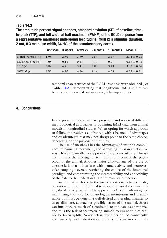
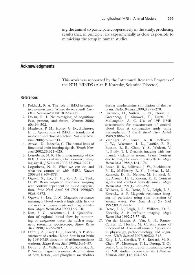
Figure 14.9 shows the fMRI response obtained from thesame animal in different MRI sessions in a time span of 10months. Excellent reproducibility of the amplitude and spatial and
Fig. 14.9. fMRI response obtained from the same animal in different MRI sessions ina time span of 10 months. a T-map demonstrating the main active areas of the brain,including SI, SII, and caudate putamen. b BOLD time-course in response to a 2-s elec-trical stimulus of both hands (2 mA, 0.3 ms, 64 Hz), obtained at five different times(0 weeks, 3 weeks, 6 weeks, 2 months, and 10 months) post-acclimatization. Excellentreproducibility of the amplitude and temporal characteristics of the BOLD response areachieved.
298 Silva et al.
Table 14.3The amplitude percent signal changes, standard deviation (SD) of baseline, time-to-peak (TTP), and full width at half maximum (FWHM) of the BOLD response froma representative marmoset undergoing longitudinal fMRI (2 s stimulus duration,2 mA, 0.3 ms pulse width, 64 Hz) of the somatosensory cortex
First scan 3 weeks 6 weeks 2 months 10 months Mean ± SD
Signal increase (%) 1.95 2.50 2.69 2.57 2.47 2.44 ± 0.28
SD of baseline (%) 0.08 0.14 0.17 0.17 0.21 0.15 ± 0.08TTP (s) 3.84 4.41 3.41 3.80 3.78 3.85 ± 0.36
FWHM (s) 3.92 4.70 4.34 4.14 4.55 4.33 ± 0.31
temporal characteristics of the BOLD response were obtained (seeTable 14.3), demonstrating that longitudinal fMRI studies canbe successfully carried out in awake, behaving animals.
4. Conclusions
In the present chapter, we have presented and reviewed differentmethodological approaches to obtaining fMRI data from animalmodels in longitudinal studies. When opting for which approachto follow, the reader is confronted with a balance of advantagesand disadvantages that may not always point to the same choice,depending on the purpose of the study.
The use of anesthesia has the advantages of ensuring compli-ance, minimizing movement, and alleviating stress in an effectiveway. However, anesthesia suppresses many homeostatic pathwaysand requires the investigator to monitor and control the physi-ology of the animal. Another major disadvantage of the use ofanesthesia is that it interferes with neural activity and neurovas-cular coupling, severely restricting the choice of the functionalparadigm and compromising the interpretability and applicabilityof the data to the understanding of human brain function.
An alternative choice to the use of anesthesia is to acclimate,condition, and train the animal to tolerate physical restraint dur-ing the data acquisition. This approach offers the advantage ofminimizing the need for physiological monitoring and mainte-nance but must be done in a well-devised and gradual manner soas to eliminate, as much as possible, stress of the animal. Stresscan introduce as much of a confound to the data as anesthesia,and thus the task of acclimatizing animals to awake studies can-not be taken lightly. Nevertheless, when performed consistentlyand correctly, acclimatization can be very effective in condition-
Longitudinal fMRI in Animal Models 299
ing the animal to participate cooperatively in the study, producingresults that, in principle, are experimentally as close as possible tomimicking the setup in human studies.
Acknowledgments
This work was supported by the Intramural Research Program ofthe NIH, NINDS (Alan P. Koretsky, Scientific Director).
References
1. Poldrack, R. A. The role of fMRI in cogni-tive neuroscience: Where do we stand? CurrOpin Neurobiol 2008;18:223–227.
2. Dolan, R. J. Neuroimaging of cognition:Past, present, and future. Neuron 2008;60:496–502.
3. Matthews, P. M., Honey, G. D., Bullmore,E. T. Applications of fMRI in translationalmedicine and clinical practice. Nat Rev Neu-rosci 2006;7:732–744.
4. Attwell, D., Iadecola, C. The neural basis offunctional brain imaging signals. Trends Neu-rosci 2002;25:621–625.
5. Logothetis, N. K. The underpinnings of theBOLD functional magnetic resonance imag-ing signal. J Neurosci 2003;23:3963–3971.
6. Logothetis, N. K. What we can do andwhat we cannot do with fMRI. Nature2008;453:869–878.
7. Ogawa, S., Lee, T. M., Kay, A. R., Tank,D. W. Brain magnetic resonance imagingwith contrast dependent on blood oxygena-tion. Proc Natl Acad Sci USA 1990;87:9868–9872.
8. Ogawa, S., Lee, T. M. Magnetic resonanceimaging of blood vessels at high fields: In vivoand in vitro measurements and image simula-tion. Magn Reson Med 1990;16:9–18.
9. Kim, S. G., Ackerman, J. J. Quantifica-tion of regional blood flow by monitor-ing of exogenous tracer via nuclear mag-netic resonance spectroscopy. Magn ResonMed 1990;14:266–282.
10. Detre, J. A., Eskey, C. J., Koretsky, A. P. Mea-surement of cerebral blood flow in rat brainby 19F-NMR detection of trifluoromethanewashout. Magn Reson Med 1990;15:45–57.
11. Detre, J. A., Williams, D. S., Koretsky, A.P. Nuclear magnetic resonance determinationof flow, lactate, and phosphate metabolites
during amphetamine stimulation of the ratbrain. NMR Biomed 1990;3:272–278.
12. Barranco, D., Sutton, L. N., Florin, S.,Greenberg, J., Sinnwell, T., Ligeti, L.,McLaughlin, A. C. Use of 19F NMRspectroscopy for measurement of cerebralblood flow: A comparative study usingmicrospheres. J Cereb Blood Flow Metab1989;9:886–891.
13. Villringer, A., Rosen, B. R., Belliveau,J. W., Ackerman, J. L., Lauffer, R. B.,Buxton, R. B., Chao, Y. S., Wedeen, V.J., Brady, T. J. Dynamic imaging with lan-thanide chelates in normal brain: Contrastdue to magnetic susceptibility effects. MagnReson Med 1988;6:164–174.
14. Rosen, B. R., Belliveau, J. W., Buchbinder,B. R., McKinstry, R. C., Porkka, L. M.,Kennedy, D. N., Neuder, M. S., Fisel, C.R., Aronen, H. J., Kwong, K. K. Contrastagents and cerebral hemodynamics. MagnReson Med 1991;19:285–292.
15. Williams, D. S., Detre, J. A., Leigh, J. S.,Koretsky, A. P. Magnetic resonance imag-ing of perfusion using spin inversion ofarterial water. Proc Natl Acad Sci USA1992;89:212–216.
16. Detre, J. A., Leigh, J. S., Williams, D. S.,Koretsky, A. P. Perfusion imaging. MagnReson Med 1992;23:37–45.
17. Van der Linden, A., Van, C. N., Ramos-Cabrer, P., Hoehn, M. Current status offunctional MRI on small animals: Applicationto physiology, pathophysiology, and cogni-tion. NMR Biomed 2007;20:522–545.
18. King, J. A., Garelick, T. S., Brevard, M. E.,Chen, W., Messenger, T. L., Duong, T. Q.,Ferris, C. F. Procedure for minimizing stressfor fMRI studies in conscious rats. J NeurosciMethods 2005;148:154–160.

300 Silva et al.
19. Lowe, A. S., Williams, S. C., Symms, M.R., Stolerman, I. P., Shoaib, M. Func-tional magnetic resonance neuroimagingof drug dependence: Naloxone-precipitatedmorphine withdrawal. Neuroimage2002;17:902–910.
20. Wu, G., Luo, F., Li, Z., Zhao, X., Li,S. J. Transient relationships among BOLD,CBV, and CBF changes in rat brain asdetected by functional MRI. Magn ResonMed 2002;48:987–993.
21. Kannurpatti, S. S., Biswal, B. B. Effectof anesthesia on CBF, MAP and fMRI-BOLD signal in response to apnea. Brain Res2004;1011:141–147.
22. Boumans, T., Theunissen, F. E., Poirier, C.,Van der Linden, A. Neural representationof spectral and temporal features of song inthe auditory forebrain of zebra finches asrevealed by functional MRI. Eur J Neurosci2007;26:2613–2626.
23. Huttunen, J. K., Grohn, O., Penttonen, M.Coupling between simultaneously recordedBOLD response and neuronal activity inthe rat somatosensory cortex. Neuroimage2008;39:775–785.
24. Hyder, F., Behar, K. L., Martin, M. A.,Blamire, A. M., Shulman, R. G. Dynamicmagnetic resonance imaging of the rat brainduring forepaw stimulation. J Cereb BloodFlow Metab 1994;14:649–655.
25. Kerskens, C. M., Hoehn-Berlage, M.,Schmitz, B., Busch, E., Bock, C., Gyngell,M. L., Hossmann, K. A. Ultrafast perfusion-weighted MRI of functional brain activationin rats during forepaw stimulation: Compar-ison with T2 -weighted MRI. NMR Biomed1996 Feb;9(1) 1996;9:20–23.
26. Gyngell, M. L., Bock, C., Schmitz, B.,Hoehn-Berlage, M., Hossmann, K. A. Vari-ation of functional MRI signal in responseto frequency of somatosensory stimulationin alpha-chloralose anesthetized rats. MagnReson Med 1996;36:13–15.
27. Bock, C., Krep, H., Brinker, G., Hoehn-Berlage, M. Brainmapping of alpha-chloralose anesthetized rats with T2∗-weighted imaging: Distinction between therepresentation of the forepaw and hindpawin the somatosensory cortex. NMR Biomed1998;11:115–119.
28. Silva, A. C., Lee, S. P., Yang, G., Iadecola,C., Kim, S. G. Simultaneous blood oxygena-tion level-dependent and cerebral blood flowfunctional magnetic resonance imaging dur-ing forepaw stimulation in the rat. J CerebBlood Flow Metab 1999;19:871–879.
29. Peeters, R. R., Tindemans, I., De Schutter,E., Van der, L. A. Comparing BOLD fMRI
signal changes in the awake and anesthetizedrat during electrical forepaw stimulation.Magn Reson Imaging 2001;19:821–826.
30. Austin, V. C., Blamire, A. M., Allers, K. A.,Sharp, T., Styles, P., Matthews, P. M., Sibson,N. R. Confounding effects of anesthesia onfunctional activation in rodent brain: A studyof halothane and alpha-chloralose anesthesia.Neuroimage 2005;24:92–100.
31. Keilholz, S. D., Silva, A. C., Raman,M., Merkle, H., Koretsky, A. P. BOLDand CBV-weighted functional magnetic res-onance imaging of the rat somatosen-sory system. Magn Reson Med 2006;55:316–324.
32. Yang, J., Shen, J. Increased oxygen con-sumption in the somatosensory cortexof alpha-chloralose anesthetized rats dur-ing forepaw stimulation determined usingMRS at 11.7 Tesla Neuroimage 2006;32:1317–1325.
33. Stefanovic, B., Bosetti, F., Silva, A. C.Modulatory role of cyclooxygenase-2 incerebrovascular coupling. Neuroimage2006;32:23–32.
34. Stefanovic, B., Schwindt, W., Hoehn, M.,Silva, A. C. Functional uncoupling of hemo-dynamic from neuronal response by inhibi-tion of neuronal nitric oxide synthase. J CerebBlood Flow Metab 2007;27:741–754.
35. Sanganahalli, B. G., Herman, P., Hyder, F.Frequency-dependent tactile responses in ratbrain measured by functional MRI. NMRBiomed 2008;21:410–416.
36. Herman, P., Sanganahalli, B. G., Hyder, F.Multimodal measurements of blood plasmaand red blood cell volumes during functionalbrain activation. J Cereb Blood Flow Metab2009;29:19–24.
37. Ueki, M., Mies, G., Hossmann, K. A. Effectof alpha-chloralose, halothane, pentobarbitaland nitrous oxide anesthesia on metaboliccoupling in somatosensory cortex of rat. ActaAnaesthesiol Scand 1992;36:318–322.
38. Soma, L. R. Anesthetic and analgesic consid-erations in the experimental animal. Ann NY Acad Sci 1983;406:32–47.
39. Silverman, J., Muir, W. W., III A reviewof laboratory animal anesthesia with chlo-ral hydrate and chloralose. Lab Anim Sci1993;43:210–216.
40. Willis, C. K., Quinn, R. P., McDonell, W.M., Gati, J., Parent, J., Nicolle, D. Func-tional MRI as a tool to assess vision indogs: The optimal anesthetic. Vet Ophthalmol2001;4:243–253.
41. Heinke, W., Schwarzbauer, C. Subanestheticisoflurane affects task-induced brain activa-tion in a highly specific manner: A functional
Longitudinal fMRI in Animal Models 301
magnetic resonance imaging study. Anesthesi-ology 2001;94:973–981.
42. Sicard, K., Shen, Q., Brevard, M. E., Sullivan,R., Ferris, C. F., King, J. A., Duong, T.Q. Regional cerebral blood flow and BOLDresponses in conscious and anesthetized ratsunder basal and hypercapnic conditions:Implications for functional MRI studies. JCereb Blood Flow Metab 2003;23:472–481.
43. Liu, Z. M., Schmidt, K. F., Sicard, K. M.,Duong, T. Q. Imaging oxygen consumptionin forepaw somatosensory stimulation in ratsunder isoflurane anesthesia. Magn Reson Med2004;52:277–285.
44. Abo, M., Suzuki, M., Senoo, A., Miyano,S., Yamauchi, H., Yonemoto, K., Watanabe,S., Edstrom, L. Influence of isoflurane con-centration and hypoxia on functional mag-netic resonance imaging for the detectionof bicuculline-induced neuronal activation.Neurosignals 2004;13:144–149.
45. Dashti, M., Geso, M., Williams, J. The effectsof anaesthesia on cortical stimulation in rats:A functional MRI study. Australas Phys EngSci Med 2005;28:21–25.
46. Masamoto, K., Kim, T., Fukuda, M., Wang,P., Kim, S. G. Relationship between neu-ral, vascular, and BOLD signals in isoflurane-anesthetized rat somatosensory cortex. CerebCortex 2007;17:942–950.
47. Duong, T. Q. Cerebral blood flow andBOLD fMRI responses to hypoxia inawake and anesthetized rats. Brain Res2007;1135:186–194.
48. Sommers, M. G., van, E. J., Booij, L. H.,Heerschap, A. Isoflurane anesthesia is a valu-able alternative for alpha-chloralose anesthe-sia in the forepaw stimulation model in rats.NMR Biomed 2009;22:414–418.
49. Hentschke, H., Schwarz, C., Antkowiak, B.Neocortex is the major target of sedative con-centrations of volatile anaesthetics: Strongdepression of firing rates and increase ofGABAA receptor-mediated inhibition. Eur JNeurosci 2005;21:93–102.
50. Todd, M. M., Drummond, J. C. A com-parison of the cerebrovascular and metaboliceffects of halothane and isoflurane in the cat.Anesthesiology 1984;60:276–282.
51. Drummond, J. C., Todd, M. M., Scheller, M.S., Shapiro, H. M. A comparison of the directcerebral vasodilating potencies of halothaneand isoflurane in the New Zealand white rab-bit. Anesthesiology 1986;65:462–467.
52. Masamoto, K., Fukuda, M., Vazquez, A.,Kim, S. G. Dose-dependent effect of isoflu-rane on neurovascular coupling in ratcerebral cortex. Eur J Neurosci 2009;30:242–250.
53. Wegener, S., Wong, E. C. LongitudinalMRI studies in the isoflurane-anesthetizedrat: Long-term effects of a short hypoxicepisode on regulation of cerebral blood flowas assessed by pulsed arterial spin labelling.NMR Biomed 2008;21:696–703.
54. Scanley, B. E., Kennan, R. P., Cannan, S.,Skudlarski, P., Innis, R. B., Gore, J. C. Func-tional magnetic resonance imaging of mediannerve stimulation in rats at 2.0 T Magn ResonMed 1997;37:969–972.
55. Lahti, K. M., Ferris, C. F., Li, F., Sotak,C. H., King, J. A. Comparison of evokedcortical activity in conscious and propofol-anesthetized rats using functional MRI.Magn Reson Med 1999;41:412–416.
56. Kalisch, R., Elbel, G. K., Gossl, C., Czisch,M., Auer, D. P. Blood pressure changesinduced by arterial blood withdrawal influ-ence bold signal in anesthesized rats at 7Tesla: Implications for pharmacologic MRI.Neuroimage 2001;14:891–898.
57. Makiranta, M. J., Lehtinen, S., Jauhiainen, J.P., Oikarinen, J. T., Pyhtinen, J., Tervonen,O. MR perfusion, diffusion and BOLD imag-ing of methotrexate-exposed swine brain. JMagn Reson Imaging 2002;15:511–519.
58. Weber, R., Ramos-Cabrer, P., Wiedermann,D., Van Camp, N., Hoehn, M. A fully non-invasive and robust experimental protocol forlongitudinal fMRI studies in the rat. Neu-roimage 2006;29:1303–1310.
59. Zhao, F., Zhao, T., Zhou, L., Wu, Q.,Hu, X. BOLD study of stimulation-inducedneural activity and resting-state connectiv-ity in medetomidine-sedated rat. Neuroimage2008;39:248–260.
60. Pawela, C. P., Biswal, B. B., Cho, Y. R.,Kao, D. S., Li, R., Jones, S. R., Schulte, M.L., Matloub, H. S., Hudetz, A. G., Hyde,J. S. Resting-state functional connectivity ofthe rat brain. Magn Reson Med 2008;59:1021–1029.
61. Pawela, C. P., Biswal, B. B., Hudetz, A. G.,Schulte, M. L., Li, R., Jones, S. R., Cho, Y.R., Matloub, H. S., Hyde, J. S. A protocolfor use of medetomidine anesthesia in rats forextended studies using task-induced BOLDcontrast and resting-state functional connec-tivity. Neuroimage 2009;46:1137–1147.
62. Lee, H. B., Blaufox, M. D. Blood volume inthe rat. J Nucl Med 1985;26:72–76.
63. Hinojosa-Laborde, C., Greene, A. S.,Cowley, A. W., Jr. Autoregulation of the sys-temic circulation in conscious rats. Hyperten-sion 1988;11:685–691.
64. Skarlatos, S., Brand, P. H., Metting, P.J., Britton, S. L. Spontaneous changes inarterial blood pressure and renal interstitial

302 Silva et al.
hydrostatic pressure in conscious rats. J Phys-iol 1994;481(Pt 3):743–752.
65. Schnell, C. R., Wood, J. M. Measurement ofblood pressure and heart rate by telemetryin conscious, unrestrained marmosets. Am JPhysiol 1993;264:H1509–H1516.
66. Hu, X., Le, T. H., Parrish, T., Erhard, P. Ret-rospective estimation and correction of physi-ological fluctuation in functional MRI. MagnReson Med 1995;34:201–212.
67. Lahti, K. M., Ferris, C. F., Li, F., Sotak, C.H., King, J. A. Imaging brain activity in con-scious animals using functional MRI. J Neu-rosci Methods 1998;82:75–83.
68. Khubchandani, M., Mallick, H. N., Jagan-nathan, N. R., Mohan, K. V. Stereo-taxic assembly and procedures for simul-taneous electrophysiological and MRIstudy of conscious rat. Magn Reson Med2003;49:962–967.
69. Tenney, J. R., Duong, T. Q., King, J. A.,Ferris, C. F. FMRI of brain activation in agenetic rat model of absence seizures. Epilep-sia 2004;45:576–582.
70. Ferris, C. F., Febo, M., Luo, F., Schmidt,K., Brevard, M., Harder, J. A., Kulka-rni, P., Messenger, T., King, J. A. Func-tional magnetic resonance imaging in con-scious animals: A new tool in behaviouralneuroscience research. J Neuroendocrinol2006;18:307–318.
71. Febo, M., Shields, J., Ferris, C. F., King, J.A. Oxytocin modulates unconditioned fearresponse in lactating dams: An fMRI study.Brain Res 2009;1302:183–193.
72. Dubowitz, D. J., Chen, D. Y., Atkinson, D.J., Grieve, K. L., Gillikin, B., Bradley, W. G.,Jr., Andersen, R. A. Functional magnetic res-onance imaging in macaque cortex. Neurore-port 1998;9:2213–2218.
73. Zhang, Z., Andersen, A. H., Avison, M. J.,Gerhardt, G. A., Gash, D. M. FunctionalMRI of apomorphine activation of the basalganglia in awake rhesus monkeys. Brain Res2000;852:290–296.
74. Dubowitz, D. J., Bernheim, K. A., Chen,D. Y., Bradley, W. G., Jr., Andersen, R.A. Enhancing fMRI contrast in awake-behaving primates using intravascular mag-netite dextran nanopartieles. Neuroreport2001;12:2335–2340.
75. Ferris, C. F., Snowdon, C. T., King, J. A.,Duong, T. Q., Ziegler, T. E., Ugurbil, K.,Ludwig, R., Schultz-Darken, N. J., Wu, Z.,Olson, D. P., Sullivan, J. M., Jr, Tannen-baum, P. L., Vaughan, J. T. Functional imag-ing of brain activity in conscious monkeysresponding to sexually arousing cues. Neu-roreport 2001;12:2231–2236.
76. Vanduffel, W., Fize, D., Mandeville, J. B.,Nelissen, K., Van Hecke, P., Rosen, B. R.,Tootell, R. B., Orban, G. A. Visual motion
processing investigated using contrast agent-enhanced fMRI in awake behaving monkeys.Neuron 2001;32:565–577.
77. Andersen, A. H., Zhang, Z., Barber,T., Rayens, W. S., Zhang, J., Grondin,R., Hardy, P., Gerhardt, G. A., Gash,D. M. Functional MRI studies in awakerhesus monkeys: Methodological andanalytical strategies. J Neurosci Methods2002;118:141–152.
78. Orban, G. A. Functional MRI in the awakemonkey: The missing link. J. Cogn Neurosci2002;14:965–969.
79. Leite, F. P., Tsao, D., Vanduffel, W., Fize,D., Sasaki, Y., Wald, L. L., Dale, A. M.,Kwong, K. K., Orban, G. A., Rosen, B. R.,Tootell, R. B., Mandeville, J. B. RepeatedfMRI using iron oxide contrast agent inawake, behaving macaques at 3 Tesla. Neu-roimage 2002;16:283–294.
80. Ferris, C. F., Snowdon, C. T., King, J. A.,Sullivan, J. M., Jr., Ziegler, T. E., Olson,D. P., Schultz-Darken, N. J., Tannenbaum,P. L., Ludwig, R., Wu, Z., Einspanier, A.,Vaughan, J. T., Duong, T. Q. Activationof neural pathways associated with sexualarousal in non-human primates. J MagnReson Imaging 2004;19:168–175.
81. Pfeuffer, J., Shmuel, A., Keliris, G. A.,Steudel, T., Merkle, H., Logothetis,N. K. Functional MR imaging in theawake monkey: Effects of motion ondynamic off-resonance and processingstrategies. Magn Reson Imaging 2007;25:869–882.
82. Goense, J. B., Ku, S. P., Merkle, H., Tolias,A. S., Logothetis, N. K. FMRI of the tem-poral lobe of the awake monkey at 7 T. Neu-roimage 2008;39:1081–1093.
83. Maier, A., Wilke, M., Aura, C., Zhu, C., Ye,F. Q., Leopold, D. A. Divergence of fMRIand neural signals in V1 during perceptualsuppression in the awake monkey. Nat Neu-rosci 2008;11:1193–1200.
84. Wang, Z., Guo, Y., Bradesi, S., Labus, J. S.,Maarek, J. M., Lee, K., Winchester, W. J.,Mayer, E. A., Holschneider, D. P. Sex dif-ferences in functional brain activation dur-ing noxious visceral stimulation in rats. Pain2009;145:120–128.
85. Stefanacci, L., Reber, P., Costanza, J.,Wong, E., Buxton, R., Zola, S., Squire,L., Albright, T. FMRI of monkey visualcortex. Neuron 1998 Jun 1998;20:1051–1057.
86. Schultz-Darken, N. J., Pape, R. M.,Tannenbaum, P. L., Saltzman, W., Abbott,D. H. Novel restraint system for neu-roendocrine studies of socially livingcommon marmoset monkeys. Lab Anim2004;38:393–405.