long latency auditory evoked potentials in meditation
TRANSCRIPT
Original Article
Long Latency Auditory Evoked Potentials During Meditation
Shirley Telles1,2, Deepeshwar Singh2, K. V. Naveen2, and Subramanya
Pailoor2
Abstract
The auditory sensory pathway has been studied in meditators using midlatency and short latency auditory evoked potentials.
The present study evaluated long latency auditory evoked potentials (LLAEPs) during meditation. Sixty male participants
aged between 18 and 31 years (group mean ± SD, 20.5 ± 3.8 years), were assessed in 4 mental states based on descriptions
in the traditional texts. They were (a) random thinking, (b) nonmeditative focusing, (c) meditative focusing, and (d)
meditation. The order of the sessions was randomly assigned. The LLAEP components studied were P1 (40-60 ms), N1 (75-
115 ms), P2 (120-180 ms), and N2 (180-280 ms). For each component, the peak amplitude and peak latency were measured
from the prestimulus baseline. There was a significant decrease in the peak latency of the P2 component during and after
meditation (P < .001; analysis of variance and post hoc analysis with Bonferroni adjustment). The P1, P2, and N2
components showed a significant decrease in peak amplitudes during random thinking (P < .01; P < .001; P < .01,
respectively) and nonmeditative focused thinking (P < .01; P < .01; P < .05, respectively). The results suggest that meditation
facilitates the processing of auditory information in the auditory association cortex, whereas the number of neurons recruited
was less in random thinking and non meditative focused thinking at the level of the secondary auditory cortex, auditory
association cortex and anterior cingulate cortex respectively.
Keywords
meditation, long latency auditory evoked potentials, yoga
Received February 17, 2014; revised May 27, 2014; accepted June 28, 2014.
Introduction
Meditation is a self-regulated conscious process and mental
training.1 The functional changes in the brain during
meditation have been studied with various techniques that
have different spatial and temporal resolutions.2
Evoked potentials have been used in meditation studies,
since the correlation between the different components of
evoked potentials and the underlying neural generators are
fairly well known.3 Evoked potentials also allow changes in
a sensory pathway to be understood, from the periphery
through brainstem evoked potentials, to central areas with
long latency auditory evoked potentials (LLAEPs).
Brainstem auditory evoked potentials (BAEPs) have
been studied in Transcendental Meditation4 and in
practitioners of meditation on OM.5 Midlatency auditory
evoked potentials (MLAEPs) have been studied in different
meditations, including the eyes-open Brahmakumaris Raj
Yoga Meditation,6 meditation on OM,5-9 and Sahaja Yoga,
which involves mental silence and awareness devoid of any
thought.10 The study of short latency AEPs in OM
meditators (n = 30; meditation experience 6 months;
Cohen’s d = 0.50) suggested that the auditory information
transmission was delayed at the inferior collicular level
during meditation with focusing.11 The report on
transcendental meditators (n = 5; meditation experience 5
years; Cohen’s d = 0.35) showed enhanced auditory
information transmission following Transcendental
Meditation. In trancendental meditation, participants
consciously reorient12(p208) their attention to the given
mantra, whereas in OM meditation the attention is allowed
to wander.5 The MLAEPs with Sahaja Yoga meditation10 (n
= 32; meditation experience 6 months; Cohen’s d = 0.41)
and OM meditation9 (n = 60; meditation experience 6
months; Cohen’s d = 0.47) showed there was a delay in
auditory information transmission during meditation at the
level of the medial geniculate and primary auditory cortex
1Patanjali Research Foundation, Haridwar, India 2ICMR Center for Advanced Research in Yoga and Neurophysiology,
S-VYASA University, Bengaluru, India
Corresponding Author:
Shirley Telles, Patanjali Research Foundation, Patanjali Yogpeeth,
Haridwar, Uttarakhand 249405, India.
Email: [email protected]
Clinical EEG and Neuroscience
1–12
© EEG and Clinical Neuroscience
Society (ECNS) 2014
Reprints and permissions:
sagepub.com/journalsPermissions.nav
DOI: 10.1177/1550059414544737
eeg.sagepub.com
Telles et al 2
during meditation whereas Brahmakumaris Raja Yoga
Meditation (n = 16; meditation experience 5 years;
Cohen’s d = 0.61) showed reduction in conduction time.6
Sahaja Yoga Meditation involves cleansing practices and
meditation to reach a state of thoughtless awareness.13
Brahmakumaris Raja Yoga Meditation is practiced with the
attention focused on a series of meaningful thoughts.14(p96)
For both brainstem and midlatency evoked potentials, the
results have differed with each meditation technique. The
results of a single study on LLAEPs in Transcendental
Meditation are detailed below.15 Transcendental meditators
showed no changes in LLAEPs. LLAEPs assess the higher
auditory processing capabilities in central and cortical
components of the auditory pathway given the scarcity of
data on LLAEPs in meditation the present study was
designed to evaluate LLAEPs in practitioners during
meditation practiced as described in the ancient texts.
A possible reason for the differences in results with
different meditation techniques, even though they all aim at
facilitating spiritual evolution, is that they differ in the
methods used.16(p448),17 Most of these techniques have
evolved in the past 200 years. This is relatively recent
compared to the ancient texts (eg, Patanajli’s Yoga Sutras;
circa 900 BC). The present study has attempted to overcome
the possible cause for differences by assessing the effects of
meditation when practiced as described in traditional yoga
texts.5
The first, most recent and comprehensive compilation of
descriptions in the ancient texts is the Patanjali’s Yoga
Sutras (circa 900 BC). There are 2 meditative states
described here. The first is meditative focusing (called
dharana in Sanskrit) during which the mind is confined to
a fixed and defined area of functioning. This is often
considered a preparatory phase (Patanjali’s Yoga Sutra,
chapter III, verse 1). The second state is considered the
actual meditation (called dhyana in Sanskrit), characterized
by effortless, mental expansion (Patanjali’s Yoga Sutra,
chapter III, verse 2). During this stage there is an
uninterrupted flow of the mind toward the object of
meditation.
When not in meditation, it is said that the mind may be
in 2 other states. These are random thinking (called
cancalata in Sanskrit; Bhagavad Gita, chapter VI, verse 34;
circa 400-600 BC) and nonmeditative focused thinking
(called ekagrata in Sanskrit; Bhagavad Gita, chapter VI,
verse 12).
Brainstem and midlatency auditory evoked potentials
have been recorded during these four states with an
encouraging degree of intersubject consistency.5,9
LLAEPs have not been studied in these 4 states. In fact
there is just one study on LLAEPs during Transcendental
Meditation.15 In that study, LLAEPs were recorded in 8
experienced meditators (meditation experience 6 years;
Cohen’s d = 0.18), before during and after meditation and
also during light sleep. No consistent changes were noted
between baseline and meditation auditory evoked
potentials or between meditation and sleep.
Hence the present study was designed to assess the
LLAEPs during the 4 mental states described above, to
determine whether the differences in mental states would
cause changes in the LLAEP components based on changes
in the underlying neural generators.
Materials and Methods
Participants
Sixty males with ages between 18 and 31 years (group
mean ± SD, 20.5 ± 3.8 years) were recruited as
participants by announcements in the university
newsletter and flyers on the notice boards. Statistical
calculation of the sample size was not done prior to the
experiment. However, post hoc analyses showed that for
the present study, with the sample size as 48 used for
final analysis, in each session (selected from the 60, as
mentioned below), and with the Cohen’s d = 0.70, the
power was 0.95.18 Cohen’s d was obtained from the P2
component peak latency in the meditation session when
“during” values were compared with “pre” values.
Participants were all students of a yoga university in
south India. Twelve participants were excluded from the
study because of motion artifact in the signals or because
of high electrode impedance during the recordings.
Hence, the data from 48 participants with ages ranging
from 17 to 30 years (group mean age ± SD, 19.3 ± 2.6
years) were included for the final analysis. To be
included in the trial, participants had to meet the
following criteria: (a) have normal health based on a
routine clinical examination; (b) male volunteers alone
were studied as auditory evoked potentials are known to
vary with the phases of the menstrual cycle19; (c) have a
minimum experience of meditation on the Sanskrit
syllable OM, for 30 minutes each day, for 5 days in a
week; and (d) the participants had to have meditation
practice for a minimum of 3 months (with a group
average experience ± SD of 20.9 ± 14.2 months). The
exclusion criteria were (a) persons on any medication or
herbal remedy; (b) presence of any illness, particularly
psychiatric or neurological disorders; and (c) any
auditory deficit. None of the participants were excluded
based on these criteria. The baseline characteristics of
participants are given in Table 1.
The project was approved by the ethics committee of the
university. The study protocol was explained to the
participants and their signed informed consent was
obtained. Participants were not given any incentive to take
part in the study.
Table 1. Characteristics of 48 Participants.
Telles et al 3
Characteristics
Age in years (group mean ± SD) 19.3 ± 2.6
Years of education, n (%)
17 17 (35.4)
Up to 15 23 (47.9)
Up to 12 8 (16.7)
Type of meditation Meditation on the Sanskrit
syllable OM
Experience of meditation practice in months, n (%)
6-12 23 (48.9)
13-24 9 (19.2)
25-36 7 (14.9)
37-48 6 (12.8)
48-60 2 (4.3)
Socioeconomic status,36 n (%)
High-income group 9 (18.7)
Mid-income group 33 (68.7)
Low-income group 6 (12.5)
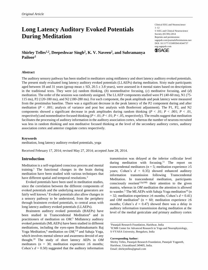
Design of the Study
Despite the fact that participants had prior experience of
OM meditation, all participants were given a 3-month
orientation program guided by an experienced meditation
teacher. The purpose of this orientation was for all
participants to practice the 2 different states of meditation,
namely, meditative focusing and effortless meditation,
based on specific instructions following a uniform method.
Each participant was assessed in 4 sessions, to which
they were assigned randomly. The sessions were
randomized using a standard random number table. Two of
them were meditation sessions. These were (a) meditative
focusing (dharana in Sanskrit) and (b) meditation without
focusing or effortless meditation (dhyana in Sanskrit). The
other 2 sessions were nonmeditation sessions. They were
(a) nonmeditative focused thinking (ekagrata in Sanskrit)
and (b) random thinking (cancalata in Sanskrit). All 4
sessions consisted of 3 states: before (5 minutes), during (20
minutes), and after (5 minutes). The design is presented
schematically in Figure 1.
Assessment Procedure
Recording Conditions. Long latency auditory evoked
potentials were assessed in the 4 sessions, that is, random
thinking (cancalata), nonmeditative focused thinking
(ekagrata), meditative focusing (dharana), and meditation
(dhyana). Participants were seated in a sound attenuated,
dimly lit cabin with sound level 26 dB normal hearing level
and monitored on a closed circuit television to detect if they
moved or fell asleep during a session. Instructions were
given through a 2-way intercom, so that participants could
remain undisturbed during a session. The LLAEPs were
recorded with eyes closed and participants seated at ease.
The temperature in the recording room was maintained at
24.0C 1.0C. The average humidity was 56% on the days
the experiments were conducted. LLAEPs were recorded in
the 250-ms, poststimulus time period without any
prestimulus delay, using a 4-channel system (Nicolet
Biomedical Inc, Madison, WI).
Electrode Positions. Ag/AgCl disk electrodes were fixed
with electrode gel (10-20 conductive EEG paste) at the
vertex (Cz) with reference electrodes on linked earlobes
(A1-A2) and with the ground electrode on the forehead
(FPz). Electrode placements were based on the international
10-20 electrode placement system.20 The electrode
impedance was kept less than 5 kohm.
Amplifier Settings. Standard settings for LLAEP recording
were used.21 The EEG activity was amplified with a
sensitivity of 100 V. The low cut filter was 0.1 Hz and the
high cut filter was 30.0 Hz. LLAEPs were averaged in 500
trial sweeps in the 0 to 500 ms range. Rejection was set at
90% of the full-scale range of the analog-to-digital
converter.
Stimulus Characteristics. Binaural click stimuli of 100-s
duration and alternating polarity at the rate of 5.0 Hz were
delivered through acoustically shielded earphones
(Amplivox, Kidlington, UK).21 The threshold of hearing
was noted for each participant to verify that their hearing
was normal. The threshold of hearing was checked as
follows (a) decreasing the intensity in 5-dB steps until the
participant could no longer hear the clicks and (b)
increasing the intensity in 5-dB steps until the clicks were
audible. The click threshold was taken as the midpoint
between the intensities at which the clicks could and could
not be heard. This procedure was repeated twice. The
thresholds ranged between 15 and 25 dB normal hearling
level (nHL). The average threshold of hearing was 14.03 ±
2.98 dB nHL. The intensity was kept at 70 dB nHL.
Participants had 100% compliance to the meditation
orientation program and for the recordings.
Interventions
Random Thinking (Cancalata). Participants were asked to
allow their thoughts to wander freely as they listened to a
compiled audio CD consisting of brief periods of
conversation, announcements, advertisements, and talks on
diverse topics recorded from a local radio station
transmission. These conversations were not connected and
hence it was thought that listening to them could induce a
state of random thinking.
Nonmeditative Focused Thinking (Ekagarta). Participants
listened to a prerecorded lecture on the process of
meditating and the object of meditation, that is, the Sanskrit
syllable OM. This was intended to induce a state of
nonmeditative focusing.
Telles et al 2
Figure 1. Schematic representation of the study design of the 4 sessions. The long latency auditory evoked potentials (LLAEPs) were recorded before, during and after the intervention. Periods of recording are shown as stippled and periods of intervention are shown as hatched.
Meditative Focusing (Dharana). Participants were asked
to open their eyes and gaze at the syllable OM as it is
written in Sanskrit. During this time guided instructions
required them to direct their thoughts to the physical
attributes of the syllable, that is, the shape and color, and
then to close their eyes and continue to visualize the
syllable mentally. The main emphasis during meditative
focusing was that thoughts are consciously brought back
(if they wander) to the single thought of OM.
Meditative Defocusing or Effortless Meditation
(Dhyana). During this session participants were
instructed to keep their eyes closed and dwell on thoughts
of OM, without any effort, particularly on the subtle
(rather than physical) attributes and connotations of the
syllable. This would gradually allow the participants to
experience brief periods of silence, which they reported
after the session.
Data Extraction
Long latency auditory evoked potential components,
namely, P1, N1, P2 and N2 waves were measured from a
zero DC baseline. Peak latency was measured from the
time of click delivery. The peak latencies and peak
amplitudes of the following components were measured,
the P1 wave between 40 and 60 ms, is the maximum
positive peak preceding the N1 wave which is a negative
component between 80 and 115 ms. The P2 wave is a
positive component between 140 and 180 ms. It is also
the first maximum positive component preceding the N2
wave component, which is between 200 and 280 ms.22
Components of LLAEPs and their neural generators are
described in Table 2.
Original Article
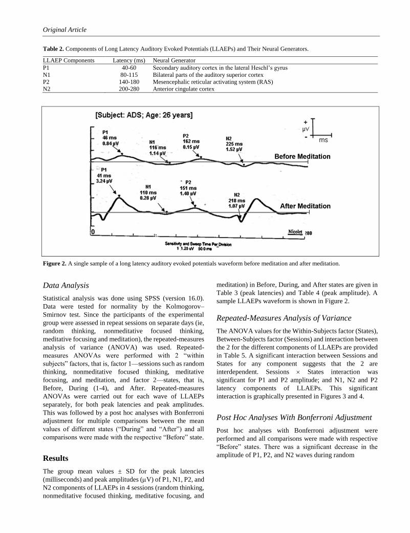
Table 2. Components of Long Latency Auditory Evoked Potentials (LLAEPs) and Their Neural Generators.
LLAEP Components Latency (ms) Neural Generator
P1 40-60 Secondary auditory cortex in the lateral Heschl’s gyrus
N1 80-115 Bilateral parts of the auditory superior cortex
P2 140-180 Mesencephalic reticular activating system (RAS)
N2 200-280 Anterior cingulate cortex
Figure 2. A single sample of a long latency auditory evoked potentials waveform before meditation and after meditation.
Data Analysis
Statistical analysis was done using SPSS (version 16.0).
Data were tested for normality by the Kolmogorov–
Smirnov test. Since the participants of the experimental
group were assessed in repeat sessions on separate days (ie,
random thinking, nonmeditative focused thinking,
meditative focusing and meditation), the repeated-measures
analysis of variance (ANOVA) was used. Repeated-
measures ANOVAs were performed with 2 “within
subjects” factors, that is, factor 1—sessions such as random
thinking, nonmeditative focused thinking, meditative
focusing, and meditation, and factor 2—states, that is,
Before, During (1-4), and After. Repeated-measures
ANOVAs were carried out for each wave of LLAEPs
separately, for both peak latencies and peak amplitudes.
This was followed by a post hoc analyses with Bonferroni
adjustment for multiple comparisons between the mean
values of different states (“During” and “After”) and all
comparisons were made with the respective “Before” state.
Results
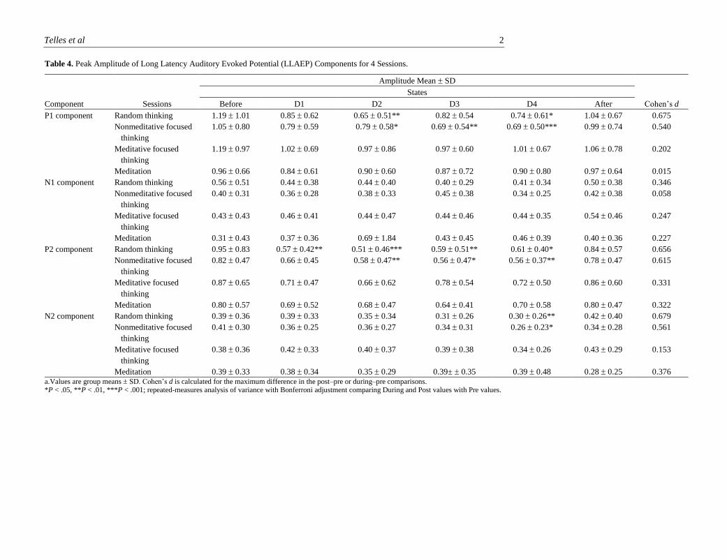
The group mean values ± SD for the peak latencies
(milliseconds) and peak amplitudes (V) of P1, N1, P2, and
N2 components of LLAEPs in 4 sessions (random thinking,
nonmeditative focused thinking, meditative focusing, and
meditation) in Before, During, and After states are given in
Table 3 (peak latencies) and Table 4 (peak amplitude). A
sample LLAEPs waveform is shown in Figure 2.
Repeated-Measures Analysis of Variance
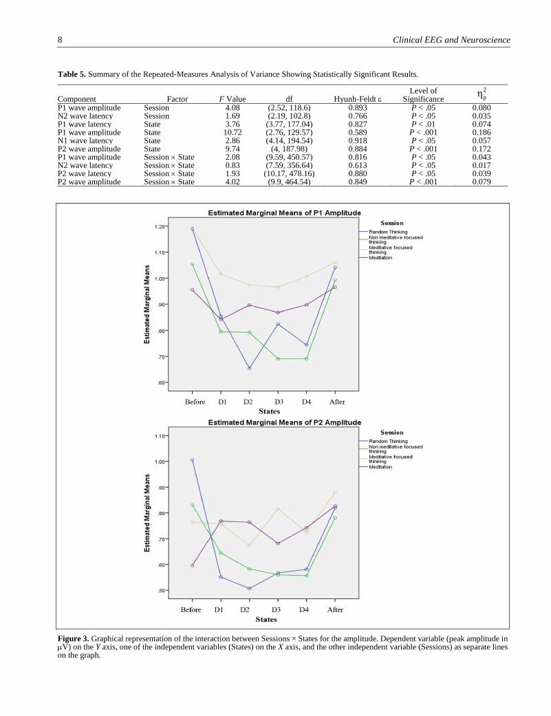
The ANOVA values for the Within-Subjects factor (States),
Between-Subjects factor (Sessions) and interaction between
the 2 for the different components of LLAEPs are provided
in Table 5. A significant interaction between Sessions and
States for any component suggests that the 2 are
interdependent. Sessions States interaction was
significant for P1 and P2 amplitude; and N1, N2 and P2
latency components of LLAEPs. This significant
interaction is graphically presented in Figures 3 and 4.
Post Hoc Analyses With Bonferroni Adjustment
Post hoc analyses with Bonferroni adjustment were
performed and all comparisons were made with respective
“Before” states. There was a significant decrease in the
amplitude of P1, P2, and N2 waves during random
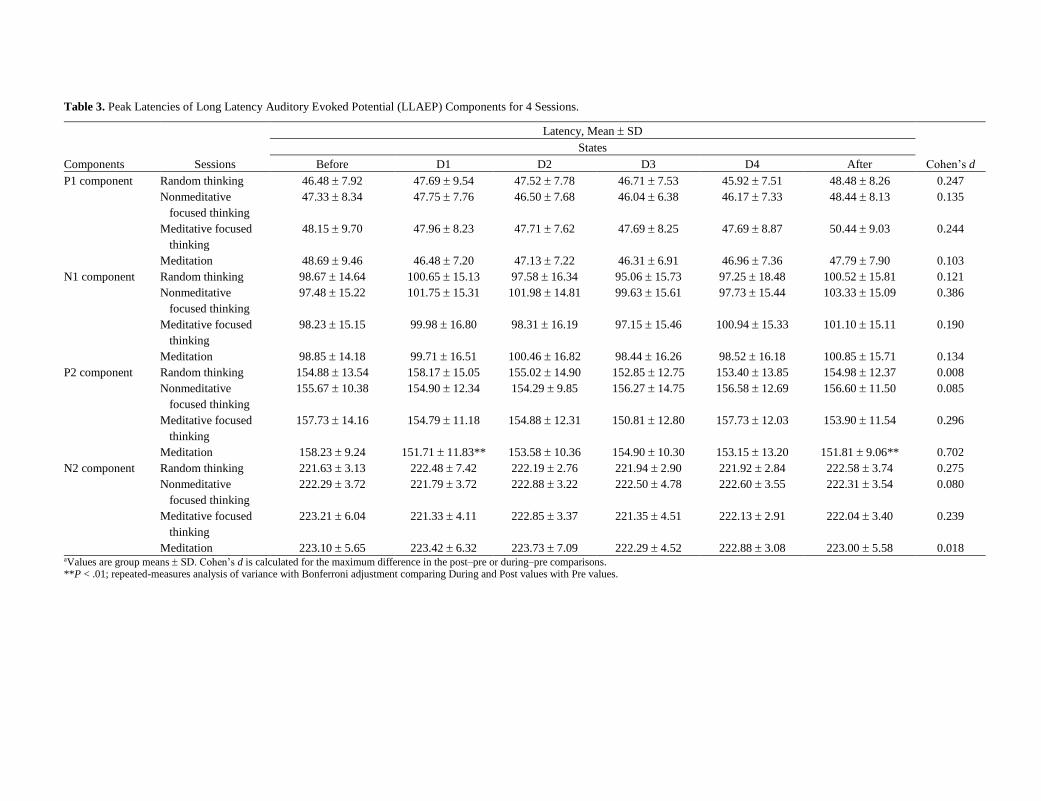
Table 3. Peak Latencies of Long Latency Auditory Evoked Potential (LLAEP) Components for 4 Sessions.
Components Sessions
Latency, Mean SD
Cohen’s d
States
Before D1 D2 D3 D4 After
P1 component Random thinking 46.48 7.92 47.69 9.54 47.52 7.78 46.71 7.53 45.92 7.51 48.48 8.26 0.247
Nonmeditative
focused thinking
47.33 8.34 47.75 7.76 46.50 7.68 46.04 6.38 46.17 7.33 48.44 8.13 0.135
Meditative focused
thinking
48.15 9.70 47.96 8.23 47.71 7.62 47.69 8.25 47.69 8.87 50.44 9.03 0.244
Meditation 48.69 9.46 46.48 7.20 47.13 7.22 46.31 6.91 46.96 7.36 47.79 7.90 0.103
N1 component Random thinking 98.67 14.64 100.65 15.13 97.58 16.34 95.06 15.73 97.25 18.48 100.52 15.81 0.121
Nonmeditative
focused thinking
97.48 15.22 101.75 15.31 101.98 14.81 99.63 15.61 97.73 15.44 103.33 15.09 0.386
Meditative focused
thinking
98.23 15.15 99.98 16.80 98.31 16.19 97.15 15.46 100.94 15.33 101.10 15.11 0.190
Meditation 98.85 14.18 99.71 16.51 100.46 16.82 98.44 16.26 98.52 16.18 100.85 15.71 0.134
P2 component Random thinking 154.88 13.54 158.17 15.05 155.02 14.90 152.85 12.75 153.40 13.85 154.98 12.37 0.008
Nonmeditative
focused thinking
155.67 10.38 154.90 12.34 154.29 9.85 156.27 14.75 156.58 12.69 156.60 11.50 0.085
Meditative focused
thinking
157.73 14.16 154.79 11.18 154.88 12.31 150.81 12.80 157.73 12.03 153.90 11.54 0.296
Meditation 158.23 9.24 151.71 11.83** 153.58 10.36 154.90 10.30 153.15 13.20 151.81 9.06** 0.702
N2 component Random thinking 221.63 3.13 222.48 7.42 222.19 2.76 221.94 2.90 221.92 2.84 222.58 3.74 0.275
Nonmeditative
focused thinking
222.29 3.72 221.79 3.72 222.88 3.22 222.50 4.78 222.60 3.55 222.31 3.54 0.080
Meditative focused
thinking
223.21 6.04 221.33 4.11 222.85 3.37 221.35 4.51 222.13 2.91 222.04 3.40 0.239
Meditation 223.10 5.65 223.42 6.32 223.73 7.09 222.29 4.52 222.88 3.08 223.00 5.58 0.018 aValues are group means SD. Cohen’s d is calculated for the maximum difference in the post–pre or during–pre comparisons. **P < .01; repeated-measures analysis of variance with Bonferroni adjustment comparing During and Post values with Pre values.
Telles et al 2
Table 4. Peak Amplitude of Long Latency Auditory Evoked Potential (LLAEP) Components for 4 Sessions.
Component Sessions
Amplitude Mean SD
Cohen’s d
States
Before D1 D2 D3 D4 After
P1 component Random thinking 1.19 1.01 0.85 0.62 0.65 0.51** 0.82 0.54 0.74 0.61* 1.04 0.67 0.675
Nonmeditative focused
thinking
1.05 0.80 0.79 0.59 0.79 0.58* 0.69 0.54** 0.69 0.50*** 0.99 0.74 0.540
Meditative focused
thinking
1.19 0.97 1.02 0.69 0.97 0.86 0.97 0.60 1.01 0.67 1.06 0.78 0.202
Meditation 0.96 0.66 0.84 0.61 0.90 0.60 0.87 0.72 0.90 0.80 0.97 0.64 0.015
N1 component Random thinking 0.56 0.51 0.44 0.38 0.44 0.40 0.40 0.29 0.41 0.34 0.50 0.38 0.346
Nonmeditative focused
thinking
0.40 0.31 0.36 0.28 0.38 0.33 0.45 0.38 0.34 0.25 0.42 0.38 0.058
Meditative focused
thinking
0.43 0.43 0.46 0.41 0.44 0.47 0.44 0.46 0.44 0.35 0.54 0.46 0.247
Meditation 0.31 0.43 0.37 0.36 0.69 1.84 0.43 0.45 0.46 0.39 0.40 0.36 0.227
P2 component Random thinking 0.95 0.83 0.57 0.42** 0.51 0.46*** 0.59 0.51** 0.61 0.40* 0.84 0.57 0.656
Nonmeditative focused
thinking
0.82 0.47 0.66 0.45 0.58 0.47** 0.56 0.47* 0.56 0.37** 0.78 0.47 0.615
Meditative focused
thinking
0.87 0.65 0.71 0.47 0.66 0.62 0.78 0.54 0.72 0.50 0.86 0.60 0.331
Meditation 0.80 0.57 0.69 0.52 0.68 0.47 0.64 0.41 0.70 0.58 0.80 0.47 0.322
N2 component Random thinking 0.39 0.36 0.39 0.33 0.35 0.34 0.31 0.26 0.30 0.26** 0.42 0.40 0.679
Nonmeditative focused
thinking
0.41 0.30 0.36 0.25 0.36 0.27 0.34 0.31 0.26 0.23* 0.34 0.28 0.561
Meditative focused
thinking
0.38 0.36 0.42 0.33 0.40 0.37 0.39 0.38 0.34 0.26 0.43 0.29 0.153
Meditation 0.39 0.33 0.38 0.34 0.35 0.29 0.39± 0.35 0.39 0.48 0.28 0.25 0.376 a.Values are group means SD. Cohen’s d is calculated for the maximum difference in the post–pre or during–pre comparisons. *P < .05, **P < .01, ***P < .001; repeated-measures analysis of variance with Bonferroni adjustment comparing During and Post values with Pre values.
8 Clinical EEG and Neuroscience
Table 5. Summary of the Repeated-Measures Analysis of Variance Showing Statistically Significant Results.
Component Factor F Value df Hyunh-Feldt Level of
Significance
P1 wave amplitude Session 4.08 (2.52, 118.6) 0.893 P < .05 0.080 N2 wave latency Session 1.69 (2.19, 102.8) 0.766 P < .05 0.035 P1 wave latency State 3.76 (3.77, 177.04) 0.827 P < .01 0.074 P1 wave amplitude State 10.72 (2.76, 129.57) 0.589 P < .001 0.186 N1 wave latency State 2.86 (4.14, 194.54) 0.918 P < .05 0.057 P2 wave amplitude State 9.74 (4, 187.98) 0.884 P < .001 0.172 P1 wave amplitude Session State 2.08 (9.59, 450.57) 0.816 P < .05 0.043 N2 wave latency Session State 0.83 (7.59, 356.64) 0.613 P < .05 0.017 P2 wave latency Session State 1.93 (10.17, 478.16) 0.880 P < .05 0.039 P2 wave amplitude Session State 4.02 (9.9, 464.54) 0.849 P < .001 0.079
Figure 3. Graphical representation of the interaction between Sessions × States for the amplitude. Dependent variable (peak amplitude in V) on the Y axis, one of the independent variables (States) on the X axis, and the other independent variable (Sessions) as separate lines on the graph.
2
pη
Telles et al 9
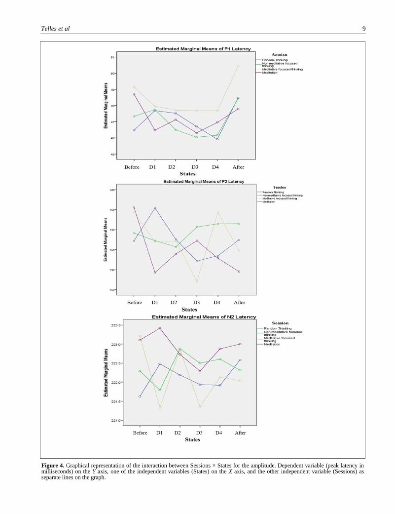
Figure 4. Graphical representation of the interaction between Sessions × States for the amplitude. Dependent variable (peak latency in milliseconds) on the Y axis, one of the independent variables (States) on the X axis, and the other independent variable (Sessions) as separate lines on the graph.
Telles et al 11
thinking (P < .01; P < .001; P < .01, respectively) and
nonmeditative focused thinking (P < .01; P < .01; P < .05,
respectively) and a decrease in the peak latency of the P2
wave during and after meditation (P < .001). All
comparisons were made with the “Pre” state. Cohen’s d
values were calculated and are provided in Table 3 (peak
latencies) and Table 4 (peak amplitude) for the 4 sessions at
Cz.
Discussion
Long latency auditory evoked potentials are generated by
thalamocortical and corticocortical auditory pathways, the
primary auditory cortex and the association cortical areas.21
The present study assessed LLAEPs during 4 mental states.
During meditation the peak latency of the P2 component
significantly reduced. A decrease in peak latency is
suggestive of a facilitation of auditory sensory transmission
because of increased speed of conduction in the underlying
neural generators.23(p278)
At present the functional significance of the P2
component is not as clear as that of components generated
more peripherally. The P2 wave partly reflects auditory
output of the mesencephalic activation system.24,25
Myoelectrography (MEG) studies have attempted to locate
the neural generators of the P2 component. Both MEG data
and EEG data from depth electrodes implanted in the
auditory cortex were collected in the same patients.26-29 It
was found that generators for the P2 component were
localized in the planum temporale as well as Brodmann area
22 (the auditory association complex). Other reports have
speculated that the P2 component may receive contributions
from cortical areas in the depth of the Sylvian fissure.30
Hence, it remains possible that the P2 component arises
from multiple sources with a center of activity close to
Heschl’s gyrus.30 The present results suggest that the
practice of meditation improves information transmission in
areas concerned with complex processing of auditory
stimuli as the auditory association cortices are possibly
involved.
During the 2 mental states that were considered for
comparison, that is, random thinking and non meditative
focusing, the peak amplitudes of the P1, P2, and N2
components reduced. A decrease in amplitude suggests that
the number of neuronal involvement recruited is less than
in the Pre state. The neural generators of the P2 component
have been mentioned above. The neuronal sources of the P1
component are difficult to localize due to low signal-to-
noise ratio. Also the brain response which generates the P1
component is preceded and followed in time within 10 to 15
ms by several EP components, which arises from sources
other than those generating P1.31 Studies on animal models
suggested that neuronal activity in the hippocampus might
contribute to sensory gating32; however, this was not proved
in human recordings.33 MEG studies have shown that there
may be a temporal lobe generator for P1 especially, located
bilaterally in the superior temporal gyrus.34 In addition, the
frontal lobe is involved in auditory sensory gating and this
activity may contribute to the P1 component. However, the
maximum contribution to the P1 activity is from the
temporal lobe.35 The N2 component of auditory evoked
potentials helps to evaluate the cognitive processes involved
in stimulus classification.36 The amplitude of the N2
component is directly related to changes in the left superior
temporal gyrus and bilateral medial temporal lobe areas.37
However, this description does not exclude the involvement
of other cortical areas in the genesis of the N2 component.
In random thinking and nonmeditative focusing sessions, a
decrease in amplitude of the P1 and N2 components
suggests that the overall neuronal activation and number of
neurons recruited in the neural generators underlying these
components was less. Other studies reported that the P2 and
N2 components decrease in amplitude with a reduction in
attention.38 Since random thinking did not involve focusing
of attention, the reduction in amplitude in P2 and N2
components is not surprising. In contrast, the reduction in
amplitude in non meditative focusing is surprising as (a)
participants were asked to focus during the session and (b)
the nature of focusing was obviously different from
meditative focusing as meditative focusing did not reduce
P2–N2 amplitudes. The P2 amplitude is also sensitive to
shifts in consciousness during the stages of sleep.39 Based
on (a) the self-report of the meditators, (b) observation of
the raw EEG recorded, and (c) observation of the
participants on the closed circuit TV. Hence, the reduced
amplitude of the three components during random thinking
and nonmeditative focusing may reflect a decrease in the
number of neurons recruited.
The present results are different from the early study
conducted on practitioners of Transcendental Meditation.
This could be due to differences in the method of meditation
and sample size. The sample size was 8 with Cohen’s d =
0.18 whereas in the present study the sample size was 48
with Cohen’s d = 0.68.
Hence, evaluating the effect of meditation based on
descriptions in the traditional texts has yielded a significant
result for long latency auditory evoked potentials. The main
difference being, as was already mentioned, that the
traditional practices started approximately over 1000 years
BC, whereas other techniques have evolved in the past 200
years.
The most important finding was the reduced latency of
the P2 component during meditation.
While the findings are reasonably straightforward, the
study has the following limitations: (a) The evaluation of
the quality of practice was based on a self reported visual
analog scale (VAS) and hence was subjective. (b) Random
thinking and nonmeditative focusing were the control
Telles et al 11
conditions. There was no control without any intervention.
(c) While the participants had been trained to switch
between the 4 states, the possibility that they did get into the
meditative state inadvertently cannot be ruled out. (d) The
transcultural generalizability of the results remains to be
determined, by conducting similar studies on a non-Indian
population. This suggests an area for future research.
Conclusion
The present results suggest that (a) meditation facilitates the
processing of auditory information in the auditory
association cortex and (b) random thinking and
nonmeditative focusing resulted in fewer neurons being
recruited in auditory association areas.
Declaration of Conflicting Interests
The author(s) declared no potential conflicts of interest with
respect to the research, authorship, and/or publication of this
article.
Funding
The author(s) disclosed receipt of the following financial support
for the research, authorship, and/or publication of this article: The
authors gratefully acknowledge the funding from the Indian
Council of Medical Research (ICMR), Government of India, as
part of a grant for a Center for Advanced Research in Yoga and
Neurophysiology (CAR-Y&N), (Project No. 2001-05010).
References
1. Murata T, Takahashi T, Hamada T, et al. Individual trait
anxiety levels characterizing the properties of Zen meditation.
Neuropsychobiology. 2004;50:189-194.
2. Mishra U, Kalita J. Clinical Neurophysiology: Nerve
Conduction, Electromyography and Evoked Potentials. New
Delhi, India: B.I. Churchill Livingstone; 1999.
3. Woods DL, Clayworth CC. Click spatial position influences
middle latency auditory evoked potentials (MLAEPs) in
humans. Electroencephalogr Clin Neurophysiol.
1985;60:122-129.
4. McEvoy TM, Frumkin LR, Harkins SW. Effects of meditation
on brainstem auditory evoked potentials. Int J Neurosci.
1980;10:165-170.
5. Kumar S, Nagendra H, Manjunath N, Naveen K, Telles S.
Meditation on OM: relevance from ancient texts and
contemporary science. Int J Yoga. 2010;3:2-5.
6. Telles S, Naveen KV. Changes in middle latency auditory
evoked potentials during meditation. Psychol Rep.
2004;94:398-400.
7. Telles S, Nagarathna R, Nagendra HR, Desiraju T. Alterations
in auditory middle latency evoked potentials during meditation
on a meaningful symbol—”Om”. Int J Neurosci. 1994;76:87-
93.
8. Telles S, Desiraju T. Recording of auditory middle latency
evoked potentials during the practice of meditation with the
syllable “OM”. Indian J Med Res. 1993;98:237-239.
9. Telles S, Raghavendra BR, Naveen KV, Manjunath NK,
Subramanya P. Mid-latency auditory evoked potentials in 2
meditative states. Clin EEG Neurosci. 2012;43:154-160.
10. Panjwani U, Selvamurthy W, Singh SH, Gupta HL,
Mukhopadhyay S, Thakur L. Effect of Sahaja yoga meditation
on auditory evoked potentials (AEP) and visual contrast
sensitivity (VCS) in epileptics. Appl Psychophysiol
Biofeedback. 2000;25:1-12.
11. Kumar S, Nagendra H, Naveen K, Manjunath N, Telles S.
Brainstem auditory-evoked potentials in two meditative
mental states. Int J Yoga. 2010;3:37-41.
12. Russell P. The TM Technique: An Introduction to
Transcendental Meditation and the Teachings of Maharishi
Mahesh Yogi. Las Vegas, NV: Elf Rock Productions; 2002.
13. Shri Mataji Nirmala Devi. Sahaja Yoga Book One. 2nd ed.
Australia: Nirmala Yoga; 1989.
14. PBKI Vishva-Vidyalaya. Raja Yoga Meditation: A General
Introduction. Mt Abu, India: Raja Yoga Centre; 1989.
15. Barwood TJ, Empson JA, Lister SG, Tilley AJ. Auditory
evoked potentials and Transcendental Meditation.
Electroencephalogr Clin Neurophysiol. 1978;45:671-673.
16. Taimni IK. The Science of Yoga: The Yoga-sūtras of Patañjali
in Sanskrit With Transliteration in Roman, Translation and
Commentary in English. Chennai, India: Theosophical
Society; 1999.
17. Saraswati M, Swami G. Bhagavad Gita. Kolkata, India:
Advaita Ashrama; 1998.
18. Erdfelder E, Faul F, Buchner A. GPOWER: A general power
analysis program. Behav Res Methods Instrum Comput.
1996;28(1):1-11.
19. Yadav A, Tandon OP, Vaney N. Auditory evoked responses
during different phases of menstrual cycle. Indian J Physiol
Pharmacol. 2002;46:449-56.
20. Jasper H. The ten-twenty electrode system of the International
Federation. Electroencephalogr Clin Neurophysiol.
1958;10:371-375.
21. Ventura LMP, Alvarenga KDF, Costa Filho OA. Protocol to
collect late latency auditory evoked potentials. Braz J
Otorhinolaryngol. 2009;75:879-883.
22. Ponton CW, Eggermont JJ, Kwong B, Don M. Maturation of
human central auditory system activity: evidence from multi-
channel evoked potentials. Clin Neurophysiol. 2000;111:220-
236.
23. Malhotra A. Auditory Evoked Responses in Clinical Practice.
New York, NY: Springer-Verlag; 1997:278.
24. Woods DL, Knight RT, Scabini D. Anatomical substrates of
auditory selective attention: behavioral and electro-
physiological effects of posterior association cortex lesions.
Brain Res Cogn Brain Res. 1993;1:227-240.
25. Picton TW, Alain C, Woods DL, et al. Intracerebral sources of
human auditory-evoked potentials. Audiol Neurootol.
1999;4:64-79.
26. Hari R, Pelizzone M, Mäkelä JP, Hällström J, Leinonen L,
Lounasmaa O V. Neuromagnetic responses of the human
auditory cortex to on- and offsets of noise bursts. Audiology.
1987;26:31-43.
27. Pantev C, Hoke M, Lütkenhöner B, Lehnertz K.
Neuromagnetic evidence of functional organization of the
auditory cortex in humans. Acta Otolaryngol Suppl.
1991;491:106-114.
Telles et al 12
28. Sams M, Paavilainen P, Alho K, Näätänen R. Auditory
frequency discrimination and event-related potentials.
Electroencephalogr Clin Neurophysiol. 1985;62:437-448.
29. Rif J, Hari R, Hämäläinen MS, Sams M. Auditory attention
affects two different areas in the human supratemporal cortex.
Electroencephalogr Clin Neurophysiol. 1991;79:464-472.
30. Crowley KE, Colrain IM. A review of the evidence for P2
being an independent component process: age, sleep and
modality. Clin Neurophysiol. 2004;115:732-744.
31. Korzyukov O, Pflieger ME, Wagner M, et al. Generators of
the intracranial P50 response in auditory sensory gating.
Neuroimage. 2007;35:814-826.
32. Freedman R, Adler LE, Myles-Worsley M, et al. Inhibitory
gating of an evoked response to repeated auditory stimuli in
schizophrenic and normal subjects. Human recordings,
computer simulation, and an animal model. Arch Gen
Psychiatry. 1996;53:1114-1121.
33. Grunwald T, Boutros NN, Pezer N, et al. Neuronal substrates
of sensory gating within the human brain. Biol Psychiatry.
2003;53:511-519.
34. Huang MX, Edgar JC, Thoma RJ, et al. Predicting EEG
responses using MEG sources in superior temporal gyrus
reveals source asynchrony in patients with schizophrenia. Clin
Neurophysiol. 2003;114:835-850.
35. Weisser R, Weisbrod M, Roehrig M, Rupp A, Schroeder J,
Scherg M. Is frontal lobe involved in the generation of
auditory evoked P50? Neuroreport. 2001;12:3303-3307.
36. O’Donnell BF, Shenton ME, McCarley RW, et al. The
auditory N2 component in schizophrenia: relationship to MRI
temporal lobe gray matter and to other ERP abnormalities. Biol
Psychiatry. 1993;34:26-40.
37. Shenton ME, Kikinis R, Jolesz FA, et al. Abnormalities of the
left temporal lobe and thought disorder in schizophrenia. A
quantitative magnetic resonance imaging study. N Engl J Med.
1992;327:604-612.
38. Hansen JC, Hillyard SA. Endogenous brain potentials
associated with selective auditory attention.
Electroencephalogr Clin Neurophysiol. 1980;49:277-290.
39. Colrain IM, Di Parsia P, Gora J. The impact of prestimulus
EEG frequency on auditory evoked potentials during sleep
onset. Can J Exp Psychol. 2000;54:243-254.