lipid nanocarriers for topical application in psoriasis and
TRANSCRIPT

HAL Id: tel-01643693https://tel.archives-ouvertes.fr/tel-01643693
Submitted on 21 Nov 2017
HAL is a multi-disciplinary open accessarchive for the deposit and dissemination of sci-entific research documents, whether they are pub-lished or not. The documents may come fromteaching and research institutions in France orabroad, or from public or private research centers.
L’archive ouverte pluridisciplinaire HAL, estdestinée au dépôt et à la diffusion de documentsscientifiques de niveau recherche, publiés ou non,émanant des établissements d’enseignement et derecherche français ou étrangers, des laboratoirespublics ou privés.
Lipid nanocarriers for topical application in psoriasisand psoriatic arthritis
Mourad Sala
To cite this version:Mourad Sala. Lipid nanocarriers for topical application in psoriasis and psoriatic arthritis. Pharma-cology. Université de Lyon, 2017. English. �NNT : 2017LYSE1193�. �tel-01643693�
1
N° d’ordre NNT : 2017LYSE1193 Année 2017
THESE DE L‘UNIVERSITE DE LYON
Délivrée par
L’UNIVERSITE CLAUDE BERNARD LYON 1
ECOLE DOCTORALE INTERDISSPLINAIRE SCIENCE SANTE (ED 205)
Spécialité Sciences pharmacotechniques et biopharmaceutiques, physicochimie
DIPLOME DE DOCTORAT
SPÉCIALITÉ: PHARMACOTECHNIE
(Arrêté du 7 août 2006)
Soutenue publiquement le 28 Septembre 2017
Par
M. Mourad SALA
*****
NANOVECTEURS LIPIDIQUES POUR UNE APPLICATION TOPIQUE DANS LE PSORIASIS ET SA COMPLICATION ARTHRITIQUE
*****
J U R Y
Dr. Christine VAUTHIER, Directeur de Recherche CNRS, Institut Galien Paris Sud, CNRS UMR 8612, Université Paris-Sud, Chatenay-Malabry, rapporteuse
Pr. Frank BOURY, INSERM U646, « Ingénierie de la vectorisation particulaire », Angers, rapporteur
Dr. Gillian BARRAT, Directeur de Recherche CNRS, UMR 8612 Centre d’Etudes Pharmaceutiques, Paris-sud, examinatrice
Dr. Abderrazzak BENTAHER Directeur de Recherche INSERM, Hopital Lyon-Sud, examinateur
Dr. Catherine CHARCOSSET, Directeur de Recherche CNRS, UMR 5007, invitée
Bernard MANDRAND, Adjuvatis, Lyon, invité
Pr. Hatem FESSI, CNRS, Université Claude Bernard-Lyon 1, France, Directeur de thèse
Dr. Abdelhamid ELAISSARI, CNRS, Université Claude Bernard-Lyon1, France, Co-Directeur de thèse
2
UNIVERSITE CLAUDE BERNARD LYON 1 Président de l’Université M. le Professeur Frédéric FLEURY
Présidence du Conseil Académique M. le Professeur Hamda BEN HADID
Vice-Président du Conseil d’Administration M. le Professeur Didier REVEL
Vice-Président de la Formation et de la Vie Universitaire
M. le Professeur Philippe CHEVALIER
Vice-Président de la Commission Recherche M. Fabrice VALLÉE
Directeur Général des Services M. Alain HELLEU
Composantes SANTE
Faculté de Médecine Lyon Est – Claude Bernard Directeur : M. le Professeur J. ETIENNE
Faculté de Médecine et de Maïeutique Lyon Sud – Charles Mérieux
Directeur : Mme la Professeure C. BURILLON
Faculté d’Odontologie Directeur : M. le Professeur D. BOURGEOIS
Institut des Sciences Pharmaceutiques et Biologiques
Directeur : Mme la Professeure C. VINCIGUERRA
Institut des Sciences et Techniques de la Réadaptation
Directeur : M. le Professeur Y. MATILLON
Département de formation et Centre de Recherche en Biologie Humaine
Directeur : Mme la Professeure A-M. SCHOTT
SCIENCES ET TECHNOLOGIES
Faculté des Sciences et Technologies Directeur : M. F. DE MARCHI
Département Biologie Directeur : M. le Professeur F. THEVENARD
Département Chimie Biochimie Directeur : Mme C. FELIX
Département GEP Directeur : M. Hassan HAMMOURI
Département Informatique Directeur : M. le Professeur S. AKKOUCHE
Département Mathématiques Directeur : M. le Professeur G. TOMANOV
Département Mécanique Directeur : M. le Professeur H. BEN HADID
Département Physique Directeur : M. le Professeur J-C PLENET
UFR Sciences et Techniques des Activités Physiques et Sportives
Directeur : M. Y.VANPOULLE
Observatoire des Sciences de l’Univers de Lyon Directeur : M. B. GUIDERDONI
3
Polytech Lyon Directeur : M. le Professeur E.PERRIN
Ecole Supérieure de Chimie Physique Electronique
Directeur : M. G. PIGNAULT
Institut Universitaire de Technologie de Lyon 1 Directeur : M. le Professeur C. VITON
Ecole Supérieure du Professorat et de l’Education
Directeur : M. le Professeur A. MOUGNIOTTE
Institut de Science Financière et d'Assurances Directeur : M. N. LEBOISNE
4
REMERCIEMENT
Ce travail de thèse a été réalisé au Laboratoire d’Automatique et de Génie des Procédés (LAGEP). Je tiens à remercier profondément Hatem Fessi et Abelhamid Elaissari pour m’avoir accueilli au sein du LAGEP afin de mener ce projet de Recherche. Mes profonds remerciements pour votre accueil au sein de votre équipe de recherche. Durant toutes ces années, j’ai pu me nourrir de vos précieux conseils, de votre expertise. Vous m’avez soutenu à toutes les étapes et vraiment rendu facile la réalisation de mes travaux. Vous m’avez appris ce qu’est être un bon chercheur. Et grâce à vous, j’ai pu découvrir un monde plein de générosité, ouvert sur les autres cultures. Vous m’avez permis de gagner en rigueur, en persévérance. D’autre part, je remercie le Dr. Christine VAUTHIER et le Pr. Frank BOURY pour avoir accepté d’être rapporteurs de ce travail de thèse. Mes remerciements s’adressent également au Dr. Gillian BARRAT, Dr. Abderrazzak BENTAHER, Dr. Catherine CHARCOSSET et Bernard Mandrand pour avoir accepté de juger ce travail. Je voulais également adresser mes sincères amitiés à toutes les personnes que j’ai eu le plaisir de connaître au cours de ces années. Merci particulièrement à Badri et Karim. Toute ma gratitude bien sûr à mes parents, mes frères et sœurs qui m’ont toujours aidé et soutenu, motivé et remotivé.
5
Résumé Le psoriasis est une maladie de peau auto-immune et chronique. Le rhumatisme psoriasique est une de ses principales complications qui est très invalidante pour les patients. Cette pathologie reste encore incurable à ce jour. L’usage des médicaments disponibles actuellement dans le psoriasis est limité par leurs effets secondaires dépendant de la dose et de la durée d’utilisation. Le but de ce travail était de développer des nanovecteurs médicamenteux à base de lipides pour un usage topique, en particulier ciblant l'épiderme viable qui est le site principal de la physiopathologie du psoriasis, mais aussi le derme et au-delà pour atteindre les articulations endommagées. Grâce à une nouvelle technique que nous avons développé et optimisé, le double déplacement de solvants, basée sur une organisation des phospholipides en deux temps, nous avons préparé des vésicules lipidiques encapsulant du diclofénac d’une part et de la ciclosporine A d’autre part. Ensuite, nous avons évalué leur aptitude à traverser la peau et cibler les régions d'intérêt. Après une étude systématique permettant d’optimiser les paramètres de préparation, les vésicules lipidiques encapsulant le diclofénac et la cyclosporine A ont montré une efficacité d'encapsulation (EE%) comprise entre 50% et 90% respectivement, selon la concentration en phospholipides. Après réalisation des études in vitro sur peau de cochon, nous avons observé que la formulation contenant une concentration basse en phospholipides (8,5 mg / mL) permettait d'encapsuler plus de 80% du diclofénac et de cibler le derme et au-delà. La formulation de vésicules lipidiques chargées de cyclosporine A qui encapsule la quantité la plus élevée (environ 80%) était également celle contenant la concentration basse de phospholipides. Contrairement au diclofénac, cette formulation n'était pas la meilleure pour cibler une couche profonde de la peau comme l'épiderme viable, alors que c’était le cas pour la formulation avec une concentration élevée de phospholipides (15 mg / mL), bien que l'EE% était d'environ 55%. Le double déplacement de solvant est une technique très prometteuse de préparation de vésicules lipidiques, capable de produire une population monodisperse d’échelle nanométrique. Cette méthode n'est que légèrement impactée lors d’une transposition d’échelle et serait donc facile à mettre en œuvre à l'échelle industrielle. Cette méthode a été conçue dès le début pour utiliser des solvants favorisant la pénétration cutanée mais l’étendue de ces applications reste à explorer. MOTS CLES Vésicule lipidique, Liposome, Préparation, Diclofénac, Ciclosporine A, Encapsulation,
Phospholipide, système d’administration cutanée
6
Abstract Psoriasis is an auto-immune and chronic skin disease. Psoriatic arthritis is the main complication which is very disabling for patients. This pathology still remains incurable to date. The currently psoriasis indicated medicines use is limited by their side effects which are dose and use duration dependent. The aim of this work was to develop lipid based nanocarriers for skin targeting, especially the viable epidermis which is the main site of psoriasis physiopathology but also the dermis and beyond in order to reach the damaged articulations. Thanks to a new technique we developed and optimized called the double solvent displacement, based on a two-step phospholipid organization, we prepared diclofenac and cyclosporine A loaded lipid vesicles. Then, we evaluated their potential to cross the skin and target the skin layers of interest. After a systematic study to optimize preparation parameters, diclofenac and cyclosporine A loaded lipid vesicles displayed an encapsulation efficiency (EE %) between 50% and 90% respectively, according to the phospholipid concentration. After in vitro skin studies, we observed that the formulation containing the lower phospholipid concentration (8.5 mg/mL) allowed to encapsulate more than 80% of diclofenac and also to target the dermis and beyond. The formulation of cyclosporine A loaded lipid vesicles which encapsulates the higher amount (around 80%) is also the one containing the lower phospholipid concentration. Unlike to diclofenac, this formulation was not the better to target the viable epidermis whereas the formulation with the higher phospholipid concentration (15 mg/mL) was even though the EE% was of around 55%. The double solvent displacement is a very promising technique of lipid vesicle preparation, capable to produce monodisperse population of nanoscale carriers. This method is hardly impacted during scale-up and would be easy to implement at an industrial scale. This method was designed from the beginning to use skin penetration enhancer solvents but the scope of its applications still remains to be explored. KEY-WORDS Lipid vesicles, Liposome, Preparation, Diclofenac, Ciclosporine A, Encapsulation, Phospholipid, Skin Drug delivery systems
7
SOMMAIRE
Introduction générale ............................................................................................................................... 8
PARTIE BIBLIOGRAPHIQUE ............................................................................................................ 14
Etat de l’art de la physiopathologie du psoriasis et ses traitements: mécanismes impliqués et perspectives innovantes des vecteurs médicamenteux pour voie topique ......................................... 17
Nanovecteurs lipidiques comme système d’administration cutanée: Propriétés, mécanismes d’intéractions cutanées et applications médicales ............................................................................. 68
CONCLUSION -- PARTIE BIBLIOGRAPHIQUE ....................................................................... 110
PARTIE EXPERIMENTALE ............................................................................................................. 111
Préparation par double déplacement de solvent de nanovésicules lipidiques encapsulant le diclofénac pour administration cutanée ........................................................................................... 114
Etude comparative entre le double déplacement de solvent et la méthode classique de l’injection à l’éthanol—de l’échelle laboratoire à une plus grande échelle. ........................................................ 149
Préparation par double déplacement de solvent de nanovecterus lipidiques encapsulant la cyclosporine A pour une application topique dans le traitement du psoriasis ................................. 175
Discussion générale ............................................................................................................................. 201
Conclusion et Perspectives .................................................................................................................. 209

8
Introduction générale

9
Les pathologies de la peau chroniques, inflammatoires et auto-immunes sont des maladies en recrudescence principalement dans les pays développés. Le psoriasis est une de ces maladies de la peau caractérisée par des lésions inflammatoires et hyperkératosiques en plaque qui se manifestent sous forme d’épisodes récurrents. Le psoriasis est une affection qui affecte 2 à 5% de la population mondiale. Les lésions sont localisées généralement au niveau des articulations (coudes, genoux) et du cuir chevelu, mais toute localisation est envisageable. Les signes cliniques de cette maladie complexe peuvent varier en intensité et se développer sur une surface plus ou moins étendue.
De plus, ces lésions cutanées peuvent évoluer soit de façon progressive ou en poussée. Tous ces éléments permettent de caractériser la sévérité de la maladie qui pourrait imposer ou non des soins hospitaliers. Son origine est actuellement inconnue, mais elle semble être causée par une combinaison de facteurs génétiques et environnementaux (alcool, tabac, infections, médicaments, stress). Chez les patients atteints de psoriasis, le pronostic est rarement engagé. C'est plutôt l'impact sur la qualité de vie qui est problématique. Les stigmates physiques visibles contribuent à accroître les difficultés rencontrées par les patients dans leurs vies socioprofessionnelle et personnelle. Une complication importante dans le psoriasis qui n'est pas le moins commun et qui renforce davantage la détérioration de la qualité de vie des patients est l'arthrite psoriasique ou rhumatisme psoriasique (RP). C'est une arthropathie inflammatoire chronique avec une prévalence allant de 6% à 39%. Cette maladie articulaire entraîne une érosion du cartilage articulaire et donc une destruction irréversible de l'articulation due à l'inflammation prolongée qui rend l'environnement propice au développement précoce des dommages articulaires et leur évolution.
Il n’existe pas à ce jour de traitement guérissant le psoriasis. Les traitements disponibles consistent principalement à atténuer les symptômes et contenir leur évolution, de façon à améliorer la qualité de vie des patients. Le traitement sélectionné par le clinicien sera fonction de nombreux éléments (le type de lésions, leur étendue et localisation, la tolérance du patient, les éventuels échecs thérapeutiques passés). Différentes stratégies thérapeutiques sont classiquement utilisées, qui parfois sont combinées pour obtenir une meilleure efficacité. On retrouve le traitement local notamment avec les dermocorticoïdes, la photothérapie, le traitement systémique avec des molécules telles que la ciclosporine A (CsA), le méthotrexate et les biothérapies anti-TNF alpha pour la majorité. Cependant, une démarche hiérarchique est généralement adoptée. Du fait du caractère chronique du psoriasis et de l’absence de cure disponible, ces traitements sont pris à vie, ce qui fait d’eux un véritable enjeu économique pour les laboratoires pharmaceutiques et les systèmes de santé des différents pays. Mais aucun ne garantit un succès dans la prise en charge des symptômes, et aucun ne prétend guérir. Ainsi, la découverte de nouvelles thérapeutiques dans le traitement du psoriasis est un énorme challenge. L’essoufflement de l’industrie pharmaceutique dans la proposition de nouvelles molécules oriente aussi vers une optimisation des molécules déjà existantes en utilisant des systèmes vecteurs innovants.
Un des domaines de pointe en pleine expansion dans le secteur pharmaceutique est la mise au point de vecteurs médicamenteux innovants basés sur l’encapsulation des médicaments. Depuis ces dernières années, peu de nouvelles molécules ont été découvertes. En effet, les échecs successifs, véritable gouffre financier, découragent les investissements. A l’heure actuelle, les activités de recherche sont plus orientées vers l’amélioration des thérapeutiques déjà existantes, ce qui est moins risqué et moins coûteux pour l’industrie pharmaceutique,
10
mais pas sans intérêt pour les patients. Différents avantages sont mis en avant comme la protection contre les effets indésirables grâce à l’administration de doses plus faibles, une amélioration de l’efficacité de médicaments de faible biodisponibilité ou de métabolisme trop rapide, mais aussi un meilleur confort pour les patients (prises répétitives, injections). Ainsi, les vecteurs médicamenteux apparaissent comme une réponse à de nombreux problèmes posés par les molécules actives pour améliorer la prise en charge des pathologies. Mais, plus encore, ils permettent de redécouvrir des molécules thérapeutiques à fort potentiel qui ont pu montrer une efficacité lors des essais précliniques mais rejetées du fait d’une toxicité trop importante au cours des essais cliniques. Les principales méthodes d’encapsulation des molécules actives font beaucoup appel aux polymères biodégradables (poly(lactic-co-glycolic acid (PLGA), polycaprolactone (PLC), chitosane, eudragit, ect). Par ailleurs, les vecteurs médicamenteux à base de lipides comme les vésicules lipidiques et les nanoparticlules lipidiques (solid lipid nanoparticle (SLN) / nanostructures lipid carrier (NLC)) sont également connus comme ayant un fort potentiel dans la délivrance de médicaments par voie cutanée. Les vésicules lipidiques consistent en l’auto-assemblage de phospholipides en milieu aqueux résultant en une ou plusieurs bicouches délimitant des compartiments. La variation de la composition par l'ajout d'un promoteur de la pénétration cutanée (PE : penetration enhancer) comme l'éthanol, le glycérol ou d’un « edge activator » comme le cholate de sodium conférant une ultradéformabilité à l’ensemble conduit à de nouvelles générations de vésicules lipidiques comme les éthosomes, les transfersomes et les « Penetration Enhancer lipid Vesicles » (PEVs). Parmi les techniques conventionnelles utilisées pour préparer des liposomes, nous retrouvons la technique dite de l'hydratation du film lipidique connue sous le nom de méthode de Bangham, la technique d'évaporation en phase inverse (REV), la technique de l’injection de solvant (éther ou éthanol) ou la dialyse détergente. Ces techniques nécessitent une grande quantité de solvant et ne sont pas adaptées à une transposition d’échelle industrielle car diverses étapes d'homogénéisation supplémentaires sont requises. D'autres techniques ont été développées, telles que l’évaporation en phase inverse super critique (SCRPE), la technique d'injection à écoulement croisé, la focalisation hydrodynamique microfluidique ou le contacteur à membrane. Outre les vésicules lipidiques, les nanoparticules lipidiques sont basées sur l'utilisation d'un lipide solide ou d'un mélange de lipides solides dispersé dans un milieu aqueux pour obtenir des SLNs et un mélange de lipides solides et liquides (huiles) dans un rapport habituellement de 70:30 utilisé pour préparer les NLCs. Ces nanoparticules lipidiques peuvent être produites par de nombreuses méthodes comme la technique de la microémulsion, l'émulsification-évaporation de solvant, l’émulsification-diffusion de solvant, la méthode d'injection de solvants, l’inversion de phase, la technique d'émulsion multiple et la technique du contacteur de membrane. Mais la plus répandue reste l'homogénéisation à haute pression qui permet une production rapide sans solvant organique. Actuellement, il n'existe qu'une seule méthode rapportée dans la littérature qui est directement utilisée pour préparer à la fois les vésicules lipidiques et les nanoparticules lipidiques: la technique des contacteurs à membrane. La littérature est riche d’exemples aussi variés les uns que les autres faisant état de formulations encapsulant des molécules d’intérêt dans la prise en charge du psoriasis. Toutes les formes ont été utilisées, de la forme polymérique à la lipidique, dans des micro- ou nano-vecteurs mais rares sont les formulations dépassant le stade de la recherche en laboratoire. Ceci est peut être lié à de mauvais résultats lors des études in vivo ou un problème de production à l’échelle industrielle, ce qui est souvent le cas.
11
En effet, un des problèmes souvent rencontré lors de la production de ces vecteurs est la difficulté d’obtenir des produits de qualité constante et aussi l’usage d’une quantité trop importante de solvants organiques. L’objectif principal était toujours le même : faire passer à travers les différentes couches de la peau le principe actif pour un accès direct au site d’action et ainsi éviter une administration per os ou parentérale qui est limitée du fait des effets indésirables au long cours. On s’intéresse, dans notre projet de recherche, à deux molécules actives très différentes, la ciclosporine A et le diclofénac dont on cherche à améliorer le ratio bénéfice/risque pour le patient.
La ciclosporine A est un immunosuppresseur largement utilisé dans le domaine de la transplantation et des pathologies auto-immunes. Mais son usage est également fréquent dans les formes avancées de psoriasis. Sa structure est un peptide cyclique de 11 acides aminés capable d’agir sélectivement sur les cellules T. Son mécanisme d’action passe par la formation d’un complexe avec la cyclophiline, une immunophiline intracellulaire, qui inhibe l’activité de la phosphatase calcineurine. Cela a pour conséquence une déplétion en lymphocytes et en macrophages et une inhibition de l’activation des lymphocytes T, des cellules NK et des cellules présentatrices de l’antigène. Par ailleurs, la ciclosporine A permet aussi d’inhiber la prolifération des kératinocytes et l’histamino-libération des mastocytes. Elle est également à l’origine d’une régulation négative de l’expression des molécules d’adhésion cellulaire à l’endothélium des capillaires du derme. Cependant, la ciclosporine A peut causer l’apparition d’effets indésirables majeurs empêchant son usage à forte dose et/ou au long cours. En effet, elle peut être responsable d’une néphrotoxicité et d’une hypertension artérielle qui sont gérées dans la mesure du possible par une adaptation de posologie (donc une réduction de l’efficacité) et la mise en place d’un traitement antihypertenseur. Il en existe d’autres, tels que l’hypertrophie gingivale, le diabète, etc. De plus, il est important de relever que la ciclosporine est également impliquée dans de nombreuses interactions médicamenteuses. En effet, elle est à la fois substrat et inhibiteur du cytochrome 3A4 et de la glycoprotéine P (Pgp). Par conséquent, malgré son efficacité clinique dans le psoriasis, du fait de son usage par vois systémique, son utilisation est fortement limitée. Le diclofénac appartient à la catégorie des antiinflammatoires non stéroïdiens (AINS) capables d'inhiber à la fois les enzymes COX-1 et COX-2. Sa fixation aux isoenzymes COX bloque la synthèse de diverses prostanoïdes comme les prostaglandines (PG) (la PGE2, la PGD2, la PGF2], les prostacyclines [PGI2] et le thromboxane [TX] A2). Diverses études ont rapportés que la PGE2 est la PG dont l'inhibition par les AINS serait le principal mécanisme de l'effet analgésique et anti-inflammatoire. Le diclofénac présente une plus grande sélectivité pour la COX-2 par rapport à la COX-1, contrairement à la plupart des AINS traditionnels. Son degré de sélectivité pour la COX-2 serait comparable à celui du célécoxib. Le diclofénac est associé à divers effets indésirables (EI) classiquement décrits pour la classe des AINS. Ces EI sont d’ordres gastro-intestinaux, cardiovasculaires et rénaux. Leur gravité est corrélée à la dose administrée. Le diclofénac est classé comme étant à faible risque de complications gastro-intestinales en raison de sa plus grande sélectivité pour la COX-2. L'administration per os du diclofénac permet une
12
absorption systémique rapide qui est directement proportionnelle à la dose. Une variabilité importante des paramètres de l'absorption du diclofénac a été observée (comme la forme du sel, la formulation galénique et le moment de la prise par rapport au repas). Le diclofénac s’accumule facilement au niveau des tissus inflammés et du liquide synovial. Le développement de formulations topiques à base de diclofénac a pour objectif de traiter localement la douleur et ce, pour minimiser l'exposition systémique exposant les patients à des effets indésirables potentiellement graves dans les traitements aux long cours. Sous forme de sel (sodique ou potassique), le diclofénac est hydrosoluble à pH neutre alors que la forme acide est lipophile. Cette propriété physico-chimique est un atout pour le passage des membranes telles que la muqueuse synoviale ou la barrière cutanée. Notre but dans ce travail est de proposer une formulation galénique encapsulant la ciclosporine et le diclofénac dans des vecteurs médicamenteux en vue d’une application topique pour traiter d’une part la composante fondamentale du psoriasis par un accès direct à son site physiopathologique et d’autre part, sa complication la plus fréquente et la plus grave, le rhumatisme psoriasique. Dans ce travail, la première partie bibliographique est consacrée à l’état de l’art en matière des dernières avancées dans la compréhension de la physiopathologie du psoriasis et de sa complication principale qu’est le rhumatisme psoriasique. Nous avons passé en revue les divers mécanismes impliqués pour identifier les cibles thérapeutiques les plus importantes. Nous avons également fait un état des lieux des stratégies thérapeutiques qui ont cours à l’heure actuelle dans le traitement du psoriasis et des nouveaux axes de recherche qui pourraient permettre une avancée significative dans la vie des patients. Dans un deuxième temps, nous avons fait un focus sur la stratégie qui consiste en l’encapsulation de molécules actives déjà connues en vue d’en améliorer le profil bénéfice/risque par un usage topique optimisé. Nous avons passé en revue les différents systèmes de vectorisation à base lipidique qui ont été développés et étudiés pour améliorer la délivrance de médicaments par voie cutanée. L'objectif a été de souligner les avancées récentes dans ce domaine en tant que technologies médicamenteuses pour traiter les maladies de la peau notamment. Nous avons également exposé les récents progrès réalisés dans la compréhension des mécanismes de pénétration à travers la peau. Quant à la partie expérimentale, elle se décline en trois parties toutes faisant état de l’introduction d’une nouvelle méthode de préparation des vésicules lipidiques : « le double déplacement de solvant ». La première partie consiste en une étude systématique du double déplacement de solvant pour identifier les paramètres à maîtriser en vue de la préparation des liposomes aux propriétés colloïdales (taille, distribution de taille, charge de surface) optimisées pour une application cutanée. Le diclofénac a été sélectionné comme molécule active modèle pour l’encapsulation notamment parce qu’il peut être utilisé en vue du traitement symptomatique du rhumatisme psoriasique. Une étude in vitro sur peau de cochon a été réalisée avec plusieurs formules pour identifier celle ayant le plus fort potentiel pour un passage transcutané. Dans la deuxième partie expérimentale, nous avons réalisé une évaluation du potentiel de notre méthode de préparation de vésicules lipidiques « Double Solvent Displacement » en vue d’une transposition d’échelle. Cette méthode a été comparée à une méthode classiquement
13
utilisée : la méthode d’injection à l’éthanol. Nous avons étudié l’influence de la variation de divers paramètres pour savoir quelle méthode offrait une meilleure stabilité des propriétés colloïdales lors d’études de scale-up et les meilleurs rendements. La troisième partie expérimentale consiste à encapsuler la ciclosporine A par double déplacement de solvant. Lors de ce travail, nous avons découvert qu’en modifiant les conditions opératoires, nous pouvions à la fois produire des liposomes mais également des SLN. Dans cette dernière partie, nous avons préparé et caractérisé différentes formulations lipidiques encapsulant la ciclosporine A (liposomes et SLN). Puis nous avons sélectionné celles qui nous paraissaient les plus prometteuses pour ensuite réaliser une étude in vitro et identifier quelle type de vecteur permettait une pénétration optimale de la ciclosporine A au niveau de l’épiderme viable en vue d’une application dans la prise en charge topique du psoriasis.

14
PARTIE BIBLIOGRAPHIQUE
15
Pour pouvoir répondre au mieux à l’objectif initial qui concerne principalement l’amélioration de la prise en charge du psoriasis, il a fallu commencer par réaliser une revue de la littérature afin d’avoir une compréhension aussi exhaustive que possible des mécanismes mis en jeu dans la physiopathologie du psoriasis. Il était essentiel de faire ce travail pour préciser le plus en amont possible quelles sont les objectifs que l’on doit absolument fixer à une formulation pour en espérer le maximum d’efficacité. A travers ce travail bibliographique, nous avons pu identifier les différents acteurs à l’origine des manifestations cliniques dermatologiques, mais également l’une des principales complications associées, l'arthrite psoriasique ou rhumatisme psoriasique. Les prises en charge du psoriasis actuelles ont été passées en revue. Ici, nous avons également tenté de donner une vision globale des perspectives d'avenir dans le traitement du psoriasis en abordant le domaine de l’encapsulation de molécules actives dans des vecteurs, axe innovant développé ces dernières années pour améliorer la prise en charge par voie topique notamment en vue d’améliorer le ratio bénéfice/risque et le confort des patients.
Les interactions entre différents acteurs (kératinocytes, cellules immunitaires, cellules endothéliales vasculaires) sont essentielles pour comprendre la pathogenèse. À ce jour, l'élément principal de déclenchement (génétique ou externe) n'est toujours pas clairement identifié. En résumé, la pathogenèse du psoriasis repose sur trois éléments clés fondamentaux:
1) Une prolifération excessive et une différenciation anormale des kératinocytes 2) Une multiplication et une croissance des vaisseaux sanguins 3) Une infiltration de la peau par les cellules immunitaires et la production de
cytokines pro-inflammatoires,
Pour ce qui est de la compréhension des mécanismes menant au rhumatisme psoriasique, ils en découleraient des mécanismes précédemment résumés. Le syndrome inflammatoire localisé au niveau de la peau qui ne cesse de s’amplifier aurait des effets qui s’étendraient au niveau des articulations et qui se manifesteraient par une synovite évolutive destructrice.
De cette étude de la littérature, il en est ressorti que la physiopathogenèse du psoriasis est localisée principalement dans le tissu cutané. Une action directe sur cette région cible offrirait une réponse thérapeutique plus rapide et plus efficace. Les kératinocytes anormaux proliférant sont des cellules présentatrices de l'antigène inconnu qui sont à la base de l'activation des lymphocytes CD4+ et CD8+. Les kératinocytes sont les cellules constituant l'épiderme dit viable. On peut ainsi considérer que si l'on souhaite une prise en charge efficace du psoriasis à un niveau précoce, cibler l'épiderme viable avec des molécules actives comme la ciclosporine A qui a un effet à la fois sur l'activation des lymphocytes T et la prolifération des kératinocytes serait prometteur.
Outre l’un des principaux axes de recherche destinés à la découverte de nouveaux anticorps monoclonaux dans la prise en charge du psoriasis, une autre stratégie consiste à enrichir l'arsenal thérapeutique existant en tirant partie des progrès dans le domaine de l'encapsulation pour offrir des systèmes innovants de distribution de médicaments visant à améliorer la pénétration cutanée et / ou transdermique d'agents actifs déjà connus mais dont l'utilisation reste limitée en raison des effets secondaires associés. Dans notre cas, envisager l’encapsulation de la ciclosporine A pourrait permettre un accès direct par voie cutanée au site d’action. Cela permettrait d’avoir une plus grande efficacité et de ne plus utiliser le passage
16
systémique à l’origine d’effets indésirables graves et plus encore d’utiliser la ciclosporine A pendant plus longtemps. Considérant la grande lipophilie de la ciclosporine A, il nous est apparu plus cohérent de nous orienter vers l’utilisation des vecteurs lipidiques qui par ailleurs, sont souvent composés de lipides comme les phospholipides déjà autorisés pour un usage chez l’Homme.
Ainsi, dans un deuxième temps, nous avons fait un focus sur la stratégie qui consiste en l’encapsulation de molécules actives déjà connues en vue d’en améliorer le profil bénéfice/risque par un usage topique optimisé. Nous avons répertorié et analysé les différents systèmes de vectorisation lipidique qui ont été développés et étudiés pour améliorer la délivrance de médicaments par voie cutanée. L'objectif de cette seconde revue est de souligner les avancées récentes dans ce domaine en tant que technologies médicamenteuses pour traiter les maladies de la peau, entre autres. Mais plus encore, nous nous sommes attelés à étudier leur structure, leur composition et les propriétés qui en découlent. Nous avons également exposé les récents progrès réalisés dans la compréhension des mécanismes de pénétration à travers la peau. Pour analyser au mieux l’ensemble des facteurs qui peuvent conditionner ou influer le passage cutané de médicaments grâce à l’encapsulation de molécules actives, il nous a tout d’abord fallu comprendre comment le tissu cutané fonctionnait, aborder sa composition et ses particularités. Il en ressortait notamment que la fonction protectrice de la peau est due à ses propriétés physiques (pH, desquamation), aux enzymes métaboliques localisées dans les espaces interstitiels de l'épiderme viable et également dans les régions des follicules capillaires et des glandes sébacées (production d'acide gras et de lysozyme) sans oublier le rôle des cellules du système immunitaire. L’ensemble constitue une barrière quasi infranchissable pour les xénobiotiques tels que les microorganismes et les composés toxiques. Compte tenu de la complexité du stratum corneum (SC) et de son renouvellement constant, il apparaissait clairement que cette couche serait un des principaux facteurs limitant le passage cutané des médicaments.
A l’issu de la première revue, nous nous sommes orientés vers une utilisation des vecteurs lipidiques pour une encapsulation de la ciclosporine A. Cependant, il existe un large choix de ce type de vecteurs décrits dans la littérature scientifique : les vésicules lipidiques telles que les liposomes et leurs variantes et les nanoparticules lipidiques. Nous avons donc mené ce travail de revue pour mieux les comprendre, connaître leurs applications, comprendre leurs mécanismes de pénétration et leur potentiel respectif pour une application topique.
De ce travail bibliographique, il en ressort que la molécule active à encapsuler doit être parfaitement connue car elle pourra orienter les types de vecteurs lipidiques à utiliser. De plus, il est important de noter que la composition des vecteurs n’étant pas la même, il est préférable de s’orienter vers des lipides bien connus et dont l’usage dans les formulations pharmaceutiques est autorisé comme pour le cas des phopholipides. Selon notre appréciation, s’orienter vers les vésicules lipidiques serait plus intéressant pour un passage cutané car ils possèdent une flexibilité structurelle qui dans notre cas nous apparait indispensable. Nous souhaitons développer des formulations pour la prise en charge par voie topique du psoriasis. En se référant à notre première revue, il en ressortait que l’un des principaux aspects de cette pathologie est l’hyperkératose. Un stratum corneum très épais sera très difficile à franchir sans déformabilité du vecteur.
17
PARTIE BIBLIOGRAPHIQUE
Etat de l’art de la physiopathologie du psoriasis et ses traitements: mécanismes impliqués et perspectives innovantes des vecteurs médicamenteux pour voie topique
Publié sous forme d’une revue dans Journal of controlled release
Journal of Controlled Release 239 (2016) 182–202
18
A travers cette revue de la littérature, nous avons tenté d’avoir une compréhension aussi exhaustive que possible des mécanismes en jeu dans la physiopathologie du psoriasis. Il était essentiel de faire ce travail pour préciser le plus en amont possible quelles sont les objectifs que l’on doit absolument fixer à une formulation pour en espérer le maximum d’efficacité. A travers ce travail bibliographique, nous avons fait un focus sur l’identification des différents acteurs à l’origine des manifestations cliniques dermatologiques, mais également un focus sur l’une des principales complications associées, l'arthrite psoriasique ou rhumatisme psoriasique. Les prises en charge du psoriasis actuelles ont été passées en revue. Ici, nous avons également tenté de donner une vision globale des perspectives d'avenir dans le traitement du psoriasis en abordant le domaine de l’encapsulation de molécules actives dans des vecteurs, axe innovant développé ces dernières années pour améliorer la prise en charge par voie topique notamment en vue d’améliorer le ratio bénéfice/risque et le confort des patients.
Les interactions entre différents acteurs (kératinocytes, cellules immunitaires, cellules endothéliales vasculaires) sont essentielles pour comprendre la pathogenèse. À ce jour, l'élément principal de déclenchement (génétique ou externe) n'est toujours pas clairement identifié. En résumé, la pathogenèse du psoriasis repose sur trois éléments clés fondamentaux:
1) Une prolifération excessive et une différenciation anormale des kératinocytes (cellules épidermiques) qui conduisent à un épaississement de la couche épineuse de l'épiderme (couche qui produit la kératine à l’origine de la dureté de la peau) qu’on appelle acanthose, mais également à une hyperkératose (épaississement du stratum corneum) et une parakératose (maturation anormale du SC).
2) Une multiplication et une croissance des vaisseaux sanguins suite à une production de facteurs de croissances (VEGF et PDGF) par les kératinocytes, ce qui favorise les phénomènes de vasodilation et l’attraction des cellules immunitaires.
3) Une infiltration de la peau par les cellules immunitaires et la production de cytokines pro-inflammatoires, entre autres, qui conduisent à la présence d’un syndrome inflammatoire chronique. L’épiderme est caractérisé par une présence anormale de lymphocytes TCD8+ alors qu’au niveau du derme, ce sont les lymphocytes TCD4+ qui prédominent. Ces deux types cellulaires jouent un rôle fondamental dans la pathogenèse du psoriasis, à la fois dans l'initiation et dans le maintien du processus inflammatoire.
Pour ce qui est de la compréhension des mécanismes menant au rhumatisme psoriasique, ils en découleraient des mécanismes précédemment résumés. Le syndrome inflammatoire localisé au niveau de la peau qui ne cesse de s’amplifier aurait des effets qui s’étendrait au niveau des articulations et qui se manifesterait par une synovite évolutive. Les mécanismes restent à ce jour non clairement identifiés mais le tissu synovial est profondément affecté avec une hyperplasie de sa membrane. Les lymphocytes T CD4+ jouent un rôle clé dans l’érosion
19
osseuse car ils induisent une production importante de RANKL, un ligand qui active les ostéoclastes, cellules assurant la destruction du tissu osseux.
À ce jour, il n'existe aucun traitement qui guérisse le psoriasis. Les traitements disponibles consistent principalement à atténuer les symptômes et à prévenir l'évolution de la maladie, afin d'améliorer la qualité de vie des patients. Différentes stratégies thérapeutiques sont classiquement utilisées, parfois combinées pour une plus grande efficacité: traitement local, photothérapie, traitement systémique. En raison de la nature chronique du psoriasis et de l'absence de cure disponible, ces traitements sont pris à vie, ce qui en fait un véritable défi économique pour les entreprises pharmaceutiques et les systèmes de santé de différents pays.
Les traitements locaux sont indiqués dans des formes légères de psoriasis. Il existe différents groupes (émollients, agents kératolytiques, analogues de la vitamine D, rétinoïdes), mais les dermocorticostéroïdes restent les principaux médicaments prescrits. Ces derniers ciblent la prolifération des kératinocytes et l'inflammation. Cependant, ces traitements locaux peuvent avoir des effets secondaires importants limitant leur usage au long cours (atrophie de la peau, amincissement de la peau, ect). Les traitements systémiques sont considérés comme le dernier recours pour faire face à l'échec des traitements antérieurs. Parmi les traitements classiques, deux médicaments sont souvent prescrits : le méthotrexate et la cyclosporine A. Le méthotrexate (un analogue de l'acide folique) a un effet inhibiteur de la prolifération cellulaire qui peut conduire à divers effets secondaires importants (nausées et vomissements, toxicités hépatique et pulmonaire). La cyclosporine A est un immunosuppresseur entraînant une dépletion en macrophages et lymphocytes, une inhibition de l'activation des cellules T, des cellules NK et des cellules présentatrices d'antigène ainsi qu’un blocage de la prolifération des kératinocytes. La cyclosporine A est également à l'origine d'effets indésirables majeurs (néphrotoxicité, hypertension, hyperplasie gingivale) qui empêchent son utilisation à haute dose et / ou au long cours. D'autres médicaments peuvent également être considérés en cas d'inefficacité ou d'intolérance: les biothérapies. Ces produits biologiques sont très coûteux, leur efficacité ne peut être garantie et l'utilisation à long terme n'est pas recommandée en raison de leur potentiel immunosuppresseur (risque d'infections, de lymphomes, etc.).
La stratégie thérapeutique du rhumatisme psoriasique repose sur trois familles de médicaments: les AINS, les glucocorticoïdes, les Disease-modifying antirheumatic drugs (DMARD) synthétiques (renfermant des molécules comme le méthotrexate et la ciclosporine A) et biologiques (anti-TNF alpha) en fonction du nombre d’atteintes articulaires. Les AINS sont le traitement de première intention chez les patients atteints d'arthrite psoriasique périphérique légère.
À l'heure actuelle, les axes de recherche dans la mise à disposition de nouvelles gammes de produits dans la prise en charge du psoriasis sont essentiellement doubles. L'une des principales lignes est destinée à la découverte de nouveaux anticorps monoclonaux. Un autre axe consiste à enrichir l'arsenal thérapeutique existant en tirant parti des progrès dans le domaine de l'encapsulation pour offrir des systèmes innovants de distribution de médicaments en améliorant la pénétration cutanée et / ou transdermique d'agents actifs déjà connus mais dont l'utilisation reste prudente en raison des effets secondaires associés. A travers cette revue de la littérature, nous avons étudié les nombreux vecteurs médicamenteux formulés ces dernières années pour augmenter l'absorption percutanée d’agents actifs dans le psoriasis. Ils
20
peuvent être composés de polymères / copolymères biodégradables (PLA, PLGA, PCL, chitosane, etc.) ou de lipides bien connus pour leur biocompatibilité. Ils sont classés en deux types: les vecteurs polymériques (microsphère / nanosphère, microcapsule / nanocapsule, micelle, niosome, hydrogel) et les vecteurs lipidiques (solid lipid nanoparticles (SLN), nanostructures lipid carriers (NLC), microémulsion/nanoémulsion, liposome/éthosome/transfersome). La physiopathogenèse du psoriasis est localisée principalement dans le tissu cutané. Une action directe sur la région cible offrirait une réponse thérapeutique plus rapide et plus efficace. Les kératinocytes anormaux proliférants sont des cellules présentatrices de l'antigène inconnu qui sont à la base de l'activation des lymphocytes CD4+ et CD8+. Les kératinocytes sont les cellules constituant l'épiderme dit viable. On peut ainsi considérer que si l'on souhaite une prise en charge efficace du psoriasis à un niveau précoce, cibler l'épiderme viable avec des molécules actives comme la ciclosporine A qui a un effet à la fois sur l'activation des lymphocytes T et la prolifération des kératinocytes est judicieux. Envisager l’encapsulation de la ciclosporine A pourrait permettre un accès direct par voie cutanée au site d’action. Cela permettrait d’avoir une plus grande efficacité et de ne plus utiliser le passage systémique à l’origine d’effets indésirables graves et plus encore d’utiliser la ciclosporine A pendant plus longtemps. Se tourner vers l’utilisation des vecteurs lipidiques paraît également plus intéressant car la ciclospsorine A est très lipophile et que divers lipides comme les phospholipides sont autorisés pour un usage chez l’Homme.
21
Advances in psoriasis physiopathology and treatments: up to date of mechanistic insights and perspectives of novel therapies based on
innovative skin drug delivery systems (ISDDS)
M. Salaa,b, A Elaissaria, H. Fessia a University Claude Bernard Lyon 1, Laboratoire d'Automatique et de Génie des Procédés, CNRS, UMR 5007, LAGEP-CPE-308G, 43 bd. du 11 Nov.1918, F-69622, Villeurbanne, France. b Pharmacie centrale, Hospices Civils de Lyon ; 57, Rue Francisque Darcieux - 69563 Saint Genis Laval, France
* Corresponding author: Phone: +33-472431841, Fax: +33-472431682 E-mail: [email protected]
Abstract:
Psoriasis is a chronic inflammatory disease affecting mainly the skin but which can be complicated by psoriatic arthritis (PsA).This autoimmune skin disorder concerns 2-5% of the world population. To date, the physiopathology of psoriasis is not still completely elucidated but many researches are ongoing which have led for example to the discovery of the Th17 / Th22 pathway. The conventional therapeutic approaches (local or systemic route) appeal to various classes of drugs with complex mechanisms of action and non negligible side effects. Although there is no therapy capable to cure psoriasis, the current goal is to relieve symptoms as longer as possible with a good benefit/risk ratio. That is one of the principal limits of conventional antipsoriatic drugs. New formulations based on nanoencapsulation are a promising opportunity to answer to this limit by offering an optimization of the conventional antipsoriatic drug use (higher activity, lower side effects and frequency of application, ect). Herein, we tried to put in perspective the mechanistic insights (histological and immunological views) proposed into scientific literature these last years in order to have a better comprehension of psoriasis physiopathology resulting in skin lesions and PsA. The therapeutic armamentarium and the different strategies in the management of psoriasis are discussed in greater details. To finish, the field of encapsulation in nanoparticles is broached in order to put forward recent advances in innovative skin drug delivery systems (ISDDSs) of antipsoriatic active agents for a better efficacy, safety and compliance.
Keywords:
Psoriasis, physiopathology, novel treatments, nanocarriers, ISDDS (innovative skin drug delivery systems).
1. Introduction 2. Pathogenesis: three basic elements 2.1 Histopathology: Proliferation and abnormal differentiation of keratinocytes 2.2 Vascular changes 2.3 Skin infiltration by inflammatory cells and cytokine production 2.3.1 Interplay between Myloïd dendritic cells (MDCs), naïve CD4+ T cell, Th1, Th17,Th22
22
2.3.2 Plasmacytoid dendritic cells (pDCs) 2.3.3 The LL-37-IFN-Th17/Th22 axis 2.3.4 CD8+ T cells 2.3.5 NK-T (Natural killer-T))cells 3. Psoriatic arthritis: a complication in psoriasis 3.1 Secretion of inflammatory cytokines: MCP-1 (monocyte chemotactic protein-1) and IL-8. 3.2 SelfDNA expression 3.3 Production of complement system factors 4. Psoriasis treatment 4.1. Local treatments 4.2. Phototherapy 4.3 Systemic therapy 4.3.1 Conventional therapy 4.3.2 Biologic therapy 4.4 Psoriatic arthritis therapies 4.5. Innovative skin drug delivery systems 4.5.1 Nanoencapsulation 4.5.2 Advantages of Innovative skin drug delivery systems (ISDDSs) 5. Conclusion 6.Acknowledgements
1. Introduction: Psoriasis is an autoimmune chronic inflammatory disease whose main clinical feature consists of erythematous scaly skin lesions well-defined. Psoriasis is a condition affecting 2-5% of the world population. The main clinical feature of psoriasis is an erythematous and scaly skin lesion wich is generally located in the joints (elbows, knees) and scalp, but any localization is possible. Signs of this complex disease can vary in intensity and develop on a more or less widespread surface. Moreover, these skin lesions may progress either in a progressive mode or in spurts. All these elements allow to define the severity of the disease which can require or not hospital care [1].Its origin is currently unknown, but it seems to be triggered by a combination of genetic (family background) and environmental factors (alcohol, tobacco, infections, medications, stress). In psoriasis patients, the prognosis is rarely engaged. It is rather the impact on quality of life which is problematic. Visible physical stigmata contribute to increase the psychological difficulties faced by patients in their socio-professional and emotional life [2]. An important complication in psoriasis that is not the least common and that accentuates even more deterioration of patient quality of life is psoriatic arthritis (PsA). That is a chronic inflammatory arthropathy with a prevalence ranging from 6% to 39% [3]. This joint disease leads to erosions of articular cartilage and therefore irreversible joint destruction because of the sustained inflammation making the environment suitable for early development of joint damages and their evolution [4].
Nowadays, treatments prescribed in psoriasis are effective only to stop the disease progression especially toward complications even more disabling and to relieve clinical symptoms. No therapy is known to heal psoriasis but the quality of life is enhanced. In function of the severity (from mild to severe psorisis form), there are three conventional therapeutic strategies: local treatment, phototherapy, systemic therapy. The different drug classes may be combined to improve the effectiveness [5–10]. The management of psoriatic arthritis require more stronger strategies thus excluding often topical treatment [11]. Psoriasis is an incurable
23
chronic disease involving a drug use for lifetime. That is a considerable challenge for national health systems and pharmaceutical firms. The understanding of the physiopathological mechanisms involved in psoriasis and the identification of the different triggering items are always major issues. These findings will be the basis of development of new therapies. For these last decades, new anti-psoriatic therapies which have been launched on the market are monoclonal antibodies targeting different items involved in psoriasis physiopathology. Currently, many researchers attend to development of innovative skin drug delivery systems (ISDDS) in order to improve skin penetration of conventional drugs leading to a reduction of the administered dose and therefore their side effects. This present review deals with psoriasis and is a focus on the last mechanistic insights involving especially immunological perturbations in skin lesions and also psoriatic arthritis. The conventional therapies are displayed. Here, we have also attempted to give a global vision of the various ISDDS as future prospects developed these last years to enhance skin drug delivery.
2. Pathogenesis: three basic elements Various studies dealing with psoriasis pathogenesis show that this condition is characterized by three basic elements: • Proliferation and abnormal differentiation of keratinocytes (histology). • Vascular changes. • Skin infiltration by inflammatory cells and cytokine production. Interactions between different actors (keratinocytes, immune cells, vascular endothelial cells) are central to understand the pathogenesis. To date, the primary trigger (genetic or external) element is still not clearly identified.
2.1 Histopathology: Proliferation and abnormal differentiation of keratinocytes
Excessive proliferation of keratinocytes (epidermal cells) was observed in histological examinations of patient’s skin with psoriasis. It is commonly called acanthosis describing a thickening of the spinous layer of the epidermis [12].
The process leading to the differentiation of keratinocytes (desquamation) is accelerated. Usually, 4-6 weeks are enough in normal skin but in psoriasis, just 3-4 days are required [13]. At the cellular level, in the basal layer of the epidermis, it can be observed an increased number of dividing keratinocytes. The stratum corneum is characterized by hyperkeratosis (thickening of the stratum corneum) and parakeratosis (abnormal maturation of the SC). This was evidenced by the presence in keratinocytes of nuclei and the loss more or less total of the cornified layer responsible of the squamous aspect [12]. That is well correlated with what Ortonne et al. highlighted in 1999 observing a shorter length of the cell cycle [14]. Several genetic studies on abnormal keratinocytes in psoriasis have shown dysregulation of several genes, including those coding for insulin-like growth factor receptor-1 and b-1 integrins [15,16]. In psoriatic lesions, their expression is too precoce taking place in the spiny layer while normally this occurs in the granular layer [13].
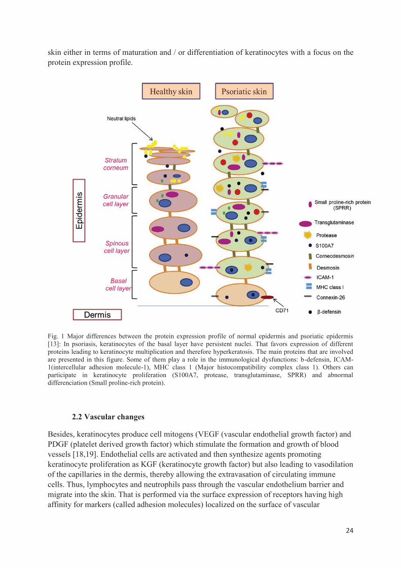
In psoriasis, abnormal epidermal barrier exhibits a structure fault due to a lack of normal corneocytes hindering the good cohesion of the stratum corneum which depends mainly on the interaction between corneocytes and synthesis of extracellular lipids produced by these cells [17]. The fig.1 summarizes all epidermis changes in psoriatic skin compared to healthy
24
skin either in terms of maturation and / or differentiation of keratinocytes with a focus on the protein expression profile.
Fig. 1 Major differences between the protein expression profile of normal epidermis and psoriatic epidermis [13]: In psoriasis, keratinocytes of the basal layer have persistent nuclei. That favors expression of different proteins leading to keratinocyte multiplication and therefore hyperkeratosis. The main proteins that are involved are presented in this figure. Some of them play a role in the immunological dysfunctions: b-defensin, ICAM-1(intercellular adhesion molecule-1), MHC class 1 (Major histocompatibility complex class 1). Others can participate in keratinocyte proliferation (S100A7, protease, transglutaminase, SPRR) and abnormal differenciation (Small proline-rich protein).
2.2 Vascular changes
Besides, keratinocytes produce cell mitogens (VEGF (vascular endothelial growth factor) and PDGF (platelet derived growth factor) which stimulate the formation and growth of blood vessels [18,19]. Endothelial cells are activated and then synthesize agents promoting keratinocyte proliferation as KGF (keratinocyte growth factor) but also leading to vasodilation of the capillaries in the dermis, thereby allowing the extravasation of circulating immune cells. Thus, lymphocytes and neutrophils pass through the vascular endothelium barrier and migrate into the skin. That is performed via the surface expression of receptors having high affinity for markers (called adhesion molecules) localized on the surface of vascular
25
endothelial cells (VCAM-1 (vascular cell adhesion molecule-1 or CD106), ICAM-1 (intracellular adhesion molecule-1 or CD54), E-selectin (CD62E) [20].
2.3 Skin infiltration by inflammatory cells and cytokine production
Psoriatic lesions are characterized by infiltration of the dermis and the epidermis by cells of the immune system which excrete proinflammatory cytokines capable to maintain and amplify the inflammatory reaction [21]. Studies on the subject have shown the abnormal infiltration of the dermis by CD4 + T cells, but also by dendritic cells (DC) (phagocytic and antigen-presenting cells) like CD11c + myeloid dendritic cells (MDC), langerhans cells. In the epidermis, a large amount of CD8+ T lymphocytes was detected and the presence of Langerhans cells and CD11c+ myeloid dendritic cells. It has been detected mainly neutrophils in the region of the horny layer [17,20]. The CD4+ and CD8+ T cells play a fundamental role in the pathogenesis of psoriasis, both in the initiation and in the maintenance of the inflammatory process. Many studies have shown the importance of T cells in psoriasis [22–32]. The T cells are activated after recognition of an antigen as a peptide combined with molecules of the class I (by CD8 +,) or II (by CD4 +) major histocompatibility complex (MHC) expressed on the surface of the antigen presenting cell, mainly dendritic cells in the skin lesions.
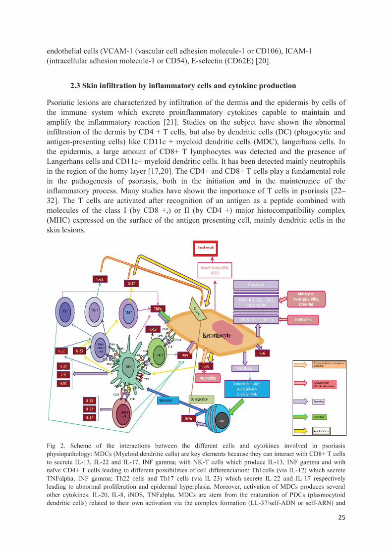
Fig 2. Scheme of the interactions between the different cells and cytokines involved in psoriasis physiopathology: MDCs (Myeloid dendritic cells) are key elements because they can interact with CD8+ T cells to secrete IL-13, IL-22 and IL-17, INF gamma; with NK-T cells which produce IL-13, INF gamma and with naïve CD4+ T cells leading to different possibilities of cell differenciation: Th1cells (via IL-12) which secrete TNFalpha, INF gamma; Th22 cells and Th17 cells (via IL-23) which secrete IL-22 and IL-17 respectively leading to abnormal proliferation and epidermal hyperplasia. Moreover, activation of MDCs produces several other cytokines: IL-20, IL-8, iNOS, TNFalpha. MDCs are stem from the maturation of PDCs (plasmocytoid dendritic cells) related to their own activation via the complex formation (LL-37/self-ADN or self-ARN) and
26
secretion of INFalpha. INFalpha can also upregulate the expression of IL-22 receptor increasing more the effect of IL-22. Moreover, a loop of amplification has been observed: activated keratinocytes by cytokines (TNF alfa, INF gamma, IL-22, IL-17) or NK-T cell interaction release IL-36 which stimulate MDCs secretion of IL-23, then the differenciation into Th17 cell and so a higher keratinocyte impact. Growth factors are produced by activated keratinocytes leading to vasodilatation, but also chemokines: MCP-1, IL-8, CXCL1, CXCL2, CXCL3,CXCL9,CXCL10,CXCL11, CCL20 and IL-36 that attract different cellular types involved in the chronical inflamatory phenomenon as lympocytes, neutrophils, monocytes.
2.3.1 Interplay between Myloïd dendritic cells (MDCs), naïve CD4+ T cell, Th1, Th17, Th22
The DC / T cell interaction through several synapses (CD40L / CD40 and MHC II / TCR for CD4 + T cell; CD80-86 / CD28 and MHC I / TCR for CD8 + T cell, CD40L / CD40 and CD1d / TCR for NK-T cell) leads to co-stimulation of the both cells which produce different types of cytokines . However, the responsible antigens have not yet been identified. It has been reported that peptides derived from proteins encoded by the virus HPV5 [33] or microbial antigens from streptococci [34] contribute to the development of psoriasis. Besides, it was also reported by Torres et al. [35] that the superantigens as staphylococcal enterotoxin A [36] or certain streptococcal antigens [37] play an important role in psoriatic lesions. These are antigens capable of binding MHC class II beta chain of the TCR of CD4 + lymphocytes inducing therefore a strong activation and selective proliferation of T lymphocytes, regardless of their specificity [13]. The different interplays between cells and cytokines involved in psoriasis pathogenesis are presented in figure 2.
Although the autoantigen enabling the MDC/T cell co-stimulation is still not known, it has been demonstrated that it is a common antigen for autoimmune T cells leading to an oligoclonal expansion [38,39]. Autoreactive T cell migration into the epidermis is controlled by alpha 1 beta 1 integrin displaying on effector T cells [40]. MDCs exhibit inflammatory elements as TLR (Toll like receptors)) and release mediators (IL-12, IL-23, TNF alpha, IL-20, IL-8, iNOS) playing essential roles in the activation, polarization and expansion of T cells and keratinocyte abnormalities [21]. MDCs induce naïve CD4+ T cell polarization into Th1 via IL-12, and into Th17 and Th22 via IL-23 respectively. Then, Th1 cells secrete INFg and TNFa, Th17 cells secrete IL-17 and TNFa, and Th22 cells secrete IL-22 respectively [12,41].
2.3.2 Plasmacytoid dendritic cells (pDCs)
pDCs have a key place in the psoriasis physiopathology. Unlike MDCs which are found in interstitial tissues as dermis and epidermis, the pDCs do not express CD11 on their surface and are located in blood and lymphoid organs [13]. pDCs secrete INFa after stimulation by a complex constituted by LL-37 (a cathelicidin belonging to the antimicrobial peptide family) with self-DNA or self-RNA from dying cells [42]. An external stimuli such as trauma (known as Koebner phenomenon), infections, stress, drugs, and alcohol can be the cause of antimicrobial peptide LL-37 release [43,42]. In psoriasis, keratinocytes express a large amount of antimicrobial peptides under the action of IL-17 and IL-22 [44]. This IFNa response is considered as the essential step at the MDC/T cell co-stimulation and the sustained production conducts to uncontrolled activation of MDCs until autoimmunity [42]. INFa can induce the maturation of pDCs into MDC [45]. Moreover, it has been observed that the LL-37- self-RNA complex could trigger a direct maturation into MDCs [46].More
27
recently, it has been found that IFNa is capable to upregulate IL-22-receptor expression on keratinocytes and therefore amplify the IL-22 related response [47]. Moreover, it has been shown that IFNa improve major histocompatibility complex class I (MCH I) expression on keratinocytes,which reinforce the CD8+ T cell activation [48].
2.3.3 The LL-37-IFN-Th17/Th22 axis
Keratinocytes are activated by TNFa and INFg leading to secretion of different cytokines, especially chemokines ((MCP-1, IL-8, CXCL1, CXCL2, CXCL3, CCL20, CXCL9, CXCL10, CXCL11)) which attract many leukocytes (Monocytes, Neutrophils, PDCs, CCR6+ Th1, CXCR3+ Th1) from the blood circulation to the skin area. This recruiting participates in amplifying and maintaining inflammation response that provoke lesions. The abnormal proliferation and epidermal hyperplasia involve mainly IL-17, IL-22 and INFg [49][12,50,51] but also to a lesser extent IL-19, IL-20 and keratinocyte-derived IL-36 [52,53]. Thus, the accent is put on the LL-37-IFN-Th17/Th22 axis as central element in the psoriasis pathogenesis [12,42]. Recently, an interesting keratinocyte-derived cytokines, the IL-36, have been involved in psoriasis as acting in synergy wtih the LL-37-IFN-Th17/Th22 axis, specifically with an important role in both psoriatic lesion inititation and maintenance. Many studies have described mutations in IL36RN, the gene encoding the IL-36R antagonist (IL-36Ra) in familial systemic pustular psoriasis [43,54,55]. IL-36 cytokines (IL-36a, IL-36b, and IL-36g) belonging to the IL-1 family are produced by keratinocytes and upregulated in psoriasis [56][57]. They participate in the abnormal proliferation and epidermal hyperplasia. Moreover, IL-36 cytokines induce IL-23 gene expression from DCs [58] which generates an amplifying loop via the IL-23/IL-17 pathway involving MDCs/Th17 cells /keratinocytes and therefore maintain the state of chronic inflammation in psoriasis. IL-36Ra is a natural antagonist which regulates the proinflammatory effects of the IL-36 cytokines by competition to the IL-36R stimulation. Because of the mutations in IL36RN, IL-36Ra is inactive. The IL-36/IL-36Ra balance is broken in favor of a more intense activity of IL-36 cytokines leading to a strong recruiting of neutrophils and the set up of a positive feedback loop of inflammation in psoriasis [12].
2.3.4 CD8+ T cells
Although CD8+ T cells have been underappreciated in the psoriasis pathogenesis, Hijnen et al. 2013 have demonstrated that they are a great source of proinflammatory cytokines including IFN-g, IL-13, IL-22 and IL-17. The rôle of IL-13 in psoriasis is to induce macrophage-derived chemokine (CCL22) expression in keratinocytes and matrix metalloproteinase-9 activation, leading to leukocyte migration into the epidermis [59,60].
2.3.5 NK-T (Natural killer-T))cells
Other cells could be also involved in psoriasis pathogenesis. NK-T cells were found in large amounts in acute and chronic psoriatic lesions [27]. Activation of these cells is through the recognition of an unknown antigen related to the CD1d molecule on the surface of keratinocytes and MDCs. In psoriasis, CD1d is overexpressed on keratinocyte surface [61,62]. Several in vivo-studies in rats have concluded to a potential role of NK-T cells in the formation of psoriatic plaques which would be due to the production of IFNg and IL-13 [27,63].
28
3. Psoriatic arthritis (PsA): a psoriasis complication Between 6-39% of individuals with psoriasis present also a joint disease related to an inflammatory arthritis affecting mainly isolated joints. This is due to systemic inflammation and extensive synovitis resulting in erosions of articular cartilage leading to joint destruction [64]. Although pathogenesis is only partly elucidated, PsA is frequently regarded as an enthesal disease [65] caused by a mix of genetic and environmental (stress, infections, trauma) factors which participates in triggering and maintaining of inflammatory/immunological processes in joints. The synovial tissue is the main one affected by these factors in the PsA development. It is characterised by synovial lining layer hyperplasia which is composed by two types of cells: type A-mononuclear phagocyte, type B-fibroblast like synoviocytes (FLS) [66,67]. This is reflected, in particular, in the stimulation of the FLSs resulting in 3 effects.
3.1 Secretion of inflammatory cytokines: MCP-1 (monocyte chemotactic protein-1) and IL-8.
They allow the chemotaxis and activation of monocytes/macrophage, which thereafter produce a variety of cytokines. TNF alpha, IL-1 and IL-6 have roles well known in inflammatory processes, stimulating synoviocyte hyperproliferation [68]. Besides, MDC (macrophage-derived chemokine) is also secreted in the synovial fluid and the synovial membrane with the particularity to be the ligand of the CCR4 receptor which is expressed by memory T cells. Via MDC, these latter are chemoattracted in the synovial tissue from the skin. Monocytes and neutrophils produce a large amount of specific molecules participating in the complex immune response. Myeloid-related protein 8 (MRP8; S100A8) and MRP14 (S100A9) belongs to the S100 family of proteins. These two calcium-binding proteins have both intracellular functions (cell differentiation and cell cycle progression, regulation of kinase activities and cytoskeleton–membrane interactions) and extracellular functions (neutrophil extension, chemoattraction, arachidonic acid metabolism and the induction of adhesion molecule expression) positioning them as major players in the cascade of inflammatory stimuli and communication between skin and joint [69].
3.2 SelfDNA expression
The innate immunity may be involved precocely. The joint and the skin are environments rich in endogenous ligands of the innate immune system as cathelicidin (LL37), an antimicrobial peptide, which could form a complexe with selfDNA taken in charged by pDC which is going to secrete INF a. As described previously, the CD4+ T cell responses lead to the production of many cytokines (IL-17, TNFa, IL-22). IL-17 induces the secretion of proinflammatory cytokines, stimulates osteoclast formation and bone resorption, recruits neutrophils and monocytes (producers of S100A8/A9), and triggers the production of granulocyte–macrophage colony-stimulating factor [70]. Otherwise, CD4 activation causes RANKL production having for effect to activate osteoclasts and subsequently provoke bone erosion, but also VEGF (vascular endothelial growth factor) production which activates endothelial cells and the formation of HEV (high endothelial venules) [64].
3.3 Production of complement system factors
29
FLSs have a stimulated production of C3 and C4 complement system factors. When activated, the complement system generates a release anaphylotoxins (C5a) and the formation of MAC (membrane attack complex). That induces cell lysis and immune cell recruitment. Moreover, the lower expression of the CD59, an inhibitor of complement system activation, accentuates further the complement system activation [66,71,67].
4. Psoriasis treatment
To date, there is no treatment healing psoriasis. Available treatments consist primarily to alleviate symptoms and prevent the evolution of the disease, in order to improve the quality of life of patients. Different therapeutic strategies are conventionally used, which are sometimes combined for greater efficiency: local treatment, phototherapy, systemic therapy. Due to the chronic nature of psoriasis and no cure available, these treatments are taken for life, which makes them a real economic challenge for pharmaceutical firms and health systems of different countries [6].
4.1. Local treatments
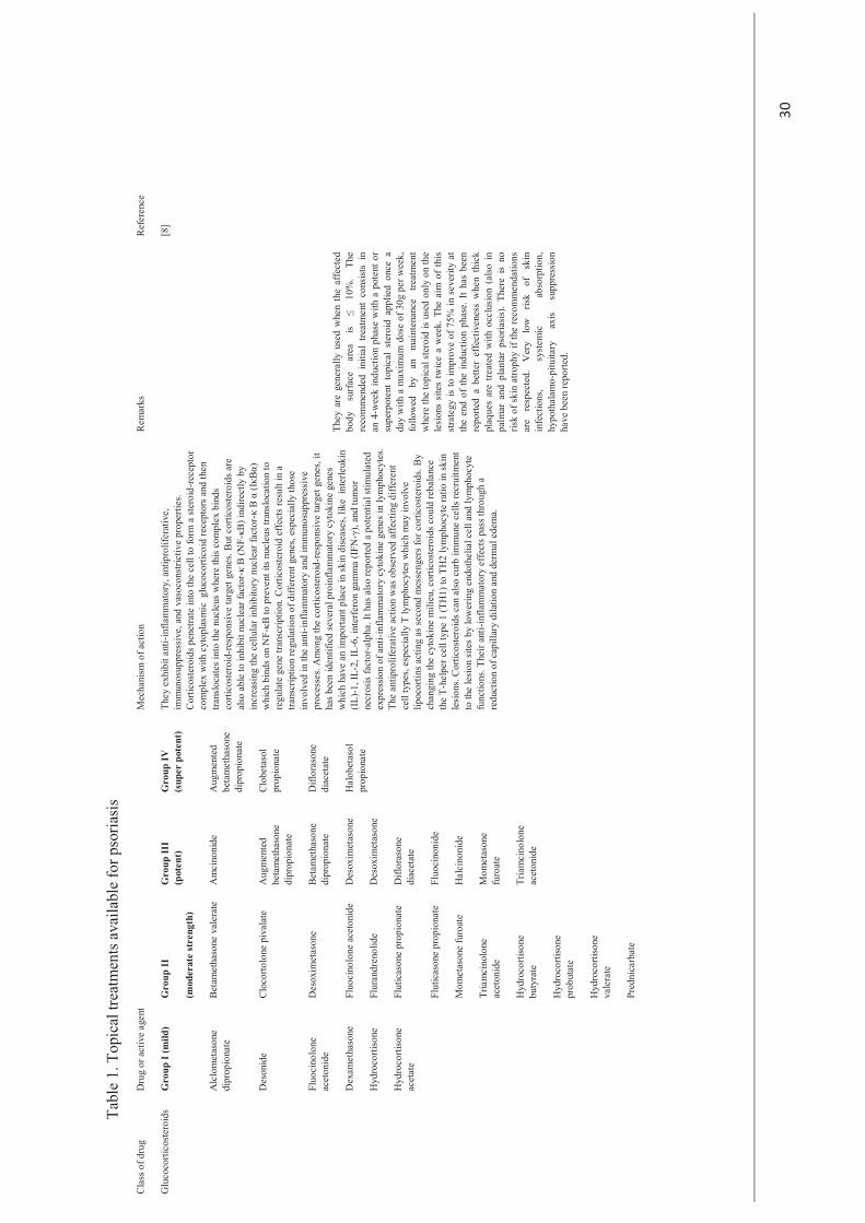
They are indicated in mild forms of psoriasis, usually defined by a skin disease affecting less than 20% of the body surface. There are different groups (table 1) (emollients, keratolytic agents, vitamin D analogues, retinoids) but dermocorticosteroids remain the main drugs prescribed. The latter group is also the richest, offering a range of different classes, the level of activity varies from very potent to mild. The primary targets of these agents are the proliferation of keratinocytes / hyperkeratosis, the immune cells, inflammation. However, these local treatments can have significant side effects that may lead to stop the treatment. Topical corticosteroids are never used for long periods. The treatment lasts up to one month because side effects may occur (skin atrophy, thin skin, skin conditions ...) in a more or less severe degree according to the dermocorticoid group. One of the drawbacks of topical corticosteroids is that after cessation of treatment, there is often a recurrence. And if discontinuation is abrupt, a severe rebound effect could be generated [8].
4.2.Phototherapy
This type of treatment is considered when local treatment is not sufficient or that the extent of damage is more than 20% of the body surface. Phototherapy is based on the use of broad-spectrum UVB and narrow-spectrum UVA.
30
Tabl
e 1.
Top
ical
trea
tmen
ts a
vaila
ble
for p
soria
sis
Cla
ss o
f dru
g D
rug
or a
ctiv
e ag
ent
Mec
hani
sm o
f act
ion
Rem
arks
R
efer
ence
Glu
coco
rtico
ster
oids
Gro
up I
(mild
) G
roup
II
(mod
erat
e st
reng
th)
Gro
up II
I (p
oten
t)
Gro
up IV
(s
uper
pot
ent)
Th
ey e
xhib
it an
ti-in
flam
mat
ory,
ant
ipro
lifer
ativ
e,
imm
unos
uppr
essi
ve, a
nd v
asoc
onst
rictiv
e pr
oper
ties.
Cor
ticos
tero
ids p
enet
rate
into
the
cell
to fo
rm a
ster
oid-
rece
ptor
co
mpl
ex w
ith c
ytop
lasm
ic g
luco
corti
coid
rece
ptor
s and
then
tra
nslo
cate
s int
o th
e nu
cleu
s whe
re th
is co
mpl
ex b
inds
co
rtico
ster
oid -
resp
onsi
ve ta
rget
gen
es. B
ut c
ortic
oste
roid
s are
al
so a
ble
to in
hibi
t nuc
lear
fact
or-κ
B (N
F-κB
) ind
irect
ly b
y in
crea
sing
the
cellu
lar i
nhib
itory
nuc
lear
fact
or-κ
B α
(IκB
α)
whi
ch b
inds
on
NF -
κB to
pre
vent
its n
ucle
us tr
ansl
ocat
ion
to
regu
late
gen
e tra
nscr
iptio
n. C
ortic
oste
roid
effe
cts
resu
lt in
a
trans
crip
tion
regu
latio
n of
diff
eren
t gen
es, e
spec
ially
thos
e in
volv
ed in
the
anti-
infla
mm
ator
y an
d im
mun
osup
pres
sive
pr
oces
ses.
Am
ong
the
corti
cost
eroi
d-re
spon
sive
targ
et g
enes
, it
has b
een
iden
tifie
d se
vera
l pro
infla
mm
ator
y cy
toki
ne g
enes
w
hich
hav
e an
impo
rtant
pla
ce in
skin
dise
ases
, lik
e in
terle
ukin
(I
L)-1
, IL-
2, IL
-6, i
nter
fero
n ga
mm
a (I
FN-γ
), an
d tu
mor
ne
cros
is fa
ctor
-alp
ha. I
t has
also
repo
rted
a po
tent
ial s
timul
ated
ex
pres
sion
of a
nti-i
nfla
mm
ator
y cy
toki
ne g
enes
in ly
mph
ocyt
es.
The
antip
rolif
erat
ive
actio
n w
as o
bser
ved
affe
ctin
g di
ffere
nt
cell
type
s, es
peci
ally
T ly
mph
ocyt
es w
hich
may
invo
lve
lipoc
ortin
s act
ing
as se
cond
mes
seng
ers
for c
ortic
oste
roid
s. B
y ch
angi
ng th
e cy
toki
ne m
ilieu
, cor
ticos
tero
ids c
ould
reba
lanc
e th
e T -
help
er c
ell t
ype
1 (T
H1)
to T
H2
lym
phoc
yte
ratio
in sk
in
lesi
ons.
Cor
ticos
tero
ids c
an a
lso c
urb
imm
une
cells
recr
uitm
ent
to th
e le
sion
site
s by
low
erin
g en
doth
elia
l cel
l and
lym
phoc
yte
func
tions
. The
ir an
ti-in
flam
mat
ory
effe
cts p
ass t
hrou
gh a
re
duct
ion
of c
apill
ary
dila
tion
and
derm
al e
dem
a.
They
are
gen
eral
ly u
sed
whe
n th
e af
fect
ed
body
su
rface
ar
ea
is ≤
10%
. Th
e re
com
men
ded
initi
al t
reat
men
t co
nsist
s in
an
4-w
eek
indu
ctio
n ph
ase
with
a p
oten
t or
supe
rpot
ent
topi
cal
ster
oid
appl
ied
once
a
day
with
a m
axim
um d
ose
of 3
0g p
er w
eek,
fo
llow
ed
by
an
mai
nten
ance
tre
atm
ent
whe
re th
e to
pica
l ste
roid
is u
sed
only
on
the
lesi
ons
site
s tw
ice
a w
eek.
The
aim
of
this
st
rate
gy is
to im
prov
e of
75%
in s
ever
ity a
t th
e en
d of
the
indu
ctio
n ph
ase.
It h
as b
een
repo
rted
a be
tter
effe
ctiv
enes
s w
hen
thic
k pl
aque
s ar
e tre
ated
with
occ
lusi
on (
also
in
palm
ar a
nd p
lant
ar p
soria
sis)
. Th
ere
is n
o ris
k of
ski
n at
roph
y if
the
reco
mm
enda
tions
ar
e re
spec
ted.
V
ery
low
ris
k of
sk
in
infe
ctio
ns,
syst
emic
ab
sorp
tion,
hy
poth
alam
o -pi
tuita
ry
axis
supp
ress
ion
have
bee
n re
porte
d.
[8]
Alc
lom
etas
one
dipr
opio
nate
Be
tam
etha
sone
val
erat
e A
mci
noni
de
Aug
men
ted
beta
met
haso
ne
dipr
opio
nate
Des
onid
e C
loco
rtolo
ne p
ival
ate
Aug
men
ted
beta
met
haso
ne
dipr
opio
nate
Clo
beta
sol
prop
iona
te
Fluo
cino
lone
ac
eton
ide
Des
oxim
etas
one
Beta
met
haso
ne
dipr
opio
nate
D
iflor
ason
e di
acet
ate
Dex
amet
haso
ne
Fluo
cino
lone
ace
toni
de
Des
oxim
etas
one
Hal
obet
asol
pr
opio
nate
H
ydro
corti
sone
Fl
uran
dren
olid
e D
esox
imet
ason
e
Hyd
roco
rtiso
ne
acet
ate
Flut
icas
one
prop
iona
te
Difl
oras
one
diac
etat
e
Flut
icas
one
prop
iona
te
Fluo
cino
nide
Mom
etas
one
furo
ate
Hal
cino
nide
Tria
mci
nolo
ne
acet
onid
e M
omet
ason
e fu
roat
e
Hyd
roco
rtiso
ne
buty
rate
Tr
iam
cino
lone
ac
eton
ide
Hyd
roco
rtiso
ne
prob
utat
e
Hyd
roco
rtiso
ne
vale
rate
Pred
nica
rbat
e
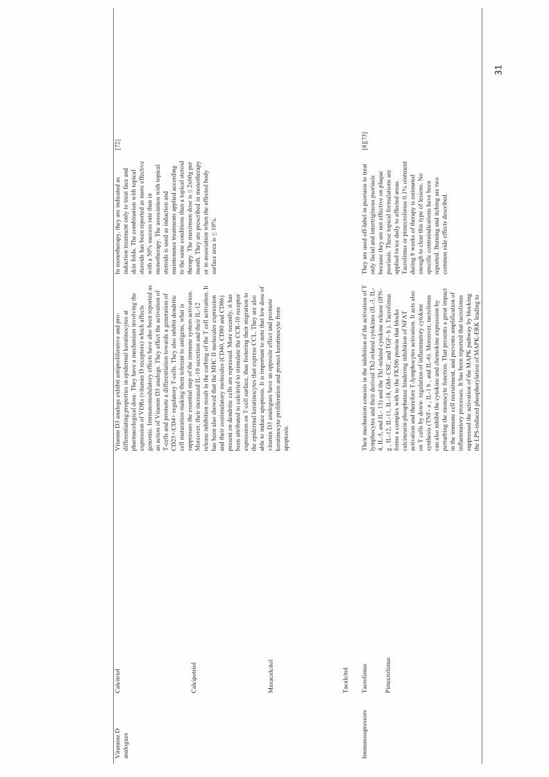
31
Vita
min
e D
an
alog
ues
Cal
citri
ol
Vita
min
D3
anal
ogs e
xhib
it an
tipro
lifer
ativ
e an
d pr
o-di
ffere
ntia
ting
prop
ertie
s in
epid
erm
al k
erat
inoc
ytes
at
phar
mac
olog
ical
dos
e. T
hey
have
a m
echa
nism
invo
lvin
g th
e ex
pres
sion
of V
DR
s (vi
tam
in D
rece
ptor
s) w
hich
affe
cts
geno
mic
. Im
mun
omod
ulat
ory
effe
cts h
ave
also
bee
n re
porte
d as
an
act
ion
of V
itam
in D
3 an
alog
s. Th
ey a
ffect
the
activ
atio
n of
T -
cells
and
pro
mot
e a
diffe
rent
iatio
n to
war
ds a
gen
erat
ion
of
CD
25+/
CD
4+ re
gula
tory
T-c
ells.
The
y al
so in
hibi
t den
driti
c ce
ll m
atur
atio
n m
akin
g th
em to
lera
nt to
ant
igen
s, w
hat i
s su
ppre
sses
the
esse
ntia
l ste
p of
the
imm
une
syst
em a
ctiv
atio
n.
Mor
eove
r, th
eir i
ncre
ased
IL-1
0 se
cret
ion
and
thei
r IL-
12
rele
ase
inhi
bitio
n re
sult
in th
e cu
rbin
g of
the
T ce
ll ac
tivat
ion.
It
has b
een
also
show
ed th
at th
e M
HC
II m
olec
ules
exp
ress
ion
and
thei
r cos
timul
ator
y m
olec
ules
(CD
40, C
D80
and
CD
86)
pres
ent o
n de
ndrit
ic c
ells
are
repr
esse
d. M
ore
rece
ntly
, it h
as
been
attr
ibut
ed to
cal
citri
ol to
stim
ulat
e th
e C
CR
-10
rece
ptor
ex
pres
sion
on T
-cel
l sur
face
, thu
s fo
ster
ing
thei
r mig
ratio
n to
th
e ep
ider
mal
ker
atin
ocyt
es th
at e
xpre
ss C
CL.
The
y ar
e al
so
able
to in
duce
apo
ptos
is. It
is im
porta
nt to
not
e th
at lo
w d
ose
of
vita
min
D3
anal
ogue
s ha
ve a
n op
posi
te e
ffec
t and
pro
mot
e ke
ratin
ocyt
e pr
olife
ratio
n an
d pr
otec
t ker
atin
ocyt
e fro
m
apop
tosi
s.
In m
onot
hera
py, t
hey
are
indi
cate
d as
in
duct
ion
treat
men
t onl
y to
trea
t fac
e an
d sk
in fo
lds.
The
com
bina
tion
with
topi
cal
ster
oids
has
bee
n re
porte
d as
mor
e ef
fect
ive
with
a 5
0% su
cces
s rat
e th
an in
m
onot
hera
py. T
he a
ssoc
iatio
n w
ith to
pica
l st
eroi
ds is
use
d as
indu
ctio
n an
d m
aint
enan
ce tr
eatm
ents
app
lied
acco
rdin
g to
the
sam
e co
nditi
ons t
han
a to
pica
l ste
roid
th
erap
y. T
he m
axim
um d
ose
is ≤
2x60
g pe
r m
onth
. The
y ar
e pr
escr
ibed
in m
onot
hera
py
or in
ass
ocia
tion
whe
n th
e af
fect
ed b
ody
surf
ace
area
is ≤
10%
.
[72]
Cal
cipo
triol
Max
acal
cito
l
Taca
lcito
l
Imm
unos
upre
ssor
s
Tacr
olim
us
Thei
r mec
hani
sm c
onsi
sts
in th
e in
hibi
tion
of th
e ac
tivat
ion
of T
ly
mph
ocyt
es a
nd th
eir d
eriv
ed T
h2-re
late
d cy
toki
nes (
IL-3
, IL-
4, IL
-5, a
nd IL
- 13)
and
the
Th1-
rela
ted
cyto
kine
rele
ase
(IFN
- g
, IL-
12, I
L-11
, IL-
18, G
M- C
SF, a
nd T
GF-
b ).
Tac
rolim
us
form
s a c
ompl
ex w
ith to
the
FK50
6 pr
otei
n th
at b
lock
s ca
lcin
eurin
pho
spha
tase
hin
derin
g in
hibi
tion
of N
FAT
activ
atio
n an
d th
eref
ore
T -ly
mph
ocyt
es a
ctiv
atio
n. It
act
s also
on
T c
ells
by d
own-
regu
latio
n of
infla
mm
ator
y cy
toki
ne
synt
hesi
s (TN
F- a
, IL
-1 b
, an
d IL
-6).
Mor
eove
r, ta
crol
imus
ca
n al
so in
hibi
t the
cyt
okin
e an
d ch
emok
ine
expr
essi
on b
y pe
rturb
ing
the
mon
ocyt
e fu
nctio
n. T
hat p
rese
nts a
gre
at im
pact
in
the
imm
une
cell
recr
uitm
ent,
and
prev
ents
am
plifi
catio
n of
in
flam
mat
ory
p roc
esse
s. It
has b
een
repo
rted
that
tacr
olim
us
supp
ress
ed th
e ac
tivat
ion
of th
e M
APK
pat
hway
by
bloc
king
th
e LP
S-in
duce
d ph
osph
oryl
atio
n of
MA
PK-E
RK
lead
ing
to
They
are
use
d of
f-la
bel i
n ps
oria
sis t
o tre
at
only
faci
al a
nd in
tertr
igin
ous p
soria
sis
beca
use
they
are
not
effe
ctiv
e on
pla
que
psor
iasis
. The
se to
pica
l for
mul
atio
ns a
re
appl
ied
twic
e da
ily to
affe
cted
are
as.
Tacr
olim
us o
r pim
ecro
limus
0.1
% o
intm
ent
durin
g 8
wee
ks o
f the
rapy
is e
stim
ated
en
ough
to c
lear
this
type
of l
esio
ns. N
o sp
ecifi
c co
ntra
indi
catio
ns h
ave
been
re
porte
d. B
urni
ng a
nd it
chin
g ar
e tw
o co
mm
on s
ide
effe
cts d
escr
ibed
.
[8][
73]
Pim
ecro
limus
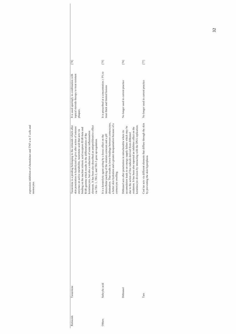
32
expr
essio
n in
hibi
tion
of c
hem
okin
es a
nd T
NF-
a in
T c
ells
and
mon
ocyt
es.
Ret
inoi
ds
Taza
rote
ne
Taza
rote
ne is
a p
rodr
ug b
elon
ging
to th
e re
tinoi
ds w
hich
afte
r sk
in p
enet
ratio
n is
hydr
olyz
ed to
giv
e af
ter a
ctio
n of
est
eras
e en
zym
es a
n ac
tive
met
abol
ite, t
azar
oten
e ac
id th
at a
cts v
ia
bind
ing
on th
e nu
clea
r ret
inoi
c ac
id re
cept
ors R
AR
-bet
a an
d R
AR
-gam
ma
whi
ch re
sults
in th
e di
ffere
ntia
tion
of th
e ke
ratin
ocyt
es, b
ut a
lso a
redu
ctio
n of
som
e in
flam
mat
ion
elem
ents
. It h
as b
een
also
des
crib
ed a
n an
tipro
lifer
ativ
e ef
fect
v i
a TI
G- 1
, TIG
-2, a
nd T
IG-3
gen
e up
-regu
latio
n.
It is
used
spar
ingl
y in
com
bina
tion
with
to
pica
l ste
roid
s the
rapy
to tr
eat r
esist
ant
plaq
ues.
[74]
Oth
ers
Salic
ylic
aci
d It
is a
kera
toly
tic a
gent
act
ing
by a
loos
e ef
fect
on
the
inte
rcel
lula
r pac
king
of t
he st
ratu
m c
orne
um a
nd a
pH
di
min
utio
n. T
hat r
esul
ts in
low
er b
indi
ngs
betw
een
corn
eocy
tes,
a be
tter s
kin
hydr
atio
n an
d a
grea
ter d
esqu
amat
ion
beca
use
of a
co
rneo
cyte
swel
ling.
It is
pres
crib
ed a
t a c
once
ntra
tion
≥ 5%
to
treat
thic
k an
d lim
ited
lesi
ons
[75]
D
ithra
nol
Dith
rano
l act
s afte
r pen
etra
tion
in m
itoch
ondr
ia w
here
its
accu
mul
atio
n le
ads t
o an
ene
rgy
supp
ly fa
ilure
whi
ch m
ay b
e du
e to
the
actio
n of
free
radi
cals
deriv
ed fr
om d
i thra
nol
oxid
atio
n. It
has
bee
n al
so re
porte
d an
inhi
bito
r effe
ct o
n th
e ke
ratin
ocyt
e di
visi
on b
y in
tera
ctin
g w
ith th
e D
NA
repl
icat
ion.
No
long
er u
sed
in c
urre
nt p
ract
ice
[76]
Ta
rs
Coa
l tar
act
s via
diff
eren
t ele
men
ts th
at d
iffus
e th
roug
h th
e sk
in
by p
reve
ntin
g th
e sk
in h
yper
plas
ia.
No
long
er u
sed
in c
urre
nt p
ract
ice
[77]
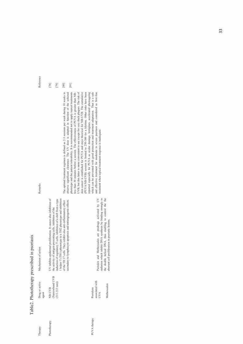
33
Tabl
e2. P
hoto
ther
apy
pres
crib
ed in
pso
riasi
s Th
erap
y D
rug
or a
ctiv
e ag
ent
Mec
hani
sm o
f act
ion
Rem
arks
R
efer
ence
Phot
othe
rapy
N
B-U
VB
(Nar
row
band
UV
B (3
11-3
13 n
m))
UV
inhi
bits
epi
derm
al p
rolif
erat
ion.
It c
ause
s also
inhi
bitio
n of
th
e ac
tivity
of a
ntig
en-p
rese
ntin
g ce
lls, i
nhib
ition
of t
he
indu
ctio
n of
regu
lato
ry T
cel
ls, in
hibi
tion
of a
shift
from
a ty
pe
1 he
lper
T (T
H1)
phe
noty
pe to
a T
H2
phen
otyp
e an
d in
hibi
tion
of th
e TH
17
cells
. The
y ex
hibi
t also
ant
i -inf
lam
mat
ory
effe
ct
seco
ndar
y to
lym
phoc
yte
apop
tosi
s and
ant
iang
ioge
nic
effe
cts.
Th
e op
timal
trea
tmen
t reg
imen
is d
efin
ed a
s 2-
3 se
ssio
ns p
er w
eek
durin
g 10
wee
ks to
ob
serv
e si
gnifi
cant
cle
aran
ce.
The
UV
dos
e is
adap
ted
in f
unct
ion
of t
he s
elec
ted
phot
otyp
e an
d th
e pa
tient
tole
rabi
lity.
It is
reco
mm
ende
d no
t to
appl
y to
pica
l tre
atm
ents
le
ss th
an 3
0 m
inut
es b
efor
e a
sess
ion.
The
eff
ectiv
enes
s of
PU
VA
is g
reat
er th
an N
B-
UV
B, b
ut th
is la
tter i
s m
ore
conv
enie
nt to
use
exc
ept f
or v
ery
thic
k pl
aque
s. Th
e ris
k of
sk
in c
ance
r is
esta
blis
hed
for
PUV
A a
nd s
tays
theo
rical
for
NB
-UV
B. T
he c
umul
ativ
e (P
UV
A/N
B-U
VB
) se
ssio
n is
limite
d to
250
-300
for
a l
ifetim
e. O
ther
risk
s ha
ve b
een
repo
rted
espe
cial
ly f
or P
UV
A a
s oc
ular
dam
age,
len
tigin
es,
acce
lera
ted
phot
oage
ing
whi
ch c
an b
e pr
even
ted
with
goo
d pr
otec
tion
and
treat
men
t ada
ptat
ion.
Th
at is
a s
afe
and
effe
ctiv
e tre
atm
ent
for
mod
erat
e to
sev
ere
psor
iasi
s an
d co
nsid
ered
as
first
-line
tre
atm
ent w
hen
topi
cal t
reat
men
t res
pons
e is
inad
equa
te.
[78]
[79]
[80]
[81]
PU
VA
ther
apy
Psor
alen
s as
soci
ated
with
U
VA
Ps
oral
en a
nd M
etho
xsal
en a
re
prod
rugs
act
ivat
ed b
y U
V
radi
atio
n w
hich
inhi
bit D
NA
syn
thes
is by
bin
ding
stro
ngly
to
the
doub
le h
elic
al D
NA
, th
us e
nabl
ing
to c
ontro
l th
e th
e ab
erra
nt c
ell p
rolif
erat
ion
in p
soria
tic le
sion
s.
Met
hoxs
alen
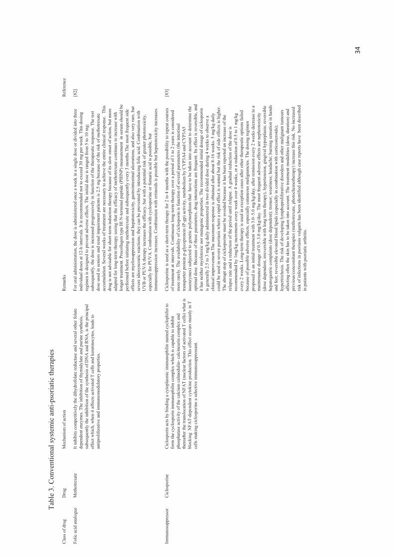
34
Tabl
e 3.
Con
vent
iona
l sys
tem
ic a
nti-p
soria
tic th
erap
ies
Cla
ss o
f dru
g D
rug
Mec
hani
sm o
f act
ion
Rem
arks
R
efer
ence
Folic
aci
d an
alog
ue
Met
hotre
xate
It in
hibi
ts c
ompe
titiv
ely
the
dihy
drof
olat
e re
duct
ase
and
seve
ral o
ther
fola
te
depe
nden
t enz
ymes
. The
inhi
bitio
n of
thym
idyl
ate
and
purin
e sy
nthe
sis,
subs
eque
ntly
the
inhi
bitio
n of
the
synt
hesi
s of D
NA
and
RN
A, i
s the
prin
cipa
l ef
fect
whi
ch, w
hen
it af
fect
s act
ivat
ed T
cel
ls an
d ke
ratin
ocyt
es, l
eads
to
antip
rolif
erat
ive
and
imm
unom
odul
ator
y pr
oper
ties.
For o
ral a
dmin
istra
tion,
the
dose
is a
dmin
iste
red
once
a w
eek
in a
sing
le d
ose
or d
ivid
ed in
to th
ree
indi
vidu
al d
oses
at 1
2 -h
inte
rval
s. It
is re
com
men
ded
not t
o ex
ceed
30
mg
per w
eek.
Thi
s dos
ing
regi
men
is d
esig
ned
to p
reve
nt a
dver
se e
ffect
s. Th
e in
itial
dos
e is
rang
ed fr
om 5
to 1
0 m
g;
subs
eque
ntly
, the
dos
e is
incr
ease
d pr
ogre
ssiv
ely
in fu
nctio
n of
the
ther
apeu
tic re
spon
se. T
he te
st
dose
use
d in
seni
ors a
n d re
nal i
mpa
irmen
t pat
ient
s is 2
.5 m
g be
caus
e of
the
risk
of m
etho
trexa
te
accu
mul
atio
n. S
ever
al w
eeks
of t
reat
men
t are
nec
essa
ry to
ach
ieve
the
entir
e cl
inic
al re
spon
se.
This
drug
is n
ot a
dvisa
ble
for s
hort -
term
indu
ctio
n th
erap
y be
caus
e of
its s
low
ons
et o
f act
ion,
but
mor
e ad
apte
d fo
r lon
g -te
rm th
erap
y se
eing
that
the
effic
acy
of m
etho
trexa
te c
ontin
ues t
o in
crea
se w
ith
long
er tr
eatm
ent.
Proc
olla
gen
type
III N
-term
inal
pep
tide
(PII
INP)
mea
sure
men
t in
seru
m s
houl
d be
pe
rform
ed b
efor
e in
itiat
ing
met
hotre
xate
and
subs
eque
ntly
eve
ry 3
mon
ths.T
he m
ost f
requ
ent s
ide
effe
cts a
re m
yelo
supp
ress
ion
and
hepa
toto
xici
ty, g
astro
inte
stin
al u
lcer
atio
ns, a
nd a
lso v
ery
rare
, but
se
vere
idio
sync
ratic
reac
tions
; the
y ca
n be
pre
vent
ed b
y in
trodu
cing
folic
aci
d. C
ombi
natio
n w
ith
UV
B or
PU
VA
ther
apy
incr
ease
s the
effi
cacy
, but
with
a p
oten
tial r
isk o
f gre
ater
pho
toto
xici
ty,
espe
cial
ly fo
r PU
VA
. Com
bina
tion
with
cyc
losp
orin
e or
fum
aric
aci
d is
poss
ible
, but
im
mun
osup
pres
sion
incr
ease
s. C
ombi
natio
n w
ith re
tinoi
ds is
pos
sible
but
hep
atot
oxic
ity in
crea
ses.
[82]
Imm
unos
uppr
esso
r
Cic
losp
orin
e
Cic
losp
orin
act
s by
bind
ing
a cy
topl
asm
ic im
mun
ophi
lin n
amed
cyc
loph
ilin
to
form
the
cycl
ospo
rin im
mun
ophi
lin c
ompl
ex w
hich
is c
apab
le to
inhi
bit
phos
phat
ase
activ
ity o
f the
cal
cium
cal
mod
ulin
- cal
cine
urin
com
plex
and
th
erea
fter t
he tr
ansl
ocat
ion
of N
FAT
(nuc
lear
fact
ors o
f act
ivat
ed T
cel
ls) w
hat i
s bl
ocki
ng N
FAT-
depe
nden
t cyt
okin
e pr
oduc
tion.
Thi
s eff
ect o
ccur
s m
ainl
y in
T
cells
mak
ing
cicl
ospo
rine
a se
lect
ive
imm
unos
uppr
essa
nt.
Cic
losp
orin
e is
used
as a
shor
t-ter
m th
erap
y fo
r 2 to
4 m
onth
s with
the
poss
ibili
ty to
repe
at c
ours
es
of tr
eatm
ent a
t int
erva
ls. C
ontin
uous
long
-term
ther
apy
over
a p
erio
d of
1 to
2 y
ears
is c
onsi
dere
d m
ore
rare
ly. T
he a
vaila
bilit
y of
cic
losp
orin
is fu
nctio
n of
seve
ral p
aram
eter
s (th
e in
test
inal
tra
nspo
rter p
rote
in p
-gly
copr
otei
n (P
-gp)
act
ivity
, met
abol
ism
by
CY
P3A
4 an
d C
YP3
A5
isoen
zym
es) s
ubje
cted
to g
enet
ic p
olym
orph
ism
that
hav
e to
be
take
n in
to a
ccou
nt to
det
erm
ine
the
optim
al d
ose.
Bec
ause
of t
hese
par
amet
ers,
drug
inte
ract
ions
are
freq
uent
. Its
effe
ct is
reve
rsib
le, a
nd
it ha
s ne
ither
mye
loto
xic
nor m
utag
enic
pro
perti
es. T
he re
com
men
ded
initi
al d
osag
e of
cic
losp
orin
is
gene
rally
2.5
to 3
mg/
kg d
aily
adm
inis
tere
d in
two
divi
ded
dose
dur
ing
4 w
eeks
to o
bser
ve a
cl
inic
al im
prov
emen
t.The
max
imum
resp
onse
is o
btai
ned
afte
r abo
ut 8
-16
wee
ks. 5
mg/
kg d
aily
co
uld
be u
sed
in se
vere
pso
riasi
s whe
re a
rapi
d ef
fect
is w
ante
d bu
t the
risk
of s
ide
effe
cts i
s hig
her.
The
abru
pt st
op o
f cic
losp
orin
e m
ust b
e av
oide
d be
caus
e it
has b
een
repo
rted
an in
crea
se o
f the
re
laps
e ra
te a
nd a
redu
ctio
n of
the
perio
d un
til re
laps
e. A
gra
dual
redu
ctio
n of
the
dose
is
reco
mm
ende
d by
1m
g/kg
incr
emen
ts e
very
wee
k o v
er 4
wee
ks, o
r a re
duct
ion
of 0
.5 to
1 m
g/kg
ev
ery
2 w
eeks
. Lon
g-te
rm th
erap
y is
used
in e
xcep
tion
case
s afte
r oth
er th
erap
eutic
opt
ions
faile
d be
caus
e of
pos
sible
adv
erse
effe
cts,
espe
cial
ly c
utan
eous
mal
igna
ncie
s. Th
e do
sing
regi
men
pr
opos
ed is
an
initi
al tr
eatm
ent w
ith 3
.0–5
.0 m
g/kg
/day
, afte
r rem
issio
n ev
ery
2 w
eeks
dec
reas
e to
a
mai
nten
ance
dos
age
of 0
.5–3
.0 m
g/kg
/day
. The
mos
t fre
quen
t adv
erse
eff
ects
are
nep
hrot
oxic
ity
(dos
e de
pend
ent,
irrev
ersi
ble
with
long
term
ther
apy)
, hyp
erte
nsio
n, g
ingi
val h
yper
plas
ia; r
ever
sibl
e he
pato
gast
ric c
ompl
aint
s (do
se d
epen
dent
); tre
mor
; wea
rines
s; h
eada
che;
bur
ning
sens
atio
n in
han
ds
and
feet
; rev
ersi
ble
elev
ated
blo
od li
pids
(esp
ecia
lly in
com
bina
tion
with
cor
ticos
tero
ids)
; hy
pertr
icho
sis.
The
risk
of d
evel
opin
g ly
mph
opro
lifer
ativ
e di
sord
ers a
nd o
ther
mal
igna
nt tu
mou
rs
affe
ctin
g of
ten
the
skin
has
to b
e ta
ken
into
acc
ount
. The
trea
tmen
t mod
aliti
es (d
ose,
dur
atio
n) a
nd
prev
ious
/con
com
itant
ther
apie
s (m
etho
trexa
te, p
hoto
chem
othe
rapy
) inc
reas
e th
is ris
k. N
o in
crea
sed
risk
of in
fect
ions
in p
soria
sis
vulg
aris
has b
een
iden
tifie
d al
thou
gh c
ase
repo
rts h
ave
bee
n de
scrib
ed
in p
atie
nts w
ith p
soria
tic a
rthrit
is.
[83]
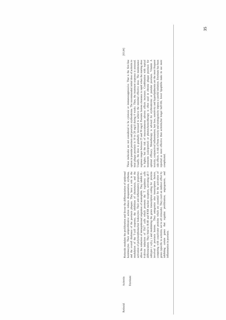
35
Ret
inoi
d
Aci
tretin
R
etin
oids
mod
ulat
e th
e pr
olife
ratio
n an
d fa
vour
the
diffe
renc
iatio
n of
epi
derm
al
kera
tinoc
ytes
. Th
eir
antip
rolif
erat
ive
actio
n re
duce
s de
squa
mat
ion,
ery
them
a,
and
the
over
all
thic
knes
s of
the
pso
riasi
s pl
aque
s. Th
ey h
ave
a ro
le i
n th
e m
odul
atio
n of
the
T-c
ell
resp
onse
, th
e in
hibi
tion
of c
hem
otax
is, a
nd t
he
activ
atio
n of
pol
ymor
phon
ucle
ar le
ukoc
ytes
. The
ir an
ti-in
flam
mat
ory
prop
ertie
s al
low
the
redu
ctio
n of
intra
epid
erm
al m
igra
tion
of n
eutro
phils
. The
y in
hibi
t IL-
6 -dr
iven
in
duct
ion
of
Th17
ce
lls
whi
ch
prom
ote
the
T re
gula
tory
ce
lls
diffe
renc
iatio
n. T
hey
bind
to R
AR
and
RX
R n
ucle
ar r
ecep
tors
, act
ivat
ing
all 3
su
btyp
es (
α,β
,ɣ )
and
mod
ulat
ing
the
gene
s tra
nscr
iptio
n co
ding
for
pr
otei
ns
invo
lved
in
psor
iasi
s le
sions
. T
hey
anta
goni
ze a
lso t
rans
crip
tion
fact
ors,
co
mpe
ting
with
coa
ctiv
ator
pro
tein
s w
hich
are
ess
entia
l fo
r th
e ac
tivat
ion
of
gene
s (s
peci
fic to
aci
tretin
, not
to a
ll re
tinoi
ds).
They
exe
rt an
indi
rect
effe
ct b
y in
hibi
ting
certa
in
gene
s th
at
regu
late
pr
olife
ratio
n,
angi
ogen
esis,
an
d in
flam
mat
ion
in p
soria
sis.
Thes
e m
olec
ules
are
not
con
side
red
to b
e cy
toto
xic
or i
mm
unos
uppr
essi
ve.
That
is
the
first
-line
op
tion
in p
ustu
lar p
soria
sis
and
psor
iatic
ery
thro
derm
a. T
he re
com
men
ded
initi
al d
ose
of a
citre
tin to
tre
at p
laqu
e ps
oria
sis
is 10
to 2
0 m
g/d
durin
g 4
wee
ks. T
hen,
the
tre
atm
ent
resp
onse
is a
sses
sed.
U
sual
ly,
the
dose
is
grad
ually
inc
reas
ed t
o ac
hiev
e th
e m
inim
al e
ffect
ive
dose
. The
mai
nten
ance
re
gim
en ra
nges
bet
wee
n 25
and
50
mg/
d. P
soria
tic le
sion
s cl
eara
nce
is ra
pid
whe
n th
e st
artin
g do
se
is hi
gher
, bu
t th
e ris
k of
muc
ocut
aneo
us a
dver
se e
ffect
s in
crea
ses.
Com
bina
tion
with
top
ical
tre
atm
ents
(ca
lcip
otrio
l) or
pho
to(c
hem
o)th
erap
y is
ofte
n us
ed i
n ch
roni
c pl
aque
pso
riasi
s fo
r op
timal
eff
icac
y. M
onot
hera
py i
s ad
vise
d fo
r er
ythr
oder
mic
or
pust
ular
pso
riasi
s. V
itam
in A
to
xici
ty, c
onju
nctiv
al in
flam
mat
ion,
hai
r los
s, se
nsiti
vity
and
hyp
erlip
idae
mia
are
the
mos
t fre
quen
t sid
e ef
fect
s. A
risk
of b
one
toxi
city
and
tera
toge
nici
ty im
pose
a c
aref
ul in
itiat
ion.
Etre
tinat
e ha
s bee
n re
porte
d as
mor
e ef
fect
ive
than
aci
tretin
,but
lon
ger
half-
life,
low
er l
ipop
hilia
mak
e its
use
mor
e co
mpl
icat
ed.
[83,
84]
Etre
tinat
e
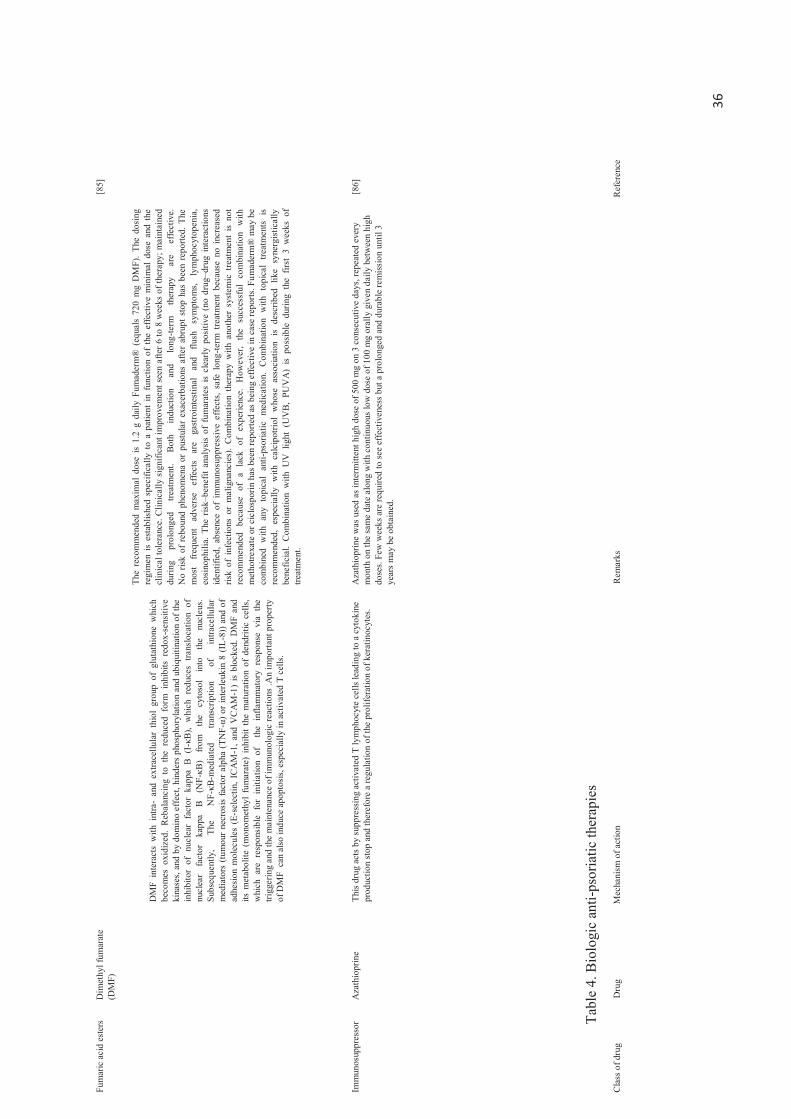
36
Fum
aric
aci
d es
ters
Dim
ethy
l fum
arat
e (D
MF)
DM
F in
tera
cts
with
int
ra-
and
extra
cellu
lar
thio
l gr
oup
of g
luta
thio
ne w
hich
be
com
es o
xidi
zed.
Reb
alan
cing
to
the
redu
ced
form
inh
ibits
red
ox-s
ensi
tive
kina
ses,
and
by d
omin
o ef
fect
, hin
ders
pho
spho
ryla
tion
and
ubiq
uitin
atio
n of
the
inhi
bito
r of
nuc
lear
fac
tor
kapp
a B
(I -
κB),
whi
ch r
educ
es t
rans
loca
tion
of
nucl
ear
fact
or
kapp
a B
(N
F -κB
) fro
m
the
cyto
sol
into
th
e nu
cleu
s.
Subs
eque
ntly
, Th
e N
F -κB
-med
iate
d tra
nscr
iptio
n of
in
trace
llula
r m
edia
tors
(tum
our
necr
osis
fact
or a
lpha
(TN
F -α)
or i
nter
leuk
in 8
(IL-
8)) a
nd o
f ad
hesi
on m
olec
ules
(E -
sele
ctin
, IC
AM
-1, a
nd V
CA
M-1
) is
bloc
ked.
DM
F an
d its
met
abol
ite (
mon
omet
hyl
fum
arat
e) i
nhib
it th
e m
atur
atio
n of
den
driti
c ce
lls,
whi
ch a
re r
espo
nsib
le f
or i
nitia
tion
of
the
infla
mm
ator
y re
spon
se v
ia t
he
trigg
erin
g an
d th
e m
aint
enan
ce o
f im
mun
olog
ic re
actio
ns .A
n im
porta
nt p
rope
rty
of D
MF
can
also
indu
ce a
popt
osis,
esp
ecia
lly in
act
ivat
ed T
cel
ls.
The
reco
mm
ende
d m
axim
al d
ose
is 1.
2 g
daily
Fum
ader
m®
(eq
uals
720
mg
DM
F).
The
dosi
ng
regi
men
is
esta
blis
hed
spec
ifica
lly t
o a
patie
nt i
n fu
nctio
n of
the
effe
ctiv
e m
inim
al d
ose
and
the
clin
ical
tole
ranc
e. C
linic
ally
sig
nific
ant i
mpr
ovem
ent s
een
afte
r 6 to
8 w
eeks
of t
hera
py; m
aint
aine
d du
ring
prol
onge
d tre
atm
ent.
Both
in
duct
ion
and
long
-term
th
erap
y ar
e ef
fect
ive.
N
o ris
k of
reb
ound
phe
nom
ena
or p
ustu
lar
exac
erba
tions
afte
r ab
rupt
sto
p ha
s be
en r
epor
ted.
The
m
ost
freq
uent
ad
vers
e ef
fect
s ar
e ga
stro
inte
stin
al
and
flush
sy
mpt
oms,
lym
phoc
ytop
enia
, eo
sino
phili
a. T
he r
isk–b
enef
it an
alys
is o
f fu
mar
ates
is c
lear
ly p
ositi
ve (
no d
rug–
drug
int
erac
tions
id
entif
ied,
abs
ence
of
imm
unos
uppr
essi
ve e
ffect
s, sa
fe l
ong -
term
tre
atm
ent
beca
use
no i
ncre
ased
ris
k of
inf
ectio
ns o
r m
alig
nanc
ies)
. C
ombi
natio
n th
erap
y w
ith a
noth
er s
yste
mic
tre
atm
ent
is no
t re
com
men
ded
beca
use
of a
la
ck o
f ex
perie
nce.
How
ever
, th
e su
cces
sful
com
bina
tion
with
m
etho
trexa
te o
r cic
losp
orin
has
bee
n re
porte
d as
bei
ng e
ffect
ive
in c
ase
repo
rts. F
umad
erm
® m
ay b
e co
mbi
ned
with
any
top
ical
ant
i -pso
riatic
med
icat
ion.
Com
bina
tion
with
top
ical
tre
atm
ents
is
reco
mm
ende
d, e
spec
ially
with
cal
cipo
triol
who
se a
ssoc
iatio
n is
desc
ribed
lik
e sy
nerg
istic
ally
be
nefic
ial.
Com
bina
tion
with
UV
lig
ht (
UV
B, P
UV
A)
is po
ssib
le d
urin
g th
e fir
st 3
wee
ks o
f tre
atm
ent.
[85]
Imm
unos
uppr
esso
r
Aza
thio
prin
e
This
drug
act
s by
supp
ress
ing
activ
ated
T ly
mph
ocyt
e ce
lls le
adin
g to
a c
ytok
ine
prod
uctio
n st
op a
nd th
eref
ore
a re
gula
tion
of th
e pr
olife
ratio
n of
ker
atin
ocyt
es.
Aza
thio
prin
e w
as u
sed
as in
term
itten
t hig
h do
se o
f 500
mg
on 3
con
secu
tive
days
, rep
eate
d ev
ery
mon
th o
n th
e sa
me
date
alo
ng w
ith c
ontin
uous
low
dos
e of
100
mg
oral
ly g
iven
dai
ly b
etw
een
high
do
ses.
Few
wee
ks a
re re
quire
d to
see
effe
ctiv
enes
s but
a p
rolo
nged
and
dur
able
rem
issi
on u
ntil
3 ye
ars
may
be
obta
ined
.
[86]
Tabl
e 4.
Bio
logi
c an
ti-ps
oria
tic th
erap
ies
Cla
ss o
f dru
g D
rug
Mec
hani
sm o
f act
ion
Rem
arks
R
efer
ence
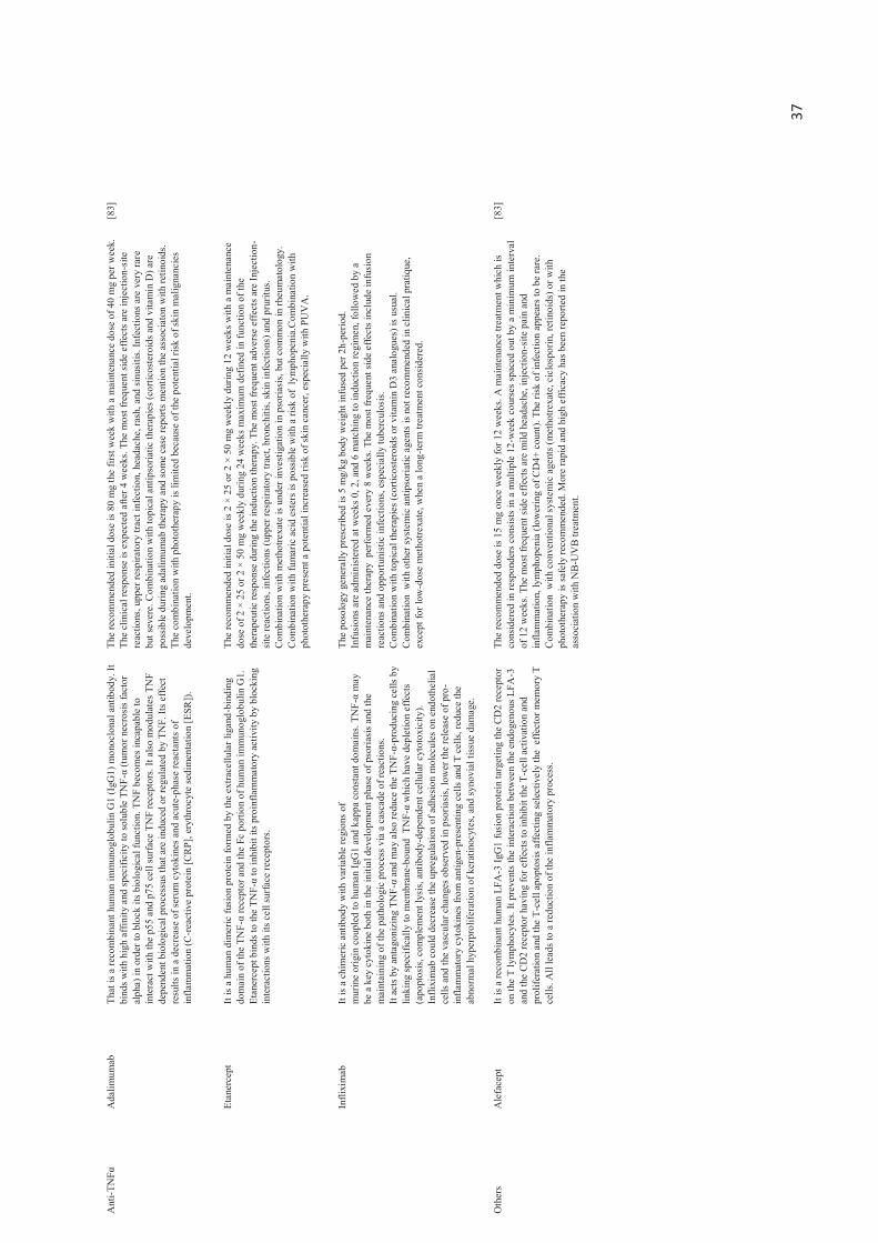
37
Ant
i-TN
Fα
Ada
limum
ab
That
is a
reco
mbi
nant
hum
an im
mun
oglo
bulin
G1
(IgG
1) m
onoc
lona
l ant
ibod
y. It
bi
nds w
ith h
igh
affin
ity a
nd sp
ecifi
city
to so
lubl
e TN
F-α
(tum
or n
ecro
sis
fact
or
alph
a) in
ord
er to
blo
ck it
s bio
logi
cal f
unct
ion.
TN
F be
com
es in
capa
ble
to
inte
ract
with
the
p55
and
p75
cell
surfa
ce T
NF
rece
ptor
s. It
also
mod
ulat
es T
NF
depe
nden
t bio
logi
cal p
roce
ssus
that
are
indu
ced
or re
gula
ted
by T
NF.
Its e
ffect
re
sults
in a
dec
reas
e of
seru
m c
ytok
ines
and
acu
te-p
hase
reac
tant
s of
infla
mm
atio
n (C
-reac
tive
prot
ein
[CR
P], e
ryth
rocy
te se
dim
enta
tion
[ESR
]).
The
reco
mm
ende
d in
itial
dos
e is
80 m
g th
e fir
st w
eek
with
a m
aint
enan
ce d
ose
of 4
0 m
g pe
r wee
k.
The
clin
ical
resp
onse
is e
xpec
ted
afte
r 4 w
eeks
. The
mos
t fre
quen
t sid
e ef
fect
s are
inje
ctio
n-si
te
reac
tions
, upp
er re
spira
tory
trac
t inf
ectio
n, h
eada
che,
rash
, and
sinu
sitis
. Inf
ectio
ns a
re v
ery
rare
bu
t sev
ere.
Com
bina
tion
with
topi
cal a
ntip
soria
tic th
erap
ies (
corti
cost
eroi
ds a
nd v
itam
in D
) are
po
ssib
le d
urin
g ad
alim
umab
ther
apy
and
som
e ca
se re
ports
men
tion
the
asso
ciat
on w
ith re
tinoi
ds.
The
com
bina
tion
with
pho
toth
erap
y is
limite
d be
caus
e of
the
p ote
ntia
l risk
of s
kin
mal
igna
ncie
s de
velo
pmen
t.
[83]
Etan
erce
pt
It is
a hu
man
dim
eric
fusi
on p
rote
in fo
rmed
by
the
extra
cellu
lar l
igan
d-bi
ndin
g do
mai
n of
the
TNF -
α re
cept
or a
nd th
e Fc
por
tion
of h
uman
imm
unog
lobu
lin G
1.
Etan
erce
pt b
inds
to th
e TN
F -α
to in
hibi
t its
pro
infla
mm
ator
y ac
tivity
by
bloc
king
in
ter a
ctio
ns w
ith it
s cel
l sur
face
rece
ptor
s.
The
reco
mm
ende
d in
itial
dos
e is
2 ×
25 o
r 2 ×
50
mg
wee
kly
durin
g 12
wee
ks w
ith a
mai
nten
ance
do
se o
f 2 ×
25
or 2
× 5
0 m
g w
eekl
y du
ring
24 w
eeks
max
imum
def
ined
in fu
nctio
n of
the
ther
apeu
tic re
spon
se d
urin
g th
e in
duct
ion
ther
apy.
The
mos
t fre
quen
t adv
erse
effe
cts a
re In
ject
ion-
site
reac
tions
, inf
ectio
ns (u
pper
resp
irato
ry tr
act,
bron
chiti
s, sk
in in
fect
ions
) and
pru
ritus
. C
ombi
natio
n w
ith m
etho
trexa
te is
und
er in
vest
igat
ion
in p
soria
sis,
but c
omm
on in
rheu
mat
olog
y.
Com
bina
tion
with
fum
aric
aci
d es
ters
is p
ossib
le w
ith a
risk
of
lym
phop
enia
.Com
bina
tion
with
ph
otot
hera
py p
rese
nt a
pot
entia
l inc
reas
ed ri
sk o
f ski
n ca
ncer
, esp
ecia
lly w
ith P
UV
A.
Infli
xim
ab
It is
a ch
imer
ic a
ntib
ody
with
var
iabl
e re
gion
s of
mur
ine
orig
in c
oupl
ed to
hum
an Ig
G1
and
kapp
a co
nsta
nt d
omai
ns. T
NF-
α m
ay
be a
key
cyt
okin
e bo
th in
the
initi
al d
evel
opm
ent p
hase
of p
soria
sis a
nd th
e m
aint
aini
ng o
f the
pat
holo
gic
proc
ess v
ia a
cas
cade
of r
eact
ions
. It
acts
by
anta
goni
zing
TN
F-α
and
may
als
o re
duce
the
TNF-
α-pr
oduc
ing
cells
by
linki
ng sp
ecifi
cally
to m
embr
ane -
boun
d T
NF-
α w
hich
hav
e de
plet
ion
effe
cts
(apo
ptos
is, c
ompl
emen
t lys
is, a
ntib
ody-
depe
nden
t cel
lula
r cyt
otox
icity
). In
flixi
mab
cou
ld d
ecre
ase
the
upre
gula
tion
of a
dhes
ion
mol
ecul
es o
n en
doth
elia
l ce
lls a
nd th
e va
scul
ar c
hang
es o
bser
ved
in p
soria
sis, l
ower
the
rele
ase
of p
ro-
infla
mm
ator
y cy
toki
nes
from
ant
igen
-pre
sent
ing
cells
and
T c
ells,
redu
ce th
e ab
norm
al h
yper
prol
ifera
tion
of k
erat
inoc
ytes
, and
syno
vial
tiss
ue d
amag
e.
The
poso
logy
gen
eral
ly p
resc
ribed
is 5
mg/
kg b
ody
wei
ght i
nfus
ed p
er 2
h-pe
riod.
In
fusi
ons a
re a
dmin
ister
ed a
t wee
ks 0
, 2, a
nd 6
mat
chin
g to
indu
ctio
n re
gim
en, f
ollo
wed
by
a m
aint
enan
ce th
erap
y p
erfo
rmed
eve
ry 8
wee
ks. T
he m
ost f
requ
ent s
ide
effe
cts
incl
ude
infu
sion
re
actio
ns a
nd o
ppor
tuni
stic
infe
ctio
ns, e
spec
ially
tube
rcul
osis.
Com
bina
tion
with
topi
cal t
hera
pies
(cor
ticos
tero
ids o
r vita
min
D3
anal
ogue
s) is
usu
al.
Com
bina
tion
with
oth
er sy
stem
ic a
ntip
soria
tic a
gent
s is
not r
ecom
men
ded
in c
linic
al p
ratiq
ue,
exce
pt fo
r low
-dos
e m
etho
trexa
te, w
hen
a lo
ng-te
rm tr
eatm
ent c
onsi
dere
d.
Oth
ers
Ale
face
pt
It is
a re
com
bina
nt h
uman
LFA
-3 Ig
G1
fusi
on p
rote
in ta
rget
ing
the
CD
2 re
cept
or
on th
e T
lym
phoc
ytes
. It p
reve
nts t
he in
tera
ctio
n be
twee
n th
e en
doge
nous
LFA
-3
and
the
CD
2 re
cept
or h
avin
g fo
r effe
cts t
o in
hibi
t the
T-c
ell a
ctiv
atio
n an
d pr
olife
ratio
n an
d th
e T-
cell
apop
tosi
s affe
ctin
g se
lect
ivel
y th
e e
ffect
or m
emor
y T
cells
. All
lead
s to
a re
duct
ion
of th
e in
flam
mat
ory
proc
ess.
The
reco
mm
ende
d do
se is
15
mg
once
wee
kly
for 1
2 w
eeks
. A m
aint
enan
ce tr
eatm
ent w
hich
is
cons
ider
ed in
resp
onde
rs c
onsi
sts i
n a
mul
t iple
12-
wee
k co
urse
s spa
ced
out b
y a
min
imum
inte
rval
of
12
wee
ks. T
he m
ost f
requ
ent s
ide
effe
cts a
re m
ild h
eada
che,
inje
ctio
n -si
te p
ain
and
infla
mm
atio
n, ly
mph
open
ia (l
ower
ing
of C
D4+
cou
nt).
The
risk
of in
fect
ion
appe
ars t
o be
rare
. C
ombi
natio
n w
ith c
onve
ntio
nal s
yste
mic
age
nts (
met
hotre
xate
, cic
losp
orin
, ret
inoi
ds) o
r with
ph
otot
hera
py is
safe
ly re
com
men
ded.
Mor
e ra
pid
and
high
eff
icac
y ha
s bee
n re
porte
d in
the
asso
ciat
ion
with
NB
-UV
B tre
atm
ent.
[83]
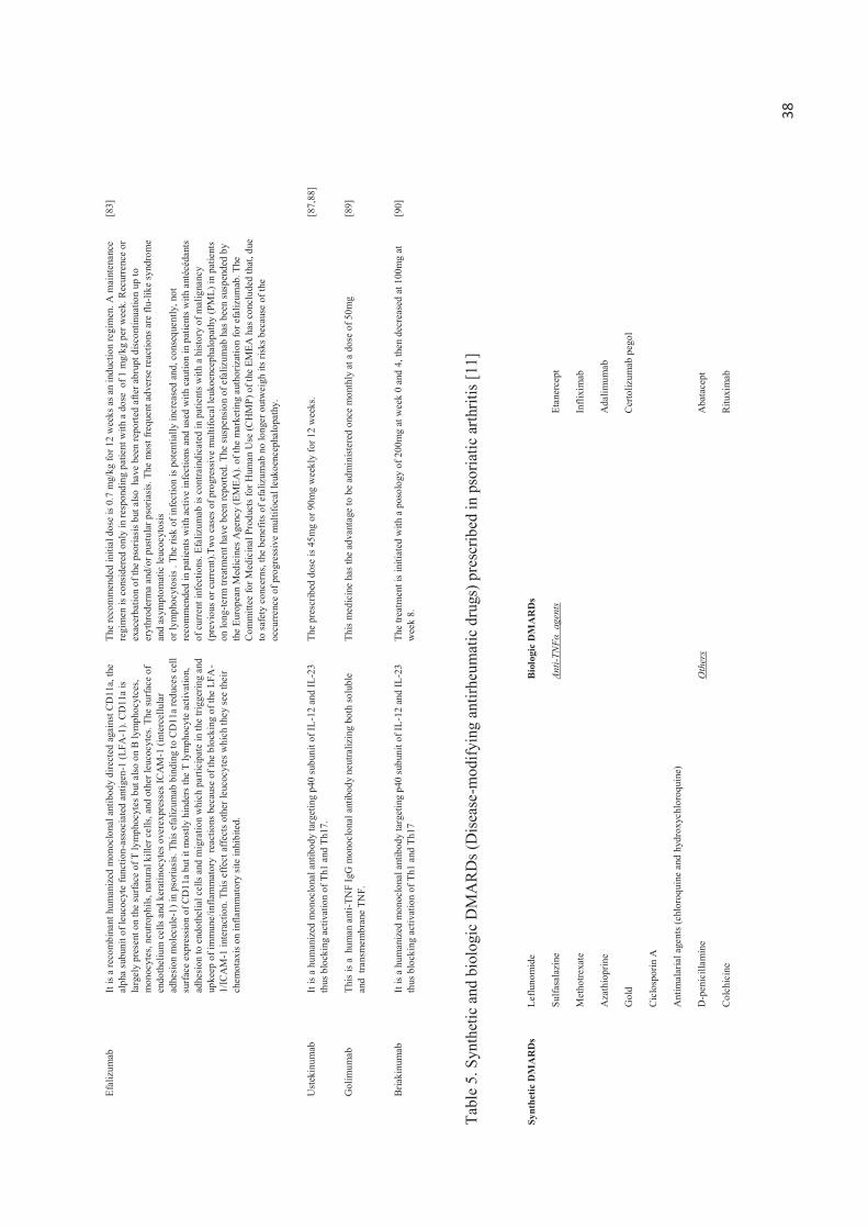
38
Efal
izum
ab
It is
a re
com
bina
nt h
uman
ized
mon
oclo
nal a
ntib
ody
dire
cted
aga
inst
CD
11a,
the
alph
a su
buni
t of l
euco
cyte
func
tion-
asso
ciat
ed a
ntig
en-1
(LFA
-1).
CD
11a
is la
rgel
y pr
esen
t on
the
surfa
ce o
f T ly
mph
ocyt
es b
ut a
lso o
n B
lym
phoc
ytce
s, m
onoc
ytes
, neu
troph
ils, n
atur
al k
iller
cel
ls, a
nd o
ther
leuc
ocyt
es. T
he su
rface
of
endo
thel
ium
cel
ls an
d ke
rati n
ocyt
es o
vere
xpre
sses
ICA
M-1
(int
erce
llula
r ad
hesi
on m
olec
ule-
1) in
pso
riasi
s. Th
is ef
aliz
umab
bin
ding
to C
D11
a re
duce
s cel
l su
rfac
e ex
pres
sion
of C
D11
a bu
t it m
ostly
hin
ders
the
T ly
mph
ocyt
e ac
tivat
ion,
ad
hesi
on to
end
othe
lial c
ells
and
mig
ratio
n w
hich
par
ticip
ate
in th
e tri
gger
ing
and
upke
ep o
f im
mun
e/in
flam
mat
ory
reac
tions
bec
ause
of t
he b
lock
ing
of th
e LF
A-
1/IC
AM
-1 in
tera
ctio
n. T
his e
ffect
affe
cts o
ther
leuc
ocyt
es w
hich
they
see
thei
r ch
emot
axis
on in
flam
mat
ory
site
inhi
bite
d.
The
reco
mm
ende
d in
itial
dos
e is
0.7
mg/
kg fo
r 12
wee
ks a
s an
indu
ctio
n re
gim
en. A
mai
nten
ance
re
gim
en is
con
side
red
only
in re
spon
ding
pat
ient
with
a d
ose
of 1
mg/
kg p
er w
eek.
Rec
urre
nce
or
exac
erba
tion
of th
e ps
oria
sis b
ut a
lso h
ave
been
repo
rted
afte
r abr
upt d
iscon
tinua
tion
up to
er
ythr
oder
ma
and/
or p
ustu
lar p
soria
sis.
The
mos
t fre
quen
t adv
erse
reac
tions
are
flu-
like
synd
rom
e an
d as
ympt
omat
ic le
ucoc
ytos
is
or ly
mph
ocyt
osis
. The
risk
of i
nfec
tion
is po
tent
ially
incr
ease
d an
d, c
onse
quen
tly, n
ot
reco
mm
ende
d in
pat
ient
s with
act
ive
infe
ctio
ns a
nd u
sed
with
cau
tion
in p
atie
nts w
ith a
ntéc
édan
ts
of c
urre
nt in
fect
ions
. Efa
lizum
ab is
con
train
dica
ted
in p
atie
nts w
ith a
hist
ory
of m
alig
nanc
y (p
revi
ous o
r cur
rent
).Tw
o ca
ses o
f pro
gres
sive
mul
tifoc
al le
ukoe
ncep
halo
path
y (P
ML)
in p
atie
nts
on lo
ng-te
rm tr
eatm
ent h
ave
been
repo
rted.
The
sus
pens
ion
of e
faliz
umab
has
bee
n su
spen
ded
by
the
Euro
pean
Med
icin
es A
genc
y (E
MEA
). of
the
mar
ketin
g au
thor
izat
ion
for e
faliz
umab
. The
C
omm
ittee
for M
edic
inal
Pro
duct
s fo
r Hum
an U
se (C
HM
P) o
f the
EM
EA h
as c
oncl
uded
that
, due
to
safe
ty c
once
rns,
the
bene
fits o
f efa
lizum
ab n
o lo
nger
out
wei
gh it
s risk
s bec
ause
of t
he
occu
rrenc
e of
pro
gres
sive
mul
tifoc
al le
ukoe
ncep
halo
path
y.
[83]
Ust
ekin
umab
It
is a
hum
aniz
ed m
onoc
lona
l ant
ibod
y ta
rget
ing
p40
subu
nit o
f IL-
12 a
nd IL
-23
thus
blo
ckin
g ac
tivat
ion
of T
h1 a
nd T
h17.
Th
e pr
escr
ibed
dos
e is
45m
g or
90m
g w
eekl
y fo
r 12
wee
ks.
[87,
88]
Gol
imum
ab
This
is a
hum
an a
nti-T
NF
IgG
mon
oclo
nal a
ntib
ody
neut
raliz
ing
both
solu
ble
and
tran
smem
bran
e TN
F.
This
med
icin
e ha
s the
adv
anta
ge to
be
adm
inist
ered
onc
e m
onth
ly a
t a d
ose
of 5
0mg
[89]
Bria
kinu
mab
It
is a
hum
aniz
ed m
onoc
lona
l ant
ibod
y ta
rget
ing
p40
subu
nit o
f IL-
12 a
nd IL
-23
thus
blo
ckin
g ac
tivat
ion
of T
h1 a
nd T
h17
The
treat
men
t is
initi
ated
with
a p
osol
ogy
of 2
00m
g at
wee
k 0
and
4, th
en d
ecre
ased
at 1
00m
g at
w
eek
8.
[90]
Tabl
e 5.
Syn
thet
ic a
nd b
iolo
gic
DM
AR
Ds (
Dis
ease
-mod
ifyin
g an
tirhe
umat
ic d
rugs
) pre
scrib
ed in
pso
riatic
arth
ritis
[11]
Synt
hetic
DM
AR
Ds
Leflu
nom
ide
Bio
logi
c D
MA
RD
s
Su
lfasa
lazi
ne
Anti-
TNFα
age
nts
Etan
erce
pt
M
etho
trexa
te
In
flixi
mab
A
zath
iopr
ine
Ada
limum
ab
G
old
C
erto
lizum
ab p
egol
C
iclo
spor
in A
A
ntim
alar
ial a
gent
s (ch
loro
quin
e an
d hy
drox
ychl
oroq
uine
)
D
-pen
icill
amin
e O
ther
s A
bata
cept
C
olch
icin
e
Ritu
xim
ab

39
Et
retin
ate
U
stek
inum
ab
PU
VA
ther
apy
Se
cuki
num
ab (a
nti-I
L-17
mon
oclo
nal a
ntib
ody)
A
prem
ilast
Brod
alum
ab (a
nti-I
L-17
rece
ptor
mon
oclo
nal a
ntib
ody)
40
This treatment can be used in combination with topical treatments (emollients or retinoids) to promote the penetration of UVB. These rays are very erythematic and impose a strict surveillance. UVA phototherapy is also used almost systematically associated with psoralens (photo-sensitizing agents). This association is commonly called photochemotherapy or PUVA. However, they are considered as second line treatment because of their contraindications (hypersensitivity to light, cataracts, liver / renal failure)[9].
4.3 Systemic therapy Systemic treatments are considered as the last resort to face to the failure of previous treatments. These products are very powerful and requires clinical follow-up because of these severe side effects.
4.3.1 Conventional therapy Systemic corticosteroids are strictly prohibited due to the risk of rebound worsening of the initial condition. Two drugs are usually prescribed at this level of management: methotrexate and cyclosporin A. Methotrexate (an analogue of folic acid) acts as a competitive inhibitor of an enzyme essential for DNA synthesis, the dihydrofolate reductase, which explains the inhibitory effect of cell proliferation. However, many significant side effects have been reported such as nausea and vomiting, especially the liver and lung toxicity [91]. Cyclosporin A is an immunosuppressant widely used in the field of transplantation and autoimmune diseases. This cyclic peptide can form a complex with cyclophilin, intracellular immunophilin, inhibiting the phosphatase activity of calcineurin. This results in a depletion of macrophages and lymphocytes and inhibition of the activation of T cells, NK cells and antigen presenting cells. Moreover, keratinocyte proliferation and histamine release of mast cells is also inhibited. In addition, cyclosporin is also the source of a negative regulation of the expression of cell adhesion molecules in the endothelium of the dermis capillaries [92]. However, cyclosporin A is also at the origin of major adverse effects that prevent its use in high doses and / or long-term [93]. Other medicines can be also considered in case of inefficiency or intolerance (Table 3).
4.3.2 Biologic therapy
The considerable advances in recent years in the field of immunopathology and biotechnology have allowed an evolution of the market with new treatment possibilities: biological agents (Table 5). In the case of psoriasis, these new therapies target T cells in order to address the underlying causes of clinical manifestations. However, they are not healing therapies. These biological agents are still little known about their tolerance and their adverse effects. It is usually antibodies or fusion proteins targeting specific markers involved in the activation and / or proliferation of T cells (cytokines, antigen presenting cells such as dendritic cells). It is important to remember that these biologics are very expensive, their effectiveness cannot be guaranteed and long-term use is not recommended due to their immunosuppressive potential (risk of infections, lymphomas , ect) [5].
4.4 Psoriatic arthritis therapies The therapeutic strategy is based on three conventional drug families: NSAIDs, glucocorticoids, DMARDs (synthetic and biological) in function of the phase of the illness (Figure 5). NSAID (ibuprofene, nimésulide,ect) is the first-line treatment in patients with peripheral psoriatic arthritis. The efficacy has been reported in two studies showing significant clinical improvement in inflammed joints. However, no positive effect was
41
observed in skin lesions [94,95]. Local treatments are advised in patients with limited number of entheses or joints [96] but usually the treatment is prescribed orally. In general, systemic glucocorticoids (methylprednisolone, prednisone) are prescribed orally in polyarticular involvement based essentially on expert opinion with an enhancement can reach 2 months [97]. Optionally, glucocorticoids may be injected directly into affected joints. Synthetic DMARDs can be initiated in case of lack efficiency with NSAIDs to treat oligo- or polyarticular psoriatic arthritis. It is largely recommended to promote synthetic DMARDs acting on skin lesions and psoriatic arthritis in patients exhibiting the two clinical manifestations like cyclosporine and methotrexate instead of sulfasalazine and leflunomide which have limited effectiveness to manage the skin aspect of psoriasis [98]. Biological DMARDs offer a broadening of prospects in the treatment of PsA. This drug class is mainly represented by anti-TNFα agents. Clinical and radiological enhancement is now largely recognized and the benefit/risk ratio is considered by health professionals as positive in overall. Several studies have reported that biological therapy must be initiated precociously and aggressively for a better clinical outcome in PsA, and that recurrence is frequent after discontinuation [64].
4.5. Innovative skin drug delivery systems (ISDDSs)
At present, the drug array available for psoriasis management allows to consider different strategies according to the therapeutic objectives. One of the main lines of research directed towards the discovery of new therapies consists to develop new monoclonal antibodies that target key elements in the pathogenesis of psoriasis such as cytokines like Ustekinumab or Golimumab. Some clinical trials are frequently conducted in order to position these new biological therapies in psoriasis management strategy. However, another axis is also considered by the research teams oriented towards an enrichment of the therapeutic arsenal by leveraging advances in the field of encapsulation to offer innovative skin drug delivery systems (ISDDS) improving dermal and / or transdermal penetration of already-known active agents but whose use remains cautious because of the associated side effects.
4.5.1 Nanoencapsulation The encapsulation consists in enclosing active agents in a nano- or micro-sized particles. The encapsulation of active agents into nanoparticles formulation improves aesthetics, protects active agents against degredation, helps to target skin layers and prolongs drug release, offers several other advantages as a better patient compliance with a non-invasive administration, especially in case of chronic pathology, and a reduction of side effects and therefore an improvement of the benefice/risk ratio [99] [100] [101].
Many nanoparticles have been formulated to increase percutaneous absorption of active agents without damaging the skin barrier function (Fig.3). They may be composed by biodegradable polymers/copolymer (PLA, PLGA, PCL, chitosan, ect.) or lipids known to be biocompatible with a human use [102] [103] [104]. These nanoparticles developed for skin delivery can be classified in two common types of particles: polymeric nanoparticles (microsphere/nanosphere, microcapsule/nanocapsule, micelle, niosome, hydrogel) and lipid nanoparticles (solid lipid nanoparticle (SLN), nanostructured lipid carrier (NLC), microemulsion/nanoemulsion, liposome, ethosome, transfersome), each of them are formed by self-assembly or not (Table 6). However, the actual trend is to improve the encapsulation technologies by adding the properties of nanoparticles in order to obtain the better
42
characteristics in function of the final application. For example, lipid nanoparticles are frequently incorporated in hydrogel in order to enhance the stability of the formulation and increase the time residence at the topical action site via a higher viscosity of the vehicles [105,106]. Another example is to incorporate polymeric nanoparticles like cyclodextrin inclusion complex in lipid nanoparticles like liposome or SLN [107,108] or coat lipid nanoparticles with a polymer like microemulsions coated with chitosan [109]. The submicron size and spherical shape of the particles can increase the interfacial area favoring the multiplication of interactions with the skin tissue and thus increase the skin permeation. In function of the formulation and the size of the nanoparticles, it has been reported a targeting effect of the skin sublayers especially the epidermis, the pathogenesis site of psoriasis [110–113].
Moreover, we can see those later years several studies dealing with the use of siRNA gene therapy as new approach to treat such skin disease involving many complex interactions[178,179]. Gene therapy using siRNA is a post-transcriptional silencing process hindering the expression of a specific gene. These small interfering RNAs (siRNA) are double-stranded RNA molecules capable to interfere with the expression of particular genes thanks to their complementary with the nucleotide sequences leading to mRNA degradation after transcription thus blocking the translation step. This promising gene therapy approach can be a great answer to difficulties met in the psoriasis management targeting TNF α which activates chronic inflammation and stat3 which activates keratinocyte proliferation[180]. To date, such therapies are not among the armamentarium used in routine because siRNA delivery does not allow currently ensuring effective and safe of a clinical use. ISDDSs carrying siRNA appears as an interesting solution to consider a topical treatment of psoriasis. We have found in literature (see Table 6) that liquid crystalline nanoparticles[179] and nucleic acid lipid nanoparticles (NALPs)[180] are usually used to encapsulate siRNA but not only. Liquid crystalline nanoparticles have been also reported to encapsulate drugs like cyclosporine A[181] and tacrolimus[182]. Their functionalization with cell-penetrating peptides (CPPs) is a strategy used to promote the skin cell uptake across the plasma membrane of charged and non-charged oligonucleotides[183].
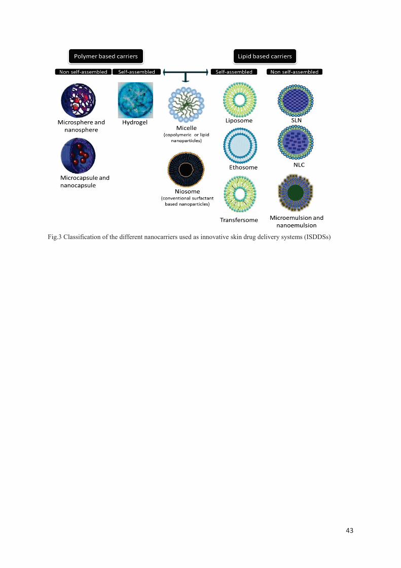
43
Fig.3 Classification of the different nanocarriers used as innovative skin drug delivery systems (ISDDSs)
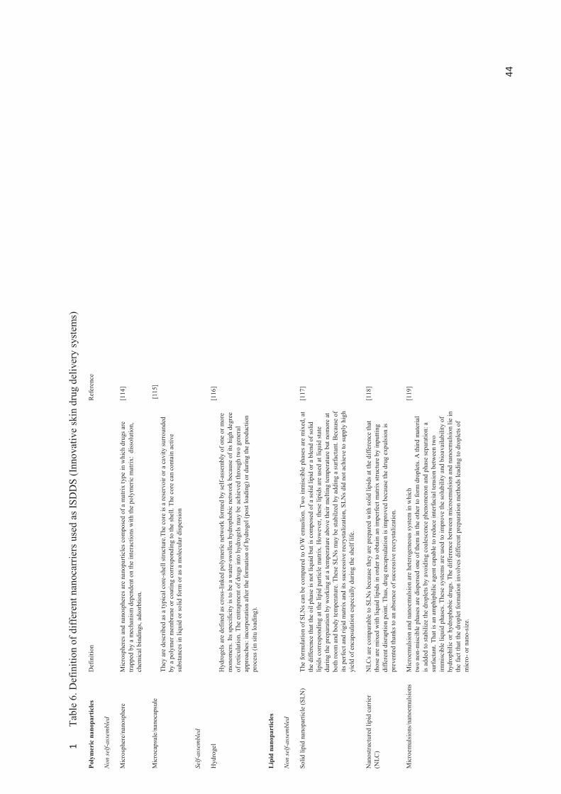
44
Tabl
e 6.
Def
initi
on o
f diff
eren
t nan
ocar
riers
use
d as
ISD
DS
(Inno
vativ
e sk
in d
rug
deliv
ery
syst
ems)
1
Poly
mer
ic n
anop
artic
les
Def
initi
on
Ref
eren
ce
Non
self-
asse
mbl
ed
Mic
rosp
here
/nan
osph
ere
Mic
rosp
here
s and
nan
osph
eres
are
nan
opar
ticle
s com
pose
d of
a m
atrix
type
in w
hich
dru
gs a
re
trapp
ed b
y a
mec
hani
sm d
epen
dent
on
the
inte
ract
ions
with
the
poly
mer
ic m
atrix
: di
ssol
utio
n,
chem
ical
bin
ding
s, ad
sorb
tion.
[114
]
Mic
roca
psul
e/na
noca
psul
e Th
ey a
re d
escr
ibed
as a
typi
cal c
ore-
shel
l stru
ctur
e.Th
e co
re is
a re
serv
oir o
r a c
avity
surr
ound
ed
by a
pol
ymer
mem
bran
e or
coa
ting
corr
espo
ndin
g to
the
shel
l. Th
e co
re c
an c
onta
in a
ctiv
e su
bsta
nces
in li
quid
or s
olid
form
or a
s a m
olec
ular
disp
ersi
on
[115
]
Self-
asse
mbl
ed
Hyd
roge
l H
ydro
gels
are
defin
ed a
s cro
ss-li
nked
pol
ymer
ic n
etw
ork
form
ed b
y se
lf-as
sem
bly
of o
ne o
r mor
e m
onom
ers.
Its sp
ecifi
city
is to
be
a w
ater
-sw
olle
n hy
drop
hobi
c ne
twor
k be
caus
e of
its
high
deg
ree
of re
ticul
atio
n. T
he e
ntra
pmen
t of d
rugs
into
hyd
roge
ls m
ay b
e ac
hiev
ed th
roug
h tw
o ge
nera
l ap
proa
ches
: inc
orpo
ratio
n af
ter t
he fo
rmat
ion
of h
ydro
gel (
post
load
ing)
or d
urin
g th
e pr
oduc
tion
proc
ess (
in s
itu lo
adin
g).
[116
]
Lip
id n
anop
artic
les
Non
self-
asse
mbl
ed
Solid
lipi
d na
nopa
rticl
e (S
LN)
The
form
ulat
ion
of S
LNs c
an b
e co
mpa
red
to O
/W e
mus
lion.
Tw
o in
mis
cibl
e ph
ases
are
mix
ed, a
t th
e di
ffere
nce
that
the
oil p
hase
is n
ot li
quid
but
is c
ompo
sed
of a
solid
lipi
d or
a b
lend
of s
olid
lip
ids c
orre
spon
ding
at t
he li
pid
parti
cle
mat
rix. H
owev
er, t
hese
lipi
ds a
re u
sed
at li
quid
stat
e du
ring
the
prep
arat
ion
by w
orki
ng a
t a te
mpe
ratu
re a
bove
thei
r mel
ting
tem
pera
ture
but
nom
ore
at
both
room
and
bod
y te
mpe
ratu
re. T
hese
SLN
s m
ay b
e st
abilz
ed b
y ad
ding
a s
urfa
ctan
t. Be
caus
e of
its
per
fect
and
rigi
d m
atrix
and
its s
ucce
ssiv
e re
crys
taliz
atio
n, S
LNs d
id n
ot a
chie
ve to
supp
ly h
igh
yiel
d of
enc
apsu
latio
n es
peci
ally
dur
ing
the
shel
f life
.
[117
]
Nan
ostru
ctur
ed li
pid
carr
ier
(NLC
) N
LCs a
re c
ompa
rabl
e to
SLN
s bec
ause
they
are
pre
pare
d w
ith so
lid li
pids
at t
he d
iffer
ence
that
th
ose
are
mix
ed w
ith li
quid
lipi
ds in
ord
er to
obt
ain
an im
perfe
ct m
atrix
stru
ctur
e by
inpu
tting
di
ffere
nt d
isrup
tion
poin
t. Th
us, d
rug
enca
psul
atio
n is
impr
oved
bec
ause
the
drug
exp
ulsi
on is
pr
even
ted
than
ks to
an
abse
nce
of su
cces
sive
recr
ysta
lizat
ion.
[118
]
Mic
roem
ulsi
ons/
nano
emul
sion
s M
icro
emul
sion
and
nan
oem
ulsi
on a
re h
eter
ogen
eous
sys
tem
in w
hich
tw
o no
n -m
iscib
le p
hase
s are
disp
erse
d on
e of
them
in th
e ot
her t
o fo
rm d
ropl
ets.
A th
ird m
ater
ial
is ad
ded
to st
abili
ze th
e dr
ople
ts b
y av
oidi
ng c
oale
scen
ce p
heno
men
on a
nd p
hase
sepa
ratio
n: a
su
rfac
tant
. Tha
t is a
n am
phip
hilic
age
nt c
apab
le to
redu
ce in
terfa
cial
tens
ion
betw
een
two
imm
isci
ble
liqui
d ph
ases
. The
se sy
stem
s are
use
d to
impr
ove
the
solu
bilit
y an
d bi
oava
ilabi
lity
of
hydr
ophi
lic o
r hyd
roph
obic
dru
gs. T
he d
iffer
ence
bet
wee
n m
icro
emul
sion
and
nan
oem
ulsi
on li
e in
th
e fa
ct th
at th
e dr
ople
t for
mat
ion
invo
lves
diff
eren
t pre
para
tion
met
hods
lead
ing
to d
ropl
ets o
f m
icro
- or n
ano-
size
.
[119
]
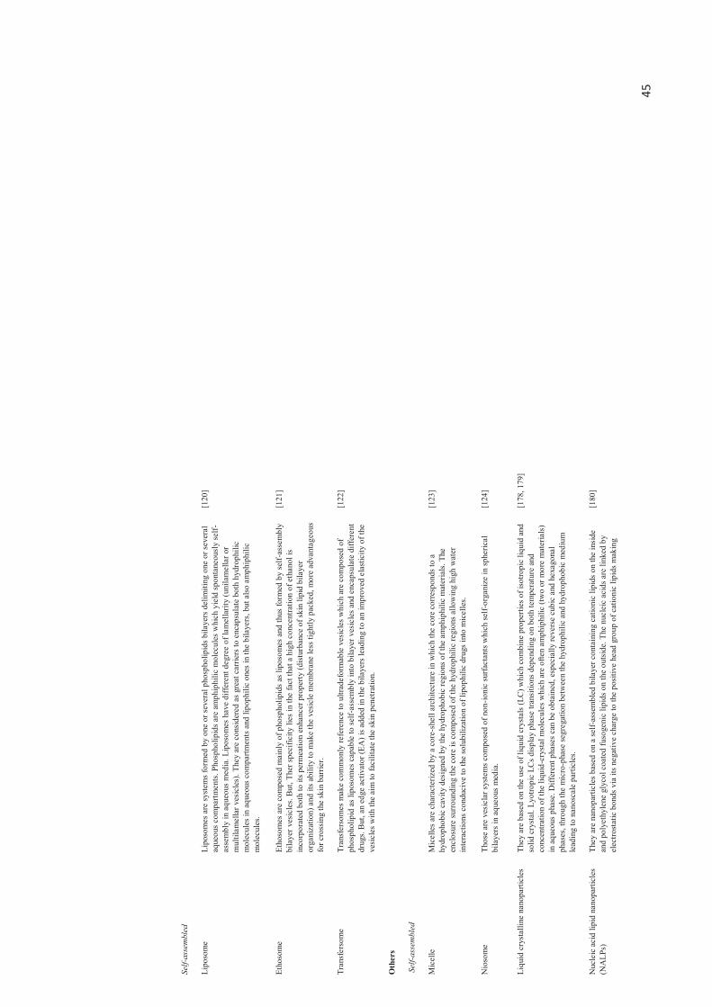
45
Self-
asse
mbl
ed
Lipo
som
e Li
poso
mes
are
syst
ems
form
ed b
y on
e or
seve
ral p
hosp
holip
ids b
ilaye
rs d
elim
iting
one
or s
ever
al
aque
ous c
ompa
rtmen
ts. P
hosp
holip
ids a
re a
mph
iphi
lic m
olec
ules
whi
ch y
ield
spon
tane
ousl
y se
lf-as
sem
bly
in a
queo
us m
edia
. Lip
osom
es h
ave
diffe
rent
deg
ree
of la
mel
larit
y (u
nila
mel
lar o
r m
ultil
amel
lar v
esic
les)
. The
y ar
e co
nsid
ered
as g
reat
car
riers
to e
ncap
sula
te b
oth
hydr
ophi
lic
mol
ecul
es in
aqu
eous
com
partm
ents
and
lipo
phili
c on
es in
the
bila
yers
, but
also
am
phip
hilic
m
olec
ules
.
[120
]
Etho
som
e Et
hoso
mes
are
com
pose
d m
ainl
y of
pho
spho
lipid
s as l
ipos
omes
and
thus
form
ed b
y se
lf-as
sem
bly
bila
yer v
esic
les.
But,
Ther
spec
ifici
ty li
es in
the
fact
that
a h
igh
conc
entra
tion
of e
than
ol is
in
corp
orat
ed b
oth
to it
s per
mea
tion
enha
ncer
pro
perty
(dist
urba
nce
of sk
in li
pid
bila
yer
orga
niza
tion)
and
its a
bilit
y to
mak
e th
e ve
sicle
mem
bran
e le
ss ti
ghtly
pac
ked,
mor
e ad
vant
ageo
us
for c
ross
ing
the
skin
bar
rier.
[121
]
Tran
sfer
som
e Tr
ansf
erso
mes
mak
e co
mm
only
refe
renc
e to
ultr
adef
orm
able
ves
icle
s whi
ch a
re c
ompo
sed
of
phos
phol
ipid
as l
ipos
omes
cap
able
to se
lf-as
sem
bly
into
bila
yer v
esic
les a
nd e
ncap
sula
te d
iffer
ent
drug
s. B
ut, a
n ed
ge a
ctiv
ator
(EA
) is a
dded
in th
e bi
laye
rs le
adin
g to
an
impr
oved
ela
stic
ity o
f the
ve
sicl
es w
ith th
e ai
m to
faci
litat
e th
e sk
in p
enet
ratio
n.
[122
]
Oth
ers
Sel
f-ass
embl
ed
Mic
elle
M
icel
les a
re c
hara
cter
ized
by
a co
re-s
hell
arch
itect
ure
in w
hich
the
core
cor
resp
onds
to a
hy
drop
hobi
c ca
vity
des
igne
d by
the
hydr
opho
bic
regi
ons o
f the
am
phip
hilic
mat
eria
ls. T
he
encl
osur
e su
rrou
ndin
g th
e co
re is
com
pose
d of
the
hydr
ophi
lic re
gion
s allo
win
g hi
gh w
ater
in
tera
ctio
ns c
ondu
cive
to th
e so
lubi
lizat
ion
of li
poph
ilic
drug
s in
to m
icel
les.
[123
]
Nio
som
e Th
ose
are
vesi
clar
sys
tem
s com
pose
d of
non
-ioni
c su
rfact
ants
whi
ch se
lf-or
gani
ze in
sphe
rical
bi
laye
rs in
aqu
eous
med
ia.
[124
]
Liqu
id c
ryst
allin
e na
nopa
rticl
es
They
are
bas
ed o
n th
e us
e of
liqu
id c
ryst
als
(LC
) whi
ch c
ombi
ne p
rope
rties
of i
sotro
pic
liqui
d an
d so
lid c
ryst
al. L
yotro
pic
LCs d
ispla
y ph
ase
trans
ition
s dep
endi
ng o
n bo
th te
mpe
ratu
re a
nd
conc
entra
tion
of th
e liq
uid -
crys
tal m
olec
ules
whi
ch a
re o
ften
amph
iphi
lic (t
wo
or m
ore
mat
eria
ls)
in a
q ueo
us p
hase
. Diff
eren
t pha
ses c
an b
e ob
tain
ed, e
spec
ially
reve
rse
cubi
c an
d he
xago
nal
phas
es, t
hrou
gh th
e m
icro
-pha
se se
greg
atio
n be
twee
n th
e hy
drop
hilic
and
hyd
roph
obic
med
ium
le
adin
g to
nan
osca
le p
artic
les.
[178
, 179
]
Nuc
leic
aci
d lip
id n
anop
artic
les
(NA
LPs)
Th
ey a
re n
anop
artic
les b
ased
on
a se
lf-as
sem
bled
bila
yer c
onta
inin
g ca
tioni
c lip
ids o
n th
e in
side
an
d po
lyet
hyle
ne g
lyco
l coa
ted
fuso
geni
c lip
ids o
n th
e ou
tsid
e. T
he n
ucle
ic a
cids
are
link
ed b
y el
ectro
stat
ic b
onds
via
its n
egat
ive
char
ge to
the
posi
tive
head
gro
up o
f cat
ioni
c lip
ids m
akin
g
[180
]
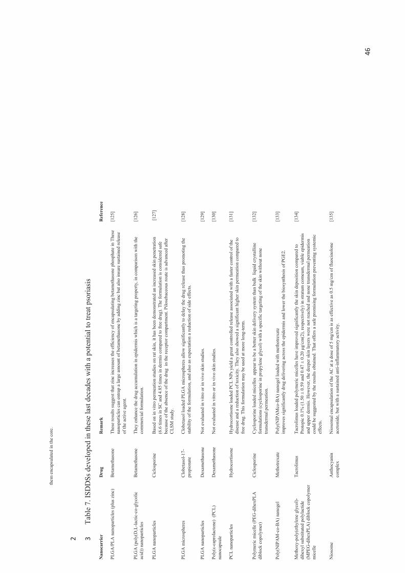
46
them
enc
apsu
late
d in
the
core
.
2
Tabl
e 7.
ISD
DSs
dev
elop
ed in
thes
e la
st d
ecad
es w
ith a
pot
entia
l to
treat
pso
riasi
s 3
Nan
ocar
rier
D
rug
Rem
ark
Ref
eren
ce
PLG
A/P
LA n
anop
artic
les (
plus
zin
c)
Beta
met
haso
ne
Thes
e re
sults
sug
gest
that
zin
c in
crea
ses t
he e
ffici
ency
of e
ncap
sula
ting
beta
met
haso
ne p
hosp
hate
in T
hese
na
nopa
rticl
es c
an e
ntra
p a
larg
e am
ount
of b
etam
etha
sone
by
addi
ng z
inc
but a
lso in
sure
sust
aine
d re
leas
e of
the
activ
e ag
ent.
[125
]
PLG
A (p
oly(
D,L
-lact
ic-c
o-gl
ycol
ic
acid
)) na
nopa
rticl
es
Beta
met
haso
ne
They
enh
ance
the
drug
acc
umul
atio
n in
epi
derm
is w
hich
is a
targ
etin
g pr
oper
ty, i
n co
mpa
rison
with
the
com
mer
cial
form
ulat
ion.
[1
26]
PLG
A n
anop
artic
les
Cic
losp
orin
e Ba
sed
on in
vitr
o pe
rmea
tion
stud
ies o
n ra
t ski
n, it
has
bee
n de
mon
stra
ted
an in
crea
sed
skin
pen
etra
tion
(6.6
tim
es in
SC
and
4.8
5 tim
es in
der
mis
com
pare
d to
free
dru
g). T
he fo
rmul
atio
n is
cons
ider
ed sa
fe
beca
use
of th
e ab
senc
e of
the
drug
in
the
rece
pto r
com
partm
ent.
Pilo
seba
ceou
s rou
te is
adv
ance
d af
ter
CLS
M st
udy.
[127
]
PLG
A m
icro
sphe
res
Clo
beta
sol-1
7-pr
opio
nate
C
lobe
taso
l loa
ded
PLG
A m
icro
sphe
res a
llow
sign
ifica
ntly
to d
elay
the
drug
rele
ase
thus
pro
mot
ing
the
stab
ility
of t
he fo
rmul
atio
n, a
nd a
lso a
s exp
ecta
tion
a re
duct
ion
of s
ide
effe
cts.
[1
28]
PLG
A n
anop
artic
les
Dex
amet
haso
ne
Not
eva
luat
ed in
vitr
o or
in v
ivo
skin
stud
ies.
[129
]
Poly
(ε-c
apro
lact
one)
(PC
L)
nano
caps
ule
Dex
amet
haso
ne
Not
eva
luat
ed in
vitr
o or
in v
ivo
skin
stud
ies.
[130
]
PCL
nano
parti
cles
H
ydro
corti
sone
H
ydro
corti
sone
load
ed P
CL
NPs
yie
ld a
gre
at c
ontro
lled
rele
ase
asso
ciat
ed w
ith a
fast
er c
ontro
l of t
he
dise
ase
and
a re
duct
ion
of to
xici
ty. T
hey
also
show
ed a
sign
ifica
nt h
ighe
r ski
n pe
rmea
tion
com
pare
d to
fr
ee d
rug.
Thi
s fo
rmul
atio
n m
ay b
e us
ed a
t mor
e lo
ng-te
rm.
[131
]
Poly
mer
ic m
icel
le (P
EG-d
ihex
PLA
di
bloc
k co
poly
mer
) C
iclo
spor
ine
Cyc
losp
orin
e lo
aded
mic
elle
s ap
pear
to b
e a
bette
r ski
n de
liver
y sy
stem
than
bul
k li
quid
cry
stal
line
form
ulat
ions
(cyc
losp
orin
e in
pro
pyle
ne g
lyco
l) w
ith a
spec
ific
targ
etin
g of
the
skin
with
out n
one
trans
derm
al p
erm
eatio
n.
[132
]
Poly
(NIP
AM
-co-
BA
) nan
ogel
M
etho
trexa
te
Poly
(NIP
AM
co-B
A) n
anog
el lo
aded
with
met
hotre
xate
im
prov
es s
igni
fican
tly d
rug
deliv
erin
g ac
ross
the
epid
erm
is a
nd lo
wer
the
bios
ynth
esis
of P
GE2
. [1
33]
Met
hoxy
-pol
y(et
hyle
ne g
lyco
l)-di
hexy
l sub
stitu
ted
poly
lact
ide
(MPE
G-d
ihex
PLA
) dib
lock
cop
olym
er
mic
elle
Tacr
olim
us
Tacr
olim
us lo
aded
pol
ymer
ic m
icel
les
have
impr
oved
sig
nific
antly
the
skin
dep
ositi
on c
ompa
red
to
Prot
opic
0.1
% (1
.50
± 0.
59 a
nd 0
.47
± 0.
20 μ
g/cm
(2),
resp
ectiv
ely)
in st
ratu
m c
orne
um, v
iabl
e ep
ider
mis
an
d up
per d
erm
is. H
owev
er, t
he d
eepe
r ski
n la
yers
wer
e no
t rea
ched
and
non
e tra
nsde
rmal
per
mea
tion
coul
d be
sugg
este
d by
the
resu
lts o
btai
ned.
Tha
t offe
rs a
safe
pro
mis
ing
form
ulat
ion
prev
entin
g sy
stem
ic
effe
cts.
[134
]
Nio
som
e A
ntho
cyan
in
com
plex
N
ioso
mal
enc
apsu
latio
n of
the
AC
at a
dos
e of
5 m
g/cm
is a
s effe
ctiv
e as
0.5
mg/
cm o
f flu
ocin
olon
e ac
eton
ide,
but
with
a su
stai
ned
anti-
infla
mm
ator
y ac
tivity
. [1
35]
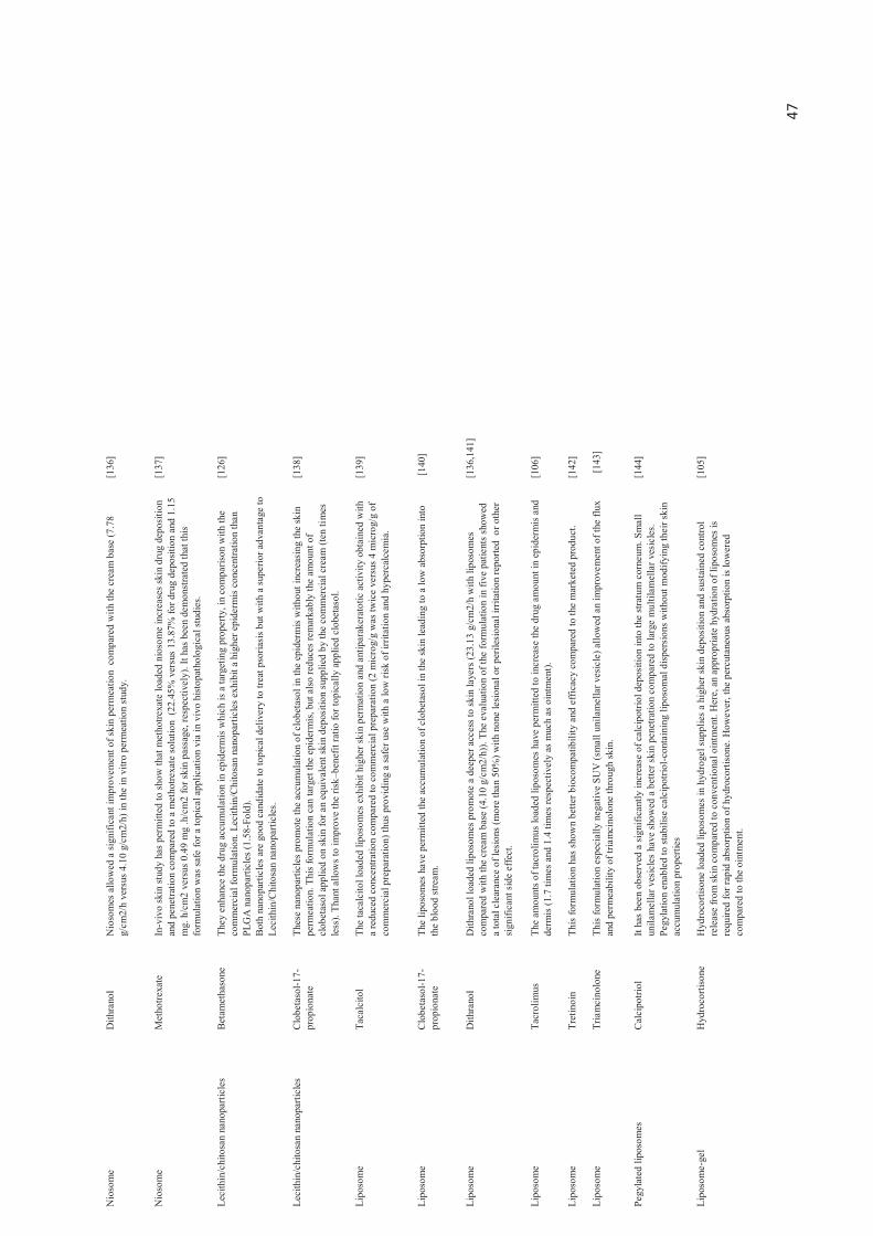
47
Nio
som
e D
ithra
nol
Nio
som
es a
llow
ed a
sign
ifica
nt im
prov
emen
t of s
kin
perm
eatio
n c
ompa
red
with
the
crea
m b
ase
(7.7
8 g/
cm2/
h ve
rsus
4.1
0 g/
cm2/
h) in
the
in v
itro
perm
eatio
n st
udy.
[1
36]
Nio
som
e M
etho
trexa
te
In-v
ivo
skin
stud
y ha
s per
mitt
ed to
show
that
met
hotre
xate
load
ed n
ioso
me
incr
ease
s ski
n dr
ug d
epos
ition
an
d pe
netra
tion
com
pare
d to
a m
etho
trexa
te so
lutio
n (2
2.45
% v
ersu
s 13.
87%
for d
rug
depo
sitio
n an
d 1.
15
mg.
h/c
m2
vers
us 0
.49
mg
.h/c
m2
for s
kin
pass
age,
resp
ectiv
ely)
. It h
as b
een
dem
onst
rate
d th
at th
is fo
rmul
atio
n w
as sa
fe fo
r a to
pica
l app
licat
ion
via
in v
ivo
hist
opat
holo
gica
l stu
dies
.
[137
]
Leci
thin
/chi
tosa
n na
nopa
rticl
es
Beta
met
haso
ne
They
enh
ance
the
drug
acc
umul
atio
n in
epi
derm
is w
hich
is a
targ
etin
g pr
oper
ty, i
n co
mpa
rison
with
the
com
mer
cial
form
ulat
ion.
Lec
ithin
/Chi
tosa
n na
nopa
rticl
es e
xhib
it a
high
er e
pide
rmis
con
cent
ratio
n th
an
PLG
A n
anop
artic
les (
1.58
-Fol
d).
Both
nan
opar
ticle
s ar
e go
od c
andi
date
to to
pica
l del
iver
y to
trea
t pso
riasi
s but
with
a su
perio
r adv
anta
ge to
Le
cith
in/C
hito
san
nano
parti
cles
.
[126
]
Leci
thin
/chi
tosa
n na
nopa
rticl
es
Clo
beta
sol-1
7-pr
opio
nate
Th
ese
nano
parti
cles
pro
mot
e th
e ac
cum
ulat
ion
of c
lobe
taso
l in
the
epid
erm
is w
ithou
t inc
reas
ing
the
skin
pe
rmea
tion.
Thi
s fo
rmul
atio
n ca
n ta
rget
the
epid
erm
is, b
ut a
lso re
duce
s rem
arka
bly
the
amou
nt o
f cl
obet
asol
app
lied
on sk
in fo
r an
equi
vale
nt sk
in d
epos
ition
sup
plie
d by
the
com
mer
cial
cre
am (t
en ti
mes
le
ss).
Than
t allo
ws t
o im
prov
e th
e ris
k–be
nefit
ratio
for t
opic
ally
app
lied
clob
etas
ol.
[138
]
Lipo
som
e Ta
calc
itol
The
taca
lcito
l loa
ded
lipos
omes
exh
ibit
high
er sk
in p
erm
atio
n an
d an
tipar
aker
atot
ic a
ctiv
ity o
btai
ned
with
a
redu
ced
conc
entra
tion
com
pare
d to
com
mer
cial
pre
para
tion
(2 m
icro
g/g
was
twic
e ve
rsus
4 m
icro
g/g
of
com
mer
cial
pre
para
tion)
thus
pro
vidi
ng a
safe
r use
with
a lo
w ri
sk o
f irr
itatio
n an
d hy
perc
alce
mia
.
[139
]
Lipo
som
e C
lobe
taso
l-17-
prop
iona
te
The
lipos
omes
hav
e pe
rmitt
ed th
e ac
cum
ulat
ion
of c
lobe
taso
l in
the
skin
lead
ing
to a
low
abs
orpt
ion
into
th
e bl
ood
stre
am.
[140
]
Lipo
som
e D
ithra
nol
Dith
rano
l loa
ded
lipos
omes
pro
mot
e a
deep
er a
cces
s to
skin
laye
rs (2
3.13
g/c
m2/
h w
ith li
poso
mes
co
mpa
red
with
the
crea
m b
ase
(4.1
0 g/
cm2/
h)).
The
eval
uatio
n of
the
form
ulat
ion
in fi
ve p
atie
nts s
how
ed
a to
tal c
lear
ance
of l
esio
ns (m
ore
than
50%
) with
non
e le
siona
l or p
erile
sion
al ir
ritat
ion
repo
rted
or o
ther
si
gnifi
cant
sid
e ef
fect
.
[136
,141
]
Lipo
som
e Ta
crol
imus
Th
e am
ount
s of t
acro
limus
load
ed li
poso
mes
hav
e pe
rmitt
ed to
incr
ease
the
drug
am
ount
in e
pide
rmis
and
de
rmis
(1.7
tim
es a
nd 1
.4 ti
mes
resp
ectiv
ely
as m
uch
as o
intm
ent).
[1
06]
Lipo
som
e Tr
etin
oin
This
form
ulat
ion
has s
how
n be
tter b
ioco
mpa
tibili
ty a
nd e
ffica
cy c
ompa
red
to th
e m
arke
ted
prod
uct.
[142
]
Lipo
som
e Tr
iam
cino
lone
Th
is fo
rmul
atio
n es
peci
ally
neg
ativ
e SU
V (s
mal
l uni
lam
ella
r ves
icle
) allo
wed
an
impr
ovem
ent o
f the
flux
an
d pe
rmea
bilit
y of
tria
mci
nolo
ne th
roug
h sk
in.
[14
3]
Pegy
late
d lip
osom
es
Cal
cipo
triol
It
has b
een
obse
rved
a si
gnifi
cant
ly in
crea
se o
f cal
cipo
triol
dep
ositi
on in
to th
e st
ratu
m c
orne
um. S
mal
l un
ilam
ella
r ves
icle
s ha
ve sh
owed
a b
ette
r ski
n pe
netra
tion
com
pare
d to
larg
e m
ultil
amel
lar v
esic
les.
Pegy
latio
n en
able
d to
stab
ilise
cal
cipo
triol
-con
tain
ing
lipos
omal
disp
ersi
ons w
ithou
t mod
ifyin
g th
eir s
kin
accu
mul
atio
n pr
oper
ties
[144
]
Lipo
som
e-ge
l H
ydro
corti
sone
H
ydro
corti
sone
load
ed li
poso
mes
in h
ydro
gel s
uppl
ies a
hig
her s
kin
depo
sitio
n an
d su
stai
ned
cont
rol
rele
ase
from
skin
com
pare
d to
con
vent
iona
l oin
tmen
t. H
ere,
an
appr
opria
te h
ydra
tion
of li
poso
mes
is
requ
ired
for r
apid
abs
orpt
ion
of h
ydro
corti
sone
. How
ever
, the
per
cuta
neou
s abs
orpt
ion
is lo
wer
ed
com
pare
d to
the
oint
men
t.
[105
]
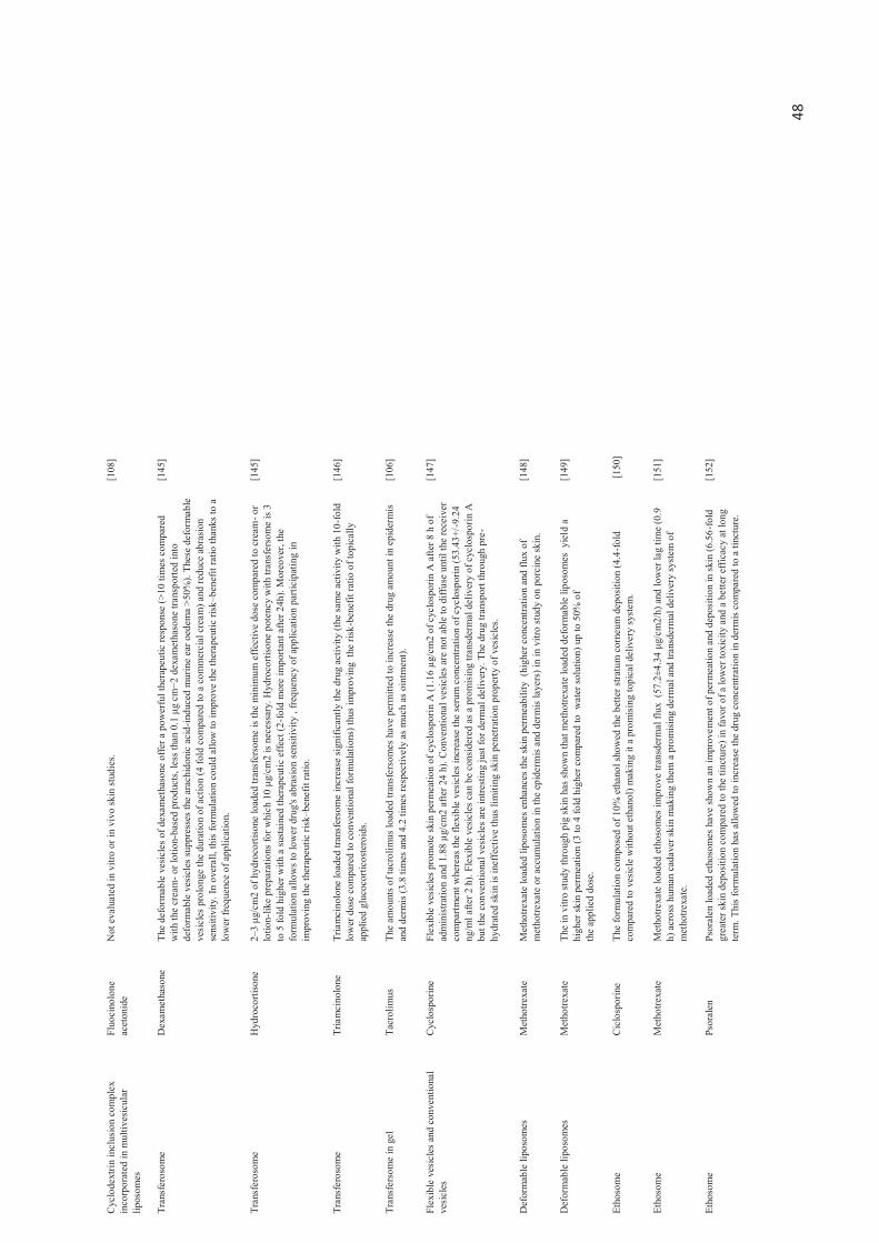
48
Cyc
lode
xtrin
incl
usio
n co
mpl
ex
inco
rpor
ated
in m
ultiv
esic
ular
lip
osom
es
Fluo
cino
lone
ac
eton
ide
Not
eva
luat
ed in
vitr
o or
in v
ivo
skin
stud
ies.
[108
]
Tran
sfer
osom
e D
exam
etha
sone
Th
e de
form
able
ves
icle
s of d
exam
etha
sone
offe
r a p
ower
ful t
hera
peut
ic re
spon
se (>
10 ti
mes
com
pare
d w
ith th
e cr
eam
- or l
otio
n-ba
sed
prod
ucts
, les
s tha
n 0.
1 μg
cm
−2 d
exam
etha
sone
tran
spor
ted
into
de
form
able
ves
icle
s sup
pres
ses t
he a
rach
idon
ic a
cid-
indu
ced
mur
ine
ear o
edem
a >5
0%).
Thes
e de
form
able
ve
sicl
es p
rolo
nge
the
dura
tion
of a
ctio
n (4
fold
com
pare
d to
a c
omm
erci
al c
ream
) and
redu
ce a
bras
ion
sens
itivi
ty. I
n ov
eral
l, th
is fo
rmul
atio
n co
uld
allo
w to
impr
ove
the
ther
apeu
tic ri
sk–b
enef
it ra
tio th
anks
to a
lo
wer
freq
uenc
e of
app
licat
ion.
[145
]
Tran
sfer
osom
e H
ydro
corti
sone
2–
3 μg
/cm
2 of
hyd
roco
rtiso
ne lo
aded
tran
sfer
som
e is
the
min
imum
effe
ctiv
e do
se c
ompa
red
to c
ream
- or
lotio
n -lik
e pr
epar
atio
ns fo
r whi
ch 1
0 μg
/cm
2 is
nece
ssar
y. H
ydro
corti
sone
pot
ency
with
tran
sfer
som
e is
3 to
5 fo
ld h
ighe
r with
a s
usta
ined
ther
apeu
tic e
ffect
(2-fo
ld m
ore
impo
rtant
afte
r 24h
). M
oreo
ver,
the
form
ulat
ion
allo
ws t
o lo
wer
dru
g's a
bras
ion
sens
itivi
ty ,
frequ
ency
of a
pplic
atio
n pa
rtici
patin
g in
im
prov
ing
the
ther
apeu
tic ri
sk–b
enef
it ra
tio.
[145
]
Tran
sfer
osom
e Tr
iam
cino
lone
Tr
iam
cino
lone
load
ed tr
ansf
erso
me
incr
ease
sign
ifica
ntly
the
drug
act
ivity
(the
sam
e ac
tivity
with
10-
fold
lo
wer
dos
e co
mpa
red
to c
onve
ntio
nal f
orm
ulat
ions
) thu
s im
prov
ing
the
risk -
bene
fit ra
tio o
f top
ical
ly
appl
ied
gluc
ocor
ticos
tero
ids.
[146
]
Tran
sfer
som
e in
gel
Ta
crol
imus
Th
e am
ount
s of t
acro
limus
load
ed tr
ansf
erso
mes
hav
e pe
rmitt
ed to
incr
ease
the
drug
am
ount
in e
pide
rmis
an
d de
rmis
(3.8
tim
es a
nd 4
.2 ti
mes
resp
ectiv
ely
as m
uch
as o
intm
ent).
[1
06]
Flex
ible
ves
icle
s and
con
vent
iona
l ve
sicl
es
Cyc
losp
orin
e Fl
exib
le v
esic
les p
rom
ote
skin
per
mea
tion
of c
yclo
spor
in A
(1.1
6 μg
/cm
2 of
cyc
losp
orin
A a
fter 8
h o
f ad
min
istra
tion
and
1.88
μg/
cm2
afte
r 24
h). C
onve
ntio
nal v
esic
les a
re n
ot a
ble
to d
iffus
e un
til th
e re
ceiv
er
com
partm
ent w
here
as th
e fle
xibl
e ve
sicl
es in
crea
se th
e se
rum
con
cent
ratio
n of
cyc
losp
orin
(53.
43+/
-9.2
4 ng
/ml a
fter 2
h).
Flex
ible
ves
icle
s can
be
cons
ider
ed a
s a p
rom
isin
g tra
nsde
rmal
del
iver
y of
cyc
losp
orin
A
but t
he c
onve
ntio
nal v
esic
les a
re in
trest
ing
just
for d
erm
al d
eliv
ery.
The
dru
g tra
nspo
rt th
roug
h pr
e -hy
drat
ed sk
in is
inef
fect
ive
thus
lim
iting
ski
n pe
netra
tion
prop
erty
of v
esic
les.
[147
]
Def
orm
able
lipo
som
es
Met
hotre
xate
M
etho
trexa
te lo
aded
lipo
som
es e
nhan
ces t
he sk
in p
erm
eabi
lity
(hig
her c
once
ntra
tion
and
flux
of
met
hotre
xate
or a
ccum
ulat
ion
in th
e ep
ider
mis
and
derm
is la
yers
) in
in v
itro
stud
y on
por
cine
skin
. [1
48]
Def
orm
able
lipo
som
es
Met
hotre
xate
Th
e in
vitr
o st
udy
thro
ugh
pig
skin
has
show
n th
at m
etho
trexa
te lo
aded
def
orm
able
lipo
som
es y
ield
a
high
er sk
in p
erm
eatio
n (3
to 4
fold
hig
her c
ompa
red
to w
ater
solu
tion)
up
to 5
0% o
f th
e ap
plie
d do
se.
[149
]
Etho
som
e C
iclo
spor
ine
The
form
ulat
ion
com
pose
d of
10%
eth
anol
show
ed th
e be
tter s
tratu
m c
orne
um d
epos
ition
(4.4
-fold
co
mpa
red
to v
esic
le w
ithou
t eth
anol
) mak
ing
it a
prom
isin
g to
pica
l del
iver
y sy
stem
. [1
50]
Etho
som
e M
etho
trexa
te
Met
hotre
xate
load
ed e
thos
omes
impr
ove
trans
derm
al fl
ux (
57.2
±4.3
4 μg
/cm
2/h)
and
low
er la
g tim
e (0
.9
h) a
cros
s hum
an c
adav
er sk
in m
akin
g th
em a
pro
misi
ng d
erm
al a
nd tr
ansd
erm
al d
eliv
ery
syst
em o
f m
etho
trexa
te.
[151
]
Etho
som
e Ps
oral
en
Psor
alen
load
ed e
thos
omes
hav
e sh
own
an im
prov
emen
t of p
erm
eatio
n an
d de
posi
tion
in sk
in (6
.56-
fold
gr
eate
r ski
n de
posi
tion
com
pare
d to
the
tinct
ure)
in fa
vor o
f a lo
wer
toxi
city
and
a b
ette
r eff
icac
y at
long
te
rm. T
his
form
ulat
ion
has a
llow
ed to
incr
ease
the
drug
con
cent
ratio
n in
der
mis
com
pare
d to
a ti
nctu
re.
[152
]
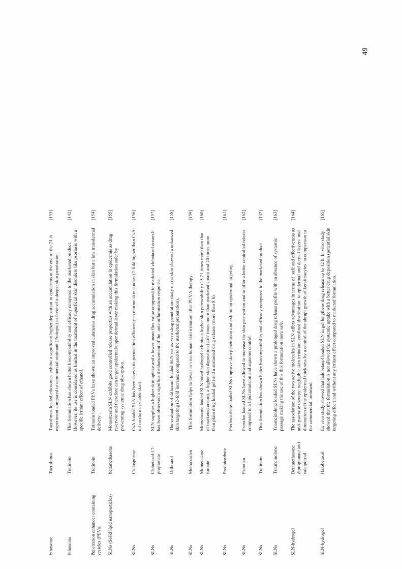
49
Etho
som
e Ta
crol
imus
Ta
crol
imus
load
ed e
thos
omes
exh
ibit
a si
gnifi
cant
hig
her d
epos
ition
in e
pide
rmis
at t
he e
nd o
f the
24-
h ex
perim
ent c
ompa
red
to c
omm
erci
al o
intm
ent (
Prot
opic
) in
favo
r of a
dee
per s
kin
pene
tratio
n.
[153
]
Etho
som
e Tr
etin
oin
This
form
ulat
ion
has s
how
n be
tter b
ioco
mpa
tibili
ty a
nd e
ffica
cy c
ompa
red
to th
e m
arke
ted
prod
uct.
How
ever
, its
use
is c
onsi
dere
d as
lim
ited
in th
e tre
atm
ent o
f sup
erfic
ial s
kin
diso
rder
s lik
e ps
oria
sis w
ith a
sp
ecifi
c irr
itant
effe
ct o
f eth
anol
.
[142
]
Pene
tratio
n en
hanc
er c
onta
inin
g ve
sicl
es (P
EVs)
Tr
etin
oin
Tetin
oin
load
ed P
EVs h
ave
show
n an
impr
oved
cut
aneo
us d
rug
accu
mul
atio
n in
skin
but
a lo
w tr
ansd
erm
al
deliv
ery
. [1
54]
SLN
s (So
lid li
pid
nano
parti
cles
) be
tam
etha
sone
M
onos
tear
in S
LN e
xhib
its g
ood
cont
rolle
d re
leas
e pr
oper
ties w
ith a
n ac
cum
ulat
ion
in e
pide
rmis
as d
rug
rese
rvoi
r and
ther
efor
e ca
n ta
rget
epi
derm
al/u
pper
der
mal
laye
r mak
ing
this
form
ulat
ion
safe
r by
prev
entin
g sy
stem
ic d
rug
abso
rptio
n.
[155
]
SLN
s C
iclo
spor
ine
CsA
-load
ed S
LN h
as b
een
show
n its
per
mea
tion
effic
ienc
y in
mur
ine
skin
stud
ies (
2-fo
ld h
ighe
r tha
n C
sA-
oil m
ixtu
re in
via
ble
skin
).
[156
]
SLN
s C
lobe
taso
l-17-
prop
iona
te
SLN
sup
plie
s a h
ighe
r ski
n up
take
and
a lo
wer
mea
n flu
x va
lue
com
pare
d to
mar
kete
d cl
obet
asol
cre
am.It
ha
s bee
n ob
serv
ed a
sign
ifica
nt e
nhan
cem
ent o
f the
ant
i-inf
lam
mat
ion
resp
onse
. [1
57]
SLN
s D
ithra
nol
The
eval
uatio
n of
dith
rano
l loa
ded
SLN
via
ex-
vivo
dru
g pe
netra
tion
stud
y on
rat s
kin
show
ed a
enh
ance
d sk
in ta
rget
ing
(2-fo
ld in
crea
se c
ompa
red
to th
e m
arke
ted
prep
arat
ion)
. [1
58]
SLN
s M
etho
xsal
en
Thi
s fo
rmul
atio
n he
lps t
o lo
wer
in v
ivo
hum
an sk
in ir
ritat
ion
afte
r PU
VA
ther
apy.
[1
59]
SLN
s M
omet
ason
e fu
roat
e M
omet
ason
e lo
aded
SLN
bas
ed h
ydro
gel e
xhib
its a
hig
her s
kin
perm
eabi
lity
(15.
21 ti
mes
mor
e th
an th
at
of m
arke
ted
crea
m),
a hi
gher
skin
dep
ositi
on (2
.67
times
mor
e th
an m
arke
ted
crea
m a
nd 2
0 tim
es m
ore
than
pla
in d
rug
load
ed g
el) a
nd a
sust
aine
d dr
ug re
leas
e (m
ore
than
8 h
).
[160
]
SLN
s Pr
edni
carb
ate
Pred
nica
rbat
e lo
aded
SLN
s im
prov
e sk
in p
enet
ratio
n an
d ex
hibi
t an
epid
erm
al ta
rget
ing.
[1
61]
SLN
s Ps
oral
en
Psor
alen
load
ed S
LNs h
ave
allo
wed
to in
crea
se th
e sk
in p
erm
eatio
n an
d to
offe
r a b
ette
r con
trolle
d re
leas
e co
mpa
red
to a
lipi
d em
ulsi
on a
nd a
queo
us c
ontro
l. [1
62]
SLN
s Tr
etin
oin
This
form
ulat
ion
has s
how
n be
tter b
ioco
mpa
tibili
ty a
nd e
ffica
cy c
ompa
red
to th
e m
arke
ted
prod
uct.
[142
]
SLN
s Tr
iam
cino
lone
Tr
iam
cino
lone
load
ed S
LNs
have
show
n a
prol
onge
d dr
ug re
leas
e pr
ofile
with
an
abse
nce
of s
yste
mic
pa
ssag
e m
akin
g th
e us
e of
this
this
form
utat
ion
mor
e sa
fe.
[163
]
SLN
-hyd
roge
l Be
tam
etha
sone
di
prop
iona
te a
nd
calc
ipot
riol
The
asso
ciat
ion
of th
e tw
o ac
tive
mol
ecul
es in
SLN
offe
rs a
dvan
tage
s in
term
s of
safe
and
effe
ctiv
enes
s as
anti -
psor
iatic
ther
apy:
neg
ligib
le sk
in ir
ritat
ion,
con
fined
dist
ribut
ion
to e
pide
rmal
and
der
mal
laye
rs a
nd
dim
inut
ion
of th
e ep
ider
mal
thic
knes
s by
a co
ntro
l of t
he a
brup
t gro
wth
of k
erat
inoc
ytes
in
com
paris
on to
th
e co
mm
erci
al o
intm
ent.
[164
]
SLN
-hyd
roge
l H
alob
etas
ol
Ex v
ivo
stud
y sh
owed
that
hal
obet
asol
load
ed S
LN in
gel
leng
then
s dru
g re
leas
e up
to 1
2 h.
In v
itro
stud
y sh
owed
that
the
form
ulat
ion
is no
t allo
wed
the
syst
emic
upt
ake
with
a b
ette
r dru
g de
posit
ion
(pot
entia
l ski
n ta
rget
ing
effe
ct) a
nd w
ithou
t any
irrit
ant e
ffect
com
pare
d to
mar
kete
d fo
rmul
atio
n.
[165
]
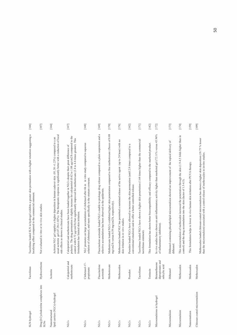
50
SLN
-hyd
roge
l Ta
crol
imus
Ta
crol
imus
load
ed S
LN in
hyd
roge
l exh
ibits
a g
reat
er sk
in p
erm
eatio
n w
ith a
hig
her r
eten
tion
sugg
estin
g a
loca
lizin
g ef
fect
com
pare
d to
the
com
mer
cial
oin
tmen
t.
[166
]
Dru
g β-
Cyc
lode
xtrin
e co
mpl
exes
into
SL
Ns
Hyd
roco
rtiso
ne
Not
eva
luat
ed in
vitr
o or
in v
ivo
skin
stud
ies.
[107
]
Nan
ostru
ctur
ed
lipid
car
riers
(NLC
s)-h
ydro
gel
Aci
tretin
A
citre
tin N
LC g
el s
uppl
ies a
hig
her d
epos
ition
in h
uman
cad
aver
skin
(81
.38
±1.2
3%) c
ompa
red
to a
an
usua
l aci
tretin
gel
(47.
28±1
.02%
). Th
e th
erap
eutic
resp
onse
is si
gnifi
cant
ly b
ette
r with
a re
duct
ion
of lo
cal
side
effe
cts a
s sho
wn
in c
linic
al st
udie
s.
[164
]
NLC
s C
alci
potri
ol a
nd
met
hotre
xate
C
alci
potri
ol a
nd m
etho
trexa
te h
ave
been
load
ed to
geth
er in
NLC
s des
pite
thei
r gre
at d
iffer
ence
of
pola
ritiy
. The
dru
g pe
rmea
tion
is sl
ight
ly in
crea
se fo
r cal
cipo
triol
(0.6
2 to
1.0
8 μg
/cm
2/h
com
pare
d to
the
cont
rol w
ith 0
.72
μg/c
m2/
h) b
ut si
gnifi
cant
ly im
prov
ed fo
r met
hotre
xate
(2.4
to 4
.4-ti
mes
gre
ater
). Th
is fo
rmul
atio
n ha
s lim
ited
skin
irrit
atio
n.
[167
]
NLC
s C
lobe
taso
l-17-
prop
iona
te
NLC
pro
mot
es a
n la
rge
accu
mul
atio
n of
clo
beta
sol a
fter 6
h in
in
vitr
o st
udy
com
pare
d to
aqu
eous
so
lutio
n of
clo
beta
sol,
and
mor
e sp
ecifi
cally
in th
e st
ratu
m c
orne
um.
[168
]
NLC
s Fl
uoci
nolo
ne
acet
onid
e Fl
uoci
nolo
ne a
ceto
nide
load
ed N
LCs e
nabl
e to
pro
long
e dr
ug re
leas
e co
mpa
red
to a
pla
in s
uspe
nsio
n an
d a
sele
ctiv
e ac
cum
ulat
ion
has b
een
obse
rved
in th
e ep
ider
mis
. [1
69]
NLC
s M
etho
trexa
te
Met
hotre
xate
load
ed N
LCs e
xhib
ited
high
er sk
in p
enet
ratio
n co
mpa
red
to fr
ee m
etho
trexa
te (f
luxe
s of 0
.88
mg/
cm2/
h ve
rsus
0.5
9 m
g/cm
2/h,
resp
ectiv
ely)
. [1
70]
NLC
s M
etho
xsal
en
Met
hoxs
alen
load
ed N
LC h
as p
erm
itted
a su
stai
ned
rele
ase
of th
e ac
tive
agen
t (u
p to
24
hour
) with
no
skin
irrit
atio
n in
in v
itro
stud
ies.
[1
76]
NLC
s Ps
oral
en
Psor
alen
load
ed N
LCs
have
allo
wed
to in
crea
se th
e sk
in p
erm
eatio
n (u
ntil
2.8
times
com
pare
d to
a
conv
entio
nal e
mul
sion
) and
to o
ffer
a b
ette
r con
trolle
d re
leas
e.
[162
]
NLC
s Ta
crol
imus
Ta
crol
imus
load
ed N
LCs h
ave
show
n a
high
er sk
in p
enet
ratio
n (1
.64
times
hig
her t
han
the
com
mer
cial
ta
crol
imus
oin
tmen
t).
[171
]
NLC
s Tr
etin
oin
This
form
ulat
ion
has s
how
n be
tter b
ioco
mpa
tibili
ty a
nd e
ffica
cy c
ompa
red
to th
e m
arke
ted
prod
uct.
[142
]
Mic
roem
ulsi
ons
in h
ydro
gel
Beta
met
haso
ne
dipr
opio
nate
and
sa
licyl
ic a
cid
In v
ivo
stud
ies h
ave
show
n an
ant
i-inf
lam
mat
ory
activ
ity h
ighe
r tha
n m
arke
ted
gel (
72.1
1% v
ersu
s 43.
96%
of
infla
mm
ator
y in
hibi
tion)
. [1
72]
Mic
roem
ulsi
on
Dith
rano
l D
ithra
nol-c
onta
inin
g ph
osph
olip
id m
icro
emul
sion
show
ed a
n im
prov
emen
t of
the
topi
cal d
eliv
ery
of
dith
rano
l. [1
73]
Mic
roem
ulsi
on
Met
hoxs
alen
Th
is m
icro
emul
sion
of m
etho
xale
n in
crea
sed
the
pene
tratio
n th
roug
h th
e sk
in (1
.9-4
.5 fo
lds h
ighe
r tha
n in
co
ntro
l) an
d th
e dr
ug a
ccum
ulat
ion
into
the
skin
(fac
tor o
f 1.5
-4.5
). [1
74]
Nan
oem
ulsi
on
Met
hoxs
alen
T
his
form
ulat
ion
help
s to
low
er in
viv
o hu
man
skin
irrit
atio
n af
ter P
UV
A th
erap
y.
[159
]
Chi
tosa
n co
ated
mic
roem
ulsi
on
Met
hoxs
alen
M
etho
xsal
en lo
aded
chi
tosa
n-co
ated
mic
roem
ulsi
on h
as sh
own
a hi
gher
skin
dep
ositi
on (1
8.75
% le
sser
th
an th
e m
icro
emul
sion
) ass
ocia
ted
with
a c
ontro
l rel
ease
of m
etho
xsal
en in
invi
tro s
tudi
es.
[109
]
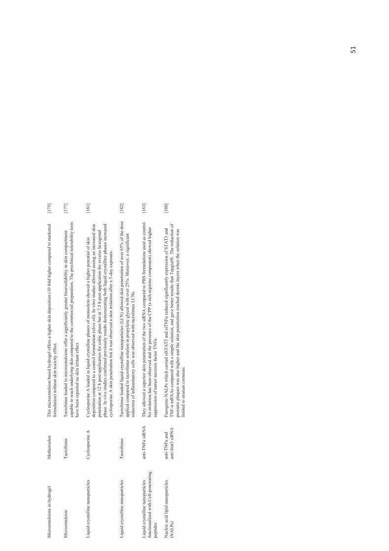
51
Mic
roem
ulsi
ons
in h
ydro
gel
Met
hoxs
alen
Th
is m
icro
emul
sion
bas
ed h
ydro
gel o
ffer
s a h
ighe
r ski
n de
posi
tion
(10
fold
hig
her c
ompa
red
to m
arke
ted
form
ulat
ion)
with
out s
kin
toxi
city
eff
ect.
[175
]
Mic
roem
ulsi
on
Tacr
olim
us
Tacr
olim
us lo
aded
in m
icro
emul
sion
s offe
r a si
gnifi
cant
ly g
reat
er b
ioav
aila
bilit
y in
skin
com
partm
ent
capa
ble
to re
ach
unde
rlyin
g sk
in c
ompa
red
to th
e co
mm
erci
al p
repa
ratio
n. T
he p
recl
inic
al to
lera
bilit
y te
sts
have
bee
n re
porte
d no
skin
irrit
ant e
ffect
.
[177
]
Liqu
id c
ryst
allin
e na
nopa
rticl
es
Cyc
losp
orin
e A
C
yclo
spor
ine
A lo
aded
in li
quid
cry
stal
line
phas
es o
f mon
oole
in s
how
ed a
hig
her p
oten
tial o
f ski
n de
posi
tion
com
pare
d to
a c
ontro
l for
mul
atio
n (o
live
oil).
In v
itro
stud
ies a
llow
ed se
eing
an
incr
ease
d sk
in
pene
tratio
n at
12
h po
st-a
pplic
atio
n fo
r cub
ic p
hase
but
at 7
.5 h
pos
t-app
licat
ion
for r
ever
se h
exag
onal
ph
ase.
In v
ivo
stud
ies c
onfir
med
pre
viou
sly
resu
lts d
emon
stra
ting
both
liqu
id c
ryst
allin
e ph
ases
incr
ease
d cy
clos
porin
e A
skin
pen
etra
tion
but i
t was
obs
erve
d a
skin
irrit
atio
n af
ter a
3-d
ay e
xpos
ure.
[181
]
Liqu
id c
ryst
allin
e na
nopa
rticl
es
Tacr
olim
us
Tacr
olim
us lo
aded
liqu
id c
ryst
allin
e na
nopa
rticl
es (L
CN
) allo
wed
skin
pen
etra
tion
of o
ver 6
5% o
f the
dos
e ap
plie
d co
mpa
red
to ta
crol
imus
solu
tion
in p
ropy
lene
gly
col w
ith o
ver 2
5%. M
oreo
ver,
a si
gnifi
cant
re
duct
ion
of in
flam
mat
ory
cells
was
obs
erve
d w
ith ta
crol
imus
LC
Ns.
[182
]
Liqu
id c
ryst
allin
e na
nopa
rticl
es
func
tiona
lized
with
Cel
l -pen
etra
ting
pept
ides
anti-
TNFα
siR
NA
Th
ey a
llow
ed a
supe
rior s
kin
pene
tratio
n of
the
two
siRN
A c
ompa
red
to P
BS
form
ulat
ions
use
d as
con
trol.
No
irrita
tion
has b
een
obse
rved
and
the
pres
ence
of t
he C
PP (a
rich
arg
inin
e co
mpo
nent
) sho
wed
hig
her
supp
ress
ion
of tu
mor
nec
rosi
s fac
tor T
NFα
.
[183
]
Nuc
leic
aci
d lip
id n
anop
artic
les
(NA
LPs)
an
ti-TN
Fα a
nd
anti -
Stat
3 siR
NA
Fu
soge
nic-
NA
LPs w
hich
car
ried
siST
AT3
and
siT
NFα
redu
ced
sign
ifica
ntly
exp
ress
ion
of S
TAT3
and
TN
F -α
mR
NA
s com
pare
d w
ith a
sim
ple
solu
tion,
and
gav
e be
tter r
esul
ts th
at T
opgr
af®
. The
redu
ctio
n of
ps
oria
tic p
laqu
es w
as a
lso h
ighe
r and
the
skin
pen
etra
tion
reac
hed
derm
is la
yers
whe
n th
e so
lutio
n w
as
limite
d to
stra
tum
cor
neum
.
[180
]
52
4.5.2 Advantages of Innovative skin drug delivery systems (ISDDSs) The physiopathogenesis of psoriasis is localized in skin tissue mainly. A direct action on the target region would provide a faster and more effective therapeutic response. There is already a whole family of topical medications for mild forms of psoriasis but with a low absorption rate. In more advanced step of the disease, drugs administered by systemic routes (oral or parenteral) are prescribed. But these molecules like methotrexate or cyclosporin A have to be used at high doses to achieve therapeutic dermal concentration. But this comes at the expense of many adverse effects. The aim of ISDDS is to provide drug formulations that penetrate deeper into the skin after overcoming the stratum corneum barrier and making them interesting for treating all forms of psoriasis. As the site of action is direct, it could help to improve the efficacy of drugs conventionally used topically and reduce the doses of those used systemically. The table 7 summarizes the different ISDDSs that have been developed in recent years to treat skin diseases as psoriasis and the improvements highlighted in in vitro and/or in vivo studies. Because of the complexicity of psoriasis, combined therapies have already used to contain its progression. Such strategies could also be interesting to consider in the field of the ISDDs as association of cyclodextrin with lipid nanoparticles [107,108] or nanoparticles capable to carry siRNA with chemical substances like cyclosporine [180-183].
5. Conclusion
The armamentarium of anti-psoriatic treatments is very large. However, none has a good benefit/risk ratio ensuring a great efficacy and safety. These available drugs help to contain the disease and relieve the symptoms without healing definitely. Without knowing the key element triggering psoriasis, the attempts to develop a curative therapy seem to doom to failure. Biotherapy is a cutting-edge research blooming on the drug market. But before prescribing therapeutic molecules belonging to this drug class, ISDDS could be an intermediate class that optimizes a maximum the conventional drugs, giving them a second life with a better benefit/risk ratio.
ISDDSs have shown that they were promising nanocarriers capable to assure an appropriate drug delivery across the skin to treat psoriatic lesions. The improvement of the skin deposition with a targeting effect especially in epidermis where the physiopathology of psoriasis is mainly localized associated with a sustained release of the active agent are arguments in favor of the success of ISDDSs. They allow administration of a lower dose for the same therapeutic efficiency and less frequently. Moreover, a lower dose is in line with a reduction of side effects. The more often, these ISDDSs prevent the passage in systemic circulation what is matching again with the safety aspects of these new nanocarriers. However, that is less observed with ethosomes and transfersomes which promote transdermal penetration that could be an advantage to treat psoriatic arthritis.
Many studies on ISDDSs have been performed to highlight their advantages to treat skin disorders like psoriasis. However, these types of formulation on the drug market are too rare (Lipotar™ S gel (Coal tar and salicylic acid), Psorisome® gel (dithranol)). Clinicals trials in this field should be multiplied to clarify the place of these new drug formulations in the management of psoriasis.
53
6.Acknowledgements: We would like to acknowledge the support of the CDIP (Centre de documentation et d' information pharmaceutique) at Pharmacie centrale des Hospices civils de Lyon which helped us in bibliographic research.
References :
[1] M. Pradhan, D. Singh, M.R. Singh, Novel colloidal carriers for psoriasis: current issues, mechanistic insight and novel delivery approaches, J. Control. Release Off. J. Control. Release Soc. 170 (2013) 380–395.
[2] T. Mabuchi, T.W. Chang, S. Quinter, S.T. Hwang, Chemokine receptors in the pathogenesis and therapy of psoriasis, J. Dermatol. Sci. 65 (2012) 4–11.
[3] O. Fitzgerald, M. Dougados, Psoriatic arthritis: one or more diseases?, Best Pract. Res. Clin. Rheumatol. 20 (2006) 435–450.
[4] D. Kane, L. Stafford, B. Bresnihan, O. FitzGerald, A prospective, clinical and radiological study of early psoriatic arthritis: an early synovitis clinic experience, Rheumatol. Oxf. Engl. 42 (2003) 1460–1468.
[5] A. Gottlieb, N.J. Korman, K.B. Gordon, S.R. Feldman, M. Lebwohl, J.Y.M. Koo, et al., Guidelines of care for the management of psoriasis and psoriatic arthritis: Section 2. Psoriatic arthritis: Overview and guidelines of care for treatment with an emphasis on the biologics, J. Am. Acad. Dermatol. 58 (2008) 851–864.
[6] A. Menter, A. Gottlieb, S.R. Feldman, A.S. Van Voorhees, C.L. Leonardi, K.B. Gordon, et al., Guidelines of care for the management of psoriasis and psoriatic arthritis: Section 1. Overview of psoriasis and guidelines of care for the treatment of psoriasis with biologics, J. Am. Acad. Dermatol. 58 (2008) 826–850.
[7] A. Menter, N.J. Korman, C.A. Elmets, S.R. Feldman, J.M. Gelfand, K.B. Gordon, et al., Guidelines of care for the management of psoriasis and psoriatic arthritis: Section 4. Guidelines of care for the management and treatment of psoriasis with traditional systemic agents, J. Am. Acad. Dermatol. 61 (2009) 451–485.
[8] A. Menter, N.J. Korman, C.A. Elmets, S.R. Feldman, J.M. Gelfand, K.B. Gordon, et al., Guidelines of care for the management of psoriasis and psoriatic arthritis: Section 3. Guidelines of care for the management and treatment of psoriasis with topical therapies, J. Am. Acad. Dermatol. 60 (2009) 643–659.
[9] A. Menter, N.J. Korman, C.A. Elmets, S.R. Feldman, J.M. Gelfand, K.B. Gordon, et al., Guidelines of care for the management of psoriasis and psoriatic arthritis: Section 5. Guidelines of care for the treatment of psoriasis with phototherapy and photochemotherapy, J. Am. Acad. Dermatol. 62 (2010) 114–135.
[10] A. Menter, N.J. Korman, C.A. Elmets, S.R. Feldman, J.M. Gelfand, K.B. Gordon, et al., Guidelines of care for the management of psoriasis and psoriatic arthritis: Section 6. Guidelines of care for the treatment of psoriasis and psoriatic arthritis: Case-based presentations and evidence-based conclusions, J. Am. Acad. Dermatol. 65 (2011) 137–174.
[11] J. Paccou, D. Wendling, Current treatment of psoriatic arthritis: update based on a systematic literature review to establish French Society for Rheumatology (SFR) recommendations for managing spondyloarthritis, Jt. Bone Spine Rev. Rhum. 82 (2015) 80–85.
54
[12] M.A. Lowes, C.B. Russell, D.A. Martin, J.E. Towne, J.G. Krueger, The IL-23/T17 pathogenic axis in psoriasis is amplified by keratinocyte responses, Trends Immunol. 34 (2013) 174–181.
[13] M. Ammar, C. Souissi-Bouchlaka, A. Gati, I. Zaraa, R. Bouhaha, S. Kouidhi, et al., [Psoriasis: physiopathology and immunogenetics], Pathol. Biol. (Paris). 62 (2014) 10–23. doi:10.1016/j.patbio.2013.07.014.
[14] J.P. Ortonne, Recent developments in the understanding of the pathogenesis of psoriasis, Br. J. Dermatol. 140 Suppl 54 (1999) 1–7.
[15] M.D. Hertle, M.D. Kubler, I.M. Leigh, F.M. Watt, Aberrant integrin expression during epidermal wound healing and in psoriatic epidermis, J. Clin. Invest. 89 (1992) 1892–1901.
[16] J.F. Krane, A.B. Gottlieb, D.M. Carter, J.G. Krueger, The insulin-like growth factor I receptor is overexpressed in psoriatic epidermis, but is differentially regulated from the epidermal growth factor receptor, J. Exp. Med. 175 (1992) 1081–1090.
[17] Y. Liu, J.G. Krueger, A.M. Bowcock, Psoriasis: genetic associations and immune system changes, Genes Immun. 8 (2007) 1–12.
[18] J.C. Ansel, J.P. Tiesman, J.E. Olerud, J.G. Krueger, J.F. Krane, D.C. Tara, et al., Human keratinocytes are a major source of cutaneous platelet-derived growth factor, J. Clin. Invest. 92 (1993) 671–678.
[19] J. Viac, S. Palacio, D. Schmitt, A. Claudy, Expression of vascular endothelial growth factor in normal epidermis, epithelial tumors and cultured keratinocytes, Arch. Dermatol. Res. 289 (1997) 158–163.
[20] M.A. Lowes, A.M. Bowcock, J.G. Krueger, Pathogenesis and therapy of psoriasis, Nature. 445 (2007) 866–873.
[21] L.M. Johnson-Huang, M.A. Lowes, J.G. Krueger, Putting together the psoriasis puzzle: an update on developing targeted therapies, Dis. Model. Mech. 5 (2012) 423–433.
[22] T. Wrone-Smith, B.J. Nickoloff, Dermal injection of immunocytes induces psoriasis, J. Clin. Invest. 98 (1996) 1878–1887.
[23] L.M. Austin, T.R. Coven, N. Bhardwaj, R. Steinman, J.G. Krueger, Intraepidermal lymphocytes in psoriatic lesions are activated GMP-17(TIA-1)+CD8+CD3+ CTLs as determined by phenotypic analysis, J. Cutan. Pathol. 25 (1998) 79–88.
[24] B.J. Nickoloff, T. Wrone-Smith, Superantigens, autoantigens, and pathogenic T cells in psoriasis, J. Invest. Dermatol. 110 (1998) 459–460. doi:10.1046/j.1523-1747.1998.00158.x.
[25] R. Arnold, M. Seifert, K. Asadullah, H.D. Volk, Crosstalk between keratinocytes and T lymphocytes via Fas/Fas ligand interaction: modulation by cytokines, J. Immunol. Baltim. Md 1950. 162 (1999) 7140–7147.
[26] B.J. Nickoloff, T. Wrone-Smith, Injection of pre-psoriatic skin with CD4+ T cells induces psoriasis, Am. J. Pathol. 155 (1999) 145–158. doi:10.1016/S0002-9440(10)65109-7.
[27] B.J. Nickoloff, Skin innate immune system in psoriasis: friend or foe?, J. Clin. Invest. 104 (1999) 1161–1164.
[28] C. Behrendt, H. Gollnick, B. Bonnekoh, Up-regulated perforin expression of CD8+ blood lymphocytes in generalized non-anaphylactic drug eruptions and exacerbated psoriasis, Eur. J. Dermatol. EJD. 10 (2000) 365–369.
55
[29] M. Friedrich, S. Krammig, M. Henze, W.-D. Döcke, W. Sterry, K. Asadullah, Flow cytometric characterization of lesional T cells in psoriasis: intracellular cytokine and surface antigen expression indicates an activated, memory/effector type 1 immunophenotype, Arch. Dermatol. Res. 292 (2000) 519–521.
[30] G.G. Krueger, K.A. Papp, D.B. Stough, K.H. Loven, W.P. Gulliver, C.N. Ellis, et al., A randomized, double-blind, placebo-controlled phase III study evaluating efficacy and tolerability of 2 courses of alefacept in patients with chronic plaque psoriasis, J. Am. Acad. Dermatol. 47 (2002) 821–833.
[31] J.C. Prinz, The role of T cells in psoriasis, J. Eur. Acad. Dermatol. Venereol. JEADV. 17 (2003) 257–270.
[32] D. Hijnen, E.F. Knol, Y.Y. Gent, B. Giovannone, S.J.P. Beijn, T.S. Kupper, et al., CD8(+) T cells in the lesional skin of atopic dermatitis and psoriasis patients are an important source of IFN-γ, IL-13, IL-17, and IL-22, J. Invest. Dermatol. 133 (2013) 973–979.
[33] M. Favre, S. Majewski, B. Noszczyk, F. Maienfisch, A. Pura, G. Orth, et al., Antibodies to human papillomavirus type 5 are generated in epidermal repair processes, J. Invest. Dermatol. 114 (2000) 403–407.
[34] R. Pérez-Lorenzo, J.F. Zambrano-Zaragoza, A. Saul, L. Jiménez-Zamudio, E. Reyes-Maldonado, E. García-Latorre, Autoantibodies to autologous skin in guttate and plaque forms of psoriasis and cross-reaction of skin antigens with streptococcal antigens, Int. J. Dermatol. 37 (1998) 524–531.
[35] B.A. Torres, H.M. Johnson, Modulation of disease by superantigens, Curr. Opin. Immunol. 10 (1998) 465–470.
[36] M.B. Nielsen, N. Odum, J. Gerwien, A. Svejgaard, K. Bendtzen, S. Bregentholt, et al., Staphylococcal enterotoxin-A directly stimulates signal transduction and interferon-gamma production in psoriatic T-cell lines, Tissue Antigens. 52 (1998) 530–538.
[37] N. Horiuchi, S. Aiba, H. Ozawa, S. Sugawara, H. Rikiishi, K. Kumagai, et al., Peripheral blood lymphocytes from psoriatic patients are hyporesponsive to beta-streptococcal superantigens, Br. J. Dermatol. 138 (1998) 229–235.
[38] G. Fierlbeck, G. Rassner, C. Müller, Psoriasis induced at the injection site of recombinant interferon gamma. Results of immunohistologic investigations, Arch. Dermatol. 126 (1990) 351–355.
[39] L.A.T. F O Nestle, Characterization of dermal dendritic cells in psoriasis. Autostimulation of T lymphocytes and induction of Th1 type cytokines., J. Clin. Invest. 94 (1994) 202–9.
[40] A.M. Bowcock, J.G. Krueger, Getting under the skin: the immunogenetics of psoriasis, Nat. Rev. Immunol. 5 (2005) 699–711.
[41] L.C. Zaba, M. Suárez-Fariñas, J. Fuentes-Duculan, K.E. Nograles, E. Guttman-Yassky, I. Cardinale, et al., Effective treatment of psoriasis with etanercept is linked to suppression of IL-17 signaling, not immediate response TNF genes, J. Allergy Clin. Immunol. 124 (2009) 1022–1010.e1–395.
[42] L. Flatz, C. Conrad, Role of T-cell-mediated inflammation in psoriasis: pathogenesis and targeted therapy, Psoriasis Targets Ther. 3 (2013) 1–10.
[43] S. Marrakchi, P. Guigue, B.R. Renshaw, A. Puel, X.-Y. Pei, S. Fraitag, et al., Interleukin-36–receptor antagonist deficiency and generalized pustular psoriasis, N. Engl. J. Med. 365 (2011) 620–628.
56
[44] S.C. Liang, X.-Y. Tan, D.P. Luxenberg, R. Karim, K. Dunussi-Joannopoulos, M. Collins, et al., Interleukin (IL)-22 and IL-17 are coexpressed by Th17 cells and cooperatively enhance expression of antimicrobial peptides, J. Exp. Med. 203 (2006) 2271–2279.
[45] R. Lande, J. Gregorio, V. Facchinetti, B. Chatterjee, Y.-H. Wang, B. Homey, et al., Plasmacytoid dendritic cells sense self-DNA coupled with antimicrobial peptide, Nature. 449 (2007) 564–569.
[46] D. Ganguly, G. Chamilos, R. Lande, J. Gregorio, S. Meller, V. Facchinetti, et al., Self-RNA–antimicrobial peptide complexes activate human dendritic cells through TLR7 and TLR8, J. Exp. Med. 206 (2009) 1983–1994.
[47] M. Tohyama, L. Yang, Y. Hanakawa, X. Dai, Y. Shirakata, K. Sayama, IFN-α enhances IL-22 receptor expression in keratinocytes: a possible role in the development of psoriasis, J. Invest. Dermatol. 132 (2012) 1933–1935.
[48] K.S. Lang, M. Recher, T. Junt, A.A. Navarini, N.L. Harris, S. Freigang, et al., Toll-like receptor engagement converts T-cell autoreactivity into overt autoimmune disease, Nat. Med. 11 (2005) 138–145.
[49] I.F. Charo, R.M. Ransohoff, The many roles of chemokines and chemokine receptors in inflammation, N. Engl. J. Med. 354 (2006) 610–621.
[50] S.K. Bromley, T.R. Mempel, A.D. Luster, Orchestrating the orchestrators: chemokines in control of T cell traffic, Nat. Immunol. 9 (2008) 970–980.
[51] M. Kennedy-Crispin, E. Billick, H. Mitsui, N. Gulati, H. Fujita, P. Gilleaudeau, et al., Human keratinocytes’ response to injury upregulates CCL20 and other genes linking innate and adaptive immunity, J. Invest. Dermatol. 132 (2012) 105–113.
[52] K. Boniface, F.-X. Bernard, M. Garcia, A.L. Gurney, J.-C. Lecron, F. Morel, IL-22 inhibits epidermal differentiation and induces proinflammatory gene expression and migration of human keratinocytes, J. Immunol. 174 (2005) 3695–3702.
[53] K.E. Nograles, L.C. Zaba, E. Guttman-Yassky, J. Fuentes-Duculan, M. Suárez-Fariñas, I. Cardinale, et al., Th17 cytokines interleukin (IL)-17 and IL-22 modulate distinct inflammatory and keratinocyte-response pathways, Br. J. Dermatol. 159 (2008) 1092–1102.
[54] A. Onoufriadis, M.A. Simpson, A.E. Pink, P. Di Meglio, C.H. Smith, V. Pullabhatla, et al., Mutations in IL36RN/IL1F5 are associated with the severe episodic inflammatory skin disease known as generalized pustular psoriasis, Am. J. Hum. Genet. 89 (2011) 432–437.
[55] K. Sugiura, A. Takemoto, M. Yamaguchi, H. Takahashi, Y. Shoda, T. Mitsuma, et al., The majority of generalized pustular psoriasis without psoriasis vulgaris is caused by deficiency of interleukin-36 receptor antagonist, J. Invest. Dermatol. 133 (2013) 2514–2521.
[56] A. Johnston, X. Xing, A.M. Guzman, M. Riblett, C.M. Loyd, N.L. Ward, et al., IL-1F5, -F6, -F8, and -F9: a novel IL-1 family signaling system that is active in psoriasis and promotes keratinocyte antimicrobial peptide expression, J. Immunol. Baltim. Md 1950. 186 (2011) 2613–2622.
[57] P. Muhr, J. Zeitvogel, I. Heitland, T. Werfel, M. Wittmann, Expression of interleukin (IL)-1 family members upon stimulation with IL-17 differs in keratinocytes derived from patients with psoriasis and healthy donors, Br. J. Dermatol. 165 (2011) 189–193.
[58] J.E. Towne, J.E. Sims, IL-36 in psoriasis, Curr. Opin. Pharmacol. 12 (2012) 486–490.
57
[59] R. Purwar, T. Werfel, M. Wittmann, IL-13-stimulated human keratinocytes preferentially attract CD4+CCR4+ T cells: possible role in atopic dermatitis, J. Invest. Dermatol. 126 (2006) 1043–1051.
[60] R. Purwar, M. Kraus, T. Werfel, M. Wittmann, Modulation of keratinocyte-derived MMP-9 by IL-13: a possible role for the pathogenesis of epidermal inflammation, J. Invest. Dermatol. 128 (2008) 59–66.
[61] P.W. Canchis, A.K. Bhan, S.B. Landau, L. Yang, S.P. Balk, R.S. Blumberg, Tissue distribution of the non-polymorphic major histocompatibility complex class I-like molecule, CD1d., Immunology. 80 (1993) 561.
[62] B. Bonish, D. Jullien, Y. Dutronc, B.B. Huang, R. Modlin, F.M. Spada, et al., Overexpression of CD1d by keratinocytes in psoriasis and CD1d-dependent IFN-gamma production by NK-T cells, J. Immunol. Baltim. Md 1950. 165 (2000) 4076–4085.
[63] B.J. Nickoloff, Characterization of Lymphocyte-Dependent Angiogenesis Using a SCID Mouse: Human Skin Model of Psoriasis, J. Investig. Dermatol. Symp. Proc. 5 (2000) 67–73.
[64] M.S. Chimenti, E. Ballanti, C. Perricone, P. Cipriani, R. Giacomelli, R. Perricone, Immunomodulation in psoriatic arthritis: focus on cellular and molecular pathways, Autoimmun. Rev. 12 (2013) 599–606.
[65] D. McGonagle, P.G. Conaghan, P. Emery, Psoriatic arthritis: a unified concept twenty years on, Arthritis Rheum. 42 (1999) 1080–1086.
[66] E. Neumann, S.R. Barnum, I.H. Tarner, J. Echols, M. Fleck, M. Judex, et al., Local production of complement proteins in rheumatoid arthritis synovium, Arthritis Rheum. 46 (2002) 934–945.
[67] E. Ballanti, C. Perricone, G. di Muzio, B. Kroegler, M.S. Chimenti, D. Graceffa, et al., Role of the complement system in rheumatoid arthritis and psoriatic arthritis: relationship with anti-TNF inhibitors, Autoimmun. Rev. 10 (2011) 617–623.
[68] B.J. Nickoloff, J.-Z. Qin, F.O. Nestle, Immunopathogenesis of psoriasis, Clin. Rev. Allergy Immunol. 33 (2007) 45–56.
[69] R. Donato, Functional roles of S100 proteins, calcium-binding proteins of the EF-hand type, Biochim. Biophys. Acta. 1450 (1999) 191–231.
[70] F. Fossiez, O. Djossou, P. Chomarat, L. Flores-Romo, S. Ait-Yahia, C. Maat, et al., T cell interleukin-17 induces stromal cells to produce proinflammatory and hematopoietic cytokines, J. Exp. Med. 183 (1996) 2593–2603.
[71] A.M. Blom, K.S. Nandakumar, R. Holmdahl, C4b-binding protein (C4BP) inhibits development of experimental arthritis in mice, Ann. Rheum. Dis. 68 (2009) 136–142.
[72] L. Trémezaygues, J. Reichrath, Vitamin D analogs in the treatment of psoriasis: Where are we standing and where will we be going?, Dermatoendocrinol. 3 (2011) 180–186.
[73] K.-T. Chang, H.Y.-H. Lin, C.-H. Kuo, C.-H. Hung, Tacrolimus suppresses atopic dermatitis-associated cytokines and chemokines in monocytes, J. Microbiol. Immunol. Infect. Wei Mian Yu Gan Ran Za Zhi. (2014).
[74] M. Duvic, S. Nagpal, A.T. Asano, R.A. Chandraratna, Molecular mechanisms of tazarotene action in psoriasis, J. Am. Acad. Dermatol. 37 (1997) S18–24.
58
[75] P.C.M. van de Kerkhof, An update on topical therapies for mild-moderate psoriasis, Dermatol. Clin. 33 (2015) 73–77.
[76] C.O. Mendonça, A.D. Burden, Current concepts in psoriasis and its treatment, Pharmacol. Ther. 99 (2003) 133–147.
[77] J.H.J. Roelofzen, K.K.H. Aben, U.T.H. Oldenhof, P.-J. Coenraads, H.A. Alkemade, P.C.M. van de Kerkhof, et al., No increased risk of cancer after coal tar treatment in patients with psoriasis or eczema, J. Invest. Dermatol. 130 (2010) 953–961.
[78] C. Paul, A. Gallini, E. Archier, E. Castela, S. Devaux, S. Aractingi, et al., Evidence-based recommendations on topical treatment and phototherapy of psoriasis: systematic review and expert opinion of a panel of dermatologists, J. Eur. Acad. Dermatol. Venereol. 26 (2012) 1–10.
[79] J.M. Carrascosa, J.L. López-Estebaranz, G. Carretero, E. Daudén, C. Ferrándiz, D. Vidal, et al., Narrowband UV-B, monochromatic excimer laser, and photodynamic therapy in psoriasis: a consensus statement of the Spanish Psoriasis Group, Actas Dermo-Sifiliográficas Engl. Ed. 102 (2011) 175–186.
[80] M. Lebwohl, S. Ali, Treatment of psoriasis. Part 1. Topical therapy and phototherapy, J. Am. Acad. Dermatol. 45 (2001) 487–502.
[81] G. Wozel, Psoriasis treatment in difficult locations: scalp, nails, and intertriginous areas, Clin. Dermatol. 26 (2008) 448–459.
[82] G. Carretero, L. Puig, L. Dehesa, J.M. Carrascosa, M. Ribera, M. Sánchez-Regaña, et al., Guidelines on the use of methotrexate in psoriasis, Actas Dermo-Sifiliográficas Engl. Ed. 101 (2010) 600–613.
[83] D. Pathirana, A.D. Ormerod, P. Saiag, C. Smith, P.I. Spuls, A. Nast, et al., European S3-guidelines on the systemic treatment of psoriasis vulgaris, J. Eur. Acad. Dermatol. Venereol. JEADV. 23 Suppl 2 (2009) 1–70.
[84] G. Carretero, M. Ribera, I. Belinchón, J.M. Carrascosa, L. Puig, C. Ferrandiz, et al., Guidelines for the use of acitretin in psoriasis. Psoriasis Group of the Spanish Academy of Dermatology and Venereology, Actas Dermo-Sifiliográficas. 104 (2013) 598–616.
[85] H.J. Bovenschen, A.M. Langewouters, P.C. van de Kerkhof, Dimethylfumarate for Psoriasis, Am. J. Clin. Dermatol. 11 (2010) 343–350.
[86] R. Gupta, Prolong remission of psoriasis with azathioprine pulse therapy, Apollo Med. (2014).
[87] C.L. Leonardi, A.B. Kimball, K.A. Papp, N. Yeilding, C. Guzzo, Y. Wang, et al., Efficacy and safety of ustekinumab, a human interleukin-12/23 monoclonal antibody, in patients with psoriasis: 76-week results from a randomised, double-blind, placebo-controlled trial (PHOENIX 1), The Lancet. 371 (2008) 1665–1674.
[88] A.A. Mustafa, I.A. Al-Hoqail, Biologic systemic therapy for moderate-to-severe psoriasis: A review, J. Taibah Univ. Med. Sci. 8 (2013) 142–150.
[89] M.A. Michelon, A.B. Gottlieb, Role of golimumab, a TNF-alpha inhibitor, in the treatment of the psoriatic arthritis, Clin. Cosmet. Investig. Dermatol. CCID. 3 (2010) 79.
[90] K.B. Gordon, R.G. Langley, A.B. Gottlieb, K.A. Papp, G.G. Krueger, B.E. Strober, et al., A phase III, randomized, controlled trial of the fully human IL-12/23 mAb briakinumab in moderate-to-severe psoriasis, J. Invest. Dermatol. 132 (2012) 304–314.
59
[91] H. Montaudié, E. Sbidian, C. Paul, A. Maza, A. Gallini, S. Aractingi, et al., Methotrexate in psoriasis: a systematic review of treatment modalities, incidence, risk factors and monitoring of liver toxicity, J. Eur. Acad. Dermatol. Venereol. JEADV. 25 Suppl 2 (2011) 12–18.
[92] A. Puigdemont, P. Brazís, L. Ordeix, A. Dalmau, E. Fuertes, A. Olivar, et al., Efficacy of a new topical cyclosporine A formulation in the treatment of atopic dermatitis in dogs, Vet. J. Lond. Engl. 1997. 197 (2013) 280–285.
[93] M. Frušić-Zlotkin, Y. Soroka, R. Tivony, L. Larush, L. Verkhovsky, F.M. Brégégère, et al., Penetration and biological effects of topically applied cyclosporin A nanoparticles in a human skin organ culture inflammatory model, Exp. Dermatol. 21 (2012) 938–943.
[94] A.J. Peeters, B.A. Dijkmans, J.G. van der Schroeff, Fumaric acid therapy for psoriatic arthritis. A randomized, double-blind, placebo-controlled study, Br. J. Rheumatol. 31 (1992) 502–504.
[95] P. Sarzi-Puttini, S. Santandrea, L. Boccassini, B. Panni, I. Caruso, The role of NSAIDs in psoriatic arthritis: evidence from a controlled study with nimesulide, Clin. Exp. Rheumatol. 19 (2001) S17–20.
[96] E. Dernis, F. Lavie, C. Salliot, R.-M. Flipo, A. Saraux, J.-F. Maillefert, et al., Pharmacological treatment (biotherapy excluded) of peripheral psoriatic arthritis: development of recommendations for clinical practice based on data from the literature and experts opinion, Jt. Bone Spine Rev. Rhum. 76 (2009) 524–531.
[97] P. Hilliquin, C. Grégoir, C.J. Menkès, [Value of pulse methylprednisolone in polyarticular forms of psoriatic rheumatism], Rev. Rhum. Mal. Ostéo-Articul. 59 (1992) 160–161.
[98] B.E. Strober, K. Siu, K. Menon, Conventional systemic agents for psoriasis. A systematic review, J. Rheumatol. 33 (2006) 1442–1446.
[99] A.A.M. Lira, P.L.A. Cordo, E.C.F. Nogueira, E.D.P. Almeida, R.A.L.C. Junior, R.S. Nunes, et al., Optimization of Topical All-Trans Retinoic Acid Penetration Using Poly-DL-Lactide and Poly-DL-Lactide-Co-Glycolide Microparticles, J. Colloid Sci. Biotechnol. 2 (2013) 123–129.
[100] V. Rosset, N. Ahmed, I. Zaanoun, B. Stella, H. Fessi, A. Elaissari, Elaboration of Argan Oil Nanocapsules Containing Naproxen for Cosmetic and Transdermal Local Application, J. Colloid Sci. Biotechnol. 1 (2012) 218–224.
[101] Y. Zhao, M.B. Brown, S.A. Jones, Pharmaceutical foams: are they the answer to the dilemma of topical nanoparticles?, Nanomedicine Nanotechnol. Biol. Med. 6 (2010) 227–236.
[102] C.M. Baena-Aristizábal, C.E. Mora-Huertas, Micro, Nano and Molecular Novel Delivery Systems as Carriers for Herbal Materials, J. Colloid Sci. Biotechnol. 2 (2013) 263–297.
[103] A. Laouini, C. Jaafar-Maalej, I. Limayem-Blouza, S. Sfar, C. Charcosset, H. Fessi, Preparation, Characterization and Applications of Liposomes: State of the Art, J. Colloid Sci. Biotechnol. 1 (2012) 147–168.
[104] A. Jada, A Special Issue on Inorganic Colloidal Particles, Synthesis, Surface Properties and Applications, J. Colloid Sci. Biotechnol. 3 (2014) 1–2.
[105] M.K. Kim, S.J. Chung, M.H. Lee, C.K. Shim, Delivery of hydrocortisone from liposomal suspensions to the hairless mouse skin following topical application under non-occlusive and occlusive conditions, J. Microencapsul. 15 (1998) 21–29. doi:10.3109/02652049809006832.
60
[106] W. Lei, C. Yu, H. Lin, X. Zhou, Development of tacrolimus-loaded transfersomes for deeper skin penetration enhancement and therapeutic effect improvement in vivo, Asian J. Pharm. Sci. 8 (2013) 336–345.
[107] R. Cavalli, E. Peira, O. Caputo, M.R. Gasco, Solid lipid nanoparticles as carriers of hydrocortisone and progesterone complexes with beta-cyclodextrins, Int. J. Pharm. 182 (1999) 59–69.
[108] S.Y. Vafaei, R. Dinarvand, M. Esmaeili, R. Mahjub, T. Toliyat, Controlled-release drug delivery system based on fluocinolone acetonide-cyclodextrin inclusion complex incorporated in multivesicular liposomes, Pharm. Dev. Technol. (2014) 1–7.
[109] J. Behera, R.K. Keservani, A. Yadav, M. Tripathi, A. Chadoker, Methoxsalen loaded chitosan coated microemulsion for effective treatment of psoriasis, Int. J. Drug Deliv. 2 (2011).
[110] M. Schaller, H. Preidel, E. Januschke, H.C. Korting, Light and electron microscopic findings in a model of human cutaneous candidosis based on reconstructed human epidermis following the topical application of different econazole formulations, J. Drug Target. 6 (1999) 361–372.
[111] C. Santos Maia, W. Mehnert, M. Schaller, H.C. Korting, A. Gysler, A. Haberland, et al., Drug targeting by solid lipid nanoparticles for dermal use, J. Drug Target. 10 (2002) 489–495.
[112] R. Alvarez-Román, A. Naik, Y.N. Kalia, R.H. Guy, H. Fessi, Enhancement of topical delivery from biodegradable nanoparticles, Pharm. Res. 21 (2004) 1818–1825.
[113] M. Schäfer-Korting, W. Mehnert, H.-C. Korting, Lipid nanoparticles for improved topical application of drugs for skin diseases, Adv. Drug Deliv. Rev. 59 (2007) 427–443.
[114] K. Letchford, H. Burt, A review of the formation and classification of amphiphilic block copolymer nanoparticulate structures: micelles, nanospheres, nanocapsules and polymersomes, Eur. J. Pharm. Biopharm. Off. J. Arbeitsgemeinschaft Für Pharm. Verfahrenstechnik EV. 65 (2007) 259–269.
[115] C.E. Mora-Huertas, H. Fessi, A. Elaissari, Polymer-based nanocapsules for drug delivery, Int. J. Pharm. 385 (2010) 113–142.
[116] E.M. Ahmed, Hydrogel: Preparation, characterization, and applications: A review, J. Adv. Res. 6 (2015) 105–121.
[117] R.H. Müller, V. Jenning, K. Mader, A. Lippacher, Lipid particles on the basis of mixtures of liquid and solid lipids and method for producing same, Google Patents, 2014. http://www.google.com/patents/US8663692 (accessed May 11, 2015).
[118] Y. Zhai, G. Zhai, Advances in lipid-based colloid systems as drug carrier for topic delivery, J. Control. Release Off. J. Control. Release Soc. 193 (2014) 90–99.
[119] S. Ganta, M. Talekar, A. Singh, T.P. Coleman, M.M. Amiji, Nanoemulsions in translational research-opportunities and challenges in targeted cancer therapy, AAPS PharmSciTech. 15 (2014) 694–708.
[120] A. El Asbahani, K. Miladi, W. Badri, M. Sala, E.H. Aït Addi, H. Casabianca, et al., Essential oils: from extraction to encapsulation, Int. J. Pharm. 483 (2015) 220–243.
[121] M. Estanqueiro, M.H. Amaral, J. Conceição, J.M. Sousa Lobo, Nanotechnological carriers for cancer chemotherapy: the state of the art, Colloids Surf. B Biointerfaces. 126 (2015) 631–648.
[122] A. Kumar, K. Pathak, V. Bali, Ultra-adaptable nanovesicular systems: a carrier for systemic delivery of therapeutic agents, Drug Discov. Today. 17 (2012) 1233–1241.
61
[123] K. Shiraishi, Y. Sanada, S. Mochizuki, K. Kawano, Y. Maitani, K. Sakurai, et al., Determination of polymeric micelles’ structural characteristics, and effect of the characteristics on pharmacokinetic behaviors, J. Control. Release Off. J. Control. Release Soc. 203 (2015) 77–84.
[124] K.M. Kazi, A.S. Mandal, N. Biswas, A. Guha, S. Chatterjee, M. Behera, et al., Niosome: A future of targeted drug delivery systems, J. Adv. Pharm. Technol. Res. 1 (2010) 374–380.
[125] T. Ishihara, N. Izumo, M. Higaki, E. Shimada, T. Hagi, L. Mine, et al., Role of zinc in formulation of PLGA/PLA nanoparticles encapsulating betamethasone phosphate and its release profile, J. Control. Release Off. J. Control. Release Soc. 105 (2005) 68–76.
[126] I. Özcan, E. Azizoğlu, T. Senyiğit, M. Özyazici, Ö. Özer, Comparison of PLGA and lecithin/chitosan nanoparticles for dermal targeting of betamethasone valerate, J. Drug Target. 21 (2013) 542–550.
[127] S. Jain, A. Mittal, A. K Jain, Enhanced topical delivery of cyclosporin-A using PLGA nanoparticles as carrier, Curr. Nanosci. 7 (2011) 524–530.
[128] U. Badıllı, T. Sen, N. Tarımcı, Microparticulate based topical delivery system of clobetasol propionate, AAPS PharmSciTech. 12 (2011) 949–957.
[129] C. Gómez-Gaete, N. Tsapis, M. Besnard, A. Bochot, E. Fattal, Encapsulation of dexamethasone into biodegradable polymeric nanoparticles, Int. J. Pharm. 331 (2007) 153–159.
[130] R.B. Friedrich, M.C. Fontana, R.C.R. Beck, A.R. Pohlmann, S.S. Guterres, Development and physicochemical characterization of dexamethasone-loaded polymeric nanocapsule suspensions, Quím. Nova. 31 (2008) 1131–1136.
[131] C. Rosado, C. Silva, C.P. Reis, Hydrocortisone-loaded poly(ε-caprolactone) nanoparticles for atopic dermatitis treatment, Pharm. Dev. Technol. 18 (2013) 710–718.
[132] M. Lapteva, V. Santer, K. Mondon, I. Patmanidis, G. Chiriano, L. Scapozza, et al., Targeted cutaneous delivery of ciclosporin A using micellar nanocarriers and the possible role of inter-cluster regions as molecular transport pathways, J. Controlled Release. 196 (2014) 9–18.
[133] G.S.L. Singka, N.A. Samah, M.H. Zulfakar, A. Yurdasiper, C.M. Heard, Enhanced topical delivery and anti-inflammatory activity of methotrexate from an activated nanogel, Eur. J. Pharm. Biopharm. Off. J. Arbeitsgemeinschaft Für Pharm. Verfahrenstechnik EV. 76 (2010) 275–281.
[134] M. Lapteva, K. Mondon, M. Möller, R. Gurny, Y.N. Kalia, Polymeric micelle nanocarriers for the cutaneous delivery of tacrolimus: a targeted approach for the treatment of psoriasis, Mol. Pharm. 11 (2014) 2989–3001.
[135] A. Priprem, S. Limsitthichaikoon, S. Thappasarapong, Anti-Inflammatory Activity of Topical Anthocyanins by Complexation and Niosomal Encapsulation, (n.d.). http://www.waset.org/publications/10000421 (accessed May 11, 2015).
[136] R. Agarwal, O.P. Katare, S.P. Vyas, Preparation and in vitro evaluation of liposomal/niosomal delivery systems for antipsoriatic drug dithranol, Int. J. Pharm. 228 (2001) 43–52.
[137] A.A. Abdelbary, M.H.H. AbouGhaly, Design and optimization of topical methotrexate loaded niosomes for enhanced management of psoriasis: Application of Box-Behnken design, in-vitro evaluation and in-vivo skin deposition study, Int. J. Pharm. 485 (2015) 235–243.
[138] T. Senyiğit, F. Sonvico, S. Barbieri, O. Ozer, P. Santi, P. Colombo, Lecithin/chitosan nanoparticles of clobetasol-17-propionate capable of accumulation in pig skin, J. Control. Release Off. J. Control. Release Soc. 142 (2010) 368–373.
62
[139] J.N. Körbel, B. Sebök, M. Kerényi, G. Mahrle, Enhancement of the antiparakeratotic potency of calcitriol and tacalcitol in liposomal preparations in the mouse tail test, Skin Pharmacol. Appl. Skin Physiol. 14 (2001) 291–295.
[140] R. G, M. R, Evaluation Of Liposomal Clobetasol Propionate Topical Formulation For Intra-Dermal Delivery, Indian J. Pharm. Sci. 62 (2000) 459.
[141] R. Agarwal, A. Saraswat, I. Kaur, O.P. Katare, B. Kumar, A novelliposomal formulation of dithranol for psoriasis: preliminary results, J. Dermatol. 29 (2002) 529–532.
[142] K. Raza, B. Singh, S. Lohan, G. Sharma, P. Negi, Y. Yachha, et al., Nano-lipoidal carriers of tretinoin with enhanced percutaneous absorption, photostability, biocompatibility and anti-psoriatic activity, Int. J. Pharm. 456 (2013) 65–72.
[143] H.-Y. Yu, H.-M. Liao, Triamcinolone permeation from different liposome formulations through rat skin in vitro, Int. J. Pharm. 127 (1996) 1–7.
[144] N.Ø. Knudsen, S. Rønholt, R.D. Salte, L. Jorgensen, T. Thormann, L.H. Basse, et al., Calcipotriol delivery into the skin with PEGylated liposomes, Eur. J. Pharm. Biopharm. Off. J. Arbeitsgemeinschaft Für Pharm. Verfahrenstechnik EV. 81 (2012) 532–539.
[145] G. Cevc, G. Blume, Hydrocortisone and dexamethasone in very deformable drug carriers have increased biological potency, prolonged effect, and reduced therapeutic dosage, Biochim. Biophys. Acta BBA-Biomembr. 1663 (2004) 61–73.
[146] H. Fesq, J. Lehmann, A. Kontny, I. Erdmann, K. Theiling, M. Rother, et al., Improved risk-benefit ratio for topical triamcinolone acetonide in Transfersome in comparison with equipotent cream and ointment: a randomized controlled trial, Br. J. Dermatol. 149 (2003) 611–619.
[147] J. Guo, Q. Ping, G. Sun, C. Jiao, Lecithin vesicular carriers for transdermal delivery of cyclosporin A, Int. J. Pharm. 194 (2000) 201–207.
[148] P. Srisuk, P. Thongnopnua, U. Raktanonchai, S. Kanokpanont, Physico-chemical characteristics of methotrexate-entrapped oleic acid-containing deformable liposomes for in vitro transepidermal delivery targeting psoriasis treatment, Int. J. Pharm. 427 (2012) 426–434.
[149] M. Trotta, E. Peira, M.E. Carlotti, M. Gallarate, Deformable liposomes for dermal administration of methotrexate, Int. J. Pharm. 270 (2004) 119–125.
[150] D.D. Verma, A. Fahr, Synergistic penetration enhancement effect of ethanol and phospholipids on the topical delivery of cyclosporin A, J. Control. Release Off. J. Control. Release Soc. 97 (2004) 55–66.
[151] V. Dubey, D. Mishra, T. Dutta, M. Nahar, D.K. Saraf, N.K. Jain, Dermal and transdermal delivery of an anti-psoriatic agent via ethanolic liposomes, J. Control. Release Off. J. Control. Release Soc. 123 (2007) 148–154.
[152] Y.-T. Zhang, L.-N. Shen, J.-H. Zhao, N.-P. Feng, Evaluation of psoralen ethosomes for topical delivery in rats by using in vivo microdialysis, Int. J. Nanomedicine. 9 (2014) 669–678.
[153] G. Li, Y. Fan, C. Fan, X. Li, X. Wang, M. Li, et al., Tacrolimus-loaded ethosomes: physicochemical characterization and in vivo evaluation, Eur. J. Pharm. Biopharm. Off. J. Arbeitsgemeinschaft Für Pharm. Verfahrenstechnik EV. 82 (2012) 49–57.
[154] M. Manconi, C. Sinico, C. Caddeo, A.O. Vila, D. Valenti, A.M. Fadda, Penetration enhancer containing vesicles as carriers for dermal delivery of tretinoin, Int. J. Pharm. 412 (2011) 37–46.
63
[155] J. Zhang, E. Smith, Percutaneous permeation of betamethasone 17-valerate incorporated in lipid nanoparticles, J. Pharm. Sci. 100 (2011) 896–903.
[156] S.T. Kim, D.-J. Jang, J.H. Kim, J.Y. Park, J.S. Lim, S.Y. Lee, et al., Topical administration of cyclosporin A in a solid lipid nanoparticle formulation, Pharm. 64 (2009) 510–514.
[157] M. Kalariya, B.K. Padhi, M. Chougule, A. Misra, Clobetasol propionate solid lipid nanoparticles cream for effective treatment of eczema: formulation and clinical implications, Indian J. Exp. Biol. 43 (2005) 233–240.
[158] M.S. Gambhire, M.R. Bhalekar, V.M. Gambhire, Statistical optimization of dithranol-loaded solid lipid nanoparticles using factorial design, Braz. J. Pharm. Sci. 47 (2011) 503–511.
[159] E.P. a Luigi Battaglia a, Lipid Nanosystems in Topical PUVA Therapy, J. Dispers. Sci. Technol. 33 (2011) 565–569.
[160] J.R. Madan, P.A. Khude, K. Dua, Development and evaluation of solid lipid nanoparticles of mometasone furoate for topical delivery, Int. J. Pharm. Investig. 4 (2014) 60–64.
[161] P. Schlupp, T. Blaschke, K.D. Kramer, H.-D. Höltje, W. Mehnert, M. Schäfer-Korting, Drug release and skin penetration from solid lipid nanoparticles and a base cream: a systematic approach from a comparison of three glucocorticoids, Skin Pharmacol. Physiol. 24 (2011) 199–209.
[162] J.-Y. Fang, C.-L. Fang, C.-H. Liu, Y.-H. Su, Lipid nanoparticles as vehicles for topical psoralen delivery: solid lipid nanoparticles (SLN) versus nanostructured lipid carriers (NLC), Eur. J. Pharm. Biopharm. Off. J. Arbeitsgemeinschaft Für Pharm. Verfahrenstechnik EV. 70 (2008) 633–640.
[163] M. Pradhan, D. Singh, M.R. Singh, Influence of selected variables on fabrication of Triamcinolone acetonide loaded solid lipid nanoparticles for topical treatment of dermal disorders, Artif. Cells Nanomedicine Biotechnol. (2014) 1–9.
[164] R. Sonawane, H. Harde, M. Katariya, S. Agrawal, S. Jain, Solid lipid nanoparticles-loaded topical gel containing combination drugs: an approach to offset psoriasis, Expert Opin. Drug Deliv. 11 (2014) 1833–1847.
[165] M.L. Bikkad, A.H. Nathani, S.K. Mandlik, S.N. Shrotriya, N.S. Ranpise, Halobetasol propionate-loaded solid lipid nanoparticles (SLN) for skin targeting by topical delivery, J. Liposome Res. 24 (2014) 113–123.
[166] Ruihua Wang, FK506-loaded solid lipid nanoparticles: Preparation, characterization and in vitro transdermal drug delivery, Afr. J. Pharm. Pharmacol. 6 (2012).
[167] Y.-K. Lin, Z.-R. Huang, R.-Z. Zhuo, J.-Y. Fang, Combination of calcipotriol and methotrexate in nanostructured lipid carriers for topical delivery, Int. J. Nanomedicine. 5 (2010) 117–128.
[168] L.A.D. Silva, S.F. Taveira, E.M. Lima, R.N. Marreto, In vitro skin penetration of clobetasol from lipid nanoparticles: drug extraction and quantitation in different skin layers, Braz. J. Pharm. Sci. 48 (2012) 811–817.
[169] M. Pradhan, D. Singh, M.R. Singh, Development characterization and skin permeating potential of lipid based novel delivery system for topical treatment of psoriasis, Chem. Phys. Lipids. 186 (2015) 9–16.
[170] M.F. Pinto, C.C. Moura, C. Nunes, M.A. Segundo, S.A. Costa Lima, S. Reis, A new topical formulation for psoriasis: development of methotrexate-loaded nanostructured lipid carriers, Int. J. Pharm. 477 (2014) 519–526.
64
[171] S.-H. Nam, X.Y. Ji, J.-S. Park, Investigation of Tacrolimus Loaded Nanostructured Lipid Carriers for Topical Drug Delivery, Bull. Korean Chem. Soc. 32 (2011) 956–960.
[172] S. Baboota, M.S. Alam, S. Sharma, J.K. Sahni, A. Kumar, J. Ali, Nanocarrier-based hydrogel of betamethasone dipropionate and salicylic acid for treatment of psoriasis, Int. J. Pharm. Investig. 1 (2011) 139–147.
[173] K. Raza, P. Negi, S. Takyar, A. Shukla, B. Amarji, O.P. Katare, Novel dithranol phospholipid microemulsion for topical application: development, characterization and percutaneous absorption studies, J. Microencapsul. 28 (2011) 190–199.
[174] B. Baroli, M.A. López-Quintela, M.B. Delgado-Charro, A.M. Fadda, J. Blanco-Méndez, Microemulsions for topical delivery of 8-methoxsalen, J. Control. Release Off. J. Control. Release Soc. 69 (2000) 209–218.
[175] A.K. Sah, S.K. Jain, R.S. Pandey, Microemulsion based hydrogel formulation of methoxsalen for the effective treatment of psoriasis, Asian J. Pharm. Clin. Res. 4 (2011) 140–145.
[176] Gajanan Shinde, K. S. Rajesh, N. Prajapati and R. S. R. Murthy, Formulation, development and characterization of nanostructured lipid carrier (NLC) loaded gel for psoriasis, Der Pharmacia Lettre, 5 (2013) 13-25.
[177] J. Wohlrab, A. Goebel, D. Scherer, D. Bingham and H.H. Reinhard, A Topical Tacrolimus Microemulsion for Plaque-Type Psoriasis Therapy, Drug Development & Delivery 12 (2012).
[178] F.T.M. de C. Vicentini, L.N. Borgheti-Cardoso, L.V. Depieri, D. de Macedo Mano, T.F. Abelha, R. Petrilli, M.V.L.B. Bentley, Delivery systems and local administration routes for therapeutic siRNA, Pharm. Res. 30 (2013) 915–931.
[179] F.T.M. de C. Vicentini, L.V. Depieri, A.C.M. Polizello, J.O. Del Ciampo, A.C.C. Spadaro, M.C.A. Fantini, M. Vitória Lopes Badra Bentley, Liquid crystalline phase nanodispersions enable skin delivery of siRNA, Eur. J. Pharm. Biopharm. Off. J. Arbeitsgemeinschaft Für Pharm. Verfahrenstechnik EV. 83 (2013) 16–24.
[180] S. Marepally, C.H.A. Boakye, A.R. Patel, C. Godugu, R. Doddapaneni, P.R. Desai, M. Singh, Topical administration of dual siRNAs using fusogenic lipid nanoparticles for treating psoriatic-like plaques, Nanomed. 9 (2014) 2157–2174.
[181] L.B. Lopes, J.L.C. Lopes, D.C.R. Oliveira, J.A. Thomazini, M.T.J. Garcia, M.C.A. Fantini, J.H. Collett, M.V.L.B. Bentley, Liquid crystalline phases of monoolein and water for topical delivery of cyclosporin A: characterization and study of in vitro and in vivo delivery, Eur. J. Pharm. Biopharm. Off. J. Arbeitsgemeinschaft Für Pharm. Verfahrenstechnik EV. 63 (2006) 146–155.
[182] R.K. Thapa, B.K. Yoo, Evaluation of the effect of tacrolimus-loaded liquid crystalline nanoparticles on psoriasis-like skin inflammation, J. Dermatol. Treat. 25 (2014) 22–25.
[183] R. Petrilli, J.O. Eloy, F.S.G. Praça, J.O. Del Ciampo, M. a. C. Fantini, M.J.V. Fonseca, M.V.L.B. Bentley, Liquid Crystalline Nanodispersions Functionalized with Cell-Penetrating Peptides for Topical Delivery of Short-Interfering RNAs: A Proposal for Silencing a Pro-Inflammatory Cytokine in Cutaneous Diseases, J. Biomed. Nanotechnol. 12 (2016) 1063–1075.
65
PARTIE BIBLIOGRAPHIQUE
Nanovecteurs lipidiques comme système d’administration cutané: Propriétés, mécanismes d’intéractions cutanées et applications médicales
Accepté sous forme de revue dans le journal « International Journal of pharmaceutics » avec révisions mineurs
66
Dans cette deuxième revue de la littérature, nous avons répertorié et analysé les différents systèmes de vectorisation à base lipidique qui ont été développés et étudiés pour améliorer la délivrance de médicaments par voie cutanée. L'objectif de cette présente revue est de souligner les avancées récentes dans ce domaine en tant que technologies médicamenteuses pour traiter les maladies de la peau, entre autres. Mais plus encore, nous nous sommes attelés à étudier leur structure, leur composition et les propriétés qui en découlent. Nous avons également exposé les récents progrès réalisés dans la compréhension des mécanismes de pénétration à travers la peau. Pour analyser au mieux l’ensemble des facteurs qui peuvent conditionner ou influer le passage cutané de médicaments grâce à l’encapsulation de molécules actives, il nous a tout d’abord fallu comprendre comment le tissu cutané fonctionnait, aborder sa composition et ses particularités.
La peau est composée de deux types de tissus qui diffèrent structurellement (composition, origine embryologique) et sont séparés par une membrane basale. Le derme est la couche plus profonde mais aussi la plus épaisse (1-4 mm). Il est riche en protéines (collagène et élastine), en sels et en eau. Il s’agirait de la couche la plus hydrophile de la peau. Différentes structures sont présentes dans le derme comme les vaisseaux sanguins et lymphatiques, les glandes sudoripares ainsi que les follicules pileux (en grande majorité) et leurs annexes. L'épiderme vient juste au-dessus du derme. Il est divisé en deux parties: l'épiderme viable (VE) (50-100 μm) et le Stratum Corneum (couche cornée) non viable mais chimiquement actif (10-15 μm). Le VE est essentiellement composé de kératinocytes. Ces cellules migrent vers la couche cornée, subissant des phases de différenciation, de maturation associées à des changements structurels pour devenir des cornéocytes. Les cornéocytes sont des cellules mortes plates contenant des filaments de kératine et entourées d'une matrice hydrophobe composée de kératine, de céramides, de cholestérol, d'esters de cholestérol et d'acides gras. Les cornéocytes sont interconnectés par des jonctions appelées cornéodesmosomes.
La fonction protectrice de la peau est due à ses propriétés physiques (pH, desquamation), aux enzymes métaboliques localisées dans les espaces interstitiels de l'épiderme viable et également dans les régions des follicules capillaires et des glandes sébacées (production d'acide gras et de lysozyme) sans oublier le rôle des cellules du système immunitaire. L’ensemble constitue une barrière quasi infranchissable pour les xénobiotiques tels que les microorganismes et les composés toxiques. Compte tenu de la complexité du SC et de son renouvellement constant, il est facile de concevoir que cette couche sera un des principaux facteurs limitant le passage cutané des médicaments.
On considère généralement qu'un agent actif franchit le SC par diffusion passive à travers trois voies possibles : la voie transcellulaire, intercellulaire et folliculaire.
La voie transcellulaire reste à ce jour controversée. Pour ce qui est de la voie intercellulaire, il est communément admis que les molécules polaires traversent difficilement alors que les molécules non polaires de poids moléculaire <500 Da et LogP 1-4 pourraient diffuser facilement. Quant à la voie folliculaire, elle permettrait un franchissement plus aisé du SC car la particularité des follicules pileux est d’être constitués d’une épaisseur plus fine avec un accès plus cours aux couches profondes de la peau.
67
Cependant, les propriétés physico-chimiques de l’agent actif influencent la perméabilité à travers la peau (poids moléculaire, solubilité et intéreactions avec le vecteur). De plus, la composition de la formulation joue aussi son rôle. Les lipides couramment utilisés dans la formulation des vecteurs lipidiques (les phospholipides, le cholesterol, ect) présentent des similitudes structurelles avec ceux qui composent l'épiderme et en particulier le SC. En plus de fournir une bonne biocompatibilité, le SC pourrait voir sa structure pertubée à travers différents mécanismes (échanges de lipides fragilisant la structure, modification de la polarité, fluidisation du SC suite à une hydratation accrue, etc).
A l’issu de la première revue, nous nous sommes orientés vers une utilisation des vecteurs lipidiques pour une encapsulation de la ciclosporine A. Cependant, il existe un large choix de ce type de vecteurs décrits dans la littérature scientifique : les vésicules lipidiques telles que les liposomes et leurs variantes et les nanoparticules lipidiques. Nous avons donc mené ce travail de revue pour mieux les comprendre, connaître leurs applications, comprendre leurs mécanismes de pénétration et leur potentiel respectif pour une application topique.
Le premier type de vecteur lipidique que nous avons passé en revue est le liposome dit conventionnel, composé essentiellement d’une source de phospholipides et de cholestérol. Notre travail bibliographique ne nous a pas permis de mettre en avant un avantage significatif de ces vecteurs dans le traitement topique des maladies de la peau. Néanmoins, il est important de garder à l'esprit que les études réalisées ont rarement considéré les aspects pathophysiologiques lors de la réalisation d'expériences in vitro / in vivo. Il a été suggéré que les liposomes pourraient pénétrer dans la peau si la barrière cutanée est altérée comme dans les cancers de la peau ou le psoriasis où la perméabilité de la peau est significativement augmentée. Cependant, des études sur l'application de liposomes classiques sur la peau humaine endommagée sont encore rares car difficiles à mettre en œuvre.
Les transfersomes sont des liposomes présentant des propriétés particulières, capables de traverser le SC et de pénétrer profondément dans la peau. Ils sont composés de plusieurs bicouches phospholipidiques mais avec un composant supplémentaire, appelé un « edge activator » (EA) comme le cholate de sodium, le désoxycholate, le Span 60, le Tween 20. Son rôle est d’introduire des zones dites d’instabilité au sein des bicouches afin de leur conférer une ultradéformabilité. Deux paramètres ont été identifiés comme régissant le passage cutané des transfersomes par un passage intercellulaire: le degré de flexibilité et le coefficient de partage de l’agent actif. Par ailleurs, d'autres travaux actuels ont avancé que le passage des transfersomes pourrait avoir lieu principalement par la voie folliculaire.
Les ethosomes sont des vésicules lipidiques composées de phospholipides et d’une grande quantité d'éthanol (20-45%). L'éthanol est connu pour être un promoteur de la pénétration cutanée (PE : Penetration Enhancer) pour les molécules hydrophiles et lipophiles dans les couches profondes de la peau. Associé à une taille proche de 100nm, la pénétration à travers la peau est significativement améliorée. L'éthanol perturberait l'organisation des lipides du SC pour un passage intercellulaire. Il conférerait aussi aux éthosomes une plus grande fluidité et flexibilité. Bien qu’un éventuel passage par voie folliculaire ait pu être évoqué pour les éthosomes, aucune preuve à l'appui n’est disponible actuellement dans la littérature. La littérature fait également état d’une nouvelle génération de vésicules lipidiques contenant
68
un PE appelée PEV pour Penetration Enhancer lipid Vesicles. Ce sont des systèmes qui favoriserait le passage à travers la peau mais aussi de solubiliser des molécules actives peu solubles dans l'eau, l'éthanol ou le méthanol. Les PE auraient des propriétés similaires à celles des EA offrant ainsi un meilleur passage cutané grâce à une plus grande flexibilité. La combinaison synergique PE et vésicules lipidiques donne lieu à des systèmes prometteurs de délivrance de médicaments par voie cutanée avec un ciblage possible pour l'épiderme viable qui reste à confirmer. Il n'existe actuellement aucune hypothèse quant au type de mécanisme impliqué dans la pénétration de la peau.
Les SLN (Solid lipid nanoparticles) sont apparus très prometteurs car ils se positionnent à l'interface entre les systèmes lipidiques (émulsions et liposomes) préexistants et les systèmes de nanoparticules polymèriques. Les SLN peuvent combiner les propriétés intéressantes de ces deux systèmes. La matrice lipidique est constituée par un lipide solide ou un mélange de lipides solides. La stabilité du système peut être améliorée par un tensioactif. Les SLN présentent de nombreux avantages comme la biocompatibilité, la protection des médicaments contre la dégradation, le contrôle de la libération des médicaments. En outre, ils sont également intéressants en raison de la possibilité d’une transposition à l’échelle industrielle et de subir un processus de stérilisation.
Les NLC (Nanostructured lipid carriers) sont une deuxième génération de nanoparticules lipidiques. La matrice lipidique des NLC est formée par le mélange de lipides solides et de lipides liquides (huiles). En réduisant l'ordre de la matrice lipidique exclusivement solide des SLN en ajoutant des lipides liquides, des défauts dans la structure cristalline sont générés, offrant plus d'espace à l'inclusion de médicaments permettant ainsi des taux d’encapsulation plus élevés et plus stables dans le temps.
Les propriétés colloïdales et la délivrance cutanée des SLN et des NLC dépendent de la composition lipidique, du médicament piégé et de la taille des particules. L'application topique de nanoparticules lipidiques (SLN et NLC) sur la peau formerait une monocouche en raison de leur propriété d'adhésivité. Il s'agit d'un film hydrophobe dont la résistance et les effets dépendent donc de la taille des particules qui le constitue. Une taille inférieure à 260 nm serait plus adaptée. L’actif diffuserait-il directement depuis le film hydrophobe ou des nanoparticules pénètreraient ensuite à partir du film ? Le mécanisme de pénétration reste encore mal élucidé.
De ce travail bibliographique, il en ressort que la molécule active à encapsuler doit être parfaitement connue car elle pourra orienter le types de vecteurs lipidiques à utiliser. De plus, il est important de noter que la composition des vecteurs n’étant pas la même, il est préférable de s’orienter vers des lipides bien connus et dont l’usage dans les formulations pharmaceutiques est autorisée comme pour le cas des phopholipides. Selon notre appréciation, s’orienter vers les vésicules lipidiques serait plus intéressant pour un passage cutané car ils possèdent une flexibilité structurelle qui dans notre cas nous apparait indispensable. Nous souhaitons développer des formulations pour la prise en charge par voie topique du psoriasis. En se référant à notre première revue, il en ressortait que l’un des principaux aspects de cette pathologie est l’hyperkératose. Un stratum corneum très épais sera très difficile à franchir sans déformabilité du vecteur.
69
Lipid nanocarriers as skin drug delivery systems: Properties, mechanisms of skin interactions and medical applications M. Salaa, R. Diabb*, A. Elaissaria*, H. Fessia
a LAGEP, UMR 5007, University Claude Bernard Lyon 1, CNRS, F-69622, Villeurbanne, France.
b SRSMC, UMR 7565, Université de Lorraine-CNRS, F-54506 Vandœuvre-lès-Nancy, France
*Corresponding author: [email protected] & [email protected]
Abstract:
During the past decades, lipid nanocarriers are gaining momentum with their multiple advantages for the management of skin diseases. Lipid nanocarriers enable to target the therapeutic payload to deep skin layers or even to reach the blood circulation making them a promising cutting-edge technology.
Lipid nanocarriers refer to a large panel of drug delivery systems. Lipid vesicles are the most conventional, known to be able to carry lipophilic and hydrophilic active agents. A variety of lipid vesicles with high flexibility and deformability could be obtained by adjusting their composition; namely ethosomes, transfersomes and penetration enhancer lipid vesicles which achieve the best results in term of skin permeation. Others are designed with the objective to perform higher encapsulation rate and higher stability, such as solid lipid nanoparticles and nanostructured lipid nanocarriers.
In this review, we attempted to give an overview of lipid based nanocarriers developed with the aim to enhance dermal and transdermal drug delivery. A special focus is put on the nanocarrier composition, behavior and interaction mechanisms with the skin. Recent applications of lipid-based nanocarriers for the management of skin diseases and other illnesses are highlighted as well.
Key words: delivery, dermal, disease, ethosomes, liposomes, nanocarrier, nanoparticles, permeation, transdermal, transfersomes.
1. Introduction 71
2. Human skin structure 72
3. Human skin functions 73
70
4. Pathways for skin permeation 74
5. Lipid-based nanocarriers as transdermal delivery systems 76
5.1. Conventional Liposomes ........................................................................................... 76
5.1.1. Definition ........................................................................................................... 76
5.1.2. Mechanisms of skin penetration ......................................................................... 77
5.2. Transfersomes ............................................................................................................ 79
5.2.1. Definition ........................................................................................................... 79
5.2.2. Vesicle ultradeformability .................................................................................. 79
5.2.3. Mechanism of skin penetration .......................................................................... 80
5.3. Ethosomes .................................................................................................................. 81
5.3.1. Definition ........................................................................................................... 81
5.3.2. Ethanol effects .................................................................................................... 82
5.3.3. Mechanism of skin penetration .......................................................................... 82
5.4. Penetration enhancer-containing vesicles (PEVs) ..................................................... 82
5.5. Solid lipid nanoparticles (SLNs) ............................................................................... 83
5.6. Nanostructured lipid carriers (NLCs) ........................................................................ 84
5.7 Mechanisms of lipid nanoparticle (SLNs and NLCs) skin permeation ..................... 85
6. Lipid carriers and medical applications 90
6.1. Treatment of skin disorders ....................................................................................... 90
6.1.1. Treatment of psoriasis and atopic dermatitis 90
6.1.1.1 Methotrexate (MTX) .............................................................................................. 90
6.1.1.2 5-Aminolevulinic acid (ALA) .................................................................................. 91
6.1.1.3 Cyclosporine A (CyA) .............................................................................................. 92
6.1.1.4 Psoralen .............................................................................................................. 92
6.1.1.5 Tretinoin (TRE) ........................................................................................................ 93
6.1.1.6 Tacrolimus ................................................................................................................ 94
6.1.1.7 Bethamethasone ........................................................................................................ 95
6.1.2 Treatment of acne vulgaris 96
6.1.2.1 Azelaïc acid (AA) ..................................................................................................... 96
6.1.2.2 Bacitracin (BCT), erythromycin (ERY) ................................................................... 96
6.1.2.3 Tretinoin tetracycline combination ........................................................................... 97
6.1.3. Treatment of skin infections ................................................................................... 98
6.1.3.1 Antiviral drugs .......................................................................................................... 98
71
6.1.3.2 Antifungal drugs ....................................................................................................... 98
6.1.3.3 Curcumin .................................................................................................................. 99
6.1.4 Treatment of skin cancer ......................................................................................... 99
6.2. Treatment of non-cutaneous disorders .................................................................... 100
6.2.1 Rheumatoid arthritis .................................................................................................. 100
6.2.2 Parkinson ................................................................................................................... 101
6.2.3 Diabetes mellitus ....................................................................................................... 101
7. Conclusion 101
8. References 102
1. Introduction Dermal and transdermal drug delivery generally related to skin drug delivery (SDD) is an attractive approach for the treatment of many diseases. Dermatological pathologies, e.g. skin cancers, inflammatory disorders, cutaneous infections, are known to be difficult to cure. Indeed, drug formulations are generally intended for the oral route or for the parenteral ones in the most serious cases. However, in skin disorders, such administration routes could have a limited interest, because of the low drug amount in the skin tissue, which is insufficient to provide a clinical enhancement. Indeed, the active agents have to overcome different biological barriers before reaching the skin layers. Accordingly, an efficient concentration in the skin tissue is usually obtained by giving high doses, which might potentiate the drug side effects. Thus, a direct and localised access to the pathological site will be more promising and more relevant in treating diseases via a topical delivery.
Transdermal drug delivery (TDD) could be also considered for treating different types of illness, not just those of the skin. Oral and parenteral administration of drugs presents some limits that TDD could overcome. TDD would enable: i) to bypass the first-pass metabolism, ii) to extend duration of drug action using a sustained release delivery systems which is most important for the drugs displaying short half-lives, iii) to reduce the drug side effects, iv) to have a more predictable and effective pharmacological response by avoiding fluctuations of the blood concentration and inter- and intra-patient variations. Moreover, a higher patient compliance could be expected because of the decreased side effects, the faster clinical effect and the non-invasive application (Darwhekar et al., 2012).
Age, temperature, status of the skin, area of application, contact time, degree of hydration and physicochemical properties of the penetrant (Alexander et al., 2012) are different parameters which influence drug penetration through the skin. Skin barrier function essentially achieved by the stratum corneum (SC) represents the principal limit to drug permeation. Moreover, physicochemical drug characteristics intended for cutaneous passage are fundamental. Moderate lipophilicity and low-molecular-weight are recognized as promoting factors of percutaneous delivery. Although several active pharmaceutical ingredients (APIs) do not
72
match these criteria, it is observed with macromolecules (insulin, human growth hormone, cyclosporine) (Paudel et al., 2010). There are three pathways generally described for drug permeation through the SC barrier: the paracellular, transcellular and follicular (appendages) pathways (Bolzinger et al., 2012). These phenomena were found to be concentration-dependent (Bhoyar et al., 2012). Moreover, it has been reported the existence of a skin metabolism similar to the hepatic first pass (Baron and Merk, 2001). Steady state flux is generally obtained after few hours of exposure because of the very slow passage of drugs across the skin; so it makes difficult to reach effective therapeutic drug level. Thus, nanoparticulate delivery systems will be very useful to administrate lower amount of drugs and protect them from skin metabolism, but also to supply a sustained release of drugs from the SC (Geusens et al., 2010).
During these last decades, many approaches have been elaborated to overcome the permeability barrier of the SC. Different strategies have been designed to improve and control the transport of a very wide range of drugs across the skin: chemical and physical methods, whose have intented respectively to increase skin permeability or to generate a driving force compelling the drug passage (Frušić-Zlotkin et al., 2012). Different physical methods have been used as high-voltage (Lei Zhang, 1997), laser-light pulse sources (Lee et al., 2001), iontophoresis (Rai et al., 2008), electroporation (Regnier et al., 1999), sonophoresis (Rai et al., 2008). Besides, chemical methods are based on the use of permeation enhancers such as: solvents like dimethylsulfoxide (El Maghraby et al., 2005), cyclodextrins (Cal and Centkowska, 2008), glycerol (Loftsson et al., 1997), lecithins (Frušić-Zlotkin et al., 2012), 1-dodecylazacyclo- heptan-2-one (Azone) (Gu et al., 2009), supersaturated solutions (Hadgraft, 1999) or highly concentrated surfactant solutions (El Maghraby et al., 2004), monoolein (Cui et al., 2013). But, there are also skin drug delivery systems, especially lipid vesicles which are appeared as very promising to overcome the skin barrier.
Here, we have attempted to give an overview of the various lipid nanocarrier technologies which were developed to enhance skin drug delivery. The aim of this present review is to highlight the recent developments in this field as drug carriers for treating skin disease, among others. Various types of lipid nanocarriers will be described (composition and properties). Recent advances will be exposed and mechanistic insights of skin interactions discussed.
2. Human skin structure
The skin of an adult represents a surface area of about 2 m2, which corresponds to 15% of the total body weight (Alexander et al., 2012). The skin is composed of two types of tissues of different structure, composition and embryological origin and are separated by a basement membrane (Teyssou et al., 2008): the dermis and the epidermis (Figure 1). The dermis is the deeper layer but also the thicker one (1-4 mm). It is rich in collagen and elastin, two fibrous proteins which are emerged in a gel of glycosaminoglycans, salts and water. Different structures are present in the dermis like nerve endings, sebaceous and sweat glands. Blood and lymphatic vessels, hair follicles and sweat ducts have primordial roles in TDD. For
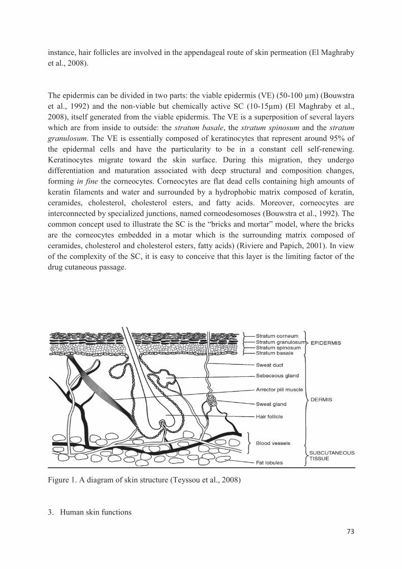
73
instance, hair follicles are involved in the appendageal route of skin permeation (El Maghraby et al., 2008).
The epidermis can be divided in two parts: the viable epidermis (VE) (50-100 μm) (Bouwstra et al., 1992) and the non-viable but chemically active SC (10-15μm) (El Maghraby et al., 2008), itself generated from the viable epidermis. The VE is a superposition of several layers which are from inside to outside: the stratum basale, the stratum spinosum and the stratum granulosum. The VE is essentially composed of keratinocytes that represent around 95% of the epidermal cells and have the particularity to be in a constant cell self-renewing. Keratinocytes migrate toward the skin surface. During this migration, they undergo differentiation and maturation associated with deep structural and composition changes, forming in fine the corneocytes. Corneocytes are flat dead cells containing high amounts of keratin filaments and water and surrounded by a hydrophobic matrix composed of keratin, ceramides, cholesterol, cholesterol esters, and fatty acids. Moreover, corneocytes are interconnected by specialized junctions, named corneodesomoses (Bouwstra et al., 1992). The common concept used to illustrate the SC is the “bricks and mortar” model, where the bricks are the corneocytes embedded in a motar which is the surrounding matrix composed of ceramides, cholesterol and cholesterol esters, fatty acids) (Riviere and Papich, 2001). In view of the complexity of the SC, it is easy to conceive that this layer is the limiting factor of the drug cutaneous passage.
Figure 1. A diagram of skin structure (Teyssou et al., 2008)
3. Human skin functions
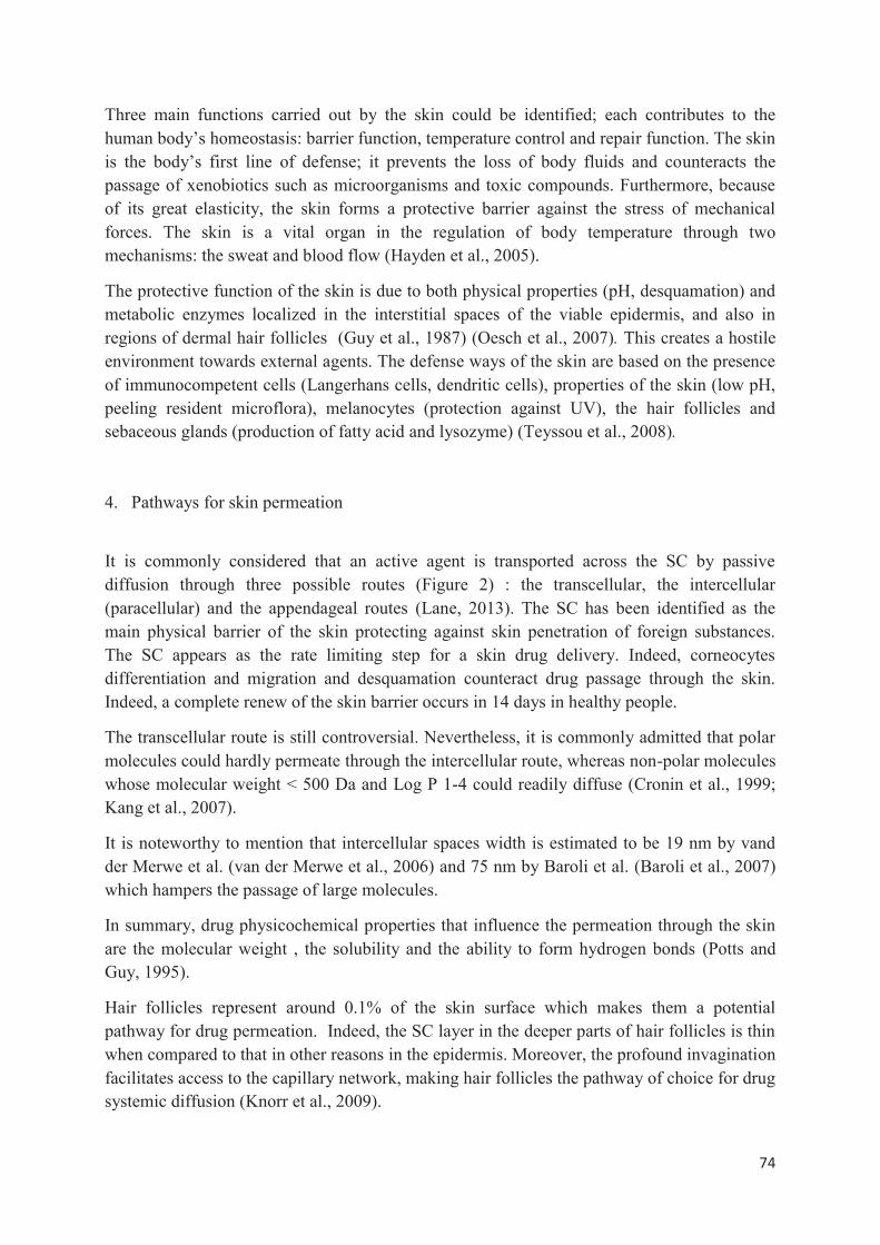
74
Three main functions carried out by the skin could be identified; each contributes to the human body’s homeostasis: barrier function, temperature control and repair function. The skin is the body’s first line of defense; it prevents the loss of body fluids and counteracts the passage of xenobiotics such as microorganisms and toxic compounds. Furthermore, because of its great elasticity, the skin forms a protective barrier against the stress of mechanical forces. The skin is a vital organ in the regulation of body temperature through two mechanisms: the sweat and blood flow (Hayden et al., 2005).
The protective function of the skin is due to both physical properties (pH, desquamation) and metabolic enzymes localized in the interstitial spaces of the viable epidermis, and also in regions of dermal hair follicles (Guy et al., 1987) (Oesch et al., 2007). This creates a hostile environment towards external agents. The defense ways of the skin are based on the presence of immunocompetent cells (Langerhans cells, dendritic cells), properties of the skin (low pH, peeling resident microflora), melanocytes (protection against UV), the hair follicles and sebaceous glands (production of fatty acid and lysozyme) (Teyssou et al., 2008).
4. Pathways for skin permeation
It is commonly considered that an active agent is transported across the SC by passive diffusion through three possible routes (Figure 2) : the transcellular, the intercellular (paracellular) and the appendageal routes (Lane, 2013). The SC has been identified as the main physical barrier of the skin protecting against skin penetration of foreign substances. The SC appears as the rate limiting step for a skin drug delivery. Indeed, corneocytes differentiation and migration and desquamation counteract drug passage through the skin. Indeed, a complete renew of the skin barrier occurs in 14 days in healthy people.
The transcellular route is still controversial. Nevertheless, it is commonly admitted that polar molecules could hardly permeate through the intercellular route, whereas non-polar molecules whose molecular weight < 500 Da and Log P 1-4 could readily diffuse (Cronin et al., 1999; Kang et al., 2007).
It is noteworthy to mention that intercellular spaces width is estimated to be 19 nm by vand der Merwe et al. (van der Merwe et al., 2006) and 75 nm by Baroli et al. (Baroli et al., 2007) which hampers the passage of large molecules.
In summary, drug physicochemical properties that influence the permeation through the skin are the molecular weight , the solubility and the ability to form hydrogen bonds (Potts and Guy, 1995).
Hair follicles represent around 0.1% of the skin surface which makes them a potential pathway for drug permeation. Indeed, the SC layer in the deeper parts of hair follicles is thin when compared to that in other reasons in the epidermis. Moreover, the profound invagination facilitates access to the capillary network, making hair follicles the pathway of choice for drug systemic diffusion (Knorr et al., 2009).
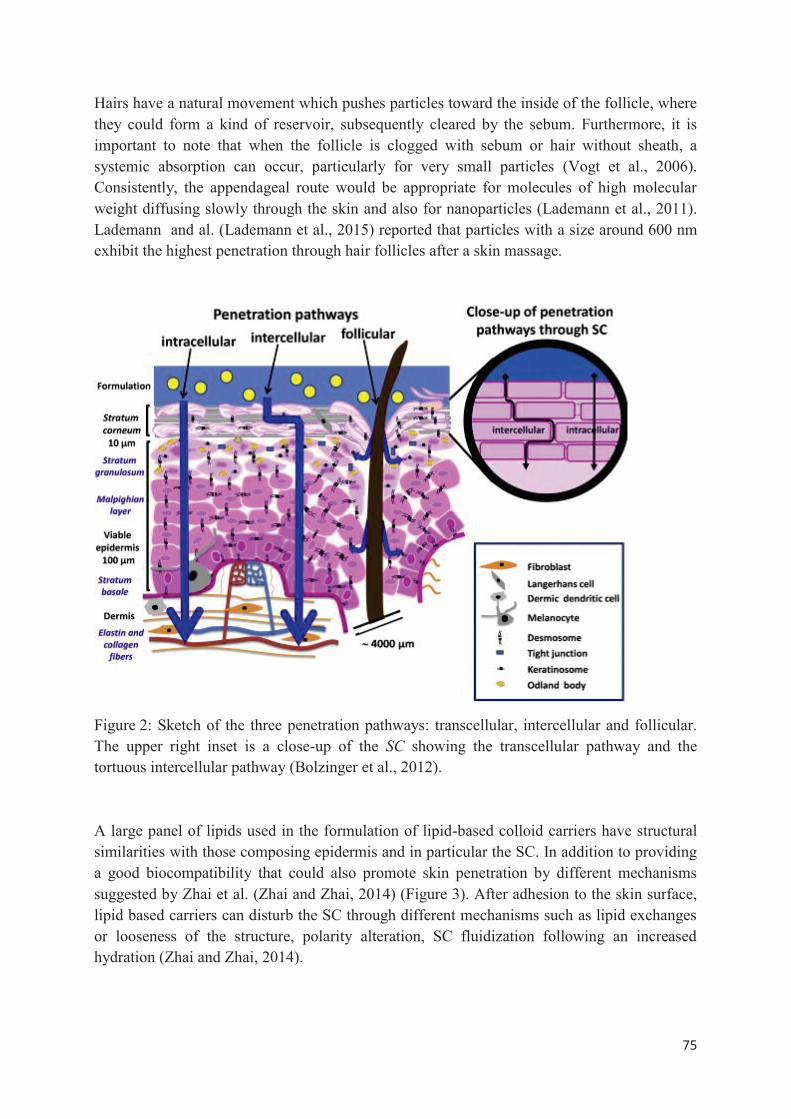
75
Hairs have a natural movement which pushes particles toward the inside of the follicle, where they could form a kind of reservoir, subsequently cleared by the sebum. Furthermore, it is important to note that when the follicle is clogged with sebum or hair without sheath, a systemic absorption can occur, particularly for very small particles (Vogt et al., 2006). Consistently, the appendageal route would be appropriate for molecules of high molecular weight diffusing slowly through the skin and also for nanoparticles (Lademann et al., 2011). Lademann and al. (Lademann et al., 2015) reported that particles with a size around 600 nm exhibit the highest penetration through hair follicles after a skin massage.
Figure 2: Sketch of the three penetration pathways: transcellular, intercellular and follicular. The upper right inset is a close-up of the SC showing the transcellular pathway and the tortuous intercellular pathway (Bolzinger et al., 2012).
A large panel of lipids used in the formulation of lipid-based colloid carriers have structural similarities with those composing epidermis and in particular the SC. In addition to providing a good biocompatibility that could also promote skin penetration by different mechanisms suggested by Zhai et al. (Zhai and Zhai, 2014) (Figure 3). After adhesion to the skin surface, lipid based carriers can disturb the SC through different mechanisms such as lipid exchanges or looseness of the structure, polarity alteration, SC fluidization following an increased hydration (Zhai and Zhai, 2014).
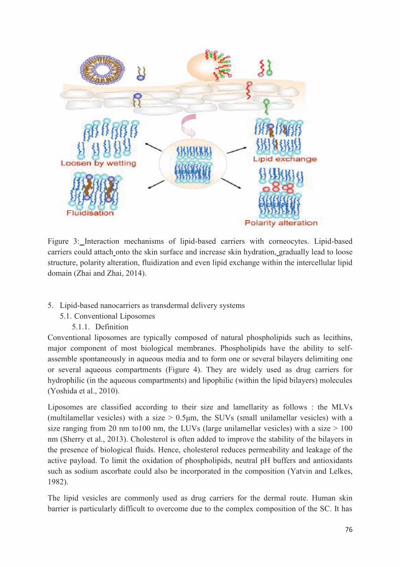
76
Figure 3: Interaction mechanisms of lipid-based carriers with corneocytes. Lipid-based carriers could attach onto the skin surface and increase skin hydration, gradually lead to loose structure, polarity alteration, fluidization and even lipid exchange within the intercellular lipid domain (Zhai and Zhai, 2014).
5. Lipid-based nanocarriers as transdermal delivery systems 5.1. Conventional Liposomes
5.1.1. Definition Conventional liposomes are typically composed of natural phospholipids such as lecithins, major component of most biological membranes. Phospholipids have the ability to self-assemble spontaneously in aqueous media and to form one or several bilayers delimiting one or several aqueous compartments (Figure 4). They are widely used as drug carriers for hydrophilic (in the aqueous compartments) and lipophilic (within the lipid bilayers) molecules (Yoshida et al., 2010).
Liposomes are classified according to their size and lamellarity as follows : the MLVs (multilamellar vesicles) with a size > 0.5μm, the SUVs (small unilamellar vesicles) with a size ranging from 20 nm to100 nm, the LUVs (large unilamellar vesicles) with a size > 100 nm (Sherry et al., 2013). Cholesterol is often added to improve the stability of the bilayers in the presence of biological fluids. Hence, cholesterol reduces permeability and leakage of the active payload. To limit the oxidation of phospholipids, neutral pH buffers and antioxidants such as sodium ascorbate could also be incorporated in the composition (Yatvin and Lelkes, 1982).
The lipid vesicles are commonly used as drug carriers for the dermal route. Human skin barrier is particularly difficult to overcome due to the complex composition of the SC. It has
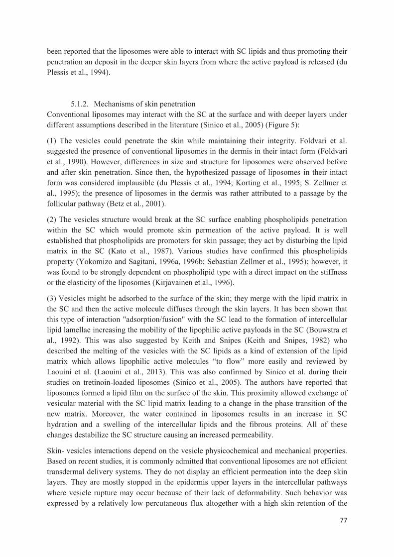
77
been reported that the liposomes were able to interact with SC lipids and thus promoting their penetration an deposit in the deeper skin layers from where the active payload is released (du Plessis et al., 1994).
5.1.2. Mechanisms of skin penetration Conventional liposomes may interact with the SC at the surface and with deeper layers under different assumptions described in the literature (Sinico et al., 2005) (Figure 5):
(1) The vesicles could penetrate the skin while maintaining their integrity. Foldvari et al. suggested the presence of conventional liposomes in the dermis in their intact form (Foldvari et al., 1990). However, differences in size and structure for liposomes were observed before and after skin penetration. Since then, the hypothesized passage of liposomes in their intact form was considered implausible (du Plessis et al., 1994; Korting et al., 1995; S. Zellmer et al., 1995); the presence of liposomes in the dermis was rather attributed to a passage by the follicular pathway (Betz et al., 2001).
(2) The vesicles structure would break at the SC surface enabling phospholipids penetration within the SC which would promote skin permeation of the active payload. It is well established that phospholipids are promoters for skin passage; they act by disturbing the lipid matrix in the SC (Kato et al., 1987). Various studies have confirmed this phospholipids property (Yokomizo and Sagitani, 1996a, 1996b; Sebastian Zellmer et al., 1995); however, it was found to be strongly dependent on phospholipid type with a direct impact on the stiffness or the elasticity of the liposomes (Kirjavainen et al., 1996).
(3) Vesicles might be adsorbed to the surface of the skin; they merge with the lipid matrix in the SC and then the active molecule diffuses through the skin layers. It has been shown that this type of interaction "adsorption/fusion" with the SC lead to the formation of intercellular lipid lamellae increasing the mobility of the lipophilic active payloads in the SC (Bouwstra et al., 1992). This was also suggested by Keith and Snipes (Keith and Snipes, 1982) who described the melting of the vesicles with the SC lipids as a kind of extension of the lipid matrix which allows lipophilic active molecules “to flow” more easily and reviewed by Laouini et al. (Laouini et al., 2013). This was also confirmed by Sinico et al. during their studies on tretinoin-loaded liposomes (Sinico et al., 2005). The authors have reported that liposomes formed a lipid film on the surface of the skin. This proximity allowed exchange of vesicular material with the SC lipid matrix leading to a change in the phase transition of the new matrix. Moreover, the water contained in liposomes results in an increase in SC hydration and a swelling of the intercellular lipids and the fibrous proteins. All of these changes destabilize the SC structure causing an increased permeability.
Skin- vesicles interactions depend on the vesicle physicochemical and mechanical properties. Based on recent studies, it is commonly admitted that conventional liposomes are not efficient transdermal delivery systems. They do not display an efficient permeation into the deep skin layers. They are mostly stopped in the epidermis upper layers in the intercellular pathways where vesicle rupture may occur because of their lack of deformability. Such behavior was expressed by a relatively low percutaneous flux altogether with a high skin retention of the
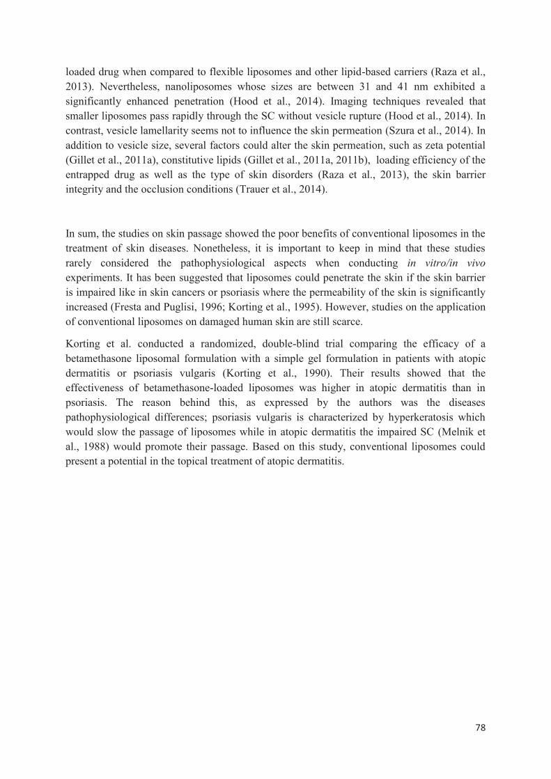
78
loaded drug when compared to flexible liposomes and other lipid-based carriers (Raza et al., 2013). Nevertheless, nanoliposomes whose sizes are between 31 and 41 nm exhibited a significantly enhanced penetration (Hood et al., 2014). Imaging techniques revealed that smaller liposomes pass rapidly through the SC without vesicle rupture (Hood et al., 2014). In contrast, vesicle lamellarity seems not to influence the skin permeation (Szura et al., 2014). In addition to vesicle size, several factors could alter the skin permeation, such as zeta potential (Gillet et al., 2011a), constitutive lipids (Gillet et al., 2011a, 2011b), loading efficiency of the entrapped drug as well as the type of skin disorders (Raza et al., 2013), the skin barrier integrity and the occlusion conditions (Trauer et al., 2014).
In sum, the studies on skin passage showed the poor benefits of conventional liposomes in the treatment of skin diseases. Nonetheless, it is important to keep in mind that these studies rarely considered the pathophysiological aspects when conducting in vitro/in vivo experiments. It has been suggested that liposomes could penetrate the skin if the skin barrier is impaired like in skin cancers or psoriasis where the permeability of the skin is significantly increased (Fresta and Puglisi, 1996; Korting et al., 1995). However, studies on the application of conventional liposomes on damaged human skin are still scarce.
Korting et al. conducted a randomized, double-blind trial comparing the efficacy of a betamethasone liposomal formulation with a simple gel formulation in patients with atopic dermatitis or psoriasis vulgaris (Korting et al., 1990). Their results showed that the effectiveness of betamethasone-loaded liposomes was higher in atopic dermatitis than in psoriasis. The reason behind this, as expressed by the authors was the diseases pathophysiological differences; psoriasis vulgaris is characterized by hyperkeratosis which would slow the passage of liposomes while in atopic dermatitis the impaired SC (Melnik et al., 1988) would promote their passage. Based on this study, conventional liposomes could present a potential in the topical treatment of atopic dermatitis.
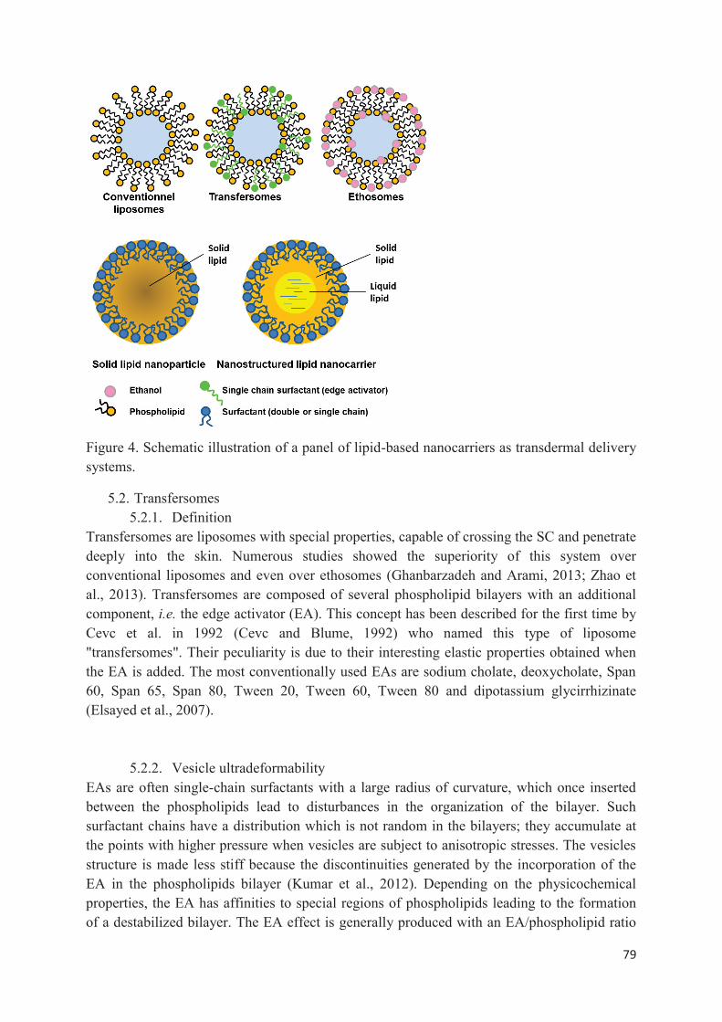
79
Figure 4. Schematic illustration of a panel of lipid-based nanocarriers as transdermal delivery systems.
5.2. Transfersomes 5.2.1. Definition
Transfersomes are liposomes with special properties, capable of crossing the SC and penetrate deeply into the skin. Numerous studies showed the superiority of this system over conventional liposomes and even over ethosomes (Ghanbarzadeh and Arami, 2013; Zhao et al., 2013). Transfersomes are composed of several phospholipid bilayers with an additional component, i.e. the edge activator (EA). This concept has been described for the first time by Cevc et al. in 1992 (Cevc and Blume, 1992) who named this type of liposome "transfersomes". Their peculiarity is due to their interesting elastic properties obtained when the EA is added. The most conventionally used EAs are sodium cholate, deoxycholate, Span 60, Span 65, Span 80, Tween 20, Tween 60, Tween 80 and dipotassium glycirrhizinate (Elsayed et al., 2007).
5.2.2. Vesicle ultradeformability EAs are often single-chain surfactants with a large radius of curvature, which once inserted between the phospholipids lead to disturbances in the organization of the bilayer. Such surfactant chains have a distribution which is not random in the bilayers; they accumulate at the points with higher pressure when vesicles are subject to anisotropic stresses. The vesicles structure is made less stiff because the discontinuities generated by the incorporation of the EA in the phospholipids bilayer (Kumar et al., 2012). Depending on the physicochemical properties, the EA has affinities to special regions of phospholipids leading to the formation of a destabilized bilayer. The EA effect is generally produced with an EA/phospholipid ratio
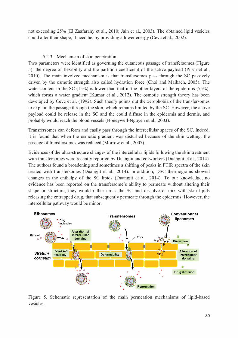
80
not exceeding 25% (El Zaafarany et al., 2010; Jain et al., 2003). The obtained lipid vesicles could alter their shape, if need be, by providing a lower energy (Cevc et al., 2002).
5.2.3. Mechanism of skin penetration Two parameters were identified as governing the cutaneous passage of transfersomes (Figure 5): the degree of flexibility and the partition coefficient of the active payload (Pirvu et al., 2010). The main involved mechanism is that transfersomes pass through the SC passively driven by the osmotic strength also called hydration force (Choi and Maibach, 2005). The water content in the SC (15%) is lower than that in the other layers of the epidermis (75%), which forms a water gradient (Kumar et al., 2012). The osmotic strength theory has been developed by Cevc et al. (1992). Such theory points out the xerophobia of the transfersomes to explain the passage through the skin, which remains limited by the SC. However, the active payload could be release in the SC and the could diffuse in the epidermis and dermis, and probably would reach the blood vessels (Honeywell-Nguyen et al., 2003).
Transfersomes can deform and easily pass through the intercellular spaces of the SC. Indeed, it is found that when the osmotic gradient was disturbed because of the skin wetting, the passage of transfersomes was reduced (Morrow et al., 2007).
Evidences of the ultra-structure changes of the intercellular lipids following the skin treatment with transfersomes were recently reported by Duangjit and co-workers (Duangjit et al., 2014). The authors found a broadening and sometimes a shifting of peaks in FTIR spectra of the skin treated with transfersomes (Duangjit et al., 2014). In addition, DSC thermograms showed changes in the enthalpy of the SC lipids (Duangjit et al., 2014). To our knowledge, no evidence has been reported on the transfersome’s ability to permeate without altering their shape or structure; they would rather cross the SC and dissolve or mix with skin lipids releasing the entrapped drug, that subsequently permeate through the epidermis. However, the intercellular pathway would be minor.
Figure 5. Schematic representation of the main permeation mechanisms of lipid-based vesicles.
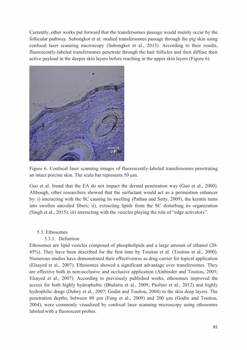
81
Currently, other works put forward that the transfersomes passage would mainly occur by the follicular pathway. Subongkot et al. studied transfersomes passage through the pig skin using confocal laser scanning microscopy (Subongkot et al., 2013). According to their results, fluorescently-labeled transfersomes penetrate through the hair follicles and then diffuse their active payload in the deeper skin layers before reaching in the upper skin layers (Figure 6).
Figure 6. Confocal laser scanning images of fluorescently-labeled transfersomes penetrating an intact porcine skin. The scale bar represents 50 μm.
Guo et al. found that the EA do not impact the dermal penetration way (Guo et al., 2000). Although, other researchers showed that the surfactant would act as a permeation enhancer by: i) interacting with the SC causing its swelling (Pathan and Setty, 2009), the keratin turns into swollen uncoiled fibers; ii), extracting lipids from the SC disturbing its organization (Singh et al., 2015); iii) interacting with the vesicles playing the role of “edge activators”.
5.3. Ethosomes 5.3.1. Definition
Ethosomes are lipid vesicles composed of phospholipids and a large amount of ethanol (20-45%). They have been described for the first time by Touitou et al. (Touitou et al., 2000). Numerous studies have demonstrated their effectiveness as drug carrier for topical application (Elsayed et al., 2007). Ethosomes showed a significant advantage over transfersomes. They are effective both in non-occlusive and occlusive application (Ainbinder and Touitou, 2005; Elsayed et al., 2007). According to previously published works, ethosomes improved the access for both highly hydrophobic (Bhalaria et al., 2009; Paolino et al., 2012) and highly hydrophilic drugs (Dubey et al., 2007; Godin and Touitou, 2004) to the skin deep layers. The penetration depths, between 80 μm (Fang et al., 2009) and 200 μm (Godin and Touitou, 2004), were commonly visualized by confocal laser scanning microscopy using ethosomes labeled with a fluorescent probes.
82
5.3.2. Ethanol effects Ethanol is known to be a skin penetration enhancer (Williams, 2003). Moreover, ethanol has also various effects on the physicochemical properties of ethosomes: ethanol reduces vesicle size (Dayan and Touitou, 2000); gives a negative surface charge which could lead to a decreased size, increased stability over time (López-Pinto et al., 2005), and to a better passage of the skin barrier (Gillet et al., 2011b).
5.3.3. Mechanism of skin penetration In an attempt to elucidate the involved mechanisms, Touitou and co-workers assayed phospholipid and ethanol that could penetrate the murine skin layers over 24 hours of exposition to ethosomes (Touitou et al., 2000). Surprisingly, 10.5% of phospholipids and 2% of ethanol were found in all the skin layers and were readily assayed in the receiver compartment of Franz diffusion cell (Dayan and Touitou, 2000). A synergistic influence of phospholipid and ethanol, interacting with each other and with the skin barrier, have been suggested as follows (Dayan and Touitou, 2000) : i) ethanol, a well know permeation enhancer, causes disturbance in the skin barrier, more precisely, it disturbs the organization of the SC lipids by both enhancing the lipid fluidity and decreasing the density of the intercellular lipid domains, ii) vesicles interact with the disturbed SC, alter the intercellular lipid lamella and hence creating their own pathways across the SC to the deep skin layers, iii) ethanol increases the vesicle fluidity and flexibility, caused by increasing the mobility of polar lipid heads of lipid molecules. Such elasticity facilitates vesicle crossing in the disturbed intercellular narrow pathways. This reasoning is supported by the fact that permeation from ethosomes was greatly enhanced when compared to that achieved by conventional liposomes or any of the system components alone.
It is noteworthy to mention that ethanol content in ethosomes seems to significantly influence permeation flux (Esposito et al., 2004; Li et al., 2012; Verma and Fahr, 2004). In a general way, the higher the ethanol content, the greater the permeation flux (Esposito et al., 2004). An additional plausible mechanism suggests ethosomes passage through the follicular pathway (Dayan and Touitou, 2000). Today, no evidence supporting this latter has been reported in the literature.
5.4. Penetration enhancer-containing vesicles (PEVs) Ethosomes are considered as the first generation of the penetration enhancer-containing vesicles (PEVs). Such systems contain penetration enhancers (PEs) in order to promote skin passage but also to solubilize active molecules that are poorly soluble in water, ethanol or methanol. PEs have similar properties to those of EA offering a better cutaneous passage via a greater flexibility (Castangia et al., 2013; Dragicevic-Curic et al., 2008; El Maghraby et al., 2000; Elsayed et al., 2007; Manconi et al., 2009; Mura et al., 2009). The core team working on this new generation of vesicles for skin applications is the group of Manconi et al. ((Manconi et al., 2011b, 2011a) Caddeo et al., 2013; Castangia et al., 2013; Manca et al., 2013). The synergistic combination of PEs and lipid vesicles produces promising skin drug delivery systems.
83
According to Chessa et al. (Chessa et al., 2011) , the use of PEG 400 (up to 40% in the aqueous phase) as PE provides a suitable skin deposition of the of quercetin, a lipophilic antioxidant agent, in different skin layers, mainly in the epidermis. In addition, a significant amount of quercetin was found in the receiver compartment of Franz diffusion cells, which is in favor of an effective transdermal passage. The penetration of the PEVs at the epidermis level is interesting for the treatment of skin diseases like psoriasis in which the epidermis has a fundamental role in the pathogenesis. Besides, this study also showed that the application of PEVs should be non-occlusive in order to obtain an effective dermal passage.
Manca et al. (Manca et al., 2013) used glycerol to increase the fluidity of lipid bilayers. It has been shown that by increasing the glycerol concentration in the aqueous phase up to 30%, the vesicles become elastic, which proves glycerol’s role as EA. The authors also reported the glycerol role. Indeed, when used at ratios 20 - 30%, glycerol provided a strong deposition of the entrapped drug, i.e. diclofenac, in the SC at the level of the epidermis. A better skin penetration was observed for PEV when compared to the marketed product. Accordingly, PEVs gather all the advantages of the previously developed vesicles, i.e. transfersomes and ethosomes. There is currently no assumption about the type of mechanism involved in skin penetration. For theses purpose, additional studies are needed.
5.5. Solid lipid nanoparticles (SLNs) SLNs were introduced in 1991 as an encouraging carrier system positioning itself at the interface between the pre-existing lipid systems (emulsions and liposomes) and polymeric nanoparticle systems. SLNs may combine interesting properties of the two previous systems. Schematically, the lipid matrix is constituted by a solid lipid or a mixture of solid lipids at a rate of 0.1% (w/w) to 30% (w/w) dispersed in aqueous medium. The stability of the system can be ensured by a surfactant with a concentration generally ranging from 0.5% (w/w) to 5% (w/w) (Pardeike et al., 2009). SLNs exhibit many advantages as biocompatibility, drug protection against degradation, drug release control. Moreover, they are also interesting because of the possibility to scale up and to undergo sterilization process. Because of the lipid nature, SLNs are considered very attractive as skin drug carriers. Indeed, interactions between skin an SLNs lipids could enhance skin permeation of the encapsulated drugs (Zhai and Zhai, 2014). SLNs have a mean particle size ranging from about 40 to 1000 nm (Pardeike et al., 2009) what could facilitate drug penetration into deeper skin layer for the smaller size, because of the greater interfacial area. SLNs have also been described as having the particularity to enhance the benefit/risk ratio of topical therapy via epidermal targeting (Porter et al., 2013). The choice of constitutive lipids is important because of their direct impact on the colloid properties, e.g. the size, drug loading, but also the skin permeation and drug release profile. Jensen et al. (Jensen et al., 2011) prepared SLNs with three lipids of different polarities (distearate, tripalmitate, cetylpalmitate) and performed in vitro studies with the aim to understand effects of SLNs composition on skin penetration. The active payload was a corticosteroid, i.e. betamethasone-17-valerate (BMV). The smaller SLNs were obtained with distearate because BMV is soluble in this lipid (150.9 ± 0.12 nm). The small size related to a
84
greater surface area is in favor of a better interaction and penetration into the skin. Although it is commonly admitted that particles with a size > 10 nm cannot penetrate through the intact skin. However, it has been reported that in diseases where the skin barrier is defective, particles with a larger size could penetrate. The higher amount of BMV in the skin and particularly in the SC was observed after application of BMV-loaded distearate SLNs. SLN with distearate which has the nearest solubility to that of skin components would exhibit a better miscibility with the skin lipids than those with tripalmitate and cetylpalmitate. Accordingly, a suitable choice of SLNs constitutive lipids enable to adjust the drug release kinetic and the drug distribution between the skin layers (Jensen et al., 2011). Moreover, SLNs behavior also depends on the physicochemical properties of the active payload. Küchler et al. (Küchler et al., 2010) used electron spin resonance (ESR) and paraelectric spectroscopy (PS) to investigate drug-particle interactions as a function of the lipophilicity of three spin probes used as drug models i.e. TEMPO, log P 2.3; TEMPOL, log P 0.6; CAT-1, log P ≤ 0. TEMPO- and TEMPOL-loaded SLNs had a larger size than CAT-1-loaded SLNs (170–190nm versus 250 – 420nm). It has been demonstrated that TEMPO was distributed over the lipid particle surface whereas TEMPOL was found to be inserted in the deeper surfactant layers and CAT-1 in the outermost surfactant layers. These results match well with the fact that more spin probes are lipophilic, more they interact with the SLNs core . Thus, two sub-compartments in SLNs have been highlighted (Küchler et al., 2010). The formation of SLNs is based on the propriety of the solid lipids to melt at high temperature and then crystallize at lower temperature. However, these relatively perfect crystals have a tendency to recrystallize during storage resulting in a drug leakage and consequently, in a low loading efficiency (Zhai and Zhai, 2014).
5.6. Nanostructured lipid carriers (NLCs) NLCs are a second generation of lipid nanoparticles. The lipid matrix of NLCs is formed by the mix of solid lipids and liquid lipids (oils) in an usual ratio of 70:30 up to a ratio of 99.9:0.1. Introducing liquid lipids causes a melting point depression in comparison with a pure solid lipid. One of the observed advantages is the possibility to work at lower temperatures. Besides, it is important to note that despite the depression of the melting point, NLCs remain solid at the body temperature. As SLNs, NLCs can provide an interesting controlled release for different active molecules, offering protection for compounds that are sensitive to light, oxidation or hydrolysis (Pardeike et al., 2009). Furthermore, NLCs display a good biocompatibility and tolerability. As described previously, SLNs were characterized by a high degree of order resulting in different drawbacks. By reducing the order of the solid lipid matrix by adding liquid lipid, defects in the crystal structure are generated offering more space to drug inclusion and thus resulting in a higher drug loading and stability in comparison to SLNs (Obeidat et al., 2010) . Moreover, water content of the NLC suspension is lower than that of SLN suspension which enables to produce concentrated suspension of lipid nanoparticles and to incorporate them directly in dermal formulation (Pardeike et al., 2009). Colloidal properties and skin delivery of NLCs are dependent on the lipid composition, the entrapped drug and the particle size. Many authors have demonstrated the influence of these parameters. For example, Phatak et Chaudhari (Phatak and Chaudhari, 2013) prepared
85
aceclofenac-loaded NLCs with Compritol 888 ATO (solid lipid), PEG-8 Miglyol 812 (liquid lipid) and Polysorbate 80 (surfactant). The effect of the excipient proportions on colloidal properties was studied. The authors showed that by increasing the solid/liquid lipid ratio, the particle size decreased from 350 nm to134 nm. The stabilizer concentration and drug/lipids ratio are also reported to affect the entrapment efficiency (Phatak and Chaudhari, 2013). Depending on the excipients used in the formulation of NLCs, the site of action and the drug release kinetics could be adjusted. For example, Chen et al. (Chen et al., 2013) prepared Coenzyme Q10 loaded NLCs using cetyl palmitate, MCT (medium-chain triglycerides) and TEGO® Care450 (Polyglyceryl-3 Methylglucose Distearate) capable of targeting the epidermis and accumulate inside to provide a sustained release. Besides, Kawadkar et al. (Kawadkar et al., 2013) have developed flurbiprofen-loaded NLCs designed with coconut oil, stearic acid and soya lecithin which were found to permeate the deep skin layers and to reach the bloodstream.
5.7 Mechanisms of lipid nanoparticle (SLNs and NLCs) skin permeation Topical application of lipid nanoparticles (SLN and NLC) on the skin forms a monolayer because of their adhesiveness property. This is an hydrophobic film whose the resistance and the effects therefore depend on the size of the particles constituting it. Generally the particle size has to be inferior to 200nm. This film formation has an occlusive effect due to its hydrophobicity. The water loss from the skin caused by natural evaporation is prevented leading to a corneocyte packing reduction and an inter-corneocyte gaps enlargement. That could potentialize a deeper skin penetration of drugs (Desai et al., 2010). Zhai, et al. (Zhai and Zhai, 2014) performed research based on the exploration of skin histopathology modifications to attempt to elucidate NLC/skin interaction mechanism. Hematoxylin/eosin staining method was used in order to appreciate the effect of NLC application on the intercellular environment of the skin. It has been observed that the stratum corneum had lost its original organization becoming more scattered and loose after NLC treatment. NLCs could disturb intercellular packing which embeds keratinocytes and corneocytes especially by acting on corneodesmosomes or intercellular lipids composition. The tight junctions anomalies in stratum corneum would lead to the barrier function defectiveness and could promote the skin permeation of drugs (Zhai and Zhai, 2014). It has been observed that for SLNs the smaller the particle size, the better the skin penetration due to a higher occlusion and a greater interaction surface with the skin. Generally, the particle size has to be less than 260 nm (Mardhiah Adib et al., 2016). It has been also seen that an increase in liquid content reduced the occlusive effect (Teeranachaideekul et al., 2008) and the SLNs had a higher occlusive effect than NLCs (Souto et al., 2004). It is also reported that the occlusion effect is function of the sample volume applied, the cristallinity and the lipid rate (Teeranachaideekul et al., 2008). Lombardi Borgia et al. (Lombardi Borgia et al., 2005) have highlighted that after application of the SLN preparation on the skin, its water content evaporates resulting in recrystallization phenomena which induce the partial release of the drug and its skin penetration.
86
The studies that have been performed on the skin permeation of conventional liposomes, which are vesicles made rigid by the presence of cholesterol, are generally agreed about the idea that they cannot penetrate the stratum corneum of an intact skin. By extrapolating these data, it appears very likely that if vesicular systems have failed to cross the intact stratum corneum, lipid nanoparticles being more rigid would give the same result. These lipid particulate systems could take the transappendageal pathway because their high lipophilicity promotes follicular deposition. Jensen et al. proposed a skin penetration mechanism for SLNs. They remained on the skin surface and in the stratum corneum forming a drug reservoir in the upper layers of the skin from where they released the encapsulated active molecules with a degree depending on the lipophilicity of the active ingredient to reach the target cells in the lower epidermis and dermis. SLN may release the active substance in a biphasic regimen, an initial burst release from the surface of the particles and the aqueous phase followed by a reservoir effect from the surface skin and stratum corneum (Jensen et al., 2011). The interaction of the SLNs with the skin lipids and sebum is observed to be influenced by the drug lipophilicity and the drug partitioning in the lipid particle, the nature of the lipid matrix and the type of interacting skin lipid. In 2009, Küchler et al. (Küchler et al., 2009) showed that SLNs prepared with compritol exhibit a platelet like shape which enable to slide between the superficial corneocytes by multiplied interactions with skin lipids. Moreover, it has been also reported that these SLNs underwent many changes over the time during skin surface contacts(Küchler et al., 2009). A study on the skin permeation of NLCs performed by Teeranachaideekul et al. (Teeranachaideekul et al., 2008) has showed that these lipid nanoparticles display a deeper penetration up to upper dermal layer when the content of liquid lipid is lower. And inversely a high content in liquid lipid limits the skin penetration at the upper viable epidermis. That is in favor of a better skin permeation of SLNs in comparison with NLCs (Teeranachaideekul et al., 2008).
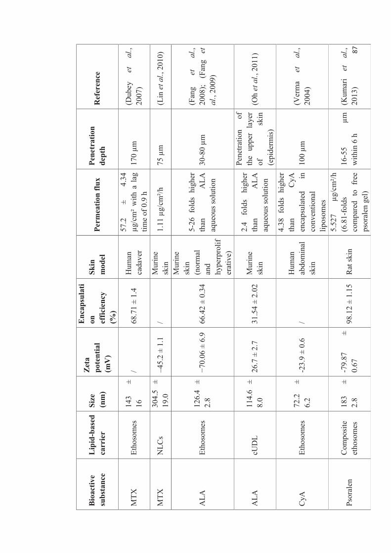
87
Bio
activ
e su
bsta
nce
Lip
id-b
ased
ca
rrie
r Si
ze
(nm
)
Zet
a po
tent
ial
(mV
)
Enc
apsu
lati
on
effic
ienc
y (%
)
Skin
m
odel
Pe
rmea
tion
flux
Pe
netr
atio
n de
pth
R
efer
ence
MTX
Et
hoso
mes
14
3 ±
16
/ 68
.71
± 1.
4 H
uman
ca
dave
r
57.2
±
4.34
μg
/cm
² w
ith a
lag
tim
e of
0.9
h
170 μm
(D
ubey
et
al
., 20
07)
MTX
N
LCs
304.
5 ±
19.0
–4
5.2
± 1.
1 /
Mur
ine
skin
1.
11 μ
g/cm
²/h
75 μ
m
(Lin
et a
l., 2
010)
ALA
Et
hoso
mes
12
6.4
± 2.
8 −7
0.06
± 6
.9
66.4
2 ±
0.34
Mur
ine
skin
(n
orm
al
and
hype
rpro
lifer
ativ
e)
5-26
fol
ds h
ighe
r th
an
ALA
aq
ueou
s sol
utio
n 30
-80 μm
(F
ang
et
al.,
2008
); (F
ang
et
al.,
2009
)
ALA
cU
DL
114.
6 ±
8.0
26.7
± 2
.7
31.5
4 ±
2.02
M
urin
e sk
in
2.4
fold
s hi
gher
th
an
ALA
aq
ueou
s sol
utio
n
Pene
tratio
n of
th
e up
per
laye
r of
sk
in
(epi
derm
is)
(Oh
et a
l., 2
011)
CyA
Et
hoso
mes
72
.2
± 6.
2 -2
3.9
± 0.
6 /
Hum
an
abdo
min
al
skin
4.38
fol
ds h
ighe
r th
an
CyA
en
caps
ulat
ed
in
conv
entio
nal
lipos
omes
100 μm
(V
erm
a et
al
., 20
04)
Psor
alen
C
ompo
site
et
hoso
mes
18
3 ±
2.8
-79.
87
± 0.
67
98.1
2 ±
1.15
R
at sk
in
5.52
7 μg
/cm
²/h
(6.8
1-fo
lds
com
pare
d to
fre
e ps
oral
en g
el)
16-5
5 μm
w
ithin
6 h
(K
umar
i et
al
., 20
13)
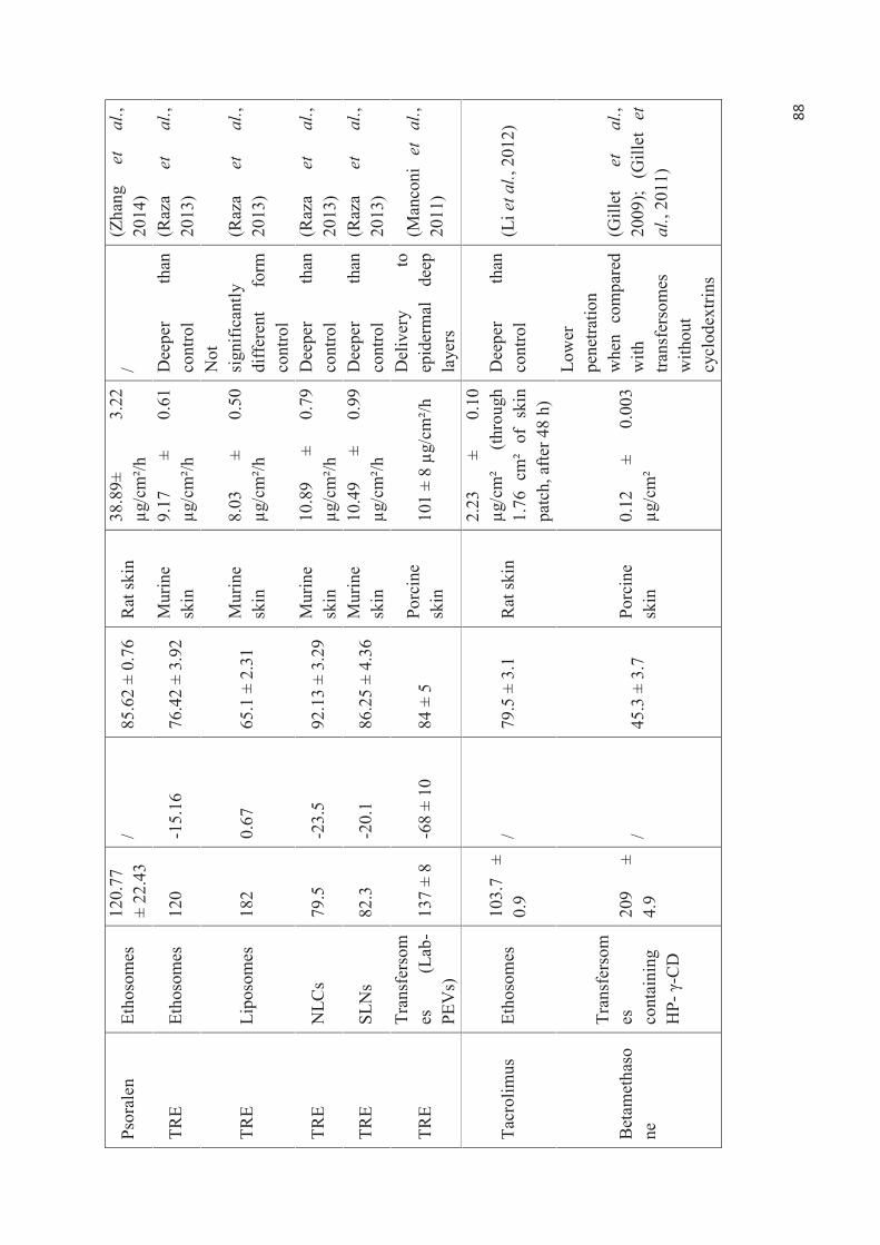
88
Psor
alen
Et
hoso
mes
12
0.77
±
22.4
3 /
85.6
2 ±
0.76
R
at sk
in
38.8
9±
3.22
μg
/cm
²/h
/ (Z
hang
et
al
., 20
14)
TRE
Etho
som
es
120
-15.
16
76.4
2 ±
3.92
M
urin
e sk
in
9.17
±
0.61
μg
/cm
²/h
Dee
per
than
co
ntro
l (R
aza
et
al.,
2013
)
TRE
Lipo
som
es
182
0.67
65
.1 ±
2.3
1 M
urin
e sk
in
8.03
±
0.50
μg
/cm
²/h
Not
si
gnifi
cant
ly
diff
eren
t fo
rm
cont
rol
(Raz
a et
al
., 20
13)
TRE
NLC
s 79
.5
-23.
5 92
.13
± 3.
29
Mur
ine
skin
10
.89
± 0.
79
μg/c
m²/h
D
eepe
r th
an
cont
rol
(Raz
a et
al
., 20
13)
TRE
SLN
s 82
.3
-20.
1 86
.25
± 4.
36
Mur
ine
skin
10
.49
± 0.
99
μg/c
m²/h
D
eepe
r th
an
cont
rol
(Raz
a et
al
., 20
13)
TRE
Tran
sfer
som
es
(Lab
-PE
Vs)
13
7 ±
8 -6
8 ±
10
84 ±
5
Porc
ine
skin
10
1 ±
8 μg
/cm
²/h
Del
iver
y to
ep
ider
mal
de
ep
laye
rs
(Man
coni
et
al
., 20
11)
Tacr
olim
us
Etho
som
es
103.
7 ±
0.9
/ 79
.5 ±
3.1
R
at sk
in
2.23
±
0.10
μg
/cm
² (th
roug
h 1.
76 c
m²
of s
kin
patc
h, a
fter 4
8 h)
Dee
per
than
co
ntro
l (L
i et a
l., 2
012)
Bet
amet
haso
ne
Tran
sfer
som
es
cont
aini
ng
HP-
γ-C
D
209
± 4.
9 /
45.3
± 3
.7
Porc
ine
skin
0.
12
± 0.
003
μg/c
m²
Low
er
pene
tratio
n w
hen
com
pare
d w
ith
trans
fers
omes
w
ithou
t cy
clod
extri
ns
(Gill
et
et
al.,
2009
); (G
illet
et
al
. , 20
11)
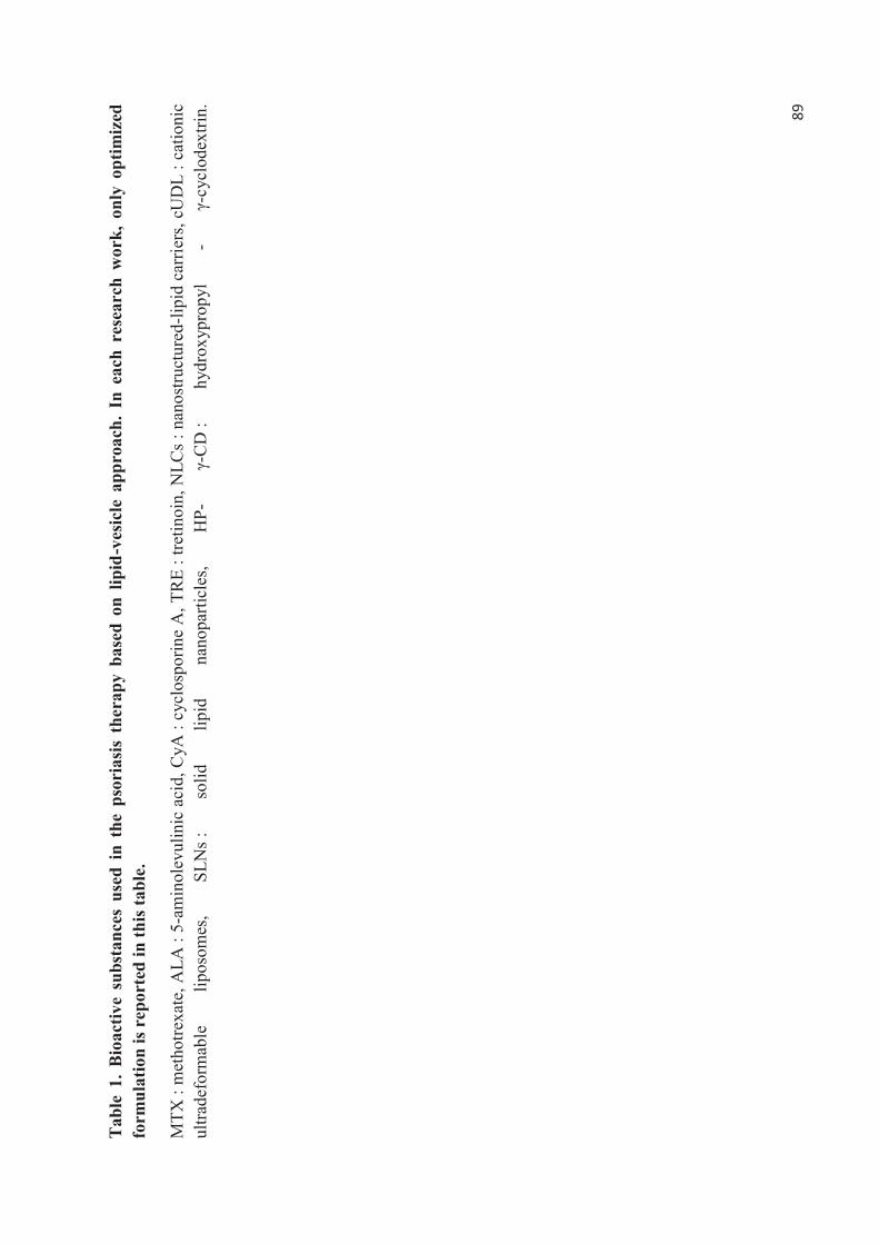
89
Tab
le 1
. B
ioac
tive
subs
tanc
es u
sed
in t
he p
sori
asis
the
rapy
bas
ed o
n lip
id-v
esic
le a
ppro
ach.
In
each
res
earc
h w
ork,
onl
y op
timiz
ed
form
ulat
ion
is r
epor
ted
in th
is ta
ble.
MTX
: m
etho
trexa
te, A
LA :
5-am
inol
evul
inic
aci
d, C
yA :
cycl
ospo
rine
A, T
RE
: tre
tinoi
n, N
LCs :
nan
ostru
ctur
ed-li
pid
carr
iers
, cU
DL
: cat
ioni
c ul
trade
form
able
lip
osom
es,
SLN
s :
solid
lip
id
nano
parti
cles
, H
P-
γ-C
D :
hydr
oxyp
ropy
l -
γ-cy
clod
extri
n.
90
6. Lipid carriers and medical applications 6.1. Treatment of skin disorders
Topical route represents the cornerstone in skin disorders treatment. High local concentrations of the loaded drug could be achieved while minimizing its systemic penetration and the related side effects. A number of marketed creams and gels were formulated with different permeation enhancers (e.g. ethanol, propylene glycol, myristate isopropyl, polysorbate 80, etc) and intended for topical delivery, such as Protopic® (tacrolimus cream), Retin A® (tretinoin cream), Méladinine® (methoxsalen gel), Finacea® (azelaic acid gel), etc. However, the superiority of lipid-based vesicles as topical carriers over marketed formulations and/or drug ethanolic solution have been demonstrated in terms of skin permeation and drug retention into the deepest skin strata, altogether with limited and, in some cases, undetectable concentrations of the drug in the systemic circulation. Recent advances in drug topical delivery based on lipid vesicle approach are reviewed hereafter.
6.1.1. Treatment of psoriasis and atopic dermatitis
6.1.1.1 Methotrexate (MTX) For the topical treatment of psoriasis, methotrexate (MTX) has been encapsulated into ethosomes (Dubey et al., 2007), transfersomes (Srisuk et al., 2012) and other lipid-based nanocarriers (Ali et al., 2008); (Lin et al., 2010); (Nagle et al., 2011). Indeed, MTX is known for its poor bioavailability, inter-individual variability and its numerous severe side effects (Hendel, 1985). Consistently, a topical or transdermal treatment would result in reduced systemic concentration of MTX altogether with high local concentration. Hence, formulations for topical use are expected to enhance the treatment effectiveness and, by the same way, to markedly limit undesirable effects. Unfortunately, transdermal permeation of MTX is limited, owing to its high hydrosolubility and high molecule weight. In this context, innovative formulations were sought. Toward this aim, Dubey and co-workers have developed ethosomal carriers of MTX that displayed nanometric size (143 ± 16 nm) and interesting encapsulation efficiency (68.71 ± 1.4 %) (Dubey et al., 2007). Skin permeation of the developed formulation has been assessed on human cadaver skin using fluorescently-labeled ethosomes and showed a 170 μm-penetration depth and a transdermal flux as high as 57.2 ± 4.34 μg/cm².
Earlier, Lin and co-workers have developed MTX-loaded nanostructured lipid carriers (NLCs) for topical delivery (Lin et al., 2010). In this work, several formulations of NLCs were carried out and characterized for their size, zeta potential and permeation for MTX through murine skin. NLCs showed an average size ranging from 267.3 ± 12.3 to 319.5 ± 12.2 nm, zeta potential values –41.8 ± 0.9 to –45.2 ± 1.1 mV. MTX amount permeating the skin was ranging from 0.61 to 1.11 μg/cm²/h, which is 2.4 to 4.4-times greater when compared to control. Skin penetration of fluorescently-labeled NLCs was observed in depths between 30 to 75 μm, i.e. in the epidermis and the upper layers of dermis.
Furthermore, deformable liposomes, also called transfersomes, were found to be promising carriers promoting skin permeability to MTX (Srisuk et al., 2012). Prepared from
91
phosphatidylcholine (PC) and oleic acid (OA) as an EA, deformable liposomes displayed enhanced permeability when compared to conventionnal liposomes, which was expressed by higher flux permeation and drug accumulation in the dermal and the epidermal layers of porcine skin (Srisuk et al., 2012). The higher performance was explained by the flexibility of deformable liposomes enabling them to penetrate through the skin pores even if they are much smaller than vesicle diameter. On the other hand, OA is a surfactant agent that causes dramatic changes of the morphology and the density of epidermal cells leading to the generation of pores on the surface of epidermal corneocytes (Srisuk et al., 2012). However, the use of OA resulted in lower encapsulation efficiency of MTX in deformable liposomes when compared to classical ones, which was attributed the repulsion between MTX and OA as they are both negatively-charged (Srisuk et al., 2012).
6.1.1.2 5-Aminolevulinic acid (ALA) 5-aminolevulinic acid (ALA) is a second-generation photosensitizer. It is used for topical photodynamic therapy (PDT) of psoriasis and acne with long-term therapeutic effects and better compliance with patients. Being highly hydrophilic, ALA use in PDT is hampered by its poor penetration into healthy or diseased skin. To address this issue, Fang and co-workers (Fang et al., 2008); (Fang et al., 2009) elaborated ethosomal and liposomal formulations of ALA with interesting physico-chemical characteristics (table1). Liposomes and ethosomes were prepared according to the thin-film hydration method and the optimized formulation consists on Phosphatidylethanolamine (PE), cholesterol (CH) and a surfactant (sodium stearate (SS)) with 0.1% ALA dissolved in 15% of ethanol solution or aqueous solution for ethosomes and liposomes preparation, respectively (Fang et al., 2008). It is noteworthy that entrapment efficiency of PE/CH/SS formulation was about two orders of magnitude higher for ethosomes than that of liposomes (without ethanol) (Fang et al., 2008).
Later, the same research group has investigated the pharmacological behavior of ALA ethosomal formulations (Fang et al., 2009). The therapeutic response was assessed in terms of ALA penetration behavior, protoporphyrin IX (PpIX) intensity in the skin and expression level of tumor necrosis factor (TNF-α). Cumulative amounts of the ethosomal ALA showed significant improvement in both normal and hyperproliferative murine skin when compared to ALA aqueous solution, and the ranges of increases were 5- to 26-fold (Fang et al., 2009). The penetration behavior was also highlighted using CLSM technique in order to visualize the intensity of PpIX, formed after the in vivo conversion of ALA. The maximal absorption of PpIX had a broad distribution of 30–80 μm, which indicates that PpIX remained in the lower epidermis and passed through the upper dermis (Fang et al., 2009). Furthermore, TNF-α is an indicator of perturbation of the skin barrier function and more precisely reflects the level of epidermal disruption. Ethosomal ALA caused about a 20% decrease in TNF-α levels compared to that of untreated group while only a slight decrease was observed using ALA ethanolic solution (Fang et al., 2009).
In a more recent research work (Oh et al., 2011), ALA was encapsulated in ultradeformable liposomes (UDL) with different surface charges, i.e. cationic, anionic and neutral. Cationic UDL (cUDL) were found to be the most efficient carriers for ALA skin delivery when compared to anionic and neutral UDL. Surprisingly, despite the fact that the SC is negatively-
92
charged and neutral or anionic vesicles are expected to better permeate through it, cUDL demonstrated higher permeability and penetration depth when compared to anionic and neutral UDL (Oh et al., 2011). Moreover, it is noteworthy to mention that two clinical trials were conducted in order to assess the therapeutic effectiveness of ALA-loaded in conventional liposomes for the treatment of inflammatory facial acne (Yeung et al., 2011); (An et al., 2011). Encapsulation into liposomes enabled low concentration of ALA to be used in PDT. Promising results were obtained in twelve subjects who participated in this study since the inflammatory acne was reduced altogether with lowering the risk of persistent post-treatment phototoxicity (Yeung et al., 2011). These results were consistent with those of another pilot study on thirteen subjects, performed in the same year by another research group (An et al., 2011).
6.1.1.3 Cyclosporine A (CyA) Cyclosporine A (CyA) is an oligopeptide composed of 11 amino acids that showed a remarkable efficacy in psoriasis (de la Brassinne & Nikkels, 2013) and other dermatological diseases such as atopic dermatitis (Roekevisch et al., 2014) and alopecia areata (Park et al., 2013). However, systemic delivery is accompanied by several side effects because of the immunosuppressive effects of CyA, the inhibition of hepatic enzymes and P-glycoprotein pump in addition to the severe nephrotoxicity (Roekevisch et al., 2014). On the other hand, the poor skin permeation of CyA makes the topical delivery unfeasible. Once again, researcher’s attention was drawn to ethosomal formulations (Verma & Fahr, 2004). The vesicles were composed of marketed mixture of phospholipids (NAT 8539), ethanol at different ratios (from 3.3 to 20 %) and CyA 0.4% (w/v) in phosphate buffered saline. CyA vesicles containing 10% and 20% ethanol showed a statistically enhanced deposition of CyA into the SC as compared to vesicles prepared without ethanol (conventional liposomes). Vesicles composed of NAT 8539/ethanol (10/10) showed the highest accumulation of CyA in the SC as compared to other formulations elaborated in this study. Moreover, the penetration depth was assessed in an abdominal human skin using radioactively-labeled CyA. Interestingly, radioactivity was detected after 6-h exposure in depths up to 1600 μm for all of the formulations, but the highest accumulation intensity was noticed for vesicles containing 10% of ethanol (Verma & Fahr, 2004).
6.1.1.4 Psoralen Psoralen is a photosentizer molecule used in the PDT of psoriasis. Kumari and his co-workers developed composite ethosomes as topical vehicle of psoralen (Kumari & Pathak, 2013). Composite ethosomes were based on soya lecithin (i.e. phosphatidylcholine), cavamax W7 (β-cyclodextrine), ethanol and propylene glycol. The injection method was adopted as the preparation method followed by a purification step by dialysis. According to the authors, the combined use of phosphatidylcholine, β-cyclodextrine, ethanol and propylene glycol displayed 7-folds enhancement of the solubility of psoralen, a highly lipophilic molecule. The first one plays the role of surfactant while the other components play the role of co-solvents. Composites ethosomes were introduced in carbopol gel and were compared with gel containing psoralen-loaded ethosomes (without β-cyclodextrine), β-cyclodextrine inclusion complex and with the free drug. Composite ethosomes-containing gel demonstrated the highest permeation flux followed by ethosomes-containing gel, inclusion complex and free
93
psoralen being the lowest one. The skin permeation rate of the composite ethosomal gel was 6.81 folds greater than free psoralen gel.
Zhang and his colleagues elaborated another ethosomal formulation of psoralen intended for topical use (Zhang et al., 2014). This formulation was based on soybean lecithin and ethanol and prepared according to injection method. Subsequently, ethosomes optimized formulation displayed 6.56 folds enhancement of the permeation rate when compared to psoralen tincture. In this study, the authors put to evidence that the ability of ethosomes to penetrate the skin is closely related to the proportion of phospholipids and ethanol in the formulation. The formulation containing the highest proportion of phospholipids (10% w/v) and ethanol (45 % v/v) achieved the lowest skin permeation at 24 h (1.98 ± 0.90 μg/cm²). However, when the proportion of both phospholipids and ethanol was reduced, higher permeation rates were observed. For instance, formulation containing 6% w/v of phospholipids and 25 % v/v of ethanol showed a permeation rate of 2.84 ± 1.46 μg/cm² at 24 h. Moreover, the authors demonstrated that the permeation rate depends of the skin region; the abdomen skin being the most permeable area when compared to the chest or scapula.
6.1.1.5 Tretinoin (TRE) Tretinoin (TRE) is used in the treatment of several skin diseases such as acne, psoriasis (Raza et al., 2013) , skin cancer (Quigley & Halpern, 2013) and photoaging (Hubbard et al., 2014). This molecule offers a good example highlighting the importance of lipid-based vesicles as drug carriers for skin delivery. TRE causes many unwanted side effects such as erythema, peeling and irritation. Furthermore, TRE is sensitive to air, light and heat that change it into ineffective substance. With regard to these drawbacks, several researches on formulation of lipid colloidal nanocarriers were conducted over the last few years.
Raza and his colleagues developed recently phospholipid-based particulate (solid lipid nanoparticles SLNs and NLCs) and vesicular (liposomes and ethosomes) nanocarriers of TRE and investigated their potential in promoting photo-stability, skin compliance and permeation (Raza et al., 2013). The composition of theses of carriers was slightly different between each other: Liposomes were composed of phosphatidylcholine (PC), cholesterol (CH) and prepared by thin-film hydration technique; ethosomes were composed of PC, CH and 20% of ethanol v/v and prepared by the cold method; SLNs and NLCs were both prepared by microemulsification technique and composed of PC, Compritol (glyceryl dibehenate) and Tween 80 (polysorbate). NLCs comprised an additional component, isopropyl myristate (IPM) known for its permeation enhancing activity. All the nanocarriers contained, butylated hydroxyl toluene (BHT) as an antioxidant (Raza et al., 2013). Once prepared, each type of nanocarriers was then incorporated into carbopol-based hydrogel. With respect to the foreseen response, each colloidal nanocarrier seemed to be less or more efficient: Regarding the stability response, the highest photoprotection was offered by NLCs followed by SLNS. Similarly, concerning the skin permeation rate, NLC-gel showed a higher permeation flux than that of SLN-gel which was in turn higher than ethosome- and liposome-gels ‘ones. In contrast, the highest drug retention was found for liposome-gel followed by NLC-gel and then by SLN-gel. Ethosome-gel showed the lowest skin retention. Interestingly, the control composed of RTE ethanolic solution showed the highest permeation skin but also
94
the lowest retention. Furthermore, deeper penetration than TRE commercial gel was observed for NLCs, SLNs and ethosomes; whereas liposomes failed to reach deeper layers of skin. Finally, liposomes and NLCs showed a significant better anti-psoriatic efficacy than the other tested products. Accordingly, the authors concluded that: “for treatment of deep-seated disorders like acne (located in sebaceous units), better penetrating carriers i.e. NLCs, SLNs and ethosomes can perform better than the liposomes. For treatment of superficial skin disorders like psoriasis, liposomes were found to be as good as SLNs and NLCs, while ethosomes were of no much utility. Appreciable biocompliance can be achieved with SLNs, NLCs and liposomes, but alcohol-containing ethosomes can be irritant” (Raza et al., 2013).
These results are consistent with previous research work demonstrating the efficiency of liposomes and niosomes to promote TRE cutaneous retention (Manconi et al., 2006). Nevertheless, the superiority of transfersomes, also called penetration enhancer-containing vesicles (PEV), as transdermal carriers of TRE remains unquestionable. Being ultra-deformable thanks to the presence of penetration enhancers (PEs) in their lipid wall, PEVs showed high suitability for intercellular passage (Chen et al., 2014). The research works conducted by Manconi and his co-workers highlighted the impact of hydrophilic PEs, playing the role of edge activators, on the performance of obtained vesicles (Manconi et al., 2011). In this work, four hydrophilic PEs were tested: decylpolyglucoside (Oramix® NS10, Or), caprylocaproyl macrogol 8-glyceride (Labrasol®, Lab), 2-(2-ethoxyethoxy)ethanol (Transcutol® P, Trc), and propylene glycol (PG). Vesicles were prepared by thin-layer hydration method using soybean phosphatidylcholine (Phospholipon 50, P50) and one of the selected PEs at predetermined concentration of 0.6% w/v. Indeed, according to the molecular structure, PE ability to interact with the bilayer structure was different, causing a molecular rearrangement and increased membrane fluidity. Trc and PG, which are water miscible and therefore are commonly used as co-solvents, were found to poorly interact with the lipid bilayer; whereas Or and Lab, hydrophilic surfactants with long hydrocarbon chains, effectively interact with the lipid bilayer. They made it more fluid decreased the energy necessary for the transition from planar lamellar phase to closed multilamellar vesicles (Manconi et al., 2011). Consistently, Lab- and Or-PEVs showed a remarkable stabilizing effect, as no relevant changes in vesicle size or encapsulation efficiency were detected over a period of 90 days (Manconi et al., 2011). Ex vivo skin permeation studies confirmed this reasoning. The highest TRE cutaneous accumulation were observed for Lab-PEVs followed by Or-PEVs, which in turn was higher than that achieved by Trc-PEVs and PG-PEVs (Manconi et al., 2011).
6.1.1.6 Tacrolimus Tacrolimus is an immunosuppressor agent commonly used for preventing rejection of organs in transplanted patients. Its topical use is intended for the treatment of many skin disorders (Scheinfeld, 2004), such as psoriasis and as a second-line therapy of atopic dermatitis also called atopic eczema in case of failure of topical corticoids. Nevertheless, a potent delivery of tacrolimus is mired by its poor permeability through biological barriers (e.g. gut and skin). Numerous attempts to overcome this issue are being done and already reviewed (Patel et al., 2012). Li and co-workers proposed ethosomal formulation of tacrolimus for topical use (Li et al., 2012). In this work, three types of ethosomes were developed using ethanol, propylene
95
glycol or a mixture of both PEs at a fixed concentration 30 % v/v. According to the authors (Li et al., 2012), ethanol showed superiority over propylene glycol in terms of encapsulation efficiency and permeation enhancing through the SC. The enhancing effect of ethanol seemed to be concentration-dependent. Besides, both types of ethosomes displayed better encapsulation efficiency and skin permeation than liposomes. The higher encapsulation efficiency was explained by the fact that tacrolimus is highly lipophilic and would be localized only in the lipid wall. Thus, the presence of ethanol or propylene glycol would allow better solubility of tacrolimus improving its distribution within the whole structure rather than to be sequestered in the lipid bilayers. Furthermore, both ethanol- and propylene glycol –containing ethosomes showed significantly greater therapeutic efficacy when compared commercial tacrolimus ointment or dexamethasone cream.
6.1.1.7 Bethamethasone Betamethasone is widely used in the treatment of inflammatory and auto-immune skin disorders such as acne, atopic dermatitis, psoriasis and cutaneous lupus erythematosus (Zenklusen & Feldmeyer, 2014). As all corticoids, betamethasone is highly hydrophobic and thus displaying poor permeation through skin strata. The early trends to improve corticoids permeation focused on liposomes (Jacobs et al., 1988). Since then, attention was directed towards more sophisticated carriers as classical liposomes enabled only limited or insufficient improvement in transdermal delivery (Korting et al., 1990). Later, a new concept combining the advantages of cyclodextrine inclusion complexes and those of deformable liposomes, also called transfersomes, was described (Gillet et al., 2009). Indeed, the use of cyclodextrins was shown to increase the drug to lipid mass ratio (Piel et al., 2006), while transfersomes remarkably enhanced transdermal delivery of several corticoids (Cevc & Blume, 2004). The main advantage of deformable liposomes when compared to classical ones consists on their high stress-dependent adaptability (Cevc & Blume, 2004). The edge activators integrated in their wall enabling them to squeeze when passing between cells without being disrupted. In the work of Gillet and his colleagues (Gillet et al., 2009); (Gillet b et al., 2011), sodium deoxycholate (SDC) was used as edge activator and soybean phosphatidylcholine (PC) or dimyristoylphosphatidylcholine (DMPC) for the preparation of deformable or non-deformable (without SDC) liposomes. Two types of cyclodextrins were used for the preparation of inclusion complexes: hydroxypropyl-γ-cyclodextrin (HP- γ-CD) and methyl-β-cyclodextrin (M- β-CD). It was found that the choice of cyclodextrine did not influence the size of deformable or non-deformable liposomes. For both deformable and classical liposomes, the use of PC and/or HP- γ-CD resulted in higher encapsulation efficiencies and drug to lipid ratios than those obtained with either DMPC or M- β-CD (Gillet et al., 2009).
Interestingly, the use of cyclodextrins slightly increased the deformability when assessed indirectly by measuring the percentage of PC recovered after vesicle extrusion through 50-nm-pore-size membrane under 5 bars-pressure (Gillet b et al., 2011). However, as expected, the percentage of recovered PC was notably higher for deformable liposomes (89.5 ± 12.9 and 84.1 ± 4.2 % with or without HP- γ-CD, respectively) if compared to classical liposomes (11.9 ± 1.7 and 3.5 ± 2.8 % with or without HP- γ-CD, respectively) (Gillet b et al., 2011). Unfortunately, deformable liposomes were found to be more sensitive to ultracentrifugation
96
than non-deformable ones (Gillet b et al., 2011). Therefore purification of liposomes at the end of preparation process would better be done by another technique. Moreover, it is found that deformable liposomes encapsulating betamethasone accumulate better in the dermis in comparison with deformable liposomes encapsulating betamethasone–cyclodextrine complexes (Gillet b et al., 2011).
6.1.2 Treatment of acne vulgaris Acne is an inflammatory skin disorders caused by excess of sebum secretion. It is characterized by hyperkeratinization and follicular hyperproliferation accompanied with bacterial colonization of follicular pilosebaceous, mainly by Propionobacterium acnes. Thus, treatment is mainly based on combinations of antibacterial (e.g. erythromycin, clindamycine and cyclines) and anti- keratinizing agents (e.g. tretinoin and azelaic acid) (Valente Duarte de Sousa, 2014). Indeed, topical therapy is the first choice for acne management. Similarly to psoriasis and atopic dermatitis context, lipid-based carriers displayed many advantages over classical topical formulations such as creams and gels containing permeation enhancers. Here are some examples of research works conducted this last decade.
6.1.2.1 Azelaïc acid (AA) Azelaic acid (AA) is a derivative of oleic acid with anti-keratinizing and bacteriostatic activities against aerobic and anaerobic bacteria (Sieber & Hegel, 2014). AA is efficient for the treatment of acne vulgaris, alopecia aerata and rosacea (Vargas-Diez et al., 2014); (Draelos et al., 2013). AA-loaded ethosomes were reported by (Esposito et al., 2004). Formulations were carried out by reverse-phase evaporation method using PC (5% w/v) and different concentrations of ethanol (from 20 to 45 % v/v). AA diffusion from ethosomes dispersions and from ethosomes incorporated in a carbomer gel was studied using 0.6 μm cellulose ester-based membrane. Diffusion was characterized by a steady-state flux for both dispersions and gels. The highest diffusion coefficients were found in the case of ethosomes containing 40% v/v of ethanol. Indeed, diffusion coefficients from gels were lower compared to dispersions, as vesicles are tightly packed in the viscous gel structure (Esposito et al., 2004). More recently, polymeric nanoparticulate systems were developed for AA topical delivery (Reis et al., 2013).
6.1.2.2 Bacitracin (BCT), erythromycin (ERY) Bacitracin (BCT) is a polypeptide antibiotic used in skin infection as a topical treatment (Cesur, 2002). Several types of lipid-based vesicles were developed as BCT carriers. Godin and Touitou elaborated BCT loaded-ethosomes with double objective (Godin & Touitou, 2004). The first one is to improve BCT delivery to the deep skin strata, where it will be efficacious against dermal and sub-dermal skin bacterial infections. Second, keeping in mind that some pathogens cause intracellular infections, an intracellular delivery of the antibiotic is crucial for their complete eradication. Ethosomes were prepared from phosphatidylcholine (phospholipon 90 PL 90) and the final concentration of ethanol in ethosomes was 25% w/w.
In order to elucidate ethosomal BCT permeation mechanisms, the authors labeled both ethosomal lipid walls with rhodamine red (RR) and BCT with fluorescein isothiocyanate
97
(FITC). With respect to the first objective, the authors checked the skin delivery of ethosomes on ex vivo human cadaver and rat skin using Franz diffusion cells. They found that the antibiotic was delivered into deep skin layers through intercorneocyte lipid domains of the SC. Both probes (RR and FITC) were delivered from ethosomes to a maximal depth of 200 μm. Moreover, co-localization of the labeled drug and labeled phospholipid was observed in this experiment, indicating that the carrier penetrated into the skin together with the drug (Godin & Touitou, 2004).
Toward the second objective of this work, intracellular penetration of ethosomal BCT in murine dermal fibroblasts (3T3) cell line was imaged by CLSM and quantified by fluorescent-activated cell sorting (FACS) (Godin & Touitou, 2004). CLSM studies revealed that ethosomes facilitated the co-penetration of antibiotic and phospholipid into fibroblasts. Consistently, FACS studies indicated that ethosomes penetrate cellular membrane releasing the entrapped molecules inside the cells (Godin & Touitou, 2004).
In a more recent work, BCT-loaded anionic niosomes were proposed as a good candidate as skin delivery system (Manosroi et al., 2010). Anionic niosomes were prepared using Tween 61, cholesterol and dicetylphosphate by freeze-dried empty liposomes method. High entrapment efficiency of BCT was achieved in anionic niosomes when compared to neutral or cationic ones (Manosroi et al., 2010). It is likely related to BCT positive charge and resulted electrical interaction with the negatively-charged component in lipidic wall (i.e. dicetylphosphate).
Besides, erythromycin (ERY) loaded-ethosomes were reported as efficient carriers for ERY delivery to deep dermal strata and subcutaneous infections (Godin & Touitou, 2005). Interestingly, improved antibacterial activity was observed for ethosomal formulations over hydroethanolic solution which was expressed by reduced minimal inhibition concentration (MIC) against Staphylococcus aureus.
6.1.2.3 Tretinoin tetracycline combination Dual skin delivery of TRE and tetracycline (TET) was aimed via their co-encapsulation in lipid nanocarriers, i.e. nanoemulsions (NEs) and NLCs (Lin et al., 2013). These systems were prepared from squalene, a non-ionic surfactant, i.e. Polyoxyethylene-polyoxypropylene copolymer (Pluronic® F68), palmitinic acid monoglyceride (Myverol®) and with glycerol distearate (Precirol®) for NLCs only. A cationic surfactant was added for the preparation of cationic NEs and cationic NLCs, i.e. soyaethyl morpholinium ethosulfate (Forestall®). NEs and NLS showed high entrapment of TRE which ranged from 60-100%., whereas TET mainly resided in the aqueous core. In vitro skin permeation through nude mouse skin, showed that NEs and NLCs significantly enhanced TET flux which was about 2-times over the control solution for TET-loaded NLCs. TRE permeation was unaffected by the nanoencapsulation as no significant difference was observed with respect to control. However, TET permeation significantly decreased by 2-fold in the dual-drug systems. TRE remained unaffected by the co-encapsulation with TET, except for cationic NLCs where TRE permeation was also lowered for the co-loaded vesicles. Nanoencapsulation did not modify the antibacterial activity of TET against Pseudomonas aeruginosa and Propionobacterium acnes. It was noticed that for both NE and NLC systems, negatively-charged formulations
98
enhanced the antibacterial activity against S. aureus compared to the control solution (Lin et al., 2013).
6.1.3. Treatment of skin infections 6.1.3.1 Antiviral drugs
Acyclovir (ACY) is a poorly-soluble molecule in both hydrophilic and lipophilic environment which hampers its skin permeation. Therefore, several attempts were done to address this issue. Cortesi and his co-workers proposed two lipid systems as ACY carriers, i.e. SLNs and ethosomes. Ethosomes seemed to entrap ACY more efficiently than SLNs (Cortesi et al., 2011). Both systems were found to enhance skin permeation to ACY. However, no significant difference was observed in the antiviral activity between ACY solution and ACY-loaded SLNs or ethosomes (Cortesi et al., 2011).
Another approach aiming to enhance ACY transdermal delivery was described (Zhou et al., 2010). This latter was based on the development of ACY lipophilic prodrug, i.e. acyclovir palmitate, which was subsequently loaded into ethosomes. A noticeably high drug quantity was delivered to the deep skin strata (i.e. 622.89 μg/cm²) over 24 h-exposition to ethosomal prodrug, which was 5.3- and 3.43 folds higher than the prodrug hydroethanolic solution and ethosomal drug, respectively (Zhou et al., 2010).
Mucoadhesive liposomal vesicles were also reported as efficient carriers for ACY (Naderkhani et al., 2014). Mucoadhesive liposomes were based on both phosphatidylcholine and phosphatidylglycerol and then incorporated into carbopol gel. The developed system displayed a significant increased in vitro permeability over ACY aqueous solution (Naderkhani et al., 2014).
6.1.3.2 Antifungal drugs Fluconazole (FLZ) loaded-ethosomes were described by (Bhalaria et al., 2009). Ethosomes were prepared by the hot method and showed to efficiently entrap FLZ. Enhanced skin diffusion together with enhance antifungal activity were observed over classical liposomes, marketed formulation or hydroethanolic solution.
Niosomes were also studied as topical nanocarriers for FLZ (Gupta et al., 2011). Niosomes were prepared by the thin layer hydration method using different types of surfactants. The choice of surfactant and the resulting vesicle size were found to significantly influence skin permeation and retention of FLZ. Niosomes containing Span 40, Span 60 and Brij 72 displayed a sustain drug release profile and a greatly enhanced skin retention (Gupta et al., 2011). Furthermore, unilamellar liposomes containing lysine oligopeptides (Lys-5 and Lys-7) were reported by (Schwarz et al., 2011). The use of Lys-5 and Lys-7 resulted in structural changes, decreased vesicle size and a retarding effect on FLZ skin permeation when compared to liposomes prepared without lysine oligopeptides (Schwarz et al., 2011). Thus, liposomes containing lysine oligopeptides are promising carriers able to bypass an undesirable fast percutaneous absorption and thereby to achieve prolonged local activity.
99
Terbinafine (TBF) is antifungal drug, mainly used for the treatment of onychomycosis. Several topical formulations are being developed in order to enhance its delivery through the nail plate barrier. Tanrıverdi and Özer recently proposed liposomal, ethosomal and gel formulation of TBF for the treatment of onychomycosis (Tanrıverdi & Özer, 2013). In this work, two types of phospholipids, i.e. Phospholipon 90 G (P90G) and Lipoid S100 (LS100), were used for liposomal and ethosomal formulations. Gels were prepared by incorporating optimized ethosomal and liposomal formulation into chitosan and poloxamer dispersions, respectively. It was found that formulations prepared from P90G achieved higher drug permeation than LS100-based formulations. Ex vivo nail permeation studies showed that the highest percentage of accumulated drug was observed with liposome poloxamer gel formulation. Moreover, ethosomal formulations showed higher irritation on nail surface after 10 days of treatment due to the high ethanol content (Tanrıverdi & Özer, 2013).
In another work, the performance of ethosomes was compared with ethosomes/transfersomes binary system as TBF-carriers in terms of skin permeation and skin retention (Zhang et al., 2012). Binary systems were prepared using ethanol and propylene glycol in a weight ratio of 7/3. It was found that binary system enabled higher penetration depth and higher skin retention of applied dose than ethosomes or transfersomes used alone. According to this study, ethosomes improved drug delivery with greater improvement in skin permeation than improvement in skin deposition (Zhang et al., 2012).
6.1.3.3 Curcumin Curcumin is a naturally-occurring polyphenol whose numerous therapeutic activities are still being discovered as an anti-inflammatory, anti-oxidant (Pyun et al., 2014), apoptotic (Terlikowska et al., 2014) and antibacterial agent (Sharma et al., 2014); (Manjunatha et al., 2013); (Liu & Huang, 2013). Transdermal formulations of curcumin are foreseen because of its poor bioavailability via systemic administration routes (Maiti et al., 2007). Different lipid-based carriers of curcumin were recently described, namely propylene glycol-containing liposomes (PGL), ethosomes and classical liposomes (Zhao et al., 2013). In this work, PGL showed superior performance over ethosomes and liposomes in terms of encapsulation efficiency, sustained-release behavior, permeation flux and skin deposition (Zhao et al., 2013). Transfersomes incorporated into carbopol gel were also reported as promising carrier of a combination of curcumin and diclofenac diethylamine for transdermal delivery (Chaudhary et al., 2014). The developed gel displayed an enhanced biological activity due to superior skin penetration of both drugs. Its average skin irritation scored 0.49 concluded to be non-irritant, safe for human use. Curcumin-loaded lipid vehicles were also developed as a novel treatment against Propionibacterium acnes (Liu & Huang, 2013). This study showed that lipid-vehicles act as curcumin reservoirs enabling a significant skin accumulation (362±8 μg/g skin) and leading to a significant antibacterial activity (Liu & Huang, 2013).
6.1.4 Treatment of skin cancer
100
Paclitaxel-loaded ethosomes® were described as a transdermal therapy of non-melanoma skin cancer (Paolino et al., 2012) drug delivery systems for the treatment of this pathology due to their suitable physicochemical characteristics and enhanced skin penetration ability for deep dermal delivery. In vitro skin permeation studies demonstrated an enhanced percutaneous flux of paclitaxcel-loaded ethosomes® which was about 23-folds than that of the hydroethanolic suspension of paclitaxcel (Paolino et al., 2012). More effective anti-proliferative activity in a squamous cell carcinoma model was obtained for ethosomes as compared to the free drug. At the same drug concentration, i.e. 5 μm, the mortality percentage of cancerous cells after 24h-exposition were double for encapsulated paclitaxcel when compared to free drug (Paolino et al., 2012). Transfersomes loaded with 5-fluorouracil (5-Fu), a chemotherapeutic agent with poor skin permeation, was developed as topical carriers for the treatment of deep-seated skin cancer (Khan et al., 2014). Two non-ionic surfactants, i.e. Tween 80 and Span 80, were used as edge activators. Transfersomes-based carbopol gel demonstrated a significantly increased percutaneous flux altogether avoiding irritant side effects of 5-FU (Khan et al., 2014).
6.2. Treatment of non-cutaneous disorders A myriad of research works are being published about the encapsulation of drug substances into phospholipid-based vesicles as transdermal delivery carriers for the treatment of systemic diseases. The following examples are not exhaustive.
6.2.1 Rheumatoid arthritis Non-steroidal anti-inflammatory drugs (NSAID) are the most prescribed drugs because of their multiple effects as anti-inflammatory, anti-pyretic and analgesics. More specifically, they are used in joint diseases as the first line therapy. Their numerous undesirable effects associated with systemic administration pushed formulators to develop transdermal delivery systems to be applied directly on the target site. Accordingly, different approaches are being applied on NSAID skin delivery, namely: transfersomes, liposomes and menthosomes. These different approaches were studied and compared by Duangjit and his co-workers as carriers for meloxicam (MX) (Duangjit a et al., 2014). Lipid vesicles were prepared from soybean phosphatidylcholine and cholesterol. Cetypyridinium chloride (CPC), a cationic surfactant, was also used in the formulation of deformable vesicles, i.e. transfersomes and menthosomes, as edge activator. L-menthol, a permeation enhancer affecting the lipid arrangement in the SC, was used in the formulation of menthosomes. Deformable liposomes displayed smaller vesicle sizes compared to conventional liposomes, with menthosomes being the smallest vesicles. Besides, menthosomes were found to be more elastic compared to transfersomes. Results of permeation studies were in accordance with other published observations (Raza et al., 2013); conventional liposomes enabled higher skin retention than deformable vesicles, while the highly elastic vesicles exhibited the highest permeation flux (Duangjit a et al., 2014). In an attempt to get a more in-depth sight, the influence of formulation components and particularly that of the EA were thoroughly studied (Duangjit b et al., 2014). Toward this aim, surfactants with different charges (anionic, neutral, and cationic), carbon chain lengths (C4, C12, and C16), with different contents (10%, 20%, and 29%) were used in the formulation of
101
MX-loaded transfersomes. The influence of these different factors on the physicochemical characteristics and the performance of obtained vesicles were studied using a response surface method incorporating multivariate spline interpolation. According to results, cationic surfactant produced vesicles with smaller size, higher elasticity and higher encapsulation efficiency when compared to neutral or anionic surfactants. The electrostatic interaction between the negatively-charge drug, i.e. MX, and the cationic surfactant may be a plausible reason. Surfactant with longer carbon chain produced vesicles with smaller size and smaller elasticity. The reduced size was explained by the fact that the hydrophobicity of long-chain carbons is greater than that of short-chain carbons, and long-chain carbons could have led to increased solubility of the surfactant molecule in the lipid bilayer. On the other hand, the deeper insertion into lipid bilayer, the tighter and less elastic vesicles are obtained. Finally, vesicle size increased with the increase of surfactant content, while elasticity and encapsulation efficiency were enhanced. Furthermore, both cationic and anionic surfactants were showed to enhance permeation flux of obtained vesicles when compared to those prepared with neutral surfactant. This observation was explained by the fact that anionic and cationic surfactants swell the SC and interact with intercellular keratin and thereby increasing skin permeation (Duangjit b et al., 2014). Glycerosomes, glycerol-containing liposomes, were described in the literature as an efficient tool for dermal and transdermal delivery of diclofenac (Manca et al., 2013). This work demonstrated that glycerol is able to act as an EA for phospholipid bilayer if used in concentration higher than 10%. Glycerosomes were found to enhance both permeation flux and skin retention when compared to conventional liposomes (Manca et al., 2013).
6.2.2 Parkinson Trihexyphenidyl HCl (THP)-loaded ethosomes were developed as a novel approach for the treatment of Parkinson’s disease (Dayan & Touitou, 2000). THP is known for its short half-life and thus requiring frequent dosing a day and so generating peak plasma levels that causes severe side effects. Indeed, THP is an ionizable molecule with limited skin permeation. In this context, Dayan and Touitou proposed ethosomal formulation of THP that displayed a percutaneous flux 87 and 4.5 times higher than from liposomes and hydroethanolic solution, respectively. Interestingly, skin retention of THP after an 18-h exposition was also enhanced compared to hydroethanolic solution (Dayan & Touitou, 2000).
6.2.3 Diabetes mellitus Trends toward transdermal delivery of insulin were carried out using transfersomes system (Cevc, 2003). Insulin-loaded transfersomes were found to be able to deliver the loaded macromolecule through the non-compromised skin barrier with a reproducible drug effect that closely similar to that of subcutaneous ultralente insulin (Cevc, 2003). Thus, transfersomes represent potential non-invasive systems for insulin delivery improving the quality of life of patients.
7. Conclusion Clearly, there is a lack of in vitro and in vivo studies dealing with skin penetration of lipid carriers implemented on skin diseases. Few elements would be in favor of higher drug
102
penetration in diseased skin (psoriasis or atopic dermatitis for example). That was particularly observed with conventional liposomes. Regarding other type of lipid vesicles containing different penetration enhancers and/or edge activators like ethosomes, PEVs and transfersomes, different mechanisms of skin interactions have been described, involving most of time an enhancement of fluidity and flexibility of lipid vesicles which would help skin passage associated with skin intercellular lipid rearrangement.
SLNs offer additional advantages in terms of drug encapsulation efficiency and colloidal stability. These elements have been further enhanced in NLCs. Regarding the mechanism of skin interactions, studies have reported the formation of a hydrophobic film on skin resulting in an occlusive effect leading to SC rearrangement which would promote skin penetration. It is of note that skin permeation also depends on the drug physic-chemical properties and the type of lipid matrix of the nanocarrier.
To conclude, no recommendation can be stated to guarantee the better skin penetration. Further comparative in vitro and in vivo studies are required to elucidate schematic nanocarriers behaviors but we always have to take into consideration that the impact of the active ingredient properties is of paramount importance.
8. References
Ainbinder, D., Touitou, E., 2005. Testosterone ethosomes for enhanced transdermal delivery. Drug Deliv. 12, 297–303. doi:10.1080/10717540500176910
Alexander, A., Dwivedi, S., Giri, T.K., Saraf, S., Saraf, S., Tripathi, D.K., others, 2012. Approaches for breaking the barriers of drug permeation through transdermal drug delivery. J. Controlled Release 164, 26–40.
Baroli, B., Ennas, M.G., Loffredo, F., Isola, M., Pinna, R., López-Quintela, M.A., 2007. Penetration of metallic nanoparticles in human full-thickness skin. J. Invest. Dermatol. 127, 1701–1712. doi:10.1038/sj.jid.5700733
Baron, J.M., Merk, H.F., 2001. Drug metabolism in the skin. Curr. Opin. Allergy Clin. Immunol. 1, 287–291.
Betz, G., Imboden, R., Imanidis, G., 2001. Interaction of liposome formulations with human skin in vitro. Int. J. Pharm. 229, 117–129.
Bhalaria, M.K., Naik, S., Misra, A.N., 2009. Ethosomes: a novel delivery system for antifungal drugs in the treatment of topical fungal diseases. Indian J. Exp. Biol. 47, 368–375.
Bhoyar, N., Giri, T.K., Tripathi, D.K., Alexander, A., . A., 2012. Recent Advances in Novel Drug Delivery System Through Gels: Review. J. Pharm. Allied Health Sci. 2, 21–39. doi:10.3923/jpahs.2012.21.39
Bolzinger, M.-A., Briançon, S., Pelletier, J., Chevalier, Y., 2012. Penetration of drugs through skin, a complex rate-controlling membrane. Curr. Opin. Colloid Interface Sci. 17, 156–165. doi:10.1016/j.cocis.2012.02.001
Bouwstra, J.A., Hofland, H.E.J., Spies, F., Gooris, G.S., Junginger, H.E., 1992. Changes in the Structure of the Human Stratum Corneum Induced by Liposomes, in: Braun-Falco, P.D. med D. h c
103
mult O., Korting, P.D. med H.C., D, P.H.I.M.M. (Eds.), Liposome Dermatics, Griesbach Conference. Springer Berlin Heidelberg, pp. 121–136. doi:10.1007/978-3-642-48391-2_13
Caddeo, C., Sales, O.D., Valenti, D., Saurí, A.R., Fadda, A.M., Manconi, M., 2013. Inhibition of skin inflammation in mice by diclofenac in vesicular carriers: liposomes, ethosomes and PEVs. Int. J. Pharm. 443, 128–136. doi:10.1016/j.ijpharm.2012.12.041
Cal, K., Centkowska, K., 2008. Use of cyclodextrins in topical formulations: practical aspects. Eur. J. Pharm. Biopharm. Off. J. Arbeitsgemeinschaft Für Pharm. Verfahrenstechnik EV 68, 467–478. doi:10.1016/j.ejpb.2007.08.002
Castangia, I., Manca, M.L., Matricardi, P., Sinico, C., Lampis, S., Fernàndez-Busquets, X., Fadda, A.M., Manconi, M., 2013. Effect of diclofenac and glycol intercalation on structural assembly of phospholipid lamellar vesicles. Int. J. Pharm. 456, 1–9. doi:10.1016/j.ijpharm.2013.08.034
Cevc, G., Blume, G., 1992. Lipid vesicles penetrate into intact skin owing to the transdermal osmotic gradients and hydration force. Biochim. Biophys. Acta 1104, 226–232.
Cevc, G., Schätzlein, A., Richardsen, H., 2002. Ultradeformable lipid vesicles can penetrate the skin and other semi-permeable barriers unfragmented. Evidence from double label CLSM experiments and direct size measurements. Biochim. Biophys. Acta 1564, 21–30.
Chen, S., Liu, W., Wan, J., Cheng, X., Gu, C., Zhou, H., Chen, S., Zhao, X., Tang, Y., Yang, X., 2013. Preparation of Coenzyme Q10 nanostructured lipid carriers for epidermal targeting with high-pressure microfluidics technique. Drug Dev. Ind. Pharm. 39, 20–28. doi:10.3109/03639045.2011.650648
Chessa, M., Caddeo, C., Valenti, D., Manconi, M., Sinico, C., Fadda, A.M., 2011. Effect of Penetration Enhancer Containing Vesicles on the Percutaneous Delivery of Quercetin through New Born Pig Skin. Pharmaceutics 3, 497–509. doi:10.3390/pharmaceutics3030497
Choi, M.J., Maibach, H.I., 2005. Elastic vesicles as topical/transdermal drug delivery systems. Int. J. Cosmet. Sci. 27, 211–221. doi:10.1111/j.1467-2494.2005.00264.x
Cronin, M.T., Dearden, J.C., Moss, G.P., Murray-Dickson, G., 1999. Investigation of the mechanism of flux across human skin in vitro by quantitative structure-permeability relationships. Eur. J. Pharm. Sci. Off. J. Eur. Fed. Pharm. Sci. 7, 325–330.
Cui, L., Liang, Y., Ma, L., Guo, X., Liu, H., Jiang, J., 2013. The combinational effect of PP electret and monoolein on transdermal delivery of cyclosporine A in vitro. J. Electrost. 71, 224–227.
Darwhekar, G., Jain, D.K., Choudhary, A., 2012. Elastic liposomes for delivery of neomycin sulphate in deep skin infection. Asian J Pharm Sci 7, 230–40.
Dayan, N., Touitou, E., 2000. Carriers for skin delivery of trihexyphenidyl HCl: ethosomes vs. liposomes. Biomaterials 21, 1879–1885.
Desai, P., Patlolla, R.R., Singh, M., 2010. Interaction of nanoparticles and cell-penetrating peptides with skin for transdermal drug delivery. Mol. Membr. Biol. 27, 247–259. doi:10.3109/09687688.2010.522203
Dragicevic-Curic, N., Scheglmann, D., Albrecht, V., Fahr, A., 2008. Temoporfin-loaded invasomes: development, characterization and in vitro skin penetration studies. J. Control. Release Off. J. Control. Release Soc. 127, 59–69. doi:10.1016/j.jconrel.2007.12.013
du Plessis, J., Ramachandran, C., Weiner, N., Müller, D.G., 1994. The influence of particle size of liposomes on the deposition of drug into skin. Int. J. Pharm. 103, 277–282. doi:10.1016/0378-5173(94)90178-3
104
Duangjit, S., Pamornpathomkul, B., Opanasopit, P., Rojanarata, T., Obata, Y., Takayama, K., Ngawhirunpat, T., 2014. Role of the charge, carbon chain length, and content of surfactant on the skin penetration of meloxicam-loaded liposomes. Int. J. Nanomedicine 9, 2005–2017. doi:10.2147/IJN.S60674
Dubey, V., Mishra, D., Dutta, T., Nahar, M., Saraf, D.K., Jain, N.K., 2007. Dermal and transdermal delivery of an anti-psoriatic agent via ethanolic liposomes. J. Control. Release Off. J. Control. Release Soc. 123, 148–154. doi:10.1016/j.jconrel.2007.08.005
El Maghraby, G.M., Barry, B.W., Williams, A.C., 2008. Liposomes and skin: from drug delivery to model membranes. Eur. J. Pharm. Sci. 34, 203–222.
El Maghraby, G.M., Williams, A.C., Barry, B.W., 2000. Skin delivery of oestradiol from lipid vesicles: importance of liposome structure. Int. J. Pharm. 204, 159–169.
El Maghraby, G.M.M., Campbell, M., Finnin, B.C., 2005. Mechanisms of action of novel skin penetration enhancers: phospholipid versus skin lipid liposomes. Int. J. Pharm. 305, 90–104. doi:10.1016/j.ijpharm.2005.08.016
El Maghraby, G.M.M., Williams, A.C., Barry, B.W., 2004. Interactions of surfactants (edge activators) and skin penetration enhancers with liposomes. Int. J. Pharm. 276, 143–161. doi:10.1016/j.ijpharm.2004.02.024
El Zaafarany, G.M., Awad, G.A.S., Holayel, S.M., Mortada, N.D., 2010. Role of edge activators and surface charge in developing ultradeformable vesicles with enhanced skin delivery. Int. J. Pharm. 397, 164–172. doi:10.1016/j.ijpharm.2010.06.034
Elsayed, M.M.A., Abdallah, O.Y., Naggar, V.F., Khalafallah, N.M., 2007. Lipid vesicles for skin delivery of drugs: reviewing three decades of research. Int. J. Pharm. 332, 1–16. doi:10.1016/j.ijpharm.2006.12.005
Esposito, E., Menegatti, E., Cortesi, R., 2004. Ethosomes and liposomes as topical vehicles for azelaic acid: a preformulation study. J. Cosmet. Sci. 55, 253–264.
Fang, Y.-P., Huang, Y.-B., Wu, P.-C., Tsai, Y.-H., 2009. Topical delivery of 5-aminolevulinic acid-encapsulated ethosomes in a hyperproliferative skin animal model using the CLSM technique to evaluate the penetration behavior. Eur. J. Pharm. Biopharm. Off. J. Arbeitsgemeinschaft Für Pharm. Verfahrenstechnik EV 73, 391–398. doi:10.1016/j.ejpb.2009.07.011
Foldvari, M., Gesztes, A., Mezei, M., 1990. Dermal drug delivery by liposome encapsulation: clinical and electron microscopic studies. J. Microencapsul. 7, 479–489. doi:10.3109/02652049009040470
Fresta, M., Puglisi, G., 1996. Application of liposomes as potential cutaneous drug delivery systems. In vitro and in vivo investigation with radioactively labelled vesicles. J. Drug Target. 4, 95–101. doi:10.3109/10611869609046267
Frušić-Zlotkin, M., Soroka, Y., Tivony, R., Larush, L., Verkhovsky, L., Brégégère, F.M., Neuman, R., Magdassi, S., Milner, Y., 2012. Penetration and biological effects of topically applied cyclosporin A nanoparticles in a human skin organ culture inflammatory model. Exp. Dermatol. 21, 938–943. doi:10.1111/exd.12051
Geusens, B., Van Gele, M., Braat, S., De Smedt, S.C., Stuart, M.C.A., Prow, T., Sanchez, W., Roberts, M.S., Sanders, N.N., Lambert, J., 2010. Flexible nanosomes (SECosomes) enable efficient siRNA delivery in cultured primary skin cells and in viable epidermis of ex-vivo human skin. J. Pharm. Pharmacol. 62, 788–788.
105
Ghanbarzadeh, S., Arami, S., 2013. Enhanced Transdermal Delivery of Diclofenac Sodium via Conventional Liposomes, Ethosomes, and Transfersomes. BioMed Res. Int. 2013, e616810. doi:10.1155/2013/616810
Gillet, A., Compère, P., Lecomte, F., Hubert, P., Ducat, E., Evrard, B., Piel, G., 2011a. Liposome surface charge influence on skin penetration behaviour. Int. J. Pharm. 411, 223–231. doi:10.1016/j.ijpharm.2011.03.049
Gillet, A., Lecomte, F., Hubert, P., Ducat, E., Evrard, B., Piel, G., 2011b. Skin penetration behaviour of liposomes as a function of their composition. Eur. J. Pharm. Biopharm. Off. J. Arbeitsgemeinschaft Für Pharm. Verfahrenstechnik EV 79, 43–53. doi:10.1016/j.ejpb.2011.01.011
Godin, B., Touitou, E., 2004. Mechanism of bacitracin permeation enhancement through the skin and cellular membranes from an ethosomal carrier. J. Control. Release Off. J. Control. Release Soc. 94, 365–379.
Gu, S., Gao, J., Hou, X., Ding, B., Zhang, W., Gao, S., Ding, X., 2009. Effects of penetration enhancers on Shuangwu traumatic formula: In vitro percutaneous absorption and in vivo pharmacodynamic evaluation of an herb medicine. Eur. J. Pharm. Biopharm. Off. J. Arbeitsgemeinschaft Für Pharm. Verfahrenstechnik EV 73, 385–390. doi:10.1016/j.ejpb.2009.08.001
Guo, J., Ping, Q., Sun, G., Jiao, C., 2000. Lecithin vesicular carriers for transdermal delivery of cyclosporin A. Int. J. Pharm. 194, 201–207.
Guy, R.H., Hadgraft, J., Bucks, D.A., 1987. Transdermal drug delivery and cutaneous metabolism. Xenobiotica 17, 325–343.
Hadgraft, J., 1999. Passive enhancement strategies in topical and transdermal drug delivery. Int. J. Pharm. 184, 1–6.
Hayden, C.G.J., Cross, S.E., Anderson, C., Saunders, N.A., Roberts, M.S., 2005. Sunscreen penetration of human skin and related keratinocyte toxicity after topical application. Skin Pharmacol. Physiol. 18, 170–174.
Honeywell-Nguyen, P.L., Wouter Groenink, H.W., de Graaff, A.M., Bouwstra, J.A., 2003. The in vivo transport of elastic vesicles into human skin: effects of occlusion, volume and duration of application. J. Control. Release Off. J. Control. Release Soc. 90, 243–255.
Hood, R.R., Kendall, E.L., Junqueira, M., Vreeland, W.N., Quezado, Z., Finkel, J.C., DeVoe, D.L., 2014. Microfluidic-enabled liposomes elucidate size-dependent transdermal transport. PloS One 9, e92978. doi:10.1371/journal.pone.0092978
Jain, S., Jain, P., Umamaheshwari, R.B., Jain, N.K., 2003. Transfersomes--a novel vesicular carrier for enhanced transdermal delivery: development, characterization, and performance evaluation. Drug Dev. Ind. Pharm. 29, 1013–1026. doi:10.1081/DDC-120025458
Jensen, L.B., Petersson, K., Nielsen, H.M., 2011. In vitro penetration properties of solid lipid nanoparticles in intact and barrier-impaired skin. Eur. J. Pharm. Biopharm. Off. J. Arbeitsgemeinschaft Für Pharm. Verfahrenstechnik EV 79, 68–75. doi:10.1016/j.ejpb.2011.05.012
Kang, L., Yap, C.W., Lim, P.F.C., Chen, Y.Z., Ho, P.C., Chan, Y.W., Wong, G.P., Chan, S.Y., 2007. Formulation development of transdermal dosage forms: quantitative structure-activity relationship model for predicting activities of terpenes that enhance drug penetration through human skin. J. Control. Release Off. J. Control. Release Soc. 120, 211–219. doi:10.1016/j.jconrel.2007.05.006
Kato, A., Ishibashi, Y., Miyake, Y., 1987. Effect of egg yolk lecithin on transdermal delivery of bunazosin hydrochloride. J. Pharm. Pharmacol. 39, 399–400.
106
Kawadkar, J., Pathak, A., Kishore, R., Chauhan, M.K., 2013. Formulation, characterization and in vitro-in vivo evaluation of flurbiprofen-loaded nanostructured lipid carriers for transdermal delivery. Drug Dev. Ind. Pharm. 39, 569–578. doi:10.3109/03639045.2012.686509
Keith, A.D., Snipes, W., 1982. Phospholipids as moisturizing agents. Princ. Cosmet. Dermatol. CV Mosby Co. St Louis 59–69.
Kirjavainen, M., Urtti, A., Jääskeläinen, I., Suhonen, T.M., Paronen, P., Valjakka-Koskela, R., Kiesvaara, J., Mönkkönen, J., 1996. Interaction of liposomes with human skin in vitro--the influence of lipid composition and structure. Biochim. Biophys. Acta 1304, 179–189.
Knorr, F., Lademann, J., Patzelt, A., Sterry, W., Blume-Peytavi, U., Vogt, A., 2009. Follicular transport route--research progress and future perspectives. Eur. J. Pharm. Biopharm. Off. J. Arbeitsgemeinschaft Pharm. Verfahrenstechnik EV 71, 173–180. doi:10.1016/j.ejpb.2008.11.001
Korting, H.C., Stolz, W., Schmid, M.H., Maierhofer, G., 1995. Interaction of liposomes with human epidermis reconstructed in vitro. Br. J. Dermatol. 132, 571–579.
Korting, H.C., Zienicke, H., Schäfer-Korting, M., Braun-Falco, O., 1990. Liposome encapsulation improves efficacy of betamethasone dipropionate in atopic eczema but not in psoriasis vulgaris. Eur. J. Clin. Pharmacol. 39, 349–351. doi:10.1007/BF00315408
Küchler, S., Herrmann, W., Panek-Minkin, G., Blaschke, T., Zoschke, C., Kramer, K.D., Bittl, R., Schäfer-Korting, M., 2010. SLN for topical application in skin diseases--characterization of drug-carrier and carrier-target interactions. Int. J. Pharm. 390, 225–233. doi:10.1016/j.ijpharm.2010.02.004
Küchler, S., Radowski, M.R., Blaschke, T., Dathe, M., Plendl, J., Haag, R., Schäfer-Korting, M., Kramer, K.D., 2009. Nanoparticles for skin penetration enhancement--a comparison of a dendritic core-multishell-nanotransporter and solid lipid nanoparticles. Eur. J. Pharm. Biopharm. Off. J. Arbeitsgemeinschaft Für Pharm. Verfahrenstechnik EV 71, 243–250. doi:10.1016/j.ejpb.2008.08.019
Kumar, A., Pathak, K., Bali, V., 2012. Ultra-adaptable nanovesicular systems: a carrier for systemic delivery of therapeutic agents. Drug Discov. Today 17, 1233–1241. doi:10.1016/j.drudis.2012.06.013
Lademann, J., Knorr, F., Richter, H., Jung, S., Meinke, M.C., Rühl, E., Alexiev, U., Calderon, M., Patzelt, A., 2015. Hair follicles as a target structure for nanoparticles. J. Innov. Opt. Health Sci. 08, 1530004. doi:10.1142/S1793545815300049
Lademann, J., Richter, H., Schanzer, S., Knorr, F., Meinke, M., Sterry, W., Patzelt, A., 2011. Penetration and storage of particles in human skin: perspectives and safety aspects. Eur. J. Pharm. Biopharm. Off. J. Arbeitsgemeinschaft Für Pharm. Verfahrenstechnik EV 77, 465–468. doi:10.1016/j.ejpb.2010.10.015
Lane, M.E., 2013. Skin penetration enhancers. Int. J. Pharm. 447, 12–21. doi:10.1016/j.ijpharm.2013.02.040
Laouini, A., Charcosset, C., Fessi, H., Holdich, R.G., Vladisavljević, G.T., 2013. Preparation of liposomes: a novel application of microengineered membranes - investigation of the process parameters and application to the encapsulation of vitamin E. RSC Adv. 3, 4985–4994. doi:10.1039/C3RA23411H
Lee, W.R., Shen, S.C., Lai, H.H., Hu, C.H., Fang, J.Y., 2001. Transdermal drug delivery enhanced and controlled by erbium:YAG laser: a comparative study of lipophilic and hydrophilic drugs. J. Control. Release Off. J. Control. Release Soc. 75, 155–166.
107
Lei Zhang, L.L., 1997. In vivo transdermal delivery of large molecules by pressure-mediated electroincorporation and electroporation: A novel method for drug and gene delivery. Bioelectrochem. Bioenerg. 42, 283–292. doi:10.1016/S0302-4598(96)05128-8
Li, G., Fan, Y., Fan, C., Li, X., Wang, X., Li, M., Liu, Y., 2012. Tacrolimus-loaded ethosomes: physicochemical characterization and in vivo evaluation. Eur. J. Pharm. Biopharm. Off. J. Arbeitsgemeinschaft Für Pharm. Verfahrenstechnik EV 82, 49–57. doi:10.1016/j.ejpb.2012.05.011
Loftsson, T., Petersen, D.S., Le Goffic, F., Olafsson, J.H., 1997. Unsaturated glycerol monoethers as novel skin penetration enhancers. Pharm. 52, 463–465.
Lombardi Borgia, S., Regehly, M., Sivaramakrishnan, R., Mehnert, W., Korting, H.C., Danker, K., Röder, B., Kramer, K.D., Schäfer-Korting, M., 2005. Lipid nanoparticles for skin penetration enhancement-correlation to drug localization within the particle matrix as determined by fluorescence and parelectric spectroscopy. J. Control. Release Off. J. Control. Release Soc. 110, 151–163. doi:10.1016/j.jconrel.2005.09.045
López-Pinto, J.M., González-Rodríguez, M.L., Rabasco, A.M., 2005. Effect of cholesterol and ethanol on dermal delivery from DPPC liposomes. Int. J. Pharm. 298, 1–12. doi:10.1016/j.ijpharm.2005.02.021
Manca, M.L., Zaru, M., Manconi, M., Lai, F., Valenti, D., Sinico, C., Fadda, A.M., 2013. Glycerosomes: a new tool for effective dermal and transdermal drug delivery. Int. J. Pharm. 455, 66–74. doi:10.1016/j.ijpharm.2013.07.060
Manconi, M., Caddeo, C., Sinico, C., Valenti, D., Mostallino, M.C., Biggio, G., Fadda, A.M., 2011a. Ex vivo skin delivery of diclofenac by transcutol containing liposomes and suggested mechanism of vesicle-skin interaction. Eur. J. Pharm. Biopharm. Off. J. Arbeitsgemeinschaft Für Pharm. Verfahrenstechnik EV 78, 27–35. doi:10.1016/j.ejpb.2010.12.010
Manconi, M., Mura, S., Sinico, C., Fadda, A.M., Vila, A.O., Molina, F., 2009. Development and characterization of liposomes containing glycols as carriers for diclofenac. Colloids Surf. Physicochem. Eng. Asp. 342, 53–58. doi:10.1016/j.colsurfa.2009.04.006
Manconi, M., Sinico, C., Caddeo, C., Vila, A.O., Valenti, D., Fadda, A.M., 2011b. Penetration enhancer containing vesicles as carriers for dermal delivery of tretinoin. Int. J. Pharm. 412, 37–46. doi:10.1016/j.ijpharm.2011.03.068
Mardhiah Adib, Z., Ghanbarzadeh, S., Kouhsoltani, M., Yari Khosroshahi, A., Hamishehkar, H., 2016. The Effect of Particle Size on the Deposition of Solid Lipid Nanoparticles in Different Skin Layers: A Histological Study. Adv. Pharm. Bull. 6, 31–36. doi:10.15171/apb.2016.006
Melnik, B., Hollmann, J., Plewig, G., 1988. Decreased stratum corneum ceramides in atopic individuals--a pathobiochemical factor in xerosis? Br. J. Dermatol. 119, 547–549.
Morrow, D.I.J., McCarron, P.A., Woolfson, A.D., Donnelly, R.F., 2007. Innovative strategies for enhancing topical and transdermal drug delivery. Open Drug Deliv. J. 1, 36–59.
Mura, S., Manconi, M., Sinico, C., Valenti, D., Fadda, A.M., 2009. Penetration enhancer-containing vesicles (PEVs) as carriers for cutaneous delivery of minoxidil. Int. J. Pharm. 380, 72–79. doi:10.1016/j.ijpharm.2009.06.040
Obeidat, W.M., Schwabe, K., Müller, R.H., Keck, C.M., 2010. Preservation of nanostructured lipid carriers (NLC). Eur. J. Pharm. Biopharm. Off. J. Arbeitsgemeinschaft Für Pharm. Verfahrenstechnik EV 76, 56–67. doi:10.1016/j.ejpb.2010.05.001
108
Oesch, F., Fabian, E., Oesch-Bartlomowicz, B., Werner, C., Landsiedel, R., 2007. Drug-metabolizing enzymes in the skin of man, rat, and pig. Drug Metab. Rev. 39, 659–698.
Paolino, D., Celia, C., Trapasso, E., Cilurzo, F., Fresta, M., 2012. Paclitaxel-loaded ethosomes®: potential treatment of squamous cell carcinoma, a malignant transformation of actinic keratoses. Eur. J. Pharm. Biopharm. Off. J. Arbeitsgemeinschaft Für Pharm. Verfahrenstechnik EV 81, 102–112. doi:10.1016/j.ejpb.2012.02.008
Pardeike, J., Hommoss, A., Müller, R.H., 2009. Lipid nanoparticles (SLN, NLC) in cosmetic and pharmaceutical dermal products. Int. J. Pharm. 366, 170–184. doi:10.1016/j.ijpharm.2008.10.003
Pathan, I.B., Setty, C.M., 2009. Chemical penetration enhancers for transdermal drug delivery systems. Trop. J. Pharm. Res. 8.
Paudel, K.S., Milewski, M., Swadley, C.L., Brogden, N.K., Ghosh, P., Stinchcomb, A.L., 2010. Challenges and opportunities in dermal/transdermal delivery. Ther. Deliv. 1, 109–131.
Phatak, A.A., Chaudhari, P.D., 2013. Development and evaluation of Nanostructured Lipid Carrier (NLC) based topical delivery of an anti-inflammatory drug. J. Pharm. Res. 7, 677–685. doi:10.1016/j.jopr.2013.08.020
Pirvu, C.D., Hlevca, C., Ortan, A., Prisada, R., 2010. Elastic vesicles as drugs carriers through the skin. Farmacia 58, 128.
Porter, C.J.H., Williams, H.D., Trevaskis, N.L., 2013. Recent advances in lipid-based formulation technology. Pharm. Res. 30, 2971–2975. doi:10.1007/s11095-013-1229-7
Potts, R.O., Guy, R.H., 1995. A predictive algorithm for skin permeability: the effects of molecular size and hydrogen bond activity. Pharm. Res. 12, 1628–1633.
Rai, K., Gupta, Y., Jain, A., Jain, S.K., 2008. Transfersomes: self-optimizing carriers for bioactives. PDA J. Pharm. Sci. Technol. PDA 62, 362–379.
Raza, K., Singh, B., Lohan, S., Sharma, G., Negi, P., Yachha, Y., Katare, O.P., 2013. Nano-lipoidal carriers of tretinoin with enhanced percutaneous absorption, photostability, biocompatibility and anti-psoriatic activity. Int. J. Pharm. 456, 65–72. doi:10.1016/j.ijpharm.2013.08.019
Regnier, V., De Morre, N., Jadoul, A., Préat, V., 1999. Mechanisms of a phosphorothioate oligonucleotide delivery by skin electroporation. Int. J. Pharm. 184, 147–156.
Riviere, J.E., Papich, M.G., 2001. Potential and problems of developing transdermal patches for veterinary applications. Adv. Drug Deliv. Rev. 50, 175–203.
Sherry, M., Charcosset, C., Fessi, H., Greige-Gerges, H., 2013. Essential oils encapsulated in liposomes: a review. J. Liposome Res. 23, 268–275. doi:10.3109/08982104.2013.819888
Singh, D., Pradhan, M., Nag, M., Singh, M.R., 2015. Vesicular system: Versatile carrier for transdermal delivery of bioactives. Artif. Cells Nanomedicine Biotechnol. 43, 282–290. doi:10.3109/21691401.2014.883401
Sinico, C., Manconi, M., Peppi, M., Lai, F., Valenti, D., Fadda, A.M., 2005. Liposomes as carriers for dermal delivery of tretinoin: in vitro evaluation of drug permeation and vesicle-skin interaction. J. Control. Release Off. J. Control. Release Soc. 103, 123–136. doi:10.1016/j.jconrel.2004.11.020
Souto, E.B., Wissing, S.A., Barbosa, C.M., Müller, R.H., 2004. Development of a controlled release formulation based on SLN and NLC for topical clotrimazole delivery. Int. J. Pharm. 278, 71–77. doi:10.1016/j.ijpharm.2004.02.032
109
Subongkot, T., Wonglertnirant, N., Songprakhon, P., Rojanarata, T., Opanasopit, P., Ngawhirunpat, T., 2013. Visualization of ultradeformable liposomes penetration pathways and their skin interaction by confocal laser scanning microscopy. Int. J. Pharm. 441, 151–161. doi:10.1016/j.ijpharm.2012.12.003
Szura, D., Ozimek, Ł., Przybyło, M., Karłowicz-Bodalska, K., Jaźwińska-Tarnawska, E., Wiela-Hojeńska, A., Han, S., 2014. The impact of liposomes on transdermal permeation of naproxen--in vitro studies. Acta Pol. Pharm. 71, 145–151.
Teeranachaideekul, V., Boonme, P., Souto, E.B., Müller, R.H., Junyaprasert, V.B., 2008. Influence of oil content on physicochemical properties and skin distribution of Nile red-loaded NLC. J. Control. Release Off. J. Control. Release Soc. 128, 134–141. doi:10.1016/j.jconrel.2008.02.011
Teyssou, R., Koeck, J.-L., Buisson, Y., 2008. La flore cutanée. /data/revues/03389898/19970291/9780114X/.
Touitou, E., Dayan, N., Bergelson, L., Godin, B., Eliaz, M., 2000. Ethosomes - novel vesicular carriers for enhanced delivery: characterization and skin penetration properties. J. Control. Release Off. J. Control. Release Soc. 65, 403–418.
Trauer, S., Richter, H., Kuntsche, J., Büttemeyer, R., Liebsch, M., Linscheid, M., Fahr, A., Schäfer-Korting, M., Lademann, J., Patzelt, A., 2014. Influence of massage and occlusion on the ex vivo skin penetration of rigid liposomes and invasomes. Eur. J. Pharm. Biopharm. Off. J. Arbeitsgemeinschaft Für Pharm. Verfahrenstechnik EV 86, 301–306. doi:10.1016/j.ejpb.2013.11.004
van der Merwe, D., Brooks, J.D., Gehring, R., Baynes, R.E., Monteiro-Riviere, N.A., Riviere, J.E., 2006. A physiologically based pharmacokinetic model of organophosphate dermal absorption. Toxicol. Sci. Off. J. Soc. Toxicol. 89, 188–204. doi:10.1093/toxsci/kfj014
Verma, D.D., Fahr, A., 2004. Synergistic penetration enhancement effect of ethanol and phospholipids on the topical delivery of cyclosporin A. J. Control. Release Off. J. Control. Release Soc. 97, 55–66. doi:10.1016/j.jconrel.2004.02.028
Vogt, A., Combadiere, B., Hadam, S., Stieler, K.M., Lademann, J., Schaefer, H., Autran, B., Sterry, W., Blume-Peytavi, U., 2006. 40 nm, but not 750 or 1,500 nm, nanoparticles enter epidermal CD1a+ cells after transcutaneous application on human skin. J. Invest. Dermatol. 126, 1316–1322. doi:10.1038/sj.jid.5700226
Williams, A., 2003. Transdermal and topical drug delivery: from theory to clinical practice. Pharmaceutical Press London.
Yatvin, M.B., Lelkes, P.I., 1982. Clinical prospects for liposomes. Med. Phys. 9, 149–175.
Yokomizo, Y., Sagitani, H., 1996a. Effects of phospholipids on the percutaneous penetration of indomethacin through the dorsal skin of guinea pigs in vitro. J. Controlled Release 38, 267–274. doi:10.1016/0168-3659(95)00127-1
Yokomizo, Y., Sagitani, H., 1996b. The effects of phospholipids on the percutaneous penetration of indomethacin through the dorsal skin of guinea pig in vitro. 2. The effects of the hydrophobic group in phospholipids and a comparison with general enhancers. J. Controlled Release 42, 37–46.
Yoshida, P.A., Yokota, D., Foglio, M.A., Rodrigues, R. a. F., Pinho, S.C., 2010. Liposomes incorporating essential oil of Brazilian cherry (Eugenia uniflora L.): characterization of aqueous dispersions and lyophilized formulations. J. Microencapsul. 27, 416–425. doi:10.3109/02652040903367327
110
Zellmer, S., Pfeil, W., Lasch, J., 1995. Interaction of phosphatidylcholine liposomes with the human stratum corneum. Biochim. Biophys. Acta 1237, 176–182.
Zellmer, S., Pfeil, W., Lasch, J., 1995. Interaction of phosphatidylcholine liposomes with the human stratum corneum. Biochim. Biophys. Acta BBA-Biomembr. 1237, 176–182.
Zhai, Y., Zhai, G., 2014. Advances in lipid-based colloid systems as drug carrier for topic delivery. J. Control. Release Off. J. Control. Release Soc. 193, 90–99. doi:10.1016/j.jconrel.2014.05.054
Zhao, Y.-Z., Lu, C.-T., Zhang, Y., Xiao, J., Zhao, Y.-P., Tian, J.-L., Xu, Y.-Y., Feng, Z.-G., Xu, C.-Y., 2013. Selection of high efficient transdermal lipid vesicle for curcumin skin delivery. Int. J. Pharm. 454, 302–309. doi:10.1016/j.ijpharm.2013.06.052
111
CONCLUSION -- PARTIE BIBLIOGRAPHIQUE
Réaliser cette étude bibliographique nous a permis d’acquérir une très bonne maitrise des mécanismes en jeu dans le psoriasis. Sa physiopathologie implique de nombreux acteurs du système immunitaire mais également le principal type cellulaire de l’épiderme : les kératinocytes. Par ailleurs, d’après les différents travaux passés en revue, sa complication rhumatismale semblerait directement découlée de l’atteinte cutanée. Le tissu cutané est le siège de la physiopathogénèse du psoriasis. Les kératinocytes y sont anormaux et prolifèrent de façon excessive. Par ailleurs, ces cellules sont présentatrices d’antigènes aux cellules immunitaires (lymphocytes CD4+ et CD8+). Ce sont ainsi ces éléments centraux dans l’apparition du tableau clinique du psoriasis qu’il faudrait cibler avec les agents thérapeutiques. Parmi l’ensemble des thérapeutiques passées en revue, il nous est apparu très intéressant de considérer la ciclosporine dans notre projet du fait de ses propriétés à la fois sur les cellules du système immunitaire et les kératinocytes. Son action multiple permettrait un effet clinique robuste et complet. Les formulations actuellement disponibles sont utilisées par voie systémique (orale ou parentérale) et nécessitent un usage sous surveillance étroite du fait des effets indésirables graves pouvant surgir notamment au niveau rénale. Considérer une administration par voie cutanée serait intéressant car l’on pourrait obtenir une action directe sur la région cible pour une réponse thérapeutique rapide et efficace et un meilleur profil de sécurité.
Cependant, il ressort des travaux de la littérature, qu’une telle molécule de par sa lipophilie et sa taille importante ne passerait que difficilement les tissus cutanés ce qui limiterait son usage par voie topique. La complexité du stratum corneum apparait comme étant un des principaux facteurs limitant le passage cutané. L’encapsulation de la ciclosporine A pourrait répondre aux problèmes soulevés précédemment. Les vecteurs lipidiques sont apparus comme étant plus appropriés pour encapsuler la ciclospsorine A de façon efficace du fait de sa grande lipophile.
Grâce à la deuxième revue de la littérature, nous avons précisé le champ de notre travail en nous concentrant sur des vecteurs lipidiques composés de lipides bien connus et dont l’usage dans les formulations pharmaceutiques est autorisé comme pour le cas des phopholipides. De plus, nous avons retenus comme type les vésicules lipidiques car elles seraient plus à même de promouvoir le passage cutané de la ciclosporine A notamment du fait de leur flexibilité indispensable pour traverser les différentes couches de la peau et tout particulièrement le stratum corneum dont l’épaisseur est plus importante dans le cas du psoriasis.
112
PARTIE EXPERIMENTALE
113
La partie expérimentale de ce travail de recherche se décline en trois parties introduisant une nouvelle méthode de préparation des vésicules lipidiques : « le double déplacement de solvant ». Mais en fin de travaux cette méthode nous a également montré qu’elle pouvait permettre de préparer aussi des SLN (solid lipid nanoparticles). La première partie a consisté en une étude systématique du double déplacement de solvant pour identifier les paramètres à maîtriser en vue de la préparation de liposomes aux propriétés colloïdales (taille, distribution de taille, charge de surface) optimisées pour une application cutanée. Suite au travail bibliographique réalisé dans la première partie, les liposomes sont considérés comme vecteurs d’encapsulation les plus appropriés pour une application cutanée en raison de leur composition lipidique proche du stratum corneum et de leur forte biocompatibilité. Dans notre cahier des charges, nous avons retenu trois caractéristiques qui seraient en faveur d’une bonne pénétration de la peau : une taille d'environ 100nm, un potentiel zêta négatif et des propriétés ultra-déformables. L'association de promoteurs de la pénétration cutanée est également recherchée pour favoriser le passage à travers la peau. L’objectif dans ce travail est de développer une nouvelle méthode de préparation de vésicules lipidiques présentant des propriétés colloïdales identifiées comme favorisant la pénétration de la peau. Cette nouvelle méthode est basée sur le contrôle de l'auto-assemblage des phospholipides en diminuant en deux étapes la solubilité des phospholipides par un passage dans des solvants polaires multiples (PEG 400, glycérol) et l'eau, l'élément essentiel permettant la formation des bicouches de phospholipides.
La méthode du double déplacement de solvant est basée sur l'utilisation de solvants miscibles. Il est indispensable que les phospholipides soient solubles dans la phase organique (ici l’éthanol) et insolubles dans les autres solvants (PEG400, glycérol et eau). Pour identifier les différents paramètres pouvant influencer les propriétés colloïdales des vésicules lipidiques préparées, nous avons réalisé une étude systématique. Après avoir travaillé au développement et l’optimisation de notre méthode, nous avons identifié les principaux paramètres de la formulation à retenir pour la préparation des vésicules lipidiques répondant aux critères de notre cahier des charges : ratio éthanol/eau de 0.3, concentration de PEG 400 proche de 8% ainsi qu’une concentration en glycérol d’environ 15%. La concentration en éthanol était fixée à 15%.
Dans un second temps, le diclofénac a été utilisé comme modèle de médicament pour l’encapsulation. Puis nous avons évalué in vitro sa capacité à traverser la peau de cochon après encapsulation dans les vésicules lipidiques. Les résultats montrent que certaines de nos formulations encapsulant le diclofénac ont un potentiel important en tant que systèmes d’administration transdermique. Elles pourraient être très efficaces pour traiter une maladie comme l'arthrite psoriasique en raison d'un accès direct au site local par voie topique, évitant ainsi les formes posologiques orales qui causent souvent effets indésirables gastro-intestinaux.
Il existe de nombreuses méthodes de préparation des vésicules lipidiques. Mais rares sont celles qui sont facilement transposables à une échelle industrielle. Dans la deuxième partie de ce travail expérimental, nous avons voulu évalué le potentiel de la technique du double déplacement de solvant en vue d’une transposition d’échelle. Nous avons comparé notre méthode à une technique largement répandue, la méthode dite de l’injection à l’éthanol. L'influence de la variabilité de divers paramètres susceptibles d’être impactés (concentration en phospholipides, ratio solvant / non-solvant) sur les propriétés colloïdales (taille et
114
distribution de la taille, potentiel zêta, morphologie) a été étudiée. De plus, nous avons réalisé des études de scale-up. L’ensemble de ces études nous ont permis de comparer leurs performances et leur capacité d’adaptation lors d’une transposition d’échelle.
Cette méthode permet de former des vésicules lipidiques homogènes et de petites tailles (proche de 100 nm) sans étapes supplémentaires et ce, de façon supérieure à la méthode de l’injection à l’éthanol en faisant varier les paramètres de préparation. En outre, de grandes quantités de liposomes pourraient être produites en utilisant des concentrations élevées de phospholipides tout en garantissant une taille nanométrique et une bonne homogénéité. Ainsi, cette méthode pourrait être mise en œuvre à l'échelle industrielle et offrir de très bons résultats. La simplicité et la rapidité de cette nouvelle technique nous permettent de produire une population de liposomes avec une bonne maîtrise du procédé, en particulier en termes de reproductibilité, offrant un rendement élevé tout en restant économiquement attractif.
Pour finir, nous avons utilisé la méthode du double déplacement de solvant pour encapsuler la ciclosporine A. Lors de ce travail, nous avons découvert qu’en modifiant les conditions opératoires ayant trait au degré de précipitation des phospholipides lors du premier déplacement de solvant, nous pouvions produire des SLN (solides lipides nanoparticles). Il s’agit là de résultats vraiment très intéressants. Dans cette dernière partie, nous avons préparé et caractérisé différentes formulations lipidiques encapsulant la ciclosporine A (liposomes et SLN). La taille des vecteurs lipidiques préparés était comprise entre 95nm et 170nm et les populations étaient monodisperses. Les résultats obtenus par TEM ont montré que les phospholipides pouvaient former des vésicules lipidiques avec un ratio éthanol / eau de 0,3 et des nanoparticules lipidiques solides avec un ratio égal à 0,15. La charge de surface des vésicules lipidiques et des SLN étaient négatives, et le taux d'encapsulation pouvait atteindre jusqu’à 80%. Les différentes formulations sont restées stables pendant quatre semaines dans les conditions de stockage à +4°C du point de vue des propriétés colloïdales et du taux d’encapsulation. Ensuite, nous avons réalisé une étude in vitro pour identifier quelle type de vecteur permettait une pénétration optimale de la ciclosporine A au niveau de l’épiderme viable en vue d’une application dans la prise en charge topique du psoriasis. Les résultats montraient que nos SLNs n’apportaient pas davantage particulier en comparaison aux vésicules lipidiques lorsque l’on recherchait un ciblage de l’épiderme viable. Par contre, dans le groupe des formulations contenant des liposomes, nous avons pu mettre en évidence une formulation permettant un passage de plus de 15% de la dose appliquée au niveau du stratum corneum et de l’épiderme viable. Cette formulation est apparue comme étant la plus prometteuse pour une application dans le psoriasis.
115
PARTIE EXPERIMENTALE Préparation par double déplacement de solvent de nanovésicules lipidiques encapsulant
le diclofenac pour administration cutanée
Publié dans le journal Pharmaceutical Research :
Pharm Res. 2017 Jun 19
116
Dans ce premier travail de recherche, nous avons travaillé au développement et l’optimisation d’une méthode de préparation de vésicules lipidiques qui soit conçue dès le début en vue d’une application en tant que vecteur médicamenteux promouvant le passage cutané de molécules actives. Suite au travail bibliographique réalisé dans la première partie, les liposomes sont souvent considérés comme les vecteurs d’encapsulation les plus appropriés pour une application cutanée en raison de leur composition lipidique proche du stratum corneum et de leur forte biocompatibilité. Dans notre cahier des charges, nous avons retenu trois caractéristiques qui seraient en faveur d’une bonne pénétration de la peau : une taille d'environ 100nm, un potentiel zêta négatif et des propriétés ultra-déformables. L'association de promoteurs de la pénétration cutanée est également recherchée pour favoriser la pénétration. Les méthodes de préparation des liposomes à l'échelle nanométrique sont nombreuses comme la technique classique dite de l’hydratation du film lipidique, la technique dite de l’injection à l’éthanol, la focalisation microhydrodynamique ou le contacteur à membrane. Néanmoins, certaines d'entre elles finissent par une étape d'homogénéisation et de réduction de taille comme une étape de sonication qui peut compromettre l'intégrité de l'agent actif choisi pour être encapsulé. D'autres méthodes peuvent nécessiter des dispositifs techniques qui peuvent rendre compliqués une future transposition industrielle. Le but de ce travail est de rapporter le développement d’une nouvelle méthode de préparation de vésicules lipidiques présentant des propriétés colloïdales identifiées comme favorisant la pénétration de la peau. Cette nouvelle méthode est basée sur le contrôle de l'auto-assemblage des phospholipides en diminuant en deux étapes la solubilité des phospholipides par un passage dans des solvants polaires multiples (PEG 400, glycérol) et l'eau, l'élément essentiel permettant la formation des bicouches de phospholipides.
La méthode du double déplacement de solvant est basée sur l'utilisation de solvants miscibles. Il est indispensable que les phospholipides soient solubles dans la phase organique (ici l’éthanol) et insoluble dans les autres solvants (PEG400, glycérol et eau). Pour ce faire, nous avons réalisé des essais de solubilité. Et nous n’avons pu observer aucune solubilité. Nous avons été jusqu’à 50mg de phospholipides dans 10ml de solvant, respectivement. Une étude systématique a été menée afin d'identifier les paramètres influençant les propriétés colloïdales et retenir ceux qui répondraient aux mieux à notre cahier des charges. Dans une deuxième partie, le diclofénac a été utilisé comme modèle de médicament pour évaluer in vitro sa capacité à traverser la peau de cochon.
Les résultats obtenus à partir de l'étude systématique ont montré que l'augmentation du ratio éthanol / PEG400 lors du premier déplacement du solvant permet une formation de liposomes d'une plus grande taille. En outre, le rapport PEG400 / glycérol n'a pas été considéré comme un paramètre affectant la taille ou la distribution de taille. Nous avons juste observé que la présence de PEG 400 augmente la taille des vésicules lipidiques de 80 nm environ. Cependant, les formulations plus stables en taille et distribution de taille sont celles contenant une proportion de PEG400 autour de 8-10% et de glycérol jusqu'à 25%. Ces formulations sont toutes stables au pH de la peau qui est proche de 4.7, ce qui est indispensable pour assurer l'intégrité des liposomes sur le site d'application. Enfin, nous avons étudié le temps d'agitation et la température utilisée pendant le deuxième déplacement du solvant. Nous avons constaté que la température appliquée à 60 ° C ou à température ambiante n'a pas affecté la taille et
117
l’homogénéité des liposomes préparés indépendamment de la durée d'agitation. La formation des vésicules lipidiques ne semble pas nécessiter une source élevée d'énergie fournie par une température élevée ou une forte agitation, le système est spontanément favorable à la préparation de liposomes homogènes et nanométriques.
Après l'incorporation du diclofénac acide, nous avons obtenu des nanoliposomes ayant une taille entre 108 ± 44 et 133 ± 66 nm, un potentiel zêta entre -34 ± 2 et -49 ± 3 mV, et l'efficacité d'encapsulation (EE%) était comprise entre 58 ± 3 et 87 ± 5%. Les études in vitro de pénétration cutanée ont montré que la formulation avec le taux d’encapsulation le plus élevé offrait les meilleurs résultats de passage transcutané (18,4% de la dose appliquée), avec une proportion particulièrement importante au niveau du derme et au-delà. Notre étude montre que certaines de nos formulations liposomales encapsulant le diclofénac ont un potentiel important en tant que système d’administration transdermique. Elles pourraient être très efficaces pour traiter une maladie comme l'arthrite psoriasique en raison d'un accès direct au site local par voie topique, évitant ainsi les formes posologiques orales qui causent souvent des effets indésirables gastro-intestinaux importants.
118
Diclofenac loaded lipid nanovesicles prepared by Double Solvent Displacement for skin drug delivery
M. Salaa,b,c, F. Locherb,c, M. Bonvalletb, G. Agustia, A Elaissaria*, H. Fessia,c
a Univ Lyon, University Claude Bernard Lyon-1, CNRS, LAGEP UMR 5007, 43 bd. du 11 Nov.1918, F-69622, Villeurbanne, France. b Hospices Civils de Lyon, Pharmacie Centrale, Laboratoire de Contrôle ; 57, Rue Francisque Darcieux - 69563 Saint Genis Laval, France. c School of pharmacy, ISPB (Institut des sciences pharmaceutiques et biologiques) of Lyon ; 8 avenue Rockefeller 69373 LYON CEDEX 08, France.
*Corresponding author: [email protected]
Abstract:
Purpose
Herein, we detail a promising strategy of nanovesicle preparation based on control of phospholipid self-assembly: the Double Solvent Displacement. A systematic study was conducted and diclofenac as drug model encapsulated. In vitro skin studies were carried out to identify better formulation for dermal/transdermal delivery.
Methods
This method consists in two solvent displacements. The first one, made in a free water environment, has allowed triggering a phospholipid pre-organization. The second one, based on the diffusion into an aqueous phase has led to liposome formation.
Results
Homogeneous liposomes were obtained with a size close to 100 nm and a negative zeta potential around -40mV. After incorporation of acid diclofenac, we obtained nanoliposomes with a size between 101±45 and 133±66 nm, a zeta potential between 34±2 and 49±3 mV, and the encapsulation efficiency (EE%) was between 58±3 and 87±5 %. In vitro permeation studies showed that formulation with higher EE% dispayed the higher transdermal passage (18,4% of the applied dose) especially targeting dermis and beyond.
Conclusions
Our results suggest that our diclofenac loaded lipid vesicles have significant potential as transdermal skin drug delivery system. Here, we produced cost effective lipid nanovesicles in a merely manner according to a process easily transposable to industrial scale.
Abbreviations :
PLGA : poly(Lactic-co-glycolic acid)
119
PLA : poly (lactic acid)
PCL : polycaprolactone
LNC : lipid nanocapsules
NLS : solid lipid nanoparticles
NLC : nanostructured lipid carriers
ISDDS : innovative skin drug delivery systems
PEG 400 : Polyethylene glycol 400
rpm : rotation per minute
EE% : encapsulation efficiency
HPLC : high-performance liquid chromatography
SD : Standard Deviation
SEM : Standard Error Mean
PCS : photon correlation spectroscopy
PDI : Polydispersity Index
TEM : transmission electron microscope
OLV : oligolamellar vesicles
ULV : unilamellar vesicles
MLV : multilamellar vesicles
SC : Stratum Corneum
CR : Compartment Receptor
Keywords: double solvent displacement, diclofenac, lipid vesicle, innovative skin drug delivery system, phospholipid.
Graphical abstract:
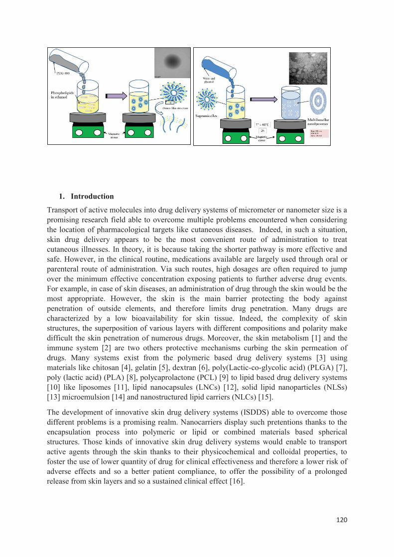
120
1. Introduction
Transport of active molecules into drug delivery systems of micrometer or nanometer size is a promising research field able to overcome multiple problems encountered when considering the location of pharmacological targets like cutaneous diseases. Indeed, in such a situation, skin drug delivery appears to be the most convenient route of administration to treat cutaneous illnesses. In theory, it is because taking the shorter pathway is more effective and safe. However, in the clinical routine, medications available are largely used through oral or parenteral route of administration. Via such routes, high dosages are often required to jump over the minimum effective concentration exposing patients to further adverse drug events. For example, in case of skin diseases, an administration of drug through the skin would be the most appropriate. However, the skin is the main barrier protecting the body against penetration of outside elements, and therefore limits drug penetration. Many drugs are characterized by a low bioavailability for skin tissue. Indeed, the complexity of skin structures, the superposition of various layers with different compositions and polarity make difficult the skin penetration of numerous drugs. Moreover, the skin metabolism [1] and the immune system [2] are two others protective mechanisms curbing the skin permeation of drugs. Many systems exist from the polymeric based drug delivery systems [3] using materials like chitosan [4], gelatin [5], dextran [6], poly(Lactic-co-glycolic acid) (PLGA) [7], poly (lactic acid) (PLA) [8], polycaprolactone (PCL) [9] to lipid based drug delivery systems [10] like liposomes [11], lipid nanocapsules (LNCs) [12], solid lipid nanoparticles (NLSs) [13] microemulsion [14] and nanostructured lipid carriers (NLCs) [15].
The development of innovative skin drug delivery systems (ISDDS) able to overcome those different problems is a promising realm. Nanocarriers display such pretentions thanks to the encapsulation process into polymeric or lipid or combined materials based spherical structures. Those kinds of innovative skin drug delivery systems would enable to transport active agents through the skin thanks to their physicochemical and colloidal properties, to foster the use of lower quantity of drug for clinical effectiveness and therefore a lower risk of adverse effects and so a better patient compliance, to offer the possibility of a prolonged release from skin layers and so a sustained clinical effect [16].
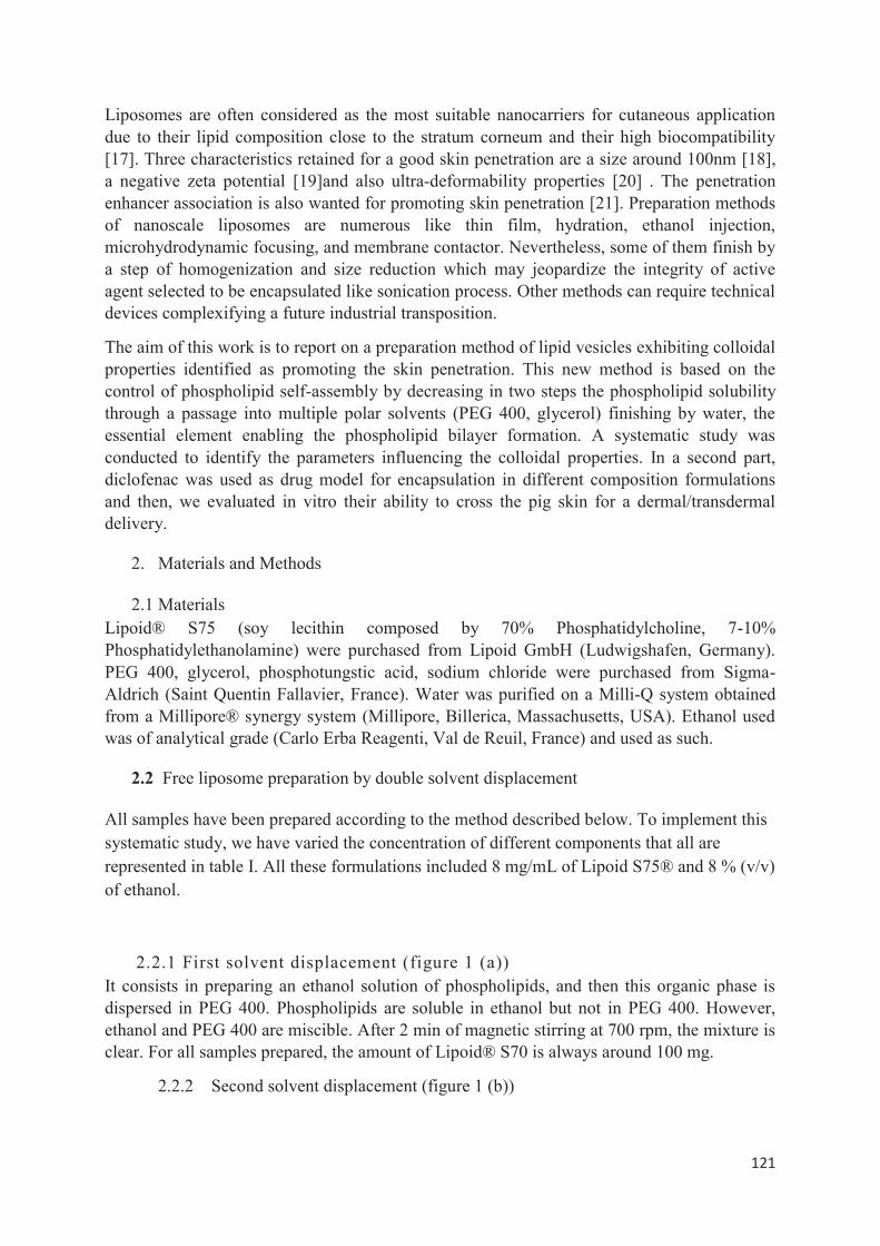
121
Liposomes are often considered as the most suitable nanocarriers for cutaneous application due to their lipid composition close to the stratum corneum and their high biocompatibility [17]. Three characteristics retained for a good skin penetration are a size around 100nm [18], a negative zeta potential [19]and also ultra-deformability properties [20] . The penetration enhancer association is also wanted for promoting skin penetration [21]. Preparation methods of nanoscale liposomes are numerous like thin film, hydration, ethanol injection, microhydrodynamic focusing, and membrane contactor. Nevertheless, some of them finish by a step of homogenization and size reduction which may jeopardize the integrity of active agent selected to be encapsulated like sonication process. Other methods can require technical devices complexifying a future industrial transposition.
The aim of this work is to report on a preparation method of lipid vesicles exhibiting colloidal properties identified as promoting the skin penetration. This new method is based on the control of phospholipid self-assembly by decreasing in two steps the phospholipid solubility through a passage into multiple polar solvents (PEG 400, glycerol) finishing by water, the essential element enabling the phospholipid bilayer formation. A systematic study was conducted to identify the parameters influencing the colloidal properties. In a second part, diclofenac was used as drug model for encapsulation in different composition formulations and then, we evaluated in vitro their ability to cross the pig skin for a dermal/transdermal delivery.
2. Materials and Methods
2.1 Materials Lipoid® S75 (soy lecithin composed by 70% Phosphatidylcholine, 7-10% Phosphatidylethanolamine) were purchased from Lipoid GmbH (Ludwigshafen, Germany). PEG 400, glycerol, phosphotungstic acid, sodium chloride were purchased from Sigma-Aldrich (Saint Quentin Fallavier, France). Water was purified on a Milli-Q system obtained from a Millipore® synergy system (Millipore, Billerica, Massachusetts, USA). Ethanol used was of analytical grade (Carlo Erba Reagenti, Val de Reuil, France) and used as such.
2.2 Free liposome preparation by double solvent displacement
All samples have been prepared according to the method described below. To implement this systematic study, we have varied the concentration of different components that all are represented in table I. All these formulations included 8 mg/mL of Lipoid S75® and 8 % (v/v) of ethanol.
2.2.1 First solvent displacement (figure 1 (a))
It consists in preparing an ethanol solution of phospholipids, and then this organic phase is dispersed in PEG 400. Phospholipids are soluble in ethanol but not in PEG 400. However, ethanol and PEG 400 are miscible. After 2 min of magnetic stirring at 700 rpm, the mixture is clear. For all samples prepared, the amount of Lipoid® S70 is always around 100 mg.
2.2.2 Second solvent displacement (figure 1 (b))
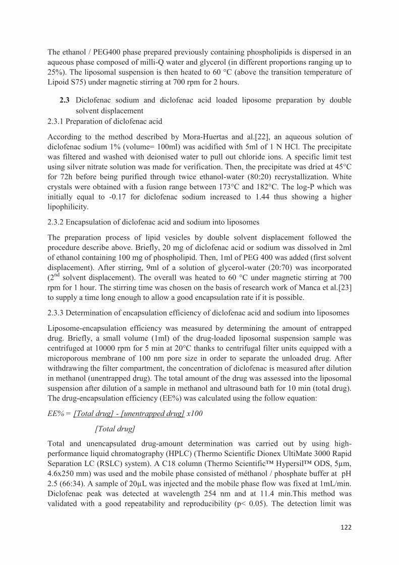
122
The ethanol / PEG400 phase prepared previously containing phospholipids is dispersed in an aqueous phase composed of milli-Q water and glycerol (in different proportions ranging up to 25%). The liposomal suspension is then heated to 60 °C (above the transition temperature of Lipoid S75) under magnetic stirring at 700 rpm for 2 hours.
2.3 Diclofenac sodium and diclofenac acid loaded liposome preparation by double solvent displacement
2.3.1 Preparation of diclofenac acid
According to the method described by Mora-Huertas and al.[22], an aqueous solution of diclofenac sodium 1% (volume= 100ml) was acidified with 5ml of 1 N HCl. The precipitate was filtered and washed with deionised water to pull out chloride ions. A specific limit test using silver nitrate solution was made for verification. Then, the precipitate was dried at 45°C for 72h before being purified through twice ethanol-water (80:20) recrystallization. White crystals were obtained with a fusion range between 173°C and 182°C. The log-P which was initially equal to -0.17 for diclofenac sodium increased to 1.44 thus showing a higher lipophilicity.
2.3.2 Encapsulation of diclofenac acid and sodium into liposomes
The preparation process of lipid vesicles by double solvent displacement followed the procedure describe above. Briefly, 20 mg of diclofenac acid or sodium was dissolved in 2ml of ethanol containing 100 mg of phospholipid. Then, 1ml of PEG 400 was added (first solvent displacement). After stirring, 9ml of a solution of glycerol-water (20:70) was incorporated (2nd solvent displacement). The overall was heated to 60 °C under magnetic stirring at 700 rpm for 1 hour. The stirring time was chosen on the basis of research work of Manca et al.[23] to supply a time long enough to allow a good encapsulation rate if it is possible.
2.3.3 Determination of encapsulation efficiency of diclofenac acid and sodium into liposomes
Liposome-encapsulation efficiency was measured by determining the amount of entrapped drug. Briefly, a small volume (1ml) of the drug-loaded liposomal suspension sample was centrifuged at 10000 rpm for 5 min at 20°C thanks to centrifugal filter units equipped with a microporous membrane of 100 nm pore size in order to separate the unloaded drug. After withdrawing the filter compartment, the concentration of diclofenac is measured after dilution in methanol (unentrapped drug). The total amount of the drug was assessed into the liposomal suspension after dilution of a sample in methanol and ultrasound bath for 10 min (total drug). The drug-encapsulation efficiency (EE%) was calculated using the follow equation:
EE% = [Total drug] - [unentrapped drug] x100
[Total drug]
Total and unencapsulated drug-amount determination was carried out by using high-performance liquid chromatography (HPLC) (Thermo Scientific Dionex UltiMate 3000 Rapid Separation LC (RSLC) system). A C18 column (Thermo Scientific™ Hypersil™ ODS, 5μm, 4.6x250 mm) was used and the mobile phase consisted of méthanol / phosphate buffer at pH 2.5 (66:34). A sample of 20μL was injected and the mobile phase flow was fixed at 1mL/min. Diclofenac peak was detected at wavelength 254 nm and at 11.4 min.This method was validated with a good repeatability and reproducibility (p< 0.05). The detection limit was
123
evaluated at 0.3μg/mL and the quantification limit at 1μg/mL. The quantification of diclofenac in methanol used calibration curves (3 curves with 5 points) ranged between 5 to 100 μg/ml, with a correlation coefficient r² > 0.99).
2.4 In vitro skin studies Skin penetration studies were implemented by using the static diffusion method based on Franz cell. Briefly, skin samples were collected from the pig's flanks (around 25 kg, Male) provided by the physiology laboratory (University of Lyon, France). After cleaning and hair removal, the subcutaneous tissue was taken off with a scalpel thus getting a skin thickness ranging between 1.2 and 1.5 mm. The skin samples were mounted on Franz-type glass diffusion cells (Laboratoire Verre Labomodula, Corbas, France) and placed in a thermostated water bath at 37°C in order to maintain a surface temperature of 32 ± 1°C. The diffusion area on the membrane was 2.54 cm2. For skin penetration analysis, the receptor compartment in contact with the dermal part was filled with a solution under magnetic stirring composed of pH 7.4 with 1.5% Brij 98 and 0.5% alpha tocopherol acetate.
1ml of prepared formulations (Table II) was uniformly spread on the skin surface. The study was carried out in non-occlusive conditions for 24h. After that, the diclofenac concentration was measured in the receptor fluid by HPLC. The Franz cell was disassembled; 2ml of receptor fluid was used to wash the skin surface before analysis by HPLC. Then, the stratum corneum was carefully removed by skin stripping. 30 adhesives with a diameter of 22 mm (D-Squame®, Monaderm, Monaco) were successively applied during 30 seconds under a weight of 500 g. The first strip was analyzed separately and the others were pooled by 9 (2-10; 11-20 and 21-30 strips). Extraction of diclofenac was performed from the adhesives by putting them in 10 mL of methanol for 2x 30 min and then for 1h in an ultrasonic bath at 40°C. Then, viable epidermis was separated from the dermis with dissection after heat treatment in water at 60°C for 45 s. The separation water was diclofenac free. And the dermis was cut in small pieces. Both were put in methanol respectively for diclofenac extraction during 1h30 in an ultrasounds bath at 40°C. Before analysis by HPLC, all samples were diluted with methanol (50:50).
2.5 Statistical Analysis Data were expressed as mean± Standard Deviation (SD) or Standard Error Mean (SEM), and each analysis was made minimum three times (n=3). Statistical analyses were carried out by ANOVA followed by the Tukey’spost-test to determine the differences between treatment groups. A threshold of p < 0.05 was considered significant statistically.
2.6 Particles characterization 2.6.1 Vesicle size, size distribution and zeta potential analysis:
Mean vesicle size, size distribution and zeta potential of prepared liposomes was determined by photon correlation spectroscopy (PCS), using Malvern Zetasizer Nanoseries (Malvern Instruments Zen 3600; Malvern, UK), after sample dilution in sodium chloride 1mM solution. Smoluchowski’s equation (Sze et al., 2003) permitted to determine the zeta potential from the electrophoretic mobility of liposomes at 25°C.
2.6.2 Morphological study by transmission electron microscopy Liposome suspensions were imaged by using a TEM (Philips CM120; Eindhoven, The Netherlands) for analysing the liposome morphology. A drop of the liposome suspension was
124
deposited on a carbon-coated copper grid, forming a thin liquid film. The films were negatively stained with 1% phosphotungstic acid solution (w/w) for 1 minute. The excess of liposomal suspension or phosphotungstic solution was removed with a filter paper. An accelerating voltage of 80 kV was necessary to characterize stained samples.
Figure 1: Schematic illustration of the first (a) and second (b) steps of the lipid vesicles formation by double solvent displacement.
b
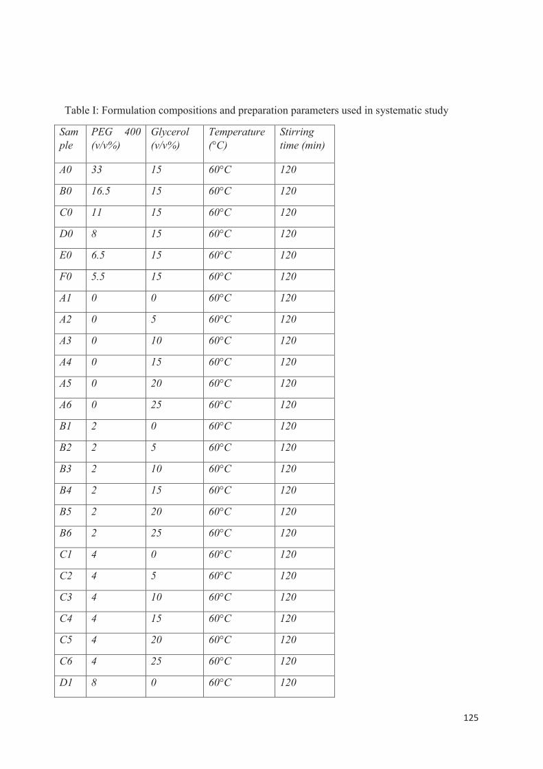
125
Table I: Formulation compositions and preparation parameters used in systematic study
Sample
PEG 400 (v/v%)
Glycerol (v/v%)
Temperature (°C)
Stirring time (min)
A0 33 15 60°C 120
B0 16.5 15 60°C 120
C0 11 15 60°C 120
D0 8 15 60°C 120
E0 6.5 15 60°C 120
F0 5.5 15 60°C 120
A1 0 0 60°C 120
A2 0 5 60°C 120
A3 0 10 60°C 120
A4 0 15 60°C 120
A5 0 20 60°C 120
A6 0 25 60°C 120
B1 2 0 60°C 120
B2 2 5 60°C 120
B3 2 10 60°C 120
B4 2 15 60°C 120
B5 2 20 60°C 120
B6 2 25 60°C 120
C1 4 0 60°C 120
C2 4 5 60°C 120
C3 4 10 60°C 120
C4 4 15 60°C 120
C5 4 20 60°C 120
C6 4 25 60°C 120
D1 8 0 60°C 120
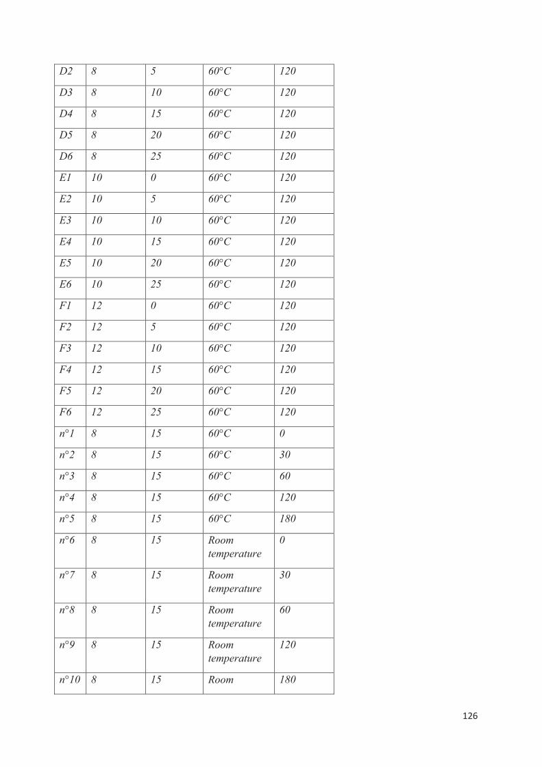
126
D2 8 5 60°C 120
D3 8 10 60°C 120
D4 8 15 60°C 120
D5 8 20 60°C 120
D6 8 25 60°C 120
E1 10 0 60°C 120
E2 10 5 60°C 120
E3 10 10 60°C 120
E4 10 15 60°C 120
E5 10 20 60°C 120
E6 10 25 60°C 120
F1 12 0 60°C 120
F2 12 5 60°C 120
F3 12 10 60°C 120
F4 12 15 60°C 120
F5 12 20 60°C 120
F6 12 25 60°C 120
n°1 8 15 60°C 0
n°2 8 15 60°C 30
n°3 8 15 60°C 60
n°4 8 15 60°C 120
n°5 8 15 60°C 180
n°6 8 15 Room temperature
0
n°7 8 15 Room temperature
30
n°8 8 15 Room temperature
60
n°9 8 15 Room temperature
120
n°10 8 15 Room 180
127
temperature
3. Results and discussion
3.1 Solvent screening
The double solvent displacement method is based on the use of miscible solvents. It is required that the phospholipids are soluble in the organic phase, but not in the other and in the mixture of the two solvents. We worked on a formulation in which the solvents match to this definition. But even more, we have also taking account the prospective route of administration, the skin.
The solubility of lipoid E80 was studied in each solvent (data not shown here) highlighting that those phospholipids are soluble in ethanol at least until a concentration of 200 mg/ml, but insoluble in PEG 400 and glycerol. However, they were very well dispersed in a co-solvent ethanol / PEG 400 and ethanol / glycerol (v / v, 75% / 25%). These various solvents have the advantages of being non-toxic, miscible and biocompatible.
Ethanol, PEG 400 and glycerol are also well known to be percutaneous penetration enhancer [24]. In cosmetics, the use of PEG 400 and glycerol is widespread, especially for their humectant properties that promote skin hydration. These are also agents which because of their high viscosities are capable of modifying the rheological properties of the formulations and improve both stability by limiting collisions and interactions between liposomes, to lengthen the duration of contact with the skin during the application, and also to make the liposomes ultra-deformable thereby facilitating their passage through the skin [25].
3.2 Morphology
3.2.1 Morphology after the first solvent displacement
The TEM analysis of sample taken from D4 in-process preparation obtained after the diffusion of the ethanolic solution containing phospholipids into the PEG 400 phase allowed to observe spherical and well-organized structures with a size from 1μm to 10μm sizes (Figure 2). It is important to note that the samples analysed here do not contain water and therefore do not enable the lipid bilayer formation into liposome. A new type of phospholipid organization is suggested, other than the self-assembly into bilayers. Indeed, TEM images show objects looking like onion like structures.
3.2.2 Morphology after the second solvent displacement
Negative-stain TEM images from D4 formulation (figure 3) showed that the lipid vesicles produced by double solvent displacement have a spherical shape and a size ranged from 50 to 100nm. Compared to what it was observed by photon correlation spectroscopy where the mean particle size was around 110nm, it can be considered that the results are concordant. That slight size difference would be due to the fact that during the sample preparation for
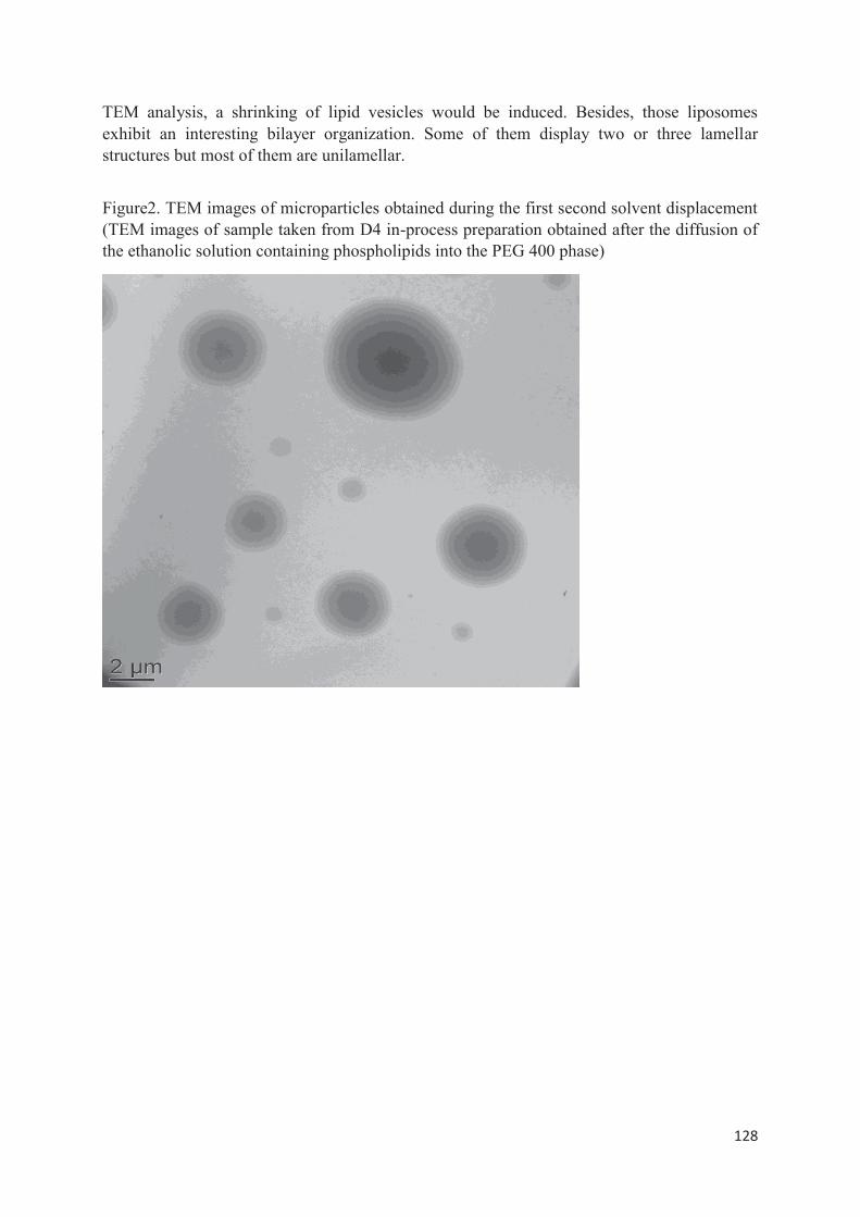
128
TEM analysis, a shrinking of lipid vesicles would be induced. Besides, those liposomes exhibit an interesting bilayer organization. Some of them display two or three lamellar structures but most of them are unilamellar.
Figure2. TEM images of microparticles obtained during the first second solvent displacement (TEM images of sample taken from D4 in-process preparation obtained after the diffusion of the ethanolic solution containing phospholipids into the PEG 400 phase)
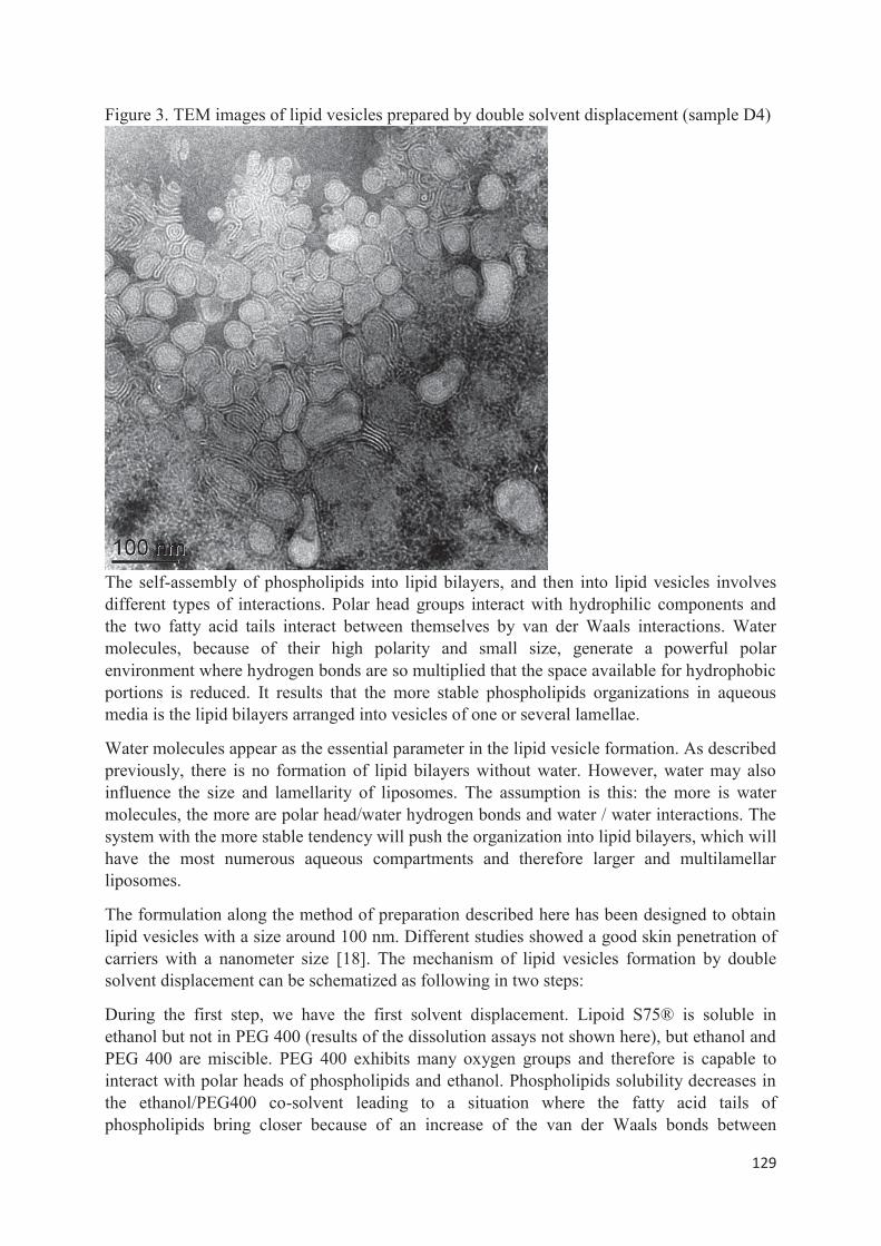
129
Figure 3. TEM images of lipid vesicles prepared by double solvent displacement (sample D4)
The self-assembly of phospholipids into lipid bilayers, and then into lipid vesicles involves different types of interactions. Polar head groups interact with hydrophilic components and the two fatty acid tails interact between themselves by van der Waals interactions. Water molecules, because of their high polarity and small size, generate a powerful polar environment where hydrogen bonds are so multiplied that the space available for hydrophobic portions is reduced. It results that the more stable phospholipids organizations in aqueous media is the lipid bilayers arranged into vesicles of one or several lamellae.
Water molecules appear as the essential parameter in the lipid vesicle formation. As described previously, there is no formation of lipid bilayers without water. However, water may also influence the size and lamellarity of liposomes. The assumption is this: the more is water molecules, the more are polar head/water hydrogen bonds and water / water interactions. The system with the more stable tendency will push the organization into lipid bilayers, which will have the most numerous aqueous compartments and therefore larger and multilamellar liposomes.
The formulation along the method of preparation described here has been designed to obtain lipid vesicles with a size around 100 nm. Different studies showed a good skin penetration of carriers with a nanometer size [18]. The mechanism of lipid vesicles formation by double solvent displacement can be schematized as following in two steps:
During the first step, we have the first solvent displacement. Lipoid S75® is soluble in ethanol but not in PEG 400 (results of the dissolution assays not shown here), but ethanol and PEG 400 are miscible. PEG 400 exhibits many oxygen groups and therefore is capable to interact with polar heads of phospholipids and ethanol. Phospholipids solubility decreases in the ethanol/PEG400 co-solvent leading to a situation where the fatty acid tails of phospholipids bring closer because of an increase of the van der Waals bonds between
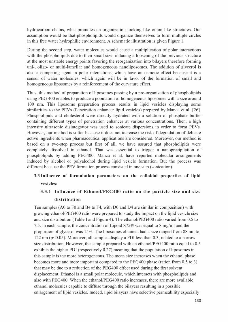
130
hydrocarbon chains, what promotes an organization looking like onion like structures. Our assumption would be that phospholipids would organize themselves to form multiple circles in this free water hydrophilic environment. A schematic illustration is given Figure 1.
During the second step, water molecules would cause a multiplication of polar interactions with the phospholipids due to their small size, inducing a loosening of the previous structure at the most unstable energy points favoring the reorganization into bilayers therefore forming uni-, oligo- or multi-lamellar and homogeneous nanoliposomes. The addition of glycerol is also a competing agent in polar interactions, which have an osmotic effect because it is a sensor of water molecules, which again will be in favor of the formation of small and homogeneous liposomes by a reinforcement of the curvature effect.
Thus, this method of preparation of liposomes passing by a pre-organization of phospholipids using PEG 400 enables to produce a population of homogeneous liposomes with a size around 100 nm. This liposome preparation process results in lipid vesicles displaying some similarities to the PEVs (Penetration enhancer lipid vesicles) prepared by Manca et al. [26]. Phospholipids and cholesterol were directly hydrated with a solution of phosphate buffer containing different types of penetration enhancer at various concentrations. Then, a high intensity ultrasonic disintegrator was used to sonicate dispersions in order to form PEVs. However, our method is softer because it does not increase the risk of degradation of delicate active ingredients when pharmaceutical applications are considered. Moreover, our method is based on a two-step process but first of all, we have assured that phospholipids were completely dissolved in ethanol. That was essential to trigger a nanoprecipitation of phospholipids by adding PEG400. Manca et al. have reported molecular arrangements induced by alcohol or polyalcohol during lipid vesicle formation. But the process was different because the PEV formation process consisted in one step (sonication).
3.3 Influence of formulation parameters on the colloidal properties of lipid
vesicles:
3.3.1 Influence of Ethanol/PEG400 ratio on the particle size and size
distribution
Ten samples (A0 to F0 and B4 to F4, with D0 and D4 are similar in composition) with growing ethanol/PEG400 ratio were prepared to study the impact on the lipid vesicle size and size distribution (Table I and Figure 4). The ethanol/PEG400 ratio varied from 0.5 to 7.5. In each sample, the concentration of Lipoid S75® was equal to 8 mg/ml and the proportion of glycerol was 15%. The liposomes obtained had a size ranged from 88 nm to 122 nm (p<0.05). Moreover, all samples display a PDI less than 0.3, related to a narrow size distribution. However, the sample prepared with an ethanol/PEG400 ratio equal to 0.5 exhibits the higher PDI (respectively 0.27) meaning that the population of liposomes in this sample is the more heterogeneous. The mean size increases when the ethanol phase becomes more and more important compared to the PEG400 phase (ration from 0.5 to 3) that may be due to a reduction of the PEG400 effect used during the first solvent displacement. Ethanol is a small polar molecule, which interacts with phospholipids and also with PEG400. When the ethanol/PEG400 ratio increases, there are more available ethanol molecules capable to diffuse through the bilayers resulting in a possible enlargement of lipid vesicles. Indeed, lipid bilayers have selective permeability especially
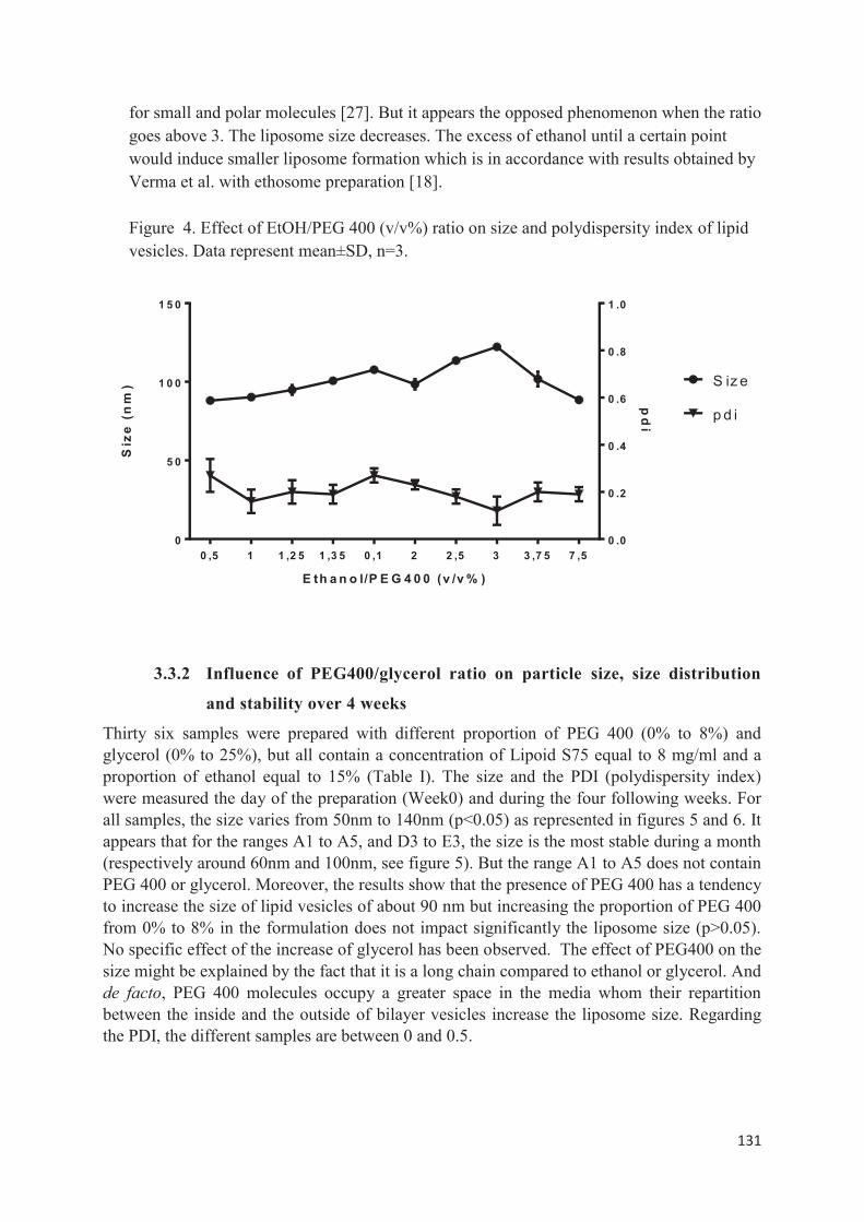
131
for small and polar molecules [27]. But it appears the opposed phenomenon when the ratio goes above 3. The liposome size decreases. The excess of ethanol until a certain point would induce smaller liposome formation which is in accordance with results obtained by Verma et al. with ethosome preparation [18]. Figure 4. Effect of EtOH/PEG 400 (v/v%) ratio on size and polydispersity index of lipid vesicles. Data represent mean±SD, n=3.
0 ,5 1 1 ,2 5 1 ,3 5 0 ,1 2 2 ,5 3 3 ,7 5 7 ,50
5 0
1 0 0
1 5 0
0 .0
0 .2
0 .4
0 .6
0 .8
1 .0
E th a n o l/P E G 4 0 0 (v /v % )
Siz
e(n
m)
pd
i
S iz e
p d i
3.3.2 Influence of PEG400/glycerol ratio on particle size, size distribution
and stability over 4 weeks
Thirty six samples were prepared with different proportion of PEG 400 (0% to 8%) and glycerol (0% to 25%), but all contain a concentration of Lipoid S75 equal to 8 mg/ml and a proportion of ethanol equal to 15% (Table I). The size and the PDI (polydispersity index) were measured the day of the preparation (Week0) and during the four following weeks. For all samples, the size varies from 50nm to 140nm (p<0.05) as represented in figures 5 and 6. It appears that for the ranges A1 to A5, and D3 to E3, the size is the most stable during a month (respectively around 60nm and 100nm, see figure 5). But the range A1 to A5 does not contain PEG 400 or glycerol. Moreover, the results show that the presence of PEG 400 has a tendency to increase the size of lipid vesicles of about 90 nm but increasing the proportion of PEG 400 from 0% to 8% in the formulation does not impact significantly the liposome size (p>0.05). No specific effect of the increase of glycerol has been observed. The effect of PEG400 on the size might be explained by the fact that it is a long chain compared to ethanol or glycerol. And de facto, PEG 400 molecules occupy a greater space in the media whom their repartition between the inside and the outside of bilayer vesicles increase the liposome size. Regarding the PDI, the different samples are between 0 and 0.5.
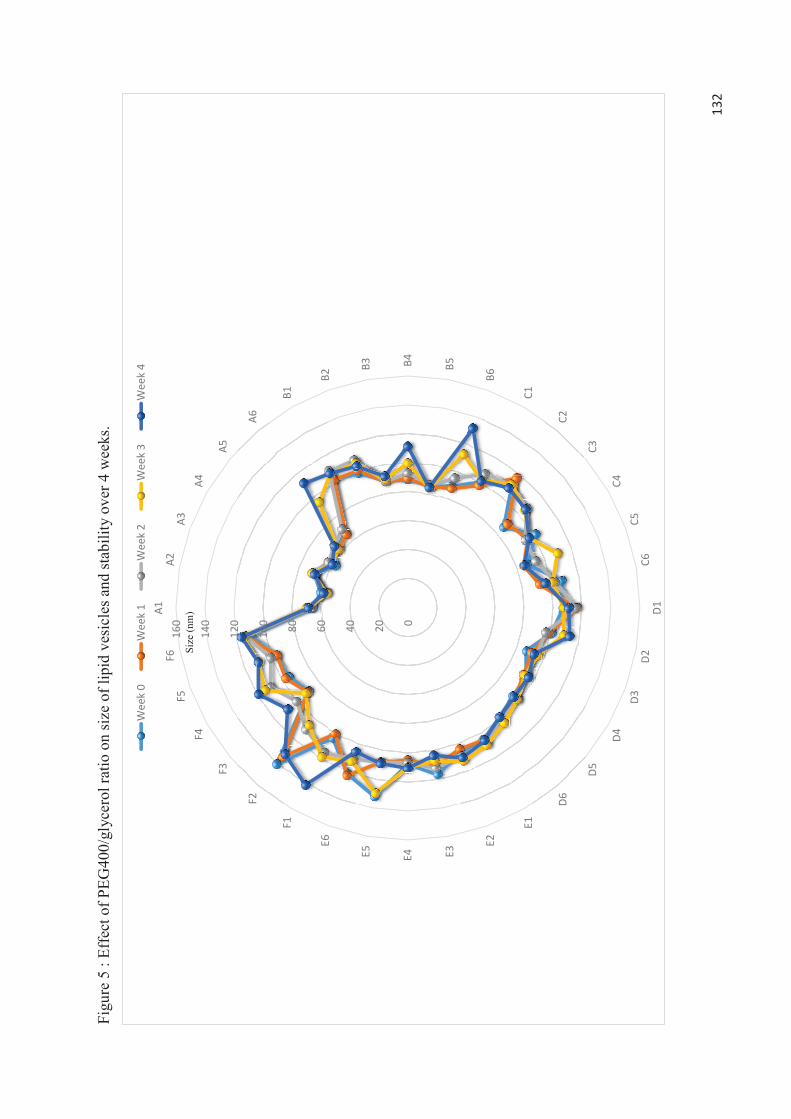
132
Figu
re 5
: Ef
fect
of P
EG40
0/gl
ycer
ol ra
tio o
n si
ze o
f lip
id v
esic
les a
nd st
abili
ty o
ver 4
wee
ks.
020406080100
120
140
160
A1A2
A3A4
A5
A6
B1
B2
B3 B4 B5
B6
C1
C2
C3
C4C5
C6D1
D2D3
D4
D5
D6
E1
E2
E3E4E5
E6
F1
F2
F3
F4F5
F6
Wee
k 0
Wee
k 1
Wee
k 2
Wee
k 3
Wee
k 4
Size
(nm
)
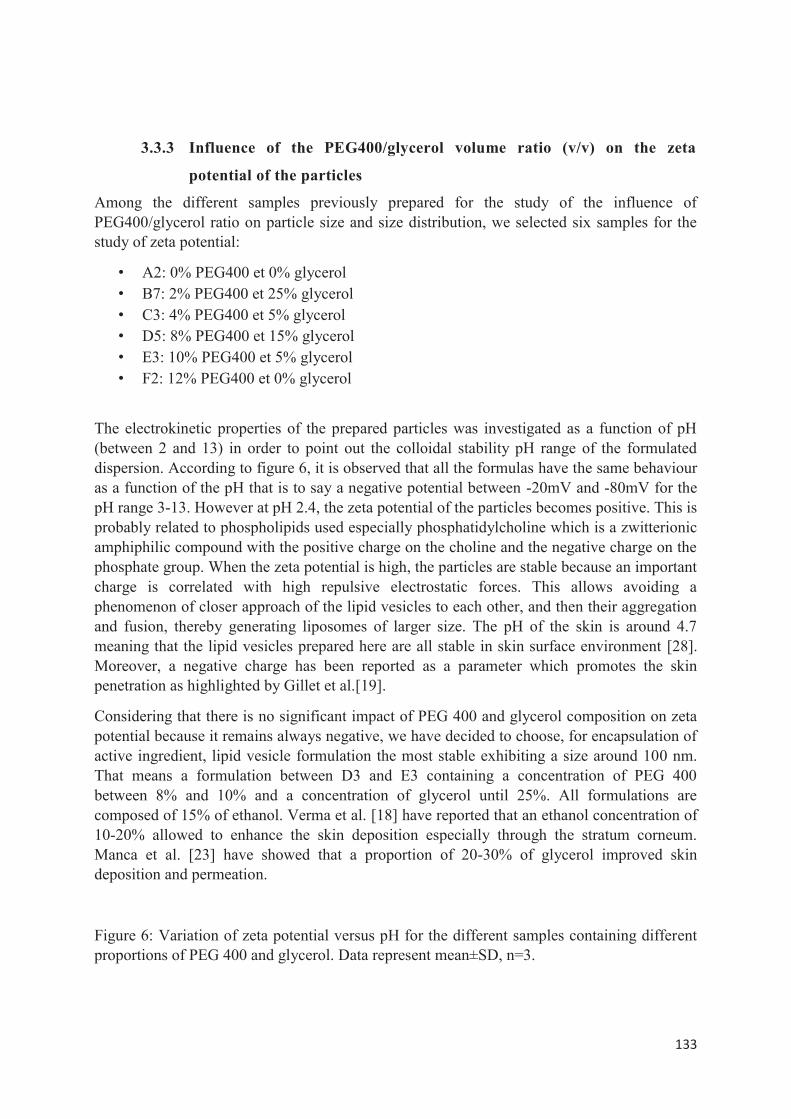
133
3.3.3 Influence of the PEG400/glycerol volume ratio (v/v) on the zeta
potential of the particles
Among the different samples previously prepared for the study of the influence of PEG400/glycerol ratio on particle size and size distribution, we selected six samples for the study of zeta potential:
• A2: 0% PEG400 et 0% glycerol • B7: 2% PEG400 et 25% glycerol • C3: 4% PEG400 et 5% glycerol • D5: 8% PEG400 et 15% glycerol • E3: 10% PEG400 et 5% glycerol • F2: 12% PEG400 et 0% glycerol
The electrokinetic properties of the prepared particles was investigated as a function of pH (between 2 and 13) in order to point out the colloidal stability pH range of the formulated dispersion. According to figure 6, it is observed that all the formulas have the same behaviour as a function of the pH that is to say a negative potential between -20mV and -80mV for the pH range 3-13. However at pH 2.4, the zeta potential of the particles becomes positive. This is probably related to phospholipids used especially phosphatidylcholine which is a zwitterionic amphiphilic compound with the positive charge on the choline and the negative charge on the phosphate group. When the zeta potential is high, the particles are stable because an important charge is correlated with high repulsive electrostatic forces. This allows avoiding a phenomenon of closer approach of the lipid vesicles to each other, and then their aggregation and fusion, thereby generating liposomes of larger size. The pH of the skin is around 4.7 meaning that the lipid vesicles prepared here are all stable in skin surface environment [28]. Moreover, a negative charge has been reported as a parameter which promotes the skin penetration as highlighted by Gillet et al.[19].
Considering that there is no significant impact of PEG 400 and glycerol composition on zeta potential because it remains always negative, we have decided to choose, for encapsulation of active ingredient, lipid vesicle formulation the most stable exhibiting a size around 100 nm. That means a formulation between D3 and E3 containing a concentration of PEG 400 between 8% and 10% and a concentration of glycerol until 25%. All formulations are composed of 15% of ethanol. Verma et al. [18] have reported that an ethanol concentration of 10-20% allowed to enhance the skin deposition especially through the stratum corneum. Manca et al. [23] have showed that a proportion of 20-30% of glycerol improved skin deposition and permeation.
Figure 6: Variation of zeta potential versus pH for the different samples containing different proportions of PEG 400 and glycerol. Data represent mean±SD, n=3.
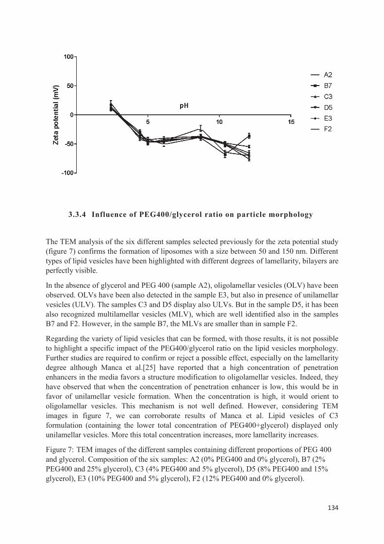
134
3.3.4 Influence of PEG400/glycerol ratio on particle morphology
The TEM analysis of the six different samples selected previously for the zeta potential study (figure 7) confirms the formation of liposomes with a size between 50 and 150 nm. Different types of lipid vesicles have been highlighted with different degrees of lamellarity, bilayers are perfectly visible.
In the absence of glycerol and PEG 400 (sample A2), oligolamellar vesicles (OLV) have been observed. OLVs have been also detected in the sample E3, but also in presence of unilamellar vesicles (ULV). The samples C3 and D5 display also ULVs. But in the sample D5, it has been also recognized multilamellar vesicles (MLV), which are well identified also in the samples B7 and F2. However, in the sample B7, the MLVs are smaller than in sample F2.
Regarding the variety of lipid vesicles that can be formed, with those results, it is not possible to highlight a specific impact of the PEG400/glycerol ratio on the lipid vesicles morphology. Further studies are required to confirm or reject a possible effect, especially on the lamellarity degree although Manca et al.[25] have reported that a high concentration of penetration enhancers in the media favors a structure modification to oligolamellar vesicles. Indeed, they have observed that when the concentration of penetration enhancer is low, this would be in favor of unilamellar vesicle formation. When the concentration is high, it would orient to oligolamellar vesicles. This mechanism is not well defined. However, considering TEM images in figure 7, we can corroborate results of Manca et al. Lipid vesicles of C3 formulation (containing the lower total concentration of PEG400+glycerol) displayed only unilamellar vesicles. More this total concentration increases, more lamellarity increases.
Figure 7: TEM images of the different samples containing different proportions of PEG 400 and glycerol. Composition of the six samples: A2 (0% PEG400 and 0% glycerol), B7 (2% PEG400 and 25% glycerol), C3 (4% PEG400 and 5% glycerol), D5 (8% PEG400 and 15% glycerol), E3 (10% PEG400 and 5% glycerol), F2 (12% PEG400 and 0% glycerol).
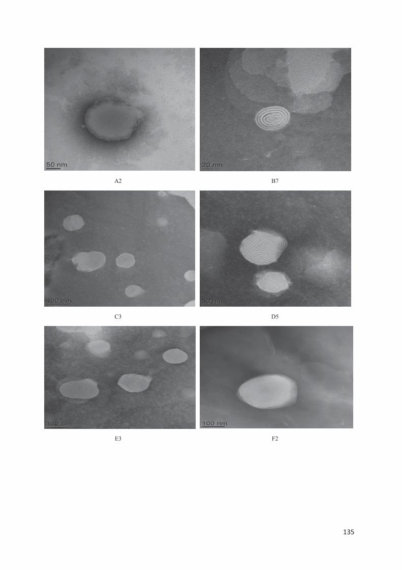
135
A2 B7
C3 D5
E3 F2
136
3.3.5 Influence of stirring time and temperature during the second step of lipid vesicle preparation by double solvent displacement
At the beginning of the method design, we chose two operating conditions during the second solvent displacement rather arbitrarily: a stirring speed at 700 rpm during 2 hours and an associated temperature of 60°C.
The stirring time was based on what it was commonly made with the most preparation method used to produce lipid vesicle: the Bangham method [29]so-called the thin film hydration method. In such case, where the thin film of phospholipids is formed a round bottom flask after rotatory evaporation of the organic solvent under reduced pressure, 2 hours of agitation at 700 rpm is sufficient to hydrate the film and enable the lipid vesicle formation (data not shown here).
Besides, the temperature of 60°C was chosen to be above a temperature essential to the liposome formation. This temperature is the gel liquid-crystalline phase transition temperature of Lipoid® S75. But this source of phospholipids is not pure. Thus, it is necessary to be above the upper limit required to the formation of lipid vesicles which is the Krafft point of lecithin, at a temperature of 58 °C [30]. We decided to study two of those parameters: stirring time and temperature.
Stirring time of the final mixture varied from 0 to 180 min at a speed of 700 rpm and at room temperature or 60°C (Samples n°1 to n°10, Table I). It appears that the vesicle size do not show a significant difference when the second solvent displacement is implemented at room temperature or 60°C and with a magnetic stirring time ranging from 0 to 180 minutes. The particle size is from 104.4 nm to 111.3 nm. Moreover, the PDI of the different samples is always less than 0.2 meaning that the population of the lipid vesicles produced is always homogeneous, irrespective of the temperature and time of stirring.
The absence of significant impact of those two operating conditions, which provide higher energy to the system can mean this system is naturally very stable and so favorable to a spontaneous formation of homogeneous nanoliposomes. That can be explained by a higher decrease of the transition temperature of phospholipids due to the mix of alcohol (ethanol) and polyols (PEG400 and glycerol). That phenomenon is well described by Koynova and Caffrey [31]. Moreover, the transition temperature has been correlated with a modification of the osmotic behaviour of phosphatidylcholine liposomes leading to a higher rate of water permeation, and thereafter, the production of small liposomes [32]. Regardless of the stirring time, the lipid vesicles produced are always small whereas Szoka & Papahadjopoulos [33] described in their review that a long time of stirring for hydration step leads to the formation of larger lipid vesicles. It is also interesting to note that to be above the transition temperature so easily, without an outside energy source, confers a better fluidity of lipid bilayers meaning a better flexibility of the vesicular structure and a lower permeability what is in favor of a good stability of encapsulation efficiency [34,35].
3.4 Preparation and characterisation of diclofenac loaded liposomes 3.4.1 Colloidal properties
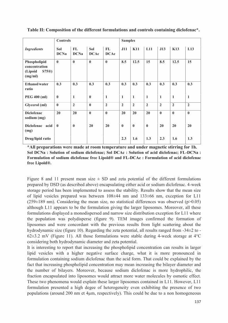
137
Table II: Composition of the different formulations and controls containing diclofenac*.
Controls Samples
Ingredients Sol DCNa
FL DCNa
Sol DCAc
FL DCAc
J11 K11 L11 J13 K13 L13
Phospholipid concentration (Lipoid S75®) (mg/ml)
0 0 0 0 8.5 12.5 15 8.5 12.5 15
Ethanol/water ratio
0.3 0.3 0.3 0.3 0.3 0.3 0.3 0.3 0.3 0.3
PEG 400 (ml) 0 1 0 1 1 1 1 1 1 1
Glycerol (ml) 0 2 0 2 2 2 2 2 2 2
Diclofenac sodium (mg)
20 20 0 0 20 20 20 0 0 0
Diclofenac acid (mg)
0 0 20 20 0 0 0 20 20 20
Drug/lipid ratio 2.3 1.6 1.3 2.3 1.6 1.3
*All preparations were made at room temperature and under magnetic stirring for 1h. Sol DCNa : Solution of sodium diclofenac; Sol DCAc : Solution of acid diclofenac; FL-DCNa : Formulation of sodium diclofenac free Lipoïd® and FL-DCAc : Formulation of acid diclofenac free Lipoïd®. Figure 8 and 11 present mean size ± SD and zeta potential of the different formulations prepared by DSD (as described above) encapsulating either acid or sodium diclofenac. 4-week storage period has been implemented to assess the stability. Results show that the mean size of lipid vesicles prepared was between 108±44 nm and 133±66 nm, exception for L11 (259±189 nm). Considering the mean size, no statistical differences was observed (p>0.05) although L11 appears to be the formulation giving the larger liposomes. Moreover, all these formulations displayed a monodispersed and narrow size distribution exception for L11 where the population was polydisperse (figure 9). TEM images confirmed the formation of liposomes and were concordant with the previous results from light scattering about the hydrodynamic size (figure 10). Regarding the zeta potential, all results ranged from -34±2 to -62±3.2 mV (Figure 11). All those formulations were stable during 4-week storage at 4°C considering both hydrodynamic diameter and zeta potential. It is interesting to report that increasing the phospholipid concentration can results in larger lipid vesicles with a higher negative surface charge, what it is more pronounced in formulation containing sodium diclofenac than the acid form. That could be explained by the fact that increasing phospholipid concentration may mean increasing the bilayer diameter and the number of bilayers. Moreover, because sodium diclofenac is more hydrophilic, the fraction encapsulated into liposomes would attract more water molecules by osmotic effect. These two phenomena would explain these larger liposomes contained in L11. However, L11 formulation presented a high degee of heterogeneity even exhibiting the presence of two populations (around 200 nm et 4μm, respectively). This could be due to a non homogeneous
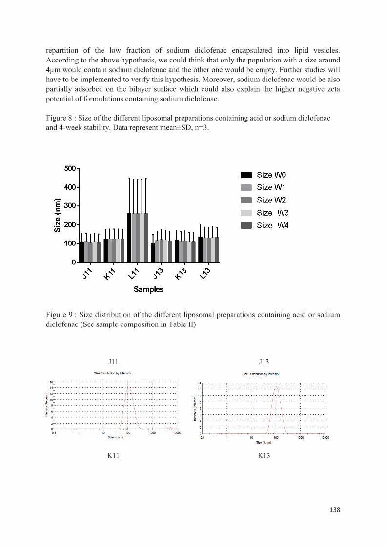
138
repartition of the low fraction of sodium diclofenac encapsulated into lipid vesicles. According to the above hypothesis, we could think that only the population with a size around 4μm would contain sodium diclofenac and the other one would be empty. Further studies will have to be implemented to verify this hypothesis. Moreover, sodium diclofenac would be also partially adsorbed on the bilayer surface which could also explain the higher negative zeta potential of formulations containing sodium diclofenac. Figure 8 : Size of the different liposomal preparations containing acid or sodium diclofenac and 4-week stability. Data represent mean±SD, n=3.
Figure 9 : Size distribution of the different liposomal preparations containing acid or sodium diclofenac (See sample composition in Table II)
J11 J13
K11 K13
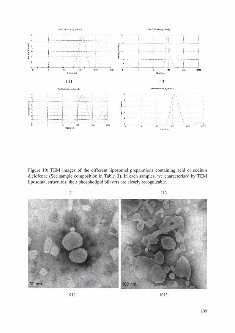
139
L11 L13
Figure 10: TEM images of the different liposomal preparations containing acid or sodium diclofenac (See sample composition in Table II). In each samples, we characterised by TEM liposomal structures, their phospholipid bilayers are clearly recognizable.
J11 J13
K11 K13
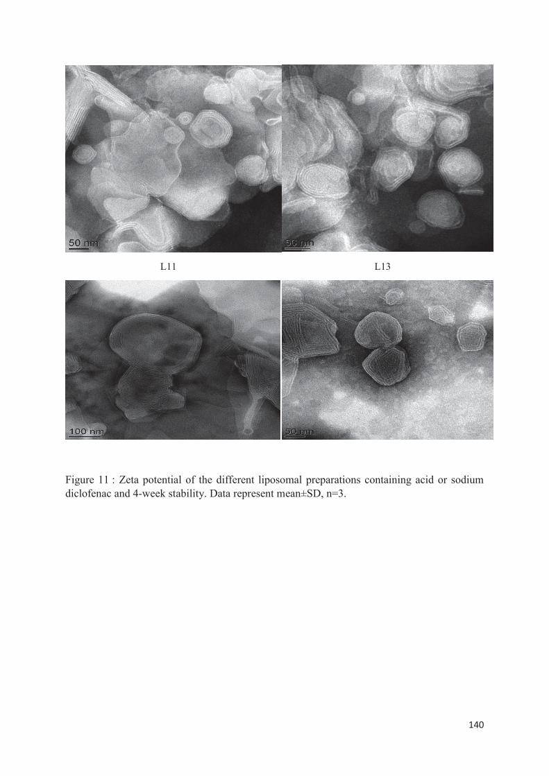
140
L11 L13
Figure 11 : Zeta potential of the different liposomal preparations containing acid or sodium diclofenac and 4-week stability. Data represent mean±SD, n=3.
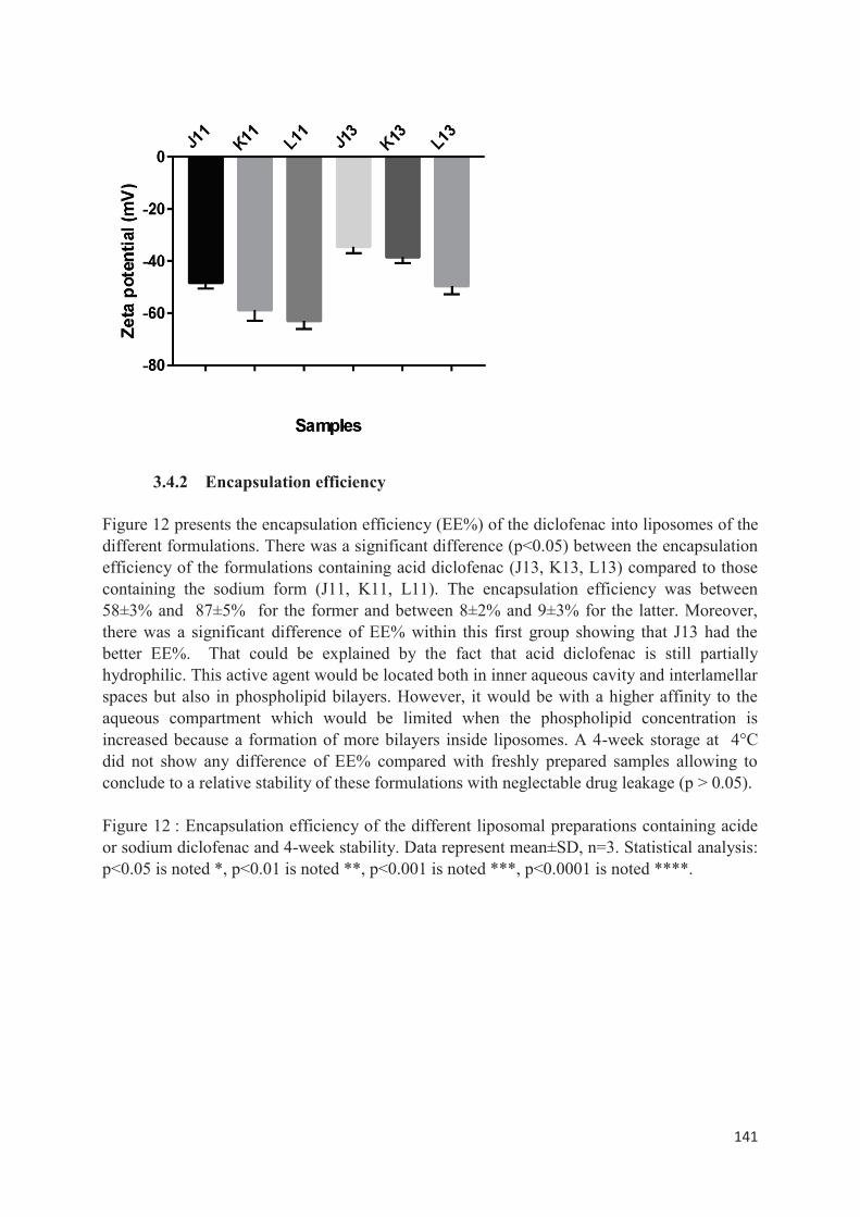
141
3.4.2 Encapsulation efficiency Figure 12 presents the encapsulation efficiency (EE%) of the diclofenac into liposomes of the different formulations. There was a significant difference (p<0.05) between the encapsulation efficiency of the formulations containing acid diclofenac (J13, K13, L13) compared to those containing the sodium form (J11, K11, L11). The encapsulation efficiency was between 58±3% and 87±5% for the former and between 8±2% and 9±3% for the latter. Moreover, there was a significant difference of EE% within this first group showing that J13 had the better EE%. That could be explained by the fact that acid diclofenac is still partially hydrophilic. This active agent would be located both in inner aqueous cavity and interlamellar spaces but also in phospholipid bilayers. However, it would be with a higher affinity to the aqueous compartment which would be limited when the phospholipid concentration is increased because a formation of more bilayers inside liposomes. A 4-week storage at 4°C did not show any difference of EE% compared with freshly prepared samples allowing to conclude to a relative stability of these formulations with neglectable drug leakage (p > 0.05). Figure 12 : Encapsulation efficiency of the different liposomal preparations containing acide or sodium diclofenac and 4-week stability. Data represent mean±SD, n=3. Statistical analysis: p<0.05 is noted *, p<0.01 is noted **, p<0.001 is noted ***, p<0.0001 is noted ****.
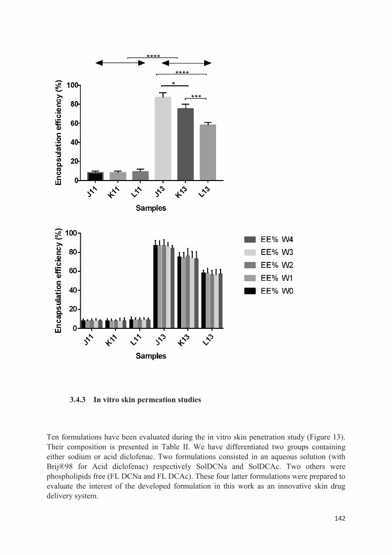
142
3.4.3 In vitro skin permeation studies Ten formulations have been evaluated during the in vitro skin penetration study (Figure 13). Their composition is presented in Table II. We have differentiated two groups containing either sodium or acid diclofenac. Two formulations consisted in an aqueous solution (with Brij®98 for Acid diclofenac) respectively SolDCNa and SolDCAc. Two others were phospholipids free (FL DCNa and FL DCAc). These four latter formulations were prepared to evaluate the interest of the developed formulation in this work as an innovative skin drug delivery system.
143
There is no significant difference of diclofenac amount crossing the skin within formulations without phospholipid or without PEG400/glycerol compared with solutions. Besides, the formulations J11, K11 and L11 did not promote the skin penetration significantly in comparison with the other formulations (p>0.05), whatever the phospholipid concentration. An exception was observed only between J11 and Sol DCNa (p<0.05). Formulations J13 and K13 have shown the better performances in skin penetration with a rate of applied dose detected in the full skin after 24h. It was around 19% and 14% respectively. The corresponding uptakes were 1.5-2 folds higher than their homologues containing the sodium diclofenac. No formulation exhibits a higher potential to reside in the SC (p>0.05). Considering the low lipophilicity of diclofenac, even though we have prepared an acid form with a higher logP, this is not enough to allow a better retention in the SC.
In coherence with the previous observations about SC, here again the different formulations were not able to bring an applied dose rate higher than 1.2%, excepted for J13 with 1.7%. Nevertheless, for the latter formulation, the t-test did not show a significant difference only in comparison with the formulation SolDCNa and SolDCa. The fraction of the penetrated amount was far larger for the formulations J13, K13 and L13 (p<0.001). However, no significant difference was observed between J13 and K13 or K13 and L13. A statistical difference was only between J13 and L13 (p<0.05). Considering the diclofenac amount measured in the receptor compartment, the formulation J13 is the only one showing a rate almost doubled compared with the other formulations (p<0.001).
J13 is definitely the most promising formulation, able to cross the skin until dermis and beyond with the higher rate. These results enable to consider this formulation as ISDDS with characteristics promoting a transdermal permeation of diclofenac. Indeed, such a formulation presents many advantages in the management of diseases involved clinical symptoms like arthritis in psoriasis for example, where this complication is very disabling. However, J13 is not interesting for the target of SC or viable epidermis. This observation is consistent regarding other research works showing that drug accumulation into these two layers depends essentially on lipophilic characteristics of active ingredients and/or the drug carrier type. In our situation, it remains to understand why with J13 we found the higher dose rate in dermis and receptor compartment compared with SC and viable epidermis. Three skin pathways for cutaneous drug delivery are generally described. It may be logical to think in first place the intercellular pathway. As described previously, J13 contains acid diclofenac with a LogP of 1.44 (versus LogP= -0.17 for sodium diclofenac). Such an improvement would have had to increase the drug amount measured in SC and viable epidermis in comparison to the same formulation containing the sodium form. Unfortunately, obtained results are not fitting in. Although acid diclofenac is more lipophilic, the hydrophilic aspect is still too much present to improve the skin passage through SC and viable epidermis. Therefore, follicular pathway hypothesis could be privileged to explain the results of the in vitro study.
The fact that diclofenac is encapsulated into liposomes appears to be responsible of deeper skin penetration. Indeed, the encapsulation efficiency in formulations J11, K11 and L11 is around 70% less than formulations J13, K13 and L13. These lipid vesicles prepared with three penetration enhancers (ethanol, PEG 400 and glycerol) would multiply interactions at dermis level through the invagination of hair follicles, promoting the diffusion of acid diclofenac. The outer root sheath of hair follicles is continuous with the basal layer of the interfollicular viable epidermis, what it is thinner than SC and so, easier to cross with a direct access to
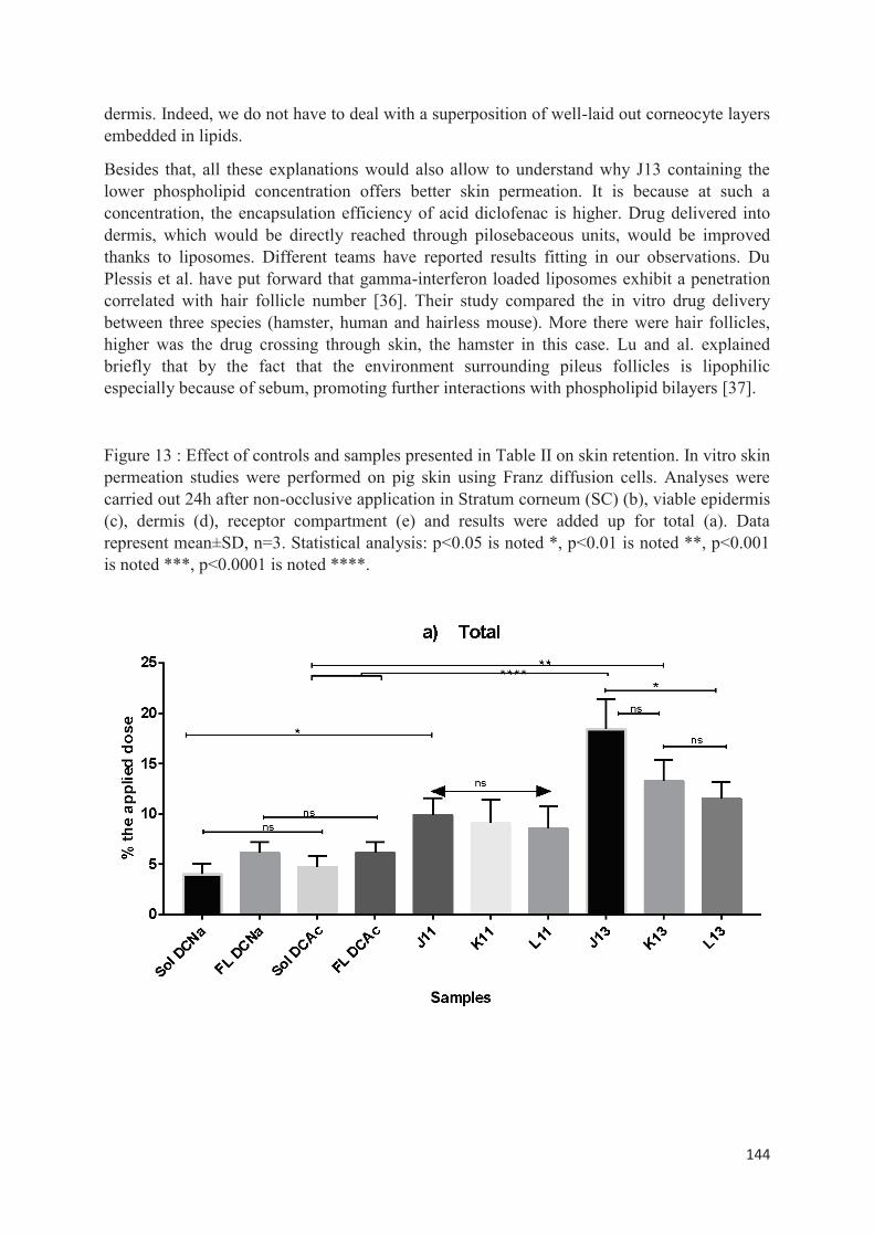
144
dermis. Indeed, we do not have to deal with a superposition of well-laid out corneocyte layers embedded in lipids.
Besides that, all these explanations would also allow to understand why J13 containing the lower phospholipid concentration offers better skin permeation. It is because at such a concentration, the encapsulation efficiency of acid diclofenac is higher. Drug delivered into dermis, which would be directly reached through pilosebaceous units, would be improved thanks to liposomes. Different teams have reported results fitting in our observations. Du Plessis et al. have put forward that gamma-interferon loaded liposomes exhibit a penetration correlated with hair follicle number [36]. Their study compared the in vitro drug delivery between three species (hamster, human and hairless mouse). More there were hair follicles, higher was the drug crossing through skin, the hamster in this case. Lu and al. explained briefly that by the fact that the environment surrounding pileus follicles is lipophilic especially because of sebum, promoting further interactions with phospholipid bilayers [37].
Figure 13 : Effect of controls and samples presented in Table II on skin retention. In vitro skin permeation studies were performed on pig skin using Franz diffusion cells. Analyses were carried out 24h after non-occlusive application in Stratum corneum (SC) (b), viable epidermis (c), dermis (d), receptor compartment (e) and results were added up for total (a). Data represent mean±SD, n=3. Statistical analysis: p<0.05 is noted *, p<0.01 is noted **, p<0.001 is noted ***, p<0.0001 is noted ****.
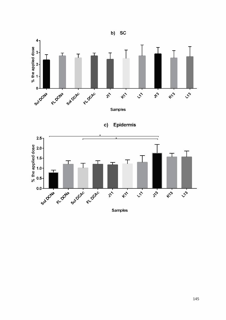
145
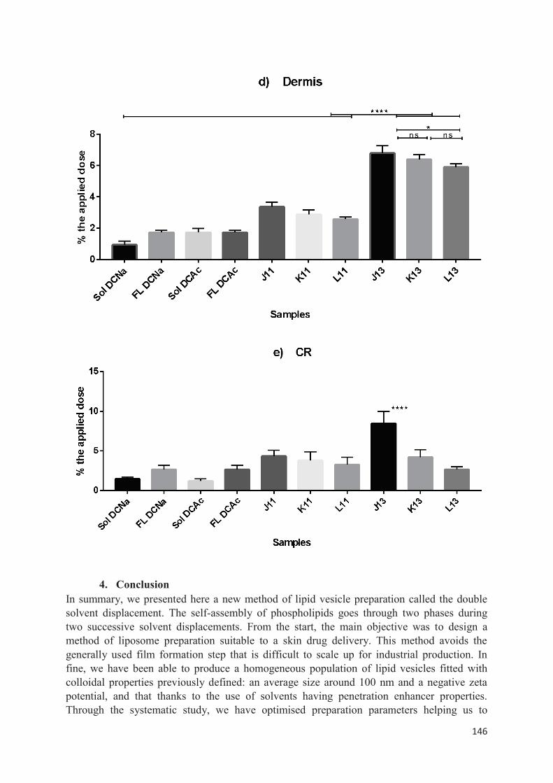
146
4. Conclusion
In summary, we presented here a new method of lipid vesicle preparation called the double solvent displacement. The self-assembly of phospholipids goes through two phases during two successive solvent displacements. From the start, the main objective was to design a method of liposome preparation suitable to a skin drug delivery. This method avoids the generally used film formation step that is difficult to scale up for industrial production. In fine, we have been able to produce a homogeneous population of lipid vesicles fitted with colloidal properties previously defined: an average size around 100 nm and a negative zeta potential, and that thanks to the use of solvents having penetration enhancer properties. Through the systematic study, we have optimised preparation parameters helping us to
147
produce diclofenac loaded liposomes. Then, we have been able to identify the formulation which is the most promising delivery systems through the transdermal route with similar performance than those of the literature.
5. References
1. Baron JM, Merk HF. Drug metabolism in the skin. Curr. Opin. Allergy Clin. Immunol. 2001;1:287–91.
2. Afshar M, Gallo RL. Innate immune defense system of the skin. Vet. Dermatol. 2013;24:32–e9.
3. Badri W, Eddabra R, Fessi H, Elaissari A. Biodegradable Polymer Based Nanoparticles: Dermal and Transdermal Drug Delivery. J. Colloid Sci. Biotechnol. 2014;3:141–149.
4. Hasanovic A, Zehl M, Reznicek G, Valenta C. Chitosan-tripolyphosphate nanoparticles as a possible skin drug delivery system for aciclovir with enhanced stability. J. Pharm. Pharmacol. 2009;61:1609–1616.
5. Sahoo N, Sahoo RK, Biswas N, Guha A, Kuotsu K. Recent advancement of gelatin nanoparticles in drug and vaccine delivery. Int. J. Biol. Macromol. 2015;81:317–331.
6. Pamornpathomkul B, Duangjit S, Laohapatarapant S, Rojanarata T, Opanasopit P, Ngawhirunpat T. Transdermal delivery of fluorescein isothiocyanate-dextrans using the combination of microneedles and low-frequency sonophoresis. Asian J. Pharm. Sci. 2015;10:415–424.
7. Lira AAM, Cordo PL, Nogueira EC, Almeida EDP, Junior RAL, Nunes RS, et al. Optimization of topical all-trans retinoic acid penetration using poly-DL-lactide and poly-DL-lactide-co-glycolide microparticles. J. Colloid Sci. Biotechnol. 2013;2:123–129.
8. Rancan F, Papakostas D, Hadam S, Hackbarth S, Delair T, Primard C, et al. Investigation of polylactic acid (PLA) nanoparticles as drug delivery systems for local dermatotherapy. Pharm. Res. 2009;26:2027–2036.
9. Mondal D, Griffith M, Venkatraman SS. Polycaprolactone-based biomaterials for tissue engineering and drug delivery: Current scenario and challenges. Int. J. Polym. Mater. Polym. Biomater. 2016;65:255–265.
10. Zhai Y, Zhai G. Advances in lipid-based colloid systems as drug carrier for topic delivery. J. Controlled Release. 2014;193:90–99.
11. Pierre MBR, Costa I dos SM. Liposomal systems as drug delivery vehicles for dermal and transdermal applications. Arch. Dermatol. Res. 2011;303:607–621.
12. Weber J, Funk NL, Motta MH, Guedes AM, Visintainer APC, Tedesco SB, et al. Association of Borage Oil and Betamethasone Dipropionate in Lipid-Core Nanocapsules: Characterization, Photostability and In Vitro Irritation Test. J. Nanosci. Nanotechnol. 2016;16:1354–1362.
13. Dasgupta S, K Ghosh S, Ray S, Mazumder B. Solid lipid nanoparticles (SLNs) gels for topical delivery of aceclofenac in vitro and in vivo evaluation. Curr. Drug Deliv. 2013;10:656–666.
14. C Vadlamudi H, Narendran H, Nagaswaram T, Yaga G, Thanniru J, R Yalavarthi P. Microemulsions based transdermal drug delivery systems. Curr. Drug Discov. Technol. 2014;11:169–180.
15. Kamble MS, Vaidya KK, Bhosale AV, Nanjwade BK, Shinde SA, Chaudhari PD. Formulation and Evaluation of Meloxicam Nanostructured Lipid Carrier. J. Colloid Sci. Biotechnol. 2014;3:167–172.
148
16. Paudel KS, Milewski M, Swadley CL, Brogden NK, Ghosh P, Stinchcomb AL. Challenges and opportunities in dermal/transdermal delivery. Ther. Deliv. 2010;1:109–131.
17. Laouini A, Jaafar-Maalej C, Limayem-Blouza I, Sfar S, Charcosset C, Fessi H. Preparation, characterization and applications of liposomes: state of the art. J. Colloid Sci. Biotechnol. 2012;1:147–168.
18. Verma DD, Fahr A. Synergistic penetration enhancement effect of ethanol and phospholipids on the topical delivery of cyclosporin A. J. Controlled Release. 2004;97:55–66.
19. Gillet A, Compère P, Lecomte F, Hubert P, Ducat E, Evrard B, et al. Liposome surface charge influence on skin penetration behaviour. Int. J. Pharm. 2011;411:223–231.
20. Romero EL, Morilla MJ. Highly deformable and highly fluid vesicles as potential drug delivery systems: theoretical and practical considerations. Int J Nanomedicine. 2013;8:3171–3186.
21. Dragicevic N, Maibach HI, others. Percutaneous penetration enhancers chemical methods in penetration enhancement. Nanocarriers [Internet]. 2016 [cited 2017 Jan 18]; Available from: http://link.springer.com/content/pdf/10.1007/978-3-662-47039-8.pdf
22. Mora-Huertas CE, Garrigues O, Fessi H, Elaissari A. Nanocapsules prepared via nanoprecipitation and emulsification–diffusion methods: Comparative study. Eur. J. Pharm. Biopharm. 2012;80:235–239.
23. Manca ML, Zaru M, Manconi M, Lai F, Valenti D, Sinico C, et al. Glycerosomes: a new tool for effective dermal and transdermal drug delivery. Int. J. Pharm. 2013;455:66–74.
24. Thong H-Y, Zhai H, Maibach HI. Percutaneous penetration enhancers: an overview. Skin Pharmacol. Physiol. 2007;20:272–282.
25. Caddeo C, Manconi M, Sinico C, Valenti D, Celia C, Monduzzi M, et al. Penetration Enhancer-Containing Vesicles: Does the Penetration Enhancer Structure Affect Topical Drug Delivery? Curr. Drug Targets. 2015;16:1438–1447.
26. Manca ML, Castangia I, Matricardi P, Lampis S, Fernàndez-Busquets X, Fadda AM, et al. Molecular arrangements and interconnected bilayer formation induced by alcohol or polyalcohol in phospholipid vesicles. Colloids Surf. B Biointerfaces. 2014;117:360–367.
27. Biological Science / Edition 5 by Scott Freeman, Kim Quillin, Lizabeth Allison | | 9780321743671 | Hardcover | Barnes & Noble, (n.d.).
28. Lambers H, Piessens S, Bloem A, Pronk H, Finkel P. Natural skin surface pH is on average below 5, which is beneficial for its resident flora. Int. J. Cosmet. Sci. 2006;28:359–370.
29. Bangham AD, Standish MM, Watkins JC. Diffusion of univalent ions across the lamellae of swollen phospholipids. J. Mol. Biol. 1965;13:238–IN27.
30. Phillips MC, Williams RM, Chapman D. On the nature of hydrocarbon chain motions in lipid liquid crystals. Chem. Phys. Lipids. 1969;3:234–244.
31. Koynova R, Caffrey M. Phases and phase transitions of the phosphatidylcholines. Biochim. Biophys. Acta BBA-Rev. Biomembr. 1998;1376:91–145.
32. Blok MC, Van Deenen LLM, De Gier J. Effect of the gel to liquid crystalline phase transition on the osmotic behaviour of phosphatidycholine liposomes. Biochim. Biophys. Acta BBA-Biomembr. 1976;433:1–12.
149
33. Szoka Jr F, Papahadjopoulos D. Comparative properties and methods of preparation of lipid vesicles (liposomes). Annu. Rev. Biophys. Bioeng. 1980;9:467–508.
34. Frézard F, Schettini DA, Rocha OG, Demicheli C. Lipossomas: propriedades físico-químicas e farmacológicas, aplicações na quimioterapia à base de antimônio. Quim Nova. 2005;28:511–518.
35. Collier JH, Messersmith PB. Phospholipid strategies in biomineralization and biomaterials research. Annu. Rev. Mater. Res. 2001;31:237–263.
36. Du Plessis J, Egbaria K, Ramachandran C, Weiner N. Topical delivery of liposomally encapsulated gamma-interferon. Antiviral Res. 1992;18:259–265.
37. Lu GW, Valiveti S, Spence J, Zhuang C, Robosky L, Wade K, et al. Comparison of artificial sebum with human and hamster sebum samples. Int. J. Pharm. 2009;367:37–43.
150
PARTIE EXPERIMENTALE
Etude comparative entre le double déplacement de solvent et la méthode classique de l’injection à l’éthanol—De l’échelle laboratoire à une plus grande échelle
Publié dans le journal Colloids and Surface A: Physicochemical and Engineering Aspects:
Colloids and Surfaces A: Physicochem. Eng. Aspects 524 (2017) 71–78
151
Il existe de nombreuses méthodes de préparation des vésicules lipidiques. Le choix de la méthode influence fortement la taille des liposomes conçus. Ce travail de recherche porte sur l’évaluation du potentiel de notre méthode de préparation de vésicules lipidiques appelée « Double Solvent Displacement » en vue d’une transposition d’échelle. Nous avons comparé notre méthode à une technique largement répandue, la méthode dite de l’injection à l’éthanol. Plus la taille des liposomes est petite, plus la surface spécifique est grande. Cela augmente considérablement le nombre d’interactions possibles avec les membranes cellulaires et a fortiori leur passage intracellulaire. Ainsi, la préparation de vésicules lipidiques d'une taille moyenne d'environ 100 nm aurait plus d'avantages dans la délivrance de molécules actives vers des cibles thérapeutiques difficiles à atteindre telles que le cerveau en raison de la barrière hémato-encéphalique, de la peau ou des articulations à cause de la barrière cutanée et le tissu osseux. Les liposomes présentant une telle taille sont classés généralement parmi les SUV (small unilamellar vesicle) caractérisés par une seule bicouche de phospholipides bien qu’il ne soit pas rare de voir se former des liposomes de cette taille mais présentant plusieurs bicouches. On parle plutôt alors de SOV (small oligolamellar vesicle) ou SMV (small multilamellar vesicle). Une des méthodes qui reste très utilisée à ce jour dans les travaux de recherche est la méthode de Bangham dite de l’hydratation du film lipidique. Un film lipidique constitué de bicouches superposées est obtenu après évaporation rotative d'un solvant organique ou d'un co-solvant dans lequel les lipides étaient dissous. Ensuite, le film lipidique est hydraté avec une solution aqueuse pour former des liposomes. Cependant, cette méthode reste difficile à normaliser. Elle donne généralement de larges liposomes multilamellaires et très hétérogènes. Ainsi, des techniques supplémentaires telles que la sonication, l'extrusion, l'homogénéisation haute pression sont associées pour réduire la taille des liposomes préalablement produits et obtenir une population plus homogène. Ces techniques présentent ces avantages, mais aussi des inconvénients limitant leur intérêt pour la production de liposomes à l'échelle industrielle. Certaines sont inutilisables à plus grande échelle, potentiellement destructrices pour les phospholipides et / ou les molécules actives d’intérêt pharmaceutique.
D'autres méthodes ont également prouvé leur intérêt pour la formation de nanovésicules lipidiques comme la focalisation microhydrodynamique ou la méthode par contacteur à membrane. Mais toutes ces méthodes nécessitent l'utilisation de systèmes sophistiqués et coûteux. La méthode de l’injection à éthanol est une technique simple qui a démontré son efficacité dans la préparation de nanovésicules lipidiques. Les phospholipides et autres substances lipophiles sont dissous dans un solvant organique (éthanol), puis cette phase est rapidement injectée dans une grande quantité de tampon aqueux dont il résulte spontanément une formation de liposomes de taille nanométrique.
Dans cet article, nous avons comparé notre nouvelle méthode, le double déplacement de solvant, avec une méthode conventionnelle largement utilisée: l'injection d'éthanol. L'influence de la variabilité de divers paramètres (concentration en phospholipides, ratio solvant / non-solvant) sur les propriétés colloïdales (taille et distribution de la taille, potentiel zêta, morphologie) a été étudiée. De plus, nous avons réalisé des études de scale-up. L’ensemble de ces études nous ont permis de comparer leur performance et leur capacité d’adaptation lors d’une transposition d’échelle.
152
Nos résultats ont montré que le double déplacement de solvant pouvait produire une grande quantité de petites vésicules lipidiques homogènes. Avec cette méthode, nous pourrions utiliser des concentrations plus élevées de phospholipides sans affecter de manière significative les propriétés colloïdales des vésicules par rapport à la méthode de l'injection à éthanol. En effet, pour des concentrations supérieures à 12,5 mg / ml, nous avons observé une augmentation continue de la taille (de 129 ± 54 nm à 318 ± 220 nm) associée à une hétérogénéité plus élevée de la population de liposomes produite par injection à l’éthanol, alors que les résultats sont restés stables pour la technique DSD (taille entre 123 ± 57 nm et 141 ± 67 nm, respectivement). La technique DSD a aussi permis de préserver les propriétés colloïdales des liposomes sur un large intervalle de ratios solvants / non solvants (éthanol /eau). Les études de scale-up ont montré une variabilité plus prononcée pour la technique d'injection d'éthanol pour laquelle les propriétés colloïdales étaient plus impactées. En effet, la taille a diminué de manière significative de 11,8% par rapport à la méthode DSD pour laquelle l'augmentation était d'environ 3,5%.
Il ressort de notre travail que la méthode de l'injection à l’éthanol ne permettrait pas de produire de plus grandes quantités de liposomes aux propriétés colloïdales homogènes si on envisageait une transposition d’échelle, bien que techniquement elle le soit. Il est imposé de travailler dans des conditions opératoires fortement diluées, ce qui pourrait également limiter le degré d'encapsulation des molécules actives. La méthode du double déplacement de solvant apparait comme très prometteuse. Elle est basée sur deux étapes effectuées dans des conditions douces réalisées via une homogénéisation simple. Cette méthode permet de former des vésicules lipidiques homogènes et de petites tailles (proche de 100 nm) sans étape supplémentaire. En outre, de grandes quantités de liposomes pourraient être produites en utilisant des concentrations élevées de phospholipides tout en garantissant une taille nanométrique et une bonne homogénéité. Ainsi, cette méthode pourrait être mise en œuvre à l'échelle industrielle et offrir de très bons résultats. La simplicité et la rapidité de cette nouvelle technique nous permettent de produire une population de liposomes avec une bonne maîtrise du procédé, en particulier en terme de reproductibilité, offrant un rendement élevé tout en restant économiquement attractif. Le double déplacement de solvant pourrait être capable de relever les défis de la production industrielle de liposomes et favoriser ainsi grandement l’ouverture du marché du médicament aux formulations liposomales.
153
Preparation of liposomes: A comparative study between the double solvent displacement and the conventional ethanol injection—From laboratory
scale to large scale.
M. Salaa, K. Miladia, G. Agustia, A. Elaissaria , H. Fessia* a University of Lyon, University Claude Bernard Lyon-1, CNRS, LAGEP UMR 5007, 43 bd. du 11 Nov.1918, F-69622, Villeurbanne, France.
* Corresponding author: Phone: +33-472431841, Fax: +33-472431682 E-mail: [email protected]
Abstract:
Here, we propose a new method for lipid vesicles preparation: double solvent displacement. Influence of various parameters on colloidal properties was studied to assess scale-up performances in comparison with ethanol injection. Lipid vesicles were prepared by double solvent displacement (DSD). The first one was performed in non-aqueous media, only with an ethanol phase containing phospholipids dispersed in PEG400. The second one consisted of a dispersion of the previous mixture in an aqueous phase containing glycerin. Our results showed that double solvent displacement could be able to produce large amount of small and homogeneous lipid vesicles. With such method, we could use higher phospholipids concentrations without significantly affecting vesicle colloidal properties compared to ethanol injection. Indeed, for concentrations that were higher than 12.5 mg/ml, we observed a continuous size increase (from 129±54 nm to 318±220 nm) associated with a higher heterogeneity of the population whereas results remained stable for the DSD technique (size between 123±57 nm and 141±67 nm, respectively). DSD technique allowed preserving colloidal properties over a large range of solvent/ non solvent (S/NS) ratios. Scale-up studies showed more pronounced impact for ethanol injection technique for which colloidal properties were more altered. Indeed, size decreased significantly by 11.8% compared to DSD for which the increase was just about 3.5%. The latter appeared to be more easily scalable than ethanol injection. The simplicity and the rapidity of this new technique enable us to produce liposomes with good proficiency of the process offering high yield while keeping economically attractive.
Keywords:
Liposomes, preparation method, ethanol injection, process scale-up, double solvent displacement, innovative drug delivery system (IDDS).
Highlights:
- Description of a new liposome preparation technique - Comparison with a conventional method: ethanol injection - Assessment of scaling-up performances
154
1. Introduction:
A cutting-edge field growing in the pharmaceutical sector is the development of innovative drug delivery systems (IDDS) based on the encapsulation of drugs [1–5]. During the last years, the research of new molecules has not been fruitful. Indeed, the successive failures are a real money pit discouraging investments. Nowadays, research activities are directed towards improving therapies already available. It is less risky and less costly for the pharmaceutical industry, but not without interest to patients. Different benefits have been highlighted like protection against side effects due to the administration of lower doses, enhancement of the effectiveness of drugs of low bioavailability or with a metabolism too fast, but also more comfortable for patients (repetitive injections). Thus, IDDSs appear as an answer to many problems met by active molecules. But, more importantly, they enable to update potential therapeutic molecules that showed their efficacy in preclinical trials, but rejected because of their excessive toxicity in clinical trials.
Liposomes are among the nanoparticle encapsulation systems frequently used. Different products of this type exist already on the market like Ambisome®, Doxil. Liposomes are systems formed by one or several phospholipids bilayers defining one or several aqueous compartments. Phospholipids are amphiphilic molecules that are able to organize themselves spontaneously in aqueous media. Liposomes can be classified in function of their size and lamellarity : the MLVs (multilamellar vesicles) with a size greater than 0.5μm , the SUVs (small unilamellar vesicles ) with a size between 20 nm and 100 nm, the LUVs (Large unilamellar vesicles) with a size greater than 100 nm [6]. They are widely used as vectors of both hydrophilic molecules in aqueous compartments and lipophilic ones in the bilayers, but also amphiphilic molecules.
Because of their lipid composition, liposomes appear to be very promising. Phospholipids are natural and biocompatible compounds. The similarity with cell membranes makes these systems relevant for intracellular delivery of active molecules. They can easily merge with them. Moreover, they are often used as a cell model to study the penetration of various substances [7]. The lipid vesicles were able to show their potential in many applications. When the size of liposomes is smaller, the specific surface area is larger. This greatly increases the interactions with cell membranes and a fortiori their intracellular passage. Thus, the preparation of lipid vesicles with an average size about 100 nm would have more advantages in the delivery of active molecules towards therapeutic targets which are difficult to attain such as the brain because of the blood-brain barrier, the skin or joints because of the skin barrier and the bone tissue, the eyes.
There are many methods for preparing lipid vesicles. The choice of the method influences highly the size of the liposomes designed. Figure 1 shows the methods used to form SUVs, as well as their advantages and disadvantages. One of the most conventionally used is the method developed by Bangham et al., [8] well known as thin film hydration method. A lipid film constituted by superimposed bilayers is obtained after rotary evaporation of organic solvent or co-solvent wherein the lipids were dissolved. Then, the lipid film is hydrated with an aqueous solution to form liposomes. Although it is the most prevalent in the literature, this method is difficult to normalize, gives large multilamellar liposomes that are very heterogeneous. Thus, additional techniques such as sonication, extrusion, high pressure homogenization have permitted to reduce the size of the liposomes previously obtained with a
155
narrow size distribution. These techniques exhibit benefits but also disadvantages limiting their interest in the production of liposomes at a scaling-up (Figure 1). Indeed, some are unusable at a larger scale, potentially destructive for phospholipids and / or active ingredients [9].
The ethanol injection for the first time described by [10] is a method that has demonstrated its effectiveness in the preparation of SUVs. Phospholipids and other lipophilic substances are dissolved in an organic solvent (ethanol), and then this phase is rapidly injected into a large amount of aqueous buffer resulting spontaneously in SUV formation. But it was reported that the formation of the liposomes was not related to the injection system but the expansion of a minor miscible-in-water organic phase into a major aqueous phase triggering the phospholipids self-assembling to form liposomes [11-11bis]. This method answers to the definition of the so-called "solvent displacement" developed by Fessi et al (1988)[12], method often called nanoprecipitation, especially in the synthesis of polymeric nanoparticles. Other methods have also proven their interest in the formation of SUVs like the microhydrodynamic focusing (MHF), the method by membrane contactor, the hydration of phospholipids deposited on a nanostructured material, but all of these methods require the use of sophisticated and expensive systems. Advantages and drawbacks of those different preparation methods are presented in table1.
The aim in this article is to compare our new method to prepare lipid vesicle, the double solvent displacement, with a conventional method largely used: the ethanol injection. The influence of various parameters (phospholipid concentration, solvent / non-solvent ratio) on colloidal properties (size and distribution of size, zeta potential, morphology) has been studied to compare their performances and their ability to a scaling-up.
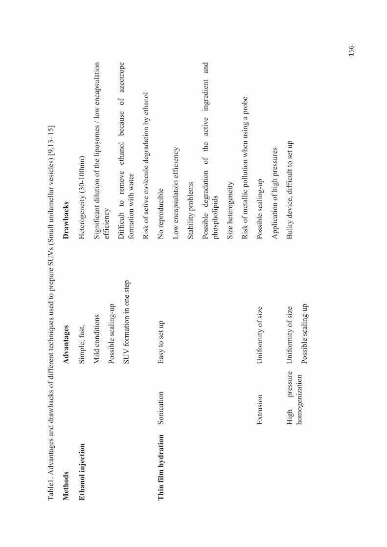
156
Tabl
e1. A
dvan
tage
s and
dra
wba
cks o
f diff
eren
t tec
hniq
ues u
sed
to p
repa
re S
UV
s (Sm
all u
nila
mel
lar v
esic
les)
[9,1
3–15
]
Met
hods
Adv
anta
ges
Dra
wba
cks
Eth
anol
inje
ctio
n
Sim
ple,
fast
,
Mild
con
ditio
ns
Poss
ible
scal
ing-
up
SUV
form
atio
n in
one
step
Het
erog
enei
ty (3
0-10
0nm
)
Sign
ifica
nt d
ilutio
n of
the
lipos
omes
/ lo
w e
ncap
sula
tion
effic
ienc
y
Diff
icul
t to
re
mov
e et
hano
l be
caus
e of
az
eotro
pe
form
atio
n w
ith w
ater
Ris
k of
act
ive
mol
ecul
e de
grad
atio
n by
eth
anol
Thi
n fil
m h
ydra
tion
Soni
catio
n Ea
sy to
set u
p N
o re
prod
ucib
le
Low
enc
apsu
latio
n ef
ficie
ncy
Stab
ility
pro
blem
s
Poss
ible
de
grad
atio
n of
th
e ac
tive
ingr
edie
nt
and
phos
phol
ipid
s
Size
het
erog
enei
ty
Ris
k of
met
allic
pol
lutio
n w
hen
usin
g a
prob
e
Extru
sion
U
nifo
rmity
of s
ize
Poss
ible
scal
ing-
up
App
licat
ion
of h
igh
pres
sure
s
Hig
h pr
essu
re
hom
ogen
izat
ion
Uni
form
ity o
f siz
e
Poss
ible
scal
ing-
up
Bul
ky d
evic
e, d
iffic
ult t
o se
t up
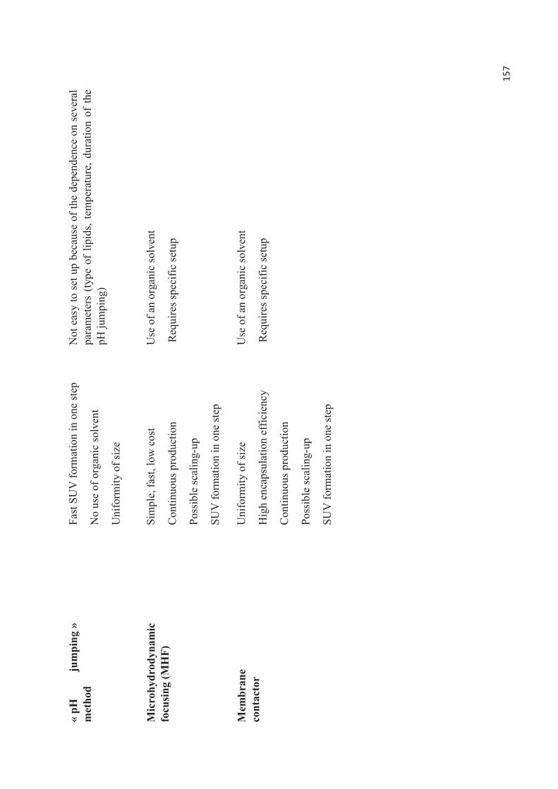
157
« pH
ju
mpi
ng »
m
etho
d
Fast
SU
V fo
rmat
ion
in o
ne st
ep
No
use
of o
rgan
ic so
lven
t
Uni
form
ity o
f siz
e
Not
eas
y to
set
up
beca
use
of th
e de
pend
ence
on
seve
ral
para
met
ers
(type
of
lipid
s, te
mpe
ratu
re, d
urat
ion
of th
e pH
jum
ping
)
Mic
rohy
drod
ynam
ic
focu
sing
(MH
F)
Sim
ple,
fast
, low
cos
t
Con
tinuo
us p
rodu
ctio
n
Poss
ible
scal
ing-
up
SUV
form
atio
n in
one
step
Use
of a
n or
gani
c so
lven
t
Req
uire
s spe
cific
setu
p
Mem
bran
e co
ntac
tor
Uni
form
ity o
f siz
e
Hig
h en
caps
ulat
ion
effic
ienc
y
Con
tinuo
us p
rodu
ctio
n
Poss
ible
scal
ing-
up
SUV
form
atio
n in
one
step
Use
of a
n or
gani
c so
lven
t
Req
uire
s spe
cific
setu
p
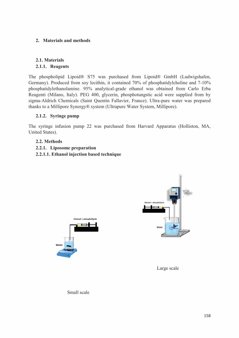
158
2. Materials and methods
2.1. Materials 2.1.1. Reagents
The phospholipid Lipoid® S75 was purchased from Lipoid® GmbH (Ludwigshafen, Germany). Produced from soy lecithin, it contained 70% of phosphatidylcholine and 7-10% phosphatidylethanolamine. 95% analytical-grade ethanol was obtained from Carlo Erba Reagenti (Milano, Italy). PEG 400, glycerin, phosphotungstic acid were supplied from by sigma-Aldrich Chemicals (Saint Quentin Fallavier, France). Ultra-pure water was prepared thanks to a Millipore Synergy® system (Ultrapure Water System, Millipore).
2.1.2. Syringe pump
The syringe infusion pump 22 was purchased from Harvard Apparatus (Holliston, MA, United States).
2.2. Methods 2.2.1. Liposome preparation 2.2.1.1. Ethanol injection based technique
Small scale
Large scale
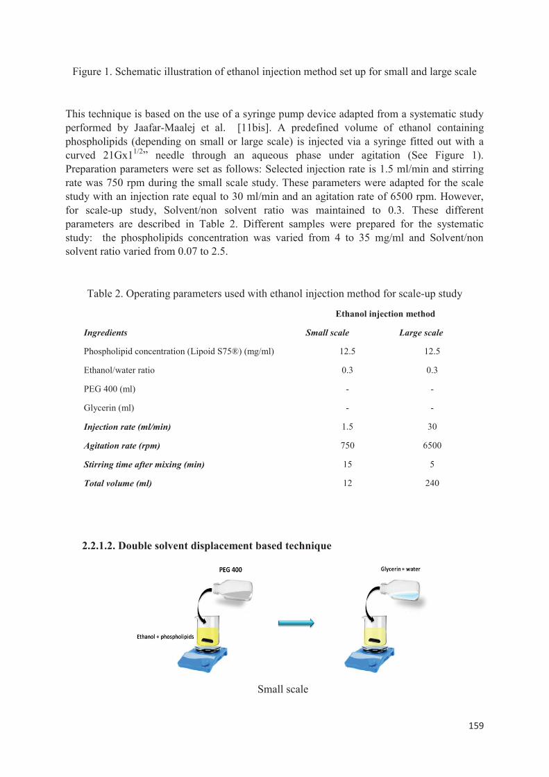
159
Figure 1. Schematic illustration of ethanol injection method set up for small and large scale
This technique is based on the use of a syringe pump device adapted from a systematic study performed by Jaafar-Maalej et al. [11bis]. A predefined volume of ethanol containing phospholipids (depending on small or large scale) is injected via a syringe fitted out with a curved 21Gx11/2” needle through an aqueous phase under agitation (See Figure 1). Preparation parameters were set as follows: Selected injection rate is 1.5 ml/min and stirring rate was 750 rpm during the small scale study. These parameters were adapted for the scale study with an injection rate equal to 30 ml/min and an agitation rate of 6500 rpm. However, for scale-up study, Solvent/non solvent ratio was maintained to 0.3. These different parameters are described in Table 2. Different samples were prepared for the systematic study: the phospholipids concentration was varied from 4 to 35 mg/ml and Solvent/non solvent ratio varied from 0.07 to 2.5.
Table 2. Operating parameters used with ethanol injection method for scale-up study
Ethanol injection method
Ingredients Small scale Large scale
Phospholipid concentration (Lipoid S75®) (mg/ml) 12.5 12.5
Ethanol/water ratio 0.3 0.3
PEG 400 (ml) - -
Glycerin (ml) - -
Injection rate (ml/min) 1.5 30
Agitation rate (rpm) 750 6500
Stirring time after mixing (min) 15 5
Total volume (ml) 12 240
2.2.1.2. Double solvent displacement based technique
Small scale
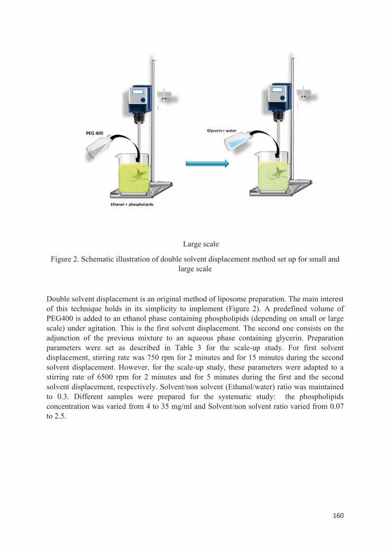
160
Large scale
Figure 2. Schematic illustration of double solvent displacement method set up for small and large scale
Double solvent displacement is an original method of liposome preparation. The main interest of this technique holds in its simplicity to implement (Figure 2). A predefined volume of PEG400 is added to an ethanol phase containing phospholipids (depending on small or large scale) under agitation. This is the first solvent displacement. The second one consists on the adjunction of the previous mixture to an aqueous phase containing glycerin. Preparation parameters were set as described in Table 3 for the scale-up study. For first solvent displacement, stirring rate was 750 rpm for 2 minutes and for 15 minutes during the second solvent displacement. However, for the scale-up study, these parameters were adapted to a stirring rate of 6500 rpm for 2 minutes and for 5 minutes during the first and the second solvent displacement, respectively. Solvent/non solvent (Ethanol/water) ratio was maintained to 0.3. Different samples were prepared for the systematic study: the phospholipids concentration was varied from 4 to 35 mg/ml and Solvent/non solvent ratio varied from 0.07 to 2.5.
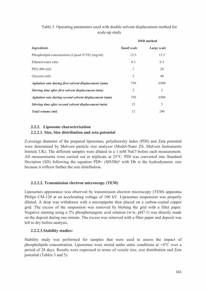
161
Table 3. Operating parameters used with double solvent displacement method for scale-up study
DSD method
Ingredients Small scale Large scale
Phospholipid concentration (Lipoid S75®) (mg/ml) 12.5 12.5
Ethanol/water ratio 0.3 0.3
PEG 400 (ml) 1 20
Glycerin (ml) 2 40
Agitation rate during first solvent displacement (rpm) 750 6500
Stirring time after first solvent displacement (min) 2 2
Agitation rate during second solvent displacement (rpm) 750 6500
Stirring time after second solvent displacement (min) 15 5
Total volume (ml) 12 240
2.2.2. Liposome characterization 2.2.2.1. Size, Size distribution and zeta potential
Z-average diameter of the prepared liposomes, polydisersity index (PDI) and Zeta potential were determined by Malvern particle size analyzer (Model-Nano ZS, Malvern Instruments limited, UK). The different samples were diluted in a 1 mM NaCl before each measurement. All measurements were carried out in triplicate at 25°C. PDI was converted into Standard Deviation (SD) following the equation PDI= (SD/Dh)² with Dh is the hydrodynamic size because it reflects further the size distribution.
2.2.2.2. Transmission electron microscopy (TEM)
Liposomes appearance was observed by transmission electron microscopy (TEM) apparatus Philips CM-120 at an accelerating voltage of 100 kV. Liposomes suspension was properly diluted. A drop was withdrawn with a micropipette then placed on a carbon-coated copper grid. The excess of the suspension was removed by blotting the grid with a filter paper. Negative staining using a 2% phosphotungstic acid solution (w/w, pH7.1) was directly made on the deposit during one minute. The excess was removed with a filter paper and deposit was left to dry before analysis.
2.2.2.3.Stability studies:
Stability study was performed for samples that were used to assess the impact of phospholipids concentration. Liposomes were stored under static conditions at +4°C over a period of 28 days. Results were expressed in terms of vesicle size, size distribution and Zeta potential (Tables 3 and 5).
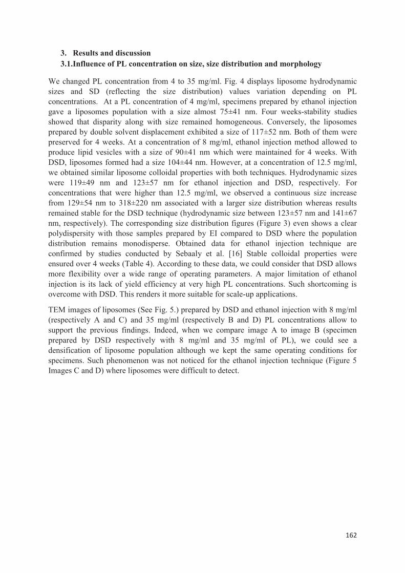
162
3. Results and discussion 3.1.Influence of PL concentration on size, size distribution and morphology
We changed PL concentration from 4 to 35 mg/ml. Fig. 4 displays liposome hydrodynamic sizes and SD (reflecting the size distribution) values variation depending on PL concentrations. At a PL concentration of 4 mg/ml, specimens prepared by ethanol injection gave a liposomes population with a size almost 75±41 nm. Four weeks-stability studies showed that disparity along with size remained homogeneous. Conversely, the liposomes prepared by double solvent displacement exhibited a size of 117±52 nm. Both of them were preserved for 4 weeks. At a concentration of 8 mg/ml, ethanol injection method allowed to produce lipid vesicles with a size of 90±41 nm which were maintained for 4 weeks. With DSD, liposomes formed had a size 104±44 nm. However, at a concentration of 12.5 mg/ml, we obtained similar liposome colloidal properties with both techniques. Hydrodynamic sizes were 119±49 nm and 123±57 nm for ethanol injection and DSD, respectively. For concentrations that were higher than 12.5 mg/ml, we observed a continuous size increase from 129±54 nm to 318±220 nm associated with a larger size distribution whereas results remained stable for the DSD technique (hydrodynamic size between 123±57 nm and 141±67 nm, respectively). The corresponding size distribution figures (Figure 3) even shows a clear polydispersity with those samples prepared by EI compared to DSD where the population distribution remains monodisperse. Obtained data for ethanol injection technique are confirmed by studies conducted by Sebaaly et al. [16] Stable colloidal properties were ensured over 4 weeks (Table 4). According to these data, we could consider that DSD allows more flexibility over a wide range of operating parameters. A major limitation of ethanol injection is its lack of yield efficiency at very high PL concentrations. Such shortcoming is overcome with DSD. This renders it more suitable for scale-up applications.
TEM images of liposomes (See Fig. 5.) prepared by DSD and ethanol injection with 8 mg/ml (respectively A and C) and 35 mg/ml (respectively B and D) PL concentrations allow to support the previous findings. Indeed, when we compare image A to image B (specimen prepared by DSD respectively with 8 mg/ml and 35 mg/ml of PL), we could see a densification of liposome population although we kept the same operating conditions for specimens. Such phenomenon was not noticed for the ethanol injection technique (Figure 5 Images C and D) where liposomes were difficult to detect.
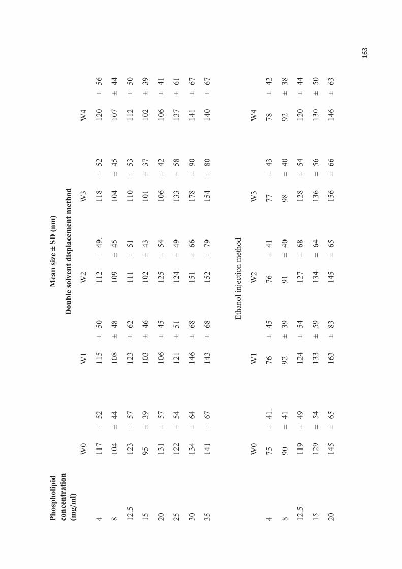
163
Phos
phol
ipid
co
ncen
trat
ion
(mg/
ml)
Mea
n si
ze ±
SD
(nm
)
Dou
ble
solv
ent d
ispl
acem
ent m
etho
d
W0
W1
W2
W3
W4
4 11
7 ±
52
115
± 50
11
2 ±
49.
118
± 52
12
0 ±
56
8 10
4 ±
44
108
± 48
10
9 ±
45
104
± 45
10
7 ±
44
12.5
12
3 ±
57
123
± 62
11
1 ±
51
110
± 53
11
2 ±
50
15
95
± 39
10
3 ±
46
102
± 43
10
1 ±
37
102
± 39
20
131
± 57
10
6 ±
45
125
± 54
10
6 ±
42
106
± 41
25
122
± 54
12
1 ±
51
124
± 49
13
3 ±
58
137
± 61
30
134
± 64
14
6 ±
68
151
± 66
17
8 ±
90
141
± 67
35
141
± 67
14
3 ±
68
152
± 79
15
4 ±
80
140
± 67
Et
hano
l inj
ectio
n m
etho
d
W
0
W
1
W
2
W
3
W
4
4 75
±
41.
76
± 45
76
±
41
77
± 43
78
±
42
8 90
±
41
92
± 39
91
±
40
98
± 40
92
±
38
12.5
11
9 ±
49
124
± 54
12
7 ±
68
128
± 54
12
0 ±
44
15
129
± 54
13
3 ±
59
134
± 64
13
6 ±
56
130
± 50
20
145
± 65
16
3 ±
83
145
± 65
15
6 ±
66
146
± 63

164
25
171
± 83
17
5 ±
94
168
± 80
17
6 ±
88
170
± 83
30
308
± 21
9 29
3 ±
203
329
± 19
4 31
1 ±
213
335
± 23
5
35
318
± 22
0 32
7 ±
224
359
± 27
3 36
2 ±
248
342
± 24
2
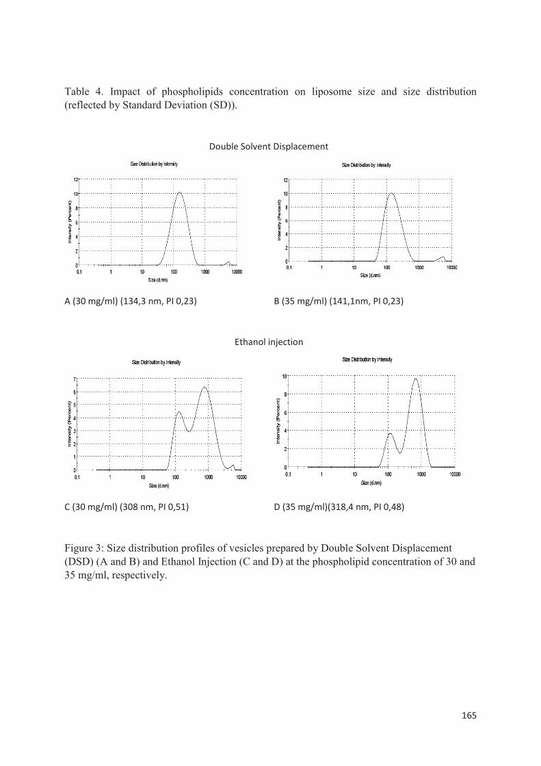
165
Table 4. Impact of phospholipids concentration on liposome size and size distribution (reflected by Standard Deviation (SD)).
Double Solvent Displacement
A (30 mg/ml) (134,3 nm, PI 0,23) B (35 mg/ml) (141,1nm, PI 0,23)
Ethanol injection
C (30 mg/ml) (308 nm, PI 0,51) D (35 mg/ml)(318,4 nm, PI 0,48)
Figure 3: Size distribution profiles of vesicles prepared by Double Solvent Displacement (DSD) (A and B) and Ethanol Injection (C and D) at the phospholipid concentration of 30 and 35 mg/ml, respectively.
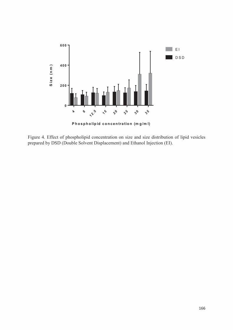
166
4 81 2 .5 1 5 2 0 2 5 3 0 3 5
0
2 0 0
4 0 0
6 0 0
P h o s p h o lip id c o n c e n tra tio n (m g /m l)
Siz
e(n
m)
D S D
E I
Figure 4. Effect of phospholipid concentration on size and size distribution of lipid vesicles prepared by DSD (Double Solvent Displacement) and Ethanol Injection (EI).
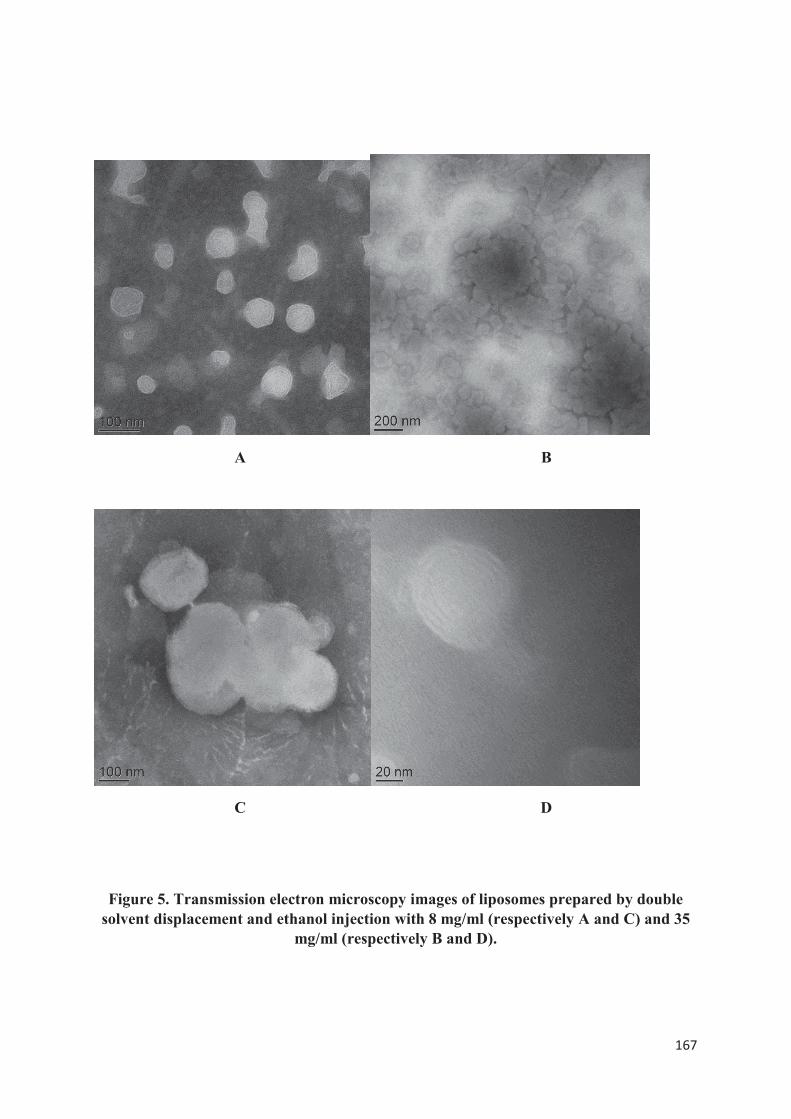
167
A B
C D
Figure 5. Transmission electron microscopy images of liposomes prepared by double solvent displacement and ethanol injection with 8 mg/ml (respectively A and C) and 35
mg/ml (respectively B and D).
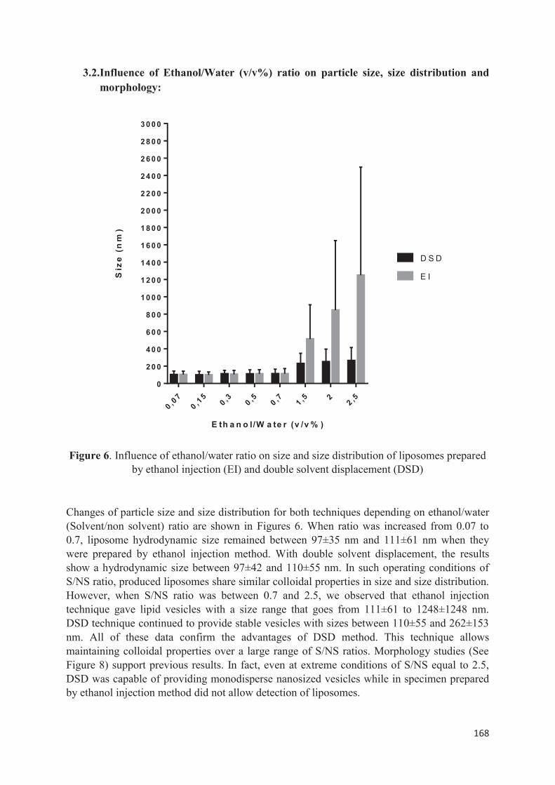
168
3.2.Influence of Ethanol/Water (v/v%) ratio on particle size, size distribution and morphology:
0 ,07
0 ,15
0 ,3 0 ,5 0 ,7 1 ,5 22 ,5
0
2 0 0
4 0 0
6 0 0
8 0 0
1 0 0 0
1 2 0 0
1 4 0 0
1 6 0 0
1 8 0 0
2 0 0 0
2 2 0 0
2 4 0 0
2 6 0 0
2 8 0 0
3 0 0 0
E th a n o l/W a te r (v /v % )
Siz
e(n
m)
D S D
E I
Figure 6. Influence of ethanol/water ratio on size and size distribution of liposomes prepared by ethanol injection (EI) and double solvent displacement (DSD)
Changes of particle size and size distribution for both techniques depending on ethanol/water (Solvent/non solvent) ratio are shown in Figures 6. When ratio was increased from 0.07 to 0.7, liposome hydrodynamic size remained between 97±35 nm and 111±61 nm when they were prepared by ethanol injection method. With double solvent displacement, the results show a hydrodynamic size between 97±42 and 110±55 nm. In such operating conditions of S/NS ratio, produced liposomes share similar colloidal properties in size and size distribution. However, when S/NS ratio was between 0.7 and 2.5, we observed that ethanol injection technique gave lipid vesicles with a size range that goes from 111±61 to 1248±1248 nm. DSD technique continued to provide stable vesicles with sizes between 110±55 and 262±153 nm. All of these data confirm the advantages of DSD method. This technique allows maintaining colloidal properties over a large range of S/NS ratios. Morphology studies (See Figure 8) support previous results. In fact, even at extreme conditions of S/NS equal to 2.5, DSD was capable of providing monodisperse nanosized vesicles while in specimen prepared by ethanol injection method did not allow detection of liposomes.
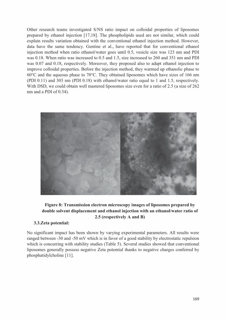
169
Other research teams investigated S/NS ratio impact on colloidal properties of liposomes prepared by ethanol injection [17,18]. The phospholipids used are not similar, which could explain results variation obtained with the conventional ethanol injection method. However, data have the same tendency. Gentine et al., have reported that for conventional ethanol injection method when ratio ethanol/water goes until 0.5, vesicle size was 123 nm and PDI was 0.18. When ratio was increased to 0.5 and 1.5, size increased to 260 and 351 nm and PDI was 0.07 and 0.18, respectively. Moreover, they proposed also to adapt ethanol injection to improve colloidal properties. Before the injection method, they warmed up ethanolic phase to 60°C and the aqueous phase to 70°C. They obtained liposomes which have sizes of 166 nm (PDI 0.11) and 303 nm (PDI 0.18) with ethanol/water ratio equal to 1 and 1.5, respectively. With DSD, we could obtain well mastered liposomes size even for a ratio of 2.5 (a size of 262 nm and a PDI of 0.34).
Figure 8: Transmission electron microscopy images of liposomes prepared by double solvent displacement and ethanol injection with an ethanol/water ratio of
2.5 (respectively A and B) 3.3.Zeta potential:
No significant impact has been shown by varying experimental parameters. All results were ranged between -30 and -50 mV which is in favor of a good stability by electrostatic repulsion which is concurring with stability studies (Table 5). Several studies showed that conventional liposomes generally possess negative Zeta potential thanks to negative charges conferred by phosphatidylcholine [11].
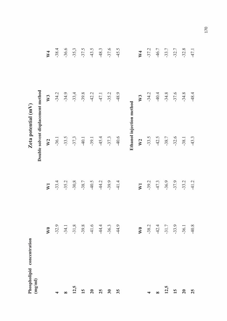
170
Phos
phol
ipid
co
ncen
trat
ion
(mg/
ml)
Zeta
pot
entia
l (m
V)
Dou
ble
solv
ent d
ispl
acem
ent m
etho
d
W
0 W
1 W
2 W
3 W
4
4 -3
2.9
-3
3.4
-3
6.1
-3
4.2
-3
8.4
8 -3
4.1
-35.
2 -3
3.5
-34.
9 -3
6.6
12,5
-3
1,8
-30,
8 -3
7,3
-33,
4 -3
5,3
15
-39.
8 -3
8.7
-40.
1 -3
9.8
-37.
5
20
-41.
6 -4
0.5
-39.
1 -4
2.2
-43.
5
25
-44.
4 -4
4.2
-45.
4 -4
7.1
-48.
3
30
-36.
3 -3
9.9
-37.
3 -3
5.2
-37.
6
35
-44.
9 -4
1.4
-40.
6 -4
8.9
-45.
5
E
than
ol in
ject
ion
met
hod
W
0 W
1 W
2 W
3 W
4
4 -3
8.2
-39.
2 -3
3.5
-34.
2 -3
7.2
8 -4
2.4
-47.
3 -4
2.5
-40.
4 -4
6.7
12,5
-3
1.7
-36.
9 -3
8.7
-34.
8 -3
3.7
15
-33.
9 -3
7.9
-32.
6 -3
7.6
-32.
7
20
-36.
1 -3
3.2
-38.
1 -3
4.8
-32.
8
25
-40.
8 -4
1.2
-43.
3 -4
8.4
-47.
1
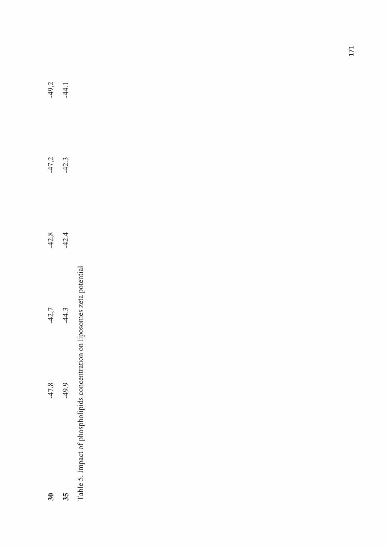
171
30
-47,
8 -4
2,7
-42,
8 -4
7,2
-49,
2
35
-49.
9 -4
4.3
-42.
4 -4
2.3
-44.
1
Tabl
e 5.
Impa
ct o
f pho
spho
lipid
s con
cent
ratio
n on
lipo
som
es z
eta
pote
ntia
l
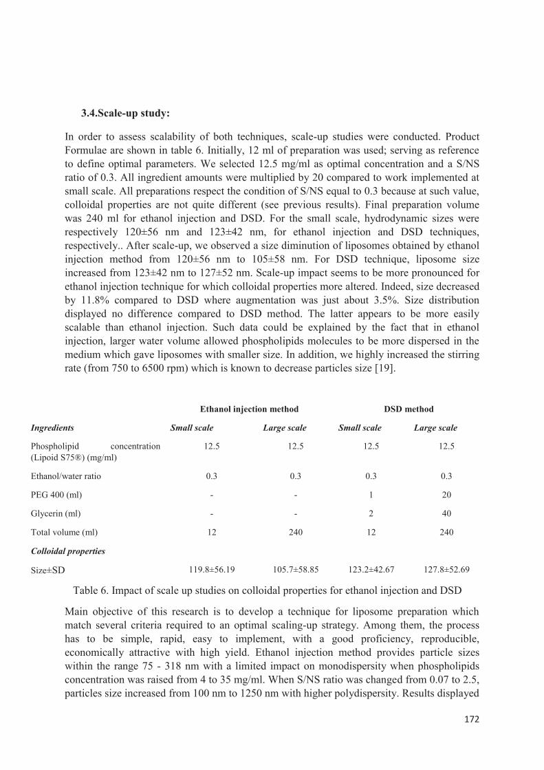
172
3.4.Scale-up study:
In order to assess scalability of both techniques, scale-up studies were conducted. Product Formulae are shown in table 6. Initially, 12 ml of preparation was used; serving as reference to define optimal parameters. We selected 12.5 mg/ml as optimal concentration and a S/NS ratio of 0.3. All ingredient amounts were multiplied by 20 compared to work implemented at small scale. All preparations respect the condition of S/NS equal to 0.3 because at such value, colloidal properties are not quite different (see previous results). Final preparation volume was 240 ml for ethanol injection and DSD. For the small scale, hydrodynamic sizes were respectively 120±56 nm and 123±42 nm, for ethanol injection and DSD techniques, respectively.. After scale-up, we observed a size diminution of liposomes obtained by ethanol injection method from 120±56 nm to 105±58 nm. For DSD technique, liposome size increased from 123±42 nm to 127±52 nm. Scale-up impact seems to be more pronounced for ethanol injection technique for which colloidal properties more altered. Indeed, size decreased by 11.8% compared to DSD where augmentation was just about 3.5%. Size distribution displayed no difference compared to DSD method. The latter appears to be more easily scalable than ethanol injection. Such data could be explained by the fact that in ethanol injection, larger water volume allowed phospholipids molecules to be more dispersed in the medium which gave liposomes with smaller size. In addition, we highly increased the stirring rate (from 750 to 6500 rpm) which is known to decrease particles size [19].
Ethanol injection method DSD method
Ingredients Small scale Large scale Small scale Large scale
Phospholipid concentration (Lipoid S75®) (mg/ml)
12.5 12.5 12.5 12.5
Ethanol/water ratio 0.3 0.3 0.3 0.3
PEG 400 (ml) - - 1 20
Glycerin (ml) - - 2 40
Total volume (ml) 12 240 12 240
Colloidal properties
Size±SD 119.8±56.19 105.7±58.85 123.2±42.67 127.8±52.69
Table 6. Impact of scale up studies on colloidal properties for ethanol injection and DSD
Main objective of this research is to develop a technique for liposome preparation which match several criteria required to an optimal scaling-up strategy. Among them, the process has to be simple, rapid, easy to implement, with a good proficiency, reproducible, economically attractive with high yield. Ethanol injection method provides particle sizes within the range 75 - 318 nm with a limited impact on monodispersity when phospholipids concentration was raised from 4 to 35 mg/ml. When S/NS ratio was changed from 0.07 to 2.5, particles size increased from 100 nm to 1250 nm with higher polydispersity. Results displayed
173
by DSD method showed liposome sizes between 96 to 141 nm with a good monodispersity depending on phospholipids concentration. Influence of ethanol/water ratio was also assessed. When this ratio was changed from 0.07 to 2.5, particles size increased from 100 to 300 nm.
Parameters change exhibited higher variability of colloidal properties with ethanol injection technique. Indeed, 4-fold increase was obtained following PL concentration increase with a size distribution becoming very heterogeneous. Similarly, 12-times increase of size was obtained when S/NS ratio was increased with high polydisperse particles population. Conversely, double solvent displacement enables 1.5 time increase in size and 3 times with monodisperse sizes at each time. Moreover, DSD appears to provide higher yield when we go beyond the limit fixed for ethanol injection. In addition, when we increase PL concentration, liposome size obtained by ethanol injection grows faster and much larger than DSD. The hypothesis is that more lipid materials would give tendency to self-organize to make bilayers longer leading to greater size whereas, in DSD, the size sounds contained which suggests the production of more liposomes. This hypothesis is consistent with morphological observation by TEM which showed dense liposome population in the case of DSD. The DSD could be considered a more simple, easy to implement and faster method to prepare liposomes regarding ethanol injection. No sophisticated system is needed. No more parameters have to be controlled like flow rate and needle diameter. This confirms that DSD is easier and faster to implement because it is based only on simple mixing steps. This made from DSD a more economically interesting method. Within a large range of parameters variation, colloidal properties remain relatively stable which proves the high reproducibility of our technique. However, PEG 400 and glycerol could be always available in external phase which renders an eventual lyophilization of the system more difficult than with ethanol injection and therefore, reduce the application spectrum.
4. Conclusion
Liposomes preparation methods described in literature exhibit several drawbacks which do not match with criteria required for a potential industrial application. The method of ethanol injection which is the most commonly used for preparing SUVs did not produce large amounts of liposomes, even if the method is transposable on an industrial scale. Indeed, it is imposed by the optimal working conditions to be in highly diluted conditions, which also limits the degree of encapsulation of active molecules. With regard to the lipid film hydration method associated with size reduction techniques such as sonication or extrusion, it does not provide possible transposition to the industrial scale. Other systems (the microhydrodynamic focusing (MHF), the method by membrane contactor) are effective but more complicated to set up and expensive.
The double solvent displacement method appears to be very promising. Indeed, the two steps are performed under mild conditions and based on simple homogenization. This method allows forming homogeneous and small size lipid vesicles (near 100 nm) without any additional steps. Moreover, large amounts of liposomes are formed using high phospholipids concentrations while maintaining a small size and good homogeneity. Thus, this method could be implemented on an industrial scale and provide very good performance. Indeed, the simplicity and the rapidity of this new technique enable us to produce liposomes population with a good proficiency of the process especially in term of reproducibility, offering high yield while keeping economically attractive. Double solvent displacement could be able to
174
meet the challenges of industrial production of liposomes, and thus greatly promote the arrival on the market of many molecules that would need to be encapsulated in liposomes.
References:
[1] W. Badri, R. Eddabra, H. Fessi, A. Elaissari, Biodegradable Polymer Based Nanoparticles: Dermal and Transdermal Drug Delivery, J. Colloid Sci. Biotechnol. 3 (2014) 141–149. doi:10.1166/jcsb.2014.1091.
[2] V. Rosset, N. Ahmed, I. Zaanoun, B. Stella, H. Fessi, A. Elaissari, Elaboration of Argan Oil Nanocapsules Containing Naproxen for Cosmetic and Transdermal Local Application, J. Colloid Sci. Biotechnol. 1 (2012) 218–224. doi:10.1166/jcsb.2012.1023.
[3] C.M. Baena-Aristizábal, C.E. Mora-Huertas, Micro, Nano and Molecular Novel Delivery Systems as Carriers for Herbal Materials, J. Colloid Sci. Biotechnol. 2 (2013) 263–297. doi:10.1166/jcsb.2013.1070.
[4] A.A.M. Lira, P.L.A. Cordo, E.C.F. Nogueira, E.D.P. Almeida, R.A.L.C. Junior, R.S. Nunes, M.V.L.B. Bentley, J.M. Marchetti, Optimization of Topical All-Trans Retinoic Acid Penetration Using Poly-DL-Lactide and Poly-DL-Lactide-Co-Glycolide Microparticles, J. Colloid Sci. Biotechnol. 2 (2013) 123–129. doi:10.1166/jcsb.2013.1041.
[5] A. Jada, A Special Issue on Inorganic Colloidal Particles, Synthesis, Surface Properties and Applications, J. Colloid Sci. Biotechnol. 3 (2014) 1–2. doi:10.1166/jcsb.2014.1071.
[6] M. Sherry, C. Charcosset, H. Fessi, H. Greige-Gerges, Essential oils encapsulated in liposomes: a review, J. Liposome Res. 23 (2013) 268–275. doi:10.3109/08982104.2013.819888.
[7] G.M. El Maghraby, B.W. Barry, A.C. Williams, Liposomes and skin: from drug delivery to model membranes, Eur. J. Pharm. Sci. Off. J. Eur. Fed. Pharm. Sci. 34 (2008) 203–222. doi:10.1016/j.ejps.2008.05.002.
[8] A.D. Bangham, M.M. Standish, J.C. Watkins, Diffusion of univalent ions across the lamellae of swollen phospholipids, J. Mol. Biol. 13 (1965) 238–252.
[9] Y.P. Patil, S. Jadhav, Novel methods for liposome preparation, Chem. Phys. Lipids. 177 (2014) 8–18. doi:10.1016/j.chemphyslip.2013.10.011.
[10] S. Batzri, E.D. Korn, Single bilayer liposomes prepared without sonication, Biochim. Biophys. Acta. 298 (1973) 1015–1019.
[11] S. STAINMESSE, H. FESSI, J.P. DEVISSAGUET et F. PUISIEUX, Process for the preparation of dispersible colloidal systems of amphiphilic lipids in the form of oligolamellar liposomes of submicron dimensions, US Pat. N° 5,174,930, December 29, 1992.
175
[11bis] C. Jaafar-Maalej, R. Diab, V. Andrieu, A. Elaissari, H. Fessi, Ethanol injection method for hydrophilic and lipophilic drug-loaded liposome preparation, J. Liposome Res. 20 (2010) 228–243. doi:10.3109/08982100903347923.
[12] Fessi, H., Puisieux, F., Devissaguet, J.P., 1988. Procédé de préparation de systèmes colloïdaux dispersibles d’une substance sous forme de nanocapsules. European Patent 274961 B1, 25 March1992
[13] A. Akbarzadeh, R. Rezaei-Sadabady, S. Davaran, S.W. Joo, N. Zarghami, Y. Hanifehpour, M. Samiei, M. Kouhi, K. Nejati-Koshki, Liposome: classification, preparation, and applications, Nanoscale Res. Lett. 8 (2013) 102. doi:10.1186/1556-276X-8-102.
[14] Z. Huang, X. Li, T. Zhang, Y. Song, Z. She, J. Li, Y. Deng, Progress involving new techniques for liposome preparation, Asian J. Pharm. Sci. 9 (2014) 176–182. doi:10.1016/j.ajps.2014.06.001.
[15] A. Laouini, C. Jaafar-Maalej, I. Limayem-Blouza, S. Sfar, C. Charcosset, H. Fessi, Preparation, Characterization and Applications of Liposomes: State of the Art, J. Colloid Sci. Biotechnol. 1 (2012) 147–168. doi:10.1166/jcsb.2012.1020.
[16] C. Sebaaly, H. Greige-Gerges, S. Stainmesse, H. Fessi, C. Charcosset, Effect of composition, hydrogenation of phospholipids and lyophilization on the characteristics of eugenol-loaded liposomes prepared by ethanol injection method, Food Biosci. 15 (2016) 1–10. doi:10.1016/j.fbio.2016.04.005.
[17] P. Gentine, L. Bourel-Bonnet, B. Frisch, Modified and derived ethanol injection toward liposomes: development of the process, J. Liposome Res. 23 (2013) 11–19. doi:10.3109/08982104.2012.717298.
[18] Y. Maitani, H. Soeda, W. Junping, K. Takayama, MODIFIED ETHANOL INJECTION METHOD FOR LIPOSOMES CONTAINING beta-SITOSTEROL beta-D-GLUCOSIDE, J. Liposome Res. 11 (2001) 115–125. doi:10.1081/LPR-100103174.
[19] S.A. Galindo-Rodríguez, F. Puel, S. Briançon, E. Allémann, E. Doelker, H. Fessi, Comparative scale-up of three methods for producing ibuprofen-loaded nanoparticles, Eur. J. Pharm. Sci. 25 (2005) 357–367. doi:10.1016/j.ejps.2005.03.013.
176
PARTIE EXPERIMENTALE
Préparation par double déplacement de solvent de nanovecterus lipidiques encapsulant la cyclosporine A pour une application topique dans le traitement du psoriasis
Prévu pour soumission au journal « International Journal of pharmaceutics »
177
La troisième partie expérimentale consiste à encapsuler la ciclospirine A par double déplacement de solvant. Après avoir travaillé au développement et l’optimisation de la méthode dite du double déplacement de solvant dans les travaux précédents, nous avions identifié les principaux paramètres de la formulation à retenir pour la préparation des vésicules lipidiques : ratio éthanol/eau de 0.3, concentration de PEG 400 proche de 8% ainsi qu’une concentration en glycérol d’environ 15%. La concentration en éthanol était fixée à 15%. Cependant pour l’encapsulation les paramètres pourraient avoir besoin d’être adaptés comme la concentration en phospholipides pour optimiser le taux d’encapsulation mais aussi obtenir le ciblage tissulaire recherché. Lors de ce travail, nous avons découvert qu’en modifiant les conditions opératoires ayant trait au degré de précipitation lors du premier déplacement de solvant, nous pouvions produire des SLN (solides lipides nanoparticles). Il s’agit là de résultats vraiment très intéressants. Jusqu’ici, rares étaient les méthodes pouvant être utilisées à la fois pour préparer des vésicules lipidiques et des SLN. La méthode dite du contacteur membranaire est quasi la seule. Mais ce qu’il y a d’encore plus intéressant est que la composition ici ne contient qu’une seule source de lipides, notamment des phospholipides issus du soja (Lipoid S75®). Dans cette dernière partie, nous avons préparé et caractérisé différentes formulations lipidiques encapsulant la ciclosporine A (liposomes et SLN). La taille des vecteurs lipidiques préparés était comprise entre 95 et 170 avec une PDI ≤ 0,30. Les résultats obtenus par TEM ont montré que les phospholipides pouvaient former des vésicules lipidiques avec un ratio éthanol / eau de 0,3 et des nanoparticules lipidiques solides avec un ratio égal à 0,15. Les courbes de distribution de taille sont toutes gaussiennes, ce qui signifie que toutes les formulations avec CsA étaient essentiellement monodisperses. Les analyses du potentiel zéta ont permis de voir que la charge superficielle des vésicules lipidiques et des SLN était négative, d'environ -48 mV et entre -43 mV et -53 mV respectivement. Par ailleurs, les formulations contenant une concentration plus faible en phospholipides ont affiché une efficacité d'encapsulation plus élevée (EE%) avec un taux d'environ 80%. Nous avons pu constater que plus le ratio actif/lipide était réduit, plus le taux d’encapsulation était faible. Pour les deux types de vecteurs, une grande stabilité a été obtenue surtout en raison de la charge de surface négative élevée empêchant l'agrégation par répulsion électrostatique. Mais une autre raison qui peut être mentionnée est la viscosité de la formulation qui est plus élevée que l'eau due au PEG 400 et au glycérol. En outre, l'efficacité d'encapsulation est restée stable pour les différentes formulations. Aucune fuite n'a été observée pendant les conditions de stockage à +4°C pendant 4 semaines. Ensuite, nous avons réalisé une étude in vitro pour identifier quelle type de vecteur permettait une pénétration optimale de la ciclosporine A au niveau de l’épiderme viable en vue d’une application dans la prise en charge topique du psoriasis. Les résultats montraient que nos SLNs n’apportaient davantage particulier en comparaison aux vésicules lipidiques lorsque l’on recherchait un ciblage de l’épiderme viable. Par contre, dans le groupe des formulations contenant des liposomes, nous avons pu mettre en évidence une formulation permettant un passage de plus de 15% de la dose appliquée au niveau du stratum corneum et de l’épiderme viable. Cette formulation est apparue comme étant la plus prometteuse pour une application dans le psoriasis.
Preparation of Cyclosporin A loaded lipid based nanocarriers by Double Solvent Displacement for topical treatment of psoriasis
M. Salaa, A. Elaissaria, H. Fessia*
178
a LAGEP, UMR 5007, University Claude Bernard Lyon 1, CNRS, F-69622, Villeurbanne, France. *Corresponding author: [email protected]
Abstract
The aim of this study was to develop a suitable topical drug delivery system encapsulating cyclosporine A (CsA) for targeting a specific skin layer: the viable epidermis which would be the main site of psoriasis physiopathology. We used the double solvent displacement method to prepare both lipid vesicles and solid lipid nanoparticles encapsulating cyclosporine A. The only source of lipid used was soybean phospholipids. These two types of lipid based carriers were characterized through colloidal properties and capacity of drug encapsulation. Then, we evaluated the different formulations through in vitro skin studies to know which one would be the most suitable for a topical delivery targeting the viable epidermis in order to consider topical management of psoriasis. Mean size was between 109 ±44 nm and 133±69 nm for lipid vesicles, and for SLNs between 95±35 nm and 174±76 nm. Size distribution of all formulations entrapping CsA was monodispersed except for SLN formulation with high content of phospholipids. Zeta potential of lipid vesicles and SLNs were negative, around -48mV and between -43mV and -53 mV respectively. CsA loaded lipid vesicles and SLNs have been able to entrap between 50% and 80% of CsA. Formulations displayed a good colloidal and encapsulation stability after four week storage at 4°C. Among the different formulations designed, lipid vesicle sample was the most promising to an application in psoriasis compared to SLNs. Indeed, they offered the higher amount of CsA especially in SC and viable epidermis with more than 15% of the applied dose.
1. Introduction
Psoriasis is an autoimmune and chronic inflammatory disease affecting 2–5% of the world population. Although it is not still completely elucidated, the physiopathogenesis of psoriasis is mainly localized in the cutaneous tissue. Direct action on the target region would provide a faster and more effective therapeutic response. Proliferating abnormal keratinocytes are cells presenting the unknown antigen which are the basis of CD4 + and CD8 + lymphocyte activation. The keratinocytes are the cells constituting the so-called viable epidermis. Thus, if effective management of psoriasis is desired at an early stage, targeting the viable epidermis with active molecules such as ciclosporin A (CsA) which has an effect on both T cell activation and proliferation of keratinocytes is judicious. Encapsulation of ciclosporin A that may allow direct access to the site of action would lead to have greater efficacy and to avoid using the systemic passage which can cause serious adverse effects (Sala et al., 2016).
Dermatological illnesses like psoriasis (Sala et al., 2016) can be effectively managed thanks to CsA. In 1983, the CsA US Food and Drug Administration (FDA)’s clinical approval was limited to prevention of allograft rejection in transplantation. In 1987, indications were enlarged for the treatment of several autoimmune disorders in 1987 and for dry eye disease in 2003 (Wretlind, 1981). CsA is an immunosuppressive agent acting by forming beforehand a complex with immunophilins, especially cyclophilin A which is a cytoplasmic receptor protein located within some cells. Then, this complex links to a specific protein called calcineurin leading to its phosphatase activity inhibition. The role of calcineurin is described
179
as a regulator of nuclear translocation and activation of the nuclear factor of activated T-cells (NFAT) transcription factors. Therefore, CsA is able to stop the dephosphorylation of NFAT commonly stimulated by cytosolic calcium and its passage into the cell core, and so to block cytokine gene transcription like IL-2, IL-4, TNF-α and INF-γ. It results that T-lymphocytes (T helper and T-cytotoxic cells) are no longer able to be activated after specific antigen stimulation and proliferate. Other effects of CsA have been reported like cardio- and neuroprotective properties via fixation to cyclophillin D, an immunophillin found in mitochondria which participates in mitochondrial permeability transition pore (mPTP) expression and mitochondrial mega-pore formation (Guada et al., 2016). CsA uses are often limited because of side effects, narrow therapeutic window and unpredictable pharmacokinetics. It is especially limited because long-term systemic administration might be responsible of harmful side effects like renal failure until chronic nephrotoxicity and hypertension (Belitsky et al., 2000; Ryan et al., 2010). These last decades, innovative CsA delivery systems based on nano- and micro-carriers have been developped and investigated. Those new CsA formulation have been studied to enhance pharmacokinetic profile after parenteral or oral administration, and also in ocular route. However, rare are works dealing with the use of CsA through skin (dermal or transdermal) delivery systems for skin diseases or others (Guada et al., 2016). Moreover, when skin penetration of CsA is considered, large molecular weight (1202 Da) and poorwater solubility (logP=2.92) (Czogalla, 2009) are two limits that we have to overcome (Choi et al., 1995). Unfortunately, conventional topical formulations of CsA have failed in the treatment of psoriasis for instance (Griffiths et al., 1987). In 1990, Ho et al. showed through a clinical double blind study that administration of CsA intralesional injections have cleared significantly plaques symptoms of psoriasis patients (Ho et al., 1990). However, it is very complicated to promote patient compliance with repeated intralesional injections into the skin.
To improve the skin drug delivery, physical delivery techniques have been studied like sonophoresis(Liu et al., 2006a), iontophoresis (Boinpally et al., 2004) and electroporation (Liu et al., 2010). However, these devices cannot be applied on large skin area and their use requires health professionals and equipments limiting the patient self-management of this chronic disease(Chen et al., 2015). The category of chemical methods like chemical penetration enhancers (Lopes et al., 2005), co-solvent systems (Liu et al., 2006b) and nanoparticle systems (Badri et al., 2014; Sala et al., 2016) are able to answer to the previously described limits because this kind of formulation makes psoriasis patients autonomy with the possibility to use them on larger skin areas and this, no invasively for a better patient compliance. Nanocarriers have been developed especially for lipophilic drugs as a promising approach to improve bioavailability owing to solubility limitation, higher blood retention and sustained drug release by delaying metabolic degradation,and also to target specific organs, cell, or proteins, etc (Kohane, 2007; Moinard-Checot et al., 2006; Sultana et al., 2013). Among diverse drug delivery systems described in literature, polymeric nanoparticles like those made with Poly(Lactide-co-Glycolide) (PLGA) have been reported as capable to induce toxicity from dose dumping, inconsistent release and drug-polymer interactions (Shive and Anderson, 1997).
Lipid based nanocarriers like lipid vesicles and SLN/NLC have been reported as being of high potential for skin drug delivery. Lipid vesicles consist in phospholipid self-assembly in
180
aqueous media resulting in one or several bilayers delimiting compartments. Variation in the composition by adding penetration enhancer like ethanol or edge activator led to new generations of lipid vesicles like ethosomes, transfersomes and penetration enhancer-containing vesicles (PEVs)(Ainbinder and Touitou, 2005; Cevc and Blume, 1992; Elsayed et al., 2007; Ghanbarzadeh and Arami, 2013; Manconi et al., 2011b, 2011a; Touitou et al., 2000; Zhao et al., 2013). Besides lipid vesicles, lipid nanoparticles are based on the use of a solid lipid or a mixture of solid lipids at a rate of 0.1% (w/w) to 30% (w/w) dispersed in aqueous medium to obtain SLNs whereas mix of solid lipids and liquid lipids (oils) in an usual ratio of 70:30 up to a ratio of 99.9:0.1(Pardeike et al., 2009) . To prepare these different drug nanocarriers, different methods are well described for lipid vesicles (Laouini et al., 2012) and lipid nanoparticles (Pardeike et al., 2009).
There are conventional techniques to prepare liposomes which are hydration of a thin lipid film known as the Bangham method(Bangham et al., 1967), Reverse-Phase Evaporation (REV) Technique (Szoka and Papahadjopoulos, 1978) , Solvent (Ether or Ethanol) Injection Technique (Szebeni et al., 1984), Detergent Dialysis (Zumbuehl and Weder, 1981). These techniques required large amount of solvent and were not suitable to scale-up because various steps of homogenization were added. Others techniques have been developed then such as heating method (Mozafari, 2005), spray drying (Skalko-Basnet et al., 2000), freeze drying (Li and Deng, 2004), Super Critical Reverse Phase Evaporation (SCRPE) (Otake et al., 2001), crossflow injection technique (Wagner et al., 2002), microfluidic hydrodynamic focusing (MHF) (Jahn et al., 2004), membrane contactor (Jaafar-Maalej et al., 2011). Lipid nanoparticles can be produced by many methods which are microemulsion technique (Gasco, 1993), emulsification-solvent evaporation (Sjöström and Bergenståhl, 1992), emulsification-solvent diffusion method (Hu et al., 2002), solvent injection (or solvent displacement) method (Schubert and Müller-Goymann, 2003), phase inversion (Heurtault et al., 2002), multiple emulsion technique (Garcıa-Fuentes et al., 2003), ultrasonication (Pietkiewicz and Sznitowska, 2004) and membrane contractor technique (Ahmed El-Harati et al., 2006). But the most used one is the high pressure homogenization reported to allow a production organic solvent free, quickly and easily scaled up in comparison with the others (Liedtke et al., 2000; Mehnert and Mäder, 2001; Wissing et al., 2004).
According to our knowledge, there is just one method reported in literature that is directly used to prepare both lipid vesicles and lipid nanoparticles: membrane contactor technique. However, regarding lipid materials used to prepare lipid based nanocarriers, only our work described the exclusive use of phospholipids to prepare both lipid vesicles and lipid nanoparticles by adapting the operating conditions.
Here, we described a unique method which is able to produce both lipid vesicles and SLN by varying operating parameters: the double solvent displacement (DSD). And the only source of lipids used here is phospholipids. Phospholipids are a common used biomaterial well known for its biodegradability and biocompatibility, track record for FDA and European Medicine Agency approval in drug delivery systems. In our work, we used the DSD to encapsulate CsA into lipid vesicles and into lipid nanoparticles. We presented a comparison of their colloidal properties and ability to encapsulate. Then, we evaluated their skin drug delivery potentials and discussed the better formulation for targeting viable epidermis which is main site of many skin diseases like psoriasis.
181
2. Materials and Methods
2.1 Materials Lipoid® S75 (soy lecithin composed by 70% Phosphatidylcholine, 7-10% Phosphatidylethanolamine) were purchased from Lipoid GmbH (Ludwigshafen, Germany). PEG 400, glycerol, phosphotungstic acid, sodium chloride and Cyclosporin A were purchased from Sigma-Aldrich (Saint Quentin Fallavier, France). Water was purified on a Milli-Q system obtained from a Millipore® synergy system (Millipore, Billerica, Massachusetts, USA). Ethanol used was of analytical grade (Carlo Erba Reagenti, Val de Reuil, France) and used as such.
2.2 Free liposome preparation by double solvent displacement
All samples have been prepared according to the method described below. This method has been already described in previous research (Sala et al.). However, concentration or ratio of different components may vary to evaluate their effects respectively. Compositions are detailed in Table 1.
2.2.1 First solvent displacement
It consists in preparing an ethanol solution of phospholipids, and then this organic phase is dispersed in PEG 400. Phospholipids are soluble in ethanol but not in PEG 400. However, ethanol and PEG 400 are miscible. After 2 min of magnetic stirring at 700 rpm, the mixture is clear. For all samples prepared, the concentration of Lipoid® S75 is ranging between 8.5 and 15 mg/mL.
2.2.2 Second solvent displacement
The ethanol / PEG400 phase prepared previously containing phospholipids is dispersed in an aqueous phase composed of milli-Q water and glycerol. The liposomal suspension is then heated to 60 °C (above the transition temperature of Lipoid S75) under magnetic stirring at 700 rpm for 1hour.
2.3 Cyclosporin A loaded liposome and lipid nanoparticle preparation by double solvent displacement
2.3.1 Encapsulation of Cyclosporin A into liposomes and lipid nanoparticles
The preparation process of lipid vesicles by double solvent displacement followed the procedure describe above. Briefly, 20 mg CsA was dissolved in 2ml or 1ml of ethanol containing various concentration of phospholipids, to prepare vesicle liposomes or solid lipid nanoparticles, respectively. Then, 1ml of PEG 400 was added (first solvent displacement). After stirring, 9ml of a solution of glycerol-water (20:70) was incorporated (2nd solvent displacement). The overall was heated to 60 °C under magnetic stirring at 700 rpm for 1 hour.
2.3.2 Determination of encapsulation efficiency of Cyclosporin A into liposomes and lipid nanoparticles
Encapsulation efficiency was measured by determining the amount of entrapped drug. Briefly, a small volume (1ml) of the drug-loaded liposomal or lipid nanoparticle suspension sample
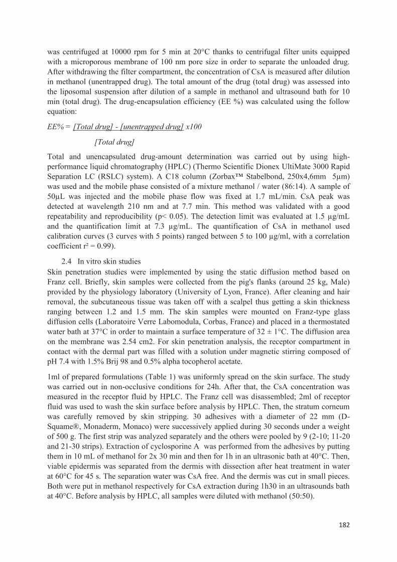
182
was centrifuged at 10000 rpm for 5 min at 20°C thanks to centrifugal filter units equipped with a microporous membrane of 100 nm pore size in order to separate the unloaded drug. After withdrawing the filter compartment, the concentration of CsA is measured after dilution in methanol (unentrapped drug). The total amount of the drug (total drug) was assessed into the liposomal suspension after dilution of a sample in methanol and ultrasound bath for 10 min (total drug). The drug-encapsulation efficiency (EE %) was calculated using the follow equation:
EE% = [Total drug] - [unentrapped drug] x100
[Total drug]
Total and unencapsulated drug-amount determination was carried out by using high-performance liquid chromatography (HPLC) (Thermo Scientific Dionex UltiMate 3000 Rapid Separation LC (RSLC) system). A C18 column (Zorbax™ Stabelbond, 250x4,6mm 5μm) was used and the mobile phase consisted of a mixture methanol / water (86:14). A sample of 50μL was injected and the mobile phase flow was fixed at 1.7 mL/min. CsA peak was detected at wavelength 210 nm and at 7.7 min. This method was validated with a good repeatability and reproducibility (p< 0.05). The detection limit was evaluated at 1.5 μg/mL and the quantification limit at 7.3 μg/mL. The quantification of CsA in methanol used calibration curves (3 curves with 5 points) ranged between 5 to 100 μg/ml, with a correlation coefficient r² = 0.99).
2.4 In vitro skin studies Skin penetration studies were implemented by using the static diffusion method based on Franz cell. Briefly, skin samples were collected from the pig's flanks (around 25 kg, Male) provided by the physiology laboratory (University of Lyon, France). After cleaning and hair removal, the subcutaneous tissue was taken off with a scalpel thus getting a skin thickness ranging between 1.2 and 1.5 mm. The skin samples were mounted on Franz-type glass diffusion cells (Laboratoire Verre Labomodula, Corbas, France) and placed in a thermostated water bath at 37°C in order to maintain a surface temperature of 32 ± 1°C. The diffusion area on the membrane was 2.54 cm2. For skin penetration analysis, the receptor compartment in contact with the dermal part was filled with a solution under magnetic stirring composed of pH 7.4 with 1.5% Brij 98 and 0.5% alpha tocopherol acetate.
1ml of prepared formulations (Table 1) was uniformly spread on the skin surface. The study was carried out in non-occlusive conditions for 24h. After that, the CsA concentration was measured in the receptor fluid by HPLC. The Franz cell was disassembled; 2ml of receptor fluid was used to wash the skin surface before analysis by HPLC. Then, the stratum corneum was carefully removed by skin stripping. 30 adhesives with a diameter of 22 mm (D-Squame®, Monaderm, Monaco) were successively applied during 30 seconds under a weight of 500 g. The first strip was analyzed separately and the others were pooled by 9 (2-10; 11-20 and 21-30 strips). Extraction of cyclosporine A was performed from the adhesives by putting them in 10 mL of methanol for 2x 30 min and then for 1h in an ultrasonic bath at 40°C. Then, viable epidermis was separated from the dermis with dissection after heat treatment in water at 60°C for 45 s. The separation water was CsA free. And the dermis was cut in small pieces. Both were put in methanol respectively for CsA extraction during 1h30 in an ultrasounds bath at 40°C. Before analysis by HPLC, all samples were diluted with methanol (50:50).
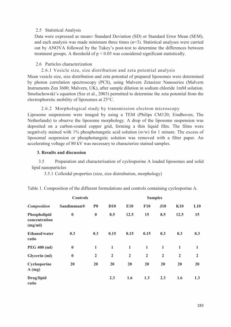
183
2.5 Statistical Analysis Data were expressed as mean± Standard Deviation (SD) or Standard Error Mean (SEM), and each analysis was made minimum three times (n=3). Statistical analyses were carried out by ANOVA followed by the Tukey’s post-test to determine the differences between treatment groups. A threshold of p < 0.05 was considered significant statistically.
2.6 Particles characterization 2.6.1 Vesicle size, size distribution and zeta potential analysis
Mean vesicle size, size distribution and zeta potential of prepared liposomes were determined by photon correlation spectroscopy (PCS), using Malvern Zetasizer Nanoseries (Malvern Instruments Zen 3600; Malvern, UK), after sample dilution in sodium chloride 1mM solution. Smoluchowski’s equation (Sze et al., 2003) permitted to determine the zeta potential from the electrophoretic mobility of liposomes at 25°C.
2.6.2 Morphological study by transmission electron microscopy Liposome suspensions were imaged by using a TEM (Philips CM120; Eindhoven, The Netherlands) to observe the liposome morphology. A drop of the liposome suspension was deposited on a carbon-coated copper grid, forming a thin liquid film. The films were negatively stained with 1% phosphotungstic acid solution (w/w) for 1 minute. The excess of liposomal suspension or phosphotungstic solution was removed with a filter paper. An accelerating voltage of 80 kV was necessary to characterize stained samples.
3. Results and discussion
3.5 Preparation and characterisation of cyclosporine A loaded liposomes and solid lipid nanoparticles
3.5.1 Colloidal properties (size, size distrubution, morphology)
Table 1. Composition of the different formulations and controls containing cyclosporine A.
Controls Samples
Composition Sandimmun® P0 D10 E10 F10 J10 K10 L10
Phospholipid concentration (mg/ml)
0 0 8.5 12.5 15 8.5 12.5 15
Ethanol/water ratio
0.3 0.3 0.15 0.15 0.15 0.3 0.3 0.3
PEG 400 (ml) 0 1 1 1 1 1 1 1
Glycerin (ml) 0 2 2 2 2 2 2 2
Cyclosporine A (mg)
20 20 20 20 20 20 20 20
Drug/lipid ratio
2.3 1.6 1.3 2.3 1.6 1.3
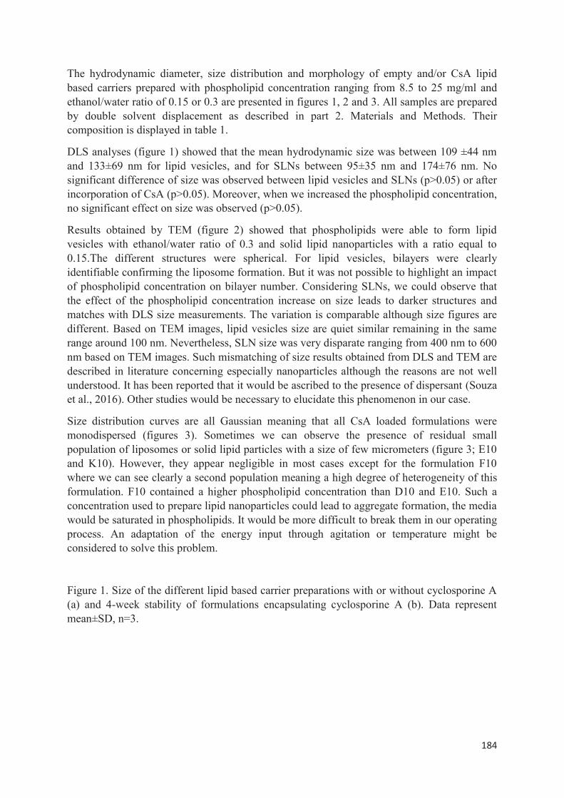
184
The hydrodynamic diameter, size distribution and morphology of empty and/or CsA lipid based carriers prepared with phospholipid concentration ranging from 8.5 to 25 mg/ml and ethanol/water ratio of 0.15 or 0.3 are presented in figures 1, 2 and 3. All samples are prepared by double solvent displacement as described in part 2. Materials and Methods. Their composition is displayed in table 1.
DLS analyses (figure 1) showed that the mean hydrodynamic size was between 109 ±44 nm and 133±69 nm for lipid vesicles, and for SLNs between 95±35 nm and 174±76 nm. No significant difference of size was observed between lipid vesicles and SLNs (p>0.05) or after incorporation of CsA (p>0.05). Moreover, when we increased the phospholipid concentration, no significant effect on size was observed (p>0.05).
Results obtained by TEM (figure 2) showed that phospholipids were able to form lipid vesicles with ethanol/water ratio of 0.3 and solid lipid nanoparticles with a ratio equal to 0.15.The different structures were spherical. For lipid vesicles, bilayers were clearly identifiable confirming the liposome formation. But it was not possible to highlight an impact of phospholipid concentration on bilayer number. Considering SLNs, we could observe that the effect of the phospholipid concentration increase on size leads to darker structures and matches with DLS size measurements. The variation is comparable although size figures are different. Based on TEM images, lipid vesicles size are quiet similar remaining in the same range around 100 nm. Nevertheless, SLN size was very disparate ranging from 400 nm to 600 nm based on TEM images. Such mismatching of size results obtained from DLS and TEM are described in literature concerning especially nanoparticles although the reasons are not well understood. It has been reported that it would be ascribed to the presence of dispersant (Souza et al., 2016). Other studies would be necessary to elucidate this phenomenon in our case.
Size distribution curves are all Gaussian meaning that all CsA loaded formulations were monodispersed (figures 3). Sometimes we can observe the presence of residual small population of liposomes or solid lipid particles with a size of few micrometers (figure 3; E10 and K10). However, they appear negligible in most cases except for the formulation F10 where we can see clearly a second population meaning a high degree of heterogeneity of this formulation. F10 contained a higher phospholipid concentration than D10 and E10. Such a concentration used to prepare lipid nanoparticles could lead to aggregate formation, the media would be saturated in phospholipids. It would be more difficult to break them in our operating process. An adaptation of the energy input through agitation or temperature might be considered to solve this problem.
Figure 1. Size of the different lipid based carrier preparations with or without cyclosporine A (a) and 4-week stability of formulations encapsulating cyclosporine A (b). Data represent mean±SD, n=3.
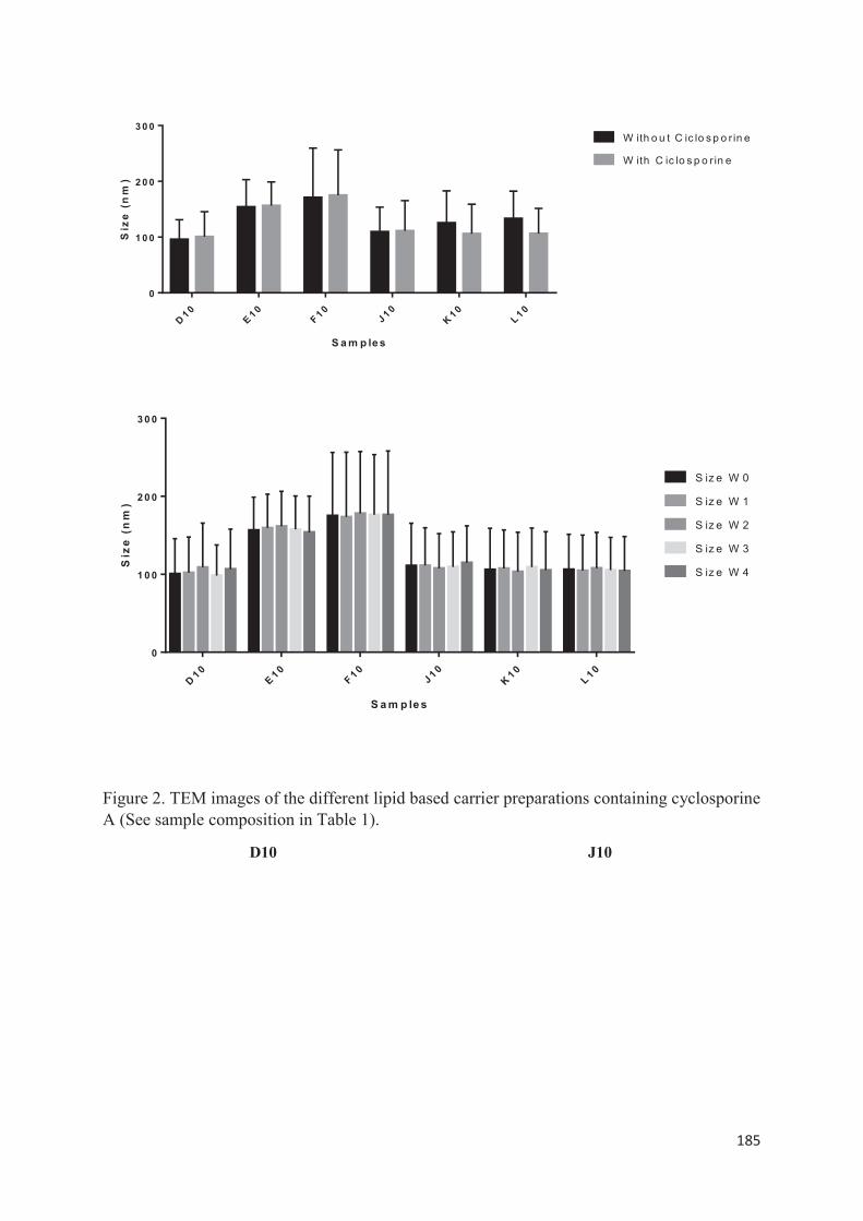
185
D 1 0E 1 0
F 1 0J 1 0
K 1 0L 1 0
0
1 0 0
2 0 0
3 0 0
S a m p le s
Siz
e(n
m)
W ith o u t C ic lo s p o r in e
W ith C ic lo s p o r in e
D 1 0E 1 0
F 1 0J 1 0
K 1 0L 1 0
0
1 0 0
2 0 0
3 0 0
S a m p le s
Siz
e(n
m)
S iz e W 0
S iz e W 1
S iz e W 2
S iz e W 3
S iz e W 4
Figure 2. TEM images of the different lipid based carrier preparations containing cyclosporine A (See sample composition in Table 1).
D10 J10
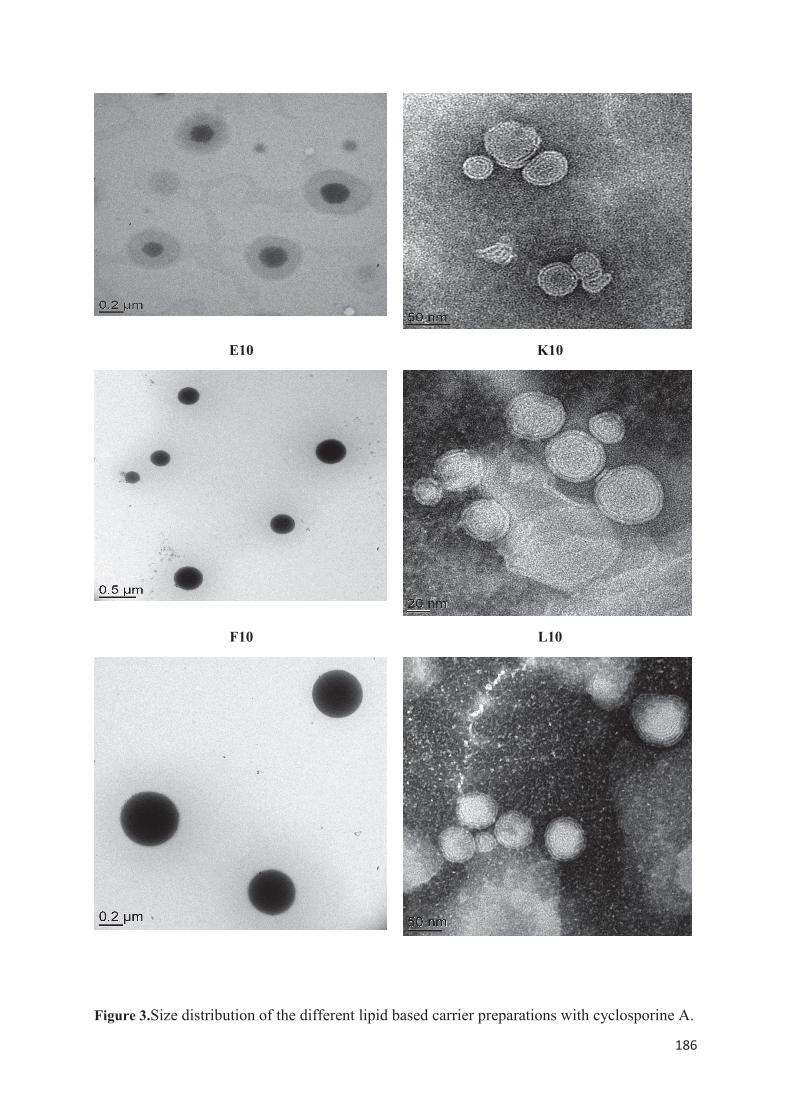
186
E10 K10
F10 L10
Figure 3.Size distribution of the different lipid based carrier preparations with cyclosporine A.
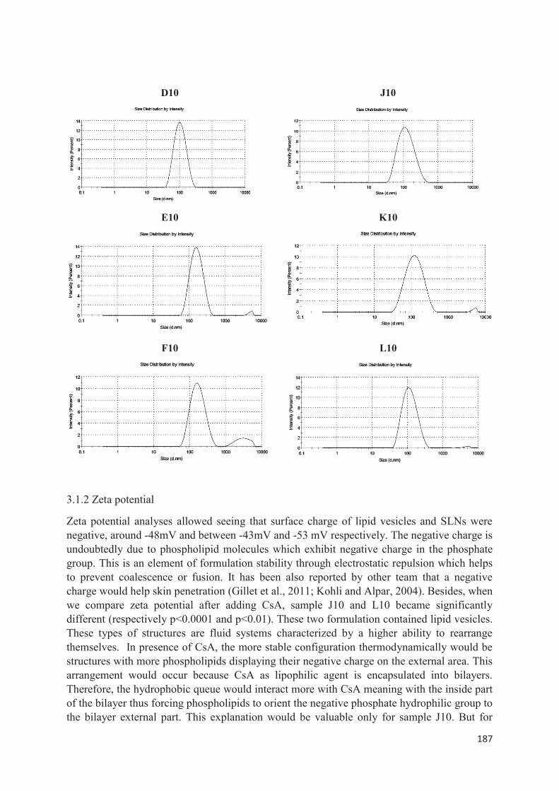
187
D10 J10
E10 K10
F10 L10
3.1.2 Zeta potential
Zeta potential analyses allowed seeing that surface charge of lipid vesicles and SLNs were negative, around -48mV and between -43mV and -53 mV respectively. The negative charge is undoubtedly due to phospholipid molecules which exhibit negative charge in the phosphate group. This is an element of formulation stability through electrostatic repulsion which helps to prevent coalescence or fusion. It has been also reported by other team that a negative charge would help skin penetration (Gillet et al., 2011; Kohli and Alpar, 2004). Besides, when we compare zeta potential after adding CsA, sample J10 and L10 became significantly different (respectively p<0.0001 and p<0.01). These two formulation contained lipid vesicles. These types of structures are fluid systems characterized by a higher ability to rearrange themselves. In presence of CsA, the more stable configuration thermodynamically would be structures with more phospholipids displaying their negative charge on the external area. This arrangement would occur because CsA as lipophilic agent is encapsulated into bilayers. Therefore, the hydrophobic queue would interact more with CsA meaning with the inside part of the bilayer thus forcing phospholipids to orient the negative phosphate hydrophilic group to the bilayer external part. This explanation would be valuable only for sample J10. But for
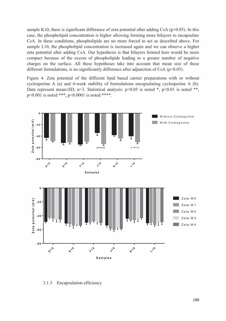
188
sample K10, there is significant difference of zeta potential after adding CsA (p>0.05). In this case, the phospholipid concentration is higher allowing forming more bilayers to encapsulate CsA. In these conditions, phospholipids are no more forced to act as described above. For sample L10, the phospholipid concentration is increased again and we can observe a higher zeta potential after adding CsA. Our hypothesis is that bilayers formed here would be more compact because of the excess of phospholipids leading to a greater number of negative charges on the surface. All these hypotheses take into account that mean size of these different formulations, is no significantly difference after adjunction of CsA (p>0.05).
Figure 4. Zeta potential of the different lipid based carrier preparations with or without cyclosporine A (a) and 4-week stability of formulations encapsulating cyclosporine A (b). Data represent mean±SD, n=3. Statistical analysis: p<0.05 is noted *, p<0.01 is noted **, p<0.001 is noted ***, p<0.0001 is noted ****.
D 1 0E 1 0
F 1 0J 1 0
K 1 0L 1 0
-8 0
-6 0
-4 0
-2 0
0
S a m p le s
Ze
tap
ote
nti
al
(mV
)
W ith o u t C ic lo s p o r in e
W ith C ic lo s p o r in e
* * * * * *
D 1 0E 1 0
F 1 0J 1 0
K 1 0L 1 0
-8 0
-6 0
-4 0
-2 0
0
S a m p le s
Ze
tap
ote
nti
al
(mV
) Z e ta W 0
Z e ta W 1
Z e ta W 2
Z e ta W 3
Z e ta W 4
3.1.3 Encapsulation efficiency
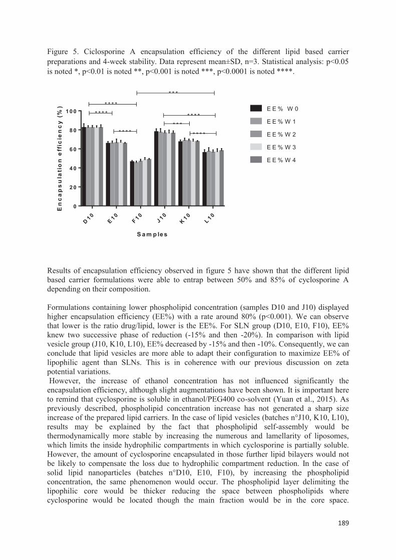
189
Figure 5. Ciclosporine A encapsulation efficiency of the different lipid based carrier preparations and 4-week stability. Data represent mean±SD, n=3. Statistical analysis: p<0.05 is noted *, p<0.01 is noted **, p<0.001 is noted ***, p<0.0001 is noted ****.
D 1 0E 1 0
F 1 0J 1 0
K 1 0L 1 0
0
2 0
4 0
6 0
8 0
1 0 0
S a m p le s
En
ca
ps
ula
tio
ne
ffic
ien
cy
(%) E E % W 0
E E % W 1
E E % W 2
E E % W 3
E E % W 4
* * * ** * * *
* * * ** * *
* * * *
* * * *
* * *
Results of encapsulation efficiency observed in figure 5 have shown that the different lipid based carrier formulations were able to entrap between 50% and 85% of cyclosporine A depending on their composition. Formulations containing lower phospholipid concentration (samples D10 and J10) displayed higher encapsulation efficiency (EE%) with a rate around 80% (p<0.001). We can observe that lower is the ratio drug/lipid, lower is the EE%. For SLN group (D10, E10, F10), EE% knew two successive phase of reduction (-15% and then -20%). In comparison with lipid vesicle group (J10, K10, L10), EE% decreased by -15% and then -10%. Consequently, we can conclude that lipid vesicles are more able to adapt their configuration to maximize EE% of lipophilic agent than SLNs. This is in coherence with our previous discussion on zeta potential variations. However, the increase of ethanol concentration has not influenced significantly the encapsulation efficiency, although slight augmentations have been shown. It is important here to remind that cyclosporine is soluble in ethanol/PEG400 co-solvent (Yuan et al., 2015). As previously described, phospholipid concentration increase has not generated a sharp size increase of the prepared lipid carriers. In the case of lipid vesicles (batches n°J10, K10, L10), results may be explained by the fact that phospholipid self-assembly would be thermodynamically more stable by increasing the numerous and lamellarity of liposomes, which limits the inside hydrophilic compartments in which cyclosporine is partially soluble. However, the amount of cyclosporine encapsulated in those further lipid bilayers would not be likely to compensate the loss due to hydrophilic compartment reduction. In the case of solid lipid nanoparticles (batches n°D10, E10, F10), by increasing the phospholipid concentration, the same phenomenon would occur. The phospholipid layer delimiting the lipophilic core would be thicker reducing the space between phospholipids where cyclosporine would be located though the main fraction would be in the core space.
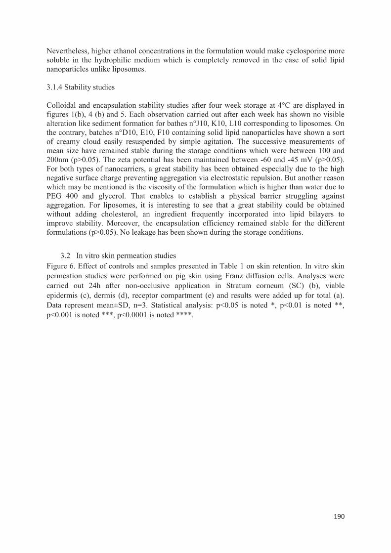
190
Nevertheless, higher ethanol concentrations in the formulation would make cyclosporine more soluble in the hydrophilic medium which is completely removed in the case of solid lipid nanoparticles unlike liposomes. 3.1.4 Stability studies Colloidal and encapsulation stability studies after four week storage at 4°C are displayed in figures 1(b), 4 (b) and 5. Each observation carried out after each week has shown no visible alteration like sediment formation for bathes n°J10, K10, L10 corresponding to liposomes. On the contrary, batches n°D10, E10, F10 containing solid lipid nanoparticles have shown a sort of creamy cloud easily resuspended by simple agitation. The successive measurements of mean size have remained stable during the storage conditions which were between 100 and 200nm (p>0.05). The zeta potential has been maintained between -60 and -45 mV (p>0.05). For both types of nanocarriers, a great stability has been obtained especially due to the high negative surface charge preventing aggregation via electrostatic repulsion. But another reason which may be mentioned is the viscosity of the formulation which is higher than water due to PEG 400 and glycerol. That enables to establish a physical barrier struggling against aggregation. For liposomes, it is interesting to see that a great stability could be obtained without adding cholesterol, an ingredient frequently incorporated into lipid bilayers to improve stability. Moreover, the encapsulation efficiency remained stable for the different formulations (p>0.05). No leakage has been shown during the storage conditions.
3.2 In vitro skin permeation studies Figure 6. Effect of controls and samples presented in Table 1 on skin retention. In vitro skin permeation studies were performed on pig skin using Franz diffusion cells. Analyses were carried out 24h after non-occlusive application in Stratum corneum (SC) (b), viable epidermis (c), dermis (d), receptor compartment (e) and results were added up for total (a). Data represent mean±SD, n=3. Statistical analysis: p<0.05 is noted *, p<0.01 is noted **, p<0.001 is noted ***, p<0.0001 is noted ****.
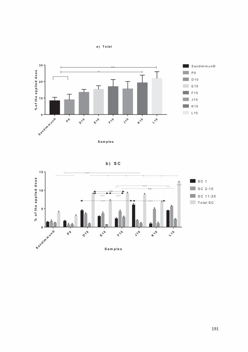
191
S a n d imm
u n ® P 0D 1 0
E 1 0F 1 0
J 1 0K 1 0
L 1 00
1 0
2 0
3 0
S a m p le s
%o
fth
ea
pp
lie
dd
os
e
S a n d im m u n ®
P 0
D 1 0
E 1 0
F 1 0
J 1 0
K 1 0
L 1 0
** *
a ) T o ta l
S a n d imm
u n ®P 0
D 1 0E 1 0
F 1 0J 1 0
K 1 0L 1 0
0
5
1 0
1 5
b ) S C
S a m p le s
%o
fth
ea
pp
lie
dd
os
e
S C 1
S C 2 -1 0
S C 1 1 -2 0
T o ta l S C
* * * *
* * * ** * * *n s
* * * * * * * *
n s
* * * * * * * *
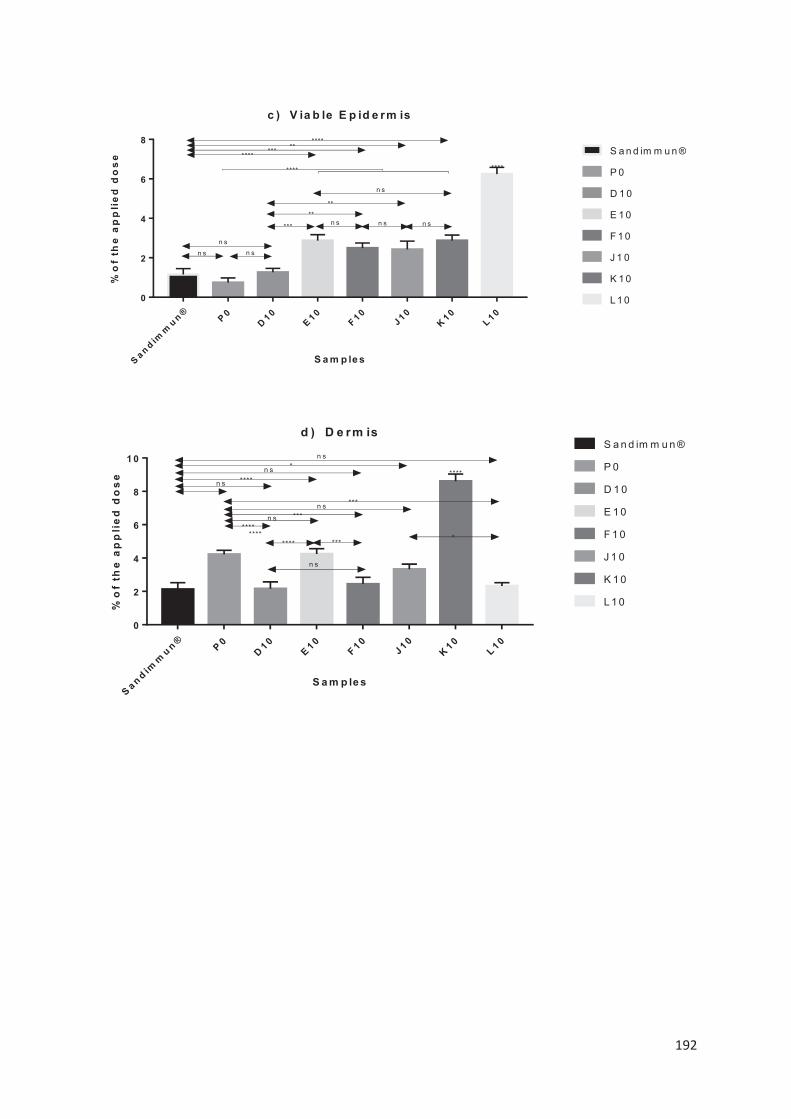
192
S a n d imm
u n ® P 0D 1 0
E 1 0F 1 0
J 1 0K 1 0
L 1 00
2
4
6
8
c ) V ia b le E p id e rm is
S a m p le s
%o
fth
ea
pp
lie
dd
os
e S a n d im m u n ®
P 0
D 1 0
E 1 0
F 1 0
J 1 0
K 1 0
L 1 0
n s
n s n s
****
***
**n s n s
**
n s
****
*********
****
n s
S a n d imm
u n ® P 0D 1 0
E 1 0F 1 0
J 1 0K 1 0
L 1 00
2
4
6
8
1 0
d ) D e rm is
S a m p le s
%o
fth
ea
pp
lie
dd
os
e
S a n d im m u n ®
P 0
D 1 0
E 1 0
F 1 0
J 1 0
K 1 0
L 1 0
********
****
n s
n s
*n s
n s ***n s ***
********
n s
****
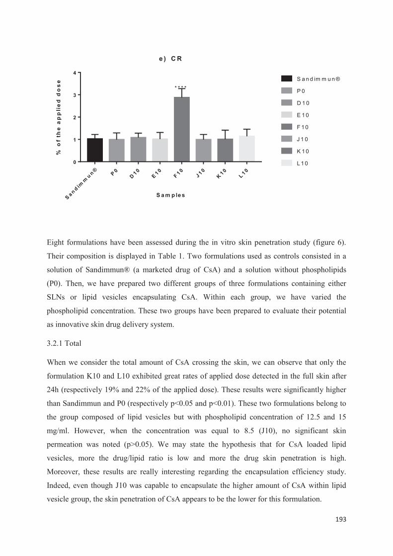
193
S a n d imm
u n ® P 0D 1 0
E 1 0F 1 0
J 1 0K 1 0
L 1 00
1
2
3
4
e ) C R
S a m p le s
%o
fth
ea
pp
lie
dd
os
e S a n d im m u n ®
P 0
D 1 0
E 1 0
F 1 0
J 1 0
K 1 0
L 1 0
* * * *
Eight formulations have been assessed during the in vitro skin penetration study (figure 6).
Their composition is displayed in Table 1. Two formulations used as controls consisted in a
solution of Sandimmun® (a marketed drug of CsA) and a solution without phospholipids
(P0). Then, we have prepared two different groups of three formulations containing either
SLNs or lipid vesicles encapsulating CsA. Within each group, we have varied the
phospholipid concentration. These two groups have been prepared to evaluate their potential
as innovative skin drug delivery system.
3.2.1 Total
When we consider the total amount of CsA crossing the skin, we can observe that only the
formulation K10 and L10 exhibited great rates of applied dose detected in the full skin after
24h (respectively 19% and 22% of the applied dose). These results were significantly higher
than Sandimmun and P0 (respectively p<0.05 and p<0.01). These two formulations belong to
the group composed of lipid vesicles but with phospholipid concentration of 12.5 and 15
mg/ml. However, when the concentration was equal to 8.5 (J10), no significant skin
permeation was noted (p>0.05). We may state the hypothesis that for CsA loaded lipid
vesicles, more the drug/lipid ratio is low and more the drug skin penetration is high.
Moreover, these results are really interesting regarding the encapsulation efficiency study.
Indeed, even though J10 was capable to encapsulate the higher amount of CsA within lipid
vesicle group, the skin penetration of CsA appears to be the lower for this formulation.
194
Besides, within the group of SLN formulation, none sample was able to show high rates of
skin penetration statistically significant versus controls. Our CsA loaded SLNs do not appear
as good skin drug delivery system unlike lipid vesicles showing better results.
3.2.2 Stratum Corneum
CsA fraction found in SC was significantly higher after application of formulations D10, E10,
F10 and J10, K10, L10 in comparison to controls (Sandimmun and P0). Results obtained went
beyond to 8% of the applied dose whereas for controls, the CsA fraction was around 3-4%.
Among these formulations, L10 displayed the greatest CsA rate in SC versus all others
(p<0.0001). Comparing formulations in SLNs group, we can say that there is non-significant
difference between the CsA fraction found in SC between formulations D10 and F10
(p>0.05). However, the fraction dose was more concentrated in SC1 and SC 2-10 for D10.
E10 dispayed lower results compared to D10 and F10 (p<0.0001). Comparing formulations in
lipid vesicles group, we can observe that L10 offered the better skin penetration (p<0.0001)
whatever the part of SC considered, except for SC1 where J10 exhibited a higher amount of
CsA (p<0.0001).
Formulations D10 and J10 provided higher amount in SC1 respectively in their own group
although the CsA fraction was higher with J10 (p<0.0001). Formulations F10 and L10 are
those allowing the better penetration in SC considering them respectively in their own group
with values of 9.5% and 13%. Moreover, regarding the skin passage through the different
layers of SC, it appears that F10 and L10 were formulations offering a better penetration into
SC 11-20 with values beyond 2% of the applied dose of CsA. F10 and L10 have in common
a phospholipid concentration similar equal to 15 mg/mL. However, advantages observed with
L10 relied on the higher CsA fraction found in SC 1 and SC 2-10.
3.2.3 Viable Epidermis
Analyses wearing on CsA rates detected in viable epidermis pointed that formulation L10
provided the better penetration (beyond 6% of the applied dose) compared with all other
formulations and controls (p<0.0001). E10, F10, J10 and K10 exhibited CsA rates
significantly higher than controls (p<0.01 minimum until p<0.0001) and no significant
difference was observed between each other (p>0.05). Only D10 showed results no significant
different versus controls (p>0.05) but highly low statistically compared with the other
formulations (p<0.01 minimum until p<0.0001). No significant difference was also observed
195
between formulations E10 and K10 (p>0.05). Those results demonstrate that only L10 may
have the potential to target viable epidermis.
3.2.4 Dermis and Receptor compartment
In vitro studies on pig skin showed that formulation K10 offered higher concentration of CsA
detected in dermis (beyond 8% of the applied dose) compared with all other formulations and
controls (p<0.0001). Compared to Sandimmun® control, we can see that only formulations
E10 and K10 allowed to find CsA rates higher statistically (p>0.0001), and in a lower way
J10 (p>0.05). No significant difference was measured after application of formulations D10,
F10 and L10 (p>0.05). Interestingly, P0 control displayed higher CsA rate in dermis than
Sandimmun® control (p>0.0001). Besides, only formulation K10 presented a CsA fraction
greater statistically than P0 (p<0.001). Among SLNs formulations, the highest CsA fraction
was found with E10 compared with E10 (p<0.0001) and F10 (p<0.001). Among lipid vesicles
formulations, it was with K10. Considering the receptor compartment, only formulation F10
showed a higher CsA rate around 3% of the applied dose compared to the other formulations
and controls (p<0.0001). Based on these results, formulations F10 and K10 exhibit a stronger
potential as transdermal CsA delivery systems.
3.2.5 In vitro skin studies discussion
In our work, we have highlighted that among all formulations assessed; L10 displayed the
greatest CsA rate in SC (12%) and in viable epidermis (6%). In comparison with other work
presented in literature, our results were higher. Verma et al. encapsulated CsA in ethosomes
and showed that their formulation containing 10% of ethanol allowed the better skin
penetration (Verma and Fahr, 2004). However, only 3% of the applied dose was found in the
SC and 0.03% in the deeper skin.
SLNs formulations did not show a particular advantage in targeting SC or viable epidermis.
However, formulations F10 and K10 exhibited a higher interest as transdermal CsA delivery
systems. Kim et al. prepared CsA-loaded SLNs with a size below to 100 nm (Kim et al.,
2009). In vitro skin studies showed that 20% of the dose applied was retained in SC and 5.3%
in the viable epidermis. Unfortunately, our formulation exhibited a lower potential to target
the SC (9%) and the (2.5%) viable epidermis.
196
Interestingly, it is noteworthy to highlight that this is not the formulations encapsulating the
higher amount of CsA which displayed the better skin penetration results. Indeed, considering
the formulation L10 belonging to lipid vesicles group, we can see that L10 presented an
encapsulation efficiency between 50-60% whereas for J10, it was almost 80%. But the impact
of the phospholipid concentration would play an important role in the capacity of the
formulation L10 to cross the skin. It appears here that when the drug/lipid ratio is lower, the
skin penetration is better. But that was observed only for the case of CsA loaded lipid
vesicles.
It is commonly deemed that lipophilic active ingredients have generally good interactions
with SC. Considering the field of skin drug delivery systems, Cheon Koo Lee et al. (Lee et al.,
1994) have reported that lipophilic active molecules are more likely to cross the skin. That
greater affinity with the skin, especially the first layer (SC) may be due undoubtedly to lipid
composition of carriers especially those made of phospholipids because of the similarity with
the compact intercorneocyte cement embedding corneocytes of SC which is composed of
phospholipids, cholesterol and ceramides. Besides, a high amount of CsA was also found in
SC with the formulation L10. This is very interesting because it has been reported that drug
accumulated in SC inside bilayers of liposomes can play a role of reservoir that may provide a
sustained release (Dayan and Touitou, 2000).
4. Conclusion.
This DSD method allows producing two types of lipid-based nanocarriers by controlling ethanol/PEG400 ratio: SLNs and liposomes. Interestingly, they exhibited almost the same colloidal properties. Populations were mainly monodispersed with a size less than 200 nm.
Obtained results through in vitro skin permeation studies are promising regarding the formulation containing CsA loaded lipid vesicles composed with higher phospholipid concentration. Indeed, we were able to target the viable epidermis which leads us to consider this formulation as highly interesting in topical management of psoriasis. Considering the physiopathology of psoriasis, viable epidermis was the target to reach for treating the skin disease. Indeed, the mechanism of action taking place in psoriasis involves various actors of immunology and inflammation located in viable epidermis.
In our work, even though properties of SLNs and lipid vesicles were almost similar, lipid vesicles had a clear advantage in skin delivery. Moreover, we conclude that even though we encapsulate a high amount of active ingredient, this is not correlated with a higher skin passage. The carrier structure appears to be of paramount importance.
Besides that, although this research work is skin-delivery-oriented, such lipid-based nanocarriers could have great potential for other administration routes and/or targeted location like brain. Indeed, the passage through the blood brain barrier should be investigated to see if,
197
as skin barrier, these innovative nanocarriers may cross. Further in vitro / in vivo studies have to be implemented to gather more data and explore all possibilities that these innovative drug delivery systems produced by a simple and cost-effective method, the DSD technique, can offer.
5. References
Ahmed El-Harati, A., Charcosset, C., Fessi, H., 2006. Influence of the formulation for solid lipid nanoparticles prepared with a membrane contactor. Pharm. Dev. Technol. 11, 153–157.
Ainbinder, D., Touitou, E., 2005. Testosterone ethosomes for enhanced transdermal delivery. Drug Deliv. 12, 297–303.
Badri, W., Eddabra, R., Fessi, H., Elaissari, A., 2014. Biodegradable Polymer Based Nanoparticles: Dermal and Transdermal Drug Delivery. J. Colloid Sci. Biotechnol. 3, 141–149.
Bangham, A.D., De Gier, J., Greville, G.D., 1967. Osmotic properties and water permeability of phospholipid liquid crystals. Chem. Phys. Lipids 1, 225–246. doi:10.1016/0009-3084(67)90030-8
Belitsky, P., Dunn, S., Johnston, A., Levy, G., 2000. Impact of Absorption Profiling on Efficacy and Safety of Cyclosporin Therapy in Transplant Recipients: Clin. Pharmacokinet. 39, 117–125. doi:10.2165/00003088-200039020-00003
Boinpally, R.R., Zhou, S.-L., Devraj, G., Anne, P.K., Poondru, S., Jasti, B.R., 2004. Iontophoresis of lecithin vesicles of cyclosporin A. Int. J. Pharm. 274, 185–190. doi:10.1016/j.ijpharm.2004.01.016
Cevc, G., Blume, G., 1992. Lipid vesicles penetrate into intact skin owing to the transdermal osmotic gradients and hydration force. Biochim. Biophys. Acta 1104, 226–232.
Chen, M., Kumar, S., Anselmo, A.C., Gupta, V., Slee, D.H., Muraski, J.A., Mitragotri, S., 2015. Topical delivery of Cyclosporine A into the skin using SPACE-peptide. J. Control. Release Off. J. Control. Release Soc. 199, 190–197. doi:10.1016/j.jconrel.2014.11.015
Choi, H.K., Flynn, G.L., Amidon, G.L., 1995. Percutaneous absorption and dermal delivery of cyclosporin A. J. Pharm. Sci. 84, 581–583.
Czogalla, A., 2009. Oral cyclosporine A--the current picture of its liposomal and other delivery systems. Cell. Mol. Biol. Lett. 14, 139–152. doi:10.2478/s11658-008-0041-6
Dayan, N., Touitou, E., 2000. Carriers for skin delivery of trihexyphenidyl HCl: ethosomes vs. liposomes. Biomaterials 21, 1879–1885.
Elsayed, M.M., Abdallah, O.Y., Naggar, V.F., Khalafallah, N.M., 2007. Lipid vesicles for skin delivery of drugs: reviewing three decades of research. Int. J. Pharm. 332, 1–16.
Garcıa-Fuentes, M., Torres, D., Alonso, M.J., 2003. Design of lipid nanoparticles for the oral delivery of hydrophilic macromolecules. Colloids Surf. B Biointerfaces 27, 159–168.
Gasco, M.R., 1993. Method for producing solid lipid microspheres having a narrow size distribution. Google Patents.
198
Ghanbarzadeh, S., Arami, S., 2013. Enhanced transdermal delivery of diclofenac sodium via conventional liposomes, ethosomes, and transfersomes. BioMed Res. Int. 2013.
Gillet, A., Compère, P., Lecomte, F., Hubert, P., Ducat, E., Evrard, B., Piel, G., 2011. Liposome surface charge influence on skin penetration behaviour. Int. J. Pharm. 411, 223–231.
Griffiths, C.E.M., Powles, A.V., Baker, B.S., Fry, L., Valdimarsson, H., 1987. TOPICAL CYCLOSPORIN AND PSORIASIS. The Lancet 329, 806. doi:10.1016/S0140-6736(87)92829-7
Guada, M., Beloqui, A., Kumar, M.N.V.R., Préat, V., Dios-Viéitez, M.D.C., Blanco-Prieto, M.J., 2016. Reformulating cyclosporine A (CsA): More than just a life cycle management strategy. J. Control. Release Off. J. Control. Release Soc. 225, 269–282. doi:10.1016/j.jconrel.2016.01.056
Heurtault, B., Saulnier, P., Pech, B., Proust, J.-E., Benoit, J.-P., 2002. A novel phase inversion-based process for the preparation of lipid nanocarriers. Pharm. Res. 19, 875–880.
Ho, V.C., Griffiths, C.E., Ellis, C.N., Gupta, A.K., McCuaig, C.C., Nickoloff, B.J., Cooper, K.D., Hamilton, T.A., Voorhees, J.J., 1990. Intralesional cyclosporine in the treatment of psoriasis. A clinical, immunologic, and pharmacokinetic study. J. Am. Acad. Dermatol. 22, 94–100.
Hu, F.Q., Yuan, H., Zhang, H.H., Fang, M., 2002. Preparation of solid lipid nanoparticles with clobetasol propionate by a novel solvent diffusion method in aqueous system and physicochemical characterization. Int. J. Pharm. 239, 121–128.
Jaafar-Maalej, C., Charcosset, C., Fessi, H., 2011. A new method for liposome preparation using a membrane contactor. J. Liposome Res. 21, 213–220. doi:10.3109/08982104.2010.517537
Jahn, A., Vreeland, W.N., Gaitan, M., Locascio, L.E., 2004. Controlled vesicle self-assembly in microfluidic channels with hydrodynamic focusing. J. Am. Chem. Soc. 126, 2674–2675.
Kim, S.T., Jang, D.-J., Kim, J.H., Park, J.Y., Lim, J.S., Lee, S.Y., Lee, K.-M., Lim, S.-J., Kim, C.-K., 2009. Topical administration of cyclosporin A in a solid lipid nanoparticle formulation. Pharm. 64, 510–514.
Kohane, D.S., 2007. Microparticles and nanoparticles for drug delivery. Biotechnol. Bioeng. 96, 203–209. doi:10.1002/bit.21301
Kohli, A.K., Alpar, H.O., 2004. Potential use of nanoparticles for transcutaneous vaccine delivery: effect of particle size and charge. Int. J. Pharm. 275, 13–17. doi:10.1016/j.ijpharm.2003.10.038
Laouini, A., Jaafar-Maalej, C., Limayem-Blouza, I., Sfar, S., Charcosset, C., Fessi, H., 2012. Preparation, characterization and applications of liposomes: state of the art. J. Colloid Sci. Biotechnol. 1, 147–168.
Lee, C.K., Uchida, T., Kitagawa, K., Yagi, A., Kim, N.-S., Goto, S., 1994. Skin permeability of various drugs with different lipophilicity. J. Pharm. Sci. 83, 562–565. doi:10.1002/jps.2600830424
199
Li, C., Deng, Y., 2004. A novel method for the preparation of liposomes: freeze drying of monophase solutions. J. Pharm. Sci. 93, 1403–1414.
Liedtke, S., Wissing, S., Müller, R.H., Mäder, K., 2000. Influence of high pressure homogenisation equipment on nanodispersions characteristics. Int. J. Pharm. 196, 183–185.
Liu, H., Li, S., Pan, W., Wang, Y., Han, F., Yao, H., 2006a. Investigation into the potential of low-frequency ultrasound facilitated topical delivery of Cyclosporin A. Int. J. Pharm. 326, 32–38. doi:10.1016/j.ijpharm.2006.07.022
Liu, H., Li, S., Wang, Y., Yao, H., Zhang, Y., 2006b. Effect of vehicles and enhancers on the topical delivery of cyclosporin A. Int. J. Pharm. 311, 182–186. doi:10.1016/j.ijpharm.2005.12.029
Liu, H.-Z., Wang, Y.-J., Xu, L., Li, S.-M., 2010. Investigation into the potential of electroporation facilitated topical delivery of cyclosporin a. PDA J. Pharm. Sci. Technol. 64, 191–199.
Lopes, L.B., Collett, J.H., Bentley, M.V.L.B., 2005. Topical delivery of cyclosporin A: an in vitro study using monoolein as a penetration enhancer. Eur. J. Pharm. Biopharm. Off. J. Arbeitsgemeinschaft Pharm. Verfahrenstechnik EV 60, 25–30. doi:10.1016/j.ejpb.2004.12.003
Manconi, M., Caddeo, C., Sinico, C., Valenti, D., Mostallino, M.C., Biggio, G., Fadda, A.M., 2011a. Ex vivo skin delivery of diclofenac by transcutol containing liposomes and suggested mechanism of vesicle–skin interaction. Eur. J. Pharm. Biopharm. 78, 27–35.
Manconi, M., Sinico, C., Caddeo, C., Vila, A.O., Valenti, D., Fadda, A.M., 2011b. Penetration enhancer containing vesicles as carriers for dermal delivery of tretinoin. Int. J. Pharm. 412, 37–46.
Mehnert, W., Mäder, K., 2001. Solid lipid nanoparticles: production, characterization and applications. Adv. Drug Deliv. Rev. 47, 165–196.
Moinard-Checot, D., Chevalier, Y., Briançon, S., Fessi, H., Guinebretière, S., 2006. Nanoparticles for drug delivery: review of the formulation and process difficulties illustrated by the emulsion-diffusion process. J. Nanosci. Nanotechnol. 6, 2664–2681.
Mozafari, M.R., 2005. Liposomes: an overview of manufacturing techniques. Cell. Mol. Biol. Lett. 10, 711.
Otake, K., Imura, T., Sakai, H., Abe, M., 2001. Development of a new preparation method of liposomes using supercritical carbon dioxide. Langmuir 17, 3898–3901.
Pardeike, J., Hommoss, A., Müller, R.H., 2009. Lipid nanoparticles (SLN, NLC) in cosmetic and pharmaceutical dermal products. Int. J. Pharm. 366, 170–184.
Pietkiewicz, J., Sznitowska, M., 2004. The choice of lipids and surfactants for injectable extravenous microspheres. Pharm.- Int. J. Pharm. Sci. 59, 325–326.
Ryan, C., Amor, K.T., Menter, A., 2010. The use of cyclosporine in dermatology: part II. J. Am. Acad. Dermatol. 63, 949-972; quiz 973-974. doi:10.1016/j.jaad.2010.02.062
200
Sala, M., Elaissari, A., Fessi, H., 2016. Advances in psoriasis physiopathology and treatments: Up to date of mechanistic insights and perspectives of novel therapies based on innovative skin drug delivery systems (ISDDS). J. Controlled Release 239, 182–202. doi:10.1016/j.jconrel.2016.07.003
Schubert, M.A., Müller-Goymann, C.C., 2003. Solvent injection as a new approach for manufacturing lipid nanoparticles–evaluation of the method and process parameters. Eur. J. Pharm. Biopharm. 55, 125–131.
Shive, null, Anderson, null, 1997. Biodegradation and biocompatibility of PLA and PLGA microspheres. Adv. Drug Deliv. Rev. 28, 5–24.
Sjöström, B., Bergenståhl, B., 1992. Preparation of submicron drug particles in lecithin-stabilized o/w emulsions I. Model studies of the precipitation of cholesteryl acetate. Int. J. Pharm. 88, 53–62. doi:10.1016/0378-5173(92)90303-J
Skalko-Basnet, N., Pavelic, Z., Becirevic-Lacan, M., 2000. Liposomes containing drug and cyclodextrin prepared by the one-step spray-drying method. Drug Dev. Ind. Pharm. 26, 1279–1284.
Souza, T.G.F., Ciminelli, V.S.T., Mohallem, N.D.S., 2016. A comparison of TEM and DLS methods to characterize size distribution of ceramic nanoparticles. J. Phys. Conf. Ser. 733, 012039. doi:10.1088/1742-6596/733/1/012039
Sultana, S., Khan, M.R., Kumar, M., Kumar, S., Ali, M., 2013. Nanoparticles-mediated drug delivery approaches for cancer targeting: a review. J. Drug Target. 21, 107–125. doi:10.3109/1061186X.2012.712130
Sze, A., Erickson, D., Ren, L., Li, D., 2003. Zeta-potential measurement using the Smoluchowski equation and the slope of the current–time relationship in electroosmotic flow. J. Colloid Interface Sci. 261, 402–410.
Szebeni, J., Breuer, J.H., Szelenyi, J.G., Bathori, G., Lelkes, G., Hollan, S.R., 1984. Oxidation and denaturation of hemoglobin encapsulated in liposomes. Biochim. Biophys. Acta BBA-Gen. Subj. 798, 60–67.
Szoka, F., Papahadjopoulos, D., 1978. Procedure for preparation of liposomes with large internal aqueous space and high capture by reverse-phase evaporation. Proc. Natl. Acad. Sci. 75, 4194–4198.
Touitou, E., Dayan, N., Bergelson, L., Godin, B., Eliaz, M., 2000. Ethosomes—novel vesicular carriers for enhanced delivery: characterization and skin penetration properties. J. Controlled Release 65, 403–418.
Verma, D.D., Fahr, A., 2004. Synergistic penetration enhancement effect of ethanol and phospholipids on the topical delivery of cyclosporin A. J. Controlled Release 97, 55–66.
Wagner, A., Vorauer-Uhl, K., Katinger, H., 2002. Liposomes produced in a pilot scale: production, purification and efficiency aspects. Eur. J. Pharm. Biopharm. 54, 213–219.
Wissing, S.A., Kayser, O., Müller, R.H., 2004. Solid lipid nanoparticles for parenteral drug delivery. Adv. Drug Deliv. Rev. 56, 1257–1272.
201
Wretlind, A., 1981. Invited Review: Development of Fat Emulsions. J. Parenter. Enter. Nutr. 5, 230–235.
Yuan, Y., Che, X., Zhao, M., Wang, Y., Liu, Y., Schwendeman, A., Li, S., 2015. Development of cyclosporine A microemulsion for parenteral delivery. J. Microencapsul. 32, 273–280. doi:10.3109/02652048.2015.1010461
Zhao, Y.-Z., Lu, C.-T., Zhang, Y., Xiao, J., Zhao, Y.-P., Tian, J.-L., Xu, Y.-Y., Feng, Z.-G., Xu, C.-Y., 2013. Selection of high efficient transdermal lipid vesicle for curcumin skin delivery. Int. J. Pharm. 454, 302–309.
Zumbuehl, O., Weder, H.G., 1981. Liposomes of controllable size in the range of 40 to 180 nm by defined dialysis of lipid/detergent mixed micelles. Biochim. Biophys. Acta BBA-Biomembr. 640, 252–262.

202
Discussion générale
203
L’objectif de ce travail de thèse a consisté à élaborer des formulations galéniques encapsulant la ciclosporine A et le diclofénac dans des vecteurs médicamenteux lipidiques en vue d’une application topique pour le traitement à la fois de la composante fondamentale du psoriasis par un accès direct à son site physiopathologique mais aussi sa complication la plus fréquente et la plus grave, le rhumatisme psoriasique. Avant de pouvoir préparer de façon pertinente la partie expérimentale, il a fallu se saisir du sujet en profondeur afin de prendre en considération les particularités physiopathologiques du psoriasis ainsi que de se familiariser avec l’ensemble des techniques et des dispositifs développés ces dernières années pour promouvoir le passage cutané des molécules d’intérêts pharmaceutiques. Le tissu cutané est apparu comme étant le site principal de déroulement de la physiopathogenèse du psoriasis. Les signes cliniques dermatologiques du psoriasis sont les résultats de la prolifération anormale des kératinocytes au niveau de l’épiderme viable. Les kératinocytes sont les cellules présentatrices d’un antigène inconnu qui sont à la base de l'activation des lymphocytes T CD4+ et CD8+. Issu de ces informations, nous avons considéré l’épiderme viable comme la cible prioritaire des agents actifs pour une prise en charge efficace du psoriasis à un niveau précoce. Mais encore faut-il que l’agent pharmacologique agisse sur les cibles cellulaires. La ciclosporine A a été sélectionnée car elle agit à la fois sur l'activation des lymphocytes T et la prolifération des kératinocytes. Cependant, il s’agit d’une molécule très large, de poids moléculaire de 1202 Da ce qui limite son passage à travers la peau. Envisager l’encapsulation de la ciclosporine A pourrait permettre un accès direct par voie cutanée au site d’action. Cela permettrait d’avoir une plus grande efficacité et ce, sans passage par voie systémique à l’origine d’effets indésirables graves. Cela permettrait également d’envisager un usage au long cours. Se tourner vers l’utilisation des vecteurs lipidiques paraît également plus intéressant. En effet, la ciclospsorine A étant très lipophile (LogP= 2.92), les vecteurs lipidiques sont bien connus pour facilement encapsuler les agents pharmacologiques lipophiles. Par ailleurs, les lipides entrant dans la composition de ces vecteurs comme les phospholipides sont autorisés pour un usage chez l’Homme. A l’issu du premier travail bibliographique, nous avons décidé d’orienter nos travaux expérimentaux vers une utilisation des vecteurs lipidiques pour une encapsulation de la ciclosporine A. Cependant, il existe un large choix de ce type de vecteurs décrits dans la littérature scientifique : les vésicules lipidiques telles que les liposomes et leurs variantes (éthosomes, transfersomes) et les nanoparticules lipidiques (SLN, NLC).
Les propriétés physico-chimiques de la molécule active à encapsuler doivent être parfaitement connues car elles pourraient orienter le type de vecteurs lipidiques à utiliser. De plus, il est important de noter que la composition des vecteurs n’étant pas la même, il vaudrait mieux se concentre sur des vecteurs lipidiques constitués de lipides bien connus et dont l’usage dans les formulations pharmaceutiques est autorisée comme pour le cas des phopholipides. Après considération de l’ensemble des données bibliographiques, nous avons porté notre choix sur l’utilisation des vésicules lipidiques pour un passage cutané de molécules actives car elles possèdent une flexibilité structurelle indispensable pour franchir les différentes structures de la peau. En vue du développement de formulations pour la prise en charge par voie topique du psoriasis, il est indispensable de prendre en considération que l’une des composantes cliniques
204
dans le psoriasis est l’hyperkératose. Cela conduit à un stratum corneum anormalement épais très difficile à franchir sans déformabilité du vecteur médicamenteux.
On considère généralement qu'un agent actif franchit le SC par diffusion passive à travers trois voies possibles : la voie transcellulaire, intercellulaire et folliculaire. La voie transcellulaire est rarement considérée dans la littérature contrairement à la voie intercellulaire. Cette dernière semble particulièrement adaptée aux molécules non polaires de poids moléculaire <500 Da et de LogP compris entre 1-4. Quant à la voie folliculaire, elle permettrait un franchissement plus aisé du SC car la particularité des follicules pileux est d’être constitué d’une épaisseur plus fine avec un accès plus cours aux couches profondes de la peau.
Les différents axes qui ont été suivis dans le travail expérimental ont permis d’aborder des questions pratiques à la fois du point de vue de l’efficacité ciblée de nos formulations au niveau cutané/transcutané mais aussi du point de vue d’une future transposition industrielle. Le premier challenge a été de travailler sur une méthode de préparation des liposomes qui répondent à des critères de simplicité et de reproductibilité mais affichant des propriétés colloïdales en faveur d’une pénétration cutanée. Notre stratégie a consisté en une étape de pré-organisation des phospholipides avant de déclencher l’auto-assemblage des phospholipides en liposomes. C’est une préparation en deux étapes comme cela est déjà le cas avec diverses autres techniques comme la technique dite de l’hydratation du film lipidique. La première étape consiste en la formation d’un film lipidique par évaporation de solvant puis une étape d’hydratation. Par ailleurs, une troisième étape d’homogénéisation est quasi toujours ajoutée. Quant à notre méthode, nous avons considéré qu’il serait intéressant d’utiliser dès le début des solvants qui soient autorisés par les législations portant sur les excipients dans les préparations pharmaceutiques mais qui soient aussi connus comme étant des agents promoteurs de la pénétration cutanée. Nous avons développé une méthode de préparation des liposomes consistant en un double déplacement de solvant (DSD) en utilisant des solvants miscibles. Le premier déplacement a lieu dans un environnement exempt d'eau et permet la formation de structures sphériques dont l’organisation semble faire évoquer une organisation en oignon. Le second déplacement de solvant basé sur la diffusion de l'ensemble obtenu précédemment dans une phase glycéro-aqueuse déclenche la formation de liposomes. Les phospholipides sont solubles dans l'éthanol mais insolubles dans le PEG 400, le glycérol et l'eau. Par ailleurs, l'éthanol, le PEG 400 et le glycérol possèdent des propriétés de types "promoteurs de pénétrations cutanées". En cosmétologie, l'usage du PEG 400 et du glycérol est largement répandu, notamment pour leurs propriétés humectantes qui améliorent l'hydratation de la peau. A l'issu de l'étude systématique nous avons déterminé les conditions de préparation qui nous permettraient de former des liposomes présentant les caractéristiques optimales recherchées pour une administration par voie topique, à savoir une population monodisperse de diamètre proche de 100 nm et un potentiel zéta négatif important. Ainsi, nous avons retenu une formulation pré-encapsulation de molécules actives pour préparation de liposomes avec un
205
ratio éthanol/eau de 0.3, une concentration de PEG 400 proche de 8% ainsi qu’une concentration en glycérol d’environ 15%. La concentration en éthanol a été fixée à 15%. Ces paramètres permettaient d’obtenir une population de liposomes monodisperse proche de 100-110 nm de diamètre avec une morphologie orientée vers une structure de type oligolamellaire. Le potentiel zéta est naturellement négatif dans les conditions de travail avec un pH pour la formulation retenue d'environ 4.5, ce qui est proche du pH de la peau égal à 4.7. Cela assure un maintien du potentiel de surface négatif au site d'action ce qui aide à la pénétration cutanée. Arrivés à la phase d’évaluation de la capacité d’encapsulation des vésicules lipidiques, nous avons porté notre choix sur l’utilisation du diclofénac comme molécules pharmacologiques. La particularité du diclofénac est qu’il existe sous différentes formes, soit sous forme acide dont le logP est de 1.44 soit sous forme de sel comme le sel sodique utilisé ici dont le logP est de -0.17. La forme acide est largement plus lipophile que le sel. Ainsi, en réalisant notre étude avec ces deux formes, nous avons apprécié l’aptitude de notre méthode à encapsuler une molécule hydrophile et une molécule lipophile. Nous avons fait varier la concentration en phospholipides pour déterminer laquelle permettait d'encapsuler la plus grande quantité de diclofénac et laquelle permettait d'obtenir la meilleure pénétration transcutanée en vue d'une application dans le rhumatisme psoriasique tout en garantissant des propriétés colloidales satisfaisantes. L'efficacité d'encapsulation est largement supérieure pour les formulations encapsulant le diclofénac acide (J13, K13, L13) que celles encapsulant la forme sodique (J11, K11, L11). Pour le premier groupe, le taux d'encapsulation est supérieur à 58% alors que pour le deuxième groupe, il reste inférieur à 10%. En général, il est difficile d’encapsuler des molécules hydrophiles dans des liposomes, les taux d’encapsulation sont généralement très faibles, les rendant inintéressant d’un point de vue industriel. Notre étude corrobore ces observations avec des taux inférieurs à 10% pour la forme sodique. Dans les études in vitro, nous avons fait le choix d’évaluer le potentiel des formulations de liposomes encapsulant le diclofénac acide dont la composition en phospholipides variait mais aussi les formulations encapsulant le diclofénac sodique, et ce, bien que les taux d’encapsulation soient réduits ou faibles. Notre postulat ici était que, même si une formulation n’est pas capable d’encapsuler une grande quantité de molécule active, elle pourrait tout de même permettre une pénétration cutanée tout aussi importante ou davantage ciblée sur une région de plus grand intérêt. La fraction de diclofénac retrouvée dans le tissu cutané était largement supérieure pour les formulations contenant le diclofénac sous forme acide (J13, K13 et L13) (p<0.001). Nous n'avons pas observé de différence significative entre les formulations J13 et K13 ou K13 et L13. Cependant, il y avait une différence significative entre J13 et L13 (p<0.05). Par ailleurs, la formulation J13 a permis de retrouver une fraction de diclofénac très importante, quasiment le double comparée aux autres formulations (p<0.001). J13 a été considérée comme la formulation la plus prometteuse pour une application transcutanée car elle présente les meilleurs résultats de passage au niveau du derme et du compartiment receveur. Cette formulation pourrait détenir un fort potentiel dans la prise en charge de pathologies telle que le rhumatisme psoriasique. Cependant, elle ne présente qu'un
206
intérêt limité lorsqu'il s'agit de cibler des sites d'actions tels que le stratum corneum et l'épiderme viable. Les résultats obtenus ont permis de conclure que les liposomes encapsulant le diclofénac améliorent significativement la pénétration cutanée, pour le cas de la forme acide qui est plus facile à vectoriser du fait de sa lipophilie plus importante. La préparation des liposomes impliquant dès le début l'introduction de solvants promoteurs de la pénétration cutanée aiderait à la multiplication des interactions au niveau du derme à travers les multiples invaginations des follicules pileux. La gaine externe de ces derniers est en continuité avec la couche basale de l'épiderme interfolliculaire, l’épaisseur du tissu est moindre comparé au SC et par conséquent, plus aisé à traverser avec un accès direct au derme. Ainsi, la formulation J13 présentant le taux d'encapsulation de diclofénac acide le plus élevé, offre une meilleure pénétration cutanée. Après avoir réalisée cette étude systématique qui a été très utile pour identifier les paramètres d’influence des propriétés colloïdales, puis encapsuler le diclofénac et identifier la meilleure formulation en vue d’un usage topique dans la prise en charge du rhumatisme psoriasique, nous avons abordé la question de la transposition de cette méthode de préparation à un niveau industriel mais sans actif. Pour ce faire, nous avons évalué l’impact de la variabilité des paramètres de la méthode d’un point de vue scale-up sur les propriétés colloïdales des liposomes produits. Nous avons réalisé l’ensemble de ces études en comparaison avec une méthode classiquement connue dans la préparation de liposomes de taille nanométrique, la technique dite de l’injection à l’éthanol. Il ressort de notre travail que la méthode de l'injection à l’éthanol ne permettrait pas de produire de plus grandes quantités de liposomes aux propriétés colloïdales homogènes si on envisageait une transposition d’échelle, bien que techniquement elle le soit. Pour des concentrations de phospholipides croissantes dépassant le seuil de 12,5 mg / ml, nous avons observé une augmentation continue de la taille (de 129 ± 54 nm à 318 ± 220 nm) associée à une hétérogénéité plus élevée de la population de liposomes produite par injection à l’éthanol, alors que les résultats sont restés stables pour la technique DSD (taille entre 123 ± 57 nm et 141 ± 67 nm, respectivement). Par ailleurs, la méthode de l’éthanol à injection impose de travailler dans des conditions opératoires fortement diluées, ce qui pourrait également limiter le degré d'encapsulation des molécules actives. La méthode du double déplacement de solvant est basée sur deux étapes effectuées dans des conditions douces réalisées via une homogénéisation simple. Cette méthode permet de former des vésicules lipidiques homogènes et de petite taille (proche de 100 nm) sans étapes supplémentaires. En outre, de grandes quantités de liposomes pourraient être produites en utilisant des concentrations élevées de phospholipides tout en garantissant une taille nanométrique et une bonne homogénéité. La technique DSD a aussi permis de préserver les propriétés colloïdales des liposomes sur un large intervalle de ratios solvant / non solvant (éthanol /eau). Les études de scale-up ont montré une variabilité plus prononcée pour la technique d'injection d'éthanol pour laquelle les propriétés colloïdales étaient plus impactées. La taille des liposomes a diminué de manière significative de 11,8% par rapport à la méthode DSD pour laquelle l'augmentation était d'environ 3,5%.
La méthode du double déplacement de solvant apparait comme très prometteuse en vue d’une transposition à l'échelle industrielle. La simplicité et la rapidité de cette nouvelle technique

207
sont deux critères essentiels. A plus grande échelle, cette technique nous a permis de produire une population de liposomes avec une bonne maîtrise du procédé, offrant un rendement élevé tout en restant économiquement attractif.
Pour terminer notre travail expérimental, le dernier challenge que nous avons relevé est celui d’utiliser la méthode du double déplacement de solvant pour encapsuler la ciclosporine A dans des liposomes avec pour objectif d’obtenir une formulation ciblant tout particulièrement l’épiderme viable qui est le siège de la physiopathogénèse du psoriasis. Lors de ce travail, nous avons découvert qu’en modifiant les conditions de préparations ayant trait au degré de précipitation des phospholipides lors du premier déplacement de solvant, nous pouvions produire des SLN (solides lipides nanoparticles). Il s’agit là de résultats vraiment très intéressants. Jusqu’ici, rares étaient les méthodes pouvant être utilisées à la fois pour préparer des vésicules lipidiques et des SLN. La méthode dite du contacteur membranaire est actuellement la seule décrite dans la littérature. Mais ce qu’il y a d’encore plus intéressant est que la composition ici ne contient qu’une seule source de lipides, notamment des phospholipides issus du soja (Lipoid S75®). Ainsi, nous avons dans cette dernière partie préparées et caractérisées différentes formulations encapsulant la ciclosporine A soient composées de liposomes ou bien de SLN. Nous avons également fait varier la concentration en phospholipides. La taille des vecteurs lipidiques préparés était comprise entre 95 et 170 et les populations étaient monodisperses. Les résultats obtenus par TEM ont montré que les phospholipides pouvaient former des vésicules lipidiques avec un ratio éthanol / eau de 0,3 et des nanoparticules lipidiques solides avec un ratio égal à 0,15. La charge de surface des vésicules lipidiques et des SLN étaient négative, et le taux d'encapsulation pouvait atteindre jusqu’à 80%. Les différentes formulations sont restées stables pendant quatre semaines dans les conditions de stockage à +4°C du point de vue des propriétés colloïdales et du taux d’encapsulation. L’ensemble des critères définis dans notre cahier des charges concernant les propriétés colloïdales est globalement satisfaisant. Après la phase de préparation et de caractérisation, nous avons évalué les aptitudes à la pénétration cutanée de nos liposomes et SLN encapsulant la CsA. Les résultats ne montraient pas de réels avantages dans une application cutanée en comparaison aux vésicules lipidiques. Ces dernières étaient les seules en mesure de répondre à l’objectif de ciblage de l’épiderme viable. Par contre, dans le groupe des formulations contenant des liposomes, nous avons pu mettre en évidence une formulation permettant un passage de plus de 15% de la dose appliquée au niveau du stratum corneum et de l’épiderme viable. Cette formulation est apparue comme étant la plus prometteuse pour une application dans le psoriasis. Elle permettrait de cibler l’épiderme viable mais aussi le SC ou elle pourrait former des petites zones de stockage pour une libération prolongée. Il est intéressant de souligner que ce ne sont pas les formulations qui encapsulent la plus grande quantité de CsA qui présente les meilleurs résultats de pénétration à travers la peau. En effet, la formulation L10 appartenant au groupe des vésicules lipidiques a présenté un taux d'encapsulation compris entre 50-60% alors que pour J10, il était proche de 80%. L'impact de
208
la concentration en phospholipides jouerait un rôle important dans la capacité de la formulation L10 à traverser la peau. Il apparaît ici que lorsque le taux drug/ lipid est plus faible, le passage cutané est meilleur. Mais cela n'a été observé que pour le cas des vésicules lipidiques encapsulant la CsA. En comparaison avec les résultats de l'encapsulation du diclofenac acide, la tendance décrite ci-dessus est inversée. En effet, pour le diclofenac, la formulation offrant le taux d'encapsulation le plus élevé (J13) est celle qui permet également le meilleur passage cutané. L’impact du degré de lipophilie de la molécule encapsulée apparait clairement ici. Dans le cas du diclofénac acide, le logP qui est de 1.44 est plus faible comparé à celui de la ciclosporine A qui est de 2.92. Dans les deux cas, l’encapsulation dans les liposomes est maximale pour des concentrations plus faibles en phospholipides. Mais pour ce qui est de la capacité à traverser le tissu cutané, plus la molécule est lipophile et plus elle aura besoin de phospholipides même si cela est en défaveur d’un taux d’encapsulation plus élevé. Cela pourrait d’expliquer par le fait que les mécanismes de pénétration cutanée sont différents en fonction de la lipophilie du principe actif. Pour le diclofénac, nous avions émis l’hypothèse d’un passage par voie folliculaire alors que pour la ciclosporine A, il s’agirait plus d’un passage par voie intercellullaire. Sur la base de ces hypothèses, nous comprenons mieux pourquoi la ciclosporine aurait besoin de plus de phospholipides pour traverser toute l’épaisseur du SC, couche qui est réduite au niveau des follicules pileux.

209
Conclusion et Perspectives
210
A travers ce travail de thèse, nous avions pour objectifs de proposer des formulations administrées par voie topique afin de participer à l’amélioration de la prise en charge du psoriasis dans sa composante fondamentale localisée au niveau de l’épiderme viable, mais également d’une de ses complications les plus handicapantes, le rhumatisme psoriasique. Notre stratégie basée sur notre revue de la littérature nous a orienté vers l’utilisation des vésicules lipidiques pour améliorer le passage cutané des molécules actives. Leur aptitude à la déformabilité fut l’un des principaux critères de choix. Pour leur préparation nous avons voulu développé et optimisé une méthode qui dès le départ aurait un intérêt dans l’amélioration du passage cutané. Cette méthode a été appelée le double déplacement de solvant. Le concept de cette méthode est basé sur la nanoprécipitation des phospholipides à la différence que nous envisageons le phénomène en deux temps. Cela a permis aux phospholipides de passer par une étape de pré-organisation facilitant par la suite la formation uniforme et nanométrique des liposomes. Cette technique a également montré une bonne résistance à la variabilité des paramètres lors des études de scale-up, ce qui est en faveur d’une transposition du process à l’échelle industrielle. Grâce à cette méthode nous avons pu produire des vésicules lipidiques encapsulant du diclofénac permettant un passage transcutané important, ce qui serait favorable à une action au niveau articulaire pour la prise en charge du rhumatisme psoriasique. De même, nous avons pu préparer des vésicules lipidiques encapsulant de la ciclosporine A capable de cibler tout particulièrement le stratum corneum et l’épiderme viable qui est le siège physiopathologique du psoriasis. Dans ce travail de thèse, nous nous sommes arrêtés au stade de l’évaluation in vitro sur peau de cochon des différentes formulations afin d’identifier celles qui répondaient le mieux à nos objectifs en terme de ciblage notamment. La prochaine étape consistera à l’évaluation de ces formulations chez l’animal à court terme et à plus long terme chez l’Homme. Il s’agirait donc dans un premier temps de définir un modèle animal d’étude in vivo qui puisse présenter des caractéristiques proches des signes cutanés et rhumatologique du psoriasis. Il serait intéressant lors de ces études de réaliser une comparaison avec des produits déjà existant sur le marché du médicament pour permettre une évaluation comparative du potentiel thérapeutique de nos formulations. Par ailleurs, l’utilisation de la technique du double déplacement de solvant a également permis de former des nanoparticules lipidiques solides. Leur profil ne semblant pas être intéressant pour les applications cutanées recherchées, nous n’avons pas poussé les investigations plus loin. Mais il serait intéressant de réaliser des travaux complémentaires pour mieux mettre en exergue les phénomènes en jeu et travailler sur leur potentiel dans d’autres applications pharmaceutiques que la voie cutanée.