ligand specificity of group i biotin protein ligase of mycobacterium tuberculosis
TRANSCRIPT
Ligand Specificity of Group I Biotin Protein Ligase ofMycobacterium tuberculosisSudha Purushothaman1, Garima Gupta1, Richa Srivastava1, Vasanthakumar Ganga Ramu1, Avadhesha
Surolia1,2*
1 Molecular Biophysics Unit, Indian Institute of Science, Bangalore, India, 2 National Institute of Immunology, Aruna Asaf Ali Marg, New Delhi, India
Abstract
Background: Fatty acids are indispensable constituents of mycolic acids that impart toughness & permeability barrier to thecell envelope of M. tuberculosis. Biotin is an essential co-factor for acetyl-CoA carboxylase (ACC) the enzyme involved in thesynthesis of malonyl-CoA, a committed precursor, needed for fatty acid synthesis. Biotin carboxyl carrier protein (BCCP)provides the co-factor for catalytic activity of ACC.
Methodology/Principal Findings: BPL/BirA (Biotin Protein Ligase), and its substrate, biotin carboxyl carrier protein (BCCP) ofMycobacterium tuberculosis (Mt) were cloned and expressed in E. coli BL21. In contrast to EcBirA and PhBPL, the ,29.5 kDaMtBPL exists as a monomer in native, biotin and bio-59AMP liganded forms. This was confirmed by molecular weightprofiling by gel filtration on Superdex S-200 and Dynamic Light Scattering (DLS). Computational docking of biotin and bio-59AMP to MtBPL show that adenylation alters the contact residues for biotin. MtBPL forms 11 H-bonds with biotin, relativeto 35 with bio-59AMP. Docking simulations also suggest that bio-59AMP hydrogen bonds to the conserved ‘GRGRRG’sequence but not biotin. The enzyme catalyzed transfer of biotin to BCCP was confirmed by incorporation of radioactivebiotin and by Avidin blot. The Km for BCCP was ,5.2 mM and ,420 nM for biotin. MtBPL has low affinity (Kb = 1.0661026 M)for biotin relative to EcBirA but their Km are almost comparable suggesting that while the major function of MtBPL isbiotinylation of BCCP, tight binding of biotin/bio-59AMP by EcBirA is channeled for its repressor activity.
Conclusions/Significance: These studies thus open up avenues for understanding the unique features of MtBPL and therole it plays in biotin utilization in M. tuberculosis.
Citation: Purushothaman S, Gupta G, Srivastava R, Ramu VG, Surolia A (2008) Ligand Specificity of Group I Biotin Protein Ligase of Mycobacteriumtuberculosis. PLoS ONE 3(5): e2320. doi:10.1371/journal.pone.0002320
Editor: Edathara Abraham, University of Arkansas, United States of America
Received March 13, 2008; Accepted March 25, 2008; Published May 28, 2008
Copyright: � 2008 Purushothaman et al. This is an open-access article distributed under the terms of the Creative Commons Attribution License, which permitsunrestricted use, distribution, and reproduction in any medium, provided the original author and source are credited.
Funding: This work was supported by Centre of excellence grant from the Department of Biotechnology (DBT), Government of India to A.S. and by another DBTgrant to A.S. A.S. is also J.C. Bose Fellow of the Department of Science and Technology, Government of India.
Competing Interests: The authors have declared that no competing interests exist.
* E-mail: [email protected]
Introduction
Tuberculosis still remains a major cause of death, especially in
developing countries. The resurgence of this disease has
primarily been due to the emergence of drug resistant tubercle,
especially to the most effective drug, isoniazid (INH) (1–3). The
ability of bacteria to survive inside hostile environment of host
macrophage is primarily due to its complex lipid bilayer formed
by mycolic acids, glycolipids, lipoproteins etc. In fact, it is
estimated that lipids constitute 40% of mycobacterial dry cell
weight. Thus, biosynthesis and assembly of mycolic acids and
other lipids constitute potential targets for chemotherapeutic
intervention for treating tuberculosis (4–6). M.tuberculosis genome
contains ,250 enzymes involved in fatty acid synthesis as
compared to just about 50 in E.coli (7, 8). Fatty acid synthesis
involves carboxylation of nascent chain which requires acetyl
CoA carboxylase, a biotin dependent carboxylase which has
three functionally dissimilar domains: (1) biotin carboxyl carrier
protein (BCCP); (9) biotin carboxylase component that catalyzes
Mg- ATP-dependent carboxylation of BCCP to form {COO}2-
BCCP; and transcarboxylase component that catalyzes transfer
of carboxyl group from {COO}2- BCCP to acetyl CoA to form
malonyl CoA (9,10). ApoBCCP is biotinylated by Biotin Protein
Ligase (BPL/BirA) to form holoBCCP, which then participates
in carboxylation reaction. The biotinylation reaction by BPL is a
two-step process:
Biotin+ATPRbiotinyl-59-AMP+PPi
Bio-59-AMP+apocarboxylase (BCCP)Rholocarboxylase (biotin-
BCCP)+AMP
BPL (EC 6.3.4.15) catalyses transfer of biotin to an e -amino group
of a specific lysine residue, which is usually the 35th amino acid
from C-terminal of apoBCCP and converts it to active holoBCCP
which promotes fatty acid initiation and elongation (11, 12). The
biotinylation reaction is highly specific, as BCCP subunit of biotin
dependent carboxylase is the only substrate for BPL.
There are two variants of prokaryotic BPLs classified as:
Group I, a monofunctional enzyme lacks N-terminal HTH domain
e.g. M.tuberculosis, A.aeolicus, P.horikoshii.
Group II, a bifunctional enzyme, has N-terminus HTH domain
which attributes repressor function to the protein e.g. E.coli,
B.subtilis, P.aeruginosa.
PLoS ONE | www.plosone.org 1 May 2008 | Volume 3 | Issue 5 | e2320
In Group II BPL, adenylated biotin (bio-59-AMP), an
intermediate of BPL/BirA catalyzed reaction complexes with the
protein (E.coli BirA – 321 amino acids) to cooperatively repress
genes of biotin biosynthetic pathway (12). Group I BPL
(Mycobacterium tuberculosis BPL – 266 amino acids) lacks N-terminal
DNA binding domain and hence probably does not function as a
repressor (13). So, the mechanism of repression of biotin
biosynthetic pathway of group I BPL and organisms harboring
them is yet to be ascertained.
One of the most successful anti tuberculosis drug, isoniazid, also
inhibits elongation of fatty acid. Thus, our interest was focused on
biotin protein ligase as it globally determines level of fatty acid
synthesis. In this study, we expressed purified and characterized
M.tuberculosis BPL (MtBPL) and its substrate BCCP. Moreover, this
is the first report on thermodynamic parameters of group I BPL.
Results
Determination of dimerization of BPL by Superdex S200 column:
Equimolar concentration of purified i) BPL, ii) BPL catalysed
reaction: 10 mM Tris-HCl, pH- 8.0 containing 0.1 mM BPL,
3 mM ATP, 5.5 mM MgCl2, 500 mM biotin and iii) negative
control which is reaction mixture without BPL were incubated at
37uC for 1 h. The whole enzymatic process was done in the
absence of BCCP, in order to determine the oligomeric status of
adenylate intermediate of BPL. The reaction mixture was then
individually loaded onto Superdex S-200 FPLC column. BPL
without its substrate eluted as a single peak at volume
corresponding to a Mr of ,29,5006150 Da, confirming that
BPL exists mostly as a monomer in its native state. This was
further confirmed by Dynamic Light Scattering (DLS). MtBPL
incubated with biotin also eluted as a single peak of molecular
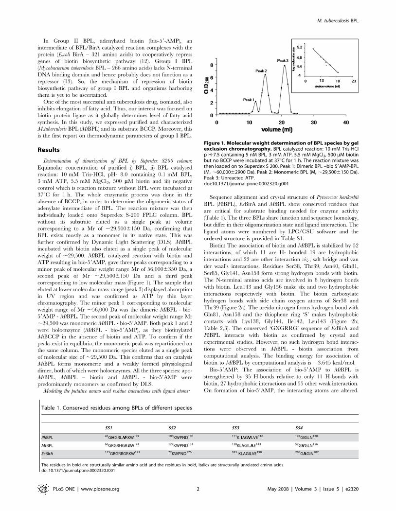
weight of ,29,500. MtBPL catalyzed reaction with biotin and
ATP resulting in bio-59AMP, gave three peaks corresponding to a
minor peak of molecular weight range Mr of 56,0006350 Da, a
second peak of Mr ,29,5006150 Da and a third peak
corresponding to low molecular mass (Figure 1). The sample that
eluted at lower molecular mass range (peak 3) displayed absorption
in UV region and was confirmed as ATP by thin layer
chromatography. The minor peak 1 corresponding to molecular
weight range of Mr ,56,000 Da was the dimeric MtBPL - bio-
59AMP - MtBPL. The second peak of molecular weight range Mr
,29,500 was monomeric MtBPL - bio-59AMP. Both peak 1 and 2
were holoenzyme (MtBPL - bio-59AMP), as they biotinylated
MtBCCP in the absence of biotin and ATP. To confirm if the
peaks exist in equilibria, the monomeric peak was repartitioned on
the same column. The monomeric species eluted as a single peak
of molecular size of ,29,500 Da. This confirms that on catalysis
MtBPL forms monomeric and a weakly formed physiological
dimer, both of which were holoenzymes. All the three species: apo-
MtBPL, MtBPL – biotin and MtBPL - bio-59AMP were
predominantly monomers as confirmed by DLS.
Modeling the putative amino acid residue interactions with ligand atoms:
Sequence alignment and crystal structure of Pyrococcus horikoshii
BPL (PhBPL), EcBirA and MtBPL show conserved residues that
are critical for substrate binding needed for enzyme activity
(Table 1). The three BPLs share function and sequence homology,
but differ in their oligomerization state and ligand interaction. The
ligand atoms were numbered by LPC/CSU software and the
ordered structure is provided in Table S1.
Biotin: The association of biotin and MtBPL is stabilized by 52
interactions, of which 11 are H- bonded 19 are hydrophobic
interactions and 22 are other interaction viz., salt bridge and van
der waal’s interactions. Residues Ser38, Thr39, Asn40, Gln81,
Ser85, Gly141, Asn158 form strong hydrogen bonds with biotin.
The N-terminal amino acids are involved in 8 hydrogen bonds
with biotin. Leu143 and Gly156 make six and two hydrophobic
interactions respectively with biotin. The biotin carboxylate
hydrogen bonds with side chain oxygen atoms of Ser38 and
Thr39 (Figure 2a). The ureido nitrogen forms hydrogen bond with
Gln81, Asn158 and the thiophene ring ‘S’ makes hydrophobic
contacts with Lys138, Gly141, Ile142, Leu143 (Figure 2b;
Table 2,3). The conserved ‘GXGRRG’ sequence of EcBirA and
PhBPL interacts with biotin as confirmed by crystal and
experimental studies. However, no such hydrogen bond interac-
tions were observed in MtBPL - biotin association from
computational analysis. The binding energy for association of
biotin to MtBPL by computational analysis is – 3.645 kcal/mol.
Bio-59AMP: The association of bio-59AMP to MtBPL is
strengthened by 35 H-bonds relative to only 11 H-bonds with
biotin, 27 hydrophobic interactions and 55 other weak interaction.
On formation of bio-59AMP, the interacting atoms are altered.
Figure 1. Molecular weight determination of BPL species by gelexclusion chromatography. BPL catalyzed reaction: 10 mM Tris-HClp H-7.5 containing 5 nM BPL, 3 mM ATP, 5.5 mM MgCl2, 500 mM biotinbut no BCCP were incubated at 37uC for 1 h. The reaction mixture wasthen loaded on to Superdex S 200. Peak 1: Dimeric BPL –bio 59AMP-BPL(Mr ,60,00062900 Da). Peak 2: Monomeric BPL (Mr ,29,5006150 Da).Peak 3: Unreacted ATP.doi:10.1371/journal.pone.0002320.g001
Table 1. Conserved residues among BPLs of different species
SS1 SS2 SS3 SS4
PhBPL 45GHGRLNRKW 53 100KWPND105 111K IAGVLVE118 124GIGLN128
MtBPL 66GRGRHGRGW 74 127KWPND131 138KLAGILAE143 52GVGLN156
EcBirA 115GRGRRGRKW123 72KWPND176 183 KLAGILVE190 203GAGIN207
The residues in bold are structurally similar amino acid and the residues in bold, italics are structurally unrelated amino acids.doi:10.1371/journal.pone.0002320.t001
M. tuberculosis BPL
PLoS ONE | www.plosone.org 2 May 2008 | Volume 3 | Issue 5 | e2320
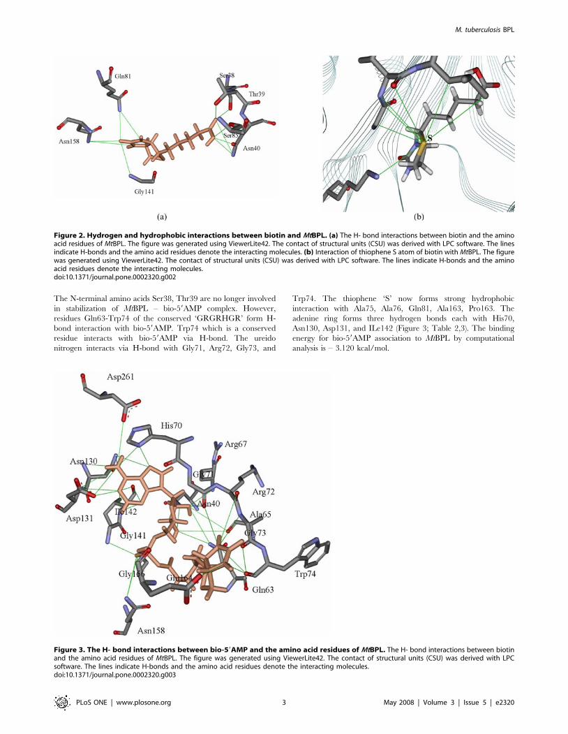
The N-terminal amino acids Ser38, Thr39 are no longer involved
in stabilization of MtBPL – bio-59AMP complex. However,
residues Gln63-Trp74 of the conserved ‘GRGRHGR’ form H-
bond interaction with bio-59AMP. Trp74 which is a conserved
residue interacts with bio-59AMP via H-bond. The ureido
nitrogen interacts via H-bond with Gly71, Arg72, Gly73, and
Trp74. The thiophene ‘S’ now forms strong hydrophobic
interaction with Ala75, Ala76, Gln81, Ala163, Pro163. The
adenine ring forms three hydrogen bonds each with His70,
Asn130, Asp131, and ILe142 (Figure 3; Table 2,3). The binding
energy for bio-59AMP association to MtBPL by computational
analysis is – 3.120 kcal/mol.
Figure 2. Hydrogen and hydrophobic interactions between biotin and MtBPL. (a) The H- bond interactions between biotin and the aminoacid residues of MtBPL. The figure was generated using ViewerLite42. The contact of structural units (CSU) was derived with LPC software. The linesindicate H-bonds and the amino acid residues denote the interacting molecules. (b) Interaction of thiophene S atom of biotin with MtBPL. The figurewas generated using ViewerLite42. The contact of structural units (CSU) was derived with LPC software. The lines indicate H-bonds and the aminoacid residues denote the interacting molecules.doi:10.1371/journal.pone.0002320.g002
Figure 3. The H- bond interactions between bio-59AMP and the amino acid residues of MtBPL. The H- bond interactions between biotinand the amino acid residues of MtBPL. The figure was generated using ViewerLite42. The contact of structural units (CSU) was derived with LPCsoftware. The lines indicate H-bonds and the amino acid residues denote the interacting molecules.doi:10.1371/journal.pone.0002320.g003
M. tuberculosis BPL
PLoS ONE | www.plosone.org 3 May 2008 | Volume 3 | Issue 5 | e2320
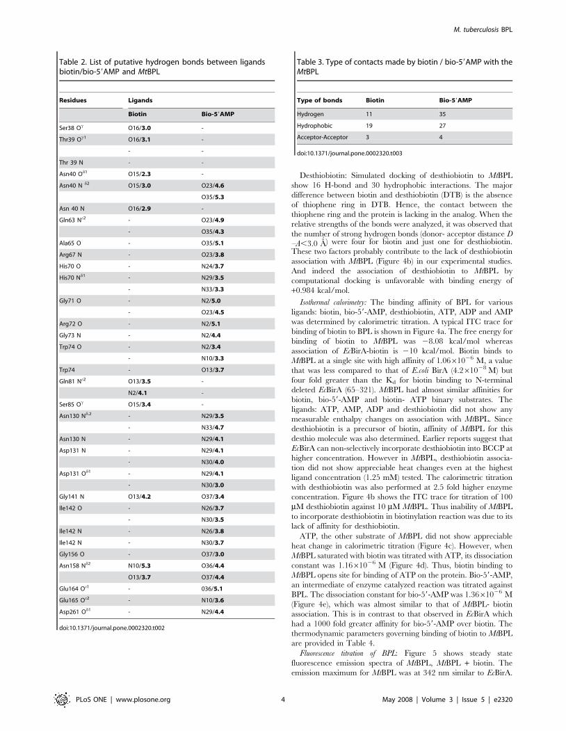
Desthiobiotin: Simulated docking of desthiobiotin to MtBPL
show 16 H-bond and 30 hydrophobic interactions. The major
difference between biotin and desthiobiotin (DTB) is the absence
of thiophene ring in DTB. Hence, the contact between the
thiophene ring and the protein is lacking in the analog. When the
relative strengths of the bonds were analyzed, it was observed that
the number of strong hydrogen bonds (donor- acceptor distance D
–A,3.0 A´ ) were four for biotin and just one for desthiobiotin.
These two factors probably contribute to the lack of desthiobiotin
association with MtBPL (Figure 4b) in our experimental studies.
And indeed the association of desthiobiotin to MtBPL by
computational docking is unfavorable with binding energy of
+0.984 kcal/mol.
Isothermal calorimetry: The binding affinity of BPL for various
ligands: biotin, bio-59-AMP, desthiobiotin, ATP, ADP and AMP
was determined by calorimetric titration. A typical ITC trace for
binding of biotin to BPL is shown in Figure 4a. The free energy for
binding of biotin to MtBPL was 28.08 kcal/mol whereas
association of EcBirA-biotin is 210 kcal/mol. Biotin binds to
MtBPL at a single site with high affinity of 1.0661026 M, a value
that was less compared to that of E.coli BirA (4.261028 M) but
four fold greater than the Kd for biotin binding to N-terminal
deleted EcBirA (65–321). MtBPL had almost similar affinities for
biotin, bio-59-AMP and biotin- ATP binary substrates. The
ligands: ATP, AMP, ADP and desthiobiotin did not show any
measurable enthalpy changes on association with MtBPL. Since
desthiobiotin is a precursor of biotin, affinity of MtBPL for this
desthio molecule was also determined. Earlier reports suggest that
EcBirA can non-selectively incorporate desthiobiotin into BCCP at
higher concentration. However in MtBPL, desthiobiotin associa-
tion did not show appreciable heat changes even at the highest
ligand concentration (1.25 mM) tested. The calorimetric titration
with desthiobiotin was also performed at 2.5 fold higher enzyme
concentration. Figure 4b shows the ITC trace for titration of 100
mM desthiobiotin against 10 mM MtBPL. Thus inability of MtBPL
to incorporate desthiobiotin in biotinylation reaction was due to its
lack of affinity for desthiobiotin.
ATP, the other substrate of MtBPL did not show appreciable
heat change in calorimetric titration (Figure 4c). However, when
MtBPL saturated with biotin was titrated with ATP, its dissociation
constant was 1.1661026 M (Figure 4d). Thus, biotin binding to
MtBPL opens site for binding of ATP on the protein. Bio-59-AMP,
an intermediate of enzyme catalyzed reaction was titrated against
BPL. The dissociation constant for bio-59-AMP was 1.3661026 M
(Figure 4e), which was almost similar to that of MtBPL- biotin
association. This is in contrast to that observed in EcBirA which
had a 1000 fold greater affinity for bio-59-AMP over biotin. The
thermodynamic parameters governing binding of biotin to MtBPL
are provided in Table 4.
Fluorescence titration of BPL: Figure 5 shows steady state
fluorescence emission spectra of MtBPL, MtBPL + biotin. The
emission maximum for MtBPL was at 342 nm similar to EcBirA.
Table 2. List of putative hydrogen bonds between ligandsbiotin/bio-59AMP and MtBPL
Residues Ligands
Biotin Bio-59AMP
Ser38 Oc O16/3.0 -
Thr39 Oc1 O16/3.1 -
- -
Thr 39 N - -
Asn40 Od1 O15/2.3 -
Asn40 N d2 O15/3.0 O23/4.6
O35/5.3
Asn 40 N O16/2.9 -
Gln63 Ne2 - O23/4.9
- O35/4.3
Ala65 O - O35/5.1
Arg67 N - O23/3.8
His70 O - N24/3.7
His70 Nd1 - N29/3.5
- N33/3.3
Gly71 O - N2/5.0
- O23/4.5
Arg72 O - N2/5.1
Gly73 N - N2/4.4
Trp74 O - N2/3.4
- N10/3.3
Trp74 - O13/3.7
Gln81 Ne2 O13/3.5 -
N2/4.1 -
Ser85 Oc O15/3.4 -
Asn130 Nd.2 - N29/3.5
- N33/4.7
Asn130 N - N29/4.1
Asp131 N - N29/4.1
- N30/4.0
Asp131 Od1 - N29/4.1
- N30/3.0
Gly141 N O13/4.2 O37/3.4
Ile142 O - N26/3.7
- N30/3.5
Ile142 N - N26/3.8
Ile142 N - N30/3.7
Gly156 O - O37/3.0
Asn158 Nd2 N10/5.3 O36/4.4
O13/3.7 O37/4.4
Glu164 Oe1 - 036/5.1
Glu165 Oe2 - N10/3.6
Asp261 Od1 - N29/4.4
doi:10.1371/journal.pone.0002320.t002
Table 3. Type of contacts made by biotin / bio-59AMP with theMtBPL
Type of bonds Biotin Bio-59AMP
Hydrogen 11 35
Hydrophobic 19 27
Acceptor-Acceptor 3 4
doi:10.1371/journal.pone.0002320.t003
M. tuberculosis BPL
PLoS ONE | www.plosone.org 4 May 2008 | Volume 3 | Issue 5 | e2320
The binding of biotin to the protein resulted in 10% quenching of
intrinsic fluorescence signal.
Biological properties of BPL: The enzymatic activity of BPL was
determined by incorporation of 14C labelled biotin into the cloned
BCCP domain of M.tuberculosis acetyl CoA carboxylase. Optimal
enzyme activity was observed at pH 7.5–8.0. The presence of
biotin, ATP, Magnesium ions, apo-BCCP was necessary for
biotinylation reaction. Enzyme activity was dependent on ATP as
nucleotide source and substitution of ATP with GTP did not result
in biotinylation reaction. BPL forms a weak dimer with bio-59-
AMP synthesized from ATP and biotin, in absence of apoBCCP as
observed from gel filtration (Figure 1). The Km for D-biotin is
,424.2656.38 nM, 21.0863.78 mM for Mg/ATP, 5.26.56 mM
for apoBCCP and 10 nM of BPL was sufficient to drive enzymatic
process (Table 5). These data thus confirm that cloned BPL of
M.tuberculosis was enzymatically active.
Specific incorporation of biotin by MtBPL: To determine if BPL can
non-specifically utilize desthiobiotin in the enzyme reaction, both
biotin and its analogue were used at concentrations ranging from
25 mM – 6 nM in biotinylation of BCCP (15 mM) by 10 nM of
BPL. As revealed by an assay akin to ELISA, biotin at ,620 nM
efficiently biotinylated BCCP whereas desthiobiotin was not
incorporated into BCCP in a 30 min biotinylation reaction
(Figure 6a). The desthiobiotin concentration was increased to
1000 mM which was .1200 fold the optimal biotin concentration
and the incubation period for 4 h. But the incorporation of
desthiobiotin into BCCP by BPL was not appreciable. This could
be due to two reasons: i) desthiobiotin was not utilized as a
substrate in enzymatic process by BPL or, ii) streptavidin HRP was
unable to detect incorporated desthiobiotin. To rule out the
second possibility, a competitive assay of streptavidin with biotin/
desthiobiotin was performed. Streptavidin HRP at 1:2500 dilution
of 1 mg/ml solution was pre-incubated with biotin/ desthiobiotin
(5 mM–5 nM) for 1 h at 37uC and the solution was then added to
previously immobilized biotinylated BCCP. Streptavidin interacts
with its cognate molecule (biotin/desthiobiotin) and any un-
reacted, free streptavidin from first step will react with biotinylated
BCCP determining affinity of streptavidin for biotin/desthiobiotin.
Figure 6b shows that streptavidin interacts efficiently with both
biotin and desthiobiotin. These experiments thus rule out the
possibility that discrepancy in incorporation of biotin/desthiobi-
otin in BCCP was not due to error in detection of desthiobioti-
nylated BCCP but because of its non-utilization.
Avidin blot: Biotinylation of apo-BCCP, a biotin acceptor protein
was detected by streptavidin HRP on Avidin blot (Figure 7a). Lane
2 is BCCP alone with no BPL. The faint band observed
corresponds to BCCP that was probably biotinylated by
endogenous EcBirA. Lane 3 is biotinylation reaction of BCCP
by BPL in presence of ATP, biotin and MgCl2.for 30 min at 37uC.
As observed BCCP was biotinylated several fold by BPL in
comparison with BCCP control (Lane 3), thus confirming transfer
of biotin to acceptor domain of BCCP by M.tuberculosis BPL.
Figure 7b shows the ability of both monomeric and dimeric
MtBPL associated with bio-59AMP to biotinylate apoBCCP in the
absence of biotin and ATP. The second step of the biotinylation
reaction, the transfer of biotin to BCCP from bio-59AMP by
MtBPL was detected by Avidin blot (Figure 7b). Lane 1 and 2 is
the peak 1 and 2 (Figure 1) of holoenzyme eluted from gel
exclusion chromatography biotinylating apoBCCP.
Discussion
Acetyl CoA carboxylase and pyruvate dehydrogenase are
enzymes crucial for fatty acid biosynthesis which require
biotinylation of their BCCP domain as a prelude to their
enzymatic activities (14, 15). Biotin protein ligase which globally
controls this pathway is a potential target for anti-mycobacterial
drugs. MtBPL shares ,25% and 31% sequence homology with
PhBPL and EcBirA respectively. It belongs to monofunctional
group I BPL as it lacks the 60 amino acid N-terminal HTH
domain. This domain in EcBirA contributes to repressor function
by binding to biotin operon (bioO). Deletion of the N-terminal
domain converted EcBirA from bifunctional to monofunctional
protein (16).
The present study shows that MtBPL differs from biotin ligase of
other species both in interaction with its substrates and molecular
mechanism of catalysis. Homodimerization is coupled to co-
repressor synthesis and is a pre-requisite for repressor function in
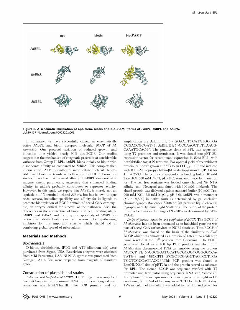
EcBirA (16). Monofunctional PhBPL (Group I) is a dimer in all the
three forms viz. apo, biotin and bio-59AMP liganded forms
(Figure 8). PhBPL in spite of lacking repressor function does exist
as a dimer. Our studies reveal that apo, biotin and bio-59AMP
liganded. MtBPL are monomers (Figure 1, 8). The peak
corresponding to monomeric MtBPL (peak 2, Figure 1) was
holoenzyme as it biotinylated MtBCCP. However, bio-59AMP
binding does promote weak physiological dimerization of MtBPL
similar to that observed when biotin binds to EcBirA. But the
ability of both peak 1 and 2 to transfer biotin to MtBCCP
confirmed that both the peaks were holoenzymes. Truncated
EcBirA lacks repressor function but is enzymatically active (bio-
59AMP synthesis). It would be interesting to study the oligomer-
ization property of this truncated protein on bio-59AMP synthesis
Thus, our gel exclusion profiling studies suggest that monofunc-
tional MtBPL does not require dimerization for catalysis. Hence
from studies on different BPLs, it is evident that oligomerization
may not be a universal phenomena for all BPLs and some of them
such as PhBPL and EcBirA, undergo oligomerization for
compaction and thermo-stability in their apo/holo forms (17, 18).
It is known that PhBPL is a dimer in all the three forms, apo,
biotin and bio-59AMP liganded, while EcBirA is a dimer when
associated with bio-59AMP but monomeric in apo and biotin-
liganded form. From our studies it was observed that MtBPL is
monomeric in all the three forms (Figure 8).
Sequence analysis shows conserved sequences between MtBPL,
EcBirA and PhBPL. Four homologous sequences between the
three BPLs are shown (19). However, the identity of other residues
dispersed in the protein sequences are also conserved but are not
specified. Sequence SS1 interacts with biotin in PhBPL and
EcBirA. Earlier studies by Kwon and Beckett (2000) show that the
consensus ‘GRGRXGR’ sequence is involved in binding of biotin
and bio-59AMP (20). Docking studies of MtBPL and biotin show
that sequence SS1 was involved in interaction with bio-59AMP but
not biotin. In fact, except for residues 66Gly and 67Arg which were
involved in hydrophobic interaction with the thiophene ring, the
reminder of the conserved sequence appears to be of little or no
consequence for the recognition of biotin. From ITC studies, it
was observed that MtBPL has a dissociation constant of
16106 M21 for biotin (Figure 4a). Hence, the hydrogen bond
between the hydrophilic residues, Ser 38, Thr39, Asn40, Gln81,
Ser85 and biotin contribute to this association. The hydrophobic
pocket formed by Ile83, Leu84, Ala140, Gly141, Ile142, Leu143,
Gly154, Val 155, and Gly156 probably dock the thiophene ring of
biotin. Bio-59AMP interacts with Arg67, His70, Gly71, Arg72 of
the conserved ‘GRGRHGR’ sequence by H-bonds and hydro-
phobic interaction. Asn158 contributes two H-bonds to the
adenylate and the purine ring stacks on the face of the indole
ring of Trp74 and makes H-bond with Asn158, and side-chain of
alanine 65. An analogous interaction in EcBirA, is provided by Phe
M. tuberculosis BPL
PLoS ONE | www.plosone.org 5 May 2008 | Volume 3 | Issue 5 | e2320
which replaces alanine of MtBPL at the homologous position. In
EcBirA, biotin binding site is a hydrophobic groove with hydrogen
binding partners Ser89 and Thr90 pre- positioned to accept biotin
(21). From our computational studies, it was observed that
homologous residues Ser38 and Thr39 of MtBPL H-bond with
biotin. Thus, residues Ser38, Thr39 for biotin and
‘GRGRHGRGW’ sequence for bio-59AMP may contribute
largely to association of ligands. From docking simulations, biotin
has 52 and bio-59AMP has 117 interactions with MtBPL. The
number of interactions involved and the larger surface area
occupied by bio-59AMP protects EcBirA from proteolysis. This
suggests that though bio-59AMP is an intermediate molecule; its
complex with BPL is most stable. However, these computational
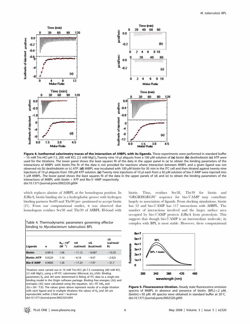
Figure 4. Isothermal calorimetry traces of the interaction of MtBPL with its ligands. These experiments were performed in standard buffer– 10 mM Tris-HCl pH-7.5, 200 mM KCl, 2.5 mM MgCl2.Twenty nine 10 ml aliquots from a 100 mM solution of (a) biotin (b) desthiobiotin (c) ATP wereused for the titrations. The lower panel shows the least squares fit of the data in the upper panel in (a) to obtain the binding parameters of theinteractions of MtBPL with biotin.The fit of the data is not provided for reactions where interaction between MtBPL and a given ligand was notobserved viz (b) desthiobiotin or (c) ATP. (d) MtBPL was incubated with 100 mM biotin for 30 min in the ITC cell and then titrated against twenty nineinjections of 10 ml aliquots from 100 mM ATP solution. (e) Twenty nine injections of 10 ml each from a 50 mM solution of bio-59AMP were injected into5 mM MtBPL. The lower panel shows the least squares fit of the data in the upper panels of (d) and (e) to obtain the binding parameters of theinteractions of MtBPL with biotin + ATP and Bio-59-AMP respectively.doi:10.1371/journal.pone.0002320.g004
Table 4. Thermodynamic parameters governing effectorbinding to Mycobacterium tuberculosis BPL
Ligands nKb6106
(M21)DH(cal/mol)
DG(kcal/mol)
DS(cal/mol/K)
Biotin 0.9814 1.06 211.12 28.087 210.35
Biotin+ATP 0.9229 1.16 28.18 29.07 22.925
Bio-59AMP 0.9800 1.36 217.25 27.97 231.7
Titrations were carried out in 10 mM Tris-HCl, ph-7.5 containing 200 mM KCl,2.5 mM MgCl2 using a VP-ITC calorimeter (Microcal, Inc.,USA). Bindingparameters Kb and DH were determined b fitting of ITC data to a single-sitebinding model in the Origin software package. Binding free energies (DG) andentropies (DS) were calculated using the equation, DG = RT lnKb andDG =DH2TDS. The values given above represent results of a single titrationwith each ligand and in multiple titrations the values of Kb and DH arereproducible within 2-fold and 1 kcal/mol.doi:10.1371/journal.pone.0002320.t004
Figure 5. Flourescence titration. Steady state fluorescence emissionspectra of MtBPL in absence and presence of biotin. [BPL] = 2 mM,[biotin] = 50 mM. All spectra were obtained in standard buffer at 20uC.doi:10.1371/journal.pone.0002320.g005
M. tuberculosis BPL
PLoS ONE | www.plosone.org 6 May 2008 | Volume 3 | Issue 5 | e2320
analysis need to be confirmed by experimental studies.
The other focus of our study is the substrate specificity of
MtBPL for biotin. Desthiobiotin did not bind to MtBPL nor was
used in the biotinylation reaction. The thiophene ring and the
hydrophobic tail would interact with the hydrophobic amino acids
and the biotin carboxyl and ureido ring would interact with the
hydrophilic amino acids. Since DTB lacks the thiophene ring, the
interaction of the molecule with the hydrophobic amino acids is
only via the hydrophobic tail and ureido ring. Also, biotin forms
relatively stronger H-bonds (bond length .3.0A´ ) as compared to
desthiobiotin. This is reflected in the unfavorable binding energy
of association between desthiobiotin and MtBPL (Biotin = 23.645
kcal/mol, Desthiobiotin = +0.984 l/mol).
A second conserved sequence of KWPNDVL shares sequence
homology with biotin binding protein avidin. However, this
sequence does not interact with the ligands in all the three BPLs.
Lys111 (PhBPL) and Lys183 (EcBirA) of the conserved
‘KXAGXLVE’ play a critical role in adenylation reaction.
However, from computational studies, no such interaction
between this conserved lysine residue and the ligands were
observed (21, 22).
Effector binding is central to a number of essential activation
processes and provides essential information on interactions
involved therein. The affinity of MtBPL for its effectors biotin
and bio-59-AMP was determined by ITC. The thermodynamic
data are categorized into enthalpy, free energy changes and
dissociation constants. MtBPL exhibited similar dissociation
constants of ,161026 M for both the effectors (Figure 4a, d
and e). In contrast, EcBirA displayed different affinities for its
ligands: biotin and bio-59-AMP. Dissociation constants for biotin
(4.261028 M) and bio-59AMP (4610211 M) was very different
and hence they have been classified as ‘weak’ and ‘strong’
effectors respectively (23–25). The affinity of MtBPL for bio-59-
AMP was 5 orders of magnitude lower than that observed in
EcBirA (4610211 M). Repressor function in EcBirA is in
association with bio-59-AMP (co-repressor) and hence very tight
binding is required to perform this function (26). However, the
N-terminal deleted EcBirA had 1000 fold reduced affinity
(,461028 M) for bio-59-AMP (16). This suggests that mono-
functional group I BPLs exhibit attenuated affinities dedicated to
perform only enzymatic function. The adenosine molecules,
ATP, ADP and AMP did not show any appreciable heat changes
in ITC. This is in contrast to crystallographic structure of PhBPL
which show interaction with adenosine molecules (22). However,
in crystallographic structures, observations of the complexes of
ligand which have extremely low affinities (,50 M21) can often
be observed which is typically difficult in biophysical studies. The
endoplasmic reticulum chaperone, calreticulin, for example,
does not show binding to glucose, though density for glucose
complexed to calnexin, a very closely related molecule was seen
in the crystal structure (27).
Bio-59AMP, an intermediate of MtBPL catalyzed reaction
involves biotin and ATP as substrates. Hence, it was essential to
determine the mechanism of association of MtBPL with its
substrates. These studies show sequential binding pattern of
MtBPL to them, as ATP (Figure 4c) does not interact with BPL
directly but on saturation with biotin, it exhibits a Kd of
,1.166106 M (Figure 4d). Saccharomyces cerevisiae BPL interacts
first with ATP followed by biotin, in contrast to MtBPL and
EcBirA (28). Biotin is the limiting substrate in MtBPL catalyzed
Table 5. Kinetic constants for the interaction of Mt BPL with itssubstrates- biotin, MgATP and MtBCCP
Substrate Km (mM) kcat (s21)kcat/Km
(s21M21)6103
Biotin 0.4206.056 0.0346.007 8262.1
Mg/ATP 21.0863.78 0.02826.003 1.336.09
BCCP 5.260.56 0.0306.001 5.776.37
The experiment was carried out in triplicates as described in Experimentalprocedures.doi:10.1371/journal.pone.0002320.t005
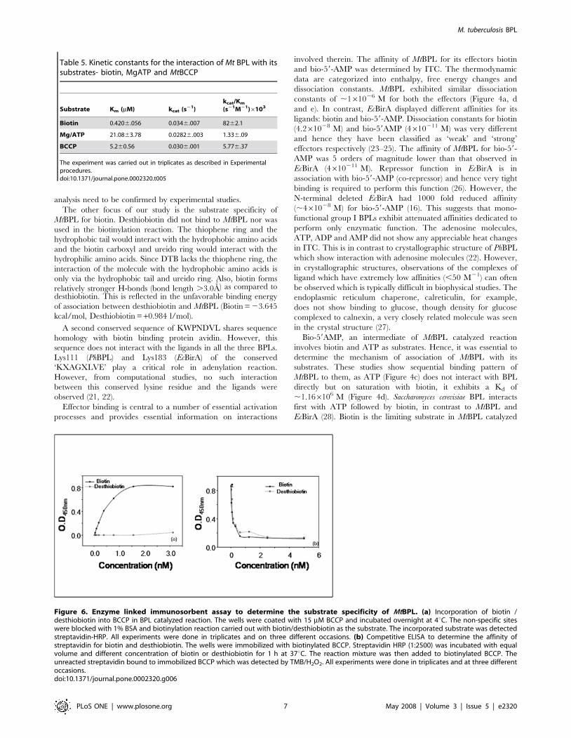
Figure 6. Enzyme linked immunosorbent assay to determine the substrate specificity of MtBPL. (a) Incorporation of biotin /desthiobiotin into BCCP in BPL catalyzed reaction. The wells were coated with 15 mM BCCP and incubated overnight at 4uC. The non-specific siteswere blocked with 1% BSA and biotinylation reaction carried out with biotin/desthiobiotin as the substrate. The incorporated substrate was detectedstreptavidin-HRP. All experiments were done in triplicates and on three different occasions. (b) Competitive ELISA to determine the affinity ofstreptavidin for biotin and desthiobiotin. The wells were immobilized with biotinylated BCCP. Streptavidin HRP (1:2500) was incubated with equalvolume and different concentration of biotin or desthiobiotin for 1 h at 37uC. The reaction mixture was then added to biotinylated BCCP. Theunreacted streptavidin bound to immobilized BCCP which was detected by TMB/H2O2. All experiments were done in triplicates and at three differentoccasions.doi:10.1371/journal.pone.0002320.g006
M. tuberculosis BPL
PLoS ONE | www.plosone.org 7 May 2008 | Volume 3 | Issue 5 | e2320
reaction similar to EcBirA. Biotin binding perhaps contributes to
structural transitions that may be required for interaction of ATP
to form ternary complex between MtBPL, biotin and ATP.
The Gibbs free energy for binding of biotin and bio-59-AMP to
MtBPL were almost similar. But for EcBirA, there was a gain of 24
kcal/mol of free energy on bio-59-AMP binding. This is probably to
ensure that repressor was always in active form for binding to bioO
(29, 30). MtBPL does not require any enhanced BPL-bio-59-AMP
stability for transfer of biotin to apoBCCP. Thus, thermodynamic
studies reveal that molecular mechanisms of ligand interaction are
very different between MtBPL and EcBirA on account of additional
burden of repressor function in the latter molecule.
Binding of bio-59-AMP to MtBPL is associated with larger
enthalpy changes (217.2 kcal/mol) in comparison with 211.12
kcal/mol for biotin (Table 4). However in EcBirA, enthalpy
changes are larger for biotin (219.2 kcal/mol) compared to that
for bio-59-AMP (-12.3 kcal/mol). According to Brown and Beckett
(24), the absence of a charged functional group to interact with the
adenosine moiety could be the reason for the enthalpic penalty. In
EcBirA, there is a biotin binding loop (BBL) 115–120 residues
which is conserved and adenylate binding loop (ABL) consisting of
residues 212–223 (24, 25). On limited proteolysis, the region
between amino acid residues 217–218 was protected by bio-59-
AMP (26). This suggests that adenosine of bio-59-AMP interacts at
the ABL. BBL becomes ordered on biotin binding and then
adenylate binding site is formed in EcBirA. In contrast, there is no
separate ABL and BBL in PhBPL hence the biotin moiety of
biotinyl-59-AMP and biotin share the same binding site. Also, the
adenosyl moiety of bio-59-AMP and ADP interact almost at the
same position. This sharing of binding site in PhBPL which is
strikingly different from that observed in EcBirA suggests that
catalytic binding sites are organized such that major conforma-
tional changes are perhaps not required in group I BPLs during
the reaction (22). Bagautdinov et al (22) co-crystallized PhBPL with
biotin and bio-59AMP suggesting that the interaction does not
involve any conformational change. On the other hand, exposure
of EcBirA crystals to bio-59-AMP resulted in cracking and crystal
destruction due to conformational changes. Together, these data
underscore striking differences in the organization of biotin and
ATP binding site between MtBPL and EcBirA as well as the
differences in the conformational changes they undergo upon
binding of biotin/bio-59-AMP.
The dissociation constant of MtBPL for its ligands was high
relative to EcBirA (Table 4, Figure 4), hence enzyme turnover was
determined by steady state kinetics. The kinetic constants show low
Km for both biotin (0.42 mM) and ATP (21 mM). This is similar to
that observed in eukaryotic biotin auxotrophs such as Saccharomyces
cerevisiae and A.thaliana which exhibit Km in nanomolar range for
biotin and lower micromolar concentration for MgATP (28, 32).
EcBirA has comparable Km of 0.3 mM for biotin, but has higher Km
of 300 mM for ATP. Though MtBPL has higher dissociation constant
for both biotin and bio-59-AMP, the Km values were comparable to
that of EcBirA (Table 4,5). Thus, the lower binding affinity of MtBPL
for its ligands did not alter its enzymatic function.
Desthiobiotin (DTB) is precursor molecule for biotin but
differs from it in several ways; both have an ureido ring but
desthiobiotin lacks thiophene ring (Table S1). Wu et al (33) had
suggested that EcBirA incorporates desthiobiotin into SAPK, a
protein with a biotinable lysine residue. Their assay was driven
to 100% incorporation at a concentration of 1 mM in 4 h. But
MtBPL did not promote desthiobiotin incorporation into BCCP
at increased concentration and incubation time (Figure 6a,b).
From structural analysis of PhBPL, it is evident that both
thiophene and ureido side chains are accommodated in the
hydrophilic bottom provided by glycine residues (Gly
45,47,127,129) and the thiophene moiety interacts with the
hydrophobic wall (22). DTB which lacks the thiophene side
chain will not fit efficiently in such a structural frame work. The
hexanoic acid attached close to ureido nitrogen may hamper
interaction of ureido side chain as well. This argument is
strengthened by the binding energy of MtBPL (+0.984 kcal/mol)
and EcBirA (21.897 kcal/mol) for desthiobiotin by computa-
tional modeling. Monofunctional MtBPL has an unfavorable
binding energy for the association and this is confirmed by the
inability of the enzyme to either bind or incorporate desthiobi-
otin. EcBirA prefers biotin (23.491 kcal/ mol) to desthiobiotin
(21.897 kcal/mol) and hence utilizes desthiobiotin at higher
molar concentration and increased incubation period. Biotin
synthase (BioB) utilizes desthiobiotin to synthesize biotin and
activity of the enzyme is determined by bioassay. This is a very
tedious assay and there is a need for more reliable, easy,
quantitative and rapid assay. Biotin synthase enzymatically
generates one molecule of biotin from desthiobiotin. At
equimolar concentration of biotin/desthiobiotin, MtBPL prefer-
ably incorporates biotin and this can be exploited to assay biotin
synthase activity in a sensitive manner.
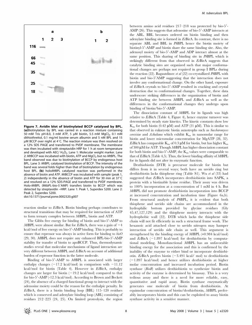
Figure 7. Avidin blot of biotinylated BCCP catalysed by BPL.(a)Biotinylation by BPL was carried in a reaction mixture containing50 mM Tris pH-8.0, 3 mM ATP, 5 mM biotin, 5.5 mM MgCl2, 0.1 mMdithiothreitol, 0.1 mg/ml bovine serum albumin and 5 nM BPL and 15mM BCCP over night at 4uC. The reaction mixture was then resolved ona 12% SDS PAGE and transferred to PVDF membrane. The membranewas then incubated with streptavidin HRP for 1 h at room temperatureand developed with AEC/ H2O2. Lane 1: Molecular weight marker. Lane2: MtBCCP was incubated with biotin, ATP and MgCl2 but no MtBPL. Theband observed was due to biotinylation of BCCP by endogenous hostBPL. Lane 3: MtBPL catalysed biotinylation of BCCP. The intensity of theband was several folds higher than that of biotinylation by endogenoushost BPL. (b) holoMtBPL catalyzed reaction was performed in theabsence of biotin and ATP. MtBCCP was incubated with sample (peak 1,2) independently in the absence of biotin and ATP for 30 min at 37uCand resolved on a 12% SDS-PAGE and transferred to PVDF membrane.Holo-MtBPL (MtbPL-bio-59AMP) transfers biotin to BCCP which wasdetected by streptavidin –HRP. Lane 1: Peak 1, Superdex S200. Lane 2:Peak 2, Superdex S200.doi:10.1371/journal.pone.0002320.g007
M. tuberculosis BPL
PLoS ONE | www.plosone.org 8 May 2008 | Volume 3 | Issue 5 | e2320
In summary, we have successfully cloned an enzymatically
active MtBPL and biotin acceptor molecule, BCCP of M.
tuberculosis. Our protocol variation of reduced growth and
induction time yielded nearly 90% apo-BCCP. Our studies
suggest that the mechanism of enzymatic process is at considerable
variance from Group II BPL. MtBPL binds initially to biotin with
a moderate affinity as compared to EcBirA. This complex then
interacts with ATP to synthesize intermediate molecule bio-59-
AMP and biotin is transferred efficiently to BCCP. From our
studies, it is clear that reduced affinity of MtBPL does not alter
enzyme kinetic parameters, suggesting that enhanced binding
affinity in EcBirA probably contributes to repressor activity.
However, in this study we report that MtBPL is merely not an
equivalent of N-terminal deleted EcBirA, but has its own unique
modus operandi, including specificity and affinity for its ligands to
promote biotinylation of BCCP domain of acetyl CoA carboxyl-
ase, an enzyme critical for survival of the pathogen. Also, the
differences in the architecture of biotin and ATP binding site of
MtBPL and EcBirA and the exquisite specificity of MtBPL for
biotin over desthiobiotin can be harnessed for synthesizing
inhibitors for this important enzyme which should aid in
combating global spread of tuberculosis.
Materials and Methods
BiochemicalsD-biotin, desthiobiotin, IPTG and ATP (disodium salt) were
purchased from Sigma, USA. Restriction enzymes were obtained
from MBI Fermentas, USA. Ni-NTA agarose was purchased from
Novagen. All buffers were prepared from reagents of standard
grade.
Construction of plasmids and strainsExpression and purification of MtBPL: The BPL gene was amplified
from M.tuberculosis chromosomal DNA by primers designed with
restriction sites NdeI/HindIII. The PCR primers used for
amplification are MtBPL F1: 59- GGAATTCCATATGGTGA
CCGACCGCGAT -39; MtBPL B1: 59-CCCAAGCTTTTAACG-
CAAATGCAC-39. The putative clone of BPL was sequenced
using T7 promoter and terminator. It was cloned into pET 28a
expression vector for recombinant expression in E.coli BL21 with
hexahistidine tag at N-terminus. For optimal yield of recombinant
protein, cells were grown at 37uC to an O.D600 , 0.7 and induced
with 0.1 mM isopropyl-1-thio-b-D-galactopyranoside (IPTG) for
4 h at 25uC. The cells were suspended in binding buffer (10 mM
Tris-HCl, 500 mM NaCl, pH- 8.0), sonicated twice for 1 min on
ice. The cell free sonicate was loaded onto charged Ni- NTA
affinity resin (Novagen) and eluted with 100 mM imidazole. The
eluted protein was dialyzed against standard buffer (10 mM Tris,
200 mM KCl, 2.5 mM MgCl2, pH-8.0). MtBPL was a monomer
(Mr ,29,500) in native form as determined by gel exclusion
chromatography (Superdex S200) on fast pressure liquid chroma-
tography and Dynamic Light Scattering. The purity of the protein
thus purified was in the range of 95–98% as determined by SDS-
PAGE.
Design of primers, expression and purification of BCCP: The BCCP of
M.tuberculosis has not been annotated as an individual gene but was
part of acetyl CoA carboxylase in NCBI database. Thus BCCP of
M.tuberculosis was cloned on the basis of the similarity to E.coli
BCCP which was annotated as a protein of 156 amino acids with
lysine residue at the 35th position from C-terminal. The BCCP
gene was cloned as a 468 bp PCR product amplified from
M.tuberculosis chromosomal DNA as template using the primers
MtBCCP F1: 59-CGCGGATCCATGCGCGGCGGGGGCCA-
TATG-39 and MtBCCPF1: 59CGCTCGAGCTAGTCCTTGA
TCCTCGCCAGTACC-39.This PCR product was cloned at
BamHI/XhoI sites of pET28a and the protein served as substrate
for BPL. The cloned BCCP was sequence verified with T7
promoter and terminator using sequencer DNA star, Wisconsin.
For optimal protein expression, cells were grown overnight in LB
containing 30 mg/ml of kanamycin at 37uC for 14 h. Next day,
15% inoculum of this culture was added to fresh LB and grown for
Figure 8. A schematic illustration of apo form, biotin and bio-59AMP forms of PhBPL, MtBPL and EcBirA.doi:10.1371/journal.pone.0002320.g008
M. tuberculosis BPL
PLoS ONE | www.plosone.org 9 May 2008 | Volume 3 | Issue 5 | e2320
1 h at 37uC. The culture was induced with 0.1 mM IPTG for 1 h
at 25uC to prevent endogenous biotinylation by host BPL. The
soluble protein did not bind to Ni- NTA agarose, probably
because hexahistidine tag was masked. Hence, cells were purified
under denaturation conditions (10 mM Tris, 100 mM NaH2PO4,
10 mM imidazole, 8 M Urea, pH- 8.0) and sonicated for 2 min,
twice on ice. The cleared sonicate was loaded onto to a charged
NiNTA column and the protein was eluted with elution buffer
(10 mM Tris, 100 mM NaH2PO4, 10 mM imidazole, 8 M urea,
pH- 4.5).The protein was further dialyzed against 10 mM Tris-
HCl, pH- 7.5 containing decreasing concentration of urea.
Determination of protein concentrationConcentration of protein was determined by Lowry’s method
and ultraviolet absorbance using an extinction coefficient of
1.26 mL mg21 cm21 for BPL and 1.373 mL mg21cm21 for
BCCP. Both BPL and BCCP were exhaustively dialyzed against
standard buffer of 10 mM Tris-HCl (pH- 7.5), containing
200 mM KCl and 2.5 mM MgCl2.
Synthesis of Bio-59-AMPBio-59- AMP was synthesized by a modified method of Lane et
al (34): To biotin (244 mg) dissolved in 4 ml of 75% aqueous
pyridine with gentle heating 347 mg of ATP in 3 ml of 75%
aqueous pyridine was added. To the reaction mixture, pre cooled
4.5 g of dicyclohexyl carbodiimide in 5 ml of absolute pyridine
was added and reaction was carried out for 8 h at 0uC with
constant stirring. The reaction mixture was kept aside at 4uC for
24 h to precipitate DCU which was removed by passing through a
sintered glass filter. Bio-59-AMP was separated from unreacted
biotin and ATP by RP-HPLC performed on Waters equipment
consisting of a Waters 2487 Dual Absorbance Detector. Analyses
were carried out on an analytical column (Nucleosil 100 A´ 5 mm
C18 particles, 25064.6 mm) using following solvent system:
solvent A, 0.1% TFA in water; solvent B, 0.1% TFA in
acetonitrile; flow rate, 1.0 mL min21 with UV monitoring at
214 nm and 254 nm. Preparative column (Delta-PakTM 300A´ 15
mm C18 particles, 20062.5 mm) was used with an identical
solvent system at a flow rate of 25 ml min21. Mass spectra were
obtained by electron spray ionization (ESI-MS).
Analytical gel filtration chromatographyThe oligomeric status of purified BPL was determined by gel
filtration on Superdex S200 column and a fast protein liquid
chromatography system (Pharmacia Biotech Inc). The column
dimension was 30 cm62.3 cm. The mobile phase was 50 mM
Tris-HCl buffer, pH- 7.5, and flow rate was 0.2 ml/min. The
molecular mass of standard proteins in kDa 150, 66, 45, 29 & 14
were, respectively, alcohol dehydrogenase, BSA, ovalbumin,
carbonic anhydrase and lysozyme. The eluted proteins were
detected by their absorbance at 280 nm.
The enzymatic reaction was carried out with 100 mM BPL,
3 mM ATP and 500 mM biotin, 5.5 mM MgCl2 at 37uC for
30 min and 300 ml of reaction mixture was loaded onto the
column. The void volume (Vo) was determined by calibrating
column with blue dextran. From elution volume of standard
proteins, molecular size of unknown protein, BPL and its
enzymatic reaction mixture were determined.
Computational modeling of ligand docking to MtBPL: The structure of
MtBPL was modeled using SWISS-PROT software with Mycobac-
terium tuberculosis Biotin Protein Ligase as the template (Code:
2cghB) as the template (Ma Q. and Wilmanns M. unpublished).
The ligands: biotin, desthiobiotin and bio-59AMP were built using
MOE (Molecular Operating Environment) and energy minimized
under the MMFF946forcefield. The docking of the ligands to
MtBPL was also performed with MOE using AMBER99 forcefield
and the docked complex with least energy was chosen. The H
bond interaction between the ligand atoms and amino acid
residues of MtBPL was obtained by LPC/CSU software (35).
Assay of BPL: BPL activity was assayed by measuring
incorporation of [14C]- biotin (GE Healthcare, UK) into BCCP
156 as described by Chapman-Smith et al (10,11). The reaction
contained 10 mM Tris-HCl, 200 mM KCl, 3 mM ATP, 5.5 mM
MgCl2, 10 mM biotin and 1 mM of [14C] biotin (sp.activity 58.0
mCi/mmol), 0.1 mM DTT, 0.1 mg bovine serum albumin and 15
mM BCCP. The reaction was initiated by addition of 10 nM of
BPL and incubated at 37uC for 30 min. The reaction mixture was
added to biotin and trichloroacetic acid treated filters. After air
drying, the filters were washed twice with 10% ice-cold TCA,
ethanol and air dried and acid insoluble radioactivity was
measured. A unit of BPL activity was defined as amount of
enzyme required to incorporate 1 nM of biotin per min. Values for
Km and Vmax were determined by non-linear least squares fit of
the plots of substrate concentration versus velocity of reaction to
Michaelis-Menten equation.
Determination of thermodynamic parameters byIsothermal calorimetry
A MicroCal VP-ITC was used to determine binding parameters
of biotin and desthiobiotin to BPL. All protein samples were
dialyzed exhaustively against standard buffer, filtered and degassed
before titration. Each calorimetric experiment was performed at
20uC; 10 mM of enzyme in reaction cell was titrated against 100
mM biotin, desthiobiotin, ATP, ADP, AMP from the syringe. But
for determination of binding constant for bio-59-AMP, 5 mM BPL
was used. All heats of binding were corrected for heat of dilution
by subtraction of average heat associated with multiple injections
of ligand performed after saturation of protein (12). The binding
constant (Kb), enthalpy (DH), and stoichiometry (n) of the reaction
were obtained from this corrected data by non-linear least square
analysis using Origin 7.0 software package. The Gibbs free energy
(DG), and entropy (DS), were calculated using the equation
DG ~ {RT lnKb ð1Þ
DG ~ DH { TDS ð2Þ
To determine association of biotin in presence of ATP, 10 mM
of enzyme in standard buffer was allowed to equilibrate with 100
mM of biotin for 30 min in the reaction cell prior to titration with
100 mM of ATP.
Fluorescence titrationFluorescence titrations were made using a Fluorolog, tau-3 life-
time spectrofluorimeter system according to the method of Xu et al
(25). Measurements were performed in buffer containing 10 mM
Tris-HCl, 200 mM KCl, 2.5 mM MgCl2, pH-7.560.02 at
20uC.The excitation wavelength was 295 nm, and emission was
monitored from 310–450 nm. The excitation and emission slit
widths were maintained at 4 nm. All measurements were made in
ratio mode using rhodamine B as a standard. Titrations were
performed by sequential addition of ligand to enzyme. The
mixture was allowed to equilibrate for 2 min prior to measure-
ment of spectra. The spectra were corrected for sample dilution
and no correction was required for inner filter effect because
M. tuberculosis BPL
PLoS ONE | www.plosone.org 10 May 2008 | Volume 3 | Issue 5 | e2320
absorbance of ATP at 295 nm at concentrations used was less than
0.005.
Enzyme linked immunosorbent assay to determinespecific incorporation of biotin
a) To determine preferential utilization of substrate by BPL, a
method akin to ELISA was used. The maxisorp plates (Nunc)
were coated overnight with 15 mM BCCP in phosphate
buffered saline (PBS) at 4uC.The wells were washed with PBS
and dried off buffer. The BCCP was biotinylated at 37uC for
30 min and then streptavidin conjugated horseradish
peroxidase (HRP) was incubated at 1:5000 dilutions for 1 h
at 37uC. The wells were washed and bound streptavidin was
detected by TMB/H2O2 (Bangalore Genei, India).
b) Competitive ELISA was performed to determine difference
in interaction specificity of streptavidin for biotin/desthiobi-
otin using the ability of streptavidin to recognize biotin and
desthiobiotin. In this assay, 100 ml of streptavidn HRP
(1:2500) was incubated with equal volume of varying
concentration of biotin/desthiobiotin (5 mM–5 nM) for 1 h
at 37uC. Then 100 ml of this solution was added to solid-
phase biotinylated BCCP and incubated for 1 h at 37uC. Free
unreacted streptavidin reacted with biotinylated BCCP. The
bound streptavidin HRP was then detected by TMB/ H2O2.
Avidin blot: Biotinylation of BCCP by BPL was confirmed
quantitatively by avidin blot. The reaction was carried out in a
mixture containing 50 mM Tris, pH- 8.0, 3 mM ATP, 500 mM
biotin, 5.5 mM MgCl2, 0.1 mM dithiothreitol, 0.1 mg/ml bovine
serum albumin,15 mM BCCP. Reaction was initiated by 20 nM
BPL at 37uC for 1 h. The reaction mixture was then resolved on
12% polyacrylamide gel and transferred to PVDF membrane. The
non-specific sites on membrane were blocked with 1% BSA and
then incubated with streptavidin conjugated to horseradish
peroxidase (1:2000) for 1 h at room temperature. The membrane
was developed with AEC/ H2O2
Supporting Information
Table S1 Structures of biotin, bio-59AMP and desthiobiotin.
The ligand atoms are numbered as per the LPC/CSU software.
Found at: doi:10.1371/journal.pone.0002320.s001 (0.05 MB
DOC)
Author Contributions
Conceived and designed the experiments: AS SP. Performed the
experiments: SP GG RS VG. Analyzed the data: AS SP. Contributed
reagents/materials/analysis tools: AS. Wrote the paper: SP.
References
1. Barry BR, Murray JL (1992) Tuberculosis: Commentary on a re-emergent killer.
Science 257: 1055–1064.
2. Johnsson K, King DS, Schultz PG (1995) Studies on the mechanism of action of
isoniazid and ethionamide in the chemotherapy of tuberculosis. J. Am. Chem.
Soc. 117: 5009–5010.
3. Cox H, Hargreaves S, Ismailov G (2003) Effect of multi- drug resistance on
global tuberculosis. The Lancet 362: 1858–1859.
4. Winder FG, Collins PB (1970) Inhibition by isoniazid of synthesis of mycolic
acids in Mycobacterium tuberculosis. J. Gen. Microbiol. 63: 41–48.
5. Winder FG, Collins PB, Rooney SA (1979) Effects of isoniazid on mycolic acid
synthesis in Mycobacterium tuberculosis and on its cell envelope. Biochemical J 117:
127P.
6. Cole ST, Alzari PM (2007) Towards new tuberculosis drugs. Biochem.Soc.
Trans. 35: 1321–1324.
7. Cole ST, Brosch R, Parkhill J, Garnier T, Churcher C, Harris D, Gordon SV, et
al. (1998) Deciphering the biology of Mycobacterium tuberculosis from the complete
genome sequence. Nature 393: 537–544.
8. Marrakchi H, Laneelle G, Quemard A (2000) InhA, a target of the anti-
tuberculosis drug isoniazid, is involved in a mycobacterial fatty acid elongation
system. FAS-II. Microbiology 146: 289–296.
9. Reche P, Perham RN (1999) Structure and selectivity in post-translational
modification : attaching the biotinyl – lysine and lipolyl- lysine swinging arms in
multifunctional enzymes. EMBO J. 18: 2673–2682.
10. Reche P, Li YL, Fuller C, Eichhorn K, Perham RN (1998) Selectivity of post-
translational modification in binotinylated proteins: the carboxyl carrier protein
of acetyl-CoA carboxylase of E.coli. Biochem. J 329: 589–596.
11. Chapman-Smith A, Cronan JE (1999) The enzymatic biotinylation of proteins: a
post- translational modification of exceptional specificity. Trends. Biochem. Soc.
24: 359–363.
12. Chapman- Smith A, Cronan JE (1999) Molecular biology of biotin attachment
to proteins. J. Nutr. 129: 477S–482S.
13. Mukhopadhyay B, Purwantini E, Kreder CL, Wolfe RS (2001) Oxaloacetate
synthesis in the Methanarchaeon Methanosarcina barkeri : Pyruvate carboxylase
genes and a putative E.coli- type bifunctional biotin protein ligase gene (bpl/birA)
exhibit a unique organization. J. Bac. 183: 3802–3810.
14. Choi -Rhee E, Cronan JE (2003) The biotin carboxylase – biotin carboxylase
carrier protein complex of E.coli acetyl-CoA carboxylase. J. Biol.Chem. 278:
30806–30812.
15. Chapman-Smith A, Morris TW, Wallace JC, Cronan JE (1999) Molecular
recognition in a post-translational modification of exceptional specificity.
Mutants of the biotinylate domain of acetyl- CoA caboxylase defective in
recognition by biotin protein ligase. J. Biol. Chem. 274: 1449–1457.
16. Xu Y, Beckett D (1996) Evidence for interdomain interaction in the E.coli
repressor of biotin biosynthesis from the studies of an N-terminal domain
deletion mutant. Biochemistry 35: 1783–1792.
17. Brown PH, Cronan JE, Grøti M, Beckett D (2004) The biotin repressor:
modulation of allostery by co-repressor analogs. J. Mol. Biol. 337: 857–869.
18. Weaver LH, Kwon K, Beckett D, Mathews BW (2001) Co-repressor induced
organization and assembly of the biotin repressor: a model for allosteric
activation of a transcriptional regulator. Proc. Natl. Acad. Sci. USA 98:
6045–6050.
19. Thompson JD, Higgins DG, Gibson TJ (1994) Clustal W: Improving the
sensitivity of progressive multiple sequence alignment through sequence
weighting, positions- specific gap penalties and weight matrix choice. Nucleic
Acids. Res. 22: 4673–4680.
20. Kwon K, Streaker ED, Beckett D (2002) Binding specificity and the ligand
dissociation process in the E.coli biotin holoenzyme synthetase. Protien Sci. 11:
558–570.
21. Wood ZA, Weaver LH, Brown PH, Beckett D, Mathews BW (2006) Co-
repressor induced order and biotin repressor dimerization : a case for divergent
followed by convergent evolution. J. Mol. Biol. 357: 509–523.
22. Bagautdinov B, Kuroishi C, Sugahara M, Kunishima N (2005) crystal structure
off biotin protein ligase from Pyrococcus horikoshii OT3 and its complexes:
structural basis of biotin activation. J. Mol. Biol. 353: 322–333.
23. Kwon K, Beckett D (2000) Function of a conserved sequence motif in biotin
holoenzyme synthetase. Protein Sci. 8: 1530–1539.
24. Brown PH, Beckett D (2005) Use of binding enthalpy to drive an allosteric
transition. Biochemistry 44: 3112–3121.
25. Xu Y, Beckett D (1996) Thermodynamic analysis of small ligand binding to the
E.coli repressor of biotin biosynthesis. Biochemistry 35: 5509–5517.
26. Xu Y, Beckett D (1995) Evidence for distinct ligand – bound conformational
states of the multifunctional E.coli repressor of biotin biosynthesis. Biochemistry
343: 16624–16631.
27. Schrag JD, Bergeron JJ, Li Y, Borisova S, Hahn M, Thomas DY, Cygler M
(2001) The structure of calnexin, an ER chaperone involved in quality control of
protein folding. Mol. Cell. 8: 633–644.
28. Polyak SW, Chapman- Smith A, Brautigan PJ, Wallace JS (1999) Biotin protein
ligase from Saccharomyces cerevisiae. The N-terminal domain is required for
complete activity. J. Biol. Chem. 274: 32847–32854.
29. Streaker ED, Beckett D (2006) The biotin regulatory system: kinetic control of a
transcriptional switch. Biochemistry 45: 6417–6425.
30. Streaker ED, Beckett D (2003) Coupling of protein assembly and DNA binding :
biotin repressor dimerization precedes biotin operator binding. J. Mol. Biol. 325:
937–948.
31. Wilson KP, Shewchuk LM, Brennan RG, Ostuka AJ, Mathew BW (1992)
E.coli biotin holoenzyme syntheatase/ bio-repressor crystal structure delineates
the biotin and DNA- binding domains. Proc. Natl. Acad. Sci. USA 89:
9257–9261.
32. Clarke DJ, Joseph C, Baillie R, Campopiano DJ (2003) Biotinylation in the
hyperthermophile Aquifes aeolicus. Isolation of a cross- linked BPL: BCCP
complex. Eur. J. Biochem. 270: 1277–1287.
33. Wu S-C, Wong S-L (2004) Development of an enzymatic method for site-
specific incorporation of desthiobiotin to recombinant protein in vitro. Anal.
Biochem. 331: 340–348.
M. tuberculosis BPL
PLoS ONE | www.plosone.org 11 May 2008 | Volume 3 | Issue 5 | e2320
34. Lane DM, Rominger KL, Young DL, Lyen F (1964) The enzymatic synthesis of
holo- transcarboxylase from apo-carboxylase and (+) 2 biotin. J. Biol. Chem.239: 2865–2871.
35. Sobolev V, Sorokine A, Prilusky J, Abola EE, Edelman M (1999) Automated
analysis of interatomic contacts in proteins. Bioinformatics 15: 327–332.
M. tuberculosis BPL
PLoS ONE | www.plosone.org 12 May 2008 | Volume 3 | Issue 5 | e2320