lab-on-a-chip systems for cellular assays
TRANSCRIPT

1
Lab-on-a-Chip Systems for Cellular Assays
Bernhard Wolf, Martin Brischwein*, Helmut Grothe, Christoph Stepper, Johann Ressler, Thomas Weyh
Heinz Nixdorf Lehrstuhl für Medizinische Elektronik Technische Universität München Theresienstrasse 90, Geb. N 3 D-80333 München, FRG. * Contact: Dr. Martin Brischwein Tel.: (089) 289 22344 Fax: (089) 289 22950 E-mail: [email protected]
München, Mai 2004

2
Table of Contents
1 Introduction
2 Chip design and Fabrication
3 Cell Culture on Chips and Accessory Devices
4 Detectable Cellular Output Signals
4.1 Cell Metabolism
4.1.1 Extracellular Acidification
4.1.2 Cellular Oxygen Exchange
4.1.3 Miscellanous Metabolic Parameters
4.2 Cell Morphology
4.3 Electrical Patterns
5 Cell Manipulation on Chips
6 Conclusions and Future Prospects

1 Introduction
Animal and plant cells are dynamic, nanostructured microsystems which have
evolved during billions of years. They contain functional subunits located in distinct
compartments which are interconnected by a complex signalling network. Some of
these compartments can be made visible by electron microscopy (Figure 1).
Figure 1: Transmission electron microscopy photograph of an explanted human tumor tissue. Some of the different cellular nano-sized structures with specific functions are visible.
3
For a selective sensing of input signals from the outside the cell uses specific
receptor proteins. These transmit and amplify the signals to elecit an immediate
response or to change gene activities in the cell nucleus. Figure 2 shows
schematically parts of the complex intracellular signalling network. A differentiated
eukaryotic cell can perform 103-104 different chemical reactions simultaneously
(Davidson, 2002) in a volume of a few picoliters. In order to better understand the
dynamic properties of this network and its input-output relationship, approaches of

system analysis have to employed adequate for a description of the massively
parallel structure of cellular signal computing (Kraus, 1995; Hartwell, 1999; Dumont,
2001). From these studies we have learned that multiparametric planar microsensor
arrays might be ideal tools for a on-line and real-time acquisition of complex cellular
reaction patterns and for the realization of biohybrid components (Kraus, 1995).
Figure 2: Schematic representation of the complexity of interacting signal transduction and metabolic pathways. Right panel adapted from (Dumont, 2001).
Not only neurons have computing capabilities, but also cell types from different
organs. In a typical cellular reaction pattern, a weak biochemical stimulation of the
cellular signal perception apparatus is amplified by intracellular signalling cascades to
perform an appropriate functional reaction. The functional analogy in data processing
capabilities between a technical neuron and a eukaryotic cell is illustrated in Fig. 3.
4

Figure 3: Analogy of the signal transmission in a technical neuron and in a eukaryotic cell. Both calculate an "output signal" by weighting of different incoming signals. The state of the art in functional analysis of living cells is the use of microtestplates in
combination with various optical readout systems (Taylor, 2001). Components such
as optical microscopes, fiber optics or CCD cameras can be used to detect visible,
fluorescent or luminescent signals from cells or tissues. The advent of cellular
engineering to include reporter elements such as green fluorescent protein has
allowed to detect specific cellular signalling events. Although the ability to array and
to screen cells in high-density formats meets the demands of many users, optical
screening seems less suited for long term monitoring of cultured cells and tissues,
mostly due to toxic or phototoxic properties of many dyes. In contrast to
microelectrode structures, optic setups with light sources, light guides and detectors
are less easily integrated into small, portable lab-on-a-chip systems. Besides, no truly
appropriate optical technique is available for studies on cell adhesion, cell metabolic
activity or electric activity.
The response to input signals from the environment, i.e. the cellular "output signal", is
usually accompanied by a change in the rates of energy metabolism, by changes in
5

6
electric activity and/or by cell morphological alterations. To investigate cellular
signalling properties for diagnostic or screening approaches, these changes can be
monitored in real-time and non-invasively by allowing cells and tissues to make
contact with adequate microsensors. This coupling is usually achieved by growing
cells directly on the surface of silicon- or glass-based sensor chips, which are inert
and non-toxic materials and accepted by many cells as substrate for stable adhesion
and growth.
The chips may integrate different types of sensor structures which have, in the case
of electric microsensors, a potentiometric, amperometric or impedimetric principle of
function. The setup may be regarded as a cell-transducer hybrid which can be used
in different applications such as pharmacology, neurobiology, research in cell biology,
medical diagnostic tests, and in cell-based biosensors.
After having produced a sensor chip (which may integrate microfluidic structures or
not), there are additional challenges to meet the development of practical analytical
tools. For assays and screening applications, the sensor chips have to be arranged in
high density arrays. It requires substantial technological efforts in chip-integrated
electronic sensor control and in data pre-evaluation to reduce the number of
necessary electric connections to the external electronic equipment. Chip packaging,
such as the incorporation of chips into a multiwellplate format with all the electric
connections shielded savely against liquids, must be optimized in order to come up
with an inexpensive and convenient device. Microfluidics must be characterised
properly in terms of flow dynamics and physics of the interactions of aqeous samples
with the different surfaces, regardless of what kind of microfluidic system is chosen.
Finally, the importance of computation of final results from sensor raw data, statistical
design and extraction of information from response libraries will increase along with
increasing array densities.
Most of the cited work on lab-on-a-chip concepts for cell sampling, cell trapping and
sorting, cell treatment and cell analysis derives from the past five years. The majority
of assays on intact cells seems to be suited for a short-term analysis of cell
suspensions without extended cell culturing. An exception may be those microfluidic
carrier devices, which allow the culturing, inspection and characterisation of cells on
a microscope stage (Rädler, 2004; Yang, 2002). On the other hand, existing sensor
chips for a dynamic monitoring of cell parameters such as electric activity or
metabolic rates usually do not contain microfluidic components (Gross, 1994; Wolf,

7
1998; Cooper, 1999). It appears that only very few lab-on-a-chip systems for a
simultaneous cultivation and monitoring of cells with integrated sensor functions (i.e.
µTAS systems with microbioreactors) have been described so far (Hediger, 2001;
Park, 2003). While in many cases cultured cell populations are examined as a whole,
there are various approaches to manipulate and to characterise single cells which
have been reviewed recently (Anderson, 2004).
The field of application for cell-based bioanalytical systems is as large as the variety
of different cell types to investigate. The source and preparation of cells and tissues
is most important for the quality of the analysis. While primary cell cultures often
provide the greatest approximation to the in-vivo phenotype, they also present
potential sources of variability. The discovery of different types of stem cells and the
possibility to induce differentiation in vitro presents a new opportunity to obtain a
source of cells with less functional defects and alterations than found in ordinary cell
lines. On the other hand, a range of applications requires cellular material freshly
derived from individuals. As example, predictive, individualized drug screening in
oncology is performed on tumor tissue biopsies. It appears that miniaturisation and
the possibility to directly follow up the response profiles of short term cultures in the
course of the drug treatments makes chip technologies particularly suitable for this
kind of cell analysis (since the amount of the available tissue material is very limited)
(Ekelund, 1998; Hafner, 2000; Metzger, 2001; Otto, 2003). Again however, the
preparation of the tissue (e.g. consideration of functional interactions between
malignant cell and stroma cells) and the composition of the test medium (e.g.
presence of serum components) have to be considered carefully.
In cell-based biosensors, cellular recognition and signaling capabilities can also be
exploited to detect a physiological response to possibly hazardous substances. In
this case, cells are used for providing the appropriate receptor proteins and for the
generation and amplification of an output signal which is detectable by a physical
transducer. In fact, one of the most important advantages of using living cells as
biological signal discriminators and amplifiers is their ability to regenerate the signal
receptors and amplifying signaling cascades permanently in a native state. Such
sensors which could be specific for toxins might be used, for example, in deterring
biological warfare weapons or they could serve as a biomonitor of waste water
effluents (Schubnell, 1999; DeBusschere, B.D., 2001; Rudolph, 2001; Pancrazio,
2003). For such devices, additional criteria must be met. Apart from the selection of
8
the target cell/organism (which may be genetically manipulated) appropriate for the
specific range of compounds to detect, there are numerous issues to be considered.
These include energy management for small, portable devices, sample preparation
(filtration, bioavailability, sterilization), response time, possible effects of physiological
adaptation by sub-toxic concentrations of chemical compounds, life support systems
for robust and invariable sensitivities and well controlled abiotic factors such as
temperature and light exposition. The question of the minimal detectable
concentration is linked not only to the inherent sensitivity of the cellular target, but
also to the determination of any sources of noise in the system. The probability of
false alarm needs to be minimized by correlation analysis of parallel sensors.
The aim of this contribution is to give a general overview on the state of art in cellular
assays on chips. The emphasis will be on technologies and applications related to
the author`s research objectives. These include the current microsensor strategies
for cellular assays but also approaches for manipulation of cells on chips.
2 Design and Fabrication of Chips for Cell Based Assays
In the following section, a brief description of the basics of sensor chip design and
processing is provided. It is confined to the fabrication of ISFETs, LAPS and various
electrode structures on silicon and glass substrates as performed in the available
cleanroom facilities.
For the silicon chip a layout was developed which includes different types of sensors
for multiparametric readout (Figure 4). Four ISFETs, two pO2-FETs, one
amperometric pO2 sensor, an IDES, a temperature diode and a reference transistor
are processed.

Figure 4: (A) Silicon chip layout. The outer dimensions are 7.5 x 7.5 mm. Four pH-ISFETs, one interdigitated electrode structure (electrode width and distance: 50 µm), one amperometric sensor structure and a temperature diode are integrated. The chip is bonded to a printed wire board (24 x 24 mm) and packaged to form a free circular opening (diameter: 6 mm) for sensors and cell culturing. (B) Packaged chip, on PLCC-68 socket, with fluidic setup.
Most of the process steps are standard MOS. Due to the particular application for cell
culture however, special materials and a different order of process steps must be
applied.
Silicon chip fabrication is started with diffusion of the source and drain areas (n-
channel-MOSFETs). Then dielectric material for the gate area is processed by dry
oxidation and additional deposition of a thin silicon nitride layer (10 nm SiO2, 20-30
9

10
nm Si3N4). This non-standard step is included to obtain better cell adhesion on the
nitride layer. Other materials with different physical parameters (dielectric constant,
proton binding characteristics, surface charge) such as Al2O3 and TaO5 are currently
being tested. After defining the active areas by local oxidation and deposition of a
polysilicon protection layer, a titanium layer of a few nm thickness is deposited to
ensure better adhesion of the following sputtered platinum or palladium metallization
(200-300 nm). The metallization layer yields the electrode structures for IDES, pO2-
FET and amperometric pO2-sensor by a lift-off photoresist step. The use of these
metals implements another non-standard step in the fabrication process but it is
necessary for catalyzing electrochemical reactions and for ensuring biocompatibility.
On the ISFETs two passivation layers (silicon nitride and/or silicon oxide) are
deposited and opened on the gate areas. It is important that these passivation layers
provide good adhesion and sealing against liquids to prevent any leakage current.
The completed silicon chips are mounted and bonded on printed wire boards fitting
into standard PLCC 68 sockets. They are encapsulated using a mold that filled with a
biocompatible epoxy resin. Prior to cell culture, the packaged chips are sterilized
either with 70% ethanol or by autoclaving.
Another silicon sensor for pH measurements is the light-adressable potentiometric
sensor (LAPS). As in ISFET fabrication, all process steps are standard MOS, but
fewer steps are required. The fabrication starts with an n- or p-doped silicon
substrate, on which a dielectric layer (SiO2) is deposited by dry oxidation. For better
proton exchange characteristics at the sensor surface, an additional silicon nitride
layer (Si3N4) can be deposited. For an ohmic contact on the backside of the silicon
substrate, a titanium adhesive layer and a gold metalization layer are sputtered. This
metallization layer is structured by a lift-of photoresist step to get an open area, on
which an IR-LED illuminates the silicon chip.
Like the ISFET, the LAPS is an electrolyte-insulator-semiconductor structure. In the
case of the LAPS, a DC bias voltage is applied to this structure. The width of the
depletion layer which is formed at the insulator-semiconductor interface and thus the
capacitance of this interface is affected by the pH-dependent surface potential. The
variation of the capacitance is read out as a photocurrent, induced by the modulated
light of a infrared light emitting diode.
Potentiometric methods aimed at the measurement of absolute concentrations (e.g.
of protons) are dependent on the availability of a high-quality reference potential.

11
Obviously, the integration of Ag/AgCl half cells on chips with electrolyte solutions
saturated with Ag+ ions is problematic and indeed, the advantages of planar,
potentiometric sensors are often attenuated when planar sensor structures have to
be combined with conventional "tube and wire" reference electrodes. Fortunately,
some applications tolerate a certain degree of signal drift, which may be caused by
simplified, so called "pseudo-reference" electrodes (e.g. micro-planar Ag/AgCl
structures, directly immersed into the cell culture medium). An example for the use of
such chips is the monitoring of changes in pH due to cell metabolism within short
time intervals (e.g. some minutes). However, it should be noted that the
determination of absolute values of pH is not the objective of such configurations.
Its excellent optic properties makes glass an attractive substrate material for cell
chips. It provides the option that cells are studied with optic microscopes or other
photonic techniques in addition to measurements with electric sensors. An economic
advantage in comparison to silicon-based sensor technologies are the lower
fabrication costs, especially at small and medium production rates. Using
photosensitive glass materials (e.g. Foturan, Schott), several process steps can be
facilitated. The chip made in the author`s laboratory (Figure 5) includes two
amperometric three-electrode-structures and two IDES. The metal structures are
fabricated using the same platinum lift-off process as described above. Afterwards, a
single passivation layer is deposited (Si3N4, about 500 nm) to prevent electric
shortcuts in liquids. Sensors for pH are either integrated by a glass-silicon hybrid
technology (insertion of ISFET-silicon chips into the glass chip) or by deposition of
proton-sensitive layers of metal oxides such as Al2O3, TaO5 or RuO2. A special
passivation (hard mask) is deposited on the glass prior to etching in order to protect
the surface.

Figure 5: Glass chip layout. The outer dimensions are 24.0 x 33.8 mm, the cell culture area has 12 mm in diameter. Two interdigitated electrode structures and two amperometric electrode structures are integrated. For pH-measurement, a 3.5 x 7.0 mm silicon chip can be inserted to form a glass-silicon hybrid. Alternatively, pH-sensitive metal oxide structures such as ruthenium oxide or iridium oxide can be deposited using thin film technology. Electric connections are made with needle contacts.
Based on such glass chips, the fabrication of chip arrays with densities approaching
those which are necessary for screening purposes can be started. In a first step, 24
IDES were integrated on a common glass plate forming the bottom of a 24 well plate.
The structures were processed on a glass plate covered with an ITO layer (indium tin
oxide, transparent and conductive) by photoresist-masked etching. As the number of
electric pathways and contacts for sensors rapidly increases with the density of such
plates, an electronic readout system was developed which allowed a successive
impedance measurement in each well (Ressler, 2003).
12

Figure 6: 24-well plate. In this layout, sensors for electric impedance are combined with optic sensors for pH and oxygen (PreSens GmbH, Regensburg, Germany, blue and red spots). This plate allows functional assays on cell metabolic activity and cell morphologic alterations.
3 Cell Culture on Chips and Microfluidic Systems
Cells require a defined environment in order to survive and to respond reproducibly
to signals from the outside. As a general rule it is desirable to mimic the cell`s
physiological conditions. The major conditions that have to be maintained in culturing
cells are (1) physicochemical properties of the culture medium, (2) temperature, and
(3) sterility.
Physicochemical properties of the cell culture medium include pH, oxygen partial
pressure, osmolarity, and defined concentrations of nutrients. In the course of cell
culturing, metabolic waste products and secreted compounds (e.g. proteins) become
additional constituents. For medium- and long-term assays on chips, a liquid handling
system is necessary to adjust and to maintain the required physico-chemical
parameters and, eventually, drug concentrations of the medium. To ensure sensitive
13

14
measurements of metabolic rates, small, closed microvolumes should surround the
cell cultures: Only when a sufficiently high ratio of cell number to medium volume is
achieved, those rates can be recorded precisely (Owicki, 1994). This brings about an
additional requirement, namely the repeated exchange of cell culture media (with
cycle times of some minutes) while retaining the cells in the microchambers.
Significant advances have been recently made in the development of micro-scale
devices for biomedical purposes. One category out of these "lab-on-a-chip" systems
aims at the fabrication of micro-containments for a miniaturized cell culture. The
surfaces of the resulting cell microarrays can be patterned with molecular structures
with selective cell adhesion properties (Curtis, 2001; Koh, 2003; Weigl, 2003).
The ability to decrease the consumption of valuable reagent compounds and
(primary) cells is obviously the most attractive feature of cellular lab-on-a-chip
devices. Some basic design considerations for microfluidic systems to meet
microenvironmental requirements of cells have been summarized recently (Walker,
2004). In microfluidic systems however, there is a limit to the miniaturisation of
microchannels since beyond a critical size aggregation of cells and debris may clog
up the system. With flows driven by electroosmotic forces, possible side effects on
cellular physiology exerted at high electric field strenghts must be considered, since
electric fields in the range of several hundreds of volts per centimeter are required
(Bousse, 2000; Wheeler, 2003). Another difficulty which often moderates the
advantages of microfluidic systems is the necessary interfacing between microchip
and "macroworld". Samples and media usually have to be introduced manually or
with laboratory pumps. A more practicable way for interfacing cell chips with
"macrofluidic" components, suitable for high throughput purposes, may be the use of
pipetting robots: This allows the exchange of culture media, addition of drug
solutions, the adjustment of microvolumes and the aspiration of cell suspensions in
combination with cell chip arrays (Farinas, 2001).
There are also efforts to stabilize and to conserve mammalian cells on solid
substrates for the preparation of cell-based biosensors with extended shelf lives.
Such efforts may become meaningful for applications such as environmental
monitoring, where robust and portable devices are most important (Bloom, 2001).

Figure 7 (A): Front view of a screening device equipped with six channels (modules), each with cell culture vials on silicon sensor chips connected to a fluid perfusion system. An internet port allows for on line transfer of data. Figure 7 (B): Single modules can be operated independently as portable cell signal analyzers. For remote control applications, sensor data can be transmitted by wireless communication to a mobile phone.
15

Figure 8: Experimental setup for a 24-well plate. The glass bottom of the cell culture plate is equipped with an array of 24 sensor units. Each unit consists of a indium-tin-oxide (ITO-) structure for electric impedance measurement and optic sensor spots for pH- and oxygen measurement (read out with optic interfaces at the bottom of the plate). A pipetting robot (not shown) coordinates microvolume adjustment and medium exchange.
4 Detectable Cellular Output Signals
Currently, chip-based sensors are capable of detecting four major cellular output
signals (Figure 9): (1) Changes in the rate of proton extrusion are detected with pH-
microsensors, namely ion sensitive field effect transistors (ISFETs) or light activated
potentiometric sensors (LAPS). (2) Changes in the rate of cellular oxygen exchange
are detected with planar oxygen sensors such as amperometric oxygen sensors or
so called pO2-FETs (an ISFET-electrode combination, see below). (3) Changes in
cell morphology and membrane associated electrical effects are monitored with
impedance sensors such as interdigitated electrode structures (IDES). (4)
Electrophysiologic activity of cells, i.e. rapid changes in the membran potential of
16

electrically active cells or currents of ion channels are detected with microelectrode
arrays or with patch-clamp chips.
In most systems, only one parameter is recorded on a given chip. However, there are
efforts to develop multiparametric chips combining several complementary sensors
for a comprehensive understanding of dynamic cellular behavior (Brischwein, 2003;
Lorenzelli, 2003).
Figure 9: Parameters of living cells, which are currently monitored by chip-based sensors
4.1 Cell Metabolism 4.1.1 Extracellular Acidification For the analysis of proton fluxes across the cell membrane pH-ISFETs with gate
regions of about 10 x 100 µm2 (LxW) are used. In such transistors, a low current
17

passing through a semiconductor channel between two electrodes ("source and
drain") is controlled by the voltage of a third, non metallic "gate-area” on top of this
channel. In pH-ISFETs, the gate is covered with an isolating material which binds
selectively protons, resulting in a pH-dependent potential (potential between gate and
source connection, UGS). With silicon nitride as gate isolator material, the pH-
sensitivity is between 40 and 55 mV/pH. Due to the pH-sensitivity of the structures
the rate of extracellular acidification of cells growing directly on the sensors can be
detected (Baumann, 1999; Martinoia, 2001). Since the typical noise level of pH
recordings by ISFETs is about 0,5 mV, pH variations of about 0,01 pH can be
detected. Most important for a low noise level is a fluid connection to the external
reference electrode. In a cell culture setting a simple and practicable method is to use
the culture medium itself as the electrolyte solution for a Ag/AgCl- reference
electrode, if precautions are made to avoid an intoxication with silver ions.
Figure 10: A measurement sequence reflecting a cyclic pattern of extracellular acidification of LS 174 T cells (a cell line derived from human colon adenocarcinoma) growing on an ISFET on a silicon sensor chip. Addition of Triton X-100 leaves a residual signal which is subtracted upon raw data evaluation. For evaluation, the slope of the graph (dU/dt) during the flow-off intervals is calculated.
The method of monitoring extracellular acidification rates employs a stop-and-flow
mode of a fluid perfusion: A drug substance is introduced during a first flow intervall,
18

19
followed by a stop interval for acid accumulation. The next flow interval re-
equilibrates the medium through renewed perfusion. A series of such stop-and-flow
cycles affecting the measured UGS of an ISFET is shown in Figure 11. Values were
measured every 6 seconds. For the transformation of ISFET raw data into
quantitative information about extracellular acidification, the slope of the graph
(voltage vs. time) during the stop phase is calculated by linear regression
(Brischwein, 2003).
An alternative chip-based technology to analyse the pH is the LAPS, which has been
employed successfully for microphysiometry. A description of this transducer
technology and of the physical chemistry and cell biology underlying the
measurement of extracellular acidification is given in (Owicki, 1992; Owicki, 1994). In
a comparative study, LAPS and ISFETs showed similar features with respect to pH-
sensitivity and drift (Fanigliulo, 1996). Studies have also been done to evaluate the
feasibility of an imaging of surface potentials (which could be influenced by the cell
metabolic activity) with highly integrated sensors (George, 2000). Since cell cultures
or tissue specimen (e.g. slices of explanted tumor biopsies) are often heterogeneous,
the possibility of resolving measured pH values spatially would improve the assay. A
reasonable setup for obtaining multisite records can be an ISFET array which
monitors pH values directly beneath the cells or a CMOS camera (Stepper, 2003).
An optical technique for monitoring extracellular acidification rates called "Bead
Injection Spectrophotometry" has also been developed (Lähdesmäki, 2000) and
realized as a semi-automated "lab-on-a-valve" system (Erxleben, 2004). This
approach uses microbeads loaded with pH-indicators. The absorbance of the
indicator changes upon pH variations caused by the extracellular acidification of cells
is detected with a spectrophotometer.
4.1.2 Cellular Oxygen Exchange The measurement of dissolved oxygen in a cell biological context is a convenient way
to analyse respiratory activity. Usually amperometric Clark-type sensor electrodes
are employed. The technical conditions necessary for these recordings can be
estimated from the typical rate of oxygen consumption in mammalian cell cultures,

20
which is roughly between 10-16 and 10-17 mol/cell*sec (Schlage, 1983; Peterson,
1985; Casciari, 1992). Despite the development of technological solutions for planar
miniaturized amperometric oxygen sensors (Lambrechts, 1992) and despite the
potential advantages of planar, chip-integrated sensors for monitoring in cell cultures,
only a few published reports exist with an application in cell biology (Amano, 1999;
Brischwein, 2003). One of the reasons is probably the poor long term stability of most
of these sensors. Moreover, the fabrication technology, which includes the
deposition of reference electrode and membrane structures, is not compatible with a
standard CMOS process.
Generally, both three electrode systems controlled by a potentiostatic electronic
circuit and two electrode systems are feasible. Using a three electrode system, a net
consumption of oxygen in the vicinity of the cathode and a consumption of metallic
silver in reference electrodes can be avoided. However, this would not be serious
points if the cathodic current is limited to a few nanoamperes and the life time of the
reference electrode is at least equivalent to the life time of the whole chip. Among the
different technological solutions for planar amperometric oxygen sensors tested in
our laboratory, a simplified palladium (Pd) electrode structure without gas permeable
membrane (but with a poly-HEMA hydrogel cover to prevent cells from directly
growing on the electrodes) yielded promising results. As a (pseudo-) reference
electrode, a bare Pd electrode is connected. The constant concentration of chloride
ions in cell culture media is able to sufficiently stabilize the reference electrode
potential. However, such a sensor configuration cannot be used for the determination
of absolute pO2 values but only for the analysis of relative changes within short time
periods. A measurement with cells growing on the bare electrode structures is shown
in Figure 11. The advantage of avoiding additional membrane structures would
clearly be the feasibility of a low cost, CMOS-compatible fabrication of those sensors.
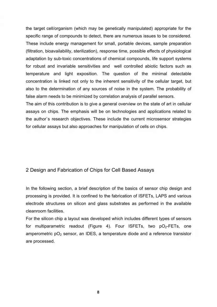
Figure 11: A measurement sequence reflecting a cyclic pattern of oxygen depletion due to cell respiration in a cell culture of LS 174 T cells, which has been grown to confluence directly on an amperometric sensor structure. During the stop intervals of the fluid systems, the cellular oxygen consumption results in decreasing current values. Addition of Triton X-100 disrupts cell membranes and cell respiration ceases. For evaluation of sensor raw data an algorithm simular to that used for ISFET evaluation (see Figure 10) was used.
Another method to analyse cellular oxygen consumption is based on the formation of
OH- ions during oxygen reduction and the corresponding pH shift (Sohn, 1996;
Lehmann, 2001). If the pH-sensitive gate area of an ISFET is surrounded by an
electrode, the ISFET will detect an increase of pH when the palladium- (or platinum-)
electrode catalyzes oxygen reduction at a cathodic potential (-750 mV vs. Ag/AgCl).
In order to prevent deterioration of electrocatalytic activity, intermittent anodic
potentials are applied. In a recent work (Ekelund, 2003) a cytosensor
microphysiometer was modified in order to measure cellular oxygen consumption
rates. However, the voltammetric measurement was not performed with planar
electrodes on the (LAPS-) silicon chip but with nafion-coated platinum wires inserted
into the plunger head of the microphysiometer. Oxygen determination based on
quenched luminescence was used by another group for a respirometric assay
(Alderman, 2004). Since this assay is performed in sealed microchambers with 3 µl
21

22
volume, the sensitivity is reported to exceed considerably the sensitivity of former
optical respiration assays.
4.1.3 Miscellaneous Metabolic Parameters A further detection concept for assaying cellular metabolism is directed to the
measurement of metabolic heat with micro-machined, planar calorimetric sensors.
The transducer principle is based on the Seebeck effect. The employed material
combinations (e.g. p+ polysilicon/aluminium) have a high Seebeck coefficient. The
sensitivity is further increased by the arrangement of a high number of thermocouples
in series to form a thermopile structure and by limiting the heat conduction between
thermopile junctions which is achieved by etching thin membraneous conductors. If a
cell culture is grown on one side, metabolic heat will cause a temperature difference
and thus a measurable voltage difference (Verhaegen, 1999; Johannessen, 2002).
Obviously there are other approaches to employ (amperometric) sensors for central
metabolic species such as glucose for cell based assays (von Woedtke, 2002). Such
efforts resulted in the fabrication of a microfluidic system with an integrated
microbioreactor, which is reported to yield glucose consumption and lactate release
rates of hepatocyte cell lines. For detection, a fiber optic equipment was connected to
the chip and a NAD+-coupled assay was performed (Schulz, 2002). Multianalyte
sensor systems with planar electrochemical glucose and lactate detection have also
been developed, which could be employed in vitro for cell based assays (Petrou,
2003).
4.2. Cell Morphology Further critical parameters in cell culture that can be assessed with sensors on chips
are the number and the growth of viable cells as well as changes in cell adhesion or
cell morphology. In the past, impedance measurements proved to be well applicable
for the analysis of a variety of such parameters, including cell growth, cell attachment
and spreading, cell motility and cell migration, barrier function of confluent layers,
cell-substratum spacing and cell death (Giaever, 1993; Ehret, 1997; Ehret, 1998,

Wegener, 2000 a; Jager, 2002; Luong, 2001; Arndt, 2004; Yang, 2004). In a recent
report it was even shown, that a glucose sensing device with a rather linear response
up to 14 mM glucose could be realized based on an impedance sensor structure
grown with fibroblasts (Tlili, 2003). In all these configurations, adherent cells were
cultured directly on electrode structures and a low-amplitude alternating current in a
frequency range of about 10-20 kHz was applied. The measured electric impedance
is the ratio of a sinusoidal voltage applied to a pair of electrodes to the sinusoidal
component of the current. Unless the system is purely resistive, the impedance is a
complex value, because the current and voltage have different phase angles. The
cytoplasmic membranes of living cells are effective insulators for alternating current
at the frequency used.
The measured impedance values reflect the process of cell spreading/cell adhesion
and subtle rearrangements of the cytoskeleton, which is linked to cell-cell and cell-
matrix junctions. For example, changes in intracellular calcium ion concentration are
known to alter the structure of the cell cytoskeleton, resulting in morphological
changes that can be detected by impedance sensors. Figure 13 illustrates the
experimental setup while Figure 14 shows an exemplary experiment with the
recordings of two sensors (interdigitated electrode structures, IDES) on the same
glass chip, detecting the morphological response of a confluent monolayer of Hela
cells to stimulation of the histamine (H1-) receptor.
Figure 12: Geometry of IDES (left side) and IDES-based impedance measurement on adherent cell cultures (right side). The insulating properties of cell membranes increase the measured electric impedance in the frequency
23

range used (10 kHz). If the cell morphology changes upon drug stimulation, this change is usually reflected by a change in impedance.
Figure 13: Effect of histamine on the capacitive component of the IDES-impedance. The glass chip has two identical IDES, both were grown with a confluent monolayer of Hela tumor cells. Although no distinct morphological alteration can be detected with the microscope, a reversible effect occurs, possibly on cell adhesion. The insert diagram shows the biphasic response of impedance in detail. Histamine does not cause measurable effects neither on extracellular acidification nor on cell respiration (data not shown). At the end of the experiment the cells were killed with 0,1% Triton X-100.
As the most simple equivalent model for the system composed of electrode, cell layer
and cell culture medium a circuit with a resistor and a capacitor in parallel can be
selected. The results can be represented either as a pair of Capacitance Cpar and
Resistance Rpar or as a pair of the absolute value of the impedance ΙZΙ and the phase
angle. Although refined equivalent circuits of the cell-electrode interface can be
designed, the value of such models for the interpretation of measured impedance
data is limited and depends on additional experimental information (Wegener, 1999).
A completely different transducer type which has been used for the detection of
morphological changes of adherent cells is a piezoelectric quartz crystal
microbalance: Perturbations of the cytoskeleton of endothelial cell layers are reported
24

25
to cause a shift in resonance frequency and resonance admittance (Wegener, 2000
b; Marx, 2001).
4.3 Electrical Patterns Inspirated by the pioneering work of G. Gross (Gross, 1979), several groups started
to use multi-electrode arrays on planar substrates for the multi-site analysis of activity
patterns of explanted networks of neuron cells or muscle cell cultures (Pine, 1980;
Hämmerle, 1994; Hofer, 1994; Hämmerle, 1994; Blau 1997; Bove, 1998; Stenger,
2001; Stett, 2003 a). For the first time it was feasible to address experimentally
questions such as how the nature of a neuron`s connections with its neighbours
affects its ability to generate action potentials. Arrays with different size, spacing and
geometry of electrodes are meanwhile offered commercially (even arrays with three-
dimensional, protruding electrodes are available). The arrays are produced on glass
using varying photolithographic techniques. Cultures of explanted primary cells as
well as whole brain tissue slices (Egert, 2002) can be placed on the arrays to monitor
spontaneous - or microelectrically stimulated - electrophysiological activity with
remarkable sensitivity. Sampling rates of at least 10 kHz are necessary for a
sufficient time resolution. However, the network of connections between
neighbouring neurons is typically so complex, that it is difficult to assign the inputs of
any given cell. In order to overcome this problem, attempts are made to guide
neurons on the chip surface by patterning organic compounds attracting or repelling
the cells (Stenger, 1998). Microelectrode arrays were also utilized to set up a
differential cell-based biosensor with both genetically engineered and wild-type
mammalian cells of the same type (Aravantis, 2001). Starting with the work of
Fromherz, who succeeded to record electrical activity of a leech neuron by placing it
on top of a silicon field effect transistor (Fromherz, 1991), the use of ISFETs for
electropyhsiological measurements is now emerging (Offenhäuser, 1997;
Offenhäuser, 2001; Besl, 2002). The noise level of measurements, however, seems
to be consistently higher as compared to microelectrodes. More recently, the
fabrication of an array of 16384 sensors on a single silicon chip was published,
allowing records of cellular electric activity with high spatio-temporal resolution
(www.infineon.com/bioscience).
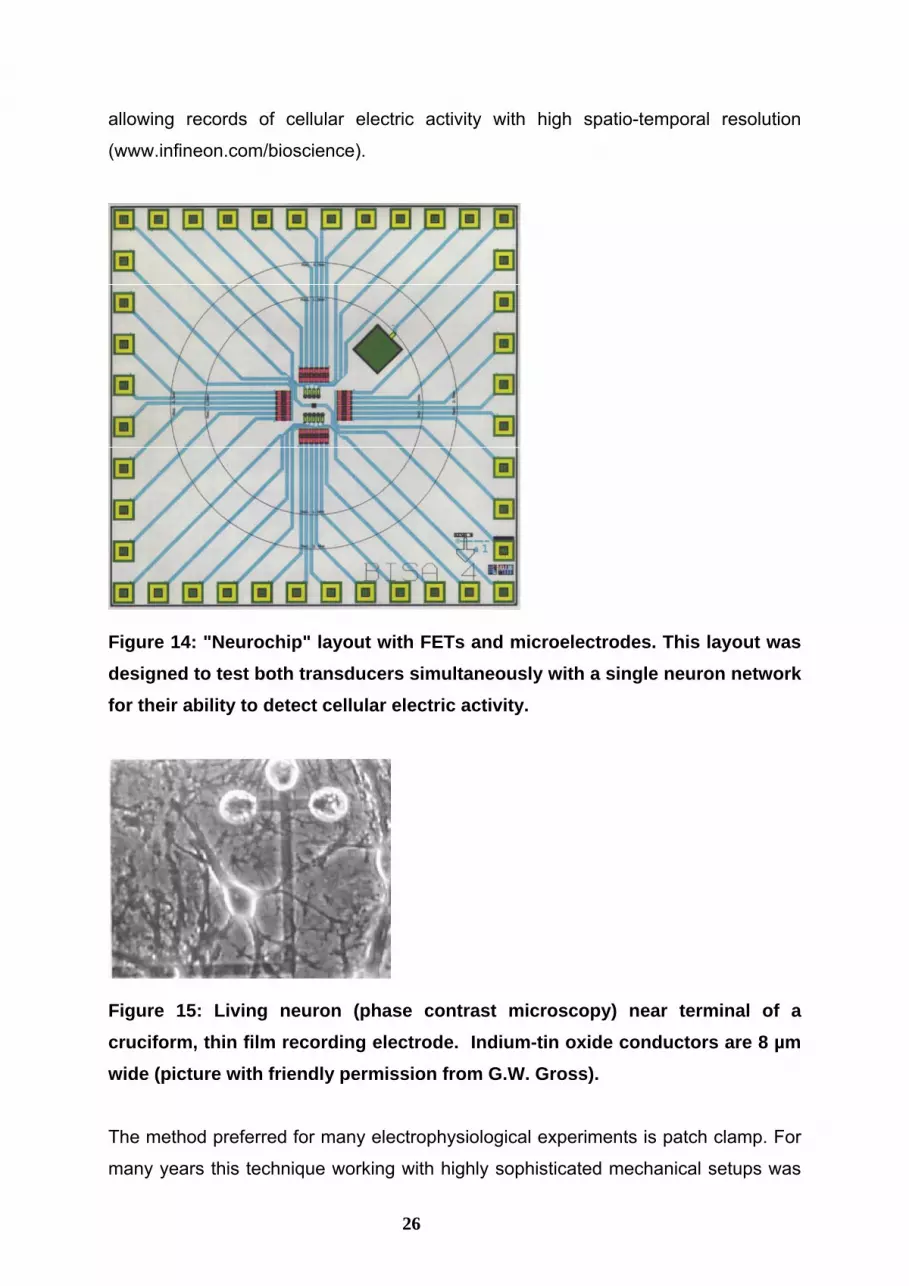
Figure 14: "Neurochip" layout with FETs and microelectrodes. This layout was designed to test both transducers simultaneously with a single neuron network for their ability to detect cellular electric activity.
Figure 15: Living neuron (phase contrast microscopy) near terminal of a cruciform, thin film recording electrode. Indium-tin oxide conductors are 8 µm wide (picture with friendly permission from G.W. Gross).
The method preferred for many electrophysiological experiments is patch clamp. For
many years this technique working with highly sophisticated mechanical setups was
26

27
left up to trained personal. Recently, the invention of planar glass chips (patch clamp
chips) with µm-apertures paved the way for a simplified instrumentation, for
automation and thus for considerable increases of experimental throughput in
electrophysiological screening (Fertig, 2002; Stett, 2003 b; Brueggemann, 2004).
Cells are drawn to the small apertures in glass chips (formed e.g. by laser ablation)
by applying a controlled negative pressure from beneath the glass. This is followed
by the development of a very high shunt resistance ("gigaseal"), the prerequisite for
successfull measurements. Using a more advanced and costly technology,
micronozzles can be structured in silicon substrates for the immobilisation and
electrical characterisation of cells (Lehnert, 2002). The focus of patch-clamp
technology is the characterisation of ion channels. Malfunctions of ion channels are
involved in the molecular pathophysiology of many diseases. These channels are,
therefore, most important targets of pharmaceutical drug screening.

28
Parameter Technology Examples, selected References
ISFETs (1) Baumann W H et al (1999): Microelectronic sensor system for microphysiological application on living cells. Sens Act B 55, 77-89 (2) Martinoia S, Rosso N, Grattarola M, Lorenzelli L, Margesin B, Zen M (2001) Development of ISFET array-based microsystems for bioelectrochemical measurements of cell populations. Biosens Bioelectron 16:1043-50
LAPS (1) McConnell, H. M.; Owicki, J. C.; Parce, J. W.; Miller, D. L.; Baxter, G. T.; Wada, H. G.; Pitchford, S. (1992): The cytosensor microphysiometer: biological applications of silicon technology. Science 257, 1906-12 (2) Metzger R et al (2001): Towards in-vitro prediction of an in-vivo cytostatic response of human tumor cells with a fast chemosensitivity assay. Toxicology 166, 97-108
extracellular acidification
optic sensors (1) Erxleben, H.A., Manion, M.K., Hockenbery, D.M., Scampavia, L., Ruzicka, J. (2004): A novel approach for monitoring extracellular acidification rates: based on bead injection spectrophotometry and the lab-on-valve system. The Analyst 129, 205-12
amperometric sensors
(1) Amano Y et al (1998): Measuring Respiration of Cultured Cell with Oxygen Electrode as a Metabolic Indicator for Drug Screening. Hum Cell 12, 3-10 (2) Brischwein, M., Motrescu, E.R., Otto, A.M., Cabala, E., Grothe, H., Wolf, B. (2003) Functional Cellular Assays with Multiparametric Silicon Sensor Chips. Lab on a Chip 3, 234-240
oxygen consumption
optic sensors
(1) Alderman, J., Hynes, J., Floyd, S.M., Krüger, J., O`Connor, R., Papkovsky, D.B. (2004): A low-volume platform for cell-respirometric screening based on quenched-luminescence oxygen sensing. Biosensors and Bioelectronics 19, 1529-35
glucose/ lactate exchange
optic sensors (1) Schulz, C.M., Scampavia, L., Ruzicka, J. (2002): Real-time monitoring of lactate extrusion and glucose consumption of cultured cells using a lab-on-valve system. The Analyst 127, 1583-88
changes in cellular morphology
electric impedance sensors
(1) Ehret R et al (1997): Monitoring of cellular behaviour by impedance measurements on interdigitated electrode structures. Biosensors & Bioelectronics 12, 29-41 (2) Wegener, J et al (2000): Electric Cell-Substrate Impedance Sensing (ECIS) as a Noninvasive Means to Monitor the Kinetics of Cell Spreading to Artificial Surfaces. Exp Cell Res 259, 158-166
patch-clamp chips (1) Fertig, N. et al (2002): Whole Cell Patch Clamp Recording Performed on a Planar Glass Chip. Biophysical Journal 82, 3056-62 (2) Stett, A.; Bucher, V; Burkhardt, C.; Weber, U; Nisch, W. (2003): Patch-clamping of primary cardiac cells with micro-openings in polyimide films. Medical & Biological Engineering & Computing, 41, 233-240
microelectrode arrays
(1) Gross, GW et al (1995): The use of neuronal netwoks on multielectrode arrays as biosensors. Biosensors & Bioelectronics 10, 553-567 (2) Egert, U et al. (1998): A novel organotypic long-term culture of the rat hippocampus on substrate-integrated multielectrode arrays. Brain Res Prot 2, 229-242
electric activity
FET arrays (1) Ingebrandt, S et al. (2001): Cardiomyocyte-transistor-hybrids for sensor application. Biosensors & Bioelectronics 16, 565-570 (2) www.infineon.com/bioscience (3) Stepper, C., Wolf, B., Wiest, J., Loeser, M., Brischwein, M., Grothe, H., Hansch, W., Schmitt-Landsiedel, D.: Technological Pre-Investigations on the Realization of a Local Resolution Microphysiologic Cell Chip for Medical Diagnostics and Pharmaceutical Screening. Proceedings of Sensor 2003 11th International Conference, 13.-15. May 2003, Nürnberg, Germany. Part II, p. 335-338
Table 1: Current methods for cell-based assays on chips and a selection of relevant publications

29
5 Cell Manipulation on Chips Engineering advances in microfluidics have allowed the construction of versatile
devices transporting substances through microchannels of a glass or plastic chip by
electroosmosis, capillary forces or pressure. Both bacteria and eukaryotic cells have
been successfully transported in microfluidic systems (Weigl, 2003). The number of
publications describing different types of cell manipulations, including complex
multistep procedures, such as cell separation (Fiedler, 1998; Fu, 1999; Huang,
2002), cell fusion (Stromberg, 2001; Chiu, 2001), electroporation (Huang, 2003), cell
lysis (Gao, 2004, Chiu, 2001), and incubation in different media performed (Yang,
2002) in such devices is rapidly increasing. A recent review discusses the use of
microtechnologies as a very useful tool for cell manipulation and analysis (Anderson,
2003). Microfluidic structures on chips for cell manipulations are frequently combined
with analytical functions in order to realize cellular assays. These assays include both
a chemical analysis of cell constituents after cell lysis (Schilling, 2002; McClain, 2003;
Gao, 2004) and the analysis of intact cells, the latter most often based on labeling
with specific fluorescent dyes and read out with a fluorescence detector (Matsubara,
2004; Yang, 2002).
For cell counting, the principle of cytometry, focussing cells into a single line for
individual analysis has been adapted to microfluidic devices by different groups (see
e.g. Gawad, 2001; McClain, 2001; Fu, 2002; Wolff, 2003). Commercially available
products are already utilized for cell staining and cell analysis on chip systems
(Buhlmann, 2003). Versatile microfabricated flow-cells as a basis for applications in
cell analysis have been described by the group of Vellekoop (Vellekoop, 2003).
Unlike the Coulter Counter principle, which relies on measuring the volume displaced
by passing cells, the polarisation response of cells passing through the channel of an
integrated microfluidic device in an electric field region was reported to give
information about the DNA content of the cell (Sohn, 2000).
A typical application of cell chips is the detection and quantification of viable bacteria
in aqueous solutions with electrode structures. In the present chip setup (Figure 16),
an interdigitated electrode structure is used both to accumulate the bacteria in their
vicinity using dielectrophoretic forces between the cells and the electrodes (prior to
the measurement) and to detect the bacteria by impedance measurement. The grade
of accumulation can be changed by the voltage applied to the sensor electrodes.
Figure 17 shows an accumulation of E. coli at an electrode structure at 2 V and 10

kHz. Varying the voltage causes a change in impedance, which depends also on the
concentration of the bacteria.
Figure 16: E.coli bacteria, stained with a fluorescent dye, are attracted to the electrodes by dielectrophoretic forces. Typical values for voltage and frequency are 1-2 Volts and 10-100 kHz.
There are numerous examples for the use of dielectrophoretic forces in lab-on-a-chip
devices for cell separations and cell trapping (Gray, 2004; Gambari, 2003). Using
various types of electric fields, it is possible to move, separate, fuse, perforate, or
deform cells. Such methods are broadly applied in biotechnology. In contrast to the
effects of weak electric and electromagnetic fields, in these techniques rather strong
fields are used, with an energy input significantly larger than the energy of thermic
noise. For example, dielectrophoretic field cages can be created with electrode
structures for handling of single cells on chips. In addition, such field cages are useful
for a dielectrical characterisation of cells by electrorotation (Gimsa, 2001). To avoid
various drawbacks associated with dielectrophoresis (such as unwanted
electrochemical effects), "electrodeless" devices have been described (Chou, C,
2003; Cummings 2003), creating a constriction and thus a high gradient of an electric
field in a conductive solution. Another non-contact method for cell manipulation is
based on superimposed magnetic AC-fields, acting on cells labelled with magnetic
30

31
nanoparticles and independent of any material constants of the extracellular liquid
(Koch, 2004).
For many applications, the use of mathematical methods is necessary in order to
obtain optimal electrode geometries. For example, a glass chip was constructed with
a novel sensor structure for measuring the concentration of bacteria in an aqueous
solution using dielectrophoretic forces. The four sensor electrodes have a sandwich
structure, with two similarly designed electrode layers separated by an isolating layer.
In each layer two similarly modelled electrodes are arranged in two insulated layers.
The electrodes of one single layer are active for the collection mode of the sensor. In
the measuring mode, two of the back-to-back lying electrodes are used to measure
the concentration-dependent impedance. The collecting property can be achieved by
a combination of high electric field strength and a large field gradient. A minimal
capacity is necessary for a high sensitivity of the sensor. The electrode structure was
therefore optimized by three-dimensional numerical field calculation. With this
program the dielectric properties of the glass substrate, the electrodes, and the
aqueous solution were simulated. The result of optimization was an electrode
arrangement consisting of one even and two zigzag electrodes (Figure 17).

Figure 17: Electric field distribution in a distance of 20µm above the electrode surface. These data were used to optimize the electrode geometry.
6 Conclusions and Future Prospects
Micro- and nanotechnologies are rapidly expanding into biomedical applications.
Among these applications, live cell studies are becoming increasingly important not
only in pharmaceutical drug discovery, but also in clinical diagnostics and monitoring
of environmental toxicants and pathogens. There is a considerable demand for
practical and low-cost tools for cell studies in all these areas. Cells are the minimum
functional and communicating unit of any living system and the ultimate target of any
drug. Although cellular signal amplification mechanisms often result in impressive
cellular responses, such "output signals" are not easily detected with conventional
32

33
analytical methods, at least in small cell numbers. A substantial aim of micro- and
nanotechnology is to integrate the "microsystem" of the living cell with technical
microsystems providing the necessary physiological environment, the microfluidic
systems for controlled supply of media and drug solutions, and transducers for a
sensitive recording of subtle changes in cellular behavior. If not only a short-term cell
culture is involved, it will be important for the lab-on-a-chip system to create an in-
vitro cellular microenvironment as close as possible to in-vivo conditions. Groups
working with cell-based assays are frequently facing problems with a gradual cell
culture deterioration in the time course of the studies. Therefore, conditions including
the protection of cell culture media from evaporation, adjustment of the gas
composition and measures against contamination must be guaranteed. The structural
versatility of microfluidic systems should help to approach this demand. Chip devices
integrating microbioreactors, microfluidics and transducer functions are expected to
emerge within the next few years.
Research and development in cell based assays strongly focusses on detection
principles based on fluorescence optics. This is due to a steady improvement of live
cell stains directed to a great variety of cellular targets and goes along with a similar
progress in instrumental technology adapted to a very versatile readout of
fluorescence parameters. On the other hand, the equipment for optical detection
(optic fibers, microscope lenses) sometimes appears to attenuate the advantages of
small and cheap cell chips. Compromises are obviously necessary. Moreover,
photobleaching and (photo-)toxic properties inherent to virtually any dye counteract a
monitoring of cells on a time scale longer than a few minutes.
With microelectric transducers, special attention has to be paid to a practical solution
for electric contacting and to a condensation and pre-processing of the generated
data, particularly with respect to multiparametric sensor chips and high-densitity
plates. To achieve this aim, substantial technological efforts in chip processing are
necessary: One of the most important benefits of CMOS technology is the possibility
of on-chip circuitry for signal amplification, data analysis and sensor self-testing. On-
chip sensor multiplexing is a precondition for the construction of 96- or 384- multiwell-
arrays since the number of necessary electric connections becomes unmanagable.
Although single-parameter assays are reasonable for many purposes, it would
increase the efficacy of cell studies if it were not necessary to combine data from
different assays which might have been performed under slightly different

34
experimental conditions. Multiparametric cell chips would help to reveal various
aspects of cellular events within a single assay, and they would do so in a dynamic
and real-time mode. With glass chips, the combination of electric sensors with optic
sensors, e.g. for pH and oxygen is obvious. If required, even high-resolution light
microscopy providing imaging information can be combined with on-line sensor
monitoring of the cells.
Although miniaturization of cell chips has its advantages by saving valuable (primary)
cells and drug compounds and although the size of single sensor elements on chips
can be as small as about 100 µm2 (e.g. the sensitive gate area of a pH-ISFET or a
microelectrode for the extracellular detection of action potentials), it is mostly not
intended to analyse single cells with electric transducers. It should be emphasized,
that single-cell measurements which do not take into account the typical social
context of cells must sometimes be assessed critically. This fact is highlightened by
neuronal networks on microelectrode arrays, which begin to develop after
explantation and which can only be reasonably assessed as whole functional units.
Nevertheless, a direct analysis of single cells on chips which does not involve long-
term cell culturing is possible and even a direct chemical analysis of single cells (e.g.
cell lysis followed by PCR) is expected to emerge in the next years (Anderson, 2004).
In metabolic assays, efforts dedicated to metabolic imaging using highly integrated
two dimensional chips arrays are only in their beginnings. Generally, the strength of
metabolic (and morphologic) assays is their unspecificity, allowing to detect a great
variety of cellular responses. This is because cell metabolism and cell morphology
itself are closely coupled to the signaling apparatus and can thus be regarded as
first-step signal transducers. For the interpretation of results, a multiparametric and
kinetic analysis may provide first indications on a drug`s mode of action. For
interpretations on a molecular level, methods such as the comparison of different,
transformed cell lines or the use of specifically acting biochemical inhibitors or
antibodies have to be applied. Thus, it is likely that cell-chip systems will find their
place in the screening of pharmaceudatical agents for activity or toxicity, as an
additional source of information about cellular behavior. An example for a
multiparametric record of tumor cells treated with a cytotoxic drug is given in Figure
18.

Figure 18: Effect of chloroacetaldehyde (50 µM) on LS 174 T cell cultures (human colon adenocarcinoma cell line, two cultures have been run in parallel), monitored with pH-, oxygen and impedance sensors. The drug was added twice for one hour to test for reversibility of drug effects. A strong, but reversible effect on cell respiration is observed. Impedance measurements however, reveal that cell death is not the predominant effect (cell death would be accompanied by early morphologic changes and cell attachment, detectable with IDES). At the end of the experiment, the cells were killed with 0,1% Triton X-100.
References: Alderman, J., Hynes, J., Floyd, S.M., Krüger, J., O`Connor, R., Papkovsky, D.B. (2004): A low-volume
platform for cell-respirometric screening based on quenched-luminescence oxygen sensing. Biosensors and Bioelectronics 19, 1529-35
Amano, Y., Okumura, C., Yoshida, M., Katayama, H., Unten, S., Arai, J., Tagawa, T., Hoshina, S., Hashimoto, H., Ishikawa, H. (1999): Measuring Respiration of Cultured Cell with Oxygen Electrode as a Metabolic Indicator for Drug Screening. Human Cell 12, 3-10
Anderson, H., van den Berg, A. (2003): Microfluidic devices for cellomics: a review. Sensors and Actuators B 92, 315-25
35

36
Anderson, H., van den Berg, A. (2004): Microtechnologies and nanotechnologies for single-cell analysis. Current Opinion in Biotechnology 15, 44-49
Aravantis, A.M., DeBusschere, B.D., Chruscinski, A.J., Gilchrist, K.H., Kobilka, B.K., Kovacs, G.T.A. (2001): A genetically engineered cell-based biosensor for functional classification of agents. Biosensors and Bioelectronics 16, 571-77
Arndt, S., Seebach, J., Psathaki, K., Galla, H.-J., Wegener, J. (2004): Bioelectrical impedance assay to monitor changes in cell shape during apoptosis. Biosensors and Bioelectronics 19, 583-94
Baumann W, Lehmann M, Schwinde A, Ehret R, Brischwein M, Wolf B (1999) Microelectronic sensor system for microphysiological application on living cells. Sensors and Actuators B 55:77-89
Besl B, Fromherz P (2002): Transistor array with an organotypic brain slice: field potential records and synaptic currents, European Journal of Neuroscience 15:999-1005
Blau A, Ziegler C, Heyer M, Endres F, Schwitzgebel G, Matthies T, Stieglitz T, Meyer JU, Göpel W (1997): Characterization and optimization of microelectrode arrays for in vivo nerve signal recording and stimulation, Biosensors and Bioelectronics 12:883-92
Bloom FR, Price P, Lao G, Xia JL, Crowe JH, Battista JR, Helm RF, Slaughter S, Potts M (2001): Engineering mammalian cells for solid-state sensor applications. Biosensors and Bioelectronics 16:603-08
Bousse, L, Cohen, C., Nikiforov, T., Chow, A., Kopf-Sill, A.R., Dubrow, R., Parce, J.W. (2000): Electrokinetically Controlled Microfluidic Analysis Systems. Annuals Reviews of Biophysics and Biomolecular Structure 29, 155-181
Bove M, Martinoia S, Verreschi G, Guigliano M, Grattarola M (1998): Analysis of the signals generated by networks of neurons coupled to planar arrays of microtransducers in simulated experiments Biosensors and Bioelectronics 15:601-12
Brischwein, M., Motrescu, E.R., Otto, A.M., Cabala, E., Grothe, H., Wolf, B. (2003) Functional Cellular Assays with Multiparametric Silicon Sensor Chips. Lab Chip 3, 234-240
Brueggemann, A., George, M., Klau, M., Beckler, M., Steindl, J., Behrends, J.C. (2004): Ion Channel Drug Discovery and Research: The Automated Nano-Patch-Clamp Technology. Current Drug Discovery Technologies 1, 91-96
Buhlmann, C., Preckel, T., Chan, S., Luedke, G., Valer, M. (2003): A New Tool for Routine Testing of Cellular Protein Expression: Integration of Cell Staining and Analysis of Protein Expression on a Microfluidic Chip-Based System. Journal of Biomolecular Techniques 14, 119-27
Casciari JJ, Sotirchos SV, Sutherland RM (1992): Variations in tumor cell growth rates and metabolism with oxygen concentration, glucose concentration and extracellular pH. Journal of Cellular Physiology 151:386-94
Chiu, D.T. (2001): A microfluidics platform for cell fusion. Current Opinion in Chemical Biology 5, 609-12
Chou, C.-F., Zenhausern, F. (2003): Electrodeless Dielectrophoresis for Micro Total Analysis Systems. IEEE Engineering in Medicine and Biology Magazine 22, 62-67
Cooper, J.M. (1999): Toward electronic Petri Dishes and picolitre-scale single cell technologies. Trends in Biotechnology 17, 226-230
Cummings, E.B. (2003): Streaming Dielectrophoresis for Continuous-Flow Microfluidic Devices. IEEE Engineering in Medicine and Biology Magazine 22, 75-84
Curtis, A., Wilkinson, C. (2001): Nanotechniques and approaches in biotechnology. Trends in Biotechnology 19, 97-101
Davidson, M., Karlsson, M., Sinclair J., Scott, K., Orwar, O. (2002): Nanotubevesicle networks with functionalized membranes and interiors. Journal of the American Chemical Society 125, 374-78
DeBusschere, B.D., Kovacs, G.T.A. (2001): Portable cell-based biosensor system using integrated CMOS cell-cartridges. Biosensors and Bioelectronics 16, 543-56
Dumont, E.D., Pécasse, F., Maenhaut, C.(2001) Crosstalk and specificity in signalling. Are we crosstalking ourselves into general confusion? Cellular Signalling 13, 4457-463

37
Duport S, Millerin C, Muller D, Corrèges P (1999): A metallic multisite recording system designed for continuous long-term monitoring of electrophysiological activity in slice cultures, Biosensors and Bioelectronics 14:369-76
Egert, U., Heck, D., Aertsen, A. (2002): Two-dimensional monitoring of spiking networks in acute brain slices. Experimental Brain Research 142, 268-274
Ehret R, Baumann W, Brischwein M, Schwinde A, Stegbauer K, Wolf B (1997): Monitoring of cellular behaviour by impedance measurements on interdigitated electrode structures. Biosensors and Bioelectronics 12, 29-41
Ehret R, Baumann W, Brischwein M, Schwinde, A., Wolf, B. (1998): On-line control of cellular adhesion with impedance measurements using interdigitated electrode structures. Medical & Biological Engineering & Computing 36, 365-70
Ekelund, S., Nygren, P, Larsson, R. (1998): Microphysiolmetry: new technology for evaluation of anticancer drug activity in human tumor cells in vitro. Anti-Cancer Drugs 9, 531-38
Ekelund, S., Cliffel, D.E., Kozlov, E., Prokop, A., Wikswo, J., Baudenbacher, F. (2003): Modification of the CytosensorTM microphysiometer to simultaneously measure extracellular acidification and oxygen consumption rates. Analytica Chimica Acta 496, 93-101
Erxleben, H.A., Manion, M.K., Hockenbery, D.M., Scampavia, L., Ruzicka, J. (2004): A novel approach for monitoring extracellular acidification rates: based on bead injection spectrophotometry and the lab-on-valve system. The Analyst 129, 205-12
Fanigliulo A, Accossato P, Adami M, Lanzi M, Martinoia S, Paddeu S, Parodi MT, Rossi A, Sartore M, Grattarola M, Nicolini C (1996): Comparison between a LAPS and a FET-based sensor for cell-metabolism detection. Sensors and Actuators B 32:41-48
Farinas, J., Chow, A.W., Wada, H.G. (2001): A Microfluidic Device for Measuring Cellular Membrane Potential. Analytical Biochemistry 295, 138-42
Fertig, N., Blick, R.H., Behrends, J.C. (2002): Whole cell patch clamp recording performed on a planar glass chip. Biophysical Journal 82, 3056-62
Fiedler, S., Shirley, S.G., Schnelle, T., Fuhr, G. (1998): Dielectrophoretic Sorting of Particles and Cells in a Microsystem. Analytical Chemistry 70, 1909-15
Fromherz P, Offenhäuser A, Vetter T, Weis J (1991): A neuron-silicon junction: a Retzius cell of the leech on an insulated-gate field-effect transistor, Science 252:1290-93
Fu, A.Y., Spence, C., Scherer, A., Arnold, F.H., Quake, S.R. (1999) Nature Biotechnology 17, 1109-11
Fu, A. Y., Chou, H., Spence, C., Arnold, F.H., Quake, S.R. (2002): An integrated microfabricated cell sorter. Analytical Chemistry 74, 2451-57
Gambari, R., Borgatti, M., Altomare, L., Manaresi, N., Medoro, G., Romani, A., Tartagni, M., Guerrieri, R. (2003): Applications to cancer research of "lab-on-a-chip" devices based on dielectrophoresis (DEP). Technology in Cancer Research & Treatment 2, 31-40
Gao, J., Yin, X., Fang, Z. (2004): Integration of single cell injection, cell lysis, separation and detection of intracellular constituents on a microfluidic chip. Lab Chip 4, 47-52
Gawad, S., Schild, L, Renaud, P. (2004): Micromachined impedance spectroscopy flow cytomyter for cell analysis and particle sizing. Lab Chip 4, 47-52
George M, Parak WJ, Gaub HE (2000): Highly integrated surface potential sensors. Sensors and Actuators B 69:266-75
Giaever I, Keese CR (1993): A morphological biosensor for mammalian cells. Nature 366, 591-92
Gimsa, J. (2001): A comprehensive approach to electro-orientation, electrodeformation, dielectro-phoresis, and electrorotation of ellipsoidal particles and biological cells, Bioelectrochemistry 54, 23-31
Gross, G.W. (1979): Simultaneous single unit recording in vitro with a photoetched deinsulated gold microelectrode surface. IEEE Transactions on Biomedical Engineering 26, 273-77
Gross GW, Kowalski JM (1991) In: Antognetti P, Milutinovic V (eds.) Neuronal Networks: Concepts, Applications and Implementations. Academic Press, New York, p 47

38
Gross, G.W., Schwalm, F.U. (1994): A closed flow chamber for long-term multichannel recording and optical monitoring. Journal of Neuroscience Methods 52, 73-85
Hafner, F (2000): Cytosensor Microphysiometer: technology and recent applications. Biosensors and Bioelectronics 15:149-58
Hämmerle H, Egert U, Mohr A, Nisch W (1994): Extracellular recording in neuronal networks with substrate integrated microelectrode arrays Biosensors and Bioelectronics 9, 691-6
Hartwell, L.H., Hopfield, J.J., Leibler, S., and Murray, A.W. (1999) From molecular to modular cell biology. Nature 402, C47-C52
Hediger, S., Sayah, A., Horisberger, J.D., Gijs, M.A.M. (2001): Modular microsystem for epithelial cell culture and electrical characterisation. Biosensors and Bioelectronics 16, 689-94
Hofer E, Urban G, Spach MS, Schafferhofer I, Mohr G, Platzer D (1994): Measuring activation patterns of the heart at a microscopic size scale with thin-film sensors, American Journal of Physiology 266:H2136-45
Huang, Y., Joo, S., Duhon, M., Heller, M., Wallace, B., Xu, X. (2002): Dielelctrophoretic cell separation and gene expression profiling on microelectronic chip arrays. Analytical Chemistry 74, 3362-71
Huang, Y., Sekhon, N., Borninsky, J., Chen, N., Rubinsky, B. (2003): Instantaneous, quantitative single-cell viability assessment by electrical evaluation of cell membrane integrity with microfabricated devices. Sensors and Actuators A 105, 31-39
Jager, E.W.H., Immerstrand, C., Peterson, K.H., Magnusson, K., Lundström, I., Inganäs, O. (2002): The Cell Clinic: Closable microvials for Single Cell Studies. Biomedical Microdevices 4, 177-87
Johannessen, E.A., Weaver, J.M.R., Bourova, L., Svoboda, P., Cobbold, P.H., Cooper, J. (2002): Micromachined Nanocalorimetric Sensor for Ultra-Low-Volume Cell-Based Assays. Analytical Chemistry 74, 2190-97
Koch, M., Weyh, T., Wolf, B. (2004): Dynamic Marker. Proceedings of the Fourth IEEE Conference on Nanotechnology, Munich, August 17-19 2004.
Koh, W, Itle, L.J., Pishko, M.V. (2003): Molding of hydrogel microstructures to create multiphenotype cell microarrays. Analytical Chemistry 75, 5783-89
Kraus M, Wolf B Structured Biological Modelling (1995) A New Approach to Biophysical Cell Biology. CRC Press, Inc., Boca Raton
Lähdesmäki, I., Ruzicka, J., Ivaska, A. (2000): Novel flow injection methods for drug-receptor interaction studies, based on probing cell metabolism. The Analyst 125, 1889-95
Lambrechts M, Sansen W (1992) Biosensors: Microelectrochemical Devices IOP Publishing Ltd., Bristol
Lehnert, T., Gijs, M.A.M., Netzer, R., Bischoff, U. (2002): Realization of SiO2 micronozzles for electrical measurements on living cells. Applied Physics Letters 81, 5063-65
Lehmann M, Baumann W, Brischwein M, Gahle HJ, Freund I, Ehret R, Drechsler S, Palzer H, Kleintges M, Sieben U, Wolf B (2001): Simultaneous measurement of cellular respiration and acidification with single CMOS ISFET Biosensors and Bioelectronics 16, 195-203
Lorenzelli, L., Margesin, B., Martinoia, S., Tedesco, M.T., Valle, M. (2003): Bioelectrochemical signal monitoring of in-vitro cultured cells by means of an automated microsystem based on solid state sensor-array. Biosensors and Bioelectronics 18, 621-26
Luong JH, Habibi-Rezaei M, Meghrous J, Xiao C, Male KB, Kamen A (2001): Monitoring Motility, Spreading, and Mortality of Adherent Insect Cells Using an Impedance Sensor. Analytical Chemistry 73, 1844-48
Martinoia S, Rosso N, Grattarola M, Lorenzelli L, Margesin B, Zen M (2001) Development of ISFET array-based microsystems for bioelectrochemical measurements of cell populations. Biosensors and Bioelectronics 16:1043-50
Matsubara, Y., Murakami, Y., Kobayashi, M., Morita, Y., Tamiya, E. (2004): Application of on-chip cell cultures for the detection of allergic response. Biosensors and Bioelectronics 19, 741-47

39
Marx KA, Zhou T, Montrone A, Schulze H, Braunhut S (2001): A quartz crystal microbalance cell biosensor: detection of microtubule alterations in living cells at nM nocodazole concentrations Biosensors and Bioelectronics 16, 773-82
McClain, M.A., Culbertson, C.T., Jacobson, S.C., Allbritton, N.L., Sims, C.E., Ramsey, J.M. (2003): Microfluidic Devices for the High-Throughput Chemical Analysis of Cells. Analytical Chemistry 75, 5646-55
McClain, M.A., Culbertson, C.T., Jacobson, S.C., Ramsey, J.M. (2001): Flow Cytometry of Escherichia coli on Microfluidic Devices. Analytical Chemistry 73, 5334-38
Metzger, R., Deglmann, C.J., Hoerrlein, S., Zapf, S., Hilfrich, J. (2001): Towards in-vitro prediction of an in-vivo cytostatic response of human tumor cells with a fast chemosensitivity assay. Toxicology 166, 97-108
Offenhäuser A, Sprössler C, Matsuzawa M, Knoll W (1997): Field-effect transistor array for monitoring electrical activity from mammalian neurons in culture Biosensors and Bioelectronics 12, 819-26
Offenhäuser A, Knoll W (2001): Cell-transistor hybrid systems and their potential application. Trends in Biotechnology 19, 62-66
Otto, A.M. Brischwein, M., Niendorf, A., Henning, T., Wolf, B. (2003) Microphysiological testing for chemosensitivity of living tumor cells with multiparametric microsensor chips. Cancer Detection and Prevention 27, 291-96
Owicki,J.C. and Parce,J.W. (1992) Biosensors based on the energy metabolism of living cells: the physical chemistry and cell biology of extracellular acidification. Biosensors and Bioelectronics 7, 255-72
Owicki JC, Bousse LJ, Hafeman DG, Kirk GL, Olson JD, Wada HG, Parce JW (1994) The Light-Addressable Potentiometric Sensor: Principles and Biological Applications Annual Reviews of Biophysics and Biomolecular Structures 23, 87-114
Pancrazio, J.J., Gray, S., Shubin, Y.S., Kulagina, N., Cuttino, D.S., Shaffer, K.M., Eisemann, K., Curran, A., Zim, B., Gross, G.W., O`Shaughnessy, T.J. (2003): A portable microelectrode array recording system incorporating neuronal networks for neurotoxin detection. Biosensors and Bioelectronics 18, 1339-47
Park, T.H., Shuler, M.L. (2003): Integration of Cell Culture and Microfabrication Technology. Biotechnology Progress 19, 243-53
Peterson A, Walum E (1985): Use of a perfusion technique for measurements of respiratory activity in cultured cells. In vitro Cellular and Developmental Biology 21, 622-26
Petrou, P.S., Moser, I., Jobst, G. (2003): Microdevice with integrated dialysis probe and biosensor array for continuous multi-analyte monitoring. Biosensors and Bioelectronics 18, 613-19
Pine, J. (1980): Recording action potentials from cultured neurons with extracellular microcircuit electrodes. Journal of Neuroscience Methods 2, 19-28
Rädler, U.: The µ-slide for live cell imaging - combining cell biology with optical microscopy. American Biotechnology Laboratory 2004, accepted
J. Ressler, H. Grothe, E.R. Motrescu, B. Wolf: New concepts for chip-supported multi-well-plates: Realization of a 24-well-plate with integrated impedance-sensors for functional cellular screening applications and automated microscope aided cell-based assays, Proceedings of the 26th Annual International Conference of the IEEE EMBS, SanFrancisco, 2004, pp. 2074-2077
Rudolph, A.S., Reasor, J. (2001): Cell and tissue based technologies for environmental detection and medical diagnostics. Biosensors and Bioelectronics 16, 429-31
Schilling, E. A. Kamholz, A. E., Yager, P. (2002): Cell Lysis and Protein Extraction in a Microfluidic Device with Detection by a Fluorogenic Enzyme Assay. Analytical Chemistry 74, 1798-1804
Schlage WK, Bereiter-Hahn J (1983): A microscope perfusion respirometer for continuous respiration measurement of cultured cells during microscopic observation Microscopica Acta 87, 19-34

40
Schubnell D, Lehmann M, Baumann W, Rott FG, Wolf B, Beck CF (1999): An ISFET-algal (Chlamydomonas) hybrid provides a system for eco-toxicological tests. Biosensors and Bioelectronics 14, 465-73
Schulz, C.M., Scampavia, L., Ruzicka, J. (2002): Real-time monitoring of lactate extrusion and glucose consumption of cultured cells using a lab-on-valve system. The Analyst 127, 1583-88
Selinger, J.V., Pancrazio, J.J., Gross, G.W. (2004): Measuring synchronisation in neuronal networks for biosensor applications. Biosensors and Bioelectronics 19, 675-83
Sohn B, Kim C (1996): A new pH-ISFET based dissolved oxygen sensor by employing electrolysis of oxygen. Sensors and Actuators B 34, 435-40
Sohn, L.L., Saleh, O.A., Facer, G.R., Beavis, A.J., Allan, R.S., Notterman, D.A. (2000): Capacitance cytometry: measuring biological cells one by one. Proceedings of the National Academy of Sciences of the United States of America 97, 10687-90
Stenger DA, Hickman JJ, Bateman KE, Ravenscroft MS, Ma W, Pancrazio JJ, Shaffer K, Schaffner AE, Cribbs DH, Cotman CW (1998): Microlithographic determination of axonal/dendritic polarity in cultured hippocampal neurons Journal of Neuroscience Methods 82, 167-73
Stenger, D.A., Gross, G.W., Keefer, E.W., Shaffer, K.M., Andreadis, J.D., Ma, W., Pancrazio, J.J. (2001): Detection of physiologically active compounds using cell-based biosensors. Trends in Biotechnology 19, 304-09
Stepper, C., Wolf, B., Wiest, J., Loeser, M., Brischwein, M., Grothe, H., Hansch, W., Schmitt-Landsiedel, D.: Technological Pre-Investigations on the Realization of a Local Resolution Microphysiologic Cell Chip for Medical Diagnostics and Pharmaceutical Screening. Proceedings of Sensor 2003 11th International Conference, 13.-15. May 2003, Nürnberg, Germany. Part II, p. 335-338
Stett, A., Egert, U., Guenther, E., Hofmann, F., Meyer, T., Nisch, W., Haemmerle, H. (2003): Biological application of microelectrode arrays in drug discovery and basic research. Analytical and Bioanalytical Chemistry 377, 486-95
Stett, A.; Bucher, V; Burkhardt, C.; Weber, U; Nisch, W. (2003): Patch-clamping of primary cardiac cells with micro-openings in polyimide films. Medical & Biological Engineering & Computing, 41, 233-40
Stromberg, A., Karlsson, A., Ryttsen, F., Davidson, M., Chiu, D.T., Orwar, O. (2001): Microfluidic Device for Combinatorial Fusion of Liposomes and Cells. Analytical Chemistry 73, 126-30
Taylor, D.L., Woo, E.S., Giuliano, K.A. (2001): Real-time molecular and cellular analysis: the new frontier of drug discovery. Current Opinion in Biotechnology 12, 75-81
Tlili, C., Reybier, K., Geloen, A. Ponsonnet, L., Martelet, C., Ben Ouada, H., Lagarde, M., Jaffrezic-Renault, N. (2003): Fibroblast Cells: A Sensing Bioelement for Glucose Detection by Impedance Spectroscopy. Analytical Chemistry 75, 3340-44
Nieuwenhuis, J.H., Bastemeijer, J., Sarro, P.M., Vellekoop, M.J. (2003): Integrated flow-cells for novel adjustable sheath flows. Lab Chip 3, 56-61
Verhaegen, K., Simaels, J., Van Driessche, W., Baert, K., Sansen, W., Puers, B., Hermans, L., Mertens, R. (1999): A Biomedical Microphysiometer. Biomedical Microdevices 2, 93-98;
von Woedtke, T., Jülich, W.D., Alhitari, N., Hanschke, R., Abel, P.U. (2002): Biosensor-controlled perfusion culture to estimate the viability of cells. Medical & Biological Engineering & Computing 40, 704-11
Walker, G.M., Zeringue, H.C., Beebe, D.J. (2004): Microenvironment design considerations for cellular scale studies. Lab Chip 4, 91-97
Weigl, B.H., Bardell, R.L., Cabrera, C.R. (2003): Lab-on-a-chip for drug development. Advanced Drug Delivery Reviews 55, 349-77
Wheeler, A.R., Throndset, W.R., Whelan, R.J., Leach, A.M., Zare, R.N., Liao, Y, Farrell, K. Manger, I.D., Daridon, A. (2003): Microfluidic Device for Single-Cell Analysis. Analytical Chemistry 75, 3581-86

41
Wegener J, Zink S, Rösen P, Galla HJ (1999): Use of electrochemical impedance measurements to monitor β-adrenergic stimulation of bovine aortic endothelial cells. European Journal of Physiology 437, 925-34
Wegener J, Keese CR, Giaever I (2000): Electric Cell–Substrate Impedance Sensing (ECIS) as a Noninvasive Means to Monitor the Kinetics of Cell Spreading to Artificial Surfaces. Experimental Cell Research 259, 158-66
Wegener J, Seebach J, Janshoff A, Galla HJ (2000): Analysis of the Composite Response of Shear Wave Resonators to the Attachment of Mammalian Cells Biophysical Journal 78, 2821-33
Wolf, B.W., Brischwein, M., Baumann, W., Ehret, R., Kraus, M. (1998): Monitoring of cellular signalling and metabolism with modular sensor-technique: The PhysioControl-Microsystem (PCM). Biosensors and Bioelectronics 13, 501-09
Wolff, A., Perch-Nielsen, I.R., Larsen, U.D., Friis, P., Goranovic, g., Poulsen, C.R., Kutter, J.P., Telleman, P. (2003): Integrating advanced functionality in a microfabricated high-throughput fluorescent-activated cell sorter. Lab Chip 3, 22-27
Yang, M., Li, C., Yang, J. (2002): Cell Docking and On-Chip Monitoring of Cellular Reactions with a Controlled Concentration Gradient on a Microfluidic Device. Analytical Chemistry 74, 3991-4001
Yang, L., Li, Y., Griffis, C.L., Johnson, M.G. (2004): Interdigitated microelectrode (IME) impedance sensor for the detection of viable Salmonella typhimurium. Biosensors and Bioelectronics 19, 1139-47
www.infineon.com/bioscience