jasicon 2020 souvenier - asi jharkhand
TRANSCRIPT

Dates: Venue:th th 7 & 8 November 2020 Virtual Platform ASI Jharkhand Chapter|
th19 Annual Conference of Association of Surgeons of India Jharkhand Chapter
“The Renaissance of General Surgery”
E- Souvenir
HALL A
CLICK HERE TO LOGIN SCIENTIFICSCHEDULE
Visit website for updates www.asijharkhand.in
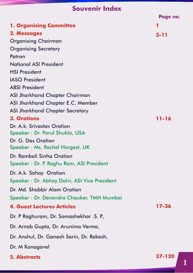
1. Organising Committee
2. Messages
3. Orations
4. Guest Lectures Articles
5. Abstracts
Organising Chairman
Organising Secretary
Patron
National ASI President
HSI President
IASO President
ABSI President
ASI Jharkhand Chapter Chairman
ASI Jharkhand Chapter E.C. Member
ASI Jharkhand Chapter Secretary
Dr. A.k. Srivastav Oration
Dr. G. Das Oration
Dr. Rambali Sinha Oration
Dr. A.k. Sahay Oration
Dr. Md. Shabbir Alam Oration
Dr. P Raghuram, Dr. Somashekhar .S. P.,
Dr. Arnab Gupta, Dr. Arunima Verma,
Dr. Anshul, Dr. Ganesh Sarin, Dr. Rakesh,
Dr. M Kanagavel
Speaker : Dr. Parul Shukla, USA
Speaker : Ms. Rachel Hargest, UK
Speaker : Dr. P. Raghu Ram, ASI President
Speaker : Dr. Abhay Dalvi, ASI Vice President
Speaker : Dr. Devendra Chauker, TMH Mumbai
1
3-11
11-16
17-36
37-120
Souvenir IndexPage no.
1
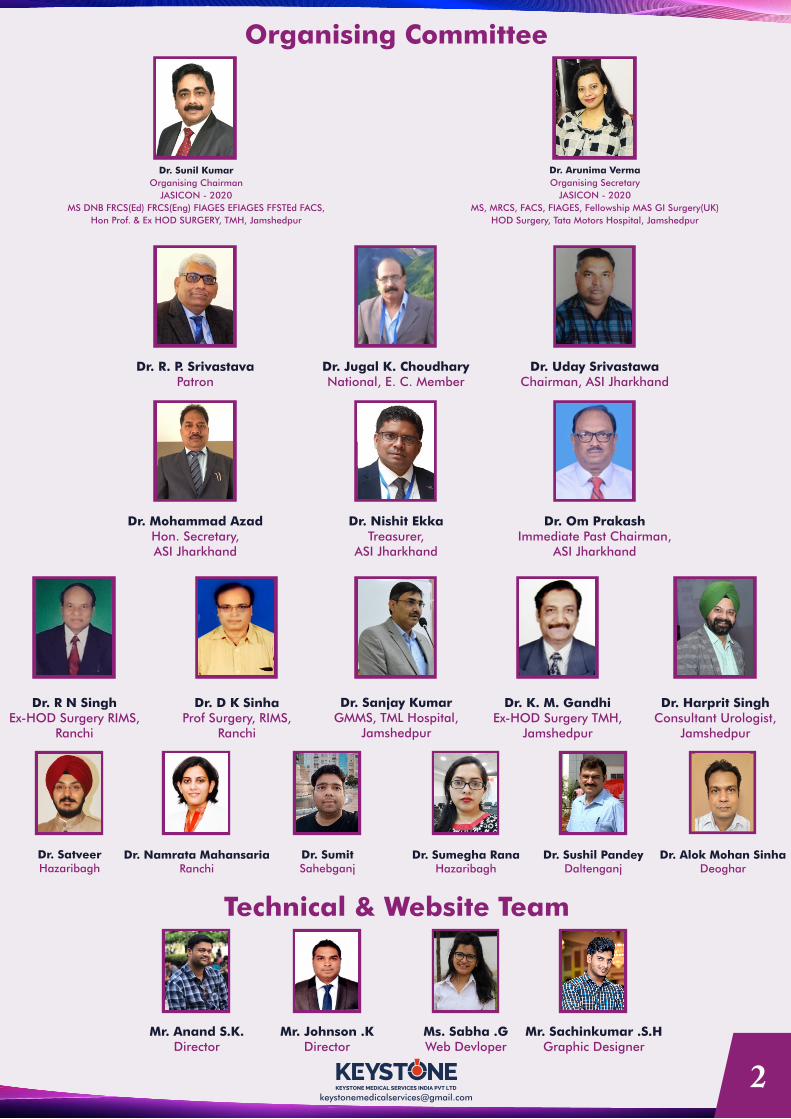
Organising Committee
Technical & Website Team
Dr. R. P. SrivastavaPatron
Dr. D K SinhaProf Surgery, RIMS,
Ranchi
Dr. Jugal K. ChoudharyNational, E. C. Member
Dr. Uday SrivastawaChairman, ASI Jharkhand
Dr. Mohammad AzadHon. Secretary,ASI Jharkhand
Mr. Anand S.K.Director
Dr. Nishit EkkaTreasurer,
ASI Jharkhand
Mr. Johnson .KDirector
Dr. Om PrakashImmediate Past Chairman,
ASI Jharkhand
Ms. Sabha .GWeb Devloper
Mr. Sachinkumar .S.HGraphic Designer
Dr. Sanjay KumarGMMS, TML Hospital,
Jamshedpur
Dr. R N SinghEx-HOD Surgery RIMS,
Ranchi
Dr. K. M. GandhiEx-HOD Surgery TMH,
Jamshedpur
Dr. Harprit SinghConsultant Urologist,
Jamshedpur
Dr. SatveerHazaribagh
Dr. Namrata MahansariaRanchi
Dr. SumitSahebganj
Dr. Sumegha RanaHazaribagh
Dr. Sushil PandeyDaltenganj
Dr. Alok Mohan SinhaDeoghar
KEYSTONE MEDICAL SERVICES INDIA PVT LTD
K YST NE
Dr. Sunil Kumar
Organising Chairman
JASICON - 2020
MS DNB FRCS(Ed) FRCS(Eng) FIAGES EFIAGES FFSTEd FACS,
Hon Prof. & Ex HOD SURGERY, TMH, Jamshedpur
Dr. Arunima Verma
Organising Secretary
JASICON - 2020
MS, MRCS, FACS, FIAGES, Fellowship MAS GI Surgery(UK)
HOD Surgery, Tata Motors Hospital, Jamshedpur
2
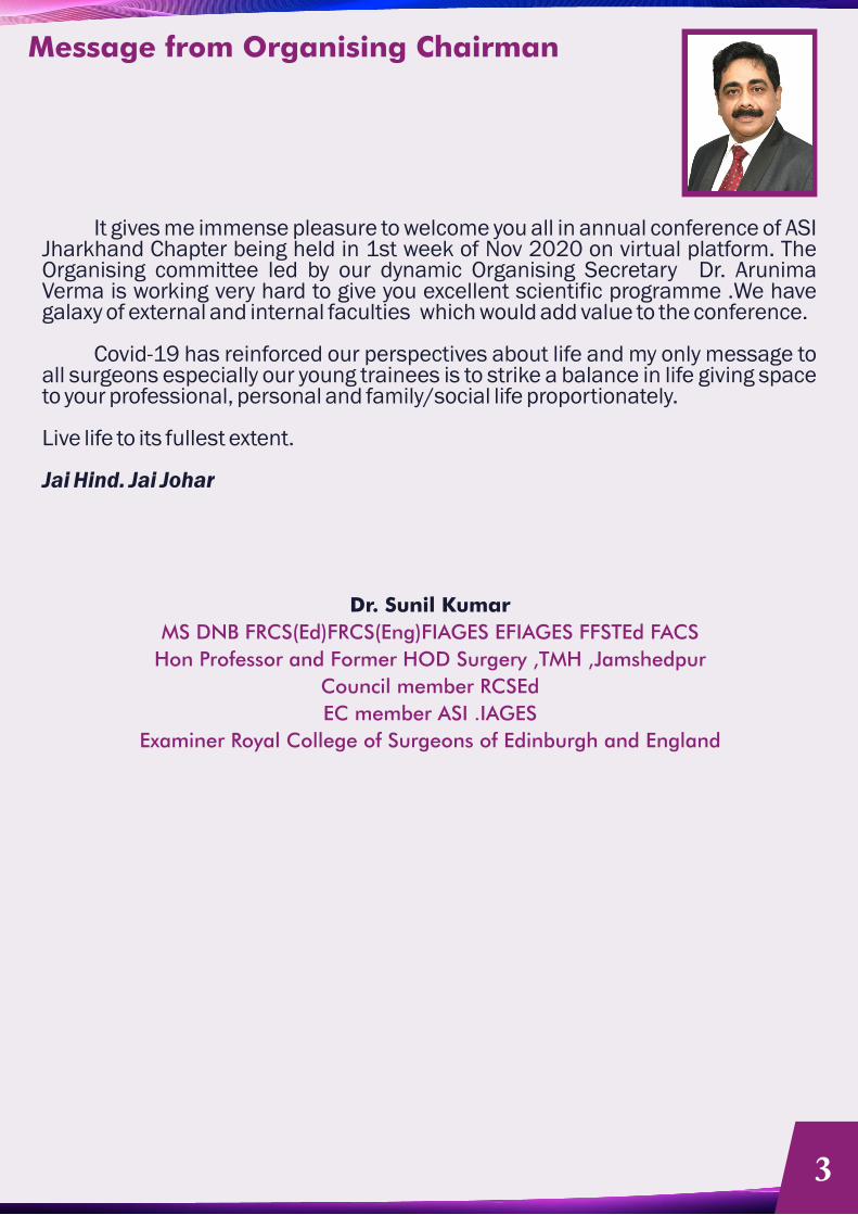
Message from Organising Chairman
It gives me immense pleasure to welcome you all in annual conference of ASI Jharkhand Chapter being held in 1st week of Nov 2020 on virtual platform. The Organising committee led by our dynamic Organising Secretary Dr. Arunima Verma is working very hard to give you excellent scientific programme .We have galaxy of external and internal faculties which would add value to the conference.
Covid-19 has reinforced our perspectives about life and my only message to all surgeons especially our young trainees is to strike a balance in life giving space to your professional, personal and family/social life proportionately.
Live life to its fullest extent.
Jai Hind. Jai Johar
Dr. Sunil Kumar
MS DNB FRCS(Ed)FRCS(Eng)FIAGES EFIAGES FFSTEd FACS
Hon Professor and Former HOD Surgery ,TMH ,Jamshedpur
Council member RCSEd
EC member ASI .IAGES
Examiner Royal College of Surgeons of Edinburgh and England
3
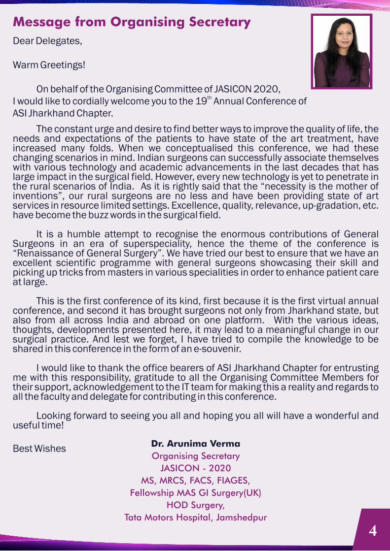
Message from Organising Secretary
Dear Delegates,
Warm Greetings!
On behalf of the Organising Committee of JASICON 2020, thI would like to cordially welcome you to the 19 Annual Conference of
ASI Jharkhand Chapter.
The constant urge and desire to find better ways to improve the quality of life, the needs and expectations of the patients to have state of the art treatment, have increased many folds. When we conceptualised this conference, we had these changing scenarios in mind. Indian surgeons can successfully associate themselves with various technology and academic advancements in the last decades that has large impact in the surgical field. However, every new technology is yet to penetrate in the rural scenarios of India. As it is rightly said that the “necessity is the mother of inventions”, our rural surgeons are no less and have been providing state of art services in resource limited settings. Excellence, quality, relevance, up-gradation, etc. have become the buzz words in the surgical field.
It is a humble attempt to recognise the enormous contributions of General Surgeons in an era of superspeciality, hence the theme of the conference is “Renaissance of General Surgery”. We have tried our best to ensure that we have an excellent scientific programme with general surgeons showcasing their skill and picking up tricks from masters in various specialities in order to enhance patient care at large.
This is the first conference of its kind, first because it is the first virtual annual conference, and second it has brought surgeons not only from Jharkhand state, but also from all across India and abroad on one platform. With the various ideas, thoughts, developments presented here, it may lead to a meaningful change in our surgical practice. And lest we forget, I have tried to compile the knowledge to be shared in this conference in the form of an e-souvenir.
I would like to thank the office bearers of ASI Jharkhand Chapter for entrusting me with this responsibility, gratitude to all the Organising Committee Members for their support, acknowledgement to the IT team for making this a reality and regards to all the faculty and delegate for contributing in this conference.
Looking forward to seeing you all and hoping you all will have a wonderful and useful time!
Best Wishes Dr. Arunima Verma
Organising Secretary
JASICON - 2020
MS, MRCS, FACS, FIAGES,
Fellowship MAS GI Surgery(UK)
HOD Surgery,
Tata Motors Hospital, Jamshedpur
4
Message from Patron
JOHAR !
A virtual congregation, an imagination of past comes to reality and JASICON 2020 organising committee makes it happen. Fittingly, it is Jamshedpur team of Dr Sunil and Dr Arunima along with support of well chosen teammates. The duo are showing worthy of representing the state in EC and IJS. It is indeed a treat for me to watch Jharkhand chapter scaling every challenge and reaching newer heights.
This congress is another milestone.
Well chosen faculty, orations, great mobilisation of enthusiastic participation of delegates reflect concerted efforts of state officials and organising team.
Watching every event from confines of your home will give the fellowship a new dimension as the family members too will be involved and will feel a sense of belonging.
I wish “JASICON 2020” a grand success.
Dr. R. P. Srivastava
Patron
5
Message from National ASI President
On behalf of our Association, nothing gives me more pleasure ththan to welcomeyou to the 19 Annual Conference of Jharkhand
Chapter of ASI, being held for the first time ever on a virtual platform.
Dr. P. Raghu Ram
MS, FRCS (Edin), FRCS (Eng), FRCS (Glasg), FRCS (Irel), Hon. FRCS (Thailand), FACS
Padma Shri awardee (2015)
Dr.B.C.Roy National awardee (2016)
President
The Association of Surgeons of India
I commend the Organising Committee very ably led by dear friends, Dr. Sunil Kumar & Dr Arunima Verma actively supported by dear colleagues Dr. Uday Srivastawa, Dr. Mohammad Azad, Dr Jugal Kishore Choudhary alongside the 'DYNAMIC TEAM JHARKHAND', whohave left no stone unturned to ensure that your “virtual” participation in the Congress is enjoyable and memorable – like never before!
Well over 70% of population reside in rural India and it is indeed the General Surgeon that caters to bulk of the surgical care needs of citizenry in the Country. Therefore, the Conference theme “Renaissance of General Surgery”,which brings to focus the“GENERAL SURGEON” is very thoughtful, apt & proper.
Over the past ten months, ASI has been on the forefront in championing a number of initiatives for the greater good of the membership, our trainees and to the community at large. With folded hands, most grateful to Jharkhand Chapter for your active contribution in this endeavor.
To me personally, recognising the “new normal”, quickly adapting to the “change” and startegially implementing VISION 2020 during the unprecedented COVID pandemic, have been life's powerful learning lessons. Despite the doom and gloom around us, I have been doing my very best towards ensuring Creative Leadership and Accountable Governance, which has been my “mantra” all along.
In closing, I am confident that the intense online academic activity of our Association being undertaken all over India throughout the year will seamlessly transition to ASICON 2020 – our flagship annual Conference, which would be a fitting finale to the “Year” that was…
Take care…Be Safe…
6

Message from HSI President
thI feel privileged and proud to be a part of JASICON 2020, the 19 Annual Conference of Association of Surgeons of India Jharkhand chapter. I really like the theme of the conference “The Renaissance of General Surgery”. The Scientific content of the conference is very rich. The faculties include the stalwarts of Indian and International surgery I have known Dr. Arunima Verma for more than a decade. She is very academic, a very good organiser and Surgeon par excellence. She has a very able Chairman in Dr. Sunil Kumar who needs no introduction to the surgical fraternity.
It is going to be a great meeting and the organising team led by Dr. Arunima and Dr. Sunil have worked very hard for this meeting. I wish them all the best for this meeting
Dr. Ramesh Agarwalla
HSI President
7
Message from IASO President
I, on behalf of IASO, would like to congratulate the Organising thcommittee of the JASICON 2020 -19 Annual Conference of
“Association of Surgeons of India- Jharkhand chapter” to be held on Virtual platform on 7th& 8th November, 2020.
I can see galaxy of eminent Faculties- at State, National & International levels, who will be sharing their huge experiences being doyens in their own fields. The program has been very well laid out and I am sure it will do justice to the selected theme for the Conference in true sense. Many General Surgeons will get benefited with the Scientific deliberations over 2 days.
Although Covid 19 has caught even the most developed country in the wrong foot, the Medical fraternity has shown great courage and sacrificing their lives for the sake of the suffering humanity. It has also continued with the Academic activities with even more zeal taking advantage of the Virtual platforms, which have immensely helped in boosting the Scientific knowledge especially amongst the Post Graduates and the young surgeons.
With Prof Sunil Kumar & Dr. Arunima Verma at the helm of affairs as Organising Chairman & Organising Secretary respectively, this conference is going to be another resounding success, similar to the recently held ABSICON by the same team. Looking forward to the Conference and be a part of it.
Prof. Dr. Arnab Gupta
President, Indian Association of Surgical Oncology
8
Message National ABSI President
As the president of ABSI (Association of Breast surgeons of India), an association which is committed to improve the art & science of Breast surgery by serving as an advocate for surgeons who seek excellence in the care of patients with breast disease, I would like to congratulate Dr. Sunil Kumar and Dr. Arunima Verma the organising committee of JASICON 2020 for their impressive work in orchestrating this conference, for the Breast cancer awareness month which is marked in countries across the world every October.
The theme of this year “The Renaissance of General Surgery” aptly depicts the rebirth of this discipline, incorporating into it multiple subspecialties and widening the horizon of general surgery.
Prof. Dr. Somashekhar .S.P.
MS, MCh (Oncosurgery), FRCS.Ed
President ABSI
Chairman Surgical Oncology MHEPL
Head of Department - Department of Surgical Oncology
Consultant Surgical Oncologist & Robotic Surgeon
Manipal Comprehensive Cancer Centre,
Manipal Hospital, Bangalore - [email protected]
9
Message ASI Jharkhand Chapter Chairman
Message ASI Jharkhand Chapter E.C. Member
With Best ComplimentsDr. Uday Srivastawa
ASI Jharkhand Chapter Chairman
10

Message ASI Jharkhand Chapter Secretary
It is a matter of great pleasure that for the first time in the history, JASICON 2020 is going to be organised on a virtual platform, Due to Covid – 19. The Organising committee of this JASICON, Led by Dr. Sunil Kumar as Organising chairman and Dr. Arunima Verma as organising secretary with their team of highly efficient and competent members have left no stones unturned to make this virtual conference a grand success. Every aspect related to the conference has been wel thought, planned & going to be executed with SURGICAL PRECISION. All the main activities of a conference i.e – Orations, Guest Lectures , Debates panel discussion, Symposium, Award papers and video sessions have been included. Top level speaker of National and International repute are going to be part of this conference, which has a very apt theme – THE RENAISSANCE OF GENERAL SURGERY .
I wish this conference a grand success and congratulate in advance and compliment each and every one who has contributed directly or indirectly to make this JASICON 2020 a memorable event.
Wishing GOOD HEALTH, HAPPINESS and success in all spheres of life for all members and participants and their families. JAI HIND
Dr. MD. Azad
M.S. (CONSULTANT GENERAL SURGEON )
SECRETARY, A.S.I. JHARKHNAD CHAPTER
AZAD NURSING HOME, BHANDARIDIH, GIRIDIH.
Mail – [email protected], Mob – 9431144342
11
th19 Annual Conference of Association of Surgeons of India Jharkhand Chapter
Theme: “The Renaissance of General Surgery”
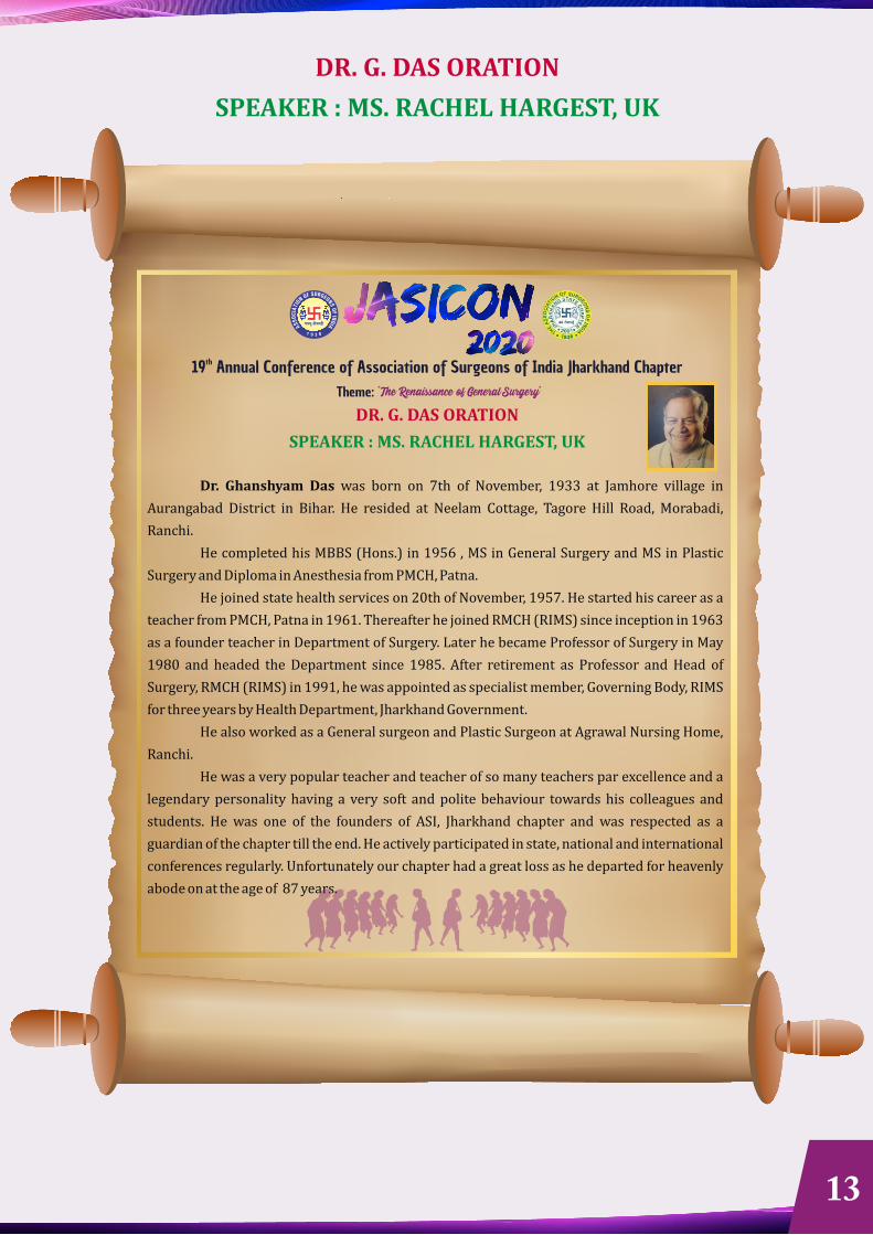
DR. A.K. SRIVASTAV ORATION
SPEAKER : DR. PARUL SHUKLA, USA
DR. A.K. SRIVASTAV ORATION
SPEAKER : DR. PARUL SHUKLA, USA
Dr. A.K. Srivastav was born on 27th December 1943 in Darbhanga to Mr. Kapiljee Shrivastav. He got
admitted to Darbhanga Medical College & passed MBBS with Honours in forensic Medicine & surgery in 1969.
He Joined Bihar Health services in 1971. Following this he joined as Resident Surgical Officer in Surgery at
Darbhanga Medical College in 1973.
He pursued his love for surgery by joining the MS course in DMCH which he passed in 1975.
Thereafter he joined Rajendra Medical College & Hospital in 1976 as Surgical Registrar. From thereon, he
steadily ascended the academic ladder & was promoted to the past of Professor in 1966.
He was a compassionate, daring, untiring & meticulous surgeons as evidenced by the countless patients
who still remember him fondly.
As a teacher he went beyond the call of duty to ensure not only academic but wholesome development of his
students. His mastery of the subject of surgery was only surpassed by his zeal to impart it to his students, many of
whom are renowned all over the world, and are also present in this August gathering.
In the January of 1977, he graced the post of Head of the Department. He was an able administrator who
believed in inclusive & particpatory department administration. He was the first President of the Jharkhand chapter
of ASI. He was a councillor of National Academy of Burns as well as remember of the Executive Council of ASI.
His organizational skills are well evidenced by several conferences & CME like JASICON & burns update
that he has organised.
Following his retirement in 2001 he craved for the academic environment which he missed dearly. For this
he joined STM Medical College, Haldwani, Uttarakahnd. There he Continued his surgical academics & finally ascended
to the post of Principal, before finally at the age of 70.
Thereafter he returned to Ranchi & continued to treat patients & provide consultation & encouragement to
his students.
In July 2019 he breathed hi last in Gangtok, while visiting his son Dr. Kumar Nishant. He is survived by his
wife Dr. Renu Bala, retired professor of FMT.
12
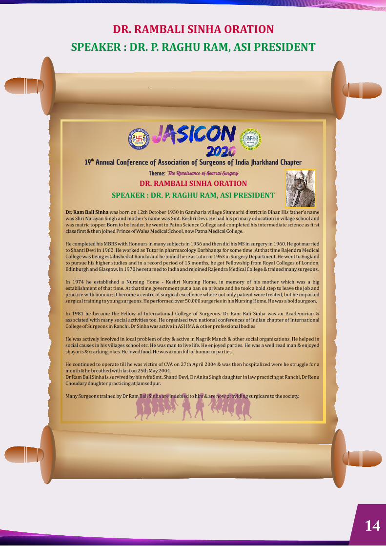
Dr. Ghanshyam Das was born on 7th of November, 1933 at Jamhore village in
Aurangabad District in Bihar. He resided at Neelam Cottage, Tagore Hill Road, Morabadi,
Ranchi.
He completed his MBBS (Hons.) in 1956 , MS in General Surgery and MS in Plastic
Surgery and Diploma in Anesthesia from PMCH, Patna.
He joined state health services on 20th of November, 1957. He started his career as a
teacher from PMCH, Patna in 1961. Thereafter he joined RMCH (RIMS) since inception in 1963
as a founder teacher in Department of Surgery. Later he became Professor of Surgery in May
1980 and headed the Department since 1985. After retirement as Professor and Head of
Surgery, RMCH (RIMS) in 1991, he was appointed as specialist member, Governing Body, RIMS
for three years by Health Department, Jharkhand Government.
He also worked as a General surgeon and Plastic Surgeon at Agrawal Nursing Home,
Ranchi.
He was a very popular teacher and teacher of so many teachers par excellence and a
legendary personality having a very soft and polite behaviour towards his colleagues and
students. He was one of the founders of ASI, Jharkhand chapter and was respected as a
guardian of the chapter till the end. He actively participated in state, national and international
conferences regularly. Unfortunately our chapter had a great loss as he departed for heavenly
abode on at the age of 87 years.
th19 Annual Conference of Association of Surgeons of India Jharkhand Chapter
Theme: “The Renaissance of General Surgery”
DR. G. DAS ORATION
SPEAKER : MS. RACHEL HARGEST, UK
DR. G. DAS ORATION
SPEAKER : MS. RACHEL HARGEST, UK
13
Dr. Ram Bali Sinha was born on 12th October 1930 in Gamharia village Sitamarhi district in Bihar. His father’s name was Shri Narayan Singh and mother's name was Smt. Keshri Devi. He had his primary education in village school and was matric topper. Born to be leader, he went to Patna Science College and completed his intermediate science as first class first & then joined Prince of Wales Medical School, now Patna Medical College.
He completed his MBBS with Honours in many subjects in 1956 and then did his MS in surgery in 1960. He got married to Shanti Devi in 1962. He worked as Tutor in pharmacology Darbhanga for some time. At that time Rajendra Medical College was being estabished at Ranchi and he joined here as tutor in 1963 in Surgery Department. He went to England to pursue his higher studies and in a record period of 15 months, he got Fellowship from Royal Colleges of London, Edinburgh and Glasgow. In 1970 he returned to India and rejoined Rajendra Medical College & trained many surgeons.
In 1974 he established a Nursing Home - Keshri Nursing Home, in memory of his mother which was a big establishment of that time. At that time government put a ban on private and he took a bold step to leave the job and practice with honour; It become a centre of surgical excellence where not only patient were treated, but he imparted surgical training to young surgeons. He performed over 50,000 surgeries in his Nursing Home. He was a bold surgeon.
In 1981 he became the Fellow of International College of Surgeons. Dr Ram Bali Sinha was an Academician & associated with many social activities too. He organised two national conferences of Indian chapter of International College of Surgeons in Ranchi. Dr Sinha was active in ASI IMA & other professional bodies.
He was actively involved in local problem of city & active in Nagrik Manch & other social organizations. He helped in social causes in his villages school etc. He was man to live life. He enjoyed parties. He was a well read man & enjoyed shayaris & cracking jokes. He loved food. He was a man full of humor in parties.
He continued to operate till he was victim of CVA on 27th April 2004 & was then hospitalized were he struggle for a month & he breathed with last on 25th May 2004.Dr Ram Bali Sinha is survived by his wife Smt. Shanti Devi, Dr Anita Singh daughter in law practicing at Ranchi, Dr Renu Choudary daughter practicing at Jamsedpur.
Many Surgeons trained by Dr Ram Bali Sinha are indebied to him & are now providing surgicare to the society.
th19 Annual Conference of Association of Surgeons of India Jharkhand Chapter
Theme: “The Renaissance of General Surgery”
DR. RAMBALI SINHA ORATION
SPEAKER : DR. P. RAGHU RAM, ASI PRESIDENT
DR. RAMBALI SINHA ORATION
SPEAKER : DR. P. RAGHU RAM, ASI PRESIDENT
14
Dr. Awani Kant Sahay (26.01.1943-26.12.2012)
Born on 26.01.1943 to Shn Shree Kant Sahay & Mrs. Chandrakanta Sahay at Fatehpur,
Gaya dist. (now Nawada), the only child, did his early education at Gaya, then shifted to
Hazaribag & did his schooling from here, passed from St. Columbas college ,Hazaribag. He
passed his M.B.B.S from Rajendra medical college, Ranchi in the year 1966.
After completing Houseman-ship he worked in different specialties at U.K did F.R.C.S
from Edinburgh, U.K. He published articles in British journal of surgery, lancet etc.
He joined Patliputra medical college in 1976, retired from here as H.O.D department
of Surgery in January 2002.
He was President of Dhanbad chapter of Surgeons; President Dhanbad I.M.A in the
year 2003 was President of Jharkhand Surgeons association in the year 2010-11 was an active
Rotarian and a former President.
Wife Pramila Sahay blessed with two sons, elder son Dr. Vibbas Sahay (general &
laparoscopic surgeon) married to Dr. Neetu Sahay (obstetrician & Gynecologist), grandson
Aditya & Advik, younger son Dr. Vikas Sahay married to Svetlana Sahay settled at St.
Petersburg, Russia, grand-daughter Ameliya.
He left all his near and dear ones in tears after a massive brain hemorrhage on 26th
Dec,2012...
But a person like him carves his name on hearts.... His legacy shall remain alive in
mind of others and stories they share about him.
th19 Annual Conference of Association of Surgeons of India Jharkhand Chapter
Theme: “The Renaissance of General Surgery”
DR. A.K. SAHAY ORATION
SPEAKER : DR. ABHAY DALVI, ASI VICE PRESIDENT
DR. A.K. SAHAY ORATION
SPEAKER : DR. ABHAY DALVI, ASI VICE PRESIDENT
15

Article - Dr. P. Raghu Ram
BREAST CANCER ADVOCACY & SCREENING –
A MODEL FOR JHARKHAND?
Dr. P. Raghu Ram
MS, FRCS (Edin), FRCS (Eng), FRCS (Glasg), FRCS (Irel), Hon. FRCS (Thailand), FACS
Padma Shri awardee
(2015)
Dr.B.C.Roy awardee
(2016)
President,
The Association of Surgeons of India
www.asiindia.org
Founder, CEO & Director, Ushalakshmi Breast Cancer Foundation, Hyderabad, India
www.ubf.org.in
Director, KIMS-USHALAKSHMI Centre for Breast Diseases, Hyderabad, India
www.breastcancerindia.org
Introduction
With some 165, 000 new cases being diagnosed every year, the incidence of Breast cancer has overtaken cervical cancer to become the most common cancer affecting women in India. What is more alarming is that it
is being
increasingly diagnosed at a younger age (a decade earlier) in India compared to the West.
Breast cancer is a “taboo” –
a “closet” issue in our Country, particularly in rural India. Unlike the United Kingdom and other Western Countries, India does not have a robust organsed population based Breast Cancer Screening Programme.
These are the two primary reasons why more than 60% of women with breast cancer present in the advanced stages,
and as a consequence, majority succumb to the disease within a year of being diagnosed. With 87, 000 deaths per annum, tragically, a woman loses her life to breast cancer every ten minutes in India.
The aim of my Presentation
is to share my thoughts relating to the impact of the Breast cancer advocacy & population based screening implmeneted in the southern Indian States of Telangana & Andhra Pradesh for well over a decade, and
equally, riase the possibility of replicating this initiative in Jhakrhand.
17
Breast Cancer advocacy
Having identified lack of awareness as one of the main reasons behind high mortality for Breast Cancer in India, a „not for profit?
breast cancer Charity was founded in 2007. Based out of the State of the State of Telangana, the Foundation bears my mother?s name to honour her struggle in the fight against breast cancer (Ushalakshmi Breast Cancer Foundation). At 87 years, my mother, Dr Ushalakshmi, who at one point of time was a very well known doctor –
a former Professor of Obstetrics/Gynecology, is a gritty breast cancer “conqueror” and has been spearheading the Foundation?s activities over
the past 13 years.
Addressing the issue of „lack of awareness?
The Power of PINK:
For well over a decade (2007 –
2020), Ushalakshmi Breast Cancer Foundation has been working
with focused determination to create
the much needed awareness about the importance of “EARLY DETECTION” through a number of unique and innovative initiatives in Telangana and Andhra Pradesh.
The “Pink Ribbon Walk”,
which is organized to mark the beginning of International Breast Cancer awareness month (October) is now a
benchmark annual
event in Hyderabad?s calendar. What started off with just 50 people
attending this 2 km walk in 2008, has transformed to a major event, which attracts thousands of people from all walks of life to salute breast cancer „conquerors? in their fight against breast cancer, and equally, spread the message of Hope, Courage & Survival in the fight against the commonest cancer affecting women in India.
Similarly, under “Paint the city Pink” campaign, which started off in 2010, several monuments & historic buildings are illuminated in PINK
to raise the
curiosity amongst the community and reinforce the message of early detection. Hyderabad is the only
city in Asia where so many buildings turn PINK in October. Inspired by this landmark initiative spearheaded by the
Foundation for a decade (2010 –
2020), the Hon?ble Governor of Telangana has announced that the Raj Bhavan would be illuminated in Pink later this month in support of this cause. This would be the very first Governor?s residence in the Country to turn Pink during the international breast cancer awareness month.
It is well known that 70% of our population live in villages and nearly 90% of households in India have a mobile phone. And, therefore, motivated by Prime Minister of India?s ambitious aim to transform India into a digitally empowered society & knowledge economy, and equally, encouraged by the enormous impact/heightened levels of breast
health awareness generated over the past decade through information booklets about various aspects of Breast health published in English and Telugu, the
Foundation embarked upon making available this vital information on a
MOBILE APP
that aims to digitally empower people all over the Country
in
TWELVE LANGUAGES
(English, Hindi, Marathi, Gujarati, Punjabi, Bengali, Telugu, Tamil, Kannada, Malayalam, Oriya & Assamese).
18
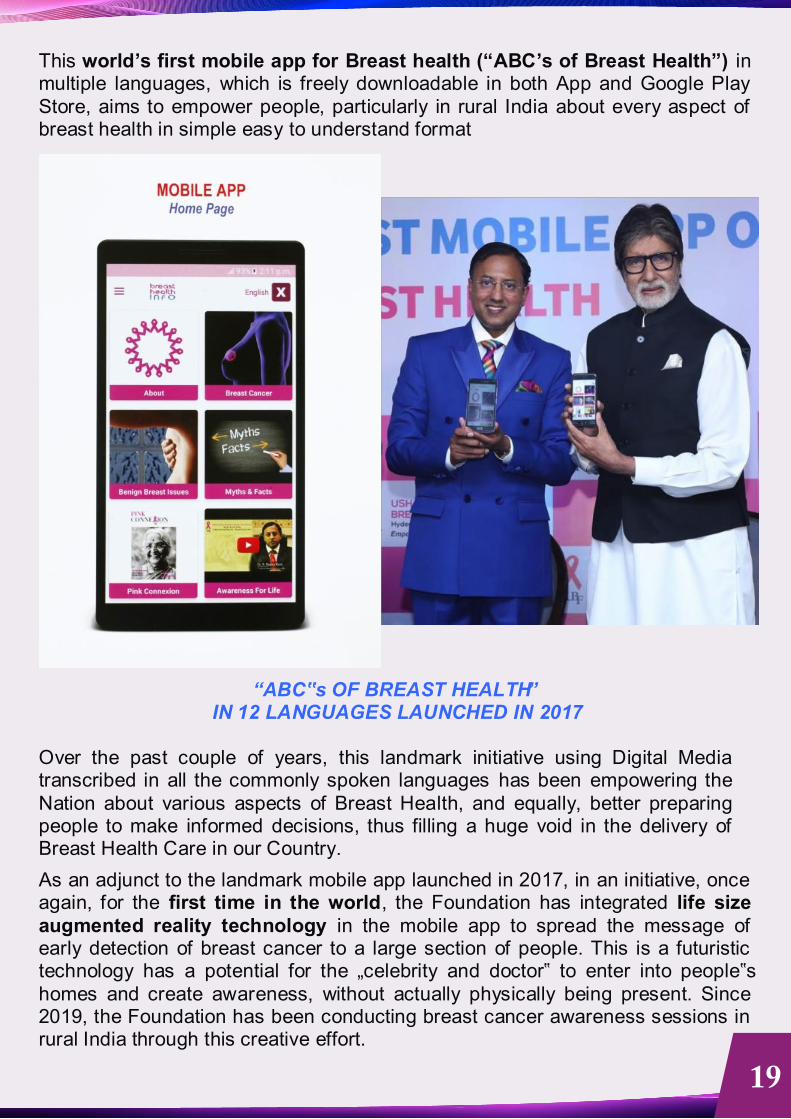
This world’s first mobile app for Breast health (“ABC’s of Breast Health”)
in multiple languages, which is freely downloadable in both App and Google Play Store, aims to empower people, particularly in rural India about every aspect of breast health in simple easy to understand format
“ABC?s OF BREAST HEALTH”
IN 12 LANGUAGES LAUNCHED IN 2017
Over the past couple of years, this landmark initiative using Digital Media transcribed in all the commonly spoken languages has been
empowering
the Nation about various aspects of Breast Health, and equally, better preparing
people to make informed
decisions, thus filling a huge void in the delivery of Breast Health Care in our Country.
As an adjunct to the landmark mobile app launched in 2017, in an initiative, once again, for the first time in the world, the
Foundation has integrated
life size augmented reality technology in the mobile app to spread the message of
early detection of breast cancer to a large section of people. This is a futuristic technology has a potential for the „celebrity and doctor? to enter into people?s homes and create awareness, without actually physically being present. Since 2019, the Foundation has been conducting breast cancer awareness sessions in rural India through this creative effort.
19
Population based Breast Cancer Screening Programme
Screening mammography is not
a viable option for population-based screening in India. Reasons are enormous costs, early age at diagnosis, huge variation in mammographic reporting and quality assurance issues. With the singular aim of ensuring early detection of breast cancer and to find an „Indian solution? that would save many lives, particularly in rural India, Ushalakshmi Breast Cancer Foundation in partnership with the Governments of Telangana and Andhra Pradesh has implemented South Asia’s largest Clinical Breast Examination (CBE) based Breast Cancer Screening Programme. Between 2012 –
2016, over 200,000 underprivileged women between the ages of 35 and 65, spread across 4,000 villages in the Telugu States have been screened for early signs of breast cancer by way of CBE performed
by over 3000
trained healthcare workers, employed with the Governments of Telangana and Andhra Pradesh.
It is a well known fact that underprivileged women in rural India are innately shy of doctors and reluctant to discuss anything as intimate as breast care. Therefore, I had approached the governments of the two States to suggest they use existing healthcare workers, who were known and trusted. Both the State Governments readily agreed to my proposal. Core trainers in both the states were identified and trained to perform CBE under the auspices of Ushalakshmi Breast Cancer Foundation. These core trainers further trained all other healthcare workers across the region. Breast cancers detected through this initiative have been treated free of charge through the State Government-funded Aarogyasri scheme.
This milestone project made national impact in 2016 and I was invited to be part of a high-powered Steering Committee and Technical Advisory Group (TAG) set up by the Union Ministry of Health that approved
the proposal to replicate this CBE based Breast cancer Screening Programme all over India. The breast cancer screening initiative is currently being rolled out nationwide under the auspices of National Health Mission (NHM) alongside screening for Oral and cervical cancer.
After the Foundation introduced the innovative Pink Ribbon Campaign in 2007, over the recent years, I am absolutely thrilled and delighted that many NGOs and Hospitals in Telangana ,Andhra Pradesh & beyond have
embarked upon conducting similar programmes not only for breast cancer awareness, but for other illnesses as well, which
also desperately need urgent attention. This speaks volumes of the “Campaign” that has enthused individuals, Organisations and Institutions to take up & spearhead
impactful awareness activities.
And therefore, I am so very happy
that Hyderabad is contributing in a big way to the
“awareness” journey.
20

However, much more needs to be done across the Country.
There is still a significant proportion of people in India
who still shy away from visiting a doctor when they notice a lump in the breast. THIS MUST CHANGE.
In closing, I am very confident
& hopeful
that the hugely successful initiatives of Ushalakshmi Breast Cancer Foundation could be replicated by the surgical fraternity in Jharkhand
as well, which would undoubtedly pave the path towards
ensuring early detection, and thereby translate to saving scores of lives in this region.
MY HEART BEATS FOR EARLY DETECTION OF BREAST CANCER…
DOES YOURS???
21
Article - Dr. Somashekhar .S. P.
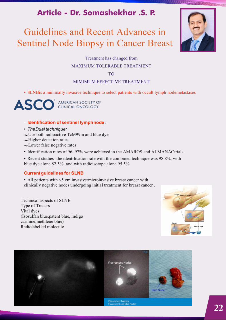
Guidelines and Recent Advances in Sentinel Node Biopsy in Cancer Breast
Treatment has changed from
MAXIMUM TOLERABLE TREATMENT
TO
MIMIMUM EFFECTIVE TREATMENT
• SLNBis a minimally invasive technique to select patients with occult lymph nodemetastases
Identification of sentinel lymph node : -
• The Dual technique: Use both radioactive TcM99m and blue dye Higher detection rates Lower false negative rates
• Identification rates of 96–97% were achieved in the AMAROS and ALMANACtrials.
• Recent studies- the identification rate with the combined technique was 98.8%, with blue dye alone 82.5% and with radioisotope alone 95.5%.
Current guidelines for SLNB
• All patients with <5 cm invasive/microinvasive breast cancer with clinically negative nodes undergoing initial treatment for breast cancer .
Technical aspects of SLNB Type of Tracers Vital dyes (Isosulfan blue,patent blue, indigo carmine,methlene blue) Radiolabelled molecule
22
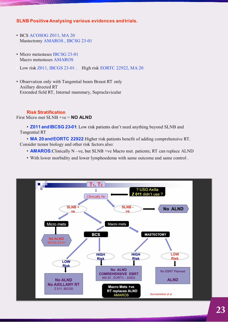
SLNB Positive- Analysing various evidences and trials.
• BCS ACOSOG Z011, MA 20 Mastectomy AMAROS , IBCSG 23-01
• Micro metastases IBCSG 23-01 Macro metastases AMAROS
Low risk Z011, IBCGS 23-01 . High risk EORTC 22922, MA 20
• Observation only with Tangential beam Breast RT only Axillary directed RT Extended field RT, Internal mammary, Supraclavicular
Risk Stratification: First Micro met SLNB +ve = NO ALND
• Z011 and IBCSG 23-01: Low risk patients don’t need anything beyond SLNB and Tangential RT
• MA 20 and EORTC 22922: Higher risk patients benefit of adding comprehensive RT. Consider tumor biology and other risk factors also:
• AMAROS:Clinically N –ve, but SLNB +ve Macro met. patients; RT can replace ALND
• With lower morbidity and lower lymphoedema with same outcome and same control .
23
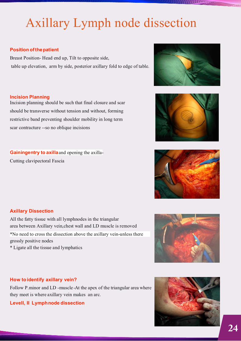
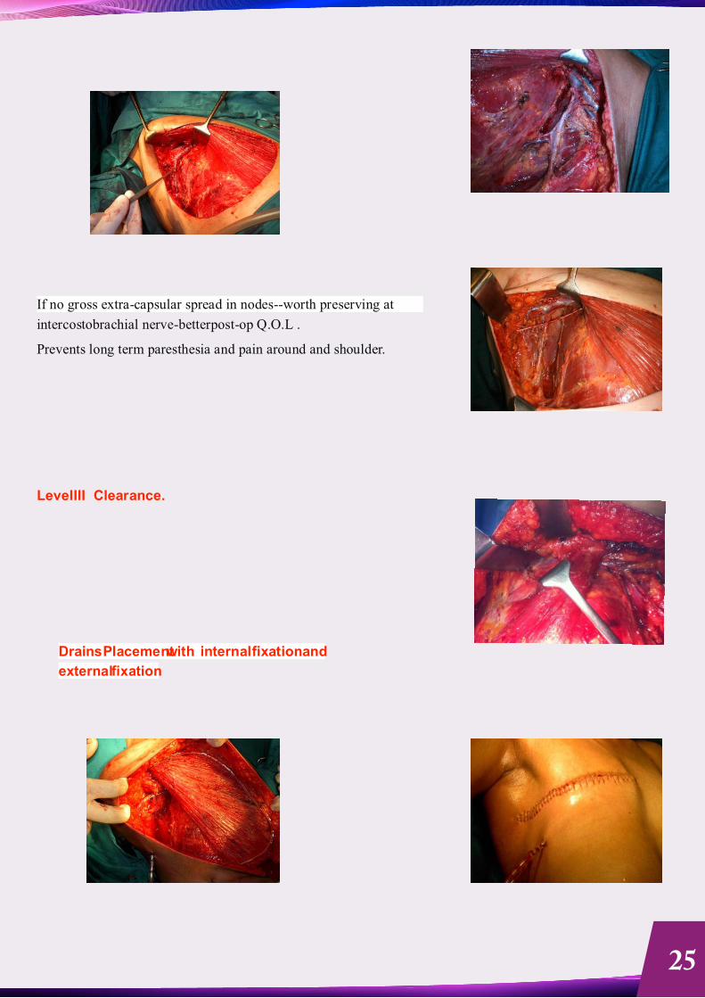
Axillary Lymph node dissection
Position of the patient
Breast Position- Head end up, Tilt to opposite side,
table up elevation, arm by side, posterior axillary fold to edge of table.
Incision Planning Incision planning should be such that final closure and scar
should be transverse without tension and without, forming
restrictive band preventing shoulder mobility in long term
scar contracture --so no oblique incisions
Gaining entry to axilla and opening the axilla-
Cutting clavipectoral Fascia
Axillary Dissection
All the fatty tissue with all lymphnodes in the triangular
area between Axillary vein,chest wall and LD muscle is removed
*No need to cross the dissection above the axillary vein-unless there
grossly positive nodes
* Ligate all the tissue and lymphatics
How to identify axillary vein?
Follow P.minor and LD -muscle - At the apex of the triangular area where
they meet is where axillary vein makes an arc.
Level I, II Lymph node dissection
24
If no gross extra-capsular spread in nodes--worth preserving at
intercostobrachial nerve-betterpost-op Q.O.L .
Prevents long term paresthesia and pain around and shoulder.
Level III Clearance.
Drains Placementwith internal fixation and
external fixation
25
Article - Dr. Arnab Gupta
Total Skin Sparing Mastectomy (Nipple Sparing Mastectomy)-
Abstract of a Video presentation
Dr
Arnab Gupta,
Medical Director, Prof, Dept of Surgical Oncology ,
Saroj Gupta Cancer Centre & Research Institute, Thakurpukur, Kolkata
President, IASO
Introduction:
In the era of conservation in Cancer Surgery, thanks to all the modern adjuvant therapy that are
available, Oncoplastic Breast surgery has taken a giant leap.Moving from Halsted`s Radical
Mastectomy, Modified Radical Mastectomy
(MRM)
and Breast Conservation surgery (BCS) have
become a common place in last 3 decades. More recently Nipple
sparing mastectomy (also
known as NSM or Total Skin sparing Mastectomy i.e. TSSM) has been practiced. It has great
cosmetic advantage, carrying the benefits of Modified radical mastectomy removing the entire
breast parenchyma and axillary nodes, but retaining the envelope of breast including Nipple
Areola complex, inferior mammary crease.
Indications of Nipple Sparing Mastectomy:
1.Prophylactic mastectomy as in BRCA1/3 where there is 65% chance of developing Breast
cancer.
2. Multicentric disease without involvement of Skin or NAC.
3. Small Tumours, atleastcms away from NAC.
4. Extensive DCIS
Contra-indications:
a)
Bloody Nipple discharge
b)
Previous RT to the breast
c)
Previous incisions around NAC
d)
Active smokers
e)
BMI >30
f)
Large (cup size C or more) ptotic breasts
Pre-operative Mammography is recommended to ensure the tumour is not invading Skin or NAC.
Operative procedure:
Incisions-
transverse lateral to NAC/ along Mid
axillary line/ inferior mammary/
Tennis racquet
26
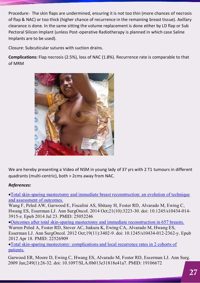
Procedure-
The skin flaps are undermined, ensuring it is not too thin (more chances of necrosis
of flap & NAC) or too thick (higher chance of recurrence in the remaining breast tissue). Axillary
clearance is done. In the same sitting the volume replacement is done either by LD flap or Sub
Pectoral Silicon Implant (unless Post-operative Radiotherapy is planned in which case Saline
Implants are to be used).
Closure: Subcuticular sutures with suction drains.
Complications:
Flap necrosis (2.5%), loss of NAC (1.8%). Recurrence rate is comparable to that
of MRM
We are hereby presenting a Video of NSM in young lady of 37 yrs with 2 T1 tumours in different
quadrants (multi-centric), both > 2cms away from NAC.
References:
·Total skin-sparing mastectomy and immediate breast reconstruction: an evolution of technique and assessment of outcomes.
Wang F, Peled AW, Garwood E, Fiscalini AS, Sbitany H, Foster RD, Alvarado M, Ewing C, Hwang ES, Esserman LJ. Ann SurgOncol. 2014 Oct;21(10):3223-30. doi: 10.1245/s10434-014-3915-z. Epub 2014 Jul 23. PMID: 25052246
·Outcomes after total skin-sparing mastectomy and immediate reconstruction in 657 breasts.
Warren Peled A, Foster RD, Stover AC, Itakura K, Ewing CA, Alvarado M, Hwang ES, Esserman LJ. Ann SurgOncol. 2012 Oct;19(11):3402-9. doi: 10.1245/s10434-012-2362-y. Epub 2012 Apr 18. PMID: 22526909
·Total skin-sparing mastectomy: complications and local recurrence rates in 2 cohorts of patients.
Garwood ER, Moore D, Ewing C, Hwang ES, Alvarado M, Foster RD, Esserman LJ. Ann Surg. 2009 Jan;249(1):26-32. doi: 10.1097/SLA.0b013e31818e41a7. PMID: 19106672
27
Article - Dr. Ganesh Sarin
“Vascular specialityin India” A thought for the future
Healthcare system and facilities are significantly different
in different
parts of India. I come from
state of Bihar.I
did my undergraduate and postgraduate training
at JLNU, RIMS and Patna Medical
College. There was no such specialist as”
vascular
specialist” in any
of the three teaching hospitals.
The commonest vascular ailments we use to see during our training days were
ischemic leg in young
adults and
very rarely
diabetic foot. There was no modality of any basic specific investigation. Buerger’s disease was only diagnosis
made just on a
clinical ground.
Lumbar sympathectomy and
in
due course major amputation was only treatment option
available.
I witnessed this
scenario in late
70’s and 80’s until I was working in India.
I came to UK in 1990. My first placement in UK was with a Vascular/General Surgeon at a district
general hospital which catered a population of about two hundred thousand. I saw how patients
with vascular disease were investigated and managed. To me at first it was a big surprises and an eye
opener
too. The investigations were just
like hand held Doppler, duplex scan and digital subtraction
angiography. Since then I have nearly spent more than twenty-five years in vascular speciality. I have
witnessed unprecedented changes in the management
of vascular disease. The major change which
has been seen is one
from open surgery to
minimal access surgery and endovascular interventions.
The philosophy of “big surgeons make big incision has changed to big surgeons make small or no
incision”. This has been unprecedented and unimaginable change
witnessed by our generation.
I had opportunity to attend
quite a
few academic meeting in India and also had chance
to share my
experience of
UK in my chosen speciality of vascular surgery
with colleagues and trainees
back home
in India.
India since
I left has drastically changed. It is one of
the fastest growing economies and progressing
towards becoming a superpower. There has been significant increase in living standard,
per capita
income, prosperity,
and increase
in longevity and so on. These changes
will
on
other hand given
a
different spectrum of
diseases.
It is very interesting to note that the incidence of diabetes in India around 15% in urban and 3-5%
rural population. Hypertension 30%, 54 million people suffered from heart disease as data suggested
in 2016. The incidence cardiovascular disease has increased from 1% to nearly 10% in the last six
decades.
Smokers comprises of about 120 million people,
about 70% of adult male. Population
statistic suggests that by 2050 20% population will be over 60 years of age. All these are
risk factors
for peripheral vascular disease. It was not surprising to see that
there has been
a reported incidence
of peripheral vascular disease in a part of southern India was around 25%.
Surgeon UK
28

All these statistical finding, should make health care planner to foresee and plan how we are going
to look after people with peripheral vascular disease in future. There will be a significant aging
population
in years to come.
I haven’t taken in to account for
venous and lymphatic diseases that
will require a vascular specialist.
How the care could be affordably provided to both rural and urban population in future. This
needed to be seriously looked at. This will address the issue of providing health care facilities and
expertise equally to all who need it
irrespective geographical area
where they live and social class
where they come from. It may require
more vascular training centres to produce more specialists to
cater the population at large.
29

Article - Dr. Arunima Verma
“‘ Incisional Hernia Repair”
MS, MRCS, FACS,FIAGES, Fellowship MAS Upper & Lower GI Surgery (UK),
HOD Surgery, Tata Motors Hospital, Jamshedpur
Dr. Kumar Kshitij Abhinav, DNB, Tata Motors Hospital, Jamshedpur
Introduction:
Ventral Hernias are one of the most common pathological conditions encountered by clinicians. The European Hernia Society (EHS) has divided the abdominal wall hernias as
�
Primary abdominal wall hernias
�
Incisional abdominal wall hernias
According to this system, classification of abdominal wall hernias uses localization and size as the two variables as show in Table 1.
Table 1: European Hernia Society classification for primary abdominal wall hernias EHS
Primary Abdominal wall Hernia Classification
Diameter (cm)
Small <2cm
Medium 2-4cm
Large 4cm
Midline
Epigastric Umbilical
Lateral
Spigelian
Lumbar
Management Strategies:
Incisional hernia is one of the most common postoperative complications after abdominal surgery. Several studies have shown that incisional hernias have different etiologies which are related to the patient, the surgical technique, the suture material and experience of the surgeon. Most patients present with abdominal swelling with some level of discomfort, and in emergency the presentation is usually as bowel obstruction or strangulation which requires urgent exploration. The elective management of Incisional Hernias includes non-operative and operative management. Non-operative management may be considered in patients who are poor surgical candidates, those who require pre-operative optimization or those with high complexity hernias. Patients, who are candidates for surgery may also need a period of non-operative management for preoperative risk reduction of modifiable risk factors which are present in majority of patients.
For Surgical management of incisional hernias both open and Laparoscopic techniques are being used and the options are given in Box 1.
Box 1 – Surgical options for incisional hernia repair
Open Hernia Repair
� Suture Repair
�
Mesh Repair
Laparoscopic Repair
�
Primary Fascial Closure
�
Different mesh fixation technique
�
Robotic Hernia Repair
Abdominal wall reconstruction techniques
�
Bridged Repair
�
Anterior Component Separation (ACS)
�
Perforator Sparing ACS
�
Endoscopic ACS
�
Posterior Component Separation
Pre-operative tissue expansion
�
Tissue Expanders
�
Progressive Pneumoperitoneum
Flaps and tissue transfer
30
Open Hernia Repair:
In practice,
there are three types of open repair for incisional hernia with mesh—
the inlay, onlay, and sublay
techniques.
Onlay repair involves placement of the mesh on the anterior rectus fascia below the subcutaneous layer after approximation of the anterior rectus fascia.
Sublay
repair refers to placement of the prosthetic in the retro-muscular
space posterior to the rectus abdominis
and anterior to the posterior rectus fascia.
Underlay
mesh placement describes mesh positioning in the preperitoneal
subfascial space or the intraperitoneal
space deep to the fascia and peritoneum.
Sublay:
The retrorectus repair which was popularized by Rives and later Stoppa and Wantz, revolutionized hernia repair by offering a robust treatment of complicated incisional hernias with a low recurrence rate. The retrorectus repair addresses the attenuation and lateralization of the rectus abdominis muscles and recreates the natural tension of the lateral obliques on the abdominal wall.
Biomechanical Principles of Repair: The retrorectus space is a well vascularized position where mesh prostheses become incorporated. In an animal model, mesh placed in the retrorectus position is associated with a peri-filamentous collagen deposition with a much higher type I/III ratio compared to mesh in the onlay or premuscular condition. The higher degree of type I, or mature collagen, results in a higher tensile strength of the wound.
OPERATIVE STEPS: The operation begins with a midline incision with or without excision of the prior scar.
The Hernial Sac
It is recommended that the hernia sac should be preserved as it can be later used to make up for any deficiency in either the posterior or anterior rectus sheath. Therefore, the hernia sac should be divided in the midline and the
peritoneum is entered. A full lysis of adhesions from the anterior abdominal wall is recommended, as it will help with the mobility of closing the posterior rectus sheath and peritoneum in the midline.
Posterior Rectus Sheath Dissection
One side of the hernia sac is preserved and the dissection proceeds ventral to the hernia sac until the medial edge of the rectus sheath is encountered on the one side. Next, the rectus sheath is incised along the entire vertical length of the incision. On the contralateral side, the hernia sac may be left attached anteriorly, and the incision of the posterior rectus sheath can be made immediately lateral to the medial most edge of the hernia defect on that side. The dissection of the posterior rectus sheath is
then continued cranial and caudal to the hernia defect for a
minimum distance of 5–8 cm. This will provide ample space for mesh overlap. The posterior rectus sheath is fused to the linea alba at its lateral most aspect. To create a space for mesh placement which crosses the midline behind the rectus muscles above and below the hernia defect, the posterior sheath must be divided of
the linea alba
ensuring
preservation of the linea alba as it will be the midline thrust bearing portion of the abdominal wall ventral to the mesh in the areas both above and below the hernia. If possible, the layer of peritoneum dorsal to the linea alba can be preserved and dissected posteriorly, serving as a bridge between the cut edges of the posterior rectus sheaths above and below the hernia.
The dissection of the posterior sheath off of the overlying rectus muscle proceeds laterally. The dissection can be
performed bluntly or with cautery. If the hernia defect extends
into the
upper abdomen, the surgeon may need to
extend
the dissection up to the costal margin and behind the xiphoid process. The posterior rectus
sheath is
attached to the xiphoid process. The posterior sheath can be divided of
the xiphoid process and dropped
posteriorly and the dissection carried out in the preperitoneal plane dorsal to the xiphoid.
31
Below the arcuate line, the posterior rectus
sheath is deficient
and only transversalis fascia,
preperitoneal fat and
peritoneum remain. For hernias extending below the umbilicus, the surgeon
will need to maintain these structures
so as to have
tissue to close the visceral sac. The dissection may extend into the preperitoneal spaces of Retzius and
Bogros, exposing the pubic bone, Cooper’s ligaments, and the iliac vessels on both sides.
The posterior rectus
sheath has been disconnected from the linea alba, bilaterally, while preserving the peritoneum
which was mobilized off the linea alba
Visceral Sac Closure
Once the dissection is complete, the posterior rectus sheath is approximated in the midline in a
continuous
fashion
using
absorbable
suture. Despite the
relatively weak nature of the transversalis fascia/peritoneal layer below
the arcuate line, it easily allows for approximation and visceral sac closure. It is important that the posterior sheath is closed completely, so as to prevent any bowel from slipping in between the mesh and the posterior sheath, which could result in a bowel obstruction. Additionally, visceral sac closure ensures the mesh will not come in contact with the viscera. Should there be difficulty reapproximating the posterior sheaths in the midline due to excessive tension, two options arise. The fascial edges of the posterior sheaths can be sutured directly to the omentum, effectively closing the visceral sac. Alternatively, an absorbable mesh can be sewn as an interpositional graft to make up for any defect in the posterior sheath.
Mesh Fixation The width of each rectus muscle and thus the entire retrorectus space is quite variable between patients. Ideally, the mesh should occupy this entire retrorectus space. The space may be measured and the mesh trimmed to size. Alternatively, the uncut mesh can be placed into the space and trimmed as it is being fixated. The mesh should be fixated circumferentially with spaced, full-thickness slowly absorbable sutures. The mesh may be fixated to the costal cartilage or to the symphysis pubis and Cooper’s ligaments bilaterally, here with a permanent monofilament suture. The mesh should lay taut in this space taking into consideration the fact that the space will become even smaller once the rectus muscle is reapproximated overtop the mesh (Image 6). Ideally, the surgeon should avoid introducing wrinkles into the mesh as it decreases mesh-tissue area interface. There is no real consensus on the need for mesh fixation in this retromuscular plane. Rives et al. originally described permanent sutures, placed abundantly along the mesh perimeter. As the focus of hernia repair outcomes shifted from recurrence to postoperative pain and function, many groups modified their fixation approach.
Midline Abdominal Wall Reconstruction
At the conclusion of mesh placement, two closed suction drains are placed, through separate stab incisions, into the retromuscular space. The drains will rest directly on top of the mesh. The midline abdomen is now reconstructed by suture reapproximating the edges of the linea alba in a continuous fashion with absorbable suture. Reconstructing the midline serves three purposes. First, it restores the central tendon of the abdomen, thus producing a functional anatomic repair. Secondly, it provides an increased area of mesh/tissue interface, and a reliable backstop for the mesh to resist the pressure of the abdominal cavity. Thirdly, closure of the fascia overtops the mesh has been demonstrated to reduce the incidence of prosthetic mesh infection.
Special Considerations
Assessing Anterior Tension
At the time of midline closure, the surgeon should decide whether the bilateral rectus myofascial
release performed
will
be suffi cient enough to allow the anterior rectus sheaths to be approximated in the midline. This is done by
placing clamps on the fascial edges and pulling in opposite directions. If the tension is minimal,
then the surgeon
may proceed with anterior fascial closure. Should the tension be excessive, a
decision should be made regarding the
next step.
Options are numerous, and include leaving the fascia open. The surgeon may perform the
Ramirez
component separation which will
allow further medialization of the rectus muscles.
A newer approach is to perform
a posterior
component separation where a myofascial release is effected by dissecting between the
oblique muscle
layers, lateral to the rectus sheath. From superfi cial to deep, Mathes et al.
described the space between the
external
and internal oblique muscle. Carbonell et al. demonstrated the space between the internal
oblique and transversus abdominis muscle.
Novitsky described the transversus abdominis
release (TAR) where this muscle is
divided,
thus gaining access to the preperitoneal/pretransversalis plane lateral to the rectus muscle.
Each of these
32
myofascial releases affords further
medialization of the rectus muscles and obviates
the need for any subcutaneous
fl ap elevation,
which is required for the Ramirez, or anterior component separation. My preference is now the
TAR
for its ease and reproducibility. Of all the posterior releases, it allows the most medialization of the posterior rectus sheath as it is attached to the highly expansile peritoneum laterally.
Lateral Defect Concomitant lateral defects such as a former stoma site hernia can be addressed at the same time as the Rives-Stoppa repair. These defects can be within the rectus muscle itself, but often lie at the semilunar line, or worse yet, within the oblique musculature. To extend the retrorectus dissection lateral enough to these defects, the surgeon will need to perform a posterior component separation as previously described. This will allow a wide dissection lateral to the off-midline defect. Once the dissection is complete, the defect within the posterior rectus sheath will need to be closed, as well as the defect anteriorly within the rectus muscle or oblique complex.
Limitations
Since the Rives-Stoppa repair is a technique described for midline hernias, it should not be used for defects that are solely lateral, without a midline component. Lateral defects can be best approached directly over the defect and the preperitoneal space developed for mesh placement. Developing the retrorectus space will be exceedingly diffi cult, if not untenable in patients who have undergone resection of one or both of the rectus muscles such as women who have undergone a transverse rectus abdominis myocutaneous (TRAM) reconstruction of the breast. These patients may be better suited for an intraperitoneal or onlay placement of mesh.
Postoperative Care
Postoperatively, closed suction drains are left in position until they are draining less than 30 mL in a 24 hour period. I routinely discharge patients home with drains and do not prescribe antibiotics during this period. An abdominal binder is placed for comfort and support during the convalescent period.
It is not uncommon for patient to develop postoperative ileus due to entering the peritoneal cavity, particularly if an extensive lysis of adhesions was performed. I do not routinely leave a nasogastric tube in position after the operation; rather reserve its placement should the patient become increasingly symptomatic postoperatively. The most common complications postoperatively are wound complications. Patients with multiple cicatrices of the abdomen may have disrupted the normal vascular supply to the skin of the abdomen. These patients are best evaluated
by a plastic surgeon preoperatively to determine
the ideal placement of the incision for hernia
repair.
Wound complications include skin ischemia, skin dehiscence, seroma, hematoma, and
surgical site infection.
The
incidence of surgical site infection is directly proportional to the degree of bacterial contamination
or wound classifi
cation during the hernia
repair. Mesh in the retromuscular space is quite resistant to infection, particularly the
newer varieties
of wide-pore meshes. Multiple investigators have shown that they can often be easily salvaged
with
negative pressure wound therapy, should a deep space surgical site infection occur.
A particularly under reported
complication is
that of a postoperative interparietal hernia. This can manifest as a small bowel obstruction due to
the
small bowel becoming trapped within the space between the posterior rectus sheath and the
mesh. This occurs only
if there is a breakdown in
the posterior fascial closure, which likely occurs more than we believe. A high-index of
suspicion
for this entity should arise if a patient fails to progress postoperatively as expected. A computed
tomographic exam will demonstrate the defect in the posterior sheath closure with bowel
in the interparietal space.
Overall, the recurrence rate of the Rives-Stoppa incisional hernia repair has been shown, in multiple large series, to
be less than 10%.
In summary, the Rives-Stoppa technique for the repair of incisional hernias continues to stand
the test of time since
its inception close to 50
years ago. It should be the standard by which all other techniques are compared.
33
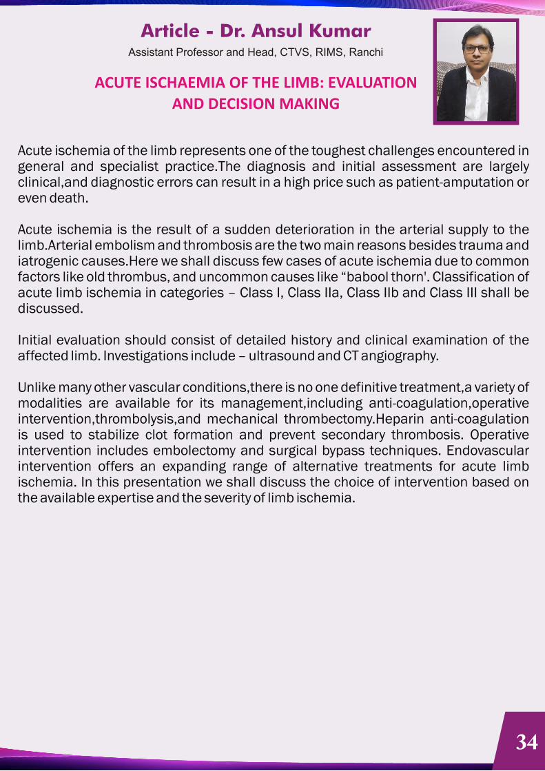
Article - Dr. Ansul Kumar
ACUTE ISCHAEMIA OF THE LIMB: EVALUATION
AND DECISION MAKING
Acute ischemia of the limb represents one of the toughest challenges encountered in general and specialist practice.The diagnosis and initial assessment are largely clinical,and diagnostic errors can result in a high price such as patient-amputation or even death.
Acute ischemia is the result of a sudden deterioration in the arterial supply to the limb.Arterial embolism and thrombosis are the two main reasons besides trauma and iatrogenic causes.Here we shall discuss few cases of acute ischemia due to common factors like old thrombus, and uncommon causes like “babool thorn'. Classification of acute limb ischemia in categories – Class I, Class IIa, Class IIb and Class III shall be discussed.
Initial evaluation should consist of detailed history and clinical examination of the affected limb. Investigations include – ultrasound and CT angiography.
Unlike many other vascular conditions,there is no one definitive treatment,a variety of modalities are available for its management,including anti-coagulation,operative intervention,thrombolysis,and mechanical thrombectomy.Heparin anti-coagulation is used to stabilize clot formation and prevent secondary thrombosis. Operative intervention includes embolectomy and surgical bypass techniques. Endovascular intervention offers an expanding range of alternative treatments for acute limb ischemia. In this presentation we shall discuss the choice of intervention based on the available expertise and the severity of limb ischemia.
Assistant Professor and Head, CTVS, RIMS, Ranchi
34
Article - Dr Rakesh Kumar
'Spectrum of vascular cases in Jharkhand, how to deal with it and what's the outcome of treatment'.
Background200 different type cases which we have come across in last 3 years at tertiary care centre and apex institute of Jharkhand RIMS. Emergency as well as elective both cases included in this study which were operated or managed conservatively in this period.
Type of study RetrospectiveLimitation single Institution study and small study groupNo conflict of interestPatients and methods
6 months to 82 yrs old patients included in this study145 male 55 femaleMost common varieties of vascular illness are congenital, aquired Traumatic and non traumatic. Among congenital patent ductus arteriosus, arteriovenous malformations and hemangioma. Traumatic vascular injury was mainly in peripheral ateries and vains,broken intravenous line during dialysis, peripheral artery pseudoaneurysm. Nontraumatic cases were arteriovenous malformation in different parts of the body, peripheral artery aneurysm, deep vein thrombosis, acute limb ischemia, chronic limb ishchemia and varicose veins.
Discussion Mode of intervention surgical intervention as well as sclerothrapy and radio frequency ablationAll traumatic cases were dealt with surgical intervention with excellent outcome. The limiting factor were time lapsed after injury and associated bony,soft tissue and nerve injury with segment loss. In avm, varicose vein and etc sclerothrapy as well as surgery have excellent outcome.PDA, peripheral artery pseudoaneurysm repaired surgically with excellent outcome.Deep vein thrombosis and chronic limb ischaemia managed medically and tehy have good outcome in long follow up.
Conclusion What ever the type of case, proper planning and timely intervention in vascular cases is the best way to achieve excellent results.
MBBS,DLO,MS(GENERAL SURGERY), MCH(CTVS) AIIMS NEW DELHI
Assistant Professor- Deptt. of Cardiothoracic & Vascular Surgery,
Rajendra Institute of Medical Sciences, Bariatu, Ranchi-834009, Jharkhand India.
35
Best PapersArticle - Dr. M. Kanagavel
SURGICAL SPECIALISATION –
A CAREER FOR YOUNGSURGEONS IN THE PRESENT ERA
Chairman, Association of Surgeons of India – Chennai City
PG Faculty and Senior Consultant
Department of General, GI and Minimal Access Surgery
St Isabel's Hospital, Chennai, India
' Evolution is towards specialization. This is also true for surgery which changes with the field of practice, illustrating the Darwinian adaptation.' Medicine has evolved from an integrated MBBS qualification to a microspecialised one. Days of Operative Medicine has evolved into General Surgery and onto various subspecialities.
Evolution to specialization is everywhere: from restaurants to book sellers. Considering surgery, in the beginning there were barbers; it took until the end of the 16th century for barber surgeons to emerge from the barbers' guild and become surgeons.
During the early years, most practised as general surgeons; however, there were some from specialized fields such as naval surgeons and military surgeons. The spectrum of surgery at this stage was very limited: treatment of trauma, treatment of haemorrhage by cauterization, tourniquet or amputation, removal of bladder stones, incision of abscesses and treatment of piles. Neurosurgery was limited to drilling trepan holes, and abdominal surgery to treatment of hernia, drainage of abscesses of the liver or of the gallbladder (the first cholecystostomy was much later) and packing of bleeding wounds. There was no need for specialization; surgeons could easily cover the limited fields of surgical knowledge and treatment.
The advancement of the surgical techniques, equipment, gadgets and high-end automation has made an earlier inaccessible human anatomy a fascinating journey of precision and fine skill based science.
Focussed training and practice in a narrow field gives the best outcome for any surgical disorder. There are still general surgeons whose practice includes a wider spectrum of these procedures, but in the near future general surgery may become very limited.
General Surgery as such is getting redundant in the modern scenario and a planned restructuring of various supspecialisation of surgical domains is the need of the hour. This helps one to get into the career early and stay focused in the specific domain. It is time we sock up and allow a graded transition from General Surgery as a Specialty when surgery evolved as a separate specialization from Medicine into multiple areas. A planned transition is optimal for the future trainees to decide their specialization early in their career. The curriculum should be to design and integrated sub specialization program and realignment of the surgical profession.
36
Best Papers
Abstracts
37

Best Papers
Sl. No.
Presenter Name
Co-Authors
Title
Email ID
1
SANJAY KUMAR YADAV
SANJAY K YADAV,ANJALI MISHRA,SAROJ KANTA MISHRA
WHAT ABOUT COST? MODERN TECHNIQUES IN THYROID AND PARATHYROID SURGERY IN LMICS
What about cost? Modern techniques in thyroid and parathyroid surgery in LMICs Introduction: There is high disparity in surgical care between high-income, and low- and middle-income countries (LMICs). Thyroid and parathyroid surgery techniques have undergone few changes in the last century. The harmonic scalpel ( UHS), Intra-operative neuromonitoring ( IONM) and Intra-operative parathyroid ( IOPTH) monitoring are being widely used increasingly in developed countries and being propagated in LMICs with great enthusiasm. The objective of the present study was to evaluate costs for thyroidectomy and parathyroidectomy performed with the aid of harmonic scalpel, intraoperative neural monitoring (IONM) and or IOPTH. Methods: To assess the impact of these modern technologies , six macro-scenarios were considered: (1) traditional thyroidectomy; (2) thyroidectomy with UHS; (3) thyroidectomy with IONM; (4) thyroidectomy with both UHS and IONM; (5) traditional parathyroidectomy ; (6) parathyroidectomy with IOPTH . Results: Hospitalization costs for a thyroidectomy / parathyroidectomy using modern technique varied from 406.99 USD to 631.53 USD (scenarios 2,3,4 and 6): 15–17% to 23.11% higher than for the traditional procedure ( 350.85 USD to 336.82 USD, scenario 1and 5, respectively). Surgery costs for a traditional surgery account for 68 to 79 % of hospitalization costs; if UHS, IONM or IOPTH is used, this percentage increases to about 80 to 92%. Conclusion: An efficient healthcare system for LMICs must enhance the capabilities of health care professionals and their patients to make informed choices regarding use of modern gadgets and whether their use is going to make a real difference in the outcome in a cost effective way for the LMICs.
Best Papers
38

Sl. No.
Presenter Name
Co-Authors
Title
Email ID
2
DR. AJAY KUMAR
DR. AJAY KUMAR,PROF.(DR.) DIPENDRA KUMAR SINHA
INCIDENCE OF HYPOCALCEMIA AFTER TOTAL THYROIDECTOMY (TT)- AN OBSERVATIONAL STUDY
ABSTRACT BACKGROUND: Hypocalcemia is one of the most common complications after total thyroidectomy due to transient or permanent postoperative hypoparathyroidism. The primary cause of hypocalcemia is secondary hypoparathyroidism. AIMS: To determine incidence of hypocalcemia after total thyroidectomy . MATERIAL AND METHODS: A total of 74 patients undergoing total thyroidectomy during approximately 1 year in RIMS Ranchi, were evaluated and the incidence of post-operative hypocalcemia was assessed using post-operative serum calcium values with 8 mg/dL as cutoff. RESULTS: The incidence of post-operative hypocalcemia was 58.90 %, with symptomatic hypocalcemia 20.93 %, (transient 18.62% and permanent 02.32%). CONCLUSION: Hypocalcemia following total thyroidectomy although common and it is usually asymptomatic and self-limiting. Symptomatic hypocalcemia is mainly transient in nature.
39
Sl. No.
Presenter Name
Co-Authors
Title
Email ID
3
DR NAMRATA MAHANSARIA
DR NAMRATA(AGARWAL) MAHANSARIA,DR A.K VERMA
IDIOPATHIC GRANULOMATOUS MASTITIS- A SURGEONS ORDEAL!
INTRODUCTION IGM is a rare inflammatory condition of the breast. It is a benign condition which very often mimics inflammatory carcinoma of the breast. The aetiology of IGM is still unknown. Histopathologically it is identified by presence of lobulo-centric granulomas after exclusion of all other granulomatous disease. Multiple treatment options are available, but none with proven benefit. Antibiotic, Non-Steroidal anti- inflammatory drugs (NSAIDs), corticosteroids, cytotoxic and immunosuppressive medications and surgery are among the therapeutic options but no consensus has been achieved so far. AIMS To study the treatment modality that can be offered to patients with IGM. Surgery vs medical management. Outcome of treatment. OBJECTIVE To study the demographics, To look at the clinical presentations Treatment modalities Follow up METHODS Retrospective review of data 1st February 2018 to 31st August 2020 Data of OP and IP files, RINCHI hospital, Centre for Cancer and Breast disease 15 patients who were diagnosed IGM by HPE Consent of patients for use of personal data prior to starting of treatment for educational purpose Inclusion criteria – IGM patients after exclusion of tuberculosis Exclusion – Patients lost to follow for at least 1 year. RESULTS 15 Patients 14 females, 1 male Median Age- 36 years Use of antibiotics followed by low dose steroid have shown good results in my study. Surgical intervention was required in only 1 patient who presented with recurrent abscess. CONCLUSION IGM a rare entity, the awareness of which is essential for the surgeon. It requires a lot of patience not only on the patients’ part but also of the treating doctor! Surgery may not be the first line of treatment for most of the patients, in fact it may be detrimental.
40

Sl. No.
Presenter Name
Co-Authors
Title
Email ID
4
TANMAY PRASAD
DR TANMAY PRASAD,DR ANANT SINHA,DR RAJ KUMAR PATHAK,DR VIVEK GOSWAMI
CHANGING PATTERN OF BURNS IN A PRIVATE HOSPITAL
Introduction: Burn patients are at a high risk for infection as a result of the nature of the burn injury itself, the immune-compromising effects of burn, prolonged hospital stays and intensive diagnostic and therapeutic procedures. Gram positive organisms are initially prevalent during hospital stay of patients; then gradually become superseded by gram negative opportunists that appear to have a greater propensity to invade. Aim and Objectives: The study was done to find out the recent pattern of burns in a private hospital and to see if there are increasing and decreasing trends in the demography of burns and accordingly focus our attention to high impact areas. Methods: A demographic survey with history, clinical evaluation, investigations and preliminary management of the burn patient was performed. The microbial colonization of wounds was studied weekly from the date of admission up to the 4th week of hospitalization. Periodic wound swabs were collected at 1st, 2nd, 3rd, and 4th weeks of hospital stay or till the resurfacing was done and findings noted. Results: : there has been an increase in the number of patients presenting to the casualty in the last 5 years. Domestic incidents have increased over time. Most affected patients in burns were women suffering burns due to domestic accidents. Most patients were in the young working age group (21-40yrs). Most patients were females. Large burns (>20% TBSA) are becoming more common in the recent times. Urban burns are more common than rural. Average duration of stay has increased. Conclusion:The pattern of burns are changing as a dynamic process. Resorting to a single line of management over a sustained long periods of time (>5-10Yrs, depending on the demography) may not be adequate. We need to reassess the problem, reformulate principles and re-strategize the management.
41

Sl. No.
Presenter Name
Co-Authors
Title
Email ID
5
AKASH KUMAR GUPTA
DR. AKASH KUMAR GUPTA,DR. MANOJ KR DAS,DR. MARSHALL D KERKETTA
OBSERVATIONAL STUDY IN PAEDIATRIC AGE GROUP UNDERGOING EMERGENCY LAPROTOMY IN TERTIARY HEALTH CENTRE: RIMS
INTRODUCTION: Acute abdomen can be defined as “syndrome included by wide variety of pathological conditions that require emergent medical or more often surgical management.” Acute abdomen is caused due to gastrointestinal diseases such as intestinal obstruction and perforation peritonitis. AIM: The aim of our study was to observe the common cause in paediatric age group undergoing emergency laprotomy in our institutions. MATERIAL AND METHODS:This prospective study included 77children aged below or equal to 15years, underwent emergency laprotomy for acute intestinal conditions between January 2019 to December 2019 in RIMS,RANCHI. We excluded neonates ,patients of jejunoileal colonic atresia and stenosis, anorectal malformation(ARM), congenital pouch colon, neonatal necrotizing enterocolitis(NEC), hirschprung’s disease, gastrointestinal tumor. RESULTS: Total of 77 laprotomies were performed in emergency in children below or equal to 15 years age,59(76.62% ) were boys and 18(23.37% )were girls with male:female ratio of 3.2:1. 36(46.75%) cases were done for acute intestinal obstruction and 41(53.24%) cases were done for perforation peritonitis.20(25.97%)emergency laprotomy was performed in the age group 1-5 years and 57(74.02% ) were performed in the age group 5-15 years. Causes in order of frequency for intestinal obstruction were intussusceptions, post operative band/adhesion, abdominal tb obstruction, meckel’s diverticulum and worm obstruction. Causes in order of frequency for perforation peritonitis were typhoid, abdominal tb, appendicular perforation and abdominal trauma. CONCLUSION: In our study maximum emergency laprotomy was performed in male patients with male:female ratio of 3.2:1. Perforation peritonitis was more common than acute intestinal obstruction. 5-15 year age group were more commonly affected. Typhoid ileal perforation was the most common cause for emergency laprotomy followed by intussusception
42

Sl. No.
Presenter Name
Co-Authors
Title
Email ID
6
DR. KUMAR NISHANT SINGH
DR. KUMAR NISHANT SINGH
MANAGEMENT OF COMPLICATED APPENDICITIS: IS LAPAROSCOPIC APPROACH A SAFE OPTION
MANAGEMENT OF COMPLICATED APPENDICITIS: IS LAPAROSCOPIC APPROACH A SAFE OPTION Singh Kumar Nishant Department of General surgery Parmeshwari Medical Centre, Garhwa, Jharkhand Email: [email protected] Abstract Background: Minimal access surgery is nowadays widely practiced in both diagnosis and management of various infective conditions of abdomen. Laparoscopy is also recommended in appendicolithiasis, perforated appendicitis, and appendicular abscess with evidence of less morbidity and hospital stay in comparison to open approach. Laparoscopic appendectomy (LA) was performed mostly on uncomplicated appendicitis due to opinions about its safety when it was first introduced. Laparoscopic appendectomy is a procedure of choice in acute or chronic appendicitis in any age group. Nevertheless, there are still concerns about surgical difficulties in managing complicated appendicitis with laparoscopy, possible post-op complications and conversion to an open appendectomy (OA) during the surgery. Methods: This is a retrospective study, consists of 15 patients who underwent laparoscopic appendectomy in Department of General Surgery at Parmeshwari Medical Centre, Garhwa for complicated appendicitis during March 2020 to September 2020. All surgeries were performed by single surgeon under same anesthesiology team. Patients diagnosis was based on clinical findings, complete blood counts, and abdominal sonography. Results: Fifteen patients underwent laparoscopic appendectomy for complicated appendicitis. Of the 15 patients, perforated appendix cases are 10, gangrenous appendix are 1, appendicular abscess only one case and 3 cases of appendiceal phlegmon. Post operative wound infection, conversion rate and hospital stay rate very less. Conclusions: The present study proved that laparoscopic appendectomy is the best approach in complicated appendicitis. Keywords: Appendicitis, Complicated appendicitis, Laparoscopic appendectomy, Open appendectomy
43

Sl. No.
Presenter Name
Co-Authors
Title
Email ID
7
SUMIT KUMAR
TIPS AND TRICKS TOWARDS SAFE PCNL: A SINGLE-CENTRE EXPERIENCE OF 250+ CASES
INTRODUCTION: Percutaneous nephrolithotomy (PCNL) is a standard modality for treatment of large renal and proximal ureteric calculi. However, there is still hesitation for fear of significant dreaded complications like urosepsis and hemorrhage. We present a single centre experience of over 250 patients with an emphasis on tips to reduce complications. METHODS: We analysed the outcomes of 268 patients undergoing PCNL at our centre between August 2018 and February 2020. Stone complexity and burden, access, dilation, stone free rate (SFR), complications (CROES Clavien system; CCS), length of hospitalisation (LOH) and perioperative strategies were analysed. RESULTS: The mean SFR was 83.5% with secondary SFR of 94.1%, median LOH was 3 days and mean operation time (OT) was 75.34 minutes. Complications wereseen in 11.94% (32/268) patients with minor (CCS I,II; 22/268) constituting 68.75% of complications and major [III-A: 25% of complications (8/268) ;IV-A: 6.25% of complications (2/268)].Staged PCNL was done for complete staghorn calculi. OT <75 minutes, single puncture, smaller tract size (18 Fr), appropriate perioperative antibiotic regimen guided by preoperative urine culture and antibiogram and placement of double-J stent when necessary, contributed to reduced complications. Location of puncture was not related to bleeding and complications in supracostal access were similar to infracostal access. CONCLUSION: Though PCNL has significant attendant complications, PCNL is an effective and a relatively safe procedure when certain principles with respect to access, dilation, preoperative preparation, staging in larger stone burden and adequate postoperative drainage are followed. The complications reduce in high volume centres.
44

Sl. No.
Presenter Name
Co-Authors
Title
Email ID
1
RASHMI KULLU
DR. RASHMI KULLU,DR.(PROF.) VINAY PRATAP
LIPASE TO AMYLASE RATIO: IS IT EFFECTIVE IN DIFFERENTIATING ACUTE ALCOHOLIC PANCREATITIS FROM ACUTE BILIARY PANCREATITIS?
Aims & objectives: The serum lipase to amylase ratio has been proposed to be of use in differentiating acute biliary pancreatitis from acute alcoholic pancreatitis. This study aims at finding out the effectiveness of this tool as both the types of acute pancreatitis may need different approaches. Methods: All patients presenting to the emergency department of Rajendra Institute of Medical sciences presenting within 36 hours of the onset of symptoms and been diagnosed as having acute pancreatitis due to either biliary causes or alcohol were included. A total of 55 patients were studied and the s.amylase and s.lipase levels calculated and then the s.lipase to s.amylase ratio was calculated. Patients with simultaneous history of alcohol intake and biliary tract diseases were excluded. Result: with n=55, M:F ratio was 4:1 with 100% (n=11) women having acute biliary pancreatitis and 25.7% men having biliary pancreatitis (n=9). It was found that the s. amylase levels in the biliary group was 765.8+/-722.7 U/L while that in gallstone induced pancreatitis was 936.5+/- 993.8 U/L (p=0.4664),. The serum lipase levels in the alcoholic group ranged from 920+/- 1071.9 U/L and that in 1935.05+/-4818.07 U/L(p=0.234). L/A ratio was 1.41+/-0.86 in alcoholic group and 1.29+/-1.7 with p value of 0.7225, which is statistically insignificant. Conclusion: Hence, there is no difference between the L/A ratio of alcoholic and biliary pancreatitis and it cannot be used to differentiate acute alcoholic from acute biliary pancreatitis
Best Post Graduate Papers
45
Sl. No.
Presenter Name
Co-Authors
Title
Email ID
2
SAURAV KUMAR SINGH
SAURAV KUMAR SINGH
STUDY OF PANCREATIC DUCT IN PANCREATITIS USING MRCP
AIM To study the pancreatic duct in pancreatitis using MRCP METHODS A prospective type of study was done comparing the clinical severity of the disease with the findings of MRCP, in the patients presenting with acute severe upper abdominal pain along threefold rise in the level of serum lipase and amylase, at Emergency department at RIMS, Ranchi. RESULT In about 68 out of 100 cases, it was seen that there was normal MPD both in diameter and course. In 19 cases, there was significant (>5 mm) dilatation of MPD. In 5 cases out of 100, there was disruption of MPD was seen. Also, in 8 cases, strictures/stenosis was seen in MPD. MRCP have diagnosed all the changes of MPD in all the cases including dilatation, disruption and stenosis. Also, in this study, the Sensitivity of MRCP in diagnosing Ductal dilatation is found 95%, and Specificity 100%. The PPV and NPV in MRCP is found to be 100% and 98.55% respectively. CONCLUSION MRCP have a very effective diagnostic accuracy in detecting the ductal changes, even better than CECT.
46
Sl. No.
Presenter Name
Co-Authors
Title
Email ID
3
DR. AJAY KUMAR
DR. AJAY KUMAR,PROF.(DR.) DIPENDRA KUMAR SINHA
INCIDENCE OF HYPOCALCEMIA AFTER TOTAL THYROIDECTOMY (TT)- AN OBSERVATIONAL STUDY
INCIDENCE OF HYPOCALCEMIA AFTER TOTAL THYROIDECTOMY (TT)- AN OBSERVATIONAL STUDY ABSTRACT BACKGROUND: Hypocalcemia is one of the most common complications after total thyroidectomy due to transient or permanent postoperative hypoparathyroidism. The primary cause of hypocalcemia is secondary hypoparathyroidism. AIMS AND OBJECTIVE: To determine incidence of hypocalcemia after total thyroidectomy . MATERIAL AND METHODS: A total of 74 patients undergoing total thyroidectomy during approximately 1 year in RIMS Ranchi, were evaluated and the incidence of post-operative hypocalcemia was assessed using post-operative serum calcium values with 8 mg/dL as cutoff. RESULTS: The incidence of post-operative hypocalcemia was 58.90 %, with symptomatic hypocalcemia 20.93 %, (transient 18.62% and permanent 02.32%). CONCLUSION: Hypocalcemia following total thyroidectomy although common and it is usually asymptomatic and self-limiting. Symptomatic hypocalcemia is mainly transient in nature.
47
Sl. No.
Presenter Name
Co-Authors
Title
Email ID
4
HIKENO K YEPTHO
BINAY KUMAR,MARSHAL DAUD KERKETTA,BIKRAM KUMAR,MANOJ KUMAR DAS
CLINICOPATHOLOGICAL STUDY AND MANAGEMENT OF BENIGN BREAST DISEASES AT RIMS, RANCHI
AIM: To study the patterns of Benign breast diseases in females, corelate them with pathologic findings and study their management at RIMS, Ranchi. METHODS: Females with Benign breast disease coming to RIMS, Ranchi over a period of one year was studied. A pre-designed Performa was used to collect this information for individual case. The data was collected, based on pre-operative diagnosis made by clinical evaluation, radiological and pathological investigations, and post-operative histopathology reports. RESULTS: Out of 329 female patients who were studied, Fibroadenoma was the most common benign breast lesion encountered (57.45%). Followed by Fibrocystic disease (26.75%).Lump in the breast was the commonest presentation of BBD, mastalgia was second most common symptom of benign breast disease. There was slight preponderance of BBD in the right breast (42.23%) as compared to left breast (38.6%).FNAC was used for planning and determining appropriate treatment modality. It was highly accurate in present study and was exceptionally reliable for fibroadenoma than for other breast lesions.Among the 329 cases presented to us in the study, 124 were treated conservatively, while 205 patients required some intervention over the study period. CONCLUSION: Benign Breast diseases are common in female and fibroadenoma is the most common. Triple assessment provided a quick diagnosis and it alleviated unnecessary anxiety of patients about breast cancer. The clinical diagnosis was confirmed by cytology and histology with high accuracy.There is increasing evidence that conservative approach is acceptable in adolescent patients.Breast Self-Examination should be emphasized as a part of female adult education.
48

Sl. No.
Presenter Name
Co-Authors
Title
Email ID
5
APURBA KUMAR NAYAK
DR APURBA KUMAR NAYAK,PROF. (DR) VINAY PRATAP
A CLINICAL STUDY OF THE SPECTRUM OF SECONDARY PERITONITIS DUE TO GASTRO INTESTINAL PERFORATION IN A TERTIARY CARE CENTER OF JHARKHAND (INDIA)
Aims and Objectives: Inflammation of peritoneum (peritonitis) is the most common surgical emergency and can result in devastating consequences unless early diagnosis and treatment is promptly done. This study was carried out to understand the recent spectrum of secondary peritonitis in a tertiary center of Jharkhand (India). Method: The study is a hospital based prospective observational study conducted at Rajendra Institute of Medical sciences, Ranchi from June 2018 to May 2020 in which 196 patients were included. The patients included were all gastrointestinal perforation peritonitis undergoing exploratory laparotomy. Observation and analysis of results outcome was done in relationship to age, sex, symptoms, associated comorbidities, history of drug abuse, time of admission after appearance of symptoms, clinical findings, laboratory findings, imaging reports, anatomical sites of perforation, complications, and outcome Result: In this study, 79.08% (n=155) of them were males and rest 41 were females, with male-to-female ratio being 3.78. The mean age of patients was 42.46 ± 12.34 years. According to site, duodenum [53 (27.04%)] was the most common site, followed by stomach [45 (22.96%)], ileum [43 (21.93%)], colon [18 (9.18%)], jejunum [16 (8.16%)], appendix [13 (6.63%)] and lowest with rectum [2 (1.02%)]. Among the gastroduodenal types, acid peptic disease was the most common etiology, with the site of perforation being more commonly the duodenum in the first part. Most common post operative complication was wound infection 61.22% (n=120) followed by post operative paralytic ileus (46.43%). In our study, the overall mortality rate was 21.94% (43 patients), out of which 18 had some kind of previous comorbidities Conclusion: Early presentation, swift diagnosis, resuscitation with fluids, and timely surgical intervention are very important for better outcome of the patient.
49

Sl. No.
Presenter Name
Co-Authors
Title
Email ID
6
AKASH KUMAR GUPTA
DR. AKASH KUMAR GUPTA,DR. MANOJ KR DAS,DR. MARSHALL D KERKETTA
OBSERVATIONAL STUDY IN PAEDIATRIC AGE GROUP UNDERGOING EMERGENCY LAPROTOMY IN TERTIARY HEALTH CENTRE: RIMS
INTRODUCTION: Acute abdomen can be defined as “syndrome included by wide variety of pathological conditions that require emergent medical or more often surgical management.” Acute abdomen is caused due to gastrointestinal diseases such as intestinal obstruction and perforation peritonitis. AIM: The aim of our study was to observe the common cause in paediatric age group undergoing emergency laprotomy in our institutions. MATERIAL AND METHODS:This prospective study included 77children aged below or equal to 15years, underwent emergency laprotomy for acute intestinal conditions between January 2019 to December 2019 in RIMS,RANCHI. We excluded neonates ,patients of jejunoileal colonic atresia and stenosis, anorectal malformation(ARM), congenital pouch colon, neonatal necrotizing enterocolitis(NEC), hirschprung’s disease, gastrointestinal tumor. RESULTS: Total of 77 laprotomies were performed in emergency in children below or equal to 15 years age,59(76.62% ) were boys and 18(23.37% )were girls with male:female ratio of 3.2:1. 36(46.75%) cases were done for acute intestinal obstruction and 41(53.24%) cases were done for perforation peritonitis.20(25.97%)emergency laprotomy was performed in the age group 1-5 years and 57(74.02% ) were performed in the age group 5-15 years. Causes in order of frequency for intestinal obstruction were intussusceptions, post operative band/adhesion, abdominal tb obstruction, meckel’s diverticulum and worm obstruction. Causes in order of frequency for perforation peritonitis were typhoid, abdominal tb, appendicular perforation and abdominal trauma. CONCLUSION: In our study maximum emergency laprotomy was performed in male patients with male:female ratio of 3.2:1. Perforation peritonitis was more common than acute intestinal obstruction. 5-15 year age group were more commonly affected. Typhoid ileal perforation was the most common cause for emergency laprotomy followed by intussusception.
50

Sl. No.
Presenter Name
Co-Authors
Title
Email ID
7
NEERAJ SINGH
01,02,03,04
OUTCOME OF LAPAROSCOPIC CBD EXPLORATION
OUTCOME OF LAPAROSCOPIC COMMON BILE DUCT EXPLORATION Dr Neeraj singh ,3rd year JR ,Department of General Surgery, Tata Motors Hospital, Jamshedpur, Jharkhand. Backgroung :Choledocholithiasis refers to the presence of stones or sludge in the common bile duct .Two-stage procedures include ERCP following the laparoscopic cholecystectomy whereas single stage procedures laparoscopic common bile duct stone extraction (LCBDE). Objectives: To study the technique of Laparoscopic common bile duct exploration with respect to morbidity and clearance rate. Material and methods: It is a observational study in which 45 paitent undergo LCBDE and Bile duct clearance rate and bile leak in post operative period . Result : The clearance rate of CBD stones in our study was 91.1%. Bile leak rate was found to be 13.3%. Mean days of hospitalisation was 4.75±0.5 with the range of 3 to 10 days. The prevalence of port site infection was 13.3%. Conclusion:Laparoscopic CBDE is more desirable due to several important reasons. Firstly, it removes the need and hence the risks of ERCP. Secondly, it reduces the inconvenience by offering a one-stage procedure in laparoscopic CBDE compared to a two-stage approach in ERCP followed by Laparoscopic Cholecystectomy. Laparoscopic CBD exploration is cost effective and permits early recovery with a reduced period of short-term disability.
51
Sl. No.
Presenter Name
Co-Authors
Title
Email ID
8
KUNAL KISHOR SINGH
KUNAL KISHOR SINGH
G A L L B L A D D E R P E R F O R AT I O N D U R I N G L A PA R O S C O P I C CHOLECYSTECTOMY: INCIDENCE & ITS CONSEQUENCES.
Abstract Introduction: Laparoscopic cholecystectomy (LC) has now become the gold standard for the management of symptomatic gallstones. During LC, accidental perforation of the gallbladder with bile and stone spillage occur occasionally and may be associated with the risk of infection and late complications. Aim: To assess the incidence of gallbladder perforation (GBP) during LC and its consequences after surgery. Material and methods: Between June 2019 and June 2020, 234 patients underwent LC at Dept. of Surgery, RIMS, Ranchi, and intraoperative Gall Bladder Perforation occurred in 32 patients. Results: Perforation was observed in 32 patients (14%). Perforation occurred during traction and grasping in 23 patients (72%), dissection of the gallbladder in 7 patients (22%), and removal of the gallbladder in 2 patients (6%). Bile spillage occurred in 23 patients (72%) while bile and stone spillage was present in 9 patients (28%). Percentage of SSIs overall was 5%, while percentage of SSIs in gallbladder content spillage was 18%. During the follow-up period, one patient was diagnosed with a sub-hepatic abscess on the 20th postoperative day for which percutaneous drainage was performed. Conclusions: During LC every attempt should be made to avoid perforation of gall bladder and ensure no bile and stone pillage. If perforation occurs, extensive prompt retrieval of the bile and stone spillage must be done along with abundant irrigation. In case of inadequate aspiration and irrigation, the patients must be closely followed. Documentation must be done in such circumstances. The clinical outcome of patients with GB perforation depends on intra op steps taken after bile and stone spillage. Also, Gall bladder content spillage is not a significant risk factor for SSIs.
52

Sl. No.
Presenter Name
Co-Authors
Title
Email ID
9
CHANDAN KUMAR SAMAL
VINOD KUMAR,MARSHAL DAUD KERKETTA,HIKENO K YEPTHO
A COMPARATIVE STUDY BETWEEN CONTINUOUS AND INTERRUPTED SUTURE IN EMERGENCY MIDLINE LAPROTOMIES
Introduction: Midline laparotomy incision is the most common technique use to open the abdomen in both emergency and elective surgeries because it is simple, provides quick exposure to all four quadrants with minimal blood loss. Among many complications, most common is wound dehiscence associated with the closure of midline laparotomy incision which further causes post operative morbidity. Wound dehiscence is related to many factors related to patient, material used and closure method. Aim and objective: This study tries to compare continuous sutures with interrupted sutures in closure of midline laparotomy wound in patients undergoing emergency midline laparotomy for acute peritonitis. Method : A total 60 patients undergoing emergency laprotomy for acute peritonitis were allotted for the study,30 of them underwent closure of abdominal wall with continuous sutures(Group A) and the other 30 underwent interrupted suturing(Group B) with late absorbable suture (PDS). All necessary preoperative data, time taken in rectus closure, length of suture material required, post operative complication like wound infection and dehiscence were analyzed. Result: The groups were comparable in means of age and sex distribution. Group A was found to have lesser time for closure of rectus, lesser suture length required when compared with Group B. Surgical site infections were similar in both groups. Patients with rectus sheath sutured interruptedly had significantly less wound dehiscence as compared with continuous sutures. Conclusions: Interrupted suture method of suturing reduces post-operative wound dehiscence, although requires more suture and consumes more time than the continuous method of suturing.
53

Sl. No.
Presenter Name
Co-Authors
Title
Email ID
10
ANISH DEEPAK BAXLA
PROF.(DR) R.G.BAXLA
CLINICAL STUDY OF GASTRIC OUTLET OBSTRUCTION
CLINICAL STUDY OF GASTRIC OUTLET OBSTRUCTION Anish Deepak Baxla Department of General Surgery, RIMS, Ranchi, Jharkhand AIMS Gastric outlet obstruction(GOO) is a clinical condition in which there is partial or complete mechanical obstruction of stomach to its content. The aim was to study adult hypertrophic pyloric stenosis(AHPS), GOO due to pseudopancreatic cyst, stenosis due to benign chronic peptic ulcer and gastric carcinoma with electrolyte imbalance and greater curvature extension between benign and malignant and various modalities of treatment done. METHODS Prospective observational study comprising 60 cases of gastric outlet obstruction. All patients are selected from Rajendra Institute of Medical Sciences Ranchi Jharkhand India presented with features of gastric outlet obstruction in emergency from January 2019 to May 2020. RESULTS AHPS present in teen age with intermittent symptoms since childhood. In cicatrized peptic ulcer males are more commonly affected who were smokers and/or alcoholics with 2 times greater risk with most common age being 30-45 years. Gastric cancer are more common among age group 45-60 years. In pseudopancreatic cyst young chronic alcoholic with age group 20-30 years, in age group 40-60 years gallstone induced is the cause. Mild epigastric pain remain the most consistent finding since long with epigastric fullness being next. Vomiting is the main symptom to present and visible gastric peristalsis from left to right in upper abdomen being most common sign. Greater electrolyte imbalance in benign than malignant. Greater curvature in benign and malignant is below umbilicus and till umbilicus respectively in auscultopercussion. CONCLUSION Distal gastrectomy with Roux-en-Y gastrojejunostomy in AHPS. Posterior gastrojejunostomy in cicatrized duodenal ulcer and long term proton pump inhibitor and prophylactic Helicobacter Pylori Kit. Distal gastric carcinoma requires curative or palliative surgery depending on stage and age. Cystogastrostomy in pseudopanceratic cyst with cholecystectomy in old patient.
54

Sl. No.
Presenter Name
Co-Authors
Title
Email ID
11
GAURAV VASHIST
EVALUATION OF OUTOME OF URETERIC CALCULI OF SIZE 4MM-8MM
TITLE: EVALUATION OF OUTOME OF URETERIC CALCULI OF SIZE 4MM-8MM Dr Gaurav Vashist, 3rd yr DNB Resident, Department of General Surgery, TATA Motors Hospital, Jamshedpur Background: Most ureteric stones of size 4mm or less will pass either spontaneously and ureteric calculi of 8mm or greater is best managed by ureteroscopic, laparoscopic or sometimes open surgeries. There is no consensus regarding the management of stones 4mm?8mm which can be treated with both medical and surgical modality of treatment Objective: To observe and evaluate the various aspects of treatment of ureteric calculus of size 4mm-8mm by medical and surgical modalities of treatment Material and methods: In this observational study 162 patients were observed as they underwent medical or surgical treatment for ureteric calculi and were evaluated for rate of clearance of calculus by both modalities, rate of conversion of medical modality to surgical modality, time taken to achieve stone free status and duration of pain in both the modalities Results: Majority of calculus of size 4mm-8mm can be treated with medical modality of treatment. Rate of clearance of calculus by surgical modality of treatment is higher than medical modality. Time to achieve stone free status is more in medical modality than surgical modality of treatment. Pain is higher in patients who are converted from medical to surgical modality followed by patients successfully treated with medical modality and is least in patients treated successfully with surgical modality of treatment. Conclusion: All the patients with ureteric calculus of size 4mm to 8mm should be given a trial with MET (medical expulsive therapy), unless they present with severe pain not controlled with i.v. analgesics or severe hydroureteronephrosis, in which case they should be treated with surgical modalities available Keywords: Ureteric calculus, Medical management, Surgical management, URS
55
Sl. No.
Presenter Name
Co-Authors
Title
Email ID
12
DR. AMIT ANAND
DR. AMIT ANAND,DR. KUMARI MADHU,DR. KRISHNA MURARI
AN OBSERVATIONAL STUDY ON GASTRIC OUTLET OBSTRUCTION IN YOUNG ADULTS IN TERTIARY CARE CENTRE RIMS, RANCHI, JHARKHAND
AIMS AND OBJECTIVES This study aims about the etiology, the clinical presentation and treatment outcomes in gastric outlet obstruction in young adults in tertiary care centre RIMS, Ranchi. METHODS This is an observational study done on 63 patients of gastric outlet obstruction admitted in the department of Surgery, RIMS, Ranchi within a period of 2 years between July 2018 and July 2020. A set of inclusion and exclusion criteria were defined and followed. RESULTS Antral carcinoma(73%) was the most common cause of gatric outlet obstruction followed by pyloric stenosis or cicatrized duodenal ulcer due to peptic ulcer disease in developing countries. Majority of the patients were between 29 to 40 years of age group with male to female ratio of 2:1. Most common presenting symptom was projectile, non-billous vomiting seen in (91%) cases followed by pain in upper part of abdomen in (81%) cases. Most common presenting sign was dehydration (69% cases). In cases of antral carcinoma, 44.1% cases underwent subtotal distal radical gastrectomy with antecolic Roux-en-y gastrojejunostomy and partial distal gastrectomy with antecolic Roux-en-y gastrojejunostomy was done in 31.7% cases. 3.2% cases of carcinoma head of pancreas underwent palliative gastrojejunostomy. Truncal vagotomy with gastrojejunostomy was done in 7.9% cases of pyloric stenosis or cicatrized duodenal ulcer. Few cases developed post-operative complication which was managed accordingly. CONCLUSION Gastric outlet obstruction is now more commonly due to antral carcinoma than pyloric stenosis in developing countries. Early diagnosis and prompt treatment of the disease helps in reducing the post-operative morbidity and mortality.
56

Sl. No.
Presenter Name
Co-Authors
Title
Email ID
13
KAMLESH KUMAR
DR KAMLESH KUMAR,PROF. (DR) V. PRATAP,DR. KUMAR GAURAV
EFFECTIVENESS OF NEOADJUVANT CHEMOTHERAPY IN DOWNSTAGING OF LOCALLY ADVANCED BREAST CANCER PATIENTS PRESENTED AT TERTIARY LEVEL HEALTH CENTRE OF JHARKHAND
Aims and objectives: In developing country like India, patients of breast cancer usually presents to health care facility when the disease become locally advanced. Neoadjuvant chemotherapy (NACT) has very important role in management of locally advanced breast carcinoma (LABC). In this study we evaluate the effectiveness of Neoadjuvant chemotherapy in down staging of locally advanced disease. Materials and methods: Total 68 patients with LABC were enrolled in this study. These patients were managed at tertiary level health centre of Jharkhand by NACT before surgery. The neoadjuvant chemotherapeutic regimen used in this study was CAF (Cyclophosphamide, Adriamycin and 5- fluorouracil). The response to chemotherapy was assessed by “Response Evaluation Criteria in Solid Tumors”. Results: Out of 68 enrolled patients in this study, 51 patients (75%) responded to NACT either in the form of complete or partial response. Complete response was seen in 11 (16.2%) patients and 39 (57.3%) patients showed partial response after NACT. Pathological complete response was seen in 6 (8.8%) patients. Conclusion: Use of neoadjuvant chemotherapy prior to surgery in LABC patients, help in down staging of the disease and increase the probability of radical cure.
57

Sl. No.
Presenter Name
Co-Authors
Title
Email ID
14
MANOJ KUMAR DAS
ARUN KUMAR TIWARY
OBSERVATIONAL STUDY OF TEMPORARY STOMA FORMATION IN PREVENTION OF ANASTOMOTIC LEAK
INTRODUCTION-. A gastrointestinal stoma is an artificial opening in gut brought out through the abdominal wall to divert the gastrointestinal secretions and waste products to the exterior. Anastomotic leakage is defined as a communication between the intra and extra luminal compartments owing to a defect in the integrity of the intestinal wall at the anastomosis. AIM- Comparative usefulness of this technique as compared to intestinal anastomosis not protected by temporary stoma. MATERIAL AND METHODS- This prospective study included 86 cases with acute obstruction and traumatic perforation in which resection and anastomosis with or without stoma was done in department of surgery, RIMS , RANCHI from march 2019 to February 2020. The method of study included clinical, pathological, radiological and endoscopic examination followed by closure of small intestinal stoma. RESULTS- Total of 86 exploratory laparotomy was performed in which 64(74.41%) were males and 22(25.58%) were females with male:female ratio of 2.9:1. Maximum patients were in age group of 31-50 years. 51(59.3%) cases were done for volvulus, 22(25.5%) for trauma, 10(11.6%) for band adhesion and 03 (3.4%) cases were done for malignant cause. Defunctioning stomas were performed only in 52(60.46%) cases. None of the 52 patients with protective stomas developed a symptomatic anastomotic fistula while symptomatic post operative anastomotic leaks occurred in 4 patients not protected by stoma. CONCLUSION- In our study maximum laparotomy was performed in male patients with male:female ratio of 2.9:1.. Defunctioning stomas were found to be protective for anastomotic leaks. Therefore creation of a temporary stoma should be considered in cases of gastrointestinal anastomosis.
58

Sl. No.
Presenter Name
Co-Authors
Title
Email ID
15
YUSUF AHMAD
DR ARAVIND KR,DR MARSHALL DAUD KERKETTA,DR PROF RAJ SHEKHAR SHARMA,DR AMRITA PRASAD
COMPARATIVE STUDY OF VARIOUS SURGICAL MODALITIES DONE FOR SIGMOID VOVULUS , DONE IN A TERTIARY CARE CENTRE IN RANCHI
ABSTRACT INTRODUCTION Sigmoid volvulus is a surgical emergency causing large bowel obstruction with high mortality and morbidity. The patient may present with features of shock, generalized abdominal tenderness and peritonitis. This condition is life threatening if emergent procedures are not done. This study analyses management options in our institute and its results. AIMS AND OBJECTIVES The purpose of this study is to compare the effectiveness of the surgical techniques used for management of sigmid volvulus based on age, general condition at admission and post operative complications. MATERIALS AND METHODS This is a case study done on patients presenting with sigmoid volvulus, in emergency department, Rajendra Institute of Medical Sciences, Ranchi, Jharkhand from October 2018 to August 2020. It includes twenty six cases who were admitted to the emergency department RESULTS Sigmoidopexy was done in 4(15%) with 50% recurrence. Resection and anastamosis done in 10(38%), of which 2(20%) had anastomotic leak and 1(10%) died. Resection and anastomosis with diversion ileostomy was done in 5(19%),all of them recovered well in postoperative period and underwent ileostomy closure later. Hartmann’s Procedure was done in 2(7%).High mortality rate was observed in this group in comparison to others(50%). Mesocoloplasty was done in 5(19%), all of which recovered well. Conclusions Although Primary resection anastomosis is considered gold standard for sigmoid volvulus but in resource poor nations, resection anastomosis with diversion ileostomy or Mesocoloplasty is found to be a safer in patients with chronic malnourishment and poor general condition. Mesocoloplasty was found to be better in poor rural population for whom nutrient rich diet is luxury and proper stoma care is not possible. Hartmann’s procedure is suitable only if the bowel is gangrenous and in elderly patients with short life expectancy.
59

Sl. No.
Presenter Name
Co-Authors
Title
Email ID
16
RASHMI KULLU
DR. RASHMI KULLU,DR.(PROF.) VINAY PRATAP
LIPASE TO AMYLASE RATIO: IS IT EFFECTIVE IN DIFFERENTIATING ACUTE ALCOHOLIC PANCREATITIS FROM ACUTE BILIARY PANCREATITIS?
ABSTRACT: Aims & objectives: The serum lipase to amylase ratio has been proposed to be of use in differentiating acute biliary pancreatitis from acute alcoholic pancreatitis. This study aims at finding out the effectiveness of this tool as both the types of acute pancreatitis may need different approaches. Methods: All patients presenting to the emergency department of Rajendra Institute of Medical sciences presenting within 36 hours of the onset of symptoms and been diagnosed as having acute pancreatitis due to either biliary causes or alcohol were included. A total of 55 patients were studied and the s.amylase and s.lipase levels calculated and then the s.lipase to s.amylase ratio was calculated. Patients with simultaneous history of alcohol intake and biliary tract diseases were excluded. Result: with n=55, M:F ratio was 4:1 with 100% (n=11) women having acute biliary pancreatitis and 25.7% men having biliary pancreatitis (n=9). It was found that the s. amylase levels in the biliary group was 765.8+/-722.7 U/L while that in gallstone induced pancreatitis was 936.5+/- 993.8 U/L (p=0.4664),. The serum lipase levels in the alcoholic group ranged from 920+/- 1071.9 U/L and that in 1935.05+/-4818.07 U/L(p=0.234). L/A ratio was 1.41+/-0.86 in alcoholic group and 1.29+/-1.7 with p value of 0.7225, which is statistically insignificant. Conclusion: Hence, there is no difference between the L/A ratio of alcoholic and biliary pancreatitis and it cannot be used to differentiate acute alcoholic from acute biliary pancreatitis
60

Sl. No.
Presenter Name
Co-Authors
Title
Email ID
17
ARUNMOZHI.S
DR.ANJAY KUMAR,DR.MRITYUNJAY SARAWGI,DR.NAVIN KUMAR SAHA ,DR.ABHISHEK KUSHWAHA
STEP-UP PROCEDURE AND ITS OUTCOMES IN CASES OF ACUTE NECROTISING PANCREATITIS
AIMS AND OBJECTIVES Acute necrotising pancreatitis continues to be one of the difficult conditions to be treated. Surgery in cases of acute necrotising pancreatitis is still a nightmare for many surgeons.The purpose of this study is to evaluate the effectiveness of step-up procedure in treatment of acute necrotising pancreatitis which has been previously described in literature. PATIENTS AND METHODS This is a observational case series analysis study and patients were included on accrual done in the Department of General Surgery, Rajendra Institute of Medical Sciences, Ranchi, Jharkhand. It includes 5 patients of age group 30-50 years who suffered from acute necrotising pancreatitis admitted to the emergency department, RIMS. RESULTS Out of the 5 patients, 4 patients recovered eventfully from the disease without any significant morbidity on follow-up upto 6 months and the average hospital stay was 15-20 days. 1 patient expired after the surgical procedure. CONCLUSION Despite all the dilemmas, surgery proves to be the saviour in some cases due to the presence of abscess or extensive necrosis of pancreas. Step-up procedure proves to be better in terms of outcome compared to direct surgery as it reduces the infective load and nutritional and biochemical factors of the patient can be improved in the mean time.
61
Sl. No.
Presenter Name
Co-Authors
Title
Email ID
18
PALLAWIT PALLAV SAHAY
PALLAWIT PALLAV SAHAY,YUSUF AHMAD,ARAVIND K R,INDU SHEKHAR
COMPARATIVE EVALUATION OF QSOFA SCORE VERSUS MODIFIED COMPUTED TOMOGRAPHY SCORE AS A PROGNOSTIC TOOL IN ACUTE PANCREATITIS IN RURAL HEALTH CARE SETTINGS
ABSTRACT AIMS AND OBJECTIVES Early diagnosis and determination of severity is essential to guide therapy in acute pancreatitis. This study is aimed to analyze the correlation of qsofa score with overall prognosis,early evaluation of patient and its comparison with mCTSI scoring. METHODS This is an observational case series analysis study and patients were included from Department of General Surgery, Rajendra Institute Of Medical Sciences, Ranchi, Jharkhand from November 2018 to August 2020.It includes 30 cases of acute pancreatitis. RESULTS Out of the 30 patients with qsofa score of more than or equal to 2, 14(46.6%) were severely morbid, 1 (3.33%) expired, local complications in 10(33.3%) and had mean duration of hospital stay of 15days. In comparison patients with mCTSI score of more than equal to 8, 12(40%) were severely morbid, 11(36.6%) recovered, 1(3.33%) expired, local complications in 12(40%) and had 12days of mean duration of hospital stay. CONCLUSION The study included patients of all age group and it was observed that both the indices showed association with development of local complications, organ failure and thus categorizing the severity of the disease.
62

Sl. No.
Presenter Name
Co-Authors
Title
Email ID
19
DR KRISHAN KUMAR
PROF DR SHITAL MALUA,DR KRISHNA MURARI,DR ZENITH HARSH KERKETTA,DR AJJU KUMAR
SARCOMA- OVERVIEW ON DIFFERENT PERSENTATION AT RIMS , RANCHI
Aim and objective-soft tissue sarcoma are rare tumour of connective tissue disorder. The tissue type of soft tissue sarcoma origin including skeletal muscle, adipose tissue ,blood and lymphatic vessel and cell with mesenchymal origin.Aim and objective of study to know about different clinical presentation ,site,complication,surgical excision and chemoradiotherapy. Method and patients-This is a case series study of patient presented mostly with painless lump in different part of body mainly extremities. In this study 10 patients of soft tissue sarcoma in last 2 years in Department of surgery , Rims , Ranchi were included. Diagnosis was done by FNAC,CT scan, MRI. Results-Age of diagnosis and histological STS subtype were closely linked. Most of the cases were treated with surgical excision and adjuvant radio-chemotherapy. Conclusion-Most common site of soft tissue sarcoma is extremity and most common histological subtype is liposarcoma
63
Sl. No.
Presenter Name
Co-Authors
Title
Email ID
21
DR. MACHAVARAPU MAHESH
DR.M.MAHESH
COMPARISON OF SIMPLE CONTINUOUS VERSUS CONTINUOUS METHOD REINFORCED WITH INTERRUPTED METHOD OF RECTUS SHEATH CLOSURE USING NON ABSORBABLE SUTURE IN PATIENTS OF ACUTE PERITONITIS WITH RESPECT TO WOUND DEHISCENCE
Aims & objectives : The aim of this study was to compare wound dehiscence between patients who underwent two different suture techniques of rectus sheath closure. Materials and Methods: • The proposed study was conducted in department of General Surgery, TATA MAIN HOSPITAL, JAMSHEDPUR for a period of 1 year from September 2019 to September 2020. A total of 8 patients were taken in the study. • All the patients of acute peritonitis were taken up for emergency laparotomy fulfilling inclusion criteria and are divided into 2 groups A and B by randomization technique. • In the present study, 8 cases of peritonitis were taken up, out of these mean age in 2 groups were 42 years and 46 years respectively with majority being male patients. • Group A contained 4 patients and underwent continuous method of abdominal fascia closure. • Group B contained 4 patients and underwent continuous method reinforced with interrupted method of abdominal fascia closure. Results: • Mean time taken for closure in continuous group was 20minutes and was 25 minutes for interrupted group. Burst abdomen in Group A was 50% (2 patients), Burst abdomen in Group B was 0 % (0 patients). Conclusions: • The conclusion of our study is that Continuous method reinforced with interrupted sutures method of rectus sheath closure is better than continuous suture in terms of wound dehiscence.
65

Sl. No.
Presenter Name
Co-Authors
Title
Email ID
22
CHANDAN KUMAR SAMAL
VINOD KUMAR,MARSHAL DAUD KERKETTA,HIKENO K YEPTHO
A COMPARATIVE STUDY BETWEEN CONTINUOUS AND INTERRUPTED SUTURE IN EMERGENCY MIDLINE LAPROTOMIES
Introduction: Midline laprotomy incision is the most common technique use to open the abdomen in both emergency and elective surgeries because it is simple, provides quick exposure to all four quadrants with minimal blood loss. Among many complications, most common is wound dehiscence associated with the closure of midline laprotomy incision which further causes post operative morbidity. Wound dehiscence is related to many factors related to patient, material used and closure method. Aim and objective: This study tries to compare continuous sutures with interrupted sutures in closure of midline laparotomy wound in patients undergoing emergency midline laparotomy for acute peritonitis. Method : A total 60 patients undergoing emergency laprotomy for acute peritonitis were allotted for the study,30 of them underwent closure of abdominal wall with continuous sutures(Group A) and the other 30 underwent interrupted suturing(Group B) with late absorbable suture (PDS). All necessary preoperative data, time taken in rectus closure, length of suture material required, post operative complication like wound infection and dehiscence were analyzed. Result: The groups were comparable in means of age and sex distribution. Group A was found to have lesser time for closure of rectus, lesser suture length required when compared with Group B. Surgical site infections were similar in both groups. Patients with rectus sheath sutured interruptedly had significantly less wound dehiscence as compared with continuous sutures. Conclusions: Interrupted suture method of suturing reduces post-operative wound dehiscence, although requires more suture and consumes more time than the continuous method of suturing.
66
Sl. No.
Presenter Name
Co-Authors
Title
Email ID
23
SHUVAM CHANDI PRASAD PATI
PROSPECTIVE COMPARATIVE STUDY BETWEEN ALVARADO SCORE,TZANAKIS SCORE AND RIPASA SCORE IN DIAGNOSING ACUTE APPENDICITIS
AIMS/OBJECTIVES ? To compare efficacy of Alvarado, Ripasa and Tzanakis score in diagnosing the acute appendicitis and to reduce the negative appendecetomy rate ? To evaluate the predictability of the three scores in preoperative diagnosis of acute appendicitis and compares its accuracy with histopathological examination MATERIALS AND METHOD ? Study design – A prospective observational study ? Study setting – The study is being conducted in the department of General Surgery, Tata Main Hospital, Jamshedpur ? Study duration- June 2019- June 2021(ongoing study) ? Study population- The study population constitutes of patients who are coming with the symptoms of acute appendictis and fulfilling the inclusion criteria ? INCLUSION CRITERIA-Patients suspected acute appendicitis in emergency department & OPD aged between 4 -60 years old. ? EXCLUSION CRITERIA-Patients with the following criteria would not b e i n c l u d e d i n t h e s t u d y : Patients not fit or not willing for surgery. Patient with Appendicular perforation, Appendicular abscess, Appendicular mass. No consent for the study. Patient leaving in DAMA within 24hoursRESULT- My ongoing study RIPASA score showed a sensitivity of 88.88% with accuracy of 85.71% and postive predictive value of 94.71% while TZANAKIS score showed a sensitivity of 88.23% and accuracy of 80.95%. On the other hand Alvarado score showed high specificity of 75% but low sensitivity of 35.29% and accuracy of 33.33% CONCLUSION- In view of accuracy RIPASA score is a superior scoring system as compared to other two scoring system that will help in diagnosing acute appendicitis and decreasing negative appendecectomy.
67

Sl. No.
Presenter Name
Co-Authors
Title
Email ID
24
DR SYED MD SHARIQUE
DR SYED MD SHARIQUE,DR MRITUNJAY SARAWAGI,DR ANJAY KUMAR
ROLE OF EARLY RE-EXPLORATION IN ENTEROCUTANEOUS FISTULA AND ITS EFFECT IN OVERALL SURVIVAL OF PATIENT –AN OBSERVATION IN A TERTIARY CENTRE OF JHARKHAND
AIMS AND OBJECTIVES Enterocutaneous fistula is an abnormal connection formed between gastrointestinal tract and skin. Around 33% cases closed spontaneously however in majority, surgical management is done only once the pt has completed nutritional and medical treatment. The purpose of this study was to do early re-exploration of bowel in patient with Enterocutaneous fistula and its effect in preventing development of sepsis and increasing survival of the patient. PATIENTS AND METHODS This is an observational case series analysis study done in department of surgery, RIMS, Ranchi, Jharkhand from September 2018 to August 2020. It includes 40 cases that came after bowel surgery done for various causes. RESULTS Out of 40, in 25 patients early surgery was done & 16% mortality was observed, whereas 53% mortality was observed in the group where late surgery was planned. CONCLUSION In patient with ECF with sepsis, we must consider early re exploration to control sepsis &provision of early enteral feeding to decrease in overall mortality of the patient.
68

Sl. No.
Presenter Name
Co-Authors
Title
Email ID
1
SHOUBHIK CHANDRA
DR SHOUBHIK CHANDRA,DR R B NERLI
PRIAPISM - A RARE PRESENTING COMPLAINT OF AN UNDERLYING CHRONIC MYELOID LEUKEMIA
AIM: Priapism is defined as full/partial erection that continues for more than 4 hours after sexual stimulation and orgasm or, is unrelated to sexual stimulus. Generally, it is of two types: Low-flow (ischemic) due to veno-occlusion and high-flow (non-ischemic) due to unregulated arterial blood flow. Most ischemic priapisms are idiopathic, whereas other cases might be caused by the injection of vasoactive substances or by hematological diseases like leukemia, sickle cell disease and multiple myeloma. The incidence of priapism in adult males with leukemia is 1% to 5% . We report a case of priapism, as the presenting symptom and first manifestation of an underlying Chronic Myeloid Leukemia (CML). METHODS: A 32-year-old man presented to the emergency department (ED) with history of an erect penis of over 7 hours. He has had two similar episodes in the past year, which resolved spontaneously. On examination, he had hepatosplenomegaly but no jaundice. The penis was erect, rigid and tender with no signs of injury. Corporal aspiration with 22-gauge needle and phenylephrine instillation under penile block was done at the ED. The erection gradually reduced & patient was admitted for further evaluation. Peripheral smear and bone marrow biopsy was suggestive of CML. FISH showed BCR-ABL1 translocation t(9:22), which confirmed the suspicion of CML as the underlying etiology of priapism. He was subsequently started on Imatinib and Bicalutamide. RESULTS: On 6 months follow up, he did not have any other episodes of priapism. His WBC counts improved and is on regular follow up with oncologist for CML. CONCLUSION: Our case is unique as it is the first & rare presentation of CML. The importance of prompt diagnosis and treatment of priapism cannot be overemphasized, as there is definite incidence of corporal fibrosis and erectile dysfunction following this condition. Keywords: Priapism, Chronic Myeloid Leukemia
Best Faculty Posters
69

Sl. No.
Presenter Name
Co-Authors
Title
Email ID
2
SHOUBHIK CHANDRA
DR SHOUBHIK CHANDRA,DR R B NERLI
INCIDENTAL PENILE METASTASIS FROM ADENOCARCINOMA OF PROSTATE – A RARE SITE
AIM: Prostate adenocarcinoma is a commonly diagnosed cancer in men. Most prostate cancer-related deaths are due to advanced disease, which may result from lymphatic, hematogenous or contiguous local spread. Metastases from the prostate to the penis is extremely rare, and the prognosis in these patients is usually very poor. We report a case of a castrate resistant prostate cancer (CRPC) who presented with poor flow of urine and deranged renal parameters. METHOD: A 68-year-old male who was a known patient of castrate resistant prostate cancer, presented with obstructive voiding symptoms. He had earlier undergone bilateral orchidectomy for androgen deprivation. Computed Tomography of the pelvis done showed a locally advanced disease with enlarged pelvic lymph nodes. Radioisotope bone scan was suggestive of thoraco-lumbar vertebral metastasis. Examination of the penis incidentally revealed two erythematous patches over the glans of about 1x1 cm. Cystoscopy revealed an enlarged prostate with extension upto the bladder neck by the tumor. A prostatic channel resection was done to facilitate adequate voiding. Biopsy of the penile lesion showed metastatic adenocarcinoma. Patient was subsequently started on Docetaxel based chemotherapy. RESULT: Patient improved clinically, was able to void with a good flow post-operatively and the renal parameters also improved. CONCLUSION : Prostate cancer metastasis to the penis is extremely rare. The mode of spread can be via direct invasion, implantation, haematogenous or lymphatic spread. Only biopsy provides a definite diagnosis. The treatment options include local excision of the tumour, radiation therapy, bilateral orchidectomy, additional hormonal and/or chemotherapy and, partial/total penectomy. Since our patient had already undergone Androgen Deprivation Therapy and the lesion on penis being small, we started him on Docetaxel based chemotherapy. Keywords: Penile Metastasis; Penis; Castrate Resistant Prostate Cancer
70
Sl. No.
Presenter Name
Co-Authors
Title
Email ID
3
NAVEEN KUMAR
NAVEEN KUMAR,SANJOY KUMAR SUREKA,UDAY PRATAP SINGH,ANEESH SRIVASTAVA,RAKESH KAPOOR
VESICO-VAGINAL FISTULA REPAIR BY TRANSVAGINAL ROUTE: COMPARISON OF RESOURCE UTILISATION AND OUTCOME WITH LITERATURE REPORTED POPULATION MATCHED MINIMALLY INVASIVE COHORT
Introduction and Objectives: VVF is conventionally repaired by open transvaginal or transabdominal routes. In last few decades, minimally invasive techniques (laparoscopic/robotic) for VVF repair have gained popularity. We have reported our experience of transvaginal vesico-vaginal fistula (VVF) repair and compared it with literature reported population matched cohort of VVF repair done by laparoscopic or robot assisted techniques. Material and Methods: Intraoperative and postoperative parameters including etiology of fistula, location, operative time, blood loss, major complications, hospital stay and success rate of 202 patients with simple VVF undergoing transvaginal repair at a tertiary care hospital from 1999-2019 were recorded. We also compared our transvaginal repair cohort (n=202) with literature reported cohort of 260 patients undergoing VVF repair by minimally invasive (laparoscopic and robot assisted) techniques in the systematic review by Miklos et al. Results: Most common etiology of VVF in series was post hysterectomy in 122 (60.39%) cases followed by trauma during emergency Cesarean section in 80 (39.60%) cases. Transvaginal route had higher success rate than minimally-invasive approach (99.50% vs 96.50%, respectively). Mean operative time was lesser in transvaginal group than the minimally-invasive group (63±16 min vs. 161.56±41.02 min, p<0.01) with shorter mean hospital stay in transvaginal group (3±1 days vs 3.5±1.16 days respectively, p <0.01). Mean estimated blood loss was significantly lesser in transvaginal repair (p <0.01). 62% patients were sexually active at last follow up. The cost of transvaginal VVF repair is significantly lower compared to repair by minimally invasive approach. Conclusion: Transvaginal VVF repair is comparable to minimally invasive approach in terms of postoperative outcomes and morbidity, however transvaginal repair performs better in terms of cost and resource utilization.
71
Sl. No.
Presenter Name
Co-Authors
Title
Email ID
4
NAVEEN KUMAR
NAVEEN KUMAR,ANEESH SRIVASTAVA,UDAY PRATAP SINGH,SANJOY KUMAR SUREKA
THE ROLE OF FDG PET/CT IN EVALUATION OF RESPONSE TO SYSTEMIC THERAPY FOR ADVANCED RENAL CELL CARCINOMA: DISSENTING VIEW AND EVIDENCE
Introduction - In metastatic RCC, CECT evaluates lesions in their anatomic context. FDG-PET/CT includes a metabolic component. Often the size of the lesion may take time to respond and metabolic activity changes first. We evaluated the role of FDG PET/CT in this setting. Methods - We prospectively observed 24 patients from June 2016 to January 2018 with metastatic RCC on systemic therapy after having undergone nephrectomy. This was done as a baseline study after cytoreductive nephrectomy and repeated after 3 months of systemic therapy. Response evaluation was done as per the PERCIST (PET-CT) and RECIST(CECT) criteria separately on each patient, based on SUVmax (standardized uptake value). Interrater agreement was done using Cohen’s kappa between the scores. Results - Among our patients, Sunitinib was given to 6 patients and pazopanib in 11. Everolimus and Axitinib were given to 3 and 4 patients respectively. Out of the 24 patients, 20 had similar scores with both PRECIST and RECIST criteria. Use of PET CT led to upgradation to Stable Metabolic Disease in 1 and Progressive Metabolic Disease(PMD) in 3 patients. This lead to a change in therapy for both these patients with PMD. Conclusions - Most guidelines state that PET CT is not recommended in the management of RCC. While this may be true for primary RCC, it is an excellent tool for baseline and follow up imaging in metastatic RCC, especially in the post nephrectomy setting and larger studies are needed to validate the same.
72
Sl. No.
Presenter Name
Co-Authors
Title
Email ID
1
AVINASH KUMAR SINHA
NILAY KUMAR,AVINASH KUMAR SINHA
SACROCOCCYGEAL TERATOMA
Teratomas are germ cell tumours composed of multiple types of cells derived from more than a single germ cell layer. The most common site of an extra gonadal teratoma is the sacrococcygeal region. We report a case of a 10-day female child with a large swelling in the sacrococcygeal region extending laterally into the buttocks. X-ray showed a large mass projecting from the lower pelvic region and delineate the extent of the tumour and the involvement of tissues. Image findings and clinical presentation led to the diagnosis of sacrococcygeal teratoma. Tumour was surgically excised with prior pre-anaesthetic checkup, post-operative period was uneventful. She was discharged with advice to come in follow-up surgical outpatient clinic. There was no evidence of tumour recurrence till 1 year of age. She had normal growth curve for her age with normal developmental milestones.
Best Post Graduate Posters
73

Sl. No.
Presenter Name
Co-Authors
Title
Email ID
2
DR. AJAY KUMAR
DR. AJAY KUMAR,PROF.(DR.) DIPENDRA KUMAR SINHA,PROF.(DR.) R.G.BAXLA
PENILE FRACTURE-AN EMERGENCY CASE
ABSTRACT INTRODUCTION Penile fracture is a rare condition, but it is a surgical emergency. Penile fracture arises as a result of a tear in the tunica albuginea of the corpus cavernosum, usually resulting from blunt trauma or bending of an erect penis. There are several etiological factors, but blunt trauma from coitus accounts for most causes. Others include falls, forceful manipulation or masturbation, rolling over an erect penis. CASE REPORT A 25-years male presented to the emergency room after 11 hours with complains of pain, swelling, discoloration and detumescence of the penis after sexual intercourse. The patient was able to pass urine without any difficulty without any blood over the urethral meatus. Physical examination revealed a swollen, ecchymotic penis. Tenderness was present over the shaft of the penis more over the right side. Patient was catheterized with Foley’s catheter 16 Fr and routine blood investigations were done. The patient underwent immediate surgical exploration and repair of the tunica albuginea. DISCUSSION A penile fracture typically occurs in the setting of blunt penile trauma to the erect penis, most often during sexual intercourse or masturbation. The characteristic symptoms of penile fracture are a “snapping” or “popping” sound, penile pain, and immediate detumescence followed by ecchymosis and swelling of the penile shaft. CONCLUSION Penile fracture is a surgical emergency. Early surgical repair of the tunica tear appears to give excellent results in the short term and avoids erectile dysfunction.
74
Sl. No.
Presenter Name
Co-Authors
Title
Email ID
3
KUNAL KISHOR SINGH
KUNAL KISHOR SINGH
SUPERNUMERARY NIPPLES IN A YOUNG MALE – A CASE REPORT
Abstract: Introduction: Accessory breasts (Polymastia) or accessory nipples (Polythelia) may occur along the milk lines when normal regression fails and can be unilateral or bilateral. The incidence of Polythelia varies from 0.2% to 5.6% in general population. Supernumerary nipples and breasts are physiologically non-functional but present as aesthetic complaints. They respond to hormonal changes in same way as normal breast tissue. Polythelia may be associated with various anomalies and malignancies of the urogenital tract. Case Report: A 24 years old male presented with accessory nipples over lower part of chest for cosmetic reasons. On physical examination – 2 supernumerary nipples were present, one on each side in inframammary region along the milk line without any associated symptoms or signs. The supernumerary nipples were surrounded by small areola and without any breast tissue. Rest of the general examination was within normal limits. No associated anomaly was found. There was no family history of accessory nipples. Ultrasound abdomen showed no renal malformations. Simple excision of supernumerary nipples was done with primary closure of the wound. Patient recovered uneventfully. Discussion: The most common form of supernumerary breast tissue is polythelia. Several studies have shown associations between polythelia and genitourinary malformations17 and malignancies that occur in about 23 - 27 % cases. Supernumerary kidneys, renal aplasia, hydronephrosis, polycystic kidney disease, duplicated renal arteries, ureteric stenosis are commonly associated with polythelia. Conclusion: Polythelia results from the persistence of mammary ridges along the milk line which normally regress with development. The accessory breast tissue is of no physiologic significance but may undergo benign and malignant transformation like normal breast tissue. Hence, surgical excision is preferred in view of malignant potential. Also, any patient with Polythelia must be evaluated for any associated genitourinary malformations and risk of malignancy.
75

Sl. No.
Presenter Name
Co-Authors
Title
Email ID
4
KRITI PATEL
DR.KRITI PATEL,DR.ZENITH H.KERKETTA ,DR.KRISHNA MURARI,PROF.(DR.)SHITAL MALUA
TOPIC: SUPERIOR MESENTRIC ARTERY(SMA) SYNDROME : A RARE CASE PRESENTATION
ABSTRACT : SMA syndrome is a rare pathology caused by compression of the third part of duodenum between superior mesentric artery and aorta. The severity of symptoms depends on degree of compression. In this study 26 year old female presented with features of duodenal obstruction and diagnosed as superior mesentric artery syndrome. Though superior mesentric artery syndrome is a rare entity but in cases of duodenal obstruction it should be kept in mind especially in dealing with thin built patients. KEYWORDS: SMA Syndrome, Aortomesentric angle, Aortomesentric distance.
76

Sl. No.
Presenter Name
Co-Authors
Title
Email ID
5
HIKENO K YEPTHO
BINAY KUMAR,MANOJ KUMAR DAS,BIKRAM KUMAR,SUDHIR KUMAR
A LARGE BLUEDOME CYST OF BLOODGOOD: A CASE REPORT
ABSTRACT FOR CASE REPORT INTRODUCTION: Cyst within breast parenchyma is fluid filled epithelial lined cavities that vary in size from microscopic to large palpable masses. The term "blue dome cyst" was first employed to designate a benign cyst filled with straw-colored fluid that showed a blue color fluid when cut down on. Breast cyst are generally small and multiple. Breast cyst are usually benign and presents at a younger age group. CASE REPORT: A 65 years old postmenopausal woman presented to our OPD with a large painless lump over her right breast for 1year measuring15x12 cm, fixed to nipple areola complex but not fixed to underlying muscle and chest wall. Ultrasound of B/L breast showed a large cystic avascular space occupying lesion in right breast. FNAC was then done which revealed features suggestive of Bluedome cyst of Bloodgood. Patient was then treated with surgical excision of cyst. CONCLUSION: Benign cyst of this size and in an older age group is not common and can be usually confused with a carcinoma and hence cause psychological disturbances to the patient and family. Treatment will vary between a Benign cyst of breast and a Breast carcinoma and hence should be evaluated carefully to rule out other probable causes of breast lump.
77
Sl. No.
Presenter Name
Co-Authors
Title
Email ID
6
APURBA KUMAR NAYAK
DR APURBA KUMAR NAYAK,PROF. (DR) VINAY PRATAP
A RARE AND BAFFLING CASE REPORT OF A STRANGULATED INTERNAL HERNIA THROUGH MESENTERY
Introduction: Cases of acute intestinal obstruction due to internal hernia is rare. The case we are reporting here has no history of surgery or trauma. and the herniation is of ileum through its own mesentery, making it extremely unusual and interesting. Methods: The patient 22y female, believed to be 8 weeks pregnant, presented to us in emergency room with signs and symptoms of acute intestinal obstruction. Ultrasonography of abdomen suggested intestinal obstruction and con?rmed the presence of gestational sac. Results: She underwent exploratory laparotomy which revealed a rare case of internal herniation of an ileal segment through its own mesentery with gangrenous changes. Gangrenous part was excised and end ileostomy created after releasing it from the constricted neck. Conclusion: Internal hernia is an uncommon differential diagnosis for acute intestinal obstruction. It should be operated once diagnosis is done and laparoscopically operated cases have lesser post operative complications.
78

Sl. No.
Presenter Name
Co-Authors
Title
Email ID
7
ABHISHEK KUMAR SINGH
DR ABHISHEK KUMAR SINGH,DR VINAY PRATAP
SECONDARY ILEAL VOLVULUS: A CASE REPORT ON UNCOMMON CAUSE OF SMALL BOWEL OBSTRUCTION
AIM & OBJECTIVES: Small bowel volvulus is a rare entity in the differentials of acute intestinal obstruction. It presents with pain in central abdomen , distension of abdomen associated with vomiting and with no passage of stool and flatus. Prompt clinical diagnosis is necessary to combat the increased risk of morbidity and mortality. CASE PRESENTATION: A 40 years old male patient, presented to emergency with a 3 day history of sudden onset of pain abdomen in the central compartment with distension of abdomen associated with multiple episodes of vomiting and non passage of flatus but stool. Physical examination revealed an average built man with ill looking face with tachycardia (118 bpm) and normotensive with rest of the vital parameters within normal limit. Clinical examination revealed distended abdomen with mild tenderness and absent bowel sounds. Rest of the systemic examination was within normal limits. RESULT: On Exploratory Laparotomy, small bowel was distended with a ileal stricture at around 8-10cm proximal to IC junction and ileal segment was rotated on its axis just proximal to the stricture ,with no gangrenous changes in bowel. Derotation of the twisted segment was done and End Ileostomy was made from the proximal segment of ileum, with the closure of the distal segment. CONCLUSION: Volvulus is a surgical emergency requiring emergent intervention.. The diagnosis can be initially made by Plain X-ray abdomen shows large dilatation of a small bowel loop signifying small bowel obstruction. Duration of vascular compromise is a factor that leads to infarction of the bowel. So the final and appropriate decision is always made on OT table by the operating team. So the reporting of such uncommon cases with varied intra-op conditions makes the surgeon aware of the different possible options to combat and the outcomes associated with that.
79

Sl. No.
Presenter Name
Co-Authors
Title
Email ID
8
AKASH KUMAR GUPTA
DR. AKASH KUMAR GUPTA,DR. BIKRAM KUMAR,DR(PROF) VINOD KUMAR
MALROTATION OF INTESTINE IN ADULT WITH ACUTE INTESTINAL OBSTRUCTION
INTRODUCTION-Intestinal malrotation is a congenital anomaly presenting in first few months of life. Its presentation as an acute intestinal obstruction is extremely rare in adults usually recognized intra-operatively, therefore a high index of suspicion is always required when dealing with any case of acute intestinal obstruction. PRESENTATION OF CASE-A 35year of male presented in the ER with pain abdomen and abdominal distension for last 5 days and vomiting for last 3days which was diagnosed to be a case of acute intestinal obstruction with the help of clinical examination and xray abdomen in supine and erect posture. Patient was then taken for exploratory laprotomy where it was diagnosed intraoperatively as a case of midgut malrotation with caecal volvulus. CONCLUSION-Anomalies like midgut malrotation can present as an operative surprise and awareness regarding these anomalies can help surgeons deal with these conditions appropriately. KEYWORDS-Malrotation, Acute intestinal obstruction, caecal volvulus.
80
Sl. No.
Presenter Name
Co-Authors
Title
Email ID
9
RAJESH S
RAJESH S ,JAYALAL ,RAJMOHAN ,DEEPAN KARTHIK
T CE LYMPHOMA- AN INTERES VARI
Introduction: Most cases of lymphoma involving the gastrointestinal (GI) tract are of B-cell lineage. T-cell lymphoma affecting the GI tract is infrequent. Herein, is a patient with cutaneous T-cell lymphoma (CTCL) who presented with a ileal perforation and found to have Multiple extra nodal non hodgkin lymphoma. Case Report: 43 years male with primary CTCL treated with conventional chemotherapy agents presented with abdominal pain for 2 days. X ray revealed air under diaphragm with Ultrasound of the abdomen showed free fluid. Exploratory laparotomy revealed perforation of distal ileum, matted interloop adhesions of small bowel . She underwent a resection of the distal ileum with reanastomosis. Discussion: GI lymphoma represents up to 10% of primary GI malignancies with B-cell lymphoma occurring more often than T-cell lymphoma. CTCL, a type of non-Hodgkin lymphoma, is diagnosed by the presence of malignant T-cells in chronically inflamed skin. Early presentation is limited to skin patches or plaques, but in one third of patients the disease spreads to blood, lymph nodes, and visceral organs. The most frequent site of GI involvement is the stomach, followed by the small intestine and ileocecal area. The incidence of perforation in biopsy-proven GI lymphoma found that 59% of perforations were associated DLBCL. In this case, the patient had secondary T-cell lymphoma, which is infrequent, and was found to have a perforation which is uncommon. Patients with a history of lymphoma who present with new onset abdominal pain should be evaluated for free air in the abdomen to rule out perforation.
81
Sl. No.
Presenter Name
Co-Authors
Title
Email ID
10
CHANDAN KUMAR SAMAL
VINOD KUMAR,HIKENO YEPTHO,MARSHAL DAUD KERKETTA
COMPOUND VOLVULUS: A RARE ENTITY OF BOWEL OBSTRUCTION
Introduction : Compound volvulus, also known as Ileosigmoid knotting or double volvulus, Ileosigmoid knotting (ISK) is a rare cause of intestinal obstruction that rapidly progresses to gangrene of the ileum as well as the sigmoid colon. Preoperative diagnosis is di?cult because of its infrequency and atypical radiographic ?ndings . It is essential to di?erentiate it from sigmoid volvulus because endoscopic reduction is a contraindication in ISK. In recent years, CT has been helpful in making a preoperative diagnosis. Generalized peritonitis and sepsis is the main cause of poor outcome. Case study: 35yr old lady presented at the emergency with a 2 days history of abdominal pain . Pain was associated with distension and bilious vomiting for same duration. On examination patient was in painful distress and dehydrated. . Abdomen was found to be distended and tender with guarding. X-ray abdomen erect showed multiple air fluid level and dilated loop. X-ray chest showed no air under diaphragm. She was resuscitated, pre-op IV antibiotics given and posted for exploratory laparotomy. Gangrenous bowel was excised with jejunotransverse anastomosis was done and sigmoid was taken out as end ileostomy. Patient and was discharged with end colostomy and reviewed after 12 weeks for end colostomy closure. patient was again admitted after around 3 months ,colostomy reversal was done with colorectal anastomosis. Conclusion: It is a rare entity of bowel obstruction . Despite the critical condition, preoperative diagnosis is not easy. The diagnostic di?culty is partly caused by the unfamiliarity of this rare entity and the confusing and self contradictory features of the disease. Therefore early suspicion about the disease and prompt intervention can prevent the morbid complication due it such as shock which may ultimately lead to the death of the patient.
82
Sl. No.
Presenter Name
Co-Authors
Title
Email ID
11
ANISH DEEPAK BAXLA
PROF.(DR) P. BODRA
CASE REPORT ON POORLY DIFFERENTIATED SIGNET RING ADENOCARCINOMA OF DUODENAL BULB
CASE REPORT ON POORLY DIFFERENTIATED SIGNET RING ADENOCARCINOMA OF DUODENAL BULB Anish Deepak Baxla, Department of General Surgery, RIMS, Ranchi ABSTRACT INTRODUCTION Adenocarcinoma account for 40% of small bowel cancer of which duodenum is most common site is typically mucosal lesion in endoscopy. Here is a case of duodenal adenocarcinoma with signet ring cell carcinoma(SRCC) of duodenal bulb. These are more common in stomach than at other sites in digestive tract. SRCC are extremely rare in duodenum with most occurring in ampulla. For that we report a case of nonampullary duodenal bulb SRCC. CASE REPORT A 26 year old female came with complain of vomiting since 4 month which was intermittent for liquid but always for solid food. She had history of melena. Pallor present icterus present fullness in upper abdomen. Endoscopy reveal nodularity and luminal narrowing in duodenal bulb. Multiple biopsies taken and diagnosed as adenocarcinoma signet ring cell carcinoma poorly differentiated, TNM classification T4N2M1, stage 4. During exploratory laparotomy pancreatic involvement was present with involvement of perigastric lymph nodes for which symptomatic treatment with gastrojejunostomy with cholecystostomy and loop ileostomy done. DISCUSSION SRCC is a rare tumor found usually in stomach than at other sites in digestive. This is a case of SRCC adenocarcinoma arising from duodenal bulb but most arise from ampulla. These tumors might arise from heterotropic gastric mucosa which are considered to be a protective response to elevated acidity and are observed in duodenal bulb of peptic ulcer patient. Since carcinoma was metastatic to porta hepatis, pancreas, omentum, transverse colon which was intraoperative finding so symptomatic treatment was given with gastrojejunostomy with cholecystostomy and loop ileostomy. CONCLUSION This is a rare case of poorly differentiated adenocarcinoma with SRCC in duodenal bulb. All duodenal ulcer should be biopsied and consider as cancer unless proved otherwise
83

Sl. No.
Presenter Name
Co-Authors
Title
Email ID
12
DR. AMIT ANAND
DR. AMIT ANAND,DR. ASIM A. MINJ,DR. ZENITH HARSH KERKETTA,DR. SHITAL MALUA
MESENTERIC CYST DURING PREGNANCY- A CASE REPORT
INTRODUCTION: Cysts can occur in the mesentery of either the small intestine (60%) or the colon (40%). Less than 1% case has been reported in the descending colon, sigmoid and rectum. It occur most commonly in adults with twice as common as women as in men. The presentation varies from asymptomatic or vague abdominal pain to acute pain. The mainstay treatment is complete excision of the mass CASE REPORT: A 35 years old, hindu married female, gravida 1, para 1 in 2nd. trimester pregnancy was admitted for pain in right upper abdomen. On per abdomen examination, there was a non-tender, spherical swelling, covering whole quadrants of abdomen of size approximately 20 x 15 cm. She underwent exploratory laparotomy. Post-operative event was uneventful. DISCUSSION: Mesenteric cysts are rare pathologic entity. These are mostly benign having very rare risk of malignant transformation [1]. Its incidence is around 1/140000 cases. It can be classified as chylolymphatic, enterogenous, urogenital remenant and dermoid. Mesenteric cysts are usually asymptomatic and approximately half are found incidentally. CONCLUSION: Exploratory laparotomy can be done safely for mesenteric cysts after 1st trimester of pregnancy.
84
Sl. No.
Presenter Name
Co-Authors
Title
Email ID
13
MANOJ KUMAR DAS
ARUN KUMAR TIWARY
A CASE OF ABDOMINOSCROTAL HYDROCELE(ASH) IN ADULT
ABSTRACT Abdominoscrotal hydrocele represents an unusual type of extremely common condition known as hydrocele. Very cases have been reported in literature. Abdominoscrotal condition usually affects children. It is unusual to find an abdominoscrotal hydrocele in middle aged adults. We report a case of 46 years old male presenting as right sided large cystic abdominal mass extending into scrotum. KEYWORDS- Hydrocele , Abdominoscrotal hydrocele
85

Sl. No.
Presenter Name
Co-Authors
Title
Email ID
14
SAXENA MEGHA DHIRENDRAKUMAR
DR. KISHORE BABU E.P.,DR. IMRAN THARIQ AJMAL
A CASE OF SOLITARY NEUROFIBROMA AT THE BACK MIMICKING AS A PAPILLOMA
CASE OF SOLITARY NEUROFIBROMA AT THE BACK MIMICKING AS A PAPILLOMA. Presenting author: 1st author: Dr. SAXENA MEGHA DHIRENDRAKUMAR, M.S. GENERAL SURGERY PG, CHETTINAD HOSPITAL AND RESEARCH INSTITUTE, KELAMBAKKAM. Co- author: Dr. KISHORE BABU E.P., ASSOCIATE PROFESSOR, DEPARTMENT OF GENERAL SURGERY, CHETTINAD HOSPITAL AND RESEARCH INSTITUTE, KELAMBAKKAM. Dr. IMRAN THARIQ AJMAL ASSOCIATE PROFESSOR, DEPARTMENT OF GENERAL SURGERY, CHETTINAD HOSPITAL AND RESEARCH INSTITUTE, KELAMBAKKAM. ABSTRACT: AIMS AND OBJECTIVES : 55 year old male presenting to the Chettinad Hospital and research institute, with complaints of swelling over the back for the past one year. Evaluation was done and on the basis of clinical examination, diagnosis was papilloma back was made. METHOD: Case report of one patient presented to Chettinad hospital and research institute, Kelambakkam. Local examination: a peduncalated mass over the mid of the back, attached to the skin with a stalk, irregular borders, firm in consistency, skin over the mass normal, no surrounding skin inflammation or induration present. Excision biopsy planned and specimen sent for HISTOPATHOLOGICAL EXAMINATION. RESULTS: The final HPE revealed Neurofibroma. CONCLUSION: Neurofibromatosis is an autosomal dominant disease. It consist of both nerves sheath and nerve cells. It accounts to approximately 20 to 25% of nerve sheath tumours. It usually affects the ras proto oncogene. It is characterized by the overgrowth of nervous tissue and skin discoloration. About 70 to 75% of the neurofibromas are benign lesions whereas malignant transformation occurs in 20 to 30% of neurofibromas. sometimes it can presents as an extremely disfiguration and eventually goes in for for malignant transformation. Keywords: Papilloma, excision biopsy, Neurofibroma
86

Sl. No.
Presenter Name
Co-Authors
Title
Email ID
15
YUSUF AHMAD
DR PALLAWIT PALLAW SAHAY,DR ARAVIND KR,DR PROF RAJ SHEKHAR SHARMA,DR AMRITA PRASAD
HURTHLE CELL NEOPLASM, A RARE VARIETY OF FOLLICULAR CANCER OF THYROID
BACKGROUND Hurthle cell thyroid cancer is usually classified as a type of follicular thyroid cancer, making up only about 3% to 5%.Unlike other follicular neoplasm, Hurthle cell cancer can spread into the lymph nodes of neck and 20% of patients may present with enlarged neck nodes.It typically starts within the thyroid as lump within thyroid.Here is a case of a middle aged woman with a painless lump in front of neck for 6 months. CASE REPORT A 54 year old female presented with complains of a painless swelling in right side of neck for the past 6 months.There is no history of trauma, night sweats, lethargy, palpitations and no recent loss of weight over past 6 months. On clinical examination swelling was identified as a solitary thyroid nodule without involvement of neck lymph nodes.Patient was euthyroid both clinically and biochemically.Ultrasonography of neck suggested multinodular goitre with largest nodule of size 3x4cm on right lobe of thyroid. FNAC suggested it to be a hurthle cell neoplasm. Total thyroidectomy was performed and specimen was sent for histopathological examination which revealed moderately differentiated follicular thyroid carcinoma with minimal extra thyroidal extension and angiolymphatic invasion,showing characteristic hurthle cells.Post thyroidectomy, patient was started on thyroid supplements (levothyroxine 100mcg/day orally).Patient was counselled for follow up. DISCUSSION hurthle cell carcinoma is more aggressive and can spread by lymphatic routes,so it is of utmost importance that all patients are examined for neck lymph nodes.Failure to remove all nodes can lead to recurrence. Patients have to be followed routinely after surgery for recurrence of disease caused due to residual cancer cells in lymph nodes which were not palpable previously. Further, radioactive iodine therapy is reserved for tumors greater than 4 cm in size showing extensive capsular and angioinvasion or with positive lymph nodes at the time of surgery.
87

Sl. No.
Presenter Name
Co-Authors
Title
Email ID
16
ARAVIND.K.R.
DR. ARAVIND.K.R.,DR. YUSUF AHMAD,PROF.DR. R.S.SHARMA
GIANT FILARIAL SCROTAL TUMOR - A RARE CASE REPORT
BACKGROUND: Lymphatic filariasis is an endemic parasitic disease caused by the nematodes Wuchereria bancrofti, Brugia malayi and Brugia timori. The adult worms have a predilection for the scrotal lymphatics which causes mild to severe enlargement of the scrotum leading to severe distress to the patient and difficulty in carrying out their daily activities. CASE REPORT: We present a case of a 35 year old male who presented with a huge scrotal enlargement for about 10 years. Ultrasonography revealed a diffuse thickening of the scrotal skin with bilateral normal testis. He was planned for complete excision of the scrotal skin (weighed about 30kg) with preservation of both the testis and scrotal reconstruction was done. DISCUSSION: Genital filariasis can manifest in a variety of ways such as epididymo-orchitis, hydrocele, lymph scrotum, elephantiasis of penis and chyluria. Scrotal enlargement is one of the most common manifestation leading to severe morbidity hence excision is necessary in such cases. CONCLUSION: This is a very rare presentation of a huge scrotal filariasis tumor which was managed by reduction scrotoplasty and preservation of testis and penis as it had normal morphology. It provided adequate cosmetic and functional results.
88

Sl. No.
Presenter Name
Co-Authors
Title
Email ID
17
DR NAVIN KUMAR SAHA
DR ANJAY KUMAR,DR MIRTYUNJAY SARAWGI,DR ARUN MOZHI ,DR SANTOSH PANDIT
POST TRAUMATIC PENILE AMPUTATION: A RARE SURGICAL EMERGENCY
POST TRAUMATIC PENILE AMPUTATION: A RARE SURGICAL EMERGENCY AIMS AND OBJECTIVE:Traumatic penile amputation is an rare surgical emergency.Most common etiological factors are self mutilation ,circumcision ,accidents ,assault, animal attack.Aim of this study to know the etiology, presentation and management of penile amputation. CASE REPORT:Here is a case report of 21years old male presented in emergency with edematous shaft of penis and glans and blackish discolouration of skin of shaft of penis after 72hrs of trauma with the concrete mixture machine.on exploration skin is found intact but there is complete loss of muscle and urethra. RESULT AND CONCLUSION:Early presented penile amputation can be treated by microreplantation procedure with good results and minimal post operative complications.If microreplantation can not be performed then old corporal reattachment can be done. In my case complete removal of amputated tissue and fixation of urthera with remaining penile stump was done.
89

Sl. No.
Presenter Name
Co-Authors
Title
Email ID
18
ARUNMOZHI.S
DR.ANJAY KUMAR,DR.MRITYUNJAY SARAWGI,DR.FARRUKH HASSAN,DR.SHADAB HASSAN
SYMMETRICAL PERIPHERAL GANGRENE DUE TO ERGOT POISONING- A RARE CASE REPORT
Background Symmetrical peripheral gangrene also known as purpura fulminans. It is a rare clinical entity where two or more extremities are involved without obstruction of large vessels or vasculitis. Case report In this study we discuss about a 20 years old patient who presented with symmetrical gangrene of both lower limbs upto the ankle and gangrene it tips of 3rd and 4th fingers of left hand following delivery of an IUD baby and took homeopathic medicine (ergot alkaloids) for pain relief. Discussion Symmetrical peripheral gangrene is associated with a wide spectrum of infective and noninfective etiological causes. Inspite of a wide range of etiologies, in many cases the diagnosis of the underlying pathology is difficult especially in resource poor areas. Conclusion Treatment in cases of symmetrical peripheral gangrene should not only focused on the affected gangrenous parts. Efforts should be made to address the underlying pathology in order to prevent the further progression of the disease.
90

Sl. No.
Presenter Name
Co-Authors
Title
Email ID
19
DR KRISHAN KUMAR
PROF. DR SHITAL MALUA,DR KRISHNA MURARI,DR ZENITH HARSH KERKETTA,DR PIYUSH TARWEY
VASCULAR COMPLICATION DUE TO CERVICAL RIB-A RARE CASE REPORT
Background- Cervical rib are rare condition occurring in 0.05 % to 3%of population . The vascular complication is most common due to cervical rib. The patient presented with pain and gangrene of phalynx. Case report- This is a case report of 28 years female presented with right hand and finger pain ,Burning sensation and gangrene in fingers since 4 months. Her right hand become pale and cold. Symptoms does not relived by physiotherapist and medication. Diagnosis-Arterial Thoracic outlet obstruction diagnosed by history ,examination and Imaging. Compression of right Brachial artery thrombosis confirmed by Doppler scan and CT angiography. Conclusion – Vascular complication due to cervical rib compression treated by Medical therapy ,cervical rib excision ,Thrombectomy and amputation of distal phalanx. For avoidance of such complication early assessments, accurate identification of causative factor and site of compression.
91

Sl. No.
Presenter Name
Co-Authors
Title
Email ID
20
PALLAWIT PALLAV SAHAY
PALLAWIT PALLAV SAHAY,INDU SHEKHAR,YUSUF AHMAD,ARAVIND K R
GIANT CONGENITAL MELANOCYTIC NEVUS
Abstract Backround Giant congenital melanocytic nevus is usually defined as melanocytic lesion present at birth that will reach a diameter of more than 20cm in adulthood. Naevi generally represent clones of genetically altered cells arising from mosaicism. Case report In this study, we discuss about a 25 year old man presenting with an extensive hyperpigmented patch over back extending from external occipital protuberance upto base of interscapular region and laterally upto spine and lateral border of scapula on both sides. He underwent successful prophylactic surgical excision. Discussion The decision of prophylactic surgical excision is based on technical difficulties of performing such procedures as well as its effectiveness in prophylaxis against melanoma. Conclusion Giant congenital melanocytic nevus is a very rare condition. Its early and correct diagnosis is essential due to the risk of development of malignant melanoma.
92
Sl. No.
Presenter Name
Co-Authors
Title
Email ID
21
DR NAVIN KUMAR SAHA
DR MRITYUNJAY SARAWAGI,DR FARRUKH HASAN,DR ANJAY KUMAR,DR SHALINI SONALI
A STUDY ON PRESENTATION AND BEHAVIOUR OF COLO-RECTAL CARCINOMA IN JHARKHAND
A study on presentation and behaviour of colo-rectal carcinoma in Jharkhand Aims and objectives:- Colo-rectal carcinoma is most frequent neoplasm of Western countries. It is 3rd most common in men and 2nd most common in women. The Aim of this study is to know different presentations and age variations and its method of diagnosis and management. Materials and methods:- This is a case series including 25 patients presented in the Emergency and OPD of RIMS, Ranchi with complaints of bleeding PR, alteration of bowel habbits, rectal pain and discharge, incontinence, abdominal pain and distention and loss of weight. Results:- Most common age of presentation is 40-50 years, most commonly involving men. Most common presentation is alteration of bowel habbits followed by bleeding PR. Diagnosis was mostly made by CECT whole abdomen and colonoscopic /Rectal biopsy. Follow-up was done by CEA levels. Conclusion:- There is increased incidence of colo-rectal carcinoma in young patients and there is tendency of late presentation of colo-rectal cancers.
93

Sl. No.
Presenter Name
Co-Authors
Title
Email ID
22
INDU SHEKHAR
DR PALLAWIT PALLAV SAHAY,DR ARAVIND K R,DR YUSUF AHMAD,DR SANDIP KUMAR
MULTIPLE LIPOMAS DUE TO PREGNANCY :A CASE REPORT
TITLE:MULTIPLE LIPOMAS DUE TO PREGNANCY;A CASE REPORT INTRODUCTION A lipoma is a cluster of fat cells which become overactive and so distended with fat that It produces a palpable swelling. Multiple lipoma often called lipomatosis is uncommon Lipomatosis during pregnancy is rare. CASE REPORT A 22 year female presented at surgery opd with three months pregnancy with multiple Painless swelling over all body.On examination there was multiple,small,nontender swelling over both upper limb,both lower limbs,back,neck abdomen.Biopsy examination of swelling confirmed it as lipoma.The sizes of all swelling increased with in increase in trimester of pregnancy. At the end of third trimester the size of largest swelling was 1cm size located at back she delivered a healthy female child at term by caesearean section.The sizes of lipomas regressed in the puperieum period .At end of 6 weeks most of the swelling disappeared with largest swelling regressed to size of 0.2cm CONCLUSION Lipomas are slow growing,benign tumours of adipose tissue.lipomas are common ,but this is apparently rare reporting of multiple lipomas appearing in pregnancy and regressing in the puperieum.
94

Sl. No.
Presenter Name
Co-Authors
Title
Email ID
23
RAJEEV KUMAR RAJ
DR (PROF)M.SARAWGI,DR(PROF )F.HASSAN,DR.ANJAY KUMAR,DR.SUSHIL KUMAR
CHOLEDOCHOLITHIASIS WITH SPONTANEOUS CBD PERFORATION PRESENTED WITH BILIARY PERITONITIS – AN UNCOMMON EVENT
CHOLEDOCHOLITHIASIS WITH SPONTANEOUS CBD PERFORATION PRESENTED WITH BILIARY PERITONITIS – AN UNCOMMON EVENT ABSTRACT BACKGROUND Spontaneous common bile duct perforation is an uncommon clinical entity in both adults and children. ONLY Few case reports have been published . PRESENTATION OF CASE Dilip Pandey 45 year-old male who suffered from choledocholithiasis presented with biliary peritonitis as a result of spontaneous common bile duct perforation . DISCUSSION To our knowledge, this is the first report of spontaneous common bile duct perforation witnessed from choledocholithiasis.. CONCLUSION Surgeons should seek out this uncommon diagnosis in the patient with suspected Choledocholithiasis who suddenly become peritoneal on physical examination so that definitive care can be expedited.
95

Sl. No.
Presenter Name
Co-Authors
Title
Email ID
24
CHANDAN KUMAR
DR ARUNIMA VERMA,DR CHANDAN KUMAR
UNUSUAL PERFORATION OF COLON BY FISH BONE
Perforation of GI tract is not uncommon due to foreign body . Most of the time perforation occurs in esophagus , stomach duodenum or ileum . It is very rare to perforate the large intestine. Clinical presentation may vary and mimic diverse medical / surgical condition. Goh et al reported that swallowed fish bone is common cause of GI perforation due to pointed sharp tip and long bodies , and most common site involved with fish bone is terminal ileum(38.6%).Here I am presenting a unusual case of perforation of transverse colon by fish bone , only few case reports available .We are presenting a case of 27 year young person who presented with acute abdomen (contained perforation of transverse colon)
96
Sl. No.
Presenter Name
Co-Authors
Title
Email ID
25
SOMYA VERMA
DR B.V. MUSANDE
MALROTATION OF GUT IN ADULT WITH CECAL VOLVULUS: A CASE REPORT
MALROTATION OF GUT IN ADULT WITH CECAL VOLVULUS: A CASE REPORT 1Somya Verma 1Junior Resident II Academic, General Surgery, MGM Aurangabad, India ABSTRACT INTRODUCTION: Intestinal malrotation can be broadly defined as any deviation from the normal 270° counterclockwise rotation of midgut during embryological development around the superior mesenteric vessels. Most patients present within the first year of life. Presentation in adults is rare, and often cause diagnostic dilemma to the surgeon intraoperatively. If this condition is not timely recognized, it may result in disastrous consequences, such as gangrene of the small gut. CASE PRESENTATION: We present a case of a 30 years old male who presented to the emergency room with abdominal pain, multiple episodes of vomiting and abdominal distension for 1 week. Patient was diagnosed clinically as a case of intestinal obstruction with X ray abdomen showing multiple air fluid level. On exploratory laparotomy cecum was found to be gangrenous due to caecal volvulus and there were findings suggestive of midgut malrotation. The gangrenous segment was then resected, and end ileostomy was taken out with the plan of future ileo-transverse anastomosis. CONCLUSION: Intestinal malrotation is uncommon amongst adults but its complications can be devastating if not recognised early. Because of its low incidence and its nonspecific presentation, it is neither suspected nor detected by surgeons or radiologists until possibly, when severe consequences such as ischaemia or necrosis have occurred. Acute on chronic abdominal pain in adult patients with other congenital malformations should raise suspicion of intestinal malrotation.
97
Sl. No.
Presenter Name
Co-Authors
Title
Email ID
26
SUBHAM ANAND
DR. SUBHAM ANAND,DR. PRIYANKA KUMARI,PROF.DR.R.S SHARMA
FIBROSARCOMA OF HIP-A RARE CASE REPORT
BACKGROUND Fibrosarcoma is a rare and highly malignant tumour of mesenchymal cell origin.It is derived from pathologically transformed rapidly dividing spindle shaped fibroblasts. Fibrosarcoma is characterized by its low sensitivity to radiotherapy and chemotherapy as well as its high rate of tumour recurrences. CASE REPORT We present a case of a 68 year old male who presented with a huge swelling in lateral aspect of right hip for 4 months.MRI reveals dedifferentiated fibrosarcoma.FNAC from right hip swelling was suggestive of dermatofibrosarcoma protuberans.He was planned for wide local excision of tumour.Excisional biopsy reveals fibrosarcoma of lateral side of right hip. DISCUSSION The best prognosis is seen when complete surgical tumour resection is done with tumour free margins ;use of agents that lead to a reduction in tumour proliferation and preventing tumour invasion and metastasis. CONCLUSION The diagnosis of fibrosarcoma is one of exclusion and an accurate diagnosis is prerequisite for adequate treatment plan.It is a rare presentation of fibrosarcoma of hip which was treated by wide local excision of the tumour followed by radiotherapy. It provided adequate functional result.
98

Sl. No.
Presenter Name
Co-Authors
Title
Email ID
27
DR SYED MD SHARIQUE
DR SYED MD SHARIQUE,DR MRITUNJAY SARAWAGI,DR FARRUKH HASAN,DR ANJAY KUMAR
A CASE REPORT OF PRIMARY BREAST TUBERCULOSIS IN A TERTIARY CENTRE OF JHARKHAND
Tuberculosis of breast is a rare occurring disease1. Low incidence of this disease are recorded because breast has high immunity which give inherent resistance against tuberculosis infection, but with the development of drug resistant strains, immunosuppressive drugs, global pandemic of AIDS has result in resurgence of this disease. It most commonly effect in the reproductive age group of women between 21-30 years and seldom in the prebuscent female and elderly women2. It can either be of primary etiology or as secondary breast Tb resulting from foci in other parts of the body. Breast tuberculosis is often misdiagnosed as a pyogenic abcess, carcinoma of breast as presentation is variable, if well defined clinical feature is absent.3&4 Diagnosing as breast tuberculosis is therefore a challenge and it requires a high degree of suspicion. Since it has variable pattern of presentation mammography or ultrasonography are unreliable in distinguishing breast TB from carcinoma. To differentiate primary TB from secondary tuberculosis of breast computed tomography (CT) scan is an optimal radiological modality. For diagnosing histopathology plays the pivotal role. For only refractory cases surgery is planned.
99

Sl. No.
Presenter Name
Co-Authors
Title
Email ID
1
VIVEK GOSWAMI
DR VIVEK GOSWAMI,DR ANANT SINHA,DR ADARSH KUMAR,DR VAIBHAV KHANNA
MICROVASCULAR APPROACH TOWARDS TRAUMATIC AND ONCOPLASTIC RECONSTRUCTION IN COVID ERA:MY EXPERIENCE
AIMS AND OBJECTIVES This study is a series of 33 patients including High velocity trauma,Traumatic Amputations and Oncoplastic Reconstructions who underwent Microvascular Reimplantations and Free flap reconstruction during the Covid times from May 2020 to September 2020.The aim is 1.to share my experience of Microvascular Reconstructive Surgery in Covid times 2.difficulties faced in outcome of surgery 3.modifications in Free Flap Surgery that i performed since last six months. Methods: This study involves free flaps for trauma reconstructions which i performed on an early basis where early intervention was the mainstay .Free Flaps like Free ALT,Gracilis,Lattisimus Dorsi and certain modifications of Free Muscle flaps of anterior compartment of thigh were primarily performed.Reimplant surgeries at the level of Arm,Forearm,Wrist and digits were performed within 3-5 hrs since the time of injury. Brachial Plexus injuries are often untouched and complex surgeries involving direct coaptation, Nerve transfers both intraplexal and extraplexal,Muscle Transferes and Free Functioning Muscle Transfer were being done at immediate,within 6 months and late plexal injuries. RESULTS: A total of 16 free flaps were performed out of which two were non salvagable and rest had optimal acceptable results with coverage of vital territories,early functional returns and patient compliance. 4 Brachial plexus surgeries were performed whose patients are in follow up and post operatively being evaluated. 5 Reimplant surgeries at different level of upper limb were performed out of which 2 were non salvageable and rest 3 had satisfactory functional outcome. 8 oncoplastic reconstructions were done with free flaps including Free ALT Flap,FRAFF and Free Fibula flaps. CONCLUSION: Early intervention should be the mainsaty of the reconstructive procedure provided the patient do not have any surgical contraindications.
Poster Non-Award
100

Sl. No.
Presenter Name
Co-Authors
Title
Email ID
1
LAVLEEN PANDEY
DR. LAVLEEN PANDEY ,DR. NILAY KUMAR ,DR. A. K. MISHRA
DIPHALLIA TERATA: A RARE CASE REPORT
title: Diphallia terata: A rare case report BY: Dr. Lavleen Pandey, DNB General Surgery GUIDE: Dr. Nilay Kumar ; HOD: Dr. A. K. Mishra General Surgery Dept Bokaro General Hospital Abstract: Diphallia terata or penile duplication is an extremely rare congenital anomaly with incidence rate of 1 in 5.5 million live births. Since the first case was reported in 1906 by Wrecker in Balogna Italy only few over hundred cases have been reported all over the world. Most of the cases are associated with genitourinary, gastrointestinal and musculoskeletal systems. Here we report a case of complete diphallia with history of imperforate anus at the time of birth for which surgical repair was done in a remote area where rectourethral fistula was missed, now the patient at the age of 6 years has been referred to us with rectourethral fistula, left hydronephrosis, deranged renal function test and anemia. Since this patient needed multiple modality treatment which was not feasible at our center hence was referred to higher center for further treatment and management.
Post Graduate Poster Non-Awards
101
Sl. No.
Presenter Name
Co-Authors
Title
Email ID
2
POTHULA YASODHARADR. P.YASHODHARA(DNB RESIDENT-PRESENTER),DR.SHRAWAN KUMAR MS (GUIDE) ,DR.SATISH CHANDRA KUMAR (DNB FMAS FIAGES,CO-GUIDE),DR.A.K.MISHRA(HOD&DIRECTOR MEDICAL HEALTH SERVICES BGH)
A CLINICAL COMPARATIVE STUDY BETWEEN LIMBERG FLAP AND KARYDAKIS FLAP IN THE MANAGEMENT OF PILONIDAL SINUS DISEASE
Pilonidal sinus is a common benign disease of the natal cleft in the sacrococcygeal region with accumulation of weak and dead hairs in the intergluteal region which leads to inflammation because of foreign body reaction causing abscess and sinus formation.It is a chronic disease and undergo acute exacerbation and is often seen in young adult males.It is first described by Anderson in 1847.It presents with abscess and discharge in acute or inflammatory phase and with a painful sinus tract in the natal cleft in chronic phase.AIM:To compare the limberg flap and karydakis flap in the treatment of pilonidal sinus.METHODS:Study design-prospective, randomized clinical trial; Sample size-80(divided into 2 groups of 40 each A&B,group A treated by limberg flap and group B by karydakis flap); Inclusion criteria-patients willing to give written informed consent,adults with recurrent disease, INR <1.5,PT<15s, platelet count>50,000, no infection at the time of surgery.DISCUSSION-In group A,limberg flap is performed where rhomboid excision is done,the lower end being 2cm lateral to midline,covering the whole area where the sinus is spread, carried down to the fascia overlying sacrum and laterally to fascia of gluteus maximus.In group B, karydakis flap is performed where an asymmetrical elliptical excision is done,the lower and upper ends being located at approx 2cm lateral to natal cleft after which,the wound edge is m o b i l i z e d a n d t h e f l a p i s s l i d b y s u t u r i n g t o t h e fascia&skin.CONCLUSION:No significant difference in both modalities statistically.Limberg is superior to karydakis in terms of its low complication rate,shorter length of hospital stay,high patient satisfaction and shorter healing duration whereas karydakis is superior to limberg in terms of better cosmetic results,less chance of flap edema&hematoma formation
102

Sl. No.
Presenter Name
Co-Authors
Title
Email ID
3
PIYUSH TARWEY
DR PIYUSH TARWEY,DR ZENITH H KERKETTA,DR KRISHNA MURARI,PROFF DR S MALUA
MECKELS DIVERTICULUM PERFORATION
Abstract – Meckels diverticulum is the most common congenital malformation of GI tract and it is a persistent remnants of omphalomesenteric duct. It mostly remains asymptomatic but when sy m p to m a t i c p re s e n t s a s h e m o r r h a g e , o b st r u c t i o n a n d inflammation.Perforation of Meckels diverticulum is rare. Keywords Meckels diverticulum,perforated MD, complicated MD.
103
Sl. No.
Presenter Name
Co-Authors
Title
Email ID
4
NARAYAN
DR. NARAYAN ,DR. KRISHNA MURARI,DR. ZENITH H. KERKETTA ,PROF.(DR.) SHITAL MALUA
CHOLEDOCHAL CYST
Choledochal cyst is an aneurysmal dilatation of the bile duct; it is a rare condition with an incidence between 1:100.000 and 1:150.000 live births in the developed countries, although it is probably more common in the developing countries. We report a case of choledochal cyst in a young 30 year-old-female patients. She presented to the out patient clinic with a history of right upper quadrant abdominal pain. The preoperative diagnosis was made by abdominal ultrasound. The patient made complete recovery after early laparotomy and excision of the cyst. Early suspicion of this rare disease is important because surgical treatment is the only way to avoid the dramatic complications of this disease. In this case report we discusses the history, physical examination, investigations, complications and treatment of this rare condition.
104

Sl. No.
Presenter Name
Co-Authors
Title
Email ID
5
MUKESH KUMAR SINGH
DR MUKESH KUMAR SINGH
A CASE OF CLEAR CELL CARCINOMA – A RARE MALIGNANCY
ABSTRACT INTRODUCTION : Clear cell sarcoma (CCS) is a rare tumour of the soft tissue often misdiagnosed, as it shares characteristics with malignant melanoma (MM). Previously, CCS has been characterised, as mal ignant melanoma of the sof t t i ssue, contemporary immunohistochemical techniques, however, have made this designation obsolete. The true incidence remains unknown, but CCS is believed to represent less than one percent of all sarcomas. PRESENTATION OF CASE : A 28 year male presented to surgery OPD with complaints of gradually increasing lump over back of left thigh for 1 year. Pain associated with bloody discharge over lump since last 5-6 months. Initially it was diagnosed as soft tissue sarcoma but later HPE examination came to be clear cell carcinoma. DISCUSSION : The patient underwent wide local excision with two cm margin followed by split thickness skin grafting to cover the raw area. FNAC of the inguinal lymph node did not show any sign of metastasis. The patient recovered well post operatively. CONCLUSION : Accurate and timely diagnosis of CCS is imperative, as initial misdiagnosis, may cause delay and further tumour growth, which is correlated to the poor prognosis.
105
Sl. No.
Presenter Name
Co-Authors
Title
Email ID
6
ANKIT VERMA
DR FARRUKH HASSAN
TRAUMATIC FAT NECROSIS OF BREAST
TRAUMATIC FAT NECROSIS OF BREAST ABSTRACT INTRODUCTION Fat Necrosis is a benign inflammatory process which can involve adipose tissue anywhere in the body. A previous history of trauma or surgery may or may not be present. Information about the clinical and radiological appearance of the lesion is very important because it mimics breast carcinoma. CASE REPORT A 40 old female came to central emergency of RIMS, RANCHI, JHARKHAND with chief complaints of wound over right breast since last 2 and half months following breast trauma 3 months back. On examination a fleshy growth of size 8cm*8cm*6cm seen protruding from the ulcer over the inner aspect of right breast with normal nipple and areola complex. Routine blood examination revealed anemia and raised erythrocyte sedimentation rate. A clinical diagnosis of Traumatic Fat Necrosis of Breast was made and tissue biopsy was done. DISCUSSION Fat necrosis is a complex entity from both the clinical and diagnostic aspect on one hand. It is easy to diagnose necrosis in patients with clear history of trauma and typical finding on imaging. However the diagnosis could be challenging in patients with suspicious finding on imaging with or without prior history of trauma. CONCLUSION Fat necrosis is a benign condition, usually the result of trauma. The appearances are either typical, in which case no further evaluation is needed, or may mimic malignancy which would require biopsy to distinguish them from recurring carcinoma.
106
Sl. No.
Presenter Name
Co-Authors
Title
Email ID
7
TUSHAR MALUA
DR PROF SHITAL MALUA,DR ZENITH H KERKETTA,DR KRITI PATEL,DR TUSHAR MALUA
A CASE OF SOFT TISSUE SARCOMA IN UPPER LEFT THIGH
AIM: We studied a case of Soft Tissue Sarcoma to gain insight into its presentation, aggressiveness, treatment and outcome for this rare disease and to determine various factors associated with outcomes. METHODS: This is a case study of a patient in her 60’s who presented to our out patient department with a tumour of size almost like a football without preceding history of trauma pain or any major disabling features. We wanted to gain an insight on the presentation and how aggressively it progresses and how long before it can get surgically incurable. We also obtained histopathological diagnosis in our patient to look for rarity of this grade of tumour. RESULTS: This tumour developed in my patient over a period of less than 18 months where it increased approximately more than 10-15 times in size. Also the site was medial aspect of thigh which also supports the incidence of most common site of this tumour. The histopathological diagnosis was fibrous histiocytoma which also is the most common found histological type. Also luckily in this patient no signs of metastasis was found. CONCLUSION: In conclusion soft tissue sarcomas are aggressive tumours and can progress very rapidly. It has also got a very propensity towards metastasis. Early diagnosis and complete excision of small primary tumour is important in treatment of soft tissue sarcoma.
107

Sl. No.
Presenter Name
Co-Authors
Title
Email ID
8
RAJAT KUMAR SINGH
RAJAT KUMAR SINGH
BILOMA:A RARE COMPLICATION OF CHOLECYSTECTOMY
BILOMA:a rare complication of Cholecystectomy ABSTRACT INTRODUCTION:Biloma is an encapsulated collection of bile outside or inside the biliary system within the abdominal cavity. It is a rare condition with an incidence of 0.3%-2%. causes of biloma include abdominal trauma ,cholecystectomy, bile duct tumors, liver infarction, percutaneous catheter drainage, transhepatic cholangiogram and endoscopic retrograde cholangiopancreatography (ERCP) . We herein present a case report of biloma as a complication of laparoscopic cholecystectomy. PRESENTATION OF CASE: A 44-year-old female presented to our hospital emergency room with complaints of fever, nausea, vomiting, and pain in the right upper quadrant after 4 weeks of laparoscopic cholecystectomy for cholecystitis. DISCUSSION: She was diagnosed with computed tomography (CT) scan quickly, and she has treated with pigtail catheter percutaneous drainage. On a follow-up visit, after four weeks, her abdominal pain had improved and white blood count was also reduced to baseline. CONCLUSION:A post-laparoscopic cholecystectomy drain is not sensitive enough to rule out bile leak. In highly susceptible patients an imaging study is the best diagnostic tool for early diagnosis.
108

Sl. No.
Presenter Name
Co-Authors
Title
Email ID
9
DR. AJJU KUMAR
DR. AJJU KUMAR,DR. ZENITH H. KERKETTA,DR. KRISHNA MURARI,PROF. DR. S. MALUA
TRANSMESOCOLIC HERNIA WITH JEJUNAL OBSTRUCTION: A RARE PRESENTATION OF AN ACUTE ABDOMEN
BACKGROUND : Various types of internal hernias include paraduodenal, pericecal, through foramen of Winslow, intersigmoid and retroanastomotic hernias. These hernias could be either congenital or acquired after abdominal surgery. They account for about 0.5-5 % of all cases of intestinal obstruction. CASE REPORT : A 40-year-old male was admitted to emergency room with a history of abdominal distension, not passing stool and flatus and vomiting of 3 days duration. Multiple dilated small bowel loops with multiple air fluid levels was observed on abdominal X-ray supine film. On exploration a herniated loop of jejunum through transverse mesocolon was identified. Herniated loop was reduced and transverse mesocolon defect was repaired. DISCUSSION : The clinical signs and symptoms of lesser sac hernia are non-specific. These rare lesser sac hernias can be lethal. Therefore, immediate diagnosis and surgery is essential. Although a rare entity, they account for significant mortality form intestinal obstruction. CONCLUSION: We reported a rare case of an internal abdominal hernia through the transverse mesocolon, in a young man which was presented with acute abdomen.
109
Sl. No.
Presenter Name
Co-Authors
Title
Email ID
10
KUMAR KSHITIZ ABHINAV
DR. ARUNIMA VERMA,DR. KUMAR KSHITIZ ABHINAV
INTESTINAL LIPOMA: AN UNUSUAL PRESENTATION
Intestinal lipomas are benign, slow-growing mesenchymal neoplasms arising from adipose connective tissue in the bowel wall. Lipomas can be found throughout the gastrointestinal tract, although they are most frequently seen in the colon. Approximately 90% of colonic lipomas are Submucosal; the remainder of these tumors are Subserosal or Intramucosal in origin. Colonic lipomas are more common in women than in men, with a predilection for the right colon in women and the left colon in men. The mean age of patients with colonic lipomas falls within the sixth decade. Although lipomas generally remain asymptomatic, when they exceed 2 cm of diameter, they may cause abdominal pain, obstruction, or intussusception and also cause radiological and endoscopic confusion with malignancy. The clinical manifestation depends upon tumor size, localization and complications. We report a young woman with a submucous lipoma in terminal ileum who presented with acute abdominal pain with signs of intestinal obstruction.
110

Sl. No.
Presenter Name
Co-Authors
Title
Email ID
11
BALA PAVITHRAN
DR.SAMIR TOPPO,PROF.DR.VINAY PRATAP,DR.A.K.KAMAL,DR.PRIYANKA KUMARI
A RARE PRESENTATION OF GANGRENOUS CHOLECYSTITIS : INTESTINAL OBSTRUCTION
A RARE PRESENTATION OF GANGRENOUS CHOLECYSTITIS : INTESTINAL OBSTRUCTION ABSTRACT BACKGROUND Gangrenous cholecystitis, a disease more common in older patients and diabetics, usually present with right upper abdominal pain , nausea with vomiting and fever but it may present as intestinal obstruction rarely. The incidence of intestinal obstruction (at presentation) in cases of gangrenous cholecystitis was very low. PRESENTATION A Woman aged 54 years presented with abdominal distention, abdominal pain, and obstipation for 6 days. P/A – Diffuse tender, distended abdomen with sluggish bowel sounds. Rectum was empty on digital rectal examination. Xray abdomen erect reveals features of acute instestinal obstruction. DISCUSSION On exploration, gall bladder found with gangrenous changes and no bowel pathology. The clinical presentation of intestinal obstruction and the lack of objective data specific for gangrenous cholecystitis made a preoperative diagnosis impossible. Thus, a high index of suspicion should increase diagnostic accuracy. CONCLUSION Surgeons should keep in mind that gangrenous cholecystitis may present as intestinal obstruction for better intraoperative management.
111

Sl. No.
Presenter Name
Co-Authors
Title
Email ID
1
SANJAY KUMAR YADAV
SANJAY K YADAV,ASHUTOSH CARPENTER,DINESH KATEHA,ASUTOSH SILODIA
AN ULTRA LOW COST VACUUM ASSISTED CORE NEEDLE BIOPSY TECHNIQUE FOR BREAST LUMPS: A PILOT STUDY IN A RESOURCE LIMITED SETTING
Background: Our aim was to evaluate the diagnostic accuracy and cost effectiveness of an ultra low cost model of vacuum assisted core needle biopsy (VACNB) technique in evaluation of breast lumps in a low resource setting. Methods: Author(SKY) devised a new model of VACNB using 50 ml syringe, 10 ml syringe and 12G needle. This model was prospectively evaluated in a pilot study involving 25 patients who had indication for surgery based on clinical and radiological findings. The overall cost of this device is 5.46 USD whereas cost of core biopsy needle in India is approximately 41.00 USD and vacuum assisted breast biopsy needle is approximately 341.00 USD. Results: A total of 25 female were evaluated using clinical assessment, radiological evaluation and VACNB. Median age of patients was 48 years (Range 14-68). In this small cohort, 19 patients were diagnosed as IDC by both VACNB and final HPR. Four patients had fibroadenoma by both. One patient had alveolar variant of rhabdomyosarcoma in a 14 years old female by both VACNB and HPR. One patient had clinical suspicion of breast carcinoma but VACNB report came as idiopathic granulomatous mastitis (IGM). We repeated biopsy with USG guided 14G core biopsy needle and this report too came as IGM. Sensitivity, specificity and diagnostic accuracy of VACNB in diagnosing breast malignancy was 100%( 95% CI 82.35% to 100.00%), 100%( 95% CI 54.07% to 100.00%) and 100%( 95% CI 86.28% to 100.00%) respectively. Conclusion: Provision of quality health care for all should be accompanied by frugal innovations to safeguard the poor from increasing catastrophic healthcare expenditure. This ultra low cost model of VACNB technique can be used in low resource settings as it is accurate and cost effective.
Free Papers
112
Sl. No.
Presenter Name
Co-Authors
Title
Email ID
2
RASHMI KULLU
DR. RASHMI KULLU,DR.(PROF.) VINAY PRATAP
EFFICACY OF DELAYED PRIMARY CLOSURE IN THE EMERGENCY SETTING IN A TERTIARY HEALTHCARE CENTRE IN JHARKHAND
Aims and objectives: surgical site infections pose the most common complication of any surgery, especially emergency surgeries. Delayed primary closure has been suggested as a means of controlling the infection rates. This study aims at studying the efficacy of delayed primary closure in emergency cases, wherein the patient’s general condition is already poor and also most of these cases are contaminated. Method: This is a prospective study, which takes into account emergency cases presenting to Rajendra Institute of Medical sciences, Ranchi, which required emergency laparotomy, between January 2020 – August 2020. It takes into account all cases requiring emergency laparotomy, irrespective of the diagnosis. The patients were then divided into 2 groups of 25 each and the 1st group was assigned a delayed primary and the 2nd group was assigned a primary closure. The patients were then followed during their stay in the hospital for signs of any wound infection in both the groups. The group with the delayed primary closure underwent closure on POD-4 to POD-5. Results: : It was found that of the 50 patients, 20 were of peptic ulcer perforation, 10 were due to blunt trauma abdomen/penetrating injury causing intestinal perforation, while the others were comprised of patients with appendicular perforation (n=1), typhoid ulcer perforation (n=9), sigmoid volvulus (n=3), acute intestinal obstruction (n=6) and ruptured liver abscess (n=1). It was found that out of the 50 patients, 22% had SSI. Primary closure was associated more with SSI (60% had SSI) as compared to delayed primary closure (2.5%). Primary closure thus was associated with extended hospital stay as compared to the delayed primary closure group. Conclusion: Delayed primary closure was thus concluded to be a superior technique as compared to primary closure in terms of local infection control.
113

Sl. No.
Presenter Name
Co-Authors
Title
Email ID
3
ZEYAULHAQUE
DR S S CHUHAN,DR S PRADHAN,DR ASHOK CHATTORAJ
OUR EXPERINCE OF SIGMOID VOLVULOUS IN DEPARTMENT OF SURGERY,TMH JAMSHEDPUR.
Introduction- sigmoid volvulous is one of the most common cause od distal colonic obstruction.Early diagnosis and surgical intervention reduces mortality and morbidity AIMS- To study the incidence of sigmoid volvuloud,age distribution,procedure adopted and postop outcome. Methodology-This prospective study was conducted upon 40 patients with sigmoid volvulous in surgical unit of our institute Results -All patients were initially treated conservatively.All the cases were operated of which three underwent sigmoidopexy, three exteriorisation,one Hartmans produre,other underwent primary resection and anastomosis. Conclusion-Early intrvention by primary resection and anastomosis is superior to other procedure and can be safely employed with satisfactory results.
114

Sl. No.
Presenter Name
Co-Authors
Title
Email ID
4
DR NISHITH M PAUL EKKA
DR NISHITH M PAUL EKKA
INTRA-ABDOMINAL HYPERTENSION AND ABDOMINAL COMPARTMENT SYNDROME: A REVIEW OF CURRENT UNDERSTANDING
Abstract Abdominal compartment syndrome (ACS) and intra-abdominal hypertension (IAH) have been a cause of complications in ICU patients, especially those of trauma and acute abdomen. ACS affects almost every system of the body namely the cardiac, respiratory, renal, CNS and the GIT. It has been under-recognized as it primarily affects patients who are already critically ill and the organ dysfunction may be incorrectly attributed to the primary illness. Since ACS can improve with treatment, it is important that the diagnosis be considered in the appropriate patient. We will review here the definition, classification, incidence, etiology, pathophysiology, clinical presentation, diagnosis and management of intraabdominal hypertension and abdominal compartment syndrome. The method employed for literature search included web search of articles in various international and national bibliographic indices. The websites used for the search include Google, PubMed, NIH.gov, Medscape.com, Science direct and Scopus.
115
Sl. No.
Presenter Name
Co-Authors
Title
Email ID
5
TANMAY PRASAD
DR TANMAY PRASAD,DR ANANT SINHA,DR RAJ KUMAR PATHAK,DR VIVEK GOSWAMI
HAND TRAUMA: OUR EXPERIENCES
Aim and Objectives: the aim of the study was to determine the pattern of hand trauma at our center and the approach of a primary care center in the management of such injuries. Methods: The patients who were received in the emergency department were assessed and managed in the out-patient or inpatient unit as applicable. A note of operative and non-operative intervention was performed. The patients were followed up and the findings were noted. Results: The etiology of the trauma were noted, the patterns were noted. The management schedule for different trauma were noted. The wounds were covered by suturing, follow up dressings, skin grafts and flaps where ever necessary. The different components of the injury such as nerves, tendons, muscles, bones, ligaments were repaired as per standard techniques. The patients were followed up in the out patient basis. Conclusion: Hand burns and crush trauma are most common presentation in our center. There are often complex trauma patterns noted with associated injuries to neurovascular bundles, fractured bones and loss to soft tissue components. These are to be taken into consideration when planning a management protocol for the patient. The post operative management is of utmost importance and physiotherapy plays a vital role. The hand should be looked as a whole functioning unit when the hand trauma is approached and a systematic plan is most vital in its management.
116

Sl. No.
Presenter Name
Co-Authors
Title
Email ID
6
VIVEK GOSWAMI
DR VIVEK GOSWAMI,DR ANANT SINHA,DR ADARSH KUMAR,DR VAIBHAV KHANNA
PYODERMA GANGRENOSUM : A LESSON FOR SURGEONS
Keywords: PG-Pyoderma-gangrenosum Aims and Objectives PG is a tricky medical condition often involving a multi speciality approach ranging from dermatology, gastroenterology, ophthalmology ,rheumatology ,histopathology and never the less plastic surgery. Methodology: Case report 1 24year old female, after multiple misdiagnosis since 1yr , she reported with purulent ulcerative lesion over flexor aspect of right forearm distal 1/3rd measuring 12cm×7cm with exposed flexor tendon. She gave a history of chronic diarrohea and occasional bloody stool. A diagnostic endoscopy assisted biopsy revealed ulcerative colitis . Biopsy of forearm lesion confirmed PG. She was immediately put on oral methyl prednisolone, azathioprine daily. Alongwith this, topical tacrolimus ointment was used. After 2 weeks of conservative treatment she underwent postage stamp grafting under. 1.5mth post operatively the lesion had completely resolved. Case report 2 A 50 year old male with raw area right hand dorsum covered with pustules and exposed tendon, measuring approximately 4cm* 3cm was referred to Plastic surgery due to poor response to conservative treatment with oral steroids under tapering dosage and oral nalidixic acid. Topical treatment with Tacrolimus was given to the patient .Subsequently the patient underwent postage stamp grafting. The patient had completely resolved raw area in a matter of 1 month period. RESULT A multispeciality approach leads to early and acceptable treatment of PG. Alongwith conservative treatment, the study reveals that a minimal approach surgery like postage stamp grafting becomes mandatory especially for unresponsive and complicated wounds. Contrary to popular belief this naïve approach not only gives an acceptable coverage to the wound but also leads to faster recovery. Topical Tacrolimus application lead to immediate and significant decrease in tenderness of wounds . CONCLUSION Conservative approach should not be the sole treatment modality of PG as minimal surgical approach has gained impedence by several other recent studies.
117

Sl. No.
Presenter Name
Co-Authors
Title
Email ID
1
DR. ARSHAD JAMAL
KUMAR P,SINGH RP,SHRIVASTAVA P
SALVAGE OF RENAL ANGIOMYOLIPOMA ASSOCIATED MASSIVE RETROPERITONEAL HAEMORRHAGE USING MINIMALLY INVASIVE MODALITY – A NEPHRON SPARING APPROACH
Introduction : Renal angiomyolipoma are the most common benign renal tumours which are composed of varying amount of fat , smooth muscle and abnormal blood vessels . 80% of Renal AML are sporadic where as remaining 20% are associated with tuberous sclerosis or pulmonary lymphangiomyomatosis . Sporadic AML occurs commonly in women aged 40- 70 years . Small AML are usually asymptomatic but large >4 cm AML may be symptomatic due to flank pain or retroperitoneal haemorrhage . Renal AML are usually reliably diagnosed with unenhanced CT scan due to fat component of the lesion . Fat poor AML and possibility of retroperitoneal liposarcoma pose diagnostic dilemma . Massive retroperitneal haemorrhage due to AML is a surgical emergency . Method: Two patients presented to department of urology RIMS, ranchi with gross hematuria . Both patients were young females in the age group of 30-40 years . First patient had gross hematuria , large palpable right flank mass , BP- 80/50mmhg , Hb -4.8g/dl , spo2 80% . The patient was managed conservatively with fluids and blood transfusion . USG and CT urography revealed large right sided renal AML with massive RP bleed . Due to severe anemia and unstable hemodynamics emergency nephrectomy was fraught with a high risk of mortality . Emergency superselective angioembolisation was done to control the ongoing bleeding . Second case presented with large palpable right sided flank mass , flank pain , gross hemturia , Hb-8g/dl, spo2 96%. USG /CT urography revealed large right sided renal AML. Superselective Angioembolisation was done . Results: Both patients improved with resolution of hematuria ,decrease of flank pain after superselective angioembolisation . Emergency surgery for excision of AML usually causes severe bleeding and usually results in nephrectomy. Use of superselective Angioembolisation decreases the vascularity , stops bleeding and causes the shrinkage of the renal mass /lesion . Partial nephrectomy can then be planned for residual renal AML in a controlled setting Conclusion : chronic kidney disease is a long term sequelae of nephrectomy hence nephron sparing approach is used for the treatment of renal masses . Renal AML is a benign entity which needs partial nephrectomy for lesion which are likely to bleed or suspicious of malignancy . Emergency partial nephrectomy is rarely successful due to bleeding and poor visibility . Superselective angioembolisation facilitates conversion of an emergency situation to an elective surgery with better results .
Best Videos
118

Sl. No.
Presenter Name
Co-Authors
Title
Email ID
2
DR RAKESH KUMAR
METICULOUS DECORTICATION OF EMPYEMA THORACIS WITH COMPLETE LUNG COLLAPSE IN PRESENT SCENARIO AND ITS OUTCOME
Background: Prolonged and fulminent tuberculosis leding to complete collapse of affected lung is seen very often at tertiary care centre and apex institute of Jharkhand RIMS. In last 3 years we operated many such patients, but good point is that metuclous decortication and repair of BPF leads to complete restroration of healthy lung. Type of study Retrospective Limitation single Institution study and small study group No conflict of interest Patients and methods 5 years to 70 yrs old patients included in this study 15 male 5 female Most of the patients (14) have history of tuberculosis and some of thw patinet(6) have history of chest trauma. They presented to us with dyspnoea, cough, weight loss and sometimes hemoptysis. On examination affected lung air entry was diminished. Routing blood investigation,tuberculosis test ,chest x ray and CECT thorax done preoperative. Few of the cases we found bilateral involvemet. We optimised the patient by chest physiotherapy and respirometry before surgery. We operated upon more severely affected lung with immediate lung restoration and long term follow up showed good results with no recurrence. Discussion Under general anesthesia with sometimes Thoracic epidural and routine intubation. We entered the thorax through 4th or 5th intercostal space by anterolateral thoracotomy. Extended the incision if nedded during course of surgery. We found thickened pleura leading to complete collapse of affected lung and thoracic cavity full of debris and slough. We gently started decortication from middle lobe, gave vertical and horizontal incision over thickened pleura and released the compressed lung cranially, caudally, anteriorly and postwrioy in sequence.we checked the air leak by inflating the affected lung by anesthetic. If we noticed any major leak then we closed the BPF with 4-0 prolene sutures. We never excised any rib and not required to do lobectomy or pneumonectomy in any of
119
Best PapersGratitudeWe the organising team of Jasicon 2020 are very grateful to all of
you for your participation ,involvement and contribution.
We would like to thank each one of you for the various roles you
played during this virtual conference of 19th annual conference of th thAssociation of Surgeons of India ,Jharkhand chapter held on 7 and 8
Nov 2020.
We take the full responsibility for any lapses and sincerely
apologise .
Looking forward to seeing you all in person soon.
Take care and look after yourself and your family .
Jai Bharat , Jai Johar
With Love
From Organising Team, JASICON 2020.