interactions between glutamate and capsaicin in inducing muscle pain and sensitization in humans
TRANSCRIPT
Interactions between Glutamate and Capsaicin in Inducing MusclePain and Sensitization in Humans
L Arendt-Nielsena, P Svenssona,b, BJ Sesslec, BE Cairnsd, and K Wanga
aCenter for Sensory-Motor Interaction, Orofacial Pain Laboratory, Aalborg University, DK-9220 Aalborg ,Denmark
bDepartment of Clinical Oral Physiology, School of Dentistry, University of Aarhus, DK-8000 Aarhus C,Denmark
cFaculty of Dentistry, The University of Toronto, Toronto, Ontario, M5G 1G6, Canada
dFaculty of Pharmaceutical Sciences, The University of British Columbia, Vancouver, V6T 1Z3, Canada
AbstractThe aim of the study was to investigate the interaction between glutamate and capsaicin in inducingmuscle pain and sensitization in humans. Fifteen male volunteers participated. Glutamate or capsaicinor isotonic saline, in a paired-sequence order, was injected randomly into the right or left massetermuscle. Two injections were given in a double-blinded design 25 minutes apart in 1 session/weekover 4 weeks: saline (A1) followed by glutamate (A2), capsaicin (B1) followed by glutamate (B2),saline (C1) followed by capsaicin (C2), and glutamate (D1) followed by capsaicin (D2). The subjectsdrew the area of perceived pain and scored pain intensity on a 0–10 visual analogue scale (VAS).Pressure pain threshold (PPT) at the injection site, at a site 2-cm away, and on the contralateral side,as well as pressure pain tolerance (PPTol) at the injection site and contralateral site, were alsomeasured before and after injection and subsequently at 5-minute intervals. Paired t-test analysesshowed that the pain drawing area was significantly smaller in the B2 compared to the A2 condition(P = 0.028), and significantly larger in the D2 compared to the C2 condition (P = 0.027). It alsorevealed significantly lower VAS peak pain intensity (P = 0.008) and smaller VAS area under thecurve (P=0.003) for the B2 compared to the A2 condition, and significantly higher VAS peak pain(P = 0.015) and larger VAS area under the curve (P = 0.037) for the D2 compared to the C2 condition.There was a significant PPT and PPTol decrease at the injection site after glutamate or capsaicininjection (ANOVA: P < 0.028). The percentage decrease in PPT or PPTol (at the injection site) wasnot significantly different for the B2 compared to the A2 condition (Paired t-test: P > 0.682) or forthe D2 compared to the C2 condition (P > 0.133). Significant PPT changes were also observed at thesite 2 cm away, but not on the contralateral side. In conclusion, these findings indicate thatintramuscular administrations of glutamate and capsaicin interact and influence pain and sensitizationof muscle nociceptors: glutamate causes a sensitization to subsequent administration of capsaicinwhereas capsaicin is associated with a desensitization to subsequent injection of glutamate. Thesefindings support previous animal data.
*Correspondence: Kelun Wang, DDS, Ph.D, Associate Professor, Center for Sensory-Motor Interaction, Aalborg University, FredrikBajers Vej 7 D-3, DK-9220, Aalborg E, Denmark. Fax: + 45 9815 4008, e-mail: [email protected]'s Disclaimer: This is a PDF file of an unedited manuscript that has been accepted for publication. As a service to our customerswe are providing this early version of the manuscript. The manuscript will undergo copyediting, typesetting, and review of the resultingproof before it is published in its final citable form. Please note that during the production process errors may be discovered which couldaffect the content, and all legal disclaimers that apply to the journal pertain.
NIH Public AccessAuthor ManuscriptEur J Pain. Author manuscript; available in PMC 2009 July 1.
Published in final edited form as:Eur J Pain. 2008 July ; 12(5): 661–670.
NIH
-PA Author Manuscript
NIH
-PA Author Manuscript
NIH
-PA Author Manuscript
KeywordsSensory Physiology; Trigeminal Pain; Allodynia; Human Experimental Pain Models
1. IntroductionSeveral lines of evidence suggest that elevated interstitial concentrations of glutamate maycontribute to chronic deep tissue pain. The elevated concentrations of glutamate have beendetected in human tendons and muscle in association with chronic non-inflammatory painconditions (Alfredson et al., 2001; Rosendal et al., 2004). Further, artificial elevation ofinterstitial glutamate concentration by injection of glutamate into the human masseter muscleis associated with muscle pain and mechanical sensitization (Cairns et al., 2001, 2003a,b;Svensson et al., 2003, 2005). Animal studies also reveal that injection of glutamate into deepcraniofacial tissues, such as the temporomandibular joint (TMJ), may sensitize deepcraniofacial tissues to noxious chemical stimuli (Cairns et al., 2000a; Lam et al., 2004).However, it is not known whether elevated deep tissue concentrations of glutamate modifyhuman pain responses to inflammatory algogens such as capsaicin in a manner similar to thatobserved for animals.
Capsaicin applied to skin and other tissues such as TMJ has been shown to produceinflammation, activate and sensitize spinal and trigeminal small-diameter nociceptive afferentsas well as dorsal horn neurons, and also to evoke nociceptive behavior in animals and intensepain, hyperalgesia and referred pain in humans (Baumann et al., 1991; LaMotte et al., 1991,1992; Simone et al., 1991, 1997; Raja et al., 1999; Ko et al., 2000; Arima et al., 2000, Wittinget al, 2000a,b; Lam et al., 2004, 2005a,b; Gazerani et al., 2005; Hu et al., 2005). When comparedwith glutamate injections, injection of capsaicin into the human masseter muscle evokes musclepain of a greater intensity and also induces mechanical sensitization (Arima et al., 2000; Wanget al., 2002). Animal research suggests that capsaicin may excite deep craniofacial tissuenociceptors in part through activation of peripheral NMDA receptors, which has beenspeculated to occur as a result of the neurogenic release of glutamate upon capsaicinadministration (deGroot et al., 2000; Tang et al., 2004; Lam et al., 2005a, b; Hu et al., 2005).However, in animals, pre-administration of capsaicin into deep craniofacial tissue attenuatesrather than enhances nociceptive responses to a subsequent injection of glutamate (Lam et al.,2004). While these findings in animal models may indicate a role for both peripheral and centralsensitization mechanisms in capsaicin-evoked pain, it is not known whether a similarinteraction between glutamate and capsaicin would occur in human subjects.
The aim of the present study was to investigate the interaction between glutamate and capsaicinin inducing pain and sensitization in human muscles. It was anticipated that in humans, priorelevation of glutamate levels would sensitize the masseter muscle to noxious chemical inputproduced by a subsequent injection of capsaicin. On the other hand, it was speculated that prioradministration of capsaicin would attenuate the pain responses to glutamate injections.
2. Materials and methods2.1. Subjects
Fifteen healthy male volunteers (mean age ± SD: 27 ± 5 years) participated in a double-blind,crossover experiment. The subjects had no signs or symptoms of temporomandibular disorders(Dworkin and LeResche, 1992) and were recruited from university students. The study protocolwas approved by the local ethics committee in Denmark (County of Aarhus: 20040074) andfollowed the guidelines set out by the Helsinki Declaration. Informed consent was obtainedfrom all subjects.
Arendt-Nielsen et al. Page 2
Eur J Pain. Author manuscript; available in PMC 2009 July 1.
NIH
-PA Author Manuscript
NIH
-PA Author Manuscript
NIH
-PA Author Manuscript
2.2. Experimental protocolGlutamate, capsaicin or isotonic saline, in a paired-sequence order, was injected randomly intothe right or left masseter muscle. Two injections were given in a double-blinded manner 25minutes apart in 1 session. Four sessions were performed with an interval of 1 week: saline(A1) followed by glutamate (A2), capsaicin (B1) followed by glutamate (B2), saline (C1)followed by capsaicin (C2), and glutamate (D1) followed by capsaicin (D2) (see Fig. 1).
After each injection, the subjects were asked to draw their perceived distribution of pain on aface-chart marking all areas of pain. The pain maps were then digitized (ACECAD, modelD9000+digitizer, Taiwan) to calculate the area of perceived pain expressed in arbitrary units(a.u.) (Svensson et al., 2003; Cairns et al., 2003ab). The induced pain was continuously scoredon a 0–10 cm visual analogue scale (VAS) during each injection for over 15 minutes. Pressurepain threshold (PPT) at the injection site, at a site 2-cm away, and at a site on the contralateralside (analogous to the injection site) as well as pressure pain tolerance (PPTol) at the injectionsite and contralateral site, were measured before and after injection and subsequently at 5-minute intervals.
2.3. Experimental jaw-muscle painGlutamate (0.2 ml, 1 M) or capsaicin (0.2 ml, 100 µg/ml) or isotonic saline (0.2 ml) was injectedinto the deep mid-portion of the masseter muscle in accordance with previously describedprocedures (Svensson et al., 1995, 2003, 2005). A 27-G needle was used and the 0.2 ml volumewas injected over 10 sec. The subjects were instructed to continuously rate the pain intensityevoked by each injection on an 0–10 cm electronic visual analogue scale (VAS). A computersampled the VAS signals every 5 s. The lower endpoint of the VAS was labeled "no pain atall" and the upper endpoint labeled "the most pain imaginable". Peak pain intensity wasmeasured as the peak VAS score (cm); the area under the VAS curve (cm × s) was used toobtain a measure of the overall amount of pain, and the onset to offset of pain was determinedfrom the VAS profiles and used as a measure of pain duration (s). At the offset of the pain, thesubjects described the quality of pain on a Danish version of the McGill Pain Questionnaire(MPQ) (Drewes et al., 1993).
2.4. Pressure pain threshold (PPT) and pressure pain tolerance (PPTol)A pressure algometer (Somedic, Sweden) was used to measure the PPTs and PPTol. The PPTwas defined as the amount of pressure (kPa) which the subjects first perceived to be painfuland the PPtols was defined when pain becomes intolerable (tolerance) (Svensson et al.,1995). The algometer probe (1 cm2 area) was applied perpendicularly to the masseter at the 3sites specified above. During the pressure stimulation, the subjects kept their teeth in theintercuspal position with minimum voluntary contraction and focused their attention on theexperimental task. The subject pushed a button to stop the pressure stimulation when thethreshold was reached. The PPTs were determined in triplicate at baseline (i.e. before anyinjection) and in duplicate after the injection at each of the 3 sites with approximately 1 minutebetween each repeated measurement.
The pressure was delivered with a constant application rate of 30 kPa/s, and PPT values wererecorded every 5 minutes for 25 minutes following each injection. A single measurement ofthe PPTol at the injection site and at the contralateral site was also determined after 5 and after25 minutes following each injection.
2.5. Statistic analysesSigmaStat for Windows Version 2.03 was used for the data analysis. Pain drawing area, VASpeak, VAS area under curve produced by the injections were compared with paired t-tests
Arendt-Nielsen et al. Page 3
Eur J Pain. Author manuscript; available in PMC 2009 July 1.
NIH
-PA Author Manuscript
NIH
-PA Author Manuscript
NIH
-PA Author Manuscript

between A2 and B2, as well as C2 and D2. The PPT and PPTol values were normalized to thebaseline value. Percentage changes in PPT or PPTol were compared to the baseline by usingone-way analysis of variance (ANOVA) with repeated measures (in time: 11 different timepoints). The percentage changes were also compared between injections with paired t-tests (intreatment: different injections). The significance level was set at P < 0.05. Mean values ± SEMare presented in the text and figures.
3. Results3.1. Pain drawing area
The injections were performed randomly on the right or left side of the masseter. Eight subjectswere injected into the right masseter and 7 subjects were injected into the left masseter. Thepain areas for all 8 injections are shown in Fig 2. The pain drawing area was smaller in thecondition where glutamate was preceded by capsaicin (B2), compared to the condition whereglutamate was preceded by isotonic saline (A2) (paired t-test: P = 0.028), and significantlylarger when capsaicin was preceded by glutamate (D2) than when capsaicin was preceded byisotonic saline (C2) (P= 0.027).
3.2. VAS painThe VAS pain scores revealed a lower peak pain intensity in the B2 condition (4.4 ± 0.8 cm)compared to the A2 condition (6.1 ± 0.8 cm) (paired t-test: P = 0.008) and a smaller VAS areaunder the curve in the B2 (825 ± 172 cm × s) compared to the A2 condition (1406 ± 298 cm× s) (P = 0.003) (Fig. 3 A, B). There was also a higher VAS peak pain intensity in the D2 (6.3± 0.8 cm) compared to the C2 condition (5.5 ± 0.8 cm) (paired t-test: P = 0.015), and a largerVAS area under the curve in the D2 (2649 ± 612 cm × s) compared to the C2 condition (1830± 0.8 cm× s) (P = 0.037) (Fig. 3 C, D).
The MPQ pain rating indices (PRI) of the sensory, affective, evaluative, and miscellaneousdimensions of pain, and the most frequently selected words from the MPQ during the 4 testconditions, were analyzed (data are not shown). There was no significant difference in the PRIbetween the A2 and B2 conditions, or between the C2 and D2 conditions (paired t-test: P >0.07).
3.3. PPT changesThe baseline measurements of the PPT at the injected site were 303 ± 16 kPa in the A, 286 ±18 kPa in the B, 303 ± 21 kPa in the C, and 286 ± 16 kPa in the D conditions. There were nosignificant differences between the 4 conditions for the baseline PPT values (ANOVA: F (3,14) = 0.670, P = 0.575).
The PPT values were normalized to the baseline values and the percentage changes during theinjection compared between the A2 and B2 conditions, and between the C2 and D2 conditions.Both the both normality test and equal variance test were passed. At the injection site, thenormalized PPT value was significantly changed in all 4 sessions (ANOVA: F (10, 14) = 4.150,P < 0.001). Post-hoc tests indicated that normalized PPTs were significantly decreased at 30min (80 ± 3.4%) and 35 min (85 ± 4.0%) compared to the baseline value (100%) in the Asession (P< 0.003) (Fig 4A); at 5 min (72 ± 6.8%), 10 min (79 ± 4.1%) and 30 min (82 ± 4.2%)compared to the baseline value in the B session (P < 0.028) (Fig 4B); at 30 min (69 ± 5.6%)and 35 min (82 ± 4.3%) compared to the baseline value in the C session (P < 0.003) (Fig 4C);and at 5 min (86 ± 1.7%), 30 min (64 ± 3.4%), 35 min (76 ± 5.9%), 40 min (82 ± 5.5%) and45 min (82 ± 5.0) compared to the baseline value in the D session (P < 0.028) (Fig 4D).
Arendt-Nielsen et al. Page 4
Eur J Pain. Author manuscript; available in PMC 2009 July 1.
NIH
-PA Author Manuscript
NIH
-PA Author Manuscript
NIH
-PA Author Manuscript

There was no significant difference in the PPT decease between the A2 and B2 conditions at30 min (paired t-test: P = 0.682) or at 35 min (P = 0.604). Similarly, there was no significantdifference between the C2 and D2 conditions at 30 min (P = 0.345), at 35 min (P = 0.133), at40 min (P = 0.147), or at 45 min (P = 0.142).
At the site 2-cm away from the injection site, the normalized PPT value was not significantlychanged in the A session (F (10,14) = 0.466, P = 0.150), but was significantly changed in B,C and D sessions (F (10,14) = 2.296, P < 0.016). It was decreased at 5 min (72 ± 5.3%) comparedto the baseline (100%) in the B session (P < 0.028) (Fig 5B); at 30 min (69 ± 3.8%) and 35min (81 ± 4.6%) compared to the baseline in the C session (P < 0.004) (Fig 5C); and at 30 min(70 ± 5.1%), 35 min (87 ± 4.8%), 40 min (84 ± 4.5%), and 45 min (82 ± 5.0%) compared tothe baseline in the D session (P < 0.003) (Fig 5D). There was no significant difference in thePPT decease between the A2 and B2 conditions (there was no significant PPT decrease in eitherthe A2 or B2 condition compared with baseline) (paired t-test: P > 0.506). Similarly, there wasno significant difference between the C2 and D2 conditions at any time point: 69% vs 70% at30 min, 87% vs 81% at 35 min, 89% vs 84% at 40 min, and 86% vs 82% at 45 min (paired t-test: P > 0.317).
The baseline measurements of the PPT at the contralateral site were 312 ± 13 kPa in the A,296 ± 17 kPa in the B, 306 ± 20 kPa in the C, and 302 ± 16 kPa in the D sessions. There wereno significant differences between the 4 sessions for the baseline PPT values (ANOVA: F (10,14) = 0.576, P = 0.778) and there were no significant PPT changes at the contralateral sitefollowing any injection ((ANOVA: F (10, 14) < 0.114, P > 0.323).
3.4. PPTol changesThe baseline measurements of the PPTol at the injection site were 488 ± 27 kPa in the A, 508± 30 kPa in the B, 497 ± 29 kPa in the C, and 507 ± 38 kPa in the D conditions. There wereno significant differences between the 4 sessions for the baseline PPTol values (ANOVA: F(4, 14) =1.459, P = 0.168).
The PPTol values were normalized to the baseline values and the percentage changes duringthe injection compared between the A2 and B2 conditions, and between the C2 and D2conditions. Both the normality test and equal variance test were passed. The normalized PPTolvalue was significantly changed in all 4 sessions (ANOVA: F (4, 14) = 7.904, P < 0.001). Post-hoc comparisons indicated that the normalized PPTol was significantly decreased at 30 min(82 ± 4.9%) compared to the baseline value (100%) in the A ession (P < 0.001) (Fig 6A); at 5min (73 ± 4.2%) and 30 min (81 ± 3.9%) compared to the baseline value in the B session (P <0.001) (Fig 6B); at 30 min (69 ± 5.3%) compared to the baseline value in the C session (P <0.001) (Fig 6C); and at 5 min (83 ± 2.2%) and 30 min (68 ± 4.7%) compared to the baselinevalue in the D session (P < 0.033) (Fig 6D).
No significant differences in the degree of PPTol decrease was detected between the A2 (82± 4.9%) and B2 (81 ± 3.9%) conditions at 30 min (Paired t-test: P = 0.865), or between the C2(69 ± 5.3%) and the D2 (68 ± 4.7%) conditions at 30 min (P = 0.808).
The baseline measurements of the PPTol at the contralateral site were 498 ± 27 kPa in the A,502 ± 29 kPa in the B, 494 ± 31 kPa in the C, and 511 ± 29 kPa in the D conditions. Therewere no significant differences between the 4 sessions for the baseline PPTol values (ANOVA:F (4, 14) = 1.012, P = 0.868), and there were no significant PPTol changes at the contralateralsite following any injection (ANOVA: F (4, 14) < 1.857, P > 0.403).
Arendt-Nielsen et al. Page 5
Eur J Pain. Author manuscript; available in PMC 2009 July 1.
NIH
-PA Author Manuscript
NIH
-PA Author Manuscript
NIH
-PA Author Manuscript

4. DiscussionTo the best of our knowledge, this is the first human study to demonstrate that capsaicin-evokedpain responses can be facilitated by the peripheral application of glutamate, and moreinterestingly that glutamate-evoked pain responses can be inhibited by the peripheralapplication of capsaicin to deep craniofacial tissue. We propose that these results reflect, atleast in part, an interaction between capsaicin and glutamate receptor mechanisms within themasseter muscle, however, it is possible that central mechanisms, such as central sensitizationand/or the recruitment of diffuse noxious inhibitory control, could also contribute to theobserved interaction between these substances. In contrast, although the PPT and PPTol valueswere significantly decreased after glutamate or capsaicin injection, there were no significantinteractions between the 2 injections. We interpret these findings to suggest that peripheralglutamate and capsaicin receptor mechanisms may interact to modulate the activation ofnociceptive processes in muscle tissue.
4.1. Glutamate-evoked muscle pain and sensitizationConsistent with our previous studies, injection of glutamate into the masseter muscle evokedtonic muscle pain and significantly decreased PPT and PPTol (Cairns et al., 2003 a,b, 2006;Castrillon et al., 2007; Svensson et al., 2003, 2005). Animal studies suggest that glutamateexcites slowly-conducting masseter afferent fibers principally through activation of NR2Bsubunit-containing peripheral NMDA receptors which are expressed by ~40–60% of trigeminalganglion neurons that innervate the masseter muscle (masseter ganglion neurons) (Cairns etal., 2003a; Dong et al 2007). Glutamate can also induce a period of prolonged afferentmechanical sensitization which appears to be mediated through activation of NMDA, non-NMDA and metabotropic glutamate (mGluR) receptors (Cairns et al., 2002b, 2007; Lee andRo et al., 2007). Our parallel findings in humans that local administration of ketamineattenuates both glutamate-evoked pain and glutamate-induced mechanical sensitization(Cairns et al., 2003a, 2006), suggest that activation of peripheral NMDA receptors may exciteand sensitize slowly conducting masseter muscle nociceptors and could contribute to painresponses in humans.
Injection of glutamate into the rat TMJ has been shown to sensitize nociceptive afferentresponses to a subsequent injection of capsaicin into the TMJ, which suggests that a peripheralmechanism may underlie our observation of enhanced human pain responses to capsaicin afterglutamate injection (Lam et al., 2004). The effects of capsaicin are mediated through activationof the transient receptor potential vanilloid 1 (TRPV1) receptors that has been found ontrigeminal afferents and trigeminal ganglion neurons innervating the rat TMJ or tooth pulp(e.g. Ichikawa et al., 2004; Morgan et al., 2005). Further, in vitro experiments indicate that~50% of masseter ganglion neurons respond to capsaicin with rapidly desensitizing currents(Tominaga and Tominaga, 2005). As mentioned, a similar percentage of masseter ganglionneurons express NMDA receptors and at least some masseter nociceptors also express themGluR5 receptor (Dong et al., 2007; Lee and Ro, 2007), which suggests that TRPV1 andperipheral glutamate receptors are likely co-localized on a subpopulation of masseternociceptors. Although the molecular pathways that might mediate an interaction betweenTRPV1 and peripheral glutamate receptors have yet to be elucidated, it bears mentioning thatactivation of the protein kinase C (PKC) pathway appears to enhance currents mediated throughthe TRPV1 receptor and that this same pathway plays a role in mechanical sensitization inducedby activation of peripheral mGluR5 receptors (Lee and Ro, 2007; Sikand and Premkumar,2007). However, injection of glutamate into the rat TMJ also induces central sensitization oftrigeminal nociceptive neurons (Lam et al. 2003), which may mean central mechanisms couldalso contribute to the effect of glutamate on capsaicin-induced muscle pain.
Arendt-Nielsen et al. Page 6
Eur J Pain. Author manuscript; available in PMC 2009 July 1.
NIH
-PA Author Manuscript
NIH
-PA Author Manuscript
NIH
-PA Author Manuscript

In the present study, a reduction in PPT and/or PPTol was taken as an indication of allodyniaor hyperalgesia of the muscle to mechanical stimuli. There was, however, no effect of pre-injections of glutamate on capsaicin-induced mechanical sensitization when compared withthe effect of pre-injection of saline at the site of injection. The lack of interaction effects onPPT and PPTol following glutamate and capsaicin injections may have been due to the smallsample size, but another possible explanation is that PPT and PPTol were already significantlyreduced following either injection, and so it was difficult to show a further reduction whencomparisons were made. The lack of significant changes in PPT and PPTol on the contralateral(control) side in the present study supports a localized peripheral mechanism of action forglutamate- and capsaicin-induced mechanical sensitization.
4.2. Capsaicin-evoked muscle pain and sensitizationCapsaicin is a widely used algogenic agent that excites nociceptive afferents and causes localneurogenic inflammation, primary heat and mechanical hyperalgesia, and secondarymechanical hyperalgesia in humans and rats (Baumann et al., 1991; LaMotte et al., 1991,1992; Simone et al., 1991, 1997; Raja et al, 1999; Ko et al., 2000; Arima te a., 2000; Wittinget al, 2000a, b; Lam et al., 2004; Gazerani et al., 2005; Hu et al., 2005). The vanilloid type 1receptor TRPV1, which is activated by noxious heat, protons or irritants such as the small-fiberexcitant capsaicin, is found on small-diameter afferent nerve fibers and dorsal root ganglionneurons (Caterina et al. 1997; Tominaga et al., 1998; Jordt et al., 2003). TRPV1 receptors havealso recently been described on trigeminal afferents and trigeminal ganglion neuronsinnervating the rat TMJ or tooth pulp (e.g. Ichikawa et al., 2004; Morgan et al., 2005).Furthermore, capsaicin injected into the rat TMJ reflexly evokes a dose-dependent increase injaw muscle EMG activity (Tang et al., 2004; Lam et al., 2005a), produces an inflammatoryresponse (Hu et al., 2005) and induces activation and sensitization of brainstem nociceptiveneurons (Lam et al., 2003, 2004, 2005b).
In the present study, capsaicin injected into the masseter muscle evoked tonic muscle pain thatwas described as “sharp”, “pulsing” and “shooting” and significantly decreased the PPT andPPTol values. The results are in accordance with previous studies (LaMotte et al., 1991; Arimaet al., 2000; Witting et al., 2000b; Sluka, 2002; Gazerani et al., 2005) demonstratingsensitization to mechanical stimuli after intramuscular injection of capsaicin.
The injection of glutamate following capsaicin evoked significantly less pain compared to itsinjection following saline, which suggests that pre-injection of capsaicin can desensitizemasseter muscle pain pathways. Again, this appears to be due to peripheral mechanisms sinceactivation of nociceptive TMJ afferents by glutamate following capsaicin injection wasabolished or significantly reduced (Lam et al., 2004). The mechanisms which mediatecapsaicin-induced chemical desensitization of afferent fibers are not yet completelyunderstood, however, evidence suggests that capsaicin can act to decrease both voltage-gatedsodium and calcium currents (Docherty et al., 1991; Balla et al. 2001; Liu et al., 2001;Hagenacker et al., 2005). It has been reported that capsaicin only desensitizes trigeminalganglion neurons that are sensitive to capsaicin, i.e. ganglion neurons in which capsaicininduces depolarizing currents and a burst of action potentials through activation of TRPV1receptors (Liu et al., 2001). This suggests that the capsaicin-induced reduction in glutamate-evoked muscle pain observed in the present study could be mediated by an interaction betweenperipheral TRPV1 and glutamate-receptor mechanisms in a subgroup of masseter muscleafferent fibers.
In addition to its ability to induce afferent desensitization, capsaicin has also been demonstratedto cause significant inflammation and edema in deep craniofacial tissues (Tang et al., 2004;Hu et al., 2005), which could dilute the injected glutamate and decrease glutamate-evokedmuscle pain. We have previously demonstrated that the magnitude of glutamate-evoked
Arendt-Nielsen et al. Page 7
Eur J Pain. Author manuscript; available in PMC 2009 July 1.
NIH
-PA Author Manuscript
NIH
-PA Author Manuscript
NIH
-PA Author Manuscript
masseter muscle pain in humans is concentration-dependent (Svensson et al., 2003). Thus, aphysical mechanism involving dilution of the injected glutamate could also contribute to ourobservations of decreased glutamate-evoked muscle pain after capsaicin.
4.3. ConclusionsThe present study has revealed that there is, indeed, an interaction between glutamate andcapsaicin injections into human masseter muscle. The glutamate injection following capsaicinevoked significantly less pain compared to its injection following isotonic saline, and thecapsaicin injection following glutamate evoked a significantly stronger pain compared to itsinjection following isotonic saline. Peripheral interactions between EEA and TRPV1 receptorsmay thus present important modulatory mechanisms influencing the magnitude ofmusculoskeletal pain, and further studies addressing such biological interactions are needed tohelp clarify the mechanisms underlying clinical pain conditions manifested in musculoskeletaltissues.
Acknowledgments
This study was Supported by US National Institutes of Health (NIH), Grant 1 R01DE015420.
ReferencesAlfredson H, Forsgren S, Thorsen K, Lorentzon RJ. In vivo microdialysis and immunohistochemical
analyses of tendon tissue demonstrated high amounts of free glutamate and glutamate NMDAR1receptors, but no signs of inflammation, in Jumper's knee. Orthop Res 2001;19:881–886.
Arima T, Svensson P, Arendt-Nielsen L. Capsaicin-induced muscle hyperalgesia in the exercised andnon-exercised human masseter muscle. J Orofac Pain 2000;14:213–223. [PubMed: 11203756]
Balla Z, Szoke E, Czeh G, Szolcsanyi J. Effect of capsaicin on voltage-gated currents of trigeminalneurones in cell culture and slice preparations. Acta Physiol Hung 2001;88:173–196. [PubMed:12162577]
Baumann TK, Simone DA, Shain CN, LaMotte RH. Neurogenic hyperalgesia: the search for the primarycutaneous afferent fibers that contribute to capsaicin-induced pain and hyperalgesia. J Neurophysiol1991;66:212–227. [PubMed: 1919668]
Cairns BE, Hu JW, Arendt-Nielsen L, Sessle BJ, Svensson P. Sex-related differences in human pain andrat afferent discharge evoked by injection of glutamate into the masseter muscle. J Neurophysiol2001;86:782–791. [PubMed: 11495950]
Cairns BE, Sim Y-L, Bereiter DA, Sessle BJ, Hu JW. Influence of sex on reflex jaw muscle activityevoked from the rat temporomandibular joint. Brain Res 2002a;957:338–344. [PubMed: 12445976]
Cairns BE, Gambarota G, Svensson P, Arendt-Nielson L, Berde CB. Glutamate-induced sensitization ofrat masseter muscle fibers. Neurosci 2002b;109:389–399.
Cairns BE, Svensson P, Wang K, Hupfeld S, Graven-Nielsen T, Sessle BJ, Berde CB, Arendt-NielsenLars. Activation of peripheral NMDA receptors contributes to human pain and rat afferent dischargesevoked by injection of glutamate into the masseter muscle. J Neurophys 2003a;90:2098–2105.
Cairns BE, Wang K, Hu JW, Sessle BJ, Arendt-Nielsen L, Svensson P. The effect of glutamate-evokedmasseter muscle pain on the human jaw-stretch reflex differs in men and women. J Orofac Pain 2003b;17:317–325. [PubMed: 14737876]
Cairns BE, Svensson P, Wang K, Castrillon E, Hupfeld S, Sessle BJ, Arendt-Nielsen L. Ketamineattenuates glutamate-induced mechanical sensitization of the masseter muscle in human males. ExpBrain Res 2006;169:467–472. [PubMed: 16292641]
Cairns BE, Dong XD, Mann MK, Svensson P, Sessle BJ, Arendt-Nielsen L, McErlane KM. Systemicadministration of monosodium glutamate elevates intramuscular glutamate levels and sensitizes ratmasseter muscle afferent fibers. Pain. 2007in press
Castrillon E, Cairns B, Ernberg M, Wang K, Sessle BJ, Arendt-Nielsen Lars, Svensson Peter. Effect ofa Peripheral NMDA Receptor Antagonist on Glutamate-Evoked Masseter Muscle Pain andMechanical Sensitization in Women. J Orofac Pain 2007;21:216–224. [PubMed: 17717960]
Arendt-Nielsen et al. Page 8
Eur J Pain. Author manuscript; available in PMC 2009 July 1.
NIH
-PA Author Manuscript
NIH
-PA Author Manuscript
NIH
-PA Author Manuscript
Caterina MJ, Schumacher MA, Tominaga M, Rosen TA, Levine JD, Julius D. The capsaicin receptor: aheat-activated ion channel in the pain pathway. Nature 1997;389:816–824. [PubMed: 9349813]
deGroot JF, Zhou S, Carlton SM. Peripheral glutamate release in the hindpaw following low and highintensity sciatic stimulation. Neuroreport 2000;11:497–502. [PubMed: 10718302]
Docherty RJ, Robertson B, Bevan S. Capsaicin causes prolonged inhibition of voltage-activated calciumcurrents in adult rat dorsal root ganglion neurons in culture. Neuroscience 1991;40:513–521.[PubMed: 2027470]
Dong XD, Mann MK, Kumar U, Svensson P, Arendt-Nielsen L, Hu JW, Sessle BJ, Cairns BE. Sex-related differences in NMDA-evoked rat masseter muscle afferent discharge result from estrogen-mediated modulation of peripheral NMDA receptor activity. Neuroscience 2007;146:822–832.[PubMed: 17382479]
Drewes AM, Helweg-Larsen S, Petersen P, Brennum J, Andreasen A, Poulsen LH, Jensen TS. McGillPain Questionnaire translated into Danish: experimental and clinical findings. Clin J Pain 1993;9:80–87. [PubMed: 8358143]
Dworkin SF, LeResche L. Research diagnostic criteria for temporomandibular disorders. Review, criteria,examinations and specifications, critique. J Craniomandib Disord Facial Oral Pain 1992;6:301–355.
Gazerani P, Andersen OK, Arendt-Nielsen L. A human experimental capsaicin model for trigeminalsensitization. Gender-specific differences. Pain 2005;118:155–163. [PubMed: 16202522]
Hagenacker T, Splettstoesser F, Greffrath W, Treede RD, Busselberg D. Capsaicin differentiallymodulates voltage-activated calcium channel currents in dorsal root ganglion neurones of rats. BrainRes 2005;1062:74–85. [PubMed: 16269136]
Hu JW, Fiorentino PM, Cairns BE, Sessle BJ. Capsaicin-induced inflammation withintemporomandibular joint involves VR-1 receptor mechanisms. Oral Biosci Med 2005;4:241–248.
Ichikawa H, Fukunaga T, Jin HW, Fujita M, Takano-Yamamoto T, Sugimoto T. VR1-, VRL-1- and P2X3receptor-immunoreactive innervation of the rat temporomandibular joint. Brain Res 2004;1008:131–136. [PubMed: 15081392]
Jordt SE, McKemy DD, Julius D. Lessons from peppers and peppermint: the molecular logic ofthermosensation. Curr Opin Neurobiol 2003;13:487–492. [PubMed: 12965298]
Keast JR, Stephensen TM. Glutamate and aspartate immunoreactivity in dorsal root ganglion cellssupplying visceral and somatic targets and evidence for peripheral axonal transport. J Comp Neurol2000;424:577–587. [PubMed: 10931482]
Ko MC, Tuchman JE, Johnson MD, Wiesenauer K, Woods JH. Local administration of mu or kappaopioid agonists attenuates capsaicin-induced thermal hyperalgesia via peripheral opioid receptors inrats. Psychopharmacol 2000;148:180–185.
Lam, DK.; Sessle, BJ.; Hu, JW. Program No. 1178. 2003 Abstract Viewer/Itinerary Planner. InternationalAssociation for Dental Research; 2003. Trigeminal Nociceptive Neuronal Activity Modulated byGlutamate and Capsaicin Application to Rat TMJ.
Lam, DK.; Sessle, BJ.; Hu, JW. Glutamate and capsaicin-induced activation and peripheral sensitisationin deep craniofacial trigeminal nociceptive primary afferents. Program No. 294.6. 2004 AbstractViewer/Itinerary Planner; Washington, DC. Society for Neuroscience; 2004. Online
Lam DK, Sessle BJ, Cairns BE, Hu JW. Peripheral NMDA receptor modulation of jaw muscleelectromyographic activity induced by capsaicin injection into the temporomandibular joint of rats.Brain Res 2005a;1046:68–76. [PubMed: 15927551]
Lam DK, Sessle BJ, Cairns BE, Hu JW. Neural mechanisms of temporomandibular joint and masticatorymuscle pain: a possible role for peripheral glutamate receptor mechanisms. Pain Res Manag 2005b;10:145–152. [PubMed: 16175250]
LaMotte R, Shain CN, Simone DA, Tsai EF. Neurogenic hyperalgesia: psychophysical studies ofunderlying mechanisms. J Neurophysiol 1991;66:190–211. [PubMed: 1919666]
LaMotte RH, Lundberg LE, Torebjork HE. Pain, hyperalgesia and activity in nociceptive C units inhumans after intradermal injection of capsaicin. J Physiol 1992;448:749–764. [PubMed: 1593488]
Lawand NB, McNearney T, Westlund KN. Amino acid release into the knee joint: key role in nociceptionand inflammation. Pain 2000;86:69–74. [PubMed: 10779662]
Arendt-Nielsen et al. Page 9
Eur J Pain. Author manuscript; available in PMC 2009 July 1.
NIH
-PA Author Manuscript
NIH
-PA Author Manuscript
NIH
-PA Author Manuscript

Lee JS, Ro JY. Peripheral metabotropic glutamate receptor 5 mediates mechanical hypersensitivity incraniofacial muscle via protein kinase C dependent mechanisms. Neuroscience 2007;146:375–383.[PubMed: 17306466]
Liu L, Oortgiesen M, Li L, Simon SA. Capsaicin inhibits activation of voltage-gated sodium currents incapsaicin-sensitive trigeminal ganglion neurons. J Neurophysiol 2001;85:745–758. [PubMed:11160509]
Marchettini P, Simone DA, Caputi G, Ochoa JL. Pain from excitation of identified muscle nociceptorsin human. Brain Res 1996;740:109–116. [PubMed: 8973804]
Morgan CR, Rodd HD, Clayton N, Davis JB, Boissonade FM. Vanilloid receptor 1 expression in humantooth pulp in relation to caries and pain. J Orofac Pain 2005;19:248–260. [PubMed: 16106719]
Petersen M, LaMotte RH. Relationships between capsaicin sensitivity of mammalian sensory neurons,cell size and type of voltage gated Ca-currents. Brain Res 1991;561:20–26. [PubMed: 1665752]
Raja, SN.; Meyer, RA.; Ringkamp, M.; Campbell, JN. Textbook of Pain. Fourth Edition. Toronto:Churchill Livingstone; 1999. p. 11-57.
Rosendal L, Larsson B, Kristiansen J, Peolsson M, Sogaard K, Kjaer M, Sorensen J, Gerdle B. Increasein muscle nociceptive substances and anaerobic metabolism in patients with trapezius myalgia:microdialysis in rest and during exercise. Pain 2004;112:324–334. [PubMed: 15561388]
Sikand P, Premkumar LS. Potentiation of glutamatergic synaptic transmission by protein kinase Cmediated sensitization of TRPV1 at the first sensory synapse. J Physiol. 2007in press
Simone DA, Sorkin LA, Oh U, Chung JM, Owens C, LaMotte RH, Willis WD. Neurogenic hyperalgesia:central correlates in responses of spinothalamic tract neurons. J Neurophysiol 1991;66:228–246.[PubMed: 1919669]
Simone DA, Marchettini P, Ochoa JL. Primary afferent nerve fibers that contribute to muscle painsensation in human. Pain Forum 1997;6:207–212.
Sluka KA. Stimulation of deep somatic tissue with capsaicin produces long-lasting mechanical allodyniaand heat hypoalgesia that depends on early activation of the cAMP pathway. J Neurosci2002;2256875693
Svensson P, Arendt-Nielsen L, Nielsen H, Larsen JK. Effect of chronic and experimental jaw musclepain on pain-pressure thresholds and stimulus-response curves. J Orofac Pain 1995;9:347–356.[PubMed: 8995906]
Svensson P, Cairns BE, Wang K, Hu JW, Graven-Nielsen T, Arendt-Nielsen L, Sessle BJ. Glutamate-evoked pain and mechanical allodynia in the human masseter muscle. Pain 2003;101:221–227.[PubMed: 12583864]
Svensson P, Wang K, Arendt-Nielsen L, Cairns BE, Sessle BJ. Pain effects of glutamate injections intohuman jaw or neck muscles. J Orofac Pain 2005;19:109–118. [PubMed: 15895833]
Tang ML, Haas DA, Hu JW. Capsaicin-induced joint inflammation is not blocked by local anesthesia.Anesth Prog 2004;51:2–9. [PubMed: 15106683]
Tominaga M, Tominaga T. Structure and function of TRPV1. Pflugers Arch 2005;451:143–150.[PubMed: 15971082]
Tominaga M, Caterina MJ, Malmberg AB, Rosen TA, Gilbert H, Skinner K, Raumann BE, Basbaum AI,Julius D. The cloned capsaicin receptor integrates multiple pain-modulating stimuli. Neuron1998;21:531–543. [PubMed: 9768840]
Wang K, Arendt-Nielsen L, Svensson P. Capsaicin-induced muscle pain alters the excitability of humanjaw-stretch reflex. J Dent Res 2002;81:650–654. [PubMed: 12202650]
Witting N, Svensson P, Arendt-Nielsen L, Jensen TS. Repetitive intradermal capsaicin: differential effecton pain and areas of allodynia and punctate hyperalgesia. Somatosens Mot Res 2000a;17:5–12.[PubMed: 10833080]
Witting N, Svensson P, Gottrup H, Arendt-Nielsen L, Jensen TS. Intramuscular and intradermal injectionof capsaicin: a comparison of local and referred pain. Pain 2000b;84:407–412. [PubMed: 10666547]
Arendt-Nielsen et al. Page 10
Eur J Pain. Author manuscript; available in PMC 2009 July 1.
NIH
-PA Author Manuscript
NIH
-PA Author Manuscript
NIH
-PA Author Manuscript
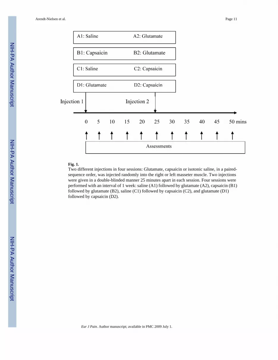
Fig. 1.Two different injections in four sessions: Glutamate, capsaicin or isotonic saline, in a paired-sequence order, was injected randomly into the right or left masseter muscle. Two injectionswere given in a double-blinded manner 25 minutes apart in each session. Four sessions wereperformed with an interval of 1 week: saline (A1) followed by glutamate (A2), capsaicin (B1)followed by glutamate (B2), saline (C1) followed by capsaicin (C2), and glutamate (D1)followed by capsaicin (D2).
Arendt-Nielsen et al. Page 11
Eur J Pain. Author manuscript; available in PMC 2009 July 1.
NIH
-PA Author Manuscript
NIH
-PA Author Manuscript
NIH
-PA Author Manuscript
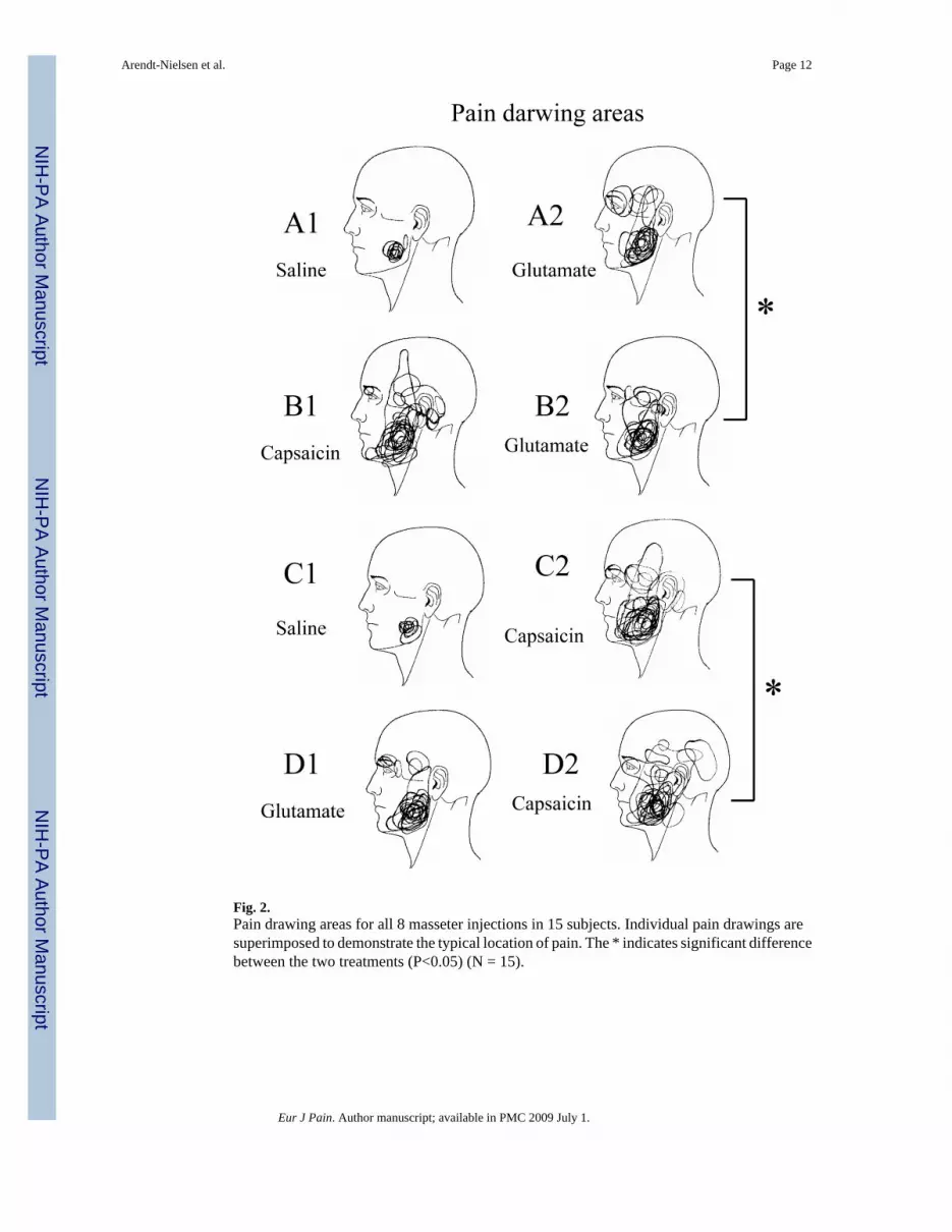
Fig. 2.Pain drawing areas for all 8 masseter injections in 15 subjects. Individual pain drawings aresuperimposed to demonstrate the typical location of pain. The * indicates significant differencebetween the two treatments (P<0.05) (N = 15).
Arendt-Nielsen et al. Page 12
Eur J Pain. Author manuscript; available in PMC 2009 July 1.
NIH
-PA Author Manuscript
NIH
-PA Author Manuscript
NIH
-PA Author Manuscript
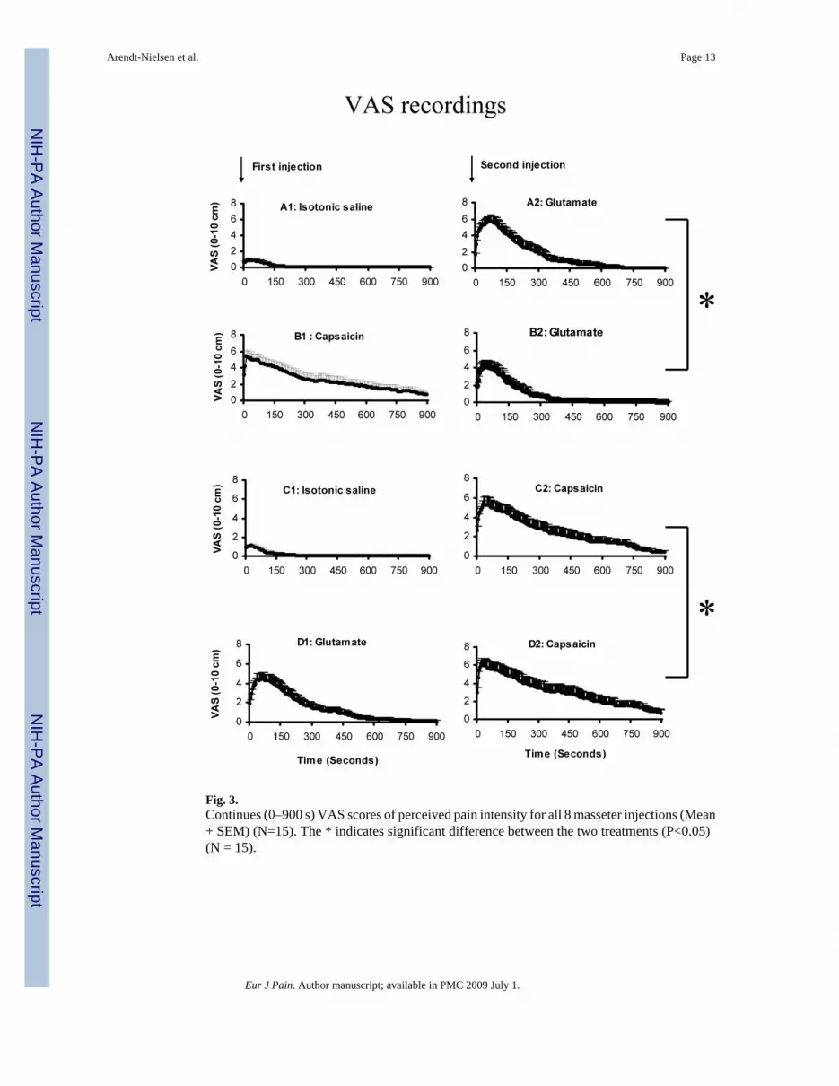
Fig. 3.Continues (0–900 s) VAS scores of perceived pain intensity for all 8 masseter injections (Mean+ SEM) (N=15). The * indicates significant difference between the two treatments (P<0.05)(N = 15).
Arendt-Nielsen et al. Page 13
Eur J Pain. Author manuscript; available in PMC 2009 July 1.
NIH
-PA Author Manuscript
NIH
-PA Author Manuscript
NIH
-PA Author Manuscript
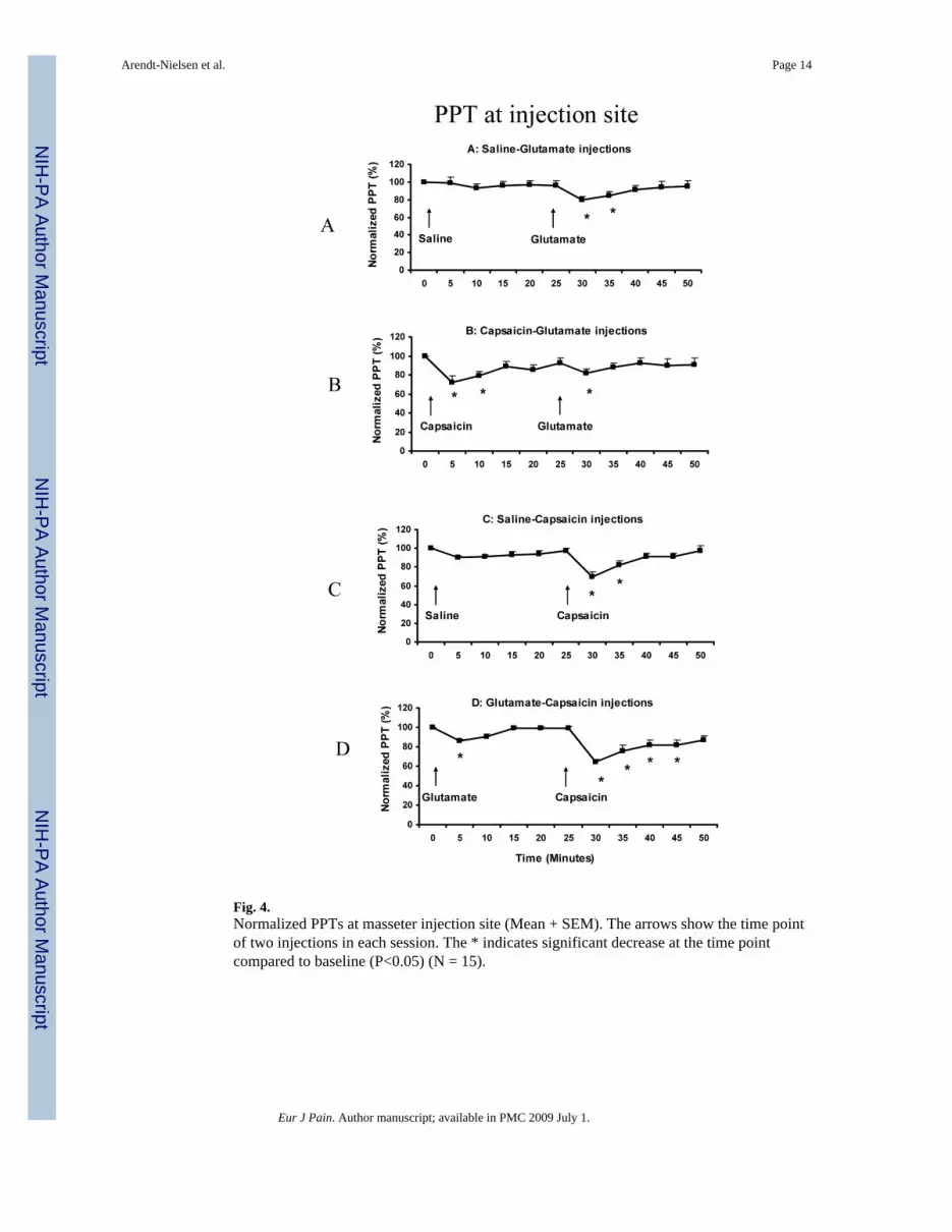
Fig. 4.Normalized PPTs at masseter injection site (Mean + SEM). The arrows show the time pointof two injections in each session. The * indicates significant decrease at the time pointcompared to baseline (P<0.05) (N = 15).
Arendt-Nielsen et al. Page 14
Eur J Pain. Author manuscript; available in PMC 2009 July 1.
NIH
-PA Author Manuscript
NIH
-PA Author Manuscript
NIH
-PA Author Manuscript
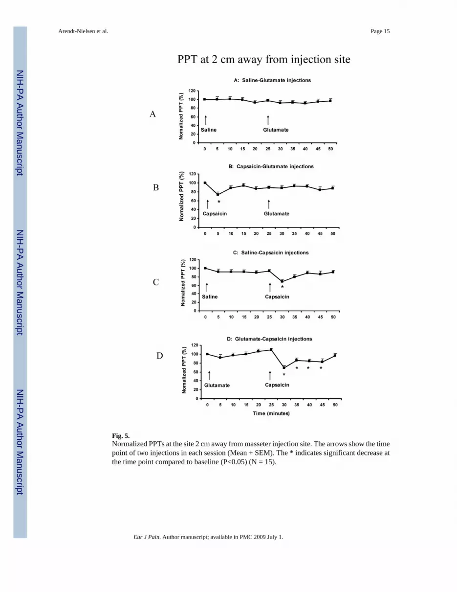
Fig. 5.Normalized PPTs at the site 2 cm away from masseter injection site. The arrows show the timepoint of two injections in each session (Mean + SEM). The * indicates significant decrease atthe time point compared to baseline (P<0.05) (N = 15).
Arendt-Nielsen et al. Page 15
Eur J Pain. Author manuscript; available in PMC 2009 July 1.
NIH
-PA Author Manuscript
NIH
-PA Author Manuscript
NIH
-PA Author Manuscript

Fig. 6.Normalized PPTols at masseter injection site. The arrows show the time point of two injectionsin each session (Mean + SEM). The * indicates significant decrease at the time point comparedto baseline (P<0.05) (N = 15).
Arendt-Nielsen et al. Page 16
Eur J Pain. Author manuscript; available in PMC 2009 July 1.
NIH
-PA Author Manuscript
NIH
-PA Author Manuscript
NIH
-PA Author Manuscript