integration of tactile input across fingers in a patient with finger agnosia
TRANSCRIPT

I
Ha
b
c
d
e
f
a
ARAA
KFSBT
1
filtlPLTRs
CN
(
0d
Neuropsychologia 49 (2011) 138–146
Contents lists available at ScienceDirect
Neuropsychologia
journa l homepage: www.e lsev ier .com/ locate /neuropsychologia
ntegration of tactile input across fingers in a patient with finger agnosia
elen A. Anemaa,e,∗, Krista E. Overvliet c,d,f, Jeroen B.J. Smeetsc, Eli Brennerc, H. Chris Dijkermana,b
Experimental Psychology, Helmholtz Institute, Utrecht University, Utrecht, The NetherlandsDepartment of Neurology, University Medical Centre Utrecht, Utrecht, The NetherlandsFaculty of Human Movement Sciences, Research Institute MOVE, VU University, Amsterdam, The NetherlandsLaboratory of Experimental Psychology, University of Leuven (K.U. Leuven), Leuven, BelgiumDepartment of Public Health, Academic Medical Centre, University of Amsterdam, Amsterdam, The NetherlandsDepartament de Psicologia Basica, Universitat de Barcelona, Barcelona, Spain
r t i c l e i n f o
rticle history:eceived 7 July 2010ccepted 7 November 2010vailable online 21 November 2010
eywords:inger agnosiaomatosensory processingody representationactile perception
a b s t r a c t
Finger agnosia has been described as an inability to explicitly individuate between the fingers, whichis possibly due to fused neural representations of these fingers. Hence, are patients with finger agnosiaunable to keep tactile information perceived over several fingers separate? Here, we tested a fingeragnosic patient (GO) on two tasks that measured the ability to keep tactile information simultaneouslyperceived by individual fingers separate. In experiment 1 GO performed a haptic search task, in whicha target (the absence of a protruded line) needed to be identified among distracters (protruded lines).The lines were presented simultaneously to the fingertips of both hands. Similarly to the controls, herreaction time decreased when her fingers were aligned as compared to when her fingers were stretchedand in an unaligned position. This suggests that she can keep tactile input from different fingers separate.In experiment two, GO was required to judge the position of a target tactile stimulus to the index finger,relatively to a reference tactile stimulus to the middle finger, both in fingers uncrossed and crossed posi-
tion. GO was able to indicate the relative position of the target stimulus as well as healthy controls, whichindicates that she was able to keep tactile information perceived by two neighbouring fingers separate.Interestingly, GO performed better as compared to the healthy controls in the finger crossed condition.Together, these results suggest the GO is able to implicitly distinguish between tactile information per-ceived by multiple fingers. We therefore conclude that finger agnosia is not caused by minor disruptionsof low-level somatosensory processing. These findings further underpin the idea of a selective impairedentat
higher order body repres. Introduction
Finger agnosia, the inability to recognize one’s own fingers orngers in general, has been frequently investigated throughout the
ast century. These studies were often performed in the context ofhe Gerstmann’s syndrome (finger agnosia, agraphia, acalculia andeft/right disorientation, e.g. Benton, 1961; Carota, Di Pietro, Ptak,oglia, & Schnider, 2004; Gerstmann, 1930; Mayer, Martory, Pegna,
andis, Delavelle, & Annoni, 1999; Roux, Boetto, Sacko, Chollet, &remoulet, 2003; Stengel, 1944; see for overview: Lebrun, 2005;usconi, Pinel, Dehaene, & Kleinschmidt, 2009). More detailed andpecific investigations targeting the mechanism underlying finger∗ Corresponding author at: Department of Public Health, Academic Medicalentre, University of Amsterdam, Meibergdreef 9, 1100 DD Amsterdam, Theetherlands. Tel.: +31 20 566 4684; fax: +31 30 697 2316.
E-mail addresses: [email protected], [email protected]. Anema).
028-3932/$ – see front matter © 2010 Elsevier Ltd. All rights reserved.oi:10.1016/j.neuropsychologia.2010.11.006
ion restricted to the fingers as underlying cause of finger agnosia.© 2010 Elsevier Ltd. All rights reserved.
agnosia are less frequently reported (Ettlinger, 1963; Kinsbourne& Warrington, 1962; Poeck & Orgass, 1969), which is unfortunatesince Benton observed already in 1961 that the four disorders thatconstitute Gerstmann’s syndrome did not mutually associate verywell (for similar critics see Critchley, 1966). Indeed, finger agnosiamight be of interest when it comes to explaining the cognitive rep-resentations of the body and as such has gained renewed interestin recent years (Anema et al., 2008; Haggard & Wolpert, 2005).
In 1944, Stengel suggested that a spatial mechanism mightbe the underlying deficit. Based on a thorough investigation of apatient who showed a general loss of spatial orientation, construc-tional apraxia and Gerstmann’s syndrome, the author proposedthat finger agnosia is “the inability of the patient to relate in spaceobjects which form part of an organised whole to each other and
to himself according to the rules acquired by experience” (Stengel,1944, p. 760). There appears to be an inability to judge the relativepositions of the fingers, more than there is an inability to recognize afinger per se. Indeed, the fact that finger agnosia patients also exhibit“toe agnosia” is consistent with the idea of a more general rather
sycho
tTaaagt1pKmaamtgtfiacafnfig“gfimweawtg“vcabgsp
ua(CHbotdst
(o(ptTfitnh
H.A. Anema et al. / Neurop
han a finger specific disorder (e.g. Mayer et al., 1999; Stengel, 1944;ucha, Steup, Smely, & Lange, 1997). However, these observationsre incompatible with Gerstmann’s definition of finger agnosia. Theuthor defined finger agnosia as being primarily a specific type ofutotopagnosia, or a loss of body orientation restricted to the fin-ers, “. . .as though the optic-tactile-kinesthetic image pertainingo the fingers were split off from the total body. . .” (Gerstmann,957, p. 867). This idea was taken even further by Benton whoroposed a concept of a distinct “finger schema” (Benton, 1959).insbourne and Warrington’s (1962) performed perhaps one of theost thorough investigations of finger agnosia. They confirmed
nd expanded Stengel’s hypothesis (1944) that finger agnosia isproblem in recognizing the serial order of the fingers. Whereasost tests of finger agnosia remain rather explicit (e.g. name the
ouched finger, touch the indicated finger, indicate the touched fin-er on a drawing of the hand) Kinsbourne and Warrington usedasks targeting implicit concepts of serial order. The authors testednger agnosia patients on a variety of haptic tasks. Twelve fingergnosic patients with Gerstman’s syndrome and 20 control casesompleted tasks in which information of relative finger positionnd knowledge of finger boundaries was essential for correct per-ormance. For example, patients were instructed to determine theumber of fingers that was in between two simultaneously touchedngers, which requires knowledge about the relation of the two fin-ers to the rest. Typically, finger agnosia patients responded withthree fingers in between” irrespective of the actual number of fin-ers. Another task investigated the ability to discriminate betweennger positions on basis of tactile features of a specific, uncom-on object. Patients’ fingers were moulded around an object, afterhich they had to pick out the corresponding object out of 4 mod-
ls, without looking at the object in their hand. All tested fingergnosia patients were unable to perform this task. Also, in a taskhich tested knowledge of finger boundaries, patients were unable
o discriminate between two simultaneously applied touches, tar-eted on one or two fingers. Patients erroneously responded withtwo” when the two touches were applied to one finger and viceersa. The authors interpreted the results as if tactile informationannot be processed in terms of the local position of the fingernd patients are unable to determine to which finger tactile inputelongs. Thus, the ability to comprehend the serial order of the fin-ers is not only lost, the fingers seem to be “fused” together into aingle representation and individuating between them is no longerossible.
Nevertheless, even though the exact mechanism is still poorlynderstood there appears to be a general agreement of fingergnosia being a disorder in the “individuation of the fingers”Haggard & Wolpert, 2005; Haggard, Kitadono, Press, & Taylor-lark, 2006; Kinsbourne & Warrington, 1962; Stengel, 1944).aggard and Wolpert (2005) discussed finger agnosia as part ofody representation disorders and categorized it as a “pathologyf segmentation”. The authors related finger agnosia to auto-opagnosia, a more general body mislocalisation disorder. In bothisorders the knowledge about the body part categories is pre-erved, but the unique position of these categorical elements withinhe overall spatial organisation of the body is lost.
A recent study conducted by some of the current authorsAnema et al., 2008) investigated finger agnosia within a theoryf dissociable representations of body image and body schemaDijkerman & de Haan, 2007). Finger agnosia was studied in threeatients with lesions affecting the angular gyrus by asking themo localise a touched finger using three different response modes.
hey were required to either reach to point towards the touchednger on their own hand, on a drawn map of a hand, name theargeted finger. The results revealed that these patients performedormally when reaching towards the touched finger on their ownand but failed to indicate this finger on a drawing of a hand orlogia 49 (2011) 138–146 139
to name it. Similar defects in the perception of other body partswere not observed. The findings provide converging evidence forfinger agnosia being a disorder of higher-order selective perceptualdifferentiation.
As described above, finger agnosia appears to be a problem inindividuating between the fingers which originates in a collec-tive fusion of the representation of the fingers and leads to theinability to accurately attribute (here) tactile information to the fin-ger receiving that information. When exploring objects for hapticobject recognition tactile input to the fingers needs to be combinedwith proprioceptive information of the location of those fingers(Lederman & Klatzky, 1987; Overvliet, Anema, Brenner, Dijkerman,& Smeets, in press). It could therefore be expected that it is problem-atic for patients with finger agnosia to integrate all the informationaccurately into a stable percept. Thus, separation might not justbe important for identifying the fingers, but may also be criti-cal for recognizing objects by touch. Interestingly, finger agnosicpatients are not known for impairments in haptic recognition ofcommon objects used in daily life and to our knowledge no studyhas reported (or even investigated) such impairments. However,it could be hypothesised that minor disruptions in more earlysomatosensory processes contribute to the inability to explicitlydistinguish between the fingers, without functionally hamperinghaptic object recognition. Perhaps the impairment arises at the pro-cessing level at which proprioceptive input about the individualfingers is combined with tactile stimulation.
In order to investigate this claim, we tested a patient (GO,see also Anema et al., 2008) with finger agnosia on two haptictasks that measured the ability to keep separate tactile informationsimultaneously perceived by individual fingers and have been pub-lished earlier by (Benedetti, 1985, 1988; Overvliet, Mayer, Smeets,& Brenner, 2008). The haptic search task published by Overvliet etal. (2008) used two different finger configurations, either fingersstretched and placed on several tangible line segments (line seg-ments condition) or fingers bent rather awkwardly in order to bealigned on one tangible continuous line (continuous line condition)(see Fig. 2). The participants were required to lift the finger underwhich they did not feel (a part of) a line. The results of Overvlietet al. (2008) study showed that healthy participants have fasterresponse times in the continuous line condition, as compared tothe line segments condition. This task additionally tests the integra-tion of somatosensory information perceived over several fingersinto a coherent percept. The authors therefore concluded that thealignment of the fingers in the continuous line condition allowedthe participants to integrate the input perceived over the fingersinto one object, which led to faster detection of the target. Thiseffect can only be achieved if tactile input to various fingers isdistinguished from each other and subsequently, in combinationwith proprioceptive input about the position of the fingertips, inte-grated into one percept. The second experiment used the paradigmof Benedetti (1985). GO was asked to judge the location of a (tar-get) touch on the fingertip relatively to the location of a second(reference) touch to the neighbouring finger tip. The two fingers(middle and ring finger) were stimulated simultaneously and theposition of the target finger was rotated around the reference fin-ger in a frontal plane (fingers uncrossed, fingers on top of eachother, exceedingly crossed over). By testing GO with these hap-tic experiments we investigated her ability to process and keepseparate simple features perceived by the fingers, and to subse-quently integrate them using proprioceptive information regardingthe positions of the fingertips.
We reason that if GO can still perform these tasks, she can stillkeep tactile information that is perceived over multiple fingers sep-arate, albeit on a low processing level. Consequently, the suggested“fusion of the fingers” must arise selectively at a higher processinglevel.
140 H.A. Anema et al. / Neuropsychologia 49 (2011) 138–146
F left pao nel, hao
2
2
wFrroi
mmtGwhhn1
raGafihtt(
oewlwa
hPf
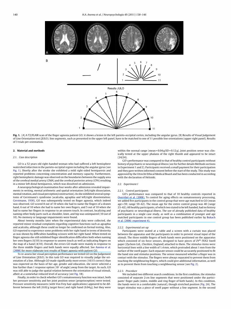
ig. 1. (A) A T2/FLAIR scan of the finger agnosia patient GO; it shows a lesion in thef Line Orientation test (JULO); line segments, such as presented in the upper left paf 3 trials per orientation.
. Material and methods
.1. Case description
GO is a 52 years old right-handed woman who had suffered a left hemisphereatershed infarction in the parieto-occipital region including the angular gyrus (see
ig. 1). Shortly after the stroke she exhibited a mild right-sided hemiparesis andeported problems concerning concentration and memory capacity. Furthermore,ight hemispheric damage was observed on the boundaries between the supply areaf the cerebral medial artery (CMA) and the cerebral posterior artery (CPA) resultingn a minor left distal hemiparesis, which was dissolved on admission.
A neuropsychological examination four weeks after admission revealed impair-ents in writing, mental arithmetic and spatial orientation (left/right dissociation,ental rotation, and visual perception/construction). As she exhibited several symp-
oms of Gerstmann’s syndrome (acalculia, agraphia and left/right disorientation;erstmann, 1930), GO was subsequently tested on finger agnosia, which indeedas observed. GO scored 8 out of 10 when she had to name the fingers of a drawnand, 6 out of 10 when she had to name her own fingers, and 5 out of 10 when shead to name her fingers in response to an unseen touch. In contrast, localizing andaming other body parts such as shoulder, knee, and hip was unimpaired (10 out of0). No memory or language impairments were found.
About twenty months later when the experimental data were collected, sheeported minor residual complaints regarding cognitive functions such as agraphiand acalculia, although these could no longer be confirmed on formal testing. Also,O reported to experience some problems with her right hand in terms of dexteritys was shown by difficulties handling scissors with her right hand. When tested onnger agnosia she still exhibited finger identification difficulties both when naminger own fingers (6/10) in response to unseen touch as well as indicating fingers onhe map of a hand (4/10). Overall, the errors GO made were mainly in response tohe three middle fingers and both hands were equally affected. See Anema et al.2008) for more elaborate test results of finger agnosia with patient GO.
To re-assess her visual perception performance we tested GO on the Judgementf Line Orientation (JULO). In this task GO was required to visually judge the ori-ntation of a line. Although GO made significantly more errors (10/15 correct) thanas expected on the basis of her age, gender and education, she never selected a
ine further than 1 response option (18◦ of angle) away from the target. As such, GO
as still able to judge the spatial relation between the orientation of visual stimuli,lbeit at a somewhat reduced level of accuracy (see Fig. 1B).Finally, in order to check whether GO’s somatosensory function was intact, both
er pressure sensitivity and her joint position sense (proprioception) were tested.ressure sensitivity measures (with Von Frey hair applications) appeared to be dif-erent between the left (0.02 g target force) and right hand (0.04 g), but they were
rieto-occipital cortex, including the angular gyrus. (B) Results of Visual Judgementve to be matched to one of 11 possible line orientations (upper right panel). Results
within the normal range (mean = 0.04 g/SD = 0.12 g). Joint position sense was clin-ically tested at the upper phalanx of the right thumb and appeared to be intact(24/24).
GO’s performance was compared to that of healthy control participants withouthistory of psychiatric or neurological illness (see for further details Methods sectionsin Experiment 1 and 2). Participants received a small payment for their participationand they gave written informed consent before the start of the study. This study wasapproved by the Utrecht Ethical Medical Board and has been conducted in accordingwith the declaration of Helsinki.
2.2. Experiment I
2.2.1. Control participantsGO’s performance was compared to that of 10 healthy controls reported in
Overvliet et al. (2008). To control for aging effects on somatosensory processing,we added five participants in the control group that were age matched to GO (meanage = 59; range 59–62). The mean age for the entire control group was 40 (range23–62). All healthy participants, of which two stated to be left handed, had no historyof psychiatric or neurological illness. The use of already published data of healthyparticipants in a single case study, as well as a combination of younger and agematched participants in one control group has been published earlier by Bukachet al. (2006: experiment 4).
2.2.2. Experimental set-upParticipants were seated at a table and a screen with a curtain was placed
between the apparatus and the participants in order to prevent visual input of thestimuli. The three middle fingers of both hands were positioned on the apparatuswhich consisted of six force sensors, designed to have pieces of ZY®-TEX2 Swellpaper (Zychem Ltd., Cheshire, England) attached to them. The stimulus items werehorizontal lines with a line width of 1.4 mm, which protruded about 1 mm from thesurface of the swell paper. Each separate sensor could be accurately positioned to fitthe hand size and stimulus positions. The sensor measured whether a finger was incontact with the stimulus. The fingers were always separated to prevent them fromtouching the neighbouring fingers, which could give additional information, as wellas to prevent them from touching a neighbouring sensor (see Fig. 2).
2.2.3. ProcedureWe included two different search conditions. In the first condition, the stimulus
consisted of separate 2 cm line segments that were positioned under the partici-pants’ finger pads (index, middle and ring finger of both hands). In this condition,the hands were in a comfortable (natural), though stretched position (Fig. 2A). Thetarget stimulus was a piece of swell paper without a line segment. In the second

H.A. Anema et al. / Neuropsycho
Fig. 2. The setup of experiment I. In the upper panel (A) a subject is performing thehotr
cifi
satfitepfislwta
2
tetiocacAfitt(ts
la
aptic search task with line segments (the target (no line) is below the middle fingerf the left hand) and in the lower panel (B) a subject is performing the haptic searchask in the continuous line condition (the target is below the middle finger of theight hand).
ondition a continuous 14.5 cm line was used instead of line segments. A 2 cm gapn this line served as the target. In this condition, the participants had to adjust theirnger positions to the line (Fig. 2B).
Each condition was tested in a separate block of 40 trials. In 25% of the trials thetimulus did not contain a target. Both blocks were repeated twice and presented inn ABBA design. Before each trial, the participant was asked specifically to positionhe fingertips on the sensor to prevent her from misplacing the fingers. When thengers were in the correct position, participants lifted the fingers and maintainedhat position, while the experimenter placed the next stimulus on the sensors. Thexperimenter started each trial by presenting a 4500 Hz tone. As soon as partici-ants heard the tone they had to lower the fingers onto the stimulus. Moving thengertips over the line stimuli was allowed as long as the fingers remained on theensors. Participants were instructed to lift the finger under which the target (noine) was positioned as soon as it was detected (target present trial). For trials in
hich the target was absent (all fingers were presented with a line; target absentrial), participants were instructed to lift all the fingers as soon as they detected thebsence.
.2.4. Design and data analysesOn both target present and absent trials, the reaction time was defined as the
ime from when the first finger contacted the sensor until a finger was lifted. Wexcluded trials on the basis of three different parameters. First, trials with reactionimes shorter than 100 ms were discarded as they were considered physiologicallymplausible. Second, trials with reaction times longer than 2 SD above the meanf the participant in question (either control or GO) were excluded as they wereonsidered outliers. Third, trials in which participants lifted the wrong finger werelso excluded from the analyses. Next, for each condition (separate line segments,ontinuous line) the median search time was computed for the remaining trials.fter these initial analyses, the data was further statistically analysed in two steps. Atrst the data of the healthy control group was tested on significant effects betweenhe line segments and continuous line conditions. This involved a 2 (target condition:arget absent, present) × 2 (line condition: continuous line, separate line segments) × 2
group: student controls, age matched controls) Repeated Measures Anova with botharget condition and line condition as within subject factors and group as betweenubjects factor. Only significant effects are reported.Second, it was tested whether GO exhibited a similar benefit of the continuousine condition as compared to the line segments in detecting a target, or detecting thebsence of a target. For each of the four conditions GO’s scores were tested against
logia 49 (2011) 138–146 141
that of the healthy controls using Crawford and Garthwaite’s test for abnormalityscores in single case studies (Crawford & Garthwaite, 2002).
3. Results
3.1. Accuracy
GO showed a larger proportion of errors as compared to the con-trol group. GO had on average 25% errors (for line segments: 78%correct, 14% false positives, no false negatives, and 8% wrong fin-ger; and for the continuous line: 71% correct, 11% false positives, 4%false negatives and 14% wrong finger). The percentage of errors wasconsiderably higher than in healthy controls (line segments: 13.5%;continuous line 14.5%, respectively). Errors were always made byGO at targets presented to the right hand, either the middle or thering finger. False positives may have been caused by reduced tac-tile sensitivity of GO on the right hand. Moreover, when testing GO,we noticed that she experienced difficulties maintaining her righthand fingers on the small strips of swell paper. Chi-square calcula-tions on the proportion of false positives revealed that these valuesdid not differ significantly between the line segments (22%) andthe continuous line condition (39%; �2 < 3.84). Similar results werefound for the proportion “wrong finger” (line segments = 35% andcontinuous line = 48%; �2 < 3.84).
The interim analyses of the accuracy scores reveal a relativelylarge number of mistakes on the trials in which the target was posi-tioned underneath one of the right hand fingers. Two observationscan provide insight in this outcome. As is described above in Sec-tion 2.1, GO’s right hand is less sensitive to touch as compared toher left hand, although within normal range. Also, the left sidedlesion lead to minor but persistent motor problems with her righthand as was reported by GO (see Section 2.1). Indeed, while testing,GO exhibited difficulties maintaining the awkward position of theright hand fingers on to the stimuli. Since her finger agnosia affectedboth hands and to keep our measurements as valid as possible weexcluded (post hoc) the right hand trials in the RT analyses.
3.2. Reaction time
Data of the controls and GO are plotted in Fig. 3. The ANOVAon the healthy control subject reaction times showed a significantmain effect of target condition (F(1,13) = 12.83, p < 0.01): reactiontimes were faster when the target was present (2011 ms) thanwhen the target was absent (2694 ms). More importantly, a maineffect of line condition was observed (F(1,13) = 9.533, p < 0.01)indicating that search time decreased when fingers were posi-tioned on one continuous line (2168 ms) as compared to thepositioning on line segments (2537 ms). Furthermore this effect didnot differ between groups (interaction F(1,13) < 1). These resultsare in line with the earlier results of Overvliet et al. (2008).However, the age matched control group was overall slower ascompared to the younger subjects (age matched = 3469 ms; youngcontrols = 1235 ms; (F(1,13) = 78.29, p < 0.01)). It has been shownmany times that older adults are slower in a wide variety of tasks(for tactile tasks: e.g. Ballesteros & Reales, 2004; Cole, Rotella, &Harper, 1998; Overvliet, Wagemans, & Krampe, 2010).
Most importantly, GO’s search times showed a similar responsepattern to the control group. We compared GO’s performance tothe age-matched control group using Crawford and Garthwaite’stest (Crawford & Garthwaite, 2002) for abnormality scores in sin-gle case studies. We did not find any significant differences in
reaction time behaviour in neither the line segment nor the contin-uous line condition. The statistics for the line segments conditionwere as follows: target absent, GO (3744 ms) versus control group(2496 ms; SD = 1614), t (df = 14) < 1 and target present, GO (2972 ms)versus control group (1813 ms; SD = 960), t (df = 14) = 1.169. For the
142 H.A. Anema et al. / Neuropsychologia 49 (2011) 138–146
F atched rget ab
cvg(
3
3
(tTbwga1tofitAa4n0erwramtTbcdu
ig. 3. Results of experiment I for the healthy control subjects, the healthy age mifferent conditions for target present (diamond symbol and dotted line) and for ta
ontinuous line the statistics were: target absent, GO (2806 ms)ersus control group (2014 ms; SD = 1407 ms), t (df = 14) < 1), tar-et present: GO (2635) versus control group (1597 ms; SD = 1015), tdf = 14) < 1.
.3. Experiment II
.3.1. Experimental set-upParticipants were seated at a table opposite the experimenter
see Fig. 4A). During testing either the left or right hand was posi-ioned on top of the table with the palm of the hand downward.o prevent participants from relying on visual feedback, a woodenoard was positioned over the participant’s hand. The other handas on top of the board to perform the responses. To enable the fin-
ers to be crossed and manipulated in various orientations we usedsimilar device as was used in the Benedetti studies (Benedetti,
985, 1988; see Fig. 4B). The index finger was inserted in a tube andhe middle finger was rotated around the index finger. The positionf the middle finger was obtained by the experimenter moving thisnger and confirmed with a calibrated clinical goniometer (protrac-or, Medizintechnik KaWe, Germany) and subsequently recorded.s a result, the exact position of the middle finger varied somewhatround the intended target orientations used in the experiment (0◦,5◦, 90◦ and 135◦). In tactile trials stimuli consisted of two simulta-eously and manually applied simple tactile stimulations, a small.5 mm diameter, plastic pin applied on the index finger (refer-nce), and a 5 mm diameter relatively blunt pen like object, furthereferred to as “ball” applied on the middle finger (target). Responsesere made by indicating the perceived position of either the finger
elative to the reference finger, or the tactile stimulus (“ball”) rel-tive to the pin on a calibrated disk (see Fig. 4B). Responses wereade in the frontal plane. The disk itself was positioned on top of
he wooden board and this board covered the stimulated fingers.
he rotating bar, which was placed on the calibrated disk, coulde aligned by means of turning the bar so that the target pointorrelated with the perceived target position. Two symbols wererawn on this bar, one in the middle indicating the reference stim-lus (index finger or .5 mm small pin) and one on the end of thed control subjects and GO separately. The mean and standard errors for the twosent (square symbols and solid line) are depicted.
bar indicating the target stimulus (middle finger or 5 mm blunt pin(“ball”)). The experimenter recorded the set angle of the bars, ofwhich horizontal was considered to be 0◦.
3.3.2. Design and procedureA 2 (task: perceived relative position of ball, perceived relative
position of finger) × 4 (position: 0◦, 45◦, 90◦ and 135◦) repeatedmeasures design was used in this experiment. Each position × taskcondition was presented 4 times, except for the 135◦ conditionthat was tested 8 times. Obtaining and maintaining a fingers-crossed position can be difficult for some participants, especiallywhen small differences in positions are requested. Therefore wetested only one crossed condition, that of almost maximally crossed(135◦). To correct for differences in the number of trials betweencrossed and uncrossed finger positions, we added 4 extra trials tothe crossed condition. Together this resulted in 40 trials in total. Allorientation × task combinations were presented in a random order.
A trial started when the experimenter placed the middle fin-ger in one of four possible target positions (0◦, 45◦, 90◦ and 135◦).The exact final target position of the middle finger was verified bythe experimenter using the goniometer. Next, in a finger conditiontrial the experimenter asked the participant to judge the orienta-tion of the middle finger with respect to the index finger. In the ballcondition trials, two simultaneous above threshold tactile stimula-tions (5 mm blunt pin on the middle finger and .05 mm sharp pinon the index finger) were manually applied to the fingers by theexperimenter for approximately 1500 ms (subjectively controlledby the experimenter) and participants were required to judge theposition of the ball with respect to the pin. In both cases responseswere made by rotating a bar on a disk (see Fig. 4A). In order toprevent participants from using other reference information, onlythe 0◦ position was indicated on the disc. After the response wasrecorded by the experimenter, the middle finger was returned to
the starting position (about −4◦) and the response bar was presetto about 0◦. Finger condition trials and ball condition trials wererandomly interleaved. Prior to testing, participants practiced thetask without the stimuli being applied to the fingers, in order toascertain correct handling of the response device.
H.A. Anema et al. / Neuropsychologia 49 (2011) 138–146 143
F hand in experiment II. The middle finger is stimulated by the “ball” and the index fingerb r to the left or right.
3
dpDiort
iGad
33faatjw(tF(1t
pj
0
0 0
0
45 45
45 45
90 90
90 90
135
135
135
135
0
45 45
4545
0
0
135
135
135
135
90
900 90
90
BALL FINGERControlsTarget
ControlsTarget
GOTarget
GOTarget
ig. 4. (A) A schematic drawing of the setup of experiment II. (B) A close up of they a pin simultaneously. (C) The response disc. Note: the bar could be rotated eithe
.3.3. Data analysesA 2 × 4 (task: perceived relative position of the ball (ball con-
ition), perceived relative position of finger (finger condition);osition: 0◦, 45◦, 90◦ and 135◦) repeated measures design was used.eviations from the target position were calculated by subtract-
ng the target position for each trial (in angular degrees) from therientation provided by the participant. A 2 × 4 (task × position)epeated measures ANOVA was performed on the averages for eachask × position combination.
We used Crawford and Garthwaite’s test for abnormality scoresn single case studies (Crawford & Garthwaite, 2002) in order to testO’s performance against that of healthy controls. When meansre mentioned in the text, standard errors are also given (meaneviation (±SE)).
.3.4. Results
.3.4.1. Age matched controls. The averaged responses for the dif-erent position conditions (when applicable, further referred tos PC 0◦, 45◦, 90◦ and 135◦) for both GO and the controlsre plotted in Fig. 5. Visual inspection of the data suggestshat in a fingers crossed position (PC = 135◦) healthy controlsudge the ball to be at a an uncrossed position (76◦ ± 2◦)
hereas they judge the fingers correctly as being crossed121◦ ± 6◦). The analysis of variance (ANOVA) of the devia-ions revealed a significant main effect of task (Ball = −10◦ ± 2.3◦;inger = −0.15 ± 2.3◦; F(1,4) = 12.60, p < 0.05) and of positionPC 0◦ = 20◦ ± 2.8◦; PC 45◦ = 11◦ ± 2.9◦; PC 90◦ = −16◦ ± 7.7; PC
35◦ = −36◦ ± 3.1◦; F(1,4) = 27.69, p < 0.05) as well as a significantask × position interaction effect (F(3,12) = 10.53, p < 0.05).The significant interaction was further explored using one sam-le t-tests (test value 0) on the difference between ball and finger
udgments for each condition. In the 135◦ condition, there was a sig-
Fig. 5. The average responses in experiment II for both the controls (upper left andright panel) and GO (lower left and right panel). The two left panels are the responsesto the tactile stimuli and the right two panels are responses to the finger configu-ration. The dotted lines represent the answers of the controls and GO and the solidlines the orientation of the stimulus.

1 sycho
n(wer(
3mafacGPpgt(
gpzctmypM0
iGGMtcbG−ah
4
gtihdfiaftFptsp
btfig
44 H.A. Anema et al. / Neurop
ificant difference in error between the ball and finger judgmentsmean difference in error = −45◦ t (df = 4) = −9.391, p < 0.0125),hereas this was not so for the other positions (mean differences in
rror at PC 0◦ = 11◦ ± 9◦; PC 45◦ = 2◦ ± 8◦; PC 90◦ = −8◦ ± 5◦). Theseesults are a replication of the results of the study by Benedetti1985).
.3.4.2. GO. Visual inspection of the data (see Fig. 5) reveals thatost aspects of GO’s performance on the finger judgement task
re within normal ranges. Indeed, for the PC 45◦, 90◦ and 135◦ weailed to observe significant differences between performance of GOnd that of the healthy controls (PC 45◦: GO finger = −2.3◦ ± 14◦;ontrol finger = 9.6◦ ± 12◦), t (df4) = −0.872, p = 0.432; PC 90◦:O finger = −8.75 ± 10◦; control finger = −11.6◦ ± 22◦, p = 0.913;C = 135◦: GO finger = −3.75◦ ± 9◦; control finger = −14.1◦ ± 12◦,= 0.243). However, when judging the relative position of the fin-er in a horizontal position (PC 0◦: GO’s performance deviated fromhat of the healthy controls (GO = 62◦ ± 8◦); control = 16◦ ± 14◦, tdf = 4) = 4.516, p < 0.05).
Furthermore, visual observation of the ball condition data sug-ests that she is able to perform the task, except for the horizontalosition. However, comparison of her performance in this hori-ontal position (PC 0◦) to that of the healthy controls, failed toonfirm a significant difference (GO = 57◦ ± 3/control = 25◦ ± 14◦,(df = 4) = 1.982, p = 0.118). In fact, the performance of the ageatched controls in the horizontal position in the ball condition,
ields more error than one would expect from the most “sim-le” position condition (Ball condition, LH = 41◦, NO = 18◦, KL = 8◦,T = 20◦, MA = 40◦; group mean (25.35) differs significantly from
◦ t (df = 4) = 3.921, p < 0.05).Her performance in the 45◦ and 90◦ conditions was sim-
lar as compared to the healthy control participants (PC 45◦:O = 25◦ ± 15◦; control = 12 ± 9◦, t (df = 4) = 1.359, p = 0.246; PC 90◦:O = −6.8◦ ± 8◦; control = −20◦ ± 13◦, t (df = 4) = 0.916, p = 0.411).ost strikingly, her performance in the 135◦ condition appears
o be closer to the veridical orientation compared to the healthyontrols. That is, the healthy control subjects judged the ball toe at a 76◦ position instead of 135◦ (error of −59◦ ± 4◦), whereasO was able to perform more accurately in this condition (error of11◦ ± 10◦). Her lower amount of error was further confirmed bysignificant difference between her performance and that of theealthy controls (t (df = 4) = 11.393, p < 0.001).
. Discussion
In two experiments we investigated how a patient with fin-er agnosia (GO), who is impaired in identifying which finger isouched, performed on two tasks that depended on implicitly keep-ng separate tactile information applied to different fingers. Despiteer inability to distinguish between her fingers, GO was able toistinguish between line segments simultaneously touched by herngers as she did not significantly differ from healthy controls inhaptic search experiment. Similar to the controls, she benefitted
rom a position where the fingers were aligned. That allowed hero integrate the perceived line stimuli into one coherent object.urthermore, in a second experiment, GO was able to judge theosition of a tactile stimulus presented to a finger (small ball) rela-ively to a second stimulus to the adjacent finger (sharp pin). To oururprise, GO performed better than controls in the fingers crossedosition.
In order to perform these tasks, tactile information processedy a finger needs to be combined with proprioceptive informa-ion about the position as well as other metric properties of thatnger. This requires the capacity to distinguish between the fin-ers, albeit at an implicit level. As such, the suggested fusion of the
logia 49 (2011) 138–146
fingers (Kinsbourne & Warrington, 1962) cannot be explained bya misinterpretation of tactile and proprioceptive information at alower level. And even so, it seems that GO’s finger agnosia is notcaused by minor disruptions of low-level somatosensory process-ing.
A surprising finding in our second experiment might providefurther insight on the aetiology of finger agnosia. In experiment IIGO was not hampered by the crossed finger condition when judgingthe spatial position of the tactile stimulus (small ball), relatively to areference stimulus (sharp pin). In line with Benedetti’s observations(1985, 1988) our age matched controls perceived the location of thesmall ball during a fingers crossed position, as if the fingers wereuncrossed. Benedetti explained this illusion by suggesting that thetactile perceptual system is limited and unable to detect the veridi-cal information when the fingers are crossed beyond the borders offunctionality.
Our observations can be further explained within an ele-gant model of somatoperceptual information processing (Longo,Azanon, & Haggard, 2010). In their article the authors describesomatosensory processing along three types of body representa-tions: a superficial schema, a model of body size and shape, and apostural schema. In order to judge the spatial position of the ballrelatively to that of the sharp pin, at first one needs to locate thetouch on the body surface, which is subserved by the superficialschema. Thus, tactile information is initially coded in a somato-topical map of skin coordinates. Subsequently, the configurationof the joints needs to be calculated and scaled along informationabout the body size and shape. The rescaling process implies thattactile location is converted from a somatotopic reference frame,to an external one, a sequence that has been demonstrated forthe arms and legs (e.g. Azanon & Soto-Faraco, 2008; Overvliet,Azanon, & Soto-Faraco, 2009; Schicke & Röder, 2006; Yamamoto& Kitazawa, 2001). In some cases this remapping process fails, asis the case in the crossed fingers condition in Benedetti’s experi-ment.
In general, uncommon body postures affect basic somatosen-sory processes in such a way that the sensory information isprocessed as if the bodily posture was normal. Indeed, Yamamotoand Kitazawa (2001) suggested that the brain has a default con-dition that assumes that body parts are rarely crossed. In theirexperiment participants, were required to judge the temporalorder of two subsequently applied tactile stimuli (stimulus inter-val range 0–1500 ms) to the left and right hand, with their eyesclosed. Responses were made by lifting the finger of the handthat was tapped first, or in the second half of the experiment,second. The results showed that when crossing the hands, manysubjects reported inverted judgments at intervals up to 200 ms.The authors concluded that these speeded temporal order judg-ments were made before the actual external location of the bodypart is incorporated in the remapping processes. The judgementsare made on basis of a “normal postural situation”; left hand in theleft hemifield, right hand in the right hemifield. When more time isseparating the two stimuli, the postural schema can be updatedwith the new postural position (left hand on the right and viceversa) and correct answers are given.
Overall, the results of Benedetti’s experiments revealed a verystrong influence of this normal posture which Longo et al. (2010)indicated as “canonical posture” or “default posture”. Even thoughhealthy controls were entirely aware of their fingers being crossed,they still perceived the tactile stimuli as if the fingers wereuncrossed. Therefore it seemed as if there was a “reluctance” to
update the postural schema with the crossed fingers position. Thisillusory feeling of uncrossed fingers is known as the “Aristotleillusion” and already described by Tastevin (1937), who exploredthe range of this illusion for other body parts (lips, tongue, face,etc.). Recent experiments in our lab, in which participants were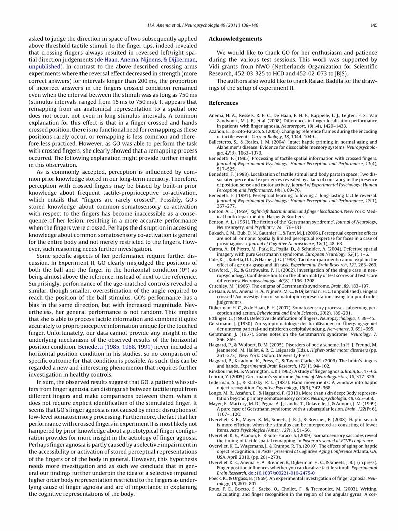
sycho
aattuecoe(rdecpfwoi
mpkwswqwkfe
cbbSsrbetafiuphsri
fddslphrPtonehlt
H.A. Anema et al. / Neurop
sked to judge the direction in space of two subsequently appliedbove threshold tactile stimuli to the finger tips, indeed revealedhat crossing fingers always resulted in reversed left/right spa-ial direction judgements (de Haan, Anema, Nijnens, & Dijkerman,npublished). In contrast to the above described crossing armsxperiments where the reversal effect decreased in strength (moreorrect answers) for intervals longer than 200 ms, the proportionf incorrect answers in the fingers crossed condition remainedven when the interval between the stimuli was as long as 750 msstimulus intervals ranged from 15 ms to 750 ms). It appears thatemapping from an anatomical representation to a spatial oneoes not occur, not even in long stimulus intervals. A commonxplanation for this effect is that in a finger crossed and handsrossed position, there is no functional need for remapping as theseositions rarely occur, or remapping is less common and there-ore less practiced. However, as GO was able to perform the taskith crossed fingers, she clearly showed that a remapping process
ccurred. The following explanation might provide further insightn this observation.
As is commonly accepted, perception is influenced by com-on prior knowledge stored in our long-term memory. Therefore,
erception with crossed fingers may be biased by built-in priornowledge about frequent tactile-proprioceptive co-activation,hich entails that “fingers are rarely crossed”. Possibly, GO’s
tored knowledge about common somatosensory co-activationith respect to the fingers has become inaccessible as a conse-
uence of her lesion, resulting in a more accurate performancehen the fingers were crossed. Perhaps the disruption in accessing
nowledge about common somatosensory co-activation is generalor the entire body and not merely restricted to the fingers. How-ver, such reasoning needs further investigation.
Some specific aspects of her performance require further dis-ussion. In Experiment II, GO clearly misjudged the positions ofoth the ball and the finger in the horizontal condition (0◦) aseing almost above the reference, instead of next to the reference.urprisingly, performance of the age-matched controls revealed aimilar, though smaller, overestimation of the angle required toeach the position of the ball stimulus. GO’s performance has aias in the same direction, but with increased magnitude. Nev-rtheless, her general performance is not random. This implieshat she is able to process tactile information and combine it quiteccurately to propropioceptive information unique for the touchednger. Unfortunately, our data cannot provide any insight in thenderlying mechanism of the observed results of the horizontalosition condition. Benedetti (1985, 1988, 1991) never included aorizontal position condition in his studies, so no comparison ofpecific outcome for that condition is possible. As such, this can beegarded a new and interesting phenomenon that requires furthernvestigation in healthy controls.
In sum, the observed results suggest that GO, a patient who suf-ers from finger agnosia, can distinguish between tactile input fromifferent fingers and make comparisons between them, when itoes not require explicit identification of the stimulated finger. Iteems that GO’s finger agnosia is not caused by minor disruptions ofow-level somatosensory processing. Furthermore, the fact that hererformance with crossed fingers in experiment II is most likely notampered by prior knowledge about a prototypical finger configu-ation provides for more insight in the aetiology of finger agnosia.erhaps finger agnosia is partly caused by a selective impairment inhe accessibility or activation of stored perceptual representationsf the fingers or of the body in general. However, this hypothesis
eeds more investigation and as such we conclude that in gen-ral our findings further underpin the idea of a selective impairedigher order body representation restricted to the fingers as under-ying cause of finger agnosia and are of importance in explaininghe cognitive representations of the body.
logia 49 (2011) 138–146 145
Acknowledgements
We would like to thank GO for her enthusiasm and patienceduring the various test sessions. This work was supported byVidi grants from NWO (Netherlands Organization for ScientificResearch, 452-03-325 to HCD and 452-02-073 to JBJS).
The authors also would like to thank Rafael Badilla for the draw-ings of the setup of experiment II.
References
Anema, H. A., Kessels, R. P. C., De Haan, E. H. F., Kappelle, L. J., Leijten, F. S., VanZandvoort, M. J. E., et al. (2008). Differences in finger localisation performancein patients with finger agnosia. Neuroreport, 19(14), 1429–1433.
Azanon, E., & Soto-Faraco, S. (2008). Changing reference frames during the encodingof tactile events. Current Biology, 18, 1044–1049.
Ballesteros, S., & Reales, J. M. (2004). Intact haptic priming in normal aging andAlzheimer’s disease: Evidence for dissociable memory systems. Neuropsycholo-gia, 42(8), 1063–1070.
Benedetti, F. (1985). Processing of tactile spatial information with crossed fingers.Journal of Experimental Psychology: Human Perception and Performance, 11(4),517–525.
Benedetti, F. (1988). Localization of tactile stimuli and body parts in space: Two dis-sociated perceptual experiences revealed by a lack of constancy in the presenceof position sense and motor activity. Journal of Experimental Psychology: HumanPerception and Performance, 14(1), 69–76.
Benedetti, F. (1991). Perceptual learning following a long-lasting tactile reversal.Journal of Experimental Psychology: Human Perception and Performance, 17(1),267–277.
Benton, A. L. (1959). Right-left discrimination and finger localization. New York: Med-ical book department of Harper & Brothers.
Benton, A. L. (1961). The fiction of the ‘Gerstmann syndrome’. Journal of Neurology,Neurosurgery, and Psychiatry, 24, 176–181.
Bukach, C. M., Bub, D. N., Gauthier, I., & Tarr, M. J. (2006). Perceptual expertise effectsare not all or none: Spatially limited perceptual expertise for faces in a case ofprosopagnosia. Journal of Cognitive Neuroscience, 18(1), 48–63.
Carota, A., Di Pietro, M., Ptak, R., Poglia, D., & Schnider, A. (2004). Defective spatialimagery with pure Gerstmann’s syndrome. European Neurology, 52(1), 1–6.
Cole, K. J., Rotella, D. L., & Harper, J. G. (1998). Tactile impairments cannot explain theeffect of age on a grasp and lift task. Experimental Brain Research, 121, 263–269.
Crawford, J. R., & Garthwaite, P. H. (2002). Investigation of the single case in neu-ropsychology: Confidence limits on the abnormality of test scores and test scoredifferences. Neuropsychologia, 40(8), 1196–1208.
Critchley, M. (1966). The enigma of Gerstmann’s syndrome. Brain, 89, 183–197.de Haan, A. M., Anema, H. A., Nijnens, M. C., & Dijkerman, H. C. (unpublished). Fingers
crossed! An investigation of somatotopic representations using temporal orderjudgements.
Dijkerman, H. C., & de Haan, E. H. (2007). Somatosensory processes subserving per-ception and action. Behavioural and Brain Sciences, 30(2), 189–201.
Ettlinger, G. (1963). Defective identification of fingers. Neuropsychologia, 1, 39–45.Gerstmann, J. (1930). Zur symptomatologie der hirnläsionen im Übergangsgebiet
der unteren parietal-und mittleren occipitalwindung. Nervenartz, 3, 691–695.Gerstmann, J. (1957). Some notes on the Gerstmann’s syndrome. Neurology, 7,
866–869.Haggard, P., & Wolpert, D. M. (2005). Disorders of body scheme. In H. J. Freund, M.
Jeannerod, M. Hallet, & R. C. Leiguarda (Eds.), Higher-order motor disorders (pp.261–273). New York: Oxford University Press.
Haggard, P., Kitadono, K., Press, C., & Taylor-Clarke, M. (2006). The brain’s fingersand hands. Experimental Brain Research, 172(1), 94–102.
Kinsbourne, M., & Warrington, E. K. (1962). A study of finger agnosia. Brain, 85, 47–66.Lebrun, Y. (2005). Gerstmann’s syndrome. Journal of Neurolinguistics, 18, 317–326.Lederman, S. J., & Klatzky, R. L. (1987). Hand movements: A window into haptic
object recognition. Cognitive Psychology, 19(3), 342–368.Longo, M. R., Azanon, E., & Haggard, P. (2010). More than skin deep: Body represen-
tation beyond primary somatosensory cortex. Neuropsychologia, 48, 655–668.Mayer, E., Martory, M. D., Pegna, A. J., Landis, T., Delavelle, J., & Annoni, J. M. (1999).
A pure case of Gerstmann syndrome with a subangular lesion. Brain, 122(Pt 6),1107–1120.
Overvliet, K. E., Mayer, K. M., Smeets, J. B. J., & Brenner, E. (2008). Haptic searchis more efficient when the stimulus can be interpreted as consisting of feweritems. Acta Psychologica (Amst), 127(1), 51–56.
Overvliet, K. E., Azanon, E., & Soto-Faraco, S. (2009). Somatosensory saccades revealthe timing of tactile spatial remapping. In Poster presented at ECVP conference.
Overvliet, K. E., Wagemans, J., & Krampe, R. Th. (2010). The effects of aging on hapticobject recognition. In Poster presented at Cognitive Aging Conference Atlanta, GA,USA, April 2010, (pp. 261–273).
Overvliet, K. E., Anema, H. A., Brenner, E., Dijkerman, H. C., & Smeets, J. B. J. (in press).
Finger position influences whether you can localize tactile stimuli. ExperimentalBrain Research, doi:10.1007/s00221-010-2475-0Poeck, K., & Orgass, B. (1969). An experimental investigation of finger agnosia. Neu-rology, 19, 801–807.
Roux, F. E., Boetto, S., Sacko, O., Chollet, F., & Tremoulet, M. (2003). Writing,calculating, and finger recognition in the region of the angular gyrus: A cor-

1 sycho
R
S
S
sensation qu’on peut y produire. Encéphale, 33, 57–84.
46 H.A. Anema et al. / Neurop
tical stimulation study of Gerstmann syndrome. Journal of Neurosurgery, 99(4),716–727.
usconi, E, Pinel, P., Dehaene, S., & Kleinschmidt, A. (2009). The enigma of Gerst-
mann’s syndrome revisited: A telling tale of the vicissitudes of neuropsychology.Brain, 33(2), 320–332.chicke, T., & Röder, B. (2006). Spatial remapping of touch across hand and foot.Proceedings of the National Academy of Science, 103(31), 11808–11813.
tengel, E. (1944). Loss of spatial orientation, constructional apraxia and Gerst-mann’s syndrome. The British Journal of Psychiatry, 90, 753–760.
logia 49 (2011) 138–146
Tastevin, J. (1937). En partant de l’experience d’Aristote – les déplacements artificielsdes parties du corps ne sont pas suivis par le sentiment de ces parties ni par les
Tucha, O., Steup, A., Smely, C., & Lange, K. W. (1997). Toe agnosia in Gerst-mann syndrome. Journal of Neurology, Neurosurgery, and Psychiatry, 63, 399–403.
Yamamoto, S., & Kitazawa, S. (2001). Reversal of subjective temporal order due toarm crossing. Nature Neuroscience, 4(7), 759–765.