inhibition of cholesterol biosynthesis impairs insulin secretion and voltage-gated calcium channel...
TRANSCRIPT
Inhibition of Cholesterol Biosynthesis Impairs InsulinSecretion and Voltage-Gated Calcium Channel Functionin Pancreatic �-Cells
Fuzhen Xia, Li Xie, Anton Mihic, Xiaodong Gao, Yi Chen, Herbert Y. Gaisano, and Robert G. Tsushima
Departments of Medicine and Physiology, University of Toronto, Toronto, Ontario, Canada M5S 1A8
Insulin secretion from pancreatic �-cells is mediated by theopening of voltage-gated Ca2� channels (CaV) and exocytosisof insulin dense core vesicles facilitated by the secretory sol-uble N-ethylmaleimide-sensitive factor attachment proteinreceptor protein machinery. We previously observed that�-cell exocytosis is sensitive to the acute removal of mem-brane cholesterol. However, less is known about the chronicchanges in endogenous cholesterol and its biosynthesis inregulating �-cell stimulus-secretion coupling. We examinedthe effects of inhibiting endogenous �-cell cholesterol biosyn-thesis by using the squalene epoxidase inhibitor, NB598. Theexpression of squalene epoxidase in primary and clonal�-cells was confirmed by RT-PCR. Cholesterol reduction of36–52% was observed in MIN6 cells, mouse and human pan-creatic islets after a 48-h incubation with 10 �M NB598. Asimilar reduction in cholesterol was observed in the subcel-
lular compartments of MIN6 cells. We found NB598 signifi-cantly inhibited both basal and glucose-stimulated insulinsecretion from mouse pancreatic islets. CaV channels weremarkedly inhibited by NB598. Rapid photolytic release of in-tracellular caged Ca2� and simultaneous measurements of thechanges in membrane capacitance revealed that NB598 alsoinhibited exocytosis independently from CaV channels. Theseeffects were reversed by cholesterol repletion. Our resultsindicate that endogenous cholesterol in pancreatic �-cellsplays a critical role in regulating insulin secretion. Moreover,chronic inhibition of cholesterol biosynthesis regulates thefunctional activity of CaV channels and insulin secretorygranule mobilization and membrane fusion. Dysregulation ofcellular cholesterol may cause impairment of �-cell function,a possible pathogenesis leading to the development of type 2diabetes. (Endocrinology 149: 5136–5145, 2008)
PANCREATIC �-CELLS secrete insulin in response toelevated glucose to maintain blood glucose homeosta-
sis. Defects in �-cell insulin secretion lead to hyperglycemiaand development of type 2 diabetes. The distal events un-derling stimulus-secretion coupling of insulin secretion havebeen well documented and are characterized by two majorevents (1). The first involves changes in electrical activity of�-cell ion channels, and the second, the function of secretorymachinery regulated by the soluble N-ethylmaleimide-sen-sitive factor attachment protein receptor (SNARE) proteins.
Uptake of glucose by �-cells enhances mitochondrial ox-idation and ATP production. The elevation of ATP to ADPratio closes ATP-sensitive K� (KATP) channels, leading tomembrane depolarization, the opening of voltage-gatedCa2� (CaV) channels, and fusion of insulin-containing secre-tory granules with the plasma membrane. Voltage-gated K�
(KV) channels play an important role in repolarizing the
membrane potential to suppress the entire process of glu-cose-stimulated insulin secretion (2). In this sequential glu-cose-stimulated insulin secretion, influx of Ca2� through CaV
channels and subsequent increase in intracellular Ca2� con-centration ([Ca2�]i) causes interaction of SNARE proteins toinitiate exocytosis (3–5).
SNARE proteins play an essential role in the fusion ofinsulin granules with plasma membranes. vesicle-associatedmembrane protein (VAMP)-2 is a SNARE protein located ondonor vesicles, whereas syntaxin 1A and synaptosomal-as-sociated protein of 25 kDa (SNAP-25) are SNARE proteinslocated on target plasma membranes (t-SNARE). Based onthe current view, SNARE proteins facilitate exocytosis bybinding SNARE protein located on donor vesicles to theircognate t-SNARE proteins, giving rise to a tight complex thatfuses secretory granules to plasma membranes (6, 7). SNAREprotein conformational changes are believed to provide en-ergy for membrane fusion. It is well established that glucose-stimulated insulin secretion is characterized by a biphasicpattern consisting of a transient first phase followed by asustained second phase secretion (8). This is reflected by thesequential release of distinct pools of insulin granules; alimited readily releasable pool and a larger reserve pool,respectively (9, 10). Granules from the readily releasable poolcan undergo exocytosis right after stimulation, whereasgranules from the reserve pool undergo mobilization and/orpriming to gain release competence, and both involve theformation of SNARE complexes (11, 12). Type 2 diabetesfrom human (13) and animal models (Zucker fa/fa and Goto-Kakizaki rats) (14, 15) manifest a reduced expression of
First Published Online July 3, 2008Abbreviations: [Ca2�]i, Intracellular Ca2� concentration; CaV, volt-
age-gated Ca2� channel; Cm, membrane capacitance; ER, endoplasmicreticulum; FBS, fetal bovine serum; GFP, green fluorescence protein;KATP, ATP-sensitive K�; KRB, Krebs-Ringer bicarbonate; KV, voltage-gated K�; MBS, MES-buffered saline; M�CD, methyl-�-cyclodextrin;MES, 2-(N-morpholine) ethane sulfonic acid; MIP, mouse insulin pro-moter; PM, plasma membrane; R, ratio; SG, secretory granule; SNAP-25,synaptosomal-associated protein of 25 kDa; SNARE, soluble N-ethyl-maleimide-sensitive factor attachment protein receptor; t-SNARE,SNARE proteins located on target PMs; VAMP, vesicle-associated mem-brane protein.Endocrinology is published monthly by The Endocrine Society (http://www.endo-society.org), the foremost professional society serving theendocrine community.
0013-7227/08/$15.00/0 Endocrinology 149(10):5136–5145Printed in U.S.A. Copyright © 2008 by The Endocrine Society
doi: 10.1210/en.2008-0161
5136
SNARE proteins, which is partially accountable for the re-duction of first-phase insulin secretion (16).
Constituting about 20% of the total membrane lipid, cho-lesterol is involved in several subcellular functions, such asinfluencing the thickness and fluidity of membranes andinsulating membranes (17, 18). Cholesterol is tightly packedwith sphingolipids to form specific microdomains termedmembrane rafts (19, 20). Numerous membrane proteins arefound to be associated with membrane rafts, in which thenormal function of targeted proteins is regulated. Caveolinsare constituent proteins of membrane rafts (21). We havedemonstrated that ion channels (CaV1.2, KV2.1) and SNAREproteins (syntaxin 1A, SNAP-25, and VAMP-2) are targetedto these cholesterol-rich membrane raft microdomains inpancreatic �- and �-cells (22, 23).
Our observations demonstrated that acute depletion ofmembrane cholesterol with methyl-�-cyclodextrin (M�CD)implicated the association of ion channels and SNARE pro-teins with membrane rafts in �- and �-cells, which plays animportant role in regulating insulin and glucagon secretion.However, less is known about the effects of chronic choles-terol depletion on �-cell function. Squalene epoxidase (aflavoprotein monooxidase located on the endoplasmic retic-ulum) is the second enzyme in the committed cholesterolbiosynthesis pathway (24). Here we demonstrate that thesqualene epoxidase inhibitor, NB598, significantly inhibitsendogenous cholesterol biosynthesis, resulting in impaired�-cell insulin secretion. Furthermore, we demonstrate thatthe mediators of this effect involve the inhibition of CaVchannels and the impairment of the exocytotic machinery.
Materials and MethodsCell culture
Mouse MIN6 cells (kindly provided by S. Seino, Chiba University,Chiba, Japan) were grown in monolayer and maintained in DMEM(Sigma, Oakville, Ontario, Canada) containing 25 mm glucose and sup-plemented with 10% fetal bovine serum, 100 U/ml penicillin, 100 �g/mlstreptomycin, 2 mm [scap]l[r]-glutamine, and 0.05 mm 2-mercaptoetha-nol at 37 C in a humidified atmosphere (5% CO2). Cells were passagedevery 4–5 d at 80% confluence.
Pancreatic islet isolation and dispersion
Mouse pancreatic islets from mouse insulin promoter (MIP)-greenfluorescence protein (GFP)-transgenic mice (kindly provided by Dr. M.Hara, University of Chicago, Chicago, IL) were isolated by collagenasedigestion as described previously (22). Human islets were isolated (25)and kindly supplied by Dr. Jonathan Lakey (JDRF Human Islet Distri-bution Program, University of Alberta, Canada). Upon arrival, isletswere immediately hand picked. For electrophysiological studies, isletswere dispersed into single cells with 0.25% trypsin in Ca2�- and Mg2�-free Hanks’ balanced salt solution (Invitrogen, Burlington, Ontario, Can-ada), placed onto coverslips, and cultured overnight before commence-ment of patch clamp experiments. Both intact islets and dispersed isletcells were cultured in RPMI 1640 (Sigma) media containing 11 mmglucose supplemented with 10% fetal bovine serum (FBS), 0.25% HEPES,100 U/ml penicillin, and 100 �g/ml streptomycin. The cultured isletcells were used within 3 d. Mice were maintained in the pathogen-freeanimal facility at the University of Toronto, and all experiments wereapproved by the University of Toronto Animal Care Committee andconducted in accord with accepted standards of humane animal care.
RNA preparation and RT-PCR
Total RNA was isolated from cultured INS-1 and MIN6 cells and thepancreatic islets from rat, mouse, and human using Tri Reagent (Sigma)
following the manufacturer’s protocol. Subsequent deoxyribonuclease I(Ambion, Austin, TX) treatment was performed to remove any residualDNA contamination. One microgram of isolated RNA was reverse tran-scribed using Omniscript RT kit (QIAGEN, Mississauga, Ontario, Can-ada) according to the manufacturer’s instructions. PCR was performedusing Hot Start Taq DNA polymerase (Fermentas, Burlington, Ontario,Canada) with the primer pair targeting the squalene epoxidase gene(forward: 5�-AGCTATGGCAGAGCCCAAT-3�; reverse: 5�-TGGTA-GATGAGAACTGGACT-3�). PCR protocol used was as follows: heatactivation of polymerase at 94 C for 5 min, followed by 35 cycles of 94C for 30 sec, 53 C for 30 sec, and 72 C for 60 sec. The amplified DNA fromsqualene epoxidase mRNA transcripts was visualized as a 280 bp bandin a 2% agarose gel.
Subcellular fractionation of plasma membranes,endoplasmic reticulum, and insulin secretory granules
MIN6 cells (4 � 108) were cultured for 48 h at 37 C in the culturemedium supplemented with 10% delipidated FBS (Cocalico BiologicalInc., Reamstown, PA), in the absence or presence of 10 �m NB598. Thecells were harvested and homogenized in fractionation buffers: 50 mm2-(N-morpholino) ethane sulfonic acid (MES), 250 mm sucrose (pH 7.2)for plasma membrane (PM) and endoplasmic reticulum (ER); 10 mm3[N-morholino]propanesulfonic acid-Tris, 270 mm sucrose (pH 6.8) forinsulin secretory granules (SG). Fractionations for PM and ER wereperformed by sucrose density gradient ultracentrifugation establishedby Ramanadham et al. (26). Insulin secretory granules were fractionatedwith Histodenz (Sigma) gradient ultracentrifugation followed by Percoll(GE Healthcare, Baie d’Urfe, Quebec, Canada) purification, as estab-lished by Brunner et al. (27). The isolated subcellular fractions werestored at �20 C for protein concentration determination and cholesterolextraction.
Cholesterol content assay
MIN6 cells (5 � 105) or 20 pancreatic islets from mouse or human werecultured for 48 h at 37 C in the relative culture media supplemented with10% delipidated FBS, in the absence or presence of 10 �m cholesterolbiosynthesis inhibitor NB598 (Sigma). Cells and islets were collected andwashed with PBS. Cholesterol was extracted by adding 50 �l of 2:1chloroform-methanol mixture, followed by 100 �l of PBS. To extractcholesterol from subcellular fractions, 50 �l of 2:1 chloroform-methanolmixture was added to different compartments. The top water phase wasremoved after centrifugation for 3 min at 10,000 rpm. Cholesterol samplewas dried and dissolved in 10–40 �l of immunoprecipitation buffercontaining (in millimoles) 150 NaCl, 20 Tris-HCl, 5 MgSO4, 1 EDTA, 1EGTA, and 1% Triton X-100. Cholesterol content was measured using afluorescence assay kit (Cayman Chemical Co., Ann Arbor, MI), follow-ing the manufacturer’s instructions.
Insulin secretion assay
Krebs-Ringer bicarbonate (KRB) buffer containing (in millimoles) 129NaCl, 5 NaHCO3, 4.8 KCl, 1.2 KH2PO4, 2.5 CaCl2, 2.4 MgSO4, 10 HEPES,and 0.1% BSA was used for insulin secretion assay for mouse pancreaticislets. The isolated islets were cultured for 48 h at 37 C in the islet culturemedium supplemented with 10% delipidated FBS, in the absence orpresence of different doses of NB598. Three hours before the secretionassay, glucose concentration in the culture medium was changed to 2.8mm to recover the islets to a basal condition. Twenty islets were washedonce with KRB and preincubated for 30 min in 1 ml KRB supplementedwith 1 mm glucose. The islets were then incubated for 1 h with 1 ml offresh KRB supplemented with 1 mm glucose, and the supernatants werecollected for the assay of basal insulin secretion. One milliliter KRBsupplemented with 16.7 mm glucose was changed to incubate the isletsfor 1 h at 37 C and the supernatants collected for the assay of glucose-stimulated insulin secretion. The islets were washed with ice-cold PBSand lysed with 1 ml of 75% ethanol/0.03 n HCl, and the tissue lysateswere kept for the determination of total insulin concentration. All sam-ples were kept at �20 C until assayed for insulin using a RIA kit(Millipore Corp., St. Charles, MO) and values for released insulin in thesupernatants were normalized to total islet insulin.
Xia et al. • Cholesterol in Insulin Secretion Endocrinology, October 2008, 149(10):5136–5145 5137
Electron microscopy
Isolated islets from MIP-GFP mice were cultured for 48 h at 37 C inislet culture medium supplemented with 10% delipidated FBS, in theabsence or presence of 10 �m NB598. They were then fixed with aKarnovsky style fixative [4% paraformaldehyde � 2.5% glutaraldehydein a 0.1 m cacodylate buffer with 5 mm CaCl2 (pH 6.8)] for 1 h, postfixedwith 1% osmium tetroxide for 30 min, and treated with 2.5% uranylacetate for 30 min. The islets were then dehydrated using a graded seriesof ethanol and infiltrated with epoxy 812 resin in polyethylene capsules.A complete polymerization of the epoxy resin occurs for 48 h at 60 C.The solid epoxy resin blocks containing the islet samples were sectionedon a Reichert Ultracut E microtome to 70–90 nm thickness and collectedon 200 mesh copper grids. The sections were counterstained for 15–20min using saturated uranyl acetate, followed by Reynold’s lead citrateand then examined and photographed in a Hitachi H7000 transmissionelectron microscope (Hitachi Limited, Tokyo, Japan) at an acceleratingvoltage of 75 kV.
Electrophysiology
The dispersed islet cells were cultured for 48 h at 37 C in islet culturemedium supplemented with 10% delipidated FBS, in the absence orpresence of different doses of NB598. Pancreatic �-cells can be easilyrecognized as being green due to the expression of GFP in MIP-GFPmice, which we have been characterized as possessing normal physio-logical function (28). Single �-cells were voltage clamped in the whole-cell configuration to measure CaV, KV, and KATP currents as previouslydescribed (22, 23).
Photolysis of caged Ca2� and cell capacitance measurement
Patch electrodes were pulled from 1.5-mm thin-walled borosilicateglass, coated close to the tip with orthodontic wax (Butler; Guelph,Ontario, Canada), and polished to a tip resistance of 2– 4 m� whenfilled with intracellular solution. Standard bath solution for the ex-periments contained (in millimoles) 138 NaCl, 5.6 KCl, 1.2 MgCl2, 2.6CaCl2, 5 d-glucose, and 5 HEPES (pH 7.4, adjusted with NaOH).Intracellular solution for flash experiments contained (in millimoles)112 Cs-glutamate, 5 o-nitrophenyl EGTA (NP-EGTA), 3.7 CaCl2, 2Mg-ATP, 0.3 Na2-GTP, and 0.2 fura-6 F (pH 7.2, adjusted with CsOH).NP-EGTA and fura-6 F were purchased from Molecular Probes (In-vitrogen, Burlington, Ontario, Canada). Cell capacitance (Cm) wasmeasured using an EPC-10 patch-clamp amplifier (HEKA, Lambre-cht, Germany) controlled by the lock-in module of PULSE software.The capacitance traces were imported to IGOR Pro software (Wave-Metrics, Lake Oswego, OR) for analysis. Flashes of UV light andfluorescence-excitation light were generated as described previously(29). In the flash experiments, exocytosis was elicited by photorelease ofcaged Ca2� preloaded into the cell via the patch pipette. [Ca2�]i was mea-sured with the Ca2� indicator dyes fura-6 F. [Ca2�]i was determined fromthe ratio (R) of the fluorescence signals excited at the two wavelengths(340/380nm), following the equation (30): [Ca2�]I � Keff � (R � Rmin)/(Rmax � R), where Keff, Rmin, and Rmax are constants obtained from intra-cellular calibration as previously described (29).
Membrane raft isolation
MIN6 cells were cultured for 48 h at 37 C in the culture mediumsupplemented with 10% delipidated FBS, in the absence or presenceof 10 �m NB598. The cells were then harvested and lysed by soni-cation with cold 1% Triton X-100 in MES-buffered saline [MBS; 25 mmMES, 150 mm NaCl (pH 6.5), supplemented with protease inhibitors].Lysed cells were centrifuged at 2000 rpm for 15 min at 4 C. Thesupernatant was diluted with equal volume of an 80% sucrose so-lution in MBS (with 1% Triton X-100) and placed into the bottom ofan ultracentrifuge tube. A 30% and 5% sucrose in MBS were loadedon top of the sample, which was centrifuged at 49,000 rpm in aMLS-50 rotor (Beckman, Fullerton, CA) for 22 h at 4 C. Ten gradientfractions (480 �l each) were collected from the top, and 20 –30 �l ofeach fraction were loaded onto an SDS-PAGE gel for Western blotanalysis.
Immunoblotting
Western blot was performed to detect the changes of protein ex-pression and membrane raft association of ion channels and SNAREproteins. MIN6 cell lysate or the fractions from sucrose gradientultracentrifugation were subjected to SDS-PAGE and transferred topolyvinylidene difluoride-plus membranes (Fisher Scientific Ltd.,Nepean, Ontario, Canada). Membranes were probed with the indi-cated primary antibodies; anti-CaV1.2 and KV2.1 (Alomone Labora-tories, Jerusalem, Israel), antisyntaxin 1A and SNAP-25 (Sigma), andanti-VAMP-2 generated as described previously (31). The boundprimary antibodies were detected with the appropriate peroxidase-conjugated secondary antimouse or antirabbit antibodies (JacksonImmunoResearch Laboratories, West Grove, PA) and then visualizedby chemiluminescence (ECL-Plus; GE Healthcare, Mississauga, On-tario, Canada) and exposure to x-ray films (Eastman Kodak Co.,Rochester, NY).
Statistical analysis
Data points represent mean � sem. An unpaired Student’s t testor a one-way ANOVA followed by a Student-Newman-Keuls test wasused to compare control values from NB598-treated groups. P � 0.05was used to denote statistical significance.
ResultsInhibition of squalene epoxidase significantly decreasescholesterol levels in �-cells
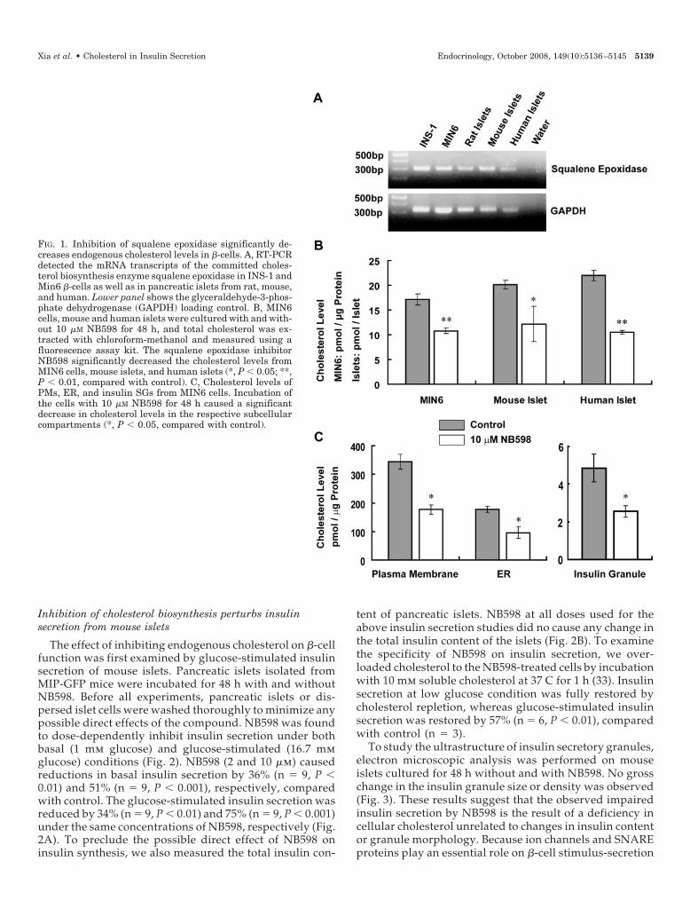
Cholesterol biosynthesis is initiated from the reductionof 3-hydroxy-3-methylglutaryl coenzyme A, undergoingover 30 steps until the final cholesterol product. 3-Hy-droxy-3-methylglutaryl coenzyme A reductase is an inhi-bition target for clinically used statins. However, inhibi-tion of this enzyme has numerous side effects due to theblockade of secondary synthetic pathways upstream fromcholesterol biosynthesis (24). Squalene epoxidase is thesecond enzyme in committed sterol biosynthesis, and in-hibition of this enzyme affects only cholesterol synthesis.To confirm the cholesterol synthesis pathway in pancreatic�-cells, we first determined the expression of squaleneepoxidase. RT-PCR detected the mRNA transcripts of thisenzyme in pancreatic islets from rat, mouse, and human aswell as the clonal INS-1 and MIN6 �-cells (Fig. 1A). Thesqualene epoxidase inhibitor, NB598, was used to reduceendogenous cholesterol biosynthesis in �-cells (32). Theinhibition efficiency of this compound on cholesterol bio-synthesis was then examined in �-cells. Delipidated FBSwas used for this and subsequent protocols to preventuptake of cholesterol (i.e. lipoproteins) normally found inFBS. Incubation with 10 �m NB598 for 48 h caused a 36 �7% reduction in total cholesterol level of MIN6 cells (n �6, P � 0.01) (Fig. 1B). A similar reduction in total choles-terol content in mouse and human islets was observed:40 � 16% (n � 4, P � 0.05) and 52 � 1% (n � 4, P � 0.01),respectively (Fig. 1B). To further examine the inhibitoryeffect of NB598 on cholesterol levels in different cellularcompartments, we isolated PMs, ER, and insulin SGs fromMIN6 cells. NB598 caused a significant decrease in cho-lesterol by 49 � 2%, 46 � 7%, and 48 � 2% from PM, ER,and SG, respectively (n � 3, P � 0.05) (Fig. 1C). Thisdemonstrates comparable reduction in cholesterol reduc-tion throughout the cell.
5138 Endocrinology, October 2008, 149(10):5136–5145 Xia et al. • Cholesterol in Insulin Secretion
Inhibition of cholesterol biosynthesis perturbs insulinsecretion from mouse islets
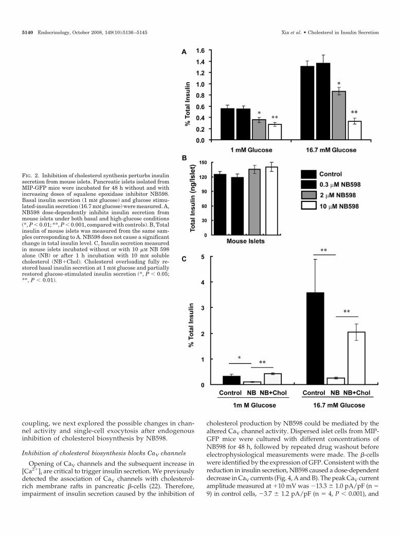
The effect of inhibiting endogenous cholesterol on �-cellfunction was first examined by glucose-stimulated insulinsecretion of mouse islets. Pancreatic islets isolated fromMIP-GFP mice were incubated for 48 h with and withoutNB598. Before all experiments, pancreatic islets or dis-persed islet cells were washed thoroughly to minimize anypossible direct effects of the compound. NB598 was foundto dose-dependently inhibit insulin secretion under bothbasal (1 mm glucose) and glucose-stimulated (16.7 mmglucose) conditions (Fig. 2). NB598 (2 and 10 �m) causedreductions in basal insulin secretion by 36% (n � 9, P �0.01) and 51% (n � 9, P � 0.001), respectively, comparedwith control. The glucose-stimulated insulin secretion wasreduced by 34% (n � 9, P � 0.01) and 75% (n � 9, P � 0.001)under the same concentrations of NB598, respectively (Fig.2A). To preclude the possible direct effect of NB598 oninsulin synthesis, we also measured the total insulin con-
tent of pancreatic islets. NB598 at all doses used for theabove insulin secretion studies did no cause any change inthe total insulin content of the islets (Fig. 2B). To examinethe specificity of NB598 on insulin secretion, we over-loaded cholesterol to the NB598-treated cells by incubationwith 10 mm soluble cholesterol at 37 C for 1 h (33). Insulinsecretion at low glucose condition was fully restored bycholesterol repletion, whereas glucose-stimulated insulinsecretion was restored by 57% (n � 6, P � 0.01), comparedwith control (n � 3).
To study the ultrastructure of insulin secretory granules,electron microscopic analysis was performed on mouseislets cultured for 48 h without and with NB598. No grosschange in the insulin granule size or density was observed(Fig. 3). These results suggest that the observed impairedinsulin secretion by NB598 is the result of a deficiency incellular cholesterol unrelated to changes in insulin contentor granule morphology. Because ion channels and SNAREproteins play an essential role on �-cell stimulus-secretion
FIG. 1. Inhibition of squalene epoxidase significantly de-creases endogenous cholesterol levels in �-cells. A, RT-PCRdetected the mRNA transcripts of the committed choles-terol biosynthesis enzyme squalene epoxidase in INS-1 andMin6 �-cells as well as in pancreatic islets from rat, mouse,and human. Lower panel shows the glyceraldehyde-3-phos-phate dehydrogenase (GAPDH) loading control. B, MIN6cells, mouse and human islets were cultured with and with-out 10 �M NB598 for 48 h, and total cholesterol was ex-tracted with chloroform-methanol and measured using afluorescence assay kit. The squalene epoxidase inhibitorNB598 significantly decreased the cholesterol levels fromMIN6 cells, mouse islets, and human islets (*, P � 0.05; **,P � 0.01, compared with control). C, Cholesterol levels ofPMs, ER, and insulin SGs from MIN6 cells. Incubation ofthe cells with 10 �M NB598 for 48 h caused a significantdecrease in cholesterol levels in the respective subcellularcompartments (*, P � 0.05, compared with control).
Xia et al. • Cholesterol in Insulin Secretion Endocrinology, October 2008, 149(10):5136–5145 5139
coupling, we next explored the possible changes in chan-nel activity and single-cell exocytosis after endogenousinhibition of cholesterol biosynthesis by NB598.
Inhibition of cholesterol biosynthesis blocks CaV channels
Opening of CaV channels and the subsequent increase in[Ca2�]i are critical to trigger insulin secretion. We previouslydetected the association of CaV channels with cholesterol-rich membrane rafts in pancreatic �-cells (22). Therefore,impairment of insulin secretion caused by the inhibition of
cholesterol production by NB598 could be mediated by thealtered CaV channel activity. Dispersed islet cells from MIP-GFP mice were cultured with different concentrations ofNB598 for 48 h, followed by repeated drug washout beforeelectrophysiological measurements were made. The �-cellswere identified by the expression of GFP. Consistent with thereduction in insulin secretion, NB598 caused a dose-dependentdecrease in CaV currents (Fig. 4, A and B). The peak CaV currentamplitude measured at �10 mV was �13.3 � 1.0 pA/pF (n �9) in control cells, �3.7 � 1.2 pA/pF (n � 4, P � 0.001), and
FIG. 2. Inhibition of cholesterol synthesis perturbs insulinsecretion from mouse islets. Pancreatic islets isolated fromMIP-GFP mice were incubated for 48 h without and withincreasing doses of squalene epoxidase inhibitor NB598.Basal insulin secretion (1 mM glucose) and glucose stimu-lated-insulin secretion (16.7 mM glucose) were measured. A,NB598 dose-dependently inhibits insulin secretion frommouse islets under both basal and high-glucose conditions(*, P � 0.01; **, P � 0.001, compared with controls). B, Totalinsulin of mouse islets was measured from the same sam-ples corresponding to A. NB598 does not cause a significantchange in total insulin level. C, Insulin secretion measuredin mouse islets incubated without or with 10 �M NB 598alone (NB) or after 1 h incubation with 10 mM solublecholesterol (NB�Chol). Cholesterol overloading fully re-stored basal insulin secretion at 1 mM glucose and partiallyrestored glucose-stimulated insulin secretion (*, P � 0.05;**, P � 0.01).
5140 Endocrinology, October 2008, 149(10):5136–5145 Xia et al. • Cholesterol in Insulin Secretion
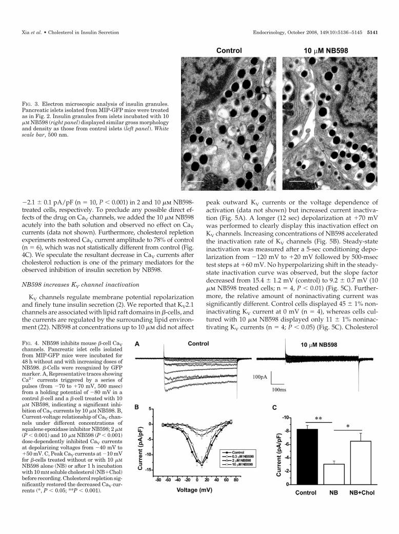
�2.1 � 0.1 pA/pF (n � 10, P � 0.001) in 2 and 10 �m NB598-treated cells, respectively. To preclude any possible direct ef-fects of the drug on CaV channels, we added the 10 �m NB598acutely into the bath solution and observed no effect on CaVcurrents (data not shown). Furthermore, cholesterol repletionexperiments restored CaV current amplitude to 78% of control(n � 6), which was not statistically different from control (Fig.4C). We speculate the resultant decrease in CaV currents aftercholesterol reduction is one of the primary mediators for theobserved inhibition of insulin secretion by NB598.
NB598 increases KV channel inactivation
KV channels regulate membrane potential repolarizationand finely tune insulin secretion (2). We reported that KV2.1channels are associated with lipid raft domains in �-cells, andthe currents are regulated by the surrounding lipid environ-ment (22). NB598 at concentrations up to 10 �m did not affect
peak outward KV currents or the voltage dependence ofactivation (data not shown) but increased current inactiva-tion (Fig. 5A). A longer (12 sec) depolarization at �70 mVwas performed to clearly display this inactivation effect onKV channels. Increasing concentrations of NB598 acceleratedthe inactivation rate of KV channels (Fig. 5B). Steady-stateinactivation was measured after a 5-sec conditioning depo-larization from �120 mV to �20 mV followed by 500-msectest steps at �60 mV. No hyperpolarizing shift in the steady-state inactivation curve was observed, but the slope factordecreased from 15.4 � 1.2 mV (control) to 9.2 � 0.7 mV (10�m NB598 treated cells; n � 4, P � 0.01) (Fig. 5C). Further-more, the relative amount of noninactivating current wassignificantly different. Control cells displayed 45 � 1% non-inactivating KV current at 0 mV (n � 4), whereas cells cul-tured with 10 �m NB598 displayed only 11 � 1% noninac-tivating KV currents (n � 4; P � 0.05) (Fig. 5C). Cholesterol
FIG. 3. Electron microscopic analysis of insulin granules.Pancreatic islets isolated from MIP-GFP mice were treatedas in Fig. 2. Insulin granules from islets incubated with 10�M NB598 (right panel) displayed similar gross morphologyand density as those from control islets (left panel). Whitescale bar, 500 nm.
FIG. 4. NB598 inhibits mouse �-cell CaVchannels. Pancreatic islet cells isolatedfrom MIP-GFP mice were incubated for48 h without and with increasing doses ofNB598. �-Cells were recognized by GFPmarker. A, Representative traces showingCa2� currents triggered by a series ofpulses (from �70 to �70 mV, 500 msec)from a holding potential of �80 mV in acontrol �-cell and a �-cell treated with 10�M NB598, indicating a significant inhi-bition of CaV currents by 10 �M NB598. B,Current-voltage relationship of CaV chan-nels under different concentrations ofsqualene epoxidase inhibitor NB598; 2 �M(P � 0.001) and 10 �M NB598 (P � 0.001)dose-dependently inhibited CaV currentsat depolarizing voltages from �40 mV to�50 mV. C, Peak CaV currents at �10 mVfor �-cells treated without or with 10 �MNB598 alone (NB) or after 1 h incubationwith 10 mM soluble cholesterol (NB�Chol)before recording.Cholesterol repletionsig-nificantly restored the decreased CaV cur-rents (*, P � 0.05; **P � 0.001).
Xia et al. • Cholesterol in Insulin Secretion Endocrinology, October 2008, 149(10):5136–5145 5141
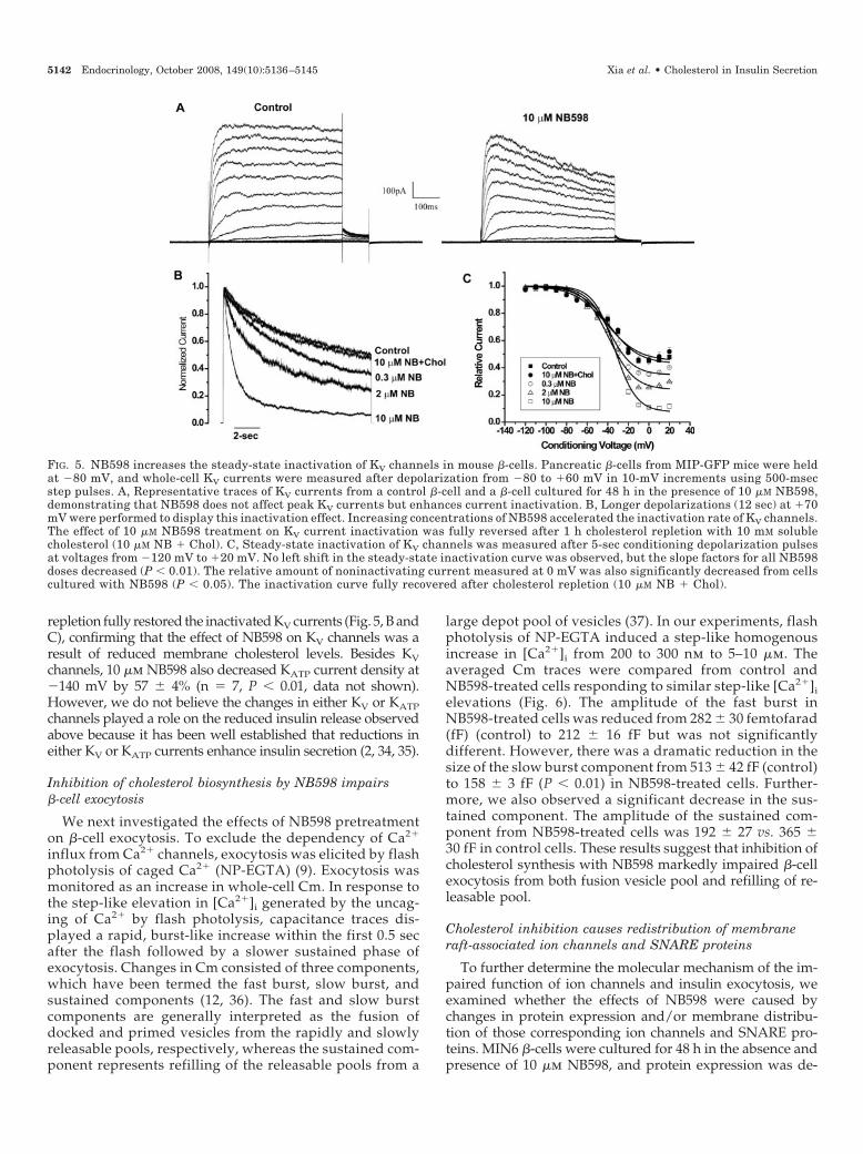
repletion fully restored the inactivated KV currents (Fig. 5, B andC), confirming that the effect of NB598 on KV channels was aresult of reduced membrane cholesterol levels. Besides KVchannels, 10 �m NB598 also decreased KATP current density at�140 mV by 57 � 4% (n � 7, P � 0.01, data not shown).However, we do not believe the changes in either KV or KATPchannels played a role on the reduced insulin release observedabove because it has been well established that reductions ineither KV or KATP currents enhance insulin secretion (2, 34, 35).
Inhibition of cholesterol biosynthesis by NB598 impairs�-cell exocytosis
We next investigated the effects of NB598 pretreatmenton �-cell exocytosis. To exclude the dependency of Ca2�
influx from Ca2� channels, exocytosis was elicited by flashphotolysis of caged Ca2� (NP-EGTA) (9). Exocytosis wasmonitored as an increase in whole-cell Cm. In response tothe step-like elevation in [Ca2�]i generated by the uncag-ing of Ca2� by flash photolysis, capacitance traces dis-played a rapid, burst-like increase within the first 0.5 secafter the flash followed by a slower sustained phase ofexocytosis. Changes in Cm consisted of three components,which have been termed the fast burst, slow burst, andsustained components (12, 36). The fast and slow burstcomponents are generally interpreted as the fusion ofdocked and primed vesicles from the rapidly and slowlyreleasable pools, respectively, whereas the sustained com-ponent represents refilling of the releasable pools from a
large depot pool of vesicles (37). In our experiments, flashphotolysis of NP-EGTA induced a step-like homogenousincrease in [Ca2�]i from 200 to 300 nm to 5–10 �m. Theaveraged Cm traces were compared from control andNB598-treated cells responding to similar step-like [Ca2�]ielevations (Fig. 6). The amplitude of the fast burst inNB598-treated cells was reduced from 282 � 30 femtofarad(fF) (control) to 212 � 16 fF but was not significantlydifferent. However, there was a dramatic reduction in thesize of the slow burst component from 513 � 42 fF (control)to 158 � 3 fF (P � 0.01) in NB598-treated cells. Further-more, we also observed a significant decrease in the sus-tained component. The amplitude of the sustained com-ponent from NB598-treated cells was 192 � 27 vs. 365 �30 fF in control cells. These results suggest that inhibition ofcholesterol synthesis with NB598 markedly impaired �-cellexocytosis from both fusion vesicle pool and refilling of re-leasable pool.
Cholesterol inhibition causes redistribution of membraneraft-associated ion channels and SNARE proteins
To further determine the molecular mechanism of the im-paired function of ion channels and insulin exocytosis, weexamined whether the effects of NB598 were caused bychanges in protein expression and/or membrane distribu-tion of those corresponding ion channels and SNARE pro-teins. MIN6 �-cells were cultured for 48 h in the absence andpresence of 10 �m NB598, and protein expression was de-
FIG. 5. NB598 increases the steady-state inactivation of KV channels in mouse �-cells. Pancreatic �-cells from MIP-GFP mice were heldat �80 mV, and whole-cell KV currents were measured after depolarization from �80 to �60 mV in 10-mV increments using 500-msecstep pulses. A, Representative traces of KV currents from a control �-cell and a �-cell cultured for 48 h in the presence of 10 �M NB598,demonstrating that NB598 does not affect peak KV currents but enhances current inactivation. B, Longer depolarizations (12 sec) at �70mV were performed to display this inactivation effect. Increasing concentrations of NB598 accelerated the inactivation rate of KV channels.The effect of 10 �M NB598 treatment on KV current inactivation was fully reversed after 1 h cholesterol repletion with 10 mM solublecholesterol (10 �M NB � Chol). C, Steady-state inactivation of KV channels was measured after 5-sec conditioning depolarization pulsesat voltages from �120 mV to �20 mV. No left shift in the steady-state inactivation curve was observed, but the slope factors for all NB598doses decreased (P � 0.01). The relative amount of noninactivating current measured at 0 mV was also significantly decreased from cellscultured with NB598 (P � 0.05). The inactivation curve fully recovered after cholesterol repletion (10 �M NB � Chol).
5142 Endocrinology, October 2008, 149(10):5136–5145 Xia et al. • Cholesterol in Insulin Secretion
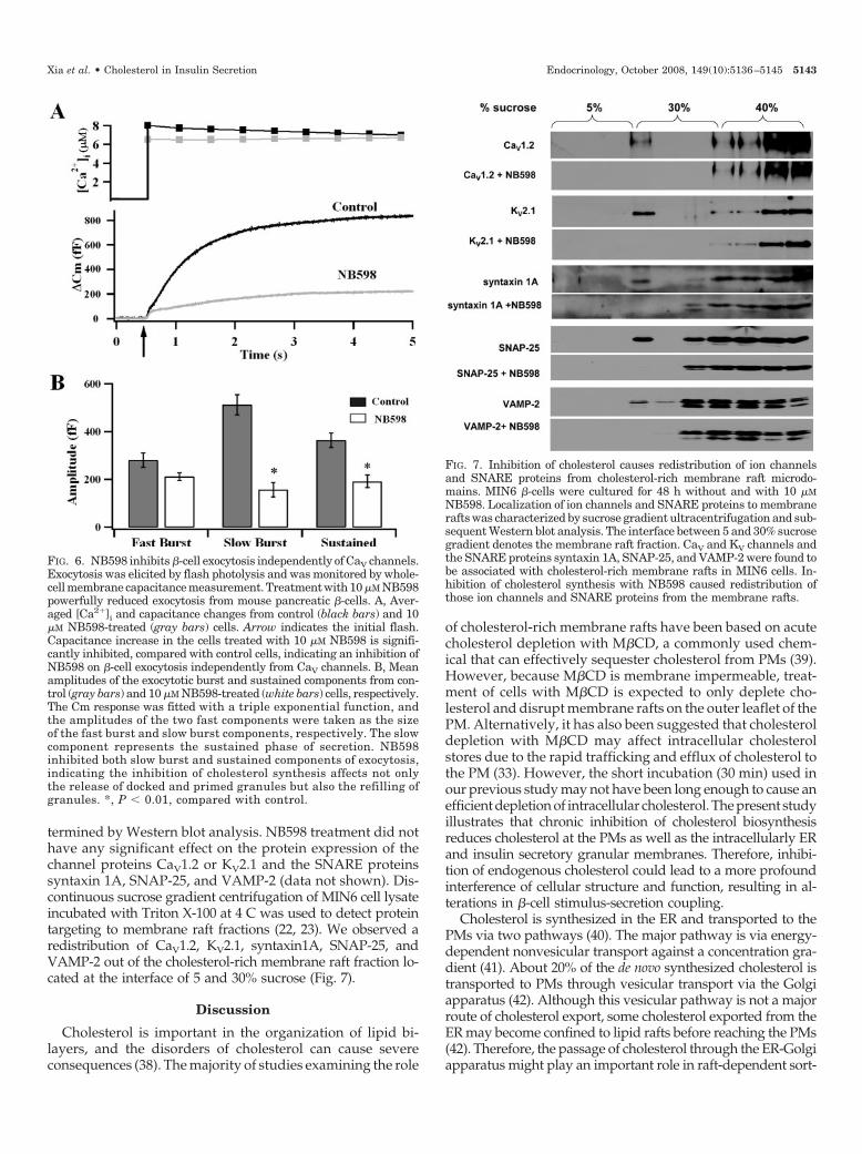
termined by Western blot analysis. NB598 treatment did nothave any significant effect on the protein expression of thechannel proteins CaV1.2 or KV2.1 and the SNARE proteinssyntaxin 1A, SNAP-25, and VAMP-2 (data not shown). Dis-continuous sucrose gradient centrifugation of MIN6 cell lysateincubated with Triton X-100 at 4 C was used to detect proteintargeting to membrane raft fractions (22, 23). We observed aredistribution of CaV1.2, KV2.1, syntaxin1A, SNAP-25, andVAMP-2 out of the cholesterol-rich membrane raft fraction lo-cated at the interface of 5 and 30% sucrose (Fig. 7).
Discussion
Cholesterol is important in the organization of lipid bi-layers, and the disorders of cholesterol can cause severeconsequences (38). The majority of studies examining the role
of cholesterol-rich membrane rafts have been based on acutecholesterol depletion with M�CD, a commonly used chem-ical that can effectively sequester cholesterol from PMs (39).However, because M�CD is membrane impermeable, treat-ment of cells with M�CD is expected to only deplete cho-lesterol and disrupt membrane rafts on the outer leaflet of thePM. Alternatively, it has also been suggested that cholesteroldepletion with M�CD may affect intracellular cholesterolstores due to the rapid trafficking and efflux of cholesterol tothe PM (33). However, the short incubation (30 min) used inour previous study may not have been long enough to cause anefficient depletion of intracellular cholesterol. The present studyillustrates that chronic inhibition of cholesterol biosynthesisreduces cholesterol at the PMs as well as the intracellularly ERand insulin secretory granular membranes. Therefore, inhibi-tion of endogenous cholesterol could lead to a more profoundinterference of cellular structure and function, resulting in al-terations in �-cell stimulus-secretion coupling.
Cholesterol is synthesized in the ER and transported to thePMs via two pathways (40). The major pathway is via energy-dependent nonvesicular transport against a concentration gra-dient (41). About 20% of the de novo synthesized cholesterol istransported to PMs through vesicular transport via the Golgiapparatus (42). Although this vesicular pathway is not a majorroute of cholesterol export, some cholesterol exported from theER may become confined to lipid rafts before reaching the PMs(42). Therefore, the passage of cholesterol through the ER-Golgiapparatus might play an important role in raft-dependent sort-
FIG. 7. Inhibition of cholesterol causes redistribution of ion channelsand SNARE proteins from cholesterol-rich membrane raft microdo-mains. MIN6 �-cells were cultured for 48 h without and with 10 �MNB598. Localization of ion channels and SNARE proteins to membranerafts was characterized by sucrose gradient ultracentrifugation and sub-sequent Western blot analysis. The interface between 5 and 30% sucrosegradient denotes the membrane raft fraction. CaV and KV channels andthe SNARE proteins syntaxin 1A, SNAP-25, and VAMP-2 were found tobe associated with cholesterol-rich membrane rafts in MIN6 cells. In-hibition of cholesterol synthesis with NB598 caused redistribution ofthose ion channels and SNARE proteins from the membrane rafts.
FIG. 6. NB598 inhibits �-cell exocytosis independently of CaV channels.Exocytosis was elicited by flash photolysis and was monitored by whole-cell membrane capacitance measurement. Treatment with 10 �M NB598powerfully reduced exocytosis from mouse pancreatic �-cells. A, Aver-aged [Ca2�]i and capacitance changes from control (black bars) and 10�M NB598-treated (gray bars) cells. Arrow indicates the initial flash.Capacitance increase in the cells treated with 10 �M NB598 is signifi-cantly inhibited, compared with control cells, indicating an inhibition ofNB598 on �-cell exocytosis independently from CaV channels. B, Meanamplitudes of the exocytotic burst and sustained components from con-trol (gray bars) and 10 �M NB598-treated (white bars) cells, respectively.The Cm response was fitted with a triple exponential function, andthe amplitudes of the two fast components were taken as the sizeof the fast burst and slow burst components, respectively. The slowcomponent represents the sustained phase of secretion. NB598inhibited both slow burst and sustained components of exocytosis,indicating the inhibition of cholesterol synthesis affects not onlythe release of docked and primed granules but also the refilling ofgranules. *, P � 0.01, compared with control.
Xia et al. • Cholesterol in Insulin Secretion Endocrinology, October 2008, 149(10):5136–5145 5143
ing of proteins (43, 44). Inhibition of endogenous cholesterolbiosynthesis may cause an inappropriate protein sorting ofmembrane proteins such as ion channels and SNARE proteinsin pancreatic �-cells. Therefore, the impaired function of ionchannels and exocytosis caused by the inhibition of endogenouscholesterol biosynthesis by NB598 could be a result of the dis-ruption of cholesterol in not only the PMs but also the ER andinsulin secretory granules.
Roles of endogenous cholesterol in regulating KV channelfunction
Inhibition of squalene epoxidase significantly increased theinactivation of KV channels in �-cells but did not affect currentdensity or the voltage dependence of channel activation. KVchannel inactivation is described by a ball-and-chain model, aprocess by which the N-terminal cytoplasmic domain of KV� orKV� subunit occludes the inner open channel pore (45). Thiscytoplasmic domain of KV channels was found to interactstrongly with membrane lipids. KV channels, as well as otherion channels, are regulated by the lipid composition of PMs (46).Modulation of membrane lipids have been shown to result inrapid inactivation (A-type currents) of noninactivating KVchannels and, conversely, endow noninactivating delayed rec-tifying properties to A-type KV currents (47). Inhibiting endog-enous cholesterol biosynthesis could have profoundly alteredthe membrane lipid composition, channel-lipid interaction, andconformational changes of the channel proteins, all of whichcould have contributed to the observed enhancement of KVchannel inactivation by NB598. The consequence of enhancedinactivation of KV channels on �-cell function remains to befurther investigated. However, we speculate these effects do notcontribute to the observed inhibition on insulin secretion be-cause inhibition of KV channels are known to enhance insulinsecretion (2, 35).
Role of endogenous cholesterol on �-cell exocytotic machinery
Chronic cholesterol synthesis inhibition markedly reducedCaV currents and insulin secretion. The link between theinhibition of cholesterol synthesis and the impairment in CaVchannel function is not clear. No effect of NB598 on theprotein expression of CaV channels was observed. The de-creased CaV currents could be caused by a possible inap-propriate membrane localization of the CaV channels out ofmembrane rafts or changes in the interactions of the differentauxiliary CaV channel subunits. Second, reduced membranecholesterol may lead to conformational changes of the chan-nel protein due to disruption of the channel-lipid interactionas we suggest may be occurring with KV channels.
SNARE proteins constitute the core of exocytotic machineryin neuroendocrine cells and are critical for the release of neu-rotransmitter and hormone. Recent studies implicate that cho-lesterol-rich membrane rafts could play an important role inregulated exocytosis through compartmentalizing SNARE pro-teins at defined sites on the plasma membrane. We and othershave previously shown that the SNARE proteins syntaxin1A,SNAP-25, and VAMP-2 are associated with cholesterol-richmembrane rafts in pancreatic �- and �-cells (22, 23, 48, 49). Thet-SNAREs syntaxin 1 and SNAP-25 were found both to clusterin plasma membrane in �-cells and PC12 cells, and their in-
tegrity is dependent on membrane cholesterol (48–51). There-fore, cholesterol being a major constituent of membrane raftscould play an essential role in regulating exocytosis throughmaintaining the function of secretory machinery. Single-cellmembrane capacitance measurement indicated that NB958treatment impaired exocytosis independently from the dys-function of CaV channels. Cholesterol could regulate exocytosisthrough protein accumulation or exclusion in the membraneraft domains or reduce the energetic barrier for vesicular-plasma membrane lipid fusion (52).
We were initially surprised by our observations in thisstudy because they are in contrast to our previous work (22),in which we observed acute membrane cholesterol depletionwith M�CD did not affect CaV currents and enhanced insulinsecretion. We concluded in the previous study that the en-hanced insulin secretion could be partially mediated by thestrong inhibition of the amplitude of the KV channels andeffects on the exocytic machinery. However, it is not unex-pected that acute and chronic manipulations in membranecholesterol could elicit markedly different cellular changes.Inhibiting cholesterol synthesis would affect membrane cho-lesterol, as well as cholesterol-mediated processes. Choles-terol and cholesterol-interacting proteins (e.g. caveolin) reg-ulate the trafficking and targeting of proteins, including ionchannels, to membrane rafts (53, 54) and are important incoordinating the assembly of calcium channels with SNAREproteins in the exocytotic domains (55, 56). Given these pos-sible explanations, we were even more astonished that acutecholesterol repletion restored much of the defects in CaVchannel activity and insulin secretion. Therefore, further in-vestigations are warranted into determining the precisemechanisms mediating the alterations in channel activityand exocytosis after chronic cholesterol depletion.
In summary, we have demonstrated a critical role for en-dogenous cholesterol in the normal function of pancreatic�-cells. Using NB598, a cholesterol biosynthesis inhibitor, wefound there are two major roles that endogenous cholesterolmay play in �-cell exocytosis. First, endogenous cholesterolmaintains normal function of CaV channels. Second, choles-terol is critical in the mobilization and fusion of insulin gran-ules with plasma membranes. Dysregulation of cellular cho-lesterol may cause impairment in �-cell function, a possiblepathogenesis leading to the development of type 2 diabetes.
Acknowledgments
Received February 5, 2008. Accepted June 25, 2008.Address all correspondence and requests for reprints to: Robert G.
Tsushima, Department of Biology, York University, 4700 Keele Street,Farquharson344,Toronto,Ontario,CanadaM3J1P3.E-mail:[email protected].
This work was supported by grants and fellowships from the Cana-dian Institutes of Health Research (MOP 77638, to R.G.T.; MOP-69083,to H.Y.G. and R.G.T.), and equipment grants from James H. CummingsFoundation, J.P. Bickell Foundation, the Banting and Best Diabetes Cen-tre (BBDC). F.X. was supported by an Ontario Graduate Scholarship,BBDC Studentship, and Canadian Diabetes Association Doctoral Stu-dentship Research Award.
Current address for Y.C.: Central Laboratory, Guangzhou Children’sHospital, Guangzhou 510120, Guangdong, China.
Current address for R.G.T.: Department of Biology, York University,Toronto, Ontario M3J 1P3, Canada.
5144 Endocrinology, October 2008, 149(10):5136–5145 Xia et al. • Cholesterol in Insulin Secretion
Disclosure Statement: F.X., L.X., A.M., X.G., Y.C., H.Y.G., and R.G.T.have nothing to declare.
References
1. Rorsman P, Renstrom E 2003 Insulin granule dynamics in pancreatic � cells.Diabetologia 46:1029–1045
2. Macdonald PE, Ha XF, Wang J, Smukler SR, Sun AM, Gaisano HY, SalapatekAM, Backx PH, Wheeler MB 2001 Members of the Kv1 and Kv2 voltage-depen-dent K� channel families regulate insulin secretion. Mol Endocrinol 15:1423–1435
3. Barg S, Ma X, Eliasson L, Galvanovskis J, Gopel SO, Obermuller S, PlatzerJ, Renstrom E, Trus M, Atlas D, Striessnig J, Rorsman P 2001 Fast exocytosiswith few Ca2� channels in insulin-secreting mouse pancreatic � cells. BiophysJ 81:3308–3323
4. Brunger AT 2000 Structural insights into the molecular mechanism of Ca2�-dependent exocytosis. Curr Opin Neurobiol 10:293–302
5. Li L, Chin LS 2003 The molecular machinery of synaptic vesicle exocytosis.Cell Mol Life Sci 60:942–960
6. Bruns D, Jahn R 2002 Molecular determinants of exocytosis. Pflugers Arch443:333–338
7. Weber T, Zemelman BV, McNew JA, Westermann B, Gmachl M, Parlati F,Sollner TH, Rothman JE 1998 SNAREpins: minimal machinery for membranefusion. Cell 92:759–772
8. Curry DL, Bennett LL, Grodsky GM 1968 Dynamics of insulin secretion bythe perfused rat pancreas. Endocrinology 83:572–584
9. Neher E 1998 Vesicle pools and Ca2� microdomains: new tools for under-standing their roles in neurotransmitter release. Neuron 20:389–399
10. Rorsman P, Eliasson L, Renstrom E, Gromada J, Barg S, Gopel S 2000 Thecell physiology of biphasic insulin secretion. News Physiol Sci 15:72–77
11. Rettig J, Neher E 2002 Emerging roles of presynaptic proteins in Ca2�-trig-gered exocytosis. Science 298:781–785
12. Xu T, Rammner B, Margittai M, Artalejo AR, Neher E, Jahn R 1999 Inhibitionof SNARE complex assembly differentially affects kinetic components of exo-cytosis. Cell 99:713–722
13. Ostenson CG, Gaisano H, Sheu L, Tibell A, Bartfai T 2006 Impaired gene andprotein expression of exocytotic soluble N-ethylmaleimide attachment proteinreceptor complex proteins in pancreatic islets of type 2 diabetic patients.Diabetes 55:435–440
14. Chan CB, MacPhail RM, Sheu L, Wheeler MB, Gaisano HY 1999 �-Cellhypertrophy in fa/fa rats is associated with basal glucose hypersensitivity andreduced SNARE protein expression. Diabetes 48:997–1005
15. Gaisano HY, Ostenson CG, Sheu L, Wheeler MB, Efendic S 2002 Abnormalexpression of pancreatic islet exocytotic soluble N-ethylmaleimide-sensitive factorattachment protein receptors in Goto-Kakizaki rats is partially restored by phlo-rizin treatment and accentuated by high glucose treatment. Endocrinology 143:4218–4226
16. Ohara-Imaizumi M, Nishiwaki C, Kikuta T, Nagai S, Nakamichi Y, NagamatsuS 2004 TIRF imaging of docking and fusion of single insulin granule motion inprimary rat pancreatic �-cells: different behaviour of granule motion betweennormal and Goto-Kakizaki diabetic rat �-cells. Biochem J 381:13–18
17. Haines TH 2001 Do sterols reduce proton and sodium leaks through lipidbilayers? Prog Lipid Res 40:299–324
18. Ohvo-Rekila H, Ramstedt B, Leppimaki P, Slotte JP 2002 Cholesterol inter-actions with phospholipids in membranes. Prog Lipid Res 41:66–97
19. Inokuchi J 2006 Insulin resistance as a membrane microdomain disorder. BiolPharm Bull 29:1532–1537
20. Michel V, Bakovic M 2007 Lipid rafts in health and disease. Biol Cell 99:129–14021. Parton RG 2003 Caveolae—from ultrastructure to molecular mechanisms. Nat
Rev Mol Cell Biol 4:162–16722. Xia F, Gao X, Kwan E, Lam PP, Chan L, Sy K, Sheu L, Wheeler MB, Gaisano
HY, Tsushima RG 2004 Disruption of pancreatic �-cell lipid rafts modifiesKv2.1 channel gating and insulin exocytosis. J Biol Chem 279:24685–24691
23. Xia F, Leung YM, Gaisano G, Gao X, Chen Y, Manning Fox JE, BhattacharjeeA, Wheeler MB, Gaisano HY, Tsushima RG 2007 Targeting of KV4, CaV1.2and SNARE proteins to cholesterol-rich lipid rafts in pancreatic �-cells: effectson glucagon stimulus-secretion coupling. Endocrinology 148:2157–2167
24. Chugh A, Ray A, Gupta JB 2003 Squalene epoxidase as hypocholesterolemicdrug target revisited. Prog Lipid Res 42:37–50
25. Shapiro AM, Lakey JR, Ryan EA, Korbutt GS, Toth E, Warnock GL, KnetemanNM, Rajotte RV 2000 Islet transplantation in seven patients with type 1 diabetesmellitus using a glucocorticoid-free immunosuppressive regimen. N Engl J Med343:230–238
26. Ramanadham S, Bohrer A, Gross RW, Turk J 1993 Mass spectrometric charac-terization of arachidonate-containing plasmalogens in human pancreatic isletsand in rat islet �-cells and subcellular membranes. Biochemistry 32:13499–13509
27. Brunner Y, Coute Y, Iezzi M, Foti M, Fukuda M, Hochstrasser DF, WollheimCB, Sanchez JC 2007 Proteomics analysis of insulin secretory granules. MolCell Proteomics 6:1007–1017
28. Leung YM, Ahmed I, Sheu L, Tsushima RG, Diamant NE, Hara M, Gaisano HY2005 Electrophysiological characterization of pancreatic islet cells in the mouseinsulin promoter-green fluorescent protein mouse. Endocrinology 146:4766–4775
29. Xu T, Binz T, Niemann H, Neher E 1998 Multiple kinetic components ofexocytosis distinguished by neurotoxin sensitivity. Nat Neurosci 1:192–200
30. Grynkiewicz G, Poenie M, Tsien RY 1985 A new generation of Ca2� indicatorswith greatly improved fluorescence properties. J Biol Chem 260:3440–3450
31. Wheeler MB, Sheu L, Ghai M, Bouquillon A, Grondin G, Weller U, BeaudoinAR, Bennett MK, Trimble WS, Gaisano HY 1996 Characterization of SNAREprotein expression in � cell lines and pancreatic islets. Endocrinology 137:1340–1348
32. Horie M, Tsuchiya Y, Hayashi M, Iida Y, Iwasawa Y, Nagata Y, Sawasaki Y,Fukuzumi H, Kitani K, Kamei T 1990 NB-598: a potent competitive inhibitorof squalene epoxidase. J Biol Chem 265:18075–18078
33. Hao M, Head WS, Gunawardana SC, Hasty AH, Piston DW 2007 Direct effectof cholesterol on insulin secretion: a novel mechanism for pancreatic �-celldysfunction. Diabetes 56:2328–2338
34. Ashcroft FM 2005 ATP-sensitive potassium channelopathies: focus on insulinsecretion. J Clin Invest 115:2047–2058
35. Macdonald PE, Sewing S, Wang J, Joseph JW, Smukler SR, Sakellaropoulos G,Wang J, Saleh MC, Chan CB, Tsushima RG, Salapatek AM, Wheeler MB 2002Inhibition of Kv2.1 voltage-dependent K� channels in pancreatic �-cells enhancesglucose-dependent insulin secretion. J Biol Chem 277:44938–44945
36. Voets T, Neher E, Moser T 1999 Mechanisms underlying phasic and sus-tained secretion in chromaffin cells from mouse adrenal slices. Neuron23:607– 615
37. Sorensen JB 2004 Formation, stabilisation and fusion of the readily releasablepool of secretory vesicles. Pflugers Arch 448:347–362
38. Maxfield FR, Tabas I 2005 Role of cholesterol and lipid organization in disease.Nature 438:612–621
39. Christian AE, Haynes MP, Phillips MC, Rothblat GH 1997 Use of cyclodex-trins for manipulating cellular cholesterol content. J Lipid Res 38:2264–2272
40. Maxfield FR, Wustner D 2002 Intracellular cholesterol transport. J Clin Invest110:891–898
41. Chang TY, Chang CC, Ohgami N, Yamauchi Y 2006 Cholesterol sensing,trafficking, and esterification. Annu Rev Cell Dev Biol 22:129–157
42. Heino S, Lusa S, Somerharju P, Ehnholm C, Olkkonen VM, Ikonen E 2000Dissecting the role of the Golgi complex and lipid rafts in biosynthetictransport of cholesterol to the cell surface. Proc Natl Acad Sci USA 97:8375– 8380
43. Simons K, Ikonen E 1997 Functional rafts in cell membranes. Nature 387:569–57244. Helms JB, Zurzolo C 2004 Lipids as targeting signals: lipid rafts and intra-
cellular trafficking. Traffic 5:247–25445. Zagotta WN, Hoshi T, Aldrich RW 1990 Restoration of inactivation in mutants
of Shaker potassium channels by a peptide derived from ShB. Science 250:568–57146. Hilgemann DW 2004 Biochemistry. Oily barbarians breach ion channel gates.
Science 304:223–22447. Oliver D, Lien CC, Soom M, Baukrowitz T, Jonas P, Fakler B 2004 Functional
conversion between A-type and delayed rectifier K� channels by membranelipids. Science 304:265–270
48. Ohara-Imaizumi M, Nishiwaki C, Kikuta T, Kumakura K, Nakamichi Y,Nagamatsu S 2004 Site of docking and fusion of insulin secretory granulesin live MIN6 � cells analyzed by TAT-conjugated anti-syntaxin 1 antibodyand total internal reflection fluorescence microscopy. J Biol Chem 279:8403– 8408
49. Takahashi N, Hatakeyama H, Okado H, Miwa A, Kishimoto T, Kojima T,Abe T, Kasai H 2004 Sequential exocytosis of insulin granules is associatedwith redistribution of SNAP25. J Cell Biol 165:255–262
50. Chamberlain LH, Burgoyne RD, Gould GW 2001 SNARE proteins are highlyenriched in lipid rafts in PC12 cells: implications for the spatial control ofexocytosis. Proc Natl Acad Sci USA 98:5619–5624
51. Lang T, Bruns D, Wenzel D, Riedel D, Holroyd P, Thiele C, Jahn R 2001SNAREs are concentrated in cholesterol-dependent clusters that define dock-ing and fusion sites for exocytosis. EMBO J 20:2202–2213
52. Churchward MA, Rogasevskaia T, Hofgen J, Bau J, Coorssen JR 2005 Cho-lesterol facilitates the native mechanism of Ca2�-triggered membrane fusion.J Cell Sci 118:4833–4848
53. Kong MM, Hasbi A, Mattocks M, Fan T, O’Dowd BF, George SR 2007Regulation of D1 dopamine receptor trafficking and signaling by caveolin-1.Mol Pharmacol 72:1157–1170
54. Pediconi MF, Gallegos CE, Los Santos EB, Barrantes FJ 2004 Metaboliccholesterol depletion hinders cell-surface trafficking of the nicotinic acetyl-choline receptor. Neuroscience 128:239–249
55. Cho WJ, Jeremic A, Jin H, Ren G, Jena BP 2007 Neuronal fusion pore assemblyrequires membrane cholesterol. Cell Biol Int 31:1301–1308
56. Nevins AK, Thurmond DC 2006 Caveolin-1 functions as a novel Cdc42 gua-nine nucleotide dissociation inhibitor in pancreatic �-cells. J Biol Chem281:18961–18972
Endocrinology is published monthly by The Endocrine Society (http://www.endo-society.org), the foremost professional society serving theendocrine community.
Xia et al. • Cholesterol in Insulin Secretion Endocrinology, October 2008, 149(10):5136–5145 5145