sin1 phosphorylation impairs mtorc2 complex ... - core
TRANSCRIPT

Sin1 phosphorylation impairs mTORC2 complex integrity andinhibits downstream Akt signaling to suppress tumorigenesis
(Article begins on next page)
The Harvard community has made this article openly available.Please share how this access benefits you. Your story matters.
Citation Liu, P., W. Gan, H. Inuzuka, A. S. Lazorchak, D. Gao, O. Arojo,D. Liu, et al. 2013. “Sin1 phosphorylation impairs mTORC2complex integrity and inhibits downstream Akt signaling tosuppress tumorigenesis.” Nature cell biology 15 (11):10.1038/ncb2860. doi:10.1038/ncb2860.http://dx.doi.org/10.1038/ncb2860.
Published Version doi:10.1038/ncb2860
Accessed February 16, 2015 12:25:34 PM EST
Citable Link http://nrs.harvard.edu/urn-3:HUL.InstRepos:12406914
Terms of Use This article was downloaded from Harvard University's DASHrepository, and is made available under the terms and conditionsapplicable to Other Posted Material, as set forth athttp://nrs.harvard.edu/urn-3:HUL.InstRepos:dash.current.terms-of-use#LAA

Sin1 phosphorylation impairs mTORC2 complex integrity andinhibits downstream Akt signaling to suppress tumorigenesis
Pengda Liu1, Wenjian Gan1, Hiroyuki Inuzuka1, Adam S Lazorchak2, Daming Gao1,Omotooke Arojo2, Dou Liu2, Lixin Wan1, Bo Zhai3, Yonghao Yu3,4, Min Yuan5, Byeong MoKim6, Shavali Shaik1, Suchithra Menon7, Steven P. Gygi3, Tae Ho Lee6, John M Asara5,Brendan D. Manning7, John Blenis3, Bing Su2, and Wenyi Wei1,8
1Department of Pathology, Beth Israel Deaconess Medical Center, Harvard Medical School,Boston, MA 022152Department of Immunobiology and The Vascular Biology and Therapeutics Program, YaleMedical School, New Haven, CT 065203Department of Cell Biology, Harvard Medical School, Boston, MA 021154Department of Biochemistry, UT Southwestern Medical Center, Dallas, TX 753905Department of Medicine, Beth Israel Deaconess Medical Center, Harvard Medical School,Boston, MA 022156Division of Gerontology, Department of Medicine, Beth Israel Deaconess Medical Center,Boston, MA 022157Department of Genetics & Complex Diseases, Harvard School of Public Health, Boston, MA02115
AbstractThe mechanistic target of rapamycin (mTOR) functions as a critical regulator of cellular growthand metabolism by forming multi-component, yet functionally distinct complexes mTORC1 andmTORC2. Although mTORC2 has been implicated in mTORC1 activation, little is known abouthow mTORC2 is regulated. Here we report that phosphorylation of Sin1 at T86 and T398suppresses mTORC2 kinase activity by dissociating Sin1 from mTORC2. Importantly, Sin1phosphorylation, triggered by S6K or Akt, in a cellular context-dependent manner, inhibits notonly insulin/IGF-1-mediated, but also PDGF or EGF-induced Akt phosphorylation by mTORC2,demonstrating a negative regulation of mTORC2 independent of IRS-1 and Grb10. Lastly, acancer patient-derived Sin1-R81T mutation impairs Sin1 phosphorylation, leading to hyper-mTORC2 activation via bypassing this negative regulation. Together, our work reveals a Sin1phosphorylation-dependent mTORC2 regulation, providing a potential molecular mechanism bywhich mutations in the mTORC1/S6K/Sin1 signaling axis might cause aberrant hyper-activationof mTORC2/Akt that facilitates tumorigenesis.
8To whom correspondence should be addressed: Wenyi Wei, Ph.D., Department of Pathology, Beth Israel Deaconess Medical Center,Harvard Medical School, 330 Brookline Ave, Boston, MA 02215, Phone: 617-735-2495, [email protected].
Author ContributionsP.L., W.G and H.I. performed most of the experiments with assistance from D.G., L.W., A.L., S.S. and O.A. W.W. and B.S. designedthe experiments and supervised the study. P.L. and W.W. wrote the manuscript. All authors commented on the manuscript.
Competing Financial InterestsThe authors declare no competing financial interests.
Note: Supplementary Information is available in the online version of the paper.
NIH Public AccessAuthor ManuscriptNat Cell Biol. Author manuscript; available in PMC 2014 May 01.
Published in final edited form as:Nat Cell Biol. 2013 November ; 15(11): . doi:10.1038/ncb2860.
NIH
-PA Author Manuscript
NIH
-PA Author Manuscript
NIH
-PA Author Manuscript

IntroductionmTOR is a highly conserved important regulator of cell growth and proliferation in aplethora of biological settings in all eukaryotes 1–8. As such, deregulated mTOR functionleads to a variety of human diseases, ranging from cancer 5,9 to immune dysfunction 3 anddiabetes 4,10. mTOR functions as a critical and essential catalytic core in at least two knownfunctionally distinct complexes, mTORC1 and mTORC2 3,5. A unique subset of associatedproteins defines each complex, such as Raptor 11 in mTORC1, or Rictor 12 and Sin1 13–15 inmTORC2. Biologically, mTORC2 mainly promotes cell proliferation and survival 2,16
through phosphorylation of the AGC kinase family members Akt and SGK 6, in addition toits initially defined role in regulating cell skeletal organization 17. On the other hand,mTORC1 directly promotes mRNA translation and protein synthesis by phosphorylatingS6K1 and 4EBP1 18, inhibits autophagy through phosphorylating ULK1 19 and indirectlyenhances ribosome biogenesis via promoting nucleophosmin (NPM) oncogenetranslation 20. As most mTORC1 functions are high energy consuming, regulation ofmTORC1 activity is tightly coupled to the energy status of the cell and regulated bynutrients, energy, stress and growth factors, thereby ensuring that cells stop growing underunfavorable conditions 1,8,21,22.
Compared to well-defined mechanisms of mTORC1 activation such as mTORC2/Akt-mediated phosphorylation of TSC2 23,24 or PRAS40 25,26, the upstream signaling thatgoverns mTORC2 activation is just beginning to be appreciated. To this end, mTORC2’sassociation with ribosome was recently found to be necessary for its activation 4.Furthermore, mTORC1/S6K-mediated phosphorylation of IRS-1 27,28 and Grb10 28,29 alsoconstitutes negative feedback mechanisms to block mTORC2 activation by insulin/IGF-1.However, both IRS-1 and Grb10 function by suppressing insulin/IGF-1 signaling upstreamof PI3K to affect both mTORC1 and mTORC2. Thus, it remains elusive whether mTORC1could directly regulate mTORC2/Akt without broadly suppressing the PI3K pathway andhow mTORC1 suppresses mTORC2/Akt in stimulation conditions other than insulin/IGF-1.
Here we show that in response to a wide spectrum of stimuli including insulin, IGF-1, PDGFand EGF, phosphorylation of Sin1 dissociates Sin1 from mTORC2 to terminate mTORC2kinase activity, revealing a negative regulation of mTORC2 function independent of thepreviously identified negative feedback regulators IRS-1 and Grb10 in suppressingmTORC2.
ResultsS6K phosphorylates Sin1 on both T86 and T398 sites
In keeping with previous reports 27–30, an inverse correlation between mTORC1/S6K andmTORC2 activities was confirmed (Supplementary Fig. S1a–d). Intriguingly, the criticalrole of Grb10 29,30 and IRS-1 27,28 in regulation of mTORC2/Akt was found to be restrictedto insulin/IGF-1, but not PDGF or EGF stimulation (Supplementary Fig. S1e–g).Consistently, inhibition of mTORC1/S6K signaling by a specific S6K1 inhibitor, S6K1-I 31
or mTORC1 inhibitor, rapamycin 11,32,33 could still augment Akt activation in TSC2−/−
MEFs depleted of endogenous IRS-1 and/or Grb10 (Supplementary Fig. S1h–m),advocating that mTORC1/S6K could exert its negative regulation of mTORC2/Aktfollowing PDGF or EGF stimulation through uncharacterized negative feedback loop(s)other than IRS-1 and Grb10. As Rictor and Sin1 are the only two unique essential mTORC2components 13,34, we reasoned that the mTORC1/S6K-dependent regulation of mTORC2might occur through Rictor or Sin1. However, S6K-dependent phosphorylation of Rictordoes not affect mTORC2 kinase activity 32,35,36, which prompted us to investigate whetherSin1 is the primary target to mediate the regulation of mTORC2 by mTORC1/S6K.
Liu et al. Page 2
Nat Cell Biol. Author manuscript; available in PMC 2014 May 01.
NIH
-PA Author Manuscript
NIH
-PA Author Manuscript
NIH
-PA Author Manuscript
In line with previous reports 13,15, insulin, IGF-1, PDGF and EGF all could effectivelyinduce Akt S473 phosphorylation in WT but not Sin1−/− MEFs (Supplementary Fig. S1n) orSin1 depleted TSC2−/− cells (Supplementary Fig. S1o). This finding prompted us to furtherexamine whether other than IRS-1 and Grb10, modifications of Sin1 may play a critical rolein mTORC1-mediated feedback regulation of mTORC2. In support of this hypothesis,phosphorylation of Sin1 was significantly reduced upon inhibition of mTORC1 or S6K, andmoderately decreased by the Akt inhibitor, AktVIII (Fig. 1a). Consistently, we detected invivo Sin1 phosphorylation triggered by S6K1 and to a lesser extent, Akt1, but not othercharacterized AGC kinases (Fig. 1b and Supplementary Fig. S2a). Notably, S6K1 inhibitionled to reduced Sin1 phosphorylation while overexpressing a WT-S6K1 (Fig. 1c), but not akinase-dead-S6K1 mutant 37 (Supplementary Fig. S2b), augmented Sin1 phosphorylation.Consistently, depletion of TSC2, which resulted in elevated S6K activity, also led toincreased Sin1 phosphorylation (Supplementary Fig. S2c). In support of Sin1 being a S6Ksubstrate, we identified two canonical AGC family kinase-recognition motifs (RxRxxpS/pT) 38 located at T86 and T398, respectively (Fig. 1d). Notably, mutation of both sites toalanines abolished S6K1-mediated Sin1 phosphorylation in cells (Fig. 1e) or in vitro (Fig.1f). Interestingly, mutation of either T86 or T398 to an alanine did not completely abolishthe S6K-dependent phosphorylation of Sin1 (Fig. 1e), indicating that both T86 and T398might be potential S6K1 sites in vivo.
Importantly, phosphorylation of T86 and T398 were detected by mass spectrometry(Supplementary Fig. S2d–e). To gain further mechanistic insights into how Sin1phosphorylation may affect mTORC2 activation under physiological conditions, wedeveloped phospho-specific antibodies against pT86-Sin1 or pT398-Sin1, respectively(Supplementary Fig. S2f–g). Using these antibodies we observed an increase in Sin1-pT86and Sin1-pT398 upon induction by insulin (Fig. 1g and Supplementary Fig. S2h), IGF-1,PDGF or EGF (Fig. 1h), whereas these phosphorylation events could be attenuated byinactivating S6K1 either through depleting Raptor (Fig. 1h and Supplementary Fig. S2i), orby S6K1-I (Supplementary Fig. S2j). Notably, S6K1 knockdown only partially reduced Sin1phosphorylation (Supplementary Fig. S2i), suggesting that other S6K isoforms mightphosphorylate Sin1. Nevertheless, recombinant S6K1 phosphorylated Sin1 in vitro(Supplementary Fig. S2k) and conversely, rapamycin could attenuate Sin1-pT86 mediatedby WT-S6K1 but not a rapamycin-resistant form of S6K1 37 (Supplementary Fig. 2l),further supporting S6K1 as a physiological kinase for Sin1.
Both Akt and S6K may phosphorylate Sin1 in a context-dependent mannerAlthough depletion of endogenous Akt1 also led to a moderate decrease in Sin1phosphorylation, its effects were less compared to inhibiting S6K1 or mTORC1 in ourexperimental conditions (Supplementary Fig. S2m–n). Moreover, Akt might regulate Sin1phosphorylation indirectly through activating mTORC1/S6K 11,13. In support of this model,Sin1-T86 phosphorylation correlated positively with S6K1 activity (evidenced by pS6) butinversely with Akt phosphorylation (Supplementary Fig. S2o–p), indicating that Sin1phosphorylation by S6K1 might act as a physiological negative regulator of Aktphosphorylation in fibroblasts. However, this conclusion might be cellular context-dependent as a recent study has suggested Akt as the Sin1-T86 phosphorylating kinase inadipocytes 39.
To gain further mechanistic insights into upstream kinases responsible for phosphorylatingSin1, we examined the possible role of S6K versus Akt in mediating Sin1 phosphorylationon T86 and T398 in 3T3-L1 cells 40. Consistent with Humphrey et al 39, Sin1-pT86 signalswere largely blocked by Akt inhibition, but only moderately reduced by inhibiting mTORC1or S6K (Supplementary Fig. S2q), confirming Akt as the major Sin1-T86 phosphorylatingkinase in 3T3-L1 cells. However, inhibitors targeting either Akt or S6K1 led to a dramatic
Liu et al. Page 3
Nat Cell Biol. Author manuscript; available in PMC 2014 May 01.
NIH
-PA Author Manuscript
NIH
-PA Author Manuscript
NIH
-PA Author Manuscript
reduction in Sin1-pT398, suggesting that both Akt and S6K are involved in phosphorylatingSin1-T398 in 3T3-L1 adipocytes. On the other hand, in epithelium-derived HeLa cells, instark contrast to 3T3-L1, inhibition of mTORC1/S6K, rather than Akt, led to a more severereduction in both Sin1-pT86 and Sin1-pT398 signals (Supplementary Fig S2r), indicatingthat in HeLa cells, S6K, but not Akt, is the major kinase responsible for Sin1-T86 and -T398phosphorylation. Collectively, these results implicate that physiological upstream kinase(s)responsible for Sin1-T86 or Sin1-T398 phosphorylation might be tissue-specific or cellularcontext-dependent. A similar tissue specific phosphorylation atlas of mouse proteins hasbeen established 41. Hence, adipocytes versus epithelial cells might require tightly regulatedand specialized, phosphorylation-dependent intracellular signaling, thereby utilizing distinctupstream signaling routes to regulate effector pathways such as mTORC2. As carcinomasprimarily derive from epithelial cells 42,43, we chose to focus on understanding howregulation of mTORC2 by S6K-mediated Sin1 phosphorylation contributes to tumorigenesisin the epithelial cell settings.
Phosphorylation of Sin1 at both T86 and T398 dissociates Sin1 from the mTORC2 complexAs Sin1 is an essential component that governs mTORC2 integrity 13–15, we next examinedwhether Sin1 phosphorylation on T86 and T398 affects mTORC2 function. Strikingly,compared to WT-Sin1, the Sin1 phospho-mimetic mutant (T86E/T398E, Sin1-EE), failed tointeract with the essential mTORC2 components Rictor, mTOR or GβL (Fig. 2a andSupplementary Fig. S3a–c), indicating that the assembly of a functional mTORC2 complexwas prevented by Sin1 phosphorylation. In supporting of this model, S6K1-mediatedphosphorylation of Sin1-WT, but not the Sin1 phospho-deficient mutant (T86A/T398A,Sin1-AA), reduced Sin1 interaction with Rictor (Fig. 2b), mTOR (Fig. 2c) and GβL (Fig.2d). Notably, disruption of the Sin1/Rictor interaction required Sin1 phosphorylation onboth T86 and T398 (Supplementary Fig. S3d,e). To further investigate the underlyingmolecular mechanism(s), we performed gel-filtration assays to examine mTORC2 complexassembly in cells, and observed that WT-Sin1, but not Sin1-EE, co-eluted with mTORC2components including Rictor, mTOR, and GβL (Fig. 2e). Furthermore, depletion of TSC2led to elevated mTORC1/S6K activity and shifted a significant portion of Sin1 to mTORC2-free fractions (Supplementary Fig. S3f,g). Strikingly, the mTORC2-free Sin1 species wereenriched with pT86-Sin1 (Supplementary Fig. S3f). Consistently, less Rictor was detected inassociation with Sin1 in TSC2−/− cells (Fig. 2f), whereas inhibiting S6K1 activity inTSC2−/− cells by S6K1-I partially restored Rictor interaction with Sin1 (Supplementary Fig.S3h,i), supporting a crucial role of S6K in regulating the Sin1-Rictor interaction.
To further understand the role of each Sin1 phosphorylation event in mediating mTORC2complex organization, we truncated Sin1 into four regions (Fig. 2g) and observed that Sin1mainly interacts with Rictor through its N-terminal region, with mTOR kinase domain viaits PH domain, and with GβL through its N, RBD and PH domains (Fig. 2h). Interestingly,phosphorylation of Sin1 at T86 in the N-terminal region abolished Sin1-N interaction withRictor (Fig. 2i), while phosphorylation on T398 in the PH domain reduced Sin1 interactionwith the mTOR kinase domain (Fig. 2j), indicating the distinct role of each phosphorylationsite in possibly mediating the organization of the mTORC2 complex, further supporting thatphosphorylation of both T86 and T398 is required to functionally inactivate mTORC2.
More importantly, under physiological stimulation conditions such as EGF (Fig. 3a andSupplementary Fig. S4a) or insulin (Fig. 3b and Supplementary Fig. S4b) induction, weobserved an inverse correlation between the induced Sin1 phosphorylation and adissociation of Sin1 from Rictor or mTOR, which could be abolished by mutating either T86or T398 to alanine (Fig. 3c), or by rapamycin treatment (Supplementary Fig. S4c–f),demonstrating a physiological role of Sin1 phosphorylation in negatively regulatingmTORC2 function. Notably, compared with Sin1-WT, impairing Sin1 phosphorylation by
Liu et al. Page 4
Nat Cell Biol. Author manuscript; available in PMC 2014 May 01.
NIH
-PA Author Manuscript
NIH
-PA Author Manuscript
NIH
-PA Author Manuscript
mutating either T86 or T398 (Fig. 3d), inhibiting S6K activity (Fig. 3e and SupplementaryFig. S4g), or depleting endogenous Raptor (Supplementary Fig. S4h,i), led to sustained Aktphosphorylation under EGF or insulin stimulation. Conversely, activating S6K by eitherdepletion of endogenous TSC2 (Supplementary Fig. S4j) or genetic ablation of TSC2(Supplementary Fig. S4k), resulted in reduced Akt S473 phosphorylation. In keeping withprevious reports 27,44, Akt phosphorylation in response to various stimuli was attenuated inTSC2−/− cells with elevated S6K activity (Supplementary Fig. S4l), confirming a negativerole for S6K-mediated phosphorylation of Sin1 in Akt activation.
Phosphorylation of Sin1 on both T86 and T398 suppresses mTORC2-mediated activationof Akt
Notably, Sin1 phosphorylation did not affect Sin1 stability (Supplementary Fig. S4m,n).However, phosphorylation of Sin1 specifically reduced its interaction with the mTORC2substrate, Akt1 (Fig. 4a–c) but not with another characterized mTORC2 substrateSGK1 45,46 (Fig. 4d–e), nor the other AGC kinase S6K1 (Fig. 4f–g), which might explainwhy Sin1 phosphorylation leads to impaired Akt activation.
To further verify the critical roles of Sin1 phosphorylation in regulating mTORC2 kinaseactivity, we performed in vitro kinase assays to show that unlike Sin1-WT or Sin1-AA,immunoprecipitated Sin1-EE failed to phosphorylate GST-Akt1 in vitro, presumably due toits deficiency in forming a functional mTORC2 complex (Fig. 5a–b). By coupling gelfiltration with in vitro kinase assays, we demonstrated that only the gel-filtration fractionswith molecular weights corresponding to the full mTORC2 complex retained the mTORC2kinase activity towards GST-Akt in vitro (Fig. 5c–d). Strikingly, consistent with previousstudies 47–49, insulin stimulation led to an oscillated pattern of Akt activation with reducedAkt phosphorylation after prolonged treatment in various cell lines (Supplementary Fig.S5a–c), a phenotype observed in many signaling pathways 47,50. However, the underlyingmechanism of the oscillation of Akt phosphorylation remains not fully understood. Notably,prolonged (45 minutes of) insulin treatment led to a severe reduction of mTORC2-mediatedin vitro phosphorylation of Akt in part by inducing pT86-Sin1 (Fig. 5c–d) to disrupt themTORC2 complex integrity. More importantly, rapamycin treatment effectively restoredmTORC2 complex integrity and kinase activity under prolonged EGF stimulation (Fig. 5e),emphasizing that S6K negatively regulates mTORC2 activity in part throughphosphorylating Sin1, serving as a molecular switch to shut down EGF or insulin-inducedactivation of mTORC2/Akt in a timely fashion (Supplementary Fig. S5d).
Given the critical role of the mTORC2/Akt signaling in cell survival and tumorigenesis, wenext examined the biological significance of S6K-mediated Sin1 phosphorylation onmTORC2 function. Consistent with in vitro kinase assay results (Fig. 5a), Sin1-WT, but notSin1-EE, rescued in vivo Akt-S473 phosphorylation in Sin1−/− MEFs (Fig. 6a,b andSupplementary Fig. S6a) 13,15. Furthermore, in echoing the in vitro kinase results (Fig. 6c),immuno-purified endogenous Akt1 from Sin1-EE-expressing Sin1−/− MEFs was deficient inphosphorylating the well-characterized Akt substrate, crosstide 51,52 in vitro (Fig. 6d).Consistently, compared with Sin1-WT expressing Sin1−/− cells, Sin1−/− cells expressingSin1-EE exhibited reduced levels of phospho-FOXOs (Fig. 6a) 53, thereby more sensitive tocell death 54,55 induced by etoposide (Fig. 6e,f and Supplementary Fig. S6b–e) or cisplatin(Fig. 6g). Similar apoptotic sensitization was observed after introducing Sin1-EE, incomparison to Sin1-WT, into Sin1-depleted OVCAR5 cells (Supplementary Fig. S6f–i,supporting a physiological role of Sin1 phosphorylation in regulating cellular survival inpart through the Akt/FOXO signaling pathway 56,57.
Liu et al. Page 5
Nat Cell Biol. Author manuscript; available in PMC 2014 May 01.
NIH
-PA Author Manuscript
NIH
-PA Author Manuscript
NIH
-PA Author Manuscript

The ovarian cancer patient-derived Sin1-R81T mutation displayed elevated oncogenicactivity in part by bypassing Sin1 phosphorylation-mediated suppression of mTORC2/Aktsignaling
Interestingly, two natural Sin1 mutations (R81T and S84L) were recently identified inovarian cancer patients 58 and skin cancer patients, respectively (Fig. 7a) from twopublically available cancer genome databases: the Cosmic database (http://cancer.sanger.ac.uk/cancergenome/projects/cosmic/) 59 and the CBio database (http://www.cbioportal.org/public-portal/) 60. Notably, these mutations appear to disrupt thecanonical S6K phosphorylation motif (Fig. 7a), and indeed we observed a significantreduction in pT86 in both Sin1-R81T (Fig. 7b) and S84L (Supplementary Fig. S7a–b)mutants, suggesting that they functionally mimicked Sin1-T86A. More importantly, asynthesized pT86-containing peptide could be efficiently recognized by the Sin1-pT86antibody regardless of Sin1-R81T mutation status (Fig. 7c), excluding a secondary antibodyeffect. As the R81T mutant impairs the stringent AGC substrate motif, we focused onstudying the pathophysiological effects of this mutant in cells.
We found that Sin1-pT86 signal was nearly abolished in the Sin1-R81T mutant in responseto a wide spectrum of stimuli, including insulin, IGF-1, PDGF and EGF (Fig. 7d). UnlikeWT-Sin1 (Fig. 3b–c), R81T-Sin1 demonstrated a relatively sustained interaction withmTORC2 components (Fig. 7e and Supplementary Fig. S7c,d), resulting in prolongedmTORC2 kinase activity towards phosphorylating Akt in vitro (Fig. 5d). In keeping withthese results, in response to various external stimuli including EGF (Fig. 7f andSupplementary Fig. S7e), insulin (Fig. 7g) and PDGF (Supplementary Fig. S7f), there is amore sustained Akt activation in Sin1-R81T compared with Sin1-WT expressing OVCAR5cells. These results support a model that the R81T mutation may enhance tumorigenesis inpart through augmenting Akt activation (Supplementary Fig. S7g).
Biologically, expression of Sin1-R81T in either Sin1−/− MEFs or shSin1-OVCAR5 cells ledto elevated phosphorylation of Akt and the Akt substrate FOXOs (Supplementary Fig.S8a,b), which subsequently conferred resistance to etoposide (Fig. 8a and SupplementaryFig. S8c–g) or cisplatin (Fig. 8b,c). Moreover, re-introduction of R81T, but not WT-Sin1,significantly promoted the anchorage-independent growth of Sin1-depleted OVCAR5 cells,arguing that R81T is a gain-of-function oncogenic mutation that favors cellulartransformation (Fig. 8d). This finding was echoed by the significantly enhanced in vivotumor formation ability of R81T expressing OVCAR5 cells in a mouse xenograft model,(Fig. 8e–g and Supplementary Fig. 8h). Although further investigation is required, theseresults provide an important clinical relevance for the oncogenic role of Sin1-R81T mutant,in part by bypassing the mTORC1-S6K-mediated negative feedback regulation onmTORC2/Akt activation. Interestingly, we observed that Sin1-R81T-expressing OVCAR5cells are more resistant to the Akt inhibitor, AktVIII (Supplementary Fig. S8i–k), indicatingthat cancer patients bearing the Sin1-R81T mutation might poorly respond to Akt or mTORinhibitor treatments.
Given that abnormal mTORC2/Akt activities are associated with numerous humandiseases 3,6,7, impaired Sin1 phosphorylation (such as caused by the R81T mutation) mightcontribute to deregulated mTORC2/Akt activity in many types of tissues and diseases. Inthis regard, we found that rapamycin treatment attenuated Sin1 T86 phosphorylation insplenic B cells freshly isolated from mice, leading to elevated Akt phosphorylation in vivo(Supplementary Fig. S8l–o). Similar results were also observed from rapamycin-treatedmouse livers (Fig. 8h). Consistently, an inverse correlation between pT86-Sin1 and pS473-Akt was observed in a panel of T-ALL cell lines as well as in ovarian caner patient samples(Fig. 8i and Supplementary Fig. S8p–r), emphasizing the possible pathological importanceof the identified negative feedback mechanism in human cancer settings. However, further
Liu et al. Page 6
Nat Cell Biol. Author manuscript; available in PMC 2014 May 01.
NIH
-PA Author Manuscript
NIH
-PA Author Manuscript
NIH
-PA Author Manuscript

studies with larger sample sizes are warranted to determine a significant inverse correlationbetween pT86-Sin1 and pS473-Akt.
DiscussionRecent studies have begun to reveal a complicated cross-communication between the twomTOR-containing complexes mTORC1 and mTORC2, while the exact molecularmechanism(s) remain largely elusive. Here we define an independent negative feedbackregulation through which S6K or Akt directly phosphorylates Sin1 to repress mTORC2activation in epithelial cells or adipocytes, respectively, thereby providing further molecularinsights into a direct and efficient strategy to timely suppress the mTORC2/Akt signaling ina possible tissue and cellular context dependent manner. Nonetheless, phosphorylation ofboth T86 and T398 of Sin1 is required for the complete inactivation of mTORC2 kinaseactivity in part by dissociating Sin1 from other mTORC2 components.
As illustrated in Supplementary Fig. S8s, mTORC1 could be regulated indirectly bymTORC2 in response to extra cellular growth factors, or controlled by multiple in vivosignal fluxes, such as amino acids, nutrients and stresses. As frequently observed in manysignaling pathways like those initiated by Ras or PI3K 61, the mTORC2-mediated growthfactor signal is only activated transiently followed by a quench of the signaling throughmultiple negative feedback regulations to ensure that their activations are presented only in a“pulse” manner 62. Most significantly, an ovarian cancer patient-derived R81T mutation ofSin1 could bypass the identified Sin1 phosphorylation-mediated negative regulation ofmTORC2 due to the lack of Sin1 phosphorylation motif, providing a molecular mechanismfor the elevated mTORC2/Akt activation that could potentially promote tumorigenesis in theovarian cancer settings. In summary, our work identified a negative regulation mechanismmediated largely by Sin1 phosphorylation on both T86 and T398 to specifically suppressmTORC2 integrity and kinase activity towards Akt-S473 phosphorylation. Moreover, wedemonstrated that deregulation of this negative regulation in pathological conditions maycontribute to accelerated tumor formation.
MethodsMethods and any associated references are available in the online version of the paper.
Supplementary MaterialRefer to Web version on PubMed Central for supplementary material.
AcknowledgmentsWe thank Alex Toker, Jianping Guo, Kai Xu, Alan W Lau and Adriana Tron for critical reading of the manuscript,Steve Elledge, Shunsuke Ishii, William Hahn, Dos Sarvassov and Jamie Dempsey for providing valuable reagents,Bruce Spiegelman for providing 3T3-L1 adipocyte cells, Lewis Cantley and Alex Toker for helpful suggestions,and members of the Wei, Blennis and Su laboratories for useful discussions. W.W. is an ACS research scholar anda LLS research scholar. Y. Yu is a CPRIT Scholar (CPRIT R1103) in Cancer Research and a Virginia MurchisonLinthicum Scholar in Medical Research. P.L. is NRSA T32 trainee and supported by 5T32HL007893. This workwas supported in part by the NIH grants (W.W., GM089763, GM094777 and CA177910; and B.S., AI063348 andPR093728).
References1. Zoncu R, Efeyan A, Sabatini DM. mTOR: from growth signal integration to cancer, diabetes and
ageing. Nat Rev Mol Cell Biol. 2011; 12:21–35. [PubMed: 21157483]
Liu et al. Page 7
Nat Cell Biol. Author manuscript; available in PMC 2014 May 01.
NIH
-PA Author Manuscript
NIH
-PA Author Manuscript
NIH
-PA Author Manuscript

2. Wullschleger S, Loewith R, Hall MN. TOR signaling in growth and metabolism. Cell. 2006;124:471–484. [PubMed: 16469695]
3. Laplante M, Sabatini DM. mTOR signaling in growth control and disease. Cell. 2012; 149:274–293.[PubMed: 22500797]
4. Guertin DA, Sabatini DM. Defining the role of mTOR in cancer. Cancer Cell. 2007; 12:9–22.[PubMed: 17613433]
5. Sabatini DM. mTOR and cancer: insights into a complex relationship. Nat Rev Cancer. 2006;6:729–734. [PubMed: 16915295]
6. Dazert E, Hall MN. mTOR signaling in disease. Curr Opin Cell Biol. 2011; 23:744–755. [PubMed:21963299]
7. Alayev A, Holz MK. mTOR signaling for biological control and cancer. J Cell Physiol. 2013;228:1658–1664. [PubMed: 23460185]
8. Jewell JL, Russell RC, Guan KL. Amino acid signalling upstream of mTOR. Nat Rev Mol CellBiol. 2013; 14:133–139. [PubMed: 23361334]
9. Guertin DA, Sabatini DM. An expanding role for mTOR in cancer. Trends Mol Med. 2005; 11:353–361. [PubMed: 16002336]
10. Inoki K, Corradetti MN, Guan KL. Dysregulation of the TSC-mTOR pathway in human disease.Nat Genet. 2005; 37:19–24. [PubMed: 15624019]
11. Kim DH, et al. mTOR interacts with raptor to form a nutrient-sensitive complex that signals to thecell growth machinery. Cell. 2002; 110:163–175. [PubMed: 12150925]
12. Sarbassov DD, et al. Rictor, a novel binding partner of mTOR, defines a rapamycin-insensitive andraptor-independent pathway that regulates the cytoskeleton. Curr Biol. 2004; 14:1296–1302.[PubMed: 15268862]
13. Jacinto E, et al. SIN1/MIP1 maintains rictor-mTOR complex integrity and regulates Aktphosphorylation and substrate specificity. Cell. 2006; 127:125–137. [PubMed: 16962653]
14. Frias MA, et al. mSin1 is necessary for Akt/PKB phosphorylation, and its isoforms define threedistinct mTORC2s. Curr Biol. 2006; 16:1865–1870. [PubMed: 16919458]
15. Yang Q, Inoki K, Ikenoue T, Guan KL. Identification of Sin1 as an essential TORC2 componentrequired for complex formation and kinase activity. Genes Dev. 2006; 20:2820–2832. [PubMed:17043309]
16. Hung CM, Garcia-Haro L, Sparks CA, Guertin DA. mTOR-dependent cell survival mechanisms.Cold Spring Harb Perspect Biol. 2012; 4
17. Jacinto E, et al. Mammalian TOR complex 2 controls the actin cytoskeleton and is rapamycininsensitive. Nat Cell Biol. 2004; 6:1122–1128. [PubMed: 15467718]
18. Ma XM, Blenis J. Molecular mechanisms of mTOR-mediated translational control. Nat Rev MolCell Biol. 2009; 10:307–318. [PubMed: 19339977]
19. Chan EY. mTORC1 phosphorylates the ULK1-mAtg13-FIP200 autophagy regulatory complex.Sci Signal. 2009; 2:pe51. [PubMed: 19690328]
20. Pelletier CL, et al. TSC1 sets the rate of ribosome export and protein synthesis throughnucleophosmin translation. Cancer Res. 2007; 67:1609–1617. [PubMed: 17308101]
21. Dibble CC, Manning BD. Signal integration by mTORC1 coordinates nutrient input withbiosynthetic output. Nat Cell Biol. 2013; 15:555–564. [PubMed: 23728461]
22. Efeyan A, Zoncu R, Sabatini DM. Amino acids and mTORC1: from lysosomes to disease. TrendsMol Med. 2012; 18:524–533. [PubMed: 22749019]
23. Inoki K, Li Y, Zhu T, Wu J, Guan KL. TSC2 is phosphorylated and inhibited by Akt andsuppresses mTOR signalling. Nat Cell Biol. 2002; 4:648–657. [PubMed: 12172553]
24. Manning BD, Tee AR, Logsdon MN, Blenis J, Cantley LC. Identification of the tuberous sclerosiscomplex-2 tumor suppressor gene product tuberin as a target of the phosphoinositide 3-kinase/aktpathway. Mol Cell. 2002; 10:151–162. [PubMed: 12150915]
25. Sancak Y, et al. PRAS40 is an insulin-regulated inhibitor of the mTORC1 protein kinase. MolCell. 2007; 25:903–915. [PubMed: 17386266]
26. Vander Haar E, Lee SI, Bandhakavi S, Griffin TJ, Kim DH. Insulin signalling to mTOR mediatedby the Akt/PKB substrate PRAS40. Nat Cell Biol. 2007; 9:316–323. [PubMed: 17277771]
Liu et al. Page 8
Nat Cell Biol. Author manuscript; available in PMC 2014 May 01.
NIH
-PA Author Manuscript
NIH
-PA Author Manuscript
NIH
-PA Author Manuscript
27. Harrington LS, et al. The TSC1-2 tumor suppressor controls insulin-PI3K signaling via regulationof IRS proteins. J Cell Biol. 2004; 166:213–223. [PubMed: 15249583]
28. Shah OJ, Wang Z, Hunter T. Inappropriate activation of the TSC/Rheb/mTOR/S6K cassetteinduces IRS1/2 depletion, insulin resistance, and cell survival deficiencies. Curr Biol. 2004;14:1650–1656. [PubMed: 15380067]
29. Hsu PP, et al. The mTOR-regulated phosphoproteome reveals a mechanism of mTORC1-mediatedinhibition of growth factor signaling. Science. 2011; 332:1317–1322. [PubMed: 21659604]
30. Yu Y, et al. Phosphoproteomic analysis identifies Grb10 as an mTORC1 substrate that negativelyregulates insulin signaling. Science. 2011; 332:1322–1326. [PubMed: 21659605]
31. Pearce LR, et al. Characterization of PF-4708671, a novel and highly specific inhibitor of p70ribosomal S6 kinase (S6K1). The Biochemical journal. 2010; 431:245–255. [PubMed: 20704563]
32. Gao D, et al. Rictor forms a complex with Cullin-1 to promote SGK1 ubiquitination anddestruction. Mol Cell. 2010; 39:797–808. [PubMed: 20832730]
33. Kim DH, et al. GbetaL, a positive regulator of the rapamycin-sensitive pathway required for thenutrient-sensitive interaction between raptor and mTOR. Mol Cell. 2003; 11:895–904. [PubMed:12718876]
34. Guertin DA, et al. Ablation in mice of the mTORC components raptor, rictor, or mLST8 revealsthat mTORC2 is required for signaling to Akt-FOXO and PKCalpha, but not S6K1. Dev Cell.2006; 11:859–871. [PubMed: 17141160]
35. Dibble CC, Asara JM, Manning BD. Characterization of Rictor phosphorylation sites reveals directregulation of mTOR complex 2 by S6K1. Mol Cell Biol. 2009; 29:5657–5670. [PubMed:19720745]
36. Treins C, Warne PH, Magnuson MA, Pende M, Downward J. Rictor is a novel target of p70 S6kinase-1. Oncogene. 2010; 29:1003–1016. [PubMed: 19935711]
37. Romanelli A, Dreisbach VC, Blenis J. Characterization of phosphatidylinositol 3-kinase-dependentphosphorylation of the hydrophobic motif site Thr(389) in p70 S6 kinase 1. J Biol Chem. 2002;277:40281–40289. [PubMed: 12183455]
38. Obata T, et al. Peptide and protein library screening defines optimal substrate motifs for AKT/PKB. J Biol Chem. 2000; 275:36108–36115. [PubMed: 10945990]
39. Humphrey SJ, et al. Dynamic Adipocyte Phosphoproteome Reveals that Akt Directly RegulatesmTORC2. Cell Metab. 2013; 17:1009–1020. [PubMed: 23684622]
40. Gupta RK, et al. Transcriptional control of preadipocyte determination by Zfp423. Nature. 2010;464:619–623. [PubMed: 20200519]
41. Huttlin EL, et al. A tissue-specific atlas of mouse protein phosphorylation and expression. Cell.2010; 143:1174–1189. [PubMed: 21183079]
42. Hanahan D, Weinberg RA. Hallmarks of cancer: the next generation. Cell. 2011; 144:646–674.[PubMed: 21376230]
43. Hanahan D, Weinberg RA. The hallmarks of cancer. Cell. 2000; 100:57–70. [PubMed: 10647931]
44. Zhang H, et al. PDGFRs are critical for PI3K/Akt activation and negatively regulated by mTOR. JClin Invest. 2007; 117:730–738. [PubMed: 17290308]
45. Garcia-Martinez JM, Alessi DR. mTOR complex 2 (mTORC2) controls hydrophobic motifphosphorylation and activation of serum- and glucocorticoid-induced protein kinase 1 (SGK1).The Biochemical journal. 2008; 416:375–385. [PubMed: 18925875]
46. Yan L, Mieulet V, Lamb RF. mTORC2 is the hydrophobic motif kinase for SGK1. TheBiochemical journal. 2008; 416:e19–21. [PubMed: 19025518]
47. Purvis JE, Lahav G. Encoding and Decoding Cellular Information through Signaling Dynamics.Cell. 2013; 152:945–956. [PubMed: 23452846]
48. Ikeda F, Lahav G. Signal transduction and signaling networks. Mol Biol Cell. 2013; 24:676.[PubMed: 23486400]
49. Purvis JE, Lahav G. Decoding the insulin signal. Mol Cell. 2012; 46:715–716. [PubMed:22749395]
50. Purvis JE, et al. p53 dynamics control cell fate. Science. 2012; 336:1440–1444. [PubMed:22700930]
Liu et al. Page 9
Nat Cell Biol. Author manuscript; available in PMC 2014 May 01.
NIH
-PA Author Manuscript
NIH
-PA Author Manuscript
NIH
-PA Author Manuscript
51. Meier R, Alessi DR, Cron P, Andjelkovic M, Hemmings BA. Mitogenic activation,phosphorylation, and nuclear translocation of protein kinase Bbeta. J Biol Chem. 1997;272:30491–30497. [PubMed: 9374542]
52. Baer K, et al. Activation of a GST-tagged AKT2/PKBbeta. Biochim Biophys Acta. 2005;1725:340–347. [PubMed: 15890450]
53. Zhang X, Tang N, Hadden TJ, Rishi AK. Akt, FoxO and regulation of apoptosis. Biochim BiophysActa. 2011; 1813:1978–1986. [PubMed: 21440011]
54. Paik JH, et al. FoxOs are lineage-restricted redundant tumor suppressors and regulate endothelialcell homeostasis. Cell. 2007; 128:309–323. [PubMed: 17254969]
55. Gan B, et al. FoxOs enforce a progression checkpoint to constrain mTORC1-activated renaltumorigenesis. Cancer Cell. 2010; 18:472–484. [PubMed: 21075312]
56. Vogt PK, Jiang H, Aoki M. Triple layer control: phosphorylation, acetylation and ubiquitination ofFOXO proteins. Cell Cycle. 2005; 4:908–913. [PubMed: 15917664]
57. Brunet A, et al. Akt promotes cell survival by phosphorylating and inhibiting a Forkheadtranscription factor. Cell. 1999; 96:857–868. [PubMed: 10102273]
58. Network CGAR. Integrated genomic analyses of ovarian carcinoma. Nature. 2011; 474:609–615.[PubMed: 21720365]
59. Forbes SA, et al. COSMIC (the Catalogue of Somatic Mutations in Cancer): a resource toinvestigate acquired mutations in human cancer. Nucleic acids research. 2010; 38:D652–657.[PubMed: 19906727]
60. Cerami E, et al. The cBio cancer genomics portal: an open platform for exploring multidimensionalcancer genomics data. Cancer discovery. 2012; 2:401–404. [PubMed: 22588877]
61. Shankaran H, et al. Rapid and sustained nuclear-cytoplasmic ERK oscillations induced byepidermal growth factor. Mol Syst Biol. 2009; 5:332. [PubMed: 19953086]
62. Chen JY, Lin JR, Cimprich KA, Meyer T. A Two-Dimensional ERK-AKT Signaling Code for anNGF-Triggered Cell-Fate Decision. Mol Cell. 2012; 45:196–209. [PubMed: 22206868]
Liu et al. Page 10
Nat Cell Biol. Author manuscript; available in PMC 2014 May 01.
NIH
-PA Author Manuscript
NIH
-PA Author Manuscript
NIH
-PA Author Manuscript
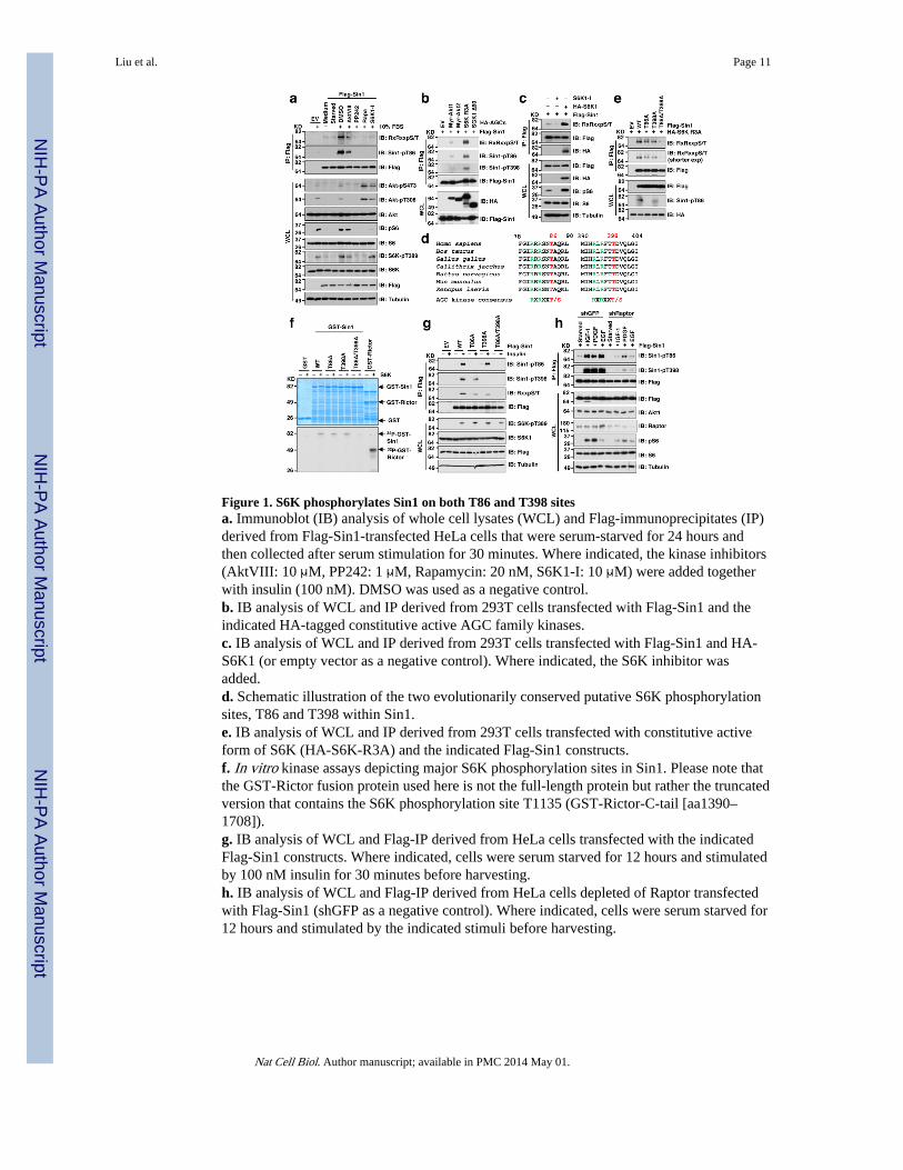
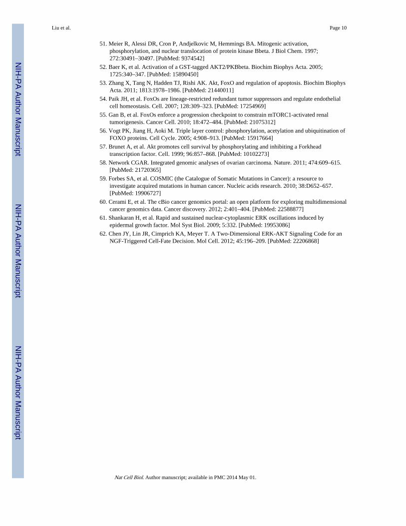
Figure 1. S6K phosphorylates Sin1 on both T86 and T398 sitesa. Immunoblot (IB) analysis of whole cell lysates (WCL) and Flag-immunoprecipitates (IP)derived from Flag-Sin1-transfected HeLa cells that were serum-starved for 24 hours andthen collected after serum stimulation for 30 minutes. Where indicated, the kinase inhibitors(AktVIII: 10 μM, PP242: 1 μM, Rapamycin: 20 nM, S6K1-I: 10 μM) were added togetherwith insulin (100 nM). DMSO was used as a negative control.b. IB analysis of WCL and IP derived from 293T cells transfected with Flag-Sin1 and theindicated HA-tagged constitutive active AGC family kinases.c. IB analysis of WCL and IP derived from 293T cells transfected with Flag-Sin1 and HA-S6K1 (or empty vector as a negative control). Where indicated, the S6K inhibitor wasadded.d. Schematic illustration of the two evolutionarily conserved putative S6K phosphorylationsites, T86 and T398 within Sin1.e. IB analysis of WCL and IP derived from 293T cells transfected with constitutive activeform of S6K (HA-S6K-R3A) and the indicated Flag-Sin1 constructs.f. In vitro kinase assays depicting major S6K phosphorylation sites in Sin1. Please note thatthe GST-Rictor fusion protein used here is not the full-length protein but rather the truncatedversion that contains the S6K phosphorylation site T1135 (GST-Rictor-C-tail [aa1390–1708]).g. IB analysis of WCL and Flag-IP derived from HeLa cells transfected with the indicatedFlag-Sin1 constructs. Where indicated, cells were serum starved for 12 hours and stimulatedby 100 nM insulin for 30 minutes before harvesting.h. IB analysis of WCL and Flag-IP derived from HeLa cells depleted of Raptor transfectedwith Flag-Sin1 (shGFP as a negative control). Where indicated, cells were serum starved for12 hours and stimulated by the indicated stimuli before harvesting.
Liu et al. Page 11
Nat Cell Biol. Author manuscript; available in PMC 2014 May 01.
NIH
-PA Author Manuscript
NIH
-PA Author Manuscript
NIH
-PA Author Manuscript

Figure 2. S6K-dependent phosphorylation of Sin1 dissociates Sin1 from the mTORC2 complexa. Immunoblot (IB) analysis of whole cell lysates (WCL) and Flag immunoprecipitates (IP)derived from 293T cells transfected with the indicated Flag-Sin1 constructs (EV: emptyvector control; WT: Sin1-WT; AA: Sin1-T86A/T398A; EE: Sin1-T86E/T398E).b–d. GST pull down assays to demonstrate that S6K phosphorylation of GST-Sin1-WT-FL(full-length) but not GST-Sin1-T86A/T398A led to impaired interaction with Rictor (b),mTOR-kinase domain (KD) (c) or GβL (d). As indicated, GST-Sin1 proteins werephosphorylated by active recombinant S6K in vitro for 1 hour before using as a bait to pulldown HA-Rictor (b), mTOR-kinase domain (KD) (c) or GβL (d) expressed in 293T cells.e. Gel filtration experiments to illustrate that comparing with WT-Sin1, Sin1-EE lostinteraction with the functional mTORC2 complex components in vivo. IB analysis of theindicated fractionations derived from the gel filtration experiment with HeLa cells co-transfected with HA-Sin1-WT and Flag-Sin1-EE constructs. Prior to running cell lysates, themolecular weight resolution of the column was first estimated by running native molecularweight markers (Thyroglobulin ~669KD, Ferritin ~440KD, Aldolase ~158KD, Conalbumin~75KD and Ovalbumin ~44KD) to determine their retention times on coomassie-stainedSDS-PAGE protein gels.f. Deletion of endogenous TSC2, which led to increased S6K kinase activity, resulted in areduction of Rictor association with Sin1. IB analysis of WCL and anti-Sin1-IP derivedfrom TSC2+/+ or TSC2−/− MEFs.g. Schematic representation of the indicated domains of Sin1 as well as the locations of thetwo Sin1 phosphorylation sites: T86 is in the N-terminal domain while T398 is located in thePH domain.h. GST pull down assays to depict the Sin1 domains that interact with Rictor, mTOR-KD orGβL, respectively (* indicates the sizes of GST-Sin1 proteins).I–j. GST pull down assays to demonstrate that Sin1 T86E or T398E mutation led to reducedinteraction with Rictor (i) or mTOR-KD (j), respectively.
Liu et al. Page 12
Nat Cell Biol. Author manuscript; available in PMC 2014 May 01.
NIH
-PA Author Manuscript
NIH
-PA Author Manuscript
NIH
-PA Author Manuscript
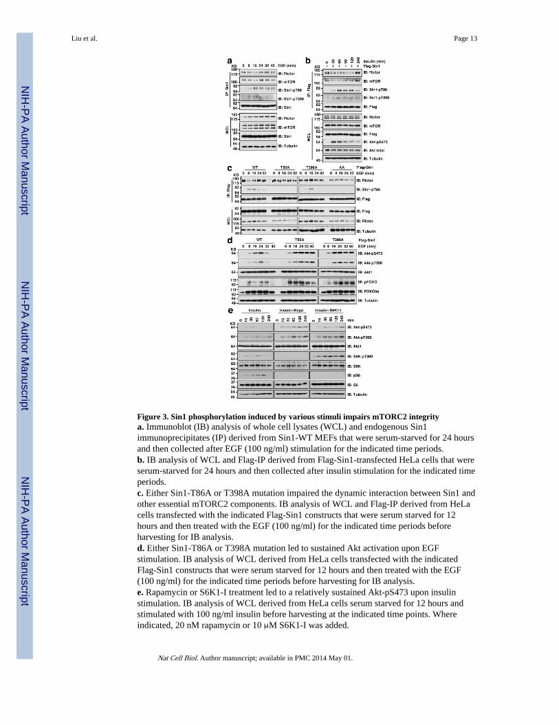
Figure 3. Sin1 phosphorylation induced by various stimuli impairs mTORC2 integritya. Immunoblot (IB) analysis of whole cell lysates (WCL) and endogenous Sin1immunoprecipitates (IP) derived from Sin1-WT MEFs that were serum-starved for 24 hoursand then collected after EGF (100 ng/ml) stimulation for the indicated time periods.b. IB analysis of WCL and Flag-IP derived from Flag-Sin1-transfected HeLa cells that wereserum-starved for 24 hours and then collected after insulin stimulation for the indicated timeperiods.c. Either Sin1-T86A or T398A mutation impaired the dynamic interaction between Sin1 andother essential mTORC2 components. IB analysis of WCL and Flag-IP derived from HeLacells transfected with the indicated Flag-Sin1 constructs that were serum starved for 12hours and then treated with the EGF (100 ng/ml) for the indicated time periods beforeharvesting for IB analysis.d. Either Sin1-T86A or T398A mutation led to sustained Akt activation upon EGFstimulation. IB analysis of WCL derived from HeLa cells transfected with the indicatedFlag-Sin1 constructs that were serum starved for 12 hours and then treated with the EGF(100 ng/ml) for the indicated time periods before harvesting for IB analysis.e. Rapamycin or S6K1-I treatment led to a relatively sustained Akt-pS473 upon insulinstimulation. IB analysis of WCL derived from HeLa cells serum starved for 12 hours andstimulated with 100 ng/ml insulin before harvesting at the indicated time points. Whereindicated, 20 nM rapamycin or 10 μM S6K1-I was added.
Liu et al. Page 13
Nat Cell Biol. Author manuscript; available in PMC 2014 May 01.
NIH
-PA Author Manuscript
NIH
-PA Author Manuscript
NIH
-PA Author Manuscript

Figure 4. Sin1 phosphomimetic mutation is deficient in interacting with the mTORC2 substrateAkt1, but not SGK1a–b. Immunoblot (IB) analysis of whole cell lysates (WCL) and HA (a) or Flag (b)immunoprecipitates (IP) derived from 293T cells that were transfected with the indicatedFlag-Sin1 constructs with HA-Akt1.c. Sin1-T86E or Sin1-T398E disrupts Sin1-N-terminus or Sin1-PH domain interaction withAkt1. Indicated GST-Sin1 proteins were used as a bait to pull down HA-Akt1 expressed in293T cells.d–e. IB analysis of WCL and HA-IP derived from 293T cells that were transfected with theindicated Flag-Sin1 constructs with HA-SGK-Δ60.f–g. IB analysis of WCL and HA-IP or Flag-IP derived from 293T cells that weretransfected with the indicated Flag-Sin1 constructs with HA-S6K1.
Liu et al. Page 14
Nat Cell Biol. Author manuscript; available in PMC 2014 May 01.
NIH
-PA Author Manuscript
NIH
-PA Author Manuscript
NIH
-PA Author Manuscript
Figure 5. Sin1 phosphorylation suppresses mTORC2 kinase activity towards phosphorylatingAkt in vitroa–b. 293T cells were transfected with the indicated Flag-tagged Sin1 constructs. 36 hourspost-transfection, whole cell lysates (WCL) were collected and the mTORC2 complex waspurified by Flag-immunoprecipitation (IP). The Flag-IPs were incubated in vitro withpurified GST-Akt1 in the presence of ATP and the kinase reaction buffer. Thirty minuteslater, the reaction was stopped by the addition of the loading buffer. Akt1 phosphorylationstatus was examined by immunoblot (IB) analysis.c–d. Prolonged insulin treatment (45 min) induces Sin1 phosphorylation, leading todissociation of mTORC2 complex and abolished Akt activation. Flag-Sin1-WT or R81Tmutant was transfected into HeLa cells and 48 hours later the transfected cells wereharvested upon insulin (100 nM) stimulation for 45 min after 12 hours of serum starvation inCHAPS buffer. The whole cell lysates were filtered and run through FPLC superdex 200column. 500 μL elute was collected for each fraction and 1/20 volume of each fraction wasincubated with 2 μg GST-Akt-tail (aa 408–480) at 30°C for 30 min. Afterwards, theresulting samples were resolved on SDS-PAGE and subjected to IB analysis.e–f. Rapamycin treatment restored Sin1 phosphorylation resulted from EGF treatment,leading to reassembly of mTORC2 complex and Akt activation. Flag-Sin1-WT wastransfected into TSC2-depleted HeLa cells and 24 hours later, the transfected cells weretreated with 20 nM rapamycin for another 12 hours prior to EGF stimulation (100 nM)before harvested and analyzed as in (c–d).
Liu et al. Page 15
Nat Cell Biol. Author manuscript; available in PMC 2014 May 01.
NIH
-PA Author Manuscript
NIH
-PA Author Manuscript
NIH
-PA Author Manuscript

Figure 6. Sin1 phosphorylation attenuates mTORC2 kinase activity towards phosphorylatingAkt in vivoa. Sin1−/− MEFs were transfected with the indicated Flag-Sin1 constructs. 30 hours post-transfection, the resulting cells were serum-starved for 24 hours and then collected afterstimulation with insulin for 30 minutes for immunoblot (IB) analysis.b. Reconstitution of Sin1−/− MEFs with WT-, but not EE-Sin1, could restore Akt-Ser473phosphorylation under various stimulation conditions. Sin1−/− MEFs were transfected withthe indicated Flag-Sin1 constructs and serum-starved for 24 hours before harvesting aftertreatment with indicated stimuli for IB analysis.c. The indicated Flag-Sin1 constructs were transfected into Sin1−/− MEFs and Flagimmunoprecipitation (IP) was recovered as the kinase source to phosphorylate GST-Akt1-tail (aa 408–480) in vitro.d. The indicated Flag-Sin1 constructs were transfected into Sin1−/− MEFs and endogenousAkt IP was performed as the kinase source to phosphorylate crosstide in vitro. Data wasshown as mean ± SD for n= 3 independent experiments.e–g. Sin1−/− MEFs were transfected with the indicated Flag-Sin1 constructs (with emptyvector as a negative control). 24 hours post-transfection, the resulting cells were cultured in10% FBS-containing medium with the indicated concentrations of etoposide (e) or cisplatin(f) for 48 hours before performing the cell viability assays (e,f) or IB analysis (g). Data wasshown as mean ± SD from n=3 independent experiments. * indicates p < 0.05 (Student’s t-test).
Liu et al. Page 16
Nat Cell Biol. Author manuscript; available in PMC 2014 May 01.
NIH
-PA Author Manuscript
NIH
-PA Author Manuscript
NIH
-PA Author Manuscript
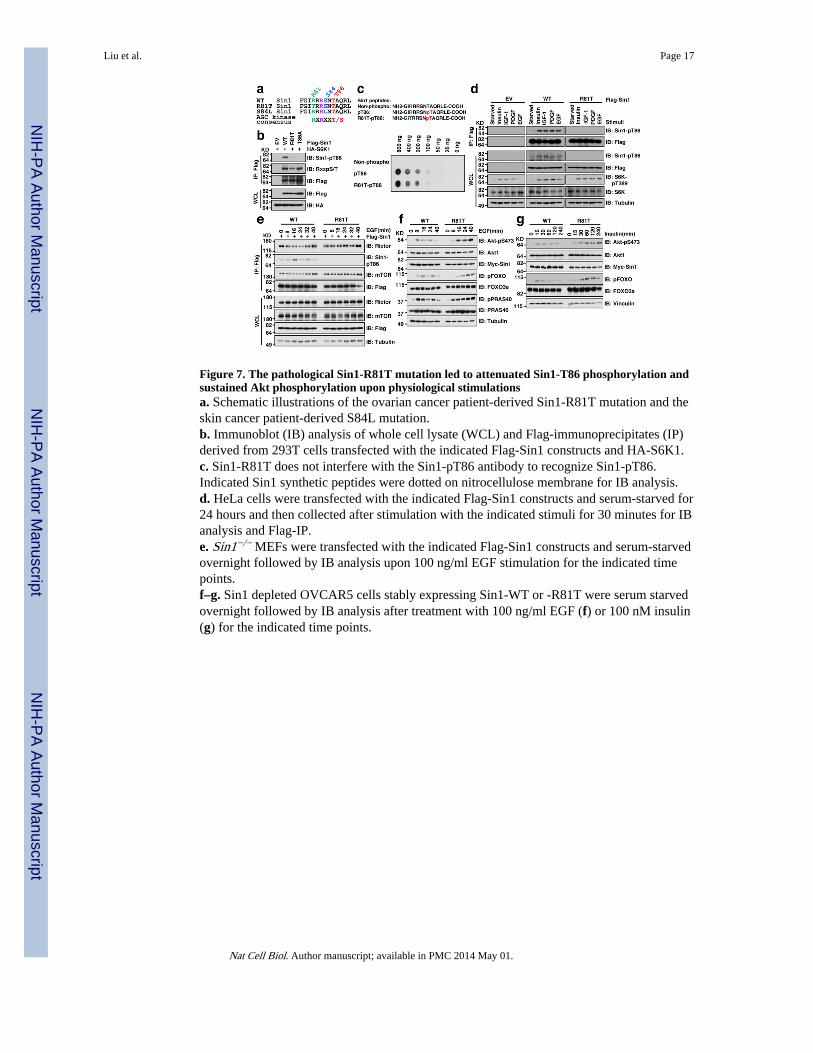
Figure 7. The pathological Sin1-R81T mutation led to attenuated Sin1-T86 phosphorylation andsustained Akt phosphorylation upon physiological stimulationsa. Schematic illustrations of the ovarian cancer patient-derived Sin1-R81T mutation and theskin cancer patient-derived S84L mutation.b. Immunoblot (IB) analysis of whole cell lysate (WCL) and Flag-immunoprecipitates (IP)derived from 293T cells transfected with the indicated Flag-Sin1 constructs and HA-S6K1.c. Sin1-R81T does not interfere with the Sin1-pT86 antibody to recognize Sin1-pT86.Indicated Sin1 synthetic peptides were dotted on nitrocellulose membrane for IB analysis.d. HeLa cells were transfected with the indicated Flag-Sin1 constructs and serum-starved for24 hours and then collected after stimulation with the indicated stimuli for 30 minutes for IBanalysis and Flag-IP.e. Sin1−/− MEFs were transfected with the indicated Flag-Sin1 constructs and serum-starvedovernight followed by IB analysis upon 100 ng/ml EGF stimulation for the indicated timepoints.f–g. Sin1 depleted OVCAR5 cells stably expressing Sin1-WT or -R81T were serum starvedovernight followed by IB analysis after treatment with 100 ng/ml EGF (f) or 100 nM insulin(g) for the indicated time points.
Liu et al. Page 17
Nat Cell Biol. Author manuscript; available in PMC 2014 May 01.
NIH
-PA Author Manuscript
NIH
-PA Author Manuscript
NIH
-PA Author Manuscript

Figure 8. The pathological Sin1-R81T mutation displayed elevated oncogenic activity in part bybypassing Sin1 phosphorylation-mediated negative regulation of Akt-pS473a–c. Sin1−/− MEFs were transfected with the indicated Flag-Sin1 constructs and weretreated with the indicated concentrations of etoposide (a) or cisplatin (b,c) for 48 hoursbefore performing the cell viability assays (a,b) or immunoblot (IB) analysis (c). Data wasshown as mean ± SD from n=3 independent experiments. * indicates p < 0.05 (t-test).d. Soft agar assays for Sin1-depleted OVCAR5 cells stably expressing EV, WT or R81T.Data was presented as mean ± SD from n=3 independent experiments.e–f. Growth curves (e) and mass of the dissected tumors (f) from xenograft experiments withthe indicated cells injected subcutaneously into n=10 mice for each cell line. The visibletumors were measured at the indicated days. Error bars, ±SEM and * indicates p < 0.05 (t-test).g. Representative images of the dissected tumors presented in Figure 8e,f.h. Eight-week-old mice were fasted overnight and then refed for 6 hours following a 30 minpretreatment with vehicle or rapamycin (10 mg/kg). N=4 mice per condition. Livers weredissected and liver lysates were subjected to IB analysis.i. Four representative images of IHC with indicated Sin1 and Akt phosphorylation status outof 58 ovarian patient samples under 400x magnification. Scale bar represents 100 μm.
Liu et al. Page 18
Nat Cell Biol. Author manuscript; available in PMC 2014 May 01.
NIH
-PA Author Manuscript
NIH
-PA Author Manuscript
NIH
-PA Author Manuscript