infrared filtometer combined with functionalized silica films
TRANSCRIPT
1
A Pocket-Sized 3D-printed Attenuated Total Reflection-
Infrared Filtometer combined with Functionalized Silica
Films for Nitrate Sensing in Water
Bettina Baumgartner, Stephan Freitag, Christoph Gasser and Bernhard Lendl*
Research Division of Environmental Analytics, Process Analytics and Sensors, Institute of Chemical
Technologies and Analytics, Technische Universität Wien, Getreidemarkt 9, 1060 Vienna, Austria
Abstract
Strong anion-exchange materials carry a positive charge that allows them to trap and concentrate anions
while releasing other anions. Here, we introduced an ion exchange group into mesoporous silica films
coated on attenuated total reflection (ATR) crystals to enrich nitrate from aqueous phase in the volume
probed by the evanescent field. The ion-exchange and enrichment capabilities of the films were
characterized using standard FTIR spectroscopy. Thereby fast analyte enrichment and full sensor recovery
were observed. In addition, high enrichment factors of up to 1600 were achieved. After characterization,
these coated ATR crystals were used in a dedicated ATR-IR filtometer comprising a Fabry-Pérot filter
detector unit and a miniaturized thermal emitter with a footprint of only 80 mm × 120 mm × 70 mm. The
filter covered the spectral region between 1250 - 1800 cm-1 allowing for recording IR spectra of nitrate
enriched into the mesoporous silica film. The sensor was calibrated using the Langmuir adsorption model
as calibration function. From this, a limit of detection of 1.2 mg L-1 was derived for the ATR-IR filtometer.
This emphasizes the high potential of functionalized mesoporous silica films combined with low-cost
filtometers for portable water sensors.
Keywords: Mid-IR Spectroscopy, Mesoporous Materials, Sensing, Ion-Exchange, Functional Coating,
Nitrate, 3D-printing
© 2019. This is the peer reviewed version of the following article: B Baumgartner, S Freitag, C Gasser, B
Lendl A Pocket-Sized 3D-printed Attenuated Total Reflection-Infrared Filtometer combined with Functionalized Silica Films for Nitrate Sensing in Water, Sensors Actuators B Chem, 2020 127847, which has been published in final form at https://doi.org/10.1016/j.snb.2020.127847. This manuscript version is made available under the CC-BY-NC-ND 4.0 license http://creativecommons.org/licenses/by-nc-nd/4.0/
2
1. Introduction
Safe water supplies are essential for human health and environmental protection. In this context, nitrate
is one of the major waterborne chemical hazards and even short term exposure leads to severe health
effects.1 Nitrate concentrations in water supplies scale with human activity particularly due to their
intensive use in agriculture and animal breeding.2 Due to their high water solubility, nitrate eventually find
its way into ground water and hence into drinking water supplies.2,3 Therefore, pollution with nitrate is
typically high in rural areas and regularly exceeds the world health organization guideline value of
50 mg L-1. Up to date, nitrate is quantified either spectrophotometrically as nitrite after reduction
catalyzed by Cd-salt or enzymes, via ion chromatography, or via ion-selective electrodes.4 In addition,
broad-band ultraviolet/visible spectroscopy-based on-line probes for water monitoring have emerged.5
These systems have been employed for complex water streams, e.g. with high turbidity, relying on
sophisticated calibrations dedicated to each application.5–7 Apart from these established techniques,
infrared (IR) spectroscopy provides quantitative chemical information that can be obtained in reagent free
measurements in real time. Ions like nitrate show strong and specific bands in the mid-IR spectrum. The
asymmetric NO3 stretching vibration is located at 1395 cm-1. However, quantification of this band in the
IR spectrum is made difficult by the strong background absorption of water, which limits the optical path
length and hence sensitivity. Attenuated total reflection (ATR) IR spectroscopy is commonly employed for
spectroscopic studies of aqueous samples.8 It was demonstrated that the sensitivity of this technique can
be vastly increased using selective enrichment layers coated on ATR crystals that cover the volume probed
by the evanescent field.9–11 Until now, they have been primarily employed for enriching apolar substances
into hydrophobic coatings. In this context, we have successfully shown that organically functionalized
mesoporous silica films can also be employed.12 Besides the short response times found for mesoporous
silica, their rich surface chemistry allows for tuning their functionality beyond hydrophobicity. The
successful nitrate removal of functionalized silica materials with a trimethylammonium-moiety as ion
exchange group has been shown for powder materials in batch experiments.13,14 However, the possibility
to fully recover these materials and use them as sensor is still pending. In this work, this question is
targeted and a trimethylammonium-moiety was covalently functionalized into a mesoporous silica films
coated on ATR crystals. The sensing performance for nitrate in aqueous solutions was studied using a
Fourier transform (FT) IR spectrometer.
These spectrometers are still considered as gold-standard for IR spectroscopy. The broad spectral
coverage and robustness of FTIR spectrometers enabled applications in a variety of analytical tasks
3
including quantitative analysis of liquids in off-line as well as in-line applications,15–20 studying reactions at
surfaces,21–23 as well as clinical diagnositics.24 An emerging, cost-effective alternative to FTIR
spectrometers are filtometers that, in comparison with benchtop spectrometers, allow on-site
measurements due to their small size and low energy consumption. In contrast to Michelson
interferometers, wavelength discrimination is achieved by the use of optical band pass filters.25 Based on
this principle, Fabry-Pérot (FP) filter designs have emerged in the 1990s that discriminate the transmitted
wavelength by the distance d between two coplanar reflective surfaces and the refractive index between
these surfaces.26 By changing d, the filter can be spectrally tuned and the spectral resolution varies
between 20 cm-1 up to 50 cm-1, depending on the wavelength range.27 A micro-electro-mechanical system
(MEMS)-based mid IR filter using a FP air resonator combined with a pyroelectric detector was first
reported in 2003 and is now commercially distributed in several MIR wavelength ranges.28,29 FP filter-
detectors have been successfully paired with miniaturized pulsed MIR emitters were employed for
process gas monitoring in transmission gas cells,30–32 for liquid sensing,33 imaging systems34 as well as
paired with a supercontinuum laser for standoff IR spectroscopy.32 These reports achieved adequate
analytical performance without the high spectral resolution and the full spectral coverage provided by
FTIR spectrometers while reducing the costs and size of the respective sensing system.
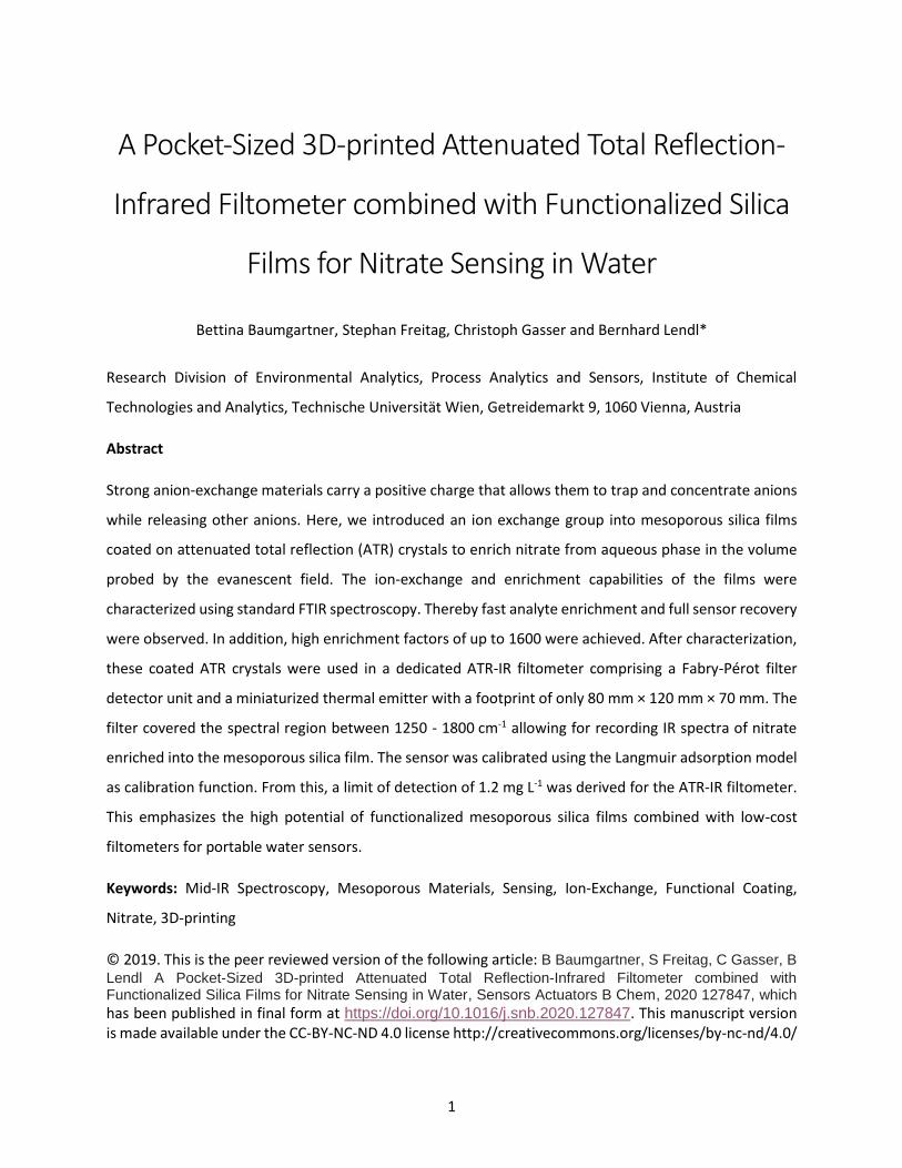
Figure 1: Ge ATR crystal (dark grey) coated with mesoporous silica film functionalized with trimethylammonium
moieties allowing for ion-exchange of Cl- to NO3- ions. The coated ATR crystal is placed in a flow cell supplied with
nitrate solutions via a peristaltic pump. IR absorbance takes place within the evanescent field (light red), which
extends beyond the crystal into the film.
The aim of this contribution is to demonstrate a small, cost-effective mid-IR sensor for nitrate in aqueous
solutions based on a 3D-printed ATR-IR filtometer. Germanium ATR crystals coated with a
4
trimethylammonium-functionalized mesoporous silica films for nitrate sensing were developed (compare
Figure 1) and their sensing performance were investigated using FTIR spectroscopy revealing short
response times of less than 1 min and high enrichment factors of up to 1600. In a subsequent step the
coated ATR crystals were transferred into a dedicated 3D-printed unit comprising light source, FP filter-
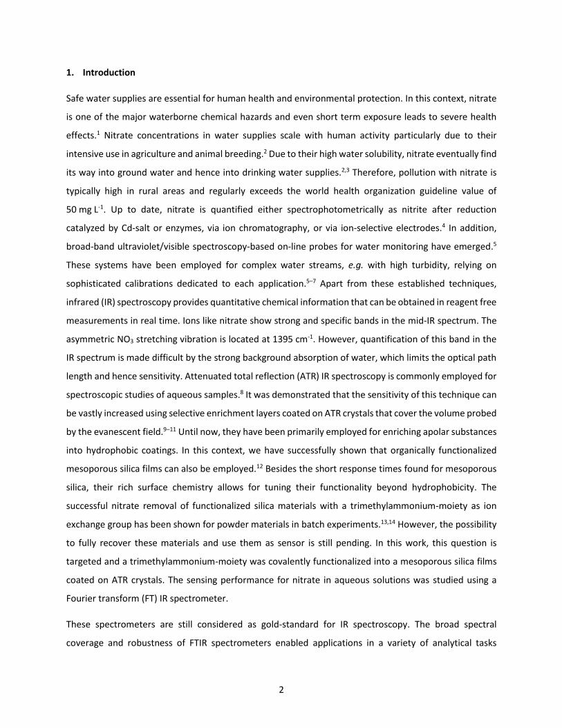
detector and printed circuit board with a footprint of only 80 mm × 120 mm × 70 mm (compare Figure 2).
Although having a lower signal to noise ratio and smaller spectral coverage compared to a FTIR
spectrometer, the high enrichment factors of the functionalized ATR crystals allowed for sensing NO3-
concentrations as low as 3.6 mg L-1. Given the high versatility of silica and low cost of the FP filters, these
results demonstrates the high potential of functionalized mesoporous materials in combination with an
ATR-IR-filtometer for water monitoring.
2. Materials and Methods
Tetraethoxysilane (TEOS, Sigma Aldrich 99.5%), abs. ethanol (Fisher, 99.6%), hydrochloric acid (VWR,
37%), cetyltrimethylammonium bromide (CTAB, Sigma Aldrich, 99%), N-[3-(trimethoxysilyl) propyl]-
N,N,N-trimethylammonium chloride (TMAC, Sigma Aldrich, 50 % in methanol), were used as received.
Aqueous NaNO3 solutions were prepared by weight.
2.1. Synthesis of mesoporous films
Mesoporous silica film synthesized using CTAB was prepared as previously reported with a final molar
ratio of 1:13:5:5 × 10⁻³:0.12 for TEOS:EtOH:H2O:HCl:CTAB.12 The mixture was stirred for 3 h at 40°C. The
film was spin coated with a spinner velocity of 2000 rpm and subsequently calcined at 400 °C for 4 h with
a heating ramp of 1 K min-1. For surface functionalization, the coated ATR crystal was placed in a 3-neck
flask and 15 mL methanol and 0.5 mL TMAC solution were added. The solution was heated to reflux
temperature for 8 h. Subsequently, films were rinsed with acetone and heated to 60 °C in the oven over
night.
The characterization of the films using X-ray diffraction and profilometry was performed accordingly to
our previous reports.12,21
2.2. Optical Setup for FTIR Spectroscopy
Mesoporous silica films were coated on Ge ATR crystals (20 × 10 × 0.5 mm3) cut from double side polished
wafers and polished narrow facets with a defined angle of 45°. This ATR crystal configuration comprises
20 active bounces and a depth of penetration (per bounce) of 420 nm at 1500 cm-1 (nGe=4.02,
5
nsample=1.33).8,35,36 The ATR crystals were inserted into a custom-made mount and fixed with a FKM O-ring
and an aluminum liquid flow cell with a volume of ca. 20 µL.12 Two gold mirrors directed the IR beam onto
the ATR facet and the detector. The optical setup was placed in the sample compartment of a Vertex 80v
FTIR spectrometer (Bruker Optics, Germany) equipped with an N2-cooled MCT detector. Spectra were
recorded with 4 cm-1 resolution and 32 scans were averaged per spectrum (double-sided, backward-
forward acquisition mode, 8 s per spectrum). Prior to spectrum acquisition, the spectrometer sample
compartment was flushed with dry air. The noise floor of the system was obtained by means of 100 %
lines, i.e. a scan of the N2 flushed flow cell with a background scan obtained under same conditions and
yielded an RMS noise of 3 · 10-5 A.U. between 1800 cm-1- 1450 cm-1. Spectra were analyzed using the
software package OPUS 7.5 (Bruker Optics, Germany).
2.3. Liquid Handling
Pure water, 0.1 M HCl and nitrate solutions with different concentrations were applied at a flow rate of
1 mL min−1 using a peristaltic pump (Ismatec, Germany), and 1/16-in (see Figure 1 for setup). PTFE tubing
with 0.5 mm inner diameter was connected to the aluminum flow cell and the peristaltic pump.
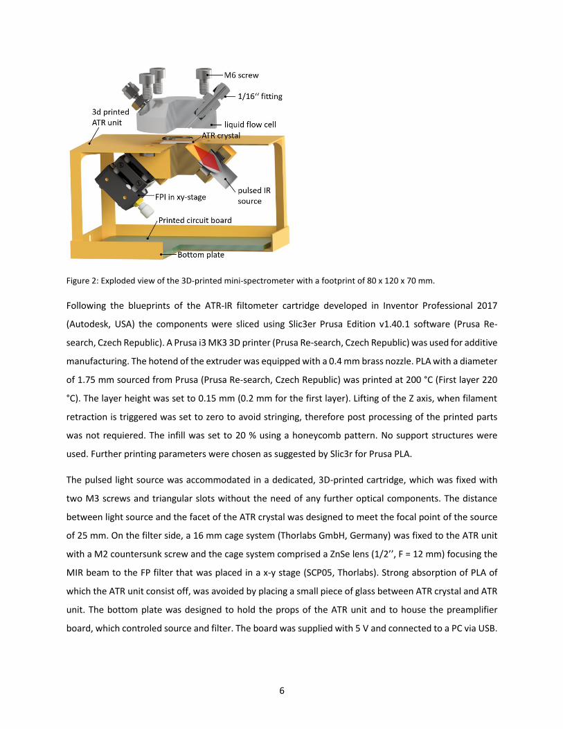
2.4. 3D-printed ATR-IR Filtometer
The ATR-IR filtometer was built using a pulsed light source EMIRS200 (Atricle No. 601.612, Reflector 3,
CaF2 window, front-vented, Axetris, Switzerland) a MEMS-based mid-IR filter using a Fabry-Pérot air
resonator combined with a pyroelectric dector (LFP-5580C-337, TO8 housing, InfraTec, Germany) covering
a spectral range between 1900 cm-1-1250 cm-1 and corresponding preamplifier electronics from InfraTec.
All parts were integrated in a 3D-printed ATR unit that housed the ATR crystal and defined the angle of
incidence for ATR crystal to be 45° (see Figure 2). Ge ATR crystals were used to assure transparence for
the mid-IR irradiation in the targeted spectral region between 1250 - 1500 cm-1.
6
Figure 2: Exploded view of the 3D-printed mini-spectrometer with a footprint of 80 x 120 x 70 mm.
Following the blueprints of the ATR-IR filtometer cartridge developed in Inventor Professional 2017
(Autodesk, USA) the components were sliced using Slic3er Prusa Edition v1.40.1 software (Prusa Re-
search, Czech Republic). A Prusa i3 MK3 3D printer (Prusa Re-search, Czech Republic) was used for additive
manufacturing. The hotend of the extruder was equipped with a 0.4 mm brass nozzle. PLA with a diameter
of 1.75 mm sourced from Prusa (Prusa Re-search, Czech Republic) was printed at 200 °C (First layer 220
°C). The layer height was set to 0.15 mm (0.2 mm for the first layer). Lifting of the Z axis, when filament
retraction is triggered was set to zero to avoid stringing, therefore post processing of the printed parts
was not requiered. The infill was set to 20 % using a honeycomb pattern. No support structures were
used. Further printing parameters were chosen as suggested by Slic3r for Prusa PLA.
The pulsed light source was accommodated in a dedicated, 3D-printed cartridge, which was fixed with
two M3 screws and triangular slots without the need of any further optical components. The distance
between light source and the facet of the ATR crystal was designed to meet the focal point of the source
of 25 mm. On the filter side, a 16 mm cage system (Thorlabs GmbH, Germany) was fixed to the ATR unit
with a M2 countersunk screw and the cage system comprised a ZnSe lens (1/2’’, F = 12 mm) focusing the
MIR beam to the FP filter that was placed in a x-y stage (SCP05, Thorlabs). Strong absorption of PLA of
which the ATR unit consist off, was avoided by placing a small piece of glass between ATR crystal and ATR
unit. The bottom plate was designed to hold the props of the ATR unit and to house the preamplifier
board, which controled source and filter. The board was supplied with 5 V and connected to a PC via USB.
7
Four M6 nuts were sunk in slits enabling mounting of the liquid flow cell (see section 2.3 further details
on the flow cell), which was attached to the peristaltic pump for sample application.
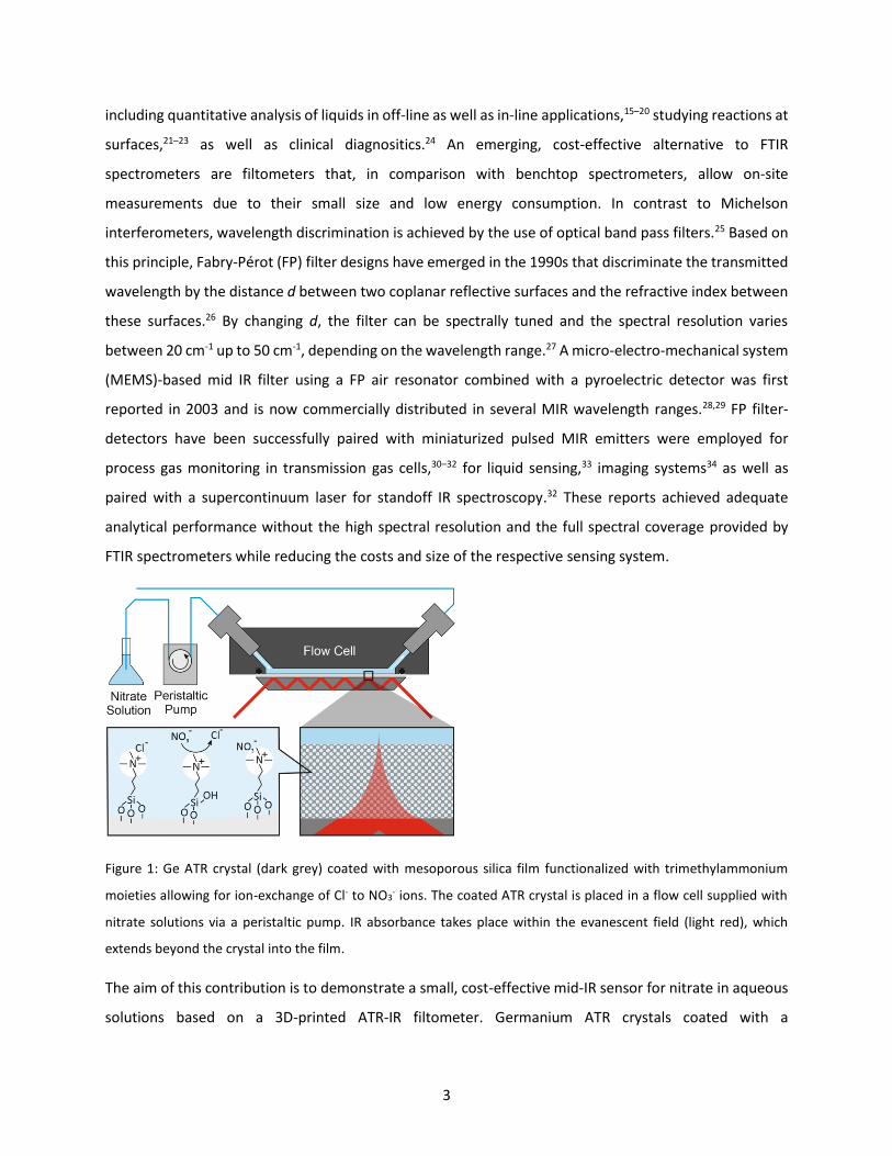
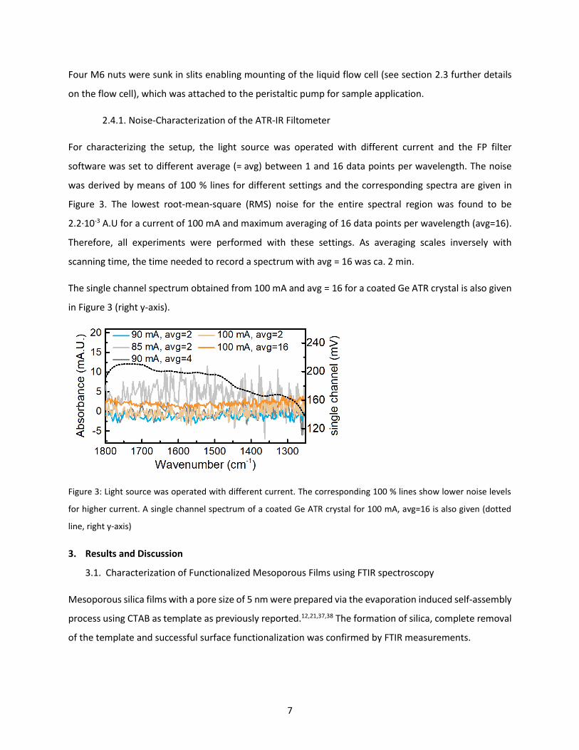
2.4.1. Noise-Characterization of the ATR-IR Filtometer
For characterizing the setup, the light source was operated with different current and the FP filter
software was set to different average (= avg) between 1 and 16 data points per wavelength. The noise
was derived by means of 100 % lines for different settings and the corresponding spectra are given in
Figure 3. The lowest root-mean-square (RMS) noise for the entire spectral region was found to be
2.2·10-3 A.U for a current of 100 mA and maximum averaging of 16 data points per wavelength (avg=16).
Therefore, all experiments were performed with these settings. As averaging scales inversely with
scanning time, the time needed to record a spectrum with avg = 16 was ca. 2 min.
The single channel spectrum obtained from 100 mA and avg = 16 for a coated Ge ATR crystal is also given
in Figure 3 (right y-axis).
Figure 3: Light source was operated with different current. The corresponding 100 % lines show lower noise levels
for higher current. A single channel spectrum of a coated Ge ATR crystal for 100 mA, avg=16 is also given (dotted
line, right y-axis)
3. Results and Discussion
3.1. Characterization of Functionalized Mesoporous Films using FTIR spectroscopy
Mesoporous silica films with a pore size of 5 nm were prepared via the evaporation induced self-assembly
process using CTAB as template as previously reported.12,21,37,38 The formation of silica, complete removal
of the template and successful surface functionalization was confirmed by FTIR measurements.
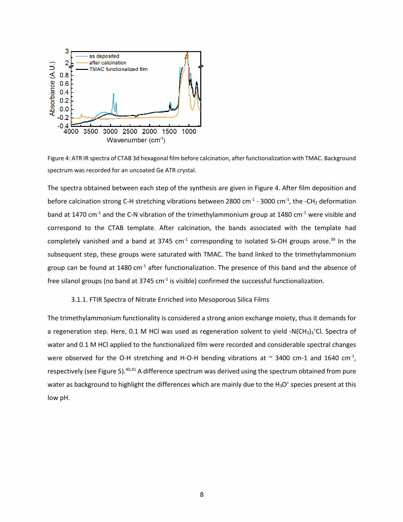
8
Figure 4: ATR IR spectra of CTAB 3d hexagonal film before calcination, after functionalization with TMAC. Background
spectrum was recorded for an uncoated Ge ATR crystal.
The spectra obtained between each step of the synthesis are given in Figure 4. After film deposition and
before calcination strong C-H stretching vibrations between 2800 cm-1 - 3000 cm-1, the -CH2 deformation
band at 1470 cm-1 and the C-N vibration of the trimethylammonium group at 1480 cm-1 were visible and
correspond to the CTAB template. After calcination, the bands associated with the template had
completely vanished and a band at 3745 cm-1 corresponding to isolated Si-OH groups arose.39 In the
subsequent step, these groups were saturated with TMAC. The band linked to the trimethylammonium
group can be found at 1480 cm-1 after functionalization. The presence of this band and the absence of
free silanol groups (no band at 3745 cm-1 is visible) confirmed the successful functionalization.
3.1.1. FTIR Spectra of Nitrate Enriched into Mesoporous Silica Films
The trimethylammonium functionality is considered a strong anion exchange moiety, thus it demands for
a regeneration step. Here, 0.1 M HCl was used as regeneration solvent to yield -N(CH3)3+Cl. Spectra of
water and 0.1 M HCl applied to the functionalized film were recorded and considerable spectral changes
were observed for the O-H stretching and H-O-H bending vibrations at ~ 3400 cm-1 and 1640 cm-1,
respectively (see Figure 5).40,41 A difference spectrum was derived using the spectrum obtained from pure
water as background to highlight the differences which are mainly due to the H3O+ species present at this
low pH.
9
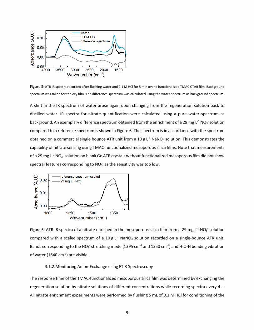
Figure 5: ATR IR spectra recorded after flushing water and 0.1 M HCl for 5 min over a functionalized TMAC CTAB film. Background
spectrum was taken for the dry film. The difference spectrum was calculated using the water spectrum as background spectrum.
A shift in the IR spectrum of water arose again upon changing from the regeneration solution back to
distilled water. IR spectra for nitrate quantification were calculated using a pure water spectrum as
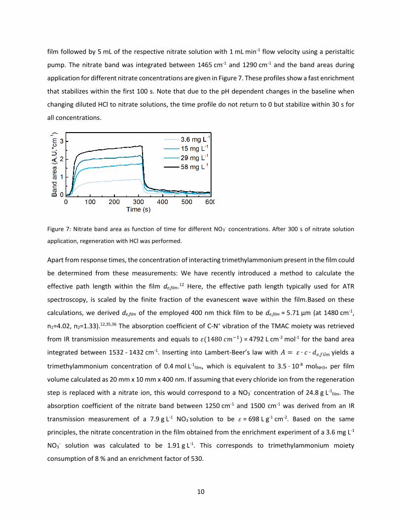
background. An exemplary difference spectrum obtained from the enrichment of a 29 mg L-1 NO3- solution
compared to a reference spectrum is shown in Figure 6. The spectrum is in accordance with the spectrum
obtained on a commercial single bounce ATR unit from a 10 g L-1 NaNO3 solution. This demonstrates the
capability of nitrate sensing using TMAC-functionalized mesoporous silica films. Note that measurements
of a 29 mg L-1 NO3- solution on blank Ge ATR crystals without functionalized mesoporous film did not show
spectral features corresponding to NO3- as the sensitivity was too low.
Figure 6: ATR IR spectra of a nitrate enriched in the mesoporous silica film from a 29 mg L-1 NO3- solution
compared with a scaled spectrum of a 10 g L-1 NaNO3 solution recorded on a single-bounce ATR unit.
Bands corresponding to the NO3- stretching mode (1395 cm-1 and 1350 cm-1) and H-O-H bending vibration
of water (1640 cm-1) are visible.
3.1.2. Monitoring Anion-Exchange using FTIR Spectroscopy
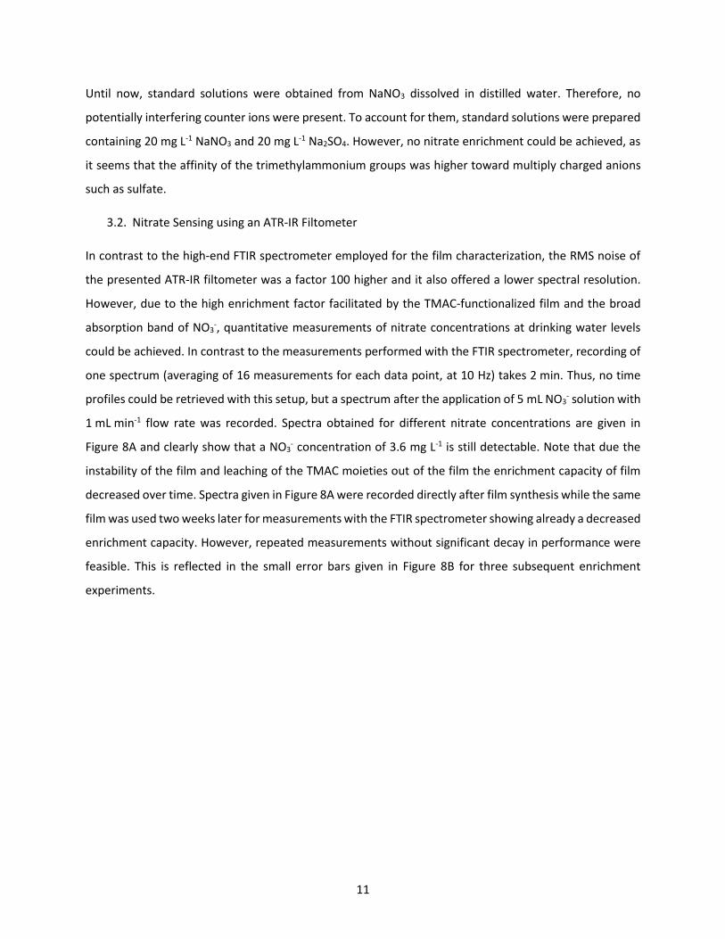
The response time of the TMAC-functionalized mesoporous silica film was determined by exchanging the
regeneration solution by nitrate solutions of different concentrations while recording spectra every 4 s.
All nitrate enrichment experiments were performed by flushing 5 mL of 0.1 M HCl for conditioning of the
10
film followed by 5 mL of the respective nitrate solution with 1 mL min-1 flow velocity using a peristaltic
pump. The nitrate band was integrated between 1465 cm-1 and 1290 cm-1 and the band areas during
application for different nitrate concentrations are given in Figure 7. These profiles show a fast enrichment
that stabilizes within the first 100 s. Note that due to the pH dependent changes in the baseline when
changing diluted HCl to nitrate solutions, the time profile do not return to 0 but stabilize within 30 s for
all concentrations.
Figure 7: Nitrate band area as function of time for different NO3- concentrations. After 300 s of nitrate solution
application, regeneration with HCl was performed.
Apart from response times, the concentration of interacting trimethylammonium present in the film could
be determined from these measurements: We have recently introduced a method to calculate the
effective path length within the film de,film.12 Here, the effective path length typically used for ATR
spectroscopy, is scaled by the finite fraction of the evanescent wave within the film.Based on these
calculations, we derived de,film of the employed 400 nm thick film to be de,film = 5.71 µm (at 1480 cm-1,
n1=4.02, n2=1.33).12,35,36 The absorption coefficient of C-N+ vibration of the TMAC moiety was retrieved
from IR transmission measurements and equals to 𝜀(1480 𝑐𝑚−1) = 4792 L cm-2 mol-1 for the band area
integrated between 1532 - 1432 cm-1. Inserting into Lambert-Beer’s law with 𝐴 = 𝜀 ∙ 𝑐 ∙ 𝑑𝑒,𝑓𝑖𝑙𝑚 yields a
trimethylammonium concentration of 0.4 mol L-1film, which is equivalent to 3.5 · 10-8 molNH3+ per film
volume calculated as 20 mm x 10 mm x 400 nm. If assuming that every chloride ion from the regeneration
step is replaced with a nitrate ion, this would correspond to a NO3- concentration of 24.8 g L-1
film. The
absorption coefficient of the nitrate band between 1250 cm-1 and 1500 cm-1 was derived from an IR
transmission measurement of a 7.9 g L-1 NO3-solution to be 𝜀 = 698 L g-1 cm-2. Based on the same
principles, the nitrate concentration in the film obtained from the enrichment experiment of a 3.6 mg L-1
NO3- solution was calculated to be 1.91 g L-1. This corresponds to trimethylammonium moiety
consumption of 8 % and an enrichment factor of 530.
11
Until now, standard solutions were obtained from NaNO3 dissolved in distilled water. Therefore, no
potentially interfering counter ions were present. To account for them, standard solutions were prepared
containing 20 mg L-1 NaNO3 and 20 mg L-1 Na2SO4. However, no nitrate enrichment could be achieved, as
it seems that the affinity of the trimethylammonium groups was higher toward multiply charged anions
such as sulfate.
3.2. Nitrate Sensing using an ATR-IR Filtometer
In contrast to the high-end FTIR spectrometer employed for the film characterization, the RMS noise of
the presented ATR-IR filtometer was a factor 100 higher and it also offered a lower spectral resolution.
However, due to the high enrichment factor facilitated by the TMAC-functionalized film and the broad
absorption band of NO3-, quantitative measurements of nitrate concentrations at drinking water levels
could be achieved. In contrast to the measurements performed with the FTIR spectrometer, recording of
one spectrum (averaging of 16 measurements for each data point, at 10 Hz) takes 2 min. Thus, no time
profiles could be retrieved with this setup, but a spectrum after the application of 5 mL NO3- solution with
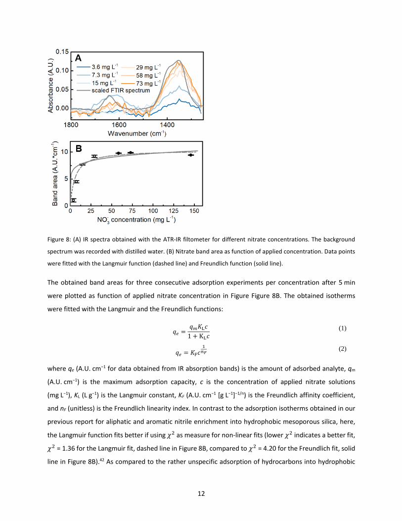
1 mL min-1 flow rate was recorded. Spectra obtained for different nitrate concentrations are given in
Figure 8A and clearly show that a NO3- concentration of 3.6 mg L-1 is still detectable. Note that due the
instability of the film and leaching of the TMAC moieties out of the film the enrichment capacity of film
decreased over time. Spectra given in Figure 8A were recorded directly after film synthesis while the same
film was used two weeks later for measurements with the FTIR spectrometer showing already a decreased
enrichment capacity. However, repeated measurements without significant decay in performance were
feasible. This is reflected in the small error bars given in Figure 8B for three subsequent enrichment
experiments.
12
Figure 8: (A) IR spectra obtained with the ATR-IR filtometer for different nitrate concentrations. The background
spectrum was recorded with distilled water. (B) Nitrate band area as function of applied concentration. Data points
were fitted with the Langmuir function (dashed line) and Freundlich function (solid line).
The obtained band areas for three consecutive adsorption experiments per concentration after 5 min
were plotted as function of applied nitrate concentration in Figure Figure 8B. The obtained isotherms
were fitted with the Langmuir and the Freundlich functions:
𝑞𝑒 =𝑞𝑚𝐾L𝑐
1 + KL𝑐
(1)
𝑞𝑒 = 𝐾F𝑐1
𝑛𝐹 (2)
where qe (A.U. cm−1 for data obtained from IR absorption bands) is the amount of adsorbed analyte, qm
(A.U. cm−1) is the maximum adsorption capacity, c is the concentration of applied nitrate solutions
(mg L−1), KL (L g−1) is the Langmuir constant, KF (A.U. cm−1 [g L−1]−1/n) is the Freundlich affinity coefficient,
and nF (unitless) is the Freundlich linearity index. In contrast to the adsorption isotherms obtained in our
previous report for aliphatic and aromatic nitrile enrichment into hydrophobic mesoporous silica, here,
the Langmuir function fits better if using 𝜒2 as measure for non-linear fits (lower 𝜒2 indicates a better fit,
𝜒2 = 1.36 for the Langmuir fit, dashed line in Figure 8B, compared to 𝜒2 = 4.20 for the Freundlich fit, solid
line in Figure 8B).42 As compared to the rather unspecific adsorption of hydrocarbons into hydrophobic
13
mesoporous silica films that allowed for multilayer adsorption (according to the Freundlich model), the
trimethylammonium functionalized films offered just one binding site per nitrate ion. This is in accordance
with the definition of Langmuir adsorption isotherms and previously reported batch experiments.13,43 The
derived Langmuir function served as calibration function. This allowed for retrieving the limit of detection
(LOD) defined as qe(LOD)= 3·σ, with σ being the standard deviation derived from 100 % lines obtained
from consecutive blank water spectra. This yielded a LOD of 1.2 mg L-1 for the presented ATR-IR filtometer.
According to quantitative considerations performed in section 3.1.2, the enriched amount of NO3- per
volume was also derived from these experiments. The band area obtained from a 7.3 mg L-1 NO3- solution
corresponds to a concentration of 11.7 g L-1film, which is equivalent to a trimethylammonium moiety
consumption of 14 % and an enrichment factor of 1600. The threefold enrichment factor compared to
FTIR measurements is in accordance with the loss of the trimethylammonium moieties as determined
from IR spectra of the as-synthesized film compared to the aged film. The maximum adsorption capacity
obtained from the Langmuir fit qm was calculated to be 10.4 A.U.·cm-1 (equal to qm,mass = qm /( 𝜀 ∙ 𝑑𝑒,𝑓𝑖𝑙𝑚)
=26.1 gNO3- L-1film), which corresponds to trimethylammonium moiety consumption of 32 %. The anion
exchange reactions can be described as an equilibrium between different monovalent anions present in
solution. Therefore, the incomplete consumption of the trimethylammonium moiety can be attributed to
the adjustment of equilibrium between NO3- and OH-, as reported for other anion pairs on similar
materials.44Nitrate enrichment experiments reported in literature have been performed using powdered
mesoporous materials for enrichment from batch solutions. 13,43 In these reports the achieved enrichment
is generally expressed as absorbed amount of analyte per employed amount of silica, mgAnalyte gSiO2-1. In
order to compare our results with these literature data, the retrieved concentrations in gNO3- L-1film were
expressed in these terms. The film density of 𝜌=1.32 g cm-3 was calculated as the weighted averaged
density from the density of bulk silica with a film porosity of 50 %.45 With this, a maximum absorption
capacity of 20 mgNO3- gSiO2-1 (derived from qm,mass/ 𝜌) was obtained, which is in similar to ~30 mgNO3- gSiO2
-1
obtained for batch experiments after 1 h of equilibration time for similar materials13,43 and in the range of
commercially available anion exchange resins.46
4. Conclusion and Outlook
A multibounce ATR crystal was combined with a FP filter and a miniaturized mid-IR emitter in a 3D-printed
ATR-IR filtometer. The ATR crystal was coated with mesoporous silica films functionalized with a
trimethylammonium ion exchange moiety allowing for nitrate enrichment. The films were characterized
using a FTIR spectrometer and showed response times of a few seconds and full recovery upon rinsing
14
with diluted HCl. Using the ATR-IR filtometer aqueous nitrate solutions with concentrations between 3.6
mg L-1 to 70 mg L-1 were measured. The obtained data were fitted with the Langmuir equation that served
as calibration function and allowed for retrieving a limit of detection of 1.2 mg L-1. The steep slope of the
Langmuir function up to a concentration of 40 mg L-1 nitrate in water enabled sensitive measurements of
nitrate concentrations at drinking water levels. Although only a third of the present trimethylammonium
groups in the film interacted with nitrate ions, high enrichment factors of up to 1600 were determined. It
was found that nitrate enrichment was not possible in the presence of multiply charged sulfate ions. To
overcome this, established knowledge of silica surface tuning routinely used for chromatography columns
materials could be transferred and utilized also for ATR spectroscopy-based sensing. For instance, the
affinity of the ion exchange group towards multiply charged ions could be decreased by using an
ammonium moiety with n-butyl groups instead of methyl groups.47–49 Due to the higher steric hindrance
of the butyl groups the ammonium cation would be less accessible to larger ions. Furthermore, cations
could be targeted by introducing a sulfonic acid moiety into the film. In summary, the rich surface
chemistry of silica in combination with the presented pocket-sized, cost effective ATR-IR filtometer, which
could be operated using a battery due to its low power consumption, resembles a promising candidate
for portable drinking water sensors.
AUTHOR INFORMATION
Corresponding Author
*Email: [email protected]
Author Contributions
All authors have given approval to the final version of the manuscript.
ACKNOWLEDGMENTS
This work is part of the AQUARIUS and WATERSPY project, which have received funding from the
European Union’s Horizon 2020 research and innovation program under grant agreement No. 731465 and
731778. These projects are an initiative of the Photonics Public Private Partnership. IMEC is acknowledged
for providing Ge samples.
References
1. World Health Organization. Guidelines for Drinking-water Quality. 1, (2017). 2. Ward, M. H., Jones, R. R., Brender, J. D., de Kok, T. M., Weyer, P. J., Nolan, B. T., Villanueva, C. M.
15
& van Breda, S. G. Drinking water nitrate and human health: An updated review. International Journal of Environmental Research and Public Health 15, (2018).
3. Laue, W., Thiemann, M., Scheibler, E. & Wiegand, K. W. Nitrates and Nitrites. in Ullmann’s Encyclopedia of Industrial Chemistry (Wiley-VCH Verlag GmbH & Co. KGaA, 2000). doi:10.1002/14356007.a17_265
4. Patton, C. J. & Kryskalla, J. R. Analytical Properties of Some Commercially Available Nitrate Reductase Enzymes Evaluated as Replacements for Cadmium in Automated , Semiautomated , and Manual Colorimetric Methods for Determination of Nitrate Plus Nitrite in Water Model of Nitrate Reducta. Sci. Investig. Rep. 2013–5033 (2013).
5. Huebsch, M., Grimmeisen, F., Zemann, M., Fenton, O., Richards, K. G., Jordan, P., Sawarieh, A., Blum, P. & Goldscheider, N. Technical Note: Field experiences using UV/VIS sensors for high-resolution monitoring of nitrate in groundwater. Hydrol. Earth Syst. Sci. 19, 1589–1598 (2015).
6. Langergraber, G., Fleischmann, N. & Hofstädter, F. A multivariate calibration procedure for UV/VIS spectrometric quantification of organic matter and nitrate in wastewater. in Water Science and Technology 47, 63–71 (IWA Publishing, 2003).
7. Rieger, L., Langergraber, G., Thomann, M., Fleischmann, N. & Siegrist, H. Spectral in-situ analysis of NO2, NO3, COD, DOC and TSS in the effluent of a WWTP. Water Sci. Technol. 50, 143–152 (2004).
8. Ramer, G. & Lendl, B. Attenuated Total Reflection Fourier Transform Infrared Spectroscopy. in Encyclopedia of Analytical Chemistry (John Wiley & Sons, Ltd, 2013).
9. Dobbs, G. T., Balu, B., Young, C., Kranz, C., Hess, D. W. & Mizaikoff, B. Mid-Infrared Chemical Sensors Utilizing Plasma-Deposited Fluorocarbon Membranes. 79, 9566–9571 (2007).
10. Lu, R., Mizaikoff, B., Li, W.-W., Qian, C., Katzir, A., Raichlin, Y., Sheng, G.-P. & Yu, H.-Q. Determination of Chlorinated Hydrocarbons in Water Using Highly Sensitive Mid-Infrared Sensor Technology. Sci. Rep. 3, 1–6 (2013).
11. Lu, Y., Han, L., Brinker, C. J., Niemczyk, T. M. & Lopez, G. P. Chemical sensors based on hydrophobic porous sol-gel films and ATR-FTIR spectroscopy. Sensors Actuators B Chem. 36, 517–521 (1996).
12. Baumgartner, B., Hayden, J., Schwaighofer, A. & Lendl, B. In Situ IR Spectroscopy of Mesoporous Silica Films for Monitoring Adsorption Processes and Trace Analysis. ACS Appl. Nano Mater. 1, 7083–7091 (2018).
13. Dioum, A. & Hamoudi, S. Mono- and quaternary-ammonium functionalized mesoporous silica materials for nitrate adsorptive removal from water and wastewaters. J. Porous Mater. 21, 685–690 (2014).
14. Saad, R., Hamoudi, S. & Belkacemi, K. Adsorption of phosphate and nitrate anions on ammonium-functionnalized mesoporous silicas. J. Porous Mater. 15, 315–323 (2008).
15. Vermeir, S., Beullens, K., Mészáros, P., Polshin, E., Nicolaï, B. M. & Lammertyn, J. Sequential injection ATR-FTIR spectroscopy for taste analysis in tomato. Sensors Actuators, B Chem. 137, 715–721 (2009).
16. Freitag, S., Baumgartner, B., Tauber, S., Gasser, C., Radel, S., Schwaighofer, A. & Lendl, B. An Acoustic Trap for Bead Injection Attenuated Total Reflection Infrared Spectroscopy. Anal. Chem. 91, 7672–7678 (2019).
17. Hinsmann, P., Haberkorn, M., Frank, J., Svasek, P., Harasek, M. & Lendl, B. Time-resolved FT-IR spectroscopy of chemical reactions in solution by fast diffusion-based mixing in a micromachined flow cell. Appl. Spectrosc. 55, 241–251 (2001).
18. Jarute, G., Kainz, A., Schroll, G., Baena, J. R. & Lendl, B. On-line determination of the intracellular poly(β-hydroxybutyric acid) content in transformed Escherichia coli and glucose during PHB production using stopped-flow attenuated total reflection FT-IR spectrometry. Anal. Chem. 76, 6353–6358 (2004).
19. Koch, C., Brandstetter, M., Wechselberger, P., Lorantfy, B., Plata, M. R., Radel, S., Herwig, C. &
16
Lendl, B. Ultrasound-enhanced attenuated total reflection mid-infrared spectroscopy in-line probe: Acquisition of cell spectra in a bioreactor. Anal. Chem. 87, 2314–2320 (2015).
20. Lumpi, D., Wagner, C., Schöpf, M., Horkel, E., Ramer, G., Lendl, B. & Fröhlich, J. Fibre optic ATR-IR spectroscopy at cryogenic temperatures: In-line reaction monitoring on organolithium compounds. Chem. Commun. 48, 2451–2453 (2012).
21. Baumgartner, B., Hayden, J., Loizillon, J., Steinbacher, S., Grosso, D. & Lendl, B. Pore Size-Dependent Structure of Confined Water in Mesoporous Silica Films from Water Adsorption/Desorption Using ATR–FTIR Spectroscopy. Langmuir 35, 11986–11994 (2019).
22. Nabers, A., Ollesch, J., Schartner, J., Kötting, C., Genius, J., Haußmann, U., Klafki, H., Wiltfang, J. & Gerwert, K. An infrared sensor analysing label-free the secondary structure of the Abeta peptide in presence of complex fluids. J. Biophotonics 9, 224–234 (2016).
23. Bürgi, T. & Baiker, A. Attenuated Total Reflection Infrared Spectroscopy of Solid Catalysts Functioning in the Presence of Liquid-Phase Reactants. Adv. Catal. 50, 227–283 (2006).
24. Finlayson, D., Rinaldi, C. & Baker, M. J. Is Infrared Spectroscopy Ready for the Clinic? Anal. Chem. 91, 12117–12128 (2019).
25. Wilks, P. Infrared Filtometers. in Handbook of Vibrational Spectroscopy (eds. Chalmers, J. M. & Griffiths, P. R.) (J. Wiley, 2006). doi:10.1002/0470027320.s0203
26. Jerman, J. H., Clift, D. J. & Mallinson, S. R. A miniature Fabry-Perot interferometer with a corrugated silicon diaphragm support. Sensors Actuators A. Phys. 29, 151–158 (1991).
27. InfraTec. Durchstimmbare Detektoren (FPI Detektor) von InfraTec. Available at: https://www.infratec.at/sensorik/fpi-detektoren/. (Accessed: 13th September 2019)
28. Neumann, N., Ebermann, M., Hiller, K. & Kurth, S. Tunable infrared detector with integrated micromachined Fabry-Perot filter. in MOEMS and Miniaturized Systems VI (eds. Dickensheets, D. L., Gogoi, B. P. & Schenk, H.) 6466, 646606 (International Society for Optics and Photonics, 2007).
29. Kurth, S., Hiller, K., Neumann, N., Heinze, M., Doetzel, W. & Gessner, T. Tunable Fabry-Perot-Interferometer for 3-5 μm wavelength with bulk micromachined reflector carrier. in SPIE 4983, MOEMS and Miniaturized Systems III (ed. Smith, J. H.) 4983, 215 (International Society for Optics and Photonics, 2003).
30. Genner, A., Gasser, C., Moser, H., Ofner, J., Schreiber, J. & Lendl, B. On-line monitoring of methanol and methyl formate in the exhaust gas of an industrial formaldehyde production plant by a mid-IR gas sensor based on tunable Fabry-Pérot filter technology. Anal. Bioanal. Chem. 409, 753–761 (2017).
31. Gasser, C., Genner, A., Moser, H., Ofner, J. & Lendl, B. Application of a tunable Fabry-Pérot filtometer to mid-infrared gas sensing. Sensors Actuators, B Chem. 242, 9–14 (2017).
32. Kilgus, J., Duswald, K., Langer, G. & Brandstetter, M. Mid-Infrared Standoff Spectroscopy Using a Supercontinuum Laser with Compact Fabry–Pérot Filter Spectrometers. Appl. Spectrosc. 72, 634–642 (2018).
33. Gasser, C., Kilgus, J., Harasek, M., Lendl, B. & Brandstetter, M. Enhanced mid-infrared multi-bounce ATR spectroscopy for online detection of hydrogen peroxide using a supercontinuum laser. Opt. Express 26, 12169 (2018).
34. Gattinger, P., Kilgus, J., Zorin, I., Langer, G., Nikzad-Langerodi, R., Rankl, C., Gröschl, M. & Brandstetter, M. Broadband near-infrared hyperspectral single pixel imaging for chemical characterization. Opt. Express 27, 12666 (2019).
35. Kischkat, J., Peters, S., Gruska, B., Semtsiv, M., Chashnikova, M., Klinkmüller, M., Fedosenko, O., Machulik, S., Aleksandrova, A., Monastyrskyi, G., Flores, Y. & Ted Masselink, W. Mid-Infrared Optical Properties of Thin Films of Aluminum Oxide, Titanium Dioxide, Silicon Dioxide, Aluminum Nitride, and Silicon Nitride. Appl. Opt. 51, 6789–6798 (2012).
36. Hale, G. M. & Querry, M. R. Optical Constants of Water in the 200-nm to 200-μm Wavelength
17
Region. Appl. Opt. 12, 555 (1973). 37. Brinker, J. C., Lu, Y., Sellinger, A. & Fan, H. Evaporation-Induced Self-Assembly: Nanostructures
Made Easy. Adv. Mater. 11, 579–585 (1999). 38. Grosso, D., Cagnol, F., Soler-Illia, G. J. D. A. A., Crepaldi, E. L., Amenitsch, H., Brunet-Bruneau, A.,
Bourgeois, A. & Sanchez, C. Fundamentals of mesostructuring through evaporation-induced self-assembly. Adv. Funct. Mater. 14, 309–322 (2004).
39. Carteret, C. Mid- and near-infrared study of hydroxyl Groups at a silica surface: H-Bond effect. J. Phys. Chem. C 113, 13300–13308 (2009).
40. Vonach, R., Lendl, B. & Kellner, R. Modulation of the pH in the determination of phosphate with flow injection and Fourier transform infrared detection. Analyst 122, 525–530 (1997).
41. Schwaighofer, A., Alcaraz, M. R., Lux, L. & Lendl, B. pH titration of β-lactoglobulin monitored by laser-based Mid-IR transmission spectroscopy coupled to chemometric analysis. Spectrochim. Acta Part A Mol. Biomol. Spectrosc. 226, 117636 (2019).
42. Ho, Y. S. Selection of Optimum Sorption Isotherm. Carbon N. Y. 42, 2115–2116 (2004). 43. Hamoudi, S. & Belkacemi, K. Adsorption of nitrate and phosphate ions from aqueous solutions
using organically-functionalized silica materials: Kinetic modeling. Fuel 110, 107–113 (2013). 44. De Campos, E. A., Da Silva Alfaya, A. A., Ferrari, R. T. & Costa, C. M. M. Quaternary ammonium salts
immobilized on silica gel: Exchange properties and application as potentiometric sensor for perchlorate ions. J. Colloid Interface Sci. 240, 97–104 (2001).
45. Baumgartner, B., Hayden, J., Loizillon, J., Steinbacher, S., Grosso, D. & Lendl, B. Pore Size-Dependent Structure of Confined Water in Mesoporous Silica Films from Water Adsorption-Desorption using ATR-FTIR Spectroscopy. Langmuir (2019). doi:10.1021/acs.langmuir.9b01435
46. Sowmya, A. & Meenakshi, S. Removal of nitrate and phosphate anions from aqueous solutions using strong base anion exchange resin. Desalin. Water Treat. 51, 7145–7156 (2013).
47. Guter, G. A. Removal of nitrate from water supplies using a tributyl amine strong base anion exchange resin. (1983).
48. McGarvey, F. X. & Gonzalez, R. Ion Exchange Studies on Strongly Basic Anion Exchange Resins Prepared with Tertiary Amines of Varying Molecular Weight. in Ion Exchange Advances 97–103 (Springer Netherlands, 1992). doi:10.1007/978-94-011-2864-3_13
49. Sata, T., Yamaguchi, T. & Matsusaki, K. Effect of hydrophobicity of ion exchange groups of anion exchange membranes on permselectivity between two anions. J. Phys. Chem. 99, 12875–12882 (1995).