imbalance in multiple sclerosis: a result of slowed spinal somatosensory conduction
TRANSCRIPT

Imbalance in Multiple Sclerosis: A Result of Slowed SpinalSomatosensory Conduction
Michelle H. Cameron, MD, PT,Department of Neurology, Oregon Health & Science University, Portland, Oregon 97239
Fay B. Horak, PhD, PT,The Neurological Sciences Institute, Oregon Health & Science University, West Campus, 505 N.W.185th Avenue, Beaverton, Oregon 97006
Robert R. Herndon, MD, andDepartment of Neurology, University of Mississippi Medical Center, 2500 North State Street,Jackson, Mississippi 39216
Dennis Bourdette, MDDepartment of Neurology, Oregon Health & Science University, Portland, Oregon 97239
AbstractBalance problems and falls are common in people with multiple sclerosis (MS) but their cause andnature are not well understood. It is known that MS affects many areas of the central nervous systemthat can impact postural responses to maintain balance, including the cerebellum and the spinal cord.Cerebellar balance disorders are associated with normal latencies but reduced scaling of posturalresponses. We therefore examined the latency and scaling of automatic postural responses, and theirrelationship to somatosensory evoked potentials (SSEPs), in 10 people with MS and imbalance and10 age-, sex-matched, healthy controls. The latency and scaling of postural responses to backwardsurface translations of 5 different velocities and amplitudes, and the latency of spinal and supraspinalsomatosensory conduction, were examined. Subjects with MS had large, but very delayed automaticpostural response latencies compared to controls (161ms ± 31 vs 102 ± 21, p < 0.01) and these posturalresponse latencies correlated with the latencies of their spinal SSEPs (r=0.73, p< 0.01). Subjects withMS also had normal or excessive scaling of postural response amplitude to perturbation velocity andamplitude. Longer latency postural responses were associated with less velocity scaling and moreamplitude scaling. Balance deficits in people with MS appear to be caused by slowed spinalsomatosensory conduction and not by cerebellar involvement. People with MS appear to compensatefor their slowed spinal somatosensory conduction by increasing the amplitude scaling and themagnitude of their postural responses.
KeywordsMultiple sclerosis; balance; proprioception; cerebellum; posturography; somatosensory evokedpotentials
Person to whom correspondence and proofs should be sent: Michelle H. Cameron, MD, PT, Department of Neurology, 3181 SW SamJackson Park Road, L226, Portland, OR 97239-3098, Phone: 559-779-8592, Fax: 503-494-7242, [email protected].
NIH Public AccessAuthor ManuscriptSomatosens Mot Res. Author manuscript; available in PMC 2009 December 7.
Published in final edited form as:Somatosens Mot Res. 2008 ; 25(2): 113–122. doi:10.1080/08990220802131127.
NIH
-PA Author Manuscript
NIH
-PA Author Manuscript
NIH
-PA Author Manuscript
IntroductionMany people with MS report having poor balance. Impaired balance can result in falls andinjury, and reduced activity participation. Although the incidence of falls in people with MSis not well documented, clinicians report that most of their patients with MS have balancedifficulties. One study found that 54% of a group of 50 patients with MS reported 1 or morefalls in the previous 2 months and 32% reported 2 or more falls in the previous 2 months(Cataneo et al. 2002). In this study, people who fell 2 or more times in the previous 2 monthswere found to have poorer balance than nonfallers. The authors of this study concluded that,“falls in the MS population commonly result from the accumulation of multiple impairments.”
Given the wide distribution of CNS lesions in people with MS, poor balance control in thispopulation may be caused by impairments in cognitive, visual, vestibular, motor, sensory and/or coordination functions, and the contributions of each of these factors may vary amongindividuals (Speers et al. 2002). However, it is also possible that certain lesion locations andresultant impairments are the primary contributors to imbalance in this population.
It is a commonly held belief that cerebellar lesions are the primary cause of imbalance in MSbecause the gait of patients with MS can appear ataxic and because cerebellar dysfunction isknown to cause ataxia and imbalance when it occurs in association with other diseases anddisorders (Thach 2004). The MS Information Sourcebook produced by the National MS societydescribes balance problems in MS as “typically resulting in a swaying and “drunken” type ofgait known as ataxia,” and, the first reason given by the National MS society website for lossof balance in people with MS is “damage in the brain, usually in the cerebellum”. However,to date, no studies clearly make an association between cerebellar dysfunction, or other specificareas of central nervous system lesions, and balance deficits in people with MS.
Balance disorders can be evaluated by quantifying automatic postural responses to surfacedisplacements and by comparing these responses to those of people without balance difficultiesand to those of people with various specific neurologic disorders (Horak 1997). Subjectswithout balance disorders respond to backwards surface translations by quickly contractingtheir gastrocnemius muscles to prevent falling forward at the ankles. This automatic posturalresponse normally occurs 70–100 milliseconds (ms) after the platform starts to move, keepingthe person’s center of mass within their base of support (Horak, Macpherson 1996). In addition,subjects without balance disorders scale the magnitude of their postural response based onsomatosensory detection of displacement velocity (Horak et al 1989). They can also scale themagnitude of their postural response to predictable amplitudes of displacement, but thisrequires expectation based on prior experience of a sequence of trials with consistentdisplacement amplitudes (Horak 1989, Horak 1990). Postural responses can only be scaled todisplacement amplitude when displacements of different amplitudes are presented inpredictable blocked sets because the response must be initiated before the platform starts tomove. Thus, velocity scaling is reactive, in response to proprioceptive coding of stimulusvelocity during the trial whereas amplitude scaling is predictive, based on memory from priorexperience.
Previous studies have shown that people with neurologic disorders associated with poorbalance have typical patterns of abnormal postural responses. People with cerebellar disordershave normal postural response latencies, larger than normal response magnitudes and normalreactive scaling to displacement velocities but, they do not scale the magnitude of theirresponses based on prediction of perturbation amplitudes (Horak 1994, Timmann 1997). Incontrast, people with peripheral neuropathy caused by diabetes have delayed postural responselatencies (by about 40 ms), reduced reactive velocity scaling but normal predictive amplitudescaling (Inglis et al.1994).
Cameron et al. Page 2
Somatosens Mot Res. Author manuscript; available in PMC 2009 December 7.
NIH
-PA Author Manuscript
NIH
-PA Author Manuscript
NIH
-PA Author Manuscript
The aim of this study was to gain a better understanding of the causes of imbalance in peoplewith MS. To achieve this we examined postural responses in people with MS and in healthycontrols. In addition, in order to assess the contribution of slowed CNS conduction in MS tobalance impairments, the relationships between somatosensory evoked potential (SSEP)latencies and postural responses were also examined. Since measures of postural performancereflect the effects of neural deficits and the nervous system’s attempts to compensate for thesedeficits, we also examined the relationships between delayed postural response latencies andpostural response scaling.
A better understanding of the cause(s) of impaired balance in MS may facilitate developmentand application of interventions that will improve balance, or compensate for deficits, in peoplewith MS and thus minimize the functional impact of balance impairments in this population.Additionally, a valid and reliable measure of balance in this population may be used asfunctional outcome measure in trials of symptomatic interventions intended to improve nerveconduction in people with MS.
Materials and MethodsSubjects
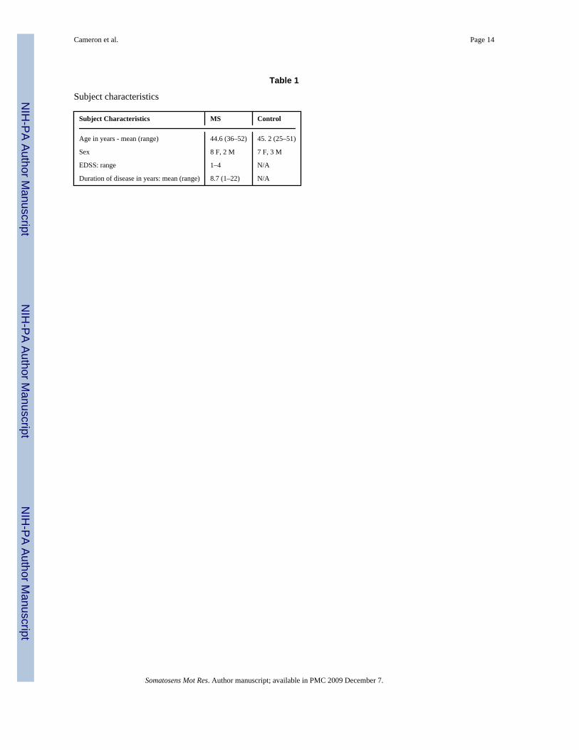
The subjects were 10 patients with MS aged 39–52 years, with complaints of balancedifficulties, and mild to moderate levels of disability (Table 1). None of the subjects had beentreated with any disease modifying agents. Ten healthy people aged 25–51 years withoutneurologic or orthopedic limitations served as controls. There was no significant difference inage or weight between the MS and control groups (p>0.05). All subjects received informedconsent for protocols approved by the Institutional Review Board and had a complete clinicalneurological evaluation by one of the authors (RH).
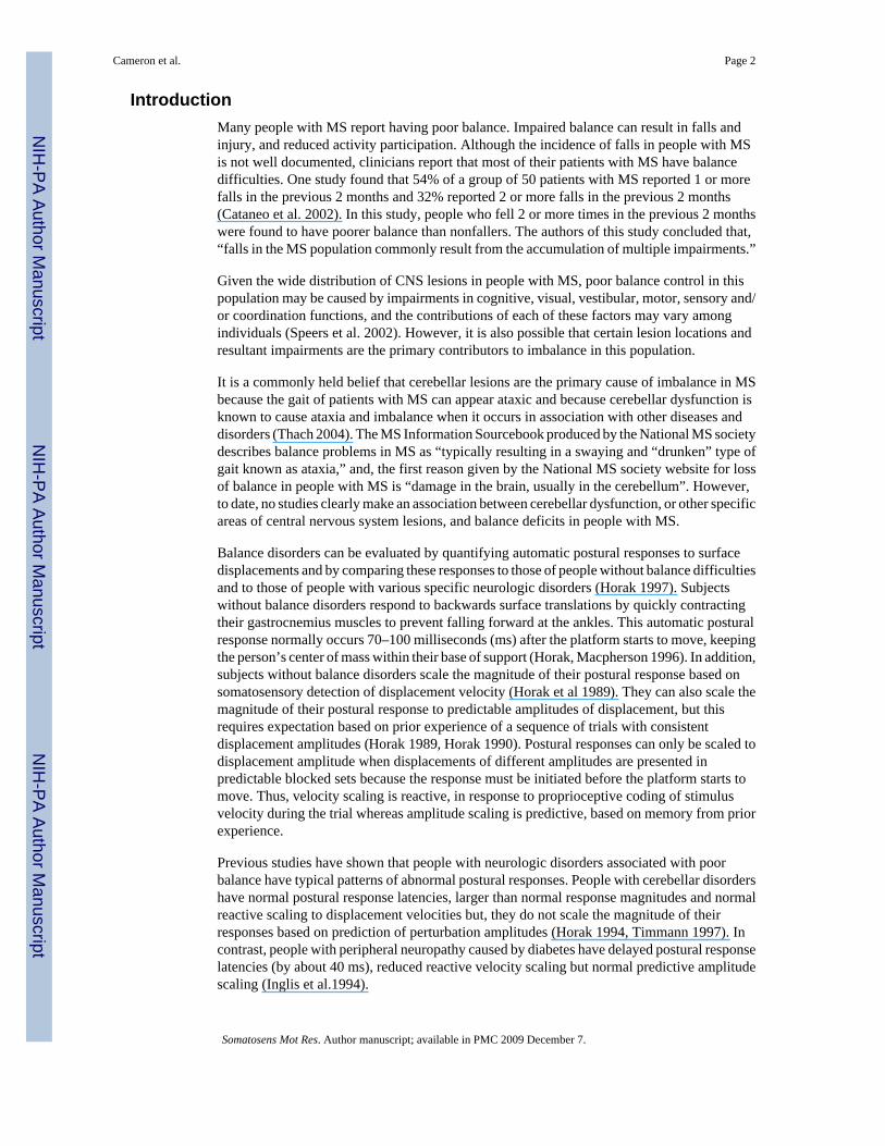
ProtocolBalance Platform Measures—This study examined, in free-standing subjects, the timingand amplitude of automatic postural responses to backward displacement of the support surfaceand the scaling of the amplitude of these responses to changes in the velocity and amplitudeof the displacement (Figure 1A). Previous publications have described, in detail, the apparatusand methods used to impose displacements and quantify the resulting responses (Diener et al.1988, Horak 1986, Horak et al. 1989). The subjects stood on two computer-servocontrolled,custom-made, hydraulic platforms that translated backward together causing forward bodysway (Horak 1986). Subjects stood with arms folded across the waist, eyes open, and feet acomfortable 6–9 cm apart at the heels.
Responses to a range of platform displacement amplitudes and velocities were examined. Fivedifferent displacement amplitudes (1.2, 3.6, 6.0, 8.4, and 12 cm), all at a velocity of 15 cm/second, were presented in blocks of 7 repetitions, each with increasing amplitude, for a totalof 35 trials. This block presentation was used to allow for predictive amplitude scaling basedon experience from prior trials (Horak et al. 1989). Five different displacement velocities (10,15, 20, 25 and 35 cm/second), all of 6 cm amplitude, were presented 5 times each in randomorder, for a total of 25 trials. This random presentation was used because velocity scaling ofpostural responses is reactive to the initial rate of movement of the support surface (Horak etal. 1989). The time between displacements was randomly varied by the experimenter (5–20sec) and was delayed until the subjects obtained the same initial position of the whole bodycenter of pressure as observed on an oscilloscope.
Surface EMG activity was recorded using two-2.5 cm2 surface electrodes placed 2 cm aparton the right and left medial gastrocnemius muscles. Amplified EMG signals were band-pass
Cameron et al. Page 3
Somatosens Mot Res. Author manuscript; available in PMC 2009 December 7.
NIH
-PA Author Manuscript
NIH
-PA Author Manuscript
NIH
-PA Author Manuscript

filtered (70–2,000Hz), rectified and stored for off-line analysis. Although no attempt was madeto calibrate EMGs on an absolute scale, amplifier gains were fixed throughout all experimentalsessions for all subjects. The latency of the postural response was the time from the onset ofsurface displacement to the first measurable increase in activity greater than 2 SD from baselinethat was sustained at least for at least 50 ms. Each EMG latency was measured with 2 msaccuracy with reference to platform displacement onset.
The plantarflexion torque of each foot on the platform and EMG activity of the left and rightmedial gastrocnemius were measured starting 200 ms before, and ending 2000 ms after, theonset of surface translation. Plantarflexion torque under each foot was calculated frominformation from four vertical strain gauges embedded under each hydraulic platform. Theslopes of the first 100 ms of rate-of-change of surface reactive torque were measured for eachfoot and represent the magnitude of the postural response (see Horak et al. 1989 for details)
Complete balance platform data for 8 of the 10 subjects with MS and all of the control subjectswas available for analysis.
SSEPsSomatosensory evoked potentials (SSEPs) were examined bilaterally from both hands and feetin subjects with MS. Total central afferent conduction time was measured for the right and leftsides and divided into spinal (ACTsp) and supraspinal (ACTsup) components. The proceduresfor measuring SSEPs are presented schematically in Figure 1B.
SSEPs were evoked by stimulation of the posterior tibial nerve in the leg and the median nervein the arm. Peripheral conduction time was assessed by measuring the conduction time fromthe periphery to the low thoracic (T12) spinal cord (N20 potential) for the posterior tibial nerve,and to Erb’s Point (N9 potential) for the median nerve. Total central conduction time wasobtained by recording at the cortex (P37) and recording at cervical spine (P27 for the posteriortibial nerve and P13 for the median nerve) was used to divide total afferent conduction time(ACT) into spinal and supraspinal components. SSEP abnormalities were defined in terms ofdelayed latencies, or as absent when well-preserved waveforms could not be recorded.Measures were obtained from both sides of the body and compared with data from controlsubjects using the same method in the same laboratory.
Data analysisThe latencies of the postural response for all trials were averaged for each subject. Linearregressions were used to calculate correlations between postural response latencies and spinaland supraspinal SSEP latencies, with right and left sides considered to be independent.
The slopes of the regression between the average postural response magnitude (rate of changeof average torque) across increasing amplitudes and velocities of surface displacements werecalculated for each subject to represent scaling of postural responses. Predictive scaling wasquantified as the slope of the magnitude of the postural response versus translation amplitudes,and reactive scaling was quantified as the slope of the magnitude of the postural response versustranslation velocities. The slopes of these regressions provided a measure of the “gain” of thepostural response and elevations of the regressions (the y-intercept) indicated the degree ofrelative “hypermetria” of the response. Each subject’s linear least squares fit regression wasplotted, with the intersubject variability of these regressions indicated by the standard deviationof slopes, r-values, and y-intercepts. In plots of control and MS group linear regressions, themean and standard deviation of torque values are shown for each group. The lines representthe average linear regression for all subjects in a group. Group linear regressions were testedfor significance (slope significantly different from 0) using a one-sided t tests (p < 0.05). The
Cameron et al. Page 4
Somatosens Mot Res. Author manuscript; available in PMC 2009 December 7.
NIH
-PA Author Manuscript
NIH
-PA Author Manuscript
NIH
-PA Author Manuscript

level of significance for each regression of initial rate of change of torque with displacementvelocity or amplitude was reported in which r = 0.715 was significant at p < 0.01 and r =0.87was significant at p < 0.005 (Shavelson 1988). Differences in the slopes and elevations (y-intercepts) of the linear regressions between the control and MS groups were compared witha one-way analysis of variance (ANOVA) with significance set at the p < 0.05 level. A linearregression across MS subject’s data was used to determine whether there was a relationshipbetween the latency of postural response and the ability to perform reactive or predictivescaling.
ResultsRelationship between postural response latencies and SSEP latencies
Postural response latencies were significantly longer in the MS subjects than in control subjects(161 ± 31 vs 102 ± 21 ms, p < 0.01). Table 2 shows that many MS subjects had different posturalresponse latencies in the right and left legs (difference range 3–37 ms; p=0.005), unlike controlsubjects who had symmetric postural response latencies (difference range 0 – 10 ms; p>0.05).
Peripheral somatosensory conduction times (N20) were normal in all subjects in this study. Inthe MS subjects, spinal SSEP latencies were absent in 6 of the 20 legs tested. In the MS subjectsin whom spinal SSEPs could be recorded, these were significantly longer than laboratorynormal values (17.2 ± 8.1 vs 7.9 ±1.8, p < 0.01) In contrast, supraspinal SSEP latencies in theMS subjects were not significantly longer than normal (11.0 ± 2.9 vs 9.5 ± 3.0, p > 0.05).Postural response latencies in MS subjects were significantly correlated with slowed spinalsomatosensory conduction (r=0.73, p<0.01) but not with supraspinal somatosensoryconduction times (r=0.1, p>0.05; Figure 2).
Scaling postural response magnitudeSubjects with MS demonstrated significantly more (steeper) scaling to predicted translationamplitudes than controls. As can be seen in Figure 3A, the slope of the linear regression betweenthe initial rate of change of surface reactive torque and translation amplitude was significantlylarger for the MS than for the control group (8.4 ± 3.5 vs 4.9 ± 1.0; p=0.02). In fact, 11 of the20 legs of the MS patients showed larger than the largest scaling values in the control subjects(see Table 2).
Reactive scaling of postural response magnitude to increasing velocities of surface translationswas often lower in subjects with MS than controls. Seven of the 20 legs of MS subjects showedvelocity scaling values lower than the lowest control subject value (see Table 2). There was asignificant difference in velocity scaling between the MS and control subjects’ lower velocityscaling legs (p=0.005) but not between the average scaling for both legs or for the higher scalinglegs (p>0.05). The slope of the linear regression between the initial rate of change of surfacereactive torque and translation velocity was 2.8 ± 2.2 in the MS subjects versus 4.4 ± 1.1 inthe controls (p= 0.08; Fig. 3B).
The magnitude of postural responses was significantly larger (hypermetric) in subjects withMS than in control subjects during both amplitude and velocity scaling (p<0.001) (Fig. 3A andB for group means and Table 2 for each subject’s average postural response magnitude acrossall conditions).
Relationships between postural response latencies and scalingMS subjects with the longest latencies of postural responses (and with the longest spinal SSEPlatencies) showed the steepest scaling of postural responses to predicted translation amplitudes.Figure 4A shows the relationship between the latency of gastrocnemius EMG for each MS leg
Cameron et al. Page 5
Somatosens Mot Res. Author manuscript; available in PMC 2009 December 7.
NIH
-PA Author Manuscript
NIH
-PA Author Manuscript
NIH
-PA Author Manuscript
tested versus their associated predictive amplitude scaling value. This relationship wassignificantly different from zero (r = 0.47, p = 0.05).
In contrast to the positive relationship between postural response latencies and predictivescaling, reactive velocity scaling was negatively correlated with postural response latencies.Figure 4B shows the relationship between the latency of gastrocnemius EMG for each MS legtested versus their associated reactive velocity scaling value. This relationship was significantlydifferent from zero (r = −0.5, p = 0.03).
DISCUSSIONThe results of this study indicate that people with imbalance due to MS have extremely delayedpostural responses. Their postural responses to surface translations have the longest averagelatency reported in the literature, except for one case report of a person with total bodysomatosensory loss due to dorsal root afferent cell body degeneration (Bloem et al 2002; Horak,2001). In the subjects with MS in this study, the delay in postural response onset correlatedwith the degree of prolongation in spinal cord somatosensory conduction, consistent withprevious studies demonstrating that postural responses to surface translations are likelytriggered by primary and/or secondary afferents from muscle spindles (Schiepatti and Nardone,1999; Stapley et al, 2002). Thus, because of slowed spinal somatosensory conduction, peoplewith MS receive somatosensory information about postural displacements late, and they thenrespond to this information late.
Subjects with MS also demonstrated differences in the delay in postural response latenciesbetween their two legs. This asymmetry is consistent with the asymmetry in their spinal SSEPsand with the apparently random distribution of spinal cord lesions in MS. People with MSprobably depend on the leg with the better somatosensory conduction, and therefore the shorterpostural response latency, for functional balance. Despite the long latency of posturalresponses, the magnitude of postural torque responses were larger than normal in people withMS, suggesting that muscle weakness, due to the impaired efferent motor conduction knownto occur in MS (Reich et al, 2007), did not contribute to imbalance when responding to thesesmall postural perturbations with feet in place. In addition, preliminary unpublished data fromour lab failed to show a correlation between delayed motor evoked potentials (MEP) andpostural response latencies in subjects with MS. In fact, MS subjects’ postural responses appearto be larger than normal, possibly to compensate for their prolonged latency. By the time thepostural response occurs (sometimes over 100 ms late) it must be larger than normal toeffectively return the body’s center of mass to the center of the base of support and thus preventthe person from falling. Delayed postural responses may result in a fall because, by the timethe patient initiates a balance response their center of mass may be too far outside of their baseof support for available postural strategies to effectively maintain balance. The larger posturalresponses seen in subjects with MS are similar to, but not as large as, the hypermetric posturalresponses in see in subjects with cerebellar ataxia and likely contribute to the clinicalimpression of ataxia and subjective perception of imbalance in people with MS (see figures3A and 3B with data from 10 subjects with cerebellar disorders (Horak et al, 1994)).
In addition to increasing the magnitude of their responses, people with MS and balanceproblems also appear to compensate for their prolonged postural response latencies byincreasing the use of prediction to scale their responses to the amplitude of an upcomingexternal perturbation. Amplitude scaling is predictive because the nervous system respondsbefore the amplitude is known. Amplitude scaling does not occur when perturbation amplitudesare randomized (Horak FB et al 1989). Enhanced predictive scaling may help to prevent fallsand reduce the need for stepping for postural correction. The enhanced predictive scaling foundin the subjects with MS in this study is in contrast to people with cerebellar disorders who
Cameron et al. Page 6
Somatosens Mot Res. Author manuscript; available in PMC 2009 December 7.
NIH
-PA Author Manuscript
NIH
-PA Author Manuscript
NIH
-PA Author Manuscript

characteristacally lack predictive scaling (Horak et al. 1994; see Figure 3A). This suggests thatthe imbalance in the people with MS in this study was not related to cerebellar dysfunction andthat these subjects had sufficient cerebellar function to allow for amplified predictive,amplitude scaling.
Although the people with MS and imbalance in this study had good predictive amplitudescaling, they often showed reduced reactive velocity scaling with at least one leg. Poor velocityscaling would be expected if the delayed postural latencies were caused by slowed centralsomatosensory conduction because scaling of postural responses to perturbation velocity isthought to depend on encoding of perturbation velocity from muscle spindles (Horak andMacpherson 1996, Schiepatti M. Nardone A. 1999). Subjects with slowed conduction in largediameter peripheral sensory nerves due to diabetic neuropathy also have reduced velocityscaling and delayed postural responses (Inglis et al. 1994). However, these delays areconsiderably smaller (average 24 ms) than in the subjects with MS in the current study (average59 ms).
In summary, the pattern of postural responses observed in subjects with MS and imbalance arecharacterized by very prolonged latency, correlation of this prolongation with spinal SSEPconduction, and enhancement of predictive response scaling. This pattern is unlike that ofsubjects with cerebellar disorders and more closely resembles that of subjects with peripheralneuropathy (See Table 3; Dickstein et al. 2003,Horak 1994,Inglis et al. 1994). Increasedpredictive postural response scaling, as found in the subjects with MS in this study, is in notablecontrast to patients with cerebellar disorders who characteristically fail to predictively scalepostural responses. Predictive scaling allows one to adapt and improve performance withrepeated presentation of similar balance challenges. Similar to people with diabetic peripheralneuropathy, people with MS and imbalance have very delayed postural response latencies anddecreased postural response velocity scaling. However, the delay in postural response latenciesis longer in subjects with MS than in subjects with diabetic peripheral neuropathy. In contrastto people with diabetic neuropathy, the subjects with MS in this study had increased, notreduced, postural response magnitudes. This suggests that people with MS have worsedisturbances in afferent conduction but better efferent motor function than people with diabeticperipheral neuropathy. Overall, these findings strongly suggest that the primary cause ofimbalance in MS is not cerebellar but rather a result of slowed afferent proprioceptiveconduction in the spinal cord.
The most distinct findings of this study are that people with MS and imbalance have verydelayed postural response latencies that correlate with delayed spinal SSEP conduction. It ismost likely that demyelination of the posterior columns of the spinal cord is the direct causeof the SSEP conduction delays, and that this causes delayed postural responses and imbalancein most people with MS. Given that postural EMG response latency (measured as the latencyof gastrocnemius (GAS) EMG response to backward surface displacement (forward bodysway)) correlates with spinal SSEP conduction velocity in people with MS, suggests thatpostural EMG response latency could be used as a functional measure of CNS conductionvelocity and demyelination in people with MS.
Postural EMG response latency has a number of advantages over other tools currently used toexamine the severity of CNS demyelination and disease impact in patients with MS includingSSEP latencies and MRI. Although SSEP latencies can indicate the degree and location of CNSdemyelination in MS, they are limited by a low ceiling effect. As found in this and other studies,when conduction is severely slowed, absence of all waves after the Erb’s point potential is notuncommon (Oken 1986). The absence of long latency responses indicates severe slowing ofconduction but fails to further quantify this slowing. In this study, long latency SSEPs wereabsent in a number of subjects with MS, but there were no subjects in whom GAS EMG
Cameron et al. Page 7
Somatosens Mot Res. Author manuscript; available in PMC 2009 December 7.
NIH
-PA Author Manuscript
NIH
-PA Author Manuscript
NIH
-PA Author Manuscript

latencies in response to postural translations could not be measured. EMG latencies to posturalperturbations can be measured for any subject who can stand unsupported for as little as a fewseconds. Unlike MRI, which provides largely qualitative information about disease progressionand activity in MS, postural response latency may provide a more continuous, quantifiable,and functional measure of disease activity, either over time or in response to interventionsintended to reduce inflammation or enhance CNS conduction.
Based on the findings of this study, we suggest that clinical examination of patients withimbalance associated with MS include measurement of postural responses. If delayed posturalresponses are found, rehabilitation therapies directed toward interventions, such as use of canes(Ashton-Miller et al. 1996, Dickstein et al. 2001), and sensory substitution with intact vestibular(Horak and Hlavacka 2001) and visual function, (if the patient has intact vestibular or visualfunction) may help compensate for the delayed transmission and response (Herndon et al,2000). In addition, patients should be encouraged to take extra care when walking on unstableor moving surfaces or in crowded areas, as these conditions are likely to contribute to falls inpeople with delayed postural responses. These changes may help reduce the morbidity,mortality, and activity and participation restrictions in this vulnerable population
This study has a number of limitations. The sample size was small and the subjects were notrandomly selected. The subjects with MS were patients with MS selected for subjectivecomplaints of imbalance but no measure of the severity of their experience of imbalance orfalls was performed. Future studies of balance in MS would benefit from a larger sample sizedrawn from multiple sites, detailed examination of MRIs, inclusion of subjects with MS withand without subjective complaints of imbalance, examination of functional degree ofimbalance including incidence of falls and evaluation of the relationship between clinicalbalance measures and the latency, magnitude and scaling of APRs.
AcknowledgmentsWe acknowledge data collection by Dr. Carol Pratt and in the electrophysiology lab of Dr. Barry Oken as well asinsightful discussions with Dr. Barry Oken. This work was supported by NIA #AG006457 to Fay Horak.
ReferencesAshton-Miller JA, Yeh ML, Richardson JK, Galloway T. A cane reduces loss of balance in patients with
peripheral neuropathy: results from a challenging unipdeal balance test. Arch Phys Med Rehabil1996;47:446–452. [PubMed: 8629920]
Bloem BR, Allum JH, Carpenter MG, Verschuuren JGM, Honegger F. Triggering of balance correctionsand compensatory strategies in a patient with total leg proprioceptive loss. Exp Brain Res 2002;142:91–107. [PubMed: 11797087]
Cataneo D, et al. Risks of falls in subjects with multiple sclerosis. Arch Phys Med Rehabil 2002;83(6):864–7. [PubMed: 12048669]
Dickstein R, Peterka R, Horak F. Effects of light fingertip touch on postural responses in subjects withdiabetic neuropathy. Journal of Neurology, Neurosurgery, and Psychiatry 2003;74:620–6.
Dickstein R, Shupert C, Horak F. Fingertip touch improves postural stability in patients with peripheralneuropathy. Exp Brain Research 2001;14(3):238–247.
Diener HC, Horak FB, Nashner LM. Influence of stimulus parameters on human postural responses. JNeurophysiol 1988;59:1888–1895. [PubMed: 3404210]
Herndon, RM.; Horak, FB. Vertigo, imbalance and incoordination. In: Burks, JS.; Johnson, KP., editors.Multiple Sclerosis: Diagnosis, Medical Management, and Rehabilitation. Demos Publishers; 2000. p.331-337.
Horak FB. Postural ataxia related to somatosensory loss. Adv Neurol 2001;87:173–182. [PubMed:11347220]
Cameron et al. Page 8
Somatosens Mot Res. Author manuscript; available in PMC 2009 December 7.
NIH
-PA Author Manuscript
NIH
-PA Author Manuscript
NIH
-PA Author Manuscript

Horak FB, Diener HC. Cerebellar control of postural scaling and central set in stance. J Neurophysiol1994;72:479–493. [PubMed: 7983513]
Horak FB, Diener HC, Nashner LM. Influence of central set on human postural responses. J Neurophysiol1989;62:841–853. [PubMed: 2809706]
Horak FB, Henry SM, Shumway-Cook A. Postural perturbations: new insights for treatment of balancedisorders. Physical Therapy 1997;77(5):517–33. [PubMed: 9149762]
Horak FB, Hlavacka F. Somatosensory loss increases vestibulospinal sensitivity. Exp Brain Research2001;86:575–585.
Horak, FB.; Macpherson, JM. Postural orientation and equilibrium. In: Shepard, J.; Rowell, L., editors.Handbook of Physiology: Section 12, Exercise: Regulation and Integration of Multiple Systems. NewYork: Oxford University Press; 1996. p. 255-292.
Horak FB, Nashner LM. Central programming of postural movements: adaptation to altered supportsurface configurations. J Neurophysiol 1986;55:1369–1381. [PubMed: 3734861]
Inglis JT, et al. The importance of somatosensory information in triggering and scaling automatic posturalresponses in humans. Exp Brain Res 1994;101:159–64. [PubMed: 7843295]
Oken, BS. Somatosensory evoked potentials in neurologic diagnosis. In: Chiappa, KH., editor. Evokedpotentials in clinical medicine. 3. New York: Lippincot-Raven; 1986. p. 379-389.
Reich DS, et al. Corticospinal tract abnormalities are associated with weakness in multiple sclerosis. AmJ Neuroradiol 2008;29:333–339. [PubMed: 17974617]
Schieppati M, Nardone A. Group II spindle afferent fibers in humans: Their possible role in the reflexcontrol of stance. Progress in Brain Res 1999;123:461–472.
Shavelson, RJ. Statistical Reasoning for the Behavioral Sciences. Boston: Allyn and Bacon; 1988.Speers RA, Kuo AD, Horak FB. Contributions of altered sensation and feedback responses to changes
in coordination of postural control due to aging. Gait & Posture 2002;16:20–30. [PubMed: 12127183]Stapley PJ, Ting LH, Hulliger M, Macpherson JM. Automatic postural responses are delayed by
pyridoxine-induced somatosensory loss. The Journal of Neuroscience 2002;22(14):5803–5807.[PubMed: 12122040]
Thach WT, Bastian AJ. Role of the cerebellum in the control and adaptation of gait in health and disease.Progress in Brain Research 2004;143:353–66. [PubMed: 14653179]
Timmann D, Horak FB. Prediction and set-dependent scaling of early postural responses in cerebellarpatients. Brain 1997;120:327–337. [PubMed: 9117379]
Cameron et al. Page 9
Somatosens Mot Res. Author manuscript; available in PMC 2009 December 7.
NIH
-PA Author Manuscript
NIH
-PA Author Manuscript
NIH
-PA Author Manuscript
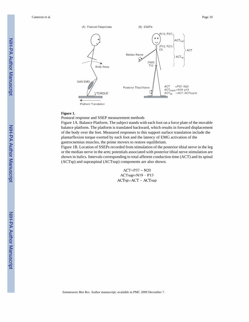
Figure 1.Postural response and SSEP measurement methodsFigure 1A. Balance Platform. The subject stands with each foot on a force plate of the movablebalance platform. The platform is translated backward, which results in forward displacementof the body over the feet. Measured responses to this support surface translation include theplantarflexion torque exerted by each foot and the latency of EMG activation of thegastrocnemius muscles, the prime movers to restore equilibrium.Figure 1B. Location of SSEPs recorded from stimulation of the posterior tibial nerve in the legor the median nerve in the arm; potentials associated with posterior tibial nerve stimulation areshown in italics. Intervals corresponding to total afferent conduction time (ACT) and its spinal(ACTsp) and supraspinal (ACTsup) components are also shown.
Cameron et al. Page 10
Somatosens Mot Res. Author manuscript; available in PMC 2009 December 7.
NIH
-PA Author Manuscript
NIH
-PA Author Manuscript
NIH
-PA Author Manuscript

Figure 2.Relationships between postural response latencies and SSEP latencies.Figure 2A and 2B. Relationship between postural response latency (GAS EMG) to backwarddisplacement and spinal SSEP conduction times (shown in Figure 2A) and supraspinal SSEPconduction times (shown in Figure 2B). The x’s on Figure 2A represent postural responselatencies for subjects with absent spinal SSEPs. These subject’s values were not included inthe linear regression.
Cameron et al. Page 11
Somatosens Mot Res. Author manuscript; available in PMC 2009 December 7.
NIH
-PA Author Manuscript
NIH
-PA Author Manuscript
NIH
-PA Author Manuscript
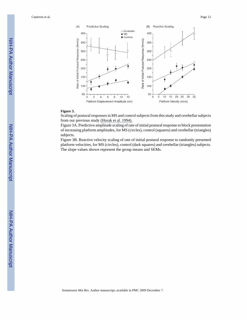
Figure 3.Scaling of postural responses in MS and control subjects from this study and cerebellar subjectsfrom our previous study (Horak et al. 1994).Figure 3A. Predictive amplitude scaling of rate of initial postural response to block presentationof increasing platform amplitudes, for MS (circles), control (squares) and cerebellar (triangles)subjects.Figure 3B. Reactive velocity scaling of rate of initial postural response to randomly presentedplatform velocities, for MS (circles), control (dark squares) and cerebellar (triangles) subjects.The slope values shown represent the group means and SEMs.
Cameron et al. Page 12
Somatosens Mot Res. Author manuscript; available in PMC 2009 December 7.
NIH
-PA Author Manuscript
NIH
-PA Author Manuscript
NIH
-PA Author Manuscript

Figure 4.Relationships of postural response latencies and scalingFigure 4A. Relationship of postural response latency (GAS EMG) to the amplitude scalingslope for each individual MS subject (values for both legs are included)Figure 4B. Relationship of postural response latency (GAS EMG) to the velocity scaling slopefor each individual MS subject (values for both legs are included).
Cameron et al. Page 13
Somatosens Mot Res. Author manuscript; available in PMC 2009 December 7.
NIH
-PA Author Manuscript
NIH
-PA Author Manuscript
NIH
-PA Author Manuscript
NIH
-PA Author Manuscript
NIH
-PA Author Manuscript
NIH
-PA Author Manuscript
Cameron et al. Page 14
Table 1
Subject characteristics
Subject Characteristics MS Control
Age in years - mean (range) 44.6 (36–52) 45. 2 (25–51)
Sex 8 F, 2 M 7 F, 3 M
EDSS: range 1–4 N/A
Duration of disease in years: mean (range) 8.7 (1–22) N/A
Somatosens Mot Res. Author manuscript; available in PMC 2009 December 7.

NIH
-PA Author Manuscript
NIH
-PA Author Manuscript
NIH
-PA Author Manuscript
Cameron et al. Page 15
Tabl
e 2
Sum
mar
y of
pos
tura
l and
SSE
P va
lues
for b
oth
side
s in
each
subj
ect w
ith M
S an
d fo
r the
con
trol a
nd c
ereb
ella
r gro
up m
eans
(fro
m H
orak
et a
l. 19
89).
Subj
ect
Side
Gas
EM
G L
aten
cy (m
s)A
vg R
espo
nse
Am
plitu
de (N
m/
ms)
AC
Tsp
(ms)
AC
Tsu
p (m
s)Pr
edic
tive
Am
plitu
de S
calin
g (n
M/
cm)
Rea
ctiv
e V
eloc
ity S
calin
g (n
M/
m/s
)
1L
168*
196
No
P27
7.2
11.5
−1.2
R17
818
2N
o P2
79.
610
.60.
7
2L
110
196
8.9
9.2
10.0
4.0
R10
6*17
610
.011
.810
.84.
4
3L
179*
214
No
P27
9.4
8.8
3.0
R19
414
3N
o P2
79.
812
.34.
9
4L
160
175
18.0
15.5
4.6
2.0
R15
2*18
1N
o re
spon
se18
.810
.21.
5
5L
209*
128
35.7
9.5
12.0
−1.7
R21
218
8N
ot to
lera
ted
Not
tole
rate
d12
.03.
7
6L
131
208
7.8
8.1
3.3
5.0
R12
7*18
610
.38.
33.
44.
3
7L
191
185
16.8
10.1
NR
NR
R18
5*18
222
.111
.4N
RN
R
8L
180
199
13.6
10.4
No
data
5.5
R15
0*13
818
.415
.8N
o da
ta2.
0
9L
125*
218
12.2
12.0
1.9
3.7
R16
219
815
.011
.26.
86.
4
10L
166
181
29.6
10.2
5.5
1.5
R14
4*18
021
.710
.69.
91.
6
MS
Mea
n (S
D)
161
(31)
183
(25)
17.2
(8.1
)11
.0 (2
.9)
8.4
(3.5
)2.
8 (2
.2)
Con
trol
mea
n (S
D)
102
(21)
132
(36)
7.9
(1.8
)9.
5(3.
0)4.
9 (1
.0)
rang
e 2.
6–6.
94.
4 (1
.1)
rang
e 2.
2 –6
.6
Cer
ebel
lar
mea
n (S
D)
106
(20)
350
(35)
Not
test
edN
ot te
sted
−1.4
(4.4
)3.
9 (2
.4)
* long
er la
tenc
y le
g fo
r eac
h su
bjec
t
Somatosens Mot Res. Author manuscript; available in PMC 2009 December 7.

NIH
-PA Author Manuscript
NIH
-PA Author Manuscript
NIH
-PA Author Manuscript
Cameron et al. Page 16
Table 3
Comparison of balance measures and SSEPs in people with MS, cerebellar disorders and diabetic peripheralneuropathy
MULTIPLE SCLEROSIS CEREBELLAR NEUROPATHY
BALANCE MEASURES
Latency Very delayed Normal Delayed
Magnitude Increased Increased Decreased
Amplitude scaling Normal/increased Decreased Normal
Velocity scaling Reduced Normal Reduced
SSEPs
Supraspinal Slightly delayed Unknown Normal
Spinal Very delayed Normal Normal
Peripheral Normal Normal Delayed
Somatosens Mot Res. Author manuscript; available in PMC 2009 December 7.