hv1 acts as a sodium sensor and promotes superoxide production in medullary thick ascending limb of...
TRANSCRIPT
541
The Dahl salt-sensitive (SS) rat is a naturally occurring model of SS hypertension that closely mimics human forms of dis-
ease observed in the black population.1–6 Several previous stud-ies indicate that oxidative stress, specifically within the renal outer medullary region, promotes the development of hyperten-sion and renal injury in the SS rat model.6–8 Within this region, renal medullary thick ascending limb (mTAL) tubular segments are thought to be the greatest source of reactive oxygen species (ROS), and much of the superoxide produced by this segment is derived from nicotinamide adenine dinucleotide phosphate (NADPH) oxidase.9 Because we have shown that superoxide production is enhanced in mTAL of SS rats compared with salt-resistant animals,10–12 it is likely that superoxide production by this segment contributes to the development of disease in these animals. The cellular mechanisms responsible for augmented superoxide production in mTAL of SS rats when fed a high-salt diet, however, remain unclear.
Superoxide production in mTAL in response to cellular H+ efflux was initially reported by Li et al.13 We have since char-acterized a novel H+ transport pathway in mTAL that when activated stimulated the production of superoxide by NADPH oxidase.10 Importantly, this pathway was enhanced in SS rats compared with salt-resistant control animals (SS.13BN).10 Several indicators suggested that HV1, a voltage-gated H+ channel expressed highly in immune cells,14 could be the unidentified H+ transport pathway we characterized in mTAL. First, because charged protons cannot readily cross the plasma membrane,15 we suspected an ion channel. Second, superox-ide production was only observed in the presence of a large outward pH gradient, not following an inward pH gradient.10 Third, proton efflux was linked to cellular depolarization and activation of the NADPH oxidase.10 HV1 fulfilled all these criteria. HV1 is specific to protons.15 The opening of HV1 is pH dependent with the channel only mediating H+ efflux, not
Abstract—We previously characterized a H+ transport pathway in medullary thick ascending limb nephron segments that when activated stimulated the production of superoxide by nicotinamide adenine dinucleotide phosphate oxidase. Importantly, the activity of this pathway was greater in Dahl salt-sensitive rats than salt-resistant (SS.13BN) rats, and superoxide production was enhanced in low Na+ media. The goal of this study was to determine the molecular identity of this pathway and its relationship to Na+. We hypothesized that the voltage-gated proton channel, HV1, was the source of superoxide-stimulating H+ currents. To test this hypothesis, we developed HV1−/− null mutant rats on the Dahl salt-sensitive rat genetic background using zinc-finger nuclease gene targeting. HV1 could be detected in medullary thick limb from wild-type rats. Intracellular acidification using an NH
4Cl prepulse in 0 sodium/BaCl
2 containing media
resulted in superoxide production in thick limb from wild-type but not HV1−/− rats (P<0.05) and more rapid recovery of intracellular pH in wild-type rats (ΔpH
I 0.005 versus 0.002 U/s, P=0.046, respectively). Superoxide production was
enhanced by low intracellular sodium (<10 mmol/L) in both thick limb and peritoneal macrophages only when HV1 was present. When fed a high-salt diet, blood pressure, outer medullary renal injury (tubular casts), and oxidative stress (4-hydroxynonenal staining) were significantly reduced in HV1−/− rats compared with wild-type Dahl salt-sensitive rats. We conclude that HV1 is expressed in medullary thick ascending limb and promotes superoxide production in this segment when intracellular Na+ is low. HV1 contributes to the development of hypertension and renal disease in Dahl salt-sensitive rats. (Hypertension. 2014;64:541-550.) • Online Data Supplement
Key Words: free radicals hypertension oxidative stress
Received March 13, 2014; first decision March 30, 2014; revision accepted May 7, 2014.From the Department of Physiology (C.J, J.S., C.A.S., H.O., B.B., S.D., A.G., R.P., P.M.O.) and Department of Pharmacology and Toxicology (N.A.L.),
Georgia Regents University, Augusta; Department of Physiology, Medical College of Wisconsin, Milwaukee (A.M.G., H.J.J.); and Department of Biology & Physics, Kennesaw State University, Atlanta, GA (S.M.E.S.).
The online-only Data Supplement is available with this article at http://hyper.ahajournals.org/lookup/suppl/doi:10.1161/HYPERTENSIONAHA. 114.03549/-/DC1.
Correspondence to Paul M. O’Connor, Department Physiology, CB2206, Georgia Regents University, 1459 Laney-Walker Blvd, Augusta, GA 30912. E-mail [email protected]
HV1 Acts as a Sodium Sensor and Promotes Superoxide Production in Medullary Thick Ascending Limb of Dahl
Salt-Sensitive RatsChunhua Jin, Jingping Sun, Carly A. Stilphen, Susan M.E. Smith, Hiram Ocasio, Brent Bermingham, Sandip Darji, Avirup Guha, Roshan Patel, Aron M. Geurts,
Howard J. Jacob, Nevin A. Lambert, Paul M. O’Connor
© 2014 American Heart Association, Inc.
Hypertension is available at http://hyper.ahajournals.org DOI: 10.1161/HYPERTENSIONAHA.114.03549
Kidney
by guest on June 11, 2016http://hyper.ahajournals.org/Downloaded from by guest on June 11, 2016http://hyper.ahajournals.org/Downloaded from by guest on June 11, 2016http://hyper.ahajournals.org/Downloaded from by guest on June 11, 2016http://hyper.ahajournals.org/Downloaded from by guest on June 11, 2016http://hyper.ahajournals.org/Downloaded from by guest on June 11, 2016http://hyper.ahajournals.org/Downloaded from by guest on June 11, 2016http://hyper.ahajournals.org/Downloaded from by guest on June 11, 2016http://hyper.ahajournals.org/Downloaded from by guest on June 11, 2016http://hyper.ahajournals.org/Downloaded from by guest on June 11, 2016http://hyper.ahajournals.org/Downloaded from
542 Hypertension September 2014
influx, under physiological conditions.15,16 HV1 opening is voltage dependent, opening in response to cellular depolariza-tion, and HV1 has been associated with NADPH oxidase.15,17,18
Given the number of characteristics shared between HV1 and the unidentified H+ transport pathway we characterized in mTAL, we hypothesized that HV1 is present in mTAL and contributes to superoxide, producing H+ currents after cellu-lar acidification. Further, because high-salt feeding is likely to promote both depolarization as well as acidification of mTAL secondary to enhanced apical Na reabsorption19,20 and increased acid delivery to this segment (factors we predict would promote opening of HV1 in vivo),21,22 we hypothesized that activation of HV1 contributes to the development of outer medullary oxidative stress, hypertension, and renal injury in vivo after high-salt feeding in Dahl SS rats. Because specific pharmacological inhibitors of HV1 are not yet available,23 we tested this hypothesis by developing an HV1−/− null mutant rat on the Dahl SS rat (Medical College of Wisconsin) genetic background.
Because our previous work indicated that ROS production in response to H+ efflux was greatly enhanced by low extracel-lular Na+,10 we also investigated the relationship among Na concentration, HV1, and NADPH oxidase activity in mTAL because alterations in Na handling could potentially be an important regulatory feedback system for ROS production in this segment.24–27
MethodsStudies used adult SS rats (Medical College of Wisconsin) main-tained ad libitum on water and a standard pellet diet containing 0.4% NaCl since weaning. All studies were conducted in accordance with the National Institutes of Health Guide for the Care and Use of Laboratory Animals. All of the protocols were approved in advance by the institutional animal care committee at Georgia Regents University or the Medical College of Wisconsin.
Bulk mTAL IsolationmTALs were isolated using a bulk dissection method as described previously28 except 2 sieves rather than 1 were used. Briefly, the kidneys were flushed with 10 mL of cold saline followed by 10 mL of HEPES-buffered Hanks balanced salt solution containing 200 U/mL of type II collagenase (Worthington Biochemical). The kidneys were collected and cut into thin slices transversely along the cortical–medullary axis. The inner stripe of the outer medulla was isolated and incubated with collagenase solution at 37°C for 30 minutes with intermittent pipetting. Every 5 minutes, the digested tissue was pipet-ted out and passed through a 100- and 70-μm sieve. mTAL segments were collected on the 70-μm sieve, and digestion stopped by 1% BSA in pH 7.4 Hanks balanced salt solution. We have previously demon-strated the collected tissue contains ≈95% mTAL.29
Respiratory Burst AssayPeritoneal macrophages (MØ) cells were collected as previously described.8 In brief, rats were anesthetized with isoflurane (2%–5%), and 50 mL of Hanks balanced salt solution was injected into the abdominal cavity followed by a small midline incision. The excess fluid was collected by syringe. The collected fluids were centri-fuged at 400g for 10 minutes. Collected cell pellets that contain predominantly MØ8 were resuspended and aliquoted onto a clear-bottom 96-well plate (Bioexpress) at ≈1×10⁶ cells per well. L-012 (1 mmol/L; Wako Pharmaceuticals) was used to determine super-oxide production using a FLUOstar Omega plate reader (BMG Labtech). Cells were maintained at 37°C and luminescence mea-sured for 30 minutes at 2-minute intervals. Addition of 100 μM
of the protein kinase C activator phorbol 12-myristate 13-acetate (PMA) was used to stimulate the respiratory burst and maximal luminescence (arbitrary units [AU]) recorded.
To set intracellular Na+ concentration, cells were preincubated for 15 minutes in solutions containing 70 mmol/L N-methyl-d-glucamine, 5.5 mmol/L d-glucose, 1.3 mmol/L CaCl
2, 1 mmol/L
MgSO4, and 5 mol/L HEPES at pH 7.4. NaCl levels were altered
to maintain intracellular [Na+] ([Na+]I) at either 5, 10, 15, 20, or 25
mmol/L and balanced to a total of 70 mmol/L with the addition of KCl. The ionophore nystatin (135 U/mL) was prepared daily to per-meabilize cells to Na+.30 To determine whether effects were due to differences in activity of Na/H exchangers (NHEs), 100 μM of the NHE inhibitor cariporide was added. ZnCl
2 (100 μM) was added to
some wells to acutely inhibit HV1 activity. Maximal superoxide pro-duction rate was normalized to solutions containing 25 mmol/L NaCl within each plate.
Transfection of Peritoneal MacrophagesCells were obtained as above. After 2-hour incubation, the cells were washed once with serum-free medium to eliminate nonadherent lym-phocytes and polymorphonuclear leukocytes from the adherent mac-rophages, then cultured in 200 μL of 10% fetal bovine serum DMEM supplemented with 500 U/mL rat granulocyte-macrophage colony-stimulating factor (GenScript, Cat# Z02992-10). The primary mac-rophages were cultured in the presence of granulocyte-macrophage colony-stimulating factor for 7 to 10 days prior to transfection.
hHvcn1 TransfectionWhen the cells reached 50% to 60% confluence, the medium was removed and 100 μL of fresh no pen/strep and no fetal bovine serum DMEM added. For each well of 96-well plates, we diluted 150 ng of hHvcn1 (human Hvcn1 cDNA was cloned into pQBI25-fC3) into 7.5 μL of Opti-MEM (Gibco, Cat# 31985-062), with 0.3 μL of PromoFectin-Macrophage Transfection solution (PromoCell GmbH, Cat# PK-CT-2000-MAC-10). We then added PromoFectin-Macrophage solution to the DNA solution, vortex mixed the solution, and spun down briefly before incubating for 30 minutes at room tem-perature. We then added 15 μL of this mixture drop-wise into each well. After 4-hour incubation, we add 85 μL of no pen/strep 10% fetal bovine serum DMEM to each well and put in a 37°C incubator. After 48 hours of hHvcn1 transfection, the respiratory burst assay was per-formed as detailed above.
Micro Dissection and Imaging of mTALRats were anesthetized with isoflurane (2%–5%) and isolation of mTAL tissue strips performed as described previously.31 Thin tissue strips containing mTALs were placed on a glass cover-slip coated with the tissue adhesive Cell-Tak (BD Biosciences) for fluorescence imaging. Tissue strips containing mTALs were loaded with either dihydroethidium (DHE; 50 mmol/L) or 2′,7′-bis-(2-carboxyethyl)-5-(and-6)-carboxyfluorescein (6 μmol/L) in Hanks balanced salt solution for 1 hour at room temperature. Coverslips were placed on a heated imaging chamber maintained at 37°C (Warner Instruments) that allowed the rapid exchange of superfusion buffer and mounted on the stage of an inverted micro-scope (IX81 Olympus). mTAL were visualized with a ×40 water immersion objective lens. The signal was detected using a high-resolution digital camera (Photometrics Evolve, Roper Scientific). Excitation was provided by a Sutter DG-4 175W xenon arc lamp (Sutter Instruments) that allowed high-speed excitation wave-length switching. mTAL epithelial cells (3–10) were selected within each tissue strip to quantify changes in fluorescent intensity of dyes using Metafluor imaging software (Universal Imaging). Intracellular [Na+] was set at either 5 or 30 mmol/L using the same solutions as that used to set [Na+]
I in MØ.
Whole-Cell Voltage-Clamp RecordingsProton currents from HV1 were recorded as previously described.32 Traces shown in Figure 1 were not corrected for liquid junction potential, and leak currents were not subtracted.
by guest on June 11, 2016http://hyper.ahajournals.org/Downloaded from
Jin et al HV1 in Medullary Thick Limb 543
In Vivo StudiesMale 9-week-old WT SS and HV1−/− SS rats were anesthetized (2%–5% isoflurane) and blood pressure telemeters (Data Sciences International, St. Paul, MN) surgically implanted. After 1 week of recovery, animals were again anesthetized and the left femoral vein catheterized for infusion of fluids. Surgically implanted catheters were then tunneled under the skin and exposed between the shoulder blades. Catheters were protected by a light wire spring attached to a swivel (Instech Laboratories Inc, Plymouth Meeting, PA), which allows free 360° movement of the animal. Rats were individually housed and allowed to recover for 1 week before beginning experimental mea-surements. Blood pressure was recorded 24 hours daily. Rats were ini-tially maintained on a low Na+ (0.4% NaCl diet) before being switched to a 8% NaCl diet for 2 weeks. On day 3 of high NaCl feeding, rats were maintained on vehicle infusions (1% dimethyl sulfoxide in saline intravenously at a rate of 6.9 μL/min) of KR32568 (Sigma) added to the infusion media (2 mg/kg/d) to stimulate HV1. On day 15, rats were anesthetized and the kidneys excised for histological analysis. After processing, histological samples were blinded to the investigator and images quantified using Metamorph analysis software.
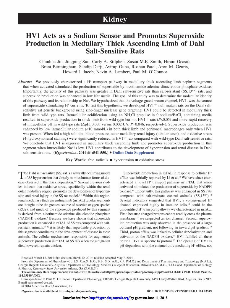
ResultsConfirmation of HV1 Deletion in Mutant RatsHV1 mutant Dahl SS rats were generated with zinc-finger nuclease technology.13,16 Genomic DNA of homozygous HV1 mutant (HV1−/−) rats was sequenced to reveal an 8-bp dele-tion, resulting in a frame-shift mutation and predicted loss of full-length HVCN1 protein (Figure 1A). Loss of HVCN1 was validated functionally using whole cell patch (Figure 1B). Step changes in applied voltage between −60 and +100 mV induced H+ currents in 7 of 7 MØ cells from 3 wild-type (WT) rats, whereas no current was observed in any of 9 cells studied from 4 HV1−/− mutant rats. These data are consistent with a single gene for HV1 and confirmed complete functional abla-tion in HV1−/− mutant rats. Because HV1 is highly expressed in immune cells of the spleen and its localization known, we first confirmed the specificity of our mRNA primer and anti-HV1 antibody using whole spleen homogenates of WT and HV1−/− mutant rats as in situ positive and negative controls, respec-tively. mRNA encoding HV1 was greatly reduced in spleen
of HV1−/− mutant rats compared with WT rats (Figure 1C). Western blot detected a band representing HV1 at the pre-dicted molecular weight (32 kD) in the membrane fraction of spleen from WT but not HV1−/− mutant rats (Figure 1D). Immunohistochemical staining of the spleen demonstrated numerous darkly stained cells within the spleen of WT rats, consistent with known distribution of HV1 in splenic tissue (Figure 1E). HV1 protein staining was also nearly undetect-able in whole cell homogenates of spleen from HV1−/− mutant rats compared with WT.
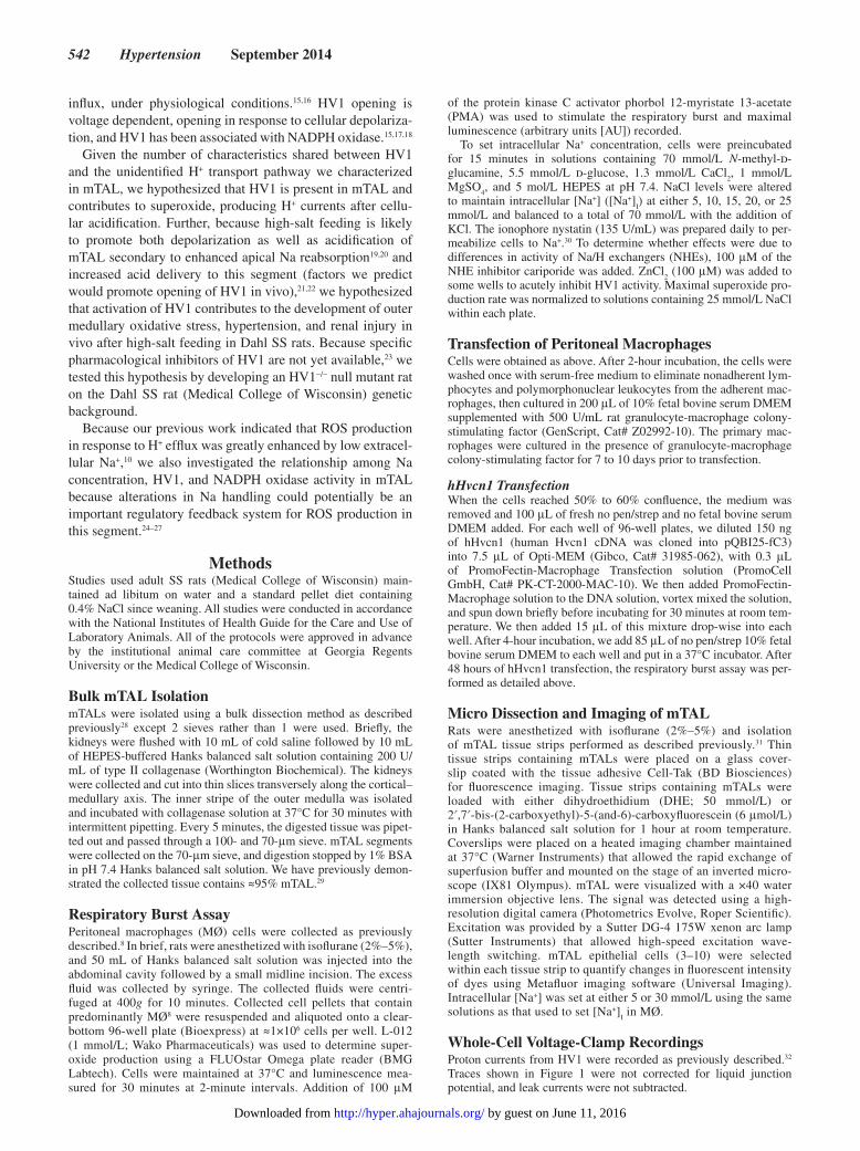
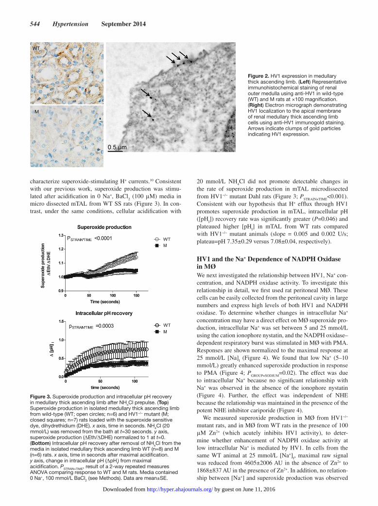
HV1 Expression in mTALTo determine whether HV1 was expressed in mTAL of SS rats, we quantified mRNA and protein expression specifically in mTAL using the same polymerase chain reaction primers and antibodies used in spleen. mTAL from mutant rats served as a negative control (Table S1). HV1 mRNA was 6-fold higher than presumed background in bulk isolated mTAL of WT compared with HV1−/− mutant rats (please see http://hyper.ahajournals.org) (Figure S2). We could not detect HV1 by Western in membrane fractions of bulk isolated mTAL in either genotype (data not shown); however, HV1 staining was identified specifically within mTAL in outer medullary sec-tions (Figure 2), and this was significantly greater than back-ground staining in mTAL from mutant rats (4382±920 versus 1458±709 AU; P=0.036, n=5) (Figure S1). Anti-HV1 immu-nogold staining and transmission electron microscopy local-ized HV1 staining largely to the apical membrane of thick ascending limb (Figure 2).
H+ Efflux Mediated Superoxide Production in mTALTo test our hypothesis that HV1 contributes to superoxide-enhancing H+ currents in mTAL, we determined super-oxide production (ΔEth/ΔDHE) in response to cellular acidification using an NH
4Cl loading/removal (20 mmol/L)
under the conditions in which we previously used to
Figure 1. Confirmation of loss of HV1 expression in mutant rats. (A) Representative gel for genotyping rats demonstrating 8-bp deletion in gene encoding HV1. Lanes 1 and 5, heterozygous HV1−/+ mutants (Het); lanes 2 and 3, homozygous HV1−/− mutants (M); lane 3, wild-type (WT). (B) A representative trace demonstrating proton currents in response to step changes in membrane potential in single peritoneal macrophages from WT (right) and mutant (left) rats. (C) Relative HV1 mRNA expression from spleen homogenates from WT (open columns) and M (closed columns) rats compared with GAPDH mRNA (n=4). (D) Western blot for HV1 in membrane fraction of WT and M rats. A band representing HV1 is shown at ≈32 kD. (E) Immunohistochemical staining of spleen using anti-HV1 in WT and M rats (n=4). (Left) Representative image of anti-HV1 stained tissue from WT and M rats at ×40. (Right) Quantification of anti-HV1 staining (integrated signal; arbitrary units [AU]). *P<0.05 using unpaired t test. Data are mean±SE.
by guest on June 11, 2016http://hyper.ahajournals.org/Downloaded from
544 Hypertension September 2014
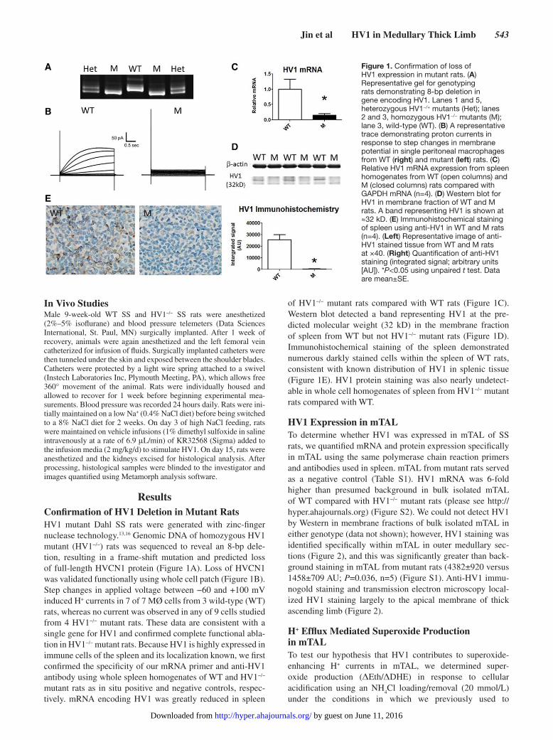
characterize superoxide-stimulating H+ currents.10 Consistent with our previous work, superoxide production was stimu-lated after acidification in 0 Na+, BaCl
2 (100 μM) media in
micro dissected mTAL from WT SS rats (Figure 3). In con-trast, under the same conditions, cellular acidification with
20 mmol/L NH4Cl did not promote detectable changes in
the rate of superoxide production in mTAL microdissected from HV1−/− mutant Dahl rats (Figure 3; P
STRAIN×TIME<0.001).
Consistent with our hypothesis that H+ efflux through HV1 promotes superoxide production in mTAL, intracellular pH ([pH
I]) recovery rate was significantly greater (P=0.046) and
plateaued higher [pHI] in mTAL from WT rats compared
with HV1−/− mutant animals (slope = 0.005 and 0.002 U/s; plateau=pH 7.35±0.29 versus 7.08±0.04, respectively).
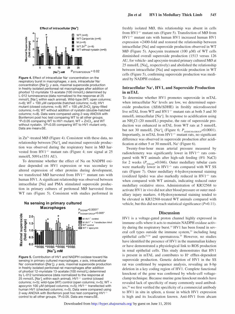
HV1 and the Na+ Dependence of NADPH Oxidase in MØWe next investigated the relationship between HV1, Na+ con-centration, and NADPH oxidase activity. To investigate this relationship in detail, we first used rat peritoneal MØ. These cells can be easily collected from the peritoneal cavity in large numbers and express high levels of both HV1 and NADPH oxidase. To determine whether changes in intracellular Na+ concentration may have a direct effect on MØ superoxide pro-duction, intracellular Na+ was set between 5 and 25 mmol/L using the cation ionophore nystatin, and the NADPH oxidase–dependent respiratory burst was stimulated in MØ with PMA. Responses are shown normalized to the maximal response at 25 mmol/L [Na]
I (Figure 4). We found that low Na+ (5–10
mmol/L) greatly enhanced superoxide production in response to PMA (Figure 4; P
GROUP×SODIUM=0.02). The effect was due
to intracellular Na+ because no significant relationship with Na+ was observed in the absence of the ionophore nystatin (Figure 4). Further, the effect was independent of NHE because the relationship was maintained in the presence of the potent NHE inhibitor cariporide (Figure 4).
We measured superoxide production in MØ from HV1−/− mutant rats, and in MØ from WT rats in the presence of 100 μM Zn2+ (which acutely inhibits HV1 activity), to deter-mine whether enhancement of NADPH oxidase activity at low intracellular Na+ is mediated by HV1. In cells from the same WT animal at 25 mmol/L [Na+]
I, maximal raw signal
was reduced from 4605±2006 AU in the absence of Zn2+ to 1868±837 AU in the presence of Zn2+. In addition, no relation-ship between [Na+] and superoxide production was observed
Figure 3. Superoxide production and intracellular pH recovery in medullary thick ascending limb after NH4Cl prepulse. (Top) Superoxide production in isolated medullary thick ascending limb from wild-type (WT; open circles; n=6) and HV1−/− mutant (M; closed squares; n=7) rats loaded with the superoxide sensitive dye, dihydrethidium (DHE). x axis, time in seconds. NH4Cl (20 mmol/L) was removed from the bath at t=30 seconds. y axis, superoxide production (ΔEth/ΔDHE) normalized to 1 at t=0. (Bottom) Intracellular pH recovery after removal of NH4Cl from the media in isolated medullary thick ascending limb WT (n=8) and M (n=6) rats. x axis, time in seconds after maximal acidification. y axis, change in intracellular pH (ΔpHI) from maximal acidification. PSTRAIN×TIME, result of a 2-way repeated measures ANOVA comparing response to WT and M rats. Media contained 0 Na+, 100 mmol/L BaCl2 (see Methods). Data are mean±SE.
Figure 2. HV1 expression in medullary thick ascending limb. (Left) Representative immunohistochemical staining of renal outer medulla using anti-HV1 in wild-type (WT) and M rats at ×100 magnification. (Right) Electron micrograph demonstrating HV1 localization to the apical membrane of renal medullary thick ascending limb cells using anti-HV1 immunogold staining. Arrows indicate clumps of gold particles indicating HV1 expression.
by guest on June 11, 2016http://hyper.ahajournals.org/Downloaded from
Jin et al HV1 in Medullary Thick Limb 545
in Zn2+-treated MØ (Figure 4). Consistent with these data, no relationship between [Na+]
I and maximal superoxide produc-
tion was observed during the respiratory burst in MØ har-vested from HV1−/− mutant rats (Figure 4; raw signal at 25 mmol/L 5091±1551 AU).
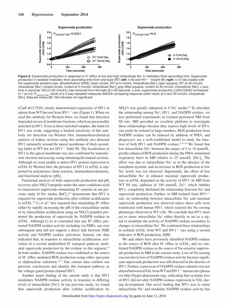
To determine whether the effect of Na on NADPH oxi-dase depended on HV1 expression or was secondary to altered expression of other proteins during development, we transfected MØ harvested from HV1−/− mutant rats with human HV1. A significant relationship was observed between intracellular [Na] and PMA stimulated superoxide produc-tion in primary cultures of peritoneal MØ harvested from WT rats (Figure 5). Consistent with studies performed in
freshly isolated MØ, this relationship was absent in cells from HV1−/− mutant rats (Figure 5). Transfection of MØ from HV1−/− mutant rats with human HV1 increased human HV1 expression ≈2400-fold and restored the relationship between intracellular [Na] and superoxide production observed in WT MØ (Figure 5). Apocynin treatment (100 μM) of WT cells diminished overall superoxide production (1513 versus 126 AU, for vehicle- and apocynin-treated primary cultured MØ at 25 mmol/L [Na]
I, respectively) and abolished the relationship
between intracellular [Na] and superoxide production in WT cells (Figure 5), confirming superoxide production was medi-ated by NADPH oxidase.
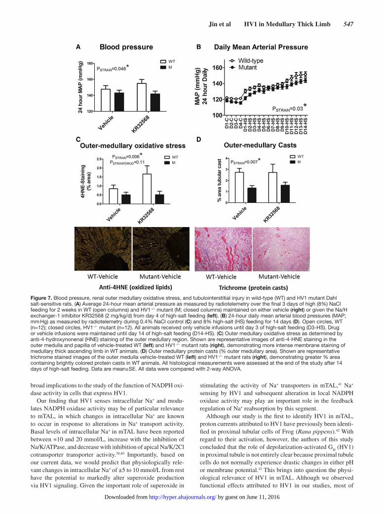
Intracellular Na+, HV1, and Superoxide Production in mTALTo determine whether HV1 promotes superoxide in mTAL when intracellular Na+ levels are low, we determined super-oxide production (ΔEth/ΔDHE) in freshly microdissected live mTAL from WT and HV1−/− mutant rats at both 5 and 30 mmol/L intracellular [Na+]. In response to acidification using an NH
4Cl (20 mmol/L) prepulse, the rate of superoxide pro-
duction was enhanced in mTAL from WT rats at 5 mmol/L but not 30 mmol/L [Na+]
I (Figure 6; P
SODIUM×GROUP<0.0001).
Importantly, in mTAL from HV1−/− mutant rats, no significant difference was observed in superoxide production after acidi-fication at either 5 or 30 mmol/L Na+ (Figure 6).
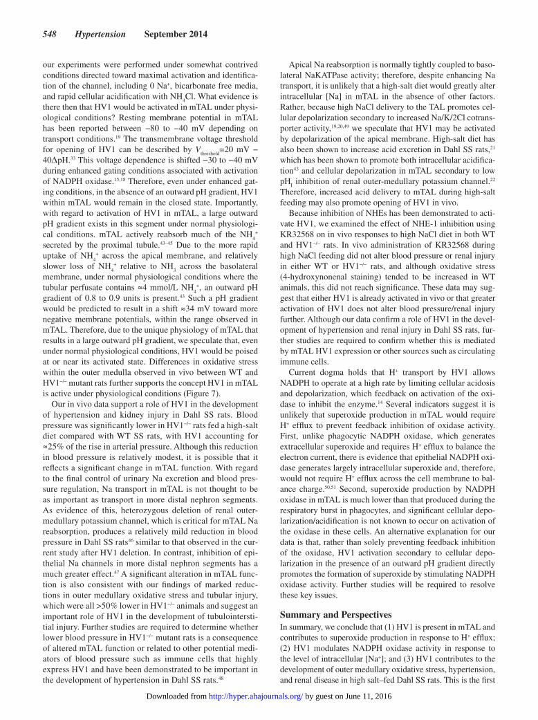
Twenty-four-hour mean arterial pressure measured by radiotelemetry was significantly lower in HV1−/− rats com-pared with WT animals after high-salt feeding (8% NaCl) for 2 weeks (P
STRAIN=0.046). Outer medullary tubular casts
were markedly lower in HV1−/− rats compared with WT SS rats (Figure 7). Outer medullary 4-hydroxynonenal staining (oxidized lipids) was also markedly reduced in HV1−/− rats when compared with WT animals, indicating reduced outer medullary oxidative stress. Administration of KR32568 to activate HV1 in vivo did not alter blood pressure or outer med-ullary injury markers. 4-Hydroxynonenal staining tended to be elevated in KR32568-treated WT animals compared with vehicle, but this did not reach statistical significance (P=0.11).
DiscussionHV1 is a voltage-gated proton channel highly expressed in immune cells where it acts to maintain NADPH oxidase activ-ity during the respiratory burst.14 HV1 has been found in sev-eral cell types outside the immune system,23 including lung epithelial cells33,34 and spermatozoa.35 However, no studies have identified the presence of HV1 in the mammalian kidney or have demonstrated a physiological link to ROS production in renal epithelial cells. This study demonstrates that HV1 is present in mTAL and contributes to H+ efflux-dependent superoxide production. Genetic deletion of HV1 in the SS rat was confirmed by sequence analysis, revealing an 8-bp deletion in a key coding region of HV1. Complete functional knockout of the gene was confirmed by whole-cell voltage-clamp technique. Because murine gene knockout models have revealed lack of specificity of many commonly used antibod-ies,36 we first verified the specificity of a commercial antibody to HV1 in situ in spleen, a tissue in which HV1 expression is high and its localization known. Anti-HV1 from abcam
Figure 4. Effect of intracellular Na± concentration on the respiratory burst in macrophages. x axis, intracellular Na+ concentration ([Na+]I). y axis, maximal superoxide production in freshly isolated peritoneal rat macrophages after addition of phorbol 12-myristate 13-acetate (100 mmol/L) determined by L-012 luminescence (data normalized to the response at 25 mmol/L [Na+]I within each animal). Wild-type (WT; open columns; n=6); WT + 100 μM cariporide (hatched columns; n=6); HV1 mutant (closed columns; n=8); WT + 100 μM ZnCl2 (gray-filled columns; n=6); WT without addition of nystatin (double-hatched columns; n=8). Data were compared using 2-way ANOVA with Bonferroni post hoc test comparing WT to all other groups. *P<0.05 comparing WT to HV1 mutant, WT + ZnCl2, and WT without nystatin. ϒP<0.05 comparing WT to HV1 mutant only. Data are mean±SE.
Figure 5. Contribution of HV1 and NADPH oxidase toward Na sensing in primary cultured macrophages. x axis, intracellular Na+ concentration ([Na+]I). y axis, maximal superoxide production in freshly isolated peritoneal rat macrophages after addition of phorbol 12-myristate 13-acetate (100 mmol/L) determined by L-012 luminescence (data normalized to the response at 25 mmol/L [Na+]I within each animal). HV1−/− control (closed columns; n=5); wild-type (WT) control (open columns; n=5); WT + apocynin 100 μM (striped columns; n=5); HV1−/− transfected with human HV1 (checked columns; n=5). Data were compared using 2-way ANOVA with Bonferroni post hoc test comparing HV1−/− control to all other groups. *P<0.05. Data are mean±SE.
by guest on June 11, 2016http://hyper.ahajournals.org/Downloaded from
546 Hypertension September 2014
(Cat# ab117520) clearly demonstrated expression of HV1 in spleen from WT but not from HV1−/− rats (Figure 1). When we used this antibody for Western blots, we found that detection depended on use of membrane fractions, which are presumably enriched in HV1. Even in these enriched samples, the band for HV1 was weak, suggesting a limited sensitivity of this anti-body for detection via Western blot. Immunohistochemical analysis of kidney sections using this antibody also detected HV1 (primarily around the apical membrane of thick ascend-ing limb) in WT but not HV1−/− Dahl SS. The localization of HV1 to the apical membrane was also confirmed by transmis-sion electron microscopy using immunogold stained sections. Although we were unable to detect HV1 protein expression in mTAL by Western blot, the presence of HV1 in mTAL is sup-ported by polymerase chain reaction, immunohistochemistry, and functional analysis (pH
I).
Functional analysis of both superoxide production and pHI
recovery after NH4Cl prepulse under the same conditions used
to characterize superoxide-stimulating H+ currents in our pre-vious study (0 Na+ media, BaCl
2)10 demonstrate that HV1 is
required for superoxide production after cellular acidification in mTAL.10 Li et al13 first reported that stimulating H+ efflux either by rapidly increasing the pH of the extracellular media or by intracellular acidification using an NH
4Cl prepulse pro-
moted the production of superoxide by NADPH oxidase in mTAL. Although Li et al concluded that any H+ efflux pro-moted NADPH oxidase activity including via NHEs, our own subsequent data did not support a direct link between NHE activity and NADPH oxidase activation. Instead, our data indicated that, in response to intracellular acidification, acti-vation of a second unidentified H+ transport pathway medi-ated superoxide production by the oxidase in this segment.10 In both studies, NADPH oxidase was confirmed as the source of H+ efflux mediated ROS production using either apocynin or diphenylene iodonium.10,13 Our current data confirm our previous conclusions and identify this transport pathway as the voltage-gated proton channel HV1.
Another major finding of the current study is that HV1 modulates NADPH oxidase activity in response to differing levels of intracellular [Na+]. In our previous study, we found that superoxide production after cellular acidification by
NH4Cl was greatly enhanced in 0 Na+ media.10 To elucidate
the relationship among Na+, HV1, and NADPH oxidase, we first performed experiments in isolated peritoneal MØ from SS rats. MØ provided an excellent platform to investigate these relationships because they express high levels of HV1, can easily be isolated in large numbers, ROS production from NADPH oxidase can be induced by addition of PMA, and phagocytes are a well-established model to study the func-tion of both HV1 and NADPH oxidase.8,37–39 We found that low intracellular Na+, between the ranges of 5 to 10 mmol/L, greatly enhanced ROS production during the PMA-stimulated respiratory burst in MØ relative to 25 mmol/L [Na+]
I. The
effect was due to intracellular Na+ as in the absence of the ionophore nystatin, and an increase in ROS production at low Na+ levels was not observed. Importantly, the effect of low intracellular Na+ to enhance maximal superoxide produc-tion in mTAL depended on the activity of HV1. In MØ from WT SS rats, addition of 100 mmol/L Zn2+, which inhibits HV1, completely abolished the relationship between Na+ and superoxide production. Further, in MØ isolated from HV1−/− rats, no relationship between intracellular Na+ and maximal superoxide production was observed unless these cells were transfected with human HV1, which restored the Na sensing phenotype observed in WT cells. We conclude that HV1 must act to sense intracellular Na+ either directly or act as a sig-nal to modulate the activity of NADPH oxidase secondary to changes in intracellular Na+. We confirmed these relationships in isolated mTAL from WT and HV1−/− rats using a second indicator of ROS production, DHE.
We and others have previously identified NADPH oxidase as the source of ROS after H+ efflux in mTAL, and we con-firmed NADPH oxidase as the source of Na-sensitive superox-ide production in MØ in the current study. Loss of Na sensing was not due to loss of NADPH oxidase activity because signifi-cant superoxide production was still detected in the absence of HV1. Further, expression of NADPH oxidase subunits was not altered between mTAL from WT and HV1−/− mutant rats (please see http://hyper.ahajournals.org), indicating that systemic loss of HV1 did not alter NADPH oxidase expression in TAL dur-ing development. Our novel finding that HV1 acts to sense intracellular Na+ and modulate NADPH oxidase activity has
Figure 6. Superoxide production in response to H± efflux at low and high intracellular Na± in medullary thick ascending limb. Superoxide production in isolated medullary thick ascending limb from wild-type (WT; left; n=6) and HV1−/− mutant (M; right; n=7) rats loaded with the superoxide sensitive dye, dihydrethidium (DHE). Open circles, WT at 5 mmol/L intracellular [Na+]; open squares, WT at 30 mmol/L intracellular [Na+]; closed circles, mutant at 5 mmol/L intracellular [Na+]; gray-filled squares, mutant at 30 mmol/L intracellular [Na+]. x axis, time in seconds. NH4Cl (20 mmol/L) was removed from the bath at t=30 seconds. y axis, superoxide production (ΔEth/ΔDHE) normalized to 1 at t=0. PSODIUM×TIME, result of a 2-way repeated measures ANOVA comparing response within strain at 5 and 30 mmol/L intracellular [Na+]. Data are mean±SE. NS indicates not significant.
by guest on June 11, 2016http://hyper.ahajournals.org/Downloaded from
Jin et al HV1 in Medullary Thick Limb 547
broad implications to the study of the function of NADPH oxi-dase activity in cells that express HV1.
Our finding that HV1 senses intracellular Na+ and modu-lates NADPH oxidase activity may be of particular relevance to mTAL, in which changes in intracellular Na+ are known to occur in response to alterations in Na+ transport activity. Basal levels of intracellular Na+ in mTAL have been reported between ≈10 and 20 mmol/L, increase with the inhibition of Na/K/ATPase, and decrease with inhibition of apical Na/K/2Cl cotransporter transporter activity.30,40 Importantly, based on our current data, we would predict that physiologically rele-vant changes in intracellular Na+ of ±5 to 10 mmol/L from rest have the potential to markedly alter superoxide production via HV1 signaling. Given the important role of superoxide in
stimulating the activity of Na+ transporters in mTAL,41 Na+ sensing by HV1 and subsequent alteration in local NADPH oxidase activity may play an important role in the feedback regulation of Na+ reabsorption by this segment.
Although our study is the first to identify HV1 in mTAL, proton currents attributed to HV1 have previously been identi-fied in proximal tubular cells of Frog (Rana pippens).42 With regard to their activation, however, the authors of this study concluded that the role of depolarization-activated G
H (HV1)
in proximal tubule is not entirely clear because proximal tubule cells do not normally experience drastic changes in either pH or membrane potential.42 This brings into question the physi-ological relevance of HV1 in mTAL. Although we observed functional effects attributed to HV1 in our studies, most of
Figure 7. Blood pressure, renal outer medullary oxidative stress, and tubulointerstitial injury in wild-type (WT) and HV1 mutant Dahl salt-sensitive rats. (A) Average 24-hour mean arterial pressure as measured by radiotelemetry over the final 3 days of high (8%) NaCl feeding for 2 weeks in WT (open columns) and HV1−/− mutant (M; closed columns) maintained on either vehicle (right) or given the Na/H exchanger-1 inhibitor KR32568 (2 mg/kg/d) from day 4 of high-salt feeding (left). (B) 24-hour daily mean arterial blood pressures (MAP; mm Hg) as measured by radiotelemetry during 0.4% NaCl control (C) and 8% high-salt (HS) feeding for 14 days (D). Open circles, WT (n=12); closed circles, HV1−/− mutant (n=12). All animals received only vehicle infusions until day 3 of high-salt feeding (D3-HS). Drug or vehicle infusions were maintained until day 14 of high-salt feeding (D14-HS). (C) Outer medullary oxidative stress as determined by anti-4-hydroxynonenal (HNE) staining of the outer medullary region. Shown are representative images of anti-4-HNE staining in the outer medulla and papilla of vehicle-treated WT (left) and HV1−/− mutant rats (right), demonstrating more intense membrane staining of medullary thick ascending limb in WT animals. (D) Outer medullary protein casts (% outer medullary area). Shown are representative trichrome stained images of the outer medulla vehicle-treated WT (left) and HV1−/− mutant rats (right), demonstrating greater % area containing brightly colored protein casts in WT animals. All histological measurements were assessed at the end of the study after 14 days of high-salt feeding. Data are mean±SE. All data were compared with 2-way ANOVA.
by guest on June 11, 2016http://hyper.ahajournals.org/Downloaded from
548 Hypertension September 2014
our experiments were performed under somewhat contrived conditions directed toward maximal activation and identifica-tion of the channel, including 0 Na+, bicarbonate free media, and rapid cellular acidification with NH
4Cl. What evidence is
there then that HV1 would be activated in mTAL under physi-ological conditions? Resting membrane potential in mTAL has been reported between −80 to −40 mV depending on transport conditions.19 The transmembrane voltage threshold for opening of HV1 can be described by V
threshold=20 mV −
40ΔpH.33 This voltage dependence is shifted −30 to −40 mV during enhanced gating conditions associated with activation of NADPH oxidase.15,18 Therefore, even under enhanced gat-ing conditions, in the absence of an outward pH gradient, HV1 within mTAL would remain in the closed state. Importantly, with regard to activation of HV1 in mTAL, a large outward pH gradient exists in this segment under normal physiologi-cal conditions. mTAL actively reabsorb much of the NH
4+
secreted by the proximal tubule.43–45 Due to the more rapid uptake of NH
4+ across the apical membrane, and relatively
slower loss of NH4
+ relative to NH3 across the basolateral
membrane, under normal physiological conditions where the tubular perfusate contains ≈4 mmol/L NH
4+, an outward pH
gradient of 0.8 to 0.9 units is present.43 Such a pH gradient would be predicted to result in a shift ≈34 mV toward more negative membrane potentials, within the range observed in mTAL. Therefore, due to the unique physiology of mTAL that results in a large outward pH gradient, we speculate that, even under normal physiological conditions, HV1 would be poised at or near its activated state. Differences in oxidative stress within the outer medulla observed in vivo between WT and HV1−/− mutant rats further supports the concept HV1 in mTAL is active under physiological conditions (Figure 7).
Our in vivo data support a role of HV1 in the development of hypertension and kidney injury in Dahl SS rats. Blood pressure was significantly lower in HV1−/− rats fed a high-salt diet compared with WT SS rats, with HV1 accounting for ≈25% of the rise in arterial pressure. Although this reduction in blood pressure is relatively modest, it is possible that it reflects a significant change in mTAL function. With regard to the final control of urinary Na excretion and blood pres-sure regulation, Na transport in mTAL is not thought to be as important as transport in more distal nephron segments. As evidence of this, heterozygous deletion of renal outer-medullary potassium channel, which is critical for mTAL Na reabsorption, produces a relatively mild reduction in blood pressure in Dahl SS rats46 similar to that observed in the cur-rent study after HV1 deletion. In contrast, inhibition of epi-thelial Na channels in more distal nephron segments has a much greater effect.47 A significant alteration in mTAL func-tion is also consistent with our findings of marked reduc-tions in outer medullary oxidative stress and tubular injury, which were all >50% lower in HV1−/− animals and suggest an important role of HV1 in the development of tubulointersti-tial injury. Further studies are required to determine whether lower blood pressure in HV1−/− mutant rats is a consequence of altered mTAL function or related to other potential medi-ators of blood pressure such as immune cells that highly express HV1 and have been demonstrated to be important in the development of hypertension in Dahl SS rats.48
Apical Na reabsorption is normally tightly coupled to baso-lateral NaKATPase activity; therefore, despite enhancing Na transport, it is unlikely that a high-salt diet would greatly alter intracellular [Na] in mTAL in the absence of other factors. Rather, because high NaCl delivery to the TAL promotes cel-lular depolarization secondary to increased Na/K/2Cl cotrans-porter activity,19,20,49 we speculate that HV1 may be activated by depolarization of the apical membrane. High-salt diet has also been shown to increase acid excretion in Dahl SS rats,21 which has been shown to promote both intracellular acidifica-tion43 and cellular depolarization in mTAL secondary to low pH
I inhibition of renal outer-medullary potassium channel.22
Therefore, increased acid delivery to mTAL during high-salt feeding may also promote opening of HV1 in vivo.
Because inhibition of NHEs has been demonstrated to acti-vate HV1, we examined the effect of NHE-1 inhibition using KR32568 on in vivo responses to high NaCl diet in both WT and HV1−/− rats. In vivo administration of KR32568 during high NaCl feeding did not alter blood pressure or renal injury in either WT or HV1−/− rats, and although oxidative stress (4-hydroxynonenal staining) tended to be increased in WT animals, this did not reach significance. These data may sug-gest that either HV1 is already activated in vivo or that greater activation of HV1 does not alter blood pressure/renal injury further. Although our data confirm a role of HV1 in the devel-opment of hypertension and renal injury in Dahl SS rats, fur-ther studies are required to confirm whether this is mediated by mTAL HV1 expression or other sources such as circulating immune cells.
Current dogma holds that H+ transport by HV1 allows NADPH to operate at a high rate by limiting cellular acidosis and depolarization, which feedback on activation of the oxi-dase to inhibit the enzyme.14 Several indicators suggest it is unlikely that superoxide production in mTAL would require H+ efflux to prevent feedback inhibition of oxidase activity. First, unlike phagocytic NADPH oxidase, which generates extracellular superoxide and requires H+ efflux to balance the electron current, there is evidence that epithelial NADPH oxi-dase generates largely intracellular superoxide and, therefore, would not require H+ efflux across the cell membrane to bal-ance charge.50,51 Second, superoxide production by NADPH oxidase in mTAL is much lower than that produced during the respiratory burst in phagocytes, and significant cellular depo-larization/acidification is not known to occur on activation of the oxidase in these cells. An alternative explanation for our data is that, rather than solely preventing feedback inhibition of the oxidase, HV1 activation secondary to cellular depo-larization in the presence of an outward pH gradient directly promotes the formation of superoxide by stimulating NADPH oxidase activity. Further studies will be required to resolve these key issues.
Summary and PerspectivesIn summary, we conclude that (1) HV1 is present in mTAL and contributes to superoxide production in response to H+ efflux; (2) HV1 modulates NADPH oxidase activity in response to the level of intracellular [Na+]; and (3) HV1 contributes to the development of outer medullary oxidative stress, hypertension, and renal disease in high salt–fed Dahl SS rats. This is the first
by guest on June 11, 2016http://hyper.ahajournals.org/Downloaded from
Jin et al HV1 in Medullary Thick Limb 549
functional evidence for a role of HV1 in the kidney and identi-fies HV1 as a potential target to limit renal oxidative stress and the development of SS hypertension and renal injury. Our find-ing that HV1 regulates NADPH oxidase activity in response to intracellular Na+ levels may have important implications with regard to tubular Na+ reabsorption and kidney function. This finding is also likely to have broad implications for other organ systems where HV1 is expressed. The development and char-acterization of a HV1−/− mutant rat model is likely to provide an important tool for the study of HV1, including its potential importance to cardiovascular disease.
AcknowledgmentsWe would like to thank the PhysGen KO program (http://rgd.mcw.edu/wg/physgenknockouts) for development of HV1−/− mutant rats. We would like to thank K. Hyndman for her careful review of the manuscript and the Electron Microscopy Core Services at Georgia Regents University.
Sources of FundingThis work was supported by an American Heart Association Scientist Development Grant to P.M. O’Connor (10SDG4150061), National Institutes of Health GO grant RC2 HL101681 (H.J. Jacob), and National Institutes of Health grant GM078319 (N.A. Lambert).
DisclosuresNone.
References 1. Sullivan JM. Salt sensitivity: definition, conception, methodology, and
long-term issues. Hypertension. 1991;17(1 suppl):I61–I68. 2. Kotchen TA, Zhang HY, Covelli M, Blehschmidt N. Insulin resistance and
blood pressure in Dahl rats and in one-kidney, one-clip hypertensive rats. Am J Physiol. 1991;261(6 pt 1):E692–E697.
3. Kidambi S, Kotchen JM, Krishnaswami S, Grim CE, Kotchen TA. Cardiovascular correlates of insulin resistance in normotensive and hyper-tensive African Americans. Metabolism. 2011;60:835–842.
4. Bloch MJ, Basile J. African American patients with hypertensive chronic kidney disease receive no benefit on kidney disease progression from the currently recommended blood pressure goal of <130/80 mm Hg unless there is significant proteinuria at baseline: long-term follow-up of the AASK study. J Clin Hypertens (Greenwich). 2011;13:214–216.
5. Campese VM. Salt sensitivity in hypertension. Renal and cardiovascular implications. Hypertension. 1994;23:531–550.
6. Cowley AW Jr. Renal medullary oxidative stress, pressure-natriuresis, and hypertension. Hypertension. 2008;52:777–786.
7. Taylor NE, Glocka P, Liang M, Cowley AW Jr. NADPH oxidase in the renal medulla causes oxidative stress and contributes to salt-sensitive hypertension in Dahl S rats. Hypertension. 2006;47:692–698.
8. Feng D, Yang C, Geurts AM, Kurth T, Liang M, Lazar J, Mattson DL, O’Connor PM, Cowley AW Jr. Increased expression of NAD(P)H oxidase subunit p67(phox) in the renal medulla contributes to excess oxidative stress and salt-sensitive hypertension. Cell Metab. 2012;15:201–208.
9. Li N, Yi FX, Spurrier JL, Bobrowitz CA, Zou AP. Production of superox-ide through NADH oxidase in thick ascending limb of Henle’s loop in rat kidney. Am J Physiol Renal Physiol. 2002;282:F1111–F1119.
10. O’Connor PM, Lu L, Liang M, Cowley AW Jr. A novel amiloride-sensitive h+ transport pathway mediates enhanced superoxide production in thick ascending limb of salt-sensitive rats, not na+/h+ exchange. Hypertension. 2009;54:248–254.
11. O’Connor PM, Lu L, Schreck C, Cowley AW Jr. Enhanced amiloride-sen-sitive superoxide production in renal medullary thick ascending limb of Dahl salt-sensitive rats. Am J Physiol Renal Physiol. 2008;295:F726–F733.
12. Mori T, O’Connor PM, Abe M, Cowley AW Jr. Enhanced superoxide pro-duction in renal outer medulla of Dahl salt-sensitive rats reduces nitric oxide tubular-vascular cross-talk. Hypertension. 2007;49:1336–1341.
13. Li N, Zhang G, Yi FX, Zou AP, Li PL. Activation of NAD(P)H oxidase by outward movements of H+ ions in renal medullary thick ascending limb of Henle. Am J Physiol Renal Physiol. 2005;289:F1048–F1056.
14. DeCoursey TE. Voltage-gated proton channels find their dream job managing the respiratory burst in phagocytes. Physiology (Bethesda). 2010;25:27–40.
15. DeCoursey TE. Voltage-gated proton channels: molecular biology, physiology, and pathophysiology of the H(V) family. Physiol Rev. 2013;93:599–652.
16. Musset B, Cherny VV, Morgan D, Okamura Y, Ramsey IS, Clapham DE, DeCoursey TE. Detailed comparison of expressed and native voltage-gated proton channel currents. J Physiol. 2008;586:2477–2486.
17. Musset B, Clark RA, DeCoursey TE, Petheo GL, Geiszt M, Chen Y, Cornell JE, Eddy CA, Brzyski RG, El Jamali A. NOX5 in human spermatozoa: expression, function, and regulation. J Biol Chem. 2012;287:9376–9388.
18. DeCoursey TE, Cherny VV, Zhou W, Thomas LL. Simultaneous activa-tion of NADPH oxidase-related proton and electron currents in human neutrophils. Proc Natl Acad Sci U S A. 2000;97:6885–6889.
19. Schlatter E, Greger R. cAMP increases the basolateral Cl- conductance in the isolated perfused medullary thick ascending limb of Henle’s loop of the mouse. Pflugers Arch. 1985;405:367–376.
20. Liu R, Garvin JL, Ren Y, Pagano PJ, Carretero OA. Depolarization of the macula densa induces superoxide production via NAD(P)H oxidase. Am J Physiol Renal Physiol. 2007;292:F1867–F1872.
21. Batlle DC, Sharma AM, Alsheikha MW, Sobrero M, Saleh A, Gutterman C. Renal acid excretion and intracellular pH in salt-sensitive genetic hypertension. J Clin Invest. 1993;91:2178–2184.
22. Bleich M, Köttgen M, Schlatter E, Greger R. Effect of NH4+/NH3 on cytosolic pH and the K+ channels of freshly isolated cells from the thick ascending limb of Henle’s loop. Pflugers Arch. 1995;429:345–354.
23. Capasso M, DeCoursey TE, Dyer MJ. pH regulation and beyond: unan-ticipated functions for the voltage-gated proton channel, HVCN1. Trends Cell Biol. 2011;21:20–28.
24. Ortiz PA, Garvin JL. Superoxide stimulates NaCl absorption by the thick ascending limb. Am J Physiol Renal Physiol. 2002;283:F957–F962.
25. Garvin JL, Ortiz PA. The role of reactive oxygen species in the regulation of tubular function. Acta Physiol Scand. 2003;179:225–232.
26. Juncos R, Garvin JL. Superoxide enhances Na-K-2Cl cotransporter activity in the thick ascending limb. Am J Physiol Renal Physiol. 2005;288:F982–F987.
27. Juncos R, Hong NJ, Garvin JL. Differential effects of superoxide on lumi-nal and basolateral Na+/H+ exchange in the thick ascending limb. Am J Physiol Regul Integr Comp Physiol. 2006;290:R79–R83.
28. Trinh-Trang-Tan MM, Bouby N, Coutaud C, Bankir L. Quick isolation of rat medullary thick ascending limbs: enzymatic and metabolic character-ization. Pflugers Arch. 1986;407:228–234.
29. Yang C, Stingo FC, Ahn KW, et al. Increased proliferative cells in the medullary thick ascending limb of the loop of Henle in the Dahl salt-sensitive rat. Hypertension. 2013;61:208–215.
30. Ortiz PA, Hong NJ, Garvin JL. NO decreases thick ascending limb chlo-ride absorption by reducing Na(+)-K(+)-2Cl(-) cotransporter activity. Am J Physiol Renal Physiol. 2001;281:F819–F825.
31. Dickhout JG, Mori T, Cowley AW Jr. Tubulovascular nitric oxide cross-talk: buffering of angiotensin II-induced medullary vasoconstriction. Circ Res. 2002;91:487–493.
32. Ramsey IS, Ruchti E, Kaczmarek JS, Clapham DE. Hv1 proton channels are required for high-level NADPH oxidase-dependent superoxide pro-duction during the phagocyte respiratory burst. Proc Natl Acad Sci U S A. 2009;106:7642–7647.
33. Cherny VV, Markin VS, DeCoursey TE. The voltage-activated hydrogen ion conductance in rat alveolar epithelial cells is determined by the pH gradient. J Gen Physiol. 1995;105:861–896.
34. Iovannisci D, Illek B, Fischer H. Function of the HVCN1 proton chan-nel in airway epithelia and a naturally occurring mutation, M91T. J Gen Physiol. 2010;136:35–46.
35. Lishko PV, Kirichok Y. The role of Hv1 and CatSper channels in sperm activation. J Physiol. 2010;588(pt 23):4667–4672.
36. Herrera M, Sparks MA, Alfonso-Pecchio AR, Harrison-Bernard LM, Coffman TM. Lack of specificity of commercial antibodies leads to misidentification of angiotensin type 1 receptor protein. Hypertension. 2013;61:253–258.
37. Morgan D, Capasso M, Musset B, Cherny VV, Ríos E, Dyer MJ, DeCoursey TE. Voltage-gated proton channels maintain pH in human neutrophils dur-ing phagocytosis. Proc Natl Acad Sci U S A. 2009;106:18022–18027.
38. El Chemaly A, Okochi Y, Sasaki M, Arnaudeau S, Okamura Y, Demaurex N. VSOP/Hv1 proton channels sustain calcium entry, neutrophil migra-tion, and superoxide production by limiting cell depolarization and acidi-fication. J Exp Med. 2010;207:129–139.
by guest on June 11, 2016http://hyper.ahajournals.org/Downloaded from
550 Hypertension September 2014
39. Kapus A, Romanek R, Qu AY, Rotstein OD, Grinstein S. A pH-sensitive and voltage-dependent proton conductance in the plasma membrane of macrophages. J Gen Physiol. 1993;102:729–760.
40. Yu M, Lopez B, Dos Santos EA, Falck JR, Roman RJ. Effects of 20-HETE on Na+ transport and Na+ -K+ -ATPase activity in the thick ascending loop of Henle. Am J Physiol Regul Integr Comp Physiol. 2007;292:R2400–R2405.
41. Schreck C, O’Connor PM. NAD(P)H oxidase and renal epithelial ion trans-port. Am J Physiol Regul Integr Comp Physiol. 2011;300:R1023–R1029.
42. Gu X, Sackin H. Effect of pH on potassium and proton conductance in renal proximal tubule. Am J Physiol. 1995;269(3 pt 2):F289–F308.
43. Watts BA 3rd, Good DW. Effects of ammonium on intracellular pH in rat medullary thick ascending limb: mechanisms of apical membrane NH4+ transport. J Gen Physiol. 1994;103:917–936.
44. Attmane-Elakeb A, Karim Z, Bichara M. Role of the Na(+)-K+(NH4+)-2Cl cotransporter of the medullary ascending limb in the regulation of renal acid-base equilibrium. Nephrologie. 2002;23:209–211.
45. Karim Z, Attmane-Elakeb A, Bichara M. Renal handling of NH4+ in relation to the control of acid-base balance by the kidney. J Nephrol. 2002;15(suppl 5):S128–S134.
46. Zhou X, Zhang Z, Shin MK, et al. Heterozygous disruption of renal outer medullary potassium channel in rats is associated with reduced blood pressure. Hypertension. 2013;62:288–294.
47. Pavlov TS, Levchenko V, O’Connor PM, Ilatovskaya DV, Palygin O, Mori T, Mattson DL, Sorokin A, Lombard JH, Cowley AW Jr, Staruschenko A. Deficiency of renal cortical EGF increases ENaC activity and contributes to salt-sensitive hypertension. J Am Soc Nephrol. 2013;24:1053–1062.
48. Mattson DL, James L, Berdan EA, Meister CJ. Immune suppression attenuates hypertension and renal disease in the Dahl salt-sensitive rat. Hypertension. 2006;48:149–156.
49. Haque MZ, Ares GR, Caceres PS, Ortiz PA. High salt differentially regulates surface NKCC2 expression in thick ascending limbs of Dahl salt-sensitive and salt-resistant rats. Am J Physiol Renal Physiol. 2011;300:F1096–F1104.
50. Lassègue B, San Martín A, Griendling KK. Biochemistry, physiology, and pathophysiology of NADPH oxidases in the cardiovascular system. Circ Res. 2012;110:1364–1390.
51. Li JM, Shah AM. Intracellular localization and preassembly of the NADPH oxidase complex in cultured endothelial cells. J Biol Chem. 2002;277:19952–19960.
What Is New?•This study demonstrates that the voltage-gated proton channel HV1 is
expressed in medullary thick ascending limb, HV1 contributes to super-oxide production in this segment, HV1 acts to modulate superoxide pro-duction in response to changes in intracellular [Na+], and HV1 contributes to blood pressure regulation and renal injury in the Dahl salt-sensitive (SS) rat model.
What Is Relevant?•Superoxide production within the renal outer medulla by NADPH oxidase
is important in long-term blood pressure control and the development of renal injury via effects on Na+ transport and renal hemodynamics and is augmented in Dahl SS rats. We now identify HV1 as a critical com-ponent contributing to enhanced superoxide production in medullary thick ascending limb of Dahl SS rats and a novel molecular target for the treatment of renal oxidative stress. Demonstration of HV1 as a Na+
sensor contributes to our understanding of the physiological stimuli that promote activation of NADPH oxidase within the kidney and in other tis-sues expressing HV1.
SummaryIn summary, we determined HV1 as the molecular identity of a source of superoxide in medullary thick ascending limb, which is augmented in Dahl SS rats. Further, we demonstrated that HV1 acts to modulate the activity of NADPH oxidase in both medullary thick ascending limb and MØ in response to physiologically rel-evant changes in intracellular Na+ and that HV1 contributes to blood pressure regulation and renal injury in the Dahl SS rat model. These data indicate that HV1 is likely to play an important physiological function as a Na+ sensor and may provide a novel target for the treatment of renal oxidative stress, hypertension, and kidney injury.
Novelty and Significance
by guest on June 11, 2016http://hyper.ahajournals.org/Downloaded from
Nevin A. Lambert and Paul M. O'ConnorBermingham, Sandip Darji, Avirup Guha, Roshan Patel, Aron M. Geurts, Howard J. Jacob,
Chunhua Jin, Jingping Sun, Carly A. Stilphen, Susan M.E. Smith, Hiram Ocasio, BrentAscending Limb of Dahl Salt-Sensitive Rats
HV1 Acts as a Sodium Sensor and Promotes Superoxide Production in Medullary Thick
Print ISSN: 0194-911X. Online ISSN: 1524-4563 Copyright © 2014 American Heart Association, Inc. All rights reserved.
is published by the American Heart Association, 7272 Greenville Avenue, Dallas, TX 75231Hypertension doi: 10.1161/HYPERTENSIONAHA.114.03549
2014;64:541-550; originally published online June 16, 2014;Hypertension.
http://hyper.ahajournals.org/content/64/3/541World Wide Web at:
The online version of this article, along with updated information and services, is located on the
http://hyper.ahajournals.org/content/suppl/2014/06/16/HYPERTENSIONAHA.114.03549.DC1.htmlData Supplement (unedited) at:
http://hyper.ahajournals.org//subscriptions/
is online at: Hypertension Information about subscribing to Subscriptions:
http://www.lww.com/reprints Information about reprints can be found online at: Reprints:
document. Permissions and Rights Question and Answer this process is available in the
click Request Permissions in the middle column of the Web page under Services. Further information aboutOffice. Once the online version of the published article for which permission is being requested is located,
can be obtained via RightsLink, a service of the Copyright Clearance Center, not the EditorialHypertensionin Requests for permissions to reproduce figures, tables, or portions of articles originally publishedPermissions:
by guest on June 11, 2016http://hyper.ahajournals.org/Downloaded from
1
ONLINE SUPPLEMENT 1
TITLE: HV1 ACTS AS A SODIUM SENSOR AND PROMOTES SUPEROXIDE 2
PRODUCTION IN MEDULLARY THICK ASCENDING LIMB OF DAHL SALT-3
SENSITIVE RATS. MANUSCRIPT NUMBER HYPE201403549D 4
5
AUTHORS: Chunhua Jin1, Jingping Sun1, Carly A. Stilphen1, Susan M. E. Smith4, 6
Hiram Ocasio1, Brent Bermingham1, Sandip Darji1, Avirup Guha1, Roshan Patel1, 7
Aron M. Geurts3, Howard J. Jacob3, Nevin A. Lambert2, Paul M. O’Connor1 8
CORRESPONDING AUTHOR: 9
Dr. Paul O’Connor 10
Department Physiology, CB2206 11
Georgia Regents University, 12
1459 Laney-Walker Blvd, 13
Augusta, GA, USA 14
30912 15
Phone: 706 721 7890 16
Fax: 706 721 7661 17
Email: [email protected] 18
Supplemental Tables = 1 19
Supplemental Figures = 2 20
21
SUPPLEMENTARY METHODS 22
Mutation of HV1 23
Zinc Finger Nucleases (ZFNs) targeting rat HV1 were designed by Sigma. mRNA 24
encoding the HV1 ZFNs targeting the exon 4 sequence 5’- 25
CACACCCAGGCCATCCCTGGACTTCAGGAGCCGGCTAAGGAA -3’, where the ZFN 26
monomer binding sites are underlined, were injected into SS/JrHsdMcwi (Dahl salt-27
sensitive; SS) rats and transferred to pseudopregnant Sprague Dawley females. The 28
SURVEYOR nuclease assay 1 detected a ZFN-induced mutation in a single male pup 29
which was sequenced to reveal an 8-bp deletion within the target sequence 30
CACACCCAGGCCATCCCT--------GGAGCCGGCTAAGGAA. This rat was backcrossed 31
to the parental SS strain and heterozygous offspring were chosen for intercross to 32
establish the SS-Hvcn1em1Mcwi (HV1-/-) mutant strain. 33
2
Rat genotyping 34
Zinc-finger nucleases (ZFNs) HV1 mutant rats on the SS background (~3 weeks old) 35
were ear punched and DNA were extracted using DNeasy Blood & Tissue Kit 36
(QIAGEN). Rat HV1 genotyping primers used were: Forward: 37
AGTGGGAGAACGAGGAGGAT, Reverse: TAGTGACCTGGTGACCTCCC. Wild-type 38
and mutant PCR sizes were detected at 302 bp and 294 bp, respectively. PCR 39
conditions: 94C 4 min, 94C 30’, 62C 30’, 72C 40’ for 35 cycles. The PCR products were 40
loaded into 15% Precast polyacrylamide gel (BioRad Cat# 345-0020) and run in 1X TBE 41
in Criterion Cell (BioRad) at 150V for 3 hours. Gels were stained with 0.5 ug/ml ethidium 42
bromide in 1X TBE on a rocker for 15 min and washed in Distilled water before being 43
imaged (UV). 44
Real-time PCR Total RNA was extracted from bulk isolated mTAL and whole spleen 45
tissue (RNeasy Mini Kit, QIAGEN). RNA (400ng) was reverse transcribed by using High 46
Capacity cDNA Reverse Transcription Kit (Applied Biosystems). Duplicate quantitative 47
real-time PCR (qRT-PCR) (Bio-Rad iCycler ) was performed by using Power SYBR 48
Green PCR Master Mix (Applied Biosystems) in a final volume of 25 µl containing 40 ng 49
of cDNA. Primer sequences are given in supplement (please see 50
http://hyper.ahajournals.org). 51
Western Blot 52
Membrane fractions of spleen were prepared from spleen tissues by homogenization in 53
ice-cold sucrose buffer solution, 0.25 M Sucrose, 30 mM Tris.HCL(PH 7.4), 1mM EDTA, 54
protease inhibitor cocktail and phosphatase inhibitor cocktail 2 (Sigma). The solution 55
was transferred to a 1.5 ml tube, sonicated and spun at 2600 RPM for 5 min at 4C to 56
pellet large debris. The supernatant was then transferred and spun at 17,000g on 57
ultracentrifuge for 20 min at 4C. This pellet is the membrane enriched fraction and was 58
re-suspended in Sucrose buffer. Protein concentration was determined (Bradford, 59
BioRad protein assay) and 50 µg of protein was loaded per lane onto a 12% Acrylamide 60
gel for electrophoresis before being transferred to a Immobilon-FL membrane 61
(Millipore). Blot was first incubated with an anti-HV1 (abcam Cat# ab117520 ) primary 62
antibody (1:1000, overnight at 4C ) followed by secondary antibody ( goat anti-rabbit 63
IgG 680 fluorescent, Invitrogen Cat#A-21076, 1:1000, for 1 hour at room temperature). 64
Immuno-reactivity was detected by scanning the blot using Odyssey 700 channel (Li-65
Cor, Inc.). The blot was washed with 1x TBS before sequential incubation with anti--66
actin mouse monoclonal (SIGMA Cat# A-1978-200ul) primary antibody (1: 25,000, 67
overnight at 4C) and secondary antibody (goat anti-mouse IgG 800, Rockland 68
antibodies and Assays, Cat# 610-132-121, 1: 10,000, 1hour at room temperature ). 69
Histological Staining Analysis 70
Spleen and kidney were harvested from WT and HV1-/- mutant male littermates aged 8-71
12 weeks fed a low 0.4% NaCl diet since weaning. Rats were anesthetized with 72
isoflurane (2-5%) and the left kidney perfused with 10 ml 0.9% saline, the kidneys and 73
spleen excised and placed in phosphate buffer containing 10% formaldehyde. Kidneys 74
were paraffin embedded in an automatic tissue processor and 3-µm cut sections 75
3
mounted on siliconized/charged slides. The kidney and spleen slides were 76
deparaffinized and hydrated and antigen retrieval performed using IHC-Tek Epitope 77
retrieval solution at steaming for 40 min (IHCWorld, cat#IW-1100). Tissue was blocked 78
with 3% hydrogen peroxide in methanol for 10 min. The primary antibody used to detect 79
HV1 was Rabbit polyclonal anti-HV1 antibody (abcam Cat#ab117520, 1mg/ml, 1:400 at 80
2.5ug/ml) overnight at 4C. The goat anti-rabbit IgG-HRP conjugated secondary antibody 81
(Santa-cruz Cat#sc-2004, 400ug/ml, 1:400 at 1ug/ml) was used for 30 min at room 82
temperature. The slides were stained with Betazoid DAB Chromogen Kit (Biocare 83
Medical Cat#BDB2004H), then counterstained with Hematoxylin. Omitting the first 84
antibody served as a negative control and resulted in no positive staining in tissue. 85
The primary monoclonal antibody used to detect peroxidation was anti 4-Hydroxy-2-86
Nonenal (4-NHE) antibody (4-HNE) (JaICA). A biotinylated rabbit anti-mouse secondary 87
antibody was used for development with avidin-biotinylated horseradish peroxidase 88
complex ( Vector Lab, Vectastain ABC-AP KIT ). The slides were stained with 89
BCIP/NBT Kit (blue/violet )(Vector). Outer-medullary tubular injury was quantified using 90
MetaMorph Imaging software (Molecular Devices). Trichrome stained slides were 91
blinded to the investigator and digitally scanned at 7200DPI using a slide scanner 92
(pathscanner enabler IV, Meyer Instruments). The percentage of the outer medullary 93
region containing protein casts was determined by color threshold as previously 94
described 2-5. 95
Electron Microscopy 96
Tissue was fixed in 4% formaldehyde, 0.2% glutaraldehyde in 0.1 M sodium cacodylate 97
(NaCac) buffer, pH 7.4, dehydrated with a graded ethanol series through 95% and 98
embedded in LR White resin. Thin sections (75 nm) were cut with a diamond knife on a 99
Leica EM UC6 ultramicrotome (Leica Microsystems, Inc, Bannockburn, IL) and collected 100
on nickel grids. Tissue was then etched with 2% hydrogen peroxide in PBS and 101
aldehyde moieties were quenched with 1M ammonium chloride in PBS. Grids were then 102
hybridized with rabbit anti-HV1 antibody (1:1,00 dilution) and anti-rabbit Ig conjugated 103
nanogold (Nanoprobes, Yaphank, NY) (1:2,000 dilution) followed by silver 104
enhancement. 105
Imaging of mTAL 106
BCECF was excited at 440/10 and 490/10 nm. A 510/40-nm band pass emission filter 107
was used to collect a BCECF fluorescent signal at 3-second intervals. Intracellular pH 108
(pHi) was calibrated in situ at the end of each experiment using a 2-point calibration 109
curve by exchanging the bath solution with saline solution containing nigericin (10 110
μmol/L) and KCl (140 mmol/L) of known pH. A 445/40-nm and a 605/55-nm band pass 111
emission filter were used to collect DHE (380/40X-445/40E) and ethidium (Eth; 112
480/40X-605/55E) signals. A Lambda-10-3 and rapid filter wheel changer (Sutter 113
Instruments) was used to collect emission signals from DHE and Eth at 3-second 114
intervals. DHE and Eth signals were then normalized so that the ratio Eth:DHE at time 0 115
was equal to 1. The change in the ratio of Eth:DHE fluorescent signal across the 116
duration of the experiment was then used as an index of superoxide production. 117
4
118
Whole cell voltage-clamp recordings 119
Intra-peritoneal MØ were isolated from female WT and HV1-/- SS rats as described in 120
methods. MØ plated onto poly-l-lysine-coated plastic culture dishes in an extracellular 121
solution that contained (in mM): tetramethylammonium methanesulfonic acid (140), 122
CaCl2 (1.8), HEPES (100) and mannitol (40); pH was adjusted to 7.2, and osmolarity to 123
300 mosM. Whole-cell patch pipettes (~5 MΩ) were filled with a solution containing (in 124
mM): N-methyl-D-glucamine (118), tetramethylammonium methanesulfonic acid (63), 125
MgCl2 (1), MES (119) and EGTA (1); pH was adjusted to 6.1, and osmolarity to 290 126
mosM. Cells were visually selected under phase contrast microscopy and subjected to 127
whole-cell voltage-clamp. Whole-cell currents were recorded in response to step 128
changes in membrane potential from a holding potential of -60mV. 129
References 130
1. Geurts AM, Cost GJ, Freyvert Y, Zeitler B, Miller JC, Choi VM, Jenkins SS, Wood 131
A, Cui X, Meng X, Vincent A, Lam S, Michalkiewicz M, Schilling R, Foeckler J, 132
Kalloway S, Weiler H, Menoret S, Anegon I, Davis GD, Zhang L, Rebar EJ, 133
Gregory PD, Urnov FD, Jacob HJ, Buelow R. Knockout rats via embryo 134
microinjection of zinc-finger nucleases. Science. 2009;325:433. 135
2. Taylor NE, Cowley AW, Jr. Effect of renal medullary h2o2 on salt-induced 136
hypertension and renal injury. Am J Physiol Regul Integr Comp Physiol. 137
2005;289:R1573-1579. 138
3. Mori T, Polichnowski A, Glocka P, Kaldunski M, Ohsaki Y, Liang M, Cowley AW, 139
Jr. High perfusion pressure accelerates renal injury in salt-sensitive hypertension. 140
J Am Soc Nephrol. 2008;19:1472-1482. 141
4. Polichnowski AJ, Cowley AW, Jr. Pressure-induced renal injury in angiotensin ii 142
versus norepinephrine-induced hypertensive rats. Hypertension. 2009;54:1269-143
1277. 144
5. Singh P, Bahrami L, Castillo A, Majid DS. Tnf-alpha type 2 receptor mediates 145
renal inflammatory response to chronic angiotensin ii administration with high salt 146
intake in mice. Am J Physiol Renal Physiol. 2013;304:F991-999. 147
6. Hwang SM, Koo NY, Jin M, Davies AJ, Chun GS, Choi SY, Kim JS, Park K. 148
Intracellular acidification is associated with changes in free cytosolic calcium and 149
inhibition of action potentials in rat trigeminal ganglion. J Biol Chem. 150
2010;286:1719-1729. 151
7. Ago T, Kitazono T, Ooboshi H, Iyama T, Han YH, Takada J, Wakisaka M, 152
Ibayashi S, Utsumi H, Iida M. Nox4 as the major catalytic component of an 153
endothelial nad(p)h oxidase. Circulation. 2004;109:227-233. 154 155
156
5
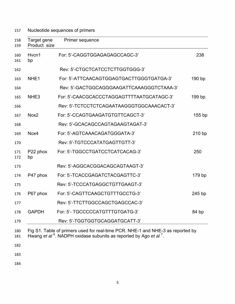
Nucleotide sequences of primers 157
Target gene Primer sequence 158
Product size 159
Hvcn1 For: 5’-CAGGTGGAGAGAGCCAGC-3’ 238 160
bp 161
Rev: 5’-CTGCTCATCCTCTTGGTGGG-3’ 162
NHE1 For: 5’-ATTCAACAGTGGAGTGACTTGGGTGATGA-3’ 190 bp 163
Rev: 5’-GACTGGCAGGGAAGATTCAAAGGGTCTAAA-3’ 164
NHE3 For: 5’-CAACGCACCCTAGGAGTTTTAATGCATAGC-3’ 199 bp 165
Rev: 5’-TCTCCTCTCAGAATAAGGGTGGCAAACACT-3’ 166
Nox2 For: 5’-CCAGTGAAGATGTGTTCAGCT-3’ 155 bp 167
Rev: 5’-GCACAGCCAGTAGAAGTAGAT-3’ 168
Nox4 For: 5’-AGTCAAACAGATGGGATA-3’ 210 bp 169
Rev: 5’-TGTCCCATATGAGTTGTT-3’ 170
P22 phox For: 5’-TGGCCTGATCCTCATCACAG-3’ 250 171
bp 172
Rev: 5’-AGGCACGGACAGCAGTAAGT-3’ 173
P47 phox For: 5’-TCACCGAGATCTACGAGTTC-3’ 179 bp 174
Rev: 5’-TCCCATGAGGCTGTTGAAGT-3’ 175
P67 phox For: 5’-CAGTTCAAGCTGTTTGCCTG-3’ 245 bp 176
Rev: 5’-TTCTTGGCCAGCTGAGCCAC-3’ 177
GAPDH For: 5’- TGCCCCCATGTTTGTGATG-3’ 84 bp 178
Rev: 5’-TGGTGGTGCAGGATGCATT-3’ 179
Fig S1. Table of primers used for real-time PCR. NHE-1 and NHE-3 as reported by 180
Hwang et al 6. NADPH oxidase subunits as reported by Ago et al 7. 181
182
183
184
6
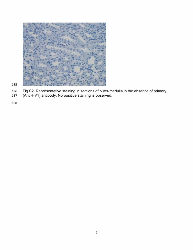
185
Fig S2. Representative staining in sections of outer-medulla in the absence of primary 186
(Anti-HV1) antibody. No positive staining is observed. 187
188
7
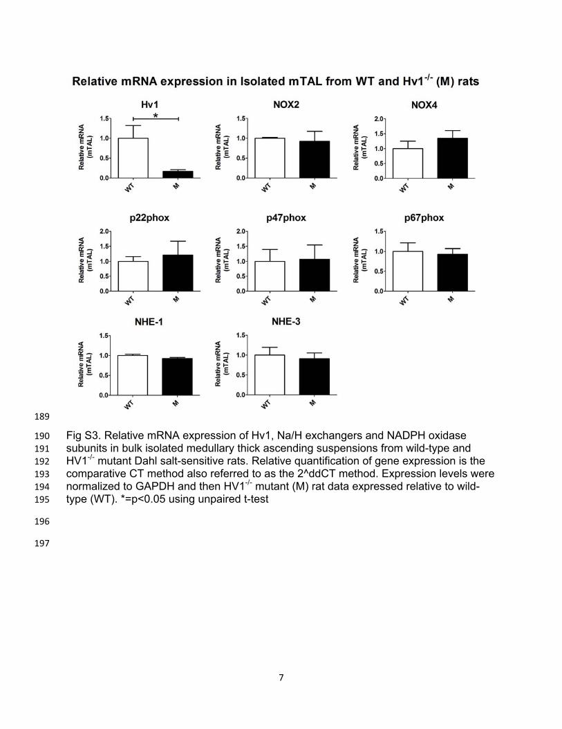
189
Fig S3. Relative mRNA expression of Hv1, Na/H exchangers and NADPH oxidase 190
subunits in bulk isolated medullary thick ascending suspensions from wild-type and 191
HV1-/- mutant Dahl salt-sensitive rats. Relative quantification of gene expression is the 192
comparative CT method also referred to as the 2^ddCT method. Expression levels were 193
normalized to GAPDH and then HV1-/- mutant (M) rat data expressed relative to wild-194
type (WT). *=p<0.05 using unpaired t-test 195
196
197

8
198