cyclosporine stimulates na+-k+-cl- cotransport activity in cultured mouse medullary thick ascending...
TRANSCRIPT
Kidney International, Vol. 58 (2000), pp. 1652–1663
Cyclosporine stimulates Na1-K1-Cl2 cotransport activity incultured mouse medullary thick ascending limb cells
MAI-SZU WU, CHIH-WEI YANG, MARCELLE BENS, KOU-CHENG PENG, HSIAO-MEI YU,and ALAIN VANDEWALLE
Division of Nephrology, Chang Gung Memorial Hospital, Taipei, Taiwan; Institut National de la Sante et de la RechercheMedicale, Unite 478, and Institut Federatif de Recherche 02, Faculte de Medecine Xavier Bichat, Paris, France
Cyclosporine stimulates Na1-K1-Cl2 cotransport activity in cul- Cyclosporine (CsA) is a fungus-derived hydrophobictured mouse medullary thick ascending limb cells. cyclic undecapeptide commonly used in the prevention
Background. Cyclosporine (CsA) has been shown to alter of renal allograft rejection [1]. Although CsA representsthe activity of plasma membrane transporters in kidney epithe-
a potent immunosuppressive agent, this drug can belial cells. In this study, we have investigated the effects of CsAnephrotoxic [2]. Several mechanisms for the CsA-on Na1,K1-ATPase and Na1-K1-Cl2 cotransport activities in
cultured cells derived from microdissected mouse medullary induced nephrotoxicity have been advanced, includingthick ascending limb (mTAL) cells. vasoconstriction and altered glomerular permeability [3].
Methods. Experiments were carried out on subcultured Clinical studies had also shown that CsA might be re-confluent mouse TAL cells. Reverse transcription-polymer- sponsible for renal tubular dysfunction [4]. Sodium re-ase chain reaction experiments showed that they expressed
tention, hyperkalemia, and hyperchloremic acidosis havethe mNKCC2 electroneutral Na1-K1-Cl2 cotransporter andbeen reported in CsA-treated kidney transplanted pa-ROM-K1 and ROMK2 potassium channel mRNA. Western
blotting also revealed the presence of the 40 kD ROMK protein tients [5–7]. However, the exact cellular mechanism ofusing an anti-ROMK antibody. The effect of CsA (100 ng/mL) action of CsA still remains in debate [7]. Previous inon ion transport was assessed by measuring the influx and vivo and in vitro studies have provided strong evidencesefflux of rubidium (86Rb1) and 36Cl2, used as tracers of K1 and
that CsA exerts a direct inhibitory action on the renalCl2 movements, on cells grown on Petri dishes or permeableNa1,K1-ATPase pump [8–10] and impairs Na1 absorp-filters.
Results. CsA inhibited by 38% the ouabain-sensitive compo- tion at the level of the proximal tubule cells [11], medul-nent of 86Rb1 influx mediated by the Na1,K1-ATPase pumps. lary thick ascending limb (mTAL) cells [10, 12], andCsA also increased by 38% the ouabain-resistant furosemide- distal tubule cells [13].sensitive component (Or-Fs) of 86Rb1 influx, reflecting the Na1-
The decrease in Na1,K1-ATPase activity caused byK1-Cl2 cotransport activity and stimulated the basolateralCsA, especially in mTAL and collecting tubule cells, isefflux of 36Cl2 from mTAL cells grown on filters. The CsA-thought to be one of the underlying mechanisms for thestimulated basal efflux of Cl2 was prevented by the basal addi-
tion of the Cl2 channel blocker 5-nitro-2-(3-phenylpropylam- observed potassium and hydrogen ion secretion defectsino) benzoate (NPPB, 1024 mol/L). Apical addition of the K1
[10]. Ferrer-Martinez et al reported that CsA, in additionchannel blocking agent Ba21 (1024 mol/L) partially prevented to its inhibitory action on Na1,K1-ATPase activity, para-the CsA-stimulated basal efflux of Cl2. Adding Ba21 to the
doxically increased the Na1-K1-Cl2 cotransporter activ-luminal side of cells grown on Petri dishes also prevented theity in cultured bovine renal NBL-1 cells [9]. These au-rise in apical 86Rb1 efflux and the increased Or-Fs component
of 86Rb1 influx caused by CsA. thors also showed that the increased in Na1-K1-Cl2
Conclusion. These results indicated that CsA may stimulate cotransporter activity was not directly linked to cell vol-the Na1-K1-Cl2 cotransport activity and also suggested that ume changes. These results indirectly suggested that CsAthis immunosuppressive agent may interfere in the recycling
might affect the activity of the Na1-K1-Cl2 cotrans-of apical K1 in this model of cultured mouse TAL cells.porter, which is predominantly located in the apicalmembrane from TAL [14]. The NaCl absorption acrossTAL cells involves the apical Na1-K1-Cl2 electroneutralKey words: immunosuppression, potassium recycling, allograft rejec-
tion, kidney transplantation, nephrotoxicity, sodium absorption. cotransport, ATP-regulated potassium channels, baso-laterally located Cl2 channel(s), and Na1,K1-ATPase
Received for publication June 1, 1999pumps [15, 16]. CsA has been shown to inhibit theand in revised form April 27, 2000
Accepted for publication May 10, 2000 Na1,K1-ATPase pumps, which are responsible for themaintenance of a favorable electrogenic gradient for Na1 2000 by the International Society of Nephrology
1652
Wu et al: CsA effects on Na1-K1-Cl2 cotransport and Na1,K1-ATPase 1653
and Cl2 absorption in cultured NBL-1 cells [10]. The Cell viabilityunexpected concomitant increase in Na1-K1-Cl2 activity Cell viability was estimated by counting the numbercaused by CsA in this bovine renal epithelial NBL-1 cell of cells that excluded trypan blue and by measuring theline raised the question of how CsA could affect the lactate dehydrogenase activity (LDH), which is usedkinetics of Cl2 and K1 transport in another cultured cell as marker of nonspecific cell injury, as previously de-system expressing both the Na1-K1-Cl2 cotransporter scribed [18].and the ROMK channels. We therefore analyzed the
cAMP assayshort-term effects of nontoxic doses of CsA on Cl2 andK1 transport mediated by the electroneutral Na1-K1- The effect of dDAVP on cell cAMP content was mea-Cl2 cotransport in a model of cultured mouse epithelial sured on confluent TAL cells grown in 12-well platescells derived from microdissected TAL cells [17, 18]. and preincubated with or without CsA for six hours.
Briefly, cells were preincubated with 0.1 mmol/L 3-isobu-tyl-1-methyl xanthine (IBMX) for 30 minutes at 378C
METHODS and then in 1 mL DMEM containing supplemented withMaterials IBMX and with or without dDAVP (1026 mol/L) for
seven minutes at 378C [17]. The reaction was stoppedSoluble CsA in cremophor was obtained from Sandozeby rapidly removing the medium and adding 1 mL ice-(Basel, Switzerland). Rapamycin (AY-22989-21) was a gen-cold 95% ethanol/5% formic acid. The supernatantserous gift from Wyeth-Ayerst Research (Princeton, NJ,were evaporated to dryness, and cAMP was measured byUSA). Fujisawa Co. (Osaka, Japan) kindly suppliedradioimmunoassay kit (Incstar Corporation, Stillwater,FK506 (Tacrolimuse). Furosemide was from Hoechst lab-MN, USA).oratories (German Remedies Co., Taipei, Taiwan). 86Rb1
and 36Cl2 were purchased from Amersham (Arlington RNA extraction and reverse transcription-polymeraseHeights, IL, USA). Deamino-8-D-arginine vasopressin chain reaction(dDAVP) was from Ferringe Pharmaceutical (Malmo,
Total RNA was extracted from isolated mTAL seg-Sweden). The avian myeloblastosis virus reverse tran-ment microdissected from the kidney of an adult mousescriptase (RT AMV) was from Boehringere (Formo In-and from confluent TAL cells by the method of Chom-dustrial Co., Ltd., Taipei, Taiwan). All other reagents wereczynski [19]. RNA, treated with RNase-free DNase Ifrom Sigma (Sigma Chemical Co., St. Louis, MO, USA).(Boehringere Mannheim, Mannheim, Germany), wasCulture media [Dulbecco’s modified Eagle’s mediumevaluated by spectrophotometry. RNA (1 mg) was re-(DMEM), Ham’s F12] were from GIBCO BRL labora-verse transcribed with RT AMV at 428C for 60 minutes.tory (Life Technologiese, Taipei, Taiwan). The perme-cDNA from microdissected and cultured TAL and non–able filters were Millicell-CM filters (0.4 mm pore size; Mil-reverse-transcribed RNA from cultured TAL cells were
lipore Continental Water Systems, Bedford, MA, USA).amplified for 32 cycles in 100 mL total volume containing50 mmol/L KCl, 20 mmol/L Tris-HCl, pH 8.4, 10 mmol/LCultured mouse TAL cellsdNTP, 1.5 mmol/L MgCl2, 1 unit Taq polymerase, and
Experiments were carried out on subcultured mTAL 10 pmol of mouse NKCC2 (mNKCC2), mouse ROMKcells microdissected out from the kidneys of one-month- (mROMK1 and mROMK2), and 10 pmol of b-actin (in-old normal mice [17]. Cultured TAL cells were routinely ternal standard) primers. The thermal cycling programgrown in a modified culture medium (DMEM:Ham’s was as follows: 948C for one minute, 608C for one minute,F12, 1:1 vol/vol, 60 nmol/L sodium selenate, 5 mg/mL and 728C for three minutes. The primers for mNKCC2,transferrin, 2 mmol/L glutamine, 5 mg/mL insulin, 50 encoding for the kidney-specific Na1-K1-Cl2 cotrans-nmol/L dexamethasone, 1 nmol/L triiodothyronine, 10 porter isoform [20], were as follows: antisense strandng/mL epidermal growth factor, 2% fetal calf serum, 20 59-C TT GGCT TCGGTTT TAGA T G ACCCG-39mmol/L HEPES, pH 7.4) at 378C in 5% C02/95% air and sense strand 59-GCAATGCTGGCATTTAGACatmosphere. All experiments were performed on sets of CCTCCG-39 (32 cycles). ROMK1 primers were anti-confluent cells (6th and 15th passages) grown on Petri sense strand 59-CCCTCATGGCAATCACAGCATTdishes or permeable filters. Confluent cells grown on GTGAC-39 and sense strand 59-CCCGACAACAGTGPetri dishes were examined under an inverted micro- CAGGAGCCGC-39 (32 cycles). ROMK2 primers werescope (Leicae) and photographed. The transepithelial antisense strand 59-CCCCCCACGTTTGCTGATCAelectrical resistance (RT) was measured on confluent cell CCGC-39 and sense strand 59-GGCAATGTAGATGClayers grown on collagen-coated permeable filters using ACAGTCGAGG-39 (32 cycles). The b-actin primersthe Millicell Electrical Resistance System (ERS; Milli- (28 cycles) were the same as previously described [21].
Amplification products were run on 4% agarose gel,pore Corporation, Bedford, MA, USA).
Wu et al: CsA effects on Na1-K1-Cl2 cotransport and Na1,K1-ATPase1654
stained with ethidium bromide, and photographed. All using radiolabeled rubidium (86Rb1) and chloride (36Cl2)sets of primers used yielded amplified products of ex- as tracer of potassium and chloride movements, respec-pected size and sequence analyses of the polymerase tively [17].chain reaction (PCR) products confirmed that they 86Rb1 influx studies were performed on confluent cellsmatched well with the excepted cDNAs analyzed (data grown in 24-well plates. Cell layers were washed withnot shown). 1 mL modified PBS (in mmol/L: NaCl 136, KCl 5,
Na2HPO4 1, glucose 5, HEPES 10, pH 7.4) prewarmedImmunoblot analysis to 378C. The medium was removed, and Rb1 influx was
Confluent mouse TAL cells grown on Petri dishes initiated by adding 1 mL PBS containing 2 mCi/mL 86Rb1.were scraped off into phosphate-buffered saline (PBS) The reaction was stopped by removing the solution andwith a rubber policeman and were centrifuged (200 3 g three rinses with 1 mL ice-cold 100 mmol/L MgCl2 solu-for 5 min). Pelleted cells were sonicated (three times for tion. Cells were solubilized with 0.5 mL 1 mol/L NaOH,30 s at 48C) in 2 mL ice-cold sucrose solution [0.25 mol/L and the radioactivity was determined by scintillationsucrose, 3 mmol/L imidazole, 1 mmol/L ethylenediamine- counting. The components of K1 influx were measuredtetraacetic acid (EDTA), pH 7.2] supplemented with by incubating cells without (total influx) or with 0.5protease inhibitor cocktail (Complete; Boehringer Man- mmol/L ouabain or ouabain plus 1025 mol/L furosemidenheim) and centrifuged at 150 3 g for 10 minutes at 48C in order to determine the ouabain-sensitive influx (Os)to remove nuclei and debris. The supernatant was then mediated by the Na1,K1-ATPase pumps and the oua-centrifuged (105,000 3 g) for 60 minutes at 48C. The bain-resistant furosemide-sensitive influx (Or-Fs) medi-pelleted membrane-enriched fraction was then resus- ated by the Na1-K1-Cl2 cotransporter. Experimentspended in 100 mL sucrose solution containing the prote- were performed in duplicate for each condition tested.ase inhibitor cocktail. Whole homogenates and mem- Protein content was measured by the method of Lowrybrane-enriched fractions were kept at 2808C until use. [23] using BSA as standard.Protein content was determined by the Bradford method Cl2 efflux studies using 36Cl2 were performed on cells[22] using bovine serum albumin (BSA) as standard. grown on collagen-coated filters in order to calculateAliquots (50 mg) of whole homogenates and membrane- the percentage of radioactivity remaining in cells and toenriched preparations were subfractionated by sodium determine the rate of constant 36Cl2 efflux from bothdodecyl sulfate-polyacrylamide gel electrophoresis (SDS- apical and basal sides of the filter [17]. Cells attachedPAGE) using a 7.5% resolving gel and a 4% stacking gel. to the filters were preincubated with 1.5 mCi/mL 36Cl2
Proteins were transferred to a polyvinylidene difluoridesupplemented medium applied to the apical (1 mL) and(PVDF) membrane in 25 mmol/L Tris-HCl, 192 mmol/Lbasal (2.5 mL) compartments of the cells. Afterward,glycine, and 25% methanol. Rainbow molecular weightthe filters were rapidly rinsed (5 times) with modifiedmarkers were used as size standards (Amersham). Theunlabeled PBS-CaCl2, and fresh PBS-CaCl2 was addedmembrane was blocked by incubation with 5% skim milkto both apical (1 mL) and basal (1 mL) sides of the filters.in TBS-T (10 mmol/L Tris-HCl, pH 8.5, 150 mmol/LA sample-and-replace technique was used to obtain 1 mLNaCl, 0.1% Tween 20) overnight at 48C. For Westernsamples from both the apical and basal sides of the filtersblotting, we used the anti-ROMK1 polyclonal antibodyat one-minute intervals on which radioactivity was counted.raised against a purified fusion protein of SchistosomaThe excised filters were then incubated with 1 mL of 1NJaponicum glutathione-S-transferase and a C-terminalNaOH overnight to measure the residual radioactivitypart of the rat ROMK1 protein (APC-001; Alomoneremaining in cells. The efflux from both apical and basalLabs, Ltd., Jerusalem, Israel). This antibody, which crosssides of the cells was estimated by calculating the ratereacts with the mouse species, recognizes an epitopeconstant using the same formula as previously [17, 24].common to all ROMK isoforms. The membrane wasThe rate constant of tracer efflux from the apical sideprobed with anti-ROMK antibody (1:5000) for two hourswas calculated as: [ln(R2) 2 ln(R1 1 B)]/(t2 2 t1). Theat room temperature and then with peroxidase-conjugatedrate constant of tracer efflux from the basal side wasgoat anti-rabbit IgG (1:5000; Dako, Trappes, France) forcalculated as: [ln(R2) 2 ln(R1 1 A)]/(t2 2 t1). A and Btwo hours at room temperature. The membrane wasrepresent the percentage of total radioactivity counted inwashed with TBS-T, and antigen-antibody complexesthe apical and basal media between t1 and t2 [17].were detected with the ECL plus Western blotting detec-
86Rb1 efflux studies were also performed on confluenttion system (Amersham, Pharmacia Biotech Europe, Or-cells grown on Petri dishes, a culture condition in whichsay, France). An experiment omitting the primary anti-the culture medium covers the apical membrane side ofbody was performed as a negative control.the cells [19]. The percentage of radioactivity remaining
K1 and Cl2 flux studies in cells and the rate constant of 86Rb1 efflux from theapical side of the cells were performed using the sameK1 and Cl2 transport were measured on confluent cell
layers grown on Petri dishes or on collagen-coated filters protocol as described by Matthews et al for the intestinal-
Wu et al: CsA effects on Na1-K1-Cl2 cotransport and Na1,K1-ATPase 1655
like T84 cells [25]. Confluent TAL cells were incubatedfor two hours with 2 mCi/mL 86Rb1. Afterward, cell layerswere rinsed (5 times) with 2 mL modified unlabeledPBS-CaCl2, and 1 mL of fresh PBS-CaCl2 was added.The same sample-and-replace technique described pre-viously in this article was used to obtain 1 mL samplesat one-minute intervals on which radioactivity wascounted. In this case, the rate constant of Rb1 efflux wascalculated using the same formula described by Ven-glarik, Bridges, and Frizzell [24]. In all cases, the effectsof CsA, FK506, rapamycin, and K1 or Cl2 channel block-ers (described in the Results section and in the Figurelegends), added either to the apical or basal side of thefilters, on 36Cl2 and 86Rb1 efflux were performed in exactsimilar conditions as for untreated cells on the same setsof TAL cells.
Statistical analysis
Results are expressed as mean 6 SEM from N experi-ments performed in duplicate or triplicate. Significantdifferences between groups were analyzed by Student’st-test and one-way analysis of variance (ANOVA). Thestatistical analysis was performed using the Statvieweprogram (Macintosh). A P value of less than 0.05 wasconsidered significant.
RESULTS
Biochemical features of cultured mouse TAL cells
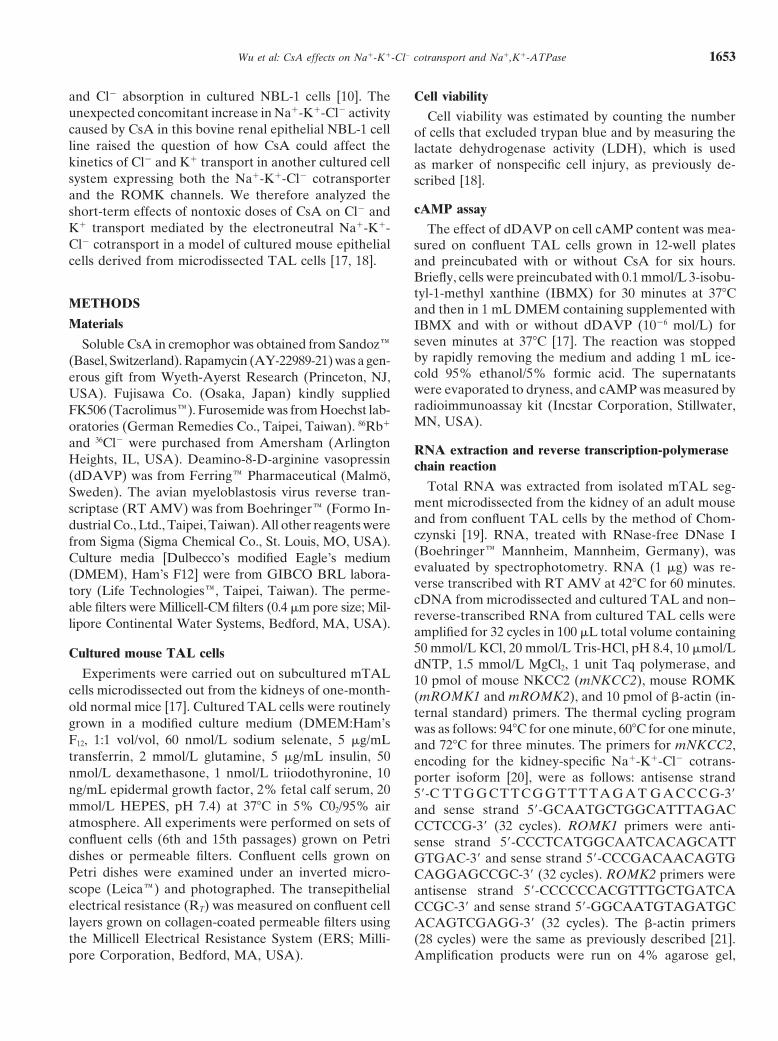
Confluent mouse TAL cells grown on collagen-coatedPetri dishes formed monolayers of cuboid-shaped cellsand formed small domes (Fig. 1A). Reverse transcrip-tion-polymerase chain reaction (RT-PCR) was used todetect the kidney-specific Na1-K1-Cl2 cotransportermRNA mNKCC2 [20] in confluent cultured TAL cells.Substantial amounts of mNKCC2 transcripts were de-tected in both microdissected medullary TAL and cul-tured TAL cells (Fig. 1B). A single 406 bp band of ex-pected size was amplified in both microdissected TAL
Fig. 1. Properties of cultured mouse thick ascending limb (TAL) cells.and cultured cells. As negative control, no amplified (A) Confluent TAL cells grown on Petri dishes formed layers of cuboid-
shaped cells and formed small domes (magnification 3250). (B) Illustra-products were obtained by using non–reversed-tran-tion of an ethidium bromide-stained 4% agarose gel showing the ampli-scribed RNA from cultured TAL cells or by omittingfied products of expected size (406 bp) obtained with the mNKCC2
cDNA (Fig. 1B). The different components of 86Rb1primers in microdissected medullary TAL (lane 1) and cultured TALcells (7th passage, lane 2). As controls, no band was detected usinginflux measured in the absence or presence of ouabainnon–reverse-transcribed RNA from cultured cells (lane 3) or by omit-and furosemide allowed to distinguish the Os componentting cDNA (lane 4). The expression of b-actin was also shown as an
of 86Rb1 influx mediated by the Na1,K1-ATPase pumps internal control. Molecular weight standards (M) were the 1 kb ladderfrom GIBCO-BRL. (C) Time course of 86Rb1 influx performed onand Or-Fs component of 86Rb1 influx reflecting the Na1-cultured TAL cells grown on Petri dishes and incubated without (j)K1-Cl2 cotransport activity (Fig. 1C). In all cases, theor with ouabain (d) and ouabain plus furosemide (m). Each point is
influx of 86Rb1 increased linearly with time up to 10 the mean 6 SE from five separate experiments performed in duplicate.minutes. Therefore, all subsequent 86Rb1 influx experi-ments used an incubation time of four minutes. and 456 bp bands of expected size were amplified using
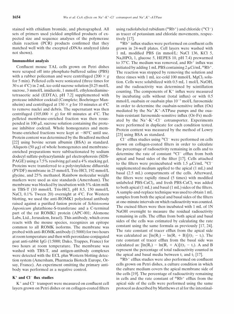
Reverse transcribed-PCR experiments also showed mROMK1 and mROMK2 primers, respectively, in boththat cultured TAL cells expressed both the ROMK1 mouse kidney extract and cultured TAL cells (Fig. 2A).and ROMK2 K1 channels, which have been previously The Western blot analysis to detect ROMK was per-
formed on whole homogenate and membrane-enrichedshown to be presented in rat TAL [26]. Single 498 bp
Wu et al: CsA effects on Na1-K1-Cl2 cotransport and Na1,K1-ATPase1656
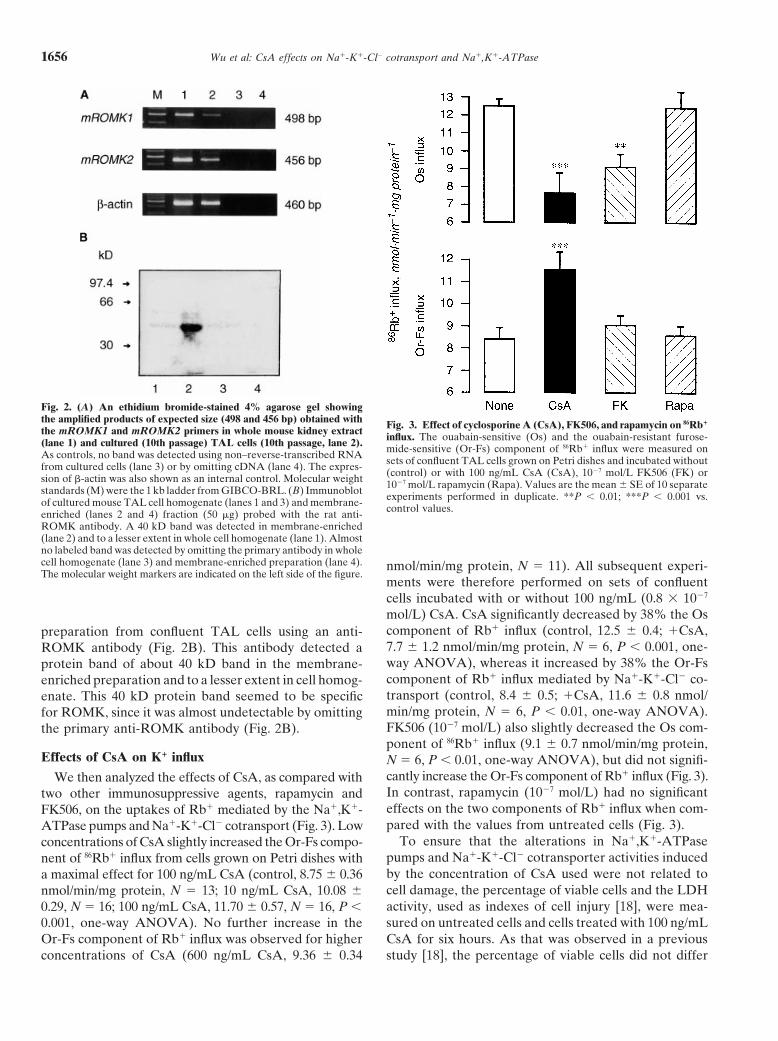
Fig. 2. (A) An ethidium bromide-stained 4% agarose gel showingthe amplified products of expected size (498 and 456 bp) obtained with Fig. 3. Effect of cyclosporine A (CsA), FK506, and rapamycin on 86Rb1
the mROMK1 and mROMK2 primers in whole mouse kidney extract influx. The ouabain-sensitive (Os) and the ouabain-resistant furose-(lane 1) and cultured (10th passage) TAL cells (10th passage, lane 2). mide-sensitive (Or-Fs) component of 86Rb1 influx were measured onAs controls, no band was detected using non–reverse-transcribed RNA sets of confluent TAL cells grown on Petri dishes and incubated withoutfrom cultured cells (lane 3) or by omitting cDNA (lane 4). The expres- (control) or with 100 ng/mL CsA (CsA), 1027 mol/L FK506 (FK) orsion of b-actin was also shown as an internal control. Molecular weight 1027 mol/L rapamycin (Rapa). Values are the mean 6 SE of 10 separatestandards (M) were the 1 kb ladder from GIBCO-BRL. (B) Immunoblot experiments performed in duplicate. **P , 0.01; ***P , 0.001 vs.of cultured mouse TAL cell homogenate (lanes 1 and 3) and membrane- control values.enriched (lanes 2 and 4) fraction (50 mg) probed with the rat anti-ROMK antibody. A 40 kD band was detected in membrane-enriched(lane 2) and to a lesser extent in whole cell homogenate (lane 1). Almostno labeled band was detected by omitting the primary antibody in wholecell homogenate (lane 3) and membrane-enriched preparation (lane 4). nmol/min/mg protein, N 5 11). All subsequent experi-The molecular weight markers are indicated on the left side of the figure.
ments were therefore performed on sets of confluentcells incubated with or without 100 ng/mL (0.8 3 1027
mol/L) CsA. CsA significantly decreased by 38% the Oscomponent of Rb1 influx (control, 12.5 6 0.4; 1CsA,preparation from confluent TAL cells using an anti-7.7 6 1.2 nmol/min/mg protein, N 5 6, P , 0.001, one-ROMK antibody (Fig. 2B). This antibody detected away ANOVA), whereas it increased by 38% the Or-Fsprotein band of about 40 kD band in the membrane-component of Rb1 influx mediated by Na1-K1-Cl2 co-enriched preparation and to a lesser extent in cell homog-transport (control, 8.4 6 0.5; 1CsA, 11.6 6 0.8 nmol/enate. This 40 kD protein band seemed to be specificmin/mg protein, N 5 6, P , 0.01, one-way ANOVA).for ROMK, since it was almost undetectable by omittingFK506 (1027 mol/L) also slightly decreased the Os com-the primary anti-ROMK antibody (Fig. 2B).ponent of 86Rb1 influx (9.1 6 0.7 nmol/min/mg protein,
Effects of CsA on K1 influx N 5 6, P , 0.01, one-way ANOVA), but did not signifi-cantly increase the Or-Fs component of Rb1 influx (Fig. 3).We then analyzed the effects of CsA, as compared withIn contrast, rapamycin (1027 mol/L) had no significanttwo other immunosuppressive agents, rapamycin andeffects on the two components of Rb1 influx when com-FK506, on the uptakes of Rb1 mediated by the Na1,K1-pared with the values from untreated cells (Fig. 3).ATPase pumps and Na1-K1-Cl2 cotransport (Fig. 3). Low
To ensure that the alterations in Na1,K1-ATPaseconcentrations of CsA slightly increased the Or-Fs compo-pumps and Na1-K1-Cl2 cotransporter activities inducednent of 86Rb1 influx from cells grown on Petri dishes withby the concentration of CsA used were not related toa maximal effect for 100 ng/mL CsA (control, 8.75 6 0.36cell damage, the percentage of viable cells and the LDHnmol/min/mg protein, N 5 13; 10 ng/mL CsA, 10.08 6activity, used as indexes of cell injury [18], were mea-0.29, N 5 16; 100 ng/mL CsA, 11.70 6 0.57, N 5 16, P ,sured on untreated cells and cells treated with 100 ng/mL0.001, one-way ANOVA). No further increase in the
Or-Fs component of Rb1 influx was observed for higher CsA for six hours. As that was observed in a previousstudy [18], the percentage of viable cells did not differconcentrations of CsA (600 ng/mL CsA, 9.36 6 0.34
Wu et al: CsA effects on Na1-K1-Cl2 cotransport and Na1,K1-ATPase 1657
N 5 5; 1CsA, 138 6 0.2 V cm2, N 5 5). The discretedecrease in RT was, however, only transient, since RT
values were almost identical or even higher in cellstreated for longer time (more than one hour) with CsA.However, the changes in RT did not followed the increasein Cl2 and K1 efflux induced by CsA (discussed later inthis article). It could be due to the fact that the mousecultured TAL cells formed a leaky epithelial cell mono-layer and exhibited paracellular ion flux pathway [30],making difficult to interpret the changes in RT after theaddition of CsA. Furthermore, RT remained unchangedwhen furosemide (1025 mol/L) or ouabain (0.5 mmol/L)were added to the apical or basal sides of the filters,respectively. Confluent cells grown on filters were thenused to study the effects of CsA on the kinetics of Cl2
and K1 efflux. Control experiments showed that apicaladdition of 1025 mol/L furosemide reduced the basal rateconstant of Cl2 efflux by 38% (control, 0.021 6 0.001;furosemide, 0.013 6 0.002, N 5 18, P , 0.001), whereasthe basal addition of furosemide had no effect on Cl2
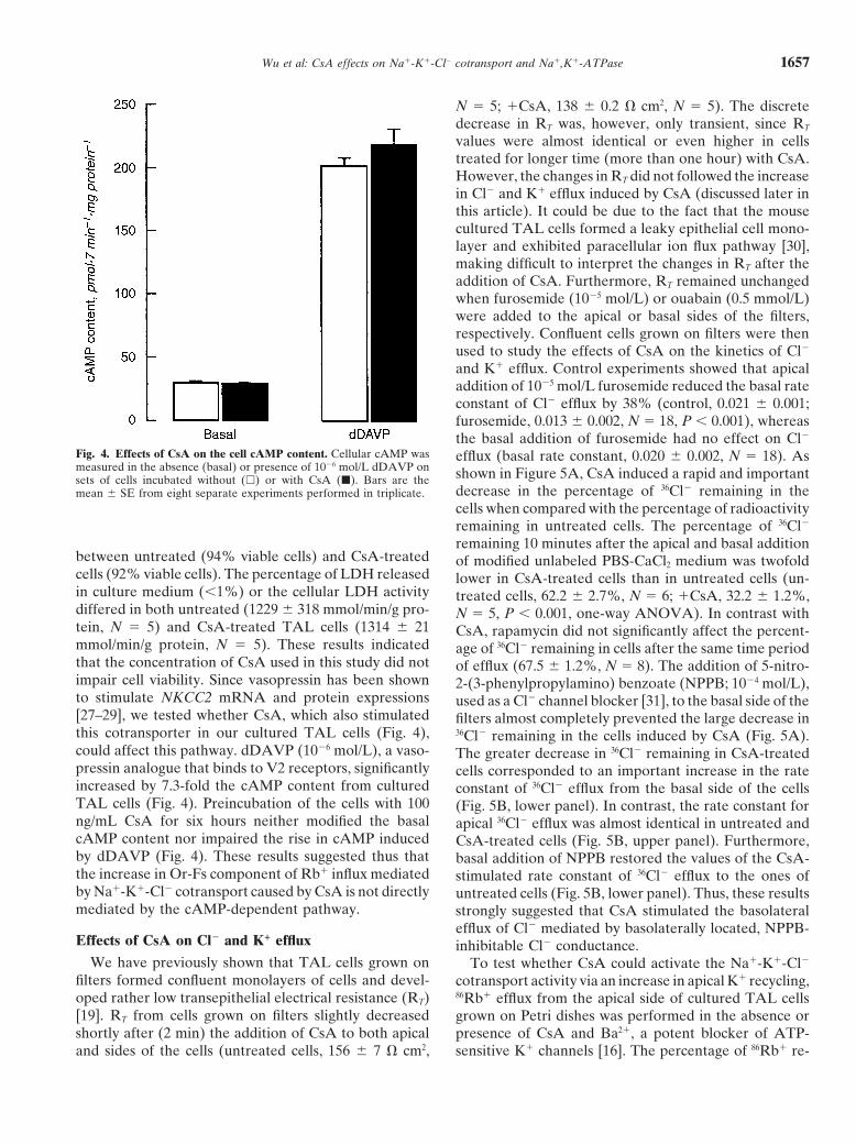
Fig. 4. Effects of CsA on the cell cAMP content. Cellular cAMP was efflux (basal rate constant, 0.020 6 0.002, N 5 18). Asmeasured in the absence (basal) or presence of 1026 mol/L dDAVP on shown in Figure 5A, CsA induced a rapid and importantsets of cells incubated without (h) or with CsA (j). Bars are the
decrease in the percentage of 36Cl2 remaining in themean 6 SE from eight separate experiments performed in triplicate.cells when compared with the percentage of radioactivityremaining in untreated cells. The percentage of 36Cl2
remaining 10 minutes after the apical and basal additionbetween untreated (94% viable cells) and CsA-treated of modified unlabeled PBS-CaCl2 medium was twofoldcells (92% viable cells). The percentage of LDH released lower in CsA-treated cells than in untreated cells (un-in culture medium (,1%) or the cellular LDH activity treated cells, 62.2 6 2.7%, N 5 6; 1CsA, 32.2 6 1.2%,differed in both untreated (1229 6 318 mmol/min/g pro- N 5 5, P , 0.001, one-way ANOVA). In contrast withtein, N 5 5) and CsA-treated TAL cells (1314 6 21 CsA, rapamycin did not significantly affect the percent-mmol/min/g protein, N 5 5). These results indicated age of 36Cl2 remaining in cells after the same time periodthat the concentration of CsA used in this study did not of efflux (67.5 6 1.2%, N 5 8). The addition of 5-nitro-impair cell viability. Since vasopressin has been shown 2-(3-phenylpropylamino) benzoate (NPPB; 1024 mol/L),to stimulate NKCC2 mRNA and protein expressions used as a Cl2 channel blocker [31], to the basal side of the[27–29], we tested whether CsA, which also stimulated filters almost completely prevented the large decrease inthis cotransporter in our cultured TAL cells (Fig. 4), 36Cl2 remaining in the cells induced by CsA (Fig. 5A).could affect this pathway. dDAVP (1026 mol/L), a vaso- The greater decrease in 36Cl2 remaining in CsA-treatedpressin analogue that binds to V2 receptors, significantly cells corresponded to an important increase in the rateincreased by 7.3-fold the cAMP content from cultured constant of 36Cl2 efflux from the basal side of the cellsTAL cells (Fig. 4). Preincubation of the cells with 100 (Fig. 5B, lower panel). In contrast, the rate constant forng/mL CsA for six hours neither modified the basal apical 36Cl2 efflux was almost identical in untreated andcAMP content nor impaired the rise in cAMP induced CsA-treated cells (Fig. 5B, upper panel). Furthermore,by dDAVP (Fig. 4). These results suggested thus that basal addition of NPPB restored the values of the CsA-the increase in Or-Fs component of Rb1 influx mediated stimulated rate constant of 36Cl2 efflux to the ones ofby Na1-K1-Cl2 cotransport caused by CsA is not directly untreated cells (Fig. 5B, lower panel). Thus, these resultsmediated by the cAMP-dependent pathway. strongly suggested that CsA stimulated the basolateral
efflux of Cl2 mediated by basolaterally located, NPPB-Effects of CsA on Cl2 and K1 efflux inhibitable Cl2 conductance.
We have previously shown that TAL cells grown on To test whether CsA could activate the Na1-K1-Cl2
filters formed confluent monolayers of cells and devel- cotransport activity via an increase in apical K1 recycling,oped rather low transepithelial electrical resistance (RT) 86Rb1 efflux from the apical side of cultured TAL cells[19]. RT from cells grown on filters slightly decreased grown on Petri dishes was performed in the absence orshortly after (2 min) the addition of CsA to both apical presence of CsA and Ba21, a potent blocker of ATP-
sensitive K1 channels [16]. The percentage of 86Rb1 re-and sides of the cells (untreated cells, 156 6 7 V cm2,
Wu et al: CsA effects on Na1-K1-Cl2 cotransport and Na1,K1-ATPase1658
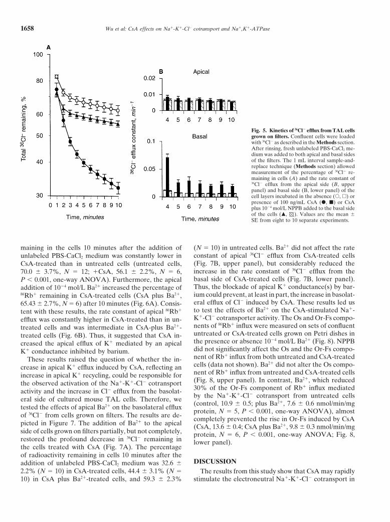
Fig. 5. Kinetics of 36Cl2 efflux from TAL cellsgrown on filters. Confluent cells were loadedwith 36Cl2 as described in the Methods section.After rinsing, fresh unlabeled PBS-CaCl2 me-dium was added to both apical and basal sidesof the filters. The 1 mL interval sample-and-replace technique (Methods section) allowedmeasurement of the percentage of 36Cl2 re-maining in cells (A) and the rate constant of36Cl2 efflux from the apical side (B, upperpanel) and basal side (B, lower panel) of thecell layers incubated in the absence (s, h) orpresence of 100 ng/mL CsA (d, j) or CsAplus 1024 mol/L NPPB added to the basal sideof the cells (m, ). Values are the mean 6SE from eight to 10 separate experiments.
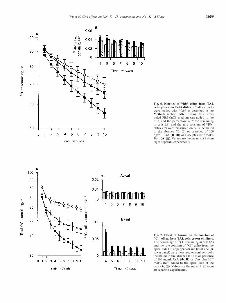
maining in the cells 10 minutes after the addition of (N 5 10) in untreated cells. Ba21 did not affect the rateconstant of apical 36Cl2 efflux from CsA-treated cellsunlabeled PBS-CaCl2 medium was constantly lower in(Fig. 7B, upper panel), but considerably reduced theCsA-treated than in untreated cells (untreated cells,increase in the rate constant of 36Cl2 efflux from the70.0 6 3.7%, N 5 12; 1CsA, 56.1 6 2.2%, N 5 6,basal side of CsA-treated cells (Fig. 7B, lower panel).P , 0.001, one-way ANOVA). Furthermore, the apicalThus, the blockade of apical K1 conductance(s) by bar-addition of 1024 mol/L Ba21 increased the percentage ofium could prevent, at least in part, the increase in basolat-86Rb1 remaining in CsA-treated cells (CsA plus Ba21,eral efflux of Cl2 induced by CsA. These results led us65.43 6 2.7%, N 5 6) after 10 minutes (Fig. 6A). Consis-to test the effects of Ba21 on the CsA-stimulated Na1-tent with these results, the rate constant of apical 86Rb1
K1-Cl2 cotransporter activity. The Os and Or-Fs compo-efflux was constantly higher in CsA-treated than in un-nents of 86Rb1 influx were measured on sets of confluenttreated cells and was intermediate in CsA-plus Ba21-untreated or CsA-treated cells grown on Petri dishes intreated cells (Fig. 6B). Thus, it suggested that CsA in-the presence or absence 1024 mol/L Ba21 (Fig. 8). NPPBcreased the apical efflux of K1 mediated by an apicaldid not significantly affect the Os and the Or-Fs compo-K1 conductance inhibited by barium.nent of Rb1 influx from both untreated and CsA-treatedThese results raised the question of whether the in-cells (data not shown). Ba21 did not alter the Os compo-crease in apical K1 efflux induced by CsA, reflecting annent of Rb1 influx from untreated and CsA-treated cellsincrease in apical K1 recycling, could be responsible for(Fig. 8, upper panel). In contrast, Ba21, which reducedthe observed activation of the Na1-K1-Cl2 cotransport30% of the Or-Fs component of Rb1 influx mediatedactivity and the increase in Cl2 efflux from the basolat-by the Na1-K1-Cl2 cotransport from untreated cellseral side of cultured mouse TAL cells. Therefore, we(control, 10.9 6 0.5; plus Ba21, 7.6 6 0.6 nmol/min/mgtested the effects of apical Ba21 on the basolateral effluxprotein, N 5 5, P , 0.001, one-way ANOVA), almost
of 36Cl2 from cells grown on filters. The results are de- completely prevented the rise in Or-Fs induced by CsApicted in Figure 7. The addition of Ba21 to the apical (CsA, 13.6 6 0.4; CsA plus Ba21, 9.8 6 0.3 nmol/min/mgside of cells grown on filters partially, but not completely, protein, N 5 6, P , 0.001, one-way ANOVA; Fig. 8,restored the profound decrease in 36Cl2 remaining in lower panel).the cells treated with CsA (Fig. 7A). The percentageof radioactivity remaining in cells 10 minutes after the
DISCUSSIONaddition of unlabeled PBS-CaCl2 medium was 32.6 62.2% (N 5 10) in CsA-treated cells, 44.4 6 3.1% (N 5 The results from this study show that CsA may rapidly
stimulate the electroneutral Na1-K1-Cl2 cotransport in10) in CsA plus Ba21-treated cells, and 59.3 6 2.3%
Wu et al: CsA effects on Na1-K1-Cl2 cotransport and Na1,K1-ATPase 1659
Fig. 6. Kinetics of 86Rb1 efflux from TALcells grown on Petri dishes. Confluent cellswere loaded with 86Rb1 as described in theMethods section. After rinsing, fresh unla-beled PBS-CaCl2 medium was added to thedish, and the percentage of 86Rb1 remainingin cells (A) and the rate constant of 86Rb1
efflux (B) were measured on cells incubatedin the absence (s, h) or presence of 100ng/mL CsA (d, j) or CsA plus 1024 mol/LBa21 (m, ). Values are the mean 6 SE fromeight separate experiments.
Fig. 7. Effect of barium on the kinetics of36Cl2 efflux from TAL cells grown on filters.The percentage of 36Cl2 remaining in cells (A)and the rate constant of 36Cl2 efflux from theapical side (B, upper panel) and basal side (B,lower panel) were measured on confluent cellsincubated in the absence (s, h) or presenceof 100 ng/mL CsA (d, j) or CsA plus 1024
mol/L Ba21 added to the apical side of thecells (m, ). Values are the mean 6 SE from10 separate experiments.
Wu et al: CsA effects on Na1-K1-Cl2 cotransport and Na1,K1-ATPase1660
The mechanisms of action of CsA on the Na1-K1-Cl2
cotransport activity still remains poorly understood. Al-though CsA has been shown to stimulate the uptake of Rb1
by the cotransporter in NBL-1 cells [37], another studyon Madin-Darby canine kidney cells revealed conflictingresults on the effect of CsA on ion transport mediatedby the Na1-K1-Cl2 cotransport [8]. Because medullaryTALs, normally expressing the Na1-K1-Cl2 cotransportersensitive to the so-called “loop” diuretics, are sensitiveto the action of CsA [10], cultured mouse TAL cellsseemed to us an appropriate cell system to analyze theaction of CsA on the electroneutral cotransporter.
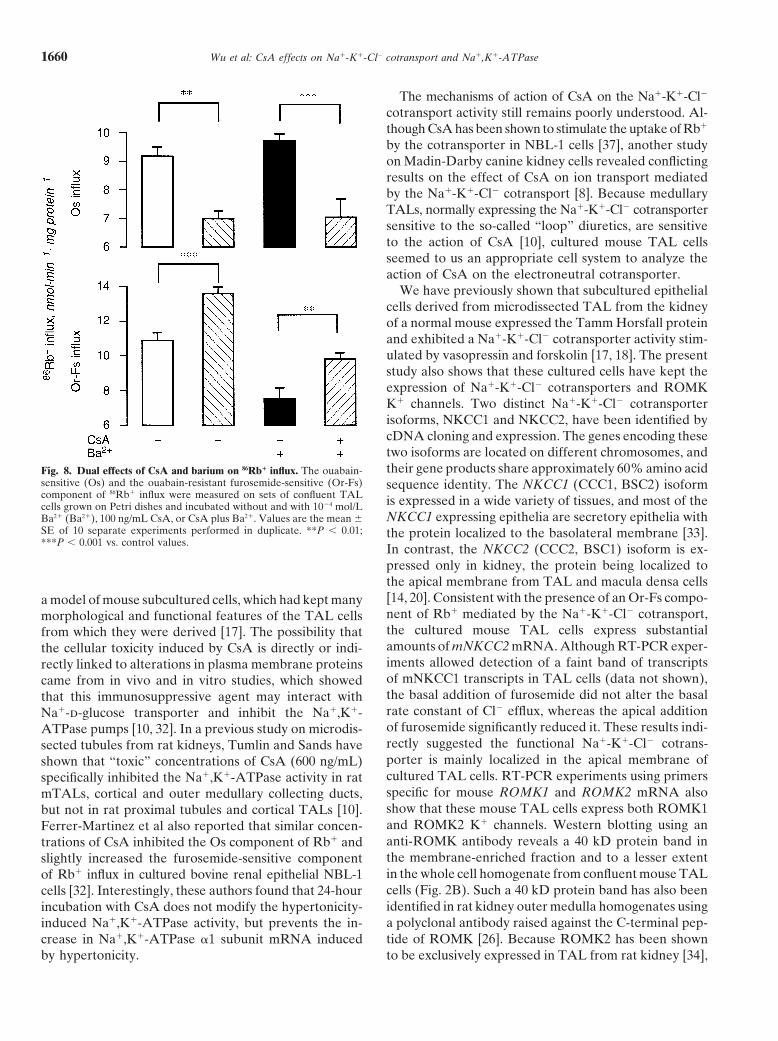
We have previously shown that subcultured epithelialcells derived from microdissected TAL from the kidneyof a normal mouse expressed the Tamm Horsfall proteinand exhibited a Na1-K1-Cl2 cotransporter activity stim-ulated by vasopressin and forskolin [17, 18]. The presentstudy also shows that these cultured cells have kept theexpression of Na1-K1-Cl2 cotransporters and ROMKK1 channels. Two distinct Na1-K1-Cl2 cotransporterisoforms, NKCC1 and NKCC2, have been identified bycDNA cloning and expression. The genes encoding thesetwo isoforms are located on different chromosomes, andtheir gene products share approximately 60% amino acidFig. 8. Dual effects of CsA and barium on 86Rb1 influx. The ouabain-
sensitive (Os) and the ouabain-resistant furosemide-sensitive (Or-Fs) sequence identity. The NKCC1 (CCC1, BSC2) isoformcomponent of 86Rb1 influx were measured on sets of confluent TAL is expressed in a wide variety of tissues, and most of thecells grown on Petri dishes and incubated without and with 1024 mol/L
NKCC1 expressing epithelia are secretory epithelia withBa21 (Ba21), 100 ng/mL CsA, or CsA plus Ba21. Values are the mean 6SE of 10 separate experiments performed in duplicate. **P , 0.01; the protein localized to the basolateral membrane [33].***P , 0.001 vs. control values. In contrast, the NKCC2 (CCC2, BSC1) isoform is ex-
pressed only in kidney, the protein being localized tothe apical membrane from TAL and macula densa cells[14, 20]. Consistent with the presence of an Or-Fs compo-a model of mouse subcultured cells, which had kept manynent of Rb1 mediated by the Na1-K1-Cl2 cotransport,morphological and functional features of the TAL cellsthe cultured mouse TAL cells express substantialfrom which they were derived [17]. The possibility thatamounts of mNKCC2 mRNA. Although RT-PCR exper-the cellular toxicity induced by CsA is directly or indi-iments allowed detection of a faint band of transcriptsrectly linked to alterations in plasma membrane proteinsof mNKCC1 transcripts in TAL cells (data not shown),came from in vivo and in vitro studies, which showedthe basal addition of furosemide did not alter the basalthat this immunosuppressive agent may interact withrate constant of Cl2 efflux, whereas the apical additionNa1-d-glucose transporter and inhibit the Na1,K1-of furosemide significantly reduced it. These results indi-ATPase pumps [10, 32]. In a previous study on microdis-rectly suggested the functional Na1-K1-Cl2 cotrans-sected tubules from rat kidneys, Tumlin and Sands haveporter is mainly localized in the apical membrane ofshown that “toxic” concentrations of CsA (600 ng/mL)cultured TAL cells. RT-PCR experiments using primersspecifically inhibited the Na1,K1-ATPase activity in ratspecific for mouse ROMK1 and ROMK2 mRNA alsomTALs, cortical and outer medullary collecting ducts,show that these mouse TAL cells express both ROMK1but not in rat proximal tubules and cortical TALs [10].and ROMK2 K1 channels. Western blotting using anFerrer-Martinez et al also reported that similar concen-anti-ROMK antibody reveals a 40 kD protein band intrations of CsA inhibited the Os component of Rb1 andthe membrane-enriched fraction and to a lesser extentslightly increased the furosemide-sensitive componentin the whole cell homogenate from confluent mouse TALof Rb1 influx in cultured bovine renal epithelial NBL-1cells (Fig. 2B). Such a 40 kD protein band has also beencells [32]. Interestingly, these authors found that 24-houridentified in rat kidney outer medulla homogenates usingincubation with CsA does not modify the hypertonicity-a polyclonal antibody raised against the C-terminal pep-induced Na1,K1-ATPase activity, but prevents the in-tide of ROMK [26]. Because ROMK2 has been showncrease in Na1,K1-ATPase a1 subunit mRNA induced
by hypertonicity. to be exclusively expressed in TAL from rat kidney [34],
Wu et al: CsA effects on Na1-K1-Cl2 cotransport and Na1,K1-ATPase 1661
the mRNA expression of both ROMK2 and ROMK1 in ways on apical K1 conductance(s). Barium added to theour subcultured mouse TAL cells raised the question of apical side of TAL grown on Petri dishes significantlythe proper origin of the cells. Indeed, the cells have been reduced the increased Or-Fs component of 86Rb1 uptakederived from single microdissected TAL segments [17], induced by CsA. Furthermore, barium did not affect thebut one cannot completely rule out the possibility of Os component of Rb1 influx in both untreated and CsA-unexpected gene expression in growing cells. treated cells (Fig. 8). These results favor the hypothesis
The rather low concentration of CsA used in this study that CsA may directly affect a barium-sensitive K1 con-was sufficient to decrease the Os component and increase ductance(s) located in the apical membrane from TALin the Or-Fs component of Rb1 uptake. In native TAL cells. A small conductance (22 pS) K1 channel with acells, inhibition of basolateral Na1,K1-ATPase leads to high open probability over a wide range of cell mem-dissipation of the Na1 gradient and subsequently inhibits brane potentials, highly selective for K1 and sensitive tothe apical entry of Cl2 [30, 35]. Maneuvers that inhibit ATP, has been identified by patch-clamp analysis in theNa1,K1-ATPase also induce the reduction of macro- apical membrane of isolated TAL of rabbit kidneys [41].scopic K1 conductance from rabbit TAL [36] and of There are lines of evidence that the ROMK channelthe apically located low-conductance K1 channels of rat isolated by expression cloning from rat renal outer me-principal collecting duct cells [37]. In the present study, dulla [42] corresponds to the native low-conductanceCsA induces an increase in Na1-K1-Cl2 cotransport ac- K1 channel identified by patch-clamp [43]. The ROMKtivity despite the concomitant decrease of Na1,K1- protein has been found to localize primarily at the apicalATPase activity. Thus, these results suggest that cultured membrane of TAL, macula densa, and distal and con-mouse TAL cells may not have the same ion transport necting tubules from rat kidney [44]. Since ROMK1 andproperties than intact mTAL from which they are de- ROMK2 are expressed in our model of cultured mouserived. It has been shown that cultured renal epithelial TAL cells, our results suggest that the CsA-stimulatedcells may not always be fully polarized. Increased Na1- and Ba21-inhibitable efflux of 86Rb1 efflux from the api-K1-Cl2 cotransporter activity has also been reported in cal side of cultured mouse TAL cells can be mediatedrapidly growing SV40-transformed cultured renal epi- by the ROMK channel(s). However, in the absence ofthelial cells [38]. We have shown that CsA enhances cell reliable antibody directed against the mouse ROMK iso-proliferation in TAL cultured cells [39]. Dong et al have forms, the antibody, used for Western blotting, does notalso reported that inhibition of Na1,K1-ATPase by oua- allow us to define with certainty which ROMK proteinbain stimulated Na1-K1-Cl2 cotransport activity in is detected in cultured TAL cells. As Ba21 reduced theSV40-transformed rabbit nonpigmented ciliary epithelial Or-Fs component of Rb1 influx induced by CsA withoutcells [40]. Although the transport properties of ciliary any changes in the decrease of the Os component ofcells may differ than those of TAL cells, it cannot be Rb1 influx, it is tempting to speculate that CsA mayexcluded that noticeable differences occur in the regula- have a direct action on the apical K1 channel from TALtion of the Na1-K1-Cl2 cotransporter in cultured TAL cells, putatively ROMK, to increase the apical recycling(or ciliary) cells as compared with their native counter- of K1 and thereby stimulate the activity of the Na1-parts. The rapid action of CsA observed during the short K1-Cl2 cotransport. However, blockade of apical K1
incubation period of four minutes also strongly suggest channels by Ba21 has been shown to inhibit ion transportthat the increased Na1-K1-Cl2 cotransport and de- completely in microperfused TAL [45]. Thus, it cannotcreased Na1,K1-ATPase activities is probably due to a be excluded that CsA may directly stimulate the electro-direct action of CsA on the membranous transporter neutral Na1-K1-Cl2 cotransporter, leading to an in-rather than a genomic action. Consistent with the in- crease in intracellular K1 and an increase in K1 effluxcrease of the Or-Fs component of Rb1 uptake, CsA by apical Ba21-sensitive K1 channels.rapidly increases the efflux of 36Cl2 from the basal side In conclusion, the results from this study suggest thatof TAL cells grown on filters. The Cl2 channel blocker CsA can rapidly stimulate the Na1-K1-Cl2 cotransportNPPB prevented the increase in Cl2 efflux induced by activity in cultured mouse TAL cells, but the direct mech-CsA. This raises the question of how to know whether anism of action of this immunosuppressive drug on thethe immunosuppressive agents exerted a direct action on electroneutral cotransporter and/or apical K1 channelsbasolaterally located Cl2 conductance and/or primarily still remains to be determined.activated the Na1-K1-Cl2 cotransporter by anothermechanism. Apical addition of barium, a potent inhibitorof the ATP-sensitive K1 channels [41], almost com- ACKNOWLEDGMENTSpletely prevented the CsA-dependent increase of apical
This work was supported by a grant from the Taiwan NMRP66386Rb1 efflux and also partially prevented the increase inand NMRP804H and in part by the INSERM (France). K.-C. Peng
basal 36Cl2 efflux from CsA-treated TAL cells. These holds an INSERM postdoctoral (Port Vert) fellowship supported bythe Conseil Regional d’Ile de France.results indirectly suggest that CsA may interact in some
Wu et al: CsA effects on Na1-K1-Cl2 cotransport and Na1,K1-ATPase1662
Reprint requests to Mai-Szu Wu, M.D., Division of Nephrology, 20. Igarashi P, Whyte DA, Li K, Nagami GT: Cloning and kidneycell-specific activity of the promoter of the murine renal Na-K-ClChang Gung Memorial Hospital, 199, Tun Hwa North Road, Taipei,cotransporter gene. J Biol Chem 271:9666–9674, 1996Taiwan.
21. Yang CW, Vlassara H, Peten EP, He CJ, Striker GE, StrikerE-mail: [email protected]: Advanced glycation end products up-regulate gene expressionfound in diabetic glomerular disease. Proc Natl Acad Sci USA
REFERENCES 91:9436–9440, 199422. Bradford MM: A rapid and sensitive method for the quantitation1. Cohen DJ, Loertscher R, Rubin MF, Tilney NL, Carpenter CB,
of microgram quantities of protein utilizing the principle of protein-Strom TB: Cyclosporine: A new immunosuppressive agent fordye binding. Anal Biochem 72:248–254, 1976organ transplantation. Ann Intern Med 101:667–682, 1984
23. Lowry OH, Rosebrough NJ, Farr AL, Kandall JR: Protein2. Myers BD, Sibley R, Newton L, Tomlanovich SJ, Boshkos C,measurements with the folin phenol reagent. J Biol Chem 193:265–Stinson E, Luetscher JA, Whitney DJ, Krasny D, Coplon NS:275, 1951The long-term course of cyclosporine-associated chronic nephropa- 24. Venglarik CJ, Bridges RJ, Frizzell RA: A simple assay forthy. Kidney Int 33:590–600, 1988 agonist-regulated Cl and K conductances in salt- secreting epithe-3. Barros EJ, Boim MA, Ajzen H, Ramos OL, Schor N: Glomerular lial cells. Am J Physiol 259:C358–C364, 1990hemodynamics and hormonal participation on cyclosporine neph- 25. Matthews JB, Awtrey CS, Madara JL: Microfilament-dependent
rotoxicity. Kidney Int 32:19–25, 1987 activation of Na1/K1/2Cl2 cotransport by cAMP in intestinal epi-4. Myers BD, Ross J, Newton L, Luetscher J, Perlroth M: thelial monolayers. J Clin Invest 90:1608–1613, 1992
Cyclosporine-associated chronic nephropathy. N Engl J Med 311: 26. Mennitt PA, Wade JB, Ecelbarger CA, Palmer LG, Frindt G:699–705, 1984 Localization of ROMK channels in the rat kidney. J Am Soc
5. Ciresi DL, Lloyd MA, Sandberg SM, Heublein DM, Edwards Nephrol 8:1823–1830, 1997BS: The sodium retaining effects of cyclosporine. Kidney Int 27. Schlatter E, Greger R: cAMP increases the basolateral Cl2-41:1599–1605, 1992 conductance in the isolated perfused medullary thick ascending
6. Adu D, Turney J, Michael J, McMaster P: Hyperkalaemia in limb of Henle’s loop of the mouse. Pflugers Arch 405:367–376,cyclosporin-treated renal allograft recipients. Lancet 2:370–372, 19851983 28. Molony DA, Reeves WB, Hebert SC, Andreoli TE: ADH in-
7. Heering P, Degenhardt S, Grabensee B: Tubular dysfunction creases apical Na1, K1, 2Cl2 entry in mouse medullary thick as-following kidney transplantation. (editorial) Nephron 74:501–511, cending limbs of Henle. Am J Physiol 252:F177–F187, 19871996 29. Kim GH, Ecelbarger CA, Mitchell C, Packer RK, Wade JB,
8. Deppe CE, Heering PJ, Tinel H, Kinne-Saffran E, Grabensee Knepper MA: Vasopressin increases Na-K-2Cl cotransporter ex-B, Kinne RK: Effect of cyclosporine A on Na1/K(1)-ATPase, pression in thick ascending limb of Henle’s loop. Am J PhysiolNa1/K1/2Cl2 cotransporter, and H1/K(1)-ATPase in MDCK cells 276:F96–F103, 1999and two subtypes, C7 and C11. Exp Nephrol 5:471–480, 1997 30. Hebert SC, Culpepper RM, Andreoli TE: NaCl transport in
9. Ferrer-Martinez A, Felipe A, Barcelo P, Casado FJ, Ballarin mouse medullary thick ascending limbs. I. Functional nephronJ, Pastor-Anglada M: Effects of cyclosporine A on Na,K-ATPase heterogeneity and ADH-stimulated NaCl cotransport. Am J Phys-expression in the renal epithelial cell line NBL-1. Kidney Int iol 241:F412–F431, 198150:1483–1489, 1996 31. Wangemann P, Wittner M, Di Stefano A, Englert HC, Lang
10. Tumlin JA, Sands JM: Nephron segment-specific inhibition of HJ, Schlatter E, Greger R: Cl2-channel blockers in the thickNa1/K1-ATPase activity by cyclosporin A. Kidney Int 43:246–251, ascending limb of the loop of Henle: Structure activity relationship.1993 Pflugers Arch 407:S128–S141, 1986
11. Wheatley HC, Datzman M, Williams JW, Miles DE, Hatch 32. Ferrer-Martinez A, Casado FJ, Felipe A, Pastor-Anglada M:FE: Long-term effects of cyclosporine on renal function in liver Regulation of Na1,K1-ATPase and the Na1/K1/Cl2 co-transportertransplant recipients. Transplantation 43:641–647, 1987 in the renal epithelial cell line NBL-1 under osmotic stress. Bio-
12. Gnutzmann KH, Hering K, Gutsche HU: Effect of cyclosporine chem J 319(Pt 2):337–342, 1996on the diluting capacity of the rat kidney. Clin Nephrol 25(Suppl 33. Xu JC, Lytle C, Zhu TT, Payne JA, Benz EJ, Forbush BR:1):S51–S56, 1986 Molecular cloning and functional expression of the bumetanide-
13. Propper DJ, Whiting PH, Mackay J, Catto GR: Glomerulotubu- sensitive Na-K-Cl cotransporter. Proc Natl Acad Sci USA 91:2201–lar function in long-term renal allograft recipients: A comparison of 2205, 1994conventional therapy with cyclosporine. Transplantation 50:72–75, 34. Hebert SC: Roles of Na-K-2Cl and Na-Cl cotransporters and1990 ROMK potassium channels in urinary concentrating mechanism.
14. Kaplan MR, Plotkin MD, Lee WS, Xu ZC, Lytton J, Hebert Am J Physiol 275:F325–F327, 1998SC: Apical localization of the Na-K-Cl cotransporter, rBSC1, on 35. Burg MB, Green N: Function of the thick ascending limb ofrat thick ascending limbs. Kidney Int 49:40–47, 1996 Henle’s loop. Am J Physiol 224:659–668, 1973
15. Greger R, Schlatter E, Lang F: Evidence for electroneutral 36. Greger RM: Couple transport in nephron, in Mechanisms andsodium chloride cotransport in the cortical thick ascending limb Pathophysiology, edited by Hoshi T, Tokyo, Miura Medical Res.of Henle’s loop of rabbit kidney. Am J Physiol 271:F588–F594, Foundation, 1984, pp 96–1181996 37. Wang WH, Geibel J, Giebisch G: Mechanism of apical K1 channel
16. Reeves WB, Winters CJ, Zimniak L, Andreoli TE: Properties modulation in principal renal tubule cells: Effect of inhibition ofand regulation of medullary thick limb basolateral Cl2 channels. basolateral Na1-K1-ATPase. J Gen Physiol 101:673–694, 1993Kidney Int 53(Suppl 65):S24–S28, 1998 38. Vandewalle A, Vuillemin T, Teulon J, Baudouin B, Wahbe
17. Wu MS, Bens M, Cluzeaud F, Vandewalle A: Role of F-actin F, Bens M, Cassingena R, Ronco P: K1 fluxes mediated by Na1-in the activation of Na1-K1-Cl2 cotransport by forskolin and vaso- K1-Cl2 cotransport and Na1-K1-ATPase pumps in renal tubulepressin in mouse kidney cultured thick ascending limb cells. J cell lines transformed by wild-type and temperature-sensitiveMembr Biol 142:323–336, 1994 strains of Simian virus 40. J Cell Physiol 154:466–477, 1993
18. Wu MS, Yang CW, Bens M, Yu HM, Huang JY, Wu CH, Huang 39. Wu MS, Yu HM, Hong JJ, Lai BC, Huang CC, VandewalleCC, Vandewalle A: Cyclosporin inhibits nitric oxide production A: Cyclosporine, but not FK 506 and rapamycin, enhances cellin medullary ascending limb cultured cells. Nephrol Dial Transplant proliferation in mouse medullary thick ascending cultured cells.13:2814–2820, 1998 Transplant Proc 30:3565–3566, 1998
19. Chomczynski P: A reagent for the single-step simultaneous isola- 40. Dong J, Delamere NA, Coca-Prados M: Inhibition of Na1-K1-tion of RNA, DNA and proteins from cell and tissue samples. ATPase activates Na1-K1-2Cl2 cotransporter activity in cultured
ciliary epithelium. Am J Physiol 266:C198–C205, 1994Biotechniques 15:532–534, 536–537, 1993
Wu et al: CsA effects on Na1-K1-Cl2 cotransport and Na1,K1-ATPase 1663
41. Wang WH, White S, Geibel J, Giebisch G: A potassium channel inhibits activity of cloned renal K1 channel, ROMK1. Am J Physiol271:F588–F594, 1996in the apical membrane of rabbit thick ascending limb of Henle’s
loop. Am J Physiol 258:F244–F253, 1990 44. Xu JZ, Hall AE, Peterson LN, Bienkowski MJ, Eessalu TE,Hebert SC: Localization of the ROMK protein on apical mem-42. Ho K, Nichols CG, Lederer WJ, Lytton J, Vassilev PM, Kana-
zirska MV, Hebert SC: Cloning and expression of an inwardly branes of rat kidney nephron segments. Am J Physiol 273:F739–F748, 1997rectifying ATP-regulated potassium channel. Nature 362:31–38,
1993 45. Hebert SC, Andreoli TE: Control of NaCl transport in the thickascending limb. Am J Physiol 246:F745–F756, 198443. Macica CM, Yang Y, Hebert SC, Wang WH: Arachidonic acid