humoral and cell-mediated immunity to pandemic h1n1 influenza in a canadian cohort one year...
TRANSCRIPT

Humoral and Cell-Mediated Immunity to Pandemic H1N1Influenza in a Canadian Cohort One Year Post-Pandemic:Implications for VaccinationLisa E. Wagar1, Laura Rosella2,3, Natasha Crowcroft2,3,4, Beth Lowcock2, Paulina C. Drohomyrecky1, Julie
Foisy2, Jonathan Gubbay2,4, Anu Rebbapragada2,4, Anne-Luise Winter2, Camille Achonu2, Brian J. Ward5,
Tania H. Watts1*
1 Department of Immunology, University of Toronto, Toronto, Ontario, Canada, 2 Public Health Ontario, Toronto, Ontario, Canada, 3 Dalla Lana School of Public Health,
University of Toronto, Toronto, Ontario, Canada, 4 Department of Laboratory Medicine and Pathobiology, University of Toronto, Toronto, Ontario, Canada, 5 Research
Institute of the McGill University Health Centre, Montreal, Quebec, Canada
Abstract
We evaluated a cohort of Canadian donors for T cell and antibody responses against influenza A/California/7/2009 (pH1N1)at 8-10 months after the 2nd pandemic wave by flow cytometry and microneutralization assays. Memory CD8 T cellresponses to pH1N1 were detectable in 58% (61/105) of donors. These responses were largely due to cross-reactive CD8 Tcell epitopes as, for those donors tested, similar recall responses were obtained to A/California 2009 and A/PR8 1934 H1N1Hviruses. Longitudinal analysis of a single infected individual showed only a small and transient increase in neutralizingantibody levels, but a robust CD8 T cell response that rose rapidly post symptom onset, peaking at 3 weeks, followed by agradual decline to the baseline levels seen in a seroprevalence cohort post-pandemic. The magnitude of the influenza-specific CD8 T cell memory response at one year post-pandemic was similar in cases and controls as well as in vaccinatedand unvaccinated donors, suggesting that any T cell boosting from infection was transient. Pandemic H1-specific antibodieswere only detectable in approximately half of vaccinated donors. However, those who were vaccinated within a few monthsfollowing infection had the highest persisting antibody titers, suggesting that vaccination shortly after influenza infectioncan boost or sustain antibody levels. For the most part the circulating influenza-specific T cell and serum antibody levels inthe population at one year post-pandemic were not different between cases and controls, suggesting that natural infectiondoes not lead to higher long term T cell and antibody responses in donors with pre-existing immunity to influenza.However, based on the responses of one longitudinal donor, it is possible for a small population of pre-existing cross-reactive memory CD8 T cells to expand rapidly following infection and this response may aid in viral clearance andcontribute to a lessening of disease severity.
Citation: Wagar LE, Rosella L, Crowcroft N, Lowcock B, Drohomyrecky PC, et al. (2011) Humoral and Cell-Mediated Immunity to Pandemic H1N1 Influenza in aCanadian Cohort One Year Post-Pandemic: Implications for Vaccination. PLoS ONE 6(11): e28063. doi:10.1371/journal.pone.0028063
Editor: Ralph Tripp, University of Georgia, United States of America
Received September 9, 2011; Accepted October 31, 2011; Published November 23, 2011
Copyright: � 2011 Wagar et al. This is an open-access article distributed under the terms of the Creative Commons Attribution License, which permitsunrestricted use, distribution, and reproduction in any medium, provided the original author and source are credited.
Funding: This research was supported by grants MOP-74492 and TPA-90194 from the Canadian Institutes of Health Research (CIHR) to T.H.W. and by a contractfrom the Public Health Agency of Canada to N.C. T.H.W. holds the Sanofi Pasteur Chair in Human Immunology at the University of Toronto. L.E.W. was funded by aCIHR doctoral award. The funders had no role in study design, data collection and analysis, decision to publish, or preparation of the manuscript.
Competing Interests: The authors have read the journal’s policy and have the following conflicts: Jonathan Gubbay has received grants from GlaxoSmithKlineInc. and Hoffmann-La Roche Ltd Inc. B.J.W. is co-investigator on a CIHR grant with the VP research of GSK; he is on the Scientific advisory board of Medicago andhas served on ad hoc advisory committees (clinical and scientific) for all of the major vaccine manufacturers. This does not alter the authors’ adherence to all thePLoS ONE policies on sharing data and materials. Please note: Although the GSK vaccine was mentioned in this study, they did not provide vaccine for the study.The authors did not vaccinate the cohort but asked the participants to report on whether they had received the vaccine, which in the season reported wasavailable through a public vaccine program, using vaccine purchased by the Canadian Government from GSK. Similarly the longitudinal donor in the study whoreports 2 vaccinations obtained these vaccines through the public mechanism, not through the study.
* E-mail: [email protected]
Introduction
A novel swine-origin H1N1 influenza virus (pH1N1) emerged in
North America in mid-April of 2009, resulting in widespread
infection [1,2]. The infectious behavior of the novel 2009 strain met
pandemic criteria set by the World Health Organization in mid-
June, 2009. A second wave of infection with the same strain
occurred in the autumn of 2009. By August 2010, influenza
outbreaks had subsided and influenza incidence in the population
had returned to normal seasonal rates. Contrary to typical seasonal
influenza, attack rates were observed to be highest in younger
people [1,3,4]. However, infection in older age groups resulted in
more severe illness and increased mortality rates compared to the
general population [3,5,6]. It has been suggested that older people
who had been exposed to an H1N1 influenza from the early 20th
century may have been protected by pre-existing cross-reactive
antibodies [7,8], as strains originating from the 1918 pandemic are
antigenically similar to the 2009 strain [9]. T cells produced against
pH1N1 2009 are able to respond to challenge with the 1918
pandemic H1N1 strain [10] and memory T cells generated against
past seasonal infections can respond to pH1N1 challenge [11–13],
suggesting that T cell cross-reactivity exists in primed hosts.
While it has been established that influenza-specific B cell
memory can be very long-lived [8,14], there are limited data on
PLoS ONE | www.plosone.org 1 November 2011 | Volume 6 | Issue 11 | e28063

the magnitude and persistence of antibody and T cell responses to
influenza post-pandemic. To address this, we analyzed humoral and
T cell-mediated immunity to pH1N1 in a cross-sectional cohort of
the Toronto population, approximately 8-10 months post 2009
pandemic as well as before, during and after infection of one donor
from whom a series of longitudinal samples was available.
Materials and Methods
Ethics statementEthics approval was granted by the Research Ethics Board of the
University of Toronto. All subjects gave written informed consent.
Study design and sample collectionIndividuals who were at least 18 years of age were invited to
participate in a case/control or a seroprevalence cohort study.
Individuals self-reported vaccination in all study groups. The
vaccine they would have received through the publicly funded
Canadian vaccine program was the GlaxoSmithKline monova-
lent, inactivated, split-virion pandemic H1N1 influenza vaccine
containing 3.75 mg hemagglutinin (HA) with AS03 adjuvant
(unadjuvanted vaccine was also available but was only given to
pregnant women and young children). Donors reported vaccina-
tion with the pandemic H1N1 vaccine from October 2009 to
January 2010.
Case/control cohort. Case/control donors (the Ontario
population of a previous study [15]) were recruited during early
autumn of 2009. All participants had medically attended influenza-
like illness (ILI) and were subsequently tested for influenza A/
California7/2009-like strains by PCR using nasopharyngeal swabs,
performed from April to November 2009, largely prior to vaccine
availability. Case/control volunteers provided blood for influenza-
specific antibody and T cell testing in July-August of 2010,
approximately 8–10 months after initial PCR testing for pH1N1.
Case participant ages ranged from 19–76, with a mean age of 44;
control participants were aged 29–74, with a mean age of 51.
Seroprevalence cohort. A seroprevalence study was
undertaken beginning August 2009 [16]; Toronto residents were
recruited through an advertising/email/web-based campaign; those
who completed an online questionnaire were invited to give a blood
sample. From April-June 2010, participants were asked to provide a
second blood sample and complete a questionnaire on risk factors,
health status (such as ILI) and vaccination history. The mean
participant age for this cohort was 50, with an age range of 24-76.
Longitudinal analysis. Longitudinal samples were available
from one subject (who was not part of the case/control or
seroprevalence studies), a 55-year old male with no co-morbid
conditions, allergies, or relevant medications who reported an
influenza-like illness in mid-June 2009 and was confirmed PCR
positive for pH1N1. He exhibited general malaise with a modest
fever, which began 4–5 days after onset of illness and a mild-
moderate dry cough that began approximately one week post-onset.
Peripheral blood mononuclear cells (PBMC) had been stored for
this individual over one year prior to the onset of symptoms and
further samples were obtained at several times during and after
infection. This donor was subsequently vaccinated (through the
publically available Canadian vaccine program) for influenza
approximately 20 weeks (GSK AS03-adjuvanted monovalent
vaccine) and 17 months (seasonal trivalent inactivated vaccine,
including A/California/07/2009) post-infection with pH1N1.
Viruses and mediaA/California/07/2009-like virus (H1N1) was isolated from a
nasopharangeal swab by Public Health Ontario laboratories and
confirmed at the National Microbiology Laboratory using a
hemagglutination inhibition (HAI) assay. Influenza A/Puerto
Rico/8/34 (PR8) was grown in embryonated chicken eggs. Dr.
Pamela S. Ohashi, University Health Network, Toronto, kindly
provided the Lymphocytic choriomeningitis virus (LCMV)
Armstrong. Complete PBMC medium was RPMI 1640 HEPES
modification (Sigma-Aldrich), 10% fetal bovine serum (FBS,
Invitrogen), 1% non-essential amino acids (Invitrogen), 100 U/
mL penicillin, 0.1mg/mL streptomycin, 1 mM L-glutamine,
1 mM sodium pyruvate, and 0.1% b-mercaptoethanol.
pH1N1-specific antibody detection in human serumSerum antibodies were detected by microneutralization (MN),
adapted from the World Health Organization method [17] or
HAI, adapted from the World Health Organization method [18].
An ELISA similar to that recently reported by Mavrouli and
colleagues [19] was optimized to detect antibodies binding to
recombinant pandemic H1 hemagglutinin. Briefly, wells of
enzyme immunoassay/RIA flat-bottom 96-well plates (Costar)
were coated with 0.4 mg/well recombinant HA derived from
strains H1N1 A/California/07/2009 or A/California/04/2009,
H1N1 A/Brisbane/59/2007, H3N2 A/Brisbane/10/2007, or
H9N2 A/Hong Kong/1073/1999 (all from Protein Sciences
Corporation) and blocked with 1% bovine serum albumin.
Human sera were plated in serial two-fold dilutions and incubated
for 2 h at room temperature. HRP-conjugated anti-human IgG
(Sigma, St. Louis, MO) diluted 1/6000 was added for 1 h at room
temperature followed by addition of the substrate for visualization.
Plates were read with a Multiskan EX spectrometer (Thermo
Electron) at 405 nm. Optical density of samples was corrected for
background based on the OD of uncoated, serum-treated wells.
Titers reported herein were determined by microneutralization
unless otherwise noted.
Assessment of T cell responses to live influenza viruschallenge
PBMC were isolated from whole blood by Ficoll density
gradient separation and frozen in FBS with 20% DMSO (Sigma).
Cryopreserved PBMC were thawed in complete medium, plated
at 1 million cells per well in a 96-well round-bottom microtitre
plate and infected with 1 hemagglutination unit (HAU) of A/
California/7/2009, 5 HAU of PR8, or 2000 PFU of LCMV
Armstrong per well and incubated for 18 hours at 37uC, 5% CO2.
Brefeldin A (BD Biosciences) was added for the final 6 hours of
stimulation at 1.5 mL/mL.
Flow cytometryAnti-human monoclonal antibodies used were CD27, CD8, CD3,
IFNc, CD45RA, and TNFa (eBioscience), granzyme B (Invitrogen),
and CD107a (BD Biosciences). Anti-CD107a was added for the final
6 hours of stimulation. For detecting intracellular cytokines, PBMC
were surface stained, fixed and permeabilized using Cytokfix/
Cytoperm buffers (BD Biosciences), washed, and stained intracellu-
larly for 30 minutes at 4uC. Data were collected using an LSR II (BD
Biosciences). Background fluorescence was established with fluores-
cence minus one (FMO) controls except CD107a, for which an
isotype control was used. Approximately 16105 to 6.56105 total T
cell events were collected per condition per donor.
Data AnalysisFlowJo (TreeStar) was used to gate and analyze flow cytometry
data. Plotting and statistical analyses were performed with Prism
(GraphPad) software.
Immune Memory to Pandemic H1N1 Influenza
PLoS ONE | www.plosone.org 2 November 2011 | Volume 6 | Issue 11 | e28063

Results
Whole virus stimulation assay to detect T cell responsesto pH1N1
To identify influenza-specific memory T cells in a Toronto
population post-pandemic, we used flow cytometry analysis
following whole virus stimulation of PBMC. Flow cytometry was
chosen over ELiSPOT as it offered the advantage of multi-
parameter analysis. Influenza-responsive CD8 and CD4 T cells
were identified on the basis of their production of IFNc, after 18hrs
stimulation (Figure 1A). As 18h is too short a time frame for the cells
to proliferate, this restimulation assay reports the frequency of
influenza-specific memory T cells circulating in the individual at the
time of PBMC harvest. To rule out non-specific innate immune
responses to viral stimulation ex vivo, we stimulated PBMC with a
rodent virus LCMV to which we did not expect recall responses and
for the most part, the results were similar to those obtained with
unstimulated controls (Figure 1B). By analyzing the product of the
median fluorescence intensity (MFI) and the frequency of T cells
with IFNc staining, the response value reported takes into account
both the proportion of cells responding to influenza virus and the
amount of IFNc per cell. We defined a cutoff for CD8 T cell
responses based on these parameters. A donor’s response to
influenza was considered positive if: a) the frequency of IFNc+
CD8 T cells was greater than that of stimulation controls, b) the
frequency of IFNc+ cells was greater than 0.01% of the total CD8 T
cell pool, and c) the product of the MFI and the frequency of IFNc+
CD8 T cells was at least two-fold above the control cultures’ values.
IFNc staining of T cell populations for a representative CD8 T cell
pH1N1 non-responder, weak responder, and strong responder is
Figure 1. Detection of influenza-responsive CD8 T cells by multicolour flow cytometry. Total PBMC were stimulated for 18 hours withpH1N1 influenza, or as a control, with LCMV Armstrong, or left unstimulated and then assessed for IFNc production by intracellular cytokine stainingand flow cytometry. Gates are based on fluorescence minus one controls. (A) Representative gating used to identify IFNc+ CD8 T cells from totalPBMC. (B) Sample non-responder, weak responder, and strong responder to pH1N1 identified in the Toronto cohort 8-10 months post-pandemic;positive versus non-responder is defined in the results. A representative ‘‘weak’’ responder was arbitrarily chosen from the bottom third of positiveresponses whereas the ‘‘strong’’ responder was from the top third of responders.doi:10.1371/journal.pone.0028063.g001
Immune Memory to Pandemic H1N1 Influenza
PLoS ONE | www.plosone.org 3 November 2011 | Volume 6 | Issue 11 | e28063

shown in Figure 1B and a summary of results for each cohort is
described in Table 1.
Frequency and functional analysis of T cell memory toinfluenza in the Toronto cohort
Based on the above analysis, we found detectable influenza-
specific CD8 T cell responses (IFNc positive cells) in over half of all
donors, with 18% of subjects tested showing responses greater than
0.05% (Figure 2A). The mean percentage of IFNc+ CD8 T cells,
after background correction, was 0.038% in responders, compared
to a mean of 0.015% in non-responders (medians 0.028% and
0.006% respectively) (Figure 2A). Detectable CD4 T cell responses
were generally much lower than those of CD8 T cells and showed
higher background (mean CD4 IFNc+ = 0.015% after back-
ground correction). The level of the CD8 response declined
slightly with age (Figure 2B).
In addition to measuring IFNc, the major cytokine produced
upon restimulation of virus-specific CD8 T cells, we analyzed cells
for the simultaneous production of TNF as well as for expression
of granzyme B (required for killing of virus-infected cells) and
CD107a (a marker of degranulation) for donors with detectable T
cell responses. Almost half of influenza-specific CD8 T cells
produced only IFNc However, a significant proportion of the T
cells also made granzyme B (Figure 2C) and there were also small
but detectable populations of T cells that produced other
combinations of effector molecules. In other viral infections, the
presence of multifunctional CD4/CD8 T cells correlates with viral
control [20,21]. Moreover, these multifunctional T cells have been
reported to produce larger amounts of cytokines per cell [22]. This
was also the case for the memory T cells specific for influenza in
our study, as donors with the highest CD8 recall response (MFI x
frequency) were those with the most multifunctional T cells
(Figure 2D). The prevalence of multifunctional T cells did not
appreciably change as a function of donor age (data not shown).
Cases and controls have an indistinguishable frequencyof influenza-specific memory CD8 T cells post-pandemic
A PCR test for pH1N1 from nasopharyngeal swabs was
performed on all case/control participants. In this cohort, we
found no significant difference between cases and controls for the
CD8 T cell responses to pH1N1 in PBMC samples collected 8–10
months after PCR testing (Figure 2E). As with the total T cell
responder population, both PCR-confirmed cases and controls with
increased IFNc+ CD8 T cell populations had a higher frequency of
responding cells with multiple effector functions, although no
observable difference was detected between the proportions of
multifunctional T cells in cases versus controls (Figure 2F).
The T cell response against pH1N1 is greatly enhancedafter the onset of PCR-confirmed pH1N1 illness andgradually returns to pre-pandemic baseline
As T cell responses were measured approximately 8–10 months
post-pandemic, it was not possible from the cohort analysis to
know how large the response might have been shortly after
exposure. However, the availability of longitudinal PBMC samples
from one PCR-confirmed pandemic H1N1 case, before and after
infection, offered the opportunity to follow an acute T cell
response to influenza infection in a human subject. Prior to
infection, this donor had a weak positive CD8 T cell response to
pH1N1 (Figure 3A). By 10 days post-onset of illness, there was a
dramatic increase in IFNc-producing CD8 T cells. The majority
of these cells co-expressed granzyme B as well as CD107, a
phenotype associated with T cells’ ability to kill virus-infected cells.
The peak CD8 T cell response was attained approximately 3
weeks after the initial onset of illness, with a modest decline
observed by day 78. Cells expressing all 3 functional markers were
maintained and the total IFNc+ CD8 T cell response remained
above the baseline measurement even two and a half months post-
infection (Figure 3A). However, by day 700, the frequency of CD8
T cells responding to pH1N1 had returned to pre-infection
baseline frequencies and was at a level similar to that of the cross
sectional cohort at one year post-pandemic. At these later time
points, multifunctional T cells were largely absent in both the
cohort and the longitudinal donor.
In the longitudinal samples, we observed differential kinetics of
expansion for influenza-specific CD8 T cell memory responses
delineated on the basis of their expression of two surface markers,
CD45RA and CD27 (Figure 3B). CD27 is a costimulatory
receptor on T cells that is lost from effector memory cells in some
viral infections and whose loss is often associated with a more
Table 1. Donor self-reported information and pandemic-specific antibody and CD8 T cell responses in the Toronto cohorts.
No. donors(n = 105)
Meanage
Receivedpandemicmonovalentvaccine
DetectablepandemicH1N1 titersautumn 2009a
DetectablepandemicH1N1 titerssummer 2010
CD8 T cellresponsedetected
All donors All 68b 50 41c (61) 10 (15) 23 (34) 44 (65)
Seroprevalence cohort Vaccinated 41 54 41 (100) 4 (10) 19 (46) 25 (61)
Unvaccinated 26 43 0 (0) 6 (23) 4 (15) 18 (69)
All 20 44 5 (25) NA 15 (75) 10 (50)
Confirmed cases Vaccinated 5 55 5 (100) NA 5 (100) 3 (60)
Case/control cohort Unvaccinated 15 40 0 (0) NA 10 (67) 7 (47)
All 17 51 9 (53) NA 4 (24) 7 (41)
Controls Vaccinated 9 50 9 (100) NA 3 (33) 4 (44)
Unvaccinated 8 52 0 (0) NA 1 (13) 3 (38)
aa titer of 40 was used as the cutoff for seropositivity.bone donor did not provide vaccination history and was therefore not included in the vaccination stratification and subsequent vaccine recipient frequency.cnumber of donors within category; parentheses indicates percentage.doi:10.1371/journal.pone.0028063.t001
Immune Memory to Pandemic H1N1 Influenza
PLoS ONE | www.plosone.org 4 November 2011 | Volume 6 | Issue 11 | e28063

Figure 2. T cell analysis in the Toronto seroprevalence and case/control cohorts. (A) Bin separation of IFNc responses in CD8 and CD4 Tcells specific to pH1N1 stimulation. Frequencies have been corrected for background IFNc production in LCMV and unstimulated control cultures. (B)Spearman correlation between pH1N1-responding CD8 T cells and donor age. (C) Combinations of effector molecule expression of IFNc+ CD8 T cellsfrom the responder subset. P values above the bars indicate the level of statistical significance compared to all other bars as determined by ANOVAand Tukey test. (D) Spearman correlation between the CD8 T cell response to pH1N1 and the frequency of responding cells with multiple effectorfunctions. (E) CD8 T cell response in case and control subjects. Groups were compared using a nonparametic Mann-Whitney test. (F) Spearmancorrelation for pH1N1 response and frequency of CD8 T cells with multiple effector functions in cases and controls.doi:10.1371/journal.pone.0028063.g002
Immune Memory to Pandemic H1N1 Influenza
PLoS ONE | www.plosone.org 5 November 2011 | Volume 6 | Issue 11 | e28063

differentiated effector cell phenotype [23–25]. CD45RA is a marker
that is normally present on naı̈ve T cells, disappears upon their
differentiation to effector and memory T cells, but can reappear on
more terminally differentiated effector cells [23–27]. The
CD45RA2CD272 influenza-specific T cell population was the first
response to peak, and based on the absence of CD27, likely
represented reactivation of a pre-existing effector/memory popu-
lation. A population of effector-like T cells (CD45RA+CD272)
transiently increased before the final, largest wave of influenza-
specific T cells emerged. The last wave of influenza-specific T cells
expressed CD27, suggesting expansion from a less differentiated
memory T cell pool. These CD45RA2CD27+ CD8 T cells were the
most persistent phenotype in the longitudinal donor, similar to those
detected in the Toronto cohort post-pandemic (data not shown). We
also observed three distinct influenza-specific CD4 T cell subsets,
although their kinetics of expansion were not distinguishable
(Figure 3C).
We tested sera from the longitudinal donor and found that
pandemic influenza-specific antibodies were low, except for weakly
positive titers detected by MN and ELISA (but not HAI) between
2–3 weeks post-infection (Figure 3D). Two subsequent vaccina-
tions containing pH1N1 antigen did not result in long-term
persisting protective titers against pH1N1.
Similar CD8 recall T cell responses to H1N1 from 1934and 2009 implies cross-reactive T cell responses
CD8 T cell epitopes are mainly derived from internal viral
proteins and are from the most conserved part of the influenza
virus. Indeed the sequences of influenza NP and M proteins have
changed little between the 1918 and 2009 pandemic strains and T
cells specific for A/California 2009 are cross-reactive with the
1918 H1N1 pandemic strain [10]. Therefore, it was likely that a
significant proportion of the T cell responses we observed to A/
California 2009 would cross-react with other H1N1 strains. To
test this hypothesis we compared the responses of a subset of
donors to A/California 2009 and a 1934 virus, A/PR8/34. In the
25 donors tested with the A/PR8/34 virus, the proportion of
influenza-responding CD8 T cells was indistinguishable from the
pH1N1 strain (Figure 4), consistent with the possibility that a
significant proportion of the CD8 T cells that respond to A/
California 2009 are cross-reactive.
Vaccination after infection with pH1N1 results in higherpersisting antibody titers but not T cell responses
Antibody responses to pH1N1 in the seroprevalence and the
case/control cohort are summarized in Table 1. Interestingly, as
previously noted [16] among unvaccinated but laboratory
confirmed infected donors, one third were seronegative for
H1N1 A/California/7/2009 by the summer of 2010 as measured
by MN (Table 1), HAI, and ELISA (data not shown). However,
with the exception of the longitudinal donor, all of the donors with
a confirmed infection who were also vaccinated were seropositive
for influenza A H1N1 2009 antibodies by summer of 2010
(Table 1). For the seroprevalence cohort, 10% of vaccinated
donors were seropositive ($1:40) for pH1N1 in the autumn of
2009 (prior to vaccination), increasing to 46% in the summer of
Figure 3. Acute and persisting antibody and memory T cell responses to pandemic H1N1 infection in one PCR case-confirmeddonor. Longitudinal samples of unfractionated PBMC were challenged with influenza virus or controls for 18h. (A) Frequency of pandemic H1N1-responsive CD8 T cells out of total CD8 T cells as measured by IFNc staining. IFNc responsive CD8 T cells were also sub-divided by expression of othereffector markers, granzyme B and CD107a. (B) Memory phenotypes of influenza-responsive CD8 T cells at various times post-onset of influenzasymptoms. (C) Frequency and phenotypes of IFNc+ CD4 T cells after pandemic H1N1 challenge. (D) Antibody titers in serum as detected bymicroneutralization (MN), hemagglutination inhibition (HAI), and a pandemic H1-specific ELISA assay. BLD = below the limits of detection.doi:10.1371/journal.pone.0028063.g003
Immune Memory to Pandemic H1N1 Influenza
PLoS ONE | www.plosone.org 6 November 2011 | Volume 6 | Issue 11 | e28063

2010, as measured using the microneutralization assay. Unvacci-
nated donors showed a decline in seropositive titers with time,
from 23% in autumn 2009 to 15% by summer 2010. Indeed,
vaccinated individuals were more likely to have detectable
persisting antibody titers to pH1N1 8-10 months post-pandemic
compared to unvaccinated individuals within the entire Toronto
cohort (Figure 5A). In contrast to antibody responses, T cell
responses were no higher in vaccinated compared to unvaccinated
individuals (Figure 5B).
We observed a rather diverse range of antibody titers to pH1N1
in the post-pandemic samples. Indeed, rather surprisingly, it
appears possible to have confirmed infection without seroconver-
sion, albeit based on a single longitudinal donor. Thus we wanted
to know if the antibody titers reflected the overall level of immune
response. In other words, would those with a detectable antibody
response be those with the highest T cell response as well.
However, we found that the overall level of T cell response was not
different between those with an antibody response and those
without. Of all donors, 23% had detectable pandemic-specific
antibodies as well as influenza-specific CD8 T cells, 35% had only
the H1N1-specific CD8 T cells, 17% only antibodies, and 25%
had neither detectable antibodies nor pH1N1-specific CD8 T cells
(Figure 5C). Those donors who had both pH1N1-reactive T cells
and antibodies did not on average have higher T cell responses
than those who lacked antibody responses (Figure 5C).
A wide variety of antibody titers were detected even within the
cases of the Toronto case/control cohort. Therefore we divided
PCR-confirmed cases or controls based on their pH1N1
vaccination status and examined their T cell responses and
antibody titers. Interestingly, the CD8 T cell responses in the case/
control vaccinated or unvaccinated groups 8-10 months post-
pandemic did not vary significantly (Figure 5D), suggesting that
CD8 T cell responses in infected persons have returned to baseline
levels. However, we found that subjects who were both infected
and vaccinated had significantly higher antibody titers than any
other group of the case/control cohort, although some naturally
infected, unvaccinated donors did have measurable levels of
pandemic-specific antibodies (Figure 5E). However, most of the
unexposed control donors, regardless of their vaccination status,
did not show detectable pH1N1 titers. Taken together, these data
show that vaccination within a short time after confirmed infection
increases the level of antibody titers observed at 1 year post-
pandemic without affecting the level of persisting T cell memory.
Discussion
In this study we evaluated antibody and T cell memory
responses to H1N1 influenza approximately one year post-
pandemic. The results show that 61 of 105 donors (58%) had
detectable circulating CD8 T cell memory to H1N1 influenza.
The magnitude of the T cell memory pool and the markers of T
cell effector function measured at 1 year post-pandemic did not
differ between cases and controls and was not significantly altered
by pH1N1 vaccination. Clinical studies of the adjuvanted
pandemic H1N1 vaccine showed very good efficacy for both
working age and older adults [28], however these studies were
limited to the short term. In our study, which examined antibody
titers to pH1N1 8-10 months after vaccination, we found only
modest increases in titers in vaccinated compared to unvaccinated
donors in the combined cohorts (Fig. 5A). In members of a
seroprevalence cohort, there was an increase in seroconversion in
vaccinated individuals between 2009 and 2010; however, only
46% of vaccinated donors were seropositive almost one year post-
vaccination. Only 75% of confirmed cases in the case-control
study were seropositive for pH1N1 in the summer of 2010.
However, with the exception of the longitudinal donor, the
infected donors who were subsequently vaccinated were all
seropositive and had the highest antibody titers 1 year post-
pandemic (Table 1 and Fig. 5E). These findings argue that
although the serum antibody response to influenza infection or
vaccination may be short-lived, there could be value in vaccinating
individuals who have had confirmed influenza as this appears to
sustain their antibody levels to titers considered protective [29–31].
This study offered the rare opportunity to observe the T cell
response to influenza virus in real time for one donor. This donor
was 55, and therefore it was likely not his first influenza exposure,
but rather a recall response. Although interpretation of these
results must be tempered, as only one donor was available for
longitudinal analysis, this donor was similar in age and had a
similar T cell memory pool more than 1 year post-infection to that
of the cross-sectional cohort, making it less likely that his response
is abnormal. A barely detectable population of influenza-
responsive T cells at the pre-infection time point was able to
expand more than ten-fold over the course of illness. Although
CD8 T cell responses remained high in the blood at least a month
after onset of illness, a significant decrease in the responding cells
occurred between one and three months after infection.
Interestingly, even at somewhat later time points, the frequency
of IFNc-producing CD8 T cells had not yet returned to baseline
levels. However, after nearly two years and two subsequent
vaccinations, CD8 T cell memory to pH1N1 detected in the blood
was very similar to pre-infection levels. This and the fact that most
IFNc CD8 T cell responses to pH1N1 were under 0.1% in the
Toronto cohort suggests that influenza-responsive CD8 T cell
boosting from influenza infection is transient. Although we do not
know the magnitude of the initial response of the cross-sectional
cases to influenza infection, based on the analysis of the
longitudinal donor, we can speculate that even when present at
these low levels, influenza-specific memory T cells can rapidly
expand and become polyfunctional post-infection. Thus, although
Figure 4. CD8 T cell IFNc responses to pH1N1 may be cross-reactive with other influenza strains. IFNc response to A/California/7/2009 and A/PR8 in a set of 25 donors. A paired t test wasused to compare the differentially stimulated cultures on a per-donorbasis.doi:10.1371/journal.pone.0028063.g004
Immune Memory to Pandemic H1N1 Influenza
PLoS ONE | www.plosone.org 7 November 2011 | Volume 6 | Issue 11 | e28063
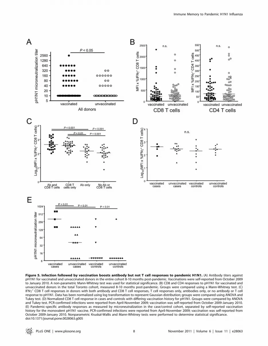
Figure 5. Infection followed by vaccination boosts antibody but not T cell responses to pandemic H1N1. (A) Antibody titers againstpH1N1 for vaccinated and unvaccinated donors in the entire cohort 8-10 months post-pandemic. Vaccinations were self-reported from October 2009to January 2010. A non-parametric Mann-Whitney test was used for statistical significance. (B) CD8 and CD4 responses to pH1N1 for vaccinated andunvaccinated donors in the total Toronto cohort, measured 8-10 months post-pandemic. Groups were compared using a Mann-Whitney test. (C)IFNc+ CD8 T cell responses in donors with both antibody and CD8 T cell responses, T cell responses only, antibodies only, or no antibody or T cellresponse to pH1N1. Data has been normalized using log transformation to represent Gaussian distribution; groups were compared using ANOVA andTukey test. (D) Normalized CD8 T cell response in cases and controls with differing vaccination history for pH1N1. Groups were compared by ANOVAand Tukey test. PCR-confirmed infections were reported from April-November 2009; vaccination was self-reported from October 2009-January 2010.(E) Pandemic-specific antibody responses as measured by microneutralization in the case/control cohort, separated by self-reported vaccinationhistory for the monovalent pH1N1 vaccine. PCR-confirmed infections were reported from April-November 2009; vaccination was self-reported fromOctober 2009-January 2010. Nonparametric Kruskal-Wallis and Mann-Whitney tests were performed to determine statistical significance.doi:10.1371/journal.pone.0028063.g005
Immune Memory to Pandemic H1N1 Influenza
PLoS ONE | www.plosone.org 8 November 2011 | Volume 6 | Issue 11 | e28063

the rate of expansion of these memory T cells may be too slow to
control initial infection, CD8 T cells may help prevent serious
complications and shorten the duration of symptoms.
The T cells that responded most rapidly to acute influenza
infection in the longitudinal donor likely come from pre-existing
influenza-specific memory pools. Indeed, the CD8 T cell epitopes
to influenza virus are much more conserved than antibody
epitopes and have changed little between 1918 and the present day
[10,32]. Consistent with this observation, the magnitude of the
memory CD8 T cell response to A/California/2009 and A/PR8/
34 was similar regardless of donors’ documented history of recent
infection and serological status, arguing that a high proportion of
the memory pool may be cross-reactive. Some of these cross-
reactive T cells may be from pre-existing memory T cells (Fig. 3A),
whereas for recently infected donors, some may reflect the
recruitment of new T cells into the response
As the inactivated influenza vaccine does not directly increase
CD8 T cell responses to influenza [33], concerns have been raised
that vaccination may decrease subsequent protection to influenza by
preventing the CD8 T cell boosting that comes from periodic
natural infection [34]. However, in this study we found that the pool
of influenza-reactive memory T cells persisting at 8–10 months post-
vaccination or infection did not differ between the two groups.
Thus, while vaccination of older people does not increase the CD8
T cell memory pool, neither does it diminish T cell memory relative
to natural infection. However, this issue remains relevant in younger
people whose primary exposure to influenza antigens may be
through vaccination rather than natural infection and who may not
develop influenza-specific memory T cell responses.
A limitation of our study is that we measured antibody levels in
blood, rather than at the site of neutralization, the upper airways.
Moreover, we cannot rule out that memory B cells would give rise
to an antibody response at those sites. Notwithstanding this
limitation, serum Ig levels are the standard of measurement for the
presence of neutralizing antibodies to influenza infection, and both
nasal and serum antibodies correlate with protection from
influenza-induced disease severity [35]. Epidemiological studies
have also shown that influenza-specific antibodies in the blood
correlate with protection, and that a 1:40 titer is the point at which
50% of individuals in a population would be protected [29–31].
At one year post-pandemic, the serum antibody levels in a
substantial minority of vaccinated or infected individuals were
below levels considered seroprotective (,1:40). This argues that
either they did not seroconvert initially, which seems unlikely
based on reported seroconversion rates in clinical trials [36], or did
not maintain antibody levels. This is contrary to antibody
responses generated from many other vaccines and infections.
For example, smallpox-specific antibodies continue to be detect-
able in serum over 60 years after vaccination [37].
Despite the low levels of circulating serum antibody detected at
1 year post-influenza infection, recent studies have shown rapid
mobilization of memory B cells as demonstrated by the isolation of
B cell plasmablasts that produce influenza-specific antibodies at
day 5 post-infection [7,38]. The authors suggest that this rapid
mobilization may allow memory B cells to increase antibody levels
rapidly enough to provide some protection in donors with
appropriate memory B cells to influenza [38]. In the absence of
measurement of B cell memory populations or of antibody titers in
the first few weeks post-infection or vaccination, we cannot state
whether the substantial minority of subjects who failed to maintain
titers to H1N1 above 1:40 at 1 year post-vaccination or infection
were in fact capable of mounting this response transiently.
However, in following a single donor post-H1N1 infection, we
observed only a small increase in neutralizing antibody responses
to pandemic infection as detected by microneutralization assay
and ELISA (but undetectable by HAI). This suggests a rather poor
antibody response to pH1N1 infection in this individual.
Moreover, this weak antibody response had returned to baseline
pre-infection levels by 700 days, despite two intervening H1N1
vaccinations. We speculate that the poor antibody response from
this single longitudinal donor could be due to an exhaustion of his
memory B cell population for this influenza strain, perhaps due
to repetitive exposure to influenza over a lifetime. Further studies
will be required to compare both serum antibody and memory
B cell responses during the acute phase of influenza infection
to determine the source of the weak antibody response to
pH1N1.
In sum, this study shows that almost 60% of a Toronto cohort
had cross-reactive memory T cell responses to influenza virus at
one year post-pandemic. The size of the long-lived pH1N1-
reactive memory T cell pool is not different between infected,
vaccinated and unvaccinated individuals, consistent with the
finding that the memory T cell response to influenza increases
only transiently post-infection and is not boosted by current
vaccines. However, based on one donor, we find that these T cells
can expand significantly post-infection. While too late to prevent
infection, these cross-reactive T cells may offer some contribution
to viral control and improved outcome. There appear to be
relatively low levels of antibody to influenza persisting at 1 year
post-infection or vaccination, suggesting that serum antibody levels
are not maintained at high levels post vaccination or infection.
However, the finding that vaccination can at least transiently
increase antibody levels seen at one year post-pandemic in
individuals who had confirmed infections argues that even in
those recently infected with influenza, taking the subsequent
seasonal vaccine may be able to increase their level of antibody to
titers that have been shown on a population basis to be protective.
Acknowledgments
We thank Dionne White for help with flow cytometry, Birinder Ghumman
for reagent preparation, and Shaza Fadel for data collection.
Author Contributions
Conceived and designed the experiments: LEW LR NC BJW AR CA
THW. Performed the experiments: LEW AR PCD. Analyzed the data:
LEW BL NC LR PCD THW. Contributed reagents/materials/analysis
tools: JG BJW. Wrote the paper: LEW THW. Edited the manuscript: BL
LR NC CA ALW BJW. Oversight/support/recruitment of subjects and
sample management: JF ALW.
References
1. Dawood FS, Jain S, Finelli L, Shaw MW, Lindstrom S, et al. (2009) Emergence
of a novel swine-origin influenza A (H1N1) virus in humans. N Engl J Med 360:
2605–2615.
2. Reed C, Angulo FJ, Swerdlow DL, Lipsitch M, Meltzer MI, et al. (2009)
Estimates of the prevalence of pandemic (H1N1) 2009, United States, April-July
2009. Emerg Infect Dis 15: 2004–2007.
3. Louie JK, Acosta M, Winter K, Jean C, Gavali S, et al. (2009) Factors Associated
With Death or Hospitalization Due to Pandemic 2009 Influenza A(H1N1)
Infection in California. JAMA: Journal of the American Medical Association 302:
1896–1902.
4. Girard MP, Tam JS, Assossou OM, Kieny MP (2010) The 2009 A (H1N1)
influenza virus pandemic: A review. Vaccine 28: 4895–4902.
Immune Memory to Pandemic H1N1 Influenza
PLoS ONE | www.plosone.org 9 November 2011 | Volume 6 | Issue 11 | e28063

5. Louie JK, Jean C, Acosta M, Samuel MC, Matyas BT, et al. (2011) A Review of
Adult Mortality Due to 2009 Pandemic (H1N1) Influenza A in California. PLoS
ONE 6: e18221.
6. Skowronski DM, Hottes TS, McElhaney JE, Janjua NZ, Sabaiduc S, et al.
(2011) Immuno-epidemiologic correlates of pandemic H1N1 surveillance
observations: higher antibody and lower cell-mediated immune responses with
advanced age. J Infect Dis 203: 158–167.
7. Wrammert J, Koutsonanos D, Li G-M, Edupuganti S, Sui J, et al. (2011)
Broadly cross-reactive antibodies dominate the human B cell response against
2009 pandemic H1N1 influenza virus infection. Journal of Experimental
Medicine 208: 181–193.
8. Hancock K, Veguilla V, Lu X, Zhong W, Butler E, et al. (2009) Cross-Reactive
Antibody Responses to the 2009 Pandemic H1N1 Influenza Virus. New
England Journal of Medicine 361: 1945–1952.
9. Xu R, Ekiert DC, Krause JC, Hai R, Crowe JE, et al. (2010) Structural Basis of
Preexisting Immunity to the 2009 H1N1 Pandemic Influenza Virus. Science
328: 357–360.
10. Gras S, Kedzierski L, Valkenburg SA, Laurie K, Liu YC, et al. (2010) Cross-
reactive CD8+ T-cell immunity between the pandemic H1N1-2009 and H1N1-
1918 influenza A viruses. Proceedings of the National Academy of Sciences of
the United States of America 107: 12599–12604.
11. Tu W, Mao H, Zheng J, Liu Y, Chiu SS, et al. (2010) Cytotoxic T lymphocytes
established by seasonal human influenza cross-react against 2009 pandemic
H1N1 influenza virus. Journal of virology 84: 6527–6535.
12. Scheible K, Zhang G, Baer J, Azadniv M, Lambert K, et al. (2011) CD8+ T cell
immunity to 2009 pandemic and seasonal H1N1 influenza viruses. Vaccine 29:
2159–2168.
13. Wagar LE, Gentleman B, Pircher H, McElhaney JE, Watts TH (2011)
Influenza-specific T cells from older people are enriched in the late effector
subset and their presence inversely correlates with vaccine response. PLoS ONE
(accepted).
14. Yu X, Tsibane T, McGraw PA, House FS, Keefer CJ, et al. (2008) Neutralizing
antibodies derived from the B cells of 1918 influenza pandemic survivors. Nature
455: 532–536.
15. Skowronski DM, De Serres G, Crowcroft NS, Janjua NZ, Boulianne N, et al.
(2010) Association between the 2008-09 seasonal influenza vaccine and
pandemic H1N1 illness during Spring-Summer 2009: four observational studies
from Canada. PLoS medicine 7: e1000258.
16. Achonu C, LaFreniere M, Gubbay J, Rosella L, Deeks S, et al. (2010)
A seroprevalence study of pandemic influenza A H1N1 among Ontarians;
Lisbon.
17. World Health Organization (2010) Serological diagnosis of influenza by
microneutralization assay.
18. World Health Organization (2002) WHO manual on animal influenza diagnosis
and surveillance.
19. Mavrouli MD, Routsias JG, Maltezou HC, Spanakis N, Tsakris A (2011)
Estimation of Seroprevalence of the Pandemic H1N1 2009 Influenza Virus
Using a Novel Virus-Free ELISA Assay for the Detection of Specific Antibodies.
Viral Immunol 24: 221–226.
20. Betts MR, Nason MC, West SM, De Rosa SC, Migueles SA, et al. (2006) HIV
nonprogressors preferentially maintain highly functional HIV-specific CD8+ T
cells. Blood 107: 4781–4789.
21. Harari A, Petitpierre S, Vallelian F, Pantaleo G (2004) Skewed representation of
functionally distinct populations of virus-specific CD4 T cells in HIV-1-infected
subjects with progressive disease: changes after antiretroviral therapy. Blood 103:
966–972.22. Darrah PA, Patel DT, De Luca PM, Lindsay RW, Davey DF, et al. (2007)
Multifunctional TH1 cells define a correlate of vaccine-mediated protection
against Leishmania major. Nat Med 13: 843–850.23. Sallusto F, Geginat J, Lanzavecchia A (2004) Central Memory and Effector
Memory T Cell Subsets: Function, Generation, and Maintenance. AnnualReview of Immunology 22: 745–763.
24. Hamann D, Baars PA, Rep MHG, Hooibrink B, Kerkhof-Garde SR, et al.
(1997) Phenotypic and Functional Separation of Memory and Effector HumanCD8+ T cells. Journal of Experimental Medicine 186: 1407–1418.
25. Hamann D, Kostense S, Wolthers K, Otto S, Baars P, et al. (1999) Evidence thathuman CD8+CD45RA+CD27- cells are induced by antigen and evolve through
extensive rounds of division. International Immunology 11: 1027–1033.26. Champagne P, Ogg GS, King AS, Knabenhans C, Ellefsen K, et al. (2001)
Skewed maturation of memory HIV-specific CD8 T lymphocytes. Nature 410:
106–111.27. Appay V, Dunbar PR, Callan M, Klenerman P, Gillespie GMA, et al. (2002)
Memory CD8+ T cells vary in differentiation phenotype in different persistentvirus infections. Nature Medicine 8: 379–385.
28. Wichmann O, Stocker P, Poggensee G, Altmann D, Walter D, et al. (2010)
Pandemic influenza A(H1N1) 2009 breakthrough infections and estimates ofvaccine effectiveness in Germany 2009-2010. Euro Surveill 15.
29. Hobson D, Curry RL, Beare AS, Ward-Gardner A (1972) The role of serumhaemagglutination-inhibiting antibody in protection against challenge infection
with influenza A2 and B viruses. J Hyg (Lond) 70: 767–777.30. de Jong JC, Palache AM, Beyer WE, Rimmelzwaan GF, Boon AC, et al. (2003)
Haemagglutination-inhibiting antibody to influenza virus. Dev Biol (Basel) 115:
63–73.31. Coudeville L, Bailleux F, Riche B, Megas F, Andre P, et al. (2010) Relationship
between haemagglutination-inhibiting antibody titres and clinical protectionagainst influenza: development and application of a bayesian random-effects
model. BMC Med Res Methodol 10: 18.
32. Bui H-H, Peters B, Assarsson E, Mbawuike I, Sette A (2007) Ab and T cellepitopes of influenza A virus, knowledge and opportunities. Proceedings of the
National Academy of Sciences of the United States of America 104: 246–251.33. McMichael A, Gotch G, Cullen P, Askonas B, Webster R (1981) The human
cytotoxic T cell response to influenza A vaccination. Clinical and ExperimentalImmunology 43: 276–284.
34. Bodewes R, Kreijtz JH, Baas C, Geelhoed-Mieras MM, de Mutsert G, et al.
(2009) Vaccination against human influenza A/H3N2 virus prevents theinduction of heterosubtypic immunity against lethal infection with avian
influenza A/H5N1 virus. PLoS One 4: e5538.35. Clements ML, Betts RF, Tierney EL, Murphy BR (1986) Serum and nasal wash
antibodies associated with resistance to experimental challenge with influenza A
wild-type virus. J Clin Microbiol 24: 157–160.36. Roman F, Vaman T, Gerlach B, Markendorf A, Gillard P, et al. (2010)
Immunogenicity and safety in adults of one dose of influenza A H1N1v 2009vaccine formulated with and without AS03A-adjuvant: preliminary report of an
observer-blind, randomised trial. Vaccine 28: 1740–1745.37. Crotty S, Ahmed R (2004) Immunological memory in humans. Semin Immunol
16: 197–203.
38. Wrammert J, Smith K, Miller J, Langley WA, Kokko K, et al. (2008) Rapidcloning of high-affinity human monoclonal antibodies against influenza virus.
Nature 453: 667–671.
Immune Memory to Pandemic H1N1 Influenza
PLoS ONE | www.plosone.org 10 November 2011 | Volume 6 | Issue 11 | e28063