high-frequency stimulation of the nucleus accumbens core and shell reduces quinpirole-induced...
TRANSCRIPT

BEHAVIORAL NEUROSCIENCE
High-frequency stimulation of the nucleus accumbenscore and shell reduces quinpirole-inducedcompulsive checking in rats
Adrian Mundt,1,* Julia Klein,1,* Daphna Joel,2 Andreas Heinz,1 Anais Djodari-Irani,1 Daniel Harnack,3 Andreas Kupsch,3
Helmut Orawa,4 Georg Juckel,4,6 Rudolf Morgenstern5 and Christine Winter1
1Department of Psychiatry and Psychotherapy, Charite Campus Mitte, Charite University Medicine, Berlin, Germany2Department of Psychology, Tel Aviv University, Israel3Department of Neurology, Charite Campus Virchow Klinikum, Charite University Medicine, Berlin, Germany4Department of Biostatistics and Data Management, Coordination Center for Clinical Studies (KKS), Campus Virchow Klinikum,Charite University Medicine, Berlin, Germany5Institute of Pharmacology and Toxicology, Charite Campus Mitte, Charite University Medicine, Berlin, Germany6Department of Psychiatry, Ruhr-University, Bochum, Germany
Keywords: behavior, deep brain stimulation, dopamine agonist, obsessive compulsive disorder
Abstract
Electrical deep brain stimulation (DBS) is currently studied in the treatment of therapy-refractory obsessive compulsive disorders(OCDs). The variety of targeted brain areas and the inconsistency in demonstrating anti-compulsive effects, however, highlight theneed for better mapping of brain regions in which stimulation may produce beneficial effects in OCD. Such a goal may be advancedby the assessment of DBS in appropriate animal models of OCD. Currently available data on DBS of the nucleus accumbens (NAc)on OCD-like behavior in rat models of OCD are contradictory and partly in contrast to clinical data and theoretical hypotheses abouthow the NAc might be pathophysiologically involved in the manifestation of OCD. Consequently, the present study investigates theeffects of DBS of the NAc core and shell in a quinpirole rat model of OCD. The study demonstrates that electrical modulation of NAccore and shell activity via DBS reduces quinpirole-induced compulsive checking behavior in rats. We therefore conclude that both,the NAc core and shell constitute potential target structures in the treatment of OCD.
Introduction
Obsessive compulsive disorder (OCD) represents a highly impairingpsychiatric disorder with a lifetime prevalence of 1–3% (Rasmussen &Eisen, 1992; Sasson et al., 1997). Although the etiology of OCD islargely unknown, several brain regions have been implicated in itspathophysiology, including the dopaminergic and serotonergic sys-tems and the basal ganglia-thalamo-cortical circuits (Saxena et al.,1998). In patients refractory to pharmacotherapy and behavioraltherapy, ablative lesions of pathways within these circuitries, i.e.cingulotomy, limbic leucotomy, subcaudate tractotomy and anteriorcapsulotomy, have been shown to reverse clinical symptoms (Jenike,1998; Lippitz et al., 1999; Rauch et al., 2001; Lopes et al., 2004). Inrecent years, ablative lesions have been widely replaced by electricaldeep brain stimulation at high frequencies [high-frequency stimulation(HFS)] in the treatment of several neurologic and psychiatric disorders(Krack et al., 2003; Breit et al., 2004; Temel & Visser-Vandewalle,
2004; Flaherty et al., 2005; Deuschl et al., 2006; Kupsch et al., 2006;Blomstedt et al., 2007). Meanwhile, there has also been an attempt toestablish HFS of structures within or associated with the basal ganglia-thalamo-cortical circuits for the treatment of OCD. There are reports ofanti-compulsive effects of HFS of the anterior limb of the internalcapsule (Gabriels et al., 2003; Abelson et al., 2005), the ventralcaudate nucleus (Aouizerate et al., 2004, 2005), and the nucleusaccumbens (NAc) and ventral capsule ⁄ ventral striatum (Sturm et al.,2003; Greenberg et al., 2006; Rauch et al., 2006) in individual patientswith OCD. There are also reports on anti-compulsive effects of HFS ofthe subthalamic nucleus in patients with co-morbid Parkinson’sdisease and OCD (Mallet et al., 2002; Fontaine et al., 2004). However,the variety of targeted brain areas highlights the need for bettermapping of brain regions in which stimulation produces the mostbeneficial effects in the treatment of OCD. This goal may be advancedby the assessment of the effects of HFS in appropriate animal modelsof OCD (Klavir et al., 2008; Kuyck et al., 2008; Winter et al., 2008b).The aim of the present study was to test whether HFS of the NAc
would induce an anti-compulsive effect in the quinpirole (QNP) ratmodel of OCD (Szechtman et al., 1998; Man et al., 2004; Eilam &Szechtman, 2005; Joel, 2006). Because the NAc is anatomically and
Correspondence: Dr Christine Winter, as above.E-mail: [email protected]
*A.M. and J.K. contributed equally to this work.
Received 19 May 2008, revised 14 April 2009, accepted 15 April 2009
European Journal of Neuroscience, Vol. 29, pp. 2401–2412, 2009 doi:10.1111/j.1460-9568.2009.06777.x
ª The Authors (2009). Journal Compilation ª Federation of European Neuroscience Societies and Blackwell Publishing Ltd
European Journal of Neuroscience

functionally subdivided into a shell and a core subregion, and smallchanges in electrode placement have been shown to have a substantialeffect on behavior (Okun et al., 2003), the present project assessed theeffects of HFS of the NAC shell and core separately. Recently, weshowed that HFS and pharmacological inactivation (via intracerebralinjection of the GABA agonist muscimol) of the subthalamic nucleusreversibly reduced compulsive checking in the QNP rat model of OCD(Winter et al., 2008b). We found these data to be supported in thesignal attenuation model of OCD (Klavir et al., 2008). Together, ourprevious results and the present experiment may promote theestablishment of a model serving as a screening tool for the detectionof targets for HFS in OCD.
Materials and methods
Animals
The present study was carried out in accordance with the EuropeanCommunities Council Directive of November 24th, 1986(86 ⁄ 609 ⁄ EEC) for the care of laboratory animals and after approvalof the local ethics committee (senate of Berlin). All efforts were madeto reduce the number of animals used. Fifty-six naive male Wistar rats(Harlan-Winkelmann, Borchen, Germany, 220–450 g during theexperiment) were housed in a temperature- and humidity-controlledvivarium with a 12 h light ⁄ dark cycle (lights on from 06:00 to18:00 h). All experiments were performed during the day time. Foodand water were available ad libitum.
Apparatus and behavioral procedure
Prior to the beginning of the experimental procedure, rats werehandled for about 2 min daily for 5 days. At the start of theexperiment, rats were injected subcutaneously twice weekly for a totalof 15 injections with either saline (control group) or QNP (QNPgroup). At 15 min after each injection animals were placed in an openfield and their behavior was videotaped continuously throughout a30 min session. The open field consisted of a glass table(140 · 140 cm and 20 cm high) with four plexiglas boxes varyingin shape and size at fixed locations. The platform was subdivided into25 rectangles (locales). A computer, interfaced with the videorecorder, was used to score locomotor behavior during playback ofvideo records (TSE VideoMot 2 system; Technical & ScientificEquipment, Bad Homburg, Germany).The following measures were assessed for each session and rat: (i)
total distance traveled; (ii) frequency of stops at each open fieldlocale; (iii) mean duration of return time to a given locale, i.e. theinterval from departure from a given locale to the next arrival at thesame locale; (iv) mean stop duration at a given locale; and (v) totalduration of stops at a given locale, where stops ⁄ visits refer to periodsof no locomotion (Szechtman et al., 1998). For each rat the localewith the highest total duration of stops was defined as the home base(Eilam & Golani, 1989) and compulsive checking behavior wasanalysed with reference to the home base. According to Szechtmanet al. (1998) compulsive checking is present if a rat meets thefollowing three performance criteria: the rat returns to the home baseexcessively often, excessively rapidly and visits less places beforereturning to the home base compared with control rats. The followingmeasures were therefore analysed: the total number of visits to thehome base, the mean time to return to the home base and the meannumber of stops ⁄ visits before returning to the home base. In addition,because repeated administration of QNP increases locomotion(Szechtman et al., 1994a; Szumlinski et al., 1997) and because
checking behavior requires locomotion, a calculation was appliedallowing the assessment of changes in checking behavior independentfrom changes in locomotion. Specifically, for each rat the expectedrate of return to a locale was calculated by dividing the total numberof visits made at a given session to the number of locales visited bythe rat in this session. Next, the ratio of observed to expected homebase visits was calculated by dividing the number of visits to thehome base with the expected rate of return to a locale (Szechtmanet al., 1998; Winter et al., 2008b). QNP-treated rats meet additionalcriteria for compulsive checking, i.e. ritual-like behavior and contextdependency (Szechtman et al., 1998), which have repeatedly beendemonstrated to behave in a similar way to the parameters mentionedabove (Szechtman et al., 1998, 2001). Therefore, they were notevaluated in this study.
Design
The experiment consisted of two phases. In phase I, rats received 10injections (two injections per week with a 3–4 day test-free period) ofeither 0.5 mg ⁄ kg QNP (QNP group, n = 30) or saline (control group,n = 26), followed by behavioral testing in the open field. We andothers have shown that the behavioral effects of chronic treatment withQNP reach a plateau after 8–10 drug injections (Einat & Szechtman,1993a; Szechtman et al., 1994a, b; Szumlinski et al., 1997; Winteret al., 2008b). After the 10th behavioral testing, the QNP-treated ratswere randomly assigned to four groups, i.e. stimulated core (n = 10),stimulated shell (n = 10), sham-stimulated core (n = 5) and sham-stimulated shell (n = 5). Equally, the NaCl-treated control rats wererandomly assigned to four groups, i.e. stimulated core (n = 8),stimulated shell (n = 8), sham-stimulated core (n = 5) and sham-stimulated shell (n = 5). Directly after the 10th testing, QNP andcontrol rats of all groups underwent bilateral implantation ofelectrodes into either the NAc core or shell. In phase II, control andQNP-treated rats underwent five additional injections (two injectionsper week) of saline or QNP, respectively, followed by behavioraltesting (sessions 11–15), starting with the 11th test at 2–3 days post-operatively. HFS ⁄ sham HFS was applied during the 12–14th sessions.Stimulation started at the beginning of the behavioral session (15 minafter QNP ⁄ NaCl injection) and continued for the 30 min of thebehavioral session. Stimulation was applied in current intensities of75, 100 and 150 lA in a random order on the 12–14th sessions. Nostimulation was applied on the last (15th) test session, which served toassess the reversibility of the treatment manipulation. Sham-stimulatedrats did not receive stimulation but were connected to the wires onsessions 12–14.
Surgery
Stereotaxic operations were performed after the 10th session and werecarried out under sodium pentobarbital anesthesia (60 mg ⁄ kg i.p.).The incisor bar was set at 3.3 mm below the interaural line.
Electrode implantation
Two electrodes (concentric bipolar SNEX 100 with connector, RMI,Woodland Hills, CA, USA) were implanted bilaterally into either theNAc shell (1.2 mm anterior to bregma, 1.5 mm lateral to the midlineand 8.2 mm ventral to dura) or the NAc core (1.6 mm anterior tobregma, 1.5 mm lateral to the midline and 7.0 mm ventral to dura)(Paxinos & Watson, 1997). Electrodes were fixed to the skull surfacewith stainless steel screws and dental acrylic cement (Technovit�,Heraeus-Kulzer, Hanau, Germany).
2402 A. Mundt et al.
ª The Authors (2009). Journal Compilation ª Federation of European Neuroscience Societies and Blackwell Publishing LtdEuropean Journal of Neuroscience, 29, 2401–2412

Systemic drug administration
Quinpirole hydrochloride was dissolved in 0.9% NaCl to a concen-tration of 0.5 mg ⁄ mL and injected subcutaneously under the nape ofthe neck at a dose of 0.5 mg ⁄ kg body weight. Control subjectsreceived the same volume of saline.
Stimulation
The HFS was performed with an isolated stimulator (CoulbournInstruments, Allentown, PA, USA). Implanted electrodes wereconnected to the stimulator via an isolated cable system hangingfrom the ceiling of the behavioral room. A swivel and a minimalresistance hairspring connected the cable system to the implantedelectrodes and allowed the rat to freely turn and move on the entireplatform without being constricted or tangled up by the cable systemduring stimulation or sham stimulation. The following parameterswere used for stimulation: constant current mode, frequency 130 Hz,pulse width 60 ls, current intensity 75, 100 or 150 lA. A frequencyof 130 Hz and a narrow pulse duration of 60 ls were chosenaccording to the parameters generally applied in rats for assessing theeffects of HFS in other brain areas (Benazzouz et al., 1995; Windelset al., 2000; Salin et al., 2002; Meissner et al., 2003; Desbonnet et al.,2004; Shi et al., 2006; Baunez et al., 2007; Winter et al., 2008a, b) andare in close proximity to the clinical situation (Moro et al., 2002;Sturm et al., 2003; Okun et al., 2007).
Histology
After the 15th session, rats were anesthetized with chloral hydrate(50 mg ⁄ kg, Merck, Darmstadt, Germany) and perfused transcardiallywith 0.1 m phosphate-buffered saline, followed by ice-cold 4%paraformaldehyde. Brains were removed from the skulls and post-fixed overnight in the same fixative and then stored at 4 �C in 30%sucrose. Frozen coronal sections (40 lm) were cut using a cryostat.For histological examination, every second section was stained withcresyl violet. Verification of placements used the atlas of Paxinos &Watson (1997). Only animals with the electrodes placed correctlyin the target areas were included in the statistical analysis of theresults.
Statistical analysis
Statistical analysis was performed as described previously (Winteret al., 2008b).
Phase I
t-tests were performed for comparisons between the performance ofthe two groups (QNP and control) on the last session (10th) of phase I.
Phase II
For comparisons between treatment conditions within a group,repeated-measures anova was performed, followed by the HolmSidak post-hoc tests comparing the stimulation and remission sessionswith the baseline session, when appropriate (Winter et al., 2008b). Inorder to study the effect of several factors on one of the outcomeparameters simultaneously (QNP vs. NaCl treatment, electrodeimplantation ⁄ placement, HFS vs. sham HFS, measurement repeti-tions ⁄ current intensities), generalized estimating equations (GEEs)were performed using the GENMOD procedure (SAS 9.1.3) toanalyse main effects (factors) and interaction terms (product of factors)
(for detailed description of the GEEs please see Supporting informa-tion, Appendix S1). In order to study whether the therapeutic effects ofHFS on measures of compulsive checking depended on locomotion,GEEs were performed using the GENMOD procedure (SAS 9.1.3)with locomotion as a covariate. The cut-off level for statisticalsignificance was taken at P = 0.05.
Results
Electrode placement
Figure 1A and B presents photomicrographs (at a magnification of25·) of a coronal section taken from representative rats implanted withan electrode in either the NAc shell or core, respectively. The electrodetracks toward the targeted region are visible on the photomicrographs.Figure 1C and D presents a schematic reconstruction of electrode tipsin the NAc shell and core, respectively, of all QNP-treated rats thatunderwent deep brain stimulation and were integrated into the study.Equivalent distribution patterns of electrode tip placements werefound in NaCl-treated stimulated as well as QNP- or NaCl-treatedsham-stimulated rats (data not shown). Due to dysfunction of theelectrode (detectable during on-site oscilloscope recording) or inap-propriate localization of the electrode (detectable via histologicalprocessing), four rats were excluded from the QNP-treated group(stimulated core, 2; stimulated shell, 1; sham-stimulated core, 1) andtwo rats were excluded from the NaCl-treated control group (stimu-lated shell, 1; sham-stimulated core, 1). Thus, the final analysisincluded the following number of animals: (i) in the QNP-treatedgroup: stimulated core, 8; stimulated shell HFS, 9; sham-stimulatedcore, 4; sham-stimulated shell, 5 and (ii) in the NaCl-treated controlgroup: stimulated core, 8; stimulated shell HFS, 7; sham-stimulatedcore, 5; sham-stimulated shell, 5.
Behavioral measures
Phase I
Quinpirole-induced compulsive checking behavior. Quinpirole treat-ment over a total of 10 injections induced compulsive checkingbehavior as demonstrated by three performance measures of compul-sive checking previously introduced by Szechtman et al. (1998).(i) QNP-treated rats visited their home base significantly more oftenthan did saline-treated animals (Fig. 2A, P < 0.001). This was also truewhen taking into account the higher total number of visits to all localesin QNP-treated rats compared with control rats. Thus, the ratio ofobserved to expected visits to the home base (Fig. 2B) was significantlyhigher in QNP-treated compared with control rats (P < 0.001). (ii) Themean return time to the home base (Fig. 2C) was about 10-fold shorterin QNP-treated than in control rats (P < 0.001). (iii) QNP-treated ratsvisited fewer places than control rats before returning to their homebase (Fig. 2D, P < 0.001). In addition, chronic intermittent applicationof QNP led to locomotor sensitization, evident in the significantlylonger total distance traveled by QNP-treated compared with controlrats during the 10th session (Fig. 2E, P < 0.001).
Phase II
The effects of electrode implantation on locomotion and quinpirole-induced checking behavior. Electrode implantation into either theNAc shell or NAc core and sham HFS of both regions did not affectlocomotion and parameters used for the quantification of compulsivechecking behavior in QNP- and saline-treated control rats (data notshown).
NAc-HFS and obsessive compulsive disorder 2403
ª The Authors (2009). Journal Compilation ª Federation of European Neuroscience Societies and Blackwell Publishing LtdEuropean Journal of Neuroscience, 29, 2401–2412

Effects of high-frequency stimulation on locomotion and measures ofcompulsive checking behavior in control ratsHigh-frequency stimulation of the nucleus accumbens shell: HFS
of the NAc shell significantly increased locomotion measured as thetotal distance traveled specifically at 100 lA (F4,34 = 4.69, P = 0.008,Table 1A). However, HFS of the NAc shell did not affect measures ofcompulsive checking behavior in control rats (Table 1A), as measuredin the total number of returns to the home base (F4,34 = 1.02,P = 0.422), the ratio of expected to observed home base visits(F4,34 = 0.3, P = 0.876), the return time to the home base(F4,34 = 1.11, P = 0.384) and visits to other places before revisitingthe home base (F4,34 = 1.25, P = 0.32).High-frequency stimulation of the nucleus accumbens core: HFS of
the NAc core significantly increased locomotion measured as the totaldistance traveled at 100 and 150 lA (F4,39 = 3.98, P = 0.012, Table1B). However, HFS of the NAc core did not affect measures of
compulsive checking behavior in control rats (Table 1B), as measuredin the total number of returns to the home base (F4,39 = 1.02,P = 0.422), the ratio of expected to observed home base visits(F4,39 = 0.22, P = 0.922), the return time to the home base(F4,39 = 0.43, P = 0.789) and visits to other places before revisitingthe home base (F4,39 = 0.21, P = 0.93).Effects of high-frequency stimulation of the nucleus accumbens
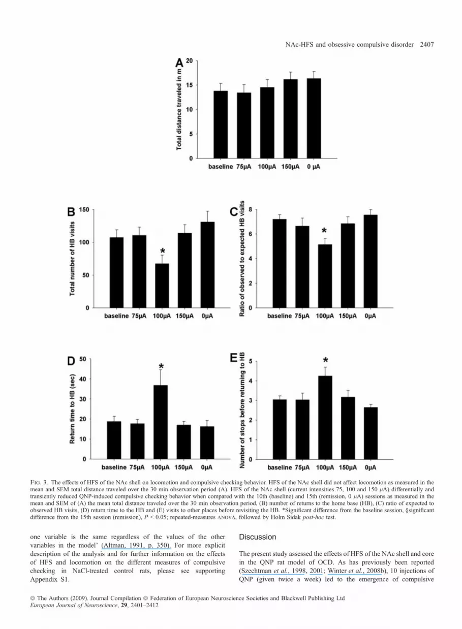
shell on locomotion and quinpirole-induced checking behavior. Fig-ure 3A–E presents the total distance traveled by QNP-treated rats andthe different measures of compulsive checking on the baseline session(session 10), under HFS with different current intensities (sessions12–14) and without stimulation (session 15). As can be seen, HFS ofthe NAc shell did not affect locomotion in QNP-treated rats(F4,44 = 1.84, P = 0.146, Fig. 3A). However, on the four measuresof compulsive checking, HFS of the NAc shell attenuated QNP-induced compulsive checking at a current intensity of 100 lA,
Fig. 1. Post-mortem histology. Photomicrographs of a coronal section stained with cresyl violet and taken from representative rats showing the tip of the electrodein the NAc core (A) or shell (B). Schematic reconstructions of electrode tip placement in the NAc core (C) or shell (D) of QNP-treated stimulated rats. Equivalentdistribution patterns of electrode tip placements were found in NaCl-treated stimulated as well as QNP- or NaCl-treated sham-stimulated rats. Schematicreconstruction of these findings was left out in order to avoid confusion of the relevant data.
2404 A. Mundt et al.
ª The Authors (2009). Journal Compilation ª Federation of European Neuroscience Societies and Blackwell Publishing LtdEuropean Journal of Neuroscience, 29, 2401–2412

whereas at current intensities of 75 and 150 lA it had no effect oncompulsive checking measures. Specifically, QNP-treated rats underHFS with 100 lA visited their home base significantly less often thanthey did without HFS (sessions 10 and 15) or under HFS with current
intensities of 75 and 150 lA (F4,44 = 3.357, P = 0.022, Fig. 3B).Also, after adjusting for the total number of visits, returns to the homebase were significantly reduced in QNP-treated rats under HFS with100 lA. Thus, the ratio of observed to expected visits to the home
Fig. 2. Induction of compulsive checking behavior. Compulsive checking behavior is analysed with reference to the home base established by each rat during the10th session and recognized as the locale with the longest total duration of stops. QNP-treated animals met compulsive checking criteria: (A) more frequent returns tothe home base, (B) a higher than expected rate of returning to the home base, (C) reduced return time to home base and (D) fewer visits to other places beforerevisiting home base compared with saline-treated controls. Additionally, QNP-treated rats displayed an increased locomotion as measured in the mean and SE of themean total distance traveled over the 30 min observation period (E). *P < 0.05, t-test. Values are expressed as mean ± SEM.
NAc-HFS and obsessive compulsive disorder 2405
ª The Authors (2009). Journal Compilation ª Federation of European Neuroscience Societies and Blackwell Publishing LtdEuropean Journal of Neuroscience, 29, 2401–2412

base was significantly lower under HFS with 100 lA than withoutHFS (sessions 10 and 15) or under HFS with current intensities of 75and 150 lA (F4,44 = 3.822, P = 0.013, Fig. 3C). The mean return timeto the home base was almost twofold longer in the QNP-treated ratsunder HFS with 100 lA than in the same QNP-treated rats withoutHFS (sessions 10 and 15) or under HFS with current intensities of 75and 150 lA (F4,44 = 5.22, P = 0.003, Fig. 3D). QNP-treated ratsunder HFS visited significantly more locales before returning to theirhome base than they did under all other conditions (F4,44 = 6.871,P < 0.001, Fig. 3E).Effects of high-frequency stimulation of the nucleus accumbens coreon locomotion and quinpirole-induced checking behavior. Figure4A–E presents the total distance traveled by QNP-treated rats and thedifferent measures of compulsive checking on the baseline session(session 10), under HFS with different current intensities (sessions12–14) and without stimulation (session 15). As can be seen, HFS ofthe NAc core significantly reduced locomotion in QNP-treated ratsat 150 lA but had no effect on locomotion at 100 and 75 lA(F4,39 = 3.972, P = 0.014, Fig. 4A). Furthermore, on the fourmeasures of compulsive checking, HFS of the NAc core attenuatedQNP-induced compulsive checking at current intensities of 100 and150 lA, whereas it had no effect at 75 lA. Specifically, the highcurrent intensity (150 lA) significantly decreased the number ofvisits to the home base compared with the no-stimulation sessions(sessions 10 and 15), the intermediate current intensity (100 lA)decreased this measure only in comparison to the 10th but not the15th session, and the lowest intensity (75 lA) had no effect(F4,39 = 3.32, P = 0.025, Fig. 4B). After adjusting for the totalnumber of visits, returns to the home base were significantly reducedby the two higher current intensities (100 and 150 lA) but not bythe lowest current intensity (75 lA, F4,39 = 9.55, P < 0.001, Fig.4C). Similarly, the mean return time to the home base wassignificantly increased by the higher current intensities (100 and150 lA) but not by the lowest current intensity (75 lA) when
compared with the 10th session (but not when compared with the15th session) (F4,39 = 3.46, P = 0.023, Fig. 4D). The number ofstops before returning to the home base was significantly increasedby the higher current intensities (100 and 150 lA) but not by thelowest current intensity (75 lA) when compared with both sessionswithout stimulation (sessions 10 and 15) (F4,39 = 6.54, P < 0.001,Fig. 4E).The GEE analysis basically corroborated the results detailed
above (please see supporting Appendix S1). GEE analysis withlocomotion as a covariate revealed that, in QNP-treated ratsstimulated in the NAc shell, HFS significantly affected the totalnumber of home base visits (depending on the current intensity,P = 0.001), the return time to the home base (P = 0.029) and thenumber of stops before coming back to the home base (P = 0.008).These parameters were not affected by locomotion (each P > 0.05).Notably, the behavioral parameter ‘ratio of observed to expectedhome base visits’ was significantly affected by both HFS (depend-ing on current intensity, P < 0.0001) and locomotion (P < 0.0001).GEE analysis with locomotion as a covariate further revealedthat, in QNP-treated rats stimulated in the NAc core, HFS butnot locomotion significantly affected the ratio of observed toexpected home base visits (depending on the current intensity,P < 0.0001) and the number of stops before coming back to thehome base (P = 0.024; depending on current intensity, P = 0.010).Notably, the behavioral parameters ‘total number of home basevisits’ and ‘return time to the home base’ were significantlyaffected by both HFS (total number of home base visits: dependingon current intensity, P < 0.0001; return time to the home base:depending on current intensity, P = 0.002) and locomotion (totalnumber of home base visits: P = 0.002; return time to the homebase: P = 0.003). The effects of HFS and locomotion on behavioralmeasures of compulsive checking are independent in the sense of asubtle unstated assumption of multivariate models of regression:‘the effects of each variable are independent, so that the effect of
Table 1. Performance of NaCl-treated control rats stimulated in the NAc shell or core
Distancetraveled (m)
Total numberof HB visits
Observed ⁄ expectedHB visits
Return timeto HB (s)
Number of stopsbefore revisiting HB
(A) NAc shellBaseline 3.1 ± 0.3 7.6 ± 1.2 3.1 ± 0.2 208.2 ± 18.3 6.2 ± 0.8
75 lA 5.8 ± 0.8 12.9 ± 2.1 3.3 ± 0.4 171.0 ± 34.1 7.4 ± 1.7100 lA 9.1 ± 0.2* 12.3 ± 4.4 2.9 ± 0.4 162.5 ± 41.2 8.8 ± 1.0150 lA 5.3 ± 0.5 12.5 ± 1.5 3.0 ± 0.6 150.9 ± 19.3 9.0 ± 2.80 lA 5.5 ± 0.3 11.9 ± 1.8 3.3 ± 0.5 174.1 ± 26.4 7.4 ± 1.3
Repeated-measures anova
F4,34-value 4.69 1.02 0.3 1.11 1.25P-value 0.008 0.422 0.876 0.384 0.32
(B) NAc coreBaseline 5.1 ± 0.7 13.1 ± 2.0 2.6 ± 0.3 172.2 ± 35.8 8.9 ± 0.875 lA 4.7 ± 0.9 11.5 ± 2.1 2.7 ± 0.3 211.2 ± 46.7 8.5 ± 1.3100 lA 8.1 ± 1.6* 14.8 ± 1.9 2.7 ± 0.2 171.7 ± 31.6 9.4 ± 1.2150 lA 8.8 ± 1.6*,� 14.8 ± 1.4 2.8 ± 0.4 158.6 ± 27.0 9.3 ± 1.70 lA 6.0 ± 0.8 13.2 ± 3.6 2.5 ± 0.4 182.7 ± 36.7 9.9 ± 1.9
Repeated-measures anova
F4,39-value 3.98 0.48 0.22 0.43 0.21P-value 0.012 0.75 0.922 0.789 0.93
Values are expressed as mean ± SEM. High-frequency stimulation (HFS) of the NAc core and shell significantly increased locomotion as expressed in the totaldistance traveled at current intensities of 100 lA (NAc shell) or 100 and 150 lA (NAc core). HFS of the NAc core and shell had no effect on the behavioralparameters specific for compulsive checking in saline-treated control rats. *P < 0.05 vs. 10th session and �P < 0.05, vs. 15th session (remission), repeated-measuresanova, followed by Holm Sidak post-hoc test for B and C. HB, home base.
2406 A. Mundt et al.
ª The Authors (2009). Journal Compilation ª Federation of European Neuroscience Societies and Blackwell Publishing LtdEuropean Journal of Neuroscience, 29, 2401–2412
one variable is the same regardless of the values of the othervariables in the model’ (Altman, 1991, p. 350). For more explicitdescription of the analysis and for further information on the effectsof HFS and locomotion on the different measures of compulsivechecking in NaCl-treated control rats, please see supportingAppendix S1.
Discussion
The present study assessed the effects of HFS of the NAc shell and corein the QNP rat model of OCD. As has previously been reported(Szechtman et al., 1998, 2001; Winter et al., 2008b), 10 injections ofQNP (given twice a week) led to the emergence of compulsive
Fig. 3. The effects of HFS of the NAc shell on locomotion and compulsive checking behavior. HFS of the NAc shell did not affect locomotion as measured in themean and SEM total distance traveled over the 30 min observation period (A). HFS of the NAc shell (current intensities 75, 100 and 150 lA) differentially andtransiently reduced QNP-induced compulsive checking behavior when compared with the 10th (baseline) and 15th (remission, 0 lA) sessions as measured in themean and SEM of (A) the mean total distance traveled over the 30 min observation period, (B) number of returns to the home base (HB), (C) ratio of expected toobserved HB visits, (D) return time to the HB and (E) visits to other places before revisiting the HB. *Significant difference from the baseline session, §significantdifference from the 15th session (remission), P < 0.05; repeated-measures anova, followed by Holm Sidak post-hoc test.
NAc-HFS and obsessive compulsive disorder 2407
ª The Authors (2009). Journal Compilation ª Federation of European Neuroscience Societies and Blackwell Publishing LtdEuropean Journal of Neuroscience, 29, 2401–2412

checking in QNP-treated rats. Specifically, QNP-treated rats revisitedtheir home base excessively often and rapidly compared with otherlocales and with saline-treated controls, and stopped at only a few otherlocales before returning to the home base. In addition to compulsive
checking, QNP-treated rats also developed locomotor sensitization, asreported previously (Einat & Szechtman, 1993b; Mattingly et al., 1993;Szechtman et al., 1994a, b; Kostrzewa, 1995; Einat et al., 1996;Szumlinski et al., 1997; Culver et al., 2000; Winter et al., 2008b).
Fig. 4. The effects of HFS of the NAc core on locomotion and compulsive checking behavior. HFS of the NAc shell did not affect locomotion as measured in themean and SEM total distance traveled over the 30 min observation period (A). HFS of the NAc core (current intensities 75, 100 and 150 lA) differentially andtransiently decreased locomotion when compared with the 10th (baseline) and 15th (remission, 0 lA) sessions as measured in the mean and SEM total distancetraveled over the 30 min observation period (A). Furthermore, HFS of the NAc core (current intensities 75, 100 and 150 lA) differentially and transiently reducedQNP-induced compulsive checking behavior when compared with the 10th (baseline) and 15th (remission, 0 lA) sessions as measured in the (B) number of returnsto the home base (HB), (C) ratio of expected to observed HB visits, (D) return time to the HB and (E) visits to other places before revisiting the HB. *Significantdifference from the baseline session, §significant difference from the 15th session (remission), P < 0.05; repeated-measures anova, followed by Holm Sidakpost-hoc test.
2408 A. Mundt et al.
ª The Authors (2009). Journal Compilation ª Federation of European Neuroscience Societies and Blackwell Publishing LtdEuropean Journal of Neuroscience, 29, 2401–2412

In saline-treated control rats, HFS of the NAc core and shellresulted in a current intensity-dependent increase in locomotion.This is in line with previous studies that have shown that electricalstimulation and electrolytic lesion of the NAc increase locomotionand explorative behavior in naive rats (Kelly & Roberts, 1983;Kubos et al., 1987; Starkstein et al., 1988; van Kuyck et al., 2003)as well as rats pre-treated with a selective serotonin 1A receptoragonist (van Kuyck et al., 2003). The similar effects of HFS ofeither the NAc shell or core on locomotion in drug-naive ratsreflects the ongoing controversy on whether the NAc shell or coreis more involved in locomotion (Maldonado-Irizarry & Kelley,1994; Johnson et al., 1996; Weiner et al., 1996, 1998; Gal et al.,1997).
Interestingly, HFS induced a converse effect on locomotion inQNP-sensitized, i.e. hyperlocomotive, rats, i.e. HFS of the NAccore reduced locomotion in QNP-treated rats when stimulationwas performed at the highest current intensity. anova revealed thatHFS of the NAc shell did not affect locomotion. The differentialfindings on locomotion may reflect functional differences betweenthe NAc shell and core region, which become apparent only afterdopamine challenge, i.e. chronic intermittent QNP treatment, butnot under control conditions (see above). These findings mayfurther suggest that a locomotor sensitization induced by HFS ofboth the NAc core and shell may not become apparent in alreadysensitized, i.e. hyperlocomotive, rats. It may also be hypothe-sized that the reduction of checking behavior is paralleled byreduced locomotion that is outweighed by the hyperlocomotiveeffects of HFS in all treatment conditions except the abovementioned, where the potential decrease in locomotion parallelingthe anti-compulsive effect of HFS is stronger than the hyperloco-motive effect of QNP.
The main finding of the present study is that HFS of the NAcshell and core attenuated compulsive checking in QNP-treated rats.This effect was reversible as demonstrated by the fact thatcompulsive checking returned to its baseline level on the lastsession (15th), when no stimulation was applied. Specifically, underHFS, QNP-treated rats behaved more similarly to saline-treated ratswith respect to the number of visits to the home base, the number ofstops in other locales before returning to the home base and the timespent away from the home base. This anti-compulsive effect cannotbe accounted for by a non-selective effect on locomotion because(i) HFS of the shell and core decreased the ratio of observed toexpected visits to the home base, which is a measure of compulsivechecking that is not dependent on general changes in locomotion,and increased the number of stops before returning to the home base,which, if anything, should be inversely correlated with the generallevel of locomotion; (ii) HFS of the NAc core at 150 lA reducedboth compulsive checking and locomotion but, at 100 lA, HFS ofthe NAc core decreased only compulsive checking; and (iii) GEEanalysis with locomotion as a covariate revealed no correlationbetween the effect of HFS on behavioral measures of locomotionand of compulsivity in QNP-treated rats (see supporting AppendixS1). Taken together, the present experiments reveal a specific effectof HFS on compulsive measures not biased by effects on locomo-tion. Furthermore, these data reinforce the notion that compulsivechecking, as defined in the QNP model, is not merely a by-productof QNP-induced locomotor sensitization.
The finding that HFS of the shell was effective only at 100 lA,whereas HFS of the core was more effective at 150 lA than at100 lA, may have implications as to the best target for HFS within theNAc. The differential effect may be due to either an unspecificmechanism such as current spread to neighboring nerve fibers and
brain areas or, alternatively, may reflect HFS-dependent modulationsof different subregion-specific efferents.The distance up to which current spreads depends on (i) current
intensity and (ii) electrode and tissue properties (Ranck, 1975;Perlmutter & Mink, 2006). It is therefore likely that, at a givenelectrode and tissue condition, the distance up to which neurons areaffected by HFS positively correlates with the current intensity.Furthermore, at a given electrode and current intensity, the distanceup to which neurons are affected by HFS crucially may depend ontissue properties such as cell bodies (small- vs. large-diameter axonsand dendrites are differentially sensitive towards electrical stimula-tion) (Holsheimer et al., 2000; Yousif & Liu, 2007). Consideringthe interplay of current intensity and tissue properties, currentintensities of 150 lA may induce a certain profile of acti-vated ⁄ inhibited cell bodies and different types of axons within acertain region adjacent to the stimulation site that may be distinctlydifferent from the profile induced in another adjacent region andunder a current intensity of 100 lA or even lower currentintensities. The finding that a stronger anti-compulsive effect ofHFS of the NAc core was obtained under current intensities of150 lA rather than 100 lA therefore suggests that stimulationof structures neighboring the NAc core contributed to the anti-compulsive effect. In the rat, the NAc core is surrounded by theNAc shell as well as the caudate putamen and is nerved bythe anterior commissure. In fact, HFS of the caudate nucleus(Aouizerate et al., 2004), the NAc shell (Sturm et al., 2003) and theanterior commissure (Nuttin et al., 1999, 2003) have previouslybeen reported to be effective in the treatment of OCD in humans. Incontrast, possible explanations for the finding that stimulation of theNAc shell was anti-compulsive only at 100 lA but not at the highercurrent intensity (150 lA) include the possibility that stimulationof neighboring areas (e.g. the anterior ventral pallidum, the nucleusof the vertical limb of the diagonal band, the Island of Calleja andthe ‘a’ component of the medial forebrain bundle) (Paxinos &Watson, 1997) exerted a pro-compulsive effect.Alternatively, the differential effects may reflect HFS-dependent
modulations of different subregion-specific efferents (Heimer et al.,1997; Groenewegen et al., 1999; Zahm, 1999, 2000). There is stilldebate about how HFS may work. Most of what is known stems fromstudies assessing the effects of HFS of the subthalamic nucleus or theglobus pallidus in the treatment of Parkinson’s disease. Previousstudies have shown that HFS of these structures reduces the overallactivity of targeted neurons (Salin et al., 2002; Tai et al., 2003;Benazzouz et al., 2004; Filali et al., 2004; Welter et al., 2004;Meissner et al., 2005), which has been discussed to be the result of anexcitation of inhibitory afferents (Salin et al., 2002; Tai et al., 2003;Bacci et al., 2004; Benazzouz et al., 2004; Meissner et al., 2007), adirect inhibition of targeted cell bodies (Benabid et al., 2005) ormodulation of efferent projections of the stimulated region (Windelset al., 2000; Hashimoto et al., 2003; Maurice et al., 2003; McIntyreet al., 2004; Stefani et al., 2005).We may consequently speculate that the differential effects of
HFS of the NAc core and shell may result from modulations ofdifferent subregions and subregion-specific efferents (Heimer et al.,1997; Groenewegen et al., 1999; Zahm, 1999, 2000). The NAcshell is reciprocally connected with the ventral to dorsal prefron-tocortical areas via the ventromedial pallidum and the NAc core isreciprocally connected to the more conventional basal gangliacircuitry via the ventrolateral pallidum (for review see Zahm, 2000).These circuitries are probably differentially involved in locomotivebehavior as well as OCD pathophysiology, both induced by QNPsensitization.
NAc-HFS and obsessive compulsive disorder 2409
ª The Authors (2009). Journal Compilation ª Federation of European Neuroscience Societies and Blackwell Publishing LtdEuropean Journal of Neuroscience, 29, 2401–2412

Taken together, differential findings of HFS of the NAc core andshell on both compulsive checking behavior and locomotionhighlight the functional differentiation of the NAc into twosubregions (Kelly & Roberts, 1983; Jongen-Relo et al., 2002,2003; Sturm et al., 2003) potentially associated with differentanatomical systems that subserve different functions (Zahm & Brog,1992; Groenewegen et al., 1999) and may thus suggest that thelocus of the anti-compulsive effect following HFS of the NAc is theshell. The demonstration of such an anti-compulsive effect follow-ing HFS of the NAc is in line with a recent publication on NAc-HFS in rats with schedule-induced polydipsia (Kuyck et al., 2008)but contrasts a previous finding by the same group that electricalstimulation of the NAc increased compulsive responding in anotherrat model of OCD (8-OH-DPAT-induced perseveration in a T-maze)(van Kuyck et al., 2003). One plausible reason for the contrastingresults may be the difference in stimulation frequency, as thepresent study used high frequency (130 Hz), whereas van Kuycket al. (2003) used low frequencies close to 10 Hz (pulse pairs witha 10 ms interpulse interval, given at 5 Hz), a frequency that isineffective for most deep brain stimulation indications in the clinic(Benabid et al., 1991; Limousin et al., 1995; Ushe et al., 2006;Kuyck et al., 2008).As there are very few studies on the physiological and biochem-
ical effects of HFS of the NAc we can only speculate on themechanism by which this manipulation exerts its anti-compulsiveeffect. So far, it has been found that HFS of the NAc reduces firingrates of neurons in the orbitofrontal cortex (McCracken & Grace,2007). These authors speculate that HFS of the NAc region mayreduce OCD symptoms by reducing activity in orbitofrontal cortexneurons. This hypothesis is in line with several in-vivo microdialysisstudies showing altered neurotransmission and consequently activityunder HFS in projection areas of the stimulated region (Hiller et al.,2007; Winter et al., 2008a).Repeated QNP administration has been shown to decrease basal
dopamine levels in the striatum (Koeltzow et al., 2003) and the NAcprojects densely to the dopaminergic neurons that innervate thestriatum (Joel & Weiner, 2000). It is possible that HFS of the NAccounteracted the altered functioning of the dopaminergic systembrought about by repeated QNP administration. Another structure thatmay be involved in mediating the anti-compulsive effect of NAc-HFSis the ventral pallidum, another projection target of the NAc whosefunctioning has been shown to be altered following repeatedadministration of QNP (Carpenter et al., 2003; Richards et al.,2007). Furthermore, it is of interest to note that we have recently foundthat HFS of the subthalamic nucleus (which also projects to the ventralpallidum) also exerts an anti-compulsive effect in the QNP model(Winter et al., 2008b).
Conclusions
The present study demonstrated that acute HFS of the NAc core andshell selectively reduces compulsive checking behavior in the QNP ratmodel of OCD. Equivalently, HFS of the NAc has been found toreduce obsessive compulsive behavior in patients (Sturm et al., 2003;Greenberg et al., 2006; Rauch et al., 2006; Okun et al., 2007). Thepresent study consequently supports the predictive validity of the QNPmodel for mapping regions for HFS for the treatment of OCD. Inaddition, although the extrapolation from an animal model to theclinical condition is problematic, the present findings demonstrate thatthe exact electrode placement, even within a single brain region, has acrucial impact on the therapeutic outcome.
Supporting Information
Additional supporting information may be found in the online versionof this article:Appendix S1. Analysis of generalized estimating equations (GEN-MOD procedure (SAS 9.1.3)).Please note: Wiley-Blackwell are not responsible for the content orfunctionality of any supporting materials supplied by the authors. Anyqueries (other than missing material) should be directed to thecorresponding author for the article.
Acknowledgements
We wish to thank C. Koelske, J. Kopetzki and R. Winter for their excellenttechnical assistance. This study was supported by GIF grant (851 ⁄ 2004). C.W.is a Rahel-Hirsch Fellow of the Humboldt University, Berlin, Germany.
Abbreviations
GEE, generalized estimating equation; HFS, high-frequency stimulation; NAc,nucleus accumbens; OCD, obsessive compulsive disorder; QNP, quinpirole.
References
Abelson, J.L., Curtis, G.C., Sagher, O., Albucher, R.C., Harrigan, M., Taylor,S.F., Martis, B. & Giordani, B. (2005) Deep brain stimulation for refractoryobsessive-compulsive disorder. Biol. Psychiatry, 57, 510–516.
Altman, D.G. 1991. Practical statistics for medical research. Chapman & Hall,London.
Aouizerate, B., Cuny, E., Martin-Guehl, C., Guehl, D., Amieva, H., Benazzouz,A., Fabrigoule, C., Allard, M., Rougier, A., Bioulac, B., Tignol, J. &Burbaud, P. (2004) Deep brain stimulation of the ventral caudate nucleus inthe treatment of obsessive-compulsive disorder and major depression. Casereport. J. Neurosurg., 101, 682–686.
Aouizerate, B., Martin-Guehl, C., Cuny, E., Guehl, D., Amieva, H., Benazzouz,A., Fabrigoule, C., Bioulac, B., Tignol, J. & Burbaud, P. (2005) Deep brainstimulation for OCD and major depression. Am. J. Psychiatry, 162, 2192.
Bacci, J.J., Absi, E.H., Manrique, C., Baunez, C., Salin, P. & Kerkerian-LeGoff, L. (2004) Differential effects of prolonged high frequency stimulationand of excitotoxic lesion of the subthalamic nucleus on dopaminedenervation-induced cellular defects in the rat striatum and globus pallidus.Eur. J. Neurosci., 20, 3331–3341.
Baunez, C., Christakou, A., Chudasama, Y., Forni, C. & Robbins, T.W. (2007)Bilateral high-frequency stimulation of the subthalamic nucleus on atten-tional performance: transient deleterious effects and enhanced motivation inboth intact and parkinsonian rats. Eur. J. Neurosci., 25, 1187–1194.
Benabid, A.L., Pollak, P., Gervason, C., Hoffmann, D., Gao, D.M., Hommel,M., Perret, J.E. & de, R.J. (1991) Long-term suppression of tremor bychronic stimulation of the ventral intermediate thalamic nucleus. Lancet,337, 403–406.
Benabid, A.L., Wallace, B., Mitrofanis, J., Xia, R., Piallat, B., Chabardes, S. &Berger, F. (2005) A putative generalized model of the effects and mechanismof action of high frequency electrical stimulation of the central nervoussystem. Acta Neurol. Belg., 105, 149–157.
Benazzouz, A., Piallat, B., Pollak, P. & Benabid, A.L. (1995) Responses ofsubstantia nigra pars reticulata and globus pallidus complex to highfrequency stimulation of the subthalamic nucleus in rats: electrophysiolog-ical data. Neurosci. Lett., 189, 77–80.
Benazzouz, A., Tai, C.H., Meissner, W., Bioulac, B., Bezard, E. & Gross, C.(2004) High-frequency stimulation of both zona incerta and subthalamicnucleus induces a similar normalization of basal ganglia metabolic activity inexperimental parkinsonism. FASEB J., 18, 528–530.
Blomstedt, P., Hariz, G.M., Hariz, M.I. & Koskinen, L.O. (2007) Thalamicdeep brain stimulation in the treatment of essential tremor: a long-termfollow-up. Br. J. Neurosurg., 21, 504–509.
Breit, S., Schulz, J.B. & Benabid, A.L. (2004) Deep brain stimulation. CellTissue Res., 318, 275–288.
Carpenter, T.L., Pazdernik, T.L. & Levant, B. (2003) Differences in quinpirole-induced local cerebral glucose utilization between naive and sensitized rats.Brain Res., 964, 295–301.
2410 A. Mundt et al.
ª The Authors (2009). Journal Compilation ª Federation of European Neuroscience Societies and Blackwell Publishing LtdEuropean Journal of Neuroscience, 29, 2401–2412
Culver, K.E., Rosenfeld, J.M. & Szechtman, H. (2000) A switch mechanismbetween locomotion and mouthing implicated in sensitization to quinpirolein rats. Psychopharmacology (Berl.), 151, 202–210.
Desbonnet, L., Temel, Y., Visser-Vandewalle, V., Blokland, A., Hornikx, V. &Steinbusch, H.W. (2004) Premature responding following bilateral stimula-tion of the rat subthalamic nucleus is amplitude and frequency dependent.Brain Res., 1008, 198–204.
Deuschl, G., Schade-Brittinger, C., Krack, P., Volkmann, J., Schafer, H., Botzel,K., Daniels, C., Deutschlander, A., Dillmann, U., Eisner, W., Gruber, D.,Hamel, W., Herzog, J., Hilker, R., Klebe, S., Kloss, M., Koy, J., Krause, M.,Kupsch, A., Lorenz, D., Lorenzl, S., Mehdorn, H.M., Moringlane, J.R.,Oertel, W., Pinsker, M.O., Reichmann, H., Reuss, A., Schneider, G.H.,Schnitzler, A., Steude, U., Sturm, V., Timmermann, L., Tronnier, V.,Trottenberg, T., Wojtecki, L., Wolf, E., Poewe, W. & Voges, J. (2006) Arandomized trial of deep-brain stimulation for Parkinson’s disease. N. Engl.J. Med., 355, 896–908.
Eilam, D. & Golani, I. (1989) Home base behavior of rats (Rattus norvegicus)exploring a novel environment. Behav. Brain Res., 34, 199–211.
Eilam, D. & Szechtman, H. (2005) Psychostimulant-induced behavior as ananimal model of obsessive-compulsive disorder: an ethological approach tothe form of compulsive rituals. CNS Spectr., 10, 191–202.
Einat, H. & Szechtman, H. (1993a) Environmental modulation of bothlocomotor response and locomotor sensitization to the dopamine agonistquinpirole. Behav. Pharmacol., 4, 399–403.
Einat, H. & Szechtman, H. (1993b) Longlasting consequences of chronictreatment with the dopamine agonist quinpirole for the undrugged behaviorof rats. Behav. Brain Res., 54, 35–41.
Einat, H., Einat, D., Allan, M., Talangbayan, H., Tsafnat, T. & Szechtman, H.(1996) Associational and nonassociational mechanisms in locomotor sensi-tization to the dopamine agonist quinpirole. Psychopharmacology (Berl.),127, 95–101.
Filali, M., Hutchison, W.D., Palter, V.N., Lozano, A.M. & Dostrovsky, J.O.(2004) Stimulation-induced inhibition of neuronal firing in human subtha-lamic nucleus. Exp. Brain Res., 156, 274–281.
Flaherty, A.W., Williams, Z.M., Amirnovin, R., Kasper, E., Rauch, S.L.,Cosgrove, G.R. & Eskandar, E.N. (2005) Deep brain stimulation of theanterior internal capsule for the treatment of Tourette syndrome: technicalcase report. Neurosurgery, 57, E403.
Fontaine, D., Mattei, V., Borg, M., von, L.D., Magnie, M.N., Chanalet, S.,Robert, P. & Paquis, P. (2004) Effect of subthalamic nucleus stimulation onobsessive-compulsive disorder in a patient with Parkinson disease. Casereport. J. Neurosurg., 100, 1084–1086.
Gabriels, L., Cosyns, P., Nuttin, B., Demeulemeester, H. & Gybels, J. (2003)Deep brain stimulation for treatment-refractory obsessive-compulsive disor-der: psychopathological and neuropsychological outcome in three cases.Acta Psychiatr. Scand., 107, 275–282.
Gal, G., Joel, D., Gusak, O., Feldon, J. & Weiner, I. (1997) The effects ofelectrolytic lesion to the shell subterritory of the nucleus accumbens ondelayed non-matching-to-sample and four-arm baited eight-arm radial-mazetasks. Behav. Neurosci., 111, 92–103.
Greenberg, B.D., Malone, D.A., Friehs, G.M., Rezai, A.R., Kubu, C.S., Malloy,P.F., Salloway, S.P., Okun, M.S., Goodman, W.K. & Rasmussen, S.A. (2006)Three-year outcomes in deep brain stimulation for highly resistant obsessive-compulsive disorder. Neuropsychopharmacology, 31, 2384–2393.
Groenewegen, H.J., Wright, C.I., Beijer, A.V. & Voorn, P. (1999) Convergenceand segregation of ventral striatal inputs and outputs. Ann. N Y Acad. Sci.,877, 49–63.
Hashimoto, T., Elder, C.M., Okun, M.S., Patrick, S.K. & Vitek, J.L. (2003)Stimulation of the subthalamic nucleus changes the firing pattern of pallidalneurons. J. Neurosci., 23, 1916–1923.
Heimer, L., Alheid, G.F., de Olmos, J.S., Groenewegen, H.J., Haber, S.N.,Harlan, R.E. & Zahm, D.S. (1997) The accumbens: beyond the core-shelldichotomy. J. Neuropsychiatry Clin. Neurosci., 9, 354–381.
Hiller, A., Loeffler, S., Haupt, C., Litza, M., Hofmann, U. & Moser, A.(2007) Electrical high frequency stimulation of the caudate nucleusinduces local GABA outflow in freely moving rats. J. Neurosci. Methods,159, 286–290.
Holsheimer, J., Demeulemeester, H., Nuttin, B. & de Sutter, P. (2000)Identification of the target neuronal elements in electrical deep brainstimulation. Eur. J. Neurosci., 12, 4573–4577.
Jenike, M.A. (1998) Neurosurgical treatment of obsessive-compulsive disorder.Br. J. Psychiatry Suppl., 35, 79–90.
Joel, D. (2006) Current animal models of obsessive compulsive disorder: acritical review. Prog. Neuropsychopharmacol. Biol. Psychiatry, 30, 374–388.
Joel, D. & Weiner, I. (2000) The connections of the dopaminergic system withthe striatum in rats and primates: an analysis with respect to the functionaland compartmental organization of the striatum. Neuroscience, 96, 451–474.
Johnson, K., Churchill, L., Klitenick, M.A., Hooks, M.S. & Kalivas, P.W.(1996) Involvement of the ventral tegmental area in locomotion elicited fromthe nucleus accumbens or ventral pallidum. J. Pharmacol. Exp. Ther., 277,1122–1131.
Jongen-Relo, A.L., Kaufmann, S. & Feldon, J. (2002) A differentialinvolvement of the shell and core subterritories of the nucleus accumbensof rats in attentional processes. Neuroscience, 111, 95–109.
Jongen-Relo, A.L., Kaufmann, S. & Feldon, J. (2003) A differentialinvolvement of the shell and core subterritories of the nucleus accumbensof rats in memory processes. Behav. Neurosci., 117, 150–168.
Kelly, P.H. & Roberts, D.C. (1983) Effects of amphetamine and apomorphineon locomotor activity after 6-OHDA and electrolytic lesions of the nucleusaccumbens septi. Pharmacol. Biochem. Behav., 19, 137–143.
Klavir, O., Flash, S., Winter, C. & Joel, D. (2008) High frequency stimulationand pharmacological inactivation of the subthalamic nucleus reduces‘compulsive’ lever-pressing in rats. Exp. Neurol., 215, 101–109.
Koeltzow, T.E., Austin, J.D. & Vezina, P. (2003) Behavioral sensitization toquinpirole is not associated with increased nucleus accumbens dopamineoverflow. Neuropharmacology, 44, 102–110.
Kostrzewa, R.M. (1995) Dopamine receptor supersensitivity. Neurosci. Biobe-hav. Rev., 19, 1–17.
Krack, P., Batir, A., Van, B.N., Chabardes, S., Fraix, V., Ardouin, C.,Koudsie, A., Limousin, P.D., Benazzouz, A., LeBas, J.F., Benabid, A.L. &Pollak, P. (2003) Five-year follow-up of bilateral stimulation of thesubthalamic nucleus in advanced Parkinson’s disease. N. Engl. J. Med.,349, 1925–1934.
Kubos, K.L., Moran, T.H. & Robinson, R.G. (1987) Differential andasymmetrical behavioral effects of electrolytic or 6-hydroxydopaminelesions in the nucleus accumbens. Brain Res., 401, 147–151.
Kupsch, A., Benecke, R., Muller, J., Trottenberg, T., Schneider, G.H., Poewe,W., Eisner, W., Wolters, A., Muller, J.U., Deuschl, G., Pinsker, M.O.,Skogseid, I.M., Roeste, G.K., Vollmer-Haase, J., Brentrup, A., Krause, M.,Tronnier, V., Schnitzler, A., Voges, J., Nikkhah, G., Vesper, J., Naumann, M.& Volkmann, J. (2006) Pallidal deep-brain stimulation in primary general-ized or segmental dystonia. N. Engl. J. Med., 355, 1978–1990.
Kuyck, K., Brak, K., Das, J., Rizopoulos, D. & Nuttin, B. (2008) Comparativestudy of the effects of electrical stimulation in the nucleus accumbens, themediodorsal thalamic nucleus and the bed nucleus of the stria terminalis inrats with schedule-induced polydipsia. Brain Res., 1201, 93–99.
van Kuyck, K., Demeulemeester, H., Feys, H., De Weerdt, W., Dewil, M.,Tousseyn, T., De, S.P., Gybels, J., Bogaerts, K., Dom, R. & Nuttin, B. (2003)Effects of electrical stimulation or lesion in nucleus accumbens on thebehaviour of rats in a T-maze after administration of 8-OH-DPAT or vehicle.Behav. Brain Res., 140, 165–173.
Limousin, P., Pollak, P., Benazzouz, A., Hoffmann, D., Le Bas, J.F., Broussolle,E., Perret, J.E. & Benabid, A.L. (1995) Effect of parkinsonian signs andsymptoms of bilateral subthalamic nucleus stimulation. Lancet, 345, 91–95.
Lippitz, B.E., Mindus, P., Meyerson, B.A., Kihlstrom, L. & Lindquist, C.(1999) Lesion topography and outcome after thermocapsulotomy or gammaknife capsulotomy for obsessive-compulsive disorder: relevance of the righthemisphere. Neurosurgery, 44, 452–458.
Lopes, A.C., de Mathis, M.E., Canteras, M.M., Salvajoli, J.V., Del Porto, J.A.& Miguel, E.C. (2004) Update on neurosurgical treatment for obsessivecompulsive disorder. Rev. Bras. Psiquiatr., 26, 62–66.
Maldonado-Irizarry, C.S. & Kelley, A.E. (1994) Differential behavioral effectsfollowing microinjection of an NMDA antagonist into nucleus accumbenssubregions. Psychopharmacology (Berl.), 116, 65–72.
Mallet, L., Mesnage, V., Houeto, J.L., Pelissolo, A., Yelnik, J., Behar, C.,Gargiulo, M., Welter, M.L., Bonnet, A.M., Pillon, B., Cornu, P., Dormont,D., Pidoux, B., Allilaire, J.F. & Agid, Y. (2002) Compulsions, Parkinson’sdisease, and stimulation. Lancet, 360, 1302–1304.
Man, J., Hudson, A.L., Ashton, D. & Nutt, D.J. (2004) Animal models forobsessive-compulsive disorder. Curr. Neuropharmacol., 2, 169–181.
Mattingly, B.A., Rowlett, J.K. & Lovell, G. (1993) Effects of daily SKF 38393,quinpirole, and SCH 23390 treatments on locomotor activity and subsequentsensitivity to apomorphine. Psychopharmacology (Berl.), 110, 320–326.
Maurice, N., Thierry, A.M., Glowinski, J. & Deniau, J.M. (2003) Spontaneousand evoked activity of substantia nigra pars reticulata neurons during high-frequency stimulation of the subthalamic nucleus. J. Neurosci., 23, 9929–9936.
McCracken, C.B. & Grace, A.A. (2007) High-frequency deep brain stimulationof the nucleus accumbens region suppresses neuronal activity and selectively
NAc-HFS and obsessive compulsive disorder 2411
ª The Authors (2009). Journal Compilation ª Federation of European Neuroscience Societies and Blackwell Publishing LtdEuropean Journal of Neuroscience, 29, 2401–2412

modulates afferent drive in rat orbitofrontal cortex in vivo. J. Neurosci., 27,12601–12610.
McIntyre, C.C., Savasta, M., Walter, B.L. & Vitek, J.L. (2004) How does deepbrain stimulation work? Present understanding and future questions J. Clin.Neurophysiol., 21, 40–50.
Meissner, W., Harnack, D., Reese, R., Paul, G., Reum, T., Ansorge, M.,Kusserow, H., Winter, C., Morgenstern, R. & Kupsch, A. (2003) High-frequency stimulation of the subthalamic nucleus enhances striatal dopaminerelease and metabolism in rats. J. Neurochem., 85, 601–609.
Meissner, W., Leblois, A., Hansel, D., Bioulac, B., Gross, C.E., Benazzouz, A.& Boraud, T. (2005) Subthalamic high frequency stimulation resetssubthalamic firing and reduces abnormal oscillations. Brain, 128, 2372–2382.
Meissner, W., Guigoni, C., Cirilli, L., Garret, M., Bioulac, B., Gross, C.,Bezard, E. & Benazzouz, A. (2007) Impact of chronic subthalamic highfrequency stimulation on metabolic basal ganglia activity: a 2-deoxyglucoseuptake and cytochrome oxidase mRNA study in a macaque model ofParkinson’s disease. Eur. J. Neurosci., 25, 1492–1500.
Moro, E., Esselink, R.J., Xie, J., Hommel, M., Benabid, A.L. & Pollak, P.(2002) The impact on Parkinson’s disease of electrical parameter settings inSTN stimulation. Neurology, 59, 706–713.
Nuttin, B., Cosyns, P., Demeulemeester, H., Gybels, J. & Meyerson, B. (1999)Electrical stimulation in anterior limbs of internal capsules in patients withobsessive-compulsive disorder. Lancet, 354, 1526.
Nuttin, B.J., Gabriels, L., van, K.K. & Cosyns, P. (2003) Electrical stimulationof the anterior limbs of the internal capsules in patients with severeobsessive-compulsive disorder: anecdotal reports. Neurosurg. Clin. N. Am.,14, 267–274.
Okun, M.S., Green, J., Saben, R., Gross, R., Foote, K.D. & Vitek, J.L. (2003)Mood changes with deep brain stimulation of STN and GPi: results of a pilotstudy. J. Neurol. Neurosurg. Psychiatry, 74, 1584–1586.
Okun, M.S., Mann, G., Foote, K.D., Shapira, N.A., Bowers, D., Springer, U.,Knight, W., Martin, P. & Goodman, W.K. (2007) Deep brain stimulation inthe internal capsule and nucleus accumbens region: responses observedduring active and sham programming. J. Neurol. Neurosurg. Psychiatry, 78,310–314.
Paxinos, G. & Watson, C. (1997) The Rat Brain. Academic Press, San Diego.Perlmutter, J.S. & Mink, J.W. (2006) Deep brain stimulation. Annu. Rev.Neurosci., 29, 229–257.
Ranck, J.B. Jr (1975) Which elements are excited in electrical stimulation ofmammalian central nervous system: a review. Brain Res., 98, 417–440.
Rasmussen, S.A. & Eisen, J.L. (1992) The epidemiology and differentialdiagnosis of obsessive compulsive disorder. J. Clin. Psychiatry Suppl., 53,4–10.
Rauch, S.L., Makris, N., Cosgrove, G.R., Kim, H., Cassem, E.H., Price, B.H.,Baer, L., Savage, C.R., Caviness, V.S. Jr, Jenike, M.A. & Kennedy, D.N.(2001) A magnetic resonance imaging study of regional cortical volumesfollowing stereotactic anterior cingulotomy. CNS Spectr., 6, 214–222.
Rauch, S.L., Dougherty, D.D., Malone, D., Rezai, A., Friehs, G., Fischman,A.J., Alpert, N.M., Haber, S.N., Stypulkowski, P.H., Rise, M.T., Rasmussen,S.A. & Greenberg, B.D. (2006) A functional neuroimaging investigation ofdeep brain stimulation in patients with obsessive-compulsive disorder.J. Neurosurg., 104, 558–565.
Richards, T.L., Pazdernik, T.L. & Levant, B. (2007) Clorgyline-inducedmodification of behavioral sensitization to quinpirole: effects on localcerebral glucose utilization. Brain Res., 1160, 124–133.
Salin, P., Manrique, C., Forni, C. & Kerkerian-Le, G.L. (2002) High-frequencystimulation of the subthalamic nucleus selectively reverses dopaminedenervation-induced cellular defects in the output structures of the basalganglia in the rat. J. Neurosci., 22, 5137–5148.
Sasson, Y., Zohar, J., Chopra, M., Lustig, M., Iancu, I. & Hendler, T. (1997)Epidemiology of obsessive-compulsive disorder: a world view. J. Clin.Psychiatry, 58(Suppl. 12), 7–10.
Saxena, S., Brody, A.L., Schwartz, J.M. & Baxter, L.R. (1998) Neuroimagingand frontal-subcortical circuitry in obsessive-compulsive disorder. Br. J.Psychiatry Suppl., 35, 26–37.
Shi, L.H., Luo, F., Woodward, D.J. & Chang, J.Y. (2006) Basal ganglia neuralresponses during behaviorally effective deep brain stimulation of thesubthalamic nucleus in rats performing a treadmill locomotion test. Synapse,59, 445–457.
Starkstein, S.E., Moran, T.H., Bowersox, J.A. & Robinson, R.G. (1988)Behavioral abnormalities induced by frontal cortical and nucleus accumbenslesions. Brain Res., 473, 74–80.
Stefani, A., Fedele, E., Galati, S., Pepicelli, O., Frasca, S., Pierantozzi, M.,Peppe, A., Brusa, L., Orlacchio, A., Hainsworth, A.H., Gattoni, G.,Stanzione, P., Bernardi, G., Raiteri, M. & Mazzone, P. (2005) Subthalamicstimulation activates internal pallidus: evidence from cGMP microdialysis inPD patients. Ann. Neurol., 57, 448–452.
Sturm, V., Lenartz, D., Koulousakis, A., Treuer, H., Herholz, K., Klein, J.C. &Klosterkotter, J. (2003) The nucleus accumbens: a target for deep brainstimulation in obsessive-compulsive- and anxiety-disorders. J. Chem.Neuroanat., 26, 293–299.
Szechtman, H., Dai, H., Mustafa, S., Einat, H. & Sullivan, R.M. (1994a) Effectsof dose and interdose interval on locomotor sensitization to the dopamineagonist quinpirole. Pharmacol. Biochem. Behav., 48, 921–928.
Szechtman, H., Talangbayan, H., Canaran, G., Dai, H. & Eilam, D. (1994b)Dynamics of behavioral sensitization induced by the dopamine agonistquinpirole and a proposed central energy control mechanism. Psychophar-macology (Berl.), 115, 95–104.
Szechtman, H., Sulis, W. & Eilam, D. (1998) Quinpirole induces compulsivechecking behavior in rats: a potential animal model of obsessive-compulsivedisorder (OCD). Behav. Neurosci., 112, 1475–1485.
Szechtman, H., Eckert, M.J., Tse, W.S., Boersma, J.T., Bonura, C.A.,McClelland, J.Z., Culver, K.E. & Eilam, D. (2001) Compulsive checkingbehavior of quinpirole-sensitized rats as an animal model of Obsessive-Compulsive Disorder(OCD): form and control. BMC Neurosci., 2, 4.
Szumlinski, K.K., Allan, M., Talangbayan, H., Tracey, A. & Szechtman, H.(1997) Locomotor sensitization to quinpirole: environment-modulatedincrease in efficacy and context-dependent increase in potency. Psychophar-macology (Berl.), 134, 193–200.
Tai, C.H., Boraud, T., Bezard, E., Bioulac, B., Gross, C. & Benazzouz, A.(2003) Electrophysiological and metabolic evidence that high-frequencystimulation of the subthalamic nucleus bridles neuronal activity in thesubthalamic nucleus and the substantia nigra reticulata. FASEB J., 17, 1820–1830.
Temel, Y. & Visser-Vandewalle, V. (2004) Surgery in Tourette syndrome. Mov.Disord., 19, 3–14.
Ushe, M., Mink, J.W., Tabbal, S.D., Hong, M., Schneider, G.P., Rich, K.M.,Lyons, K.E., Pahwa, R. & Perlmutter, J.S. (2006) Postural tremorsuppression is dependent on thalamic stimulation frequency. Mov. Disord.,21, 1290–1292.
Weiner, I., Gal, G., Rawlins, J.N. & Feldon, J. (1996) Differential involvementof the shell and core subterritories of the nucleus accumbens in latentinhibition and amphetamine-induced activity. Behav. Brain Res., 81, 123–133.
Weiner, I., Feldon, J., Tarrasch, R., Hairston, I. & Joel, D. (1998) Fimbria-fornix cut affects spontaneous activity, two-way avoidance and delayednon matching to sample, but not latent inhibition. Behav. Brain Res., 96,59–70.
Welter, M.L., Houeto, J.L., Bonnet, A.M., Bejjani, P.B., Mesnage, V., Dormont,D., Navarro, S., Cornu, P., Agid, Y. & Pidoux, B. (2004) Effects of high-frequency stimulation on subthalamic neuronal activity in parkinsonianpatients. Arch. Neurol., 61, 89–96.
Windels, F., Bruet, N., Poupard, A., Urbain, N., Chouvet, G., Feuerstein, C. &Savasta, M. (2000) Effects of high frequency stimulation of subthalamicnucleus on extracellular glutamate and GABA in substantia nigra and globuspallidus in the normal rat. Eur. J. Neurosci., 12, 4141–4146.
Winter, C., Lemke, C., Sohr, R., Meissner, W., Harnack, D., Juckel, G.,Morgenstern, R. & Kupsch, A. (2008a) High frequency stimulation of thesubthalamic nucleus modulates neurotransmission in limbic brain regions ofthe rat. Exp. Brain Res., 185, 497–507.
Winter, C., Mundt, A., Jalali, R., Joel, D., Harnack, D., Morgenstern, R.,Juckel, G. & Kupsch, A. (2008b) High frequency stimulation and temporaryinactivation of the subthalamic nucleus reduce quinpirole-induced compul-sive checking behavior in rats. Exp. Neurol., 210, 217–228.
Yousif, N. & Liu, X. (2007) Modeling the current distribution across the depthelectrode-brain interface in deep brain stimulation. Expert. Rev. Med.Devices, 4, 623–631.
Zahm, D.S. (1999) Functional-anatomical implications of the nucleus accum-bens core and shell subterritories. Ann. N Y Acad. Sci., 877, 113–128.
Zahm, D.S. (2000) An integrative neuroanatomical perspective on somesubcortical substrates of adaptive responding with emphasis on the nucleusaccumbens. Neurosci. Biobehav. Rev., 24, 85–105.
Zahm, D.S. & Brog, J.S. (1992) On the significance of subterritories inthe ‘‘accumbens’’ part of the rat ventral striatum. Neuroscience, 50, 751–767.
2412 A. Mundt et al.
ª The Authors (2009). Journal Compilation ª Federation of European Neuroscience Societies and Blackwell Publishing LtdEuropean Journal of Neuroscience, 29, 2401–2412