subthalamic nucleus stimulation in severe obsessive–compulsive disorder
TRANSCRIPT

T h e n e w e ngl a nd j o u r na l o f m e dic i n e
n engl j med 359;8 www.nejm.org august 21, 2008 33
original article
Subthalamic Nucleus Stimulation in Severe Obsessive–Compulsive Disorder
Luc Mallet, M.D., Ph.D., Mircea Polosan, M.D., Nematollah Jaafari, M.D., Nicolas Baup, M.D., Marie-Laure Welter, M.D., Ph.D., Denys Fontaine, M.D.,
Ph.D., Sophie Tezenas Du Montcel, M.D., Ph.D., Jérôme Yelnik, M.D., Isabelle Chéreau, M.D., Christophe Arbus, M.D., Sylvie Raoul, M.D., Ph.D.,
Bruno Aouizerate, M.D., Ph.D., Philippe Damier, M.D., Ph.D., Stephan Chabardès, M.D., Ph.D., Virginie Czernecki, Ph.D.,
Claire Ardouin, Ph.D., Marie-Odile Krebs, M.D., Ph.D., Eric Bardinet, Ph.D., Patrick Chaynes, M.D., Ph.D., Pierre Burbaud, M.D., Ph.D., Philippe Cornu, M.D.,
Philippe Derost, M.D., Thierry Bougerol, M.D., Ph.D., Benoit Bataille, M.D., Vianney Mattei, M.D., Didier Dormont, M.D., Ph.D., Bertrand Devaux, M.D.,
Marc Vérin, M.D., Ph.D., Jean-Luc Houeto, M.D., Ph.D., Pierre Pollak, M.D., Ph.D., Alim-Louis Benabid, M.D., Ph.D.,
Yves Agid, M.D., Ph.D., Paul Krack, M.D., Ph.D., Bruno Millet, M.D., Ph.D., and Antoine Pelissolo, M.D., Ph.D., for the STOC Study Group*
The authors’ affiliations are listed in teh Appendix. Address reprint requests to Dr. Mallet at the INSERM Avenir Team IFR 70 — Behavior, Emotion, and Basal Ganglia, Centre d’Investigation Clinique, CHU Pitié-Salpêtrière, 47 Bd de L’Hôpital, 75013 Paris, France, or at [email protected].
*Members of the French Stimulation dans le Trouble Obsessionnel Compul-sif (STOC) Study Group are listed in the Appendix.
N Engl J Med 2008;359:xxx-xx.Copyright © 2008 Massachusetts Medical Society.
A BS TR AC T
Background
Severe, refractory obsessive–compulsive disorder (OCD) is a disabling condition. Stimu-lation of the subthalamic nucleus, a procedure that is already validated for the treat-ment of movement disorders, has been proposed as a therapeutic option.
Methods
In this 10-month, crossover, double-blind, multicenter study assessing the efficacy and safety of stimulation of the subthalamic nucleus, we randomly assigned eight pa-tients with highly refractory OCD to undergo active stimulation of the subthalamic nucleus followed by sham stimulation and eight to undergo sham stimulation fol-lowed by active stimulation. The primary outcome measure was the severity of OCD, as assessed by the Yale-Brown Obsessive Compulsive Scale (Y-BOCS), at the end of two 3-month periods. General psychopathologic findings, functioning, and toler-ance were assessed with the use of standardized psychiatric scales, the Global As-sessment of Functioning (GAF) scale, and neuropsychological tests.
Results
After active stimulation of the subthalamic nucleus, the Y-BOCS score (on a scale from 0 to 40, with lower scores indicating less severe symptoms) was significantly lower than the score after sham stimulation (mean [±SD], 19±8 vs. 28±7; P = 0.01), and the GAF score (on a scale from 1 to 90, with higher scores indicating higher levels of functioning) was significantly higher (56±14 vs. 43±8, P = 0.005). The ratings of neuropsychological measures, depression, and anxiety were not modified by stimu-lation. There were 15 serious adverse events, including 1 intracerebral hemorrhage and 2 infections; there were also 23 nonserious adverse events.
Conclusions
These preliminary findings suggest that stimulation of the subthalamic nucleus may reduce the symptoms of severe forms of OCD but is associated with a substantial risk of serious adverse events. (ClinicalTrials.gov number, NCT00169377.)

T h e n e w e ngl a nd j o u r na l o f m e dic i n e
n engl j med 359;8 www.nejm.org august 21, 200834
Severe obsessive–compulsive disorder (OCD) is characterized by intrusive, anxious thoughts and repetitive, ritualized behaviors.
It is one of the most disabling of the chronic psy-chiatric disorders and has considerable repercus-sions on family relationships, social life, and the ability to function at work.1 The current treatment of OCD consists of a combination of serotonin-reuptake inhibitors and cognitive–behavioral ther-apy; with this treatment, however, 25 to 40% of patients have persistent symptoms and lasting functional repercussions.2 In the hope of reducing the disability and debilitation of patients whose OCD is highly refractory, ablative neurosurgical stereotactic treatments have been attempted, but the efficacy of these treatments has been variable.3 In contrast, deep-brain stimulation, which has been proved effective in the treatment of move-ment disorders, is a therapeutic alternative that is adaptable and reversible, permitting the modula-tion of the dysfunctional neural networks4 that are involved in the pathophysiology of OCD.3,5 Differ-ent parts of the orbito–fronto–striato–thalamo–cortical circuit, including the ventral striatum, in-ternal capsule, and nucleus accumbens, have been targeted for stimulation, as described in several case reports6-11 and in a report on a prospective open-label study12; the long-term results have been variable but promising. Furthermore, studies of stimulation in patients with Parkinson’s disease have highlighted the putative role of the subtha-lamic nucleus in behavioral integration13 and the efficacy of subthalamic nucleus stimulation in re-ducing repetitive behaviors,14 anxiety,15 obsessive–compulsive symptoms,16 and OCD.17,18 These re-sults, combined with the long-term effects of stimulation of the subthalamic nucleus19 and the ability to target small, well-defined structures20 with the use of validated procedures,21,22 led us to propose the subthalamic nucleus as a target for the treatment of highly resistant OCD. Here we re-port on a randomized, double-blind, crossover study comparing stimulation of the subthalam-ic nucleus with sham stimulation. The change in symptoms of OCD was the primary outcome measure.
Me thods
Patients
We enrolled patients with refractory OCD in the study. Patients were eligible for inclusion if they
were between 18 and 60 years of age and had re-ceived a primary diagnosis of OCD, defined accord-ing to the criteria of the Diagnostic and Statistical Manual of Mental Disorders, fourth edition (DSM-IV), and established with the use of the Diagnostic In-terview for Genetic Studies,23 with a disease dura-tion of more than 5 years, a score on the Yale-Brown Obsessive Compulsive Scale (Y-BOCS)24 of more than 25 (on a scale from 0 to 40, with lower scores indicating less severe symptoms) or one subscale score of more than 15 (on a scale of 0 to 20); a score on the Global Assessment Functioning (GAF)25 scale of less than 40 (on a scale from 1 to 90, with higher scores indicating higher levels of function-ing); and a score for severity of illness on the Clin-ical Global Impression (CGI) scale of more than 4 (on a scale of 1 to 7, with higher scores indicat-ing greater severity of the disease).26
Additional inclusion criteria were a lack of re-sponse to drug therapy after adequate adminis-tration (defined as more than 12 weeks at the maximum tolerated dose) of at least three sero-tonin-reuptake inhibitors, one of which had to be clomipramine, with augmentation over a period of at least 1 month with risperidone or pimozide and one of the following: lithium, clonazepam, buspirone, or pindolol2; lack of response to cogni-tive–behavioral therapy (exposure and response-prevention technique) over the course of 1 year of therapy or after 20 sessions with at least two therapists; normal cognitive status (a score of >130 on the Mattis Dementia Rating Scale, which ranges from 0 to 144, with lower scores indicat-ing more severe dementia)27; normal findings on magnetic resonance imaging (MRI) of the brain; and no contraindications to surgery or anesthesia.
Exclusion criteria were schizophrenic disorder; bipolar disorder; substance abuse or dependence (except for dependence on nicotine), as assessed with the use of the Mini-International Neuropsy-chiatric Interview (MINI 5.0.0)28; cluster A or B personality disorder according to DSM-IV criteria, as assessed with the use of the Structured Clini-cal Interview II29; a current severe major depressive episode, determined according to DSM-IV criteria (as assessed with the use of the MINI 5.0.0) and defined by a Montgomery and Åsberg Depression Scale (MADRS)30 score of more than 20 (on a scale from 0 to 60, with higher scores indicating greater severity of depressive symptoms) (a depressive epi-sode with a MADRS score of <20 or a MADRS score of ≥20 without depressive-episode criteria

Subthalamic Nucleus Stimulation in Severe OCD
n engl j med 359;8 www.nejm.org august 21, 2008 35
was not a criterion for exclusion); and a risk of suicide (a score of >2 on MADRS item 10).
For each patient, unstructured interviews were conducted by three psychiatrists to establish the appropriateness of neurosurgery. The reports on these evaluations and interviews were reviewed by an independent selection committee of three ex-pert psychiatrists, who made the final decisions with respect to eligibility.
Study Design
The study had a randomized, double-blind, cross-over design with two 3-month phases separated by a 1-month washout period (Fig. 1). The trial was conducted at 10 academic centers in France in ac-cordance with the Declaration of Helsinki and was approved by the ethics committee of the Pitié-Salpêtrière University Hospital. All patients pro-vided written informed consent. Eligible patients were randomly assigned in a 1:1 ratio to one of two groups: one group underwent active stimulation followed by a sham-stimulation period (the on–off group) and the other underwent sham stimulation followed by an active-stimulation period (the off–on group). We used a blocking-scheme and a cen-tralized procedure for randomization, without stratification. A clinical examination was per-formed at each visit by a psychiatrist and a neurolo-gist who were unaware of the stimulation status. Any new symptom or worsening of a preexisting symptom was classified as an adverse event. An adverse event was classified as serious if the pa-tient required hospitalization, if sequelae were present, or if the clinician considered the event to be serious.
Surgery and Stimulation
The subthalamic nucleus was preoperatively tar-geted by means of stereotactic MRI21 and, depend-ing on the local surgical protocol, by means of ventriculography, with additional targeting per-formed by the coordinating center20,31 (Fig. 2). The target in our patients with OCD was 2 mm ante-rior to and 1 mm medial to the target that is used in patients with Parkinson’s disease,32 at the bound-ary of the associative and limbic territories of the subthalamic nucleus (Fig. 2).20,33 Intraoperative mi-crorecordings were performed along three to five trajectories (central, anterior, posterior, medial, and lateral) with the use of standardized procedures34 by the electrophysiology teams from both the lo-cal and the coordinating centers. Intraoperative
macrostimulation was performed along the same trajectories to evaluate the immediate effects.35 The four-contact definitive electrode (model 3389 DBS, Medtronic) was implanted along the trajectory with the best ratio of beneficial to adverse effects.35 The position of the electrode was confirmed by atlas-based neuroimaging13,20,22 before the implanta-tion of the pulse generator (Soletra or Kinetra, Medtronic).
Each contact was tested separately 2 months after surgery (Fig. 1). Stimulation frequency and pulse duration were 130 Hz and 60 μsec, respec-tively, with the voltage adjusted to the individual patient. Side effects were investigated by testing each contact with a progressive voltage increase up to 4 V. The therapeutic contact was selected ac-cording to the best immediate clinical effect or, if there was no immediate improvement, accord-ing to the intraoperative data and the anatomical localization of each contact as determined postop-eratively with the digital atlas.20 In the absence of side effects, the most ventral contact within the subthalamic nucleus was selected. The voltage for the randomization period was set below the side-effect threshold, and as close as possible to the usual stimulation settings that are used in pa-tients with Parkinson’s disease.19 It was recom-mended that the patient’s medical treatment re-main stable. Treatment adjustments necessitated by the patient’s psychiatric condition were care-fully recorded.
Statistical Analysis
The power calculation was based on our estimate that at baseline, the patients’ mean Y-BOCS score would be 26, with a standard deviation of 4.65 for the difference between the on-stimulation and off-stimulation periods. The study was designed to have an overall power of 80% to detect a 50% re-duction in the primary end point (the Y-BOCS score) during the on-stimulation period (a benefit amount-ing to a 13-point reduction in the score) and a 10% reduction during the off-stimulation period (a pla-cebo effect amounting to a 2.6-point reduction) as calculated with the use of the Wilcoxon rank-sum test, with six patients per group (two-tailed, type I error rate of 5%). Because of an unexpected increase in the number of eligible patients in most centers and in order to increase the power for secondary end points, the total number of enrolled patients was increased to 18. All analyses included all ran-domly assigned patients. The primary outcome was

T h e n e w e ngl a nd j o u r na l o f m e dic i n e
n engl j med 359;8 www.nejm.org august 21, 200836
the change in the Y-BOCS score at the end of each period. Analyses of the primary and secondary ef-ficacy outcomes were performed by testing three effects: carryover (some effects, physical or psycho-logical, of the first treatment are still present when the patient enters the second treatment period), period (the effect of stimulation was different in the on–off group than in the off–on group), and treatment effects.36 All reported P values are two-tailed. A type I error rate of 5% was used except for the analysis of the carryover effect, in which it was fixed at 10%.36 No interim analysis was per-formed. For the treatment effect on secondary out-comes, a Bonferroni correction was applied accord-ing to the disciplinary fields, which included two subscores of the Y-BOCS, two measures of global health and functioning (GAF25 and CGI26), a self-reported measure of functional impairment (Shee-han Disability Scale),37 two measures of major psy-chiatric symptoms (MADRS30 and Brief Scale for Anxiety38), and seven neuropsychological measures assessing fronto–subcortical functions (attention, executive functions, verbal learning, and decision making).39-44 The definitions of a response with respect to Y-BOCS and GAF scores — which were not prespecified in the protocol — were defined as a 25% decrease and increase, respectively, at the end of the first phase (6 months after surgery [Fig. 1]).2 An additional response criterion was defined by a GAF score that was higher than 51, which corresponds to “moderate symptoms or
moderate difficulty in social or occupational func-tioning.”
R esult s
Study Population
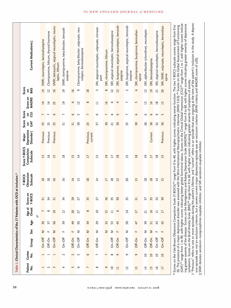
A total of 18 patients were enrolled between Jan-uary 2005 and April 2006. One patient withdrew from the study before the procedure, and stimula-tors were implanted in the remaining 17 patients. The stimulator was removed from 1 patient before randomization because of an infection; thus, 16 pa-tients completed the randomization period (Ta-ble 1). At the time of the patients’ inclusion in the study, the mean duration of disease was 18 years (range, 6 to 47); two patients fulfilled the criteria for current major depressive disorder but had a MADRS score that was lower than 20 (Table 1). One patient who abused alcohol, which represented a minor deviation from the protocol, was included because the abuse was revealed between the time of inclusion and surgery and was moderate and limited in time. There was no significant difference in baseline (month 3) clinical characteristics be-tween the patients in the two groups (Table 2). Ta-ble 1 lists the medications the patients were tak-ing at the time of inclusion. Two patients (Patients 5 and 7) were taking no medication at baseline at their request. Medication was held constant during the 10 months of the protocol except for a transient increase in benzodiazepine therapy in three pa-
33p9
AUTHOR:
FIGURE:
JOB:
4-CH/T
RETAKE
SIZE
ICM
CASE
EMail LineH/TCombo
Revised
AUTHOR, PLEASE NOTE: Figure has been redrawn and type has been reset.
Please check carefully.
REG F
Enon
1st2nd
3rd
Mallet
1 of 3
08-21-08
ARTIST: ts
35908 ISSUE:
Inclusion 0 2 3 6 7 10
Evaluation Evaluation Evaluation Evaluation Evaluation
Surgery
Determinationof stimulation
settings
Double-blind phase
Randomization
Sham stimulation
Active stimulation
Active stimulation
Sham stimulation
Double-blind phase
Month
Washout
Figure 1. Randomized, Double-Blind, Crossover Design of the Study.
The study included two 3-month phases (month 3 to month 6 and month 7 to month 10) separated by a 1-month washout period. Patients were evaluated at inclusion, less than 2 months before surgery; 3 months after surgery, before active or sham stimulation; 6 months after surgery, at the end of the first randomization period; 7 months after surgery, at the end of the 1-month washout period; and 10 months after surgery, at the end of the second ran-domization period. The optimal stimulation settings for the randomization period were determined 2 months after surgery by the neurologist at each center, who was unaware of the randomized treatment assignments.

Subthalamic Nucleus Stimulation in Severe OCD
n engl j med 359;8 www.nejm.org august 21, 2008 37
tients (two during the on-stimulation period and one during the off-stimulation period) and aug-mentation of neuroleptic treatment in one patient (off-stimulation period) owing to exacerbated anxi-ety. All 16 patients completed both periods of the study.
Efficacy of the Stimulation
The Y-BOCS score was significantly lower at the end of the active stimulation (on-stimulation pe-
riod) than at the end of the sham stimulation (off-stimulation period) (mean score, 19±8 vs. 28±7; P = 0.01), independently of the group and the pe-riod. We did not detect any significant carryover effect in Y-BOCS scores (P = 0.71), indicating that the effects of the first treatment period did not persist after the washout period. Patients who were assigned to have active stimulation first and sham stimulation second (the on–off group) tended to have a larger treatment effect as measured by the
33p9
AUTHOR
FIGURE
JOB: ISSUE:
4-CH/T
RETAKE 1st2nd
SIZE
ICM
CASE
EMail LineH/TCombo
Revised
AUTHOR, PLEASE NOTE: Figure has been redrawn and type has been reset.
Please check carefully.
REG F
FILL
TITLE3rd
Enon ARTIST:
Mallet
2a-d
8-21-08
mst
35908
A C
DB
Figure 2. Atlas-Based Targeting on Preoperative MRI and Localization of the Electrodes and Contacts on Postoperative MRI, as Shown in Patient 1.
Panel A shows the preoperative atlas-based MRI alignment, performed with the use of a three-dimensional histolog-ic atlas of the basal ganglia, adapted to fit the patient’s brain geometry. The tracings of the putamen (blue) and thal-amus (green) are well aligned with the corresponding structures in the axial plane. Panel B shows localization of the target in the anteromedial subthalamic nucleus (stn) (intersection of the red lines) in the axial plane, near the boundary between its associative (violet) and limbic (yellow) territories. The sensorimotor territory is green. The colored structures include the caudate nucleus (cd), cerebral peduncle (cp), optic tract (ot), red nucleus (rn), puta-men (pu), and zona incerta (zi). Panel C shows a three-dimensional, superior view of the postoperative MRI scan in the axial plane, with the two electrodes entering the associative territory of the subthalamic nucleus (violet). The sensorimotor territory is green, and the limbic territory is yellow. Panel D shows oblique views of each subthalamic nucleus, presented along the long axis of each electrode with transparent rendering of the three territories. The ac-tive contacts (yellow) are in the anteromedial (probable associative–limbic) part of the subthalamic nucleus. The as-sociative territory is violet, the sensorimotor territory is green, and the limbic territory is yellow.
T h e n e w e ngl a nd j o u r na l o f m e dic i n e
n engl j med 359;8 www.nejm.org august 21, 200838
Tabl
e 1.
Clin
ical
Cha
ract
eris
tics
of th
e 17
Pat
ient
s w
ith O
CD
at I
nclu
sion
.*
Patie
ntN
o.C
ente
r N
o.G
roup
Sex
Age
Age
at
O
nset
Scor
e on
Y-
BO
CS
Y-B
OC
S
Obs
essi
on
Subs
cale
Scor
e Y-
BO
CS
C
ompu
lsio
n Su
bsca
le
Maj
or
Dep
ress
ive
Dis
orde
r†
Scor
e on
G
AF
Scor
e on
C
GI
Scor
e on
M
AD
RS
Scor
e on
B
AS
Cur
rent
Med
icat
ions
‡
yr
11
On–
Off
M36
731
1813
Prev
ious
357
1414
SNR
I, ne
urol
eptic
, ben
zodi
azep
ine
21
Off–
On
F37
834
1816
Prev
ious
357
1412
Clo
mip
ram
ine,
lith
ium
, thy
roid
hor
mon
e
31
On–
Off
M56
1031
1615
Prev
ious
356
2516
SNR
I, te
trac
yclic
, aty
pica
l neu
role
ptic
, neu
ro-
lept
ic, l
ithiu
m
42
On–
Off
F39
1234
1618
317
1919
SNR
I, cl
omip
ram
ine,
bet
a-bl
ocke
r, b
enzo
di-
azep
ine
52
Off–
On
F49
2029
1415
316
616
65
On–
Off
M50
2727
1314
305
129
Clo
mip
ram
ine,
bet
a-bl
ocke
r, v
alpr
oate
, neu
-ro
lept
ic, b
enzo
diaz
epin
e
76
Off–
On
M34
835
1817
Prev
ious
, cu
rren
t25
78
18
87
Off–
On
M29
1027
1413
376
113
SRI,
atyp
ical
neu
role
ptic
, val
proa
te, c
lona
ze-
pam
97
M32
1536
1719
307
168
SRI,
clom
ipra
min
e, li
thiu
m
108
On–
Off
M53
1135
1817
216
612
SRI,
atyp
ical
neu
role
ptic
, ben
zodi
azep
ine
119
Off–
On
F45
630
1218
306
412
SRI,
busp
iron
e, a
typi
cal n
euro
lept
ic, b
enzo
di-
azep
ine
129
On–
Off
M50
1435
2015
357
72
SRI,
busp
iron
e, a
typi
cal n
euro
lept
ic, b
enzo
di-
azep
ine
139
Off–
On
F47
1731
1318
306
56
SRI,
clom
ipra
min
e, b
uspi
rone
, ben
zodi
az-
epin
e
1410
Off–
On
M39
2137
1819
266
1313
SRI,
alph
a-bl
ocke
r (c
loni
dine
), n
euro
lept
ic
1510
Off–
On
M51
1635
1817
Cur
rent
366
1619
SRI,
benz
odia
zepi
ne
1610
On–
Off
F43
1132
1418
326
1821
SRI,
atyp
ical
neu
role
ptic
, ben
zodi
azep
ine
1710
On–
Off
F42
1730
1515
Prev
ious
365
1526
SRI,
SNR
I, va
lpro
ate,
neu
role
ptic
, ben
zodi
az-
epin
e
* Sc
ores
on
the
Yale
-Bro
wn
Obs
essi
ve C
ompu
lsiv
e Sc
ale
(Y-B
OC
S)24
ran
ge fr
om 0
to
40, w
ith h
ighe
r sc
ores
indi
catin
g w
orse
func
tion.
The
tw
o Y-
BO
CS
subs
cale
sco
res
rang
e fr
om 0
to
20. T
he p
rese
nce
of a
maj
or d
epre
ssiv
e di
sord
er w
as a
sses
sed
with
the
Min
i-Int
erna
tiona
l Neu
rops
ychi
atri
c In
terv
iew
(M
INI
5.0.
0).28
Sco
res
on t
he G
loba
l Ass
essm
ent
of F
unct
ioni
ng
(GA
F)25
ran
ge fr
om 1
to
90, w
ith h
ighe
r sc
ores
indi
catin
g be
tter
glo
bal f
unct
iona
l sta
tus.
Sco
res
on t
he C
linic
al G
loba
l Im
pres
sion
(C
GI)
26 r
ange
from
1 t
o 7,
with
hig
her
scor
es in
dica
t-in
g gr
eate
r se
veri
ty o
f the
dis
ease
. Sco
res
on t
he M
ontg
omer
y an
d Å
sber
g D
epre
ssio
n Sc
ale
(MA
DR
S)30
ran
ge fr
om 0
to
60, w
ith h
ighe
r sc
ores
indi
catin
g gr
eate
r se
veri
ty o
f dep
ress
ive
sym
ptom
s. S
core
s on
the
Bri
ef A
nxie
ty S
cale
(B
AS)
38 r
ange
from
0 t
o 60
, with
hig
her
scor
es in
dica
ting
grea
ter
seve
rity
of s
ympt
oms
of a
nxie
ty.
† “
Prev
ious
” re
fers
to
one
or m
ore
epis
odes
dur
ing
the
patie
nt’s
life
time,
and
“cu
rren
t” r
efer
s to
an
epis
ode
that
was
ong
oing
at
the
time
of t
he p
atie
nt’s
incl
usio
n in
the
stu
dy. A
dep
res-
sive
epi
sode
was
not
an
excl
usio
n cr
iteri
on, b
ut a
sev
ere
curr
ent
maj
or d
epre
ssiv
e ep
isod
e w
as a
n ex
clus
ion
crite
rion
(M
INI
crite
ria
and
MA
DR
S sc
ore
of ≥
20).
‡ S
NR
I de
note
s se
roto
nin–
nore
pine
phri
ne r
eupt
ake
inhi
bito
r, a
nd S
RI
sero
toni
n-re
upta
ke in
hibi
tor.

Subthalamic Nucleus Stimulation in Severe OCD
n engl j med 359;8 www.nejm.org august 21, 2008 39
Y-BOCS score than those who had sham stimula-tion first and active stimulation second (the off–on group) (P=0.06 for the period effect). The GAF score (in which higher scores indicate higher lev-els of functioning) was significantly higher after active stimulation than after sham stimulation (mean score at the end of active stimulation, 56±14 vs. 43±8; P = 0.005). The CGI score (in which lower scores indicate lesser severity of disease) was sig-nificantly lower at the end of active stimulation than at the end of sham stimulation (P = 0.008), with more improvement during active stimulation observed in the on–off group than in the off–on group (P = 0.03 for the period effect). Scores on MADRS, the Brief Scale for Anxiety, neuropsycho-logical ratings, and the Sheehan Disability Scale at the end of active stimulation did not differ signifi-cantly from the scores at the end of sham stimu-lation. At the end of the first phase (i.e., 3 months after randomization), six of eight patients (75%) had a response as measured by the Y-BOCS score and eight of eight (100%) had a response as mea-sured by the GAF after active stimulation, as com-pared with three of eight (38%) as measured by both Y-BOCS and GAF after sham stimulation (Fig. 3B). In addition, five of eight patients (62%) had an increase in the GAF score to 51 after active stim-ulation as compared with one of eight (12%) after sham stimulation (Fig. 3B).
Electrode Localization and Stimulation Settings
For 27 of 34 implanted electrodes (79%), the elec-trode trajectory that was chosen was the central one. The anterior trajectory was chosen for 4 elec-trodes, the posterior for 2, and the internal for 1. Of the 32 electrodes that were implanted in the 16 study patients, 4 electrodes, in 4 different pa-tients, were not localized within the subthalamic nucleus — 3 were medial to the subthalamic nu-cleus in zona incerta and field H2 of Forel, and 1 was lateral to the subthalamic nucleus in the in-ternal capsule. However, each patient had at least one contact within the subthalamic nucleus. Post-randomization anatomical analysis showed that among the 33 contacts selected as therapeutic, stimulation reached the anteromedial part of the subthalamic nucleus in 24 (Fig. 2), the zona incerta in 4, the internal capsule in 4, the substantia nigra in 3, and field H2 of Forel in 2 (contacts located at the boundary of two regions were counted twice). In two patients, stimulation was applied unilater-
ally; in one patient (Patient 8), adverse effects were noted on all contacts from one electrode during the tests 2 months after surgery, and in the other patient (Patient 2), one Soletra stimulator was dis-connected owing to infection. Current was deliv-ered through one contact for 27 of 30 electrodes, and two contiguous contacts were used in 3 elec-trodes (bipolar stimulation). The mean (±SD) volt-age was 2.0±0.8 V.
Adverse Events
Fifteen serious adverse events, of which four were related to surgery, were reported in 11 patients (Table 3). The most serious event was a parenchy-mal brain hemorrhage resulting in a permanent finger palsy in one patient. Seven transient motor and psychiatric symptoms induced by active stim-ulation occurred in the first month of stimulation and resolved spontaneously or rapidly after adjust-ment of the setting. Four serious adverse events that were unrelated to either surgery or stimula-tion were reported in one patient before surgery and in two patients during the washout and sham-stimulation phases. Twenty-three nonserious ad-verse events were reported in 10 patients (Table 3). During the active-stimulation period, seven behav-ioral adverse events were reported in five patients.
Discussion
In this double-blind, crossover trial, stimulation of the subthalamic nucleus reduced symptoms in patients with severe, highly refractory, primary OCD, with no concomitant neuropsychological del-eterious effects. Moreover, the reversibility of the stimulation was demonstrated by the fact that symptoms of OCD tended to return to baseline in patients who underwent stimulation in the first period. There was no significant effect of the stim-ulation on measures of depression or anxiety, neu-ropsychological measures, or self-assessment of disability. There were 15 serious adverse events, of which 4 were related to the surgical procedure, including 1 intracerebral hemorrhage and 2 infec-tions requiring removal of the electrode, and 7 were related to the stimulation and were transient. Therefore, the benefits of this surgical treatment for symptoms of OCD should be carefully weighed against the potential occurrence of such serious adverse events.
The improvement in scores observed in the prerandomization postoperative period (months

T h e n e w e ngl a nd j o u r na l o f m e dic i n e
n engl j med 359;8 www.nejm.org august 21, 200840
0 to 3) and the trend toward an effect of the order of the study treatments (in favor of the patients who underwent active stimulation during the first period) could reflect nonspecific therapeutic or placebo effects attributable to the positive effect of enrollment and surgery. However, the crossover, double-blind design of the study and the improve-ment in patients who underwent active stimula-tion during the second period (the off–on group) do not support this hypothesis. It is unlikely that
the decrease in obsessive–compulsive symptoms reflected an antidepressant effect,11,12 since no mood changes were reported during the study. Finally, the placement of the electrode in the sub-thalamic nucleus was meticulously determined and verified with the use of a precise, well-con-trolled procedure,13,20 and the intensity of the stimulation was sufficiently limited to confine the current to the targeted part of the structure.13
This study confirms our previous finding that
Table 2. Changes in the Severity of OCD, Global Health and Functioning, Anxiety, and Depression.
Scale Active Stimulation Followed by Sham Stimulation (On–Off Group)
Baseline (Month 3)
End of On Period (Month 6)
Start of Off Period (Month 7)
End of Off Period
(Month 10)
median (range)
Y−BOCS
Overall score 30 (18 to 37) 19 (0 to 28) 28 (24 to 32) 30 (18 to 36)
Obsession subscale 14 (9 to 19) 10 (0 to 13) 16 (12 to 17) 15 (9 to 18)
Compulsion subscale 15 (9 to 19) 8 (0 to 15) 14 (9 to 15) 16 (9 to 18)
GAF 39 (30 to 48) 52 (45 to 90) 40 (35 to 56) 41 (31 to 50)
CGI 6 (5 to 7) 4 (1 to 5) 6 (5 to 6) 6 (5 to 7)
SDS‡
Work 9 (7 to 10) 4 (0 to 10) 8 (3 to 10) 9 (1 to 10)
Social life and home activities 9 (8 to 10) 4 (0 to 10) 8 (4 to 10) 8 (6 to 10)
Family life and home responsibilities 9 (8 to 10) 7 (1 to 8) 8 (4 to 10) 8 (6 to 10)
MADRS 8 (3 to 26) 8 (0 to 15) 13 (4 to 27) 16 (4 to 27)
BAS 11 (3 to 23) 9 (2 to 15) 12 (5 to 21) 12 (1 to 24)
Hopkins Verbal Learning Test — no. of words
24 (12 to 31) 23 (15 to 29) 24 (18 to 32) 26 (15 to 32)
Trail Making Test
Test A — sec 48 (30 to 91) 40 (26 to 66) 44 (30 to 81) 32 (22 to 64)
Test B−A — sec§ 52 (34 to 114) 81 (38 to 202) 42 (−1 to 93) 58 (27 to 174)
Stroop interference index¶ 6 (−8 to 1) 4 (−2 to 16) 7 (−2 to 17) 5.5 (−10 to 19)
Digit ordering — no. of correct responses 84 (38 to 97) 82 (46 to 97) 82 (38 to 93) 85 (34 to 96)
Digit symbol coding — no. of correct responses
32 (11 to 53) 34 (20 to 57) 40 (17 to 48) 40 (18 to 60)
Lexical fluency — no. of words 50 (38 to 81) 52 (37 to 80) 47 (41 to 75) 50 (36 to 89)
* Bonferroni correction was applied to the two Y−BOCS subscales, the two global health and functioning measures (GAF, CGI), the two psychiatric measures (MADRS, BAS), the three SDS scores, and the seven neuropsychological tests (Hopkins Verbal Learning Test,40 Trail Making Test A,41 Trail Making Test B−A, Interference Stroop score,42 Digit Order ing,43 Digit Symbol Coding,44 and Lexical Fluency). All carryover and period effects were not significant except for the period effect for the CGI scale (P = 0.03).
† P values are for the between-group comparison of the difference between active and sham stimulation at the end of each period (month 6 and month 10).
‡ The range for each subscale of the SDS is 1 to 10; higher numbers indicate greater disability.§ Test B-A refers to the score on Trail Making Test B minus the score on Trail Making Test A.¶ Higher positive values on the stroop interference index indicate greater difficulty in blocking interference. BAS denotes
Brief Scale for Anxiety, CGI Clinical Global Impression, GAF Global Assessment of Functioning, MADRS Montgomery and Åsberg Depression Scale, SDS Sheehan Disabilities Scale, and Y−BOCS Yale-Brown Obsessive Compulsive Scale.

Subthalamic Nucleus Stimulation in Severe OCD
n engl j med 359;8 www.nejm.org august 21, 2008 41
obsessive–compulsive symptoms are reduced af-ter stimulation of the anteromedial subthalamic nucleus,17 which receives limbic and associative cortical information through an orbitofrontal–striato–pallido–thalamo–cortical circuit.31 We therefore propose that the decrease in obsessive–compulsive symptoms is due to changes in neu-ronal activity in the subthalamic nucleus, a theory that is consistent with the concept that the sub-thalamic nucleus is an integrative center for the motor, cognitive, and emotional components of behavior.13 Moreover, considering that patients with OCD are engaged in repetitive thoughts that result in the deferral of decision making and ac-tion, we propose that stimulation of the subtha-lamic nucleus may modify the maintenance of a decision-deferring process, as shown in patients with Parkinson’s disease,45 and therefore decrease obsessive–compulsive symptoms.
Previous studies of stimulation in patients with
OCD have consisted of uncontrolled, open-label designs3; therefore, a comparison of the findings of those studies with the results of our study is difficult. Two blinded procedures in four patients involved the anterior limb of the internal capsule and produced limited benefit with high voltages.7,9 In an open collaborative study involving 10 pa-tients whose preoperative clinical characteristics were similar to those of the patients in our study, Y-BOCS scores were reduced by more than 25% in 50% of the patients after 3 months of stimulation of the ventral striatum12; in contrast, 75% of the patients in our study had reduced Y-BOCS scores. After 36 months of unblinded stimulation that allowed optimal management of the settings in eight patients, the asymptotic best values for the OCD-severity and global-functioning scores12 showed less improvement than the scores in our study after 3 months of double-blind stimulation. In all previous studies,6-12 electrical-stimulation
Sham Stimulation Followed by Active Stimulation (Off–On Group)*
Difference between Active and Sham Stimulation in the
On–Off Group
Difference between Active and Sham Stimulation in the
Off–On Group
P Value for Treatment
Effect†
Baseline (Month 3)
End of Off Period (Month 6)
Start of On Period (Month 7)
End of On Period
(Month 10)
median (range) mean (95% CI)
31 (21 to 36) 26 (13 to 36) 28 (15 to 37) 24 (9 to 30) −13 (−23 to −3) −4 (−12 to 5) 0.01
15 (11 to 19) 12 (7 to 18) 14 (8 to 19) 12 (0 to 15) −6 (−12 to −0.8) −21 (−7 to 3) 0.04
16 (10 to 19) 13 (6 to 18) 15 (7 to 18) 12 (9 to 15) −7 (−12 to −2) −2 (−5 to 2) 0.03
40 (25 to 51) 42 (25 to 55) 40 (25 to 55) 52 (35 to 80) 16 (3 to 30) 12 (−0.6 to 25) 0.005
6 (5 to 7) 5 (3 to 7) 5 (3 to 7) 5 (2 to 6) −3 (−4 to −0.8) −0.3 (−2 to 1) 0.008
9 (1 to 10) 8 (3 to 10) 8 (3 to 10) 8 (2 to 10) −4 (−7 to −0.5) −0.6 (−3 to 2) 0.15
8 (5 to 10) 8 (0 to 10) 8 (0 to 10) 8 (1 to 9) −3 (−6 to 0.5) 0.1 (−3 to 3) 0.66
8 (3 to 10) 7 (0 to 9) 5 (0 to 10) 5 (1 to 10) −2 (−5 to 0.6) −0.4 (−4 to 3) 1
6 (2 to 19) 8 (4 to 24) 8 (4 to 20) 12 (3 to 24) −9 (−19 to 1) 2 (−8 to 11) 0.58
6 (5 to 24) 6 (1 to 27) 6 (2 to 21) 10 (0 to 22) −3 (−11 to 4) 0.6 (−8 to 7) 1
28 (12 to 32)
28 (18 to 30) 27 (16 to 36) 28 (18 to 33) −2 (−4 to 0.6) 2 (−1 to 5) 1
32 (24 to 53) 32 (20 to 76) 26 (19 to 48) 26 (21 to 50) 7 (1 to 13) −6 (−17 to 5) 1
50 (31 to 127) 38 (17 to 77) 36 (24 to 67) 45 (19 to 96) 7 (−32 to 46) 3 (−16 to 22) 1
1 (−7 to 12) 2 (−22 to 12) 0.5 (−3 to 21) 1 (−5 to 9) 0.1 (−6.4 to 6.6) 2 (−6 to 9) 1
82 (41 to 98) 86 (29 to 92) 85 (36 to 91) 84 (47 to 96) 1 (−5 to 7) 3 (−3 to 9) 1
36 (24 to 57) 40 (26 to 55) 42 (19 to 62) 43 (27 to 60) −3 (−8 to 2) 3 (−3 to 9) 1
60 (33 to 79) 58 (37 to 84) 58 (38 to 83) 60 (33 to 79) 1 (−5 to 8) 0.4 (−7 to 8) 1

T h e n e w e ngl a nd j o u r na l o f m e dic i n e
n engl j med 359;8 www.nejm.org august 21, 200842
settings were more variable and higher than those in our study. The rate of serious adverse events related to the surgical procedure in our study was similar to that reported previously for stimulation of the subthalamic nucleus in patients with Par-
kinson’s disease and for neurostimulation in pa-tients with OCD (a hematoma rate of 1 to 6% and an infection rate of 1 to 15%).12,19,46 In our study, hypomania was the main psychiatric serious ad-verse event. The fact that the symptoms of hypo-
33p9
40
Y-B
OC
S Sc
ore 30
10
20
0I 3 6 70 10 I 3 6 70 10
Off Off
OnOn
70
GA
F Sc
ore
60
30
40
50
0
Off Off
OnOn
B
A
AUTHOR:
FIGURE:
JOB:
4-CH/T
RETAKE
SIZE
ICM
CASE
EMail LineH/TCombo
Revised
AUTHOR, PLEASE NOTE: Figure has been redrawn and type has been reset.
Please check carefully.
REG F
Enon
1st
2nd
3rd
Mallet
3 of 3
08-21-08
ARTIST: ts
35908 ISSUE:
40
Y-B
OC
S Sc
ore 30
10
20
0I 0 3 6 7 10
100
GA
F Sc
ore
75
25
50
0I 0 3 6 7 10
100
GA
F Sc
ore
75
25
50
40
Y-B
OC
S Sc
ore 30
10
20
0I 0 3 6 7 10
0I 0 3 6 7 10
Month Month
Month Month
Month Month
On–Off On–Off
Off–On Off–On
Figure 3. Changes in the Severity of OCD and Global Functioning in 16 Patients during the Crossover Study of Stimulation of the Subthalamic Nucleus.
Panel A shows the mean (±SD) scores on the Yale-Brown Obsessive Compulsive Scale (Y-BOCS) and the Global As-sessment of Functioning (GAF) scale for the on–off group and the off–on group. Data are shown at the time of in-clusion in the study (I), at the time of surgery (month 0), before (month 3) and after (month 6) the first period of active or sham stimulation, and before (month 7) and after (month 10) the second period of active or sham stimu-lation. Panel B shows the individual Y-BOCS and GAF scores for the on–off group and the off–on group. The active-stimulation period is represented in gray. One patient in each group (Patient 6 in the on–off group and Patient 4 in the off–on group) had higher Y-BOCS scores and one patient in the off–on group had a lower GAF score during ac-tive stimulation (dashed lines).

Subthalamic Nucleus Stimulation in Severe OCD
n engl j med 359;8 www.nejm.org august 21, 2008 43
Table 3. Adverse Events.
Adverse Event Permanent Transient
no. of events
Serious
Before surgery
Anxiety 0 1 (Patient 1)
After surgery, before randomization (month 0 to month 3)
Intracerebral hemorrhage 1 (Patient 6) 0
Clumsiness and diplopia with perielectrode edema 0 1 (Patient 13)
Infection leading to removal of pulse generator and electrode 2 (Patients 4 and 9)* 0
After randomization (month 3 to month 10)
Active-stimulation period
Hypomanic status 0 3 (Patients 3, 4, and 10)
Anxiety 0 2 (Patients 1 and 14)
Disabling dyskinesias with impulsivity 0 1 (Patient 12)
Facial asymmetry, dysarthria, dysphagia, and walking difficulties 0 1 (Patient 5)
Sham-stimulation period
Anxiety 0 1 (Patient 12)
Depressive symptoms with suicidal ideas 0 2 (Patient 17)
Nonserious
After surgery, before randomization (month 0 to month 3)
Urinary infection 0 1 (Patient 10)
Nocturnal enuresis 0 1 (Patient 16)
Headaches 0 1 (Patient 11)
Pain associated with the neurostimulator 0 1 (Patient 9)
Bronchitis 0 1 (Patient 15)
Anxiety 0 1 (Patient 14)
Obsessions 0 1 (Patient 11)
Lumbosciatic syndrome 0 1 (Patient 17)
Dyspnea 0 1 (Patient 12)
Hypomanic symptoms while stimulation settings were being determined 0 1 (Patient 1)
After randomization (month 3 to month 10)
Active-stimulation period
Dyskinesia 0 1 (Patient 13)
Hypomanic status with irritability and impulsivity 0 2 (Patient 12)
Manic symptoms with euphoria 0 1 (Patient 14)
Depressive symptoms 0 1 (Patient 11)
Anxiety 0 1 Patient (14)
Obsessions 0 1 (Patient 13)
Peripheral vertigo 0 1 (Patient 12)
Achilles tendinitis 0 1 (Patient 1)
Diagnosis of diabetes mellitus 1 (Patient 15) 0
Sham-stimulation period
Obsessions 0 1 (Patient 12)
Influenza-like syndrome 0 1 (Patient 16)
Hemorrhoidectomy 0 1 (Patient 15)
* In Patient 9, the two electrodes and the pulse generator were removed, and the patient did not complete the random-ization period.

T h e n e w e ngl a nd j o u r na l o f m e dic i n e
n engl j med 359;8 www.nejm.org august 21, 200844
mania resolved after adjustment of the stimulation settings suggests that they were induced by stimu-lation of the subthalamic nucleus and highlights the necessity of multidisciplinary expertise in the medical care of patients undergoing stimulation of the subthalamic nucleus. The study did not show an effect on functional impairment at work as measured by the Sheehan Disability Scale. Fifteen of 16 patients had not worked for many years, and given the short (3-month) crossover trial period, we did not expect patients to return to work at the end of the protocol. Finally, interesting short-term and long-term therapeutic results have been shown in studies of the use of cingulotomy or capsulo-tomy in patients with refractory OCD,3,47 with vari-able surgical risks and adverse events; however, given the methodologic heterogeneity of all these procedures, a direct comparison would be neces-sary to assess precisely the advantages and limi-tations of each strategy.
The multicenter design of this study has poten-tial limitations. Variation in the targeting of the stimulation was minimized by anatomical and electrophysiological identification performed si-multaneously by local and coordinating teams. In addition, patients who had unexpected responses were examined thoroughly at the detailed clinical follow-up examination that was required by the study protocol. For example, two patients had a higher Y-BOCS score after active stimulation (Fig. 3). In one of these patients (Patient 6), the level of anxiety increased when a palsy of his right hand, the hand with which he had engaged in compul-sive activities preoperatively, developed as a result of a parenchymal brain hemorrhage after surgery. In the other patient (Patient 2), because low-inten-sity stimulation of the subthalamic nucleus in-duced side effects, the intensity chosen for the ac-tive-stimulation period (month 7) may have been too low to reduce the symptoms of OCD. More-over, adjustment of the settings was purposely limited in this study to be under the threshold for the induction of side effects in order to preserve
the blinded nature of the protocol. Thus, the stimulation settings are a further possible limita-tion of the study. Continued follow-up of patients undergoing stimulation of the subthalamic nucle-us is needed to assess any long-term effects of stimulation that have not yet been identified.
In conclusion, findings from this 3-month crossover study suggest that stimulation of the subthalamic nucleus may lessen the severity of obsessive–compulsive symptoms and improve glo-bal functioning in patients with refractory, severe OCD. Serious adverse events occurred in 11 of the 17 patients in whom stimulators were implanted. The occurrence of severe adverse events, the small number of patients, and the short duration of the study highlight the risks of stimulation of the sub-thalamic nucleus and the need for larger studies with longer follow-up. In addition to assessment in a larger number of patients, a comparison with other stimulation targets and surgical procedures would be desirable, as would an evaluation of the long-term benefits of stimulation of the subtha-lamic nucleus in patients with OCD, notably with respect to their quality of life and their ability to function in social and work environments.
Supported by grants from the Direction Régionale de la Re-cherche Clinique Assistance-Publique-Hôpitaux de Paris (AOM 03141) and the ANR Agence Nationale de la Recherche Program for Young Researchers (R05121DS). The stimulators were pur-chased from Medtronic, which had no role in the study. Medtronic provided funds for the meetings of the investigators of the study.
Dr. Cornu reports receiving consulting fees from Medtronic; Dr. Houeto, lecture fees from Medtronic; Dr. Benadid, lecture fees and grant support from Medtronic; Dr. Agid, consulting fees from Medtronic; Dr. Krack, consulting and lecture fees from Medtronic; and Dr. Millet, grant support from Medtronic. No other potential conflict of interest relevant to this article was reported.
We thank Drs J. Adès, J. Cottraux, M. Goudemand, J.C. Aussi-loux, S. Blond, J. Feingold, and D. Sicard for their independent contributions to the study with respect to ethical and safety is-sues; staff members of Association Française de Personnes Souffrant de Troubles Obsessionnels et Compulsifs (the French association of patients with OCD) for help in referring patients, and its president, C. Demonfaucon, for his helpful comments on inclusion criteria and ethics in the preparation of the protocol; and Max Westby and Marie Vidailhet for their thoughtful com-ments on the manuscript.
AppendixFrom INSERM, Avenir Team, Behavior, Emotion, and Basal Ganglia, IFR70 (L.M., M.-L.W., J.Y., E.B.), Clinical Investigation Center (L.M., M.-L.W., V.C., Y.A.), Pitié-Salpêtrière University Hospital, Assistance Publique-Hôpitaux de Paris, Paris; Departments of Psychia-try (M.P., T.B.), Neurosurgery (S.C., A.-L.B.), and Neurology (C. Ardouin, P.P., P.K.), Grenoble University Hospital, Grenoble; Depart-ments of Psychiatry (N.J.) and Neurosurgery (B.B.), University Hospital, Poitiers; INSERM, U796, University of Paris Descartes, Univer-sity Department of Psychiatry (N.B., M.-O.K.), and Department of Neurosurgery (B.D.), Sainte-Anne Hospital, Paris; Departments of Neurosurgery (D.F.) and Psychiatry (V.M.), University Hospital, Nice; Biostatistics and Medical Informatics Unit and Clinical Research Unit, Pitié-Salpêtrière University Hospital, Université Pierre et Marie Curie-Paris 6, EA 3974, Modeling in Clinical Research, Paris (S.T.D.M.); Neurologie & Thérapeutique Expérimentale, INSERM-U679, Université Pierre et Marie Curie-Paris 6, Institut Fédératif de Recherche de Neurosciences Unité Mixte de Recherche S679, Paris (J.Y., Y.A.); Departments of Psychiatry (I.C.) and Neurology (P.D.), University Hospital, Clermont-Ferrand; Departments of Psychiatry (C. Arbus) and Neurosurgery (P.C.), University Hospital, Toulouse;

Subthalamic Nucleus Stimulation in Severe OCD
n engl j med 359;8 www.nejm.org august 21, 2008 45
Departments of Neurosurgery (S.R.) and Neurology (P.D.), University Hospital, Nantes; Department of Psychiatry Charles Perrons Hosptial (B.A.) and Centre National de la Recherche Scientifique (CNRS) UMR5543 (B.A., P.B.), University Bordeaux 2; INSERM-U610 (V.C.), Department of Neurosurgery (P.C.), Department of Radiology (D.D.), Fédération de Neurologie (M.-L.W, Y.A.), and Department of Psychiatry (A.P.), Pitié-Salpêtrière University Hospital, Paris; Laboratoire de Neurosciences Cognitive & Imagerie Cérébrale, CNRS Unité propre de Recherche 640-LENA, Paris (E.B.); Department of Neurology (M.V.) and Department of Psychiatry (B.M.), University Hospital, Rennes; and Department of Neurology, EA 3808, University of Poitiers, CHU de Poitiers (J.L.H.) — all in France.
Members of the French STOC Study Group are as follows: Trial Coordination: L. Mallet. Steering Committee: Y. Agid, B. Aouizerate, C. Arbus, T. Bougerol, P. Damier, D. Fontaine, J.L. Houeto, M.O. Krebs, J.J. Lemaire, L. Mallet, B. Millet, P. Pollak. Logistics and Mo-nitoring: D. Hourton, S. Aprelon, C. Jourdain. Coordinating teams: Anatomy — E. Bardinet, J. Yelnik; Electrophysiology — P. Burbaud, M.L. Welter, A.H. Clair; Neuropsychology — V. Czernecki, M. Vérin. Data Management and Statistical Analysis: S. Tézenas du Montcel, D. Madar. Writing Committee: L. Mallet, A. Pelissolo, S. Tezenas du Montcel, M.L. Welter, J. Yelnik. Centers: Coordinating Center, Paris Pitié-Salpêtrière Hospital — L. Mallet, A. Pelissolo, Y. Agid, P. Cornu, S. Navarro, M.L. Welter, A. Hartmann, B. Pidoux, D. Gra-bli, V. Czernecki, D. Dormont, D. Galanaud, J. Yelnik, E. Bardinet, C. Béhar, Y. Worbe, A.H. Clair, B. Moutaud, Centre d’Investigation Clinique staff and nurses; Bordeaux — B. Aouizerate, P. Burbaud, E. Cuny, D. Guehl; Clermont-Ferrand — P.M. Llorca, I Chéreau, J.J. Lemaire, F. Durif, P. Derost, J. Coste; J. Gabrillargues, M. Barget, I. de Chazeron; Grenoble — T. Bougerol, M. Polosan, A.L. Benabid, S. Chabardès, E. Seigneuret, P. Krack, P. Pollak, C. Ardouin, J.F. Le Bas; Nantes — P. Damier, Y. Lajat, S. Raoul; Nice — V. Mattei, D. Fontaine, M. Borg, P. Paquis, E. Michel, P. Robert; E. Michel, F. Papetti; Paris Sainte-Anne Hospital — N. Baup, B. Devaux, M.O. Krebs, C. Oppenheimer; J.P. Olié, D. Ranoux, M. Chayet; Poitiers — J.L. Houeto, N. Jaafari, B. Bataille, V. Mesnage; R. Gil, V. Audouin, J.L. Senon; Rennes — B. Millet, M. Vérin, D. Drapier, P. Sauleau; S. Drapier; Toulouse — C. Arbus, Y. Lazorthe, P. Chaynes, N. Fabre, M. Simonetta, L. Schmitt, J.A. Lotterie, C. Camassel. Sponsor: N. Best. Independent Safety Committee: J.C Aussilloux, S. Blond, J. Feingold, D. Sicard. Independent Selection Committee: J. Adès, J. Cottraux, M. Goudemand.
References
Stein DJ. Obsessive-compulsive disor-1. der. Lancet 2002;360:397-405.
Pallanti S, Quercioli L. Treatment-re-2. fractory obsessive-compulsive disorder: methodological issues, operational defi-nitions and therapeutic lines. Prog Neurop-sychopharmacol Biol Psychiatry 2006;30: 400-12.
Lipsman N, Neimat JS, Lozano AM. 3. Deep brain stimulation for treatment- refractory obsessive-compulsive disorder: the search for a valid target. Neurosur-gery 2007;61:1-13.
Benabid AL, Wallace B, Mitrofanis J, 4. et al. Therapeutic electrical stimulation of the central nervous system. C R Biol 2005; 328:177-86.
Aouizerate B, Guehl D, Cuny E, et al. 5. Pathophysiology of obsessive-compulsive disorder: a necessary link between phe-nomenology, neuropsychology, imagery and physiology. Prog Neurobiol 2004;72: 195-221.
Nuttin B, Cosyns P, Demeulemeester 6. H, Gybels J, Meyerson B. Electrical stimu-lation in anterior limbs of internal cap-sules in patients with obsessive-compul-sive disorder. Lancet 1999;354:1526.
Nuttin BJ, Gabriëls LA, Cosyns PR, et 7. al. Long-term electrical capsular stimula-tion in patients with obsessive-compul-sive disorder. Neurosurgery 2003;52:1263-74.
Anderson D, Ahmed A. Treatment of 8. patients with intractable obsessive-compul-sive disorder with anterior capsular stim-ulation: case report. J Neurosurg 2003;98: 1104-8.
Abelson JL, Curtis GC, Sagher O, et al. 9. Deep brain stimulation for refractory ob-sessive-compulsive disorder. Biol Psychia-try 2005;57:510-6.
Sturm V, Lenartz D, Koulousakis A, et 10. al. The nucleus accumbens: a target for
deep brain stimulation in obsessive-com-pulsive- and anxiety-disorders. J Chem Neu-roanat 2003;26:293-9.
Aouizerate B, Cuny E, Martin-Guehl 11. C, et al. Deep brain stimulation of the ventral caudate nucleus in the treatment of obsessive-compulsive disorder and ma-jor depression: case report. J Neurosurg 2004;101:682-6.
Greenberg BD, Malone DA, Friehs GM, 12. et al. Three-year outcomes in deep brain stimulation for highly resistant obsessive-compulsive disorder. Neuropsychopharma-cology 2006;31:2384-93. [Erratum, Neurop-sychopharmacology 2006;31:2394.]
Mallet L, Schüpbach M, N’Diaye K, et 13. al. Stimulation of subterritories of the subthalamic nucleus reveals its role in the integration of the emotional and motor aspects of behavior. Proc Natl Acad Sci U S A 2007;104:10661-6.
Ardouin C, Voon V, Worbe Y, et al. 14. Pathological gambling in Parkinson’s dis-ease improves on chronic subthalamic nucleus stimulation. Mov Disord 2006;21: 1941-6.
Houeto JL, Mallet L, Mesnage V, et al. 15. Subthalamic stimulation in Parkinson disease: behavior and social adaptation. Arch Neurol 2006;63:1090-5.
Alegret M, Junqué C, Valldeoriola F, et 16. al. Effects of bilateral subthalamic stimu-lation on cognitive function in Parkinson disease. Arch Neurol 2001;58:1223-7.
Mallet L, Mesnage V, Houeto JL, et al. 17. Compulsions, Parkinson’s disease, and stimulation. Lancet 2002;360:1302-4.
Fontaine D, Mattei V, Borg M, et al. 18. Effect of subthalamic nucleus stimulation on obsessive-compulsive disorder in a pa-tient with Parkinson disease: case report. J Neurosurg 2004;100:1084-6.
Krack P, Batir A, van Blercom N, et al. 19. Five-year follow-up of bilateral stimula-
tion of the subthalamic nucleus in ad-vanced Parkinson’s disease. N Engl J Med 2003;349:1925-34.
Yelnik J, Bardinet E, Dormont D, et al. 20. A three-dimensional, histological and deformable atlas of the human basal gan-glia. I. Atlas construction based on im-munohistochemical and MRI data. Neu-roimage 2007;34:618-38.
Bejjani BP, Dormont D, Pidoux B, et al. 21. Bilateral subthalamic stimulation for Par-kinson’s disease by using three-dimen-sional stereotactic magnetic resonance imaging and electrophysiological guidance. J Neurosurg 2000;92:615-25.
Yelnik J, Damier P, Demeret S, et al. 22. Localization of stimulating electrodes in patients with Parkinson disease by using a three-dimensional atlas-magnetic reso-nance imaging coregistration method. J Neurosurg 2003;99:89-99.
Preisig M, Fenton BT, Matthey ML, 23. Berney A, Ferrero F. Diagnostic Interview for Genetic Studies (DIGS): inter-rater and test-retest reliability of the French ver-sion. Eur Arch Psychiatry Clin Neurosci 1999;249:174-9.
Goodman WK, Price LH, Rasmussen 24. SA, et al. The Yale-Brown Obsessive Com-pulsive Scale. I. Development, use, and reliability. Arch Gen Psychiatry 1989;46: 1006-11.
Diagnostic and statistical manual of 25. mental disorders, 4th ed. text rev.: DSM-IV-TR. Washington, DC: American Psychi-atric Association, 2000.
Clinical Global Impression. In: Guy W. 26. ECDEU assessment manual for psychop-harmacology. Rev. ed. Rockville, MD: Na-tional Institute of Mental Health, 1976:218-22.
Mattis S. Dementia Rating Scale. In: 27. Psychological Assessment Resources, Odes-sa, FL, 1988.

n engl j med 359;8 www.nejm.org august 21, 200846
Subthalamic Nucleus Stimulation in Severe OCD
Sheehan DV, Lecrubier Y, Sheehan 28. KH, et al. The Mini-International Neurop-sychiatric Interview (M.I.N.I.): the devel-opment and validation of a structured di-agnostic psychiatric interview for DSM-IV and ICD-10. J Clin Psychiatry 1998;59:Suppl 20:22-57.
Maffei C, Fossati A, Agostoni I, et al. 29. Interrater reliability and internal consis-tency of the Structured Clinical Interview for DSM-IV axis II personality disorders (SCID-II), version 2.0. J Personal Disord 1997;11:279-84.
Montgomery SA, Asberg M. A new de-30. pression scale designed to be sensitive to change. Br J Psychiatry 1979;134:382-9.
Karachi C, Yelnik J, Tandé D, Trem-31. blay L, Hirsch EC, François C. The palli-dosubthalamic projection: an anatomical substrate for nonmotor functions of the subthalamic nucleus in primates. Mov Dis-ord 2005;20:172-80.
Benabid AL, Koudsie A, Benazzouz A, 32. Le Bas JF, Pollak P. Imaging of subtha-lamic nucleus and ventralis intermedius of the thalamus. Mov Disord 2002;17:Suppl 3:S123-S129.
Karachi C, François C, Parain K, et al. 33. Three-dimensional cartography of func-tional territories in the human striatopal-lidal complex by using calbindin immu-
noreactivity. J Comp Neurol 2002;450: 122-34.
Hutchison WD, Allan RJ, Opitz H, et 34. al. Neurophysiological identification of the subthalamic nucleus in surgery for Parkinson’s disease. Ann Neurol 1998;44: 622-8.
Pollak P, Krack P, Fraix V, et al. Intraop-35. erative micro- and macrostimulation of the subthalamic nucleus in Parkinson’s disease. Mov Disord 2002;17:Suppl 3:S155-S161.
The 2×2 cross-over trial with conti-36. nous data. In: Jones B, Kenward MG. De-sign and analysis of cross-over trials. London: Chapman and Hall, 1989:16-88.
Sheehan DV, Harnett-Sheehan K, Raj 37. BA. The measurement of disability. Int Clin Psychopharmacol 1996;11:Suppl 3:89-95.
Tyrer P, Owen RT, Cicchetti DV. The 38. brief scale for anxiety: a subdivision of the Comprehensive Psychopathological Rat-ing Scale. J Neurol Neurosurg Psychiatry 1984;47:970-5.
Pillon B, Boller F, Levy R, Dubois B. 39. Cognitive deficit and dementia in Parkin-son’s disease. In: Boller F, Cappa S, eds. Aging and dementia. Vol. 6 of Handbook of neuropsychology. 2nd ed. Amsterdam: Elsevier, 2001:311-72.
Brandt J. The Hopkins Verbal Learn-40. ing Test: development of a new memory
test with six equivalent forms. Clin Neu-ropsychol 1991;5:125-42.
Reitan RM. Trail Making Test results 41. for normal and brain-damaged children. Percept Mot Skills 1971;33:575-81.
Stroop JR. Studies of interference in 42. serial verbal reactions. J Exp Psychol 1935;18:643-62.
Cooper JA, Sagar HJ, Jordan N, Harvey 43. NS, Sullivan EV. Cognitive impairment in early, untreated Parkinson’s disease and its relationship to motor disability. Brain 1991;114:2095-122.
Weschler D. Wechsler Memory Scale 44. — Third Edition. San Antonio, TX: The Psychological Corporation, 1997.
Frank MJ, Samanta J, Moustafa AA, 45. Sherman SJ. Hold your horses: impulsivi-ty, deep brain stimulation, and medica-tion in parkinsonism. Science 2007;318: 1309-12.
Rezai AR, Kopell BH, Gross RE, et al. 46. Deep brain stimulation for Parkinson’s disease: surgical issues. Mov Disord 2006; 21:Suppl 14:S197-S218.
Dougherty DD, Baer L, Cosgrove GR, 47. et al. Prospective long-term follow-up of 44 patients who received cingulotomy for treatment-refractory obsessive-compulsive disorder. Am J Psychiatry 2002;159:269-75.Copyright © 2008 Massachusetts Medical Society.
full text of all journal articles on the world wide web
Access to the complete text of the Journal on the Internet is free to all subscribers. To use this Web site, subscribers should go to the Journal’s home page (www.nejm.org) and register by entering their names and subscriber numbers as they appear on their mailing labels. After this one-time registration, subscribers can use their passwords to log on for electronic access to the entire Journal from any computer that is connected to the Internet. Features include a library of all issues since January 1993 and abstracts since January 1975, a full-text search capacity, and a personal archive for saving articles and search results of interest. All articles can be printed in a format that is virtually identical to that of the typeset pages. Beginning 6 months after publication, the full text of all Original Articles and Special Articles is available free to nonsubscribers.