high-frequency hippocampal oscillations activated by optogenetic stimulation of transplanted human...
TRANSCRIPT
Brief Communications
High-Frequency Hippocampal Oscillations Activated byOptogenetic Stimulation of Transplanted HumanESC-Derived Neurons
Juan C. Pina-Crespo,1* Maria Talantova,1* Eun-Gyung Cho,1* Walid Soussou,1 Nima Dolatabadi,1 Scott D. Ryan,1
Rajesh Ambasudhan,1 Scott McKercher,1 Karl Deisseroth,2 and Stuart A. Lipton1
1Del E. Webb Center for Neuroscience, Aging, and Stem Cell Research, Sanford-Burnham Medical Research Institute, La Jolla, California 92037, and2Department of Psychiatry and Behavioral Sciences, Stanford University, Stanford, California 94305
After transplantation, individual stem cell-derived neurons can functionally integrate into the host CNS; however, evidence that neuronsderived from transplanted human embryonic stem cells (hESCs) can drive endogenous neuronal network activity in CNS tissue is stilllacking. Here, using multielectrode array recordings, we report activation of high-frequency oscillations in the � and � ranges (10 –100Hz) in the host hippocampal network via targeted optogenetic stimulation of transplanted hESC-derived neurons.
IntroductionThe ability of transplanted stem cell-derived neurons to re-ceive and send functional excitatory and inhibitory inputs is akey indicator of successful functional integration into the CNS(Kim et al., 2002; Benninger et al., 2003; Wernig et al., 2004;Lee et al., 2007; Li et al., 2008; Weick et al., 2010, 2011);however, it remains to be shown that whole populations oftransplanted human embryonic stem cell (hESC)-derived neu-rons can drive the activity of local neural networks in the brain.Since local neural circuits in organotypic hippocampal slices re-tain the ability to generate synchronized high-frequency oscilla-tions (Fischer et al., 2002), and they can support thematuration and integration of transplanted stem cell-derivedneurons (Benninger et al., 2003; Scheffler et al., 2003; Opitz etal., 2007), we used this system to ask whether transplantedhESC-derived neurons can integrate and drive neuronal net-work activity in the CNS. By combining the high spatial reso-lution provided by optogenetics [via expression of geneticallyencoded photosensitive membrane proteins (Zhang et al.,2006) in transplanted hESC-derived neurons] with electro-physiological recording of local neuronal network activity [us-ing multielectrode array (MEA) techniques (Taketani and
Baudry, 2006)], we were able to elicit high-frequency oscilla-tions in the �/� range (10 –100 Hz) in the host hippocampalnetwork. Since light stimulation specifically activated thetransplanted hESC-derived neurons, we could show that thesenew neurons behaved not only as conductors of electrical im-pulses along a network but could also actively engage localneuronal networks into firing at frequency rates commonlyobserved during normal processing of neural information inthe hippocampus (Traub et al., 1999a).
Materials and MethodshESC culture and neural induction. Differentiation of hESCs into neu-rons was performed using a modification of previously describedmethods (Elkabetz and Studer, 2008; Li et al., 2008; Cho et al., 2011).Briefly, undifferentiated H9 hESCs were grown on a monolayer offibroblasts from human foreskin (Hs27, ATCC). Cell culture mediumwas composed of DMEM/F12, knock-out serum replacement (20%),basic fibroblast growth factor (8 ng/ml), �-mercaptoethanol (0.1mM), and nonessential amino acids (1 mM). Media changes were per-formed daily and cells were subcultured on a weekly basis. For neuralstem/progenitor cell (NPC) induction, hESCs were incubated for 24 hin cell culture medium composed of DMEM/F12:Neurobasal (1:1),B27 serum-free supplement (2%), and N2 serum-free supplement(1%). They were then mechanically dissociated until small clusters ofcells were obtained. These cell clusters were cultured for up to 72 h inthe same medium. For neural expansion, cells were grown for up to6 d in cell culture medium composed of DMEM/F12:Neurobasal (1:1), B27 serum-free supplement (1%), N2 serum-free supplement(0.5%), basic fibroblast growth factor (20 ng/ml), and epidermalgrowth factor (20 ng/ml). Cells were then plated on dishes coated with10 �g/ml laminin and grown for up to 48 h until they took a rosette-like appearance. These rosette-neural stem cells (R-NSCs) were re-trieved using a needle and transferred to a dish where they werecultured for 3–30 d in the same medium.
Immunocytochemistry and immunoblots. Epifluorescence micros-copy for immunocytochemical analysis and gel electrophoresis fol-lowed by immunoblot analysis were performed as previouslydescribed (Li et al., 2008; Cho et al., 2011). Primary antibodies in-
Received Aug. 3, 2012; accepted Sept. 9, 2012.Author contributions: J.C.P.-C., M.T., E.-G.C., W.S., S.M., and S.A.L. designed research; J.C.P.-C., M.T., E.-G.C.,
W.S., N.D., S.D.R., S.M., and R.A. performed research; K.D. contributed unpublished reagents/analytic tools; J.C.P.-C.,M.T., E.-G.C., W.S., and S.M. analyzed data; J.C.P.-C., M.T., E.-G.C., and S.A.L. wrote the paper.
This work was supported by California Institute for Regenerative Medicine (CIRM) Comprehensive Grant RC1-00125-1 and NIH Grants P01 HD29587, P01 ES016738, P30 NS076411, R01 EY05477, and R01 EY09024 (to S.A.L.).We thank Traci Fang Newmeyer for excellent technical assistance.
*J.C.P.-C., M.T., and E.-G.C. contributed equally to this work.Correspondence should be addressed to Stuart A. Lipton or Juan C. Pina-Crespo at the above address. E-mail:
[email protected] or [email protected]. Cho’s present address: R&D Center, Amorepacific Corporation, Yongin-si, Gyeonggi-do, South Korea.W. Soussou’s present address: Quantum Applied Science and Research, San Diego, California.DOI:10.1523/JNEUROSCI.3735-12.2012
Copyright © 2012 the authors 0270-6474/12/3215837-06$15.00/0
The Journal of Neuroscience, November 7, 2012 • 32(45):15837–15842 • 15837
cluded the following: �-tubulin III (TuJ1; mouse, 1:1000, Covance),microtubule-associated protein 2 (MAP2; mouse, 1:1000, Sigma),synaptophysin (rabbit, 1:1000, Dako), tyrosine hydroxylase (mouse,1:1000, Pel-Freez; rabbit, 1:1000, Millipore Bioscience Research Re-agents), glutamate transporter 1 protein (mouse, 1:1000), and glu-tamic acid decarboxylase (rabbit, 1:500, Abcam). Alexa 488-, 555-, or647-conjugated goat anti-mouse or anti-rabbit IgGs (1:1000, Invitro-gen) were used as secondary antibodies.
Lentiviral infection of R-NSCs and transplantation. Cells at the R-NSCstage were transduced with lentiviral expression vectors. As previouslydescribed, to enhance neurogenesis, we expressed a lentivirus containinga constitutively active form of the transcription factor myocyte enhancer
factor-2 (MEF2CA) (Li et al., 2008; Cho et al., 2011). Additional lentiviralvectors included pSynapsinI-hChR2-EGFP and pEF1-NpHR-mCherry(Zhang et al., 2006). Once infected, R-NSCs were dissociated and trans-ferred to plates coated with a mixture of 10 �g/ml poly-l-ornithine and 1�g/ml laminin. These R-NSCs were grown to become human NPCs(hNPCs) as a monolayer in the medium for neural expansion. At thispoint, hNPCs were enzymatically dissociated into a single cell suspensionusing Accutase. For transplantation, hNPCs were washed in Ca 2�/Mg 2�-free HBSS and concentrated. Cells were placed onto the CA3 re-gion using a glass micropipette attached to a nanoliter injector (Nanoject,Drummond). Each slice received a single injection of a single-cell sus-pension containing 50,000 –100,000 cells. Slices were placed back in a
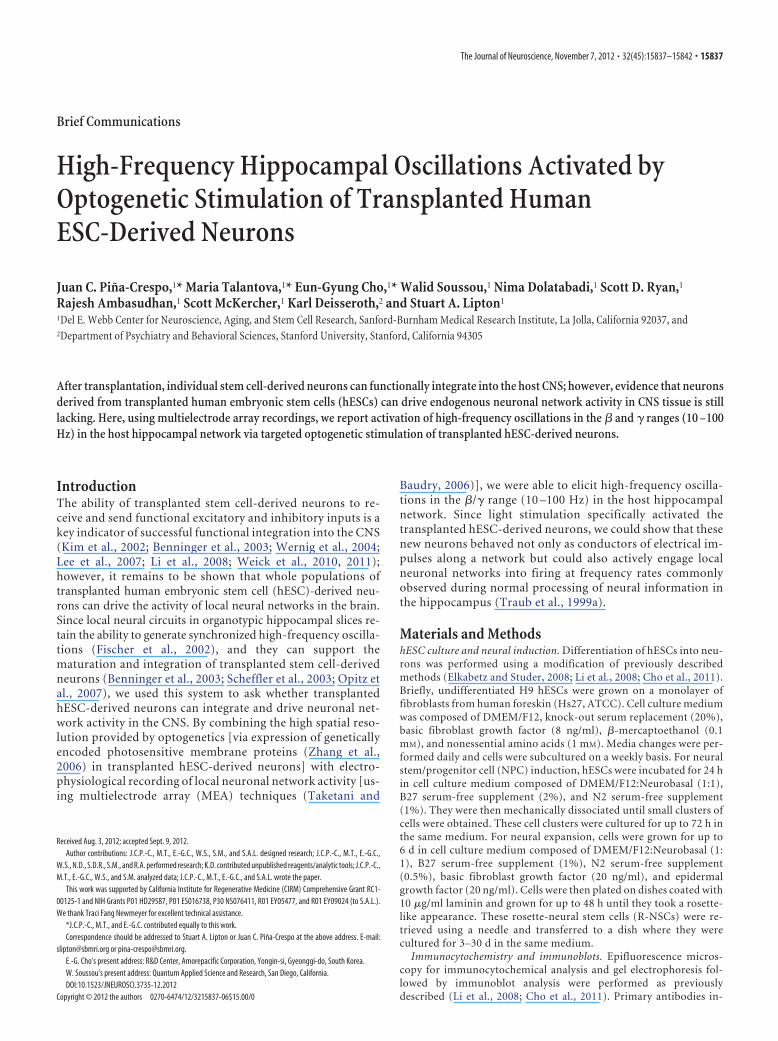
Figure 1. hESC-derived neurons programmed with MEF2CA expressing both ChR2 and NpHR show light-activated whole-cell responses. A, Differential interference contrast microscopy andepifluorescent images of hESC-derived neurons in culture that were transduced with lentiviral expression vectors carrying genes encoding light-gated conductances [channelrhodopsin-2 (pSynI-ChR2-EYFP) and halorhodopsin (pEF1-NpHR-mCherry)]. B, Train of spontaneous action potentials generated by a hESC-derived neuron under current-clamp mode (Iclamp; �3 months in culture).Spontaneous action potentials were sensitive to tetrodotoxin (TTX, 1 �M), a selective sodium channel blocker. C, Current-clamp recording (Iclamp) of light-activated whole-cell responses fromhESC-derived neurons in culture 2 weeks posttransduction. Whole-cell recordings were performed using a K-gluconate-based internal solution. Resting membrane potential was ��40 mV. Toreduce sodium-channel inactivation at this depolarized resting membrane potential, cells were first hyperpolarized by activating NpHR-mediated responses using light at a wavelength of 585 nmfollowed by ChR2-mediated depolarization with light at a wavelength of 475 nm. Bottom, ChR2-mediated whole-cell response and action potentials from a cultured neuron. D, Typical stainingpattern of neurites from MEF2CA-programmed and control vector-infected hESC-derived neurons. Panels show expression pattern of the neuronal proteins MAP2 and tau, and the synaptic markerssynapsin I, synaptophysin, and vesicular GABA transporter (vGAT). E, Western blot analysis of neuronal markers from MEF2CA-programmed and control vector-infected hESC-derived neurons.MEF2CA-programmed neurons displayed higher levels of neurotransmitter synthetic enzymes for GABA [glutamic acid decarboxylase (GAD65/67)] and dopamine [tyrosine hydroxylase (TH)], butreduced levels of vesicular glutamate transporter 1 protein (vGlut1). Note the higher levels of the specific neuronal proteins TuJ1 and synaptophysin (SYPH) confirm the enhanced level ofneurogenesis produced by MEF2CA. F, Representative engrafted hESC-derived neuron in an organotypic hippocampal slice 2 weeks after transplantation. Transplanted hESC-derived neuronsdeveloped dendrite-like and axon-like neurites that extended across the slice. Scale bars, 25 �m. G, Lack of response to photostimulation of a control slice engrafted with MEF2CA-programmedhESC-derived neurons expressing GFP but without optogenetic constructs. Left, Photomicrographs of differential interference contrast and fluorescent images of the slice. Right, MEA traces andspectral analysis show absence of high-frequency oscillations.
15838 • J. Neurosci., November 7, 2012 • 32(45):15837–15842 Pina-Crespo et al. • Optogenetic Analysis of Transplanted hESC-Derived Neurons
37°C/5% CO2 incubator and analyzed for electrophysiological activityevery week for at least 4 weeks postinjection.
Organotypic hippocampal slices. Organotypic slices were prepared asdescribed previously (Stoppini et al., 1991; Scheffler et al., 2003; Opitz etal., 2007). Briefly, 400-�m-thick transverse hippocampal slices were cutfrom P6 male and female rats pups (Sprague Dawley, Harlan) in ice-coldcalcium-free MEM. Immediately after sectioning, the slices were trans-ferred on Millicell-CM membrane inserts (Millipore) in wells containingculture medium of the following composition: 50% Basal Medium Eagle,25% horse serum, 19% Earle’s balanced salt solution, 25 mM HEPES, 32mM glucose; 2 mM glutamine, 100 �g/ml streptomycin, 2.5 �g/ml am-photericin B, pH 7.20. Slices were kept in a humidified incubator at 37°Cin 5% CO2. After 24 h, slices were transferred to a maintenance culturemedium of similar composition with a lower concentration of horseserum (5%). Media was changed every 3– 4 d.
Electrophysiology. For cultures, whole-cell recordings were per-formed at room temperature (22°C) using a patch-clamp amplifier(Multiclamp 700B, Molecular Devices). Cells on coverslips wereplaced in a 150 �l recording chamber mounted on the stage of an
Olympus IX71 inverted microscope. Cells were continuously super-fused with HEPES-buffered external solution of the following composi-tion (in mM): 137 NaCl, 1 NaHCO3, 0.34 Na2HPO4, 2.5 KCl, 0.44KH2PO4, 2.5 CaCl2, 5 HEPES, 22.2 glucose, pH adjusted to 7.3 withNaOH. Patch pipettes were pulled from borosilicate glass capillaries(G150F-3, Warner Instruments) using a Flaming/Brown micropipettepuller (P-97, Sutter Instruments). Patch pipettes had open tip resistancesof 4 –10 M�. For organotypic slices, extracellular field potentials wererecorded at 35°C using a multielectrode array (MEA60, Multi ChannelSystems). The MEA chamber contained 60 electrodes, each 30 �m indiameter and placed 200 �m apart. Slices were held down against theelectrodes by a ring with a fine mesh and continuously superfused withbicarbonate-buffered artificial CSF composed of the following (in mM):125 NaCl, 2.5 KCl, 2 CaCl2, 1 MgCl2, 1.25 NaH2PO4, 25 NaHCO3, 10D-glucose, pH 7.4, osmolarity 310 mOsm. Photostimulation of optogeneticconstructs was controlled via a xenon lamp in a Lambda DG-4 High SpeedFilter Changer (Sutter Instruments) equipped with appropriate excitationfilters (ChR2: FF01–475/35–25; NpHR: FF01–585/40–25, Semrock).
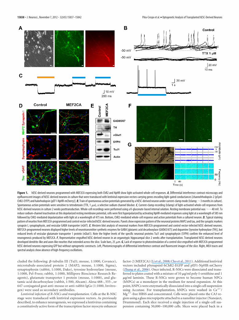
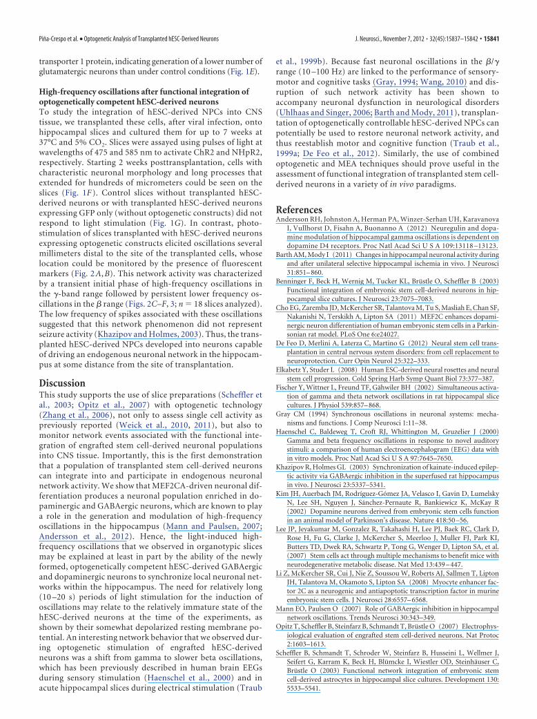
Figure 2. Optogenetic activation of engrafted hESC neurons elicits high-frequency oscillations in the � and � ranges (10 –100 Hz) in the hippocampus in vitro. A, Representative differentialinterference contrast image showing organotypic hippocampal slice containing engrafted hESC-derived neurons 17 d postseeding. The surface of the slice containing the cells was placed in directcontact with planar electrodes of an MEA recording chamber. B, Fluorescent image of the same slice showing engrafted hESC-derived neurons. The white square shows a region of the slice devoidof engrafted hESC-derived neurons from which extracellular activity was continuously recorded. Scale bar, 100 �m. C, Traces show characteristic extracellular local field potentials after stimulationwith blue light (wavelength, 475 nm) to stimulate ChR2. Neuronal network activity was recorded before, during, and after light stimulation. Extracellular local field potentials were recorded inartificial CSF at 32°C. D, Same trace as in C on an expanded time scale showing characteristic extracellular activity observed in the same region of the slice devoid of engrafted hESC-derived neurons.E, Fast Fourier transform analysis of the entire trace (300 s) reveals a main peak frequency at 18 Hz and a second peak at �33 Hz. F, Spectral frequency versus time for the same trace reveals stablehigh-frequency activity.
Pina-Crespo et al. • Optogenetic Analysis of Transplanted hESC-Derived Neurons J. Neurosci., November 7, 2012 • 32(45):15837–15842 • 15839
ResultsGeneration of optogenetically competenthESC-derived neuronsTo confer the ability for optogenetic stimulation to our trans-planted cells, neural progenitor cells (hESC-derived NPCs)were transduced with lentiviral expression vectors containingchannelrhodopsin (pSynI-ChR2-EYFP) as an excitatory ionchannel, and halorhodopsin (pEF1-NpHR-mCherry) as an in-hibitory transporter (Fig. 1A) (Zhang et al., 2006). When as-sayed electrophysiologically, hESC-derived neurons displayedresting membrane potentials of ��50 mV and spontaneousaction potentials that were sensitive to the selective sodiumchannel blocker tetrodotoxin (1 �M; Fig. 1B). Next, we re-corded from these hESC-derived neurons with a patch elec-trode in voltage-clamp mode to obtain evidence for theexpression of the optogenetic constructs by observing bothdepolarizing and hyperpolarizing light-activated responses
(Fig. 1C, n � 4 cells). The hESC-derived NPCs had differenti-ated to the neuronal phenotype as evidenced by electrophysi-ological criteria, including firing action potentials (Fig. 1B)and immunocytochemical evidence based on multiple mark-ers (Fig. 1D) (Li et al., 2008; Cho et al., 2011).
MEF2CA-driven neuronal differentiation (Li et al., 2008; Choet al., 2011) produced neurons with typical neuronal phenotype,as seen with immunocytochemical staining for the specific neu-ronal markers synapsin 1 and MAP2, synaptophysin, tau protein(Fig. 1D), and TuJ1 (Fig. 1E). When MEF2CA-programmed(compared with vector-infected control) hESC-derived neuronswere analyzed for the presence of neurotransmitter specificmarkers, they showed high levels of expression of the syntheticenzymes tyrosine hydroxylase and glutamic acid decarboxylase(Fig. 1E), indicating an abundance of dopaminergic and GABA-ergic neurons, respectively. At the same time, hESC-derivedneurons manifested relatively low levels of vesicular glutamate
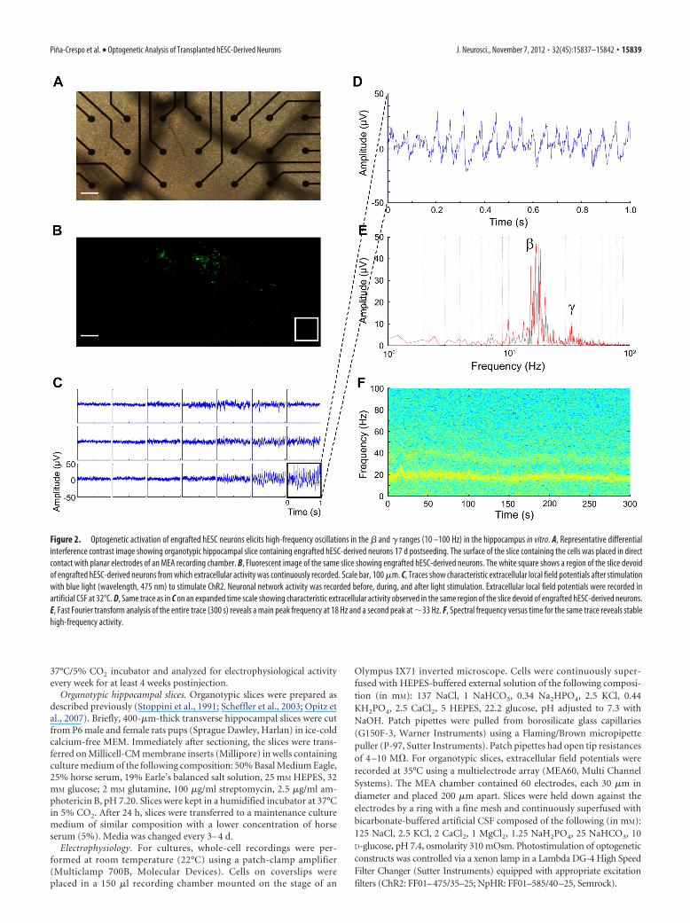
Figure 3. Power spectral analysis of extracellular field potentials generated during transient photostimulation of organotypic hippocampal slice transplanted with hESC-derived neurons. A,Analysis of 125 s epoch of extracellular activity recorded simultaneously in 16 different electrodes during transient photostimulation. Graphic representation of frequency versus time revealselectrical activity elicited by application of light with peak wavelength 585 nm (orange boxes) and 475 nm (blue boxes). As shown in Fig. 1C, a pattern of photostimulation consisting of orange light(585 nm) to elicit hyperpolarization via NpHR, followed by blue light (475 nm) to elicit depolarization via ChR2, consistently evoked neuronal activity. The large upward and downward deflectionsin the baseline are the stimulus artifacts at the beginning and the end of each light pulse. B, C, Magnified panels from A, show that photostimulation generated transient bursts of high-frequencyactivity that reached well into the gamma range (100 Hz). Once evoked, these oscillations outlasted the light stimulation and were unresponsive to subsequent illumination with hyperpolarizingorange light (585 nm).
15840 • J. Neurosci., November 7, 2012 • 32(45):15837–15842 Pina-Crespo et al. • Optogenetic Analysis of Transplanted hESC-Derived Neurons
transporter 1 protein, indicating generation of a lower number ofglutamatergic neurons than under control conditions (Fig. 1E).
High-frequency oscillations after functional integration ofoptogenetically competent hESC-derived neuronsTo study the integration of hESC-derived NPCs into CNStissue, we transplanted these cells, after viral infection, ontohippocampal slices and cultured them for up to 7 weeks at37°C and 5% CO2. Slices were assayed using pulses of light atwavelengths of 475 and 585 nm to activate ChR2 and NHpR2,respectively. Starting 2 weeks posttransplantation, cells withcharacteristic neuronal morphology and long processes thatextended for hundreds of micrometers could be seen on theslices (Fig. 1F ). Control slices without transplanted hESC-derived neurons or with transplanted hESC-derived neuronsexpressing GFP only (without optogenetic constructs) did notrespond to light stimulation (Fig. 1G). In contrast, photo-stimulation of slices transplanted with hESC-derived neuronsexpressing optogenetic constructs elicited oscillations severalmillimeters distal to the site of the transplanted cells, whoselocation could be monitored by the presence of fluorescentmarkers (Fig. 2 A, B). This network activity was characterizedby a transient initial phase of high-frequency oscillations inthe �-band range followed by persistent lower frequency os-cillations in the � range (Figs. 2C–F, 3; n � 18 slices analyzed).The low frequency of spikes associated with these oscillationssuggested that this network phenomenon did not representseizure activity (Khazipov and Holmes, 2003). Thus, the trans-planted hESC-derived NPCs developed into neurons capableof driving an endogenous neuronal network in the hippocam-pus at some distance from the site of transplantation.
DiscussionThis study supports the use of slice preparations (Scheffler etal., 2003; Opitz et al., 2007) with optogenetic technology(Zhang et al., 2006), not only to assess single cell activity aspreviously reported (Weick et al., 2010, 2011), but also tomonitor network events associated with the functional inte-gration of engrafted stem cell-derived neuronal populationsinto CNS tissue. Importantly, this is the first demonstrationthat a population of transplanted stem cell-derived neuronscan integrate into and participate in endogenous neuronalnetwork activity. We show that MEF2CA-driven neuronal dif-ferentiation produces a neuronal population enriched in do-paminergic and GABAergic neurons, which are known to playa role in the generation and modulation of high-frequencyoscillations in the hippocampus (Mann and Paulsen, 2007;Andersson et al., 2012). Hence, the light-induced high-frequency oscillations that we observed in organotypic slicesmay be explained at least in part by the ability of the newlyformed, optogenetically competent hESC-derived GABAergicand dopaminergic neurons to synchronize local neuronal net-works within the hippocampus. The need for relatively long(10 –20 s) periods of light stimulation for the induction ofoscillations may relate to the relatively immature state of thehESC-derived neurons at the time of the experiments, asshown by their somewhat depolarized resting membrane po-tential. An interesting network behavior that we observed dur-ing optogenetic stimulation of engrafted hESC-derivedneurons was a shift from gamma to slower beta oscillations,which has been previously described in human brain EEGsduring sensory stimulation (Haenschel et al., 2000) and inacute hippocampal slices during electrical stimulation (Traub
et al., 1999b). Because fast neuronal oscillations in the �/�range (10 –100 Hz) are linked to the performance of sensory-motor and cognitive tasks (Gray, 1994; Wang, 2010) and dis-ruption of such network activity has been shown toaccompany neuronal dysfunction in neurological disorders(Uhlhaas and Singer, 2006; Barth and Mody, 2011), transplan-tation of optogenetically controllable hESC-derived NPCs canpotentially be used to restore neuronal network activity, andthus reestablish motor and cognitive function (Traub et al.,1999a; De Feo et al., 2012). Similarly, the use of combinedoptogenetic and MEA techniques should prove useful in theassessment of functional integration of transplanted stem cell-derived neurons in a variety of in vivo paradigms.
ReferencesAndersson RH, Johnston A, Herman PA, Winzer-Serhan UH, Karavanova
I, Vullhorst D, Fisahn A, Buonanno A (2012) Neuregulin and dopa-mine modulation of hippocampal gamma oscillations is dependent ondopamine D4 receptors. Proc Natl Acad Sci U S A 109:13118 –13123.
Barth AM, Mody I (2011) Changes in hippocampal neuronal activity duringand after unilateral selective hippocampal ischemia in vivo. J Neurosci31:851– 860.
Benninger F, Beck H, Wernig M, Tucker KL, Brustle O, Scheffler B (2003)Functional integration of embryonic stem cell-derived neurons in hip-pocampal slice cultures. J Neurosci 23:7075–7083.
Cho EG, Zaremba JD, McKercher SR, Talantova M, Tu S, Masliah E, Chan SF,Nakanishi N, Terskikh A, Lipton SA (2011) MEF2C enhances dopami-nergic neuron differentiation of human embryonic stem cells in a Parkin-sonian rat model. PLoS One 6:e24027.
De Feo D, Merlini A, Laterza C, Martino G (2012) Neural stem cell trans-plantation in central nervous system disorders: from cell replacement toneuroprotection. Curr Opin Neurol 25:322–333.
Elkabetz Y, Studer L (2008) Human ESC-derived neural rosettes and neuralstem cell progression. Cold Spring Harb Symp Quant Biol 73:377–387.
Fischer Y, Wittner L, Freund TF, Gahwiler BH (2002) Simultaneous activa-tion of gamma and theta network oscillations in rat hippocampal slicecultures. J Physiol 539:857– 868.
Gray CM (1994) Synchronous oscillations in neuronal systems: mecha-nisms and functions. J Comp Neurosci 1:11–38.
Haenschel C, Baldeweg T, Croft RJ, Whittington M, Gruzelier J (2000)Gamma and beta frequency oscillations in response to novel auditorystimuli: a comparison of human electroencephalogram (EEG) data within vitro models. Proc Natl Acad Sci U S A 97:7645–7650.
Khazipov R, Holmes GL (2003) Synchronization of kainate-induced epilep-tic activity via GABAergic inhibition in the superfused rat hippocampusin vivo. J Neurosci 23:5337–5341.
Kim JH, Auerbach JM, Rodríguez-Gomez JA, Velasco I, Gavin D, LumelskyN, Lee SH, Nguyen J, Sanchez-Pernaute R, Bankiewicz K, McKay R(2002) Dopamine neurons derived from embryonic stem cells functionin an animal model of Parkinson’s disease. Nature 418:50 –56.
Lee JP, Jeyakumar M, Gonzalez R, Takahashi H, Lee PJ, Baek RC, Clark D,Rose H, Fu G, Clarke J, McKercher S, Meerloo J, Muller FJ, Park KI,Butters TD, Dwek RA, Schwartz P, Tong G, Wenger D, Lipton SA, et al.(2007) Stem cells act through multiple mechanisms to benefit mice withneurodegenerative metabolic disease. Nat Med 13:439 – 447.
Li Z, McKercher SR, Cui J, Nie Z, Soussou W, Roberts AJ, Sallmen T, LiptonJH, Talantova M, Okamoto S, Lipton SA (2008) Myocyte enhancer fac-tor 2C as a neurogenic and antiapoptotic transcription factor in murineembryonic stem cells. J Neurosci 28:6557– 6568.
Mann EO, Paulsen O (2007) Role of GABAergic inhibition in hippocampalnetwork oscillations. Trends Neurosci 30:343–349.
Opitz T, Scheffler B, Steinfarz B, Schmandt T, Brustle O (2007) Electrophys-iological evaluation of engrafted stem cell-derived neurons. Nat Protoc2:1603–1613.
Scheffler B, Schmandt T, Schroder W, Steinfarz B, Husseini L, Wellmer J,Seifert G, Karram K, Beck H, Blumcke I, Wiestler OD, Steinhauser C,Brustle O (2003) Functional network integration of embryonic stemcell-derived astrocytes in hippocampal slice cultures. Development 130:5533–5541.
Pina-Crespo et al. • Optogenetic Analysis of Transplanted hESC-Derived Neurons J. Neurosci., November 7, 2012 • 32(45):15837–15842 • 15841

Stoppini L, Buchs PA, Muller D (1991) A simple method for organotypiccultures of nervous tissue. J Neurosci Methods 37:173–182.
Taketani M, Baudry M (2006) Advances in network electrophysiology: us-ing multi-electrode arrays. New York: Springer.
Traub RD, Jefferys JGR, Whittington MA (1999a) Fast oscillations in corti-cal circuits. Cambridge: MIT.
Traub RD, Whittington MA, Buhl EH, Jefferys JG, Faulkner HJ (1999b) Onthe mechanism of the gamma3 beta frequency shift in neuronal oscilla-tions induced in rat hippocampal slices by tetanic stimulation. J Neurosci19:1088 –1105.
Uhlhaas PJ, Singer W (2006) Neural synchrony in brain disorders: rele-vance for cognitive dysfunctions and pathophysiology. Neuron52:155–168.
Wang XJ (2010) Neurophysiological and computational principles of corti-cal rhythms in cognition. Physiol Rev 90:1195–1268.
Weick JP, Johnson MA, Skroch SP, Williams JC, Deisseroth K, Zhang SC(2010) Functional control of transplantable human ESC-derived neu-rons via optogenetic targeting. Stem Cells 28:2008 –2016.
Weick JP, Liu Y, Zhang SC (2011) Human embryonic stem cell-derivedneurons adopt and regulate the activity of an established neural network.Proc Natl Acad Sci U S A 108:20189 –20194.
Wernig M, Benninger F, Schmandt T, Rade M, Tucker KL, Bussow H, Beck H,Brustle O (2004) Functional integration of embryonic stem cell-derivedneurons in vivo. J Neurosci 24:5258 –5268.
Zhang F, Wang LP, Boyden ES, Deisseroth K (2006) Channelrhodopsin-2and optical control of excitable cells. Nat Methods 3:785–792.
15842 • J. Neurosci., November 7, 2012 • 32(45):15837–15842 Pina-Crespo et al. • Optogenetic Analysis of Transplanted hESC-Derived Neurons